polycapillary optics and x-ray analytical techniques - ICDD
polycapillary optics and x-ray analytical techniques - ICDD
polycapillary optics and x-ray analytical techniques - ICDD

Create successful ePaper yourself
Turn your PDF publications into a flip-book with our unique Google optimized e-Paper software.
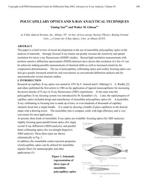
Copyright (c)JCPDS-International Centre for Diffraction Data 2002, Advances in X-<strong>ray</strong> Analysis, Volume 45. 298<br />
POLYCAPILLARY OPTICS AND X-RAY ANALYTICAL TECHNIQUES<br />
Yiming Yan a,b <strong>and</strong> Walter M. Gibson a,c<br />
a) X-Ray Optical Systems, Inc. Albany, NY , b) Inst. of Low energy Nuclear Physics, Beijing Normal<br />
Univ., c) Center for X-Ray Optics, Univ. at Albany SUNY,<br />
ABSTRACT<br />
This paper is a brief review of recent developments in the use of monolithic <strong>polycapillary</strong> <strong>optics</strong> in the<br />
analysis of materials. Strongly focused X-<strong>ray</strong> beams can greatly increase the sensitivity <strong>and</strong> spatial<br />
resolution for micro x-<strong>ray</strong> fluorescence (MXRF) studies. Recent high-resolution measurements with<br />
position sensitive diffraction spectrometer (PSDS) detectors have shown that resolution of a few eV can<br />
be achieved, making possible measurements of chemical shifts as well as increased sensitivity for<br />
composition determinations. The use of <strong>polycapillary</strong> collimating <strong>optics</strong> <strong>and</strong> weakly focusing <strong>optics</strong> can<br />
also give greatly increased sensitivity <strong>and</strong> convenience in conventional diffraction analysis <strong>and</strong> for<br />
macromolecular crystal structure studies.<br />
I. INTRODUCTION<br />
Research on capillary X-<strong>ray</strong> <strong>optics</strong> was started in 1931 by F. Jentzch <strong>and</strong> E. Nähring (1). A. Rindby [2]<br />
<strong>and</strong> others published the first article in 1986 on the application of tapered monocapillaries for increasing<br />
the power density of X-<strong>ray</strong>s in X-<strong>ray</strong> fluorescence (XRF) experiments. At the same time the<br />
<strong>polycapillary</strong> X-<strong>ray</strong> focusing system was introduced by M. Kumakhov (3). Later, the rapid progress of<br />
capillary <strong>optics</strong> included design <strong>and</strong> manufacture of monolithic <strong>polycapillary</strong> <strong>optics</strong> (4). A monolithic<br />
X-<strong>ray</strong> collimating or focusing lens is made up of tens, or even hundreds of thous<strong>and</strong>s of capillary<br />
channels fused into a single bundle. It is made by drawing a bundle of glass capillaries to the desired<br />
shape with a drawing tower. The monolithic lens is compact, solid, with high efficiency <strong>and</strong> is very<br />
convenient for most applications.<br />
At present, three kinds of monolithic X-<strong>ray</strong> <strong>optics</strong> are available: focusing <strong>optics</strong> (for XRF analysis);<br />
slightly focusing quasi-parallel beam <strong>optics</strong> (for single<br />
crystal X-<strong>ray</strong> diffraction (XRD) analysis); <strong>and</strong> parallel<br />
beam collimating <strong>optics</strong> (for wavelength dispersive<br />
XRD analysis). These three types are shown<br />
schematically in Fig. 1.<br />
In addition, the remarkable scatter-rejection properties<br />
of <strong>polycapillary</strong> <strong>optics</strong> can be utilized for monolithic<br />
angular filters for mammography <strong>and</strong> other<br />
applications (5).<br />
Figure 1. Schematic<br />
representation of<br />
three types of<br />
monolithic<br />
<strong>polycapillary</strong> <strong>optics</strong>
Copyright (c)JCPDS-International Centre for Diffraction Data 2002, Advances in X-<strong>ray</strong> Analysis, Volume 45. 299<br />
In the present paper, we give a short review of applications of <strong>polycapillary</strong> monolithic <strong>optics</strong> for X-<strong>ray</strong><br />
analysis, <strong>and</strong> discuss their advantages, limits, <strong>and</strong> expected further development. There have been a<br />
number of articles concerning the study <strong>and</strong> application of monolithic X-<strong>ray</strong> <strong>optics</strong> including some review<br />
papers (6-9).<br />
II. MICROBEAM X-RAY FLUORESCENCE (MXRF)<br />
For XRF analysis, monolithic <strong>polycapillary</strong> <strong>optics</strong> capture <strong>and</strong> focus X <strong>ray</strong>s from small-spot divergent<br />
X-<strong>ray</strong> sources or capture <strong>and</strong> focus parallel X-<strong>ray</strong> beams from synchrotron radiation sources. The main<br />
advantage of X-<strong>ray</strong> lenses for microanalysis is that they can collect <strong>and</strong> focus X-<strong>ray</strong>s, forming a very<br />
small beam spot (down to 10-15 µm (10), greatly increasing the power density of the X-<strong>ray</strong> beam<br />
(sometimes more than 1000 X). Further reduction of the beam spot size is difficult technologically,<br />
since a smaller spot requires a very short output focal distance, inconvenient for X-<strong>ray</strong> detection.<br />
The main characteristics of a focusing lens are: transmission efficiency, gain, spot size, <strong>and</strong> output focal<br />
distance (8). These parameters are dependent on the energy of the X-<strong>ray</strong> photons (8,11-12). Actually the<br />
efficiency of propagation of X-<strong>ray</strong>s in capillaries by external multiple reflection is dependent on the<br />
refractive index <strong>and</strong> absorption coefficient of the capillary material <strong>and</strong> on the specific geometry of the<br />
optic such as the capillary channel diameter <strong>and</strong> curvature. As an example, in Fig.2 is shown the<br />
dependence of the transmission <strong>and</strong> focal spot size of lens F1 on the X-<strong>ray</strong> photon energy (12). Both<br />
experimental measurements <strong>and</strong> theoretical simulations are shown. The lens parameters are: length, 50<br />
mm; entrance diameter, 5.38 mm; exit diameter, 4.36 mm; maximum diameter, 6.8 mm; input focal<br />
distance, 67 mm; output focal distance, 13.8 mm. For Mo Kα (17.4 keV) X-<strong>ray</strong>s, the measured values<br />
are: focal spot size, 28 µm; transmission, 1.04%; gain of power density, 1629.<br />
(a)<br />
(b)<br />
Figure 2. a) Transmission efficiency of lens F1 as a function of X-<strong>ray</strong> energy, <strong>and</strong><br />
b) focal spot size of lens F1 as function of X-<strong>ray</strong> energy<br />
It can be seen that for low energy photons, the transmission efficiency is higher. The high-energy limit of<br />
the transmission b<strong>and</strong>width of the lens is determined by the critical angle for total external reflection of<br />
X-<strong>ray</strong>s from the wall of the capillary. The lower energy limit is defined by absorption of X-<strong>ray</strong>s in the<br />
window of the X-<strong>ray</strong> tube, air <strong>and</strong> the capillary wall material. In practice, it is best to make a special<br />
design <strong>and</strong> manufacture in accordance with the particular application.<br />
Monolithic <strong>polycapillary</strong> focusing <strong>optics</strong> have found wide application in MXRF analysis in pure <strong>and</strong><br />
applied scientific research <strong>and</strong> industry since X-<strong>ray</strong> spectrometers based on these <strong>optics</strong> have high<br />
sensitivity (with detection limits down to subpicogram) <strong>and</strong> high spatial resolution (100-10 µm, even 1
Copyright (c)JCPDS-International Centre for Diffraction Data 2002, Advances in X-<strong>ray</strong> Analysis, Volume 45. 300<br />
µm if an aperture is added) At the beginning of this new century the analysis of micrmaterials <strong>and</strong><br />
detailed analysis of extremely small samples by microbeam X-<strong>ray</strong> fluorescence is increasingly important.<br />
"There is an increasing awareness that in order to underst<strong>and</strong> the working of a macrosystem, we require<br />
information of microscopical details since most natural <strong>and</strong> technological systems are intrinsically<br />
heterogeneous” (13). As indicated above, we here define MXRF analysis to occupy the dimensions of<br />
tens of microns to 1 micron. These are the sizes, for example, of cells, biological macromolecules, aerosol<br />
particles, microcrystals, magmatic inclusions within minerals, etc. Therefore the focusing lens can be<br />
applied in physics, chemistry, biology, medicine, material science, life science, earth science,<br />
environmental science, semiconductor industry, microelectronics <strong>and</strong> other industries, even in forensic<br />
medicine <strong>and</strong> archaeology.<br />
Recent developments can be found in (14-20). It is of interest to point out that in (20) two focusing lenses<br />
were applied to improve the detection sensitivity of MXRF analysis of radioactive materials. The first was<br />
used to increase the power density of the X-<strong>ray</strong> beam incident on the radioactive sample, <strong>and</strong> the second<br />
was placed between the sample <strong>and</strong> the detector to increase the flux of fluorescent X <strong>ray</strong>s on the detector<br />
while at the same time sharply discriminating against background radiation from the radioactive sample.<br />
This method could have many uses in nuclear engineering <strong>and</strong> materials analysis. Another success in (20)<br />
was the use of a position sensitive spectrometer that simultaneously gives high spatial <strong>and</strong> energy<br />
resolution <strong>and</strong> high sensitivity. A schematic representation of such a system is shown in Fig. 3. Fig. 4<br />
<strong>and</strong> Fig. 5 show X-<strong>ray</strong> fluorescence spectra measured with this system. The energy resolution achieved<br />
depends on the X-<strong>ray</strong> wavelength as shown in Fig. 6. This is comparable to the energy resolution<br />
obtained with scanned wavelength dispersive X-<strong>ray</strong> fluorescence (WDXRF) systems or microcalorimeter<br />
detector based XRF systems, <strong>and</strong> can be used for chemical as well as compositional analysis.<br />
T<br />
Figure 3. PSDS setup using monolithic optic<br />
Figure 4. Comparison of PSDS <strong>and</strong> Si(Li) spectra<br />
In (21) a focusing lens was used to increase by 300 X the sensitivity of a superconducting X-<strong>ray</strong> detector.<br />
Such a third generation energy dispersive detector has very high energy resolution (25 mm 2 , comparable to a high-resolution semiconductor detector. This application
Copyright (c)JCPDS-International Centre for Diffraction Data 2002, Advances in X-<strong>ray</strong> Analysis, Volume 45. 301<br />
takes advantage of the broad energy b<strong>and</strong>width of <strong>polycapillary</strong> <strong>optics</strong>.<br />
In environmental studies, a strong focusing lens can be used for single particle XRF analysis of aerosol<br />
samples. In such a measurement, one can use an aperture (1-5 µm diameter) to limit the beam size<br />
sufficiently to measure a particle separated from other particles. In this case it may be useful to employ<br />
a high power rotating anode X-<strong>ray</strong> generator. On the other h<strong>and</strong>, single particles can be isolated on a<br />
thin organic substrate film <strong>and</strong> examined with the 20-30 µm focused beam directly from<br />
Figure 5. PSDS Ti XRF spectrum<br />
Figure 6. PSDS resolution vs. X-<strong>ray</strong> energy<br />
focused the secondary X-<strong>ray</strong>s onto a ~300 µm diameter detector.<br />
In this way the effective<br />
Figure 5. PSDS Ti XRF spectrum<br />
Figure 6. PSDS resolution vs. X-<strong>ray</strong> energy<br />
the optic. In many cases, it is sufficient to examine a collection of particles with the focused beam <strong>and</strong> a<br />
compact low power x-<strong>ray</strong> source can be used.<br />
III. X-RAY DIFFRACTION ANALYSIS<br />
Parallel beam X-<strong>ray</strong> <strong>optics</strong> have been extensively used in diffraction studies (8, 9, 22-32). Parallel beam<br />
X-<strong>ray</strong> diffraction is the most important method used for material structure analysis. The parallel beam of<br />
previous diffraction instruments is obtained with an aperture with a large loss of X-<strong>ray</strong> intensity. The use<br />
of a collimating lens gives tens or even hundreds of times higher intensity than an aperture, since it can<br />
collect X-<strong>ray</strong>s over an angle of several degrees emitted from a divergent X-<strong>ray</strong> source <strong>and</strong> convert them<br />
to a quasiparallel beam with 0.1-0.2 degrees divergence (depending on the energy) (20).<br />
There are two components in the angular divergence from a monolithic <strong>polycapillary</strong> optic: the global<br />
divergence (or total divergence of the beam envelope), which defines the angular resolution of a<br />
diffractometer <strong>and</strong> the local divergence discussed above which influences the quality of local diffraction<br />
patterns. Generally speaking, the angular resolution of the central part of a collimating lens is better than<br />
that of the periphery. In principle, the local divergence can be made small if a very small X-<strong>ray</strong> source<br />
spot is used, if the channel size at the lens input is small <strong>and</strong> well aligned, <strong>and</strong> if the curvature of the<br />
channels is very smooth <strong>and</strong> free from slope error (also called waviness). At the present time the best<br />
angular resolution is about 0.10 O for Cu Kα radiation which is worse than the resolution from a<br />
conventional diffractometer. But the parallel beam lens can give tens of times stronger X-<strong>ray</strong> beam<br />
intensity, thereby greatly increasing the efficiency for diffraction measurements.<br />
Since the parallel beam lens redirects divergent X-<strong>ray</strong>s into parallel channels, it always increases the
Copyright (c)JCPDS-International Centre for Diffraction Data 2002, Advances in X-<strong>ray</strong> Analysis, Volume 45. 302<br />
small-angle portion of emitted X-<strong>ray</strong>s. So it can bring large benefit to diffractometers with very fine<br />
angular resolution such as double-crystal or four-crystal diffractometers. In this case one can put the lens<br />
between the X-<strong>ray</strong> source <strong>and</strong> the monochromator to increase the primary X-<strong>ray</strong> beam intensity.<br />
It is worth pointing out that since the lens forms many superfine quasi-parallel X-<strong>ray</strong> beams, in diffraction<br />
experiments, it does not have the focusing circle effect of conventional goniometers. So in the case of<br />
parallel-beam geometry the lens tolerates a greater positional error <strong>and</strong> makes adjustment of the<br />
instrument much easier in comparison with the st<strong>and</strong>ard Bragg-Brentano geometry. Moreover there are<br />
almost no line position changes due to sample displacements, sample roughness or shape or sample<br />
transparency in such a parallel-beam geometry compared to the usual parafocusing or Bragg-Brentano<br />
geometry<br />
.<br />
Fig.7. X-<strong>ray</strong> intensity gain of a parallel beam<br />
Figure 8. Texture pole figures. In the top figure<br />
Fig. 7. X-<strong>ray</strong> intensity gain of a parallel beam Fig. 8. Texture pole figures. In the top figure<br />
X-<strong>ray</strong> lens. Setting: Cu tube with point focus the statistics is improved by 40 x. In the<br />
X-<strong>ray</strong> generator, 12 μm Ni filter, 40 kV, 40 mA, bottom figure, the measurement time is<br />
5 mm aperture at lens exit. reduced from three days to one hour.<br />
An important step in development of use of the <strong>polycapillary</strong> <strong>optics</strong> in diffraction analysis is the design<br />
<strong>and</strong> performance of high-flux X-<strong>ray</strong> crystallography systems, optimized for diffraction measurements<br />
from small macromolecular crystals [25-30]. Earlier applications of <strong>polycapillary</strong> <strong>optics</strong> were basically<br />
related to improvement <strong>and</strong> reconstruction of available diffractometers. But M.Gubarev <strong>and</strong> others<br />
combined a small <strong>polycapillary</strong> collimating optic with only 0.91 mm input diameter, 1.8 mm output<br />
diameter <strong>and</strong> 10.14 mm length <strong>and</strong> high transmission efficiency of 41% (for the Cu Kα line) <strong>and</strong> a<br />
microfocus X-<strong>ray</strong> generator (40 W, 40 µm anode spot size) They obtained an X-<strong>ray</strong> flux of<br />
quasi-parallel beam through an aperture of 250 µm diameter which was 16 times higher than that from a<br />
3.15 kW rotating-anode generator equipped with a pyrolytic graphite monochromator [25]. In this case,<br />
the input focal distance of the <strong>polycapillary</strong> collimating optic was only 2.6 mm which required a specially<br />
designed microfocus X-<strong>ray</strong> source which could allow the lens to be very close to the X-<strong>ray</strong> source spot.<br />
Subsequently, even higher (7x) x-<strong>ray</strong> intensity was obtained <strong>and</strong> the x-<strong>ray</strong> intensity <strong>and</strong> the quality of the<br />
diffraction patterns from a protein crystal were comparable with that obtained with 5 kW rotating anode<br />
sources equipped with optimally aligned concentric multilayer focusing mirrors (26). This is quite a
Copyright (c)JCPDS-International Centre for Diffraction Data 2002, Advances in X-<strong>ray</strong> Analysis, Volume 45. 303<br />
new type of diffractometer, which takes full advantage of the: high collection efficiency of <strong>polycapillary</strong><br />
<strong>optics</strong>, the small size of the X-<strong>ray</strong> beam, <strong>and</strong> the low power, size <strong>and</strong> flexibility of the X-<strong>ray</strong> source.<br />
Furthermore, a slightly focusing monolithic optic can form X-<strong>ray</strong> beams less than 0.5 mm in diameter<br />
increasing the power density of the beam by another order of magnitude with sufficient parallelism that<br />
one can use st<strong>and</strong>ard crystallographic software, Such systems should find a wide application in X-<strong>ray</strong><br />
microanalysis. It is generally accepted that Synchrotron X-<strong>ray</strong> sources are the best choice for final, high<br />
resolution protein structure studies. However, for routine screening of protein <strong>and</strong> other macromolecular<br />
crystals the low power, inexpensive, “table top” <strong>polycapillary</strong> optic based system can play an important<br />
role.<br />
It has also been recently shown that even more strongly focused beam diffraction can be used with even<br />
higher intensity <strong>and</strong> smaller beam spot size. This is analogous to the well known convergent beam<br />
electron diffraction. For such convergent beams, the diffraction spots are elongated tangentially about<br />
the center of the beam direction (31). For such strongly focused beams the volume of reciprocal space,<br />
which is accessed in a single measurement increases compared to parallel beam diffraction <strong>and</strong> the<br />
diffraction spots become streaks. The effects of the one-dimensional streaking for a single-crystal<br />
protein diffraction patterns, are in good agreement with theoretical simulation (29) <strong>and</strong> do not result in<br />
untenable overlap for most crystal orientations. Software for analysis of highly convergent beam<br />
diffraction has been developed successfully (32).<br />
4.CONCLUSIONS<br />
Polycapillary X-<strong>ray</strong> <strong>optics</strong> or so called Kumakhov X-<strong>ray</strong> lenses were an important breakthrough in<br />
X-<strong>ray</strong> <strong>optics</strong> in the 1990s, which have made possible the modulation <strong>and</strong> control of wide b<strong>and</strong>width X-<strong>ray</strong><br />
beams <strong>and</strong> have enabled the efficient use of X-<strong>ray</strong> sources. They have found wide application in X-<strong>ray</strong><br />
<strong>analytical</strong> <strong>techniques</strong>, improved the performance of X-<strong>ray</strong> <strong>analytical</strong> instruments, promoted the<br />
innovation of some new X-<strong>ray</strong> instruments <strong>and</strong> opened up new fields of application.<br />
REFERENCES<br />
1, F. Jentzch <strong>and</strong> E. Näring, “Die Fortleitung von Licht—und Röntgenstrahlen durch Röhren,” Zeitschr. F. Techn<br />
Phys. 12, 185 (1931).<br />
2. A.Rindby, “Applications of fiber technique in the X-<strong>ray</strong> region”, Nucl. Instr. & Methods. in Phys. Research, A249,<br />
536-540 (1986).<br />
3. Kumakhov M.A. <strong>and</strong> Komarov F.F., “Multiple reflection from surface X-<strong>ray</strong> <strong>optics</strong>”, Phys. Rep., 191, No5, 289<br />
(1990).<br />
4. Walter M. Gibson <strong>and</strong> Muradin A. Kumakhov, “Application of X-<strong>ray</strong> <strong>and</strong> neutron capillary <strong>optics</strong>”, Proc. SPIE,<br />
vol.1736, 172-189 (1992).<br />
5. W.M. Gibson, H. Huang, J. Nicolich, P. Klein, <strong>and</strong> C.A. MacDonald, “Optics for Angular Filtering of X-Rays in<br />
Two Dimensions,” Proc. of Denver 2001 X-Ray Conf. (these proceedings) (2002).<br />
6. Yan Yiming <strong>and</strong> Liu Andong, “Capillary X-<strong>ray</strong> <strong>optics</strong> <strong>and</strong> X-<strong>ray</strong> focusing systems”, J. of Beijing Normal<br />
University (Natural Science) sup.31,1-14 (1995) (in Chinese)<br />
7. W.M. Gibson <strong>and</strong> C.A. MacDonald, “Polycapillary Kumakhov Optics: A Status Report”, Proc. SPIE, vol. 2278,<br />
156-67 (1994).
Copyright (c)JCPDS-International Centre for Diffraction Data 2002, Advances in X-<strong>ray</strong> Analysis, Volume 45. 304<br />
8.Yiming Yan, “The monolithic capillary X-<strong>ray</strong> lens, its basic physical characteristics <strong>and</strong> applications”, Advances in<br />
X-<strong>ray</strong> Analysis, vol. 40, Electron version ICCD 1997,1998,ISSN 1097-0002<br />
9. Carolyn A. MacDonald <strong>and</strong> Walter M. Gibson, “Polycapillary <strong>and</strong> Multichannel Plate X-Ray Optics”, Chapter 30,<br />
in H<strong>and</strong>book of Optics, Vol. III, ed, M. Bass (McGraw Hill, New York, 2000),<br />
10. N. Gao, (Private communication).<br />
11. Chen Baozhen <strong>and</strong> Yan Yiming, “Transmission Efficiency of X-<strong>ray</strong>s Through a Cylinder Capillary”, Chinese<br />
Physics Letters .16, No9, 630-631 (1999)<br />
12.Jindong Xie, Yiming Yan, Xunliang Ding, Yejun He, <strong>and</strong> Qiuli Pan, “Study of the Monolithic X-<strong>ray</strong> Focusing<br />
Lens <strong>and</strong> its Application in Microbeam X-<strong>ray</strong> Fluorescence Analysis”, X-Ray Spectrometry, 29,:305-309 (2000)<br />
13. Summary of conclusions, Report of the advisory group meeting on elemental analysis of extremely small<br />
samples, 70, Institute Jozef Stefan, Ljubljana, Slovenia, 2 - 5 July, 1996, IAEA/AL/108<br />
14. N.Gao, I.Ponomarev, Q.F.Xiao, W.M.Gibson, <strong>and</strong> D.A.Carpenter, “Application of Monolithic Focusing Optics in<br />
MXRF”, Proc. SPIE, vol. 2859, 140-148 (1996)<br />
15 .He Yejun, Xie Jingdong, Ding Xunliang, Wei Fuzhong <strong>and</strong> Yan Yiming, “Monolithic Capillary Lens <strong>and</strong> XRF”,<br />
Proc. of the European Conf. on Energy Dispersive X-<strong>ray</strong> Spectrometry (EDXRS-98), 121-124 (1998)<br />
16. X.Ding, N.Gao, G.Havrilla, “Monolithic Polycapillary X-<strong>ray</strong> Optics Engineered to meet a Wide Range of<br />
Applications”, Proc. 49 th Annual Denver X-<strong>ray</strong> Conf. 2000<br />
17. Xie Jingdong, He Yejun, Ding Xunliang, Pan Qiuli <strong>and</strong> Yan Yiming, “The Monolithic X-<strong>ray</strong> Polycapillary Lens<br />
<strong>and</strong> Application to Microbeam X-<strong>ray</strong> Fluorescence”, J. Anal. Atomic Spectrometry, 14, 391-394 (1999)<br />
18. K.Janssens, L.Vinsze, F.Wei, K.Proost, B.Vekemans, G.Vittiglio, Y.He, Y.Yan, “Feasibility of (Trace Level)<br />
Micro-XANES at Beamline L”, Annual Report, Experimental Development, Humburger Sychrotronstrahlungs-labor<br />
HASLAB am Deutschen Electronen-Sychrotron DESY,Jahresbericht, 1999<br />
19.. L.Vincze, K.Janssens, F.Wei, K.Proost, B.Vekemans, G.Vittiglio, Y.He, Y.Yan, G.Falkenburg, “Trace-Level<br />
Micro-XANES by Means of Bending Magnet Radiation Focused with a Polycapillary Lens”, Ibid, 1999<br />
20. Yan Yiming, He Yejun, Ding Xunliang, Chen Jun, Li Yude, Wei Fuzhong, Xie Jingdong, Pan Qiuli <strong>and</strong> Wang<br />
Dachun, “New Achievments in X-<strong>ray</strong> Optics - the X-<strong>ray</strong> Lens <strong>and</strong> its Applications”, Progress in Natural Science,.11<br />
(No2), 87-93 (2001)<br />
21. D.A.Wollman, C. Jezewski, G.C.Hilton, Qi-Fan Xiao, K.D.Irwin, L.L.Dilcie <strong>and</strong> J.M.Martinis, “Use of<br />
Polycapillary Optics to Increase the Effective Area of Microcalorimeter Spectromerters”, Proc. Microscopy <strong>and</strong><br />
Microanalysis, (Springer, New York, 1997) pp 1075-78<br />
22. J.B. Ullrich, K.G. Huang, S.M. Owens, D.C. Aloise, F.A. Hofmann, N. Gao, I.L. Klotzko, <strong>and</strong> W.M. Gibson,<br />
“Concentration of Synchrotron Beams by Means of Monolithic Polycapillary X-Ray Optics”, Nucl. Instr. <strong>and</strong><br />
Methods, A364 362-69 (1995).<br />
23. Xuunliang Ding, (private communication) to be published<br />
24. R.A.Clapp <strong>and</strong> M.Haller, “Parallel Beam Methods in Powder Diffraction <strong>and</strong> Texture in the Laboratory”, Proc.<br />
48 th Ann. Denver X-<strong>ray</strong> Conf, Advances in X-Ray Analysis, 43, (2000)<br />
25. M.Gubarev, E.Ciszak, I.Ponomarev, W.Gibson <strong>and</strong> M.Joy, “First Results from a Macromolecular<br />
Crystallography System with a Polycapillary Collmating Optic <strong>and</strong> a Microfocus X-<strong>ray</strong> Generator”, J. Appl. Cryst.<br />
33, 882-887 (2000)<br />
26. M. Gubarev, E. Ciszak, I. Ponomarev, W.M. Giibson, <strong>and</strong> M. Joy, “A Compact X-<strong>ray</strong> System for
Copyright (c)JCPDS-International Centre for Diffraction Data 2002, Advances in X-<strong>ray</strong> Analysis, Volume 45. 305<br />
Macromolecular Crystallography”, Rev. Sci. Instr., 71, 3900-05 (2000)<br />
27. S.M.Owens, J.B.Ullrich, I.Yu.Ponomarev, D.C.Carter, R.C.Sisk, J.X.Ho,<strong>and</strong> W.M.Gibson, “Polycapillary X-<strong>ray</strong><br />
Optics for Macromolecular Prystallography”, Proc. SPIE, vol..2859, 200-209 (1996)<br />
28. J.B.Ullrich, S.M.Owens, Q-F. Xiao, I.Yu.Ponomarev, D.C.Carter, J.X.Ho, R.C.Sisk, E.H.Snell, W.M.Gibson,<br />
“Convergent Beam Macromolecular Crystallography”, 1996<br />
29.J.X.Ho, E.H.Snell, R.C.Sisk, J.R.Ruble, D.C.Carter, S.M.Owens <strong>and</strong> W.M.Gibson, “Stationary Crystal<br />
Diffraction with a Monochromatic Convergent X-<strong>ray</strong> Source <strong>and</strong> Application for Macromolecular Crystal Data<br />
Collection”, Acta Cryst. D54, 2000-214 (1998)<br />
30. Y.He, J.Chen, Y.Li, F.Wei, D.Wang, P.Lo <strong>and</strong> Y.Yan, “X-<strong>ray</strong> Lens of Monolithic Polycapillaries for<br />
Macromolecular Crystallography”, J. X-<strong>ray</strong> Science <strong>and</strong> Technology, 8, 145-149 (1998}<br />
31. C.A.MacDonald, S.M.Owens <strong>and</strong> W.M.Gibson, “Polycapllary X-<strong>ray</strong> Optics for Microdiffraction”, J. Appl. Cryst.<br />
32, 160-167 (1999)<br />
32. H, Huang , C.A. MacDonald, W.M. Gibson, J.R. Ruble, J.X. Ho, J. Chik, A. Parsegian, <strong>and</strong> I. Ponomarev,<br />
“Focusing Polycapillary Optics for Diffraction”, Proc. 50 th Ann. Denver X-Ray Conf. (these proceedings) (2001)