
You also want an ePaper? Increase the reach of your titles
YUMPU automatically turns print PDFs into web optimized ePapers that Google loves.
<strong>Cone</strong> <strong>Beam</strong> <strong>3D</strong> <strong>Imaging</strong>
NewTom Sets the Standard<br />
in <strong>3D</strong> Maxillofacial <strong>Imaging</strong>
<strong>Cone</strong> <strong>Beam</strong> <strong>3D</strong> <strong>Imaging</strong>
The Global Market Leader<br />
The Inventors of <strong>Cone</strong> <strong>Beam</strong> <strong>3D</strong><br />
In 1996, QR srl developed the first<br />
generation of dentomaxillofacial<br />
<strong>Cone</strong> <strong>Beam</strong> systems.<br />
This invention, borne from a need<br />
for superior <strong>3D</strong> imaging, remains<br />
today’s undisputed industry leader<br />
in <strong>3D</strong> imaging technology.<br />
QR srl has consistently provided<br />
the highest quality <strong>3D</strong> images,<br />
ensuring the very best for their customers<br />
today, and in the future.
360 Degree <strong>Imaging</strong><br />
No <strong>Image</strong> Scatter or Artifacts<br />
Smallest Possible Focal Spot<br />
and Rotating Anode<br />
Provide the Clearest <strong>Image</strong>s
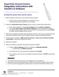
Fan-<strong>Beam</strong> Source<br />
Traditional<br />
CT Scan<br />
<strong>Cone</strong> <strong>Beam</strong> <strong>3D</strong> vs CT <strong>Imaging</strong><br />
<strong>Cone</strong> <strong>Beam</strong> <strong>3D</strong><br />
Scan<br />
Fan-<strong>Beam</strong> x-ray<br />
<strong>Image</strong> Detector Area<br />
Less Radiation than<br />
Traditional CT Scans<br />
<strong>Image</strong> Detector Area<br />
<strong>Cone</strong> <strong>Beam</strong> x-ray<br />
<strong>Cone</strong> <strong>Beam</strong> Source<br />
Traditional CT (CAT scan) uses a<br />
narrow fan beam that rotates<br />
around the patient acquiring thin<br />
axial slices with each revolution.<br />
In order to image a section of anatomy,<br />
many rotations must be completed.<br />
Due to these repeated rotations,<br />
traditional CT emits a high<br />
radiation dose and leaves a gap or<br />
break in information between each<br />
rotation. Software must fill in, or<br />
guess the missing information.<br />
<strong>3D</strong> vs 2D <strong>Imaging</strong><br />
The American Academy of Oral<br />
and Maxillofacial Radiology<br />
(AAOMR) prescribes the use of<br />
<strong>Cone</strong> <strong>Beam</strong> <strong>3D</strong> imaging in cases<br />
involving evaluation of periodontal,<br />
implant, and oral/maxillofacial<br />
surgery patients.<br />
One NewTom <strong>Cone</strong> <strong>Beam</strong> <strong>3D</strong><br />
scan captures a complete<br />
dentomaxillofacial record<br />
in a single database of digital<br />
image information.<br />
<strong>Cone</strong> <strong>Beam</strong> <strong>3D</strong> imaging uses a<br />
cone-shaped beam to acquire<br />
the entire image in a single pass,<br />
resulting in more accurate imaging<br />
without gaps in information, and<br />
with considerably lower radiation<br />
exposure.<br />
Various types of <strong>3D</strong> images can be<br />
created using NewTom NNT<br />
software.
Precise 1:1 Scale <strong>Imaging</strong><br />
With precise 1:1 scale imaging, potential errors due to the image<br />
NewTom technology eliminates the distortion and magnification<br />
magnification errors of conventional commonly found with 2D imaging<br />
cephalometric imaging technology. technology. <strong>3D</strong> imaging technology<br />
<strong>3D</strong> imaging allows the dental is the standard of care for<br />
professional to identify potentially implantologists, orthodontists,<br />
serious problems, such as airway periodontists, and oral/maxillofacial<br />
passage obstructions and soft tissue<br />
abnormalities, and helps<br />
surgeons.<br />
avoid<br />
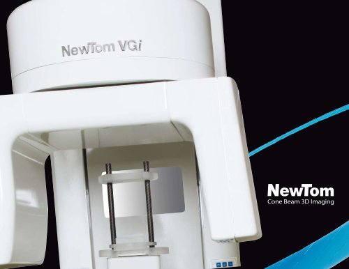
Greater Patient Comfort<br />
and Treatment Acceptance<br />
In addition to delivering a small, NewTom scans provide the practitioner<br />
and the patient unprecedent-<br />
space saving footprint, the<br />
NewTom VGi’s resemblance to a ed visualization of cranial anatomic<br />
typical panoramic unit provides information.<br />
familiarity to the patient.<br />
This leads to better diagnoses and<br />
NewTom 5G scans patients in a treatment planning and increases<br />
supine position and, thanks to its patient knowledge.<br />
unique pass-through style gantry, The result is a more cooperative<br />
new applications can be explored. and informed consent process,<br />
All NewTom units add a sense of validating the need for treatment<br />
comfort for patients. Enabling the and improving the partnership<br />
patient to remain calm and relaxed between patient and surgeon.<br />
during the scan limits patient<br />
movement and therefore improves<br />
overall image quality.
Safe<strong>Beam</strong> Technology<br />
for Patient Safety<br />
Only NewTom systems employ<br />
Safe<strong>Beam</strong> technology, the safest<br />
technology available for patient and<br />
staff. Featured in all NewTom units,<br />
Safe<strong>Beam</strong> automatically adjusts<br />
the radiation dosage according to<br />
the patient’s age and size.<br />
This technology utilizes intermittent<br />
bursts of radiation only milliseconds<br />
in length during image acquisition.<br />
Other systems deliver a constant<br />
stream of radiation and the same<br />
amount of radiation, whether<br />
scanning a 300-pound adult or a<br />
small child.<br />
Safe<strong>Beam</strong> technology<br />
automatically and continuously<br />
monitors system operations,<br />
thus eliminating the possibility of<br />
unnecessary exposures.<br />
In conjunction with our patented<br />
Safe<strong>Beam</strong> technology, when<br />
compared to other CB<strong>3D</strong> systems,<br />
NewTom VGi has a wider range<br />
with which it adjusts the X-ray<br />
power and quantity (kV=110 and<br />
mA=1-20).<br />
As a result, patient exposure is<br />
tailored and image contrast<br />
remains consistent regardless of<br />
patient size or bone density.
Software Flexibility<br />
Easier <strong>Image</strong> Sharing,<br />
Better <strong>Image</strong> Processing<br />
NewTom NNT analysis software<br />
canal, NNT is designed to<br />
professionals the capacity to<br />
is the perfect complement to<br />
deliver high quality images<br />
view images exported with<br />
<strong>Cone</strong> <strong>Beam</strong> <strong>3D</strong> imaging. NNT<br />
that can be placed into<br />
DICOM 3.0 without having to<br />
supports the identification of<br />
user-defined templates,<br />
purchase NNT.<br />
root inclination, position of<br />
deliverable digitally, on paper,<br />
<strong>Image</strong> data can be burned onto<br />
impacted and supernumerary<br />
or on film. NNT gathers<br />
CD or DVD for imaging centers<br />
teeth, resorption, hyperplastic<br />
any combination of images<br />
and referring doctors, allowing<br />
growth, and tooth structure<br />
onto one screen for custom<br />
dental and medical<br />
anomalies. With the ability<br />
to mark the mandibular<br />
reports. NNT is delivered with<br />
a standard viewer, giving other<br />
professionals the opportunity to<br />
easily share images.<br />
Figure 1<br />
Figure<br />
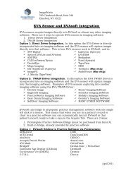
Superior Third-Party Compatibility<br />
NewTom images are<br />
and utilized in a myriad of<br />
Different software applications<br />
Figure<br />
Figure 5<br />
compatible with most major<br />
diagnostic and educational<br />
permit lifelike overlays to be<br />
third-party software vendors<br />
modes.<br />
superimposed on the CB<strong>3D</strong><br />
as well as guided implant and<br />
Software segmentation adjusts<br />
scan. This creates a host of<br />
maxillofacial surgery software.<br />
the amount of soft tissue<br />
options that aid in diagnosis,<br />
Figures 1-5 demonstrate<br />
relative to underlying hard<br />
treatment planning, pre-<br />
the versatility of <strong>3D</strong> imaging<br />
tissue by peering “into” the<br />
surgical analyses, and patient<br />
data that can be imported<br />
skull.<br />
education.<br />
Figure 3
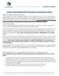
Clinical Case Studies<br />
Figure 1 Figure 2<br />
Figure 3 Figure 4<br />
Figure 5 Figure 6<br />
Courtesy of:<br />
Alan A. Winter, D.D.S.<br />
New York University College<br />
of Dentistry<br />
Courtesy of:<br />
Prof. Robert Cavézian<br />
and Prof. Gérard Pasquet, Paris<br />
Implants<br />
CB<strong>3D</strong> is one of the most effective<br />
tools available for evaluating<br />
implant sites. <strong>3D</strong> images can<br />
identify potential pathologies<br />
and structural abnormalities<br />
with unprecedented accuracy.<br />
There appears to be enough<br />
height for an implant in both sites,<br />
but the cross-sectional view (Figure<br />
2) reveals an atrophic edentulous<br />
ridge inadequate in both height and<br />
width.<br />
Figure 3 depicts a panoramic view<br />
of a mandibular edentulous site.<br />
A cross-section (Figure 4)<br />
demonstrates that a potential<br />
implant site is proximate to the<br />
Hi Res Zoom<br />
Proper assessment for Implants<br />
requires the visualization of all<br />
aspects of the mandibular canal.<br />
The ability to see small anatomical<br />
parts such as tooth roots and<br />
periodontal ligaments, as well as<br />
any present lesions, is critical in<br />
determining successful placement.<br />
mental foramen, and that a narrow<br />
ridge exists in the buccal/lingual<br />
dimension with dense cortical<br />
bone. This highlights the thickness<br />
of cortical bone and density of<br />
cancellous bone. This can affect<br />
the appropriate implant to use,<br />
implant placement, appropriate<br />
implant width, and consideration<br />
of “die back” from dense cortical<br />
bone. These are additional factors<br />
in identifying the location of the<br />
inferior alveolar nerve and mental<br />
foramen.<br />
Only <strong>3D</strong> High Resolution imaging<br />
produces both the quality and<br />
quantity of details necessary to<br />
accurately view the canal for<br />
secure implant assessment.
Endodontics<br />
Figure 1 demonstrates residual<br />
and two canals in maxillary first<br />
periapical radiolucency.<br />
premolars, but it is less common<br />
The patient had root canal therapy,<br />
but continued to complain about<br />
to fi nd two in maxillary second<br />
premolars. Once the dentist<br />
Figure 1<br />
Figure 2<br />
extreme sensitivities to hot and<br />
cold. Endodontic retreatment did<br />
not abate the problem.<br />
observed this, successful treatment<br />
could be instituted.<br />
Courtesy of:<br />
Alan A. Winter, D.D.S.<br />
New York University College<br />
of Dentistry<br />
The CB<strong>3D</strong> scan revealed that the<br />
maxillary second premolar did have<br />
a palatal root (Figure 2).<br />
It is common to expect two roots<br />
TMJ<br />
CB<strong>3D</strong> takes the imaging of the<br />
The Panoramic view gives perhaps<br />
Temporomandibular Joint to a<br />
new level. Sagittal and Coronal<br />
the most information as a gross<br />
screening tool. (Figure 4).<br />
Figure 1<br />
Figure 2 Figure 3<br />
views show joint space (Figure 1),<br />
arthritis (Figure 1) and pathology.<br />
We can see differences in condylar<br />
The <strong>3D</strong> image is the clearest<br />
picture seen to date of TMJ and<br />
height and ramus height (Figure 5),<br />
as well as dental pathology.<br />
Courtesy of:<br />
Michael L. Gelb D.D.S., M.S.<br />
New York University College<br />
of Dentistry<br />
Cervical Spine anatomy; for<br />
example, calcified stylohyoid<br />
ligament (Figure 2), Dens of C2<br />
(Figure 3).<br />
Figure 4<br />
Figure 5<br />
Accurate Planning, Successful Treatment
Figure 1<br />
Predictable Outcomes,<br />
Better Results<br />
Oral Surgery<br />
Post-Operative Scan;<br />
Roots of Impacted Third Molar.<br />
Figure 1. Transaxial views #51 and<br />
52 delineate the inferior alveolar<br />
canal at the buccal of the root<br />
apices, while views #53 through 55<br />
define these apices to be wrapped<br />
around the neural canal at the<br />
lingual aspect of the inferior border<br />
of the jaw. View #55 shows the<br />
missing buccal wall, presumably<br />
from the previous surgical attempt.<br />
Only a <strong>3D</strong> scan can demonstrate<br />
the exact individual anatomy,<br />
define anatomical structures,<br />
and motivate the discussions<br />
that lead to patients’<br />
understanding of their unique<br />
clinical circumstances, ultimately<br />
generating a cooperative and<br />
informed consent process.<br />
Courtesy of:<br />
Alan A. Winter, D.D.S.<br />
New York University College<br />
of Dentistry<br />
Maxillofacial Surgery<br />
NewTom scans are ideal<br />
considerable number of screws<br />
for post-surgery imaging due present, there are virtually no<br />
to reduced image scatter and artifacts to obstruct the images.<br />
lower radiation. This particular<br />
reconstruction case is clearly<br />
extreme.<br />
These High Resolution <strong>3D</strong> images<br />
(utilizing the MIP and Volume<br />
options), are from a post-surgical<br />
patient check. Despite the
Ortho Assessment<br />
While various pan-cephalometric<br />
machines create adequate<br />
images, cone beam scanners<br />
produce many types of images,<br />
including panoramic (Figure 1),<br />
cephalometric (Figure 2), and <strong>3D</strong><br />
(Figure 3). Based on the physics of<br />
this technology, images are more<br />
accurate than 2D dental X-rays and<br />
<strong>3D</strong> medical scanners. As a result,<br />
cephalometric tracings (Figure 4)<br />
from dental cone beam scanners<br />
can be generated with confidence.<br />
The <strong>3D</strong> image in Figure 5<br />
demonstrates an adequate amount<br />
of bone buccal to the molar roots,<br />
so that palatal expansion will<br />
not cause unwarranted gingival<br />
recession. Figure 6 demonstrates<br />
too little bone and that palatal<br />
expansion is either contraindicated,<br />
or the patient should be advised<br />
that gingival recession could occur.<br />
Figure 7 indicates adequate bone.<br />
Figure 1<br />
Figure 2 Figure 3<br />
Figure 4<br />
Courtesy of:<br />
Alan A. Winter, D.D.S.<br />
New York University College<br />
of Dentistry<br />
Maxilla VGi Hi Res Zoom<br />
Impacted teeth may cause dental There is a significant difference<br />
problems that produce few,<br />
between the demarcation<br />
if any symptoms. Only <strong>3D</strong> imaging capabilities of plain radiographs vs.<br />
provides a complete picture of <strong>3D</strong> images in determining the<br />
the scanned area and allows existence and the root shape of<br />
manipulation of both the angle and an impacted tooth in the maxilla.<br />
slick thickness of the image.<br />
Figure 5<br />
Figure 6 Figure 7<br />
Courtesy of:<br />
Prof. Robert Cavézian<br />
and Prof. Gérard Pasquet, Paris
NewTom Benefits<br />
Smallest Possible Focal Spot for the Clearest <strong>Image</strong>s<br />
360 Degree <strong>Imaging</strong> and Medical-Grade Rotating Anode<br />
Means No Scatter or Artifacts<br />
Greater Patient Comfort and Treatment Acceptance<br />
Easier <strong>Image</strong> Sharing<br />
Precise 1:1 Scale <strong>Imaging</strong><br />
Safe<strong>Beam</strong> Technology for Patient Safety<br />
Consistent <strong>Image</strong>s Regardless of Patient Size
NewTom - Today’s<br />
Standard of Care
<strong>Image</strong><strong>Works</strong><br />
250 Clearbrook Road, Suite 240<br />
Elmsford, NY 10523 USA<br />
914.592.6100 - Voice<br />
800.592.6666 - Toll Free<br />
914.592.6148 - Fax<br />
www.<strong>Image</strong><strong>Works</strong>Corporation.com<br />
Manufacturer QR srl<br />
Via SiIvestrini 20<br />
Verona, Italy 37135<br />
+39 045 8202727 or 583500 - Voice<br />
+39 045 8203040 - Fax<br />
www.qrverona.it<br />
<strong>Cone</strong> <strong>Beam</strong> <strong>3D</strong> <strong>Imaging</strong>