VIEW PDF - Wounds International
VIEW PDF - Wounds International
VIEW PDF - Wounds International

Create successful ePaper yourself
Turn your PDF publications into a flip-book with our unique Google optimized e-Paper software.
INTERNATIONAL<br />
CASE STUDIES<br />
USING SILVERCEL®<br />
NON-ADHERENT: CASE STUDIES<br />
CASE STUDIES SERIES 2012
SILVERCEL® NON-ADHERENT<br />
This document has been<br />
jointly developed by <strong>Wounds</strong><br />
<strong>International</strong> and Systagenix<br />
with financial support from<br />
Systagenix<br />
For further information about<br />
Systagenix please visit:<br />
www.systagenix.com<br />
Published by:<br />
<strong>Wounds</strong> <strong>International</strong><br />
Enterprise House<br />
1–2 Hatfields<br />
London SE1 9PG, UK<br />
Tel: + 44 (0)20 7627 1510<br />
Fax: +44 (0)20 7627 1570<br />
info@woundsinternational.com<br />
www.woundsinternational.com<br />
ABOUT THIS DOCUMENT<br />
This document contains a series of case reports describing the use of<br />
SILVERCEL® NON-ADHERENT (Systagenix) in patients with a range of<br />
wound types. All patients were treated for a minimum of four weeks<br />
and the decision to continue with SILVERCEL® NON-ADHERENT was<br />
based on continual assessment. A formal assessment was performed<br />
weekly, although in some cases dressing changes were carried out more<br />
frequently.<br />
All patients were assessed for:<br />
■■<br />
clinical signs of infection/critical colonisation<br />
■■<br />
pain prior to and during dressing changes using a visual analogue<br />
scale of 1-10, where 1 = no pain and 10 = worst possible pain (see<br />
below)<br />
■■<br />
signs of improvement, including granulation extent and reduction in<br />
wound size.<br />
No<br />
pain<br />
Distressing<br />
pain<br />
Unbearable<br />
pain<br />
The case reports presented in<br />
this document are the work<br />
of the authors and do not<br />
necessarily reflect the opinions<br />
of Systagenix.<br />
How to cite this document:<br />
<strong>International</strong> case series: Using<br />
SILVERCEL Non-Adherent: Case<br />
Studies. London: <strong>Wounds</strong><br />
<strong>International</strong>, 2012.<br />
1 2 3 4 5 6 7 8 9 10<br />
Photographs were taken weekly in the majority of cases to document<br />
wound progression. Relevant additional wound treatments, eg<br />
compression therapy, antibiotic therapy, analgesia, etc were reported.<br />
The clinicians undertaking the study were also asked to rate the<br />
dressing (from highly satisfied to dissatisfied) and to record whether<br />
the dressing needed soaking prior to removal and/or the dressing left<br />
any debris in the wound.<br />
In addition, clinicians were offered the opportunity to check the<br />
protease activity in the wound using WOUNDCHEK Protease Status<br />
test (Systagenix).<br />
The weekly assessment outcomes are cited for each case where:<br />
= reduction = increase — = no change or not present<br />
ii | INTERNATIONAL CASE STUDIES
SILVERCEL® NON-ADHERENT<br />
SILVERCEL® NON-ADHERENT: case studies<br />
Silver-containing antimicrobial dressings have been available for many<br />
years. SILVERCEL® NON-ADHERENT has been designed to eliminate<br />
the potential problems of adherence and fibre shed that are sometimes<br />
associated with some fibrous wound dressings. This document contains<br />
several case studies that describe how SILVERCEL® NON-ADHERENT has<br />
benefited patients with a range of wound types.<br />
What is SILVERCEL® NON-ADHERENT?<br />
SILVERCEL® NON-ADHERENT is a sterile absorbent antimicrobial dressing that<br />
is suitable for use in moderately to heavily exuding wounds that are infected or at<br />
increased risk of infection (Box 1).<br />
SILVERCEL® NON-ADHERENT has an outer perforated film layer designed to<br />
prevent adherence of the dressing to the wound and the shedding of fibres. It also<br />
facilitates absorption of fluid. The central absorbent core of the dressing contains<br />
silver-coated X-Staticfibres®, which provide the antimicrobial action.<br />
How does SILVERCEL® NON-ADHERENT work?<br />
The outer layer of SILVERCEL® NON-ADHERENT comprises a non-adherent<br />
wound contact layer made from ethylene methyl acrylate (EMA) (EasyLIFT®<br />
Precision Film, Systagenix). The surface of the film has a low propensity for<br />
sticking to other surfaces 1 .<br />
BOX 1: INDICATIONS FOR SILVERCEL®<br />
NON-ADHERENT<br />
Moderately to heavily exuding partial and<br />
full thickness acute and chronic wounds<br />
that are:<br />
n infected<br />
n at increased risk of infection<br />
For example, pressure ulcers, venous leg<br />
ulcers, diabetic foot ulcers, donor sites, and<br />
traumatic and surgical wounds 8<br />
BOX 2: PRECAUTIONS AND<br />
CONTRAINDICATIONS TO THE USE OF<br />
SILVERCEL® NON-ADHERENT<br />
n For patients having an MRI scan, remove<br />
dressing before scanning<br />
n Do not use on patients with a known<br />
sensitivity to alginates, carboxymethylcellulose,<br />
ethylene methyl<br />
acrylate or silver 8<br />
The perforations in the film allow fluid to be absorbed by the central core. The<br />
core is made up of a combination of highly absorbent high G calcium alginate and<br />
carboxymethylcellulose (CMC) and silver-coated fibres. The dressing delivers<br />
antimicrobial action through the release of silver ions (Ag + ) on contact with fluid.<br />
Laboratory testing has indicated that the dressing is effective against many common<br />
wound pathogens, including meticillin-resistant Staphylococcus aureus, meticillinresistant<br />
Staphylococcus epidermidis and vancomycin-resistant Enterococcus 2 .<br />
Evidence for SILVERCEL® NON-ADHERENT<br />
SILVERCEL®, the absorbent antimicrobial core of SILVERCEL® NON-<br />
ADHERENT, has been assessed in a number of laboratory and clinical studies<br />
and has been shown to have:<br />
■■<br />
Good antimicrobial activity<br />
■■<br />
High absorbent capacity, even in the presence of blood<br />
■■<br />
Good tolerability<br />
■■<br />
Low adherence<br />
■■<br />
Suitability for a wide range of wounds 3-6<br />
The performance of SILVERCEL® NON-ADHERENT was evaluated in a case series<br />
of patients with locally infected wounds, complex medical problems and a history<br />
of recurrent wound infections 7 . The dressing was easy to apply and remove, did<br />
not cause trauma and no fibres were seen in the wound bed. Several patients<br />
experienced a reduction in wound pain during use of the dressing and, as a result,<br />
needed less analgesia.<br />
SILVERCEL® NON-ADHERENT CASE STUDIES | 1
SILVERCEL® NON-ADHERENT<br />
Tips on using SILVERCEL® NON-ADHERENT<br />
■■<br />
Prior to application, prepare the wound bed according to local policies<br />
■■<br />
Cut or fold the dressing to the shape of the wound so that it does not overlap the wound edges<br />
■■<br />
If using on a wound with lower exudate levels, moisten the dressing with sterile saline<br />
■■<br />
Cover the dressing with an appropriate secondary dressing according to the wound type, wound position, exudate level and<br />
condition of surrounding skin<br />
■■<br />
Dressing change frequency is determined by exudate levels and condition of the wound and surrounding skin<br />
■■<br />
Dressing change is required when the absorbent capacity of the secondary dressing has been reached<br />
■■<br />
If the primary dressing appears to be dry at dressing change it may be saturated with sterile saline solution prior to removal<br />
ABOUT SILVERCEL® NON-ADHERENT<br />
n For further information about SILVER-<br />
CEL® NON-ADHERENT please go to:<br />
www.systagenix.com/our-products/<br />
lets-protect<br />
INTERNATIONAL CONSENSUS<br />
APPROPRIATE USE OF SILVER DRESSINGS<br />
IN WOUNDS<br />
n To download a copy of the consensus<br />
document please go to:<br />
www.woundsinternational.com<br />
Appropriate use of silver dressings<br />
A recent consensus on the appropriate use of silver dressings described the<br />
main roles of silver dressings, such as SILVERCEL® NON-ADHERENT, in the<br />
management of wounds to be the reduction of bioburden and to act as an<br />
antimicrobial barrier 9 .<br />
Whenever SILVERCEL® NON-ADHERENT is used for the treatment of increased<br />
bioburden or to prevent infection, the rationale should be fully documented in the<br />
patient's health records and a schedule for review should be specified.<br />
Reducing bioburden<br />
The consensus document recommends that silver dressings be used initially for a<br />
two week 'challenge' period. At the end of the two weeks, the wound, the patient<br />
and the management approach should be re-evaluated 9 .<br />
If after two weeks, the wound has:<br />
■■<br />
improved, but there are continuing signs of infection, it may be clinically<br />
justifiable to continue use of silver dressings with regular review<br />
■■<br />
improved and there are no longer signs or symptoms of infection, the silver<br />
dressing should be discontinued<br />
■■<br />
not improved, the silver dressing should be discontinued and the patient<br />
reviewed and a dressing containing a different antimicrobial agent initiated, with<br />
or without systemic antibiotics 9 .<br />
Prophylactic use<br />
Silver dressings, such as SILVERCEL® NON-ADHERENT, may be used as an<br />
antimicrobial barrier in wounds at high risk of infection or re-infection. They may<br />
also be used to prevent entry of bacteria at medical device entry/exit sites, such as<br />
tracheostomy tubes (see the consensus document for further information 9 ).<br />
References<br />
1. Clark R, Del Bono M, Stephens SA, et al. Development of an in-vitro model to evaluate the potential for<br />
adherence of wound healing dressings. Poster presented at: WUWHS, Texas, 2009.<br />
2. Lansdown ABG. Silver I: Its antibacterial properties and mechanism of action. J Wound Care 2002; 11(4):<br />
125-30.<br />
3. Clark R, Del Bono M, Stephens S, et al. Simulated in-use tests to evaluate a non-adherent antimicrobial silver<br />
alginate wound dressing. Poster presented at: SAWC, Texas, 2009.<br />
4. Teot L, Maggio G, Barrett S. The management of wounds using Silvercel hydroalginate. <strong>Wounds</strong> UK 2005; 1(2): 1-6.<br />
5. Kammerlander G, Afarideh R, Baumgartner A, et al. Clinical experiences of using a silver hydroalginate<br />
dressing in Austria, Switzerland and Germany. J Wound Care 2008; 17(9): 384-88.<br />
6. Di Lonardo A, Maggio G, Cupertino M, et al. The use of SILVERCEL to dress excision wounds following burns<br />
surgery. <strong>Wounds</strong> UK 2006; 2(4): 122-24.<br />
7. Ivins N, Taylor AC, Harding KG. A series of case studies using a silver non adherent dressing. Poster presented<br />
at: CSSWC, Florida, 2010.<br />
8. Clark R, Bradbury S. SILVERCEL® NON-ADHERENT Made Easy. <strong>Wounds</strong> <strong>International</strong> 2010; 1(5): Available<br />
from http://www.woundsinternational.com<br />
9. <strong>International</strong> consensus. Appropriate use of silver dressings in wounds. An expert working group consensus.<br />
London: <strong>Wounds</strong> <strong>International</strong>, 2012.<br />
2 | INTERNATIONAL CASE STUDIES
SILVERCEL® NON-ADHERENT<br />
Case 1<br />
Background<br />
In April 2012, Mrs S presented to the outpatient clinic with a venous ulcer of<br />
7 months' duration on her right lower leg, resulting from a traumatic skin injury<br />
sustained when moving a chair.<br />
Mrs S, an active and independent 90-year-old woman, underwent a left hip<br />
replacement and varicose vein surgery (7 and 40 years prior, respectively). In<br />
recent years she had reported problems with swelling in her right leg, but was not<br />
receiving medication for this. Prior to presentation, Mrs S’ ulcer had been treated by<br />
community nurses with three-layer graduated compression bandaging and various<br />
antimicrobial treatments — most recently, silver sulfadiazine (Flamazine, Smith &<br />
Nephew).<br />
Treatment<br />
The venous ulcer was located on the lateral gaiter aspect of the right leg with<br />
a surface area of approximately 8.5cm 2 . On assessment, the wound appeared<br />
heavily colonised but uninfected, with localised erythema, exudate and pain. A<br />
WOUNDCHEK Protease Status test showed low protease activity. Clinical<br />
priorities for wound management were to reduce the wound bioburden and<br />
minimise pain at dressing change. SILVERCEL® NON-ADHERENT was chosen for<br />
use in conjunction with compression bandaging.<br />
Week 1: Signs of local infection were reduced including no malodour and<br />
comparatively less exudate and erythema than seen on initial presentation. The<br />
wound bed showed evidence of 25-50% granulation tissue and a 5cm 2 reduction<br />
in wound size (from 8.5cm 2 to 3.5cm 2 ). The dressing was removed easily from<br />
the wound bed although the patient reported moderate pain during the dressing<br />
change, recorded as 5 on a visual analogue scale (VAS) of 1-10. On dressing<br />
removal the wound bed was clean. Protease activity remained low.<br />
Week 2: SILVERCEL® NON-ADHERENT was reapplied for a further week. On<br />
reassessment, Mrs S reported that her pain at dressing change was slightly<br />
reduced (from 5 to 4 using the VAS). Clinically, signs of infection were minimal<br />
with less exudate and erythema and no malodour was noted. Granulation tissue<br />
had increased to 50-75% of the wound bed and a further WOUNDCHEK<br />
Protease Status test showed no elevated protease activity (EPA).<br />
Weeks 3–4: Improvements in the wound continued over the next 2 weeks of<br />
treatment with once weekly assessments showing further reductions in pain,<br />
exudate levels, erythema distribution and ulcer size. At week 4, the final case<br />
study assessment, Mrs S reported no pain on dressing change. The wound<br />
size was 1.5cm 2 (a decrease of 7.5cm 2 in total) and there were no signs of<br />
infection.<br />
Outcome<br />
SILVERCEL® NON-ADHERENT was found to be easy to use, with clinicians<br />
reporting a high level of satisfaction overall. A reduction in exudate, erythema and<br />
pain indicated that the wound bed bioburden had diminished. Granulation tissue<br />
had increased and there was a reduction in wound size. Most importantly from the<br />
patient's perspective, pain at dressing change was eliminated.<br />
By: Jane Megson, Wound Care Research Nurse, Bradford Royal Infirmary,<br />
Bradford, UK<br />
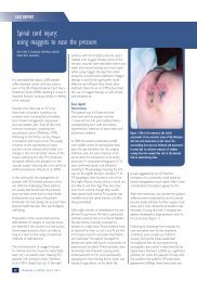
Baseline<br />
Week 2<br />
Week 4<br />
Figures 1-3: The ulcer reduced in size over the<br />
four-week case study period with evidence of new<br />
granulation tissue and an overall improved colour.<br />
Assessment<br />
Signs and<br />
symptoms of<br />
infection<br />
Pain before<br />
dressing change<br />
(VAS 1-10)<br />
Pain at dressing<br />
change (VAS<br />
1-10)<br />
Week<br />
1<br />
Week<br />
2<br />
Week<br />
3<br />
Week<br />
4<br />
none<br />
1 3 1 1<br />
5 4 3 1<br />
Protease activity Low Low Low Low<br />
Wound<br />
reduction<br />
83%<br />
SILVERCEL® NON-ADHERENT CASE STUDIES | 3
SILVERCEL® NON-ADHERENT<br />
Case 2<br />
Background<br />
In May 2012, Mr M presented with a wound on his left foot following amputation<br />
of the second toe. Surgery had been required two weeks previously to remove<br />
bone fragments. The wound had been present since July 2011, when Mr M had<br />
been unaware of a screw in his shoe that had caused skin breakdown.<br />
Mr M, a 54-year-old man, had insulin-dependent diabetes, hypertension and<br />
Barrett's oesophagus. The original wound had been treated with cadexomer iodine<br />
paste (Iodoflex®, Smith & Nephew), a Hydrofiber® dressing (Aquacel®, ConvaTec)<br />
and a silver Hydrofiber® dressing (Aquacel® Ag, ConvaTec). Mr M was not<br />
receiving systemic antibiotics.<br />
Baseline<br />
Treatment<br />
The wound on the left foot measured 30mm by 20mm and was 20mm deep.<br />
It appeared to be infected, was malodorous and exuding heavily. The edges of<br />
the wound were macerated. A WOUNDCHEK TM Protease Status test showed<br />
low protease activity. Because of the depth of the wound, SILVERCEL® NON-<br />
ADHERENT ribbon was chosen to treat the infection and manage the exudate.<br />
Week 1: The wound was reassessed after three days. There was no pain on dressing<br />
removal, although the patient reported a pain score of 4 (on a VAS of 1-10) before the<br />
dressing change. The wound had reduced in size to 25mm x 15mm x 30mm with<br />
evidence of 50-75% granulation tissue. Malodour was still present but less noticeable<br />
and the wound continued to exude heavily. SILVERCEL® NON-ADHERENT ribbon<br />
was reapplied and reviewed three times per week.<br />
Week 2: On reassessment after a further week, the wound was no longer odorous<br />
and other signs of infection were reduced. The wound was exuding less and there<br />
was no erythema. The wound had decreased in size and was now 23mm by 12mm,<br />
and 12mm deep. It was decided to continue with thrice weekly dressing changes<br />
and to reapply SILVERCEL® NON-ADHERENT ribbon.<br />
Week 3: The patient reported less pain prior to dressing change and no pain on<br />
dressing removal. The wound had continued to improve with further reductions in<br />
the amount of exudate, no erythema and a clean wound bed. The wound measured<br />
20mm by 10mm and was only 2mm deep. A flat SILVERCEL® NON-ADHERENT<br />
dressing was applied and the patient was reviewed in three days.<br />
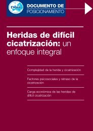
Week 1<br />
Week 4<br />
Figures 1-3: The size and depth of the ulcer reduced<br />
over the case study period with a reduction in odour<br />
and exudate.<br />
Week 4: The patient was now pain free before and during dressing removal, and<br />
the wound was considerably reduced in size to 12mm by 8mm and had no depth.<br />
There was some evidence of overgranulation and treatment was changed to a<br />
povidone iodine dressing (INADINE®, Systagenix) with silver nitrate pencil applied<br />
to the areas of overgranulation.<br />
Outcome<br />
Clinicians commented that the SILVERCEL® NON-ADHERENT ribbon and dressing<br />
were easy to use. Over the case study period, the size and depth of the wound<br />
decreased considerably in size and the reduction in malodour and exudate volume<br />
indicated that bioburden was much reduced.<br />
By: Helen Strapp, Tissue Viability Clinical Nurse Specialist, AMNCH Tallaght<br />
Hospital, Dublin, Ireland<br />
Assessment<br />
Signs and<br />
symptoms of<br />
infection<br />
Pain before<br />
dressing<br />
change (VAS<br />
1-10)<br />
Pain at dressing<br />
change (VAS<br />
1-10)<br />
Protease<br />
activity<br />
Wound<br />
reduction<br />
Week<br />
1<br />
Week<br />
2<br />
Week<br />
3<br />
Week<br />
4<br />
none<br />
4 3 2 1<br />
1 1 2 1<br />
n/a n/a n/a n/a<br />
84%<br />
4 | INTERNATIONAL CASE STUDIES
SILVERCEL® NON-ADHERENT<br />
Case 3<br />
Background<br />
In May 2012, Mr N presented to the clinic with a diabetic foot ulcer of<br />
3 years' duration, which had occurred spontaneously while walking.<br />
Mr N, a 47-year-old man, had a 6-year history of type 2 diabetes. He had<br />
undergone transmetatarsal amputation of the right foot in 2006. The<br />
present ulcer was located on the third metatarsal of the right foot below the<br />
prior amputation and measured 2.6cm 2 . Mr N had been treated previously<br />
with an alginate dressing (ALGOSTERIL®, Systagenix), a topical antimicrobial<br />
(ACTISORB® Silver 220, Systagenix) and protease modulating dressings,<br />
as well as a Walker-type offloading device. Mr N was receiving systemic<br />
antibiotics.<br />
Baseline<br />
Treatment<br />
The wound on the right foot did not appear to be infected, although it<br />
was thought to be heavily colonised. It was malodorous, highly exuding<br />
(exudate was green in colour) and the wound edges were macerated.<br />
A WOUNDCHEK TM Protease Status test showed low protease activity.<br />
SILVERCEL® NON-ADHERENT was initiated, with thrice weekly dressing<br />
changes to manage the exudate volume.<br />
Week 1: The patient did not experience pain before or during dressing<br />
changes and did not require any analgesics. It was not necessary to soak the<br />
dressing prior to removal and debris did not remain on the wound bed after<br />
removal. Signs of infection were reduced with no malodour, no erythema<br />
and the disappearance of the greenish colour of the exudate. The wound bed<br />
showed evidence of 50-75% granulation tissue and the wound had reduced<br />
in size from 2.6cm 2 to 2.4cm 2 . A further WOUNDCHEK TM Protease Status<br />
test showed no elevated protease activity (EPA).<br />
Week 2: SILVERCEL® NON-ADHERENT was reapplied for a further week.<br />
Signs of infection were reduced, with a small reduction in the volume of<br />
exudate, no malodour and the wound reduced in size to 1.5cm 2 with 50-75%<br />
granulation tissue of the wound bed. Protease activity remained low.<br />
Weeks 3–4: Signs of infection were reduced further with the wound bed<br />
showing evidence of 50-75% granulation at week 3 and increasing to<br />
100% at week 4. The wound reduced to 1.3cm 2 at week 3 with no further<br />
improvement at week 4. The exudate levels remained moderate to high and<br />
there was maceration to the periwound skin, which was managed using<br />
barrier products. A WOUNDCHEK TM Protease Status test continued to show<br />
no EPA.<br />
Outcome<br />
SILVERCEL® NON-ADHERENT was found to be easy to use with clinicians<br />
reporting a high level of satisfaction with the dressing overall. Odour<br />
and erythema were quickly eliminated indicating a reduction in wound<br />
bioburden. The patient did not experience pain at dressing change. The<br />
patient continued to receive offloading throughout the study period.<br />
By: Esther García Morales, Podiatrist, Diabetic Foot Unit, University Clinic of<br />
Podiatry, The Complutense University of Madrid, Madrid, Spain<br />
Week 2<br />
Week 4<br />
Figures 1-3: The ulcer reduced in size over the fourweek<br />
case study period with elimination of malodour<br />
and erythema by week 1.<br />
Assessment<br />
Signs and<br />
symptoms of<br />
infection<br />
Pain before<br />
dressing change<br />
(VAS 1-10)<br />
Pain at dressing<br />
change (VAS<br />
1-10)<br />
Protease<br />
activity<br />
Wound<br />
reduction<br />
Week<br />
1<br />
Week<br />
2<br />
Week<br />
3<br />
<br />
1 1 1 1<br />
1 1 1 1<br />
Week<br />
4<br />
Low Low Low Low<br />
50%<br />
SILVERCEL® NON-ADHERENT CASE STUDIES | 5
SILVERCEL® NON-ADHERENT<br />
Case 4<br />
Background<br />
Mr G, a 59-year-old man with type 2 diabetes, presented at the podiatry clinic<br />
with ulceration to the right foot. The wound had been sustained when Mr G<br />
was walking without his orthotic device at a social event. Initially the area had<br />
blistered and subsequently the skin broke down. The wound had been present for<br />
3 weeks and had been treated over that period with an absorbent foam dressing<br />
containing silver sulfadiazine (Allevyn TM , Smith & Nephew). Mr G was also on oral<br />
antidiabetes medications.<br />
Treatment<br />
The wound was located on the fifth metatarsal head, measured 20mm x 15mm and<br />
extended to bone. The patient had had intravenous antibiotics for eight days which<br />
was then changed to oral antibiotics. On examination the wound appeared to be<br />
infected. Swab results indicated a Group B Streptococcus was the causative agent.<br />
SILVERCEL® NON-ADHERENT was applied to the wound and left in place for three<br />
days. Dressing change was carried out by the district nurse between clinic visits.<br />
Week 1: Mr G returned to the clinic one week later. The dressing did not require<br />
soaking prior to removal and no debris was left on the wound bed. The patient<br />
did not complain of pain. Clinical examination indicated that the erythema was<br />
subsiding and the volume of exudate was reducing. There was evidence of 0-25%<br />
granulation tissue. There was slight malodour, but overall the wound was showing<br />
signs of improvement. The patient continued on antibiotic therapy and SILVERCEL®<br />
NON-ADHERENT as the primary dressing.<br />
Week 1<br />
Week 3<br />
Week 2: The patient had some pain prior to dressing change and on dressing<br />
removal (3 on a VAS of 1-10), but did not wish to have any analgesia. The dressing<br />
was removed easily without soaking and did not leave debris on the wound bed. The<br />
wound had reduced in size (15mm x 14mm). Exudate levels were reduced and there<br />
was no malodour. The wound bed had 25-50% granulation tissue. SILVERCEL®<br />
NON-ADHERENT was reapplied.<br />
Week 3: The patient continued to improve and there was no pain prior to or on<br />
dressing removal. There was a marked improvement in the wound bed with 25-50%<br />
granulation tissue present. A WOUNDCHEK Protease Status test indicated low<br />
protease activity. SILVERCEL® NON-ADHERENT was reapplied.<br />
Week 4: There was a marked improvement in the wound, with no malodour, a<br />
reduction in exudate volume and the wound had reduced in size (10mm x 10mm).<br />
The wound bed showed 100% granulation tissue coverage. Protease activity<br />
continued to be low.<br />
Outcome<br />
An inter-professional approach involving podiatry and nursing staff, good local<br />
wound care and antibiotic therapy were key in progressing the wound to healing<br />
in a timely fashion. Odour was quickly eliminated, exudate levels reduced and<br />
the wound bed improved. Throughout the treatment period the staff graded<br />
SILVERCEL® NON-ADHERENT as highly satisfactory in terms of ease of use.<br />
Mr G was happy with the outcome and assured staff he would wear his orthotic<br />
appliance at all times when weight bearing.<br />
By: Samantha Haycocks, Advanced Podiatrist, Salford Royal (NHS) Foundation, Salford,<br />
UK<br />
Week 4<br />
Figures 1-3: The ulcer reduced in size over the fourweek<br />
case study period with marked improvement in<br />
the wound bed with increased granulation tissue and<br />
reduction in exudate levels and malodour.<br />
Assessment<br />
Signs and<br />
symptoms of<br />
infection<br />
Pain before<br />
dressing change<br />
(VAS 1-10)<br />
Pain at dressing<br />
change (VAS<br />
1-10)<br />
Protease<br />
activity<br />
Wound<br />
reduction<br />
Week<br />
1<br />
Week<br />
2<br />
Week<br />
3<br />
<br />
1 3 1 1<br />
1 3 1 1<br />
Week<br />
4<br />
Low Low Low Low<br />
34%<br />
6 | INTERNATIONAL CASE STUDIES
SILVERCEL® NON-ADHERENT<br />
Case 5<br />
Background<br />
Mr A, a 73-year-old man, presented to the outpatient clinic in May 2012 with a<br />
venous leg ulcer of 4 years' duration. The ulcer was located over the pre-tibial<br />
aspect of the right lower leg and measured approximately 14.5cm 2 . The patient's<br />
history suggested that the ulcer had originally developed following an episode<br />
of cellulitis and skin breakdown. Various treatments had been tried with limited<br />
success. Four-layer compression bandaging had been in use throughout. The<br />
patient was also receiving pregabalin (Lyrica®, Pfizer) for neuropathic pain.<br />
Treatment<br />
On examination the ulcer was assessed to be heavily colonised although not clinically<br />
infected. A WOUNDCHEK Protease Status test revealed low protease activity.<br />
SILVERCEL® NON-ADHERENT was selected to reduce the wound bioburden and<br />
four-layer compression bandaging was applied to promote venous return.<br />
Baseline<br />
Week 1: Initially, the patient reported a low level of pain (2 on a VAS of 1-10).<br />
However, this increased at dressing change to 8 and oral analgesia was required<br />
despite soaking the dressing prior to removal (this was done to help alleviate the<br />
pain, the dressing did not adhere to the wound bed). It was noted that a second<br />
wound on the leg may have also contributed to the patient's pain.<br />
On assessment of the ulcer and surrounding area later that week, it was noted<br />
that signs of critical colonisation, including exudate levels, had diminished and the<br />
wound size had decreased from 14.5cm 2 to 12cm 2 . Protease activity remained low<br />
and granulation tissue was evident in 0-25% of the wound bed. SILVERCEL® NON-<br />
ADHERENT was continued for a further week.<br />
Week 2: The patient reported a pain score of 3 increasing to 9 on dressing removal,<br />
despite oral analgesia and soaking the dressing pre-removal. On inspection the<br />
ulcer was free of debris and signs of critical colonisation remained diminished with<br />
a reduction in exudate levels. The wound bed appeared healthy with approximately<br />
50-75% granulation tissue coverage and the wound size had decreased again<br />
(10.5cm 2 ). Protease activity remained low. Compression bandaging layers were<br />
reduced from four to three to reduce pain.<br />
Week 3: Mr A's pain score reduced from 8 to 3 at dressing change, a considerable<br />
improvement on previous weeks. No analgesia was required and the dressing was<br />
removed easily without pre-soaking. The ulcer size was slightly reduced (10cm 2 ) and<br />
the coverage of granulation tissue in the ulcer bed remained constant at 50-75%. No<br />
signs of infection were observed.<br />
Week 4: Dressing change pain remained comparatively low (VAS of 2) and the<br />
wound dimensions had decreased to 9cm 2 . Granulation tissue coverage was<br />
between 50-75% and protease activity remained low. No signs of infection or critical<br />
colonisation were noted.<br />
Outcome<br />
Quality of life can be significantly affected by chronic wounds. There was a good<br />
reduction in Mr A's pain levels at dressing change and the wound showed no signs<br />
of critical colonisation or infection by week 4.<br />
By: Jane Megson, Wound Care Research Nurse, Bradford Royal Infirmary,<br />
Bradford, UK<br />
Week 2<br />
Week 4<br />
Figures 1-3: The ulcer reduced in size over the<br />
four-week case study period with a reduction in<br />
pain during dressing changes and an increase in<br />
granulation tissue.<br />
Assessment<br />
Signs and<br />
symptoms of<br />
infection<br />
Pain before<br />
dressing change<br />
(VAS 1-10)<br />
Pain at dressing<br />
change (VAS<br />
1-10)<br />
Protease<br />
activity<br />
Wound<br />
reduction<br />
Week<br />
1<br />
Week<br />
2<br />
Week<br />
3<br />
Week<br />
4<br />
None None<br />
2 3 1 2<br />
8 9 3 2<br />
Low Low Low Low<br />
38%<br />
SILVERCEL® NON-ADHERENT CASE STUDIES | 7
SILVERCEL® NON-ADHERENT<br />
Case 6<br />
Background<br />
In May 2012, Mr S presented with a foot ulcer. He had a pressure relieving<br />
device but it had become worn resulting in trauma to the right toe. The wound<br />
had been present for 10 months. Mr S, a 40-year-old man with type 2 diabetes,<br />
had peripheral neuropathy, hypertension and venous insufficiency. He was on<br />
antidiabetes medication and had been fitted with bespoke shoes with a rocker sole<br />
to relieve pressure from the toe.<br />
Treatment<br />
The ulcer was located on the plantar aspect of the first right toe and measured<br />
16mm x 16mm. On examination there were signs of infection, localised cellulitis,<br />
malodour and moderate amounts of exudate. He also presented with cellulitis on<br />
the left foot extending to the lower leg. Mr S commenced oral antibiotic therapy for<br />
10 days. The wound was dressed with SILVERCEL® NON-ADHERENT ribbon. The<br />
patient undertook dressing changes himself on alternate days.<br />
Week 1: The patient reported a pain score of 3 (measured on a VAS of 1-10) prior to<br />
dressing change. A pain score of 4 was also recorded on dressing removal, but the<br />
patient indicated that he did not require analgesia to control the pain. The dressing<br />
was easy to remove and did not require soaking. On removal no debris remained in<br />
the wound bed. Although there was no decrease in wound size and some malodour<br />
was still present, signs of infection decreased and the wound bed contained 50-<br />
75% granulation tissue. There was an increased level of haemoserous exudate and<br />
it was decided to change the dressing daily. A WOUNDCHEK Protease Status<br />
test showed low protease activity.<br />
Week 2: There was no pain prior to or during dressing change. Wound<br />
debridement was carried out. There were reduced signs of infection and there<br />
was evidence of 50-75% granulation tissue, with slight maceration to the<br />
periwound skin. Slight malodour was still present, the wound had reduced in<br />
size (15mm x 15mm).<br />
Week 3: Exudate levels had reduced and there was no malodour; there was slight<br />
localised erythema. Due to the general improvement, wound dressings were reduced<br />
to alternate days. Treatment with SILVERCEL® NON-ADHERENT was continued.<br />
Week 4: Wound debridement was carried out resulting in an increase in wound<br />
size (20mm x 17mm). No erythema, heat or swelling were noted and the wound<br />
bed had 50-75% granulation tissue. The general improvement in the wound meant<br />
Mr S was able to be more mobile. This was important for general wellbeing but it<br />
did lead to an increase in exudate levels. The wound dressing was changed at this<br />
time to a superabsorbent dressing to manage the higher exudate levels. Protease<br />
activity remained low.<br />
Outcome<br />
During the course of treatment, the clinical staff rated the dressing as satisfactory<br />
or highly satisfactory in terms of ease of use. Debridement of non-viable tissue and<br />
topical antimicrobial therapy avoided a further course of antibiotics in this instance.<br />
The patient was fitted with new pressure relieving footwear and he was able to<br />
resume his normal activities.<br />
By: Paul Chadwick, Principal Podiatrist, Salford Royal (NHS) Foundation, Salford, UK<br />
Baseline<br />
Week 2<br />
Week 4<br />
Figures 1-3: The general improvement in the wound<br />
over the four-week case study period meant that the<br />
patient was able to be more mobile.<br />
Assessment<br />
Signs and<br />
symptoms of<br />
infection<br />
Pain before<br />
dressing change<br />
(VAS 1-10)<br />
Pain at dressing<br />
change (VAS<br />
1-10)<br />
Week<br />
1<br />
Week<br />
2<br />
Week<br />
3<br />
<br />
3 1 1 1<br />
4 1 1 1<br />
Week<br />
4<br />
Protease activity Low Low Low Low<br />
Wound<br />
reduction<br />
— — <br />
8 | INTERNATIONAL CASE STUDIES
SILVERCEL® NON-ADHERENT<br />
Case 7<br />
Background<br />
Mr K, a 63-year-old man, presented to the Accident and Emergency Department<br />
with a post-surgical wound on his left foot of 5 months' duration. He gave a<br />
history of diabetes and peripheral vascular disease for which he was on a range of<br />
medications. The wound started as a blister to the third toe, due to poorly fitting<br />
shoes, which became infected then necrotic. Due to the poor circulation to his limb<br />
and the risk of spreading infection, amputation of the toe was undertaken, but the<br />
surgical wound had failed to heal.<br />
Treatment<br />
On referral to the clinic one week later, the wound at the site of amputation<br />
measured 30mm x 15mm x 2mm. It showed signs of clinical infection and<br />
maceration of the periwound skin. Previous treatments included Hydrofiber®<br />
dressings (Aquacel®/Aquacel® Ag, ConvaTec). The patient was not currently<br />
receiving antibiotics, although oral antibiotics had been prescribed previously.<br />
A WOUNDCHEK Protease Status test indicated no EPA. SILVERCEL® NON-<br />
ADHERENT ribbon was applied to the wound and the patient was given an<br />
appointment to return to clinic four days later.<br />
Week 1: On return to the clinic the wound dressing was removed. The dressing came<br />
off easily and Mr K reported no pain either prior to or on removal. The wound bed<br />
was clean and while the wound had not decreased in size, there was no malodour<br />
and clinical signs of infection had subsided. The wound bed was composed of<br />
50-75% granulation tissue. SILVERCEL® NON-ADHERENT was applied with gauze<br />
dressings on top. Dressings were changed on alternate days. Protease activity<br />
remained low.<br />
Week 2: Mr K reported slight pain (score of 2 on VAS) prior to dressing change and<br />
on dressing removal, but did not require analgesia. The wound dressing was easily<br />
removed. The wound bed was clean, there was no malodour and signs of infection<br />
were continuing to subside. There was a small amount of serous exudate and slight<br />
erythema, but the wound bed was in good condition with 50%-75% granulation<br />
tissue. The dressing regimen was maintained.<br />
Week 3–4: The wound had reduced slightly in size; by the end of week 4 it measured<br />
25mm x 15mm x 2mm. There was some maceration to the skin surrounding the<br />
wound and as the wound no longer appeared infected, the primary dressing was<br />
changed to Aquacel® to manage the exudate.<br />
Outcome<br />
The nurses who cared for Mr K found SILVERCEL® NON-ADHERENT to be easy<br />
to use and rated it highly satisfactory in terms of application, removal and a lack of<br />
debris remaining on the wound bed on dressing removal. <strong>Wounds</strong> in patients with<br />
peripheral vascular disease and diabetes are challenging to manage and this case<br />
was no exception. However, with good assessment and care based on the best<br />
available evidence, Mr K’s wound infection subsided and there was a reduction in<br />
wound size. Properly fitted footwear supplied by orthotics departments is essential<br />
in such cases to avoid further trauma.<br />
By: Helen Strapp, Tissue Viability Clinical Nurse Specialist, AMNCH Tallaght<br />
Hospital, Dublin, Ireland<br />
Baseline<br />
Week 2<br />
Week 4<br />
Figures 1-3: The ulcer reduced in size over the<br />
four-week case study period with evidence of new<br />
granulation tissue and an overall improved colour.<br />
Assessment<br />
Signs and<br />
symptoms of<br />
infection<br />
Pain before<br />
dressing change<br />
(VAS 1-10)<br />
Pain at dressing<br />
change (VAS<br />
1-10)<br />
Protease<br />
activity<br />
Wound<br />
reduction<br />
Week<br />
1<br />
Week<br />
2<br />
Week<br />
3<br />
Week<br />
4<br />
None None<br />
1 2 2 2<br />
1 2 2 2<br />
Low Low Low Low<br />
— 17%<br />
SILVERCEL® NON-ADHERENT CASE STUDIES | 9
SILVERCEL® NON-ADHERENT<br />
Case 8<br />
Background<br />
Mr S, a 74-year-old man, presented in May 2012 with a venous leg ulcer located<br />
over the left medial malleolus that measured 4cm 2 . The ulcer had started with a<br />
gradual breakdown of the skin 10 years previously; various treatments had been<br />
tried, including larval therapy and a topical silver dressing (Acticoat 7, Smith &<br />
Nephew). At presentation Mr S’ ulcer was managed with a two-layer compression<br />
hosiery system.<br />
Treatment<br />
The ulcer was critically colonised, heavily exuding and malodorous with evidence of<br />
25-50% granulation tissue at the wound bed. A WOUNDCHEK Protease Status<br />
test recorded low protease activity. SILVERCEL® NON-ADHERENT was selected<br />
to reduce bioburden. This was used in conjunction with two-layer compression<br />
hosiery to enhance venous return.<br />
Week 1: Exudate levels remained high and some malodour was noted. Erythema<br />
had reduced and the ulcer area had decreased from 4cm 2 to 3.5cm 2 . The ulcer bed<br />
comprised 25-50% granulation tissue. Prior to dressing removal Mr S reported a<br />
pain score of 4 (on a VAS of 1-10), but did not wish to receive analgesia to control<br />
the pain. The dressing was removed easily and the pain score fell to 1 (no pain).<br />
Baseline<br />
Week 2<br />
Week 2: There was no malodour and erythema was further reduced. Protease<br />
activity remained low and granulation coverage remained at 25-50% of the wound<br />
bed. The wound size remained constant (3.5cm 2 ) and pain levels were again<br />
recorded as 4 on the VAS reducing to 1 on dressing removal with no requirement<br />
for analgesia. The dressing was removed easily with no debris left in the wound.<br />
SILVERCEL® NON-ADHERENT was reapplied for a further week.<br />
Week 3: The ulcer continued to improve, although exudate levels remained high.<br />
Granulation tissue remained at 25-50% of the wound bed. Protease activity<br />
continued to be low according to a WOUNDCHEK Protease Status test. The<br />
wound reduced in size from 3.5cm 2 to 3cm 2 . The pre-dressing change VAS score<br />
was also slightly lower at 3; the VAS on dressing removal was consistent with other<br />
weeks at 1 (no pain). The dressing was removed easily and there was no debris left<br />
in the wound bed. SILVERCEL® NON-ADHERENT was reapplied.<br />
Week 4: Ulcer size remained constant at 3cm 2 and Mr S reported no pain pre- and<br />
during dressing change. The wound bed appeared improved; while exudate levels<br />
remained high there were no other signs of local infection.<br />
Outcome<br />
SILVERCEL® NON-ADHERENT was found to be easy to use. There was a reduction<br />
in wound bioburden over the four weeks of treatment and the patient’s pain score<br />
diminished considerably. Used in conjunction with two-layer compression hosiery,<br />
the dressing was effective in achieving a reduction of wound size, dressing change<br />
pain and erythema.<br />
By: Jane Megson, Wound Care Research Nurse, Bradford Royal Infirmary,<br />
Bradford, UK<br />
Week 4<br />
Figures 1-3: The ulcer reduced in size over the fourweek<br />
case study period with a reduction in erythema<br />
and pain.<br />
Assessment<br />
Signs and<br />
symptoms of<br />
infection<br />
Pain before<br />
dressing<br />
change (VAS<br />
1-10)<br />
Pain at<br />
dressing<br />
change (VAS<br />
1-10)<br />
Protease<br />
activity<br />
Wound<br />
reduction<br />
Week<br />
1<br />
Week<br />
2<br />
Week<br />
3<br />
<br />
4 4 3 1<br />
1 1 1 1<br />
Week<br />
4<br />
Low Low Low Low<br />
— 25%<br />
10 | INTERNATIONAL CASE STUDIES
SILVERCEL® NON-ADHERENT<br />
Case 9<br />
Background:<br />
In May 2012, Mrs G, a 42-year-old woman with type 1 diabetes of 35 years'<br />
duration, presented with a neuropathic foot ulcer of 24 months' duration<br />
located on the right plantar mid-foot. The ulcer probed to bone and surgical<br />
debridement to remove devitalised tissue and bone was undertaken.<br />
Mrs G had a history of supracondylar traumatic amputation of the lower<br />
left limb, hypercholesterolemia, retinopathy and led a sedentary lifestyle.<br />
She was allergic to quinolone antibacterial agents and was taking a range of<br />
medications.<br />
Baseline<br />
Treatment<br />
The ulcer had a surface area of 4.2cm 2 . The wound did not appear to be<br />
infected, although it was heavily colonised and had low levels of serous<br />
exudate. Mrs G had previously received topical negative pressure therapy to<br />
treat the ulcer (therapy duration, 7 days), oral antibiotics (oral co-amoxiclav,<br />
2 weeks) and pressure offloading. A WOUNDCHEK TM Protease Status test<br />
showed no EPA. SILVERCEL® NON-ADHERENT was initiated with thrice<br />
weekly dressing changes.<br />
Week 1: Signs of local infection were reduced, with no malodour, light<br />
exudate levels and evidence of 50-75% granulation coverage of the wound<br />
bed. There were areas of slough that were not fixed to the wound bed and<br />
were easily removed. There was also evidence of periwound hyperkeratosis.<br />
The wound size had reduced from 4.2cm 2 to 2.6cm 2 .<br />
The patient did not experience pain before or during dressing changes and<br />
did not require analgesia. It was not necessary to soak the dressing prior<br />
to removal and no debris remained in the wound bed after removal. A<br />
WOUNDCHEK TM Protease Status test showed low levels of protease activity.<br />
Weeks 2–4: SILVERCEL® NON-ADHERENT was reapplied for a further<br />
3 weeks and was changed three times a week. Signs of local infection<br />
continued to reduce, with no malodour, light exudate levels and evidence of<br />
50-75% granulation tissue. As previously noted, there were areas of slough<br />
that were not fixed to the wound bed and periwound hyperkeratosis.<br />
The wound size showed a further reduction to 1.8cm 2 at week 2, 0.9cm 2<br />
at week 3 and 0.7cm 2 at week 4. Protease activity continued to be low<br />
following a further WOUNDCHEK TM Protease Status test.<br />
Outcome<br />
SILVERCEL® NON-ADHERENT was found to be easy to use, with clinicians<br />
reporting a high level of satisfaction with the dressing overall. The signs and<br />
symptoms of infection reduced and there was a reduction in wound size. The<br />
patient did not experience pain at dressing change.<br />
By: Esther García Morales, Podiatrist, Diabetic Foot Unit, University Clinic of<br />
Podiatry, The Complutense University of Madrid, Madrid, Spain<br />
Week 2<br />
Week 4<br />
Figures 1-3: The ulcer reduced in size over the fourweek<br />
case study period with a reduction in the signs<br />
and symptoms of infection.<br />
Assessment<br />
Signs and<br />
symptoms of<br />
infection<br />
Pain before<br />
dressing change<br />
(VAS 1-10)<br />
Pain at dressing<br />
change (VAS<br />
1-10)<br />
Week<br />
1<br />
Week<br />
2<br />
Week<br />
3<br />
<br />
1 1 1 1<br />
1 1 1 1<br />
Week<br />
4<br />
Protease activity Low Low Low Low<br />
Wound<br />
reduction<br />
83%<br />
SILVERCEL® NON-ADHERENT CASE STUDIES | 11
SILVERCEL® NON-ADHERENT<br />
Case 10<br />
Background<br />
Mr M, a 68-year-old man with a long-standing history of leg ulcers, presented<br />
with a wound on the lateral aspect of his lower right leg which had occurred<br />
spontaneously 8 months previously. During this time several advanced wound<br />
dressings had been applied along with toe to knee compression. The leg was<br />
eczematous, indicating venous hypertension. He had no significant past medical<br />
history and was not taking any medication.<br />
Treatment<br />
The wound measured 2.5cm x 1.8cm x 3mm (depth). The wound did not appear<br />
infected but was considered to have been critically colonised for 3 months based<br />
on clinical signs. The surrounding skin was hyperkeratotic further indicating venous<br />
incompetence. A WOUNDCHEK Protease Status test indicated elevated protease<br />
activity (EPA). The wound was dressed with a SILVERCEL® NON-ADHERENT 11cm<br />
x 11cm dressing. Emollient was applied to hydrate the hyperkeratotic areas of the<br />
leg and an inelastic two-layer compression bandage was applied toe to knee. The<br />
dressing was to be changed every 3 days in clinic.<br />
Week 1: Mr M reported slight pain prior to dressing removal. The dressing was<br />
removed easily and did not cause the patient any pain. A small amount of debris<br />
remained on the wound. The wound length and breadth remained the same but<br />
the depth had decreased (2.5cm x 1.8cm x 2.5mm). Granulation tissue was evident<br />
(25-50%), exudate levels had decreased and there was no odour. The wound was<br />
tested using WOUNDCHEK TM Protease Status test and was shown to have elevated<br />
protease activity (EPA). The wound was re-dressed with a SILVERCEL® NON-<br />
ADHERENT 11cm x11cm dressing. Emollient was reapplied and inelastic two-layer<br />
compression continued. The management plan remained unchanged.<br />
Week 2: No pain was reported before or during dressing removal. Only a small<br />
amount of debris remained on the wound. The wound's size had decreased (2.0cm<br />
x1.5cm x 2mm) and there was 50% to 75% granulation tissue. All other clinical<br />
signs indicated that the wound was improving and a WOUNDCHEK Protease<br />
Status test was not completed. Treatment remained the same.<br />
Week 3: The wound edges showed good signs of epithelialisation. Wound size<br />
was 1.3cm x 0.9cm x 1.5mm. The patient reported no pain during the visit. Protease<br />
activity was low. Wound management remained the same.<br />
Week 4: All clinical signs indicated the wound was healing. Healthy<br />
granulation tissue was seen on the wound bed and the surrounding skin had<br />
improved.<br />
Outcome<br />
The clinical staff rated the dressing as satisfactory or highly satisfactory in<br />
terms of ease of use. The wound had been critically colonised for a prolonged<br />
period of time, which led to the decision to renew the dressing every 3<br />
days. This intensive treatment reduced bacteria and protease activity, and<br />
subsequently the wound progressed towards healing.<br />
By: Professor Marco Romanelli, Consultant Dermatologist, University of Pisa, Italy<br />
Baseline<br />
Week 3<br />
Week 4<br />
Figures 1-3: The wound size decreased over the study<br />
period and the condition of the surrounding skin<br />
improved.<br />
Assessment<br />
Signs and<br />
symptoms of<br />
infection<br />
Pain before<br />
dressing change<br />
(VAS 1-10)<br />
Pain at dressing<br />
change (VAS<br />
1-10)<br />
Week<br />
1<br />
Week<br />
2<br />
Week<br />
3<br />
<br />
Week<br />
4<br />
2 None None None<br />
1 None None None<br />
Protease activity EPA n/a Low n/a<br />
Wound reduction >77%<br />
12 | INTERNATIONAL CASE STUDIES
SILVERCEL® NON-ADHERENT<br />
SILVERCEL® NON-ADHERENT CASE STUDIES | 13
SILVERCEL® NON-ADHERENT<br />
A <strong>Wounds</strong> <strong>International</strong> publication<br />
www.woundsinternational.com<br />
14 | INTERNATIONAL CASE STUDIES