All Products 2013 / PDF brochure - Soredex
All Products 2013 / PDF brochure - Soredex
All Products 2013 / PDF brochure - Soredex

You also want an ePaper? Increase the reach of your titles
YUMPU automatically turns print PDFs into web optimized ePapers that Google loves.
SOREDEX ®<br />
products<br />
Diagnostic<br />
performance
SOREDEX<br />
Since 1977 SOREDEX has been a leader in providing innovative<br />
imaging solutions for demanding professionals. Through<br />
continuous evolution and refinement we have set the highest<br />
industry standards for Quality, Reliability and Efficiency.<br />
We are committed to follow in this path today and in the future.<br />
DIGORA® Vidi<br />
Intraoral imaging<br />
Contents<br />
Intraoral Intraoral imaging imaging<br />
DIGORA® Vidi<br />
DIGORA® Toto<br />
MINRAY®<br />
DIGORA® Optime UV<br />
DIGORA® Optime<br />
2D Extraoral imaging<br />
CRANEX® Excel, CRANEX® Excel Ceph<br />
CRANEX® Novus e<br />
CRANEX® D, CRANEX® D Ceph<br />
2D/3D Extraoral imaging<br />
CRANEX® 3D<br />
3D imaging<br />
SCANORA® 3D<br />
SCANORA® 3Dx<br />
Software solutions<br />
SCANORA®<br />
SOREDEX® TWAIN<br />
SorCom<br />
DIGORA® Vidi - intraoral camera<br />
DIGORA® Vidi intraoral camera is ergonomic and easy to use.<br />
Optical and lighting innovations provide consistently clear, precise<br />
and natural-looking images.<br />
EASY TO USE<br />
• Free focus: From one tooth to portrait<br />
without manual focus adjustment<br />
• Image capturing with optimally located<br />
“EasyTouch” touch button for sharp images<br />
PRACTICAL ERGONOMICS<br />
• Rounded shape and reduced dimensions of camera<br />
head for maximum patient comfort<br />
• Perfect visibility even on hard to reach distal areas<br />
with 15 degrees tilted viewing angle<br />
PRECISE IMAGES<br />
• Uniform illumination and images of natural color with<br />
optimized high-intensity, daylight color LED lighting<br />
• High quality aspheric, wide angle lens for consistently<br />
high image quality:<br />
- Large depth of field shows near and far objects<br />
sharp in the same image<br />
- Minimized geometric distortion<br />
3
Intraoral imaging<br />
DIGORA® Toto<br />
MINRAY®<br />
Intraoral imaging<br />
DIGORA® Toto – intraoral sensors, sizes 1 and 2<br />
DIGORA® Toto sensors are easy to use and provide superb, repeatable<br />
clinical results. They feature many improvements over traditional<br />
intraoral sensors.<br />
EASY<br />
• Fluent workflow, continuously ready to capture instant images<br />
• Sensor part design optimized for durability and easy positioning<br />
• Graphics supporting positioning and orientation<br />
• Size 1 and 2 sensors can be connected and ready for use<br />
at the same time<br />
SMART<br />
• Slim sensor part with all corners and edges rounded for<br />
optimal patient comfort<br />
• Images instantly available for diagnosis<br />
• One piece system: saved desktop space and convenient<br />
REAL plug and play portability<br />
ACCURATE<br />
• CMOS sensor chips tolerate wide range of exposure times<br />
and provide crystal-clear images<br />
• Repeatable image quality with automatic image optimization:<br />
- Minimized need to adjust images<br />
- Virtually no under- or overexposures<br />
- Reduced need to change exposure settings<br />
• Images with rounded corners and maximized active area<br />
MINRAY® – intraoral X-ray unit<br />
MINRAY® – Versatile and reliable high-frequency DC intraoral X-ray unit.<br />
The unique customizable horizontal arm can be adjusted exactly to the<br />
optimum length.<br />
A convenient tubehead handle together with stable arm system makes<br />
aiming smooth and precise, keeping the tubehead motionless during<br />
exposure.<br />
EASY<br />
• Well balanced, easy to position and stable arm system<br />
• Integrated control panel with programmable timer settings<br />
• Compatible with all digital systems and film<br />
PRECISE<br />
• High frequency DC generator with precise repeatability<br />
• Wide and exact exposure time settings<br />
• Proven, reliable design<br />
ADJUSTABLE<br />
• Unique customizable horizontal arm for<br />
optimized ergonomics<br />
• Optional remote control panel, mobile stand<br />
and dental unit integration<br />
• Output 60/70kV – 7 mA<br />
4 5
Intraoral imaging<br />
DIGORA® Optime UV<br />
DIGORA® Optime<br />
New!<br />
Intraoral imaging<br />
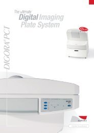
DIGORA® Optime UV – digital intraoral imaging plate system<br />
DIGORA® Optime – digital intraoral imaging plate system<br />
Uniquely hygienic, efficient, fast and easy visually guided system for<br />
demanding dental professionals, for all intraoral applications and patient<br />
sizes.Provides leading clinical results for all diagnostic needs repeatedly,<br />
automatically and efficiently, with proven reliability.<br />
FAST AND EASY<br />
• Quick and easy to learn and use, guided with graphical user interface<br />
• Images in seconds, processing time not depending on the resolution<br />
• Smart automated functions support fluent and error-free workflow<br />
EFFORTLESS, END-TO-END HYGIENIC WORKFLOW<br />
• Touchless operation (patent pending) and automatic internal<br />
UV-disinfection system (patent pending)<br />
• Unique and proprietary hygiene accessories made from safe materials<br />
(Biocompatible according to ISO 10993-1, no Latex or PVC)<br />
EXCELLENT CLINICAL RESULTS AND PATIENT COMFORT<br />
• Latest generation of durable imaging plates with<br />
patented IDOT identification system<br />
• Repeatable image quality automatically and repeatedly<br />
DESIGNED FOR YOUR PRACTICE<br />
• Reliable and maintenance free design, proven with intensive<br />
use in large dental clinics and imaging centers<br />
• Compatible with all intraoral sizes and applications<br />
• Cost-effective sharing between operatories over local<br />
area network, supported with display<br />
Easy and smart dental diagnostic tool for all intraoral applications and<br />
patient sizes. Provides leading clinical results repeatedly,<br />
automatically and efficiently, with top reliability.<br />
EASY<br />
• Easy to set up, instantly ready for use<br />
• Intuitive, simple to learn and use with clear indicators for the user<br />
• Familiar, film-like workflow<br />
SMART<br />
• Sharp, clear images for all intraoral needs automatically<br />
and repeatedly: Minimized need to adjust images, virtually<br />
no under- or overexposures<br />
• Smart automated functions and economical<br />
network sharing solution<br />
• Comfortable and safe with unique, proprietary hygiene accessories<br />
(Latex / PVC free, food grade hygiene bags with Biocompatibility<br />
according to ISO 10993-1 and Opticover protective cover)<br />
EFFICIENT<br />
• Images in seconds<br />
• Reliable, maintenance free design<br />
• Wireless, reusable and durable imaging plates with<br />
patented IDOT imaging plate identification system to<br />
support quality control (optional)<br />
6 7
2D Extraoral imaging<br />
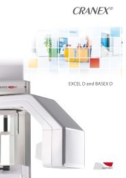
CRANEX® Excel,<br />
CRANEX® Excel Ceph<br />
CRANEX® Novus e<br />
New!<br />
2D Extraoral imaging<br />
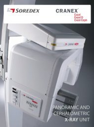
CRANEX® Excel family – classic panoramic and cephalometric film unit<br />
CRANEX® Excel provides outstanding image quality, it is easy to use<br />
and includes unique features thanks to many SOREDEX innovations.<br />
• Easy and accurate patient positioning without moving the patient<br />
• Motorized dual-speed vertical movement<br />
• Automatic kV selection AutoSet<br />
• High-frequency DC generator<br />
• Adult and child panoramic program<br />
• Partial images of the dentition (left / right / middle)<br />
• Open and closed bilateral TMJ program<br />
CRANEX® Excel Ceph special features<br />
• Adjustable soft tissue filter<br />
• Dedicated tube head, especially beneficial for taking<br />
frequent cephalometric images<br />
• Lateral, AP and PA images<br />
• Head holder can be turned and locked in 45° for oblique projections<br />
• Supported formats: 18 cm × 24 cm (8” × 10”) and 24 × 30 cm (10” × 12”)<br />
When digital images are required, CRANEX® Excel, CRANEX® Excel Ceph<br />
and digital extraoral Imaging Plate system is the ideal solution for all your<br />
needs. CRANEX® Excel family is also compatible with most medical<br />
CR systems (optional).<br />
CRANEX® Novus e – fast and easy-to-use digital panoramic X-ray system<br />
It is designed for dental offices that demand a first-class digital panoramic unit.<br />
The next generation CRANEX® Novus e provides excellent image quality with<br />
extended imaging values and enhanced operation with the ClearTouch control<br />
panel. Designed for fast operation and easy workflow, CRANEX® Novus e delivers<br />
maximum efficiency. With 9-second adult panoramic exposure time, the patient<br />
exposure dose is minimized, while reducing the potential for movement artifacts.<br />
Excellent image quality<br />
• Expanded imaging exposure values, 63-77 kV, 10 mA<br />
• Panoramic, Child panoramic, TMJ and Optional Bitewing Program<br />
• Advanced spinal compensation<br />
• Extremely sensitive panoramic CCD sensor<br />
• Accurate patient positioning system<br />
Easy-to-use and fast<br />
• ClearTouch control panel<br />
• 9-second adult panoramic exposure time<br />
• 5-point patient stabilization system<br />
Efficient solution for a daily practice<br />
• Compact design<br />
• Intuitive operation and workflow<br />
8 9
2D Extraoral imaging<br />
CRANEX® D<br />
CRANEX® D Ceph<br />
CRANEX® 3D<br />
2D/3D Extraoral imaging<br />
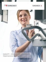
CRANEX® D – perfect digital panoramic and cephalometric X-ray system<br />
CRANEX® D produces superb panoramic images with exceptionally wide<br />
anterior layer thickness, which ensures good visualization of both the crown<br />
and apex even with patients with malocclusion. CRANEX® D Ceph utilizes the<br />
central cephalometric projection technique resulting in an anatomically correct<br />
imaging geometry.<br />
Excellent diagnostic image quality<br />
• Extra wide anterior layer thickness<br />
• Excellent diagnostic image quality with the fast scan capability<br />
• Even better high quality (HiQ) panoramic imaging modes<br />
Ease of use<br />
• Quick and easy-to-use control panel<br />
• The best patient positioning system in the market<br />
• Automatic functions to make daily work even easier<br />
Flexibility<br />
• Cephalometric option<br />
• Dual Sensor Design<br />
• Choice of two column designs<br />
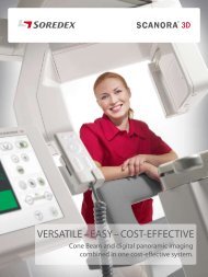
CRANEX® 3D – Dental imaging system for demanding dental clinics<br />
CRANEX® 3D is a high quality dental imaging system with panoramic,<br />
optional cephalometric and Cone Beam CT imaging programs. Its versatility<br />
offers dental clinics one of the most dynamic imaging systems available to<br />
meet their needs.<br />
DYNAMIC<br />
• High performance with versatile range of imaging programs<br />
• RealPAN (CMOS) 6 panoramic programs and sectional imaging<br />
• Cephalometric imaging with 2 lateral, PA/AP and carpus programs<br />
• Cone Beam 3D with 6 × 4 cm and 6 × 7,8 cm<br />
• Free FOV position in dental and facial area<br />
• SARA – SOREDEX Advanced Reconstruction Algorithm<br />
• SMAR – SOREDEX Metal Artefact Reduction<br />
DIRECT<br />
• ClearTouch control panel<br />
• The SOREDEX familiar patient positioning system<br />
• AES - Automatic exposure settings<br />
• PickPoint and EasyScout for accurate 3D positioning<br />
DURABLE<br />
• Robust system designed for intensive use<br />
• Long service life<br />
10 11
SCANORA® 3D<br />
SCANORA® 3Dx<br />
3D imaging<br />
3D imaging<br />
SCANORA® 3D – medium field-of-view Cone Beam CT<br />
system with dedicated panoramic imaging for day- to-day<br />
dentomaxillofacial imaging<br />
Excellent 3D image quality and High-resolution Panoramic imaging<br />
combined characterize this system. With SCANORA® 3D, advanced<br />
dental imaging required for accurate diagnostics, implant treatment<br />
planning and oral surgery can now be done in your practice.<br />
EASY<br />
• Fully motorized chair for seated and stable patient positioning<br />
• 12” HD clear touch control panel for ensuring easy workflow<br />
EFFECTIVE<br />
• RealPAN (optional) CCD - sensor for high quality dental<br />
panoramic imaging with AutoSwitch 2D/3D mode change<br />
• Compatible with leading drill guide and surgical navigation systems<br />
• Low dose<br />
VERSATILE<br />
• Four (one optional), fields-of-view from 60 × 60 mm to 130 × 145 mm<br />
• The FOV can be freely located to different areas of<br />
the dento-maxillofacial area<br />
• Open software architecture for 3rd party applications<br />
• DICOM® / PACS compatibility<br />
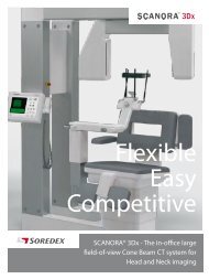
SCANORA® 3Dx – in-office large field-of-view Cone<br />
Beam CT system for Head and Neck imaging, available with<br />
dedicated panoramic imaging<br />
Maximum flexibility, excellent image quality in 2D and 3D and the SCANORA®<br />
typical smooth workflow characterize this system. The SCANORA® 3Dx Pointof-Care<br />
system covers the entire range of applications starting from a single<br />
implant to complex ENT and orthognathic planning and diagnostic.<br />
FLEXIBLE<br />
• Eight (two optional), fields-of-view from 50 × 50 mm to 240 × 165 mm<br />
• The FOV can be freely located to different areas of the head and neck<br />
• Comprehensive software offering<br />
• Compatible with leading navigated surgery and drill guide systems<br />
EASY<br />
• Fully motorized chair for seated and stable patient positioning<br />
• Smooth workflow, fast and easy to use<br />
• 12” HD clear touch control panel<br />
COMPETITIVE<br />
• DICOM®/PACS compatibility<br />
• RealPAN (optional) CCD – sensor for high resolution panoramic<br />
imaging with AutoSwitch 2D/3D mode change<br />
• Small foot print<br />
12 13
Software solutions<br />
SOREDEX software<br />
solutions<br />
Notes:<br />
SCANORA®: A complete solution for dental image management<br />
• Comprehensive set of tools for image capture<br />
and image management<br />
• Optimized, flexible image capture workflow to suit your practice<br />
• Modality-based image capture settings and filters enable fine-tuning<br />
image appearance<br />
• Freely-distributable image viewer for patients and referring clinicians<br />
• High-precision 16-bit image processing of radiographic images<br />
• Extensive DICOM support for integration into<br />
healthcare environments<br />
• Integrated Cone Beam 3D workflow with OnDemand3D<br />
• Open Architecture supports third party 3D treatment planning<br />
SOREDEX® TWAIN<br />
Software for connecting SOREDEX units to third-party imaging<br />
software application via the TWAIN interface<br />
• Includes standard image tools and simple, easy to learn user interface<br />
SorCom: Provides a simple “DICOM® bridge” to connect SOREDEX<br />
units to DICOM®/PACS environments<br />
• Includes standard image tools and simple,<br />
easy to learn user interface<br />
• Supports retrieval of patient information via Modality Worklist<br />
and storing images to PACS via DICOM® Storage<br />
For further details please contact your SOREDEX dealer.
Head office and factory:<br />
SOREDEX<br />
Nahkelantie 160, Tuusula<br />
P.O. Box 148, FI-04301 Tuusula<br />
Finland<br />
Tel. +358 10 270 2000<br />
Fax +358 9 701 5261<br />
info@soredex.com<br />
SOREDEX USA<br />
1245 W. Canal Street<br />
Milwaukee, WI 53233 U.S.A.<br />
Tel. +1 800 558 6120<br />
Fax +1 414 481 8665<br />
usainfo@soredex.com<br />
SOREDEX Germany<br />
Schutterstrasse 12<br />
77746 Schutterwald<br />
Germany<br />
Tel: +49 (0) 781 28 41 98-0<br />
Fax: +49 (0) 781 28 41 98-30<br />
kontakt@soredex.de<br />
Digital<br />
imaging<br />
made<br />
easy<br />
204159-4 02/13<br />
SOREDEX®/SCANORA®/CRANEX®/DIGORA®/MINRAY®/ Digital imaging made easy/<br />
ClearTouch/RealPAN/AutoSwitch/IDOT is a registered trademark / a common<br />
law trademark of SOREDEX. Other product names and trademarks are the property of<br />
their respective owners. CE-marked, NB (CE) number 0537. Electrical safety meets the<br />
IEC 60601-1 standard. Manufacturing complies with ISO 13485:2003, ISO 9001:2008,<br />
and ISO 14001:2004.<br />
DICOM® is the registered trademark of the National Electrical Manufacturers<br />
Association for its standards publications relating to digital communications<br />
of medical information.<br />
SOREDEX reserves the right to make changes in specifications and features<br />
shown herein at any time without notice or obligation.<br />
Contact your SOREDEX representative for the most up-to-date information.<br />
© <strong>2013</strong> SOREDEX<br />
SOREDEX dealer:<br />
www.soredex.com • www.soredex.de • www.soredex.com/usa