
You also want an ePaper? Increase the reach of your titles
YUMPU automatically turns print PDFs into web optimized ePapers that Google loves.
L a s e r<br />
©<br />
Imaging Plate Technology<br />
! ! ! ! ! ! ! ! ! ! ! ! ! ! ! ! ! ! ! ! ! ! ! ! ! ! ! ! ! ! ! ! ! ! ! ! ! ! ! ! ! ! ! ! ! ! ! ! ! ! ! ! ! ! ! ! ! ! ! ! ! ! !<br />
4 DITABIS micron Operation Principle<br />
The DITABIS micron is an<br />
high-performance Imaging Plate<br />
readout system dedicated for<br />
TEM use. Unlike medical and<br />
biological applications where<br />
Imaging Plates are used too,<br />
TEM imaging requires small<br />
pixels, highest image quality and<br />
a high dynamic range.<br />
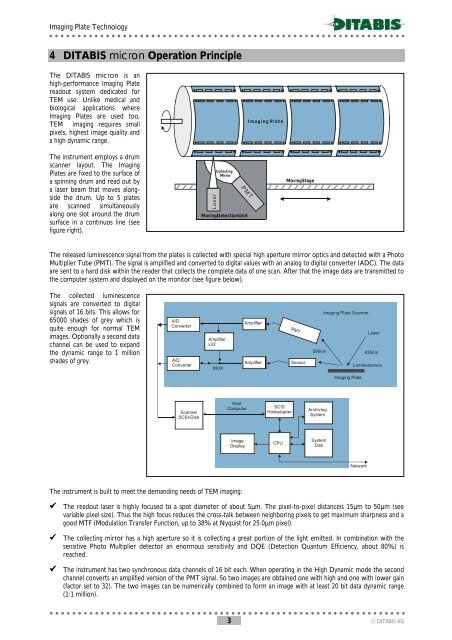
The instrument employs a drum<br />
scanner layout. The Imaging<br />
Plates are fixed to the surface of<br />
a spinning drum and read out by<br />
a laser beam that moves alongside<br />
the drum. Up to 5 plates<br />
are scanned simultaneously<br />
along one slot around the drum<br />
surface in a continuos line (see<br />
figure right).<br />
Collecting<br />
Mirror<br />
P M T<br />
MovingDetectionUnit<br />
Imaging<br />
Drum<br />
Plate<br />
MovingStage<br />
The released luminescence signal from the plates is collected with special high aperture mirror optics and detected with a Photo<br />
Multiplier Tube (PMT). The signal is amplified and converted to digital values with an analog to digital converter (ADC). The data<br />
are sent to a hard disk within the reader that collects the complete data of one scan. After that the image data are transmitted to<br />
the computer system and displayed on the monitor (see figure below).<br />
The collected luminescence<br />
signals are converted to digital<br />
signals of 16 bits. This allows for<br />
65000 shades of grey which is<br />
quite enough for normal TEM<br />
images. Optionally a second data<br />
channel can be used to expand<br />
the dynamic range to 1 million<br />
shades of grey.<br />
A/D<br />
Converter<br />
A/D<br />
Converter<br />
Amplifier<br />
x32<br />
MUX<br />
Amplifier<br />
Amplifier<br />
PMT<br />
Sensor<br />
Imaging Plate Scanner<br />
Laser<br />
390nm<br />
635nm<br />
Luminescence<br />
Imaging Plate<br />
Scanner<br />
SCSI-Disk<br />
Host<br />
Computer<br />
SCSI<br />
Hostadapter<br />
Archiving<br />
System<br />
Image<br />
Display<br />
CPU<br />
System<br />
Disk<br />
Network<br />
The instrument is built to meet the demanding needs of TEM imaging:<br />
# The readout laser is highly focused to a spot diameter of about 5µm. The pixel-to-pixel distanceis 15µm to 50µm (see<br />
variable pixel size). Thus the high focus reduces the cross-talk between neighboring pixels to get maximum sharpness and a<br />
good MTF (Modulation Transfer Function, up to 38% at Nyquist for 25.0µm pixel).<br />
# The collecting mirror has a high aperture so it is collecting a great portion of the light emitted. In combination with the<br />
sensitive Photo Multiplier detector an enormous sensitivity and DQE (Detection Quantum Efficiency, about 80%) is<br />
reached.<br />
# The instrument has two synchronous data channels of 16 bit each. When operating in the High Dynamic mode the second<br />
channel converts an amplified version of the PMT signal. So two images are obtained one with high and one with lower gain<br />
(factor set to 32). The two images can be numerically combined to form an image with at least 20 bit data dynamic range<br />
(1:1 million).<br />
! ! ! ! ! ! ! ! ! ! ! ! ! ! ! ! ! ! ! ! ! ! ! ! ! ! ! ! ! ! ! ! ! ! ! ! ! ! ! ! ! ! ! ! ! ! ! ! ! ! ! ! ! ! ! ! ! ! ! ! ! ! !<br />
3 .<br />
DITABIS AG