H. El-Khushman, A. Sharara, J. Momani, A. Al-Suleihat, M. Al ...
H. El-Khushman, A. Sharara, J. Momani, A. Al-Suleihat, M. Al ...
H. El-Khushman, A. Sharara, J. Momani, A. Al-Suleihat, M. Al ...

You also want an ePaper? Increase the reach of your titles
YUMPU automatically turns print PDFs into web optimized ePapers that Google loves.
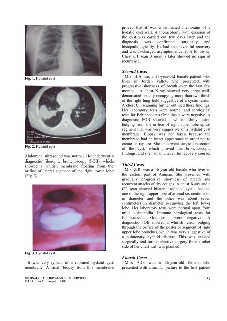
proved that it was a laminated membrane of a<br />
hydatid cyst wall. A thoracotomy with excision of<br />
the cyst was carried out few days later and the<br />
diagnosis was confirmed surgically and<br />
histopathologically. He had an uneventful recovery<br />
and was discharged asymptomatically. A follow up<br />
Chest CT scan 3 months later showed no sign of<br />
recurrence.<br />
Fig. 1. Hydatid cyst<br />
Fig. 2. Hydatid cyst<br />
Abdominal ultrasound was normal. He underwent a<br />
diagnostic fiberoptic bronchoscopy (FOB), which<br />
showed a whitish membrane floating from the<br />
orifice of lateral segment of the right lower lobe<br />
(Fig. 3).<br />
Fig. 3. Hydatid cyst<br />
It was very typical of a ruptured hydatid cyst<br />
membrane. A small biopsy from this membrane<br />
Second Case:<br />
Mrs. H.A was a 39-year-old female patient who<br />
lives in Jordan valley. She presented with<br />
progressive shortness of breath over the last few<br />
months. A chest X-ray showed very large welldemarcated<br />
opacity occupying more than two thirds<br />
of the right lung field suggestive of a cystic lesion.<br />
A chest CT scanning further outlined these findings.<br />
Her laboratory tests were normal and serological<br />
tests for Echinococcus Granulosus were negative. A<br />
diagnostic FOB showed a whitish shiny lesion<br />
bulging from the orifice of right upper lobe apical<br />
segment that was very suggestive of a hydatid cyst<br />
membrane. Biopsy was not taken because the<br />
membrane had an intact appearance in order not to<br />
create its rapture. She underwent surgical resection<br />
of the cyst, which proved the bronchoscopic<br />
findings, and she had an uneventful recovery course.<br />
Third Case:<br />
Mrs. Z.R. was a 46-year-old female who lives in<br />
the eastern part of Amman. She presented with<br />
gradually progressive shortness of breath and<br />
recurrent attacks of dry coughs. A chest X-ray and a<br />
CT scan showed bilateral rounded cystic lesions;<br />
one in the right upper lobe of around six centimeters<br />
in diameter and the other was about seven<br />
centimeters in diameter occupying the left lower<br />
lobe. Her laboratory tests were normal apart from<br />
mild eosinophilia. Immune serological tests for<br />
Echinococcus Granulosus were negative. A<br />
diagnostic FOB showed a whitish lesion bulging<br />
through the orifice of the posterior segment of right<br />
upper lobe bronchus, which was very suggestive of<br />
a pulmonary hydatid disease. This was excised<br />
surgically and further elective surgery for the other<br />
side of her chest wall was planned.<br />
Fourth Case:<br />
Miss A.G. was a 26-year-old female who<br />
presented with a similar picture to the first patient<br />
JOURNAL OF THE ROYAL MEDICAL SERVICES<br />
Vol. 15 No. 2 August 2008<br />
57