stranica / page 106-112 - Kardio.hr
stranica / page 106-112 - Kardio.hr
stranica / page 106-112 - Kardio.hr

You also want an ePaper? Increase the reach of your titles
YUMPU automatically turns print PDFs into web optimized ePapers that Google loves.
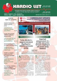
TABLE OF CONTENTS<br />
♥ Koronarografija<br />
kompjutoriziranom<br />
tomografijom<br />
Coronary computed<br />
tomography angiography p. <strong>106</strong><br />
♥ Prva implantacija<br />
kardioverter-defibrilatora u<br />
OpÊoj bolnici Dubrovnik<br />
The first implantation<br />
of cardioverter-defibrillator<br />
in the Dubrovnik<br />
General Hospital p. 113<br />
♥ <strong>Kardio</strong>loπka radionica u<br />
OpÊoj bolnici Dubrovnik<br />
Cardiac Workshop<br />
in the Dubrovnik<br />
General Hospital p. 115<br />
♥ Razmjena Ëasopisa<br />
Exchange of Journals p. 116<br />
♥ Zapisnik sa sjednice<br />
Upravnog odbora HKD<br />
17. svibnja 2010.<br />
Minutes of the Meeting<br />
of the Croatian Cardiac<br />
Society Management Board<br />
of 17 th May 2010 p. 117<br />
♥ Zapisnik sa sjednice<br />
Upravnog odbora HKD<br />
18. lipnja 2010.<br />
Minutes of the Meeting<br />
of the Croatian Cardiac<br />
Society Management<br />
Board of 18 th Juni 2010 p. 123<br />
♥ Uputa autorima<br />
Guidelines for authors p. 126<br />
♥ Gliklazid u tableti s<br />
prilagoappleenim oslobaappleanjem —<br />
sulfonilurea s dokazanom<br />
koristi kod pacijenata s<br />
dijabetesom tipa 2 u monoterapiji<br />
i kombiniranoj terapiji<br />
Gliclazide in modified release<br />
formulation — the sulphonylurea<br />
with proven benefits for patients<br />
with type 2 diabetes mellitus in<br />
monotherapy and in<br />
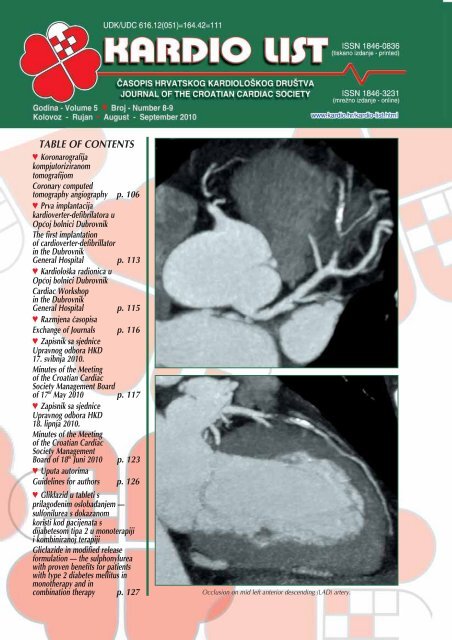
combination therapy p. 127 Occlusion on mid left anterior descending (LAD) artery.
2010;5(8-9):<strong>106</strong>.<br />
Pregledni rad<br />
Koronarografija kompjutoriziranom<br />
tomografijom<br />
Review article<br />
Coronary computed<br />
tomography angiography<br />
Mladen JukiÊ, Ladislav PaviÊ, Jasna »erkez-Habek*<br />
Poliklinika Sunce, Zagreb, Hrvatska<br />
Sunce Clinics, Zagreb, Croatia<br />
SAÆETAK: Cilj rada je prikazati najnovije kliniËke dokaze<br />
o korisnosti koronarografije kompjutoriziranom tomografijom<br />
(MSCT koronografija), ove joπ uvijek nove i kod nas nedovoljno<br />
etablirane metode, koja ima sve znaËajniju ulogu u dijagnostici<br />
koronarne bolesti srca (KBS). MSCT koronarografija<br />
predstavlja neinvazivnu dijagnostiËku metodu za evaluaciju<br />
KBS s odliËnim slikovnim prikazom i izvrsnom dijagnostiËkom<br />
preciznoπÊu koja je komparabilna s invazivnom koronarografijom<br />
koja se smatra zlatnim standardom u dijagnostici KBS.<br />
KLJU»NE RIJE»I: MSCT koronarografija, koronarna bolest<br />
srca, neinvazivna kardioloπka dijagnostika.<br />
SUMMARY: The aim of the article is to present the latest<br />
clinical evidence of benefit of coronary computed tomography<br />
angiography (CCTA), which is still a new and insufficiently established<br />
method in our country which plays more and more<br />
important role in the diagnostics of coronary heart disease<br />
(CHD). CCTA represents a non-invasive diagnostic method for<br />
the evaluation of CHD with excellent image presentation and<br />
excellent diagnostic precision that is comparable with invasive<br />
coronarography that is considered a golden standard in the<br />
CHD diagnostics.<br />
KEYWORDS: Coronary computed tomography angiography,<br />
coronary artery disease, noninvasive cardiac diagnostics.<br />
CITATION: <strong>Kardio</strong> list. 2010;5(8-9):<strong>106</strong>-<strong>112</strong>.<br />
Volume-rendering technique (VRT) presentation of functionaly significant bridge on mid LAD artery.<br />
Cilj ovog rada je prikazati kliniËke dokaze o korisnosti,<br />
koronarografije kompjutoriziranom tomografijom<br />
(MSCT koronarografija) joπ uvijek nove i kod nas nedovoljno<br />
etablirane metode, koja ima sve znaËajniju ulogu<br />
u dijagnostici koronarne bolesti srca (KBS). Ova bolest<br />
predstavlja vodeÊi uzrok smrtnosti u veÊini razvijenih zemalja,<br />
a za pravovremeno i uËinkovito lijeËenje KBS<br />
kljuËno je njeno rano otkrivanje. TragiËnu Ëinjenicu predstavlja<br />
πto je u 50-60% sluËajeva, u do tada asimptomatskih<br />
bolesnika, prvi znak bolesti infarkt miokarda, a<br />
nerijetko i iznenadna srËana smrt 1-13 .<br />
Temeljem analize podataka 398.978 pacijenata bez<br />
poznate KBS, ukljuËenih u American College of Cardiology<br />
National Cardiovascular Data Registry, u kojih je uËinjena<br />
elektivna koronarografija, znaËajni oblik KBS je potvrappleen<br />
u samo u 38% sluËajeva. Voditelji registra zakljuËu-<br />
The aim of this article is to present clinical evidence of<br />
benefit of coronary computed tomography angiography<br />
(CCTA), which is still a new and insufficiently established<br />
method in our country which plays more and<br />
more important role in the diagnostics of coronary heart<br />
disease (CHD). This disease is a leading cause of mortality<br />
in the great number of developed countries, while early detection<br />
of CHD is crucial for timely and efficient treatment.<br />
The tragic fact is that in some 50-60% of cases in asymptomatic<br />
patients so far, the first sign of disease is the myocardial<br />
infarction, and usually a sudden cardiac death 1-13 .<br />
Based on data analyses of 398,978 patients without<br />
known CHD, included in American College of Cardiology<br />
National Cardiovascular Data Registry, for whom elective<br />
coronarography was made, the more important form of<br />
CHD was confirmed in only 38% of cases. The Registry<br />
leaders conclude that the current existing strategies for
2010;5(8-9):107.<br />
ju da trenutno postojeÊe strategije za obradu pacijenata<br />
bez poznate KBS, a s postojeÊim rizikom, nisu zadovoljavajuÊe<br />
i potrebno ih je znaËajno unaprijediti 4 . Upravo ovdje<br />
vidimo najveÊu potencijalnu korist od MSCT koronarografije<br />
koja, ako se provodi na primjerenim ureappleajima i na<br />
primjeren naËin, moæe iskljuËiti KBS s negativnom prediktivnom<br />
vrijednoπÊu veÊom od 95% 5 i na taj naËin mogu se<br />
izbjeÊi daljnja nepotrebna testiranja poput perfuzijske<br />
scintigrafije i invazivne kardioloπke obrade koje imaju svoje<br />
rizike i komplikacije, a i znaËajno su skuplje od MSCT<br />
koronarografije.<br />
workup of patients without known CHD with the existing<br />
risk are not satisfactory and they need to be greatly improved<br />
4 . This is where we see the greatest potential benefit<br />
from CCTA which, if conducted by using adequate devices<br />
and in an adequate manner, may exclude CHD with negative<br />
predictive value greater than 95% 5 and this is how<br />
further unnecessary tests such as perfusion scintigraphy<br />
and invasive cardiac diagnostics may be avoided accompanied<br />
by their risks and complications, and they are much<br />
more expensive than CCTA.<br />
A. Coronary computed tomography angiography: subtotal<br />
stenosis of left circumflex artery.<br />
Postupak oslikavanja<br />
Oslikavanje je potrebno vrπiti na CT-ureappleajima posebno<br />
konstruiranim za snimanje srca i koronarnih arterija.<br />
Prema danaπnjim standardima radi se o ureappleajima s minimalno<br />
64 uzastopna sloja oslikavanja. Jedino na ovakvim<br />
ureappleajima moguÊe je postiÊi zadovoljavajuÊu kvalitetu<br />
snimke uz prihvatljivo nisku dozu zraËenja. Ureappleaj takoappleer<br />
mora minimalno omoguÊavati i modulaciju intenziteta<br />
zraËenja ovisno o EKG-u pacijenta te anatomskoj graapplei.<br />
Frekvenciju rada srca je potrebno sniziti na maksimalno<br />
65/min. Ukoliko pacijent ima viπu frekevenciju tada se<br />
ordinira beta blokator per os ili ËeπÊe parenteralno (Tenormin,<br />
amp. á 5 mg, doza lijeka se individualno prilagoappleava).<br />
Neposredno prije samog snimanja ordinira se i nitrat<br />
kratkog djelovanja sublingvalno (Nitrolingual spray) uz<br />
kontrolu krvnog tlaka. Ovo istovremeno omoguÊava koriπtenje<br />
protokola s malim dozama zraËenja te maksimalno<br />
suæavanje podruËja oslikavanja u sve tri prostorne ravnine.<br />
Takoappleer je potrebno jakost i napon struje na RTG-cijevi<br />
individualno prilagoditi svakom pacijentu obzirom na<br />
indeks tjelesne mase (BMI), opseg prsiπta, frekvenciju i stabilnost<br />
rada srca.<br />
Na ovaj naËin moguÊe je znaËajno smanjiti doze<br />
zraËenja, koje kod uobiËajene populacije mogu biti u razini<br />
2-3 mSv, a kod idealnih pacijenata (BMI ≤25, stabilna<br />
frekvencija ≤60/min) i ispod 1 mSv 6 . Uporabom neprimjerenih<br />
ureappleaja te uz neprimjeren postupak oslikavanja,<br />
Ëak i na vrhunskim ureappleajima, doze zraËenja mogu biti i<br />
viπestruko veÊe.<br />
B. Invasive coronarography: before percutaneous coronary<br />
intervention.<br />
Imaging procedure<br />
Imaging is to be done on CT devices especially designed<br />
for imaging of heart and coronary arteries. According<br />
to the recent standards, these are the devices with minimum<br />
64 consecutive imaging slices. This is the only device<br />
that may provide satisfactorily quality of the image<br />
with an acceptable low level of radiation. The device must<br />
minimally ensure modulation of the radiation intensity depending<br />
on patient’s ECG and anatomical structure.<br />
The heart rate needs to be lowered to maximum<br />
65/min. If a patient has a higher rate, then beta blocker per<br />
os or more often parenterally are administered (Tenormin,<br />
amp. á 5 mg, medication dose is adjusted individually). Immediately<br />
prior to the imaging itself, nitrate with short effect<br />
is administered sublingually (Nitrolingual spray) thereby<br />
controlling blood pressure. This simultaneously enables<br />
using protocol with small radiation doses and maximum<br />
reduction of imaging area in all t<strong>hr</strong>ee spatial plains.<br />
Strong current and voltage in X-ray pipes need to be individually<br />
adjusted for every patient considering the body<br />
mass index (BMI), thorax range, and heart beat rate and stability.<br />
In this way it is possible to greatly reduce the radiation<br />
doses that in ordinary population may be at the level 2-3<br />
mSv, and in ideal patients (BMI ≤25, stabile heart rate<br />
≤60/min) and below 1 mSv 6 . The use of inappropriate devices<br />
and application of inappropriate imaging procedure<br />
even on top quality devices may lead to radiation doses<br />
higher by several times.
2010;5(8-9):108.<br />
U svakodnevnom radu primjenjuju se preporuke American<br />
Heart Association te International Commission on Radiation<br />
Protection poznatije kao ALARA (As Low As Reasonably<br />
Achievable) protokol 7 .<br />
Nakon rapidnog tehnoloπkog razvoja u posljednjem<br />
desetljeÊu MSCT koronarografija je dokazano najpreciznija<br />
neinvazivna slikovna tehnika koja s 99% preciznoπÊu<br />
moæe iskljuËiti KBS. Danas se primjenjuje u viπe od 2.000<br />
centara samo u Sjedinjenim AmeriËkim Dræavama i pokrivena<br />
je zdravstvenim osiguranjem u svih 50 dræava. Metoda<br />
se sve viπe primjenjuje i u ostalim zemljama diljem svijeta,<br />
a od 2007. godine i u Republici Hrvatskoj. U Poliklinici<br />
Sunce u Zagrebu od oæujka 2007. do sredine 2010.<br />
godine uËinjeno je viπe od 3.000 MSCT koronarografija, a<br />
kliniËka zapaæanja i iskustva podudaraju se s navedenim<br />
evidence-based rezultatima 8 .<br />
Razvojem CT ureappleaja sa 64 i viπe slojeva, posebno<br />
konstruiranih za oslikavanje srca, MSCT koronarografija je<br />
vrlo brzo postala etablirana metoda u oslikavanju koronarnih<br />
arterija, a kod sumnje na priroappleenu anomaliju koronarnih<br />
arterija i metoda izbora.<br />
In routine daily work, the recommendations by the<br />
American Heart Association and International Commission<br />
on Radiation Protection known as ALARA (As Low As Reasonably<br />
Achievable) protocols are used. 7<br />
Following the rapid technologic development during<br />
the last decade, CCTA is proved to be the most precise<br />
noninvasive imaging technique that with 99% precision<br />
may exclude CHD. Today it is used in more than 2,000<br />
centers only in the United States of America and it is covered<br />
by health insurance in all 50 states. The method is<br />
more and more applied in other countries all over the<br />
world and since 2007 it has been applied in Croatia as<br />
well. More than 3,000 CCTA were conducted from March<br />
2007 till mid 2010 in the Sunce Polyclinic in Zagreb, while<br />
clinical observations and experience overlap with the aforementioned<br />
evidence-based results 8 .<br />
With development of CT device with 64 slices and<br />
more than that, especially created for imaging the heart,<br />
CCTA has soon become a very established method in imaging<br />
coronary arteries and in case of suspecting congenital<br />
anomaly of coronary arteries it has become a method of<br />
choice.<br />
The anomalous starting point of the right coronary artery (RCA) from the left main coronary artery. RCA passes between the aorta and<br />
artery pulmonalis which may be dangerous for life.
2010;5(8-9):109.<br />
Indikacije za MSCT koronarografiju<br />
Indikacije za MSCT koronarografiju 9,10 :<br />
• Bol u prsima kod pacijenta s intermedijarnim rizikom<br />
• Dvojben razultat stres testa<br />
• Anomalije koronaranih arterija<br />
• Evaluacija kardiomiopatije<br />
• Preoperativna obrada kod nekoronaranih kardijalnih<br />
operacija<br />
• Suspektna patologija aorte ili pluÊne arterije<br />
• Prije i poslje elektrofizioloπkih ispitivanja.<br />
Novije indikacije su vezane su uz 64-slojni CT:<br />
• Evaluacija boli u prsima ili zaduhe kod pacijenta s<br />
prethodnim aortokoronarnim premoπtenjem ili implantacijom<br />
stenta<br />
• Evaluacija akutne boli u prsima u hitnoj sluæbi.<br />
Temeljna uloga MSCT koronarografije, zbog njene visoke<br />
negativne prediktivne vrijednosti, je iskljuËenje signifikantne<br />
koronarne bolesti srca kod simptomatskih pacijenata<br />
s niskim ili srednjim rizikom. Pacijenti s tipiËnom<br />
kliniËkom slikom i visokim rizikom za koronarnu bolest s<br />
jasno pozitivnim testom optereÊenja imaju indikaciju za<br />
invazivnu koronarografiju. Primjena MSCT koronarografije<br />
kod asimptomatskih pacijenata se ne preporuËa.<br />
Danaπnji ureappleaji imaju temporalnu rezoluciju ≤150<br />
msec πto uz submilimetarsku prostornu rezoluciju omogu-<br />
Êava vjeran prikaz koronarnih arterija kod veÊine pacijenata.<br />
S obzirom da se ovom metodom direktno prikazuje i<br />
stijenka krvnih æila, CT je nakon intravaskularnog ultrazvuka<br />
(IVUS) najosjetljivija metoda za prikaz aterosklerotskog<br />
plaka. ZnaËajno je osjetljivija od invazivne koronarografije<br />
koja prikazuje samo prohodan lumen krvne æile.<br />
Indications for CCTA<br />
Indications for CCTA 9,10 :<br />
• Chest pain with patients with intermediary risk<br />
• An inconclusive stress test<br />
• Coronary artery anomalies<br />
• Cardiomyopathy evaluation<br />
• Pre-operative workup with non-coronary cardiac<br />
operations<br />
• Suspect aorta and pulmonary artery pathology<br />
• Prior and following electrophysiologic procedures.<br />
The most recent indications are related with 64-slice<br />
CT:<br />
• Evaluation of chest pain and dyspnoea with patients<br />
with previous aortocoronary bypass or stent implantation<br />
• Evaluation of acute chest pain in emergency.<br />
The basic role of CCTA due to its negative predictive<br />
value is the exclusion of significant coronary heart disease<br />
with symptomatic patients with low or middle risk. The patients<br />
with typical clinical manifestations and high risk for<br />
coronary disease with clearly positive stress test have indication<br />
for invasive coronarography. The use of CCTA with<br />
asymptomatic patients is not recommended.<br />
The modern devices have temporal resolution of ≤150<br />
msec which with submilimeter spatial resolution provides<br />
a true presentation of coronary arteries in a great number<br />
of patients. Since the wall of blood vessels is directly<br />
shown by applying this method, CT is after intravascular<br />
ultrasound (IVUS) the second most sensitive method for<br />
presenting atherosclerotic plaque. It is much more sensitive<br />
than the invasive coronarography which only shows<br />
the passable lumen of the blood vessel.<br />
The images of invasive coronarographies with nonsignificant stenosis of left main coronary artery.<br />
The MSCT coronarography images after the invasive diagnostics and eight months later: reduction of the volume of mixed plaque in left<br />
main coronary artery with much higher density of plaque 8 months later (70HU vs. 35 HU) which indicates stabilization of the plaque<br />
by applying pharmacological therapy.
2010;5(8-9):110.<br />
Uz navedeno CT omoguÊava analizu graapplee plaka, buduÊi<br />
da moæe razlikovati masni sadræaj od kalcija pa Ëak i<br />
veziva. Novija istraæivanja pokazuju da je, posebno kod<br />
veÊih krvnih æila, moguÊe pouzdano razlikovati plak koji<br />
je zbog “mekoÊe” svog sadræaja nestabilan 14, 16 .<br />
Prema najnovijim studijama 64-slojni CT ima vrlo dobru<br />
korelaciju s IVUS u evaluaciji aterosklerotskog plaka<br />
pa se postavlja pitanje da li je IVUS joπ zaista “zlatni standard”<br />
za evaluaciju aterosklerotskog plaka?<br />
Glavno ograniËenje ove metode je bila razmjerno visoka<br />
doza zraËenja. Razvojem tehnologije ovdje su uËinjeni<br />
znaËajni pomaci te se danas ne preporuËuje izvoditi MSCT<br />
koronarografiju na ureappleajima s manje od 64 sloja. Ovo je<br />
dovelo i do bitnog proπirenja indikacija, kao πto su oslikavanje<br />
koronarnih stentova i premosnica, Ëije oslikavanje<br />
na starijim generacijama MSCT nije bilo zadovoljavaju-<br />
Êe 11 .<br />
Starija istraæivanja doze zraËenja kod CT koronarografije<br />
davala su vrlo πarolike rezultate, Ëesto s neprihvatljivo<br />
visokim dozama zraËenja (do 30 mSv ekspozicijske doze).<br />
Ovakvi rezultati bili su posljedica koriπtenja neprimjerene<br />
tehnologije (ureappleaji s 4, 6 ili 16 slojeva) te nedovoljne<br />
vjeπtine u pripremi i oslikavanju pacijenata.<br />
Obzirom da doza zraËenja kod MSCT uvelike ovisi o<br />
pripremi pacijenta i parametrima snimanja, ovo iziskuje<br />
posebno educirani kardioloπko-radioloπki tim i individual-<br />
Besides the above entioned, CT enables an analysis of<br />
the plaque composition since it may differentiate between<br />
fat contents and calcium and connective tissue as well. The<br />
recent researches show that especially in larger blood vessels<br />
it is with reliability possible to differ plaque that is due<br />
to its “softness” of its contents instable 14, 16 .<br />
According to the most recent studies 64-slice CT has a<br />
very good correlation with IVUS in evaluation of atherosclerotic<br />
plaque, so the issue is raised whether IVUS is still<br />
a “golden standard” for the evaluation of atherosclerotic<br />
plaque?<br />
The main limitation of this method was proportionally<br />
high radiation dose. The development of the technology<br />
has resulted in significant improvements, consequently today<br />
CCTA is not recommended to be done on devices having<br />
fewer than 64 slices. This has led to significant widening<br />
of the indications such as imaging of coronary stents<br />
and bypasses whose imaging on older multislice CT generations<br />
was not satisfactory 11 .<br />
Subsequent researches of radiation dose during CT<br />
coronarography provided very different results, often with<br />
unacceptable high radiation dose (maximum 30 mSv exposition<br />
dose). Such results were the consequence of using<br />
inappropriate technology (devices with 4, 6 or 16 slices)<br />
and insufficient skill in preparing and imaging patients.<br />
Since the radiation dose in CCTA greatly depends on<br />
the preparation of a patient and imaging parameters, this<br />
requires especially educated cardiac-radiologic team and<br />
The condition following the quaternary aortocoronary bypass, two vein bypasses occluded, while the bypasses for the LAD and RCA as<br />
well as the stents in them are passable.
2010;5(8-9):111.<br />
ni pristup svakom pacijentu, kako bi se uz Ëim manju dozu<br />
zraËenja dobile snimke odgovarajuÊe dijagnostiËke kakvoÊe.<br />
Novija istraæivanja na 64-slojnim ureappleajima i uz koriπtenje<br />
strategija za sniæavanje doze zraËenja, poput vjerojatno<br />
najrelevantnije PROTECT-I studije, pokazuju znaËajno<br />
manje doze zraËenja 6, 15, 17 . Na ovoj generaciji ureappleaja,<br />
uz njihovo primjereno koriπtenje, ekspozicijske doze zapravo<br />
su usporedive s onima kod invazivne koronarografije.<br />
Primjerice prosjeËna ekspozicijska doza kod CT koronarografije<br />
je izmjerena na oko 9 mSv, a kod invazivne<br />
koronarografije na oko 7 mSv. S obzirom na nepouzdanost<br />
mjerenja te metodologiju izraËuna ekspozicijske doze,<br />
prema preporukama International Commission on Radiation<br />
Protection razlike u efektivnoj dozi do faktora 2 se zapravo<br />
ne smatraju znaËajnima 6, 17, 18 .<br />
Takoappleer je bitno napomenuti da se kod CT koronarografije<br />
zraËi iskljuËivo pacijent, dok je kod invazivne obrade<br />
zraËenju izloæen cijeli dijagnostiËki tim.<br />
U PROTECT-I studiji je koriπtena generacija ureappleaja iz<br />
2005. godine 17 . Odonda je CT tehnologija znaËajno napredovala<br />
te danaπnji ureappleaji postiæu doze od 2-3 mSv kod<br />
veÊine pacijenata, a optimalne pacijente mogu se oslikavati<br />
i s dozama ispod 1 mSv 19 . Ovdje naæalost joπ ne postoji<br />
nezavisnih studija o dozi zraËenja, no nekoliko takvih je u<br />
tijeku.<br />
U Tablici 1 su prikazane efektivne doze zraËenja kod<br />
razliËitih metoda oslikavanja koje je na temelju najrecentnijih<br />
istraæivanja sastavilo posebno tijelo American Heart<br />
Association 6 .<br />
individual approach to every patient as to obtain radiograms<br />
of adequate diagnostic quality with the smallest possible<br />
radiation.<br />
Recent researches on 64-slice devices thereby using<br />
strategies for reduction of the radiation dose such as probably<br />
the most relevant PROTECT-I study show considerably<br />
smaller radiation dose 6, 15, 17 . The exposition doses are<br />
actually on this device generation thereby using them adequately<br />
comparable with those used in invasive coronarography.<br />
For example, an average exposition dose in CT coronarography<br />
is measured at 9 mSv, and in non-invasive<br />
coronarography at around 7 mSv. Considering lack of reliability<br />
of measurements and methods of calculation of exposition<br />
dose according to recommendations of International<br />
Commission on Radiation Protection the differences<br />
in effective dose to factor 2 are actually not considered important<br />
6, 17, 18 .<br />
It is worth mentioning that only a patient is radiated in<br />
case of CT caronarography, while the whole diagnostic<br />
team is exposed to radiation in case of invasive diagnostics.<br />
In PROTECT-I study the device generation from 2005<br />
was used 17 . Since then, CT technology has been greatly improved<br />
and today’s devices reach doses of 2-3 mSv in most<br />
of the patients, while optimum patients may be imaged<br />
with the doses below 1 mSv 19 . Here, unfortunately there<br />
are no non-independent studies of radiation doses, but several<br />
studies of that type are underway.<br />
In Table 1 effective radiation doses are presented when<br />
using different imaging methods prepared according to the<br />
most recent researches by a special body American Heart<br />
Association 6 .<br />
Table 1. Representative values and<br />
ranges of effective dose estimates reported<br />
in the literature for selected radiological<br />
studies.<br />
CTA = CT angiography<br />
*CT studies published since 2005 only<br />
Adapted from reference 6.<br />
OgraniËenja MSCT koronarografije<br />
MSCT koronarografija ima i svoja ograniËenja. Velika<br />
koliËina kalcija na koronarnim arterijama ometa evaluaciju<br />
prohodnosti koronarne arterije te je kod pacijenta s izrazitim<br />
kalcifikatima nemoguÊe procijeniti stupanj eventualne<br />
stenoze 20 . Evaluacija luminalne stenoze oteæana je<br />
kod pacijenta koji imaju ubrzanu frekvenciju srca, fibrilaciju<br />
atrija ili uËestalu ekstrasistoliju.<br />
Respiratorni artefakt je moguÊ ukoliko pacijent nije<br />
suradljiv te ne prati upute o zadræavanju daha tijekom<br />
oslikavanja ili to ne moæe uËiniti. Potrebno je naglasati da<br />
izrazito pretili pacijenti, Ëiji je BMI preko 30, nisu optimalni<br />
kanditati za MSCT koronarografiju. MoguÊa je alergijska<br />
reakcija na kontrasno sredstvo.<br />
Limitations of CCTA<br />
MSCT coronarography has its limits. The great quantity<br />
of calcium in coronary arteries obstructs evaluation of passage<br />
of coronary artery, and with patients with very high<br />
calcificates it is impossible to evaluate a degree of potential<br />
stenosis 20 . The evaluation of luminal stenosis is made<br />
difficult with patients having accelerated heart frequency,<br />
atrial fibrillation or frequent premature beats.<br />
Respiratory artifact is possible if a patient is not cooperative<br />
and does not follow instructions for keeping breath<br />
during imaging or if he/she cannot do it. It is worth mentioning<br />
that very obese patients whose BMI is over 30 are<br />
not good candidates for CCTA. Allergic reaction to contrast<br />
agent is possible.
2010;5(8-9):<strong>112</strong>.<br />
ZakljuËak<br />
Sve viπe se postavlja pitanje je li je MSCT koronarografija<br />
spremna zamijeniti dijagnostiËku invazivnu koronarografiju,<br />
a neki se Ëak pitaju: je li doπao kraj invazivnoj koronarografiji<br />
kao dijagnostiËkoj metodi?<br />
21, 22<br />
S tim u vezi mogli bi zakljuËiti da invazivna koronarografija<br />
u kombinaciji s izrazito pozitivnim stres testom ostaje<br />
metoda izbora kod pacijenata s visokom vjerojatnoπÊu<br />
KBS i oËekivanom koronarnom intervencijom (PCI). Meappleutim,<br />
kod veÊine pacijenata s malim ili srednjim rizikom<br />
KBS metoda MSCT koronarografije moæe biti pouzdana,<br />
kliniËki uËinkovita i ekonomski isplativa neinvazivna alternativa<br />
invazivnoj koronarografiji.<br />
Conclusion<br />
The issue as to whether MSCT is to replace diagnostic<br />
invasive coronarography is raised more and more, and some<br />
people are wondering: is the invasive coronarography<br />
21, 22<br />
as a diagnostic method close to its end?<br />
In connection with this, we may conclude that invasive<br />
coronarography combined with extremely positive stress<br />
test remains the method of choice for patients with high<br />
probability of CHD and expected coronary intervention<br />
(PCI). However, in most patients with small or middle risk<br />
CHD method of CCTA may be reliable, clinically efficient<br />
and economically profitable non-invasive alternative to invasive<br />
coronarography.<br />
Received: 23 rd Jun 2010 Updated 8 th Jul 2010<br />
*Address for correspondence: Poliklinika Sunce, Trnjanska cesta<br />
108, HR-10000 Zagreb, Croatia<br />
Phone: +385-1-5497555 Fax: +385-1-5497509<br />
E-mail: jasna.cerkez-habek@sunce.<strong>hr</strong><br />
Literature<br />
1. Hoffmann MH, Shi H, Schmitz BL, Schmid FT, Lieberknecht M, Schulze R, et al. Noninvaisve coronary angiography with multislice computed tomography.<br />
JAMA. 2005;293:2471-8.<br />
2. Miller JM, Rochitte CE, Dewey M, Rbab-Zadeh A, Niinuma H, Gottlieb I, et al. Diagnostic performance of coronary angiography by 64-row CT. N<br />
Engl J Med. 2008;2324-36.<br />
3. Meijboom WB, Meijs MF, Schuijf JD, Cramer MJ, Mollet NR, van Mieghem CA, et al. Diagnostic accuracy of 64-slice computed tomography coronary<br />
angiography: a prospective, multicenter, multivendor study. J Am Coll Cardiol. 2008;2135-44.<br />
4. Patel MR, Peterson ED, Dai D, Brennan JM, Redberg RF, Anderson HV, et al. Low diagnostic yield of elective coronary angiography. N Engl J Med.<br />
2010;362:886-95.<br />
5. Budoff MJ, Dowe D, Jollis JG, Gitter M, Sutherland J, Halamert E, et al. Diagnostic performance of 64-multidetector row coronary computed tomographic<br />
angiography for evaluation of coronary artery stenosis in individuals without known coronary artery disease: results from the prospective multicenter<br />
ACCURACY (Assessment by Coronary Computed Tomographic Angiography of Individuals Undergoing Invasive Coronary Angiography) trial. J Am Coll<br />
Cardiol. 2008;52:1724-32.<br />
6. Gerber TC, Carr JJ, Arai AE, Dixon RL, Ferrari VA, Gomes AS, et al. Ionizing radiation in cardiac imaging: a science advisory from the American Heart<br />
Association Committee on Cardiac Imaging of the Council on Clinical Cardiology and Committee on Cardiovascular Imaging and Intervention of the Council<br />
on Cardiovascular Radiology and Intervention. Circulation. 2009;119(7):1056-65.<br />
7. Rehani MM. Managing patient dose in computed tomography (CT). http://www.icrp.org/docs/icrp_87_ct_s.pps (8. 7. 2010)<br />
8. JukiÊ M, PaviÊ L, »erkez-Habek J, MedakoviÊ P. Cardiac CT angiography - State of art and our preliminary results. LijeË Vjesn. 2008;3 Suppl:58.<br />
9. Budoff MJ, Achenbach S, Blumenthal RS, et al. Assessment of coronary artery disease by cardiac computed tomography: a scientific statement from<br />
the American Heart Association Committee on Cardiovascular Imaging and Intervention, Council on Cardiovascular Radiology and Intervention, and Committee<br />
on Cardiac Imaging, Council on Clinical Cardiology. Circulation. 2006;114:1761-91.<br />
10. American College of Cardiology Foundation Task Force on Expert Consensus Documents, Mark DB, Berman DS, Budoff MJ, Carr JJ, Gerber TC,<br />
Hecht HS, et al. ACCF/ACR/AHA/NASCI/SAIP/SCAI/SCCT 2010 expert consensus document on coronary computed tomographic angiography: a report of<br />
the American College of Cardiology Foundation Task Force on Expert Consensus Documents. Circulation. 2010;121:2509-43.<br />
11. Yusuf S. Reddy S, Ounpuu S, Anand S. Global Burden of Cardiovascular Diseases: Part I: General Considerations, the Epidemiologic Transition, Risk<br />
Factors, and Impact of Urbanization. Circulation. 2001;104:2746-53.<br />
12. American Heart Association. Heart disease and Stroke Statistics - 2009. Update, American Heart Association, Dallas, TX, USA, 2009.<br />
13. The World Health Organization. The World Health Organization Web Site - Cardiovascular Diseases, World Health Organization, Geneva Switzerland,<br />
2004.<br />
14. JukiÊ M, PaviÊ L, »erkez-Habek J, ©ikiÊ A, TiËinoviÊ K, MedakoviÊ P, et al. Uloga kompjuterizirane tomografije u evaluaciji aterosklerotskog plaka:<br />
prikaz pacijenta. LijeË Vjesn. 2009;2 Supl:90-1.<br />
15. ACCF/ACR/SCCT/SCMR/ASNC/NASCI/SCAI/SIR 2006 appropriateness criteria for cardiac computed tomography and cardiac magnetic resonance<br />
imaging: a report of the American College of Cardiology Foundation Quality Strategic Directions Committee Appropriateness Criteria Working Group, American<br />
College of Radiology, Society of Cardiovascular Computed Tomography, Society for Cardiovascular Magnetic Resonance, American Society of Nuclear<br />
Cardiology, North American Society for Cardiac Imaging, Society for Cardiovascular Angiography and Interventions, and Society of Interventional Radiology.<br />
J Am Coll Cardiol. 2006;48:1475-97.<br />
16. Achenbach S, Raggi P. Imaging of coronary atherosclerosis by computed tomography. Eur Heart J. 2010;31:1442-8.<br />
17. Hausleiter J, Meyer T, Hermann F, Hadamitzky M, Krebs M, Gerber TC, et al. Estimated radiation dose associated with cardiac CT angiography.<br />
JAMA. 2009;301:500-7.<br />
18. International Commission on Radiological Protection (ICRP). 2007 Recommendations of the International Commission on Radiological Protection<br />
(publication 103). Ann ICRP. 2007;37(2-4):1-332.<br />
19. Leipsic J. Oral abstract 03. Presented at: Society of Cardiovascular Computed Tomography 4th Annual Scientific Meeting; July 16-19, 2009.<br />
20. Min JK, Berman D. Anatomic and functional assessment of coronary artery disease: convergence of 2 aims in a single setting. Circ Cardivasc Imaging.<br />
2009;2(3).163-5.<br />
21. Nieman K. Can CT angiography replace catheter coronary angiography? EuroIntervention. 2010;SupplG:G65-71.<br />
22. Gonçalves Pde A, Marques H. Cardiac CT: the end of invasive coronary angiography as a diagnostic procedure? Rev Port Cardiol. 2009;28:825-42.