Download - Kimberly-Clark Health Care
Download - Kimberly-Clark Health Care
Download - Kimberly-Clark Health Care
Create successful ePaper yourself
Turn your PDF publications into a flip-book with our unique Google optimized e-Paper software.
e<br />
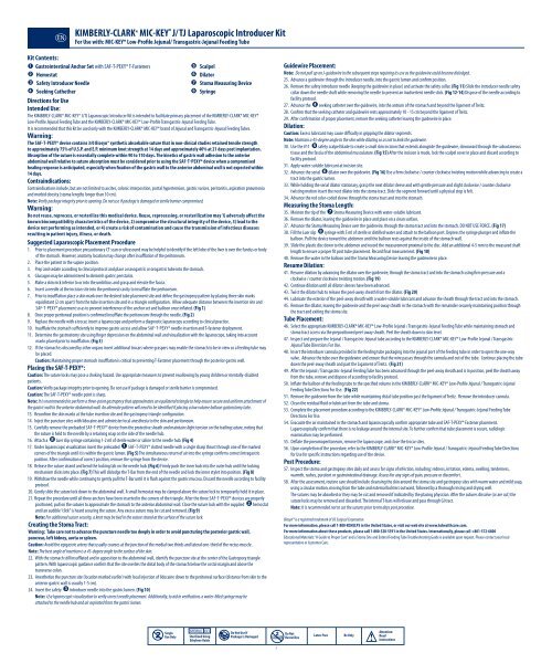
KIMBERLY-CLARK* MIC-KEY * J/ TJ Laparoscopic Introducer Kit<br />
For Use with: MIC-KEY* Low-Profile Jejunal/ Transgastric-Jejunal Feeding Tube<br />
Kit Contents:<br />
Gastrointestinal Anchor Set with SAF-T-PEXY* T-Fasteners<br />
Scalpel<br />
Hemostat<br />
Dilator<br />
Safety Introducer Needle<br />
Stoma Measuring Device<br />
Seeking Cathether<br />
Syringe<br />
Directions for Use<br />
Intended Use:<br />
The KIMBERLY-CLARK* MIC-KEY* J/TJ Laparoscopic Introducer Kit is intended to facilitate primary placement of the KIMBERLY-CLARK* MIC-KEY*<br />
Low-Profile Jejunal Feeding Tube and the KIMBERLY-CLARK* MIC-KEY* Low-Profile Transgastric-Jejunal Feeding Tube.<br />
It is recommended that this kit be used only with the KIMBERLY-CLARK* MIC-KEY* brand of Jejunal and Transgastric-Jejunal Feeding Tubes.<br />
Warning:<br />
The SAF-T-PEXY* device contains 3/0 Biosyn® synthetic absorbable suture that in non-clinical studies retained tensile strength<br />
to approximately 75% of U.S.P. and E.P. minimum knot strength at 14 days and approximately 40% at 21 days post implantation.<br />
Absorption of the suture is essentially complete within 90 to 110 days. The kinetics of gastric wall adhesion to the anterior<br />
abdominal wall relative to suture absorption must be considered prior to using the SAF-T-PEXY* device when a compromised<br />
healing response is anticipated, especially when fixation of the gastric wall to the anterior abdominal wall is not expected within<br />
14 days.<br />
Contraindications:<br />
Contraindications include, but are not limited to ascites, colonic interposition, portal hypertension, gastric varices, peritonitis, aspiration pneumonia<br />
and morbid obesity (stoma lengths longer than 10 cm).<br />
Note: Verify package integrity prior to opening. Do not use if package is damaged or sterile barrier compromised.<br />
Warning:<br />
Do not reuse, reprocess, or resterilize this medical device. Reuse, reprocessing, or resterilization may 1) adversely affect the<br />
known biocompatibility characteristics of the device, 2) compromise the structural integrity of the device, 3) lead to the<br />
device not performing as intended, or 4) create a risk of contamination and cause the transmission of infectious diseases<br />
resulting in patient injury, illness, or death.<br />
Suggested Laparoscopic Placement Procedure<br />
1. Prior to placement procedure precautionary CT scan or ultrasound may be helpful to identify if the left lobe of the liver is over the fundus or body<br />
of the stomach. However, anatomy location may change after insufflation of the peritoneum.<br />
2. Place the patient in the supine position.<br />
3. Prep and sedate according to clinical protocol and place a nasogastric or orogastric tube into the stomach.<br />
4. Glucagon may be administered to diminish gastric peristalsis.<br />
5. Make a skin nick inferior to or into the umbilicus and grasp and elevate the fascia.<br />
6. Insert a needle at the incision site into the peritoneal cavity to insufflate the peritoneum.<br />
7. Prior to insufflation place a skin mark over the desired tube placement site and define the gastropexy pattern by placing three skin marks<br />
equidistant (2 cm apart) from the tube insertion site and in a triangle configuration. Allow adequate distance between the insertion site and<br />
SAF-T-PEXY* placement so as to prevent interference of the anchor set and balloon once inflated. (Fig 1)<br />
8. Once proper peritoneal position is confirmed insufflate the peritoneum through the needle. (Fig 2)<br />
9. Replace the needle with a trocar, insert a laparoscope and perform a diagnostic laparoscopy according to clinical practice.<br />
10. Insufflate the stomach sufficiently to improve gastric access and allow SAF-T-PEXY* needle insertion and T-fastener deployment.<br />
11. Determine the gastrostomy site using finger depression on the abdominal wall and visualization with the laparoscope, taking into account<br />
marks placed prior to insufflation. (Fig 3)<br />
12. If the stomach is obscured by other organs insert additional trocars where graspers may enable the stomach to be in view so a feeding tube may<br />
be placed.<br />
Caution: Maintaining proper stomach insufflation is critical to preventing T-Fastener placement through the posterior gastric wall.<br />
Placing the SAF-T-PEXY*:<br />
Caution: The suture locks may pose a choking hazard. Use appropriate measures to prevent swallowing by young children or mentally-disabled<br />
patients.<br />
Caution: Verify package integrity prior to opening. Do not use if package is damaged or sterile barrier is compromised.<br />
Caution: The SAF-T-PEXY* needle point is sharp.<br />
Note: It is recommended to perform a three-point gastropexy that approximates an equilateral triangle to help ensure secure and uniform attachment of<br />
the gastric wall to the anterior abdominal wall. An alternate pattern will need to be identified if placing a low volume balloon gastrostomy tube.<br />
13. Reconfirm the skin marks at the tube insertion site and the gastropexy triangle configuration.<br />
14. Inject the puncture sites with lidocaine and administer local anesthesia to the skin and peritoneum.<br />
15. <strong>Care</strong>fully remove the preloaded SAF-T-PEXY* device from the protective sheath and maintain slight tension on the trailing suture, noting that<br />
the suture is held to the needle by a retaining snap on the side of the needle hub.<br />
16. Attach a Luer slip syringe containing 1-2 ml of sterile water or saline to the needle hub. (Fig 4)<br />
17. Under laparoscopic visualization insert the preloaded SAF-T-PEXY* slotted needle with a single sharp thrust through one of the marked<br />
corners of the triangle until it is within the gastric lumen. (Fig 5) The simultaneous return of air into the syringe confirms correct Intragastric<br />
position. After confirmation of correct position, remove the syringe from the device.<br />
18. Release the suture strand and bend the locking tab on the needle hub. (Fig 6) Firmly push the inner hub into the outer hub until the locking<br />
mechanism clicks into place. (Fig 7) This will dislodge the T-Bar from the end of the needle and lock the inner stylet into position. (Fig 8)<br />
19. Withdraw the needle while continuing to gently pull the T-Bar until it is flush against the gastric mucosa. Discard the needle according to facility<br />
protocol.<br />
20. Gently slide the suture lock down to the abdominal wall. A small hemostat may be clamped above the suture lock to temporarily hold it in place.<br />
21. Repeat the procedure until all three anchors have been inserted in the corners of the triangle. After the three SAF-T-PEXY* devices are properly<br />
positioned, pull on the sutures to approximate the stomach to the anterior abdominal wall. Close the suture lock with the supplied hemostat<br />
until an audible “click” is heard securing the suture. Any excess suture may be cut and removed. (Fig 9)<br />
Note: For additional suture security, a knot may be tied in the suture strand at the surface of the suture lock.<br />
Creating the Stoma Tract:<br />
Warning: Take care not to advance the puncture needle too deeply in order to avoid puncturing the posterior gastric wall,<br />
pancreas, left kidney, aorta or spleen.<br />
Caution: Avoid the epigastric artery that usually courses at the junction of the medial two thirds and lateral one-third of the rectus muscle.<br />
Note: The best angle of insertion is a 45-degree angle to the surface of the skin.<br />
22. With the stomach still insufflated and in apposition to the abdominal wall, identify the puncture site at the center of the Gastropexy triangle<br />
pattern. With laparoscopic guidance confirm that the site overlies the distal body of the stomach below the costal margin and above the<br />
transverse colon.<br />
23. Anesthetize the puncture site (location marked earlier) with local injection of lidocaine down to the peritoneal surface (distance from skin to the<br />
anterior gastric wall is usually 1-5 cm).<br />
24. Insert the safety introducer needle into the gastric lumen. (Fig 10)<br />
Note: Use laparoscopic visualization to verify correct needle placement. Additionally, to aid in verification, a water-filled syringe may be<br />
attached to the needle hub and air aspirated from the gastric lumen.<br />
Guidewire Placement:<br />
Note: Do not pull up on J-guidewire in the subsequent steps requiring its use as the guidewire could become dislodged.<br />
25. Advance a guidewire through the introducer needle, into the gastric lumen and confirm position.<br />
26. Remove the safety introducer needle (keeping the guidewire in place) and activate the safety collar. (Fig 11) Slide the introducer needle safety<br />
collar down the needle shaft while removing the needle to prevent an inadvertent needle stick. (Fig 12-14) Dispose of the needle according to<br />
facility protocol.<br />
27. Advance the seeking catheter over the guidewire, into the antrum of the stomach and beyond the ligament of Treitz.<br />
28. Confirm that the seeking catheter and guidewire rests approximately 10 - 15 cm beyond the ligament of Treitz.<br />
29. After confirmation of proper placement, remove the seeking catheter leaving the guidewire in place.<br />
Dilation:<br />
Caution: Excess lubricant may cause difficulty in gripping the dilator segments.<br />
Note: Maintain a 45-degree angle to the skin while dilating so as not to kink the guidewire.<br />
30. Use the #11 safety scalpel blade to create a small skin incision that extends alongside the guidewire, downward through the subcutaneous<br />
tissue and the fascia of the abdominal musculature. (Fig 15) After the incision is made, lock the scalpel cover in place and discard according to<br />
facility protocol.<br />
31. Apply water-soluble lubricant at incision site.<br />
32. Advance the serial dilator over the guidewire. (Fig 16) Use a firm clockwise / counter clockwise twisting motion while advancing to create a<br />
tract into the gastric lumen.<br />
33. While holding the serial dilator stationary, grasp the next dilator sleeve and with gentle pressure and slight clockwise / counter clockwise<br />
twisting motion insert the next dilator into the stoma tract. Slide the segment forward until a physical stop is felt.<br />
34. Advance the red color-coded sleeve through the stoma tract and into the stomach.<br />
Measuring the Stoma Length:<br />
35. Moisten the tip of the Stoma Measuring Device with water-soluble lubricant.<br />
36. Remove the dilator, leaving the guidewire in place and place on a clean surface.<br />
37. Advance the Stoma Measuring Device over the guidewire, through the stoma tract and into the stomach. DO NOT USE FORCE. (Fig 17)<br />
38. Fill the Luer slip syringe with 5 ml of sterile or distilled water and attach to the balloon port. Depress the syringe plunger and inflate the<br />
balloon. Pull the device toward the abdomen until the balloon rests against the inside of the stomach wall.<br />
39. Slide the plastic disc down to the abdomen and record the measurement proximal to the disc. Add an additional 4-5 mm to the measured shaft<br />
length to ensure a proper fit post tube placement. Record final measurement. (Fig 18)<br />
40. Remove the water in the balloon and the Stoma Measuring Device leaving the guidewire in place.<br />
Resume Dilation:<br />
41. Resume dilation by advancing the dilator over the guidewire, through the stoma tract and into the stomach using firm pressure and a<br />
clockwise / counter clockwise twisting motion. (Fig 19)<br />
42. Continue dilation until all dilator sleeves have been advanced.<br />
43. Twist the dilator hub to release the peel-away sheath from the dilator. (Fig 20)<br />
44. Lubricate the exterior of the peel-away sheath with a water-soluble lubricant and advance the sheath through the tract and into the stomach.<br />
45. Remove the dilator, leaving the guidewire and the peel-away sheath in the stomach with the remainder securely maintaining position through<br />
the tract and exiting the stoma site.<br />
Tube Placement:<br />
46. Select the appropriate KIMBERLY-CLARK* MIC-KEY* Low-Profile Jejunal / Transgastric-Jejunal Feeding Tube while maintaining stomach and<br />
stoma tract access via the prepositioned peel-away sheath. Peel the sheath down to skin level.<br />
47. Inspect and prepare the Jejunal / Transgastric-Jejunal tube according to the KIMBERLY-CLARK* MIC-KEY* Low-Profile Jejunal / Transgastric-<br />
Jejunal Tube Directions For Use.<br />
48. Insert the introducer cannula provided in the feeding tube packaging into the jejunal port of the feeding tube in order to open the one-way<br />
valve. Advance the tube over the guidewire and ensure that the wire passes through the cannula and out of the tube. Continue placing the tube<br />
down the peel-away sheath and past the Ligament of Treitz. (Fig 21)<br />
49. After the Jejunal / Transgastric-Jejunal Feeding Tube has been advanced through the peel-away sheath and is in position, peel the sheath away<br />
from the tube, remove and dispose of according to facility protocol.<br />
50. Inflate the balloon of the feeding tube to the specified volume in the KIMBERLY-CLARK* MIC-KEY* Low-Profile Jejunal / Transgastric-Jejunal<br />
Feeding Tube Directions For Use. (Fig 22)<br />
51. Remove the guidewire from the tube while maintaining distal tube position past the ligament of Treitz. Remove the introducer cannula.<br />
52. Clean the residual fluid or lubricant from the tube and stoma.<br />
53. Complete the placement procedure according to the KIMBERLY-CLARK* MIC-KEY* Low-Profile Jejunal / Transgastric-Jejunal Feeding Tube<br />
Directions For Use.<br />
54. Evacuate the air maintained in the stomach and laparoscopically confirm appropriate tube and SAF-T-PEXY* Fastener placement.<br />
Laparoscopically confirm that there is no leakage around the internal site. To further confirm that tube placement is secure, radiologic<br />
examination may be performed.<br />
55. Deflate the pneumoperitoneum, remove the laparoscope, and close the trocar sites.<br />
56. Upon completion of the procedure, refer to the KIMBERLY-CLARK* MIC-KEY* Low-Profile Jejunal / Transgastric-Jejunal Feeding Tube Directions<br />
for Use for specific instructions regarding use of the device.<br />
Post Procedure:<br />
57. Inspect the stoma and gastropexy sites daily and assess for signs of infection, including: redness, irritation, edema, swelling, tenderness,<br />
warmth, rashes, purulent or gastrointestinal drainage. Assess for any signs of pain, pressure or discomfort.<br />
58. After the assessment, routine care should include cleansing the skin around the stoma site and gastropexy sites with warm water and mild soap,<br />
using a circular motion, moving from the tube and external bolsters outward, followed by a thorough rinsing and drying well.<br />
The sutures may be absorbed or they may be cut and removed if indicated by the placing physician. After the sutures dissolve (or are cut) the<br />
suture locks may be removed and discarded. The internal T-bars will release and pass through GI tract.<br />
Note: It is recommended not to cut the sutures prior to ten days post procedure.<br />
Biosyn® is a registered trademark of US Surgical Corporation.<br />
For more information, please call 1-800-KCHELPS in the United States, or visit our web site at www.kchealthcare.com.<br />
For more information about these products, please call 1-800-528-5591 in the United States. Internationally, please call +801-572-6800<br />
Educational Materials: “A Guide to Proper <strong>Care</strong>” and a Stoma Site and Enteral Feeding Tube Troubleshooting Guide is available upon request. Please contact your local<br />
representative or Customer <strong>Care</strong>.<br />
Single<br />
Use Only<br />
Sterilized Using<br />
Ethylene Oxide<br />
Do Not Use If<br />
Package Is Damaged<br />
Do Not<br />
Resterilize<br />
Latex-Free<br />
Rx Only<br />
Attention:<br />
Read<br />
Instructions<br />
3


