Simultaneous analysis of inorganic anions, organic acids, amino ...
Simultaneous analysis of inorganic anions, organic acids, amino ...
Simultaneous analysis of inorganic anions, organic acids, amino ...

Create successful ePaper yourself
Turn your PDF publications into a flip-book with our unique Google optimized e-Paper software.
<strong>Simultaneous</strong> <strong>analysis</strong> <strong>of</strong> <strong>in<strong>organic</strong></strong><br />
<strong>anions</strong>, <strong>organic</strong> <strong>acids</strong>, <strong>amino</strong> <strong>acids</strong><br />
and carbohydrates using the Agilent<br />
Basic Anion Buffer<br />
Application Note<br />
Food<br />
Author<br />
Tomoyoshi Soga<br />
Institute for Advanced Biosciences,<br />
Keio University, Japan<br />
Absorbance<br />
[mAU]<br />
12.5<br />
10<br />
7.5<br />
5<br />
2.5<br />
0<br />
-2.5<br />
-5<br />
14<br />
10 15 20 25 30 35<br />
Time [min]<br />
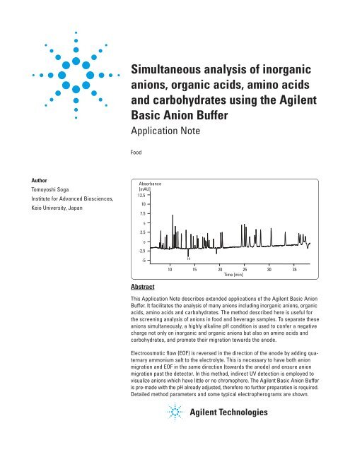
Abstract<br />
This Application Note describes extended applications <strong>of</strong> the Agilent Basic Anion<br />
Buffer. It facilitates the <strong>analysis</strong> <strong>of</strong> many <strong>anions</strong> including <strong>in<strong>organic</strong></strong> <strong>anions</strong>, <strong>organic</strong><br />
<strong>acids</strong>, <strong>amino</strong> <strong>acids</strong> and carbohydrates. The method described here is useful for<br />
the screening <strong>analysis</strong> <strong>of</strong> <strong>anions</strong> in food and beverage samples. To separate these<br />
<strong>anions</strong> simultaneously, a highly alkaline pH condition is used to confer a negative<br />
charge not only on <strong>in<strong>organic</strong></strong> and <strong>organic</strong> <strong>anions</strong> but also on <strong>amino</strong> <strong>acids</strong> and<br />
carbohydrates, and promote their migration towards the anode.<br />
Electroosmotic flow (EOF) is reversed in the direction <strong>of</strong> the anode by adding quaternary<br />
ammonium salt to the electrolyte. This is necessary to have both anion<br />
migration and EOF in the same direction (towards the anode) and ensure anion<br />
migration past the detector. In this method, indirect UV detection is employed to<br />
visualize <strong>anions</strong> which have little or no chromophore. The Agilent Basic Anion Buffer<br />
is pre-made with the pH already adjusted, therefore no further preparation is required.<br />
Detailed method parameters and some typical electropherograms are shown.
Necessary supplies<br />
The following parts are necessary for<br />
the simultaneous <strong>analysis</strong> <strong>of</strong> <strong>anions</strong>:<br />
Component Quantity Part No.<br />
Agilent Basic Anion 50 mL 5064-8209<br />
Buffer*<br />
Fused silica capillary 1 pk G1600-64211<br />
(id = 50 µm, l=104 cm<br />
L=112.5 cm)<br />
CE buffer vials 100/pk 5182-9697<br />
2 mL (glass**)<br />
CE sample vials PUR, 1000/pk 9301-0978<br />
100 µL (polypropylene)<br />
CE caps 100/pk 5181-1512<br />
(polyurethane)<br />
CE water 500 mL 5062-8578<br />
* For this buffer, only 50-µm id straight<br />
capillaries are usable.<br />
** It is recommended to use glass vials rather<br />
than polypropylene vials.<br />
Procedures<br />
Buffer preparation<br />
The Basic Anion Buffer is premade<br />
and ready to use.<br />
Do not leave the solution uncapped<br />
since the buffer is a highly alkaline<br />
solution and will readily absorb<br />
gaseous carbonate from the atmosphere.<br />
This will cause its pH to drop<br />
immediately. Make certain the bottle<br />
is capped after use.<br />
Do not store opened bottles which are<br />
a third or less full. These should be<br />
discarded because gaseous carbonate<br />
in the bottle can be adsorbed and<br />
will reduce the pH <strong>of</strong> the residual buffer.<br />
The buffer should be stored at room<br />
temperature (not less than 20 °C,<br />
since some buffer components may<br />
crystallize at lower temperatures).<br />
Buffer and waste vials<br />
Prepare three vials (one flushing vial<br />
and two home vials). When using 2-mL<br />
glass vials, fill each vial with 1.4 mL <strong>of</strong><br />
the buffer. Also prepare a waste vial<br />
(filled with 300-mL CE water or deionized<br />
water).<br />
Since buffers used for indirect UV<br />
detection have limited buffering capacity,<br />
the buffer should be replaced<br />
every 8 runs when using 2-mL glass<br />
vials.<br />
For this application, do not use the<br />
replenishment system for buffer replacement.<br />
The level <strong>of</strong> dissolved carbonate<br />
may increase due to pressurization<br />
<strong>of</strong> the buffer bottle, causing a pH<br />
drop. Also, crystallization <strong>of</strong> the buffer<br />
might block the tubes.<br />
Standard preparation<br />
Individual stock solutions <strong>of</strong> <strong>in<strong>organic</strong></strong><br />
and <strong>organic</strong> <strong>anions</strong> should be prepared<br />
from their sodium salts or free<br />
<strong>acids</strong>. Since carbohydrates are unstable,<br />
they should be prepared shortly<br />
before use.<br />
For <strong>amino</strong> <strong>acids</strong>, individual stock solution<br />
<strong>of</strong> Tyr, Cys-Cys, Asp, Trp, Leu, Ile<br />
and Phe should be prepared at a concentration<br />
<strong>of</strong> 10 g/L in 0.1 M NaOH.<br />
Other <strong>amino</strong> <strong>acids</strong> should be prepared<br />
in 0.01 M HCl. The working mixture<br />
standard should be prepared by diluting<br />
stock solutions with deionized water.<br />
If a commercially available <strong>amino</strong> acid<br />
standard mixture in 0.1 M HCl is used,<br />
Arg may not be detected since the pKa<br />
value <strong>of</strong> Arg is 10.76. Arg is positively<br />
charged in 0.1 M HCl and migrates<br />
toward the opposite direction <strong>of</strong> the<br />
detector. In this case, the standard<br />
solution should be diluted with 0.01 M<br />
NaOH.<br />
The concentration <strong>of</strong> the standard<br />
mixture should be in the range <strong>of</strong> 50 to<br />
1000 mg/L to obtain good peak shape<br />
and sensitivity.<br />
Sample preparation<br />
For actual samples, dilution with<br />
deionized water is necessary in order<br />
to reduce the conductivity <strong>of</strong> the<br />
samples, for example, 1:50 dilution for<br />
soy sauce.<br />
If the sample contains proteins and<br />
the migration times increase from run<br />
to run, removal <strong>of</strong> the proteins is<br />
recommended by using centrifugal filtering<br />
through a 30-kDa cut<strong>of</strong>f filter.<br />
Capillary<br />
Only 50-µm id straight capillaries are<br />
suitable for this method. Baseline<br />
noise is markedly increased if a 75-µm<br />
id capillary is used due to the high UV<br />
absorptivity <strong>of</strong> the buffer. Neither bubble<br />
cell capillaries nor the High<br />
Sensitivity Detection Cell should be<br />
used. A 50-µm id capillary (L=112.5 cm,<br />
l=104 cm) is recommended.<br />
Capillary conditioning<br />
Avoid capillary conditioning with<br />
sodium hydroxide since this degrades<br />
the performance <strong>of</strong> this application.<br />
Prior to first use, a new capillary<br />
should be flushed only with the run<br />
buffer for 15 minutes.<br />
Between analyses it is recommended<br />
that the capillary be flushed for 4 minutes<br />
with buffer from the flushing vial.<br />
Capillary storage<br />
If the capillary is removed from the<br />
instrument it should be washed for 10<br />
minutes with deionized water and then<br />
flushed with air for 10 minutes. When<br />
the capillary is to be reinstalled, it is<br />
necessary to flush with the run buffer<br />
for at least 15 minutes.<br />
2
Method summary<br />
The following method can be used to<br />
separate most <strong>in<strong>organic</strong></strong> <strong>anions</strong>, <strong>organic</strong><br />
<strong>acids</strong>, <strong>amino</strong> <strong>acids</strong> and carbohydrates<br />
simultaneously. Below are the general<br />
analytical conditions.<br />
Chromatographic conditions<br />
Capillary:<br />
Fused silica id = 50 µm,<br />
l=104 cm, L=112.5 cm<br />
(Agilent part number<br />
G1600-64211)<br />
Injection:<br />
1. Pressure: 50 mbar for<br />
6 seconds from sample<br />
vial<br />
2. Post-injection <strong>of</strong><br />
buffer from InHome vial,<br />
50 mbar for 4 seconds<br />
Applied voltage: - 30 kV<br />
Capillary temperature: 15 °C<br />
Detection wavelength: Signal 350/20 nm,<br />
reference 230/10 nm<br />
Preconditioning: Buffer flush for<br />
4 minutes at 1 bar prior<br />
to each run<br />
The method as it should be entered<br />
into the Agilent ChemStation is as<br />
follows:<br />
HPCE mode CE<br />
Home values<br />
Lift Offset 4<br />
Cassette<br />
Temperature 15.00 °C<br />
Inlet Home<br />
Vial 10 Place the buffer<br />
vials at position<br />
10 and 11.<br />
Outlet Home<br />
Vial 11 Vial locations are<br />
exemplary only.<br />
Replenishment and Preconditioning<br />
Serial processing<br />
Replenishment Entries<br />
No Replenishment Do not use<br />
used<br />
replenishment. The<br />
level <strong>of</strong> dissolved<br />
carbonate might<br />
increase due to<br />
pressurization <strong>of</strong><br />
the buffer bottle<br />
causing a pH drop.<br />
Preconditioning Entries<br />
Function Parameter<br />
1 Flush 4.00 min, I: 9, O:1 Place flushing vial<br />
at 9 and waste vial<br />
at 1. Remember to<br />
monitor the waste<br />
vial volume for<br />
overflow.<br />
Postcondition Entries<br />
No Postconditioning used<br />
Injection Table Entries<br />
Function Parameter<br />
1 PRESSURE 50.0 mbar, 6.0 sec,<br />
I: InjectVial, O:<br />
OutHomeVial<br />
May be increased<br />
or decreased<br />
depending on<br />
sample concentration.<br />
2 PRESSURE 50.0 mbar, 4.0 sec,<br />
I: InHomeVial,<br />
O:OutHomeVial<br />
The post injection<br />
plug helps to minimize<br />
sample loss<br />
upon application<br />
<strong>of</strong> voltage. A voltage<br />
ramp is used<br />
for the same<br />
purose.<br />
Electric<br />
Electric<br />
On<br />
Polarity Negative Negative<br />
polarity is used<br />
since EOF is<br />
reversed.<br />
Voltage<br />
30.0 kV<br />
Current 150.0 mA A current limit<br />
is not necessary<br />
but may be used<br />
to prevent<br />
excessive current<br />
generation in<br />
case the wrong<br />
vial is used.<br />
Power<br />
System Limit<br />
Low Current<br />
Limit 2 µA<br />
Store Data<br />
Collect voltage Yes<br />
Collect current Yes It is recommended<br />
to store the current<br />
for every<br />
<strong>analysis</strong>. Current<br />
in this method<br />
shows from<br />
-30 to -40 µA.<br />
Collect power No<br />
Collect pressure No<br />
Collect temperature Yes<br />
Time entries<br />
Stop time 40.00 min Adjust as needed<br />
when running<br />
actual sample.<br />
Post time Off<br />
Time Table is empty.<br />
Diode array detector<br />
Settings<br />
Stop Time as HPCE: 40.00 min<br />
Post Time Off<br />
Response Time 1.3 sec This is recommended<br />
to reduce<br />
baseline noise.<br />
Peak width > 0.1 min<br />
Prerun Autobalance On<br />
Postrun Autobalance Off<br />
Spectrum<br />
Store<br />
None<br />
Signals<br />
Store<br />
Reference<br />
Signal, Bw Reference, Bw [nm]<br />
A: Yes 350/20 230/10<br />
3
Results and discussion<br />
<strong>Simultaneous</strong> <strong>analysis</strong> <strong>of</strong> <strong>in<strong>organic</strong></strong><br />
<strong>anions</strong>, <strong>organic</strong> <strong>acids</strong>,<br />
<strong>amino</strong> <strong>acids</strong> and carbohydrates<br />
Figure 1 shows a typical electropherogram<br />
<strong>of</strong> a 43-component anion sample<br />
including seven <strong>in<strong>organic</strong></strong> <strong>anions</strong>, five<br />
<strong>organic</strong> <strong>acids</strong>, 16 <strong>amino</strong> <strong>acids</strong> and<br />
15 carbohydrates using the standard<br />
method. If the results are not similar<br />
to these please refer to the section<br />
Troubleshooting in this note.<br />
In this method most <strong>in<strong>organic</strong></strong> <strong>anions</strong><br />
and carbohydrates can be separated.<br />
However, migration times <strong>of</strong> several<br />
<strong>organic</strong> <strong>acids</strong> such as tartarate, succinate,<br />
malate and -ketoglutarate are<br />
close. This separation can be improved<br />
by using the Organic Acids Solutions<br />
kit from Agilent Technologies (Agilent<br />
part number 5063-6510).<br />
With respect to <strong>amino</strong> <strong>acids</strong>, Leu and<br />
Ile cannot be resolved. Although Arg<br />
is not observed in figure 1, Arg can be<br />
detected at approximately 35 minutes<br />
if the sample is dissolved in an alkaline<br />
Absorbance<br />
[mAU]<br />
12.5<br />
10<br />
7.5<br />
5<br />
2.5<br />
0<br />
-2.5<br />
-5<br />
1 Br 9 HPO 4 17 Ala 25 Phe 34 Glucose<br />
2 Cl 10 Malate 18 Ser 26 Lys 35 Galactosamine<br />
3 NO 2 11 Asp 19 Thr 27 Trp 36 Galactose<br />
4 NO 3 12 Glu 20 Pro 28 NGNA 37 Fucose<br />
5 SO 4 13 Acetate 21 Val 29 NANA 38 Sucrose<br />
6 Oxalate 14 Tyr 22 Met 30 Ribose 39 Mannitol<br />
7 F 15 Gly 23 His 31 Mannose 40 Sorbitol<br />
8 Citrate 16 Lactate 24 Leu+Ile 32 Xylose 41 Xylitol<br />
33 Glucosamine 42 Inositol<br />
7 23<br />
19<br />
21<br />
22 24<br />
20<br />
11<br />
9 10<br />
17<br />
2<br />
13<br />
5 6 8 12<br />
28<br />
15 16 18 26<br />
1 3 4 25<br />
27<br />
10 15 20 25 30 35<br />
Time [min]<br />
Figure 1<br />
Analysis <strong>of</strong> 43-component anion standard mixture.<br />
14<br />
29<br />
30<br />
31<br />
32<br />
35 36<br />
37<br />
34<br />
33<br />
Chromatographic conditions<br />
Buffer: Agilent Basic Anion Buffer (Agilent part number 5064-8209)<br />
Sample: Cl 110 mg/L, carbohydrates, 200 mg/L each, others 50 mg/L each<br />
Capillary: Fused silica, I=104 cm, L=112.5 cm, id= 50 µm (Agilent part number G1600-64211)<br />
Injection: 300 mbar x s<br />
Temperature: 15 °C<br />
Voltage: -30 kV<br />
Detection: Signal 350/20 nm, reference 230/10 nm<br />
38<br />
40<br />
39<br />
41<br />
42<br />
Compound<br />
Mobility<br />
(X10 -4 cm -2 /Vs)<br />
Compound<br />
Mobility<br />
(X10 -4 cm -2 /Vs)<br />
Compound<br />
Mobility<br />
(X10 -4 cm -2 /Vs)<br />
Compound<br />
Mobility<br />
(X10 -4 cm -2 /Vs)<br />
Table 1<br />
Electrophoretic mobilities <strong>of</strong> <strong>anions</strong> at 20 °C.<br />
4
solution. Since Tyr and Trp have UV<br />
absorbance at 230 nm, they are recorded<br />
as negative peaks. Ser migrates<br />
just before a system peak.<br />
Electrophoretic mobilities at 20 °C<br />
The effective mobilities <strong>of</strong> 82 compounds<br />
including nine <strong>in<strong>organic</strong></strong><br />
<strong>anions</strong>, 23 <strong>organic</strong> <strong>acids</strong>, 18 <strong>amino</strong><br />
<strong>acids</strong> and 32 carbohydrates were<br />
determined by this method at 20 °C<br />
and are listed in table 1. If the mobilities<br />
<strong>of</strong> the compounds <strong>of</strong> interest are<br />
close, they are difficult to separate.<br />
In this case investigating a change <strong>of</strong><br />
temperature or pH is recommended.<br />
Absorbance<br />
[mAU]<br />
35<br />
30<br />
25<br />
20<br />
15<br />
10<br />
5<br />
0<br />
1<br />
6<br />
3<br />
2<br />
4 5<br />
1 Cl 6 Glu 11 Ser 16 Leu+Ile<br />
2 Citrate 7 Acetate 12 Thr 17 Phe<br />
3 Succinate 8 Gly 13 Pro 18 Lys<br />
4 Malate 9 Lactate 14 Val 19 Glucose<br />
5 Asp 10 Ala 15 Met 20 Galactose<br />
7 8 9 1011 12<br />
1314 1516<br />
17 18 19<br />
20<br />
10 15 20 25 30<br />
Time [min]<br />
Applications<br />
Soy sauce <strong>analysis</strong><br />
Figure 2 shows the <strong>analysis</strong> <strong>of</strong> a soy<br />
sauce. The sample was diluted 1:50<br />
with CE water. Centrifugal filtering<br />
through a 30 kDa cut<strong>of</strong>f filter was<br />
applied to remove proteins and peptides.<br />
A well-defined electropherogram<br />
was obtained without interference<br />
from other matrix compounds.<br />
Satisfactory reproducibilities were<br />
obtained for all compounds with RSD<br />
values (n=5) for migration times better<br />
than 0.3 % and for peak areas between<br />
0.6 and 5.4 %.<br />
Pineapple <strong>analysis</strong><br />
This method was applied to the <strong>analysis</strong><br />
<strong>of</strong> <strong>organic</strong> <strong>acids</strong> and carbohydrates<br />
in pineapple. In the agriculture<br />
industry, technical experts are trying<br />
to develop a new crossbreed <strong>of</strong> fruits.<br />
Since the content <strong>of</strong> <strong>organic</strong> <strong>acids</strong> and<br />
carbohydrates determines the taste <strong>of</strong><br />
the pineapple juice, their <strong>analysis</strong> can<br />
help to characterize the product. If<br />
citrate and malate concentrations are<br />
high, the taste tends to be sour. If the<br />
carbohydrate concentration is high,<br />
the taste issweet. Determination <strong>of</strong><br />
these compounds is traditionally<br />
performed using two HPLC methods.<br />
Figure 2<br />
Analysis <strong>of</strong> soy sauce.<br />
Chromatographic conditions<br />
Buffer: Agilent Basic Anion Buffer (Agilent part number 5064-8209)<br />
Sample: Soy sauce, 1:50 diluted with water, ultrafiltration with 30-kDa cut<strong>of</strong>f filter<br />
Preconditioning: 4 min with run buffer<br />
Capillary: Fused silica, I=104 cm, L=112.5 cm, id= 50 µm, Injection 300 mbar x s<br />
(Agilent part number G1600-64211)<br />
Temperature: 15 °C<br />
Voltage: -30 kV<br />
Detection signal: 350/20 nm, reference 230/10 nm<br />
Absorbance<br />
[mAU]<br />
30<br />
25<br />
20<br />
15<br />
10<br />
Figure 3<br />
Analysis <strong>of</strong> pineapple.<br />
5<br />
0<br />
Citrate<br />
Malate<br />
Fructose<br />
Glucose<br />
4 6 8 10 12 14 16<br />
Time [min]<br />
Sucrose<br />
Chromatographic conditions<br />
Buffer: Agilent Basic Anion Buffer (Agilent part number 5064-8209)<br />
Sample: Pineapple, 1:50 diluted with water,<br />
Capillary: Fused silica, I=72 cm, L=80.5 cm, id= 50 µm (Agilent part number G1600-62211)<br />
Injection: 300 mbar x s<br />
Temperature: 20 °C<br />
Voltage: -25 kV (reversed polarity)<br />
Detection signal: 350/20 nm, reference 275/10 nm<br />
5
However, the method described here<br />
enables the simultaneous <strong>analysis</strong> <strong>of</strong><br />
both <strong>organic</strong> <strong>acids</strong> and carbohydrates<br />
in a much shorter time (less than<br />
18 minutes) and in a single run. In<br />
order to reduce the <strong>analysis</strong> time, a<br />
shorter length capillary was used for<br />
this sample. Squeezed pineapple juice<br />
was diluted 50-fold with CE water<br />
prior to injection. Figure 3 shows a<br />
result <strong>of</strong> the <strong>analysis</strong>.<br />
Troubleshooting<br />
Problem<br />
Poor resolution or broad/split<br />
peaks<br />
Peak leading<br />
No signal<br />
Noisy baseline<br />
Poor reproducibility<br />
Unstable current<br />
Possible Cause<br />
Buffer is old<br />
Capillary damaged<br />
Sample overloaded<br />
Capillary too short<br />
Buffer absorbed carbonate<br />
Sample not injected<br />
Capillary damaged<br />
Wrong buffer used<br />
Wrong setting <strong>of</strong> power supply polarity<br />
Detection wavelength incorrect<br />
Wrong setting <strong>of</strong> response time<br />
Capillary window not adjusted<br />
Capillary window dirty<br />
Lamp is old<br />
Buffer pH higher than 12.3<br />
Buffer overused<br />
Capillary broken<br />
Capillary not filled with buffer<br />
Solution<br />
Use new buffer<br />
Replace capillary<br />
Dilute sample<br />
Replace capillary<br />
Use new buffer<br />
Verify no air bubble trapped in bottom <strong>of</strong> sample vial<br />
Verify inlet capillary set correctly<br />
Replace capillary<br />
Verify buffer<br />
Check that polarity is negative<br />
Verify signal: 350/20, reference: 230/10 nm<br />
Verify response time 1.3 seconds DAD<br />
Examine capillary window<br />
Examine and clean with lint-free paper/MeOH<br />
Replace lamp<br />
Verify buffer pH<br />
Replace buffer<br />
Replace capillary<br />
Increase flush time<br />
6
References<br />
1.<br />
Soga, T. and Ross, G. A. “Capillary<br />
Electrophoretic Determination <strong>of</strong><br />
In<strong>organic</strong> and Organic Anions using<br />
2,6-pyridinedicarboxylic Acid: Effect <strong>of</strong><br />
Electrolyte's Complexing Ability”, J.<br />
Chromatogr. A, 767, 223-230, 1997.<br />
2.<br />
Soga, T. and Heiger, D. N.<br />
“<strong>Simultaneous</strong> Determination <strong>of</strong><br />
Monosaccharides in Glycoproteins by<br />
Capillary Electrophoresis”, Anal.<br />
Biochem., 261, 73-78, 1998.<br />
3.<br />
Soga, T. and Ross, G. A.<br />
“<strong>Simultaneous</strong> Determination <strong>of</strong><br />
In<strong>organic</strong> Anions, Organic Acids,<br />
Amino Acids and Carbohydrates by<br />
Capillary Electrophoresis” J.<br />
Chromatogr., 837, 231-239, 1999.<br />
7
www.agilent.com/chem/ce<br />
© Agilent Technologies, Inc., 2000-2008<br />
Published November 1, 2008<br />
Publication Number 5989-9853EN