Spectroscopy in a Suitcase - Royal Society of Chemistry
Spectroscopy in a Suitcase - Royal Society of Chemistry
Spectroscopy in a Suitcase - Royal Society of Chemistry

Create successful ePaper yourself
Turn your PDF publications into a flip-book with our unique Google optimized e-Paper software.
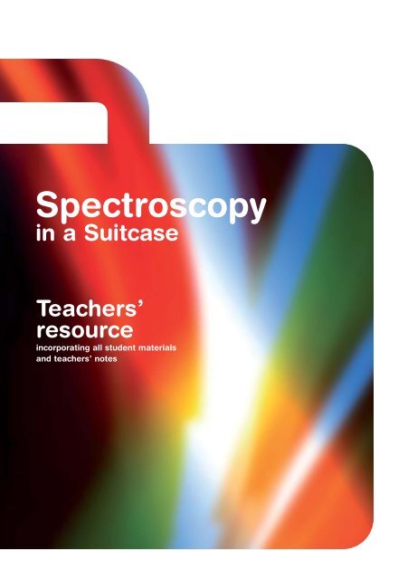
<strong>Spectroscopy</strong><br />
<strong>in</strong> a <strong>Suitcase</strong><br />
Teachers’<br />
resource<br />
<strong>in</strong>corporat<strong>in</strong>g all student materials<br />
and teachers’ notes
<strong>Spectroscopy</strong><br />
<strong>in</strong> a <strong>Suitcase</strong><br />
Welcome<br />
to the SIAS Resource folder<br />
These folders are designed to facilitate the delivery <strong>of</strong><br />
SIAS workshops by provid<strong>in</strong>g demonstrators, teachers<br />
and students with all <strong>of</strong> the resources needed.<br />
All folders conta<strong>in</strong>:<br />
• Introductory material for each spectroscopic technique<br />
which has been written by teachers to ensure alignment<br />
with current school curricula.<br />
• A series <strong>of</strong> workshops which <strong>in</strong>troduce the application <strong>of</strong><br />
each spectroscopic technique, provid<strong>in</strong>g background<br />
<strong>in</strong>formation, and step-by-step <strong>in</strong>structions and questions.<br />
For demonstrators and teachers:<br />
The teachers’ folder conta<strong>in</strong>s <strong>in</strong>troductory material for each<br />
spectroscopic technique along with setup up <strong>in</strong>structions<br />
for the Infra-red and Ultraviolet spectrometers.<br />
In addition each workshop conta<strong>in</strong>s:<br />
• Materials list<br />
• Answer sheets (signified by P ) which are to be photocopied<br />
and handed out to students<br />
• Model answers<br />
• Model spectra<br />
Copyright © 2009 <strong>Royal</strong> <strong>Society</strong> <strong>of</strong> <strong>Chemistry</strong> www.rsc.org
<strong>Chemistry</strong><br />
for our Future<br />
<strong>Chemistry</strong> for our Future (CFOF) began on<br />
1st September 2006 as a two-year, £3.6m pilot<br />
programme f<strong>in</strong>anced by the Higher Education<br />
Fund<strong>in</strong>g Council for England (HEFCE) and<br />
managed by the <strong>Royal</strong> <strong>Society</strong> <strong>of</strong> <strong>Chemistry</strong> (RSC).<br />
The RSC secured a further £1.65m <strong>of</strong> HEFCE fund<strong>in</strong>g<br />
to extend the programme until July 2009.<br />
The programme aims to secure a strong and susta<strong>in</strong>able future for<br />
the chemical sciences. Over 30 separate projects and 35 universities<br />
are <strong>in</strong>volved <strong>in</strong> the programme, tackl<strong>in</strong>g a wide range <strong>of</strong> issues from<br />
recruitment <strong>of</strong> students to development <strong>of</strong> higher education curricula<br />
and provision <strong>of</strong> high quality careers advice.<br />
To learn more about the project, visit the website (www.rsc.org/cf<strong>of</strong>)<br />
or send us an email (education@rsc.org).<br />
SpectraSchool<br />
www.spectraschool.org<br />
A website has been designed<br />
to enhance the teach<strong>in</strong>g <strong>of</strong><br />
spectroscopy, provid<strong>in</strong>g a variety<br />
<strong>of</strong> resource materials for students<br />
and teachers <strong>in</strong>clud<strong>in</strong>g real spectra.<br />
SpectraSchool can be used to<br />
assist SIAS sessions as well as<br />
be<strong>in</strong>g an <strong>in</strong>dependent aid to<br />
teach<strong>in</strong>g, learn<strong>in</strong>g and revision.<br />
<strong>Spectroscopy</strong> <strong>in</strong> a <strong>Suitcase</strong> (SIAS)<br />
Instrumental techniques are a core part <strong>of</strong> post-16<br />
curricula <strong>in</strong> chemistry and many schools visit<br />
universities to see scientific equipment.<br />
<strong>Spectroscopy</strong> <strong>in</strong> a <strong>Suitcase</strong> provides schools<br />
and colleges with access to robust, portable,<br />
state-<strong>of</strong>-the-art spectroscopic equipment;<br />
contextualis<strong>in</strong>g the subject, br<strong>in</strong>g<strong>in</strong>g it to life<br />
and engag<strong>in</strong>g students.<br />
SIAS br<strong>in</strong>gs the university laboratory <strong>in</strong>to the school<br />
classroom, deliver<strong>in</strong>g challeng<strong>in</strong>g hands-on experiments <strong>in</strong><br />
an excit<strong>in</strong>g context. Workshops are delivered by tra<strong>in</strong>ed<br />
postgraduate students and teachers, ensur<strong>in</strong>g the activities<br />
compliment the content <strong>of</strong> all current chemistry syllabi.<br />
SIAS is ideally suited to A-level and GCSE students study<strong>in</strong>g<br />
the physical sciences. To f<strong>in</strong>d out more about the project<br />
visit the website www.rsc.org/cf<strong>of</strong>. To obta<strong>in</strong> electronic<br />
copies <strong>of</strong> these resources visit www.spectraschool.org.<br />
ACKNOWLEDGEMENTS<br />
The <strong>Royal</strong> <strong>Society</strong> <strong>of</strong> <strong>Chemistry</strong> (RSC) thanks all contributors to this resource, especially the <strong>Chemistry</strong> for our Future Teacher Fellows<br />
and Regional Coord<strong>in</strong>ators David Brentnall, Cheryl Sacht, Anne Willis, Tracy McGhie and Jayne Shaw. The RSC would also like to thank the<br />
follow<strong>in</strong>g organisations for their generous contributions to the <strong>Spectroscopy</strong> <strong>in</strong> a <strong>Suitcase</strong> project: Sigma Aldrich, The Higher Education Fund<strong>in</strong>g<br />
Council for England (HEFCE), the University <strong>of</strong> Leicester and University College London (UCL).
Introduction to<br />
<strong>Spectroscopy</strong><br />
What is spectroscopy?<br />
One <strong>of</strong> the frustrations <strong>of</strong> be<strong>in</strong>g a chemist is the fact that<br />
no matter how hard you stare at your test tube or<br />
round-bottomed flask you can’t actually see the <strong>in</strong>dividual<br />
molecules you have made! Even though your product<br />
looks the right colour and seems to give sensible results<br />
when you carry out chemical tests, can you be really sure<br />
<strong>of</strong> its precise structure?<br />
Fortunately, help is at hand. Although you might not be<br />
able to ‘see’ molecules, they do respond when light<br />
energy hits them, and if you can observe that response,<br />
then maybe you can get some <strong>in</strong>formation about that<br />
molecule. This is where spectroscopy comes <strong>in</strong>.<br />
<strong>Spectroscopy</strong> is the study <strong>of</strong> the way light<br />
(electromagnetic radiation) and matter <strong>in</strong>teract.<br />
There are a number <strong>of</strong> different types <strong>of</strong> spectroscopic<br />
techniques and the basic pr<strong>in</strong>ciple shared by all is to<br />
sh<strong>in</strong>e a beam <strong>of</strong> a particular electromagnetic radiation<br />
onto a sample and observe how it responds to such a<br />
stimulus; allow<strong>in</strong>g scientists to obta<strong>in</strong> <strong>in</strong>formation about<br />
the structure and properties <strong>of</strong> matter.<br />
What is light?<br />
Light carries energy <strong>in</strong> the form <strong>of</strong> t<strong>in</strong>y particles known as<br />
photons. Each photon has a discrete amount <strong>of</strong> energy,<br />
called a quantum. Light has wave properties with<br />
characteristic wavelengths and frequency (see the<br />
diagram below).<br />
The energy <strong>of</strong> the photons is related to the frequency (m)<br />
and wavelength (l) <strong>of</strong> the light through the two equations:<br />
E=hm and m = c /l<br />
(where h is Planck's constant and c is the speed <strong>of</strong> light).<br />
Therefore, high energy radiation (light) will have high<br />
frequencies and short wavelengths.<br />
The range <strong>of</strong> wavelengths and frequencies <strong>in</strong> light is<br />
known as the electromagnetic spectrum. This spectrum<br />
is divided <strong>in</strong>to various regions extend<strong>in</strong>g from very short<br />
wavelength, high energy radiation (<strong>in</strong>clud<strong>in</strong>g gamma rays<br />
and X-rays) to very long wavelength, low energy radiation<br />
(<strong>in</strong>clud<strong>in</strong>g microwaves and broadcast radio waves).<br />
The visible region (white light) only makes up a small part<br />
<strong>of</strong> the electromagnetic spectrum considered to be<br />
380-770 nm. [Note that a nanometre is 10 -9 metres].<br />
WAVELENGTH(METRES)<br />
RADIO<br />
MICROWAVE<br />
INFARED<br />
10 3 10 -2<br />
10 -5 VISIBLE<br />
5x10 -6<br />
0 -6<br />
10 -8 10 -10 10 -12<br />
ULTRAVIOLET<br />
X-RAY<br />
GAMMA RAY<br />
MOLECULES<br />
ATOMS<br />
BUTTERFLY<br />
BUILDINGS<br />
HUMANS<br />
PINHEAD<br />
INCREASING WAVELENGTH l<br />
PROTOZOANS<br />
INCREASING FREQUENCY m<br />
INCREASING ENERGY E<br />
ATOMIC<br />
NUCLEUS<br />
10 4 10 8 10 12 10 15 10 16 10 18 10 20<br />
FREQUENCY(HZ)<br />
Copyright © 2009 <strong>Royal</strong> <strong>Society</strong> <strong>of</strong> <strong>Chemistry</strong> www.rsc.org
SPECTROSCOPY<br />
INTRODUCTION 2<br />
When matter absorbs electromagnetic radiation the<br />
change which occurs depends on the type <strong>of</strong> radiation,<br />
and therefore the amount <strong>of</strong> energy, be<strong>in</strong>g absorbed.<br />
Absorption <strong>of</strong> energy causes an electron or molecule to go<br />
from an <strong>in</strong>itial energy state (ground state) to a high energy<br />
state (excited state) which could take the form <strong>of</strong> the<br />
<strong>in</strong>creased rotation, vibration or electronic excitation.<br />
By study<strong>in</strong>g this change <strong>in</strong> energy state scientists are able<br />
to learn more about the physical and chemical properties<br />
<strong>of</strong> the molecules.<br />
• Radio waves can cause nuclei <strong>in</strong> some atoms to<br />
change magnetic orientation and this forms the basis<br />
<strong>of</strong> a technique called nuclear magnetic resonance<br />
(NMR) spectroscopy.<br />
• Molecular rotations are excited by microwaves.<br />
• Electrons are promoted to higher orbitals by<br />
ultraviolet or visible light.<br />
• Vibrations <strong>of</strong> bonds are excited by <strong>in</strong>frared radiation.<br />
The energy states are said to be quantised because a<br />
photon <strong>of</strong> precise energy and frequency (or wavelength)<br />
must be absorbed to excite an electron or molecule from<br />
the ground state to a particular excited state.<br />
S<strong>in</strong>ce molecules have a unique set <strong>of</strong> energy states that<br />
depend on their structure, IR, UV-visible and NMR<br />
spectroscopy will provide valuable <strong>in</strong>formation about the<br />
structure <strong>of</strong> the molecule.<br />
To ‘see’ a molecule we need to use light hav<strong>in</strong>g a<br />
wavelength smaller than the molecule itself (approximately<br />
10 –10 m). Such radiation is found <strong>in</strong> the X-ray region <strong>of</strong> the<br />
electromagnetic spectrum and is used <strong>in</strong> the field <strong>of</strong><br />
X-ray crystallography. This technique yields very detailed<br />
three-dimensional pictures <strong>of</strong> molecular structures –<br />
the only drawback be<strong>in</strong>g that it requires high quality<br />
crystals <strong>of</strong> the compound be<strong>in</strong>g studied. Although other<br />
spectroscopic techniques do not yield a three-dimensional<br />
picture <strong>of</strong> a molecule they do provide <strong>in</strong>formation about its<br />
characteristic features and are therefore used rout<strong>in</strong>ely <strong>in</strong><br />
structural analysis.<br />
Mass spectrometry is another useful technique used by<br />
chemists to help them determ<strong>in</strong>e the structure <strong>of</strong><br />
molecules. Although sometimes referred to as mass<br />
spectroscopy it is, by def<strong>in</strong>ition, not a spectroscopic<br />
technique as it does not make use <strong>of</strong> electromagnetic<br />
radiation. Instead the molecules are ionised us<strong>in</strong>g high<br />
energy electrons and these molecular ions subsequently<br />
undergo fragmentation. The result<strong>in</strong>g mass spectrum<br />
conta<strong>in</strong>s the mass <strong>of</strong> the molecule and its fragments<br />
which allows chemists to piece together its structure.<br />
In all spectroscopic techniques only very small quantities<br />
(milligrams or less) <strong>of</strong> sample are required, however, <strong>in</strong><br />
mass spectrometry the sample is destroyed <strong>in</strong> the<br />
fragmentation process whereas the sample can be<br />
recovered after us<strong>in</strong>g IR, UV-visible and NMR<br />
spectroscopy.<br />
TECHNIQUE RADIATION WHAT CAN IT SEE?<br />
Nuclear Magnetic<br />
Resonance (NMR)<br />
spectroscopy<br />
Radio waves<br />
(10 -3 m)<br />
10 -3 m<br />
Electrons flipp<strong>in</strong>g magnetic sp<strong>in</strong><br />
How neighbour<strong>in</strong>g atoms <strong>of</strong><br />
certa<strong>in</strong> nuclei (e.g. 1 H, 13 C,<br />
19 F, 31 P) <strong>in</strong> a molecule are<br />
connected together, as well as<br />
how many atoms <strong>of</strong> these type<br />
are present <strong>in</strong> different locations<br />
<strong>in</strong> the molecule.<br />
Infra-red<br />
spectroscopy<br />
Infra-red<br />
(10 -5 m)<br />
10 -5 m NOTE<br />
Molecule vibrations<br />
The functional groups which are<br />
present <strong>in</strong> a molecule.<br />
UV-visible<br />
spectroscopy<br />
Ultra-violet<br />
(10 -8 m)<br />
10 -8 m<br />
NOTE<br />
Electrons promoted<br />
to higher energy state<br />
Conjugated systems (i.e.<br />
alternat<strong>in</strong>g s<strong>in</strong>gle and double<br />
bonds) <strong>in</strong> organic molecules as well<br />
as the metal-ligand <strong>in</strong>teractions <strong>in</strong><br />
transition metal complexes.<br />
X-ray<br />
crystallography<br />
X-rays<br />
(10 -10 m)<br />
10 -10 m x-ray<br />
How all the atoms <strong>in</strong> a<br />
molecule are connected <strong>in</strong> a<br />
three-dimensional arrangement.<br />
Mass<br />
spectrometry<br />
Non-spectroscopic<br />
technique<br />
Molecules fragment<br />
+<br />
+<br />
+<br />
The mass to charge ratio <strong>of</strong> the<br />
molecular ion (i.e. the molecular<br />
weight) and the fragmentation<br />
pattern which may be related to<br />
the structure <strong>of</strong> the molecular ion.<br />
Copyright © 2009 <strong>Royal</strong> <strong>Society</strong> <strong>of</strong> <strong>Chemistry</strong> www.rsc.org
Ultraviolet - Visible<br />
<strong>Spectroscopy</strong> (UV)
UV<br />
Introduction to Ultraviolet -<br />
Visible <strong>Spectroscopy</strong> 1<br />
(UV)<br />
Background Theory<br />
Absorption <strong>of</strong> ultraviolet and visible radiation<br />
Absorption <strong>of</strong> visible and ultraviolet (UV) radiation is<br />
associated with excitation <strong>of</strong> electrons, <strong>in</strong> both atoms<br />
and molecules, from lower to higher energy levels.<br />
S<strong>in</strong>ce the energy levels <strong>of</strong> matter are quantized, only light<br />
with the precise amount <strong>of</strong> energy can cause transitions<br />
from one level to another will be absorbed.<br />
The possible electronic transitions that light might cause are 2 :<br />
All molecules will undergo electronic excitation follow<strong>in</strong>g<br />
absorption <strong>of</strong> light, but for most molecules very high<br />
energy radiation (<strong>in</strong> the vacuum ultraviolet,
ULTRAVIOLET - VISIBLE SPECTROSCOPY (UV)<br />
INTRODUCTION 2<br />
Molecules that conta<strong>in</strong> conjugated systems,<br />
i.e. alternat<strong>in</strong>g s<strong>in</strong>gle and double bonds, will have their<br />
electrons delocalised due to overlap <strong>of</strong> the p orbitals <strong>in</strong> the<br />
double bonds. This is illustrated below for buta-1,3-diene.<br />
Beta-carotene absorbs throughout the UV region but<br />
particularly strongly <strong>in</strong> the visible region between 400<br />
and 500 nm with a peak at 470 nm.<br />
Groups <strong>in</strong> a molecule which consist <strong>of</strong> alternat<strong>in</strong>g s<strong>in</strong>gle<br />
and double bonds (conjugation) and absorb visible light<br />
are known as chromophores.<br />
Benzene is a well-known example <strong>of</strong> a conjugated<br />
system. The Kekulé structure <strong>of</strong> benzene consists <strong>of</strong><br />
alternat<strong>in</strong>g s<strong>in</strong>gle double bonds and these give rise to the<br />
delocalised V system above and below the plane <strong>of</strong> the<br />
carbon – carbon s<strong>in</strong>gle bonds.<br />
Transition metal complexes are also highly coloured,<br />
which is due to the splitt<strong>in</strong>g <strong>of</strong> the d orbitals when<br />
the ligands approach and bond to the central metal ion.<br />
Some <strong>of</strong> the d orbitals ga<strong>in</strong> energy and some lose<br />
energy. The amount <strong>of</strong> splitt<strong>in</strong>g depends on the central<br />
metal ion and ligands.<br />
The difference <strong>in</strong> energy between the new levels affects<br />
how much energy will be absorbed when an electron is<br />
promoted to a higher level. The amount <strong>of</strong> energy will<br />
govern the colour <strong>of</strong> light which will be absorbed.<br />
As the amount <strong>of</strong> delocalisation <strong>in</strong> the molecule <strong>in</strong>creases<br />
the energy gap between the V bond<strong>in</strong>g orbitals and V<br />
anti-bond<strong>in</strong>g orbitals gets smaller and therefore light <strong>of</strong><br />
lower energy, and longer wavelength, is absorbed.<br />
For example, <strong>in</strong> the octahedral<br />
copper complex, [Cu(H 2 O) 6 ] 2+ ,<br />
yellow light has sufficient energy to<br />
promote the d electron <strong>in</strong> the lower<br />
energy level to the higher one.<br />
Although buta-1,3-diene absorbs light <strong>of</strong> a longer<br />
wavelength than ethene it is still absorb<strong>in</strong>g <strong>in</strong> the UV<br />
region and hence both compounds are colourless.<br />
However, if the delocalisation is extended further the<br />
wavelength absorbed will eventually be long enough to<br />
be <strong>in</strong> the visible region <strong>of</strong> the spectrum, result<strong>in</strong>g <strong>in</strong> a<br />
highly coloured compound. A good example <strong>of</strong> this is the<br />
orange plant pigment, beta-carotene – which has<br />
11 carbon-carbon double bonds conjugated together.<br />
Copyright © 2009 <strong>Royal</strong> <strong>Society</strong> <strong>of</strong> <strong>Chemistry</strong> www.rsc.org
ULTRAVIOLET - VISIBLE SPECTROSCOPY (UV)<br />
INTRODUCTION 3<br />
If a substance<br />
absorbs here...<br />
495 nm<br />
190 nm<br />
450 nm<br />
Blue<br />
UV Region<br />
Violet<br />
Red<br />
Visible Region Colour Wheel<br />
Green<br />
570 nm<br />
Yellow<br />
400 nm<br />
800 nm - Visible Region<br />
Orange<br />
590 nm<br />
400 nm<br />
620 nm<br />
...it appears<br />
as this colour<br />
Red<br />
Orange<br />
Yellow<br />
Green<br />
Blue<br />
Violet<br />
620-750 nm<br />
590-620 nm<br />
570-590 nm<br />
496-570 nm<br />
450-495 nm<br />
380-450 nm<br />
It is possible to predict which wavelengths are likely to<br />
be absorbed by a coloured substance. When white light<br />
passes through or is reflected by a coloured substance,<br />
a characteristic portion <strong>of</strong> the mixed wavelengths is<br />
absorbed. The rema<strong>in</strong><strong>in</strong>g light will then assume the<br />
complementary colour to the wavelength(s) absorbed.<br />
This relationship is demonstrated by the colour wheel<br />
shown on the right. Complementary colours are<br />
diametrically opposite each other.<br />
Red<br />
UV-Visible Spectrometer<br />
Orange<br />
Yellow<br />
Green<br />
Blue<br />
Violet<br />
MONOCHROMATOR<br />
REFERENCE<br />
CELL<br />
DETECTORS<br />
SOURCE<br />
PRISM<br />
BEAM<br />
SPLITTER<br />
SAMPLE<br />
CELL<br />
RATIO<br />
UV-visible spectrometers can be used to measure the<br />
absorbance <strong>of</strong> ultra violet or visible light by a sample,<br />
either at a s<strong>in</strong>gle wavelength or perform a scan over a<br />
range <strong>in</strong> the spectrum. The UV region ranges from 190<br />
to 400 nm and the visible region from 400 to 800 nm.<br />
The technique can be used both quantitatively and<br />
qualitatively. A schematic diagram <strong>of</strong> a UV-visible<br />
spectrometer is shown above.<br />
The light source (a comb<strong>in</strong>ation <strong>of</strong> tungsten/halogen<br />
and deuterium lamps) provides the visible and near<br />
ultraviolet radiation cover<strong>in</strong>g the 200 – 800 nm.<br />
The output from the light source is focused onto the<br />
diffraction grat<strong>in</strong>g which splits the <strong>in</strong>com<strong>in</strong>g light <strong>in</strong>to<br />
its component colours <strong>of</strong> different wavelengths,<br />
like a prism (shown below) but more efficiently.<br />
For liquids the sample is held <strong>in</strong> an optically flat, transparent<br />
conta<strong>in</strong>er called a cell or cuvette. The reference cell or<br />
cuvette conta<strong>in</strong>s the solvent <strong>in</strong> which the sample is<br />
dissolved and this is commonly referred to as the blank.<br />
For each wavelength the <strong>in</strong>tensity <strong>of</strong> light pass<strong>in</strong>g<br />
through both a reference cell (I o ) and the sample cell (I)<br />
is measured. If I is less than I o , then the sample has<br />
absorbed some <strong>of</strong> the light.<br />
The absorbance (A) <strong>of</strong> the sample is related to I and I o<br />
accord<strong>in</strong>g to the follow<strong>in</strong>g equation:<br />
The detector converts the <strong>in</strong>com<strong>in</strong>g light <strong>in</strong>to a current,<br />
the higher the current the greater the <strong>in</strong>tensity. The chart<br />
recorder usually plots the absorbance aga<strong>in</strong>st wavelength<br />
(nm) <strong>in</strong> the UV and visible section <strong>of</strong> the electromagnetic<br />
spectrum. (Note: absorbance does not have any units).<br />
Copyright © 2009 <strong>Royal</strong> <strong>Society</strong> <strong>of</strong> <strong>Chemistry</strong> www.rsc.org
ULTRAVIOLET - VISIBLE SPECTROSCOPY (UV)<br />
INTRODUCTION 4<br />
UV-Visible Spectrum<br />
The diagram below shows a simple UV-visible absorption<br />
spectrum for buta-1,3-diene. Absorbance (on the vertical<br />
axis) is just a measure <strong>of</strong> the amount <strong>of</strong> light absorbed.<br />
One can readily see what wavelengths <strong>of</strong> light are<br />
absorbed (peaks), and what wavelengths <strong>of</strong> light are<br />
transmitted (troughs). The higher the value, the more<br />
<strong>of</strong> a particular wavelength is be<strong>in</strong>g absorbed.<br />
absorbance<br />
1.0<br />
0.8<br />
0.6<br />
0.4<br />
0.2<br />
0<br />
Maximum absorption at this wavelength<br />
max=217nm<br />
200 220 240 260 280 300<br />
wavelength (nm)<br />
The absorption peak at a value <strong>of</strong> 217 nm, is <strong>in</strong> the ultraviolet<br />
region, and so there would be no visible sign <strong>of</strong> any<br />
light be<strong>in</strong>g absorbed mak<strong>in</strong>g buta-1,3-diene colourless.<br />
The wavelength that corresponds to the highest absorption<br />
is usually referred to as “lambda-max” (lmax).<br />
The spectrum for the blue copper complex shows that the<br />
complementary yellow light is absorbed.<br />
The Beer-Lambert Law<br />
Accord<strong>in</strong>g to the Beer-Lambert Law the absorbance is<br />
proportional to the concentration <strong>of</strong> the substance <strong>in</strong><br />
solution and as a result UV-visible spectroscopy can also<br />
be used to measure the concentration <strong>of</strong> a sample.<br />
The Beer-Lambert Law can be expressed <strong>in</strong> the form <strong>of</strong><br />
the follow<strong>in</strong>g equation:<br />
A = ecl<br />
Where<br />
A<br />
l<br />
= absorbance<br />
= optical path length, i.e. dimension <strong>of</strong> the cell<br />
or cuvette (cm)<br />
c = concentration <strong>of</strong> solution (mol dm -3 )<br />
e = molar ext<strong>in</strong>ction, which is constant for a<br />
particular substance at a particular<br />
wavelength (dm 3 mol -1 cm -1 )<br />
If the absorbance <strong>of</strong> a series <strong>of</strong> sample solutions <strong>of</strong><br />
known concentrations are measured and plotted<br />
aga<strong>in</strong>st their correspond<strong>in</strong>g concentrations, the plot <strong>of</strong><br />
absorbance versus concentration should be l<strong>in</strong>ear<br />
if the Beer-Lambert Law is obeyed. This graph is known<br />
as a calibration graph.<br />
A calibration graph can be used to determ<strong>in</strong>e the<br />
concentration <strong>of</strong> unknown sample solution by measur<strong>in</strong>g<br />
its absorbance, as illustrated below.<br />
absorbance<br />
350 400 450 500 550 600 650 700<br />
wavelength (nm)<br />
S<strong>in</strong>ce the absorbance for dilute solutions is directly<br />
proportional to concentration another very useful<br />
application for UV-visible spectroscopy is study<strong>in</strong>g<br />
reaction k<strong>in</strong>etics. The rate <strong>of</strong> change <strong>in</strong> concentration <strong>of</strong><br />
reactants or products can be determ<strong>in</strong>ed by measur<strong>in</strong>g<br />
the <strong>in</strong>crease or decrease <strong>of</strong> absorbance <strong>of</strong> coloured<br />
solutions with time. Plott<strong>in</strong>g absorbance aga<strong>in</strong>st time one<br />
can determ<strong>in</strong>e the orders with respect to the reactants and<br />
hence the rate equation from which a mechanism for the<br />
reaction can be proposed.<br />
Copyright © 2009 <strong>Royal</strong> <strong>Society</strong> <strong>of</strong> <strong>Chemistry</strong> www.rsc.org
ULTRAVIOLET - VISIBLE SPECTROSCOPY (UV)<br />
INTRODUCTION 5<br />
Modern Applications <strong>of</strong> UV <strong>Spectroscopy</strong><br />
UV-visible spectroscopy is a technique that readily allows<br />
one to determ<strong>in</strong>e the concentrations <strong>of</strong> substances and<br />
therefore enables scientists to study the rates <strong>of</strong> reactions,<br />
and determ<strong>in</strong>e rate equations for reactions, from which a<br />
mechanism can be proposed. As such UV spectroscopy is<br />
used extensively <strong>in</strong> teach<strong>in</strong>g, research and analytical<br />
laboratories for the quantitative analysis <strong>of</strong> all molecules<br />
that absorb ultraviolet and visible electromagnetic radiation.<br />
Other applications <strong>in</strong>clude the follow<strong>in</strong>g:<br />
• In cl<strong>in</strong>ical chemistry UV-visible spectroscopy is used<br />
extensively <strong>in</strong> the study <strong>of</strong> enzyme k<strong>in</strong>etics.<br />
Enzymes cannot be studied directly but their activity can<br />
be studied by analys<strong>in</strong>g the speed <strong>of</strong> the reactions which<br />
they catalyse. Reagents or labels can also be attached to<br />
molecules to permit <strong>in</strong>direct detection and measurement<br />
<strong>of</strong> enzyme activity. The widest use <strong>in</strong> the field <strong>of</strong> cl<strong>in</strong>ical<br />
diagnostics is as an <strong>in</strong>dicator <strong>of</strong> tissue damage.<br />
When cells are damaged by disease, enzymes leak <strong>in</strong>to<br />
the bloodstream and the amount present <strong>in</strong>dicates the<br />
severity <strong>of</strong> the tissue damage. The relative proportions <strong>of</strong><br />
different enzymes can be used to diagnose disease,<br />
say <strong>of</strong> the liver, pancreas or other organs which<br />
otherwise exhibit similar symptoms.<br />
• UV-visible spectroscopy is used for dissolution test<strong>in</strong>g <strong>of</strong><br />
tablets and products <strong>in</strong> the pharmaceutical <strong>in</strong>dustry.<br />
Dissolution is a characterisation test commonly used by<br />
the pharmaceutical <strong>in</strong>dustry to guide formulation design<br />
and control product quality. It is also the only test that<br />
measures the rate <strong>of</strong> <strong>in</strong>-vitro drug release as a function <strong>of</strong><br />
time, which can reflect either reproducibility <strong>of</strong> the<br />
product manufactur<strong>in</strong>g process or, <strong>in</strong> limited cases,<br />
<strong>in</strong>-vivo drug release.<br />
• In the biochemical and genetic fields UV-visible<br />
spectroscopy is used <strong>in</strong> the quantification <strong>of</strong> DNA<br />
and prote<strong>in</strong>/enzyme activity as well as the thermal<br />
denaturation <strong>of</strong> DNA.<br />
• In the dye, <strong>in</strong>k and pa<strong>in</strong>t <strong>in</strong>dustries UV-visible<br />
spectroscopy is used <strong>in</strong> the quality control <strong>in</strong> the<br />
development and production <strong>of</strong> dye<strong>in</strong>g reagents, <strong>in</strong>ks and<br />
pa<strong>in</strong>ts and the analysis <strong>of</strong> <strong>in</strong>termediate dye<strong>in</strong>g reagents.<br />
• In environmental and agricultural fields the quantification<br />
<strong>of</strong> organic materials and heavy metals <strong>in</strong> fresh water can<br />
be carried out us<strong>in</strong>g UV-visible spectroscopy.<br />
INSTRUMENT SETUP - WPA Lightwave II UV-Visible Spectrometer<br />
Operat<strong>in</strong>g Instructions<br />
The Spectrometer can be used stand alone with <strong>in</strong>tegral pr<strong>in</strong>ter or can be connected to a laptop<br />
and separate portable pr<strong>in</strong>ter.<br />
Stand Alone Use<br />
Sett<strong>in</strong>g up the Spectrometer<br />
• Plug <strong>in</strong> and power on<br />
• Press 1 for Applications<br />
• Choose your application e.g. press 3 for Wave scan<br />
• Enter start wavelength on keypad,<br />
use down arrow to move to end wavelength,<br />
enter value us<strong>in</strong>g the keypad<br />
• Select Absorbance<br />
• Press Ok (green button).<br />
Runn<strong>in</strong>g a sample<br />
• To run a reference sample place cuvette <strong>in</strong> sample<br />
compartment (arrows on top if <strong>in</strong>strument <strong>in</strong>dicate<br />
direction <strong>of</strong> light through cell)<br />
• Press the blue button (OA/100%T)<br />
• Remove blank, then run the samples<br />
• Place sample <strong>in</strong> the cell sample compartment,<br />
press the green button.<br />
Us<strong>in</strong>g the Integral Pr<strong>in</strong>ter<br />
• The pr<strong>in</strong>ter can be set to pr<strong>in</strong>t<br />
automatically after each run.<br />
• To turn the pr<strong>in</strong>ter on or <strong>of</strong>f<br />
• At the ma<strong>in</strong> menu, press 4 – Utilities<br />
• Press 3 – Pr<strong>in</strong>ter<br />
• Use arrow keys to toggle on and <strong>of</strong>f.<br />
Optional Use with Laptop and Pr<strong>in</strong>ter<br />
Requites Laptop, Pr<strong>in</strong>ter and USB Connection cables x 2<br />
Set up for laptop and pr<strong>in</strong>ter use:<br />
• Connect UV-vis to laptop via left hand front USB port<br />
(Com 5) (Important Note: Only works on this port!)<br />
• Connect pr<strong>in</strong>ter to any USB port on the laptop<br />
• From spectrometer menu Select pr<strong>in</strong>ter /<br />
auto pr<strong>in</strong>t on / Computer USB / OK<br />
• Open PVC program on laptop, set auto pr<strong>in</strong>t to on or<br />
<strong>of</strong>f depend<strong>in</strong>g on your requirements.<br />
Note: The red button can be used to take you back to the menu.<br />
Copyright © 2009 <strong>Royal</strong> <strong>Society</strong> <strong>of</strong> <strong>Chemistry</strong> www.rsc.org
EXERCISE 1<br />
Food dye analysis<br />
1<br />
INTRODUCTION<br />
The electromagnetic spectrum ranges from radio waves<br />
with wavelengths the size <strong>of</strong> build<strong>in</strong>gs down to gamma<br />
rays, the size <strong>of</strong> atomic nuclei. White light forms a small<br />
part <strong>of</strong> this spectrum and is composed <strong>of</strong> a range <strong>of</strong><br />
different wavelengths which can be dispersed us<strong>in</strong>g a<br />
prism <strong>in</strong>to its component colours. The colour an object,<br />
or a solution, appears will depend on which light is<br />
transmitted or reflected <strong>in</strong> the visible spectrum and which<br />
light is absorbed. Us<strong>in</strong>g a UV-visible spectrometer and a<br />
range <strong>of</strong> food dyes you will test how the absorbance<br />
wavelength value relates to the colour <strong>of</strong> the solution.<br />
UV-Visible Spectrometer<br />
UV-visible spectrometers can be used to measure the absorbance <strong>of</strong> ultra violet or visible light by a sample.<br />
The spectrum produced is a plot <strong>of</strong> absorbance versus wavelength (nm) <strong>in</strong> the UV and visible section <strong>of</strong> the<br />
electromagnetic spectrum. Instruments can be used to measure at a s<strong>in</strong>gle wavelength or perform a scan over<br />
a range <strong>in</strong> the spectrum. The UV region ranges from 190 to 400 nm and the visible region from 400 to 800 nm.<br />
The technique can be used both quantitatively and qualitatively.<br />
Copyright © 2009 <strong>Royal</strong> <strong>Society</strong> <strong>of</strong> <strong>Chemistry</strong> www.rsc.org
ULTRAVIOLET - VISIBLE SPECTROSCOPY (UV)<br />
EXERCISE 1 - FOOD DYE ANALYSIS 2<br />
METHOD<br />
1. Prepare a dilute sample for each colour to be tested<br />
us<strong>in</strong>g a cuvette and distilled water (approximately 1 drop<br />
food colour<strong>in</strong>g to 100 ml distilled water).<br />
2. For each colour sample fill a plastic cuvette and stopper<br />
with a lid.<br />
3. Prepare a blank sample cuvette conta<strong>in</strong><strong>in</strong>g distilled water<br />
only and stopper with a lid.<br />
4. Use the colour wheel to predict absorbance values<br />
for each solution and record your predictions <strong>in</strong> the<br />
table provided.<br />
5. Set up the spectrometer to scan the visible region from<br />
350-800 nm and run each sample. Pr<strong>in</strong>t out the<br />
spectrum and note the wavelength for each <strong>of</strong> the<br />
absorbance peaks. Compare these with your predictions.<br />
190 nm<br />
UV Region<br />
400 nm<br />
Red<br />
Orange<br />
Yellow<br />
Green<br />
Blue<br />
Violet<br />
620-750 nm<br />
590-620 nm<br />
570-590 nm<br />
496-570 nm<br />
450-495 nm<br />
380-450 nm<br />
If a substance<br />
absorbs here...<br />
450 nm<br />
400 nm<br />
800 nm - Visible Region<br />
Violet<br />
Blue<br />
Red<br />
495 nm<br />
Visible Region Colour Wheel<br />
620 nm<br />
Green<br />
Orange<br />
570 nm<br />
Yellow<br />
590 nm<br />
...it appears<br />
as this colour<br />
MATERIALS<br />
Chemicals<br />
• Food colour<strong>in</strong>g samples -<br />
Red, yellow, green, blue, p<strong>in</strong>k, black<br />
• De-ionised/distilled water<br />
Apparatus<br />
• Disposable plastic cuvettes and stoppers<br />
• Wash bottles x 4<br />
• 100 ml beakers x 10<br />
• 1 box pasteur pipettes and teats<br />
(Plastic for younger children)<br />
• Tissues<br />
Red<br />
Orange<br />
Yellow<br />
Green<br />
Blue<br />
Violet<br />
Instrument<br />
• UV-visible Spectrometer (<strong>in</strong>tegral pr<strong>in</strong>ter and paper)<br />
• Laptop (optional)<br />
• Pr<strong>in</strong>ter (optional)<br />
• Connection cables x 2 (optional)<br />
Set up for laptop and pr<strong>in</strong>ter use:<br />
• Connect UV-vis to laptop via left hand front<br />
USB port (Com 5)<br />
• Connect pr<strong>in</strong>ter to any USB port<br />
• From spectrometer menu Select pr<strong>in</strong>ter /<br />
auto pr<strong>in</strong>t on / Computer USB / OK<br />
• Open PVC program, set auto pr<strong>in</strong>t to on or <strong>of</strong>f<br />
depend<strong>in</strong>g on requirements.<br />
Copyright © 2009 <strong>Royal</strong> <strong>Society</strong> <strong>of</strong> <strong>Chemistry</strong> www.rsc.org
ULTRAVIOLET - VISIBLE SPECTROSCOPY (UV)<br />
EXERCISE 1 - FOOD DYE ANALYSIS 3<br />
RESULTS<br />
COLOUR<br />
PREDICTED ABSORBANCE<br />
VALUE (nm)<br />
ACTUAL ABSORBANCE<br />
VALUE (nm)<br />
NOTES<br />
Red<br />
496 - 570<br />
519 & 528<br />
Absorbs Green<br />
Yellow<br />
380 - 450<br />
428<br />
Absorbs Violet<br />
Green<br />
620 - 750<br />
427 & 635<br />
Absorbs Red<br />
Blue<br />
590 - 620<br />
409 & 628<br />
Absorbs Orange<br />
P<strong>in</strong>k<br />
496 - 470<br />
570 - 590<br />
510<br />
Absorbs possibly<br />
Green/Yellow<br />
Black<br />
?<br />
519 & 635<br />
Note: This absorbs both<br />
<strong>in</strong> the Red and Green which<br />
are directly opposite,<br />
solution appears black<br />
Higher<br />
Frequency<br />
Visible Spectrum<br />
Lower<br />
Frequency<br />
UV<br />
VIOLET BLUE GREEN YELLOW ORANGE RED<br />
IR<br />
400 500 600 700 800<br />
wavelength (nm)<br />
Copyright © 2009 <strong>Royal</strong> <strong>Society</strong> <strong>of</strong> <strong>Chemistry</strong> www.rsc.org
ULTRAVIOLET - VISIBLE SPECTROSCOPY (UV)<br />
EXERCISE 1 - FOOD DYE ANALYSIS 4<br />
MODEL SPECTRA<br />
Red: 519 and 528 nm<br />
Yellow: 428 nm<br />
Copyright © 2009 <strong>Royal</strong> <strong>Society</strong> <strong>of</strong> <strong>Chemistry</strong> www.rsc.org
ULTRAVIOLET - VISIBLE SPECTROSCOPY (UV)<br />
EXERCISE 1 - FOOD DYE ANALYSIS 5<br />
Green: 635 nm<br />
Blue: 628 nm<br />
Copyright © 2009 <strong>Royal</strong> <strong>Society</strong> <strong>of</strong> <strong>Chemistry</strong> www.rsc.org
ULTRAVIOLET - VISIBLE SPECTROSCOPY (UV)<br />
EXERCISE 1 - FOOD DYE ANALYSIS 6<br />
P<strong>in</strong>k: 510 nm<br />
Black: 519 and 635 nm<br />
Copyright © 2009 <strong>Royal</strong> <strong>Society</strong> <strong>of</strong> <strong>Chemistry</strong> www.rsc.org
ULTRAVIOLET - VISIBLE SPECTROSCOPY (UV)<br />
EXERCISE 1 - FOOD DYE ANALYSIS 7<br />
STUDENT WORK SHEET<br />
COLOUR<br />
PREDICTED ABSORBANCE<br />
VALUE (nm)<br />
ACTUAL ABSORBANCE<br />
VALUE (nm)<br />
NOTES<br />
Red<br />
Yellow<br />
Green<br />
Blue<br />
P<strong>in</strong>k<br />
Black<br />
Higher<br />
Frequency<br />
Visible Spectrum<br />
Lower<br />
Frequency<br />
UV<br />
VIOLET BLUE GREEN YELLOW ORANGE RED<br />
IR<br />
400 500 600 700 800<br />
wavelength (nm)<br />
P<br />
Copyright © 2009 <strong>Royal</strong> <strong>Society</strong> <strong>of</strong> <strong>Chemistry</strong> www.rsc.org
EXERCISE 2<br />
Reaction <strong>of</strong> Blue<br />
Food Dye with Bleach<br />
2<br />
INTRODUCTION<br />
In the experiment, you will study the rate <strong>of</strong> the reaction<br />
<strong>of</strong> FD&C Blue #1 (Blue #1 is denoted by E number<br />
E133 <strong>in</strong> food stuff) with sodium hypochlorite (NaClO).<br />
S<strong>in</strong>ce this reaction is very visible, you will use a<br />
spectrophotometer to quantitatively follow the rate <strong>of</strong><br />
disappearance <strong>of</strong> the coloured reagent. Your data will<br />
allow you to determ<strong>in</strong>e the rate law and to propose a<br />
possible mechanism for the reaction.<br />
Figure 1: FD&C Blue<br />
Copyright © 2009 <strong>Royal</strong> <strong>Society</strong> <strong>of</strong> <strong>Chemistry</strong> www.rsc.org
ULTRAVIOLET - VISIBLE SPECTROSCOPY (UV)<br />
EXERCISE 2 - REACTION OF BLUE FOOD DYE WITH BLEACH 2<br />
Because <strong>of</strong> the extended conjugation <strong>of</strong> alternat<strong>in</strong>g double<br />
bonds with<strong>in</strong> the molecule, the R – R* absorption occurs <strong>in</strong><br />
the visible region <strong>of</strong> the spectrum at 628 nm. When the dye<br />
reacts with hypochlorite, the colour disappears. Even though<br />
the product <strong>of</strong> this reaction is not completely known, we<br />
can still carry out rate studies with the reagents to determ<strong>in</strong>e<br />
the mechanism that occurs. One possible explanation<br />
for this reaction is that the bleach oxidises the central<br />
methylene carbon atom so that the molecule no longer has<br />
the extend conjugation system and the R – R* absorption<br />
<strong>of</strong> the less conjugated product occurs at a lower<br />
wavelength outside <strong>of</strong> the visible region <strong>of</strong> the spectrum.<br />
The product might be an alcohol compound depicted <strong>in</strong><br />
the reaction below. A study <strong>of</strong> how the concentration <strong>of</strong><br />
the reactants affect the rate <strong>of</strong> the reaction gives <strong>in</strong>sight<br />
<strong>in</strong>to the mechanism and whether this simple explanation<br />
might be correct.<br />
Figure 2: Possible mechanism <strong>of</strong> FD&C Blue #1 react<strong>in</strong>g with the hypochlorite ion<br />
The Rate Law:<br />
The rate <strong>of</strong> a reaction can be represented either by the<br />
disappearance <strong>of</strong> reactants or the appearance <strong>of</strong><br />
products. S<strong>in</strong>ce Blue #1 is the only coloured species <strong>in</strong> the<br />
reaction, we can monitor the rate <strong>of</strong> the reaction shown<br />
above by record<strong>in</strong>g the decrease <strong>in</strong> the colour <strong>of</strong> solution<br />
with time. That is:<br />
Where the exponents a and b <strong>in</strong>dicate the order <strong>of</strong> the<br />
reaction with respect to each reagent, and k is the overall<br />
rate constant for the reaction at room temperature.<br />
The overall rate <strong>of</strong> reaction is the sum <strong>of</strong> a and b.<br />
The square brackets, [ ], represent the concentration <strong>of</strong><br />
the given reagent. The objective <strong>of</strong> this experiment is to<br />
determ<strong>in</strong>e the values <strong>of</strong> the exponents a and b and the<br />
value <strong>of</strong> k at room temperature.<br />
Beer-Lambert Law<br />
In order to measure the concentration <strong>of</strong> the dye<br />
solution over the course <strong>of</strong> the reaction you will be<br />
us<strong>in</strong>g a spectrophotometer. The spectrophotometer<br />
will be set to a wavelength, which corresponds to an<br />
absorption peak <strong>of</strong> the dye (628 nm), and the absorbance<br />
will be measured over the course <strong>of</strong> reaction.<br />
Look<strong>in</strong>g at the Beer-Lambert law:<br />
A=ecl<br />
It can be seen that the Absorbance (A) is directly<br />
proportional to the concentration (c); therefore, for this<br />
experiment the absorbance will be used <strong>in</strong>stead <strong>of</strong> the<br />
concentration.<br />
Copyright © 2009 <strong>Royal</strong> <strong>Society</strong> <strong>of</strong> <strong>Chemistry</strong> www.rsc.org
ULTRAVIOLET - VISIBLE SPECTROSCOPY (UV)<br />
EXERCISE 2 - REACTION OF BLUE FOOD DYE WITH BLEACH 3<br />
Zero-order Reactions<br />
These are reactions whose rate does<br />
not change when the concentration <strong>of</strong><br />
a reactant changes. If this applies to<br />
reactant A whose rate equation is:<br />
Rate=k[A] x<br />
Then the expression for [A] x must<br />
always equal 1. This is achieved by<br />
us<strong>in</strong>g the power zero; hence the<br />
reaction is a zero-order reaction.<br />
Rate=k[A] 0 which gives rate=k<br />
Figure: Graphs for a zero-order reaction (a) rate plotted aga<strong>in</strong>st time,<br />
and (b) concentration plotted aga<strong>in</strong>st time.<br />
First-order Reactions<br />
This is where the rate <strong>of</strong> a reaction<br />
is directly proportional to the<br />
concentration <strong>of</strong> one species;<br />
tak<strong>in</strong>g this species to be A, the rate<br />
equation for reactant A is given as:<br />
rate =k[A] 1 or rate=k[A]<br />
Any such reaction is called a<br />
first-order reaction with respect to A,<br />
where [A] <strong>in</strong> the rate equation is the<br />
value for the concentration <strong>of</strong> A.<br />
If the concentration doubles, the rate<br />
<strong>of</strong> the reaction will also double.<br />
Another way <strong>of</strong> determ<strong>in</strong><strong>in</strong>g if a<br />
reaction is first order with respect to<br />
A is plot a graph <strong>of</strong> ln[A] versus time.<br />
If the plot results <strong>in</strong> a straight l<strong>in</strong>e then<br />
the reaction is first order and the rate<br />
constant k is equal to the slope <strong>of</strong> the<br />
l<strong>in</strong>e (i.e. k=-gradient).<br />
Figure: Graphs for a first-order reaction (a) concentration plotted aga<strong>in</strong>st time,<br />
and (b) ln[A] plotted aga<strong>in</strong>st time.<br />
Second-order Reactions<br />
This is where the rate <strong>of</strong> a reaction is<br />
proportional to the concentration <strong>of</strong><br />
one species by a factor <strong>of</strong> 2;<br />
tak<strong>in</strong>g this species to be A, the rate<br />
equation for reactant A is given as:<br />
rate=k[A] 2<br />
If the concentration doubles,<br />
the rate <strong>of</strong> the reaction will quadruple.<br />
Another way <strong>of</strong> determ<strong>in</strong><strong>in</strong>g if a<br />
reaction is second order with respect<br />
to A is plot a graph <strong>of</strong> 1/[A] versus<br />
time. If the plot results <strong>in</strong> a straight l<strong>in</strong>e<br />
then the reaction is second order and<br />
the rate constant k is equal to the<br />
slope <strong>of</strong> the l<strong>in</strong>e (i.e. k=gradient).<br />
Figure: Graphs for a second-order reaction (a) concentration plotted aga<strong>in</strong>st time,<br />
and (b) 1/[A] plotted aga<strong>in</strong>st time.<br />
Copyright © 2009 <strong>Royal</strong> <strong>Society</strong> <strong>of</strong> <strong>Chemistry</strong> www.rsc.org
ULTRAVIOLET - VISIBLE SPECTROSCOPY (UV)<br />
EXERCISE 2 - REACTION OF BLUE FOOD DYE WITH BLEACH 4<br />
METHOD<br />
Group Allocation:<br />
GROUP<br />
VOL. OF DYE<br />
SOLUTION (ml)<br />
VOL. OF DISTILLED<br />
WATER (ml)<br />
VOL. OF BLEACH<br />
(ml)<br />
1 3.0 1.0 0.5<br />
2 4.0 0.0 0.5<br />
3 3.0 0.5 1.0<br />
4 2.0 1.5 1.0<br />
5 2.0 0.5 2.0<br />
Solution Preparation:<br />
1. Pour 10 ml <strong>of</strong> the dye solution <strong>in</strong> to a small labelled<br />
beaker. Pour 10 ml <strong>of</strong> the distilled water <strong>in</strong>to a small<br />
labelled beaker. Pour 10 ml <strong>of</strong> the bleach <strong>in</strong>to a small<br />
labelled beaker. These will be your stock solutions.<br />
2. In cuvette A place 4.5 ml <strong>of</strong> distilled water <strong>in</strong>to it.<br />
This will be your reference sample.<br />
3. In cuvette B, place the volume <strong>of</strong> dye solution that is<br />
<strong>in</strong>dicated <strong>in</strong> the table above.<br />
4. In cuvette B place the volume <strong>of</strong> distilled water that is<br />
<strong>in</strong>dicated <strong>in</strong> the table above.<br />
DO NOT ADD YOUR BLEACH SOLUTION YET<br />
5. Take cuvette A and B, with your stock solution <strong>of</strong> bleach<br />
over to the spectrophotometer. A technician will help you<br />
with the runn<strong>in</strong>g <strong>of</strong> the <strong>in</strong>strument.<br />
6. Record a reference spectrum us<strong>in</strong>g your<br />
reference sample.<br />
7. Prepare your k<strong>in</strong>etics sample by first measur<strong>in</strong>g out the<br />
required volume <strong>of</strong> bleach. Quickly add the bleach to<br />
your k<strong>in</strong>etics solution. Place the lid on the cuvette and<br />
turn the cuvette over twice. Place the k<strong>in</strong>etics sample <strong>in</strong><br />
the spectrometer and record the absorbance over a<br />
period time (i.e. two m<strong>in</strong>utes) on your worksheet.<br />
Practice run<br />
Before carry<strong>in</strong>g out the experiment us<strong>in</strong>g the<br />
spectrophotometer do a test run. Prepare a dye<br />
solution <strong>in</strong> a curvette and quickly add the bleach.<br />
Place the lid on curvette and turn the curvette<br />
over twice. Leave curvette on your workbench<br />
and observe the colour change.<br />
8. Once the k<strong>in</strong>etic program has f<strong>in</strong>ished get a pr<strong>in</strong>t-out<br />
<strong>of</strong> all the data.<br />
ANALYSIS OF RESULTS<br />
1.On your worksheet f<strong>in</strong>d the natural logarithm <strong>of</strong> each<br />
absorbance value. (Note: On your calculator this is<br />
designated as “ln”).<br />
2.Aga<strong>in</strong>, <strong>in</strong> the same table f<strong>in</strong>d the <strong>in</strong>verse (reciprocal) <strong>of</strong><br />
each absorbance value.<br />
3.Plot (us<strong>in</strong>g the graph paper provided) a graph <strong>of</strong><br />
ln[Abs] versus Time.<br />
4.Plot (us<strong>in</strong>g the graph paper provided) a graph <strong>of</strong><br />
1/[Abs] versus Time.<br />
6.From the graph that appeared l<strong>in</strong>ear determ<strong>in</strong>e the<br />
gradient (the rate constant k) <strong>of</strong> the graph.<br />
7.Once you have determ<strong>in</strong>ed your rate constant place it<br />
on the group result table provided and take note <strong>of</strong> the<br />
other groups values.<br />
8.Use the group results to determ<strong>in</strong>e b and the order<br />
<strong>of</strong> the reaction with respect to the hypochlorite<br />
concentration (a).<br />
Rate= k [Blue #1] a [OCl - ] b<br />
5.By <strong>in</strong>spection <strong>of</strong> the graphs, what is the order <strong>of</strong> the<br />
reaction with respect to the dye?<br />
Copyright © 2009 <strong>Royal</strong> <strong>Society</strong> <strong>of</strong> <strong>Chemistry</strong> www.rsc.org
ULTRAVIOLET - VISIBLE SPECTROSCOPY (UV)<br />
EXERCISE 2 - REACTION OF BLUE FOOD DYE WITH BLEACH 5<br />
MATERIALS<br />
Chemicals<br />
• FD&C Blue #1 Erioglauc<strong>in</strong>e (Cas# 3844-45-9)<br />
Mw = 792.86 g/mol (C 37 H 34 N 2 O 9 S 3 •2Na)<br />
• Stock solution: 0.0104 g <strong>in</strong> 50 cm 3 ;<br />
c=2.263x10 -4 mol dm -3<br />
• 2 cm 3 <strong>of</strong> stock solution <strong>in</strong> 50 cm 3 ;<br />
c=9.05x10 -6 mol dm -3 to give an absorbance<br />
<strong>of</strong> ~0.7 at lmax. (15 ml per student/pair)<br />
• De-ionised water (15 ml per student/pair)<br />
• Bleach solution – Hypochlorite 6% w/v<br />
(15 ml per student/pair)<br />
Apparatus (Per student or pair)<br />
• 3 x 25 ml beaker<br />
• 3 x 2 ml plastic disposable syr<strong>in</strong>ge without needle<br />
(Pipettes can be used <strong>in</strong>stead but syr<strong>in</strong>ge quicker for<br />
tak<strong>in</strong>g k<strong>in</strong>etics measurements and to aid mix<strong>in</strong>g)<br />
• 2 x disposable plastic cuvettes and stoppers<br />
• Tissues<br />
Instrument<br />
• UV-visible Spectrometer (<strong>in</strong>tegral pr<strong>in</strong>ter and paper)<br />
Initial scan 400 – 700 nm then set to s<strong>in</strong>gle<br />
wavelength approximately 628 nm<br />
• Laptop (optional)<br />
• Pr<strong>in</strong>ter (optional)<br />
• Connection cables x 2 (optional)<br />
Set up for laptop and pr<strong>in</strong>ter use:<br />
• Connect UV-vis to laptop via left hand front<br />
USB port (Com 5)<br />
• Connect pr<strong>in</strong>ter to any USB port<br />
• From spectrometer menu Select pr<strong>in</strong>ter /<br />
auto pr<strong>in</strong>t on / Computer USB / OK<br />
• Open PVC program, set auto pr<strong>in</strong>t to on or <strong>of</strong>f<br />
depend<strong>in</strong>g on requirements.<br />
RESULTS<br />
GROUP RATE CONSTANT (s -1 ) VOL. OF BLEACH (ml)<br />
1 0.00889 0.5<br />
2 0.00882 0.5<br />
3 0.01498 1.0<br />
4 0.01577 1.0<br />
5 0.02692 1.5<br />
Order <strong>of</strong> the reaction<br />
Order with respect to dye is ‘first order’ as a plot <strong>of</strong><br />
ln(Abs) vs. Time gives a straight l<strong>in</strong>e.<br />
Look<strong>in</strong>g at the rate constants and the volume <strong>of</strong> bleach<br />
one can work out the order the reaction with respect to<br />
the bleach. If the reaction is first order with respect to the<br />
bleach, then doubl<strong>in</strong>g the volume <strong>of</strong> the bleach will<br />
double the rate constant. From the group data table<br />
above you can see this does <strong>in</strong> fact happen,<br />
e.g. Group 1 and Group 3.<br />
Therefore, the reaction is first order with respect to the<br />
bleach. Furthermore, tak<strong>in</strong>g Group 1 and Group 5,<br />
triple the volume <strong>of</strong> bleach, triples the rate constant.<br />
The overall order <strong>of</strong> the reaction is second order.<br />
Copyright © 2009 <strong>Royal</strong> <strong>Society</strong> <strong>of</strong> <strong>Chemistry</strong> www.rsc.org
ULTRAVIOLET - VISIBLE SPECTROSCOPY (UV)<br />
EXERCISE 2 - REACTION OF BLUE FOOD DYE WITH BLEACH 6<br />
RESULTS - RAW DATA<br />
Group 1 Group 2<br />
TIME(s) ABS LN(ABS) 1/(ABS)<br />
15 0.495 -0.7032 2.0202<br />
30 0.424 -0.85802 2.35849<br />
45 0.371 -0.99155 2.69542<br />
60 0.328 -1.11474 3.04878<br />
75 0.286 -1.25176 3.4965<br />
90 0.251 -1.3823 3.98406<br />
105 0.218 -1.52326 4.58716<br />
120 0.191 -1.65548 5.2356<br />
135 0.17 -1.77196 5.88235<br />
TIME(s) ABS LN(ABS) 1/(ABS)<br />
15 0.348 -1.05555 2.87356<br />
30 0.298 -1.21066 3.3557<br />
45 0.264 -1.33181 3.78788<br />
60 0.231 -1.46534 4.329<br />
75 0.201 -1.60445 4.97512<br />
90 0.179 -1.72037 5.58659<br />
105 0.157 -1.85151 6.36943<br />
120 0.136 -1.9951 7.35294<br />
135 0.119 -2.12863 8.40336<br />
0.50 Group 1<br />
0.45<br />
0.35<br />
Group 2<br />
0.40<br />
0.30<br />
Absorbance<br />
0.35<br />
0.30<br />
0.25<br />
Absorbance<br />
0.25<br />
0.20<br />
0.20<br />
0.15<br />
0.15<br />
0 20 40 60 80 100 120 140<br />
Time (seconds)<br />
0.10<br />
0 20 40 60 80 100 120 140<br />
Time (seconds)<br />
-0.6<br />
Group 1<br />
-1.0<br />
Group 2<br />
-0.8<br />
-1.2<br />
-1.0<br />
-1.4<br />
ln(Abs)<br />
-1.2<br />
-1.4<br />
[22/08/2008 11:34 "/Graph1" (2454700)]<br />
L<strong>in</strong>ear Regression for Group1_lnAbs:<br />
Y = A + B * X<br />
ln(Abs)<br />
-1.6<br />
-1.8<br />
[22/08/2008 11:39 "/Graph1" (2454700)]<br />
L<strong>in</strong>ear Regression for Group2_lnAbs:<br />
Y = A + B * X<br />
-1.6<br />
Parameter Value Error<br />
------------------------------------------------------------<br />
A -0.58372 0.00628<br />
B -0.00889 7.44321E-5<br />
------------------------------------------------------------<br />
-2.0<br />
Parameter Value Error<br />
------------------------------------------------------------<br />
A -0.93426 0.00612<br />
B -0.00882 7.24821E-5<br />
------------------------------------------------------------<br />
-1.8<br />
R SD N P<br />
------------------------------------------------------------<br />
-0.99975 0.00865 9
ULTRAVIOLET - VISIBLE SPECTROSCOPY (UV)<br />
EXERCISE 2 - REACTION OF BLUE FOOD DYE WITH BLEACH 7<br />
Group 3 Group 4<br />
TIME(s) ABS LN(ABS) 1/(ABS)<br />
15 0.484 -0.72567 2.06612<br />
30 0.373 -0.98618 2.68097<br />
45 0.3 -1.20397 3.33333<br />
60 0.242 -1.41882 4.13223<br />
75 0.192 -1.65026 5.20833<br />
90 0.157 -1.85151 6.36943<br />
105 0.126 -2.07147 7.93651<br />
120 0.098 -2.32279 10.20408<br />
135 0.078 -2.55105 12.82051<br />
TIME(s) ABS LN(ABS) 1/(ABS)<br />
15 0.343 -1.07002 2.91545<br />
30 0.256 -1.36258 3.90625<br />
45 0.201 -1.60445 4.97512<br />
60 0.164 -1.80789 6.09756<br />
75 0.128 -2.05573 7.8125<br />
90 0.102 -2.28278 9.80392<br />
105 0.08 -2.52573 12.5<br />
120 0.062 -2.78062 16.12903<br />
135 0.051 -2.97593 19.60784<br />
Group 3<br />
0.35<br />
Group 4<br />
0.5<br />
0.30<br />
0.4<br />
0.25<br />
Absorbance<br />
0.3<br />
0.2<br />
Absorbance<br />
0.20<br />
0.15<br />
0.10<br />
0.1<br />
0.05<br />
0.0<br />
0 20 40 60 80 100 120 140<br />
Time (seconds)<br />
0 20 40 60 80 100 120 140<br />
Time (seconds)<br />
-0.5<br />
Group 3<br />
Group 4<br />
-1.0<br />
-1.0<br />
-1.5<br />
-1.5<br />
ln(Abs)<br />
-2.0<br />
[22/08/2008 11:46 "/Graph1" (2454700)]<br />
L<strong>in</strong>ear Regression for Group3_lnAbs:<br />
Y = A + B * X<br />
Parameter Value Error<br />
------------------------------------------------------------<br />
A -0.51916 0.01104<br />
B -0.01498 1.30812E-4<br />
------------------------------------------------------------<br />
ln(Abs)<br />
-2.0<br />
-2.5<br />
[22/08/2008 11:51 "/Graph2" (2454700)]<br />
L<strong>in</strong>ear Regression for Group4_lnAbs:<br />
Y = A + B * X<br />
Parameter Value Error<br />
------------------------------------------------------------<br />
A -0.86881 0.01578<br />
B -0.01577 1.86921E-4<br />
------------------------------------------------------------<br />
-2.5<br />
R SD N P<br />
------------------------------------------------------------<br />
-0.99973 0.0152 9
ULTRAVIOLET - VISIBLE SPECTROSCOPY (UV)<br />
EXERCISE 2 - REACTION OF BLUE FOOD DYE WITH BLEACH 8<br />
Group 5<br />
TIME(s) ABS LN(ABS) 1/(ABS)<br />
15 0.3 -1.20397 3.33333<br />
30 0.185 -1.6874 5.40541<br />
45 0.128 -2.05573 7.8125<br />
60 0.091 -2.3969 10.98901<br />
75 0.062 -2.78062 16.12903<br />
90 0.036 -3.32424 27.77778<br />
105 0.025 -3.68888 40<br />
120 0.017 -4.07454 58.82353<br />
135 0.012 -4.42285 83.33333<br />
0.30 Group 5<br />
0.25<br />
0.20<br />
Absorbance<br />
0.15<br />
0.10<br />
0.05<br />
0.00<br />
0 20 40 60 80 100 120 140<br />
Time (seconds)<br />
-1.0<br />
Group 5<br />
-1.5<br />
-2.0<br />
-2.5<br />
ln(Abs)<br />
-3.0<br />
-3.5<br />
-4.0<br />
-4.5<br />
[22/08/2008 11:54 "/Graph2" (2454700)]<br />
L<strong>in</strong>ear Regression for Group5_lnAbs:<br />
Y = A + B * X<br />
Parameter Value Error<br />
------------------------------------------------------------<br />
A -0.82913 0.03748<br />
B -0.02692 4.44067E-4<br />
------------------------------------------------------------<br />
R SD N P<br />
------------------------------------------------------------<br />
-0.99905 0.0516 9
ULTRAVIOLET - VISIBLE SPECTROSCOPY (UV)<br />
EXERCISE 2 - REACTION OF BLUE FOOD DYE WITH BLEACH 9<br />
STUDENT WORK SHEET<br />
TIME (s) ABSORBANCE LN(ABSORBANCE) 1/(ABSORBANCE)<br />
0<br />
15<br />
30<br />
45<br />
60<br />
75<br />
90<br />
105<br />
120<br />
Calculations<br />
Determ<strong>in</strong><strong>in</strong>g k<br />
Gradient <strong>of</strong> L<strong>in</strong>e (k) =<br />
units:<br />
Rate <strong>of</strong> the reaction with respect to the hypochlorite<br />
1.Us<strong>in</strong>g the rate constants determ<strong>in</strong>ed from the other groups determ<strong>in</strong>e b, the order <strong>of</strong> the reaction with respect to the<br />
hypochlorite concentration.<br />
Overall Rate Law<br />
Rate= k [Blue #1] a [OCl - ] b<br />
a= b=<br />
Overall order =<br />
P<br />
Copyright © 2009 <strong>Royal</strong> <strong>Society</strong> <strong>of</strong> <strong>Chemistry</strong> www.rsc.org
ULTRAVIOLET - VISIBLE SPECTROSCOPY (UV)<br />
EXERCISE 2 - REACTION OF BLUE FOOD DYE WITH BLEACH 10<br />
0<br />
15<br />
30<br />
45<br />
60<br />
75<br />
90<br />
105<br />
120<br />
135<br />
150<br />
ln[Absorbance]<br />
Time (seconds)<br />
P<br />
Copyright © 2009 <strong>Royal</strong> <strong>Society</strong> <strong>of</strong> <strong>Chemistry</strong> www.rsc.org
ULTRAVIOLET - VISIBLE SPECTROSCOPY (UV)<br />
EXERCISE 2 - REACTION OF BLUE FOOD DYE WITH BLEACH 11<br />
0<br />
15<br />
30<br />
45<br />
60<br />
75<br />
90<br />
105<br />
120<br />
135<br />
150<br />
1/[Absorbance]<br />
Time (seconds)<br />
P<br />
Copyright © 2009 <strong>Royal</strong> <strong>Society</strong> <strong>of</strong> <strong>Chemistry</strong> www.rsc.org
EXERCISE 3<br />
Body <strong>in</strong> a Lab:<br />
Aspir<strong>in</strong> Overdose<br />
3<br />
INTRODUCTION<br />
A body has been found <strong>in</strong> the Lab! The deceased,<br />
Mr Blue, was known to be tak<strong>in</strong>g aspir<strong>in</strong> and a sample <strong>of</strong><br />
his blood plasma has been sent for analysis. Use UV<br />
spectroscopy to determ<strong>in</strong>e the concentration <strong>of</strong> aspir<strong>in</strong> <strong>in</strong><br />
the body and ascerta<strong>in</strong> if the amount present was<br />
enough to be the cause <strong>of</strong> death.<br />
Analysis <strong>of</strong> Salicylate <strong>in</strong> Blood Plasma<br />
by UV-Visible <strong>Spectroscopy</strong><br />
Aspir<strong>in</strong> or acetyl salicylic acid is a widely available drug<br />
with many useful properties. It was one <strong>of</strong> the first drugs<br />
to be commonly available and it is still widely used with<br />
approximately 35,000 tonnes produced and sold each<br />
year, equat<strong>in</strong>g to approximately 100 billion aspir<strong>in</strong> tablets.<br />
Copyright © 2009 <strong>Royal</strong> <strong>Society</strong> <strong>of</strong> <strong>Chemistry</strong> www.rsc.org
ULTRAVIOLET - VISIBLE SPECTROSCOPY (UV)<br />
EXERCISE 3 - BODY IN A LAB: ASPRIN OVERDOSE 2<br />
Aspir<strong>in</strong> is prepared by the acetylation <strong>of</strong> salicylic acid<br />
us<strong>in</strong>g acetic anhydride. Its many properties as a drug<br />
<strong>in</strong>clude its uses as an analgesic to reduce pa<strong>in</strong>,<br />
anti-<strong>in</strong>flammatory to reduce <strong>in</strong>flammation, antipyretic to<br />
reduce temperature, and platelet aggregation <strong>in</strong>hibitor to<br />
th<strong>in</strong> the blood and stop clott<strong>in</strong>g.<br />
Therapeutic levels taken after a heart attack are typically<br />
150 – 300 mg/L and for post by-pass operations 75 mg/L.<br />
The levels <strong>of</strong> salicylate present <strong>in</strong> blood plasma can be<br />
analysed us<strong>in</strong>g UV-visible spectroscopy to <strong>in</strong>dicate if the<br />
subject has taken a therapeutic dose or an overdose.<br />
(see follow<strong>in</strong>g table).<br />
Therapeutic<br />
Moderate Overdose<br />
Severe Overdose<br />
< 300 mg/L<br />
500 – 750 mg/L<br />
>750 mg/L<br />
Most adult deaths occur when the measured plasma<br />
level is greater than 700 mg/L. (Note: the maximum<br />
salicylate plasma levels usually occur approximately 4-6<br />
hrs after <strong>in</strong>gestion).<br />
METHOD<br />
This method <strong>in</strong>volves measur<strong>in</strong>g the absorbance <strong>of</strong> the<br />
red-violet complex <strong>of</strong> ferric and salicylate ions at about<br />
530 nm us<strong>in</strong>g a UV/Visible spectrometer.<br />
A 5% iron (III) chloride solution has been prepared for<br />
you (5g iron (III) chloride <strong>in</strong> 100 ml <strong>of</strong> de-ionised water).<br />
1. Preparation <strong>of</strong> Salicylate Calibration Standards<br />
A stock solution <strong>of</strong> 2000 mg/L salicylate <strong>in</strong> 250 ml <strong>of</strong><br />
de-ionised water has been prepared for you by dissolv<strong>in</strong>g<br />
580 mg sodium salicylate <strong>in</strong> a 250 ml volumetric flask.<br />
Make up Standard Calibration Solutions<br />
(if this has not already been done for you)<br />
In 100 ml standard volumetric flask dilute appropriate<br />
volumes <strong>of</strong> the stock solution to give 100, 200, 300, 400,<br />
and 500 mg/L salicylate calibration standards us<strong>in</strong>g the<br />
dilutions given below.<br />
2. Prepare a Blank<br />
In a test tube prepare a blank solution by tak<strong>in</strong>g<br />
1 ml <strong>of</strong> de-ionised water and add<strong>in</strong>g 4 ml <strong>of</strong> 5% iron (III)<br />
chloride solution.<br />
3. Prepare Standards and Unknown Plasma Sample<br />
for UV/Vis Analysis<br />
Prepare each <strong>of</strong> the standards and the unknown plasma<br />
sample by pipett<strong>in</strong>g 1 ml <strong>in</strong>to a separate test tubes and<br />
add<strong>in</strong>g 4 ml <strong>of</strong> 5% iron (III) chloride solution to each<br />
(mak<strong>in</strong>g sure each is carefully mixed).<br />
4. Record the Absorbance<br />
Transfer the calibration solutions, blank and unknown<br />
sample to separate cuvettes to record the absorbance.<br />
For each sample record the absorbance <strong>in</strong> the visible<br />
region between 400 – 600 nm. A peak should be<br />
observed at about 530 nm (see your demonstrator for<br />
<strong>in</strong>structions on us<strong>in</strong>g the UV/Visible spectrometer).<br />
CONCENTRATION<br />
DILUTION<br />
100 mg/L 5 ml Stock Salicylate Solution <strong>in</strong> 100 ml De-ionised water<br />
200 mg/L 10 ml Stock Salicylate Solution <strong>in</strong> 100 ml De-ionised water<br />
300 mg/L 15 ml Stock Salicylate Solution <strong>in</strong> 100 ml De-ionised water<br />
400 mg/L 20 ml Stock Salicylate Solution <strong>in</strong> 100 ml De-ionised water<br />
500 mg/L 25 ml Stock Salicylate Solution <strong>in</strong> 100 ml De-ionised water<br />
ANALYSIS OF RESULTS<br />
1.Us<strong>in</strong>g the Beer-Lambert law plot the absorbance<br />
versus concentration calibration graph for the<br />
standards and us<strong>in</strong>g this f<strong>in</strong>d the unknown<br />
concentration <strong>of</strong> the salicylate present <strong>in</strong> the plasma.<br />
2.Use this result to decide if the subject had taken a<br />
therapeutic or life threaten<strong>in</strong>g dose.<br />
Copyright © 2009 <strong>Royal</strong> <strong>Society</strong> <strong>of</strong> <strong>Chemistry</strong> www.rsc.org
ULTRAVIOLET - VISIBLE SPECTROSCOPY (UV)<br />
EXERCISE 3 - BODY IN A LAB: ASPRIN OVERDOSE 3<br />
REFERENCES<br />
1.Encyclopaedia <strong>of</strong> Analytical Science,<br />
ed. Paul Worsfold, Alan Townshend, Col<strong>in</strong> Poole,<br />
Worsfold, Paul J. (2005), 543.003 ENC<br />
2.The Aspir<strong>in</strong> Foundation<br />
http://www.aspir<strong>in</strong>-foundation.com/what/<strong>in</strong>dex.htm<br />
3.Article by Pr<strong>of</strong>essor A.N.P. van Heijst/ Dr A. van Dijk<br />
http://www.<strong>in</strong>chem.org/documents/pims/pharm/aspir<strong>in</strong>.htm<br />
4.Analysis <strong>of</strong> Analgesics<br />
http://faculty.mansfield.edu/bganong/biochemistry/aspir<strong>in</strong>.htm<br />
5.TLC <strong>of</strong> Analgesic Drugs<br />
http://www2.volstate.edu/msd/CHE/122/Labs/TLC.htm<br />
MATERIALS<br />
Chemicals<br />
• De-ionised water<br />
• 50 ml per group 5% iron (III) chloride solution<br />
(5g iron (III) chloride <strong>in</strong> 100 ml de-ionised water)<br />
• 2000 mg/L stock salicylate solution for prepar<strong>in</strong>g<br />
calibration standard solutions (580 mg sodium salicylate<br />
<strong>in</strong> 250 ml de-ionised water) give 100 ml per group if<br />
students to prepare standard solutions themselves.<br />
• 1 ml per group unknown salicylate solution<br />
(12.5 ml stock salicylate <strong>in</strong> 100 ml de-ionised water)<br />
• 1ml per group standard solutions<br />
Concentration<br />
Stock Salicylate Solution <strong>in</strong><br />
100 ml De-ionised water<br />
100 mg/L 5 ml<br />
200 mg/L 10 ml<br />
300 mg/L 15 ml<br />
400 mg/L 20 ml<br />
500 mg/L 25 ml<br />
Apparatus<br />
• Scann<strong>in</strong>g Vis Spectrometer 400-600 nm<br />
Items per group<br />
• 7 x disposable cuvettes and lids<br />
• 7 x test tubes and test tube rack<br />
• 5 ml graduated pipette<br />
• 1 ml graduated pipette<br />
• Wash bottle<br />
• Pipette filler<br />
• Disposable pipettes for fill<strong>in</strong>g cuvettes<br />
Additional Optional Apparatus for<br />
Calibration Standard Solutions<br />
• 5 x 100 ml volumetric flasks<br />
• Suitable bulb or graduated pipettes<br />
Adm<strong>in</strong><br />
• Posters<br />
• Scripts<br />
• Risk assessment and hazard sheets<br />
• Sample spectra<br />
• Feedback forms<br />
• Pens<br />
Please note<br />
This scenario is cont<strong>in</strong>ued <strong>in</strong> the IR section and<br />
concluded <strong>in</strong> the MS section.<br />
Copyright © 2009 <strong>Royal</strong> <strong>Society</strong> <strong>of</strong> <strong>Chemistry</strong> www.rsc.org
ULTRAVIOLET - VISIBLE SPECTROSCOPY (UV)<br />
EXERCISE 3 - BODY IN A LAB: ASPRIN OVERDOSE 4<br />
RESULTS<br />
SAMPLE ABSORBANCE CHOSEN PEAK WAVELENGTH (nm)<br />
Blank De-ionised Water 0 528 nm<br />
100 mg/L Calibration Soln 0.224 528 nm<br />
200 mg/L Calibration Soln 0.433 528 nm<br />
300 mg/L Calibration Soln 0.638 528 nm<br />
400 mg/L Calibration Soln 0.845 528 nm<br />
500 mg/L Calibration Soln 1.047 528 nm<br />
Plasma Sample 0.533 528 nm<br />
Beer-Lambert Calibration Plot – Salicylate <strong>in</strong> Blood Plasma<br />
1.200<br />
1.000<br />
0.800<br />
Absorbance<br />
0.600<br />
0.400<br />
0.200<br />
0.000<br />
1.00E+02 2.00E+02 3.00E+02 4.00E+02 5.00E+02<br />
Concentration (mg/L)<br />
Analysis <strong>of</strong> Results<br />
1.The concentration <strong>of</strong> the salicylate present <strong>in</strong> the plasma is 250 mg/L<br />
2.This is a therapeutic dose.<br />
Copyright © 2009 <strong>Royal</strong> <strong>Society</strong> <strong>of</strong> <strong>Chemistry</strong> www.rsc.org
ULTRAVIOLET - VISIBLE SPECTROSCOPY (UV)<br />
EXERCISE 3 - BODY IN A LAB: ASPRIN OVERDOSE 5<br />
MODEL SPECTRA<br />
Copyright © 2009 <strong>Royal</strong> <strong>Society</strong> <strong>of</strong> <strong>Chemistry</strong> www.rsc.org
ULTRAVIOLET - VISIBLE SPECTROSCOPY (UV)<br />
EXERCISE 3 - BODY IN A LAB: ASPRIN OVERDOSE 6<br />
STUDENT WORK SHEET<br />
SAMPLE ABSORBANCE CHOSEN PEAK WAVELENGTH (nm)<br />
Blank De-ionised Water<br />
100 mg/L Calibration Soln<br />
200 mg/L Calibration Soln<br />
300 mg/L Calibration Soln<br />
400 mg/L Calibration Soln<br />
500 mg/L Calibration Soln<br />
Plasma Sample<br />
Absorbance<br />
P<br />
Concentration (mg/L)<br />
Copyright © 2009 <strong>Royal</strong> <strong>Society</strong> <strong>of</strong> <strong>Chemistry</strong> www.rsc.org
EXERCISE 4<br />
Investigat<strong>in</strong>g Transition<br />
Metal Complexes<br />
4<br />
INTRODUCTION<br />
Colour is a well known property <strong>of</strong> the transition metals.<br />
The colour produced as parts <strong>of</strong> the visible spectrum<br />
are due to electron transitions between energy levels.<br />
The colour we see is due to absorption by electrons <strong>in</strong> the<br />
d orbitals therefore, if the atom has no d electrons or a full<br />
d orbital with 10 electrons, the compound will absorb<br />
outside the visible region, and appear white or colourless.<br />
Ligands – Splitt<strong>in</strong>g the d orbitals<br />
In a transition metal atom the d orbitals all have the same<br />
energy level, however ligands split the d orbitals <strong>in</strong>to two<br />
groups with a difference <strong>in</strong> energy <strong>of</strong> \E. If the ligand <strong>of</strong> a<br />
transition metal is changed it alters the value for \E and<br />
therefore the colour will also change.<br />
Factors affect<strong>in</strong>g the \E value <strong>in</strong>clude:<br />
• The type and size <strong>of</strong> the ligand.<br />
• The bond strength between the ligand and metal ion.<br />
• The shape and co-ord<strong>in</strong>ation number <strong>of</strong> the complex.<br />
• The oxidation state.<br />
UV-Visible <strong>Spectroscopy</strong> - Analysis <strong>of</strong> Colour<br />
A UV-visible spectrometer can be used to measure<br />
absorption <strong>of</strong> light <strong>in</strong> the ultra-violet and visible region.<br />
A typical spectrometer has two sources, usually a<br />
deuterium lamp to provide frequencies <strong>in</strong> the ultra-violet<br />
region and a tungsten lamp to provide the visible<br />
frequencies. To run a sample the <strong>in</strong>strument needs to<br />
compare the sample solution with a reference or blank<br />
solution. This reference solution is the solvent be<strong>in</strong>g used<br />
for the sample solution. The blank or reference is run first<br />
for a s<strong>in</strong>gle beam <strong>in</strong>strument or <strong>in</strong> parallel with the<br />
samples for a double beam <strong>in</strong>strument. A diffraction<br />
grat<strong>in</strong>g or prism is used to separate the radiation <strong>in</strong>to the<br />
different frequencies produc<strong>in</strong>g monochromatic radiation.<br />
The spectra plotted show the absorbance <strong>of</strong> the sample<br />
at particular wavelengths.<br />
Copyright © 2009 <strong>Royal</strong> <strong>Society</strong> <strong>of</strong> <strong>Chemistry</strong> www.rsc.org
ULTRAVIOLET - VISIBLE SPECTROSCOPY (UV)<br />
EXERCISE 4 - INVESTIGATING TRANSITION METAL COMPLEXES 2<br />
METHOD<br />
Investigation 1<br />
The Colour <strong>in</strong> Transition Metal Complexes<br />
1. Prepare a blank sample by fill<strong>in</strong>g a cuvette with the<br />
appropriate solvent and stopper with a lid.<br />
2. The demonstrator will set up the spectrometer to scan<br />
the visible region from 350-900 nm. Run a blank and<br />
each sample as <strong>in</strong>structed. Pr<strong>in</strong>t out the spectrum and<br />
note the wavelength for each <strong>of</strong> the absorbance peaks.<br />
3. Investigate how the absorbance changes as the ligands<br />
are changed, record the changes <strong>in</strong> absorbance <strong>in</strong> the<br />
table provided.<br />
Wavelengths <strong>of</strong> Different Coloured Visible Light<br />
Red 620-750 nm<br />
Orange 590-620 nm<br />
Yellow 570-590 nm<br />
Green 496-570 nm<br />
Blue 450-495 nm<br />
Violet<br />
380-450 nm<br />
UV Region<br />
190 nm<br />
400 nm<br />
If a substance<br />
absorbs here...<br />
450 nm<br />
400 nm<br />
800 nm - Visible Region<br />
Violet<br />
Blue<br />
Red<br />
495 nm<br />
Visible Region Colour Wheel<br />
620 nm<br />
Green<br />
Orange<br />
570 nm<br />
Yellow<br />
590 nm<br />
...it appears<br />
as this colour<br />
Investigation 2<br />
Beer-Lambert Law<br />
Experiment<br />
Red<br />
Orange<br />
Yellow<br />
1. You are provided with a solution <strong>of</strong> potassium<br />
Green<br />
permanganate <strong>of</strong> accurately known concentration<br />
(4.0 x 10 -4 mol dm -3 ).<br />
Blue<br />
Violet<br />
2. Use this solution to make up 50 cm 3 <strong>of</strong> each <strong>of</strong> the<br />
follow<strong>in</strong>g potassium permanganate solutions us<strong>in</strong>g a<br />
50 cm 3 burette to dispense your stock solution to the<br />
volumetric flasks.<br />
Concentration<br />
Stock solution <strong>in</strong><br />
50 cm 3 de-ionised water<br />
3.2 x 10 -4 mol dm -3 40 cm 3<br />
2.4 x 10 -4 mol dm -3 30 cm 3<br />
3. Us<strong>in</strong>g the spectrophotometer measure the absorbance<br />
<strong>of</strong> each <strong>of</strong> your solutions <strong>in</strong> the visible region.<br />
The peak should occur at 540 nm. Also measure the<br />
absorbance <strong>of</strong> the unknown solution and record all<br />
data <strong>in</strong> the table provided.<br />
4. Show that the Beer-Lambert Law is obeyed by plott<strong>in</strong>g a<br />
graph <strong>of</strong> absorbance (y- axis) versus concentration<br />
(x-axis) us<strong>in</strong>g the graph paper provided. Draw the l<strong>in</strong>e <strong>of</strong><br />
best fit through your po<strong>in</strong>ts and the orig<strong>in</strong>. Determ<strong>in</strong>e the<br />
concentration <strong>of</strong> the unknown solution from the graph.<br />
Questions<br />
1. How do you know if the Beer-Lambert Law<br />
has been obeyed?<br />
2. What is the concentration <strong>of</strong> the unknown solution?<br />
1.6 x 10 -4 mol dm -3 20 cm 3<br />
0.8 x 10 -4 mol dm -3 10 cm 3<br />
Copyright © 2009 <strong>Royal</strong> <strong>Society</strong> <strong>of</strong> <strong>Chemistry</strong> www.rsc.org
ULTRAVIOLET - VISIBLE SPECTROSCOPY (UV)<br />
EXERCISE 4 - INVESTIGATING TRANSITION METAL COMPLEXES 3<br />
MATERIALS<br />
Investigation 1<br />
The Colour <strong>in</strong> Transition Metal Complexes<br />
Chemicals<br />
• Transition Metal compounds selection <strong>of</strong> different<br />
colours – eg copper sulphate, copper chloride,<br />
cobalt sulphate, vanadyl sulphate etc.<br />
• Dilute ammonia solution<br />
• 6M HCl<br />
• Dilute sodium hydroxide<br />
• De-ionised/distilled water<br />
Apparatus<br />
• Disposable plastic cuvettes and stoppers<br />
• Wash bottles x 4<br />
• 100 cm 3 beakers 4<br />
• 1 box pasteur pipettes and teats<br />
• Tissues<br />
• Spatulas<br />
Investigation 2<br />
Beer Lambert Law<br />
Chemicals<br />
• Potassium permanganate solution<br />
4.0 x 10 -4 mol dm -3 (0.016 g <strong>in</strong> 250 cm 3 )<br />
• Unknown 25 cm 3 stock <strong>in</strong> 50 cm 3<br />
• De-ionised water<br />
Apparatus<br />
• 5 x 50 cm 3 Volumetric Flasks<br />
• Burette<br />
• Funnel<br />
• Disposable plastic cuvettes and stoppers<br />
• Wash bottles x 4<br />
• 1 box pasteur pipettes and teats<br />
• Tissues<br />
• Graph paper<br />
Instrument<br />
• UV-visible Spectrometer (<strong>in</strong>tegral pr<strong>in</strong>ter and paper)<br />
• Laptop (optional)<br />
• Pr<strong>in</strong>ter (optional)<br />
• Connection cables x 2 (optional)<br />
Set up for laptop and pr<strong>in</strong>ter use:<br />
• Connect UV-vis to laptop via left hand front<br />
USB port (Com 5)<br />
• Connect pr<strong>in</strong>ter to any USB port<br />
• From spectrometer menu Select pr<strong>in</strong>ter /<br />
auto pr<strong>in</strong>t on / Computer USB / OK<br />
• Open PVC program, set auto pr<strong>in</strong>t to on or <strong>of</strong>f<br />
depend<strong>in</strong>g on requirements.<br />
Copyright © 2009 <strong>Royal</strong> <strong>Society</strong> <strong>of</strong> <strong>Chemistry</strong> www.rsc.org
ULTRAVIOLET - VISIBLE SPECTROSCOPY (UV)<br />
EXERCISE 4 - INVESTIGATING TRANSITION METAL COMPLEXES 4<br />
RESULTS<br />
Investigation 1<br />
The Colour <strong>in</strong> Transition Metal Complexes<br />
Chang<strong>in</strong>g Ligand<br />
LIGAND COLOUR SHAPE ABSORBANCE VALUE<br />
[Cu(H 2 O) 6 ] 2+ Blue Octahedral 880 nm<br />
[Cu(NH 3 ) 4 (H 2 O) 2 ] 2+ Blue-Violet Octahedral 660 nm<br />
Chang<strong>in</strong>g Co-ord<strong>in</strong>ation Number<br />
LIGAND COLOUR SHAPE ABSORBANCE VALUE<br />
[Cu(H 2 O) 6 ] 2+ Blue Octahedral 880 nm<br />
[CuCI 4 ] 2- Yellow Tetrahedral 880 nm and 380 nm<br />
Chang<strong>in</strong>g Oxidation State<br />
LIGAND COLOUR SHAPE ABSORBANCE VALUE<br />
Copyright © 2009 <strong>Royal</strong> <strong>Society</strong> <strong>of</strong> <strong>Chemistry</strong> www.rsc.org
ULTRAVIOLET - VISIBLE SPECTROSCOPY (UV)<br />
EXERCISE 4 - INVESTIGATING TRANSITION METAL COMPLEXES 5<br />
Investigation 2<br />
Beer Lambert Law<br />
CONCENTRATION (mol dm -3 )<br />
ABSORBANCE<br />
4.0 x 10 -4 0.775<br />
3.2 x 10 -4 0.639<br />
2.4 x 10 -4 0.516<br />
1.6 x 10 -4 0.336<br />
0.8 x 10 -4 0.194<br />
Unknown 0.395<br />
Beer-Lambert Calibration Plot – Potassium Permanganate Solution<br />
0.900<br />
0.800<br />
0.700<br />
0.600<br />
Absorbance<br />
0.500<br />
0.400<br />
0.300<br />
0.200<br />
0.100<br />
0.000<br />
0.0E+00 5.0E-05 1.0E-04 1.5E-04 2.0E-04 2.5E-04 3.0E-04 3.5E-04 4.0E-04 4.5E-04<br />
Concentration (mol dm -3 )<br />
Answers<br />
1.The plot <strong>of</strong> absorbance versus concentration is a straight l<strong>in</strong>e graph.<br />
2.From the graph the solution is 2.1 x 10 -4 mol dm -3 the unknown solution prepared was 2.0 x 10 -4 mol dm -3<br />
Copyright © 2009 <strong>Royal</strong> <strong>Society</strong> <strong>of</strong> <strong>Chemistry</strong> www.rsc.org
ULTRAVIOLET - VISIBLE SPECTROSCOPY (UV)<br />
EXERCISE 4 - INVESTIGATING TRANSITION METAL COMPLEXES 6<br />
STUDENT WORK SHEET<br />
Investigation 1<br />
The Colour <strong>in</strong> Transition Metal Complexes<br />
Chang<strong>in</strong>g Ligand<br />
LIGAND COLOUR SHAPE ABSORBANCE VALUE<br />
[Cu(H 2 O) 6 ] 2+<br />
[Cu(NH 3 ) 4 (H 2 O) 2 ] 2+<br />
Chang<strong>in</strong>g Co-ord<strong>in</strong>ation Number<br />
LIGAND COLOUR SHAPE ABSORBANCE VALUE<br />
[Cu(H 2 O) 6 ] 2+<br />
[CuCI 4 ] 2-<br />
Chang<strong>in</strong>g Oxidation State<br />
LIGAND COLOUR SHAPE ABSORBANCE VALUE<br />
P<br />
Copyright © 2009 <strong>Royal</strong> <strong>Society</strong> <strong>of</strong> <strong>Chemistry</strong> www.rsc.org
ULTRAVIOLET - VISIBLE SPECTROSCOPY (UV)<br />
EXERCISE 4 - INVESTIGATING TRANSITION METAL COMPLEXES 7<br />
Investigation 2<br />
Beer Lambert Law<br />
CONCENTRATION (mol dm -3 )<br />
ABSORBANCE<br />
4.0 x 10 -4<br />
3.2 x 10 -4<br />
2.4 x 10 -4<br />
1.6 x 10 -4<br />
0.8 x 10 -4<br />
Unknown<br />
Absorbance<br />
Concentration (mol dm -3 )<br />
Concentration <strong>of</strong> Unknown:<br />
P<br />
Copyright © 2009 <strong>Royal</strong> <strong>Society</strong> <strong>of</strong> <strong>Chemistry</strong> www.rsc.org
Infrared <strong>Spectroscopy</strong><br />
(IR)
Introduction to Infrared<br />
<strong>Spectroscopy</strong> (IR)<br />
IR<br />
Infrared <strong>Spectroscopy</strong> (IR)<br />
One <strong>of</strong> the first scientists to observe <strong>in</strong>frared radiation<br />
was William Herschel <strong>in</strong> the early 19th century.<br />
He noticed that when he attempted to record the<br />
temperature <strong>of</strong> each colour <strong>in</strong> visible light, the area just<br />
beyond red light gave a marked <strong>in</strong>crease <strong>in</strong> temperature<br />
compared to the visible colours. However it was only <strong>in</strong><br />
the early 20th century that chemists started to take an<br />
<strong>in</strong>terest <strong>in</strong> how <strong>in</strong>frared radiation <strong>in</strong>teracted with matter<br />
and the first commercial <strong>in</strong>frared spectrometers were<br />
manufactured <strong>in</strong> the USA dur<strong>in</strong>g the 1940s.<br />
Interaction with matter<br />
The bonds with<strong>in</strong> molecules all vibrate at temperatures<br />
above absolute zero. There are several types <strong>of</strong><br />
vibrations that cause absorptions <strong>in</strong> the <strong>in</strong>frared region.<br />
Probably the most simple to visualise are bend<strong>in</strong>g and<br />
stretch<strong>in</strong>g, examples <strong>of</strong> which are illustrated below us<strong>in</strong>g<br />
a molecule <strong>of</strong> water.<br />
If the vibration <strong>of</strong> these bonds result <strong>in</strong> the change <strong>of</strong> the<br />
molecule’s dipole moment then the molecule will absorb<br />
<strong>in</strong>frared energy at a frequency correspond<strong>in</strong>g to the<br />
frequency <strong>of</strong> the bond’s natural vibration. This absorption<br />
<strong>of</strong> energy result<strong>in</strong>g <strong>in</strong> an <strong>in</strong>crease <strong>in</strong> the amplitude <strong>of</strong> the<br />
vibrations is known as resonance.<br />
An IR spectrometer detects how the absorption <strong>of</strong> a<br />
sample varies with wavenumber, cm -1 , which is the<br />
reciprocal <strong>of</strong> the wavelength <strong>in</strong> cm (1/wavelength).<br />
The wavenumber is proportional to the energy or<br />
frequency <strong>of</strong> the vibration <strong>of</strong> the bonds <strong>in</strong> the molecule.<br />
Symmetric stretch Asymmetric stretch Bend<br />
Vibration <strong>in</strong> the x-axis Vibration <strong>in</strong> the y-axis Vibration <strong>in</strong> the z-axis<br />
Copyright © 2009 <strong>Royal</strong> <strong>Society</strong> <strong>of</strong> <strong>Chemistry</strong> www.rsc.org
INFRARED SPECTROSCOPY (IR)<br />
INTRODUCTION 2<br />
Carbon dioxide and IR<br />
Carbon dioxide is probably the molecule most people<br />
associate with the absorption <strong>of</strong> <strong>in</strong>frared radiation, as this<br />
is a key feature <strong>of</strong> the greenhouse effect. If carbon<br />
dioxide was perfectly still it would not have a permanent<br />
dipole moment as its charge would be spread evenly<br />
across both sides <strong>of</strong> the molecule.<br />
However the molecule is always vibrat<strong>in</strong>g and when it<br />
undergoes an asymmetric stretch, an uneven distribution<br />
<strong>of</strong> charge results. This gives the molecule a temporary<br />
dipole moment, enabl<strong>in</strong>g it to absorb <strong>in</strong>frared radiation.<br />
Hence even some molecules without a permanent<br />
uneven distribution <strong>of</strong> charge can absorb IR radiation.<br />
_<br />
+ _<br />
_<br />
_<br />
+ _<br />
_<br />
+<br />
b<br />
a<br />
b<br />
(a) No permenant dipole moment when the molecule is stationary due to equal and opposite dipoles.<br />
(b) As molecules undergo assymmetric stretch the permenant dipoles become uneven and temporary dipole moments are created.<br />
Factors that affect vibrations<br />
Type <strong>of</strong> Vibration<br />
The energy absorbed when particular bonds vibrate<br />
depends on several factors. To get your head around<br />
this it is helpful to use an analogy; you can th<strong>in</strong>k <strong>of</strong> a<br />
bond as a spr<strong>in</strong>g between two atoms. Imag<strong>in</strong>e try<strong>in</strong>g<br />
to bend or stretch the spr<strong>in</strong>g. Generally it is easier to<br />
bend than stretch, so bend<strong>in</strong>g vibrations are <strong>of</strong> lower<br />
energy than stretch<strong>in</strong>g vibrations for the same bond.<br />
Therefore, absorptions due to bend<strong>in</strong>g tend to occur<br />
at lower wavenumbers than stretches.<br />
Stretch<br />
Bend<br />
Higher Energy<br />
Lower Energy<br />
Hence a C–H Stretch <strong>in</strong> an alkane absorbs at 2600-2800 cm-1<br />
but a C–H Bend <strong>in</strong> an alkane absorbs at 1365-1485 cm-1<br />
Strength <strong>of</strong> Bonds<br />
You can th<strong>in</strong>k <strong>of</strong> a strong bond as a stiff spr<strong>in</strong>g.<br />
This will need more energy to make the ‘spr<strong>in</strong>g’<br />
bond vibrate, so stronger bonds absorb at<br />
higher wavenumbers.<br />
S<strong>in</strong>gle bond<br />
weaker, so lower energy<br />
Triple bond<br />
stronger, so higher energy<br />
Hence C–N absorbs at roughly 1050 - 1650 cm-1<br />
but a C N absorbs at roughly 2200 cm -1<br />
Mass <strong>of</strong> Atoms<br />
F<strong>in</strong>ally the atoms <strong>in</strong> the bond can be thought <strong>of</strong> as<br />
masses at the end <strong>of</strong> the spr<strong>in</strong>g. Heavy masses on a<br />
spr<strong>in</strong>g vibrate more slowly than lighter ones. Us<strong>in</strong>g<br />
this analogy we can imag<strong>in</strong>e that heavier atoms<br />
vibrate at a lower frequency than lighter ones.<br />
Therefore you would expect a C-Br bond to absorb<br />
at a lower frequency than a C-Cl bond as brom<strong>in</strong>e is<br />
heavier than chlor<strong>in</strong>e.<br />
Atom with less mass<br />
so faster vibration<br />
Atom with greater mass<br />
so slower vibration<br />
Hence C–CI bond absorbs at about 600-800 cm -1<br />
but a C–Br bond absorbs at about 500-600 cm-1<br />
Copyright © 2009 <strong>Royal</strong> <strong>Society</strong> <strong>of</strong> <strong>Chemistry</strong> www.rsc.org
INFRARED SPECTROSCOPY (IR)<br />
INTRODUCTION 3<br />
Infrared Spectrometers<br />
Infrared spectrometers work <strong>in</strong> a variety <strong>of</strong> ways but all<br />
<strong>of</strong> them pass <strong>in</strong>frared radiation across the full IR range<br />
through a sample. The sample can be a th<strong>in</strong> film <strong>of</strong> liquid<br />
between two plates, a solution held <strong>in</strong> special cells, a<br />
mull (or paste) between two plates or a solid mixed with<br />
potassium bromide and compressed <strong>in</strong>to a disc.<br />
The cells or plates are made <strong>of</strong> sodium or potassium<br />
halides, as these do not absorb <strong>in</strong>frared radiation.<br />
However the IR spectrometer ‘<strong>in</strong> the suitcase’ can use a<br />
technique called attenuated total reflectance (ATR),<br />
which allows IR spectra to be run on solid and liquid<br />
samples without any additional preparation.<br />
Interpret<strong>in</strong>g a spectrum<br />
As molecules <strong>of</strong>ten conta<strong>in</strong> a number <strong>of</strong> bonds, with<br />
many possible vibrations, an IR spectrum can have many<br />
absorptions. This can be helpful as it results <strong>in</strong> each<br />
molecule’s spectrum be<strong>in</strong>g unique. If the spectrum <strong>of</strong> a<br />
molecule has already been recorded on a database, any<br />
spectrum produced can be compared to that database<br />
to help identify the molecule. The spectrum can also be<br />
used to highlight specific bonds and hence functional<br />
groups with<strong>in</strong> a molecule, to help determ<strong>in</strong>e its structure.<br />
Look at this spectrum <strong>of</strong> ethanol, CH 3 CH 2 OH, to see<br />
which bonds are responsible for particular absorptions.<br />
Although IR is not able to provide enough <strong>in</strong>formation to<br />
f<strong>in</strong>d the exact structure <strong>of</strong> a ‘new’ molecule, <strong>in</strong><br />
conjunction with other spectroscopic tools, such as NMR<br />
and mass spectrometry, IR can help provide valuable<br />
<strong>in</strong>formation to help piece together the overall structure.<br />
Ethanol<br />
CH 2 CH 3 OH<br />
Uses <strong>of</strong> IR spectroscopy<br />
Students and research chemists regularly use IR <strong>in</strong><br />
structure determ<strong>in</strong>ation and IR cont<strong>in</strong>ues to have a wide<br />
range <strong>of</strong> applications <strong>in</strong> both research chemistry and<br />
wider society. For <strong>in</strong>stance, IR is be<strong>in</strong>g used to help<br />
identify the structure <strong>of</strong> complex molecules <strong>in</strong> space and<br />
to help determ<strong>in</strong>e the time scale for the fold<strong>in</strong>g <strong>of</strong><br />
complex prote<strong>in</strong> structures <strong>in</strong>to function<strong>in</strong>g biological<br />
molecules.<br />
Many police forces across the world now rout<strong>in</strong>ely use IR,<br />
almost certa<strong>in</strong>ly without realis<strong>in</strong>g it. This is because many<br />
‘breathalysers’ used to collect evidence to determ<strong>in</strong>e<br />
levels <strong>of</strong> alcohol <strong>in</strong> breath are IR spectrometers that look<br />
specifically for absorptions at around 1060 cm -1 , which<br />
corresponds the vibration <strong>of</strong> the C-O bond <strong>in</strong> ethanol!<br />
Copyright © 2009 <strong>Royal</strong> <strong>Society</strong> <strong>of</strong> <strong>Chemistry</strong> www.rsc.org
INFRARED SPECTROSCOPY (IR)<br />
INTRODUCTION 4<br />
Copyright © 2009 <strong>Royal</strong> <strong>Society</strong> <strong>of</strong> <strong>Chemistry</strong> www.rsc.org
INFRARED SPECTROSCOPY (IR)<br />
INTRODUCTION 5<br />
INSTRUMENT SETUP - Sett<strong>in</strong>g up the IR equipment<br />
• Check you have all the equipment<br />
You should have an IR Spectrometer and power lead,<br />
laptop computer and power lead, pr<strong>in</strong>ter and power<br />
lead, two USB cables and a flat head screwdriver.<br />
• Choose a spot to set up<br />
You'll need to be close to 3 ma<strong>in</strong>s power sockets.<br />
• Plug <strong>in</strong> the spectrometer<br />
Place the spectrometer on the left <strong>of</strong> your setup area<br />
and attach the power cable to the socket marked<br />
Power Supply on the back <strong>of</strong> the spectrometer. Use<br />
the screwdriver to secure it and plug the other end<br />
<strong>in</strong>to the ma<strong>in</strong>s.<br />
• Plug <strong>in</strong> the laptop<br />
Place the laptop to the right <strong>of</strong> the spectrometer<br />
and use the laptop power cable to connect the<br />
laptop to the ma<strong>in</strong>s.<br />
• Connect the spectrometer to the laptop<br />
Plug one end <strong>of</strong> a USB cable <strong>in</strong>to the socket marked<br />
'USB' on the back <strong>of</strong> the spectrometer; plug the<br />
other end <strong>in</strong>to a USB socket on the laptop. The<br />
laptop socket will be marked with the symbol.<br />
• Plug <strong>in</strong> the pr<strong>in</strong>ter<br />
Position the pr<strong>in</strong>ter to the right <strong>of</strong> the laptop and<br />
use the pr<strong>in</strong>ter power cable to plug the pr<strong>in</strong>ter<br />
<strong>in</strong>to the ma<strong>in</strong>s.<br />
• Connect the pr<strong>in</strong>ter to the laptop<br />
Plug one end <strong>of</strong> a USB cable <strong>in</strong>to the USB socket<br />
on the pr<strong>in</strong>ter and the other end <strong>in</strong>to a free USB<br />
socket on the laptop. Both sockets are marked with<br />
this symbol: .<br />
• Switch the spectrometer, laptop and<br />
pr<strong>in</strong>ter on at the ma<strong>in</strong>s<br />
• Switch the computer on and let it boot up<br />
• Switch the pr<strong>in</strong>ter on<br />
• Switch the spectrometer on<br />
The switch is on the back.<br />
• Open EZ OMNIC<br />
On the laptop, double-click on the EZ OMNIC icon.<br />
You are now ready to run a spectrum...<br />
Runn<strong>in</strong>g spectra us<strong>in</strong>g macros<br />
Two macros have been constructed for SIAS<br />
experiments and can be accessed through the EZ<br />
OMNIC s<strong>of</strong>tware.<br />
1.IR Liq: This macro will take you through record<strong>in</strong>g<br />
spectra <strong>of</strong> liquid samples.<br />
2.IR Solid: This macro will take you through record<strong>in</strong>g<br />
spectra <strong>of</strong> solid samples and requires the use <strong>of</strong> the<br />
Thunderdome (ATR) unit.<br />
[Macros are located <strong>in</strong> the<br />
C:\My Documents\OMINC\Macro directory]<br />
Copyright © 2009 <strong>Royal</strong> <strong>Society</strong> <strong>of</strong> <strong>Chemistry</strong> www.rsc.org
EXERCISE 1<br />
Body <strong>in</strong> a Lab:<br />
Compound Identification<br />
1<br />
INTRODUCTION<br />
Background<br />
A body has been found <strong>in</strong> the lab!<br />
The victim, Mr Blue, was known to have a heart condition<br />
but on the bench at the scene where the victim had been<br />
work<strong>in</strong>g a large bottle <strong>of</strong> concentrated acid had been<br />
upturned and spilt. All around this acid were different<br />
chemical bottles which also had been knocked over and<br />
may have mixed with the acid. A medic<strong>in</strong>e bottle was also<br />
present with unknown tablets <strong>in</strong>side (Sample X - the tablets<br />
have been ground ready for analysis).<br />
Objective<br />
Try to establish cause <strong>of</strong> death by us<strong>in</strong>g <strong>in</strong>frared analysis<br />
to discover the functional groups present <strong>in</strong> the chemical<br />
samples collected. Decide if any <strong>of</strong> these are likely to be<br />
toxic or may have formed a lethal toxic gas on contact<br />
with the spilt acid. Establish the identity <strong>of</strong> the medic<strong>in</strong>e<br />
found by library comparison <strong>of</strong> the spectra and suggest<br />
possible implications.<br />
Copyright © 2009 <strong>Royal</strong> <strong>Society</strong> <strong>of</strong> <strong>Chemistry</strong> www.rsc.org
INFRARED SPECTROSCOPY (IR)<br />
EXERCISE 1 - BODY IN A LAB: COMPOUND IDENTIFICATION 2<br />
METHOD<br />
You are provided unknown samples A – H<br />
and medic<strong>in</strong>e sample X<br />
1. Run a liquid film on all liquid samples.<br />
2. Use the ATR attachment to run all solid samples.<br />
(Note: Care must be taken with this expensive and<br />
fragile equipment, use only when supervised by a<br />
demonstrator).<br />
Interpretation <strong>of</strong> Spectra<br />
To <strong>in</strong>terpret the spectra obta<strong>in</strong>ed from a sample it is<br />
necessary to refer to correlation charts and tables <strong>of</strong><br />
<strong>in</strong>frared data. There are many different tables available<br />
for reference, one <strong>of</strong> which has been provided -<br />
Introduction page 4.<br />
3. Us<strong>in</strong>g the correlation chart provided <strong>in</strong>terpret your<br />
spectra and identify the functional groups present <strong>in</strong><br />
the chemical. Record your results <strong>in</strong> the table provided.<br />
Identification <strong>of</strong> Unknown Compound<br />
While IR spectroscopy is a very useful tool<br />
for identify<strong>in</strong>g the functional groups <strong>in</strong> an unknown<br />
compound, it does not provide sufficient evidence to<br />
confirm the exact structure. Chemists make use <strong>of</strong> a<br />
variety <strong>of</strong> techniques <strong>in</strong> order to piece together<br />
the structure <strong>of</strong> a molecule.<br />
4. Use your <strong>in</strong>terpreted IR spectra and the mass spectra<br />
provided to determ<strong>in</strong>e the structure <strong>of</strong> all unknown<br />
compounds.<br />
5. Identify any chemicals that you th<strong>in</strong>k may be toxic or<br />
where the functional group may possibly release a toxic<br />
gas on contact with an acid.<br />
6. Suggest what other <strong>in</strong>strumental technique or<br />
techniques would be required to confirm the identity<br />
<strong>of</strong> the chemicals. (Your demonstrator will then be able<br />
to provide you with additional data for confirmation<br />
<strong>of</strong> analysis).<br />
7. Identify sample X by us<strong>in</strong>g the library spectra.<br />
MATERIALS<br />
Infrared <strong>Spectroscopy</strong> and<br />
Identification <strong>of</strong> Functional Groups<br />
Chemicals<br />
Unknown Samples<br />
A Benzoic Acid Acid<br />
B Propan-2-ol Alcohol<br />
C Propanone Ketone<br />
D Ethylbenzoate Ester<br />
E Acetonitrile Nitrile<br />
F Propionamide Amide<br />
G Benzaldehyde Aldehyde<br />
H Ethanol Alcohol<br />
X Acetylsalicylic Acid Acid<br />
Apparatus<br />
• FTIR <strong>in</strong>frared spectrometer with ATR<br />
attachment and plate holder<br />
• 4 sets sodium chloride plates<br />
• Disposable pipettes and teats<br />
• 2 x tissues<br />
• Acetone wash bottle<br />
• Propan-2-ol wash bottle<br />
• 2 x micro-spatula<br />
• Disposable gloves large<br />
• Disposable gloves medium<br />
Adm<strong>in</strong>istration<br />
• Poster<br />
• Scripts<br />
• 5 x lam<strong>in</strong>ated correlation charts<br />
• Risk assessment and hazard data<br />
• Sample spectra for IR and mass spec.<br />
• Feedback forms<br />
• Pens<br />
Please note<br />
This scenario is cont<strong>in</strong>ued <strong>in</strong> the UV section and<br />
concluded <strong>in</strong> the MS section.<br />
Copyright © 2009 <strong>Royal</strong> <strong>Society</strong> <strong>of</strong> <strong>Chemistry</strong> www.rsc.org
INFRARED SPECTROSCOPY (IR)<br />
EXERCISE 1 - BODY IN A LAB: COMPOUND IDENTIFICATION 3<br />
IDENTIFICATION OF UNKNOWN COMPOUNDS<br />
Use <strong>in</strong>terpreted IR spectra and mass spectra below to determ<strong>in</strong>e the structure <strong>of</strong> the unknown compounds<br />
Sample A<br />
Empirical Formula C7H6O2<br />
Sample B<br />
Empirical Formula C3H8O<br />
Copyright © 2009 <strong>Royal</strong> <strong>Society</strong> <strong>of</strong> <strong>Chemistry</strong> www.rsc.org
INFRARED SPECTROSCOPY (IR)<br />
EXERCISE 1 - BODY IN A LAB: COMPOUND IDENTIFICATION 4<br />
Sample C<br />
Empirical Formula C3H6O<br />
Sample D<br />
Empirical Formula C9H10O2<br />
Copyright © 2009 <strong>Royal</strong> <strong>Society</strong> <strong>of</strong> <strong>Chemistry</strong> www.rsc.org
INFRARED SPECTROSCOPY (IR)<br />
EXERCISE 1 - BODY IN A LAB: COMPOUND IDENTIFICATION 5<br />
Sample E<br />
Empirical Formula C2H3N<br />
Sample F<br />
Empirical Formula C3H7NO<br />
Copyright © 2009 <strong>Royal</strong> <strong>Society</strong> <strong>of</strong> <strong>Chemistry</strong> www.rsc.org
INFRARED SPECTROSCOPY (IR)<br />
EXERCISE 1 - BODY IN A LAB: COMPOUND IDENTIFICATION 6<br />
Sample G<br />
Empirical Formula C7H6O<br />
Sample H<br />
Empirical Formula C2H6O<br />
Copyright © 2009 <strong>Royal</strong> <strong>Society</strong> <strong>of</strong> <strong>Chemistry</strong> www.rsc.org
INFRARED SPECTROSCOPY (IR)<br />
EXERCISE 1 - BODY IN A LAB: COMPOUND IDENTIFICATION 7<br />
Sample X<br />
Infra Red Spectrum<br />
Mass Spectrum<br />
Empirical Formula C9H8O4<br />
Copyright © 2009 <strong>Royal</strong> <strong>Society</strong> <strong>of</strong> <strong>Chemistry</strong> www.rsc.org
INFRARED SPECTROSCOPY (IR)<br />
EXERCISE 1 - BODY IN A LAB: COMPOUND IDENTIFICATION 8<br />
RESULTS<br />
SAMPLE<br />
IMPORTANT<br />
PEAK VALUES<br />
(cm -1 )<br />
FUNCTIONAL GROUP<br />
AND RANGE (cm -1 )<br />
CLASS OF CHEMICAL<br />
(acid, alcohol, Ketone,<br />
aldehyde, ester, amide, nitrile)<br />
NAME OF CHEMICAL<br />
(Us<strong>in</strong>g additional<br />
<strong>in</strong>formation)<br />
A<br />
2500-3000<br />
1695<br />
OH 2500-3000<br />
C=O 1670-1720<br />
Aromatic Acid<br />
Benzoic Acid<br />
B 3350 OH 3000-3400 Alcohol Propan-2-ol<br />
C 1714 C=O 1665-1725 Aldehyde or Ketone Propanone<br />
D 1720 C=O 1715-1750<br />
Ester, Aldehyde<br />
or Ketone<br />
Ethylbenzoate<br />
E 2252 C=N 2220-2260 Nitrile Acetonitrile<br />
F<br />
3187-3360<br />
1660<br />
N-H 3100-3450<br />
C=O 1630-1690<br />
Amide<br />
Propanamide<br />
G 1701 C=O 1680-1725<br />
Aromatic Aldehyde,<br />
Ketone or Ester<br />
Benzaldehyde<br />
H 3331 OH 3000-3400 Alcohol Ethanol<br />
X<br />
2500-3000<br />
1750<br />
1683<br />
OH 2500-3000<br />
Acid C=O<br />
Aryl Ester C=O<br />
1690-1720<br />
Aromatic Acid<br />
*Complex sample<br />
other groups present<br />
Acetylsalicylic<br />
Acid (Aspir<strong>in</strong>)<br />
Copyright © 2009 <strong>Royal</strong> <strong>Society</strong> <strong>of</strong> <strong>Chemistry</strong> www.rsc.org
INFRARED SPECTROSCOPY (IR)<br />
EXERCISE 1 - BODY IN A LAB: COMPOUND IDENTIFICATION 9<br />
MODEL SPECTRA<br />
Sample A – Benzoic Acid<br />
Sample B – Propan-2-ol<br />
Copyright © 2009 <strong>Royal</strong> <strong>Society</strong> <strong>of</strong> <strong>Chemistry</strong> www.rsc.org
INFRARED SPECTROSCOPY (IR)<br />
EXERCISE 1 - BODY IN A LAB: COMPOUND IDENTIFICATION 10<br />
Sample C – Propanone<br />
Sample D – Ethylbenzoate<br />
Copyright © 2009 <strong>Royal</strong> <strong>Society</strong> <strong>of</strong> <strong>Chemistry</strong> www.rsc.org
INFRARED SPECTROSCOPY (IR)<br />
EXERCISE 1 - BODY IN A LAB: COMPOUND IDENTIFICATION 11<br />
Sample E – Acetonitrile<br />
Sample F – Propanamide<br />
Copyright © 2009 <strong>Royal</strong> <strong>Society</strong> <strong>of</strong> <strong>Chemistry</strong> www.rsc.org
INFRARED SPECTROSCOPY (IR)<br />
EXERCISE 1 - BODY IN A LAB: COMPOUND IDENTIFICATION 12<br />
Sample G – Benzaldehyde<br />
Sample H – Ethanol<br />
Copyright © 2009 <strong>Royal</strong> <strong>Society</strong> <strong>of</strong> <strong>Chemistry</strong> www.rsc.org
INFRARED SPECTROSCOPY (IR)<br />
EXERCISE 1 - BODY IN A LAB: COMPOUND IDENTIFICATION 13<br />
Sample X – Acetylsalicylic Acid (Aspir<strong>in</strong>)<br />
Copyright © 2009 <strong>Royal</strong> <strong>Society</strong> <strong>of</strong> <strong>Chemistry</strong> www.rsc.org
INFRARED SPECTROSCOPY (IR)<br />
EXERCISE 1 - BODY IN A LAB: COMPOUND IDENTIFICATION 14<br />
STUDENT WORK SHEET<br />
SAMPLE<br />
IMPORTANT<br />
PEAK VALUES<br />
(cm -1 )<br />
FUNCTIONAL GROUP<br />
AND RANGE (cm -1 )<br />
CLASS OF CHEMICAL<br />
(acid, alcohol, Ketone,<br />
aldehyde, ester, amide, nitrile)<br />
NAME OF CHEMICAL<br />
(Us<strong>in</strong>g additional<br />
<strong>in</strong>formation)<br />
A<br />
B<br />
C<br />
D<br />
E<br />
F<br />
G<br />
H<br />
X<br />
P<br />
Copyright © 2009 <strong>Royal</strong> <strong>Society</strong> <strong>of</strong> <strong>Chemistry</strong> www.rsc.org
Mass Spectrometry<br />
(MS)
Introduction to<br />
Mass Spectrometry (MS)<br />
MS<br />
Mass Spectrometry (MS)<br />
This is a very powerful analytical tool that can provide <strong>in</strong>formation on both molecular mass and molecular structure.<br />
Molecules come <strong>in</strong> all shapes and sizes. Here are just a few examples.<br />
SUBSTANCE Hydrogen<br />
FORMULA H 2<br />
RELATIVE MOLECULAR MASS 2<br />
SUBSTANCE Methane<br />
FORMULA CH 4<br />
RELATIVE MOLECULAR MASS 16<br />
SUBSTANCE Caffe<strong>in</strong>e<br />
FORMULA C 8 H 10 N 4 O 2<br />
RELATIVE MOLECULAR MASS 194<br />
SUBSTANCE Carbon dioxide<br />
FORMULA CO 2<br />
RELATIVE MOLECULAR MASS 44<br />
SUBSTANCE Warfar<strong>in</strong><br />
FORMULA C 19 H 16 O 4<br />
RELATIVE MOLECULAR MASS 308<br />
Copyright © 2009 <strong>Royal</strong> <strong>Society</strong> <strong>of</strong> <strong>Chemistry</strong> www.rsc.org
MASS SPECTROMETRY (MS)<br />
INTRODUCTION 2<br />
SUBSTANCE Cyanocobalam<strong>in</strong> (Vitam<strong>in</strong> B-12)<br />
FORMULA C 63 H 88 CoN 14 O 14 P<br />
RELATIVE MOLECULAR MASS 1355<br />
SUBSTANCE Polystyrene (1 monomer)<br />
FORMULA (C 8 H 9 )n<br />
RELATIVE MOLECULAR MASS 170,000<br />
SUBSTANCE Nylon 6,6 (1 monomer)<br />
FORMULA (C 14 H 28 N 2 O 2 )n<br />
RELATIVE MOLECULAR MASS 14,000-20,000<br />
In the same way that f<strong>in</strong>gerpr<strong>in</strong>ts can be used to identify<br />
<strong>in</strong>dividuals, mass spectrographs can be used to identify<br />
substances and large comparison sites can be accessed<br />
for this purpose.<br />
Mass Spectrometry<br />
This technique is about 1000 times more sensitive than<br />
IR or NMR analysis. Extremely small samples<br />
(a few nanograms) can be analysed us<strong>in</strong>g this technique.<br />
VALUE SYMBOL NAME<br />
10 3 g kg kilogram<br />
10 -3 g mg milligram<br />
10 -6 g µg microgram<br />
10 -9 g ng nanogram<br />
Background <strong>in</strong>formation<br />
Any wire carry<strong>in</strong>g an electric current – a flow <strong>of</strong> negative<br />
electrons - has a magnetic field surround<strong>in</strong>g it, known as<br />
an electromagnetic field. If a current carry<strong>in</strong>g wire is placed<br />
–<br />
Magnetic<br />
Field<br />
Current<br />
+<br />
<strong>in</strong> an external magnetic<br />
field it would ‘jump’ as it is<br />
deflected when the two<br />
magnetic fields <strong>in</strong>teract.<br />
An electromagnetic field<br />
can also be generated by a<br />
flow <strong>of</strong> positively charged<br />
ion, such as those<br />
generated by a mass<br />
spectrometer.<br />
Copyright © 2009 <strong>Royal</strong> <strong>Society</strong> <strong>of</strong> <strong>Chemistry</strong> www.rsc.org
MASS SPECTROMETRY (MS)<br />
INTRODUCTION 3<br />
How it works<br />
In a mass spectrometer a stream <strong>of</strong> positively charged<br />
ions is produced along with an associated magnetic field<br />
and their deflection <strong>in</strong> a controlled external magnetic field<br />
is studied <strong>in</strong> detail.<br />
It is important that the atoms or the molecules <strong>of</strong> the<br />
substance be<strong>in</strong>g <strong>in</strong>vestigated are free to move so if the<br />
sample is not a gas it must first be VAPORISED.<br />
Vaporisation Ionisation Acceleration Deflection Detection<br />
Sample<br />
vapourised<br />
Ionisation<br />
chamber<br />
+ +<br />
Magnetic field<br />
Heavier<br />
particles<br />
Sample<br />
Electron Gun<br />
Vacuum<br />
Intermediate<br />
mass particles<br />
Lighter<br />
particles<br />
Ion detector<br />
Next, the sample must be IONISED. This is achieved by<br />
bombard<strong>in</strong>g the sample with high energy electrons from<br />
an electron gun. These knock <strong>of</strong>f an electron to produce a<br />
positive ion.<br />
e.g. consider a helium atom He(g) + e - He + (g) + 2e -<br />
Sometimes doubly charged ions may also be produced but<br />
this only occurs <strong>in</strong> smaller amounts because more energy<br />
would be required.<br />
Ionisation<br />
Ionisation and fragmentation<br />
e - lost<br />
therefore<br />
positively<br />
charged<br />
Molecule<br />
fragments <strong>in</strong>to<br />
positively<br />
charged ions<br />
Copyright © 2009 <strong>Royal</strong> <strong>Society</strong> <strong>of</strong> <strong>Chemistry</strong> www.rsc.org
MASS SPECTROMETRY (MS)<br />
INTRODUCTION 4<br />
The high energy electron bombardment may also cause<br />
molecules to be broken <strong>in</strong>to many different fragments.<br />
e.g. methane molecules CH 4 can be fragmented to.<br />
produce CH 3+ CH 2+ CH + and C +<br />
Fragmentation is dealt with <strong>in</strong> more detail <strong>in</strong> a later section.<br />
NOTE: Because the positive ion formed has an unpaired<br />
electron it is sometimes shown with a dot <strong>in</strong>dicat<strong>in</strong>g that it<br />
is a free radical, e.g. CH<br />
+•<br />
3<br />
The positive ions are then ACCELERATED by an electric<br />
field and focused <strong>in</strong>to a f<strong>in</strong>e beam by pass<strong>in</strong>g through a<br />
series <strong>of</strong> slits with <strong>in</strong>creas<strong>in</strong>g negative potential. It is<br />
important that the ions can move freely through the<br />
apparatus without collid<strong>in</strong>g with air molecules so the<br />
system has all the air removed to create a vacuum.<br />
The beam <strong>of</strong> fast mov<strong>in</strong>g positive ions is DEFLECTED<br />
by a strong external magnetic field. The magnitude <strong>of</strong><br />
deflection depends upon two factors:<br />
• The mass (m) <strong>of</strong> the ion – the lighter it is the more<br />
it will be deflected.<br />
• The charge (z) on the ion – ions with 2 + charges<br />
are deflected more than 1 + .<br />
These two factors are comb<strong>in</strong>ed <strong>in</strong>to the mass to charge<br />
ratio (m/z). When m/z is small the deflection is large.<br />
F<strong>in</strong>ally ions which make it right through the mach<strong>in</strong>e are<br />
DETECTED electronically. As the positive ions arrive at<br />
the detector they pick up electrons to become neutral.<br />
This movement <strong>of</strong> electrons is detected, amplified and<br />
recorded. The external magnetic field <strong>in</strong>volved <strong>in</strong><br />
deflection can be adjusted so that ions with different m/z<br />
ratios can be detected. A pr<strong>in</strong>tout <strong>of</strong> <strong>in</strong>tensity vs m/z<br />
ratio is produced.<br />
A simple mnemonic may help you remember these stages<br />
VICTOR<br />
IS<br />
A<br />
DAFT<br />
DUCK<br />
Vaporisation<br />
Ionisation<br />
Acceleration<br />
Deflection<br />
Detection<br />
Interpret<strong>in</strong>g the pr<strong>in</strong>touts<br />
The mass spectrum <strong>of</strong> chlor<strong>in</strong>e Cl 2<br />
ISOTOPE<br />
OBSERVED MASS<br />
35 Cl 35 m/z<br />
37 Cl 37 m/z<br />
35 Cl- 35 Cl 70 m/z<br />
35 Cl- 37 Cl 72 m/z<br />
37 Cl- 37 Cl 74 m/z<br />
The multitude <strong>of</strong> peaks is seen because chlor<strong>in</strong>e has two<br />
common isotopes 35 Cl and 37 Cl.<br />
The peak at m/z=35 represents the [ 35 Cl] + ion and that at<br />
m/z=37 the [ 37 Cl] + ion. The ratio <strong>of</strong> the peak heights is 3:1<br />
<strong>in</strong>dicat<strong>in</strong>g the relative abundance <strong>of</strong> these isotopes;<br />
account<strong>in</strong>g for the Relative Atomic Mass <strong>of</strong> 35.5 a.m.u.<br />
The cluster <strong>of</strong> peaks at the higher mass result from the<br />
diatomic molecules i.e. Cl 2 where m/z=70 represents the<br />
[ 35 Cl- 35 Cl] + ion. That at m/z=72 the [ 37 Cl- 35 Cl] + ion and<br />
that at m/z=74 the [ 37 Cl- 37 Cl] + ion.<br />
Relative Abundance<br />
m/z<br />
Copyright © 2009 <strong>Royal</strong> <strong>Society</strong> <strong>of</strong> <strong>Chemistry</strong> www.rsc.org
MASS SPECTROMETRY (MS)<br />
INTRODUCTION 5<br />
As the molecule gets bigger the possibility <strong>of</strong> fragmentation <strong>in</strong>creases and the mass spectra become more complex.<br />
F<strong>in</strong>al decisions about structure are made after comb<strong>in</strong><strong>in</strong>g evidence from mass spectroscopy with other analytical tools<br />
such as IR, UV and NMR.<br />
Ethanol<br />
C 2 H 6 O<br />
Ionisation and<br />
fragmentation<br />
CH 3 C 2 H 5 CH 3 O C 2 H 5 O<br />
m/z=15 m/z=29 m/z=31 m/z=45<br />
100<br />
Relative Intensity<br />
80<br />
60<br />
40<br />
Ethanol<br />
m/z=46<br />
20<br />
0<br />
10 15 20 25 30 35 40 45<br />
m/z<br />
Many more mass spectra are available at http://www.le.ac.uk/spectraschool/<br />
Modern Applications <strong>of</strong> MS<br />
LC-MS (Liquid Chromatography-Mass Spectrometry)<br />
This process allows complex mixtures to be separated by<br />
liquid chromatography us<strong>in</strong>g small capillary columns.<br />
The most up to date are less than 100µm across allow<strong>in</strong>g<br />
very small quantities <strong>of</strong> sample to be used. This is very<br />
important as mass spectrometry destroys the sample.<br />
As the separated substances leave the column they are<br />
automatically fed <strong>in</strong>to a mass spectrometer so that<br />
identification <strong>of</strong> each component <strong>of</strong> the mixture can be made.<br />
This technique has many applications <strong>in</strong>clud<strong>in</strong>g:<br />
• Proteomics – the study <strong>of</strong> prote<strong>in</strong>s <strong>in</strong>clud<strong>in</strong>g digestion<br />
products.<br />
• Pharmaceutics – drug development, identification <strong>of</strong><br />
drugs and drug metabolites – remember the Olympics<br />
and the competitors drug test<strong>in</strong>g.<br />
• Environmental – detection and analysis <strong>of</strong> herbicides and<br />
pesticides and their residues <strong>in</strong> foodstuffs.<br />
Copyright © 2009 <strong>Royal</strong> <strong>Society</strong> <strong>of</strong> <strong>Chemistry</strong> www.rsc.org
MASS SPECTROMETRY (MS)<br />
INTRODUCTION 6<br />
GC-MS (Gas Chromatography-Mass Spectrometry)<br />
This technique is grow<strong>in</strong>g <strong>in</strong> popularity due to the compact<br />
nature <strong>of</strong> the equipment, the speed <strong>of</strong> use (less than 90<br />
seconds for the best equipment) and it’s relatively low cost.<br />
Aga<strong>in</strong> it comb<strong>in</strong>es a chromatography step to separate out<br />
the components <strong>in</strong> a mixture, this time us<strong>in</strong>g an <strong>in</strong>ert gas<br />
as the mobile phase.<br />
Some <strong>of</strong> its many applications <strong>in</strong>clude:<br />
• Airport security – for drug and explosive detection.<br />
• Fire forensics – us<strong>in</strong>g the debris from fires to try to<br />
expla<strong>in</strong> the causes.<br />
• Astrochemistry – probes conta<strong>in</strong><strong>in</strong>g GC-MS have<br />
been sent to Mars, Venus and Titan to analyse<br />
atmosphere and planet surfaces. The Rosetta space<br />
mission aims to rendezvous with a comet <strong>in</strong> 2014<br />
to analyse its constituents.<br />
High resolution mass spectrometry<br />
High resolution mass spectrometry can dist<strong>in</strong>guish<br />
compounds with the same nom<strong>in</strong>al mass but different<br />
actual mass caused by the different elemental composition.<br />
For example C 2 H 6 , CH 2 O and NO all have a nom<strong>in</strong>al mass<br />
<strong>of</strong> 30, however their exact masses are 30.04695039,<br />
30.01056487 and 29.99798882, respectively.<br />
These subtle differences can be dist<strong>in</strong>guished by<br />
this high resolution technique.<br />
It is becom<strong>in</strong>g <strong>in</strong>creas<strong>in</strong>gly important as a technique for<br />
analys<strong>in</strong>g the <strong>in</strong>teractions between drugs and body tissues<br />
at the scale <strong>of</strong> DNA.<br />
Common Fragmentations<br />
When a molecule is split dur<strong>in</strong>g fragmentation the pieces formed tend to be the more stable types and the height <strong>of</strong> the<br />
detected peak provides an <strong>in</strong>dication <strong>of</strong> how stable the fragment is. Some typical examples are provided <strong>in</strong> the table.<br />
COMMONLY LOST FRAGMENTS<br />
COMMON STABLE IONS<br />
Copyright © 2009 <strong>Royal</strong> <strong>Society</strong> <strong>of</strong> <strong>Chemistry</strong> www.rsc.org
EXERCISE 1<br />
Body <strong>in</strong> a Lab: Murder<br />
Mystery “Who Dunnit?”<br />
1<br />
INTRODUCTION<br />
Background Information<br />
Story so far...<br />
A body has been found <strong>in</strong> a lab and an <strong>in</strong>vestigation is<br />
be<strong>in</strong>g conducted to determ<strong>in</strong>e the cause <strong>of</strong> death.<br />
IR <strong>Spectroscopy</strong><br />
IR has been used to determ<strong>in</strong>e whether Mr Blue’s death<br />
was caused by any <strong>of</strong> the various chemicals found<br />
around the body. This stage <strong>of</strong> the <strong>in</strong>vestigation<br />
concluded that the death was not accidental and<br />
identified a bottle <strong>of</strong> aspir<strong>in</strong> near the body.<br />
UV <strong>Spectroscopy</strong><br />
UV spectroscopy was used to analyse the blood plasma<br />
sample from Mr Blue to ascerta<strong>in</strong> whether the quantity <strong>of</strong><br />
aspir<strong>in</strong> present was a lethal dose. The result proved<br />
that Mr Blue possessed only therapeutic levels <strong>of</strong><br />
medication <strong>in</strong> his blood, consistent with the dose<br />
prescribed by the doctor.<br />
At this stage the death is look<strong>in</strong>g <strong>in</strong>creas<strong>in</strong>gly suspicious<br />
and <strong>in</strong>vestigation is focus<strong>in</strong>g on the people who had<br />
contact with Mr Blue on the lead up to his death.<br />
Copyright © 2009 <strong>Royal</strong> <strong>Society</strong> <strong>of</strong> <strong>Chemistry</strong> www.rsc.org
MASS SPECTROMETRY (MS)<br />
EXERCISE 1 - BODY IN A LAB: MURDER MYSTERY “WHO DUNNIT?” 2<br />
Evidence<br />
Extract from scene <strong>of</strong> Crime Report<br />
Mr Blue, a university researcher, was found dead<br />
surrounded by chemical bottles that had been knocked<br />
over and spilt. On the bench concentrated acid and<br />
various organic chemicals were found. There was also a<br />
bottle <strong>of</strong> unlabelled tablets next to the body.<br />
F<strong>in</strong>gerpr<strong>in</strong>ts from four other people were found at the<br />
scene, Mr Green, the laboratory technician, Mrs Blue<br />
(estranged wife) and Mr Maroon, both fellow researchers<br />
and Miss Scarlet, a PhD Student. A letter was found <strong>in</strong><br />
Mr Blues pocket addressed to the university Human<br />
Resources Department which claimed that Mr Green<br />
had a suspected drug addiction and had been caught<br />
order<strong>in</strong>g chemicals for drug manufactur<strong>in</strong>g and that he<br />
should be dismissed immediately.<br />
Extract from the Doctors Report<br />
From the doctors report it could be seen that the victim,<br />
Mr Blue had previously had a mild heart attack and was<br />
tak<strong>in</strong>g aspir<strong>in</strong> daily as medication.<br />
Witness/Suspect Statements<br />
Mr Green - Laboratory Technician<br />
Mr Green has worked for one year as a technician <strong>in</strong> the<br />
research laboratories. Mr Green said he saw the victim<br />
argu<strong>in</strong>g with Mr Maroon about research results.<br />
He said the victim had claimed Mr Maroon had stolen<br />
the results from him and published it as his own work.<br />
Mr Maroon stood to ga<strong>in</strong> significant fund<strong>in</strong>g and high<br />
pr<strong>of</strong>ile publicity from this research work. If it got out that<br />
the results were stolen from a colleague his career and<br />
reputation would be ru<strong>in</strong>ed. Mr Green said that he had<br />
also seen Mr Blue return from lunch a little earlier with<br />
Miss Scarlet. He seemed to have been dr<strong>in</strong>k<strong>in</strong>g and<br />
Miss Scarlet made him a dr<strong>in</strong>k.<br />
Miss Scarlet – PhD Student<br />
Miss Scarlet stated that she had been <strong>in</strong> a relationship<br />
with the victim for the last 6 months, she claimed that they<br />
<strong>in</strong>tended to get engaged <strong>in</strong> the new year. Miss Scarlet was<br />
a keen horse rider, she spent her spare time help<strong>in</strong>g at a<br />
pr<strong>of</strong>essional stables where she was good friends with<br />
Mrs Silver, the Vet.<br />
Mrs Blue - Researcher and estranged wife<br />
Mrs Blue, who had separated the previous year from<br />
Mr Blue, stated that the victim had no <strong>in</strong>tention <strong>of</strong> gett<strong>in</strong>g<br />
married to Miss Scarlet, she said that Miss Scarlet had<br />
misunderstood Mr Blue’s <strong>in</strong>tentions, and that he was <strong>in</strong><br />
fact, plann<strong>in</strong>g to move jobs to another University without<br />
Miss Scarlet. She also claimed that Mr Blue had even<br />
talked about gett<strong>in</strong>g back together with her.<br />
Recently Mrs Blue had been on a trip to India for a<br />
conference on natural Indian medic<strong>in</strong>es.<br />
Mr Maroon – Pr<strong>of</strong>essor<br />
Mr Maroon has worked at the University for twelve years<br />
as a high pr<strong>of</strong>ile research Pr<strong>of</strong>essor but <strong>of</strong> late had not<br />
made much progress <strong>in</strong> his field. Mr Maroon needed to<br />
secure further fund<strong>in</strong>g and was under pressure to produce<br />
some results. Mr Maroon also stated that Mr Green had<br />
f<strong>in</strong>ancial problems and had approached him for a loan.<br />
Mr Maroon had recently called <strong>in</strong> the exterm<strong>in</strong>ators for a<br />
rat problem <strong>in</strong> one <strong>of</strong> the basement laboratories.<br />
Copyright © 2009 <strong>Royal</strong> <strong>Society</strong> <strong>of</strong> <strong>Chemistry</strong> www.rsc.org
MASS SPECTROMETRY (MS)<br />
EXERCISE 1 - BODY IN A LAB: MURDER MYSTERY “WHO DUNNIT?” 3<br />
Forensic Laboratory Report<br />
Sample:<br />
Post mortem ur<strong>in</strong>e sample from Mr Blue<br />
Analysis Requested:<br />
GC-Mass spectrometry analysis for<br />
common drugs or poisons<br />
Results: Mr Blue Ur<strong>in</strong>e sample<br />
100<br />
80<br />
60<br />
40<br />
20<br />
0.0<br />
0.0 100 200 300 400<br />
Ref NIST <strong>Chemistry</strong> webbook<br />
m/z<br />
Forensic Laboratory Report<br />
Sample:<br />
Rat Poison Sample from Laboratory<br />
Analysis Requested:<br />
GC-Mass spectrometry analysis<br />
Results: Rat Poison Sample<br />
100<br />
80<br />
60<br />
40<br />
20<br />
0.0<br />
0.0 50 100 150 200 250 300 350<br />
m/z<br />
Ref NIST <strong>Chemistry</strong> webbook<br />
METHOD<br />
Who did it?<br />
Objective<br />
Use the small database <strong>of</strong> common poisons and forensic report provided to identify the chemical found <strong>in</strong> the ur<strong>in</strong>e<br />
sample <strong>of</strong> the victim. From this evidence identify what further <strong>in</strong>formation would be necessary to establish that this<br />
poison was the cause <strong>of</strong> death, then use the witness statements to identify the most likely suspect.<br />
Copyright © 2009 <strong>Royal</strong> <strong>Society</strong> <strong>of</strong> <strong>Chemistry</strong> www.rsc.org
MASS SPECTROMETRY (MS)<br />
EXERCISE 1 - BODY IN A LAB: MURDER MYSTERY “WHO DUNNIT?” 4<br />
MASS SPECTROMETRY BACKGROUND INFORMATION<br />
Common Drugs and Poisons<br />
Warfar<strong>in</strong> C 19 H 16 O 4 RMM: 308.33<br />
• Is a widely prescribed anticoagulant drug used to<br />
prevent thrombosis and blood clots.<br />
• Is commonly used as a pesticide for rats and mice.<br />
• Overdose can cause death by <strong>in</strong>ternal bleed<strong>in</strong>g such<br />
as gastro<strong>in</strong>test<strong>in</strong>al haemorrhage.<br />
Copyright © 2009 <strong>Royal</strong> <strong>Society</strong> <strong>of</strong> <strong>Chemistry</strong> www.rsc.org
MASS SPECTROMETRY (MS)<br />
EXERCISE 1 - BODY IN A LAB: MURDER MYSTERY “WHO DUNNIT?” 5<br />
Hero<strong>in</strong> C 21 H 23 NO 5 RMM: 369.41<br />
• Is used as a pa<strong>in</strong>killer and illegal, highly addictive<br />
recreational drug.<br />
• Large doses <strong>of</strong> hero<strong>in</strong> can cause fatal respiratory<br />
depression, and the drug has been used for suicide<br />
or as a murder weapon.<br />
Copyright © 2009 <strong>Royal</strong> <strong>Society</strong> <strong>of</strong> <strong>Chemistry</strong> www.rsc.org
MASS SPECTROMETRY (MS)<br />
EXERCISE 1 - BODY IN A LAB: MURDER MYSTERY “WHO DUNNIT?” 6<br />
Strychn<strong>in</strong>e C 21 H 22 N 2 O 2 RMM: 334.41<br />
• Is used as a pesticide for kill<strong>in</strong>g small mammals,<br />
birds and rodents.<br />
• Strychn<strong>in</strong>e causes muscular convulsions and<br />
eventually death through asphyxia or exhaustion.<br />
• The use <strong>of</strong> strychn<strong>in</strong>e as a medic<strong>in</strong>e was abandoned<br />
once safer alternatives became available.<br />
• The most common source is from the seeds <strong>of</strong> the<br />
Strychos nux vomica tree found <strong>in</strong> Asia.<br />
Copyright © 2009 <strong>Royal</strong> <strong>Society</strong> <strong>of</strong> <strong>Chemistry</strong> www.rsc.org
MASS SPECTROMETRY (MS)<br />
EXERCISE 1 - BODY IN A LAB: MURDER MYSTERY “WHO DUNNIT?” 7<br />
Ketam<strong>in</strong>e C 13 H 16 ClNO RMM: 237.73<br />
• Is a drug used <strong>in</strong> medic<strong>in</strong>e as an anesthetic<br />
or analgesic.<br />
• It is also widely used <strong>in</strong> veter<strong>in</strong>ary medic<strong>in</strong>e as<br />
an anesthetic.<br />
• It is a chiral compound and exists as two enantiomers,<br />
the (S) enantiomer be<strong>in</strong>g more active.<br />
• Deaths have been attributed to ketam<strong>in</strong>e due to<br />
chock<strong>in</strong>g, vomit<strong>in</strong>g and overheat<strong>in</strong>g.<br />
Copyright © 2009 <strong>Royal</strong> <strong>Society</strong> <strong>of</strong> <strong>Chemistry</strong> www.rsc.org
MASS SPECTROMETRY (MS)<br />
EXERCISE 1 - BODY IN A LAB: MURDER MYSTERY “WHO DUNNIT?” 8<br />
Results<br />
From the mass spectra provided, the drug identified<br />
from the post mortem sample by the forensic laboratory<br />
is strychn<strong>in</strong>e. The amount present would need to be<br />
quantified with a known standard(s) us<strong>in</strong>g an analytical<br />
technique, for example, Gas Chromatography.<br />
This would then need to be compared to the known<br />
lethal dose for the poison.<br />
Conclusion<br />
The amount <strong>of</strong> strychn<strong>in</strong>e quantified <strong>in</strong> the sample<br />
exceeded the known lethal dose <strong>in</strong>dicat<strong>in</strong>g the victim<br />
was <strong>in</strong>deed murdered. The lethal dose has been cited as<br />
32 mg (equivalent to 1/2 a gra<strong>in</strong>), but people have been<br />
known to die from as little as 5 mg.<br />
Given this evidence the ma<strong>in</strong> suspect would be Mrs Blue.<br />
She had recently travelled to India where strychn<strong>in</strong>e is<br />
available from the seeds <strong>of</strong> the Strychos nux vomica tree<br />
and it is used <strong>in</strong> natural Indian medic<strong>in</strong>es.<br />
On further question<strong>in</strong>g Mrs Blue broke down and<br />
admitted that she killed her husband as she was jealous <strong>of</strong><br />
Mr Blue’s relationship with Miss Scarlet.<br />
She claimed Mr Blue told her he had filed for divorce and<br />
<strong>of</strong> the plan to get engaged just before her trip to India.<br />
Mrs Blue claimed that this was a crime <strong>of</strong> passion as she<br />
was still <strong>in</strong> love with her husband and stated that if she<br />
could not have him, no one would!<br />
Copyright © 2009 <strong>Royal</strong> <strong>Society</strong> <strong>of</strong> <strong>Chemistry</strong> www.rsc.org