LSM 5 EXCITER - Carl Zeiss
LSM 5 EXCITER - Carl Zeiss
LSM 5 EXCITER - Carl Zeiss

You also want an ePaper? Increase the reach of your titles
YUMPU automatically turns print PDFs into web optimized ePapers that Google loves.
Fluorescence Mode<br />
See Your Samples in a Different Light<br />
The horizons of materials microscopy are broadening. Here is another angle<br />
from which to analyze material properties and defects: In the fluorescence mode,<br />
the <strong>LSM</strong> 5 <strong>EXCITER</strong> delivers unique information that increases your certainty in<br />
assessing the structure and properties of your materials.<br />
Complex materials analysis<br />
Is a material impaired by structural impurities? Does it<br />
change under mechanical loads? Are the constituents of<br />
compositions distributed homogeneously, or are there<br />
phase separations? Questions like these arise in the analysis<br />
of polymers and fiber materials. The <strong>LSM</strong> 5 <strong>EXCITER</strong> working<br />
in the fluorescence mode can answer them exactly. It<br />
visualizes and measures autofluorescent inclusions, phases<br />
or particle aggregations several hundreds of micrometers<br />
deep in a semitransparent matrix.<br />
Unlimited slope angles<br />
The use of fluorescent dyes is helpful also in the visualization<br />
and analysis of holes and crack patterns. Unlike in the<br />
reflection mode, the image will show any slope angle up<br />
to 90°.<br />
Multifluorescence in biomaterials<br />
Biomaterials play a growing part in basic research and<br />
industry. Bioactive materials are being developed for medical<br />
and dental use; neuro- and protein chips interface<br />
information processing with biotechnologies. Where cell<br />
aggregates and tissues in contact with inorganic materials<br />
are investigated, multiple fluorescence experiments with<br />
the <strong>LSM</strong> 5 <strong>EXCITER</strong> supply substantial information that is<br />
difficult or impossible to extract by other microscopic techniques.<br />
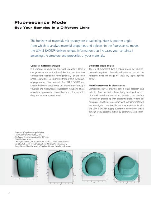
Front end of a polymeric optical fiber.<br />
Fluorescence excitation at 633 nm.<br />
3D shadow projections, rotated by 40° each.<br />
Plan-Neofluar 10x/0.3;<br />
1302.7 µm x 1302.7 µm x 1184.0 µm, 512 x 512 pixels x 161 sections.<br />
Sample: Prof. Hartl, Prof. Dr. Poisel, Mr. Förster, Department EFI,<br />
Georg-Simon-Ohm University of Applied Sciences, Nürnberg, Germany<br />
12