Linear IgA dermatosis induced by captopril - edigraphic.com
Linear IgA dermatosis induced by captopril - edigraphic.com
Linear IgA dermatosis induced by captopril - edigraphic.com

Create successful ePaper yourself
Turn your PDF publications into a flip-book with our unique Google optimized e-Paper software.
m<strong>edigraphic</strong><br />
Artemisa<br />
en línea<br />
Localizador<br />
08-027<br />
Casos Clínicos<br />
<strong>Linear</strong> <strong>IgA</strong> <strong>dermatosis</strong> <strong>induced</strong> <strong>by</strong> <strong>captopril</strong><br />
Dermatosis <strong>IgA</strong> lineal<br />
N. Menezes 1 , P.Varela 1 ,A. Furtado 2 , G. Mota 1 ,A. Baptista 1<br />
1<br />
Serviço de Dermatologia e Venereologia. 2 Serviço de Anatomia Patológica. Centro Hospitalar de Vila Nova de Gaia/Espinho<br />
Correspondencia:<br />
N. Menezes<br />
Serviço de Dermatologia e Venereologia<br />
Centro Hospitalar de Vila Nova de Gaia/Espinho<br />
Rua ConceiÇão Fernandes<br />
4430 VN Gaia<br />
Tel.: +351936262253<br />
Fax: +351227832755<br />
e-mail: nuno.menezes.dermatologia@gmail.<strong>com</strong><br />
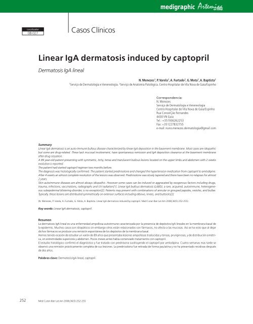
Summary<br />
<strong>Linear</strong> <strong>IgA</strong> <strong>dermatosis</strong> is an auto-immune bullous disease characterized <strong>by</strong> linear <strong>IgA</strong> deposition in the basement membrane. Most cases are idiopathic<br />
but some are drug-related. These lack mucosal involvement, have spontaneous remission and <strong>IgA</strong> deposition clearance at the basement membrane<br />
after drug cessation.<br />
A 89 year-old patient presenting with symmetric, itchy, tense and translucent bullous lesions located on the upper limbs and abdomen with 2 weeks<br />
evolution is reported.<br />
The patient had started <strong>captopril</strong> regimen two months before.<br />
The diagnosis was histologically confirmed. The patient started prednisolone and changed the hypertension medication from <strong>captopril</strong> to amlodipine.<br />
After 4 weeks an almost <strong>com</strong>plete resolution of the lesions was observed. Prednisolone was slowly tapered and there have been no relapses for almost<br />
2 years.<br />
Skin autoimmune diseases are almost always idiopathic. However some cases can be <strong>induced</strong> or aggravated <strong>by</strong> exogenous factors including drugs,<br />
trauma, infections, vaccinations, radiographs and UV radiation[1]. <strong>Linear</strong> <strong>IgA</strong> bullous <strong>dermatosis</strong> (LABD), a rare, acquired, autoimmune, heterogeneous<br />
subepidermal blistering disorder, is no exception[2]. Patients may present with <strong>com</strong>binations of annular or grouped papules, vesicles, and bullae.<br />
Typically, these lesions are distributed symmetrically on extensor surfaces including elbows, knees, and buttocks[2].<br />
(N. Menezes, P. Varela, A. Furtado, G. Mota, A. Baptista. <strong>Linear</strong> <strong>IgA</strong> <strong>dermatosis</strong> <strong>induced</strong> <strong>by</strong> <strong>captopril</strong>. Med Cutan Iber Lat Am 2008;36(5):252-255)<br />
Key words: <strong>Linear</strong> <strong>IgA</strong> <strong>dermatosis</strong>, <strong>captopril</strong>.<br />
Resumen<br />
La <strong>dermatosis</strong> <strong>IgA</strong> lineal es una enfermedad ampollosa autoinmune caracterizada por la presencia de depósitos <strong>IgA</strong> lineales en la membrana basal de<br />
la epidermis. Muchos casos son idiopáticos sin embargo otros están relacionados con fármacos, no afecta a las mucosas. Así se ha visto que al dejar<br />
dichos fármacos se produce una remisión espontánea de los depósitos de la membrana basal.<br />
Hemos tenido ocasión de estudiar un varón de 89 años que presentaba lesiones ampollosas traslúcidas y tensas, pruriginosas, y de distribución simétrica,<br />
en extremidades superiores y abdomen. Pocos meses antes había <strong>com</strong>enzado tratamiento con <strong>captopril</strong>.<br />
El estudio histológico confirmó el diagnóstico y fue tratado con prednisona sustituyendo el <strong>captopril</strong> por amlodipina. Cuatro semanas mas tarde se<br />
observó una remisión prácticamente <strong>com</strong>pleta de sus lesiones. La prednisolona fue retirada de forma paulatina y no ha presentado recidivas después<br />
de dos años.<br />
Palabras clave: Dermatosis <strong>IgA</strong> lineal, <strong>captopril</strong>.<br />
252<br />
Med Cutan Iber Lat Am 2008;36(5):252-255
N. Menezes et al. <strong>Linear</strong> <strong>IgA</strong> <strong>dermatosis</strong> <strong>induced</strong> <strong>by</strong> <strong>captopril</strong><br />
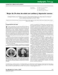
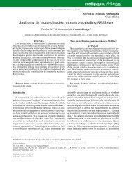
Figure 1. Bullous lesions involving the upper limbs and the<br />
trunk.<br />
Case report<br />
A 89 year-old patient presuming <strong>by</strong> symmetric, itchy, tense<br />
and translucent bullous lesions located on the upper limbs<br />
and abdomen with 2 weeks evolution is reported. The lesions<br />
first started around the umbilicus and distal parts of upper<br />
limbs with progressive spreading to the back and thoracic<br />
region (Figure 1). An intense burning sensation was described<br />
<strong>by</strong> the patient preceding the appearance of new lesions. The<br />
patient was taking <strong>captopril</strong> for 2 month due to hypertension.<br />
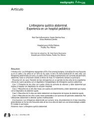
Biopsy specimens were obtained from lesional and perilesional<br />
skin for histopathologic examination with hematoxylin-eosin<br />
staining and direct immunofluorescence (IF). A<br />
subepidermal blister with abundant fibrine and eosinophils<br />
in the blister fluid was observed. In the papillary dermis a<br />
sparse mixed inflammatory infiltrate (neutrophils and eosinophils)<br />
was present (Figure 2).<br />
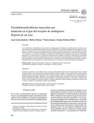
Sections 4µm-thick were prepared and stained with fluorescein<br />
isothiocyanate-labeled antibodies to human IgG,<br />
IgM, <strong>IgA</strong> and C3. The direct IF showed continuous linear<br />
deposits of <strong>IgA</strong> at the basement membrane but no evidence<br />
of IgG, IgM and C3 (Figure 3).<br />
Therapy with prednisolone (1 mg/kg/day) was started,<br />
suspecting of a bullous disease and the hypertension medication<br />
was changed from <strong>captopril</strong> to amlodipine.<br />
After 4 weeks an almost <strong>com</strong>plete resolution of the<br />
lesions was observed and prednisolone was slowly tapered<br />
for three month period.<br />
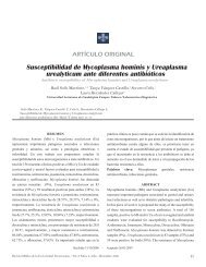
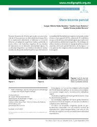
After <strong>com</strong>plete resolution and prednisolone withdrawal<br />
a new skin biopsy was performed which revealed a<br />
normal skin with a negative direct IF. After 2 years of<br />
follow-up the patient is without lesions and had no new<br />
flares (Figure 4).<br />
Comment<br />
LABD is a rare subepidermal autoimmune bullous <strong>dermatosis</strong><br />
almost always idiopathic but several drugs medications<br />
were implied as etiologic factors. Van<strong>com</strong>ycin is the most frequent<br />
agent reported[3-5], but other drugs with the potential<br />
to induce LABD include diclofenac , furosemide , <strong>captopril</strong>,<br />
lithium, cefamandole, somatostatin[3] phenytoin, trimethoprim-sulfamethoxazole,<br />
rifampicin, IL-2, interferon gamma,<br />
amiodarone, penicillin G, carbamazepine, piroxicam, atorvastatin<br />
and topical iodine[3, 6-19].<br />
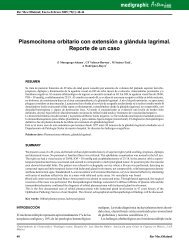
Figure 2. Subepidermal blistering with abundant fibrine and<br />
eosinophils in the blister fluid. A sparse mixed inflammatory<br />
infiltrate (neutrophils and eosinophils) was present in the papillary<br />
dermis. (Hematoxilin-eosin 4x).<br />
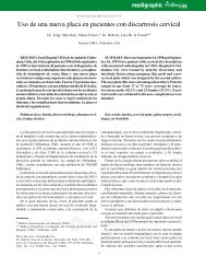
Figure 3. Direct IF showed continuous linear deposits of <strong>IgA</strong> at<br />
the basement membrane but no evidence of IgG, IgM and C3.<br />
Med Cutan Iber Lat Am 2008;36(5):252-255 253
N. Menezes et al. <strong>Linear</strong> <strong>IgA</strong> <strong>dermatosis</strong> <strong>induced</strong> <strong>by</strong> <strong>captopril</strong><br />
Figure 4. The patient without lesions.<br />
Captopril, an angiotensin-II-converting enzyme inhibitor,<br />
is a largely used antihypertensive agent that has been associated<br />
with a wide variety of cutaneous reactions, including<br />
angioedema, urticaria, lichenoid eruptions and pityriasis<br />
rosea-like rash. Despite being already established as an etiologic<br />
factor to pemphigus, it’s role as an inducer of other<br />
bullous disease has not been frequently reported[20-25].<br />
There are very few case reports from LABD <strong>induced</strong> <strong>by</strong> <strong>captopril</strong>,<br />
the last one being from 1996.<br />
Both idiopathic and drug-<strong>induced</strong> LABD are heterogeneous<br />
in clinical presentation[26].<br />
A spectrum of clinical presentations has been described<br />
regarding this disease; patients have lesions similar to<br />
dermatitis herpetiformis or bullous pemphigoid, but the<br />
unique immunofluorescence pattern with linear <strong>IgA</strong> deposition<br />
along the basement membrane allow the diagnosis[3].<br />
Most typically, tense vesicles are arranged in herpetiform,<br />
sausage-like, rosette-like or arciform patterns on<br />
erythematous or normal-appearing skin. Histology usually<br />
reveals subepidermal bullae and an inflammatory infiltrate<br />
of the upper dermis, sometimes with microabscesses.<br />
Direct IF is a useful tool to allow the differential diagnosis<br />
between bullous diseases. Indirect immunofluorescence in<br />
LABD only occasionally reveals circulating <strong>IgA</strong> anti-basement-membrane-zone<br />
antibodies[21].<br />
There are reports of unrelated antibodies in LABD<br />
(detected <strong>by</strong> indirect IF), to a 285 Kd antigen in the lamina<br />
densa and sublamina densa regions, a 250 Kd dermal antigen<br />
corresponding to collagen VII of anchoring fibrils,<br />
BP230 antigen and a 97 kd antigen in the upper lamina lucida<br />
which is believed to be an ectodomain of the BP230 antigen<br />
produced <strong>by</strong> proteolysis[3, 27-29], but direct IF remains<br />
the gold standard for diagnosis.<br />
However, drug-<strong>induced</strong> LABD, according to Kuechle et<br />
al. appears to differ from idiopathic cases in some aspects,<br />
namely lack of mucosal involvement, spontaneous remission<br />
after drug cessation and <strong>IgA</strong> deposition clearance at<br />
the basement membrane once the cutaneous lesions<br />
resolve[3]. However these clues for a drug-<strong>induced</strong> <strong>IgA</strong><br />
bullous <strong>dermatosis</strong> can’t be viewed in a dogmatic way,<br />
because in a literature review conducted <strong>by</strong> Palmer, 13 of<br />
29 patients with LADB drug-<strong>induced</strong> had mucosal involvement[27].<br />
Our case lacked mucosal involvement and had a significant<br />
improvement of the lesions after suspicious drug withdrawal.<br />
We also performed a skin biopsy after <strong>dermatosis</strong><br />
resolution which was normal, with a negative direct IF to all<br />
antibodies tested. This together with the time relationship<br />
between start of <strong>captopril</strong> intake and the appearance of the<br />
lesions, despite the absence of a drug rechallenge, make us<br />
believe this is a case of drug <strong>induced</strong> LABD.<br />
In drug-<strong>induced</strong> cases the withdrawal of the offending<br />
agent normally induces rapid resolution of the <strong>dermatosis</strong>.<br />
However some authors treat actively the disease with the<br />
objective of a more rapid remission[3, 28]. In our case due to<br />
the pemphigoid-like presentation corticosteroids were started<br />
and slowly tapered. After that there were no flairs, being<br />
without lesions for three years now.<br />
References<br />
1. Salmhofer W, Soyer P, Wolf P, Födinger D, Hödl<br />
S, Kerl H. UV light-<strong>induced</strong> linear <strong>IgA</strong> <strong>dermatosis</strong>.<br />
J Am Acad Dermatol 2004;50:109-15.<br />
2. Camilleri M, Pace JL. <strong>Linear</strong> <strong>IgA</strong> bullous <strong>dermatosis</strong><br />
<strong>induced</strong> <strong>by</strong> piroxicam. J Eur Acad<br />
Dermatol Venereol 1998;10:70-2.<br />
3. Kuechle MK, Stegemeir E, Maynard B, Gibson<br />
LE, Kristin ML, Peters MS: Drug-<strong>induced</strong><br />
<strong>IgA</strong> bullous <strong>dermatosis</strong>: Report of six cases<br />
and review of the literature. J Am Acad Dermatol<br />
1994;30:187-92.<br />
4. Danielsen AG, Thomsen K. Van<strong>com</strong>ycin<strong>induced</strong><br />
linear <strong>IgA</strong> bullous disease. Br J Dermatol<br />
1999;141:756-7.<br />
5. Baden LA, Apovian C, Imber MJ, Dover JS.<br />
Van<strong>com</strong>ycin-<strong>induced</strong> linear <strong>IgA</strong> bullous <strong>dermatosis</strong>.<br />
Arch Dermatol 1988;124:1186-8.<br />
6. Gabrielsen TO, Staerfelt F, Thrune PO. Drug<strong>induced</strong><br />
bullous <strong>dermatosis</strong> with linear <strong>IgA</strong><br />
deposits along the basement membrane.<br />
Acta Derm Venereol 1981;61:439-41.<br />
7. Cerottini JP, Ricci C, Guggisberg D, Panizzon<br />
Rg. Drug-<strong>induced</strong> linear <strong>IgA</strong> bullous <strong>dermatosis</strong><br />
probably<strong>induced</strong> <strong>by</strong> furosemid. J Am<br />
Acad Dermatol 1999;41:103-5.<br />
8. Klein L, Shmunes E, Carter J. <strong>Linear</strong> <strong>IgA</strong><br />
bullous <strong>dermatosis</strong> related to <strong>captopril</strong> treatment.<br />
Cutis 1989;41:103-5.<br />
9. McWhirter JD, Hashimoto K, Fayne S, ItoK.<br />
<strong>Linear</strong> <strong>IgA</strong> bullous <strong>dermatosis</strong> related to<br />
lithium carbonate. Arch Dermatol 1987;123:<br />
1120-2.<br />
10. Argenyi Z, Bergfeld W, Valenzuela R, Mc<br />
MahonJ, Taylor J. Adult linear <strong>IgA</strong> disease<br />
associated with an erythema multiforme-like<br />
drug reaction. Cleve Clin J Med 1987;54:<br />
445-50.<br />
11. Acostamadiedo JM, Perniciaro C, Rogers RS<br />
III. Phenytoin-<strong>induced</strong> linear <strong>IgA</strong> bullous disease.<br />
J Am Acad Dermatol 1998;38:352-6.<br />
12. Travan A, Pezen DS, Medenica M, Michelson<br />
GC, Vogelzang N, Soltani KM. Interleu-<br />
254<br />
Med Cutan Iber Lat Am 2008;36(5):252-255
N. Menezes et al. <strong>Linear</strong> <strong>IgA</strong> <strong>dermatosis</strong> <strong>induced</strong> <strong>by</strong> <strong>captopril</strong><br />
kin-2 associated linear <strong>IgA</strong> bullous <strong>dermatosis</strong>.<br />
J Am Acad Dermatol 1996;35:865-7.<br />
13. Guillaume JC, Escudier B, Espagne E, Roujeau<br />
JC, Prost C, Domart P. Dermatose<br />
bulleuse avec dépôts linéaire d’<strong>IgA</strong> le long de<br />
la membrane basale au cours d’un traitement<br />
par interferon gamma et interleukin 2.<br />
Ann Dermatol Venereol 1990;117:899-902.<br />
14. Primka EJ, Liranzo MO, Bergfeld WF, Dijkstra<br />
JW. Amiodarone-<strong>induced</strong> linear <strong>IgA</strong> disease.<br />
J Am Acad Dermatol 1994;32:809-11.<br />
15. Combermale P, Gavaud C, Cozzani E, Nicolas<br />
JF, Guennoc B, Dusseau JY. Dermatose à<br />
<strong>IgA</strong> linéaire induite par la penicilline G. Ann<br />
Dermatol Venereol 1993;120:847-8.<br />
16. Cohen LM, Ugent RB. <strong>Linear</strong> <strong>IgA</strong> bullous<br />
<strong>dermatosis</strong> occurring after carbamazepine.<br />
J Am Acad Dermatol 2002;46:S32-3.<br />
17. Plunkett RW, Chiarello SE, Beutner EH. <strong>Linear</strong><br />
<strong>IgA</strong> bullous dermatitis in one of two piroxican-<strong>induced</strong><br />
eruptions: a distinct direct<br />
immunofluorescence trend revealed <strong>by</strong> the<br />
literature. J Am Acad Dermatol 2001;44:<br />
689-92.<br />
18. Konig C, Eickert A, Scharfetter-Kochanek K,<br />
Krieg T, Hunzelmann N. <strong>Linear</strong> <strong>IgA</strong> bullous<br />
<strong>dermatosis</strong> <strong>induced</strong> <strong>by</strong> atorvastatin. J Am<br />
Acad Dermatol 2001;44:689-92.<br />
19. Wakelin SH, Wojnarowka F. <strong>Linear</strong> <strong>IgA</strong> disease<br />
exacerbated <strong>by</strong> topical iodine preparations.<br />
Br J Dermatol 1994;31:809-11.<br />
20. Friedman IS, Rudikoff D, Phelps RG, Sapadin<br />
AN. Captopril-triggered linear <strong>IgA</strong><br />
bullous <strong>dermatosis</strong>. International Journal of<br />
Dermatology 1998;37:608-12.<br />
21. Wood SM, Mann RD, Rawlins MD. Angioedema<br />
and urticaria associated with angiotensin<br />
converting enzyme inhibitors. Br Med<br />
J 1987;294:91-2.<br />
22. Phillips WG, Vaughan-Jones S, Jenkins R,<br />
Brathnach SM. Captopril-<strong>induced</strong> lichenoid<br />
eruption. Clin Exp Dermatol 1994;19:317-<br />
20.<br />
23. Wilken JK, Kirkendall WM. Pityriasis rosealike<br />
rash from <strong>captopril</strong>. Arch Dermatol<br />
1982;118:186-87.<br />
24. Kaplan RP, Potter TS, Fox JN. Drug-<strong>induced</strong><br />
pemphigus related to angiotensin-converting<br />
enzyme inhibitors. J Am Acad Dermatol<br />
1992;26:364-66.<br />
25. Sala F, Crosti C, Monti M, DeBitonto A. Cutaneous<br />
pathology due to <strong>captopril</strong> – observations<br />
on 2 clinical cases. G Ital Dermatol<br />
Venerol 1983;118:89-93.<br />
26. Janniger CK, Wiltz H, Schartz RA, et al. <strong>Linear</strong><br />
<strong>IgA</strong> bullous <strong>dermatosis</strong>: a polymorphic<br />
disorder. Cutis 1990;45:37-42.<br />
27. Palmer RA, Ogg G, Allen J, Banerjee A, Ryatt<br />
KS, Ratnavel R, Wojnarowska. Van<strong>com</strong>ycin<strong>induced</strong><br />
linear <strong>IgA</strong> disease with autoantibodies<br />
to BP 180 and LAD 285. Br J Dermatol<br />
2001;145:816-20.<br />
28. Wojnarowska F, Whitehead P, Leigh IM.<br />
Identification of the target antigen in chronic<br />
bullous disease of childhood and linear <strong>IgA</strong><br />
disease of adults. Br J Dermatolm 1991;<br />
124:157-62.<br />
29. Zone JJ, Taylor TB, Kadunce DP. Identification<br />
of the cutaneous basement membrane<br />
zone antigen and isolation of antibody in<br />
linear immunoglobulin A bullous <strong>dermatosis</strong>.<br />
J Clin Invest 1990;85:812-20.<br />
Med Cutan Iber Lat Am 2008;36(5):252-255 255