Activation of Human Effector Cells by a Tumor Reactive ...
Activation of Human Effector Cells by a Tumor Reactive ...
Activation of Human Effector Cells by a Tumor Reactive ...

Create successful ePaper yourself
Turn your PDF publications into a flip-book with our unique Google optimized e-Paper software.
Vol. 2, 1951-1959. December 1996 Clinical Cancer Research 1951<br />
<strong>Activation</strong> <strong>of</strong> <strong>Human</strong> <strong>Effector</strong> <strong>Cells</strong> <strong>by</strong> a <strong>Tumor</strong> <strong>Reactive</strong><br />
Recombinant Anti-Ganglioside GD2 Interleukin-2<br />
Fusion Protein (chl4.18-1L2)’<br />
Jacquelyn A. Hank,2 Jean E. Surfus, Jacek Gan,<br />
Peter Jaeger, Stephen D. Gillies,<br />
Ralph A. Reisfeld, and Paul M. Sondel<br />
Department <strong>of</strong> <strong>Human</strong> Oncology. University <strong>of</strong> Wisconsin-Madison,<br />
Madison. Wisconsin 53792 Ii. A. H.. J. E. S., J. G.. P. J., P. M. 5.1:<br />
Fuji ImmunoPharmaceuticals Corporation. Lexington. Massachusetts<br />
02173 15. D. G.l: The Scripps Research Institute. La Jolla. Califrrnia<br />
92037 iR. A. RI: and Departments <strong>of</strong> Pediatrics and Medical<br />
Genetics, University <strong>of</strong> Wisconsin-Madison, Madison, Wisconsin<br />
53792 P. M. 5.1<br />
ABSTRACT<br />
Cytotoxic effector cells interact with target cells<br />
through various mechanisms. CTLs use the antigen-specific<br />
T cell receptor, whereas Fe receptor-positive natural killer<br />
cells use this receptor to interact with antibody-coated target<br />
cells. We evaluated the tumor-binding and lymphocyte-activating<br />
capability <strong>of</strong> a recombinant fusion protein consisting<br />
<strong>of</strong> a tumor-selective human/mouse chimeric anti-ganglioside<br />
GD2 antibody (chl4.18) and recombinant human<br />
interleukin-2 (1L2) (chl4.18-IL2). This fusion protein bound<br />
specifically to GD2-positive melanoma and neuroblastoma<br />
tumor cell lines, and its 1L2 component stimulated in vitro<br />
proliferation <strong>of</strong> an IL2-dependent cell line, as well as peripheral<br />
blood mononuclear cells, in healthy control individuals<br />
and in cancer patients receiving continuous infusion <strong>of</strong> 1L2.<br />
The 1L2 presented <strong>by</strong> the fusion protein, when bound to<br />
tumor cells, induced proliferation <strong>of</strong> IL2-responsive cells as<br />
well as a comparable amount <strong>of</strong> soluble 1L2 did. This suggests<br />
that localization <strong>of</strong> 1L2 at the site <strong>of</strong> contact between<br />
tumor and effector cells is an effective way <strong>of</strong> presenting this<br />
cytokine to 1L2-responsive cells. The chl4.18-1L2 fusion<br />
protein also mediated antibody-dependent cellular cytotoxicity<br />
with Fc receptor-positive effector cells to an extent<br />
similar to chl4.18. These results, together with those <strong>of</strong><br />
previous studies documenting antitumor efficacy against human<br />
tumor xenografts in SCID mice and GD2-positive murine<br />
tumors in immunocompetent syngeneic mice, suggest<br />
that the chl4.18-1L2 fusion protein should be tested in Phase<br />
I and II trials in patients with GD2-positive tumors.<br />
INTRODUCTION<br />
Immunotherapy with IL23 is <strong>of</strong> benefit to some patients<br />
with renal cell cancer and melanoma ( I ). Although multiple<br />
immune mechanisms are activated in patients receiving IL2, the<br />
immune components induced <strong>by</strong> lL2 necessary for antitumor<br />
activity have not been identified. Continuous infusion <strong>of</strong> IL2. at<br />
doses tolerated in the outpatient setting. induces systemic<br />
lymphoid activation in virtually all treated patients: however,<br />
only a minority <strong>of</strong> such patients achieve antitumor responses<br />
( 1-4). Included in this lymphoid cell activation is an expansion<br />
and activation <strong>of</strong> the CDl6, FcR NK cells (5. 6). Currently.<br />
attempts are being made clinically to target these cells to tumor<br />
through the use <strong>of</strong> tumor-specific mAbs (7. 8). These include<br />
clinical evaluation <strong>of</strong> regimens combining effector cell activation<br />
through 1L2 infusions with infusions <strong>of</strong> munine or chimeric<br />
tumor-selective mAb, such as the l4.G2a or chl4. 18 antibodies,<br />
which recognize the ganglioside GD2 expressed on neuroblastoma.<br />
melanoma, and certain other tumors (7).<br />
In an effort to augment the stimulation <strong>of</strong> the FcR antibody-directed”<br />
effector cells and activate FcR effector cells<br />
that express IL2 receptors (2, 9), a chl4.l8-IL2 fusion protein<br />
has been constructed <strong>by</strong> fusion <strong>of</strong> a synthetic sequence coding<br />
for human IL2 to the carboxyl end <strong>of</strong> the C-yl gene <strong>of</strong> the mAb<br />
ch I 4. 1 8 ( 1 0). When the antitumor variable region <strong>of</strong> this fusion<br />
protein binds to tumor, IL2 should be concentrated in the tumor<br />
microenvironment and provide activation <strong>of</strong> FcR effectors,<br />
such as NK cells, that did bind to the immunoglobulin Fe<br />
domain <strong>of</strong> the tumor-bound fusion protein. Furthermore, the IL2<br />
component <strong>of</strong> this tumor-bound fusion protein may activate<br />
IL2-responsive cells. such as cytotoxic T cells and helper T<br />
cells, that do not necessarily have FcRs and the subpopulation <strong>of</strong><br />
NK cells that express IL2 receptors but lack the FcR (9), there<strong>by</strong><br />
recruiting additional effector cells into the tumor microenvironment.<br />
The chI4.l8-1L2 fusion protein was previously shown to<br />
maintain antigen-binding activity and 1L2 activity ( 10) and has<br />
provided antitumor effects in SCID mice bearing human tumor<br />
xenografts <strong>of</strong> neuroblastoma ( I I ) and melanoma ( I 2), as well as<br />
in a syngeneic murine melanoma model ( 13). We have extended<br />
these findings <strong>by</strong> demonstrating that chl4. l8-IL2 bound to<br />
GD2 tumor cells can be visualized <strong>by</strong> flow cytornetry, detecting<br />
either the chl4.l8 or the 1L2 portion <strong>of</strong> this construct. The<br />
IL2 component <strong>of</strong> the fusion protein is able to stimulate lL2-<br />
dependent cells. The fusion protein bound to GD2 tumors<br />
Received 10/24/95; revised 8/27/96: accepted 9/18/96.<br />
‘ This research was supported <strong>by</strong> NIH grants and contracts CA-5344 I.<br />
CA-05436. CA-32685, CM-87290, CA- I 4520, CA- I 3539. CM-47669,<br />
HL-02I43. RR-03186, and American Cancer Society Grant CH-237.<br />
2 To whom requests for reprints should be addressed, at K4/454 CSC.<br />
6()0 Highland Avenue, Madison. WI 53792. Phone: (608) 263-7262:<br />
Fax: (608) 263-4226.<br />
3 The abbreviations used are: lL2. interleukin 2: NK. natural killer: FcR,<br />
Fe receptor: mAb. monoclonal antibody: ADCC. antibody-dependent<br />
cellular cytotoxicity: HLR, H<strong>of</strong>fmann LaRoche; PBMC, peripheral<br />
blood mononuclear cell: LAK. lymphokine-activated killer.
1952 Anti-GD2-IL2 Fusion Protein<br />
stimulates proliferation to the same extent as soluble fusion<br />
protein or soluble 1L2. The chl4.l8-1L2 fusion protein also<br />
stimulates cytolytic activity in vitro <strong>by</strong> cells able to mediate<br />
ADCC. FcR NK cells obtained from melanoma patients in<br />
vivo following therapy with continuous infusion 1L2 are targeted<br />
to GD2 tumors that have bound chl4.l8-IL2 in vitro, and the<br />
lytic activity was comparable to that induced <strong>by</strong> the combination<br />
<strong>of</strong> free antibody and soluble 1L2.<br />
MATERIALS AND METHODS<br />
Recombinant IL2. HLR IL2 was provided through the<br />
Cancer Treatment and Evaluation Program <strong>of</strong> the National Cancer<br />
Institute. The National Cancer Institute-Biological Response<br />
Modifiers program standard for unit dosage was used, and the<br />
specific activity <strong>of</strong> the IL2 was 15 X 106 units/mg. This unit<br />
corresponds closely with the international standard for IL2<br />
unitage (14).<br />
Chimenc Antibody and Fusion Protein. The mouse/<br />
human chimeric 14.18 antibody was constructed <strong>by</strong> combining<br />
the variable regions <strong>of</strong> the murine anti-GD2 14.18 antibody with<br />
the constant regions <strong>of</strong> human IgG1 heavy chain and K light<br />
chain (15, 16). The 14.l8-IL2 fusion protein was constructed <strong>by</strong><br />
fusion <strong>of</strong> a synthetic sequence coding for human IL2 to the<br />
carboxyl end <strong>of</strong> the human C-yl gene <strong>of</strong> the mAb chl4. 18 (10).<br />
The fused gene was inserted into the vector pdHL2-l4. 18 as<br />
described previously (15). Transfection <strong>of</strong> the expression plasmids<br />
in Sp2/0-Agl4 cells and propagation <strong>of</strong> the clones secreting<br />
chl4. 18-1L2, as well as its purification, have been described<br />
previously (10). To compare the 1L2 activity in the soluble IL2<br />
preparation and in the fusion protein, concentrations were based<br />
on weight/volume calculations <strong>of</strong> 1L2 in the two preparations.<br />
Because the fusion protein molecule consists <strong>of</strong> 80% chimeric<br />
14.18 immunoglobulin and 20% 1L2 <strong>by</strong> molecular weight, the<br />
fusion protein 1L2 concentration was based on IL2 comprising<br />
20% <strong>of</strong> the weight <strong>of</strong> the fusion protein. Thus, 50 ng/rnl <strong>of</strong><br />
fusion protein would correspond to 10 ng/ml <strong>of</strong> IL2 in the fusion<br />
protein. Because 10 ng/ml <strong>of</strong> soluble IL2 corresponds to 150<br />
units/mi <strong>of</strong> soluble HLR 1L2, we have used this conversion to<br />
describe the units <strong>of</strong> 1L2 anticipated for the fusion protein<br />
preparation based on the molecular weight <strong>of</strong> IL2 and using the<br />
specific activity <strong>of</strong> 15 X 106 units/mg for the HLR IL2.<br />
<strong>Tumor</strong> Cell Lines. The GD2-positive LA-N-S neuroblastoma<br />
target cell line, kindly provided <strong>by</strong> R. Seeger (Children’s<br />
Hospital <strong>of</strong> Los Angeles, Los Angeles, CA), was maintamed<br />
as an adherent monolayer in Leibovitz’ s medium<br />
supplemented with 15% heat-inactivated fetal bovine serum. A<br />
trypsin-EDTA solution was used to harvest the cell monolayer.<br />
The M2l human melanoma line (GD2’) was described previously<br />
(15), and the BT-20 human breast carcinoma cell line<br />
(GD2) was obtained from American Type Culture Collection.<br />
Both cell lines were maintained as adherent monolayers in<br />
RPM! 1640 supplemented with penicillin and streptomycin<br />
(P/S), L-glutamine, HEPES buffer, and 10% fetal bovine serum.<br />
Flow Cytometry. Cell-bound fusion protein and antibody<br />
were detected <strong>by</strong> standard indirect immun<strong>of</strong>luorescence<br />
methods (Becton Dickinson, San Jose, CA). Antibodies ineluded<br />
a goat antihuman IgG (Caltag, San Francisco, CA) and a<br />
rabbit antihuman IL2 (Genzyme, Cambridge, MA).<br />
Proliferation Assays. Fresh PBMCs from healthy volunteer<br />
human donors or from patients who were treated with a<br />
96-h continuous infusion <strong>of</strong> IL2 (3. 4, 17) were cultured in<br />
0.2-ml round-bottom microplates at a concentration <strong>of</strong> 1 X l0<br />
cells/well in RPMI 1640 supplemented with 10% human serum<br />
(Pel-Freez, Rogers, AR), 25 msi HEPES, 100 units/mI penicillin,<br />
and 100 p.g/ml streptomycin sulfate (RPMI-HS). Recombinant<br />
IL2 and fusion protein were added at concentrations as<br />
indicated in the “Results”. Concentrations <strong>of</strong> recombinant soluble<br />
1L2 were also tested, which corresponded to the concentration<br />
<strong>of</strong> 1L2 in the fusion protein preparation based on the<br />
molecular weight and concentration <strong>of</strong> fusion protein: I i.g <strong>of</strong><br />
the fusion protein contains approximately 3000 units <strong>of</strong> IL2<br />
(I 1). In experiments in which chl4. I 8 or fusion protein-coated<br />
M21 and LA-N-S cells were used as a proliferative stimulus, the<br />
antibody or fusion protein at S ig/ml was incubated with the<br />
tumor cells for 1 h on ice. Irradiation <strong>of</strong> these cells took place<br />
during this incubation, with LA-N-S and M2l receiving 10,000<br />
and 40,000 rads, respectively. Cultures were incubated at 37#{176}C<br />
in 5% CO2 for 48-72 h, pulsed with I pCi [3H]thymidine for<br />
18 h, and harvested with a Filterrnate 196 Packard harvester, and<br />
[3H]thymidine incorporation was quantitated with a Matrix<br />
9600 direct 13 counter with a 5-mm counting period. Informed<br />
consent forms, approved <strong>by</strong> the University <strong>of</strong> Wisconsin <strong>Human</strong><br />
Subjects Committee, were obtained prior to collection <strong>of</strong> all<br />
human blood specimens.<br />
For some proliferative studies, the 1L2-responsive cells<br />
used were the Tf-l myeloid leukemia cell line transfected with<br />
the gene for the 1L2 receptor 3 chain. This transfected line was<br />
designated Tf-l , and the mock-transfected control line contaming<br />
the LXSN vector but no IL2R3 gene was designated<br />
Tf-IL (18). This Tf-l3 cell line responded to 1L2 using intermediate<br />
affinity receptor complexes ( 1 7. I 8) and thus is<br />
analogous to the majority <strong>of</strong> NK cells in 1L2-treated patients,<br />
which also use intermediate affinity IL2 receptors (2). The Mik<br />
11 monoclonal antibody directed against the p70 IL-2 receptor<br />
1 chain was used in the blocking studies ( 19. 20).<br />
ADCC and Fusion Protein-mediated Cellular Cytotoxicity.<br />
All ADCC assays were performed in RPMI-HS. <strong>Effector</strong><br />
cells in a total volume <strong>of</strong> SO pi were plated in quadruplicate<br />
into 96-well U-bottomed microtiter plates at the indicated effector/target<br />
ratios. Just prior to the addition <strong>of</strong> target cells, SO<br />
il <strong>of</strong> antibody, antibody plus 1L2, or fusion protein were added<br />
to the effectors. While the effectors were being prepared, target<br />
cells were labeled for 2 h with 250 pCi <strong>of</strong> 5tCr in 0.2 ml <strong>of</strong><br />
RPMI-HS. Target cells were mixed every 15-30 mm during<br />
labeling to keep the cells in suspension. After being washed<br />
twice with RPMI, S x l0 target cells in 50 il <strong>of</strong> RPMI-HS<br />
were added to effector cells and centrifuged at 200 X g for S<br />
mm. In the experiments using fusion protein-coated target cells.<br />
chl4.l8-1L2 was added to the targets following one wash and<br />
incubated on ice for 1 h. Two subsequent washes removed<br />
excess fusion protein and 51Cr. The effector cells were also<br />
plated in medium and in IL2 to determine their ability to<br />
mediate lysis <strong>of</strong> target cells in the absence <strong>of</strong> antibody or fusion<br />
protein. The plates were incubated at 37#{176}Cat 5% CO2 for 4 h,<br />
and the supernatants were harvested using the Skatron Harvesting<br />
System (Skatron, McLean, VA). Maximum 5tCr release was<br />
measured <strong>by</strong> lysing target cells with the detergent cetrimide
Clinical Cancer Research 1953<br />
M21<br />
LAN-5<br />
BT-20<br />
chl4.18<br />
chl4.18-1L2<br />
JL<br />
Rabbit<br />
IgG<br />
Rabbit<br />
Anti-1L2<br />
Rabbit<br />
lgG<br />
Rabbit<br />
Anti-1L2<br />
. . . .-<br />
1u<br />
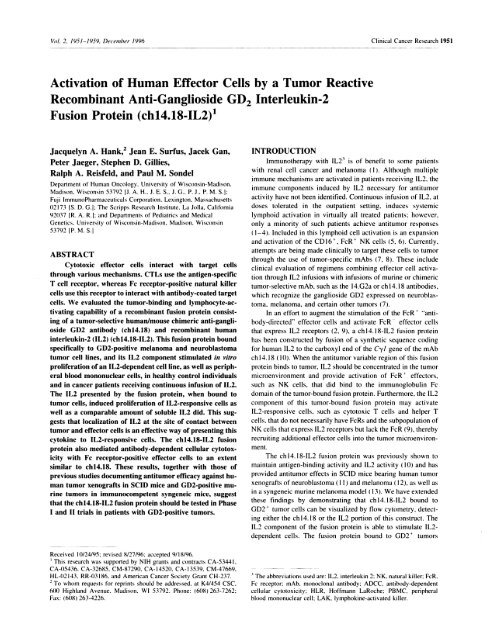
Fig. I Detection <strong>of</strong> antibody component and 1L2 component in tumor-bound fusion protein. The GD2 M2 1 melanoma and LA-N-S neuroblastoma<br />
and GD2 BT-20 breast carcinoma cell lines were each coated with I p.g <strong>of</strong> either chl4.18-1L2 fusion protein or chl4.18 antibody. To detect the<br />
antibody component (top panels). a fluorescein-conjugated goat antihuman IgG secondary antibody was used. To detect the 1L2 component (bottom<br />
panels). a rabbit anti-IL2 antibody was used, followed <strong>by</strong> a phycoerythrin-conjugated goat antirabbit antibody.<br />
(Sigma). Spontaneous 5tCr release was measured <strong>by</strong> incubating<br />
target cells in RPMI-HS alone. Percent cytotoxicity values were<br />
calculated for each effector/target ratio as follows:<br />
C/<br />
cytotoxicity<br />
experimental release - spontaneous release<br />
=l00X-- .<br />
maximum release - spontaneous release<br />
Results are expressed as percent cytotoxicity or as lytic<br />
units, where 1 lytic unit is the number <strong>of</strong> effector cells necessary<br />
to achieve 20% lysis <strong>of</strong> S X I0 targets.<br />
RESULTS<br />
Fusion Protein chl4.18-1L2 Binds to GD2 <strong>Tumor</strong><br />
<strong>Cells</strong>. The data shown in Fig. I demonstrate the binding <strong>of</strong><br />
chl4.l8-IL2 to the GD2 M2l human melanoma and LA-N-S<br />
human neuroblastoma cell lines. The fusion protein binds to the<br />
tumor cells with a fluorescence intensity similar to that <strong>of</strong> the<br />
parental ch I 4. 18 antibody (Fig. 1, top panels). The specificity <strong>of</strong><br />
ch I 4. 18-IL2 and ch I 4. 18 antibody is demonstrated <strong>by</strong> their lack<br />
<strong>of</strong> binding to the GD2 BT-20 human breast carcinoma cell<br />
line. The IL2 component <strong>of</strong> ch 14. 1 8-1L2 can be detected <strong>by</strong><br />
rabbit anti-IL2 antibody when the fusion protein is bound to<br />
M-2l and LA-N-S cells, respectively (Fig. I. bottom pane/c).<br />
These data document that the IL2 component <strong>of</strong> the fusion<br />
protein remains associated with the tumor cells that bind<br />
chl4.l8-1L2 on their surfaces and is detectable <strong>by</strong> anti-IL2<br />
antibody. Furthermore, separate flow cytometric analyses have<br />
shown that M-2l and LA-N-S cells do not express either the a<br />
or 3 chains <strong>of</strong> the 1L2 receptor (data not shown). Thus, binding<br />
<strong>of</strong> the fusion protein to these cells is mediated through the<br />
chl4.18 rather than the IL2 component <strong>of</strong> the fusion protein.<br />
Soluble chl4.18-1L2 Stimulates IL2-induced Proliferative<br />
Responses. Fig. 2 documents proliferative responses to<br />
1L2 attained with the Tf-I3 cell line and PBMCs from a melanoma<br />
patient obtained 24 h after a 96 h continuous iv. infusion<br />
<strong>of</strong> IL2. Tf-l is a GM-CSF-dependent myeloid leukemia-derived<br />
cell line that constitutively expresses the common cytokine<br />
receptor -y chain. A variant cell line bearing functional intermediate<br />
affinity IL2 receptors (3-y dimers), designated Tf- I , was<br />
obtained <strong>by</strong> stable transfection <strong>of</strong> Tf- I with eDNA encoding the<br />
1 chain <strong>of</strong> the IL2 receptor ( 1 8). The Tf- 1 3 cell line that<br />
retained responsiveness to GM-CSF also responded to IL2 in a<br />
dose-dependent fashion. The mock-transfected Tf-lL cell line<br />
did not proliferate in response to 1L2, and the Tf-l3 cell line did<br />
not respond to chl4.l8 antibody (data not shown). The results <strong>of</strong><br />
proliferative assays shown in Fig. 2 were obtained <strong>by</strong> using<br />
dilutions <strong>of</strong> IL2 and chl4.l8-1L2 to achieve equivalent molar
1954 Anti-GD2-IL2 Fusion Protein<br />
Cl)<br />
I-<br />
z<br />
0<br />
C)<br />
1200<br />
1000<br />
10000<br />
600<br />
400<br />
200<br />
8000<br />
6000<br />
4000<br />
2000<br />
0<br />
0 -<br />
400<br />
L2 UImI 1.6 3.0 00 12 25 50 100 200<br />
FP JmI.B2O 1.20 2.0 5.0 10 20 40 80 ISO<br />
Fig. 2 Proliferation induced <strong>by</strong> soluble IL2 and IL2 within the soluble<br />
fusion protein. The Tf-l3 cell line (A) and PBMCs obtained from a<br />
patient following continuous infusion lL2 (B) were stimulated with<br />
increasing concentrations <strong>of</strong> soluble IL2 (A) and soluble chl4.l8-1L2<br />
fusion protein (s). Dilutions <strong>of</strong> IL2 and fusion protein were made to<br />
achieve equivalent molar concentrations <strong>of</strong> IL2. The counts were determined<br />
for [3H]TdR incorporation <strong>by</strong> proliferating cells.<br />
concentrations <strong>of</strong> IL2. Assuming the same specific activity <strong>of</strong><br />
1L2 within the fusion protein as the recombinant HLR product,<br />
there are approximately 400 units/ml <strong>of</strong> 1L2 contained within<br />
the chl4.18-IL2 when it is applied at a concentration <strong>of</strong> 160<br />
ng/ml. When these data are expressed based on the concentration<br />
<strong>of</strong> 1L2, dose-response curves indicate that at concentrations <strong>of</strong><br />
1L2 less than or equal to SO units/rn], the fusion protein is slightly<br />
more efficient than free 1L2 at stimulating the Tf-l(3 cell line to<br />
proliferate. This finding was reproduced in two additional proliferative<br />
assays using the Tf- 13 cell line as the responding cell.<br />
PBMCs obtained from six patients with melanoma who<br />
had just completed a continuous infusion <strong>of</strong> IL2 were also tested<br />
for their responsiveness to IL2 and chl4.l8-IL2. Results from<br />
one representative patient are shown at the bottom <strong>of</strong> Fig. 2.<br />
Previous studies have shown that NK cells obtained following in<br />
vivo IL2 treatment show rapid proliferative responses to restimulation<br />
with IL2 in vitro, using primarily the intermediate<br />
affinity 13’y receptor complex (2, 21). Similar proliferation <strong>by</strong><br />
these cells was observed at most concentrations <strong>of</strong> IL2 and<br />
chl4.18-IL2 tested. These data indicate that the antibody component<br />
in the fusion protein did not adversely affect the ability<br />
<strong>of</strong> IL2 to interact with the IL2R complexes either on the Tf-l 3<br />
cells or on the PBMCs <strong>of</strong> a melanoma patients treated with IL2,<br />
nor did it affect the ability <strong>of</strong> the 1L2 component <strong>of</strong> the fusion<br />
protein to stimulate proliferation. chl4. I 8 antibody alone, at<br />
comparable concentrations, did not stimulate patient PBMCs to<br />
proliferate (data not shown). Table 1 presents data from two<br />
separate experiments indicating the in vitro proliferative response<br />
induced <strong>by</strong> 100 units <strong>of</strong> soluble IL2 and <strong>by</strong> a similar<br />
amount <strong>of</strong> IL2 contained within 40 ng <strong>of</strong> chl4.18-IL2. The<br />
responding cells were PBMCs obtained from five melanoma<br />
patients and PBMCs from one healthy control individual. The<br />
patient PBMCs were obtained 24 h after completion <strong>of</strong> a 96-h<br />
continuous infusion <strong>of</strong> IL2 in vivo. In these experiments, the<br />
fusion protein stimulated proliferation that was similar to that<br />
induced <strong>by</strong> the soluble 1L2 for patient I and for the control<br />
donor in experiment 1 and for the three patients in experiment 2.<br />
The chimeric chl4.l8 antibody itself was not stimulatory.<br />
Fusion Protein-induced Proliferation Mediated through<br />
an Intermediate Affinity #{176}y 1L2 Receptor Is Specifically<br />
Blocked <strong>by</strong> Antibody to the Chain <strong>of</strong> the IL2 Receptor.<br />
Fig. 3 demonstrates the proliferation induced <strong>by</strong> IL2 and fusion<br />
protein on cells with intermediate affinity receptors for 1L2,<br />
Tf-l 3, and PBMCs obtained from cancer patients following a<br />
4-day continuous infusion <strong>of</strong> 1L2. <strong>Cells</strong> with high-affinity aj<br />
IL2 receptors (Kit 225 cells) were also assayed. Tf- 1 3 and<br />
patient PBMCs responded to 1L2 and the fusion protein in a<br />
Table I Proliferative response to soluble IL2 and to chl4.l8-1L2 fusion protein<br />
PBMCs from a healthy volunteer individual (control) and from two patients were assayed in experiment 1. and PBMCs from three patients were<br />
assayed in experiment 2. The patient PBMCs were obtained 24 h after a 96-h continuous infusion <strong>of</strong> IL2. They were stimulated with IL2 or<br />
chl4.l8-IL2 fusion protein at concentrations containing equivalent amounts <strong>of</strong> IL2 or with an excess <strong>of</strong> chl4.I8 antibody. The cells were cultured<br />
for 4 days at 37#{176}C in 5% CO,. pulsed with 3H-thymidine for 16 h, and harvested, and counts per 5 mm were obtained with a Packard 9600 Matrix<br />
counter.<br />
3H-Thymidine incorporati on (count per 5 mm)<br />
Experiment<br />
I<br />
Control<br />
Patient 1<br />
Patient 2<br />
II Patient 16<br />
Patient 17<br />
Patient 20<br />
Medium 100 units/mI IL2 40 ng/ml chl4.18-1L2 O.5g/ml chl4.18<br />
197<br />
160<br />
184<br />
165<br />
49<br />
31<br />
14,194<br />
9,741<br />
24,381<br />
9,959<br />
2,321<br />
4,135<br />
13.387<br />
7,916<br />
7,413<br />
11,201<br />
2,700<br />
6,317<br />
190<br />
161<br />
207<br />
59<br />
46<br />
43
Clinical Cancer Research 1955<br />
120000 120000<br />
90000 90000<br />
L1<br />
I-<br />
60000 60000<br />
30000 30000<br />
0 - 0<br />
200 50 12.5 66.5 16.6 4.1<br />
16000 16000<br />
C-)<br />
0.<br />
I-<br />
a-<br />
12000<br />
8000<br />
4000<br />
0<br />
200<br />
50<br />
I 2000<br />
8000<br />
Fig. 3 Antibody to the 3 chain <strong>of</strong> the IL2 receptor<br />
blocks fusion protein induced proliferation <strong>by</strong> cells<br />
expressing intermediate affinity, but not cells expressing<br />
high-affinity lL2 receptors. Tf-l cells.<br />
PBMCs, and KIT 225 cells were cultured with lL2<br />
(leftpa,iels) or the chI4.18-1L2 fusion protein (right<br />
panels). The humanized Mik 3l antibody at a final<br />
concentration <strong>of</strong> 3 p.g/ml (A) or human serum diluted<br />
66 5 16 6 4 1 1 1/50 as the control () was added at the initiation <strong>of</strong><br />
12.5 . . a 3-day I3HITdR incorporation assay. #{149}, medium.<br />
100000<br />
100000<br />
I0<br />
C.”<br />
75000<br />
75000<br />
I-<br />
50000<br />
50000<br />
25000<br />
25000<br />
0-<br />
200<br />
‘ . 0 ‘<br />
50 12.5 66.5 16.6 4.1 1<br />
IL-2 CONCENTRATION ( U/mi) 14.18-IL-2 CONCENTRATiON (ug/mi)<br />
dose-dependent manner, and this proliferation was abrogated <strong>by</strong><br />
inclusion <strong>of</strong> the Mik 3I antibody, which recognizes the 3 chain Coated<br />
<strong>of</strong> the IL2 receptor (20). Over the dose range examined, there <strong>Tumor</strong> <strong>Cells</strong><br />
was a strong<br />
. . #{149}500<br />
proliferative response to both soluble IL2 and 60000<br />
#{149} <br />
fusion protein <strong>by</strong> the Kit 225 cell line, which expresses the U)<br />
50<br />
high-affinity afty IL2 receptor. The Mik 3 1 antibody did not I-<br />
block this proliferative response, as shown previously for IL2 z<br />
40000<br />
(19).<br />
chl4.18-1L2 Bound to GD2 <strong>Cells</strong> Stimulates Proliferation.<br />
The flow cytometric studies (Fig. 1 ) document that the<br />
chl4.18-IL2 binds to GD2 tumor cells via the antibody vanable<br />
region and that its IL2 component is recognized <strong>by</strong> rabbit<br />
antihuman 1L2 antibody. Subsequent experiments tested the<br />
functional activity <strong>of</strong> the IL2 component <strong>of</strong> the fusion protein<br />
when bound to tumor cells. In this case, the M21 and LA-N-S<br />
cell lines were coated with either chl4.18-IL2 or chl4.l8 and<br />
then washed free <strong>of</strong> any excess and cultured with the Tf-l<br />
responding cell line or the Tf-lL control cell line. The data<br />
shown in Fig. 4 demonstrate that the ch 14. 18-IL2 fusion protein.<br />
when bound to a GD2 tumor cell line, effectively presents IL2<br />
to the Tf- 1 3 cell line. When ch I 4. 18-IL2 is bound to S X 102<br />
M21 or LA-N-S cells it is able to efficiently present the bound<br />
0<br />
C)<br />
80000<br />
20000<br />
chl4.18<br />
M21<br />
FP<br />
LIt<br />
chl4.18<br />
FP<br />
LA-N-5<br />
Fig. 4 Proliferation induced <strong>by</strong> the lL2 component <strong>of</strong> the tumor-bound<br />
fusion protein. The M2l melanoma and LA-N-S neuroblastoma cell<br />
lines were coated for I h at 4#{176}Cwith either chI4.18 antibody or the<br />
chl4.18-1L2 fusion protein (FP). Irradiation took place during this<br />
incubation. These coated tumor target cells were washed twice and<br />
diluted to 500, 100, or 50 cells per well and used to stimulate l0 Tf-l<br />
cells per well. Proliferation was quantitated after 72 h <strong>by</strong> I3HITdR<br />
incorporation.
1956 Anti-GD2-lL2 Fusion Protein<br />
Table 2 Stimulatory activity <strong>of</strong> IL2 in tumor-bound fusion protein<br />
The Tf-l3 and control Tf-lL cell lines (experiment 1) or Tf-l3 cells and PBMCs from IL2-treated patients (experiment 2) were stimulated in<br />
a 3-day proliferative assay with soluble IL2 or tumor cells coated with chI4.18 antibody or the chl4.18-IL2 fusion protein. <strong>Tumor</strong> cells (106) were<br />
incubated with S i.g <strong>of</strong> chl4.18, washed twice, and diluted. Sixty % <strong>of</strong> the fusion protein used to coat the M2l cell remained bound to the cells:<br />
therefore, 100 cells have 0.3 ng <strong>of</strong> fusion protein, corresponding to approximately 1 unit <strong>of</strong> bound lL2. The total volume <strong>of</strong> the microwell is 0.2 ml,<br />
resulting in a total <strong>of</strong> 5 units <strong>of</strong> IL2/ml in the wells with chl4.18-IL2-coated tumor cells. The cells were cultured for 2.5 days at 37#{176}Cin 5% CO2.<br />
pulsed with 3H-thymidine for 16 h, and then harvested, and counts per 5 mm were obtained with a Packard 9600 Matrix counter. The proliferation<br />
<strong>of</strong> Tf- 113 cells induced <strong>by</strong> 5 units/mI 1L2 as tumor-bound fusion protein was similar to the proliferation induced <strong>by</strong> 50 units/ml <strong>of</strong> soluble IL2. and<br />
the proliferation <strong>of</strong> patient PBMCs induced <strong>by</strong> 25 units/mI 1L2 as tumor-bound fusion protein was similar to the proliferation induced <strong>by</strong> SO units/mI<br />
<strong>of</strong> soluble IL2.<br />
3H-TdR incorporation (coun<br />
Is X I0), responding cell<br />
Experiment<br />
I<br />
Experiment 2<br />
Stimulus<br />
Medium<br />
GM-CSF (5 ng/ml)<br />
1L2 (100 units/mI)<br />
IL2 (50 units/mI)<br />
1L2 (25 units/mI)<br />
100 M21 cells labeled with 0.5 ng <strong>of</strong> chl4.18-1L2 fusion protein (5.0 units <strong>of</strong> 1L2/ml)<br />
500 M2l cells labeled with 0.5 ng <strong>of</strong> chl4.l8-1L2 fusion protein (25 units <strong>of</strong> IL2/ml)<br />
‘a NT, not tested.<br />
Tf-l 3 Tf- 1L<br />
6.7 10.8<br />
67.4 91.1<br />
64.2 13.9<br />
50.7 12.1<br />
41.9 12.8<br />
56.6 14.4<br />
70.8 13.4<br />
Tf- I 3<br />
Patient<br />
0.8 .03<br />
NT’<br />
NT<br />
18.6 4.1<br />
9.3 1.7<br />
3.2 0.6<br />
1.2 0.4<br />
4.2 2.1<br />
IL2. As few as SO such cells coated with the fusion protein can<br />
still produce a significant 1L2-specific proliferative response.<br />
The control Tf-lL cell line did not respond to tumor-bound<br />
fusion protein in these experiments (data not shown).<br />
Proliferative Response to “Equivalent” Doses <strong>of</strong> <strong>Tumor</strong>-bound<br />
Fusion Protein. In an attempt to determine<br />
whether there is a dose effect <strong>of</strong> the cell-bound fusion protein on<br />
the proliferative response, we compared the proliferation induced<br />
<strong>by</strong> soluble 1L2 to that induced <strong>by</strong> an equivalent amount <strong>of</strong><br />
ch I 4. 18-1L2 coated on tumor cells. To make dose comparisons,<br />
we determined the amount <strong>of</strong> fusion protein that remained<br />
bound to the coated GD2 tumor cells, based on the measurement<br />
<strong>of</strong> free fusion protein concentration in the supernatant<br />
following incubation. This indicated that approximately 60% <strong>of</strong><br />
the chl4.l8-IL2 used to coat the cells actually remained bound<br />
to the cells. Data shown in Table 2 are from an experiment in<br />
which 106 M2l cells were coated with S ag <strong>of</strong>chl4.18-1L2 and<br />
washed twice. These cells were then diluted, and 100 or 500<br />
fusion protein-coated M2 1 cells per well were used to stimulate<br />
Tf- I 13 cells. With 60% <strong>of</strong> fusion protein bound to the M2 I cells,<br />
this corresponds to a total <strong>of</strong> 0.3 ng <strong>of</strong> fusion protein or approximately<br />
1 unit <strong>of</strong> IL2 per 100 cells in the 0.2-ml microtiter well.<br />
This corresponds to 5.0 and 25 units <strong>of</strong> IL2/ml in the proliferative<br />
assay with 100 and 500 fusion protein-coated M21 cells,<br />
respectively. Two experiments are presented in Table 2.<br />
chl4.18-IL2 fusion protein-coated M2l melanoma cells stimulated<br />
proliferative responses <strong>by</strong> the Tf-l 3 cell line (experiments<br />
1 and 2) and patient PBMCs obtained following an in vivo<br />
infusion <strong>of</strong> IL2 (experiment 2). These results indicate that the<br />
IL2 component <strong>of</strong> the fusion protein that remains coated to M2l<br />
cells is presented in a conformation able to stimulate the IL-2<br />
receptor and show that the tumor-bound IL-2 is as stimulatory as<br />
soluble 1L2.<br />
chl4.18-1L2 Fusion Protein Is Capable <strong>of</strong> Eliciting<br />
ADCC <strong>by</strong> PBMCs on GD2 <strong>Tumor</strong> Targets. PBMCs from<br />
two melanoma patients receiving IL2 were assayed for lytic<br />
L<br />
Y<br />
I<br />
C<br />
U<br />
N<br />
T<br />
S<br />
400.<br />
300.<br />
200.<br />
oo<br />
200.<br />
150.<br />
100.<br />
50.<br />
LAN 5m4 18<br />
LAN-5<br />
<br />
+112 FP<br />
c81418 +11.2<br />
cI,14.1O<br />
12<br />
chI4.18<br />
IL.2 (units) 2.5 25 ‘ 100<br />
FP (ng) I 10 40<br />
Fr<br />
Patient 1<br />
Patient 2<br />
Control<br />
M2i<br />
M24.18+1U<br />
±14 10 +112<br />
414.18<br />
18<br />
Fp<br />
11.2<br />
112 Fr<br />
2.5 25 100<br />
I 10 40<br />
Fig. 5 Soluble fusion protein facilitated ADCC on GD2 tumor cells.<br />
PBMCs obtained from two patients following a 96-h continuous infusion <strong>of</strong><br />
1L2 and from a volunteer control donor were the effector cells in a 4-h 51Cr<br />
relea.se assay. The GD2 LA-N-S and M21 cell lines were used as targets.<br />
The assay was performed in medium supplemented with 1L2 alone at 2.5.<br />
25, or 100 units/mI; with chl4.18-IL2 fusion protein (FP) alone at 1, 10, or<br />
40 ng/ml (corresponding to 2.5, 25, and 100 units <strong>of</strong> 1L2/ml): with the<br />
chI4.l8 antibody alone at 1, 10, or 40 ng/ml: or with a combination <strong>of</strong> 1L2<br />
and chl4.18 antibody at the same concentrations.
Clinical Cancer Research 1957<br />
300<br />
250<br />
M21<br />
A<br />
600 <br />
S<br />
800<br />
600<br />
8 igG<br />
200<br />
150<br />
0 IgG+iL2<br />
iichl4.18<br />
Oichl4.18+1L2<br />
U FP<br />
U)<br />
400<br />
z<br />
2000 200<br />
:<br />
, 400<br />
100<br />
. 0<br />
(I)<br />
50<br />
0<br />
#{163}<br />
,Ju1,J<br />
chl4.18 FP chl4.18 FP<br />
M21<br />
LA-N-5<br />
600<br />
_I<br />
5oo<br />
400<br />
LA-N-5<br />
B Fig. 7 ADCC <strong>of</strong>GD2 M21 and LA-N-S cells was facilitated <strong>by</strong> either<br />
chl4.l8 antibody or chl4.l8-IL2 fusion protein. Six separate 51Cr<br />
release cytotoxic assays using PBMCs obtained from nine patients<br />
receiving continuous infusion IL2 are included. Three <strong>of</strong>the assays used<br />
target cells coated with the chl4.18 antibody and fusion protein (-)<br />
and 3 assays used the antibody and fusion protein in soluble form<br />
( ). For each experiment, the paired comparisons <strong>of</strong> ADCC <strong>by</strong> the<br />
chl4.18 or the fusion protein are connected <strong>by</strong> the line.<br />
300<br />
200<br />
100<br />
0<br />
Patient 6 Patient 7 PatIent 20 Patient 17<br />
Fig. 6 <strong>Tumor</strong>-bound fusion protein facilitated ADCC. Patient PBMCs<br />
obtained from four patients following a 96-h continuous infusion <strong>of</strong> 1L2<br />
were cryopreserved and thawed on the morning <strong>of</strong> the 4 h 51Cr release<br />
assay. The GD2 M2l (A) and LA-N-S (B) targets were coated with the<br />
chl4.18, control antibody (IgG), or the chl4.l8-IL2 fusion protein (FP)<br />
during the 51Cr labeling. For the chl4.18 antibody-coated targets. soluble<br />
1L2 was added at l()O units/mI during the 4-h assay. Targets coated<br />
with the chl4.l8-IL2 fusion protein received no additional soluble 1L2.<br />
activity against the GD2 LA-N-S and M2l cell lines (Fig. 5).<br />
The fusion protein added in soluble form during the 4-h 51Cr<br />
release assay enhanced lysis mediated <strong>by</strong> both patients PBMCs.<br />
The ch 14. 18-IL2 fusion protein mediated levels <strong>of</strong> ADCC similar<br />
to those mediated <strong>by</strong> ch I 4. 18 alone or antibody combined<br />
with soluble IL2 when tested on the NK-resistant LA-N-S target<br />
cells. Against the M2 I targets, the fusion protein also enhanced<br />
lysis over that achieved with 1L2 alone; however, the fusion<br />
protein was not as effective as antibody combined with soluble<br />
IL2. Similar effects were observed with PBMCs from a healthy<br />
volunteer donor that were not previously primed in vito with<br />
IL2.<br />
ADCC with <strong>Tumor</strong> Target <strong>Cells</strong> Coated with chl4.18-<br />
1L2 Fusion Protein. PBMCs obtained from four melanoma<br />
patients 24 h after a 96-h continuous infusion <strong>of</strong> IL2 were<br />
cryopreserved and thawed the day <strong>of</strong> the assay for use as<br />
effector cells. The GD2 tumor cell targets were coated with<br />
either chl4.l8-IL2, chI4.l8, or control lgG immediately following<br />
the 5tCr labeling. Soluble IL2 (100 units/ml) was added<br />
to the medium during the 4-h assay with control IgG or mAb<br />
chl4.l8. <strong>Tumor</strong>-bound fusion protein elicited ADCC with both<br />
M21 and LA-N-S targets (Fig. 6, A and B). The ADCC against<br />
M2 1 target cells was comparable to that induced <strong>by</strong> mAb<br />
chl4.l8 alone. The level <strong>of</strong> cytotoxicity against the LA-N-S<br />
target cell was comparable to the augmented cytotoxicity observed<br />
when soluble IL2 was combined with chl4.l8. This<br />
similar pattern <strong>of</strong> lysis was also noted with the fusion protein<br />
added in soluble form (Fig. 4). These results suggest that for the<br />
LA-N-S neuroblastoma target, fusion protein is capable <strong>of</strong> augmenting<br />
the level <strong>of</strong> ADCC observed with chl4.l8 alone.<br />
Fig. 7 shows the results from six separate experiments<br />
using PBMC samples from nine different cancer patients comparing<br />
the cytotoxicity on both M2l melanoma and LA-N-S<br />
neuroblastoma targets. In three <strong>of</strong> the experiments, ch 14.18<br />
antibody or fusion protein was added in soluble form (as in Fig.<br />
5). and in three experiments the target cells were coated with the<br />
ch 14. 18 antibody or the fusion protein and washed free <strong>of</strong> any<br />
excess prior to the cytotoxic assay (as in Fig. 6). No reproducible<br />
difference in ADCC was noted between coated targets or<br />
targets to which soluble fusion protein was added. Overall, the<br />
data presented in Fig. 7 show that the ADCC mediated <strong>by</strong> the<br />
ch I 4. 1 8 fusion protein was comparable to or better than that<br />
seen with an equivalent concentration <strong>of</strong> chl4.l8 antibody<br />
against both targets.<br />
DISCUSSION<br />
The fusion protein chl4. 18-1L2 was designed to create a<br />
molecule that would achieve enhanced in vivo effects over the<br />
combined use <strong>of</strong> the antibody and IL2 as separate molecules ( 10,<br />
1 1 ). The function <strong>of</strong> the antibody component <strong>of</strong> this fusion<br />
protein is to facilitate ADCC and to target the cytokine IL2 to<br />
the area <strong>of</strong> GD2 tumors. Thus, chl4.l8-IL2 is a single molecule<br />
containing both antitumor specificity and immunopotenti-
1958 Anti-GD2-lL2 Fusion Protein<br />
ating capabilities. The results <strong>of</strong> in vitro experiments presented<br />
here confirm and extend previous reports demonstrating that the<br />
anti-GD2 cytokine fusion protein chl4.l8-IL2 retains the functional<br />
activity <strong>of</strong> both the antibody and IL2 (10). The fusion<br />
protein bound specifically to GD2 tumor cell lines, resulting in<br />
the same fluorescence intensity as the parent chl4. 1 8 antibody<br />
(Fig. 1 and Ref. 10). In addition, we demonstrated that tumor<br />
cell-bound fusion protein could be detected with antibody specific<br />
for human IgG or IL2.<br />
A previous study using murine CTLL-2 cells as responders<br />
(10) demonstrated that the IL2 component <strong>of</strong> the chl4.l 8-1L2<br />
fusion protein was as active as native soluble 1L2 in inducing<br />
proliferative responses. In contrast, Fell et al. (22), using a<br />
fusion protein consisting <strong>of</strong> the Fab’ region <strong>of</strong> the human<br />
carcinoma-specific L6 antibody linked to IL2, found the specific<br />
activity <strong>of</strong> the IL2 component <strong>of</strong> that fusion protein to be<br />
200-fold less than native rIL2 when measured in a proliferative<br />
assay with the CTLL-2 cell line. We examined the ability <strong>of</strong><br />
chl4.18-IL2 to stimulate proliferation <strong>of</strong> a human myeloid Ieukemia<br />
line, Tf-l3, previously shown to respond to IL2 in a<br />
dose-dependent manner (18) and to stimulate PBMCs obtained<br />
from melanoma patients following 96-h continuous infusions <strong>of</strong><br />
IL2. Both <strong>of</strong> these responding cell populations proliferate to IL2<br />
via interaction with the intermediate affinity (3y) IL2 receptor<br />
complex (2, 17, 21, 23). Both <strong>of</strong> these responding cell populations<br />
demonstrated that the 1L2 component <strong>of</strong> the chl4. l8-IL2<br />
fusion protein was as stimulatory as soluble recombinant human<br />
1L2. In addition, the Mik 3l antibody specific for the 3 chain <strong>of</strong><br />
the IL-2 receptor blocked these fusion protein-induced proliferative<br />
responses, demonstrating that the activation was due to the<br />
IL-2 component. More importantly, we noted that tumor-bound<br />
fusion protein was capable <strong>of</strong> stimulating 1L2-induced proliferative<br />
responses. Quantitative comparisons indicated that the 1L2<br />
component <strong>of</strong> the tumor cell-bound fusion protein was as stimulatory<br />
as soluble 1L2.<br />
Inclusion <strong>of</strong> 1L2 in vitro in the 51Cr release assay has been<br />
noted to augment 1L2-dependent LAK and ADCC activity mediated<br />
<strong>by</strong> PBMCs obtained following in vivo therapy with IL2<br />
(24, 25). We have noted that the IL2 within the chl4.18-IL2<br />
fusion protein also facilitates 1L2-dependent LAK killing <strong>of</strong> the<br />
Daudi target <strong>by</strong> patient PBMCs (data not shown). The current<br />
experiments demonstrate that the 1L2 component <strong>of</strong> the tumorbound<br />
fusion protein also augments ADCC. When the fusion<br />
protein was used to coat tumor target cells prior to washing and<br />
inclusion in the cytotoxic assay, enhanced ADCC (over that<br />
induced <strong>by</strong> the chl4.l8 antibody) was noted with patient PB-<br />
MCs as effectors on the LAN-S target in over 50% <strong>of</strong> the assays.<br />
On the M2 I melanoma target, the level <strong>of</strong> ADCC was comparable<br />
to that seen with antibody alone. This ADCC activity<br />
extends the prior observation that chl4. l8-IL2 fusion protein<br />
can enhance T cell-mediated killing <strong>of</strong> an autologous tumor cell<br />
line (10). In the previously published case, using the human 660<br />
TIL line as a tumor-specific effector population, increased lysis<br />
<strong>of</strong> the autologous GD2 tumor was obtained <strong>by</strong> addition <strong>of</strong> the<br />
chl4. 18-IL2 fusion protein as compared to that mediated <strong>by</strong><br />
addition <strong>of</strong> chl4.l8 antibody or IL2 alone (10).<br />
There is reason to hypothesize that in vivo localization <strong>of</strong><br />
IL2 to the tumor via the tumor-selective chl4.18-1L2 fusion<br />
protein may induce more effective antitumor destruction than an<br />
equivalent amount <strong>of</strong> free soluble ]L2. Other studies have demonstrated<br />
that 1L2 produced in viva <strong>by</strong> tumor cells (following<br />
gene transfer) can result in enhanced rejection <strong>of</strong> the IL2-<br />
producing tumor cells (26). The fact that this rejection was<br />
immunologically mediated suggests that a higher local concentration<br />
<strong>of</strong> 1L2 may improve immune recognition <strong>of</strong> tumor.<br />
Previous findings with the 14.l8-IL2 fusion protein demonstrated<br />
suppression <strong>of</strong> human neuroblastoma tumor growth in an<br />
experimental hepatic metastases model in SCID mice (I I). In<br />
this model, human LAK cells and relatively low doses <strong>of</strong><br />
chl4.18-IL2 fusion protein induced prolonged survival <strong>of</strong> animals<br />
bearing micrometastases, comparable to the survival noted<br />
only with very high doses <strong>of</strong> recombinant human 1L2 (1 1).<br />
Mixtures <strong>of</strong> low doses <strong>of</strong> mAb chl4.18 plus IL2 do not effectively<br />
prolong the life span nor eradicate established metastases<br />
<strong>of</strong> neuroblastoma or melanoma in SCID mice, whereas the<br />
fusion protein chl4.l8-IL2 is able to accomplish both <strong>of</strong> these<br />
tasks (11, 12). Becker et a/. (13) also demonstrated that this<br />
chl4.l8-IL2 fusion protein is effective against pulmonary and<br />
hepatic melanoma metastases in a syngeneic murine tumor<br />
model and that T cells are essential for achieving this antitumor<br />
effect. We note in the present in vitro study that in some<br />
instances, such as the lytic activity against M2l and LA-N-S<br />
targets, the fusion protein did not function any better than the<br />
combination <strong>of</strong> 1L2 and chl4.l8 as 2 independent reagents, yet<br />
four separate murine studies have noted a clear advantage <strong>of</strong> the<br />
fusion protein in vivo over the combination <strong>of</strong> soluble IL2 and<br />
chl4.l8 antibody (1 1-13, 27). This could be due to a number <strong>of</strong><br />
factors. The human immunoglobulin component <strong>of</strong> the fusion<br />
protein actually lengthens the serum half life <strong>of</strong> IL2 (27) from a<br />
li/2f3 <strong>of</strong> 6 mm for recombinant human IL2 to a ‘1/2(3 <strong>of</strong> 30 h for<br />
the chl4.l8-IL2 fusion protein. In addition, in vivo localization<br />
studies have shown targeting <strong>of</strong> anti-GD2 mAb to GD2 tumor<br />
in viva (28). Thus, it is likely that the ch 14. 1 8-IL2 fusion protein<br />
can specifically localize to tumor sites in vivo and deliver IL2<br />
directly to tumor sites. This may result in augmented stimulation<br />
<strong>of</strong> IL2-responsive effector cells, including both NK and T cells.<br />
These preclinical in vitro data presented here and the<br />
previously obtained in vivo data from the munine xenograft and<br />
syngeneic models suggest that the chl4. 18-1L2 fusion protein<br />
prolongs the serum half-life <strong>of</strong> IL2, localizes to sites <strong>of</strong> GD2<br />
tumor metastases, activates ADCC through FeR-bearing effectors,<br />
and activates IL2-responsive NK and T cells at tumor sites,<br />
potentially mediating a protective antitumor response. The current<br />
in vitro study documents that the 1L2 component <strong>of</strong> the<br />
tumor-bound fusion protein is as effective as soluble 1L2 in<br />
stimulating proliferation and cytotoxicity <strong>by</strong> effector cells obtamed<br />
from patients following in vivo therapy with IL2. The IL2<br />
component <strong>of</strong> the fusion protein facilitates ADCC <strong>of</strong> neuroblastoma<br />
and melanoma targets <strong>by</strong> in vivo activated human effector<br />
cells. Toxicity testing <strong>of</strong> this fusion protein in experimental<br />
animals is now underway to determine how best to test ch 14. 18-<br />
IL2 in Phase I clinical trials for patients with GD2 tumors,<br />
including melanoma (29) and neuroblastoma.<br />
REFERENCES<br />
I. Rosenberg, S. A., Lotze. A. I., Yang, I. C., Aebersold, P. M.,<br />
Linehan, W. M., Seipp. C. A., and White, D. E. Experience with the use
Clinical Cancer Research 1959<br />
<strong>of</strong> high-dose interleukin-2 in the treatment <strong>of</strong> 652 cancer patients. Ann.<br />
Surg., 210: 474-484. 1989.<br />
2. Weil-Hillman, G., Voss, S. D., Finch, P., SchelI, K., Hank, J. A.,<br />
Sosman, J. A.. Sugamura, K., and Sondel, P. M. Natural killer cells<br />
activated <strong>by</strong> Interleukin-2 treatment in rho respond to Interleukin-2<br />
primarily through the p75 receptor and maintain the p55 (TAC) negative<br />
phenotype. Cancer Res.. 50: 2683-2691 . I 990.<br />
3. Sosman, I. A., Kohler, P. C., Hank, J. A., Moore, K. H., Bechh<strong>of</strong>er,<br />
R.. Storer. B.. and Sondel, P. M. Repetitive weekly cycles <strong>of</strong> interleukin-2.<br />
II. Clinical and immunologic effects <strong>of</strong> dose, schedule. and<br />
indomethacin. I. Nail Cancer Inst., 80: 1451-1460, 1988.<br />
4. Sosman, J. A.. Hank. J. A., Moore, K. H., Borchert, A., Schell, K.,<br />
Kohler. P. C., Goldstein, D., Bechh<strong>of</strong>er, R., Storer, B., Albertini, M. R..<br />
Leung, R., Levitt, D., and Sondel, P. M. Prolonged interleukin-2 (IL-2)<br />
treatment can augment immune activation without enhancing antitumor<br />
activity in renal cell carcinoma. Cancer Invest., 9: 35-48, 1991.<br />
5. Phillips, J. H.. Gemlo, B. I.. Myers, W. W., Rayner, A. A., and<br />
Lanier, L. L. In vito and in vitro activation <strong>of</strong> natural killer cells in<br />
advanced cancer patients undergoing recombinant interleukin-2 and<br />
LAK cell therapy. J. Clin. Oncol.. 5: 1933-1941. 1987.<br />
6. McMannis, J. D., Fisher, R. I., Creekmore, S. P., Braun, D. P., Harris,<br />
J. E., and Ellis, T. M. In vito effects <strong>of</strong> recombinant IL-2. I. Isolation <strong>of</strong><br />
circulating leul9+ lymphokine-activated killer effector cells from cancer<br />
patients receiving recombinant IL-2. J. Immunol., 140: 1335-1340,<br />
1988.<br />
7. Hank. J., Surfus, J., Gan, I., Chew, T-L.. Hong, R., Tans. K..<br />
Reisfeld, R.. Seeger, R., Reynolds, C. P., Bauer, M., Wiersma, S.,<br />
Hammond, D., and Sondel, P. M. Treatment <strong>of</strong> neuroblastoma patients<br />
with antiganglioside GD7 antibody plus interleukin-2 induces antibody<br />
dependent cellular cytotoxicity against neuroblastoma detected in vitro.<br />
J. Immunother., 15: 29-37. 1994.<br />
8. Ziegler, L. D., Palazzolo, P., Cunningham. J., Janus, M., Itoh, K.,<br />
Hayakawa, K., Helistrom, I., Hellstrom, K. E., Nicaise, C., and Dennin,<br />
R. Phase I trial <strong>of</strong> murine monoclonal antibody L6 in combination with<br />
subcutaneous interleukin-2 in patients with advanced carcinoma <strong>of</strong> the<br />
breast, colorectum, and lung. J. Clin. Oncol.. 10: 1470-1478, 1992.<br />
9. Weil-Hillman, G.. Fisch, P., Prieves, A. F., Sosman, I. A., Hank.<br />
J. A., and Sondel, P. M. Lymphokine-activated killer activity induced <strong>by</strong><br />
in vito interleukin 2 therapy: predominant role for lymphocytes with<br />
increased expression <strong>of</strong> CD2 and Leu I 9 antigens but negative expression<br />
<strong>of</strong> CD16 antigens. Cancer Res., 49: 3680-3688, 1989.<br />
10. Gillies, S. D., Reilly, E. B., Lo, K. M.. and Reisfeld, R. A. Antibody-targeted<br />
interleukin 2 stimulates I-cell killing <strong>of</strong> autologous tumor<br />
cells. Proc. Nail. Acad. Sci. USA, 89: 1428-1432, 1992.<br />
1 1 . Sabzevari, H., Gillies, S. D., Mueller, B. M., Pancook, I. D., and<br />
Reisfeld, R. A. A recombinant antibody-interleukin 2 fusion protein<br />
suppresses growth <strong>of</strong> hepatic human neuroblastoma metastases in severe<br />
combined immunodeficiency mice. Proc. NatI. Acad. Sci. USA, 9/:<br />
9626-9630, 1994.<br />
12. Becker. J. C., Pancook, I. D., Gillies, S. D., Mendelsohn, I.,<br />
Reisfeld, R. A. Eradication <strong>of</strong> human hepatic and pulmonary melanoma<br />
metastases in SCID mice <strong>by</strong> antibody-interleukin 2 fusion proteins.<br />
Proc. NatI. Acad. Sci. USA, 93: 2702-2707, 1996.<br />
13. Becker. J. C.. Pancook, I. D., Gillies, S. D., Furukawa, K., and<br />
Reisfeld, R. A. T cell mediated eradication <strong>of</strong> murine metastatic melanoma<br />
Induced <strong>by</strong> targeted Interleukin 2 therapy. I. Exp. Med., 183:<br />
2361-2366. 1996.<br />
14. Gearing. A. J. H., and Thorpe, R. The international standard for<br />
human interleukin-2. Calibration <strong>by</strong> international collaborative study.<br />
J. Immunol. Methods, 114: 3-9, 1988.<br />
15. Mueller, B. M., Romberdahl, C. A., Gillies, S. D., and Reisfeld.<br />
R. A. Enhancement <strong>of</strong> antibody-dependent cytotoxicity with chimeric<br />
anti-GD2 antibody. I. Immunol., 144: 1282-1286, 1990.<br />
16. Gillies, S. D.. Young, D., Lo, K-M., Foley, S. F.. and Reisfeld, R. A.<br />
Expression <strong>of</strong> genetically engineered immunoconjugates <strong>of</strong> lymphotoxin<br />
and a chimeric anti-ganglioside GD2 antibody. Hybridoma, /0:<br />
347-356, 1991.<br />
17. Sondel, P. M., Kohler, P. C., Hank. J. A.. Moore, K. H.. Rosenthal,<br />
N., Sosman, J., Bechh<strong>of</strong>er, R., and Storer, B. Clinical and immunological<br />
effects <strong>of</strong> recombinant Interleukin-2 given <strong>by</strong> repetitive weekly<br />
cycles to patients with cancer. Cancer Res.. 48: 2561-2567, 1988.<br />
18. Farner, N. L., Voss, S. D., Leary, T. P.. Gan, I., Hakimi, J., Evans,<br />
G., Ju, G., and Sondel, P. M. Distinction between -y detection and<br />
function in YT lymphoid cells and in the GM-CSF responsive human<br />
myeloid cell line. Tf-l. Blood, 86: 4568-4578. 1995.<br />
19. Tsudo, M., Kitamura, F., and Miyasaka, M. Characterization <strong>of</strong> the<br />
interleukin 2 receptor B chain using 3 distinct monoclonal antibodies.<br />
Proc. NatI. Acad. Sci. USA, 86: 1982-1986. 1989.<br />
20. Hakimi, J., Ha, V. C., Lin, P., Campbell, E., Gately, M. K., Tsudo,<br />
M., Payne, P. W., Waldman, T. A., Grant, A, J., and Tsien, W. H.<br />
<strong>Human</strong>ized Mik 3 I : a humanized antibody to the IL-2 receptor betachain<br />
that acts synergistically with humanized anti-TAC. J. Immunol.,<br />
15/: 1075-1085, 1993.<br />
21. Voss, S. D.. Sondel, P. M., and Robb, R. I. Characterization <strong>of</strong> the<br />
Interleukin 2 receptors (IL-2R) expressed on human natural killer cells<br />
activated in 100 <strong>by</strong> IL-2: association <strong>of</strong> the p64 IL-2R -y chain with the<br />
IL-2R y chain with the Il-2R 3 chain in functional intermediate-affinity<br />
IL-2R. I. Exp. Med., 176: 531-541, 1992.<br />
22. Fell. H. P.. Gayle, M. A., Grosmaire, L., and Ledbetter, J. A.<br />
Genetic construction and characterization <strong>of</strong> a fusion protein consisting<br />
<strong>of</strong> a chimeric F(ab’) with specificity for carcinomas and human IL-2.<br />
J. Immunol., 146: 2446-2452, 1991.<br />
23. Hank, J. A., Sosman, J. A., Kohler, P. C., Bechh<strong>of</strong>er, R., Storer, B.,<br />
and Sondel, P. M. Depressed in vitro T cell responses concomitant with<br />
augmented Interleukin-2 responses <strong>by</strong> lymphocytes from cancer patients<br />
following in on’o treatment with interleukin-2. J. Biol. Response Modif.,<br />
9. 5-14, 1990.<br />
24. Hank, J. A. Robinson, R. R., Surfus, I.. Mueller, B. M., Reisfeld,<br />
R. A., Cheung, N-K., and Sondel, P. M. Augmentation <strong>of</strong> antibody<br />
dependent cell mediated cytotoxicity following in vito therapy with<br />
recombinant Interleukin-2. Cancer Res., 50: 5234-5239. 1990.<br />
25. Munn, D. H., and Cheung, N. K. V. Interleukin-2 enhancement <strong>of</strong><br />
monoclonal antibody mediated cellular cytotoxicity against human melanoma.<br />
Cancer Res., 47: 6600-6605, 1987.<br />
26. Fearon, E., Pardoll, D., Itaya, T., Golumbeck. P., Levitsky. H. I..<br />
Simons, J. W., Karasuyama, H., Vogelstein, B., and Frost, P. Interleukin-2<br />
production <strong>by</strong> tumor cells <strong>by</strong>passes T helper function in the<br />
generation <strong>of</strong> an antitumor response. Cell, 60: 397-403, 1990.<br />
27. Gillies, S. D., Young, D., Lo, K-M., and Roberts, S. Biological<br />
activity and in vito clearance <strong>of</strong> antitumor antibody/cytokine fusion<br />
proteins. Bioconjugate Chem., 4: 230-235, 1993.<br />
28. Reuland, P., Handgretinger, R., Smykowski, H., Dopfer, R.,<br />
Klingebiel. T., Miller, B. M., Reisfeld, R. A., Gallagher, S., Koscelniak,<br />
E., and Treuner, J. Application <strong>of</strong> the murine anti-GD-2 antibody 14.<br />
GD-2a for diagnosis and therapy <strong>of</strong> neuroblastoma. Intl. J. Radiat. AppI.<br />
Instrum. B, 18: 121-125, 1991.<br />
29. Albertini. M. R., Gan, J., Jaeger, P., Hank, I. A., Storer. B.. Rivest,<br />
T.. Surfus, I., Reisfeld, R. A., Schiller, J. H.. and Sondel, P. M. Systemic<br />
Interleukin-2 treatment can inhibit the anti-idiotypic response to chimeric<br />
anti-GD2 antibody in melanoma patients. I. Immunother.. in<br />
press, 1996.