Activation of CD4-Positive T Cells by Polysaccharide Fractions ...
Activation of CD4-Positive T Cells by Polysaccharide Fractions ...
Activation of CD4-Positive T Cells by Polysaccharide Fractions ...

You also want an ePaper? Increase the reach of your titles
YUMPU automatically turns print PDFs into web optimized ePapers that Google loves.
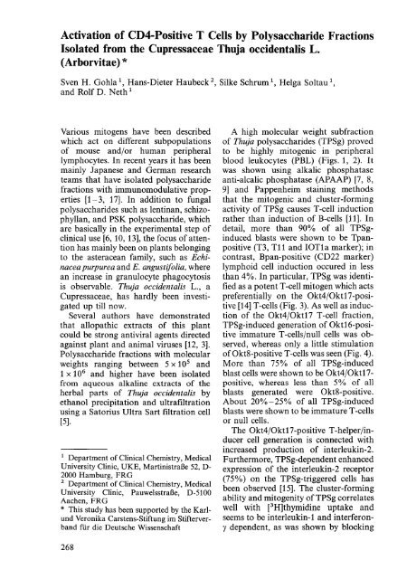
<strong>Activation</strong> <strong>of</strong> <strong>CD4</strong>-<strong>Positive</strong> T <strong>Cells</strong> <strong>by</strong> <strong>Polysaccharide</strong> <strong>Fractions</strong><br />
Isolated from the Cupressaceae Thuja occidentalis L.<br />
(Arborvitae) *<br />
Sven H. Gohla 1, Hans-Dieter Haubeck 2 , Silke Schrum \ Helga Soltau \<br />
and Rolf D. Neth 1<br />
Various mitogens have been described<br />
which act on different subpopulations<br />
<strong>of</strong> mouse and/or human peripheral<br />
lymphocytes. In recent years it has been<br />
mainly Japanese and German research<br />
teams that have isolated polysaccharide<br />
fractions with immunomodulative properties<br />
[1-3, 17]. In addition to fungal<br />
polysaccharides such as lentinan, schizophyllan,<br />
and PSK polysaccharide, which<br />
are basically in the experimental step <strong>of</strong><br />
clinical use [6, 10, 13], the focus <strong>of</strong> attention<br />
has mainly been on plants belonging<br />
to the asteracean family, such as Echinacea<br />
purpurea and E. angustifolia, where<br />
an increase in granulocyte phagocytosis<br />
is observable. Thuja occidentalis L., a<br />
Cupressaceae, has hardly been investigated<br />
up till now.<br />
Several authors have demonstrated<br />
that allopathic extracts <strong>of</strong> this plant<br />
could be strong antiviral agents directed<br />
against plant and animal viruses [12, 3].<br />
<strong>Polysaccharide</strong> fractions with molecular<br />
weights ranging between 5 x 10 5 and<br />
1 x 10 6 and higher have been isolated<br />
from aqueous alkaline extracts <strong>of</strong> the<br />
herbal parts <strong>of</strong> Thuja occidentalis <strong>by</strong><br />
ethanol precipitation and ultrafiltration<br />
using a Satorius Ultra Sart filtration cell<br />
[5].<br />
1 Department <strong>of</strong> Clinical Chemistry, Medical<br />
University Clinic, UKE, MartinistraBe 52, D-<br />
2000 Hamburg, FRO<br />
2 Department <strong>of</strong> Clinical Chemistry, Medical<br />
University Clinic, PauwelsstraBe, D-5100<br />
Aachen, FRO<br />
* This study has been supported <strong>by</strong> the Karlund<br />
Veronika Carstens-Stiftung im Stifterverband<br />
fUr die Deutsche Wissenschaft<br />
A high molecular weight subfraction<br />
<strong>of</strong> Thuja polysaccharides (TPSg) proved<br />
to be highly mitogenic in peripheral<br />
blood leukocytes (PBL) (Figs. 1, 2). It<br />
was shown using alkalic phosphatase<br />
anti-a1calic phosphatase (APAAP) [7, 8,<br />
9] and Pappenheim staining methods<br />
that the mitogenic and cluster-forming<br />
activity <strong>of</strong> TPSg causes T-cell induction<br />
rather than induction <strong>of</strong> B-cells [11]. In<br />
detail, more than 90% <strong>of</strong> all TPSginduced<br />
blasts were shown to be Tpanpositive<br />
(T3, T11 and IOTla marker); in<br />
contrast, Bpan-positive (CD22 marker)<br />
lymphoid cell induction occured in less<br />
than 4%. In particular, TPSg was identified<br />
as a potent T-cell mitogen which acts<br />
preferentially on the Okt4/0kt17-positive<br />
[14] T-cells (Fig. 3). As well as induction<br />
<strong>of</strong> the Okt4/0kt17 T-cell fraction,<br />
TPSg-induced generation <strong>of</strong> Okt16-positive<br />
immature T-cells/null cells was observed,<br />
whereas only a little stimulation<br />
<strong>of</strong>Okt8-positive T-cells was seen (Fig. 4).<br />
More than 75% <strong>of</strong> all TPSg-induced<br />
blast cells were shown to be Okt4/0kt17-<br />
positive, whereas less than 5% <strong>of</strong> all<br />
blasts generated were Okt8-positive.<br />
About 20%-25% <strong>of</strong> all TPSg-induced<br />
blasts were shown to be immature T -cells<br />
or null cells.<br />
The Okt4/0kt17-positive T -helper/inducer<br />
cell generation is connected with<br />
increased production <strong>of</strong> interleukin-2.<br />
Furthermore, TPSg-dependent enhanced<br />
expression <strong>of</strong> the interleukin-2 receptor<br />
(75%) on the TPSg-triggered cells has<br />
been observed [15]. The cluster-forming<br />
ability and mitogenity <strong>of</strong> TPSg correlates<br />
well with [3H]thymidine uptake and<br />
seems to be interleukin-1 and interferony<br />
dependent, as was shown <strong>by</strong> blocking<br />
268
Fig. 1. Phase contrast micrograph <strong>of</strong> a 4-day-old culture <strong>of</strong> TPSg-triggered peripheral blood<br />
leukocytes. Magnification 550 x . Peripheral blood leukocytes (PBL) were obtained <strong>by</strong> means <strong>of</strong><br />
density gradient centrifugation (Ficoll-Hypaque, Pharmacia). The "buffy coat" was harvested<br />
after having been centrifugated for 20 min and washed 5 times with phosphate buffered saline<br />
pH 7.2. 5 x 10 5 cells were seeded out per well in a flat bottomed 96-well microtiter plate with a<br />
final volume <strong>of</strong> 100 III Du1beccois modified Eagle's medium (DMEM) + 10% pooled human<br />
blood group AB serum. The cells were incubated with 100 Ill/well <strong>of</strong>a TPSg solution in DMEM<br />
+ 10% pooled human AB serum with a final concentration <strong>of</strong> 1 mg/ml <strong>of</strong> TPSg<br />
3H·thymidine·inc.<br />
cpm x 1000<br />
40 - PHA·P (10 ug/mll<br />
0-0 TPS 1 (1 mg/ml) /<br />
..... TPS 2 (1 mg/ml/<br />
0-0 Low control<br />
(RPMI 1640 + 10 % AS·serum)<br />
30<br />
./<br />
20<br />
10<br />
0<br />
0<br />
0 2 3 4 5<br />
Days <strong>of</strong> incubation in culture<br />
0<br />
0<br />
0- 0<br />
6 7 8<br />
Fig. 2. Kinetics <strong>of</strong> the DNA-synthesis in PBL and the influence <strong>of</strong> TPSg. Thuja polysaccharide<br />
fraction. 5 x 10 5 PBL, resuspended in RPMI 1640, supplement with 6% fetal calf serum (FCS)<br />
and 4% human AB serum, were incubated at a final concentration <strong>of</strong> 1 mg/ml per well in a flat<br />
bottomed, microtiter plate. On days 0, 2, 4, 6, and 8, 0.5 IlCi/50 III RPMI 1640 was added for<br />
the last 12 h <strong>of</strong> incubation. Afterwards the cells were harvested (Scatron cell harvester) and the<br />
DNA synthesis rate was measured in a f3-liquid scintillation counter. Supplemented medium was<br />
used as low control and Phytohemagglutinin. PHA-P, final concentration 10 Ilg/ml, was used as<br />
high control. Both controls were incubated under the same conditions as the TPSg cultures. All<br />
cultures were performed in triplicate. The results shown are the mean values <strong>of</strong> two experiments<br />
269
Fig. 3. Anti-Okt4 alkaline phosphatase anti-alkaline phosphatase (APAAP) staining <strong>of</strong> TPSgtriggered<br />
peripheral blood leukocytes. Magnification 630 x . Air-dried cells were stained <strong>by</strong> the<br />
APAAP method (Erber et al.). The cells were fixed for 1 min with an acetone, methanol and<br />
formaldehyde (95: 95: 10) fixative. The following steps were carried out at room temperature<br />
(25°C) in a humidified chamber. The first <strong>CD4</strong> + surface antigen specific antibody (Dakko, cat.<br />
no. M716, Denmark) was incubated for 30 min. Subsequent washing was done using TBS<br />
(Tris-buffered saline, pH 7.6) unless otherwise indicated. After washing with TBS (1 min), the<br />
second incubation step with rabbit anti-mouse (RaM) bridge antibodies (Dakko, cat. no. Z259)<br />
was carried out for another 30 min. After washing, the third incubation with APAAP complex<br />
(Dakko, cat no. D651) was carried out for another 30 min. The quality <strong>of</strong> the staining can be<br />
improved if all incubation steps are done twice. The "fast red" method was used as a detection<br />
system. The sample was washed after APAAP incubation and was incubated with the substrate<br />
[dimethylformamide (200 J.lI); levamisole (30 J.lI); "fast red" (10 mg); 0.1 M Tris, pH 8.2; naphthol-AS-MX-phosphate<br />
(2 mg)] for a period <strong>of</strong> 30 min. After washing counterstaining with<br />
hematoxylin was done (1 min). All slides were placed in Apathy's mountian medium, Highman's<br />
modification<br />
270<br />
Fig. 4. Anti-T-suppressor/cytotoxic<br />
staining (OKT8<br />
marker). Magnification<br />
630 x . For details<br />
<strong>of</strong> methods<br />
used see Fig. 3
300.------------------------------------------<br />
c::J lOW CONTROL (PBl 5 x 10⁵ per well)<br />
280 IZZZl TPSg (1 mg/ml + PBl 5 x 10⁵ per well)<br />
260 c::J PBl (5 x 10⁵ per well) without monocytes<br />
e2I PBl (5 x 10⁵ per well) + TPSg (1 mg/ml) without monocytes<br />
240 a::IJ] T cells (enriched) without monocytes<br />
w 220<br />
~<br />
m'SI T cells (enriched) + TPSg (1 mg/ml) without monocytes<br />
CZD T cells (enriched) + TPSg (1 mglml) + monocytes<br />
~ 200<br />
E:lZSD T cells (enriched) + monocytes<br />
~ 180<br />
~ 160<br />
Q 140<br />
~1<br />
I<br />
~ 100<br />
~ 80<br />
60<br />
Fig. 5. The influence <strong>of</strong>TPSg and enriched T-Iymphocytes in the presence or absence <strong>of</strong> peri pheral<br />
blood monocytes. The peripheral blood monocytes were separated from the lymphocyte<br />
fraction <strong>by</strong> the monocyte adherence method described elsewhere and incubated separately under<br />
standard conditions. 5 x 10 5 PBL and 5 x 10 5 enriched T -cells were incubated with TPSg<br />
medium, final concentration 1 mg/ml, or with non-lectin-supplemented complete medium. The<br />
T-cell enrichement was performed according to the standard procedure using nylon wool as<br />
separation column. The T-cell fraction was obtained <strong>by</strong> rinsing the nylon wool 2 times with cold<br />
PBS. It could be shown that TPSg only works as a mitogen on PBL and enriched T-cell fractions<br />
in the presence <strong>of</strong> autologous monocytes. All results are mean values <strong>of</strong> two independent<br />
experiments. All concentrations per experiment were determined in triplicate<br />
the mitogenic effect with interleukin-land<br />
interferon-y-specific antibodies. It<br />
was also shown that Okt4-positive T-cell<br />
induction depends on the presence <strong>of</strong><br />
autologous monocytes/macrophages<br />
(Fig. 5). Whether it is possible to use this<br />
polysaccharide fraction as an adjuvant in<br />
the therapy <strong>of</strong> immune deficiency syndromes<br />
and cancer must now be further<br />
investigated.<br />
References<br />
1. Beuscher N (1982) Uber die medikamentose<br />
Beeinflussung zellularer und humoraler<br />
Resistenzmechanismen im Tierversuch.<br />
ArzneimitteIforschung 32: 134 ff<br />
2. Beuscher N (1982) Uber die medikamentose<br />
Beeinflussung zellularer und humoraler<br />
Resistenzmechanismen im Tierversuch.<br />
III. Steigerung der Leukozytenmobilisation<br />
bei der Maus durch pflanzliche<br />
Reizkorper. Arzneimittelforschung<br />
30:821 ff<br />
3. Beuscher N, Kopanski W (1986) Purification<br />
and biological characterization <strong>of</strong> antiviral<br />
substances from Thuja occidentalis<br />
L. In: 37 th Annual congress on medical<br />
plant research, Hamburg, 22-27 Sept.<br />
1986, Abstract <strong>of</strong> short lectures and poster<br />
presentation, Planta Medica, Thieme Verlag,<br />
p 75<br />
4. Beuscher N, Beuscher H, Otto B, Schafer<br />
B (1977) Uber die medikamentose Beeinflussung<br />
zellularer und humoraler Resistenzmechanismen<br />
im Tierversuch. II. In<br />
vitro Untersuchungen an Peritoneal<br />
Leukozyten und Seren der Ratte.<br />
Arzneimittelforschung 27: 1655 ff<br />
5. Caldes G, Prescott B, Thomas II CA,<br />
Bahr PI (1981) Characterization <strong>of</strong> a<br />
polysaccharide from Carthamus tinctorius<br />
that cross reacts with type III pneumococcae<br />
polysaccharide. 1 Gen Appl Microbiol<br />
27: 157 ff<br />
6. Chihara G, Maeda 1, Hamuro 1, Tahuma<br />
S, Fumiko F (1969) Inhibition <strong>of</strong> mouse<br />
sarcoma 180 <strong>by</strong> polysaccharides from<br />
Lentinus etodes (Berk.) Sing. Nature<br />
222: 687 ff<br />
7. Cordell JL, Falini B, Erber WN (1984)<br />
Immunoenzymatic labeling <strong>of</strong> monoc1on-<br />
271
al antibodies using immune complexes <strong>of</strong><br />
alkaline phospatase and monoclonal antialkaline<br />
phosphatase (APAAP complexes).<br />
J Histochem Cytochem 32:219ff<br />
8. Erber WN, Pinching AJ, Mason DY<br />
(1984) Immunocytochemical detection <strong>of</strong><br />
T and B cell populations in routine blood<br />
smears. Lancet I: 1042 ff<br />
9. Erber WN, Mynheer LC, Mason DY<br />
(1986) APAAP labelling <strong>of</strong> blood and<br />
bone-marrow samples <strong>of</strong> phenotyping<br />
leukemia. Lancet I: 761 ff<br />
10. Fujimoto S, Takahashi M, Minami T,<br />
Ishigami H, Miyazaki M, Itoh K (1979)<br />
Clinical value <strong>of</strong> immunotherapy with<br />
OK432 or PS-K for stomach cancer patients.<br />
Jpn J Surg 9: 190ff<br />
11. Gohla S, Haubeck H-D, Neth RD (1988)<br />
Mitogenic activity <strong>of</strong> high molecular<br />
polysaccharide fractions from the plant<br />
"Thuja occidentale L.". I. Monocyte-dependent<br />
induction <strong>of</strong> <strong>CD4</strong> + T-helper<br />
cells. Leukemia 2:528-533<br />
12. Khurana PSM (1971) Effect <strong>of</strong> homoeopathic<br />
drugs on plant viruses. Planta<br />
Medica 20: 142 ff<br />
13. Maeda JY, Chihara G (1971) Lentinan, a<br />
new immuno-accelerator <strong>of</strong> cell-mediated<br />
responses. Nature 229: 634ff<br />
14. Thomas Y, Rogozinski L, Rothman P,<br />
Rabbani LE, Andrews S, Irigoyen OH,<br />
Chess L (1982) Further dissection <strong>of</strong> the<br />
functional heterogeneity within the<br />
OKT4 + and OKT8 + human t-cell subsets.<br />
J Clin Immunol 2: 85 ff<br />
15. Uchiyama T, Nelson DL, Fleidher TA,<br />
Waldmann TA (1981) A monoclonal antibody<br />
(anti-TAC) reactive with activated<br />
and functionally mature human T-cells. J<br />
Immunol 126: 1398 -1404<br />
16. Vollmar A, Schafer W, Wagner H (1986)<br />
Immunologically active polysaccharides<br />
<strong>of</strong> Eupatorium cannabinum and Eupatorium<br />
peifoliatum. Phytochemistry 25: 377 ff<br />
272