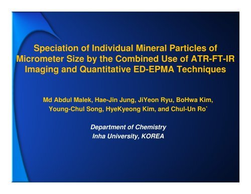
Speciation of Individual Mineral Particles of Micrometer Size by the ...
Speciation of Individual Mineral Particles of Micrometer Size by the ...
Speciation of Individual Mineral Particles of Micrometer Size by the ...

You also want an ePaper? Increase the reach of your titles
YUMPU automatically turns print PDFs into web optimized ePapers that Google loves.
<strong>Speciation</strong> <strong>of</strong> <strong>Individual</strong> <strong>Mineral</strong> <strong>Particles</strong> <strong>of</strong><br />
<strong>Micrometer</strong> <strong>Size</strong> <strong>by</strong> <strong>the</strong> Combined Use <strong>of</strong> ATR-FT-IR<br />
Imaging and Quantitative ED-EPMA Techniques<br />
Md Abdul Malek, Hae-Jin Jung, JiYeon Ryu, BoHwa Kim,<br />
Young-Chul Song, HyeKyeong Kim, and Chul-Un Ro *<br />
Department <strong>of</strong> Chemistry<br />
Inha University, KOREA
Airborne mineral dust particles<br />
<br />
Airborne mineral dust - <strong>the</strong> most abundant PM in coarse atmospheric aerosols<br />
<br />
Silicate minerals constitute ~90% <strong>of</strong> <strong>the</strong> Earth crust.<br />
<br />
<strong>Mineral</strong> dust plays multiple roles in influencing global climate (e.g., scattering and<br />
absorbing radiations, CCN).<br />
<br />
Heterogeneous chemical reactions can also alter <strong>the</strong> chemical balance <strong>of</strong> <strong>the</strong><br />
atmosphere.<br />
<br />
Hygroscopic property <strong>of</strong> mineral dust can change <strong>the</strong>ir reactivity.<br />
<br />
Arid and semi-arid areas <strong>of</strong> <strong>the</strong> Saharan desert and central China are <strong>the</strong> global<br />
scale sources.
Characterization <strong>of</strong> standard soil minerals<br />
<br />
<br />
<br />
<br />
<br />
<br />
Airborne mineral particles – from soil minerals<br />
Soil minerals rarely exist in a single-phase, pure mineral form.<br />
Bulk FT-IR technique is a common practice for mineral analysis.<br />
Bulk analysis - not sufficient for <strong>the</strong> speciation <strong>of</strong> mixed minerals<br />
Analysis on a single-particle level can more clearly identify different mineral types.<br />
The combined use <strong>of</strong> ATR-FT-IR imaging and quantitative low-Z particle EPMA<br />
techniques gives complementary information<br />
<br />
<br />
low-Z particle EPMA on <strong>the</strong> morphology and elemental concentrations<br />
ATR-FT-IR imaging on mineral types<br />
<br />
The combined use <strong>of</strong> <strong>the</strong>se two single-particle analytical techniques has great<br />
potential for <strong>the</strong> characterization <strong>of</strong> airborne mineral dust particles.
Low-Z particle EPMA (Electron Probe X-ray<br />
Microanalysis) for single particle analysis<br />
1. SEM-EDX (Scanning Electron Microscopy –<br />
Energy Dispersive X-ray Spectrometer)<br />
- <strong>Individual</strong> Particle Analysis<br />
* shape and size : secondary / backscattered electron images<br />
* chemical compositions : X-ray spectrum<br />
2. Ultra-thin window EDX for low-Z elements detection (e.g., C, N, O, F)<br />
3. Metallic collecting substrates for minimizing charging effect (e.g., Ag, Al)<br />
4. Monte Carlo calculation for Quantification<br />
5. Chemical speciation <strong>of</strong> aerosol particles – Expert System
Monte Carlo Calculation for Quantification<br />
Measurement<br />
Simulation<br />
Electron detector<br />
Electron beam<br />
with 10keV<br />
X-ray detector<br />
1000<br />
100<br />
C<br />
O<br />
Ca<br />
CaCO3 measured data<br />
Simulated<br />
SEM<br />
metal foil<br />
Intensity(arb. units)<br />
10<br />
1<br />
0.1<br />
0.01<br />
0 1 2 3 4 5 6 7 8 9 10<br />
X-ray Energy (keV)<br />
Measured and simulated spectra for a<br />
CaCO 3<br />
standard particle on a Be substrate
ATR-FT-IR imaging for single particle analysis<br />
♦ ATR-FT-IR (Attenuated Total Reflectance-FT-IR Spectrometry)<br />
- <strong>Individual</strong> Particle Analysis<br />
* location : optical image<br />
* functional groups, molecular species, and crystal structure : IR spectra<br />
Dual detector<br />
Visible radiation<br />
d = 600 μm<br />
▲ ATR imaging accessory<br />
Cassegrain system<br />
Motorized sample stage<br />
Ge crystal – sample contact surface ▲<br />
sample<br />
IR radiation<br />
◄ Ge crystal for imaging
ATR-FT-IR Imaging for Single Particle Analysis
ATR-FT-IR and XRD measurements <strong>of</strong> minerals<br />
ATR-FT-IR measurement<br />
Perkin Elmer Spectrum 100 FT-IR spectrometer<br />
Spectrum Spotlight 400 FT-IR optical microscope<br />
ATR accessory: Ge IRE crystal, diam. = 600 μm, RI = 4<br />
A 16 x 1 pixel Mercury Cadmium Telluride (MCT) array detector<br />
Pixel size <strong>of</strong> 1.56 μm<br />
Spectral resolution <strong>of</strong> 4 cm -1 at <strong>the</strong> range <strong>of</strong> 720 to 4000 cm -1<br />
Spatial resolution 3.9±0.5 µm at 1000 – 1200 cm -1<br />
XRD measurement<br />
Philips X’pert MPD powder X-ray diffractometer<br />
Cu Kα radiation<br />
Scanning range <strong>of</strong> 2θ is 3-65 o<br />
scanning speed is 0.02 o /s, a step size <strong>of</strong> 2θ is 0.02 o
Major minerals obtained <strong>by</strong> XRD for 24 mineral samples<br />
Major mineral types <strong>by</strong> XRD Number <strong>of</strong> samples Source <strong>of</strong> sample<br />
microcline (K-feldspar) 2 NIST, KIGAM<br />
muscovite 4 KIGAM<br />
montmorillonite 2 KIGAM<br />
kaolinite 3 KIGAM<br />
talc 2 KIGAM<br />
heulandite 1 KIGAM<br />
biotite 1 KIGAM<br />
Mg-vermiculite 1 KIGAM<br />
pyrophyllite 1 KIGAM<br />
cristobalite (SiO 2<br />
) 1 KIGAM<br />
quartz (SiO 2<br />
) 1 Aldrich<br />
apatite 1 Aldrich<br />
calcite 1 Aldrich<br />
gypsum 1 Aldrich<br />
anhydrous CaSO 4<br />
1 Aldrich<br />
magnesiumhydroxycarbonate hydrate 1 Aldrich<br />
Total 24<br />
NIST : National Institute <strong>of</strong> Standards and Technology, USA<br />
KIGAM : Korea Institute <strong>of</strong> Geoscience and <strong>Mineral</strong> Resources
1500<br />
1200<br />
*<br />
Major<br />
* microcline (K-feldspar (KAlSi 3 O 8 ) )<br />
XRD spectrum <strong>of</strong> K-feldspar<br />
NIST SRM mineral sample.<br />
counts/s<br />
900<br />
600<br />
Minors<br />
▼albite (Na-feldspar (NaAlSi 3 O 8 ) )<br />
◊quartz (SiO 2 )<br />
counts/s<br />
300<br />
0<br />
* * *<br />
*<br />
▼▼* **<br />
◊*<br />
*<br />
*<br />
* *<br />
*<br />
* * * *<br />
0 5 10 15 20 25 30 35 40 45 50 55 60<br />
▼<br />
65<br />
▼▼<br />
1200<br />
1000<br />
800<br />
600<br />
400<br />
200<br />
0<br />
+ +<br />
*<br />
*<br />
↓<br />
◊↓<br />
+▼<br />
2θ<br />
***<br />
*<br />
▼▼▼ *▼<br />
▼ ▼▼▼ ▼ ♦ ◊<br />
0 5 10 15 20 25 30 35 40 45 50 55 60 65<br />
2θ<br />
Majors<br />
▼muscovite (KAl 2 (Si 3 Al)O 10 (OH,F) 2 )<br />
* quartz (SiO 2 )<br />
Minors<br />
+ montmorillonite<br />
((0.5Ca,Na) 0.7 (Al,Mg,Fe) 4 [(Si,Al) 8 O 20 ]<br />
(OH) 4. nH 2 O)<br />
♦albite (Na-feldspar (NaAlSi 3 O 8 ))<br />
↓ orthoclase (K-feldspar (KAlSi 3 O 8 ))<br />
◊kaolinite (Al 2 Si 2 O 5 (OH) 4 )<br />
* *<br />
*<br />
*<br />
*<br />
*<br />
*<br />
*<br />
*<br />
XRD spectrum <strong>of</strong> a mineral<br />
sample <strong>of</strong> muscovite and<br />
quartz.
SE images (A) before and (B) after ATR-FT-IR measurement and ATR-FT-IR images<br />
obtained (C) <strong>by</strong> a PCA analysis and (D) for transmittance signal at 1000 cm -1<br />
(K-feldspar SRM mineral on Ag foil)<br />
(A)<br />
4<br />
5 6<br />
7<br />
1 2 3<br />
(B)<br />
4 5<br />
6<br />
(1) 1 2 3<br />
7<br />
8<br />
14 15<br />
9 10<br />
11<br />
12 13<br />
16<br />
8<br />
14 15<br />
9 10<br />
11<br />
12<br />
13<br />
16<br />
20<br />
21<br />
22<br />
23 24<br />
17 18 19<br />
26<br />
28<br />
27<br />
Δ<br />
20<br />
21<br />
Δ Δ<br />
22<br />
23 24<br />
17<br />
26<br />
28<br />
18<br />
27<br />
19<br />
1<br />
3<br />
25<br />
29<br />
25<br />
29<br />
(C)<br />
4<br />
20<br />
21<br />
1 2 3<br />
5 6 7<br />
8<br />
9 10<br />
11<br />
12 13<br />
14 15<br />
16<br />
17<br />
18<br />
22<br />
19<br />
27<br />
23 24 26 28<br />
25<br />
29<br />
(D)<br />
4 5 6<br />
20<br />
21<br />
8<br />
22<br />
7<br />
1 2 3<br />
9<br />
10<br />
11<br />
12 13<br />
14 15 16<br />
23<br />
24<br />
17<br />
25<br />
26<br />
28<br />
18<br />
27<br />
29<br />
19
Typical X-ray spectra and elemental concentrations <strong>of</strong> different feldspars<br />
observed in K-feldspar SRM mineral sample<br />
Intensity<br />
1000<br />
100<br />
(A)<br />
C<br />
O<br />
Al<br />
Si<br />
K<br />
Ag<br />
particle #5 (K-feldspar)<br />
Diameter: 5.46 µm<br />
Elemental concentration in at. %<br />
C 3.8 O 54.0<br />
Al 8.5 Si 26.2<br />
K 7.5<br />
1000<br />
100<br />
Intensity<br />
(B)<br />
C<br />
O<br />
Na Al Si<br />
Ag<br />
particle #2 (Na-feldspar)<br />
Diameter: 3.63 µm<br />
Elemental concentration in at. %<br />
C 3.5 O 50.8<br />
Na 8.2 Al 9.5<br />
Si 28.0<br />
10<br />
10<br />
Intensity<br />
1<br />
1000<br />
100<br />
10<br />
0 1 2 3 4 5 6 7 8 9 10<br />
keV<br />
(C)<br />
C<br />
O<br />
Na<br />
Al<br />
Si<br />
Ag<br />
K<br />
particle #22 ((Na, K)-feldspar)<br />
Diameter: 2.2 µm<br />
Elemental concentration in at. %<br />
C 1.0 O 57.6<br />
Na 2.9 Al 9.0<br />
Si 25.6 K 3.9<br />
Intensity<br />
1<br />
1000<br />
100<br />
10<br />
0 1 2 3 4 5 6 7 8 9 10<br />
keV<br />
(D)<br />
C<br />
O<br />
Fe<br />
Si<br />
Al<br />
Ag<br />
K<br />
particle #9 ((K, Fe)-feldspar)<br />
Diameter: 1.79 µm<br />
Elemental concentration in at. %<br />
C 2.1 O 51.4<br />
Al 15.0 Si 23.7<br />
K 4.4 Fe 3.4<br />
Fe<br />
1<br />
0 1 2 3 4 5 6 7 8 9 10<br />
keV<br />
1<br />
0 1 2 3 4 5 6 7 8 9 10<br />
keV
Typical ATR-FT-IR spectra <strong>of</strong> different feldspars observed in K-feldspar SRM mineral<br />
sample.<br />
(Conventional transmission FT-IR spectra <strong>of</strong> K- and Na-feldspar bulk samples reported<br />
<strong>by</strong> <strong>the</strong> o<strong>the</strong>r study and an ATR-FT-IR spectrum obtained from bulk K-feldspar SRM<br />
mineral powder are also shown in an inset.)<br />
A<br />
0.32<br />
0.28<br />
0.24<br />
0.2<br />
0.16<br />
A<br />
1<br />
0.8<br />
0.6<br />
0.4<br />
0.2<br />
0<br />
1720<br />
transmission FT-IR<br />
spectrum <strong>of</strong> Na-feldspar<br />
transmission FT-IR<br />
spectrum <strong>of</strong> K-feldspar<br />
1520<br />
1145:Si-O<br />
1163:Si-O<br />
ATR-FT-IR spectrum<br />
<strong>of</strong> K-feldspar SRM <strong>Mineral</strong><br />
1320<br />
1047:Si(Al)-O<br />
1109: Si-O<br />
1144<br />
1120<br />
1058<br />
995<br />
1018<br />
1133<br />
1088<br />
1018:Si(Al)-O<br />
1001:Si(Al)-O<br />
1048<br />
920<br />
Matteson et. al., J. Sediment.<br />
Petrol. 1993, 63, 1144-1148<br />
760: Si-Si<br />
788: Si-Si 747: Si-(Al)Si<br />
725: Si-(Al)Si<br />
775 735<br />
725<br />
769<br />
720<br />
1037:Si(Al)-O<br />
1039:Si(Al)-O<br />
1133: Si-O<br />
1116:Si-O<br />
1133:Si-O<br />
1003:Si(Al)-O<br />
1016:Si(Al)-O<br />
770: Si-Si<br />
729:<br />
Si-(Al)Si<br />
0.12<br />
0.08<br />
wavenumber, cm -1<br />
particle #5 (K-feldspar)<br />
particle #22 ((Na, K)-feldspar)<br />
1153<br />
1029<br />
1097 1130<br />
1002<br />
774<br />
732<br />
0.04<br />
0<br />
particle #2 (Na-feldspar)<br />
particle #9 ((K, Fe)-feldspar)<br />
1126<br />
1021<br />
762<br />
788<br />
809<br />
760<br />
744<br />
727<br />
3720<br />
3220<br />
2720<br />
2220<br />
1720<br />
1220<br />
720<br />
wavenumber, cm -1
SEIs SEM (A) before and (B) after ATR-FT-IR measurement and ATR-FT-IR images obtained (C) <strong>by</strong> a<br />
PCA analysis and (D) for transmittance signal at 1030 cm -1 <strong>of</strong> a muscovite mineral sample on Ag foil.<br />
(A)<br />
12<br />
14*<br />
13<br />
20<br />
1<br />
14<br />
22<br />
4<br />
21<br />
5<br />
9<br />
15<br />
2<br />
16<br />
6<br />
7<br />
8<br />
3<br />
10<br />
17<br />
11<br />
24<br />
18<br />
19<br />
23<br />
(B)<br />
1 (K-F)<br />
4 (K-F)<br />
2 (MUS)<br />
5 (Q)<br />
6 (K-F)<br />
3 (MON)<br />
7 (MUS)<br />
8 (Na-F)<br />
9 (Q)<br />
12 (MON)<br />
10 (MUS)<br />
13 (Q)<br />
11 (MUS)<br />
16 (MON)<br />
18 (MON)<br />
15 (K-F)<br />
14 (K-F)<br />
17 (MUS)<br />
19 (Q)<br />
20*<br />
21 (Na-F)<br />
14* (MON)<br />
23 (Q)<br />
22 (K-F)<br />
*<br />
24 (KAO)<br />
(C)<br />
1<br />
4<br />
12<br />
13<br />
14<br />
14*<br />
20<br />
22<br />
21<br />
2<br />
6<br />
3<br />
5<br />
7<br />
8<br />
9 10<br />
15<br />
16<br />
17<br />
11<br />
18<br />
19<br />
24<br />
23<br />
(D)<br />
12<br />
14*<br />
2<br />
1<br />
6<br />
4 5<br />
8<br />
3<br />
7<br />
13<br />
9 10<br />
16<br />
17<br />
15<br />
14<br />
*20<br />
21<br />
22<br />
11<br />
18<br />
19<br />
24<br />
23<br />
* (MON: montmorillonite, MUS: muscovite, Q: quartz, Na-F: albite, K-F: orthoclase (K-feldspar), KAO: kaolinite)
Typical X-ray spectra and elemental concentrations <strong>of</strong> different minerals<br />
observed in a muscovite sample.<br />
10000<br />
1000<br />
Intensity<br />
100<br />
10<br />
(A)<br />
C<br />
O<br />
Si<br />
Al<br />
Ag<br />
K<br />
particle #1 (K-feldspar)<br />
Diameter: 3.85 µm<br />
Elemental concentration in at. %<br />
C 1.9 O 46.4<br />
Al 11.5 Si 30.8<br />
K 8.8<br />
Intensity<br />
10000<br />
1000<br />
C<br />
100<br />
10<br />
(B)<br />
O<br />
Al Si<br />
Mg<br />
Ag<br />
K<br />
particle #2 (muscovite)<br />
Diameter: 4.4 µm<br />
Elemental concentration in at. %<br />
C 3.6 O 59.6 Mg 0.5<br />
Al 14.8 Si 17.8 K 1.5<br />
Fe 2.1<br />
Fe<br />
Intensity<br />
1<br />
10000<br />
1000<br />
100<br />
0 1 2 3 4 5 6 7 8 9 10<br />
keV<br />
(C)<br />
C<br />
O<br />
Al Si<br />
Mg<br />
Ag<br />
particle #3 (montmorillonite)<br />
Diameter: 2.06 µm<br />
Elemental concentration in at. %<br />
C 4.6 O 60.2 Mg 1.4<br />
Al 13.3 Si 17.8 Fe 2.7<br />
Intensity<br />
1<br />
0 1 2 3 4 5 6 7 8 9 10<br />
10000<br />
keV<br />
1000<br />
100<br />
(D)<br />
C<br />
O<br />
Al<br />
Si<br />
Ag<br />
particle #13 (quartz)<br />
Diameter: 4.4 µm<br />
Elemental concentration in at. %<br />
C 1.2 O 62.8<br />
Al 0.6 Si 35.4<br />
10<br />
Fe<br />
10<br />
Intensity<br />
10000<br />
1000<br />
1<br />
0 1 2 3 4 5 6 7 8 9 10<br />
100<br />
10<br />
(E)<br />
C<br />
O Si<br />
Al<br />
Na<br />
Ag<br />
keV<br />
particle #21 (Na-feldspar)<br />
Diameter: 2.61 µm<br />
Elemental concentration in at. %<br />
C 2.9 O 50.8<br />
Na 5.8 Al 11.4<br />
Si 29.27<br />
Intensity<br />
1<br />
10000<br />
0 1 2 3 4 5 6 7 8 9 10<br />
keV<br />
1000<br />
100<br />
10<br />
(F)<br />
C<br />
O<br />
Al Si<br />
Mg<br />
Ag<br />
particle #24 (kaolinite)<br />
Diameter: 3.82 µm<br />
Elemental concentration in at. %<br />
C 0.5 O 60.9 Mg 1.2<br />
Al 14.8 Si 20.0 Fe 2.6<br />
Fe<br />
1<br />
0 1 2 3 4 5 6 7 8 9 10<br />
keV<br />
1<br />
0 1 2 3 4 5 6 7 8 9 10<br />
keV
Typical ATR-FT-IR spectra <strong>of</strong> different minerals observed in a muscovite mineral sample.<br />
(An ATR-FT-IR spectrum obtained from bulk powder <strong>of</strong> this sample is also shown in an<br />
inset.)<br />
% T<br />
100<br />
90<br />
80<br />
70<br />
60<br />
50<br />
40<br />
30<br />
20<br />
10<br />
0<br />
% T<br />
100<br />
85<br />
70<br />
55<br />
40<br />
25<br />
3687 3646<br />
3626<br />
3620<br />
3695: -OH<br />
3620: -OH<br />
3720<br />
3220<br />
2720 2220<br />
wavenumber, cm -1<br />
1638: H 2 O<br />
997: Si-O (MON, KAO, MUS)<br />
Si(Al)-O (feldspar)<br />
1720<br />
particle #21 (Na-feldspar)<br />
particle #1 (K-feldspar)<br />
particle #24 (kaolinite)<br />
particle #3 (montmorillonite)<br />
particle #2 (muscovite)<br />
particle #13 (quartz)<br />
1220<br />
795 & 777:<br />
Si-O (Q, KAO); Si-Si (feldspar)<br />
908: AlAl-OH (MON, KAO, MUS)<br />
720<br />
1651<br />
1646<br />
1653<br />
1454<br />
1456<br />
1161<br />
1163: Si-O<br />
1046: Si-O<br />
1135<br />
1089<br />
1034<br />
988<br />
1097<br />
102<br />
989: Si-O<br />
994<br />
999<br />
998<br />
913<br />
917<br />
917: AlAl-OH<br />
782<br />
778<br />
755<br />
791<br />
825<br />
756<br />
824<br />
720<br />
721<br />
747<br />
755<br />
747<br />
775: Si-O<br />
793: Si-O<br />
3720<br />
3220<br />
2720<br />
2220<br />
1720<br />
1220<br />
720<br />
wavenumber, cm -1<br />
* (MON: montmorillonite, MUS: muscovite, KAO: kaolinite, Q: quartz)
Summary<br />
Total samples analyzed: 24<br />
<br />
Data presented herein: 2 samples<br />
K-feldspar SRM sample<br />
According to NIST specification, K-feldspar SRM sample consists <strong>of</strong> 80~85 % microcline, and 10~15 % albite<br />
Based on X-ray and ATR-FT-IR spectral data <strong>of</strong> 29 individual particles,<br />
15 particles were identified as K-feldspar<br />
5 particles as Na-feldspar<br />
8 particles as (Na,K)-feldspar, and 1 particle as (K,Fe)-feldspar<br />
Muscovite and quartz sample<br />
For <strong>the</strong> muscovite and quartz sample, among 24 individual particles<br />
6 particles are observed as muscovite 5 particles as quartz<br />
5 particles as K-feldspar 5 particles as montmorillonite<br />
2 particles as Na-feldspar 1 particle as kaolinite<br />
Different types <strong>of</strong> minerals were observed in <strong>the</strong> remaining 22 samples except samples<br />
collected from Aldrich<br />
A manuscript was submitted to Anal. Chem., where data for 22 mineral samples can be found.<br />
Soil samples collected from various arid areas in China, and ambient aerosol samples<br />
are under investigation.
Conclusions<br />
<br />
<br />
<br />
<br />
<br />
<br />
The combined use <strong>of</strong> ATR-FT-IR imaging and low-Z particle EPMA<br />
allows unambiguous identification <strong>of</strong> different minerals.<br />
ATR-FT-IR imaging provides information on molecular and crystal<br />
structure, functional group, and physical state.<br />
Low-Z particle EPMA gives information on morphology and quantitative elemental<br />
concentrations.<br />
Analysis on single particle basis gives more detailed information than<br />
bulk analysis.<br />
It has great potential in elucidating <strong>the</strong> characteristics <strong>of</strong> soil-derived individual<br />
airborne particles.<br />
Our future project is to build up a good archive <strong>of</strong> mineral’s FT-IR spectra<br />
for <strong>the</strong> facile assignment <strong>of</strong> soil-derived individual airborne particles.<br />
(For low-Z particle EPMA, a library building is not necessary for X-ray spectra,<br />
which is an advantage over FT-IR technique.)