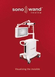
SonoWand Invite
SonoWand Invite
SonoWand Invite

You also want an ePaper? Increase the reach of your titles
YUMPU automatically turns print PDFs into web optimized ePapers that Google loves.
Visualizing the invisible<br />
2
Fundamental Surgical Needs<br />
Regardless of Brain Shift, the surgeon needs to:<br />
• Distinguish lesion from<br />
surrounding landmarks<br />
• Safely guide surgical instruments<br />
to the region of interest for resection<br />
• Control the result of resection and<br />
vascular repair<br />
• Perform Radical Resection<br />
with Minimal Morbidity<br />
3
Intraoperative Imaging<br />
is the only alternative that can fulfill these clinical needs!<br />
Intraoperative<br />
MRI<br />
Intraoperative<br />
CT<br />
Intraoperative<br />
Ultrasound<br />
4
This is<br />
the <strong>SonoWand</strong> <strong>Invite</strong><br />
5
<strong>SonoWand</strong> <strong>Invite</strong><br />
• Tight integration of imaging<br />
and navigation in one unit<br />
6
<strong>SonoWand</strong> <strong>Invite</strong><br />
• Tight integration of imaging<br />
and navigation in one unit<br />
• State-of-the-art integrated<br />
ultrasound scanner<br />
6
<strong>SonoWand</strong> <strong>Invite</strong><br />
• Tight integration of imaging<br />
and navigation in one unit<br />
• State-of-the-art integrated<br />
ultrasound scanner<br />
• High image quality<br />
6
<strong>SonoWand</strong> <strong>Invite</strong><br />
• Tight integration of imaging<br />
and navigation in one unit<br />
• State-of-the-art integrated<br />
ultrasound scanner<br />
• High image quality<br />
• Ergonomic and modern design<br />
• Compact and Easy to move<br />
6
Rapid Update of Your 3D Map<br />
Free-hand scanning<br />
3D reconstruction<br />
≈ 10 sec.<br />
≈ 10 sec.<br />
7
Clinical Navigation Accuracy<br />
How accurately can you navigate with 3D ultrasound in the brain?<br />
With a mean navigation error less than 1.6 mm!*<br />
* Published in Computer Aided Surgery 2002<br />
8
Address Brain Shift<br />
Brainshift about 8 mm<br />
9
Intraoperative Resection Control<br />
Glioblastoma<br />
10
Intraoperative Resection Control<br />
Preoperative MR<br />
Ultrasound before resection<br />
Residual tumor,<br />
left deliberately<br />
Ultrasound after some resection<br />
Resection finished<br />
Low-grade astrocytoma<br />
11
Intraoperative 3D Angio Doppler<br />
Aneurysm<br />
12
About SONOWAND AS<br />
Company:<br />
Product:<br />
Market status:<br />
Spin-off from internationally renowned scientific group<br />
(in Trondheim) and world-leading Norwegian ultrasound industry.<br />
The company is located in Trondheim, Norway.<br />
Unique, patented technology, intraoperative imaging based on<br />
3D ultrasound.<br />
The only true integration of 3D ultrasound and navigation.<br />
Comparable image quality, more flexible and affordable<br />
than iMRI.<br />
Establishing distribution network (12 Distributors / 27 Territories)<br />
More than 6000 operations and 27 scientific papers<br />
Installations in Europe, Asia and North America.<br />
Norwegian industry is among the world leaders in medical ultrasound technology.<br />
The city of Trondheim in particular, has a long tradition of fostering research and<br />
development in the field.<br />
13
Acknowledgements<br />
SONOWAND AS wishes to thank the members of the<br />
National Centre of Competence - 3D Ultrasound in Surgery<br />
for their contribution to this presentation.<br />
Please note:<br />
Copyright owner of the majority of images contained in this presentation is<br />
Department of Neurosurgery, St. Olavs Hospital, Trondheim, Norway.<br />
The images may not be reproduced in a manner without prior written permission<br />
from the owner.<br />
16
Overview<br />
Product Information <strong>SonoWand</strong> <strong>Invite</strong><br />
<strong>SonoWand</strong> <strong>Invite</strong> Lesion Visibility<br />
Using the <strong>SonoWand</strong> <strong>Invite</strong><br />
17
Product Information<br />
<strong>SonoWand</strong> <strong>Invite</strong><br />
18
<strong>SonoWand</strong> <strong>Invite</strong><br />
The first<br />
intraoperative imaging system<br />
with tight integration of<br />
high quality 3D ultrasound<br />
and neuronavigation.<br />
19
<strong>SonoWand</strong> <strong>Invite</strong><br />
• Ergonomic design<br />
light weight and compact<br />
20
<strong>SonoWand</strong> <strong>Invite</strong><br />
• Ergonomic design<br />
light weight and compact<br />
• Wide aspect, high definition<br />
24” touch-screen monitor<br />
with intuitive user interface<br />
20
<strong>SonoWand</strong> <strong>Invite</strong><br />
• Ergonomic design<br />
light weight and compact<br />
• Wide aspect, high definition<br />
24” touch-screen monitor<br />
with intuitive user interface<br />
• State-of-the-art integrated<br />
ultrasound scanner<br />
20
<strong>SonoWand</strong> <strong>Invite</strong><br />
• Ergonomic design<br />
light weight and compact<br />
• Wide aspect, high definition<br />
24” touch-screen monitor<br />
with intuitive user interface<br />
• State-of-the-art integrated<br />
ultrasound scanner<br />
• New generation<br />
optical tracking<br />
• Multi-frequency probes<br />
phased array and flat linear array<br />
20
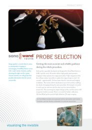
<strong>SonoWand</strong> <strong>Invite</strong> Probe Selection<br />
• 8 FPA probe for deep seated targets<br />
• 12 FLA-L probe for superficial high resolution imaging<br />
• 10 FPA MC probe for mini craniotomies (MC)<br />
• 12 FLA probe for superficial high resolution imaging<br />
21
<strong>SonoWand</strong> <strong>Invite</strong> Probe Selection<br />
8 FPA Probe<br />
• Imaging depth range 1-12 cm<br />
• Optimal imaging range 2-8 cm<br />
• Multiple frequency imaging 3-8 MHz<br />
• Footprint 24 x 17 mm<br />
• Long cable, 3 meters<br />
22
<strong>SonoWand</strong> <strong>Invite</strong> Probe Selection<br />
12 FLA-L Probe<br />
• Unique image resolution<br />
• Imaging depth range 0-5 cm<br />
• Multi frequency range 6-12 MHz<br />
• Footprint 48 x 8 mm<br />
• Cable length, 1,8 meters<br />
23
<strong>SonoWand</strong> <strong>Invite</strong> Probe Selection<br />
10 FPA-MC Probe<br />
• High image resolution 0-5 cm<br />
• Multiple frequency imaging 5-12 MHz<br />
• Unique probe sensitivity enables deep<br />
lesion imaging down to 9 cm<br />
• High definition Angiographic imaging<br />
• High frame rate > 15 FPS<br />
• Small footprint (elongated design) 15 x 13 mm<br />
• Long cable, 3 meters<br />
24
<strong>SonoWand</strong> <strong>Invite</strong> Probe Selection<br />
12 FLA Probe<br />
• High image resolution 0-5 cm<br />
• Multiple frequency imaging 5-12 MHz<br />
• Unique probe sensitivity enables deep<br />
lesion imaging down to 8 cm<br />
• High definition Angiographic imaging<br />
• High imaging frame rate > 12 FPS<br />
• Small footprint (elongated design) 31,5 x 10,5 mm<br />
• Long cable, 3 meters<br />
25
New Functions<br />
• Improved DICOM support<br />
26
New Functions<br />
• Improved DICOM support<br />
• Prepared for PACS integration<br />
26
New Functions<br />
• Improved DICOM support<br />
• Prepared for PACS integration<br />
• New and improved registration<br />
26
New Functions<br />
• Improved DICOM support<br />
• Prepared for PACS integration<br />
• New and improved registration<br />
• Image fusion (overlay)<br />
26
New Functions<br />
• Improved DICOM support<br />
• Prepared for PACS integration<br />
• New and improved registration<br />
• Image fusion (overlay)<br />
• 3D visualization<br />
26
<strong>SonoWand</strong> <strong>Invite</strong><br />
In development:<br />
• Microscope integration<br />
• Spine application<br />
• Transphenoidal ultrasound probe<br />
• ........<br />
27
<strong>SonoWand</strong> <strong>Invite</strong><br />
Lesion Visibility<br />
28
Lesion Visibility<br />
- A prerequisite for Ultrasound Guided Surgery<br />
How well can different types of lesions be visualized on ultrasound?<br />
29
Lesion Visibility<br />
- A prerequisite for Ultrasound Guided Surgery<br />
How well can different types of lesions be visualized on ultrasound?<br />
• Tumors, grade I - IV<br />
- Pilocytic Astrocytomas<br />
- Low Grade Astrocytomas<br />
- Anaplastic Astrocytomas<br />
- Glioblastomas<br />
• Ultrasound Angio Mapping<br />
• Meningiomas<br />
• Cavernomas<br />
• Skull Base Tumors<br />
•Abscesses<br />
•Cysts<br />
29
Lesion Visibility: Tumors Grade I - IV<br />
Grade I: Pilocytic Astrocytoma<br />
MRI<br />
Ultrasound<br />
Our experience with pilocytic astrocytomas is that they are well visualized<br />
with the high quality of ultrasound that the <strong>SonoWand</strong> offers.<br />
30
Lesion Visibility: Tumors Grade I - IV<br />
Grade II: Low Grade Astrocytoma<br />
MR FLAIR<br />
Ultrasound<br />
Low grade astrocytomas are difficult to image on CT and traditional MRI.<br />
The <strong>SonoWand</strong> displays the lesion well on ultrasound.<br />
31
Lesion Visibility: Tumors Grade I - IV<br />
Grade III: Anaplastic Astrocytomas<br />
MR T1<br />
Ultrasound<br />
Anaplastic astrocytomas are normally quite well visualized on the <strong>SonoWand</strong>,<br />
here displaying good correspondence with MR T1.<br />
32
Lesion Visibility: Tumors Grade I - IV<br />
Grade IV: Glioblastomas<br />
MRI<br />
Ultrasound<br />
The excellent image quality of the <strong>SonoWand</strong> displays glioblastomas well.<br />
We also see good image correspondence with MRI<br />
33
Lesion Visibility: Meningiomas<br />
MRI<br />
Ultrasound<br />
Meningiomas are well demarcated and easily visualized on the ultrasound of the <strong>SonoWand</strong>.<br />
The image correspondence with MRI is high.<br />
34
Lesion Visibility: Cavernomas<br />
MRI<br />
Ultrasound<br />
In cases with cavernomas, we often see a lot of brain shift.<br />
<strong>SonoWand</strong>’s intraoperative ultrasound addresses brainshift,<br />
thus making it possible to perform a safer surgery.<br />
35
Lesion Visibility: Pituitary Macroadenoma<br />
MRI<br />
Ultrasound<br />
A frontal approach to a pituitary adenoma<br />
36
Lesion Visibility: Ultrasound Angio Mapping<br />
MRI<br />
Ultrasound<br />
The <strong>SonoWand</strong> enables you to<br />
visualize blood flow during surgery,<br />
using the 3D Angio mode.<br />
A simple and reliable way to<br />
localize surrounding blood vessels.<br />
Ultrasound + Angio<br />
Ultrasound with<br />
Angio Mode enabled<br />
37
Lesion Visibility: Aneurysms<br />
MRI<br />
Ultrasound<br />
Aneurysms are well visualized with Angio Mode enabled.<br />
38
Lesion Visibility: Abscesses<br />
MRI<br />
Ultrasound<br />
With the <strong>SonoWand</strong> ultrasound you can distinguish between abscess and cystic tissue.<br />
39
Lesion Visibility: Cysts<br />
MRI<br />
Ultrasound<br />
An abscess appears gray while cystic tissue is black.<br />
40
Using the<br />
<strong>SonoWand</strong> <strong>Invite</strong><br />
41
Product Positioning<br />
Classic<br />
Neuronavigation<br />
Operators:<br />
- BrainLab<br />
- Medtronic<br />
- Stryker<br />
42<br />
42
Product Positioning<br />
Classic<br />
Neuronavigation<br />
Operators:<br />
- BrainLab<br />
- Medtronic<br />
- Stryker<br />
Intraoperative<br />
Imaging<br />
Operators:<br />
- Siemens<br />
- Phillips<br />
- GE<br />
- BrainLab<br />
- Medtronic<br />
42<br />
42
Product Positioning<br />
Classic<br />
Neuronavigation<br />
Operators:<br />
- BrainLab<br />
- Medtronic<br />
- Stryker<br />
Ultrasound<br />
Operators:<br />
- Aloka<br />
- Hitachi<br />
- GE<br />
- Etc.<br />
Intraoperative<br />
Imaging<br />
Operators:<br />
- Siemens<br />
- Phillips<br />
- GE<br />
- BrainLab<br />
- Medtronic<br />
42<br />
42
Product Positioning<br />
Classic<br />
Neuronavigation<br />
Operators:<br />
- BrainLab<br />
- Medtronic<br />
- Stryker<br />
Intraoperative<br />
Imaging<br />
Operators:<br />
- Siemens<br />
- Phillips<br />
- GE<br />
- BrainLab<br />
- Medtronic<br />
Ultrasound<br />
Operators:<br />
- Aloka<br />
- Hitachi<br />
- GE<br />
- Etc.<br />
42<br />
42
Product Positioning<br />
Classic<br />
Neuronavigation<br />
Operators:<br />
- BrainLab<br />
- Medtronic<br />
- Stryker<br />
Intraoperative<br />
Imaging<br />
Operators:<br />
- Siemens<br />
- Phillips<br />
- GE<br />
- BrainLab<br />
- Medtronic<br />
<strong>SonoWand</strong> <strong>Invite</strong> TM 42<br />
Ultrasound<br />
Operators:<br />
- Aloka<br />
- Hitachi<br />
- GE<br />
- Etc.<br />
42
Typical Workflow<br />
Conventional navigation + Intraoperative ultrasound<br />
Conventional navigation<br />
Preoperative<br />
planning<br />
Patient<br />
positioning<br />
Navigation<br />
&<br />
planning<br />
Draping<br />
&<br />
craniotomy<br />
43
Typical Workflow<br />
Conventional navigation + Intraoperative ultrasound<br />
Conventional navigation<br />
Intraoperative 3D ultrasound<br />
Preoperative<br />
planning<br />
Patient<br />
positioning<br />
Navigation<br />
&<br />
planning<br />
Draping<br />
&<br />
craniotomy<br />
Ultrasound<br />
&<br />
planning<br />
Resection<br />
Quality<br />
control<br />
43
Preparation before surgery<br />
• Add adhesive fiducial markers<br />
(anatomical landmarks can<br />
also be used for registration)<br />
Preoperative<br />
planning<br />
Patient<br />
positioning<br />
Navigation<br />
&<br />
planning<br />
Draping<br />
&<br />
craniotomy<br />
Ultrasound<br />
&<br />
planning<br />
Resection<br />
Quality<br />
control<br />
45
Preparation before surgery<br />
• Add adhesive fiducial markers<br />
(anatomical landmarks can<br />
also be used for registration)<br />
• Preoperative MRI / CT-scan<br />
• Import preoperative data from<br />
PACS, Network, DVD or USB<br />
Preoperative<br />
planning<br />
Patient<br />
positioning<br />
Navigation<br />
&<br />
planning<br />
Draping<br />
&<br />
craniotomy<br />
Ultrasound<br />
&<br />
planning<br />
Resection<br />
Quality<br />
control<br />
45
In the O.R. - Preparations<br />
Determine patient position to get craniotomy parallel to the floor<br />
Preoperative<br />
planning<br />
Patient<br />
positioning<br />
Navigation<br />
&<br />
planning<br />
Draping<br />
&<br />
craniotomy<br />
Ultrasound<br />
&<br />
planning<br />
Resection<br />
Quality<br />
control<br />
46
Registration<br />
image registration using landmarks or fiducial markers<br />
Attach patient reference frame holder to Mayfield<br />
Preoperative<br />
planning<br />
Patient<br />
positioning<br />
Navigation<br />
&<br />
planning<br />
Draping<br />
&<br />
craniotomy<br />
Ultrasound<br />
&<br />
planning<br />
Resection<br />
Quality<br />
control<br />
47
Planning and Navigating<br />
Preoperative<br />
planning<br />
Patient<br />
positioning<br />
Navigation<br />
&<br />
planning<br />
Draping<br />
&<br />
craniotomy<br />
Ultrasound<br />
&<br />
planning<br />
Resection<br />
Quality<br />
control<br />
48
Get Sterile Tools and Drape the Probe<br />
Drape ref. frame<br />
holder<br />
Sterile ref. frame<br />
Mount sterile<br />
ref. frame<br />
Sterile navigator<br />
Ultrasound Probe<br />
with Sterile Gel<br />
Drape the probe<br />
Add Sterile Probe-localizer<br />
Preoperative<br />
planning<br />
Patient<br />
positioning<br />
Navigation<br />
&<br />
planning<br />
Draping<br />
&<br />
craniotomy<br />
Ultrasound<br />
&<br />
planning<br />
Resection<br />
Quality<br />
control<br />
49
Ultrasound Acquisition<br />
Ultrasound Probe<br />
Free-hand scanning<br />
3D reconstruction<br />
≈ 10 sec.<br />
≈ 10 sec.<br />
Preoperative<br />
planning<br />
Patient<br />
positioning<br />
Navigation<br />
&<br />
planning<br />
Draping<br />
&<br />
craniotomy<br />
Ultrasound<br />
&<br />
planning<br />
Resection<br />
Quality<br />
control<br />
50
Navigate with Pointer<br />
Preoperative<br />
planning<br />
Patient<br />
positioning<br />
Navigation<br />
&<br />
planning<br />
Draping<br />
&<br />
craniotomy<br />
Ultrasound<br />
&<br />
planning<br />
Resection<br />
Quality<br />
control<br />
51
Resection with Navigated Ultrasound Aspirator<br />
Preoperative<br />
planning<br />
Patient<br />
positioning<br />
Navigation<br />
&<br />
planning<br />
Draping<br />
&<br />
craniotomy<br />
Ultrasound<br />
&<br />
planning<br />
Resection<br />
Quality<br />
control<br />
52
Updating the 3D Map<br />
2nd ultrasound acquisition<br />
Preoperative<br />
planning<br />
Patient<br />
positioning<br />
Navigation<br />
&<br />
planning<br />
Draping<br />
&<br />
craniotomy<br />
Ultrasound<br />
&<br />
planning<br />
Resection<br />
Quality<br />
control<br />
53
Continue Resection<br />
Preoperative<br />
planning<br />
Patient<br />
positioning<br />
Navigation<br />
&<br />
planning<br />
Draping<br />
&<br />
craniotomy<br />
Ultrasound<br />
&<br />
planning<br />
Resection<br />
Quality<br />
control<br />
54
Quality Control<br />
3rd ultrasound acquisition<br />
Preoperative<br />
planning<br />
Patient<br />
positioning<br />
Navigation<br />
&<br />
planning<br />
Draping<br />
&<br />
craniotomy<br />
Ultrasound<br />
&<br />
planning<br />
Resection<br />
Quality<br />
control<br />
55
Navigate with Pointer<br />
Continue resection ?<br />
Preoperative<br />
planning<br />
Patient<br />
positioning<br />
Navigation<br />
&<br />
planning<br />
Draping<br />
&<br />
craniotomy<br />
Ultrasound<br />
&<br />
planning<br />
Resection<br />
Quality<br />
control<br />
56
Typical Workflow<br />
Method 1<br />
Conventional navigation<br />
Intraoperative 3D ultrasound<br />
Preoperative<br />
planning<br />
Patient<br />
positioning<br />
Navigation<br />
&<br />
planning<br />
Draping<br />
&<br />
craniotomy<br />
Ultrasound<br />
&<br />
planning<br />
Resection<br />
Quality<br />
control<br />
57
3D Direct - Intraoperative 3D Ultrasound alone<br />
Method 2<br />
Conventional navigation<br />
Intraoperative 3D ultrasound<br />
Patient<br />
positioning<br />
Draping<br />
&<br />
craniotomy<br />
Ultrasound<br />
&<br />
planning<br />
Resection<br />
Quality<br />
control<br />
•No Preoperative data<br />
•No Dicom import<br />
•No Image Registration<br />
•No Patient Registration<br />
58
In the O.R. - Preparations<br />
Determine patient position to get craniotomy parallel to the floor<br />
Patient<br />
positioning<br />
Draping<br />
&<br />
craniotomy<br />
Ultrasound<br />
&<br />
planning<br />
Resection<br />
Quality<br />
control<br />
60
Get Sterile Tools and Drape the Probe<br />
Drape ref. frame<br />
holder<br />
Sterile ref. frame<br />
Mount sterile<br />
ref. frame<br />
Sterile navigator<br />
Ultrasound Probe<br />
with Sterile Gel<br />
Drape the probe<br />
Add Sterile Probe-localizer<br />
Patient<br />
positioning<br />
Draping<br />
&<br />
craniotomy<br />
Ultrasound<br />
&<br />
planning<br />
Resection<br />
Quality<br />
control<br />
61
Ultrasound Acquisition<br />
Ultrasound Probe<br />
Free-hand scanning<br />
3D reconstruction<br />
≈ 10 sec.<br />
≈ 10 sec.<br />
Patient<br />
positioning<br />
Draping<br />
&<br />
craniotomy<br />
Ultrasound<br />
&<br />
planning<br />
Resection<br />
Quality<br />
control<br />
62
Navigate with Pointer or CUSA*<br />
Image courtesy of Dr. Moiyadi, Department of Neurosurgery Tata Memorial / Actrec ,<br />
Mumbai, India<br />
Patient<br />
positioning<br />
Draping<br />
&<br />
craniotomy<br />
Ultrasound<br />
&<br />
planning<br />
Resection<br />
Quality<br />
control<br />
*still in development<br />
63
Images courtesy of Dr. Moiyadi, Department of Neurosurgery Tata Memorial / Actrec ,<br />
Mumbai, India<br />
Quality / Resection Control<br />
Patient<br />
positioning<br />
Draping<br />
&<br />
craniotomy<br />
Ultrasound<br />
&<br />
planning<br />
Resection<br />
Quality<br />
control<br />
64
3D Direct - a powerful technique<br />
an advantage offered only with the <strong>SonoWand</strong><br />
Conventional navigation<br />
Intraoperative 3D ultrasound<br />
Preoperative<br />
planning<br />
Patient<br />
positioning<br />
Navigation<br />
&<br />
planning<br />
Draping<br />
&<br />
craniotomy<br />
Ultrasound<br />
&<br />
planning<br />
Resection<br />
Quality control<br />
• Neuronavigation without preoperative images!<br />
Skip fiducials, patient transportation, MR/CT-scan, data transfer, data import,<br />
image registration, patient registration, preoperative planning.<br />
Save labor and cost.<br />
• Intraoperative images (with angio) in less than three minutes!<br />
A 3D navigation map that you can trust.<br />
• Can be applied in a significant number of cases. Saves time and cost on elective patients<br />
• Neurosurgeons can benefit from navigation and intraoperative imaging in emergency cases<br />
65
With Intraoperative Imaging<br />
- More Radical Resection<br />
“Our subjective experience was that residual tumor<br />
tissue was discovered through the last 3D ultrasound<br />
scan in 53% of the cases in which resection otherwise<br />
was considered complete. Therefore, a more radical<br />
resection was achieved in these cases because of<br />
ultrasound imaging” [1].<br />
[1] Neuronavigation by Intraoperative Three-dimensional Ultrasound: Initial Experience during Brain Tumor Resection. G. Unsgaard et al.<br />
Neurosurgery, Vol. 50, No. 4, April 2002, Page 804-812.<br />
148 262<br />
Images courtesy of Dept. of Neurosurgery, St. Olavs University Hospital, Trondheim, Norway<br />
66
Thank you!<br />
© SONOWAND AS<br />
67