Oral and Maxillofacial Radiology Service - College of Dentistry
Oral and Maxillofacial Radiology Service - College of Dentistry
Oral and Maxillofacial Radiology Service - College of Dentistry

You also want an ePaper? Increase the reach of your titles
YUMPU automatically turns print PDFs into web optimized ePapers that Google loves.
<strong>Oral</strong> <strong>and</strong> <strong>Maxill<strong>of</strong>acial</strong><br />
<strong>Radiology</strong> <strong>Service</strong><br />
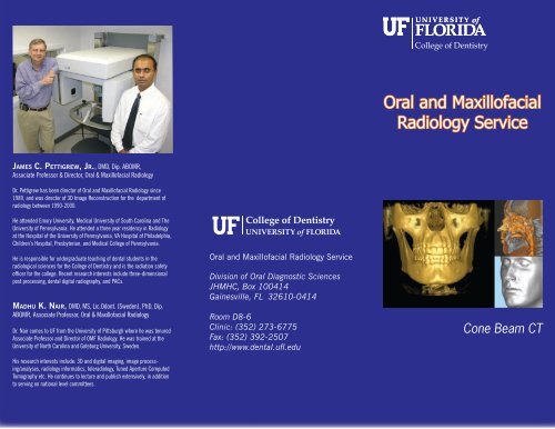
JAMES C. PETTIGREW, JR., DMD, Dip. ABOMR,<br />
Associate Pr<strong>of</strong>essor & Director, <strong>Oral</strong> & <strong>Maxill<strong>of</strong>acial</strong> <strong>Radiology</strong><br />
Dr. Pettigrew has been director <strong>of</strong> <strong>Oral</strong> <strong>and</strong> <strong>Maxill<strong>of</strong>acial</strong> <strong>Radiology</strong> since<br />
1989, <strong>and</strong> was director <strong>of</strong> 3D Image Reconstruction for the department <strong>of</strong><br />
radiology between 1990-2000.<br />
He attended Emory University, Medical University <strong>of</strong> South Carolina <strong>and</strong> The<br />
University <strong>of</strong> Pennsylvania. He attended a three year residency in <strong>Radiology</strong><br />
at the Hospital <strong>of</strong> the University <strong>of</strong> Pennsylvania, VA Hospital <strong>of</strong> Philadelphia,<br />
Children’s Hospital, Presbyterian, <strong>and</strong> Medical <strong>College</strong> <strong>of</strong> Pennsylvania.<br />
He is responsible for undergraduate teaching <strong>of</strong> dental students in the<br />
radiological sciences for the <strong>College</strong> <strong>of</strong> <strong>Dentistry</strong> <strong>and</strong> is the radiation safety<br />
<strong>of</strong>ficer for the college. Recent research interests include three-dimensional<br />
post processing, dental digital radiography, <strong>and</strong> PACs.<br />
MADHU K. NAIR, DMD, MS, Lic.Odont. (Sweden), PhD, Dip.<br />
ABOMR, Associate Pr<strong>of</strong>essor, <strong>Oral</strong> & <strong>Maxill<strong>of</strong>acial</strong> <strong>Radiology</strong><br />
Dr. Nair comes to UF from the University <strong>of</strong> Pittsburgh where he was tenured<br />
Associate Pr<strong>of</strong>essor <strong>and</strong> Director <strong>of</strong> OMF <strong>Radiology</strong>. He was trained at the<br />
University <strong>of</strong> North Carolina <strong>and</strong> Goteborg University, Sweden.<br />
His research interests include: 3D <strong>and</strong> digital imaging, image processing/analyses,<br />
radiology informatics, teleradiology, Tuned Aperture Computed<br />
Tomography etc. He continues to lecture <strong>and</strong> publish extensively, in addition<br />
to serving on national level committees.<br />
<strong>Oral</strong> <strong>and</strong> <strong>Maxill<strong>of</strong>acial</strong> <strong>Radiology</strong> <strong>Service</strong><br />
Division <strong>of</strong> <strong>Oral</strong> Diagnostic Sciences<br />
JHMHC, Box 100414<br />
Gainesville, FL 32610-0414<br />
Room D8-6<br />
Clinic: (352) 273-6775<br />
Fax: (352) 392-2507<br />
http://www.dental.ufl.edu<br />
Cone Beam CT
<strong>Oral</strong> <strong>and</strong> <strong>Maxill<strong>of</strong>acial</strong> <strong>Radiology</strong> <strong>Service</strong><br />
<strong>Oral</strong> <strong>and</strong> <strong>Maxill<strong>of</strong>acial</strong> <strong>Radiology</strong> <strong>Service</strong> <strong>of</strong>fers<br />
a variety <strong>of</strong> radiographic <strong>and</strong> consultation services<br />
to practitioners. Cone beam CT (CBCT) imaging<br />
technology is the newest addition to this array.<br />
Dictated reports by ABOMR board certified<br />
oral <strong>and</strong> maxill<strong>of</strong>acial radiologists including findings,<br />
diagnostic impressions, <strong>and</strong> pertinent comments<br />
are returned to the referring practitioner<br />
within one day for all procedures performed.<br />
Additionally, CT scans done elsewhere can be<br />
interpreted for incidental pathology <strong>and</strong> treatment<br />
planning purposes within approximately 48 hours <strong>of</strong><br />
receipt <strong>of</strong> images.<br />
CBCT is useful within the scope <strong>of</strong> dentistry<br />
for implant treatment planning, orthodontics, oral<br />
surgery, endodontics, periodontics, <strong>and</strong> pediatric<br />
dentistry. Other applications include imaging <strong>of</strong><br />
developmental anomalies, clefts, air spaces in obstructive<br />
sleep apnea patients, maxill<strong>of</strong>acial infections,<br />
facial pain, temporom<strong>and</strong>ibular joint evaluations,<br />
odontogenic disease, inner ear, sinuses <strong>and</strong> salivary<br />
gl<strong>and</strong>s. 3D reconstructions can also be produced.<br />
In volumetric tomography or CBCT, the entire<br />
volume <strong>of</strong> interest is exposed by using a conical<br />
shaped beam that exposes the desired area <strong>and</strong> then<br />
the volume is interactively viewed slice by slice. A<br />
major benefit for patients is the reduction <strong>of</strong> radiation<br />
dose, which is a fraction <strong>of</strong> the dose <strong>of</strong> a medical<br />
grade CT.<br />
Procedures <strong>and</strong>/or consultation <strong>of</strong>fered<br />
include:<br />
- Routine digital radiography <strong>of</strong> teeth<br />
<strong>and</strong> facial bones.<br />
- Temporom<strong>and</strong>ibular joint imaging,<br />
including arthrography, CT <strong>and</strong><br />
MRI.<br />
- Paranasal sinus evaluation.<br />
- Static <strong>and</strong> dynamic sialography.<br />
- Presurgical dental implant<br />
evaluations using CT.<br />
- Advice for digital imaging for private<br />
dental <strong>of</strong>fices.<br />
- Dictated reports for all studies<br />
If the desired area needs to be visualized with the<br />
use <strong>of</strong> a contrast agent, the patient is appointed for a<br />
procedure in Sh<strong>and</strong>s Hospital. However, for the majority<br />
<strong>of</strong> our treatment needs, CBCT provides excellent<br />
images with high resolution. Dedicated protocols<br />
are used for specific diagnostic tasks.<br />
Incidental pathology noted in CBCT scans<br />
acquired for other diagnostic purposes is further<br />
studied using other appropriate imaging modalities,<br />
in consultation with the referring doctors, ensuring<br />
prompt treatment for the patient.<br />
Arrangements <strong>and</strong> reporting for maxill<strong>of</strong>acial CT,<br />
magnetic resonance imaging <strong>and</strong> medical computed<br />
tomography, including 3 D reconstructions <strong>of</strong> odontogenic<br />
or non-odontogenic pathology, traumatic<br />
<strong>and</strong> developmental crani<strong>of</strong>acial abnormalities are also<br />
available.<br />
Specific questions that need to be answered<br />
should be included in the prescription. An image<br />
viewer will be provided with the images in electronic<br />
format on CD to the referring practitioner.<br />
The availability <strong>of</strong> CT in the UF <strong>College</strong> <strong>of</strong><br />
<strong>Dentistry</strong> has greatly aided patient care.<br />
Interpretation <strong>of</strong> exams by board certified<br />
radiologists can potentially result in reducing<br />
missed incidental pathology.<br />
Call us for more information: (352)273-6775