

PDF Version - Glidewell Dental Labs
PDF Version - Glidewell Dental Labs
PDF Version - Glidewell Dental Labs

Create successful ePaper yourself
Turn your PDF publications into a flip-book with our unique Google optimized e-Paper software.
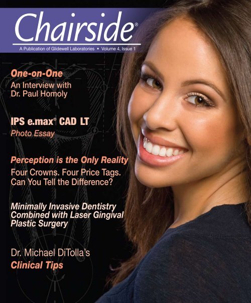
Chairside®<br />
A Publication of <strong>Glidewell</strong> Laboratories • Volume 4, Issue 1<br />
One-on-One<br />
An Interview with<br />
Dr. Paul Homoly<br />
IPS e.max ® CAD LT<br />
Photo Essay<br />
Perception is the Only Reality<br />
Four Crowns. Four Price Tags.<br />
Can You Tell the Difference?<br />
Minimally Invasive Dentistry<br />
Combined with Laser Gingival<br />
Plastic Surgery<br />
Dr. Michael DiTolla’s<br />
Clinical Tips
Contents<br />
9 Dr. DiTolla’s Clinical Tips<br />
This month’s tips include 3M ESPE Durelon ,<br />
which used to be one of the more popular permanent<br />
crown and bridge cements in the world. Today<br />
I see it used more as a long-term provisional cement.<br />
For extractions that are atraumatic and efficient, the<br />
Physics Forceps ® lives up to its “one-minute extraction”<br />
reputation. No less valuable are KaVo electric<br />
handpieces, which make prepping and polishing ceramic<br />
a breeze due to its high torque. When it comes<br />
to seating restorations, you will rarely see me without<br />
an Aidaco Bite Stick in my hand.<br />
14 Book Review: The Early Adventures of<br />
Painless Parker<br />
I had heard the name a couple of times, but I did not<br />
fully appreciate the legend of “Painless Parker” until<br />
I stumbled across this book. I loved getting some insight<br />
into the world of a scrappy dentist who sought<br />
to revolutionize how dentistry’s message would be<br />
spread. In my first Chairside book review, I give you<br />
a quick teaser on what I feel is a “must-read” for<br />
anyone who has ever wondered how to attract more<br />
patients.<br />
16 Photo Essay: IPS e.max ® CAD LT<br />
Case Study<br />
Many dentists have asked me about the effectiveness<br />
and esthetics of IPS e.max CAD LT, and I think using<br />
it in a case like this shows that it is becoming<br />
one of my “go-to” crowns. A no-prep Vivaneer on<br />
the adjacent tooth was also necessary to address the<br />
patient’s chief complaint.<br />
27 Minimally Invasive Dentistry Combined<br />
With Laser Gingival Plastic Surgery:<br />
Maximize Your Aesthetic Results<br />
It’s no secret that I hold Dr. Bob Lowe in great esteem<br />
as one of my clinical mentors. Bob is the one<br />
who taught me how to perform soft and hard tissue<br />
crown lengthening, and I think he is the only dentist<br />
presently teaching GPs how to do this. Don’t miss<br />
any opportunity to see Bob lecture near you!<br />
Cover photo by Sharon Dowd<br />
Cover illustration by Wolfgang Friebauer, MDT<br />
Contents 1
Editor’s Letter<br />
I hate to start off the new year with a face full of attitude,<br />
but if there is another dental magazine out there as interesting<br />
as Chairside, I’d like to see it. Other magazines<br />
have good articles here and there, but I strive to make<br />
every article one that you will at least want to read the<br />
callouts and thus get sucked into the article.<br />
Dr. Paul Homoly surprised me yet again in this month’s<br />
interview, as we discussed the culture of dentistry and the<br />
blind pursuit of quality dentistry. As always, Paul looks at<br />
things from a contrarian point of view, and I know you<br />
will find this interesting. Paul generates more e-mail than<br />
any other contributor, and it is all positive.<br />
I stumbled upon a book on eBay about Painless Parker,<br />
and I bought it after I went to the Wikipedia page about<br />
him and became fascinated. I loved the book and wrote<br />
a review about it in this issue. Love him or hate him, you<br />
would be hard-pressed to find a more interesting or controversial<br />
dentist.<br />
The “Perception is the Only Reality” article may open a<br />
few eyes as well. I invite you to vote for your favorite<br />
crown and try to guess which ones are which before you<br />
look at all the prices. (<strong>Glidewell</strong> was not involved in this<br />
study, so there is no hidden agenda.)<br />
Dr. Neiburger has written a fascinating article on the evolution<br />
of tooth wear and how teeth have changed over the<br />
years. Is the way we prepare food today preventing the<br />
natural, healthy wearing of our teeth?<br />
Dr. Bob Lowe rounds out this issue with another excellent<br />
clinical article on how he uses lasers to deal with gingival<br />
issues. The more cosmetic dentistry you do, the more you<br />
realize the major role the gingival plays, and how unpredictable<br />
it can be.<br />
I also include a case study with one of my favorite materials:<br />
IPS e.max ® CAD LT, used with no-prep veneers. Try<br />
IPS e.max on a patient, and see if you like this cementable<br />
all-ceramic restoration as much as I do.<br />
Yours in quality dentistry,<br />
Dr. Michael DiTolla<br />
Editor in Chief, Clinical Editor<br />
mditolla@glidewelldental.com<br />
Publisher<br />
Jim <strong>Glidewell</strong>, CDT<br />
Editor in Chief<br />
Michael DiTolla, DDS, FAGD<br />
Managing Editors<br />
Jim Shuck<br />
Mike Cash, CDT<br />
Creative Director<br />
Rachel Pacillas<br />
Clinical Editor<br />
Michael DiTolla, DDS, FAGD<br />
Copy Editors<br />
Melissa Manna<br />
Martin Yan<br />
Magazine Coordinators<br />
Sharon Dowd<br />
Lindsey Lauria<br />
Graphic Designers<br />
Jamie Austin, Deb Evans, Joel Guerra,<br />
Phil Nguyen, Gary O’Connell, Rachel Pacillas<br />
Staff Photographers<br />
Sharon Dowd<br />
Illustrators<br />
Wolfgang Friebauer, MDT<br />
Phil Nguyen<br />
Ad Representative<br />
Lindsey Lauria<br />
(lindsey.lauria@glidewelldental.com)<br />
If you have questions, comments or complaints regarding<br />
this issue, we want to hear from you. Please e-mail us<br />
at chairside@glidewelldental.com. Your comments may be<br />
featured in an upcoming issue or on our Web site.<br />
© 2009 <strong>Glidewell</strong> Laboratories<br />
Neither Chairside Magazine nor any employees involved in its publication<br />
(“publisher”), makes any warranty, express or implied, or assumes<br />
any liability or responsibility for the accuracy, completeness, or usefulness<br />
of any information, apparatus, product, or process disclosed, or<br />
represents that its use would not infringe proprietary rights. Reference<br />
herein to any specific commercial products, process, or services by<br />
trade name, trademark, manufacturer or otherwise does not necessarily<br />
constitute or imply its endorsement, recommendation, or favoring<br />
by the publisher. The views and opinions of authors expressed<br />
herein do not necessarily state or reflect those of the publisher and<br />
shall not be used for advertising or product endorsement purposes.<br />
CAUTION: When viewing the techniques, procedures, theories and materials<br />
that are presented, you must make your own decisions about<br />
specific treatment for patients and exercise personal professional judgment<br />
regarding the need for further clinical testing or education and<br />
your own clinical expertise before trying to implement new procedures.<br />
Chairside ® Magazine is a registered trademark of <strong>Glidewell</strong> Laboratories.
Contents<br />
34 One-on-One with Dr. DiTolla<br />
In our previous discussion, Dr. Paul Homoly and I<br />
shared our thoughts on how to communicate with<br />
patients who had large restorative needs. Now, in<br />
our latest interview, we discuss the culture of dentistry<br />
and the dangers of too much patient education.<br />
50 Perception is the Only Reality<br />
LMT conducted a nationwide experiment to determine<br />
whether technicians and dentists could actually<br />
differentiate between expensive and inexpensive<br />
crowns. After hundreds of comparisons among the<br />
best in the industry, there seems to be one conclusive<br />
answer. I took the liberty of sharing with you<br />
their findings, and the results may surprise you.<br />
55 The Evolution of Human Occlusion—<br />
Ancient Clinical Tips for Modern Dentists<br />
In this well-chronicled article, Dr. Ellis Neiburger<br />
discusses the general evolution of our teeth and addresses<br />
occlusal problems throughout the centuries.<br />
I was fascinated by this article and think about it every<br />
time I perform occlusal adjustment on a crown.<br />
Contents 3
Letters to the Editor<br />
“Dear Dr. DiTolla,<br />
I really love the Reverse Preparation Technique.<br />
It has made my life so much easier!<br />
One question though: I still find that I don’t<br />
have enough reduction on the lingual surfaces<br />
of tooth 8 and 9. Any suggestions on<br />
how I can make sure I have enough reduction<br />
in these areas?”<br />
- Dr. Darryl Duval, Jacksonville, FL<br />
Dear Darryl,<br />
Good question! I usually eyeball it,<br />
but as we both know that doesn’t<br />
always work. When I have doubts,<br />
I use The Reduction Ring (www.<br />
reductionring.com). I find it to be<br />
pretty fail-proof; in fact, I should<br />
use Reduction Rings all the time and<br />
put them in the Reverse Preparation<br />
Technique video.<br />
Please e-mail me back and let me<br />
know if you like them, as there are<br />
other brands out there you may like<br />
better.<br />
- Dr. DiTolla<br />
“Dear Dr. DiTolla,<br />
I recently read your article in <strong>Dental</strong> Economics<br />
and was very interested in learning<br />
4<br />
Letters to the Editor<br />
more concerning the STA System anesthesia<br />
technique.<br />
I have many of your DVDs and use your<br />
Reverse Preparation Technique religiously.<br />
The STA System technique peaked my<br />
interest, but after seeing you use and endorse<br />
it, it made me want to learn more.<br />
Do you currently have a DVD for this technique?<br />
Also, is it an easy technique to<br />
learn or does it take practice? Any additional<br />
information you can provide would<br />
be greatly appreciated.”<br />
- Dr. Rick Bray, Pennsburg, PA<br />
Dear Rick,<br />
For a single mandibular molar, I start<br />
in the buccal furcation, right at the<br />
buccal midpoint on the STA setting,<br />
not the normal or the more rapid<br />
setting. I wait for the lights to increase<br />
to show that the pressure is<br />
correctly increasing for a PDL injection.<br />
If I don’t get proper pressure<br />
in the furcation, I move the needle<br />
to the MB line angle and try it<br />
there and then move it to the<br />
DB line angle. Due to localized<br />
periodontal conditions,<br />
you may need<br />
to move the needle<br />
to an area that is<br />
healthy enough<br />
for this type of<br />
injection. If I get<br />
a good injection<br />
in the buccal<br />
furcation, I typically<br />
do not<br />
go to the lingual,<br />
although<br />
there is certainly<br />
nothing<br />
wrong<br />
with doing<br />
that. I know<br />
some dentists<br />
who give the injection<br />
at the ML and DL<br />
line angles instead of the furcation,<br />
and they report very good<br />
results with that technique, too.<br />
Basically, it doesn’t matter where<br />
the needle is as long as you are getting<br />
good pressure feedback on the<br />
unit, which tells you it has been a<br />
successful PDL injection. I like it best<br />
when it works in the buccal furcation<br />
because I know I will get great pulpal<br />
anesthesia with that single injection.<br />
For typical maxillary infiltrations, I<br />
use the normal setting if I am starting<br />
in the area of the bicuspids and<br />
moving anteriorly. If I am just anesthetizing<br />
8 and 9, for example, I will<br />
usually start the injections on the<br />
STA speed (the slowest speed), even<br />
though it is not a PDL injection. This<br />
is the most comfortable setting for<br />
the patient and halfway through the<br />
injection, when the patient is partially<br />
anesthetized, my assistant or I will<br />
switch it to normal speed. I hope that<br />
helps!<br />
- Dr. DiTolla
“Dear Dr. DiTolla,<br />
I have happily used <strong>Glidewell</strong> Laboratories<br />
for several years. I even came down from<br />
Northern California to tour the impressive<br />
facility, where I saw you working.<br />
My question is: What cement do you recommend<br />
for zirconia? Different lecturers<br />
and manufacturers give various strength<br />
numbers. I have been using Panavia F2.0<br />
(Kuraray <strong>Dental</strong>) and RelyX (3M ESPE)<br />
successfully for many years.”<br />
- Dr. Richard Jergensen, Fairfield, CA<br />
Dear Richard,<br />
Panavia F2.0 is a great choice. RelyX<br />
could be referring to either RelyX<br />
Luting Plus Cement or RelyX<br />
Unicem; either is a great choice as<br />
well. The RelyX Luting Plus Cement<br />
is a resin-reinforced glass ionomer<br />
used for conventional cementation,<br />
and Unicem is a self-etching resin<br />
cement. Both are highly acceptable<br />
choices for zirconia-based restorations.<br />
- Dr. DiTolla<br />
“Dear Dr. DiTolla,<br />
I recently watched your Rapid Anesthesia<br />
Technique on the <strong>Glidewell</strong> Web site. I<br />
think I understood most of it, but is it basically<br />
a PDL injection?<br />
Also, what gauge and length needle do<br />
you use for this technique? I have had a<br />
hard time finding a heavy enough needle<br />
short in length to use in my conventional<br />
PDL gun.”<br />
- Dr. Mark Pelletier, Irmo, SC<br />
Dear Mark,<br />
The Rapid Anesthesia Technique is<br />
a PDL injection that is done in the<br />
furcation space of a lower molar. I<br />
used to do them by hand, but I now<br />
use the STA System from Milestone<br />
Scientific (www.stais4u.com). In fact,<br />
I now do all my injections with the<br />
STA System—I love it.<br />
I used to have a problem with my<br />
30-gauge extra short needles bending<br />
as well. Since I switched over to<br />
the STA System, you have to use their<br />
needles. They hold up much better,<br />
but you can only use them with their<br />
system. Otherwise, I prefer Accuject ®<br />
needles from DENTSPLY International,<br />
Inc., but they still bend a little at<br />
times.<br />
- Dr. DiTolla<br />
“Dear Dr. DiTolla / Dr. Lowe,<br />
I was wondering what type of camera was<br />
used in Dr. Bob Lowe’s article on pages<br />
24-29 of Chairside ® Volume 3, Issue 2. The<br />
photos were great, and I would like to get<br />
all the information I can on the process<br />
used.<br />
I’d also like to know if Dr. Lowe learned<br />
this technique on his own or if he attended<br />
a class and, if so, where. Finally, what settings<br />
does he keep his camera on? Thank<br />
you for any information you can provide.”<br />
- Tracy Lindamood, CDA, Jacksonville, FL<br />
Dear Tracy,<br />
These pictures have been taken over<br />
a period of many years. Some were<br />
taken with a Fuji S-1 Pro, others with<br />
a Canon 5D. The Fuji had a ring flash,<br />
and the Canon 5D has a side-by-side<br />
dual flash. While a ring flash is easier<br />
to use, especially in the posterior region<br />
of the mouth, it tends to make<br />
images look more two-dimensional.<br />
The side-by-side flash takes a little<br />
practice to learn how to bounce light<br />
to capture posterior exposures. The<br />
anterior exposures are much more<br />
three-dimensional than those taken<br />
with a ring, particularly if you concentrate<br />
the light a little more on one<br />
side.<br />
Dr. Shavell, my mentor, was an outstanding<br />
dental photographer. He<br />
had two rules. The first rule: Fill the<br />
frame with your subject. To show a<br />
photo of one tooth, you need to go<br />
two-to-one. Today, with digital, this<br />
can be done with Adobe ® Photoshop<br />
® and cropping, but that takes<br />
time. I prefer a 2x teleconverter to<br />
take the photo at 2x, then no manipulation<br />
with computer software.<br />
The second rule: Line up the buccal<br />
surfaces of posterior mirror shots<br />
parallel to the top of the viewfinder.<br />
Center facial and labial shots using<br />
the occlusal plane as a guide.<br />
The AACD has a good pamphlet on<br />
taking intraoral photos as far as settings,<br />
which vary from camera to<br />
camera, flash set up to flash set up.<br />
The nice thing with digital is you can<br />
see if the exposure is too light or too<br />
dark and adjust the flash intensity<br />
and/or f-stop accordingly.<br />
Lastly, my friends Dr. Tony Soileau<br />
and Dr. Jim Dunn teach excellent<br />
photography courses. Google them<br />
to get more detailed contact info.<br />
I hope this helps and good luck!<br />
- Dr. Lowe<br />
WRITE US<br />
Chairside Magazine welcomes<br />
letters to the editor, which<br />
may be featured in an upcoming<br />
issue or on our Web site.<br />
Letter should include writer’s<br />
full name, address and<br />
daytime phone number.<br />
To contact us: e-mail (chairside@glidewelldental.com),<br />
mail (Letters to the Editor,<br />
Chairside Magazine, <strong>Glidewell</strong><br />
Laboratories, 4141 MacArthur<br />
Blvd., Newport Beach, CA<br />
92660) or call (888-303-4221).<br />
Letters to the Editor 5
Contributors<br />
Michael C. DiTolla, DDS, FAGD<br />
Dr. Michael DiTolla is Director of Clinical Education & Research at <strong>Glidewell</strong> Laboratories in Newport<br />
Beach, Calif. Here, he performs clinical testing on new products in conjunction with the company’s R&D<br />
Department. <strong>Glidewell</strong> dental technicians have the privilege of rotating through Dr. DiTolla’s operatory<br />
and experience his commitment to excellence through his prepping and placement of their restorations.<br />
He is a CR evaluator and lectures nationwide on both restorative and cosmetic dentistry. Dr. DiTolla<br />
has several clinical programs available on DVD through <strong>Glidewell</strong> Laboratories. For more information<br />
on his articles or to receive a free copy of Dr. DiTolla’s clinical presentations, call 888-303-4221, e-mail<br />
mditolla@glidewelldental.com, or visit www.glidewell-lab.com.<br />
Paul Homoly, DDS, CSP<br />
Dr. Paul Homoly is a world-class leader in dental education. After practicing comprehensive restorative<br />
dentistry for 20 years, Dr. Homoly earned the highest designation in professional speaking—Certified<br />
Speaking Professional (CSP)—and is the first and only dentist in the world to earn this designation. As<br />
an acclaimed educator for more than 25 years, he is best known for his innovative and practical approach<br />
to dentistry. An accredited member of the ADA, Dr. Homoly is an active author who contributes<br />
to dental journals worldwide, including a monthly column in <strong>Dental</strong> Economics. He is also president of<br />
Homoly Communications Institute located in Charlotte, N.C. To reach Dr. Homoly, call 800-294-9370,<br />
e-mail paul@paulhomoly.com, or visit www.paulhomoly.com.<br />
Robert A. Lowe, DDS, FAGD, FICD, FADI, FAC<br />
Dr. Robert A. Lowe graduated magna cum laude from Loyola University School of Dentistry in 1982,<br />
and was a Clinical Professor of Restorative Dentistry until its closure in 1993. Since January of 2000,<br />
Dr. Lowe has maintained a private practice in Charlotte, N.C. He lectures internationally and publishes<br />
in well-known dental journals on esthetic and restorative dentistry. Dr. Lowe received fellowships in<br />
the AGD, ICD, ADI, and ACD. In addition, he received the Gordon Christensen Outstanding Lecturers<br />
Award in 2004, and Diplomat status on the American Board of Esthetic Dentistry in 2005. To contact<br />
Dr. Lowe, call 704-364-4711, e-mail boblowedds@aol.com, or visit www.destinationsmile.com.<br />
Ellis Neiburger, DDS<br />
Dr. Ellis “Skip” Neiburger graduated from the University of Illinois College of Dentistry in 1968. After<br />
postgraduate research in Oral Pathology and Forensic Dentistry at the U.S. Armed Forces Institute of Pathology,<br />
Dr. Neiburger pursued a career as a paleopathologist. He has been curator of anthropology at<br />
the Lake County Museum for 17 years. Dr. Neiburger’s research on ancient anatomy and occlusion has<br />
taken him throughout the world, and his studies have been widely published in the areas of prehistoric<br />
pathology, dental computing and clinical dentistry. He is editor and vice president of the American Association<br />
of Forensic Dentists, and has written five books on dentistry. Dr. Neiburger has a general practice<br />
in Waukegan, Ill., and may be contacted at 847-244-0292 or by visiting www.drneiburger.com.<br />
Contributors 7
– ARTICLE by Michael DiTolla, DDS, FAGD<br />
– PHOTOS by Sharon Dowd<br />
PRODUCT........ Durelon <br />
Dr. DiTolla’s<br />
CLINICAL TIPS<br />
CATEGORY...... Polycarboxylate Luting Cement<br />
SOURCE.......... 3M ESPE <br />
St. Paul, MN<br />
800-364-3577<br />
www.3m.com<br />
When I graduated from dental school in 1988, Durelon<br />
was my permanent cement of choice. It seemed<br />
to work well until it started to turn mushy about five<br />
years after cementation. Maybe it’s not a bad idea<br />
to have the crown fall off every five years to check<br />
the prep! When all the hype with glass ionomer cements<br />
started, I switched over. But when I had too<br />
many cases of post-operative sensitivity to ignore, I<br />
went running back to Durelon. With the advent of an<br />
excellent class of resin-reinforced glass ionomer cements,<br />
Durelon has decreased in use as a permanent<br />
cement. My reintroduction to Durelon was through<br />
Dr. Bill Strupp, who has used Durelon as a temporary<br />
cement for decades. We started using it about<br />
10 years ago for our BioTemps ® in large crown and<br />
bridge cases, and the temps simply don’t come off.<br />
Due to its relatively neutral pH value, there is essentially<br />
no post-operative sensitivity with this cement,<br />
and it is well tolerated by the gingival as well.<br />
Dr. DiTolla’s Clinical Tips 9
Dr. DiTolla’s<br />
CLINICAL TIPS<br />
PRODUCT........ Physics Forceps ®<br />
CATEGORY...... Tooth Extraction<br />
SOURCE.......... GoldenMisch, Inc.<br />
Detroit, MI<br />
877-987-2284<br />
www.oneminuteextractions.com<br />
There are some great product names in dentistry—<br />
you may know that my favorite has long been Algi-<br />
Not, the alginate replacement product from Kerr.<br />
However, Physics Forceps from GoldenMisch, Inc.,<br />
should win an award for its reputation as the “One<br />
Minute Extraction Forceps.” That pretty much sums it<br />
up, doesn’t it? We have a lot of overpromised/underdelivered<br />
products in dentistry, and I was pretty sure<br />
Physics Forceps was going to be about as successful<br />
as the 90-second crown prep, which led to a lot of bad<br />
preps done quickly.<br />
The forceps came with a couple of study models to<br />
practice on, but the teeth seemed to come out a little<br />
too easily. If my patient’s bone was this flexible,<br />
I wouldn’t need forceps. Three days later, a patient<br />
walked in with a broken off upper first molar. My assistant<br />
grabbed the forceps while I looked at the directions<br />
one more time. I engaged the palatal root,<br />
placed the bumper on the buccal plate and, without<br />
squeezing, rotated the forceps. After 60 seconds of<br />
convincing myself nothing was happening, the tooth<br />
started to grow out of the socket! I switched to my<br />
regular forceps and lifted the tooth out 80 seconds<br />
from the time they were applied. This may be the first<br />
advance in exodontia technology in 100 years, but it<br />
was worth the wait!<br />
10 Dr. DiTolla’s Clinical Tips
Dr. DiTolla’s<br />
CLINICAL TIPS<br />
PRODUCT........ KaVo ELECTROtorque plus Handpiece<br />
CATEGORY...... Electric Handpieces<br />
SOURCE.......... KaVo <strong>Dental</strong> Corporation<br />
Lake Zurich, IL<br />
800-323-8029<br />
www.kavousa.com<br />
I see my KaVo ELECTROtorque plus handpiece the<br />
same way I see digital radiography: there is no downside<br />
except for cost. Is there any other piece of equipment<br />
that is more linked to our income than our handpieces?<br />
Why try to save money on the instrument you<br />
use to prep every inlay, veneer, crown and bridge in<br />
your practice?<br />
I don’t prep teeth faster with this electric handpiece,<br />
I just prep them better. This is because I can turn the<br />
speed down on the handpiece and turn the water off<br />
as well, due to the decreased heat with the slower<br />
revolutions. Amazingly, you still have all of the torque<br />
that you have when the handpiece is running full<br />
speed. This allows you to make perfect margins and<br />
see what you are doing without the water spray blocking<br />
your view. There is no better way to polish ceramic<br />
material intraorally than with an electric handpiece.<br />
In fact, I am not even sure you can really polish porcelain<br />
well with a traditional handpiece. That may sound<br />
a little overdramatic, but if you have loupes it will be<br />
pretty obvious to you as well. Polishing porcelain is<br />
all about torque, and you owe it to yourself to demo<br />
the KaVo ELECTROtorque plus handpiece at the next<br />
convention you attend.<br />
Dr. DiTolla’s Clinical Tips11
Dr. DiTolla’s<br />
CLINICAL TIPS<br />
PRODUCT........ Aidaco Bite Sticks<br />
CATEGORY...... Crown Seating Instrument<br />
SOURCE.......... Temrex Corporation<br />
Freeport, NY<br />
800-645-1226<br />
www.temrex.com<br />
The ubiquitous orangewood bite sticks! They show up<br />
in practically all of my DVDs because I use them on<br />
nearly every case. I was first introduced to Aidaco Bite<br />
Sticks right out of dental school, during the two years<br />
that I practiced with my dad. He used them with a<br />
mallet to hammer in anterior crowns, and he would<br />
tell patients they were going to feel a “slight tapping<br />
sensation.” Their body language suggested they were<br />
feeling a “massive jackhammer sensation.” One day I<br />
had him tap on tooth 9 in my mouth with his mallet<br />
and bite stick, and the force he was able to deliver<br />
was shocking! That was the day I decided to retire<br />
the mallet and to just use the orangewood sticks with<br />
my hands. In Dad’s defense, the cements of his day<br />
did not have the thin film thickness of today’s cements<br />
and may have needed to be pounded into place.<br />
When you try to seat crowns with just finger pressure,<br />
the crowns indent into your fingertips and it’s<br />
hard to tell if you are holding the crown in place. As<br />
you have seen, I use the sticks for crowns, veneers,<br />
even no-prep veneers in the anterior. In the posterior,<br />
I use them on every bridge by having the patient<br />
bite down on a bite stick during try-in for eight to<br />
10 minutes. Whether we like it or not, preps shift in<br />
the two weeks between appointments, even with welldone<br />
provisionals—biting on the stick helps stubborn<br />
bridges go down into place. When you look at remake<br />
rates for our doctors, bridges always have a higher remake<br />
rate due to prep shifting. It certainly helps to do<br />
some “instant orthodontics” by having the patient bite<br />
down on an orangewood bite stick with the bridge in<br />
place prior to declaring it a remake. Often my dental<br />
assistant will do this before I even enter the room, so<br />
I can begin evaluating contacts and margins as soon<br />
as I walk in.<br />
Dr. DiTolla’s Clinical Tips13
Book Review:<br />
The Early<br />
Adventures<br />
of Painless<br />
Parker<br />
– Book by Peter M. Pronych & Arden G. Christen<br />
– review by Michael DiTolla, DDS, FAGD<br />
I<br />
am a big fan of biographies of successful<br />
people from all walks of life. I don’t<br />
think I have ever read a biography I haven’t<br />
learned something from that I can relate to my pursuit<br />
of success. Unfortunately, the opportunities to read a biography of an<br />
icon in our industry are few and far between. So when I happened to find<br />
one floating around on the Internet, I jumped at the chance to read the<br />
story of a dentist who, at one point, was more famous than the President.<br />
Early in my career, I remember working on an older woman who was slightly<br />
apprehensive about having an extraction. I was able to complete the extraction<br />
without her feeling any pain, and at the end of the extraction she said,<br />
“Wow, you are a real Painless Parker.” I thanked her for the compliment, but<br />
asked her about the expression, as I had never heard it before. She went on to tell me<br />
about Painless Parker, a dentist from the turn of the century who was world-renowned for performing painless extractions.<br />
That made no sense at all to me because my dad had been practicing dentistry since the early 1960s, and he told<br />
me how unreliable Novocain was then…let alone 60 years before. I forgot all about the offhanded compliment until<br />
a couple months ago when another patient mentioned Painless Parker, and my curiosity sent me to the center of all<br />
knowledge: Wikipedia. A quick search brought me to his page, and as I read I became more fascinated with the man<br />
who had a passion for bringing dentistry to the working class for an affordable price.<br />
I found a book on Painless Parker and, once I picked it up, I could not put it down. I wanted to review it in Chairside ®<br />
because I knew how many of you would be interested in this fascinating story. Whether you love or hate his tactics, I<br />
guarantee you won’t be bored with the trials and tribulations of Painless Parker. Incidentally, when the <strong>Dental</strong> Board of<br />
California told him he could no longer call himself “Painless Parker,” he went to court and legally changed his name from<br />
“Edgar” to “Painless.” Awesome! Believe it or not, that is one of the least controversial things he did.<br />
In May of 1892, Parker graduated from Philadelphia <strong>Dental</strong> College with a Doctor of <strong>Dental</strong> Surgery degree. His graduating<br />
class consisted of four other students. After graduation, he decided to practice in his hometown of St. Martins in<br />
New Brunswick, Canada. While he wanted to tell the locals about his skills, he had been taught in dental school that it<br />
was unethical to solicit work directly. Parker was taught, however, that it was acceptable to solicit work by joining clubs<br />
and to never decline an invitation to be seen at a public place.<br />
Parker had been well-known in his hometown as an adolescent for some of his escapades, and he felt joining his local<br />
church might help shore up his reputation and get some patients in the office. When that failed to produce any patients,<br />
14 Book Review: The Early Adventures of Painless Parker
Parker decided to attend both Sunday services to<br />
appear even more pious. He began to sit in the<br />
front pew at church and took to carrying a huge<br />
Bible with him. Since he had yet to see a patient<br />
in his office, he began to volunteer for all of the<br />
tasks in the church. Parker also assisted with the<br />
services and taught Sunday school—anything<br />
to make them think he was an ideal citizen. As<br />
Parker put it, “I was determined to be ethical at<br />
all costs.” Six weeks after opening his office, he<br />
still had not seen a single patient.<br />
Hope finally arrived in the shape of a local sign<br />
painter who Parker knew hated his dentures.<br />
Parker offered to make him new dentures in exchange<br />
for a sign for the practice. The painter<br />
wanted Parker to make the dentures<br />
first so he could try them,<br />
and then he would make the<br />
sign. Parker agreed since he was<br />
out of money. The painter loved<br />
the dentures and, with much<br />
appreciation, made a huge new<br />
sign with gold paint for Parker’s<br />
practice. Parker was somewhat<br />
embarrassed by its size, so he instructed<br />
the painter to put it up<br />
at night so no one would see. The<br />
next day Parker expected there to be<br />
a line of patients around his office, but it<br />
never materialized. In fact, when Parker showed up<br />
to work the next day, he found the sign was missing! Later that<br />
day, he found it nailed to the train station’s outhouse door, most likely by<br />
one of the town’s other dentists. Embarrassed to be seen taking the sign down, Parker once again waited for the cloak<br />
of night to remove his sign and replace it at work. His sign attracted one patient in his first 90 days of practice, a tourist<br />
who needed an extraction. Parker removed the tooth and charged him one dollar ($21 in the present day, adjusted<br />
for inflation). The patient only had 75 cents with him, but Parker was happy to take the money and finally get paid for<br />
performing dentistry.<br />
Parker saw fire and brimstone preachers on the street corner converting people with their vivid descriptions of hell,<br />
messages that were considered socially acceptable. He just could not believe why it was unethical to preach the importance<br />
of taking care of your teeth, and the “hell” that awaited you if you became edentulous.<br />
Armed with an aqueous solution of cocaine he called “hydrocaine,” Parker takes his message to the street corner offering<br />
painless extractions for 50 cents. He promised that if the extraction hurt, he would pay the patient five dollars! That<br />
first night he extracted 12 teeth and didn’t have to give anyone the five dollars, which he found surprising because he<br />
ran out of hydrocaine after the seventh patient!<br />
While I certainly wouldn’t want to follow in Parker’s footsteps, I was drawn into the story of his personal struggles.<br />
Parker starts his practice with dignity, but soon finds that dignity won’t pay the bills. Unlike most dentists, he finds he<br />
likes being a dentist and a salesman at the same time, and this drives his decision to take the story of preventive dentistry<br />
straight to the people.<br />
This is on my required reading list for all dentists, young and old.<br />
One of the authors, Dr. Arden G. Christen, has limited copies of the book available for purchase, although “The Early Adventures of Painless Parker” is technically out of<br />
print. Contact Dr. Christen at achriste@iupui.edu to request a copy.<br />
Book Review: The Early Adventures of Painless Parker15
Photo Essay<br />
IPS e.max ® CAD LT Case Study<br />
– ARTICLE & CLINICAL PHOTOS by Michael DiTolla, DDS, FAGD<br />
– COVER PHOTO by Sharon Dowd<br />
16 Photo Essay: IPS e.max CAD LT Case Study
I<br />
wanted to share this case with you for a couple of reasons. First of all, it features the IPS e.max ® CAD<br />
LT crown from Ivoclar Vivadent (Amherst, NY) that I have been asked by many dentists about. It’s a<br />
restoration I am very pleased with and find myself using in more situations. This case involved<br />
removing a zirconia-based crown, which is as bad as it gets when removing old restorations. It gave<br />
me a chance to show you the two-cord technique one more time, and it required the use of a no-prep veneer<br />
on the tooth adjacent to the crown in order to address the patient’s chief complaint: the interproximal black<br />
triangle between tooth 9 and 10.<br />
Figure 1<br />
Figure 2<br />
Figure 3<br />
Figures 1-3: This 39-year-old female patient came to the office unhappy with the existing PFM crown on tooth 9. The tooth had been endodontically<br />
treated 10 years prior, and four years ago a zirconia-based crown was placed. Since then, there had been some recession of the<br />
gingival, which had exposed the darkened root from the endo.<br />
Photo Essay: IPS e.max CAD LT Case Study17
Figure 4<br />
Figure 5 Figure 6<br />
Figures 4-6: The patient’s other main complaint was the shape of tooth 10 and the resultant gingival embrasure between the two teeth. I told<br />
her that if we hoped to close the large black triangle between 9 & 10, we would have to place a restoration on tooth 10 as well.<br />
18 Photo Essay: IPS e.max CAD LT Case Study
Figure 7<br />
Figure 8<br />
Figure 7: This occlusal view is imperative when deciding what type<br />
of veneers to place on a patient. Dentists frequently send me smile<br />
pictures and ask if the patient needs no-prep or minimal-prep veneers,<br />
but you can’t have that discussion without an occlusal picture.<br />
In this case, tooth 10 is an excellent candidate for a no-prep<br />
veneer.<br />
Figure 8: A close-up look at the zirconia-based crown on tooth 9<br />
shows that the incisal edge is longer than tooth 8, and the overall<br />
shape of the crown does not match 8 either. We decided to use an<br />
all-ceramic crown without a substructure to replace the zirconia<br />
crown, in hopes of getting a better match. I opted to use an IPS<br />
e.max CAD LT crown.<br />
Figure 9<br />
Figure 10<br />
Figure 9: I still give the patient some local anesthesia since I will be<br />
placing two retraction cords. I used to try to avoid local anesthesia<br />
when possible, but since developing a painless injection technique,<br />
it is not an issue. Here I place Profound Lite (Steven’s Pharmacy,<br />
Costa Mesa, CA) topical anesthetic for 45 seconds and then rinse<br />
it off.<br />
Figure 10: After the Profound Lite has been rinsed off, I use the<br />
STA System (Milestone Scientific, Livingstone, NJ) to deliver the<br />
Septocaine ® (Septodont, New Castle, DE) on the slowest speed. After<br />
about 20 seconds, I switch the STA System to the normal speed,<br />
as the patient is already anesthetized in that area. This is the easiest<br />
way to give a painless injection.<br />
Photo Essay: IPS e.max CAD LT Case Study19
Figure 11<br />
Figure 12<br />
Figure 11: If you have never had the pleasure of cutting off a zirconia<br />
crown, you are in for a treat. You can make the task much easier<br />
by having some specialty burs on hand, such as this ZIR-CUT Bur<br />
(Axis <strong>Dental</strong>, Coppell, TX) available through all dental dealers. The<br />
blue stripe on the shank identifies it as a ZIR-CUT Bur.<br />
Figure 12: It is much easier to cut through the zirconia coping with<br />
an electric handpiece because of the additional torque. Regardless,<br />
make sure you try to cut through it with a soft touch. As the<br />
bur cuts through the last of the zirconia, you will inadvertently cut<br />
into the tooth if you have too much pressure on the handpiece.<br />
Figure 13<br />
Figure 14<br />
Figure 13: The crown is popped off with the Christensen Crown<br />
Remover and the prep is evaluated. It is slightly overtapered in the<br />
incisal third, the mesial is slightly underprepared in the gingival<br />
third, and the distal margin is slightly overprepared in the distal.<br />
That said, the prep is still acceptable if we clean up the margins<br />
and get a great impression.<br />
Figure 14: Prior to margin refinement, we place a Size 00 Ultrapak<br />
® cord (Ultradent, South Jordan, UT) as our bottom cord in the<br />
two-cord technique. Since this cord will be in place during the rest<br />
of the procedure, it contains no epinephrine or medicaments. We<br />
“floss” the cord into the distal; no packing instrument is used.<br />
20 Photo Essay: IPS e.max CAD LT Case Study
Figure 15<br />
Figure 16<br />
Figure 15: We then grab the other end of the 00 cord and “floss” it<br />
into the mesial portion of the sulcus. We try to use an instrument as<br />
little as possible so we don’t cause any bleeding at this point. Once<br />
this cord is in place and we are packing the top cord, we can safely<br />
use an instrument without bleeding.<br />
Figure 16: We then grab both ends of the 00 cord on the lingual<br />
with cotton pliers, and pull them lingually until the cord pulls tight<br />
against the facial surface. You may also do this by hand (as I used to<br />
until I read that latex powder on retraction cords may inhibit the set<br />
of impression materials, although I have not seen proof of this).<br />
Figure 17<br />
Figure 18<br />
Figure 17: We use an instrument on the facial surface to pack the<br />
cord into the sulcus because we don’t have a choice. However,<br />
by having the interproximal areas already “flossed” into place, it<br />
makes it much easier to pack the cord atraumatically. If needed,<br />
the ends of the retraction on the lingual can be pulled again, if you<br />
left too much slack on the facial.<br />
Figure 18: The two ends of the cord are cut on the lingual so that<br />
when they are packed in the sulcus they will butt up against each<br />
other and not overlap. If you compare this figure to Fig.13, you will<br />
see the tissue has been retracted approximately 0.5 mm. This is done<br />
so that when we drop the crown margin to the gingival margin, it will<br />
end up approximately 0.5 mm subgingival when the cord is removed.<br />
Photo Essay: IPS e.max CAD LT Case Study21
Figure 19<br />
Figure 20<br />
Figure 19: Using an 856 025 bur (Axis <strong>Dental</strong>), the margin of the restoration<br />
has been dropped to the gingival margin. When dropping<br />
margins on cases like this, make sure to keep the axial walls in the<br />
gingival third nearly parallel without undercutting them. Since the<br />
incisal third is overtapered, we can gain some retention and resistance<br />
in the gingival third.<br />
Figure 20: The occlusal view of the completed preparation. The distolingual<br />
is still overprepared, as it was when we removed the existing<br />
crown. However, the rest of the margin has been made more<br />
uniform through the use of the fine grit 856 025 bur. If the post had<br />
been inadequate, I would have removed and replaced it and built<br />
the tooth up.<br />
Figure 21<br />
Figure 22<br />
Figure 21: A Size 2E Ultrapak cord (Ultradent) is the top cord in<br />
the two-cord technique. Since the 00 cord is in contact with the<br />
inflamed base of the sulcus, there is no bleeding when this cord is<br />
placed. The “E” in 2E is for the strand of epi cord in this cord, and<br />
it is also available as a plain 2 cord if you prefer. A loose end of the<br />
2E cord is visible to facilitate easy removal.<br />
Figure 22: A Roeko Anatomic Comprecap (Coltene/Whaledent,<br />
Cuyahoga Falls, OH) is placed on the preparation to keep pressure<br />
on the gingival and to keep the cord in place. Comprecaps come<br />
in handy when you are impressing teeth that you shouldn’t be because<br />
the gingiva is thrashed, namely posterior teeth with broken<br />
cusps that have been packing food for a few months.<br />
22 Photo Essay: IPS e.max CAD LT Case Study
Figure 23<br />
Figure 24<br />
Figure 23: After 8-10 minutes, the top cord is removed. If there is<br />
bleeding at the gingival margins prior to cord packing, it is a good<br />
idea to re-wet the top cord before pulling it. This was a tough picture<br />
to take—I was trying to show how open the sulcus is with the<br />
two-cord technique; it is visible on the lingual.<br />
Figure 24: A Clinician’s Choice anterior QUAD-TRAY (New Milford,<br />
CT) was used to make this impression. Today, I believe an<br />
acceptable impression has to have material beyond the gingival<br />
margin to be an acceptable impression. If the impression ends at<br />
the gingival margin, it is unacceptable. Years of being at the lab<br />
have shown me this is true.<br />
Figure 25<br />
Figure 26<br />
Figure 25: Tooth 8 is an IPS e.max CAD LT crown. “LT” stands for<br />
low translucency and, in this case, it did a great job of blocking<br />
out a dark root and a gold post. It is notable that IPS e.max has<br />
no understructure, yet it can still be cemented conventionally and<br />
block-out dark stump shades—something not possible with IPS<br />
Empress ® , for example.<br />
Figure 26: The left lateral smile shows the laboratory did a nice job<br />
of closing the huge black triangle between tooth 9 & 10. In doing<br />
so, we made the tooth larger than the average lateral incisor. But,<br />
then again, the patient’s main complaint was the black triangle.<br />
Photo Essay: IPS e.max CAD LT Case Study23
Figure 27: The right lateral smile shows that the contours of both the<br />
crown and the no-prep veneer are acceptable. This is the view that<br />
really shows if we have achieved a nice facial profile or if the restorations<br />
look bulky, which can easily happen with no-prep veneers.<br />
Figure 27<br />
Figure 28: The incisal view shows that the facial profile of tooth 9<br />
& 10 are acceptable. Restored teeth always have a tendency to be<br />
larger than their adjacent unrestored teeth. These two teeth, however,<br />
are fairly close in size even though 10 is a no-prep veneer.<br />
Figure 28<br />
24 Photo Essay: IPS e.max CAD LT Case Study
– ARTICLE and CLINICAL PHOTOS by Robert A. Lowe, DDS, FAGD, FICD, FADI, FAC<br />
– COVER PHOTO by Sharon Dowd<br />
Minimally invasive dentistry<br />
combined with laser gingival plastic surgery:<br />
Maximize Your Aesthetic Results<br />
In order to design the optimal outcome for a patient during aesthetic enhancement, the restorative<br />
dentist must seek to create a symmetrical and harmonious relationship between the lips, the gingival<br />
architecture, and the positions of the natural dentate forms. In the author’s experience, the Waterlase ®<br />
YSGG laser (BIOLASE Technology, Inc., Irvine, CA) has been a useful adjunct for performing aesthetic<br />
surgical crown lengthening procedures. This article will highlight the associated biological principles and<br />
demonstrate techniques for the application of this laser in closed and open crown lengthening procedures<br />
in conjunction with the use of porcelain veneers for aesthetic dental reconstructions.<br />
Maximize Your Aesthetic Results27
■ THE DENTOGINGIVAL COMPLEX<br />
The dentogingival complex consists of a connective tissue<br />
attachment, an epithelial attachment (or junctional epithelium),<br />
and the gingival sulcus. As described by Spear 1 and<br />
Kois 2 , the most critical relationship for biologic health,<br />
when the clinician is placing a restoration at or below the<br />
free gingival margin (FGM), is the margin location relative<br />
to the crest of bone. Kois states that the distance from the<br />
FGM to the osseous crest on the facial aspect is 3.0 mm.<br />
Interproximally on anterior teeth, this distance is 4.0 mm<br />
due to the curvature of the cementoenamel junction. The<br />
height of the interdental papilla can also be predictably<br />
maintained at 4.0 mm incisal to the osseous crest between<br />
anterior teeth with normal root proximity, approximately<br />
2.0 to 3.0 mm at the osseous crest. With these parameters<br />
in mind, the clinician must first decide where the<br />
restorative margin will be placed. With all-ceramic restorations,<br />
if one does not have to block out undesirable<br />
dentin colors or core materials, then it may be desirable<br />
to place the restorative margin at the free gingival crest<br />
or even slightly supragingival. However, if an intracrevicular<br />
margin is required for aesthetic reasons, it should<br />
be placed no further than 0.5 mm into the gingival sulcus<br />
to avoid adverse biologic responses due to encroachment<br />
upon the attachment apparatus. Kois and Coslet, et al. 3<br />
also describe a variation in biologic width that compares<br />
the distance from the alveolar crest to the FGM and divide<br />
this into three categories: normal crest, high crest,<br />
and low crest. In simplified terms, normal-crest patients<br />
(about 70 percent) have approximately a 2.0 mm combined<br />
epithelial and connective tissue attachment and 1.0<br />
mm average sulcus depth. If the sulcus depth is greater<br />
than 1.0 mm, the free gingival excess can be safely resected<br />
and upon healing will result in a dentogingival<br />
complex measuring 3.0 mm on the facial aspect. Patients<br />
with a high crest often have a shallower sulcus depth<br />
and a combined epithelial and connective tissue attach-<br />
“If diastemata are present, the<br />
interproximal margin of the<br />
preparation should be carried<br />
lingually to the linguoproximal<br />
line angle and be placed slightly<br />
intracrevicularly in the proximal<br />
area to help ‘squeeze’<br />
the gingival papilla.”<br />
28 Maximize Your Aesthetic Results<br />
ment of less than 2.0 mm. These patients have relatively<br />
stable FGM positions and are not prone to recession upon<br />
manipulation of the tissues. Low-crest patients often have<br />
normal sulcus depth (1.0 mm to 3.0 mm) and a combined<br />
epithelial and connective tissue attachment that is less<br />
than 2.0 mm. These patients are highly prone to recession<br />
and must be treatment planned accordingly. The FGM of<br />
low-crest patients will tend to apically reposition and turn<br />
into a normal-crest situation after gingival retraction or<br />
surgery. Therefore, the most important factor in achieving<br />
post-restorative gingival health and stability is the position<br />
of the restorative margin relative to the bony crest,<br />
not the preoperative health and/or the position of the<br />
gingival tissues.<br />
Figure 1: A preoperative photo of a Class II, Division II patient reveals<br />
“square” veneers on the maxillary central incisors and excessive gingival<br />
display with unaesthetic gingival levels. She had previously declined the<br />
option of a LaForte III surgery to correct the maxillary vertical excess.<br />
■ SMILE DESIGN AND TOOTH DIMENSION<br />
Several parameters must be considered when designing<br />
an aesthetic smile. These include the width-to-length ratio<br />
of the maxillary central incisors; the mesiodistal proportional<br />
width of the maxillary anterior teeth; the position of<br />
the maxillary central incisors in the face (i.e., the E position);<br />
and the relative gingival-zenith positions along with<br />
the height of contour. The width of the average maxillary<br />
central incisor has been measured at approximately 10.0<br />
mm. Utilizing the Golden Proportion as a guideline, one<br />
can arrive at an appropriate measurement for the width<br />
and length of the central incisor. Since the width-to-length<br />
ratio of an aesthetic maxillary central incisor is 75 to 80<br />
percent, a 10.0 mm central incisor, if it is proportionally<br />
correct, should measure 7.5 to 8.0 mm mesiodistally. The<br />
E position (when a patient says E as a long vowel) shows<br />
the relative amount of maxillary tooth display. In the E<br />
position, it is aesthetically desirable for a patient to show<br />
50 to 70 percent of the maxillary incisor teeth. Finally, the<br />
height of the gingival tissues over the maxillary central<br />
incisors should be slightly higher (1.0 mm apically) than<br />
the height of the tissue over the maxillary lateral incisors.
The height of the maxillary canines should be at the<br />
same level apically as the central incisors, or slightly<br />
more apical. The gingival zeniths should be located<br />
at the distolabial line angles, thus creating a “raised<br />
eyebrow” over the central incisors.<br />
■ LASER-ASSISTED CROWN LENGTHENING<br />
Use of the Waterlase YSGG laser for gingival and<br />
bony recontouring has had a tremendous impact on<br />
the way periodontal surgery is performed. Since the<br />
laser cuts only at the end of the tip, the user has effective<br />
control of both soft and hard-tissue resection.<br />
Using the YSGG with a tapered tip allows the operator<br />
to make scalloped gingivectomies with surgical<br />
precision and no bleeding. When using traditional rotary<br />
instruments to perform osseous resection, there<br />
is always a risk that their rotation will damage adjacent<br />
root surfaces. Additionally, since the surgical<br />
laser wound is less traumatic, there is less chance of<br />
bony damage due to frictional heat, which is always a<br />
problem when using rotary instrumentation without<br />
proper irrigation. This minimally invasive technology<br />
translates into less postoperative discomfort and<br />
quicker healing.<br />
Figure 2: This photo demonstrates the surgical plan for the patient. An<br />
indelible marker is used to “map” the surgical plan. The gingival heights<br />
above the maxillary central incisors should be about 1.0 mm apical to<br />
the tissue levels over the maxillary lateral incisors. The gingival levels<br />
over the maxillary cuspids should be at the same position as the central<br />
incisors or slightly apical. The incisal edges are shortened accordingly to<br />
“move the teeth apically in space” without making them disproportionately<br />
long in the cervico-incisal direction.<br />
The Open Technique<br />
For an aesthetic gingival display, it is critical that symmetry<br />
(right and left) exists as it relates to cervicoincisal<br />
tooth height and gingival zenith positions.<br />
Patients that exhibit asymmetrical gingival levels, or<br />
those with greater than 3.0 mm of maxillary gingival<br />
display, or both, may be candidates for surgical gingival<br />
and/or alveolar bone repositioning to improve<br />
their aesthetics. Typically, these patient types have<br />
adequate amounts of attached gingiva so that after<br />
the resective procedure the mucogingival junction<br />
will not be encroached upon. If adequate amounts of<br />
free gingiva exist, minor asymmetries can be corrected<br />
with gingivectomy or gingivoplasty alone. A minimum<br />
sulcus depth of 1.0 mm must always remain after<br />
any tissue resection unless the alveolar bony crest<br />
is also repositioned in the apical direction as well. To<br />
give the appearance of spatially moving teeth in the<br />
cervical direction to alleviate excessive gingival display<br />
or asymmetry, often an osseous correction must<br />
be performed in conjunction with soft-tissue resection<br />
because of sulcus depth violation. As previously<br />
stated, the finished maxillary central incisors should<br />
be 10.0 to 12.0 mm in length. While the incisal edges<br />
can be shortened when adequate freeway space exists<br />
posteriorly, the amount depends on the patient’s pattern<br />
of disclusion. The shortened incisal edges must<br />
still disclude the posterior teeth in all eccentric movements<br />
to maintain occlusal harmony. A tissue marker<br />
Figure 3: A Waterlase YSGG laser is used during an “open flap” procedure<br />
to adjust the height of the alveolar crest. The tip of the laser can be<br />
marked 3.0 mm from the end so that it can be used as a guide to position<br />
the bone level precisely 3.0 mm apical to the restorative margins of the<br />
provisional restorations, ensuring that biologic width will be maintained.<br />
Figure 4: This photo shows the patient with the definitive ceramic restorations<br />
after corrective gingival and bony surgery. Tooth proportion and<br />
gingival zenith heights show improved aesthetics, and the amount of<br />
gingival display has been decreased.<br />
Maximize Your Aesthetic Results29
Figure 5: This patient had a minimal biologic width encroachment on<br />
the distoproximal margin after removal of a defective restoration.<br />
Figure 6: The Waterlase YSGG laser is used first to remove the epithelial<br />
and connective tissue attachments, and then to correct the osseous<br />
level to re-establish a 3.0 mm zone from the restorative margin to the<br />
alveolar crest.<br />
Figure 7: A three-year postoperative photo shows the closed crown<br />
lengthening technique surgical site. Note the pink, healthy marginal and<br />
papillary gingival tissues.<br />
30 Maximize Your Aesthetic Results<br />
can be used to plan the soft-tissue surgery (Figures 1<br />
and 2). Following the guidelines for aesthetic tissue<br />
levels, the perceived final gingival level is traced, thus<br />
creating the heights of contour at the distolabial line<br />
angles. The YSGG laser is used to remove the gingival<br />
tissue and to create symmetry according to the proposed<br />
surgical plan. The preparation margins are then<br />
adjusted to the corrected FGM. As the biologic width<br />
will be encroached upon, it is important that the same<br />
amount of bone be removed to recreate normal biologic<br />
parameters. An intracellular internal bevel incision<br />
is made, and a full-thickness mucoperiosteal<br />
flap is elevated. The alveolar crest correction is made<br />
using the YSGG laser and either a Z-14 600-µm or a<br />
9 mm 600-µm tip. Since the laser only cuts at the tip,<br />
it is set against the side of the root, parallel with the<br />
long axis of the tooth (Figure 3). This ensures that the<br />
dentin/cementum surface is never damaged. A black<br />
marker can be used to place a line at a point 3.0 mm<br />
from end of the tip. This is used as a guide to apically<br />
position the bone 3.0 mm from the restorative<br />
margin. Only the alveolar bone will be ablated by the<br />
laser-energized water. The root surface is then planed<br />
using a back-action chisel. The alveolar architecture<br />
should thus mimic the restorative margin 3.0 mm apically,<br />
allowing for biologic width restoration to a normal<br />
crest position. (The interproximal bone on facial<br />
aesthetic correction cases is not altered.) The flap is<br />
then sutured back using 3-0 silk and an interrupted<br />
suture technique. At the delivery appointment, the<br />
heights of the gingival zeniths above the maxillary<br />
central incisors are adjusted apically using a closed<br />
crown lengthening technique. The definitive restorations<br />
are shown three years after corrective gingival<br />
and bony surgery with the YSGG laser (Figure 4).<br />
The Closed Technique<br />
For minor, localized biological width and/or aesthetic<br />
gingival zenith corrections, the YSGG laser can be<br />
used in lieu of a flap procedure to make the correction<br />
and complete the restorative process. This can be<br />
done without the necessary healing time required for<br />
open crown lengthening surgeries. Patients with normal<br />
or thick biotypes (i.e., normal to thick keratinization)<br />
are good candidates for this procedure. The soft<br />
tissue is resected using a 400-µm tapered tip on facial<br />
areas or a 600-µm tip in proximal areas, creating the<br />
new apical position and scallop of the FGM. The osseous<br />
crest is sounded using a periodontal probe to<br />
determine the distance from the free gingival crest.<br />
Using a 9 mm 600-µm tip, the laser is then used to<br />
remove bone, holding the tip adjacent to the tooth<br />
and “walking” the tip across the affected area using<br />
a “sewing machine” (up and down) movement to a<br />
3.0 mm depth (Figures 5 and 6). After establishing the
corrected crestal level, the bone is “smoothed” by setting<br />
the laser at 50 pulses per second and moving the tip in a<br />
horizontal direction over the crestal bone. It is important<br />
to note that with both of these movements the tip of the<br />
laser is in contact with the bony crest. Next, a periodontal<br />
probe is used to verify depth by sounding to 3.0 mm. For<br />
interproximal biologic width corrections, the tip of the laser<br />
can be angled away from the tooth, slightly toward the<br />
adjacent root to blend adjacent bone and avoid digging a<br />
trench around the tooth. A final impression can then be<br />
made and provisional restoration fabricated and cemented<br />
to place. The definitive restoration can be seated two<br />
to three weeks after the closed crown lengthening procedure.<br />
The surgical area will heal by secondary intention<br />
around the finished restoration with ideal tooth contours,<br />
unlike with an ill-fitting temporary restoration. The criteria<br />
for clinical health of the dentogingival complex are a<br />
pink color demonstrating the absence of inflammation,<br />
re-establishment of a probable gingival sulcus, and the<br />
absence of bleeding upon probing (Figure 7).<br />
Figure 8: A preoperative view of a patient with a diastema between<br />
tooth 8 & 9. The teeth can be proportionally widened and lengthened,<br />
closing the space while maintaining proportions with the lateral incisors<br />
without the need to restore them.<br />
■ TOOTH PREPARATION FOR PORCELAIN VENEERS<br />
The amount of tooth reduction required depends on the<br />
specific clinical situation. In general, 0.5 to 0.7 mm of<br />
tooth reduction is needed. If changes in tooth position are<br />
required, some areas of the tooth may be prepared more,<br />
others less. It is recommended first to contour the teeth to<br />
ideal position using a cylindrical diamond, and then to use<br />
depth cutters to remove a uniform amount of tooth structure<br />
to compensate for the thickness of the restoration. In<br />
extreme situations, if the dental pulp is encroached upon,<br />
root canal therapy is recommended rather than choosing<br />
to overcontour the final restoration. In cases where a low<br />
value (dark) preoperative tooth color is to be changed<br />
to a high value (light) color, more tooth structure should<br />
be removed (1.0 to 1.5 mm) to create enough space for<br />
opacious dentin and/or opaquers to block out the underlying<br />
darkness. In general, indirect labial veneers are so<br />
thin that the underlying tooth color and luting cement<br />
may influence the final shade of the restoration. For some<br />
patients, preoperative tooth whitening may be indicated<br />
to increase the value of the underlying tooth structure, allowing<br />
for less tooth structure to be removed during the<br />
preparation process. Gingival margins should be placed<br />
at the gingival crest, or slightly above. The interproximal<br />
margins should be carried into the lingual portion of the<br />
contact area. If diastemata are present, the interproximal<br />
margin of the preparation should be carried lingually to<br />
the linguoproximal line angle and be placed slightly intracrevicularly<br />
in the proximal area to help “squeeze” the<br />
gingival papilla. Also, when closing spaces, it is important<br />
to prepare the gingival margins far enough into the<br />
proximal areas so that the restoration margins are not<br />
visible from a three-fourths or oblique view, when the<br />
patient turns the head to the side. After the preparations<br />
are finished, it is recommended to use a fine finishing<br />
diamond to make the preparations as smooth as possible.<br />
An Enhance ® point (DENTSPLY Caulk, Milford, DE) can<br />
also be used to round and smooth the corners and line<br />
angles. Fine sandpaper strips can be used interproximally<br />
to smooth interproximal enamel surfaces without compromising<br />
the proximal contact (Figures 8 through 10).<br />
“‘Minimally invasive’ also<br />
applies to a standard of<br />
restorative excellence that<br />
allows a case to have<br />
aesthetic and functional<br />
longevity, so that the teeth are<br />
not continually assaulted and<br />
‘reprepped to death.’”<br />
■ THE DEFINITIVE AESTHETIC RESTORATION<br />
One key to optimal aesthetics is the creation of the correct<br />
gingival and bony architecture in order to properly<br />
“frame” the teeth. This concept, combined with naturallooking<br />
ceramic restorations that are the minimal thickness<br />
required for structural strength and aesthetic beauty,<br />
results in an outcome that is truly magnificent while being<br />
minimally invasive. Shavell 4 once said, “Many teeth<br />
are sacrificed on the altar of ‘false’ conservatism.” Is it<br />
really more conservative (minimally invasive) to use a<br />
no-prep technique, creating overcontoured teeth and the<br />
potentially negative periodontal ramifications? On the<br />
other hand, it is definitely not necessary to over-reduce<br />
teeth with no apparent rhyme or reason as a short-cut approach<br />
to restorative dentistry.<br />
Maximize Your Aesthetic Results31
■ CONCLUSION<br />
Figure 9: Minimal preparation was done for stacked porcelain veneers.<br />
Tooth 9 was prepared to include the previous composite so that the<br />
veneer would replace the bonded portion of the incisal edge.<br />
Each case must be evaluated for the aesthetic end result<br />
(shade) and amount of tooth reduction necessary<br />
to create aesthetic contours and occlusal (functional)<br />
harmony. “Minimally invasive” also applies to a standard<br />
of restorative excellence that allows a case to<br />
have aesthetic and functional longevity, so that the<br />
teeth are not continually assaulted and “reprepped to<br />
death.” The use of excellent dental materials, precise<br />
technique, and steadfast attention to biologic principles<br />
allows the restorative dentist to create minimally<br />
invasive, naturally aesthetic dental restorations that<br />
can withstand the test of time (Figures 11 through 12).<br />
Figure 12: A postoperative view of the patient. The gingival display<br />
has been lessened apical to teeth 7-10. Width-to-length ratios<br />
have been improved. This 19-year-old patient has gone from having<br />
a smile with “childlike” teeth to having the smile of a beautiful<br />
young woman.<br />
Figure 10: Two completed porcelain veneers (Venus Porcelain [Heraeus<br />
Kulzer, South Bend, IN]) on tooth 8 & 9 immediately after delivery.<br />
To contact Dr. Robert Lowe, call 704-364-4711, e-mail boblowedds@aol.<br />
com, or visit www.destinationsmile.com.<br />
References<br />
1. Spear FM, Kokich, VG, Mathews D. Interdisciplinary management of<br />
anterior dental aesthetics. J Am Dent Assoc. 2006;137(2):160-169.<br />
2. Kois JC. Altering gingival levels: the restorative connection, part 1:<br />
biologic variables. J Esthet Dent. 1994;6(1):3-9.<br />
3. Coslet GJ, Vanarsdall R, Weisgold A. Diagnosis and classification of<br />
delayed passive eruption of the dentogingival junction in the adult. Alpha<br />
Omegan. Dec 1977;70(3):24-28.<br />
4. Shavell HM. Extreme occlusal makeover: a morphoaesthetic approach<br />
to thedynamics of occlusion. Presented at The Holiday <strong>Dental</strong> Conference,<br />
Charlotte, NC, December 1, 2005.<br />
Reprinted with permission of Dentistry Today. Copyright ©2009 Dentistry<br />
Today.<br />
Figure 11: A preoperative view of a patient with altered passive eruption<br />
and diastemata.<br />
32 Maximize Your Aesthetic Results
I<br />
34 Interview with Dr. Paul Homoly
Interview with Dr.Paul Homoly<br />
– INTERVIEW of Paul Homoly, DDS, CSP<br />
by Michael DiTolla, DDS, FAGD<br />
– PHOTOS by Sharon Dowd<br />
In this month’s “One-on-One” interview, I had the opportunity<br />
to speak with Dr. Paul Homoly again. One of the things that I<br />
like about Paul is his contrarian viewpoint, and this interview is<br />
no exception. Paul talks about the culture of dentistry and how<br />
it affects what we do and say as practitioners. Our “accidental<br />
education,” the beliefs we acquire unintentionally while learning<br />
clinical dentistry, begins in dental school and continues through<br />
organized dentistry, publications and CE courses. Read this interview<br />
with an open mind and see how you feel about Paul’s<br />
unique thoughts regarding patient education.<br />
Interview with Dr. Paul Homoly35
Paul Homoly: The culture of dentistry is like any culture in any other<br />
community. A culture is based on a widely held belief of the community.<br />
Culture means “shared belief, shared behavior, shared activity.” Our actions,<br />
our thinking, and our behavior are largely driven by belief systems. It’s similar<br />
to traveling to Italy where you’ll find a certain culture in place. Coming<br />
back to the United States after traveling there, suddenly you notice certain<br />
things about this country you didn’t see before. That’s true with professional<br />
cultures, too.<br />
Michael DiTolla: I had that same experience and here’s an example. When I came<br />
back to the United States from Europe, I noticed everybody watched television in<br />
the evening, while there, everybody socialized, often going to the pub. I miss that<br />
socializing.<br />
PH: In the sixteen years I’ve worked with dentists, I’ve spent time in other<br />
professional cultures—principally financial services and legal organizations.<br />
In working closely with these lawyers and financial planners—both personally<br />
and their associations—it’s become obvious that these industries have a<br />
culture of their own. In this sense, the culture refers to how people have developed<br />
shared beliefs, shared behavior, and often a shared language.<br />
“Now, what if suitability<br />
replaced clinical quality<br />
as the profession of dentistry’s<br />
cultural center?<br />
What if we consciously<br />
pursued suitability with<br />
the same vigor, intensity,<br />
and resources we put into<br />
pursuing clinical quality?<br />
Then we’d no longer have<br />
permission, in the cultural<br />
sense, to make huge<br />
blunders in the name<br />
of clinical quality—blunders<br />
most people can’t<br />
perceive.”<br />
When I returned to dentistry, I noticed that this profession has a cultural<br />
center, which is called clinical quality. All roads lead to clinical quality. Compare<br />
that to the cultural center of financial services—its education, periodicals,<br />
universities, academic drive—which is geared toward a return on investment.<br />
In law, the industry’s cultural center points toward influence. Whether an attorney<br />
is influencing a jury or a judge or a community or the constitution, the<br />
focus is on influence.<br />
In my experience, the standard of dental care in the United States is the best—<br />
which is a good thing. What’s the downside? Dentists might sacrifice other aspects<br />
of their lives and practices to have that high degree of clinical quality.<br />
For example, dentists might make business decisions that work against them—<br />
building or remodeling an office that’s too big, for example, and draining their<br />
finances as a result. They might drive up their overhead by purchasing too<br />
much equipment, building a facility that’s too large, or hiring too many people.<br />
Consequentially, they paint themselves into a corner. Economic pressure<br />
mounts. It gets harder to produce clinical quality. Why? Because of too much<br />
stress related to the business side. They move from a 1,500-square-foot facility<br />
to a 5,500-square-foot monster with an in-house laboratory and the whole<br />
bit. They end up with a $6,500 a month mortgage. They’re financially stressed,<br />
but their production doesn’t go up significantly. Then they become depressed;<br />
their relationship skills go down; they lose staff members. Ultimately, their<br />
pursuit of clinical quality ends up destroying their lives.<br />
MD: It’s amazing to think that a dentist might purchase something like a piece<br />
of equipment in the name of clinical quality, but it’s so expensive, the cost of that<br />
equipment hampers the dentist’s ability to deliver quality dentistry. As you say,<br />
prosperity is not our cultural center—not what we seek. Consequently, a lot of<br />
dentists are out there pursuing clinical quality, many times at the expense of their<br />
prosperity. Their decisions to pursue quality are often counter to their creating<br />
wealth in their lives.<br />
PH: Yes, you’re not aware of a particular culture you’re in until you leave it.<br />
And most dentists have never left. They know what they’ve learned in dental<br />
school, from their dental colleagues, at dental society and association meetings,<br />
and from books and periodicals—all culturally influenced. As a result,<br />
36Interview with Dr. Paul Homoly
ank-and-file dentists never question the cultural beliefs because they don’t fully recognize the<br />
ones in place!<br />
MD: And there might even be a subset of the dental culture in the dental school with the full-time instructors<br />
versus part-time instructors who have private practices on the outside, so dental students may get<br />
exposed to a certain brand of this outside culture.<br />
When you’re a student in dental school, you’re a sponge ready to absorb all this information—more so<br />
than in any future point of your career. You get exposed to a certain subset of this culture which may or<br />
may not serve you well in private practice. But while you’re focused on the gutta-percha on the X-ray,<br />
you’re getting all this cultural education at the same time.<br />
PH: Yes, these dentists think they’re doing the right thing, but it becomes an invisible poison. That<br />
is, doing things in the name of quality actually poisons them in the long term.<br />
You see, when dentists feel economically stressed, patients can sense a neediness or desperation,<br />
depending on the language used. If a dentist gets uptight about money, his or her behavior and<br />
language could cause a dissonance in the dental practice. The staff picks up on the negative vibes.<br />
What results? An environment of fear. And that’s the poison. Isn’t it ironic that it happens in the<br />
context of striving for quality?<br />
MD: With the staff, it might go beyond sensing pressure to feeling it, even panicking. The dentist might<br />
say, “We’re having a bad month. We need to get these three patients to accept treatment plans and start<br />
them today.”<br />
PH: Absolutely. It happens all the time. Some practice management specialists actually teach dentists<br />
how to have this conversation with their team, saying, “Okay, here’s the deal. We need to make<br />
production, and we need to sell these cases.” This heave-ho approach can become toxic, which<br />
ultimately disturbs the dental practice’s ability to produce quality.<br />
You see, quality as defined in our dental culture includes the physical specifications and technical<br />
characteristics of clinical outcomes. Think of it as the tightness of the margin, a 20-micron margin,<br />
the crispness of the occlusion, the esthetics of the contours, the translucency of the porcelain. But<br />
how many of these clinical specifications can the patient really appreciate?<br />
MD: Not many. It cracks me up the way dentists throw around how many microns a margin may or may<br />
not be open. The only way to measure that is to extract a tooth, section it, and put it under an SEM. Most<br />
patients are unaware of this measure. For dentists, the concept is more nebulous than they care to admit<br />
to themselves.<br />
PH: And patients are probably the last ones aware of quality above and beyond what they can<br />
readily recognize—a shade match, an appropriate bite, a ballpark reasonableness of clinical accuracy.<br />
So clinical quality doesn’t come from the patient’s experience, but from the experience of the<br />
dentist and the dental team.<br />
When I was in practice, I’d typically have a surgical patient who would arrive early, take the meds,<br />
follow instructions about not eating, and so on. When the anesthetist hit the vein with the IV, the<br />
patient would go into deep conscious sedation without a problem. I’d prep the mouth and face,<br />
make rapid incisions and clean dissections. The operating field was bloodless. I’d create implant<br />
receptor sites, drop implants into place, close the flap effortlessly—like I had magic hands. Everything<br />
worked well. That’s clinical quality—like a candle whose flame burns bright and the whole<br />
team feels it.<br />
But quality is the experience of the practitioner creating the dentistry; it’s not the outcome for the<br />
patient. Only the dental team shares that experience; the patient isn’t a participant in that event.<br />
What the patient experiences is some degree of “suitability,” which is different than quality. Suitability<br />
for the patient includes questions about being able to afford this treatment. It also includes<br />
a clean facility, friendly dental team, conveniently located office, and workable appointment times.<br />
Interview with Dr. Paul Homoly37
To sum up, suitability refers to being an easy place to do business with.<br />
Now, what if suitability replaced clinical quality as the profession of dentistry’s cultural center?<br />
What if we consciously pursued suitability with the same vigor, intensity, and resources we put into<br />
pursuing clinical quality? Then we’d no longer have permission, in the cultural sense, to make huge<br />
blunders in the name of clinical quality—blunders most people can’t perceive.<br />
MD: That certainly would require creative destruction!<br />
PH: Yes. We would have to take an intricate look at who we are and ask, “Am I a provider of quality<br />
clinical services or am I provider of suitable clinical services?” I’m not arguing against the inner<br />
experience of quality here. That inner experience kept me on fire for 20 years and drives most<br />
dentists. That’s in a dentist’s nature and culture both.<br />
However, sometimes our culture evolves more slowly than the world does. For example, I’m Catholic<br />
and when I was growing up, it was a mortal sin to eat meat on Friday. If you ate meat on Friday,<br />
and you knew it was Friday, and if you died right after that, you would go straight to hell. Well, I<br />
went to a public high school and I remember going to the cafeteria line and getting the meat ravioli.<br />
I would forget what day it is as I sat down to eat. I’m ready to eat this meat ravioli when one of<br />
my Catholic buddies across the table would say, “Hey, Homoly, it’s Friday.” So, I’d have to choose:<br />
do I starve or do I go to hell?<br />
The whole concept of not eating meat on Friday was set aside several years ago by one of the<br />
popes. Today, it’s perfectly okay to eat meat on Fridays for Catholics except during Lent and on<br />
holy days of obligation. But you know what? When I go to a restaurant, look at the menu, and get<br />
ready to order, what’s the first thing I think about?<br />
MD: What day is it?<br />
38 Interview with Dr. Paul Homoly<br />
PH: What day is it! This cultural belief hasn’t gone away. It’s still there. The culture of that belief—<br />
don’t eat meat on Friday—doesn’t go away even though the world has changed. But as dentists,<br />
we’re still experiencing a cultural belief that needs to include factors of suitability. Adopting a<br />
culture of suitability, we create an environment that participants find acceptable so they’ll remain<br />
with the practice a long time.<br />
That’s right at the heart of what is going on now in the economic downturn. Because of people’s<br />
cash flow and financial worries, they’re not ready to select a dental practice for the long run. A<br />
practice driven on clinical quality alone will tend to drive those people away. Why? Because in an<br />
environment focused on clinical quality, they can get educated right out of the practice.<br />
MD: That’s a new concept—educating patients right out of the practice. How does that hold true, especially<br />
in tough economic times?<br />
PH: Here’s a good example. A patient needs two 3-unit bridges and a garden-variety crown. Let’s<br />
say this bridge case is $8,000 to $9,000. This patient comes in complaining how his IRA has just<br />
gone down 35 percent. After a complete examination, the dentist lays out a treatment plan based<br />
on clinical quality. The patient hears the high price tag and responds, “I’m not ready. I need to go<br />
home and think about it.” Six months later this patient comes back for a cleaning and the hygienist<br />
asks, “Are you ready for your bridgework?” The patient says no because of the cost. At the next<br />
cleaning six months later, the hygienist again asks. Again, he says no. Six months later at his next<br />
cleaning, he remembers feeling irritated and says, “Don’t talk to me about the bridge!” Then he<br />
breaks his next appointment for cleaning. When he finally has the money, he gets the bridgework<br />
done by a different dentist who didn’t nag him.<br />
That’s how we can educate patients out of our practice. The dental team’s “patient education” feels<br />
like sales pressure to the patient.<br />
MD: And a slow economy not only breeds patient unreadiness, but also causes classically trained dentists<br />
to ramp up their focus on “patient education.”
PH: Yes, people in crisis hang on to their original culture. That’s why when<br />
times get tough, dentists tend to educate more. But as a dentist, I can’t<br />
change the economic climate or the stock market; the only thing I can change<br />
is me and my practice. The solution? Increase the suitability of my practice to<br />
my patients.<br />
MD: So what types of things can we dentists look at differently?<br />
PH: Let’s address the usefulness of patient education, which is at the center<br />
of our culture, one of its commandments. Is educating people really the right<br />
thing as we’ve been taught?<br />
Now, of everything our role models and teachers have said, some have worked,<br />
some didn’t—just like some things my parents said haven’t worked out. But<br />
just because some of their advice didn’t work out doesn’t invalidate their entire<br />
body of work. Even though I don’t advocate a lot of the cultural beliefs<br />
traditional dental gurus espouse, that does not mean I don’t respect or love or<br />
honor them, as I do my parents. I think dentists have a hard time with that.<br />
They’ll listen to a dental guru and believe they have to do everything he or she<br />
is doing, but that’s not the case.<br />
In fact, part of what never worked for me as a practicing dentist was blind adherence<br />
to the patient education model geared at changing patients’ behaviors.<br />
In the process, we aim to increase the value of dentistry in their eyes by educating<br />
them about the conditions in their mouths. We even attempt to change<br />
their beliefs about what’s important in their lives, making statements like, “You<br />
shouldn’t go on this vacation; you should get your teeth fixed instead.”<br />
MD: That goes beyond education.<br />
PH: Yes. Some would call it supervised neglect. That’s when patients aren’t<br />
ready for care, but they need the care so we accommodate them in our practice<br />
without doing that care. We are, in fact, guilty of supervising the neglect<br />
of their teeth. In a way, we are tacitly approving their self-neglect. The believers<br />
and the proponents of the supervised neglect axiom believe you should<br />
remove patients from your practice who are not taking your treatment recommendations<br />
seriously.<br />
MD: Then they drive that point home by saying it will be one the biggest areas of<br />
litigations within the next 10 to 15 years, and dentists will be sued.<br />
PH: Yes. They throw a fear factor out there and dentists become afraid to do<br />
anything. Why?<br />
Well, let’s look at the patient education model. It’s based on dentists changing<br />
their patients, believing that if we educate them well, we can change their<br />
belief and value systems. Once we educate them, they will see the light and<br />
fully appreciate the care, skill, and judgment of their dentists. Then, when presented<br />
with treatment recommendations, they’ll willfully embrace them and<br />
integrate them into their life. Their treatment recommendations will supersede<br />
other priorities they have in their life. I remember a guru saying that when<br />
patients fully understand their conditions in their mouths, they’ll happily go<br />
through treatment.<br />
MD: That simply fails to take into account many different variables.<br />
PH: But from dentistry’s cultural point of view, this makes perfect sense. Traditionally,<br />
the pursuit of quality is what we’re about. We influence our patients<br />
“Because of people’s<br />
cash flow and financial<br />
worries, they’re not ready<br />
to select a dental practice<br />
for the long run. A<br />
practice driven on clinical<br />
quality alone will tend to<br />
drive those people away.<br />
Why? Because in an environment<br />
focused on<br />
clinical quality, they can<br />
get educated right out of<br />
the practice.”<br />
Interview with Dr. Paul Homoly39
to think the way we think and assert ourselves to the point of saying to them,<br />
“This is what you should do with your life.”<br />
Now, it’s extremely difficult to change behavior. If you think it’s easy to change<br />
a person’s behavior, just marry him or her. But why doesn’t education work?<br />
Because the premise is false. Education does not lead to change.<br />
Even the beginning student of instructional design knows that the key to<br />
change is not education; it’s the readiness of a person to change. Take someone<br />
who doesn’t want to lose weight and put him on a weight-reduction plan,<br />
or someone who doesn’t want to stop smoking and put him on a smokingcessation<br />
program, or a person who doesn’t want to stay married and put him<br />
or her into marriage counseling—what happens? It’s their readiness, not their<br />
understanding, that drives their behavior.<br />
MD: In fact, I would assume if somebody came in ready to make a change in their<br />
dental health, they wouldn’t even need to understand the entire process. Education<br />
wouldn’t be the most important factor.<br />
PH: Absolutely. People make decisions when they’re in love with the desired<br />
outcome, even when they’re not fully aware of all the processes involved.<br />
“Yes, people in crisis hang<br />
on to their original culture.<br />
That’s why when times<br />
get tough, dentists tend<br />
to educate more. But as<br />
a dentist, I can’t change<br />
the economic climate<br />
or the stock market; the<br />
only thing I can change<br />
is me and my practice.<br />
The solution? Increase<br />
the suitability of my practice<br />
to my patients.”<br />
40 Interview with Dr. Paul Homoly<br />
So after a decade of trying to change patients’ behavior, and in the absence of<br />
their compliance, dental team members get burned out; they get cynical. They<br />
present a traditional treatment plan and explain all the steps, yet people are<br />
walking out of their offices. One day, they snap and say, “Damn these patients!<br />
They don’t appreciate us; they don’t know what quality is.” The dental team<br />
members never see the real problem: their inherently destructive culture.<br />
MD: They all put their hearts into the practice, but can’t easily see they’re failing.<br />
PH: The lucky practitioner is the one who blames the patients and the staff,<br />
but the practitioner who really gets into trouble is the one who blames himself<br />
or herself. When confidence crashes, it affects the doctor-patient relationship<br />
and the dentist team’s ability to produce clinical quality.<br />
Cynicism is a sustained stress—a sustained negative relationship with the environment.<br />
Cynics aren’t happy about a thing. The psychopathology related to<br />
perfectionism and cynicism directly results from the cultural belief that “we’re<br />
smart enough to change people.” But nobody has that power. Psychologists<br />
know their patients will only change when they are ready, so they become expert<br />
listeners striving to understand people. In fact, psychologists have insurance<br />
codes for understanding patients. But in the dentistry culture, we don’t<br />
have codes for understanding patients. We have codes for educating patients.<br />
In this culture, there is no conversational exchange between dentists and patients.<br />
It’s all directed one way.<br />
Let me ask you this: If you show me a picture of something I want and you’re<br />
not educating me, you’re actually reinforcing my desired outcome. But if you<br />
show that same picture to people who don’t want that pictured outcome,<br />
what’s the result?<br />
MD: You annoy them.<br />
PH: That’s what dentists can do. We explain why our patients should want this<br />
treatment and shouldn’t get annoyed. When they walk away, we say, “Well,<br />
they have low dental IQs.” It becomes the fault of the patient!<br />
But what if the public school education operated like that? What if a teacher
had failing students and the principal came up to her and said, “You know, your students consistently<br />
fail 60 to 80 percent of the time, right?” And the teacher replied, “Well, these students are all<br />
screwed up; they don’t value education.” What would that principal say to that?<br />
MD: “You’re fired?”<br />
PH: That’s right. But nobody has the authority to fire the dentist. So when patients come in who<br />
need advanced-care dentistry, a high percentage of them aren’t ready to invest thousands of dollars.<br />
And when the dentist tries to educate them into readiness, they walk.<br />
Typically, fewer than 5 percent of a dentist’s $10,000-plus case patients are ready to receive care<br />
the first time they hear their treatment recommendation. What about the 5 percent who agree to<br />
treatment? Their decisions are based on a lot of things, but not on the education. They say yes when<br />
their treatment plan fits into their lives, they’re ready for it, and they want the outcome. Many have<br />
already walked in wanting a specific outcome.<br />
MD: You can see how high suicide rates among dentists tie into that. In other cultures like the financial<br />
services industry and the legal industry, you don’t see the same type of belief that you do in dentistry.<br />
PH: The blind pursuit of any cultural icon results in vast disaster. So if you’re a financial services<br />
provider blindly pursuing return of investment, you could ruin lives in the process.<br />
MD: We’re seeing that right now—the blind pursuit of reward without risk, right?<br />
PH: That’s it, Mike. In dentistry, the blind pursuit of clinical quality leads us to outcomes we’d<br />
never thought we’d run up against. The “blind” quality needs to be tempered with this concept<br />
I call “suitability,” which forces dentists to ask, “Is this dental treatment the next best step in this<br />
patient’s life right now?” It’s not heresy to ask this question. In fact, you have to ask if you want to<br />
develop a practice that sustains downturns in the economy, and if you want to develop a practice<br />
that provides exquisite, consistent, high-quality care.<br />
There’s no downside to what I’m saying. When you engage the patient in a conversation about<br />
how suitable this dentistry is at this time, you’re setting into place a process that will protect your<br />
relationship with the patient in the absence of readiness. If you say, “You know, Ed, now that I’ve<br />
looked in your mouth, I know we can help you. But I’m not sure how this plan best fits into your<br />
life right now. You’ve mentioned you’re traveling to Europe a couple times a month and you’ve got<br />
boys in college. How do we fit this treatment into your life? Do we do it now, do it later, or do it a<br />
little bit at a time?” That conversation seeks to find suitability, doesn’t it?<br />
What if the center of dentistry wasn’t the blind pursuit of clinical quality but providing suitable<br />
dentistry—dentistry that fits into the patient’s life and exceeds standard of care? Here’s the good<br />
news. Most dentists already meet 50 percent of these criteria for suitability. Most dentists have already<br />
gotten the “exceeds standard of care” part.<br />
However, suitability isn’t in our culture like it is in residential real estate sales. I’ve worked with<br />
real estate agents and companies as a consultant and speaker. I’ve bought several houses, and you<br />
know how the process goes. You and your honey walk into a realty office, plop down into the<br />
chair, and start talking. “We’d like to buy a house.” The real estate agent asks, “What’s your price<br />
range?” and “How much money do you have for a down payment?” and “What neighborhood would<br />
you like to live in?” These questions land firmly on the side of suitability. That agent is “qualifying<br />
the client.”<br />
Now, if a dentist refers to “qualifying the patient,” it sounds like a mortal sin in the dentistry culture.<br />
It sounds like, “Oh, you’re just diagnosing the patient’s wallet.” Yet somebody has to make the case<br />
suitable. There’s nothing wrong with diagnosing a person’s wallet if it’s done in the spirit of suitability.<br />
You buy a car and look at the sticker price first. But there’s no sticker price on dentistry.<br />
MD: In dentistry, communicating fees is usually saved for the end of the initial appointment. Dentists<br />
frontload the conversation with expectations of clinical quality before they drop the cost bomb<br />
Interview with Dr. Paul Homoly41
at the end.<br />
42 Interview with Dr. Paul Homoly<br />
PH: Although it sounds ridiculous, that’s how it’s always been taught and what feels right. But if<br />
suitability were at the core what dentistry’s culture, it would completely change the conversation.<br />
The typical dental exam would change, both for a comprehensive care patient and a modest-care<br />
patient. There would be less emphasis on what and how we’d do the treatment, and more emphasis<br />
on when. The conversation turns to them with questions like, “Tell me about what will work for<br />
you. Let me tell you what your outcome can be. When you and I agree on the outcome you want,<br />
I can design a path to help you get there.”<br />
Here’s another example. In financial services, a fee-based planner gets paid on giving advice only,<br />
no commission from an insurance plan or will or pension. It’s purely an advocacy role. The word<br />
“advocate” means to guide. So you and your spouse visit your fee-based financial planner and say,<br />
“We’re thinking about building a $700,000 home. The whole project might cost us a million dollars.”<br />
The financial planner says, “Let’s see, you have $250,000 in savings right now. You have some<br />
bonds over here, some cash over there.” After crunching the numbers, he says, “Based on market<br />
conditions and how much money you have, it’d be wise to hold off buying for a year. Let’s stash<br />
some cash instead so you can increase the down payment and get a better interest rate.” The point<br />
is, the fee-based financial planner helps find a way to build your house but doesn’t help you build<br />
it. That’s the role of the advocate.<br />
So the way to manifest suitability is to increase the dentist’s role as the patient’s advocate. Dentists<br />
help their patients find a way to get their teeth fixed.<br />
MD: When does this conversation occur in the relationship?<br />
PH: In a new patient interview. This conversation replaces details about how often you floss or<br />
brush every day. Yes, we’d still do exams, but the emphasis, the energy, and the intention of the<br />
conversation is being the patient’s advocate, not the patient’s educator.<br />
In my conversations with patients, I’m carefully listening for condition-related disabilities. Why are<br />
they unhappy with the partial? Why are they unhappy with their front teeth? I need to get a sense<br />
of how the condition is interrupting their lives. This sets the stage in the patient’s mind that it’s a<br />
dental office quite unlike any other because we discuss the suitability of care based on the outcome<br />
he or she wants. In fact, we discuss the suitability issues before the technical issues.<br />
Here’s an example. Michelle comes in and she’s got unsightly front teeth; she’s not happy about<br />
the large composite fillings, incisal edge irregularities, and so on. I ask, “Michelle, how does this<br />
bother you? Tell me about a time when it really bothered you the most.” (This is a great question to<br />
discover disability.) She replies, “I own an art studio and people are coming in. They’re looking at<br />
my teeth and it’s embarrassing. I’ve really lost confidence with customers.” That’s her disability.<br />
Then I engage in a casual conversation and say, “Michelle, tell me about your art gallery.” Doing<br />
that levels the playing field between the dentist and the patient. The dentist is no longer in the authority<br />
position. There’s no more expert-novice relationship in that moment; we are both equals. So<br />
Michelle talks about her gallery and the conversation leads to talk about home and family. As she’s<br />
disclosing details about her life, I’m disclosing bits of mine. During that conversation, I’m listening<br />
for details that will relate to the appropriateness of her full-mouth care. I find out how stressed<br />
she is, how much money she’s spending, what social and family obligations she has. I learn that<br />
Michelle has a black-tie event coming up, that she’s active in the local Chamber of Commerce, and<br />
she travels on buying trips, going to Italy and Spain four times a year. She’s a busy woman.<br />
Then I do the exam, sit the chair up, and think about the suitability of a treatment plan for her more<br />
than I think about clinical quality. Ninety-nine out of 100 dentists sit the chair up and talk to their<br />
patients about the clinical aspects of their cases. The conversation is about what’s wrong clinically<br />
and how they can fix the problems in the mouth.<br />
MD: Instead, what would make them feel more comfortable is knowing the dentist takes into consider-
ation the suitability of a dental treatment for the person’s current circumstances.<br />
PH: Exactly right. In this case, I talk about the outcome Michelle is seeking,<br />
then I link that to her circumstances. When I sit that chair up, I’d say, “Michelle,<br />
we see a lot of folks just like you—folks who want to look better at work and<br />
have more confidence in social situations. We want you to know you’ve come<br />
to the right place. I can do all those things for you.”<br />
That’s an outcome statement to assure her that she’s in the right place. Then I<br />
say, “Michelle, I know that I can help you, but I don’t know when is the right<br />
time for you to do all of this.” I’m also saying that dentistry of this nature can<br />
be complex, expensive, and time consuming, so it can interrupt her work flow.<br />
Then I ask this question: “You’re traveling four times a year to Europe and<br />
you’re working long hours at the gallery. How can we fit this into your life,<br />
Michelle? Do we do your care now, do it later, or do it a little bit at a time? Give<br />
me a sense of how we can pace this for you.”<br />
MD: What a liberating question for the dentist, but more importantly, for the patient!<br />
To be able to put the ball in their court, let them dictate the pace of treatment<br />
rather than trying to force it along. I can see how patients would react positively to<br />
this approach. They may actually do it faster than if you dictated the pace because<br />
they now have a say in the treatment plan. It reminds me of how patients are able<br />
to control pain medication at hospitals by pushing a button themselves. Even a<br />
recent study on mammograms showed how women would push down on the plate<br />
10 percent harder when allowed to do it themselves. How powerful that is—giving<br />
patients control over their treatments.<br />
100%<br />
% Case<br />
Acceptance<br />
0%<br />
$1,000 $3,000-$5,000 $10,000+<br />
Figure 1<br />
43<br />
Interview with Dr. Paul Homoly
PH: “Liberating” is the perfect way to describe this conversation. I call this<br />
conversation an “advocacy” dialogue, and the role of the advocate is to guide.<br />
This guiding conversation does a couple of things.<br />
One, it states the outcome to the patient: “Michelle, I know I can help you. I’ve<br />
seen patients like you all the time who are busy with careers, but at the same<br />
time, you have some dental challenges that need to be addressed. We do this<br />
work all the time and we love doing it. I know I can help you.”<br />
Two, I’m telling the patient what she’s already thinking, which is, “Wow, what<br />
am I getting into?” She’s got these problems, she’s talked to friends who’ve<br />
had dental work, she’s heard good and bad reports about this condition. How<br />
bad is it for her?<br />
Next, I say to Michelle, “Listen, I know I can help you, but I don’t know if this<br />
is the right time for you.” And that’s exactly what she’s thinking, too. I’ve just<br />
made it okay for her to speak the truth instead of hiding it. And a statement<br />
like “I want to go home and think about it” is never the truth.<br />
MD: This is a good example of showing the patient how much you care by saying,<br />
“Yes, I can help you. I do this all the time. We just need to figure out how this<br />
works into your life.” The compassion in that statement of wanting to work with<br />
the patient is amazing. Dentists often tell me, “I don’t like selling, I don’t want to<br />
be in sales.” Well, there is no sales required this way. It’s no longer about whether<br />
or not they need this dentistry; it’s about accommodating them and saying, “I can<br />
fix you; how does this plan fit into your schedule?” It takes pressure off the dentist<br />
and the patient, too.<br />
100%<br />
% Case<br />
Acceptance<br />
<strong>Dental</strong> IQ<br />
<strong>Dental</strong> IQ<br />
0%<br />
$1,000 $3,000-$5,000 $10,000+<br />
Figure 2<br />
44 Interview with Dr. Paul Homoly
PH: It completely takes off the pressure. In dentistry, we use the label “patient-centeredness”<br />
but every damn seminar I’ve taken that addressed a patient-centered approach has really been a<br />
dentist-centered approach. You’ve heard the adage “patients don’t care how much you know until<br />
they know how much you care.” Is the way to show how much you care by doing a complete periodontal<br />
examination? I say that’s bull because it reinforces our own dentist-centered culture belief<br />
by imposing our beliefs onto our patients.<br />
However, using the advocacy approach, we can be truly patient-centered because we’re asking,<br />
“When will it work for you?” The question is not if I’m going to fix the patient’s teeth; it’s when I<br />
do it based on that person’s lifestyle.<br />
As a dentist, my desired outcome is for the patient to sustain a relationship with me because I<br />
know that 95 percent of the time, a patient won’t say yes to a $10,000 case the first time. But by<br />
sustaining a good relationship, that patient will come to me one or two or three years from now to<br />
do the work. In the meantime, I’ll do all the nickel-and-dime dentistry like cleanings and patching<br />
and fixing small things.<br />
To the purist—the person driven by our dental culture—that’s supervised neglect. But I’ll go toeto-toe<br />
with any proponent of that philosophy and say, “You take all the patients ready for complete<br />
care now; I’ll take all the patients who aren’t. In a few years, we’ll see who has the more vital<br />
practice.” It will be mine.<br />
MD: But if Michelle doesn’t have any money, is the purist going to treat her for free?<br />
PH: A purist probably won’t treat a patient for free, but there’s another way to treat patients—by<br />
being an advocate. Again, the role of advocacy is to ask “when?” And if she doesn’t have money or<br />
if the dentist is unable to do ideal restorations, the advocate will still help her find a way.<br />
The way isn’t to achieve optimal clinical quality. If that was the case, everybody would do implants<br />
and fix bridge work. The way is to offer suitability to a patient who doesn’t have money, which may<br />
mean tooth extractions and full dentures. Then the conversation becomes about adapting. It’s not<br />
looking so much at the clinical result but finding the suitable result for that person.<br />
When I have a patient who will lose his teeth because he can’t financially handle comprehensive<br />
care, I say something like, “Stanley, in the absence of comprehensive care, there is a high probability<br />
you’ll lose some or all of your teeth. I will help you in that process of transitioning from<br />
teeth to no teeth, and I’ll be sure to preserve your dignity in that process. And if or when you can<br />
replace the missing teeth, I want you to know that we’re experts at that, too.” You see, preserving<br />
the patient’s hope and dignity is more important than preserving each tooth.<br />
Why? Because if I can preserve a person’s dignity, I preserve the relationship; and if I preserve the<br />
relationship, that puts me in a position to influence that patient for the rest of his or her life.<br />
But, what if I get on my high horse and say, “Well, Stanley, if you can’t afford to do this, then<br />
you can’t be a patient in this practice.” Then I’ve lost all opportunity to influence him ever again.<br />
Chances are he’ll end up in the hands of a low-quality provider.<br />
MD: Exactly right.<br />
PH: So, there you have it. The patient education process is born out of the cultural belief that our<br />
role is to change people. But I believe that our role is to understand people. And that happens primarily<br />
by understanding how dentistry can fit in their lives right now. It happens through a series<br />
of lifestyle conversations that replace those about the number of overhangs and malocclusions.<br />
We save those technical conversations for consent purposes, but not for case acceptance conversations.<br />
MD: For some dentists reading this column, their heads will snap around because, from day one, they<br />
bought into this culture of having a technical conversation being one of the most basic truths in dentistry.<br />
What would it take to change that part of the culture? Could it ever happen?<br />
Interview with Dr. Paul Homoly45
46 Interview with Dr. Paul Homoly<br />
PH: Yes, through articles like this, Mike. Realize that, at one time, the whole idea of anteriorguidance<br />
was ridiculous. If you go back far enough into dentistry, the movement of the mandible<br />
was allegedly believed to be controlled by posterior determinants, condylar angles, and cusp-fossa<br />
angles. Remember that? Once the whole concept of anterior-guidance was introduced, dentists<br />
said, “Ah, okay.” And once they understood anterior-guidance, it made understanding the rest of<br />
the mouth easier, right?<br />
This is similar; it’s just a matter of getting to a tipping point. The illustration on page 43 (Figure 1)<br />
shows the level of case acceptance relative to the size of the dental fee. The case acceptance stays<br />
fairly level at 80 to 90 percent or higher up to about the $2,500 to $3,000 fee level. Then after the<br />
fee goes above $4,000, it’s a straight nosedive down to $10,000 plus.<br />
Now, the second illustration (Figure 2) on page 44 is the same graph, but this time, it has two arrows<br />
pointing up. On the left side, between 3000 and the 0, an arrow points up indicating a rise in<br />
the dental IQ. On the right side, the arrow will be twice as big because, if the patients have twice<br />
the number of problems, they will need twice the amount of education. Historically, that’s what we<br />
believe, right?<br />
But if that were true, why would the first graph be true? If raising the dental IQ was the key to case<br />
acceptance, then why does case acceptance go dramatically down when the case goes over $5,000<br />
if it were, indeed, IQ-driven behavior?<br />
Well, the truth is it’s not IQ-driven behavior. The doctor-patient relationship conversation is related<br />
to case acceptance because relationship-building in the dental practice is not based on patient education.<br />
The education model is a cultural trap that requires escaping from. For most dentists, that<br />
feels unnatural—like eating meat on Friday felt unnatural for me.<br />
The third illustration (Figure 3) is the crown-jewel of this article because it shows an inverse relationship<br />
to be aware of when dealing with patients. The horizontal axis represents the complexity<br />
of care as defined by the level of fee; the vertical axis is its relative impact on case acceptance.<br />
When the case fee is low, like $800, $1000, $2000, the cultural belief of educating the patient<br />
serves us well. Patients with typically minor conditions need to be educated about those conditions<br />
because they probably don’t know they have them in their mouth. Raising their dental IQ<br />
becomes the driving energy for patients to say “yes” to the treatment plan. They get educated into<br />
readiness.<br />
Now, let’s review the role of advocacy, which is the attitude that we help patients find ways to get<br />
their teeth fixed by saying, “I know I can help you, but is this the right time for you? Let me find<br />
the best way.”<br />
The role of advocacy below the $3,000 level doesn’t operate that much because dental insurance,<br />
credit cards, CareCredit, and third-party payers help ease the financial crunch. Also, small cases<br />
like that don’t take much time—two or three appointments—so they’re not as disruptive to<br />
the patient as long treatment plans can be. This tells us that suitability is not that big an issue<br />
below $2,000.<br />
But as the case fee increases and complexity of care increases, the role of IQ decreases and the role<br />
of advocacy increases to the point where they cross. Then, at the $10,000 level, IQ plays almost no<br />
role at all and advocacy plays the dominant role.<br />
MD: The first time I looked at that, I thought, “It seems counterintuitive for the dental IQ to be sloping<br />
downward like that.” But I took that to mean the higher the dollar amount on the case, the more obvious<br />
the problem’s going to be to the patient as opposed to back at $1,000, where they have two areas of interproximal<br />
decay. When you get to a $10,000 treatment plan, dental disability is a big problem. There’s no<br />
way a patient doesn’t know about it.<br />
PH: That’s exactly right, Mike. Patients in that category are totally aware of their disability—not all<br />
the details but certainly the overriding condition. So, in the absence of disability, IQ dominates.
In the absence of disability, raising the patient’s IQ dominates relative to case<br />
acceptance. Why? Patients are not aware of the condition because disability<br />
isn’t a factor.<br />
But in the presence of disability—and especially in extreme disability—the<br />
role of advocacy takes over. It’s more related to the size of the fee and the<br />
hassle of the case than to the depth of the disability. This is what should replace<br />
the blind pursuit of patient education—a situational approach based<br />
on current conditions and issues. For complex cases, a situational leadership<br />
model replaces the blind pursuit of education. That way, we don’t educate our<br />
patients right out of our offices.<br />
MD: I see. We approach our patients with education, but when they don’t accept<br />
treatment, it stresses us and our staff. It’s a vicious circle started by this cultural<br />
belief that’s been around so long, no one knows who came up with it. But I think<br />
it’s been around the last 50 years.<br />
PH: Here’s my call to action for your readers, Mike. They can enlarge Figure<br />
3 or have it available to download on a computer. Then they laminate it and<br />
keep it in the lab or on the desk in the treatment planning area. When they’re<br />
about to see a new patient or present a treatment plan, they pick up this<br />
laminated illustration, look at it, and ask themselves, “What do I need to do<br />
here? Do I need to be educating this patient? Or do I need to be this patient’s<br />
advocate?”<br />
Typically, dentists haven’t asked this question before because the culture hasn’t<br />
allowed for it. But what if the culture changed? How much easier would this<br />
High<br />
Impact on<br />
Acceptance<br />
<strong>Dental</strong><br />
IQ<br />
Low<br />
Advocacy<br />
$1,000 $3,000-$5,000 $10,000+<br />
Figure 3<br />
47<br />
Interview with Dr. Paul Homoly
e for the patient? How much easier would this be for the dentist? How much<br />
easier would it be to manifest clinical quality at the level deemed most appropriate<br />
in the presence of prosperity and the absence of stress?<br />
MD: Yes, and what would it do for the perception of the profession as more people<br />
and dentists start to approach these types of situations this way? It’s not about the<br />
quality; it’s about suitability.<br />
PH: The patients have always known this; the dentists are just now discovering<br />
it. When I teach suitability in workshops, many times dentists and team<br />
members approach me afterward and say, “You know, Homoly, this suitability<br />
thing you’re talking about is just good common sense.”<br />
They’re right; it is just good common sense. And chances are that if dentists<br />
were never exposed to the existing quality-centered culture, the suitabilitycentered<br />
approach would evolve naturally in their practices. Why? Because<br />
successful suitability models exist in many other business models.<br />
Unfortunately, most of us—me and you included, Mike—were educated out of<br />
common sense in the prevailing culture of dental education. It’s time to evolve<br />
our culture.<br />
MD: What’s a good starting point for dentists to evolve their thinking along these<br />
lines, Paul?<br />
PH: If your readers like this article, they’d love reading my book, Making It<br />
Easy for Patients to Say “Yes”. They can order it online at www.paulhomoly.<br />
com, or call my office at 800.294.9370 and my team will send it out.<br />
And one more thing, Mike. Thanks for making the effort to spread the message<br />
by publishing this article. It’s a big part of evolving our culture and making<br />
everyone’s life easier.<br />
To contact Dr. Paul Homoly or to purchase his book, call 800-294-9370, visit www.paulhomoly.com, or e-mail<br />
paul@paulhomoly.com.<br />
48 Interview with Dr. Paul Homoly
Perception is the only reality<br />
Four crowns. Four price tags.<br />
Can technicians and dentists tell the difference?<br />
– ARTICLE by Kelly Fessel Carr, Editor of LMT<br />
– Reprinted with permission from LMT’s September 2008 Issue<br />
Can technicians and dentists tell the difference between a $25 crown and a $325 crown?<br />
The answer, for the most part, is “no!” according to an exclusive Lab Management Today<br />
(LMT) research study.<br />
LMT selected four crowns from four different laboratories that participated in its 2007 Crown Challenge, a competition<br />
in which 228 individuals and technician teams fabricated the same crown using the same prescription and duplicate<br />
models provided by LMT. LMT chose these four crowns in particular because they had price tags that differed by $100,<br />
starting at $25 and topping out at $325. It’s important to note that the LMT Crown Challenge participants were knowingly<br />
putting their best foot forward in hopes of winning the competition, meaning this is not a random sampling.<br />
LMT traveled the country to ask dentists and technicians to examine these four crowns on the models and match them<br />
with the correct price tag. Of the 70 dentists who participated, only 3 percent matched all four crowns correctly; among<br />
the 106 technician participants, only 10 percent did so.<br />
Four crowns were fabricated for LMT’s 2007 Crown Challenge using the following prescription: Pin and section the master model and die,<br />
and fabricate a PFM crown in shade VITA A3.5, using a noble alloy, for tooth 14 with a small circumferential metal collar. Place some light fissure<br />
staining in the occlusal grooves to simulate a lifelike appearance.<br />
Please note that the crowns and models may have marks on them from the articulating paper used during evaluation.<br />
Crown 1 : $125. Fabricated by a 10-person full service laboratory located in the Southeast.<br />
50 Perception is the Only Reality
The message<br />
Given that there are no universal technical standards in our industry and that<br />
participants were judging the crowns on the model, not in the mouth, the results<br />
aren’t all that surprising. Many of the dentist-participants readily admitted<br />
that they had no idea which crown sold for which price. Their overall impressions<br />
of the four crowns ranged from one end of the spectrum to the other; for<br />
instance, one said, “None of these crowns excite me,” while another said, “All<br />
these crowns are very nicely done with good marginal fit.”<br />
So if quality is in the eye of the beholder and your dentist-clients agree with<br />
one dentist-participant who said, “A crown is just a crown,” how do you distinguish<br />
yourself from the competition? With such a disparity of opinions about<br />
the definition of quality, saying you offer a “quality product” isn’t enough of<br />
a sales pitch.<br />
Providing a restoration with acceptable form, fit and function is a given in<br />
today’s technical world. But what would make a dentist pay $100 more for a<br />
particular crown is his perception of value, which is influenced by your marketing<br />
efforts, positioning strategy and additional services. Technical support,<br />
customer service and ability to be a valuable resource to your clients are paramount<br />
to his opinion of your work, and the fee he’s willing to pay.<br />
Kristen Cabral (left), district sales manager<br />
at Knight <strong>Dental</strong> Group in Oldsmar,<br />
Fla., was among the 10 percent of<br />
technicians who correctly identified the<br />
prices of all four crowns. LMT’s Managing<br />
Editor, Kim Molinaro, looks on.<br />
Listening to the dentist-participants’ comments during the evaluation process provides a first-hand review of what they<br />
look for in a laboratory relationship.<br />
Here are some fundamental points to help get inside your dentist-client’s head:<br />
Consistency is king: “This is a decent $25 crown, but will you get that same quality from restoration to restoration?”<br />
asked one dentist-participant. “Consistency over time is what I look for in a laboratory.” Whether your laboratory<br />
is positioned as high-end, economy, or somewhere in between, your clients expect consistency on every case they receive<br />
from your laboratory.<br />
One dentist’s ceiling is another one’s floor: Dentist-participants had differing opinions about the technical<br />
aspects of these four crowns: one liked the anatomy on the $125 crown, another thought it was bulky; one liked<br />
the esthetics and staining on the $225 crown, another didn’t. Just as your employees need to know what you expect,<br />
you need to have a clear understanding of your dentist-clients’ technical expectations and document his preferences,<br />
such as how he likes his contacts and style of laterals and metal collars. Also, your employees need to have the technical<br />
expertise to be flexible in their case design in order to execute client preferences.<br />
The power of packaging: Several dentist-participants mentioned that sloppy model work and inexpensive ar-<br />
Crown 2 : $325. Fabricated by a seven-person C&B laboratory located in the West.<br />
Perception is the Only Reality51
Crown 3 : $25. Fabricated by a 10-person full service laboratory located outside the U.S.<br />
ticulators are often indicative of lower-priced work, while sophisticated pinning systems and expensive die stone are<br />
synonymous with higher-priced work. This may or may not be the reality, but it’s a perception worth noting and underscores<br />
the need to focus on the esthetics of all aspects of your case presentation—not just the denture tooth setup or<br />
porcelain layering, but also your exterior packaging.<br />
Four crowns. Four price tags. Who matched them correctly?<br />
LMT selected four crowns with fees that differed by $100, starting at $25 and topping out at $325. Technicians and<br />
dentists examined the crowns and tried to match them with the correct price tag. Here are the percentages of correct<br />
answers for the 106 technician-participants vs. the 69 dentist-participants:<br />
0 correct 1 correct 2 correct All correct<br />
Technicians (106 total) 26% 32% 32% 10%<br />
Dentists (69 total) 38% 26% 33% 3%<br />
The $25 crown vs. the $325 crown<br />
• The $25 crown was made by a 10-person full service lab located outside the U.S.<br />
• The $325 crown was made by a seven-person C&B lab in a western U.S. state.<br />
Ramzy Abdullah, owner of Highlands<br />
<strong>Dental</strong> Lab in Needham, Mass., feels all<br />
four crowns are of average quality.<br />
Scott Graule, owner of Anchor <strong>Dental</strong><br />
Lab, Charleston, S.C., sitting with<br />
LMT’s Associate Publisher/Editor, Kelly<br />
Carr, at LAB DAY Chicago.<br />
Debbie Green, director of technical<br />
services at Alpine <strong>Dental</strong> Laboratory,<br />
Lehi, Utah, scrutinizes the four crowns<br />
during the CAL-Lab Meeting in February<br />
in Chicago.<br />
52 Perception is the Only Reality
Crown 4 : $225. Fabricated by a two-person full service laboratory located in the Southeast.<br />
• Of the four crowns in LMT’s exclusive research study, the $325 and $25 were the only ones that were articulated.<br />
• Nine percent of technician-participants put a $325 price tag on the $25 crown, whereas 33 percent of dentistparticipants<br />
did so.<br />
• Nearly half of the technician-participants correctly identified the $325 crown; only 32 percent of dentist-participants<br />
did so.<br />
No conSensus among technician-participants<br />
“You could seat any one of these crowns.”<br />
“All of these crowns are acceptable.”<br />
“I wouldn’t pay $325 or $225 for any of them.”<br />
“If there’s a $25 crown here, it’s a GOOD $25 crown.”<br />
“I don’t think there’s a $325 crown here.”<br />
“There’s not as much difference between the $25 and $325 crowns as you’d expect.”<br />
“All are high in occlusion.”<br />
“None of the model work is worthy of $325.”<br />
“There’s not one here worth $325, but whoever’s getting that—God bless ‘em!”<br />
Reprinted with permission from LMT ® Communications, Inc. Copyright ©2008. Visit www.lmtcommunications.com<br />
Crown photos provided by Brad Stanton Photography, Danbury, CT<br />
Perception is the Only Reality53
of<br />
the evolution<br />
human occlusion—<br />
Ancient Clinical Tips for Modern Dentists<br />
– ARTICLE and PHOTOS by<br />
Ellis Neiburger, DDS<br />
Man evolved in an environment in which the occlusion was worn down quickly, resulting in flattened occlusal<br />
and interproximal surfaces. This rapid wear reduced occlusal decay, traumatic occlusion, malaligned teeth,<br />
impactions, and temporomandibular disease (TMD). In the last 250 years, however, new food production<br />
techniques created an environment that was less dentally abrasive than earlier diets. Teeth were not worn<br />
down as programmed in our “evolutionary blueprint.” This lack of wear resulted in increased caries, cusp<br />
fractures, bruxing, malocclusion, periodontal disease, and TMD. A practical re-creation of ancient dental wear<br />
patterns can help to reduce these modern dental diseases.<br />
Great controversies have erupted recently over the question, “What is normal occlusion?” A variety of scholars, clinicians,<br />
and other experts have suggested numerous hallmarks of “proper” occlusion, including appropriate cusp<br />
heights, degrees of canine rise, and adjustments favoring centric relation and/or centric occlusion. 1-3<br />
The Evolution of Human Occlusion55
Many of these experts and their supporting camps have battled over which theories are correct and how occlusally<br />
related diseases should be treated. In some cases, these theories have taken the forms of mystic religions, with gurus<br />
and their disciples sniping at each other over arbitrary walls of definitional purity.<br />
Today, as in the past, there is no one theory of occlusion w hich, when applied to the many dental diseases found in<br />
our society, explains and leads to cures for all cases and situations. 1,2 The wide use of money-driven new technology,<br />
gadgets, for-profit educational institutes, therapy modalities, and medications has not provided consistent, predictable<br />
relief to those who have temporomandibular disease (TMD) or serial cracked cusp syndrome. Some clinical techniques<br />
will cure one individual but not another who appears to have the same symptoms. 2,3<br />
<strong>Dental</strong> research into TMD and other occlusion syndromes often is a hit or miss affair with inadequate samples, lack of<br />
meaningful controls, and a general inability to master all of the relevant parameters, such as psychology, physiology,<br />
and individual variation. Many studies omit obvious conditions, which nullify their results. Few TMD researchers and<br />
clinicians conduct an internal examination of the patient’s ears (to check for ear pathology that may trigger TMD) or<br />
consider the high incidence of TMD-related headaches and myalgias reported by patients taking common drugs such<br />
as Isordil (25 percent incidence), Prozac (20 percent), and Procardia (23 percent). 4<br />
As more clinicians become enamored and then disillusioned with one theory/technique or another, the need for an<br />
accurate and universal understanding of occlusion becomes imperative for the educated dentist. We must know what<br />
“normal” occlusion is and how to treat the deviations that result in pathology for our patients and frustration for us.<br />
This article presents a historic definition of normal occlusion and evidence-based recommendations established by our<br />
evolutionary history.<br />
Recent history<br />
Modern theories defining normal occlusion began in the mid-1800s with Carabelli (1844) and Angle (1899) refining a<br />
classification of occlusion based on the position and interdigitation of the teeth. 5 Early theories seemed to stress religion<br />
(Bonwill’s triangle of the lower jaw [1899]: “This is God’s architecture”), temperament (Angle [1900, 1907]: “though<br />
the length of overbite varies, being greater in the teeth indicating the bilious and nervous temperaments. . .”), ideals<br />
(Christensen [1905]: “the ideal bite path must always follow spherical surfaces”), or combinations of these nonscientific<br />
notions (e.g., Monson, Moses) mixed with quasi-scientific observations. 5<br />
Many dental authors today quote and derive their theories and treatments from the inspired, anecdotal scientific work<br />
of these early dentists. Jankelson’s theory of neuromuscular occlusion (1970) and Dawson’s stress reduction/harmony<br />
theory of jaw function (1989) currently are in favor. 3,6<br />
Evolution<br />
Man is the product of his evolutionary history. “Normal” occlusion, like all other aspects of human anatomy-physiology,<br />
is the result of man’s evolution. The major evolutionary pressures of heredity and environment have shaped us over our<br />
4+ million year history. 5,7,8 These forces have had a great impact on our body design and function (Figure 1).<br />
Heredity, the first evolutionary force, links the new generations with their ancestors. Modern humans (Cro-Magnons), as<br />
compared to earlier species (e.g., Homo erectus, the australopithicines, the Neanderthals), have existed no longer than<br />
4,000 generations (100,000 years). Yet our anatomy, including our occlusion (flat plane), is stable and has not changed<br />
significantly (with one exception) (Figures 1–5). 7,8 Our teeth and supporting structures were genetically programmed to<br />
function in an environment of heavy attrition, which generally flattened them interproximally and in occlusion (Figure<br />
6). 5,7,8<br />
The second force in our evolution is environmental. It is the environment, through natural selection processes, that allows<br />
individuals (and their progeny) to survive with a variety of naturally occurring genetic mutations. 7 If a mutation in<br />
one’s anatomy (e.g., size, color, intelligence) gives an advantage to a family in a particular environmental setting, they<br />
will reproduce successfully and thrive as compared to a poorly endowed group who may become extinct during the<br />
ever-present competition for limited nutrition and living space (natural selection).<br />
56 The Evolution of Human Occlusion
Figure 1: Man’s evolutionary development from the ape-like Ramapithecus to modern (Cro-<br />
Magnon) man. It now is believed that the Neanderthals are an offshoot of our species rather<br />
than a direct ancestor.<br />
Successful genetic traits, which have remained stable<br />
over thousands of generations of changing environments,<br />
are strong evidence of positive, well-established,<br />
“normal” phenotypes. Any dental trait (tooth design, jaw<br />
shape, and so on) which has lasted unchanged over the<br />
millennia of our species’ existence must be considered<br />
essentially successful and thus be the norm (“normal”). 10<br />
We know that these traits (e.g., flat plane occlusion) have<br />
evolved and were perfected, so to speak, by natural selection<br />
because they have functioned and continue to<br />
function sufficiently well to ensure continued survival<br />
and success to the humans who have possessed them.<br />
Those individuals whose anatomy deviated from these<br />
traits (mutations) are extinct, attesting to the lack of benefit<br />
derived from the deviated traits.<br />
To identify what is normal occlusion, we must study the<br />
occlusions of our ancestors (evidence-based research)<br />
and, especially, the occlusions of modern (Cro-Magnon)<br />
man in today’s third world and first world societies. By<br />
doing this, important lessons can be learned regarding<br />
what our occlusion is, what it should be, and how we can<br />
treat associated pathology successfully.<br />
Figure 2: An australopithicine skull, more than two million years old,<br />
showing flat plane occlusion acquired by heavy dental attrition.<br />
Flat plane occlusion<br />
The earliest pre-human and human-like animals (the<br />
australopithicines, Homo erectus, Homo hablis, the Neanderthals)<br />
lived in an environment in which the occlusion<br />
was worn quickly (Figures 1–5). 8 Pointed cusps and<br />
deep fossa of new teeth, which served the purpose of<br />
efficient eruption and positioning, were flattened quickly<br />
by coarse, gritty diets and constant, day-long chewing,<br />
as were the grooved occlusal anatomy and pointed interproximal<br />
contacts of new teeth. 5,8,11 Soft, nonabrasive<br />
food was rare, and the life span essentially was determined<br />
by the ability to chew enough food adequately<br />
to extract sufficient nutrition to survive. 7 The lack (until<br />
recently) of fossils and historic skeletons with cuspal occlusion<br />
or less than half of their natural teeth attest to the<br />
fact that early man needed his teeth to survive. In a study<br />
of more than 10,000 ancient skeletons worldwide, fewer<br />
Figure 3: Homo erectus skull, approximately one million years old,<br />
showing flat plane occlusion and an edge-to-edge bite.<br />
The Evolution of Human Occlusion57
than 0.02 percent had cuspal occlusion. 7,8,11-16 In ancient times, life was brutish, mean, and short. Maintaining cuspal<br />
occlusion or losing too many teeth would reduce chances of survival. 15<br />
Early man and many present-day residents of third world nations chewed and lived with flattened, well-worn teeth<br />
(Figures 5–9). 11-15 This condition is termed flat plane occlusion (Figure 6). 12,15 In flat plane occlusion, the occlusal table<br />
is predominately flat. The teeth have low (if any) cusps and shallow fossa. The interproximal areas are worn nearly<br />
flat so that teeth contact each other on a broad surface area (Figure 5). The interproximal distance between teeth is<br />
shortened. There is essentially no canine rise and little incisor guidance. The bite often can be slipped into an edge-toedge<br />
relationship in which centric occlusion (the maximum interdigitation of the teeth) becomes centric relation (the<br />
occlusion manifested by the most superior position of the mandibular condyle in the glenoid fossa). 3 The jaw can slide<br />
easily into all excursions, including protrusion and lateral directions, with most teeth contacting each other. 11,12,15,18 This<br />
condition represents a range of wear in which cusp inclines may vary slightly (between 0 and 20 degrees) and some<br />
teeth may not occlude in concert with their neighbors. Generally, every tooth is naturally flattened and equilibrated<br />
with the others (Figures 5 & 6). 8,11,12<br />
Heavy dental wear is the primary source of flat plane occlusion<br />
(Figures 5, 6, & 8). The most common cause (seen<br />
today and assumed historically) is fine abrasives in food<br />
and a relatively long period of mastication each day. Eating<br />
gritty food contaminated with sand or earth from the<br />
natural environment or from worn millstones (used in<br />
pre-industrial societies) gradually will lead to flat plane<br />
occlusion. Based on skull studies from all societies in human<br />
history (except recently in the industrial world), this<br />
condition has been the norm for most of mankind since<br />
the time of the australopithicines (4 million years ago)<br />
(Figures 1–5). 8,12,16 Essentially, everyone had flat plane occlusion.<br />
Cuspal occlusion in adults was rare (Figure 6).<br />
Flat plane occlusion<br />
compared to cuspal occlusion<br />
People currently living in industrialized societies possess<br />
predominately cuspal occlusion. 8,11,12,15 This form of occlusion<br />
is seen in the majority of patients. This is caused<br />
by the lack of tooth attrition and, with few exceptions,<br />
was first noted 250 years ago when metal rollers replaced<br />
the grit-producing grinding stones used in food production.<br />
7 With cuspal occlusion, the teeth maintain their<br />
cusps and fossa.<br />
Flat plane occlusion is the main chewing system of prehistoric<br />
and non-industrialized man. 8,12 In flat plane occlusion,<br />
the occlusal tables are flat, reducing prematurities<br />
and traumatic occlusions that occur frequently in<br />
people who have cuspal occlusion. With low or nonexistent<br />
cusps, cracked cusp syndrome and fracturing is rare.<br />
Patients with cuspal occlusion suffer greatly from these<br />
problems, especially as they age and undergo dental<br />
restorations, which allow chewing pressures to fracture<br />
weakened cusps (Figure 6).<br />
In flat plane occlusion, the mandible is free to move, unrestricted,<br />
to any position of the mouth (e.g., occlusolaterly),<br />
reducing excessive stresses (e.g., traumatic oc-<br />
58 The Evolution of Human Occlusion<br />
Figure 4: A Neanderthal skull, approximately 200,000 years old, showing<br />
flat plane occlusion and perfect orthodontic tooth alignment.<br />
Figure 5: Occlusion of modern man (circa 1000 BC) showing flat plane<br />
occlusion. Note the flattened interproximal tooth contacts, which<br />
stabilize the arch, and minimal occlusal groves and fossa, which could<br />
attract plaque and decay.
“<br />
this condition has been the norm<br />
for most of mankind since the time of<br />
the australopithicines (4 million years ago).<br />
”<br />
clusion) on individual teeth and the temporomandibular<br />
joint (TMJ). 8,15 This condition is seen often in today’s children.<br />
9 The anterior teeth, unlike those seen in modern<br />
adult populations, are worn down quickly to a shape<br />
that renders them occlusally inert. 11,12,18,19 There is little or<br />
no incisal guidance, no canine rise, and the anterior occlusion<br />
often is noted as being edge-to-edge (Figures 2 &<br />
3). 5,7,8,11,18 In modern populations, with less tooth wear, anterior<br />
teeth will restrict and “guide” excursive jaw movements,<br />
sometimes resulting in stressed periodontium and<br />
TMJ tissues. 3,12,15,20<br />
Tooth malalignments are rare in flat plane occlusion because<br />
the inherently unstable “point” interproximal tooth<br />
contacts seen in cuspal occlusion are worn down quickly<br />
to flat, broad, stable surfaces reminiscent of stone blocks<br />
in a Roman arch (Figures 2–9). In cuspal occlusion, because<br />
of the rounded interproximal contacts, tooth slippage<br />
(buccally or lingually) occurs easily, contributing to<br />
dental malalignments in the arch and future periodontal<br />
pathology (Figure 6). 8,11,13<br />
In flat plane occlusion, mesial drift and interproximal<br />
wear, which often amount to 1.0–1.5 cm per arch, provide<br />
added arch space for the eruption of most third molars,<br />
reducing the incidence of impactions (Figure 6). 5,8,11 In<br />
cuspal occlusion, this beneficial wear is minimal, leaving<br />
a longer tooth-filled arch and causing a higher incidence<br />
of impacted third molars and associated pathology. 13 Until<br />
recent times, this was a major cause of mortality and morbidity<br />
(natural selection). If one of our predental treatment<br />
ancestors developed pericornitis around an impacted<br />
third molar, there was a high probability of the genetic<br />
lineage being terminated.<br />
In flat plane occlusion, crowding of mandibular anterior<br />
teeth seldom is observed because heavy interproximal<br />
wear compensates for the loss of arch space due to the<br />
natural lingual tipping of the anterior teeth (Figures 8 &<br />
9). 8,17 In populations with cuspal occlusion, lower incisor<br />
tipping is a serious cause of crowding and orthodontic<br />
relapse.<br />
Figure 6: Comparison of flat plane occlusion and cuspal occlusion.<br />
(A) Interproximal contacts are pointed in cuspal occlusion, permitting<br />
tooth crowding. (B) Flat plane occlusion’s flattened contacts stabilize<br />
the arch, preventing crowding. (C) Interproximal wear in flat plane occlusion<br />
allows more space for third molar eruption and less space for<br />
interproximal plaque accumulation (periodontal disease). (D) Flat plane<br />
occlusion reduces occlusal food traps and fractured cusps.<br />
Figure 7: The face of this South American Yanomami Indian typifies<br />
the hypertrophied jaw muscles often seen with flat plane occlusion.<br />
These third world people chew a tough, gritty diet for the entire day. As<br />
a result, they rarely report bruxing or experience TMD.<br />
In today’s populations who exhibit flat plane occlusion<br />
(e.g., the non-industrialized third world), chewing is an<br />
important activity and is done throughout the day (Figures<br />
7–9). 15 Because of the toughness of the diet, long periods<br />
of mastication are needed to process food. Essentially,<br />
these people graze all day long on tough, fibrous, low<br />
calorie material. They generally are thin, comfortable, and<br />
The Evolution of Human Occlusion59
appear to gain some psychological satisfaction from the habit. 12,15 Often their jaw muscles are hypertrophied, presenting<br />
the appearance of a wide middle face (Figure 7). 15 TMD is rare. 8,11,15,16<br />
By contrast, in Western societies, chewing times are reduced and usually restricted to soft foods at short prescribed<br />
mealtimes. 16 Apart from meals, mastication generally is limited to soft, high calorie snacks (often eaten all day long)<br />
and softer chewing gum, with many episodes of destructive bruxing apparently fulfilling a need to further exercise the<br />
mastication muscles. 5,12<br />
Flat plane occlusion reduces the size of the tooth’s occlusal fossa and developmental grooves. Often the tooth becomes<br />
a smooth, flat table of enamel and hardened, sclerosed secondary dentin, which is less likely to retain food or promote<br />
decay (Figure 5). There is no apparent loss in nutrition due to the reduced efficiency in this form of occlusion.<br />
Frequently, the enamel edges of the occlusal surface acquire a sharpness, which aids in mastication (thegosis). Cuspal<br />
occlusion, though somewhat more efficient in mastication because of the teeth’s pointed cusps and inclined planes,<br />
provides opportunistic food collection sites and leads to tooth decay and cusp fracture (Figure 6). 7,11-16<br />
Flat plane occlusion causes flattening of interproximal contacts and, with the help of mesial drift, reduction of the<br />
interproximal space between teeth. This in turn reduces the amount of food that can collect in these areas and helps<br />
limit related decay and periodontal disease until old age (Figure 6). 5,11,14-16 As the teeth gradually wear, they slowly erupt,<br />
re-establishing any lost vertical dimension. 10,12<br />
Disadvantages of flat plane occlusion<br />
Because flat plane occlusion relies on tooth wear, it is<br />
possible that people may wear their teeth down to a point<br />
where vertical dimension is lost and chewing is uncomfortable.<br />
Resultant pulp exposures and TMJ strain could<br />
have an adverse effect on health. Severe wear and pulp<br />
exposures would tend to cause starvation unless the society<br />
is supportive (e.g., providing special diets or prechewed<br />
food), which rarely occurs historically.<br />
Excessive interproximal attrition eventually can change<br />
the tooth anatomy to such a degree that increased food<br />
impaction promotes decay and periodontal disease. Tooth<br />
loss from periodontal disease is common in older “primitive”<br />
people. 15-18 It appears that a moderate amount of<br />
tooth attrition is beneficial, while an excessive amount of<br />
wear eventually is harmful. 5,7,8,10-12<br />
Figure 8: The flat plane dentition of a Yanomami Indian. Note that the<br />
lack of anterior crowding, incisal guidance, and canine rise allows centric<br />
relation to be equal to centric occlusion. The jaws can slide easily<br />
to any position with the teeth in full intercuspation.<br />
Discussion<br />
What does this mean to modern dental practices? How<br />
does this evolutionary history influence how we treat patients<br />
or define normal occlusion?<br />
Man’s development through the australopithicines, Homo<br />
erectus, and Cro-Magnon evolutionary stages occurred in<br />
an environment in which the teeth erupted into cuspal<br />
occlusion and quickly were worn into flat plane occlusion,<br />
which constituted the norm. Our anatomy developed,<br />
over time, on the basis of flat, worn dentition. Cuspal<br />
occlusion was relatively rare until recent times (the<br />
last 250 years) in industrialized societies. Though ancient<br />
man had horrific dental problems due to poor hygiene,<br />
60 The Evolution of Human Occlusion<br />
Figure 9: A citizen of the rural southern U.S. with flat plane occlusion,<br />
demonstrating that this condition is not limited to the third world. Note<br />
the lack of anterior crowding and associated pathology.
“<br />
When restoring a tooth, create shallow anatomy,<br />
low cusps, and fill in any deep grooves or fossa without<br />
seriously altering flat plane occlusal function.<br />
”<br />
primitive dentistry, and a rugged lifestyle, flat plane occlusion prevented many conditions now seen routinely in individuals<br />
with unworn teeth. Today, in industrialized societies, there is a high incidence of TMD, occlusal decay, bruxism,<br />
traumatic occlusion, fractured cusps, third molar impactions, and orthodontic crowding associated with cuspal occlusion.<br />
19 These afflictions are comparatively rare in fossils, ancient medical writings, and present-day third world patients<br />
who exhibit normal levels of flat plane occlusion (Figures 2–9).<br />
It is the author’s recommendation that we assist our patients in emulating this lost form of occlusion as a means of treating<br />
the pathologies listed above. This does not mean wholesale flattening of all teeth using heatless wheels at the next<br />
appointment, but a gradual re-creation of natural worn dentition as the conditions present themselves. 7,8,11<br />
Patient treatment guidelines<br />
The following guidelines are recommended for treating patients. 20,21 Obviously, discretion is required on a case-by-case<br />
basis (Figures 8 & 9).<br />
When restoring a tooth, create shallow anatomy, low cusps, and fill in any deep grooves or fossa without seriously<br />
altering flat plane occlusal function. It is not necessary to recreate textbook-like secondary developmental anatomy on<br />
the occlusals of every restored tooth. Creating flat plane anatomy will reduce occlusal decay, traumatic occlusion, and<br />
fractured cusps.<br />
When restoring a tooth with an interproximal restoration, shape it so that the contact is wide and flattened instead of<br />
the point contact advocated in many dental school texts. This will reduce food impingement and instability leading to<br />
tooth crowding and other malalignments. It will contribute to interdental space closure (mesial drift), which will reduce<br />
plaque collection.<br />
If possible, treat impactions and slight crowding (as happens frequently in mandibular anterior teeth) by lightly stripping<br />
the contacts of teeth in the arch. Stripping 0.25 mm per contact per tooth can easily gain 4.0+ mm of arch space<br />
without seriously damaging the enamel. This may be sufficient to reduce anterior crowding and allow many impacted<br />
third molars to erupt.<br />
Strip interproximal contacts so they are flat rather than rounded. This will stabilize the teeth and prevent further crowding.<br />
After active orthodontic treatment, flatten the contacts to maintain tooth position.<br />
Gently equilibrate patients at each visit, removing excessive prematurities as if an abrasive food were being eaten, causing<br />
natural attrition. Do this gradually, using articulating paper in all excursions. Ideal occlusion occurs when the jaw<br />
can slide easily into any excursion (i.e., centric occlusion equals centric relation). 3<br />
People have an evolutionary acquired need to chew stiff materials. Encourage patients with common TMD or bruxism<br />
syndromes to gently chew stiff fibrous materials (e.g., toothpicks, stimudents, or popsicle sticks). Chewing gum is too<br />
soft.<br />
Do not build up or restore high cusps, especially on the canines (canine rise). This creates uneven forces, resulting in<br />
TMJ stress and traumatic occlusion. Let the patient’s natural wear patterns guide you. Reconstruct crowns to a morphology<br />
similar to the neighboring teeth. Avoid placing a 20-year-old’s crown anatomy in a 50-year-old’s mouth.<br />
Like natural attrition, these changes must be done gradually, as if the patient were eating pumice on French fries. Follow<br />
the patient’s physiology and jaw movements rather than depending on artificial programs or measuring devices,<br />
which exhibit an inferior and limited design compared with the patient’s jaws and TMJ. Be practical and responsive to<br />
the patient’s symptoms and needs. Reshape teeth as they need repair. Do nothing extreme. If there is an improvement,<br />
The Evolution of Human Occlusion61
your patient will inform you so that you may continue. If not, you may have to rely on the older, traditional dental<br />
treatments.<br />
Remember, our evolutionary history prescribes flat plane occlusion. All living humans were designed to chew with<br />
flattened teeth. It is a part of our natural history and in our genes. Our prehistoric record substantiates this fact. Deviations<br />
from this model may cause serious problems for our patients; when this happens, re-establishing normal occlusion<br />
can help.<br />
Summary<br />
Our evolutionary blueprint, formed by millions of years of natural selection, has programmed us for acquired flat<br />
plane occlusion—that is, the gradual flattening of occlusal and interproximal tooth surfaces. In modern industrialized<br />
societies, the lack of wear on teeth has maintained immature, harmful cuspal occlusion with the resultant problems of<br />
increased occlusal caries, malaligned teeth, bruxism, fractured cusps, traumatic occlusion, and third molar impactions.<br />
Gradually returning patients to a modified flat plane occlusion by use of opportunistic occlusal and interproximal<br />
equilibrations, restorations, and chewing can be of significant benefit.<br />
Acknowledgements<br />
The author would like to thank Andent, Inc., for permission to republish their photos and Drs. H. Sutcher, H. Peck, and<br />
J. Granados for their invaluable assistance.<br />
If you would like to contact Dr. Ellis Neiburger, call 847-244-0292 or visit www.drneiburger.com.<br />
References<br />
1. Dickerson WG, Chan CA,Mazzocco MW. The scientific approach: Neuromuscular occlusion. Signature 2000;7:14-17.<br />
2. Spear F. Occlusion in the new millennium: The controversy continues. Signature 2000; 7:18-21.<br />
3. Dawson PE. Evaluation, diagnosis, and treatment of occlusal problems, ed. 2. St. Louis: Mosby-Year Book;1989:1-29, 434-456.<br />
4. Tollison CD, Kunkel RS. Headache: Diagnosis and treatment. Baltimore: Lippincott, Williams & Wilkins;1993:182-183.<br />
5. Brace CL. Occlusion to the anthropological eye. In: McNamara J, ed. The biology of occlusal development. Ann Arbor, MI: University of Michigan<br />
Press;1977:179-209.<br />
6. Dickerson WG, Chan CA, Carlson J. The human stomatognathic system: A scientific approach to occlusion. Dent Today 2001; 20:100-107.<br />
7. Neiburger E. Flat plane occlusion in the development of man. J Prosthet Dent 1977;38:459-469.<br />
8. Begg P. Stone age man’s dentition. Am J Orthod 1954;40:298-312.<br />
9. Gibbs CH,Wickwire NA, Jacobson AP, Lundeen HC,Mahan PE, Lupkiewicz SM. Comparison of typical chewing patterns in normal children and adults. JADA 1982;<br />
105:33-42.<br />
10. Berry DC, Poole DF. Masticatory function and oral rehabilitation. J Oral Rehabil 1974;1:191-205<br />
11. Begg P. Progress report on observations on attrition of the teeth in its relation to pyorrhea and tooth decay. Aust J Dent 1938; 42:315-320.<br />
12. Davies DM. The influence of teeth, diet, and habits on the human race. London: W. Heineman Medical Books;1972:62-70.<br />
13. Price W. Eskimo and indian field studies in Alaska and Canada. JADA 1936:23,417-437.<br />
14. Heuser H, Panke H. <strong>Dental</strong> caries and periodontal disease in stone age man. <strong>Dental</strong> Abstr 1960;5:478-480.<br />
15. Pedersen PO. The dental investigation of the Greenland Eskimo. Proc R Soc Med 1947;14:478.<br />
16. Alt KW, Rosing FW, Teschler-Nicola M, eds. <strong>Dental</strong> anthropology. New York: Springer Verlag;1998:203-373.<br />
17. Van der Linden FP. Theoretical and practical aspects of crowding in the human dentition. JADA 1974;89:139-153.<br />
18. Dahlberg A. The dentition of the American Indian. In: Laughlin W, ed. Papers on the physical anthropology of the American Indian. New York: Viking Fund;1949:138-<br />
177.<br />
19. Pereira CB, Evans H. Occlusion and attrition of the primitive Yanomami Indians of Brazil. Dent Clin N Am 1975;19:485-498.<br />
20. Kirveskari P. The role of occlusal adjustment in the management of temporomandibular disorders. Oral Surg Oral Med Oral Pathol 1997;83:87-90.<br />
21. McNeill C. Occlusion: What it is and what it is not. J Calif Dent Assoc 2000;28:748-758.<br />
Reprinted with permission from the Academy of General Dentistry. Copyright ©2002 by the Academy of General Dentistry. All rights reserved.<br />
62 The Evolution of Human Occlusion
“Our new midline determining<br />
device is quite accurate…<br />
this will just take a minute!”<br />
Dr. W. Kurt Labberton<br />
Yakima, WA<br />
1st place winner of a $500 lab credit<br />
“Don’t worry, we do this<br />
to all new employees. It<br />
makes the patients so<br />
much more pleasant.”<br />
Dr. J. Karl Sachs<br />
Towson, MD<br />
2nd place winner of a $100 lab credit<br />
“Don’t worry ma’am, this is<br />
how we look at your ‘eye’ tooth.”<br />
Dr. Lisa C. Elias<br />
Brunswick, OH<br />
3rd place winner of a $100 lab credit<br />
“I could have sworn this was a dental office, not a LASIK eye center.”<br />
The Chairside ®<br />
Caption Contest Winners!<br />
Honorable Mention<br />
“Doctor, are you sure this is state-of-the-art cone beam technology?”<br />
Dr. Kathrynne Dryke<br />
Duluth, MN<br />
Dr. Kenneth Yasuhara<br />
Honolulu, HI<br />
“So, this is how you get X-ray vision.”<br />
Dr. John Sartorio<br />
Pittsburgh, PA<br />
Congratulations to the winners of the Vol. 3, Issue 3 Chairside Caption Contest. The winning captions were chosen from thousands of<br />
entries submitted to Chairside Magazine when asked to add a caption to the picture shown above. Winning entries and honorable mention<br />
callouts were judged on humor and ingenuity.<br />
64 Contest Results
Sooo, you picked dentistry out of curiosity?<br />
The Chairside ®<br />
Caption Contest<br />
Send your captions for the above photo, including your name and city of practice, to: chairside@glidewelldental.com. By submitting<br />
a caption, you are authorizing Chairside Magazine to print your name in a future issue or on our Web site. You may also submit your<br />
entries online at www.chairsidemagazine.com.<br />
The winner will receive $500 in <strong>Glidewell</strong> credit or a $500 credit towards their account. The 2nd and 3rd place winners will receive<br />
$100 in <strong>Glidewell</strong> credit or a $100 credit towards their account. Entries must be received by March 1, 2009. The winners will be announced<br />
in the Spring issue of Chairside Magazine.


