Download the - Small Bone Innovations
Download the - Small Bone Innovations
Download the - Small Bone Innovations

Create successful ePaper yourself
Turn your PDF publications into a flip-book with our unique Google optimized e-Paper software.
SURGICAL<br />
TECHNIQUE<br />
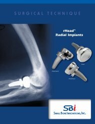
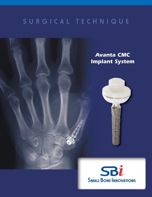
Avanta CMC<br />
Implant System
Avanta CMC Implant System<br />
SURGICAL TECHNIQUE<br />
CONTENTS<br />
Introduction 1<br />
Instructions for Use 1<br />
Surgical Procedure<br />
1. Incision and Exposure 2<br />
2. Soft Tissue Dissection 2<br />
3.TMC Exposure 3<br />
4. Metacarpal Head Resection 3<br />
5. Distal Trial Preparation 4<br />
6. Proximal Trial Preparation 4<br />
7.Trial Insertion 5<br />
8.TMC Implantation 5<br />
9. Final Evaluation of Pros<strong>the</strong>sis Before Closure 6<br />
Postoperative Care 6<br />
Sterilization 7<br />
Potential for Complications 7<br />
Risk/Benefit Decision from <strong>the</strong> Surgeon 8<br />
Clinical Data 8<br />
Adverse Events 8<br />
Indications and Contraindications 9<br />
Warnings, Precautions and<br />
Patient Counseling Information 9
Introduction<br />
The Avanta CMC trapezio-metacarpal joint pros<strong>the</strong>sis consists of an ultra-high<br />
molecular weight polyethylene (UHMWPe) component which is cemented to<br />
<strong>the</strong> prepared trapezium, and a titanium alloy stem component with integral<br />
head which is inserted into <strong>the</strong> snap-fit constrained pros<strong>the</strong>tic replacement<br />
for <strong>the</strong> basal thumb joint. The implant is available in three sizes, each of which<br />
can be used in right or left hands. A range of trial sizers for each type of<br />
implant is available to aid in bone preparation.<br />
Instructions For Use<br />
The following procedure is furnished as an example for informational<br />
purposes only. Each surgeon must evaluate <strong>the</strong> appropriateness of <strong>the</strong><br />
procedure based on <strong>the</strong> current state of <strong>the</strong> art and personal medical training<br />
and experience<br />
The MP joint is examined. If <strong>the</strong>re is a fixed extension deformity, this is<br />
addressed with soft tissue releases of <strong>the</strong> surgeon’s choice. If <strong>the</strong> joint is to be<br />
pinned in flexion, this should be left until <strong>the</strong> end of <strong>the</strong> case.<br />
Accurate alignment and placement of <strong>the</strong> implant require a good exposure of<br />
<strong>the</strong> dorsal and volar trapezial joint surface and <strong>the</strong> base of <strong>the</strong> metacarpal. An<br />
Eaton type incision is used to get a broad exposure of <strong>the</strong> trapezium and <strong>the</strong><br />
metacarpal, including <strong>the</strong> volar joint surface. Eaton’s incision is through <strong>the</strong><br />
origin or insertions of <strong>the</strong> intrinsic and gives excellent exposure for<br />
replacement arthroplasty. The planes are relatively avascular and <strong>the</strong> incision<br />
does not compromise major neurovascular structures.<br />
1
Avanta CMC Implant System Surgical Technique<br />
SURGICAL PROCEDURE<br />
1<br />
Incision and Exposure<br />
The carpometacarpal joint is palpated. A curved incision is<br />
made along <strong>the</strong> volar crease which lies at <strong>the</strong> level of <strong>the</strong> TMC<br />
joint. The incision is carried over to <strong>the</strong> lateral margin and is<br />
extended distally for two cm at <strong>the</strong> edge of <strong>the</strong> intrinsic muscle<br />
insertions. Spreading dissection is carried out to <strong>the</strong> fascia<br />
overlying <strong>the</strong> muscles and tendons. Care is taken to identify<br />
and spare <strong>the</strong> branches of <strong>the</strong> radial sensory nerve and small<br />
vessels on <strong>the</strong> dorsal radial aspect. These should be freed up<br />
and retracted dorsally (FIGURE 1).<br />
FIGURE 1<br />
2<br />
Soft Tissue Dissection<br />
The instrinsics and <strong>the</strong> first compartment tendons are clearly<br />
identified. The volar branch of <strong>the</strong> radial artery is identified,<br />
freed up from <strong>the</strong> palmar fascia and protected. In most hands,<br />
a slip of <strong>the</strong> APL inserts on <strong>the</strong> origin of <strong>the</strong> APB. This is identified,<br />
sectioned and tagged for later repair. The first compartment<br />
is opened from <strong>the</strong> volar side and <strong>the</strong> strands of <strong>the</strong> APL<br />
are inspected. A strand of <strong>the</strong> APL which inserts on <strong>the</strong> base<br />
of <strong>the</strong> metacarpal in <strong>the</strong> bone area which will be resected<br />
should be freed from its insertion and tagged for later repair<br />
(FIGURE 2).<br />
FIGURE 2<br />
2 Avanta CMC Implant System Surgical System
3<br />
TMC Exposure<br />
A 64 Beaver blade is <strong>the</strong>n used to free <strong>the</strong> instristic<br />
from <strong>the</strong>ir insertion on <strong>the</strong> schapoid, trapezium, and<br />
metacarpal base. The dissection should be subperiostial and<br />
<strong>the</strong> muscle flap should be lifted intact without transection of<br />
<strong>the</strong> fibers. Dissection is <strong>the</strong>n carried dorsally subperiostially<br />
under <strong>the</strong> extensor tendons. The gliding tissues under <strong>the</strong> EPB<br />
and EPL should not be violated. Care is taken to free <strong>the</strong> ulnar<br />
edge of <strong>the</strong> TMC joint, remembering <strong>the</strong> course of <strong>the</strong> dorsal<br />
branch of <strong>the</strong> radial artery between <strong>the</strong> first and second rays<br />
(FIGURE 3).<br />
FIGURE 3<br />
4<br />
Metacarpal Head Resection<br />
The capsule of <strong>the</strong> TMC joint is <strong>the</strong>n excised and <strong>the</strong> joint<br />
exposed. Pre-drill with standard size drill or resect metacarpal<br />
head first <strong>the</strong>n open canal (FIGURE 4).<br />
FIGURE 4<br />
3
5<br />
Distal Trial Preparation<br />
Any slips of <strong>the</strong> APL which insert on <strong>the</strong> resected bone should<br />
be dissected free and tagged for later reattachment to <strong>the</strong><br />
metacarpal.<br />
The metacarpal canal can be reamed and prepared for<br />
pros<strong>the</strong>tic insertion ei<strong>the</strong>r at this time or following preparation<br />
of <strong>the</strong> trapezium (FIGURE 5).<br />
FIGURE 5<br />
FIGURE 6<br />
6<br />
Proximal Trial Preparation<br />
The alignment of <strong>the</strong> metacarpal component is parallel to <strong>the</strong><br />
axis of <strong>the</strong> metacarpal shaft with slight volar inclination. The<br />
trapezial joint surface is evaluated. If <strong>the</strong> surface is fairly intact,<br />
blocking volar, ulnar and radial osteophytes are removed and<br />
<strong>the</strong> hole is burred for <strong>the</strong> trapezium component (FIGURE 6).<br />
4 Avanta CMC Implant System Surgical System
FIGURE 7A<br />
7<br />
Trial Insertion<br />
The appropriate trials are inserted and a trial reduction is<br />
performed. The components are evaluated for alignment,<br />
tissue tension and joint stability. If <strong>the</strong>se are satisfactory, <strong>the</strong><br />
size of <strong>the</strong> implant is selected based on <strong>the</strong> trial reduction<br />
(FIGURES 7A and 7B).<br />
FIGURE 7B<br />
FIGURE 8<br />
8<br />
TMC Implantation<br />
The components are <strong>the</strong>n cemented into place with <strong>the</strong> trapezium<br />
first and <strong>the</strong> metacarpal second. Compression<br />
should be maintained until <strong>the</strong> bone cement has completely<br />
set (FIGURE 8).<br />
5
Final Evaluation of Pros<strong>the</strong>sis Before Closure<br />
9<br />
The joint is reduced and alignment and stability are again<br />
FIGURE 9<br />
evaluated. The tourniquet is deflated, hemostasis is secured<br />
and <strong>the</strong> APL is reattached to <strong>the</strong> metacarpal. The instrinsic<br />
origin on <strong>the</strong> trapezium and <strong>the</strong> APL slip is reattached with an<br />
absorbable suture.<br />
The MP joint is pinned if this is <strong>the</strong> desire of <strong>the</strong> surgeon. If <strong>the</strong><br />
MP joint is pinned in flexion, <strong>the</strong> pins should be left under <strong>the</strong><br />
skin to reduce <strong>the</strong> chance o a pin tract infection which could<br />
infect <strong>the</strong> TMC pros<strong>the</strong>sis. Intraoperative films are obtained.<br />
The skin is closed in a routine fashion (FIGURE 9).<br />
Postoperative Care<br />
A postoperative splint is fashioned which fits over <strong>the</strong> dorsum<br />
of <strong>the</strong> TMC and MP joints and <strong>the</strong> volar ulnar surface of <strong>the</strong><br />
palm. The plaster should not extend over <strong>the</strong> eminence, and<br />
free flexion of <strong>the</strong> MP and TMC joints should be possible. The<br />
IP joint should be out of <strong>the</strong> splint entirely. This splint<br />
prevents <strong>the</strong> metacarpal from moving into extension and<br />
dislocating <strong>the</strong> TMC joint. It also prevents <strong>the</strong> metacarpal<br />
from levering on a volar plaster and pushing <strong>the</strong> arthroplasty<br />
out of joint.<br />
The TMC should be splinted in this fashion for three weeks.<br />
Active IP motion is encouraged to minimize adhesions of <strong>the</strong><br />
EPL. Active motion is started at three weeks with a removable<br />
protective resting splint used for ano<strong>the</strong>r month.<br />
X-rays may be obtained intraoperatively, and at two and eight<br />
weeks, six and twelve months postoperatively. These should<br />
be checked for alignment, subsidence, bone resorption or<br />
formation.<br />
Any post operative inflammation should be treated by a<br />
physician. The decision to salvage or remove <strong>the</strong> implant<br />
should be made by <strong>the</strong> surgeon. The salvage procedure<br />
following excision can be fusion or arthroplasty, <strong>the</strong> decision<br />
made by <strong>the</strong> surgeon.<br />
Dislocations should be treated by closed reduction under<br />
anes<strong>the</strong>sia. X-rays should be obtained to evaluate <strong>the</strong><br />
reduction, and if it is correct, <strong>the</strong> TMC joint should be splinted<br />
in flexion with <strong>the</strong> post op splint for three weeks. If a<br />
satisfactory closed reduction cannot be obtained, <strong>the</strong> joint<br />
should be reduced open through <strong>the</strong> original approach and<br />
splinted for three weeks postoperatively. Preoperative<br />
antibiotics should be used with open reductions.<br />
Patient complaints of pain, numbness, stiffness, night and<br />
wea<strong>the</strong>r related pain and passive and active range of motion<br />
of all finger joints should be recorded at each visit.<br />
6 Avanta CMC Implant System Surgical System
Sterilization<br />
1) This component has been sterilized by ethylene oxide or gamma<br />
radiation.<br />
2) The implant is provided sterile in an undamaged package. If ei<strong>the</strong>r <strong>the</strong><br />
implant or package appears damaged, expiration date has been exceeded,<br />
or if sterility is questioned for any reason, <strong>the</strong> implant should not be used.<br />
Do not resterilize.<br />
3) Trial sizer components are available to avoid having to open <strong>the</strong> sterile<br />
package prior to pros<strong>the</strong>sis implantation. The implant should be removed<br />
from its sterile package only after <strong>the</strong> implant site has been prepared and<br />
properly sized.<br />
Potential for Complications<br />
As with <strong>the</strong> use of all implant devices, <strong>the</strong> potential for intraoperative and<br />
postoperative complications is possible. It is <strong>the</strong> responsibility of <strong>the</strong> surgeon<br />
using <strong>the</strong> implant(s) to consider <strong>the</strong> clinical and medical status of each<br />
patient to be knowledgeable about all aspects of <strong>the</strong> implant(s) surgical<br />
procedure and all potential complications associated with each specific case.<br />
The implants have been designed to offer <strong>the</strong> highest strength possible for<br />
each size and configuration, however due to anatomical size constraints it is<br />
possible that high demand patients will be able to overload <strong>the</strong>ir implant(s).<br />
To insure <strong>the</strong> best possible function and longevity of <strong>the</strong> implant(s), proper<br />
implant selection and sizing on <strong>the</strong> part of <strong>the</strong> surgeon is critical.<br />
Joint implants utilize mechanical attachments and articulating bearing<br />
surfaces. These interfaces may see micro and/or macro motion between<br />
parts, as well as, between <strong>the</strong> patients anatomy with normal use. The motion<br />
is known to cause wearing of <strong>the</strong> parts, which in turn, over time, may lead to<br />
failure of <strong>the</strong> device or of <strong>the</strong> device/patient interface. Symptoms of failure,<br />
or impending failure, may include: pain, swelling, inflammation, tenderness<br />
and infection. Strenuous implant loading, excessive mobility, <strong>the</strong> presence of<br />
articular instability, improper sizing, improper patient selection and misuse all<br />
may lead to accelerated wear and early failure of <strong>the</strong> device. Patients should<br />
be made aware of <strong>the</strong>se limitations and <strong>the</strong> potential for complications<br />
arising from <strong>the</strong>m.<br />
7
Risk/Benefit Decision by <strong>the</strong> Surgeon<br />
The judgement by a surgeon to use an implant is a risk/benefit decision which<br />
must take into account <strong>the</strong> patient’s needs, desires and expectations in<br />
addition to <strong>the</strong> surgeon’s knowledge of expected results and complications as<br />
well <strong>the</strong> alternative treatments. Therefore, surgeons must balance many<br />
considerations to achieve <strong>the</strong> best result in individual patients. Providing<br />
each patient scheduled for implant surgery with documented counseling of<br />
potential complications and alternatives, which may include non-implant<br />
procedure such as soft tissue reconstruction or arthrodesis, prior to surgery is<br />
necessary.<br />
Clinical Data<br />
Results of twenty-nine patients implanted with <strong>the</strong> device were reported. 2<br />
Replacement was performed for arthritis, failure of attempted arthrodesis,<br />
previous Silastic or total joint arthroplasty failure, or postparalytic fibrous<br />
ankylosis of <strong>the</strong> joint. Twenty-two patients had achieved a good range of<br />
painless motion at <strong>the</strong> time of publication (up to 7 years follow-up). Seven<br />
cases failed to achieve normal ROM due to significant muscle imbalance or<br />
soft tissue scarring and contracture. There were no reported cases of implant<br />
fracture or infection, but three cases of demonstrated loosening at <strong>the</strong><br />
cement-bone interface.<br />
In subsequent expanded publication including an additional 21 cases in <strong>the</strong><br />
series 1 ,most of <strong>the</strong> 50 patients showed a full range of motion within four<br />
weeks of suture removal and continued to demonstrate good long-term<br />
results. Full range of asymptomatic motion was achieved in 26 osteoarthritis<br />
patients with articular derangement. Five patients showed clinical or<br />
radiographic evidence of loosening. No cases of implant fracture, surface<br />
wear, fragmentation, or infection were seen.<br />
Adverse Events<br />
Adverse events in a published series of 50 patients included<br />
loosening, transient radial neuritis, cement extrusion injury to<br />
<strong>the</strong> palmar digital nerves and de Quervain’s tendinitis 1 .<br />
Potential adverse events reported with o<strong>the</strong>r finger joint<br />
pros<strong>the</strong>ses include loosening, fracture, discoloration, or<br />
infection of <strong>the</strong> implant.<br />
Injury to <strong>the</strong> surrounding nerve, blood vessels tendons, or soft<br />
tissues can occur as a consequence of implanting this device.<br />
EVENT<br />
NUMBER OF EVENTS (n=50)<br />
Loosening 5 (10.0%)<br />
Transient Radial Neuritis 2 (4.0%)<br />
Cement Extrusion Injury 1 (2.0%)<br />
De Quervain’s Tendinitis 6 (12.0%)<br />
Metal sensitivity reactions have been reported following joint<br />
replacement.<br />
8 Avanta CMC Implant System Surgical System
INDICATIONS<br />
> The Avanta CMC trapezio-metacarpal pros<strong>the</strong>sis is<br />
indicated for total joint replacement in skeletally<br />
mature patients with pain or instability of <strong>the</strong><br />
trapezio-metacarpal joint due to trauma,<br />
inflammatory, or degenerative disease or revision of<br />
previous procedures, as an alternative to arthrodesis<br />
or reconstructive surgery.<br />
CONTRAINDICATIONS<br />
> <strong>Bone</strong>, musculature, tendons, or adjacent soft tissue<br />
comprised by disease, infection, or prior implantation<br />
which cannot provide adequate support or fixation for<br />
<strong>the</strong> pros<strong>the</strong>sis.<br />
> Any active or suspected infection in or around <strong>the</strong> thumb<br />
joint.<br />
> Skeletal immaturity.<br />
WARNINGS AND PRECAUTIONS<br />
Patient Counseling Information (see also Warnings)<br />
In addition to <strong>the</strong> patient related information contained in <strong>the</strong> Warnings and Adverse Effects<br />
sections, <strong>the</strong> following information should be conveyed to <strong>the</strong> patient:<br />
> While <strong>the</strong> expected life of total joint replacement components is difficult to estimate, it is<br />
finite. These components are made of foreign materials which are placed within <strong>the</strong> body<br />
for <strong>the</strong> potential restoration of mobility or reduction of pain. However, due to <strong>the</strong> many biological,<br />
mechanical, and physicochemical factors which affect <strong>the</strong>se devices, <strong>the</strong> components<br />
cannot be expected to withstand <strong>the</strong> activity level and loads of normal healthy bone<br />
for an unlimited period of time. The reported rate of loosening in a clinical study of this<br />
device was 10% at up to 10 years follow-up (Braun RM Total joint arthroplasty at <strong>the</strong> carpometacarpal<br />
joint of <strong>the</strong> thumb, Clin Orthop 1985;195;161-7.)<br />
> Adverse affects may necessitate reoperation, revision, or fusion of <strong>the</strong> involved joint.<br />
Precautions<br />
1) The implant is provided sterile in an undamaged package. If ei<strong>the</strong>r <strong>the</strong> implant or <strong>the</strong><br />
package appears damaged, expiration date has been exceeded, or if sterility is questioned<br />
for any reason, <strong>the</strong> implant should not be used. Do not resterilize.<br />
2) Meticulous preparation of <strong>the</strong> implant site and selection of <strong>the</strong> proper size implant increase<br />
<strong>the</strong> potential for a successful outcome.<br />
3) The implant should be removed from its sterile package only after <strong>the</strong> implants site has<br />
been prepared and properly sized.<br />
4) Implants should only be handled with blunt instruments to avoid scratching, cutting, or<br />
nicking <strong>the</strong> device.<br />
Warnings (see Patient Counseling Information Section)<br />
1) This device is for cemented use only.<br />
2) Strenuous loading, excessive mobility, and articular instability all may lead to accelerated<br />
wear and eventual failure by loosening, fracture, or dislocation of <strong>the</strong> device. Patients<br />
should be made aware of <strong>the</strong> increased potential for device failure if excessive demands are<br />
made upon it.<br />
CAUTION<br />
Federal (United States) law restricts this device to sale, distribution and use by or on <strong>the</strong> order of a physician.<br />
9
<strong>Small</strong> <strong>Bone</strong> <strong>Innovations</strong>, LLC<br />
1711 South Pennsylvania Ave.<br />
Morrisville, PA 19067<br />
(800) 778-8837<br />
Tel (215) 428-1791<br />
Fax (215) 428-1795<br />
www.totalsmallbone.com<br />
MKT-10110 Rev. a<br />
Copyright 2005, <strong>Small</strong> <strong>Bone</strong> <strong>Innovations</strong>, LLC