Dynamics of glutamatergic signaling in the mushroom ... - HAL - ESPCI
Dynamics of glutamatergic signaling in the mushroom ... - HAL - ESPCI
Dynamics of glutamatergic signaling in the mushroom ... - HAL - ESPCI

You also want an ePaper? Increase the reach of your titles
YUMPU automatically turns print PDFs into web optimized ePapers that Google loves.
S<strong>in</strong>akevitch et al. Neural Development 2010, 5:10<br />
http://www.neuraldevelopment.com/content/5/1/10<br />
Page 4 <strong>of</strong> 20<br />
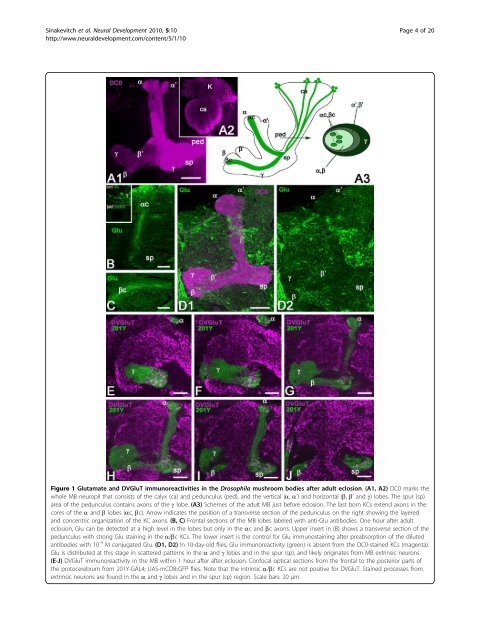
Figure 1 Glutamate and DVGluT immunoreactivities <strong>in</strong> <strong>the</strong> Drosophila <strong>mushroom</strong> bodies after adult eclosion. (A1, A2) DC0 marks <strong>the</strong><br />
whole MB neuropil that consists <strong>of</strong> <strong>the</strong> calyx (ca) and pedunculus (ped), and <strong>the</strong> vertical (a, a’) and horizontal (b, b’ and g) lobes. The spur (sp)<br />
area <strong>of</strong> <strong>the</strong> pedunculus conta<strong>in</strong>s axons <strong>of</strong> <strong>the</strong> g lobe. (A3) Schemes <strong>of</strong> <strong>the</strong> adult MB just before eclosion. The last born KCs extend axons <strong>in</strong> <strong>the</strong><br />
cores <strong>of</strong> <strong>the</strong> a and b lobes (ac, bc). Arrow <strong>in</strong>dicates <strong>the</strong> position <strong>of</strong> a transverse section <strong>of</strong> <strong>the</strong> pedunculus on <strong>the</strong> right show<strong>in</strong>g <strong>the</strong> layered<br />
and concentric organization <strong>of</strong> <strong>the</strong> KC axons. (B, C) Frontal sections <strong>of</strong> <strong>the</strong> MB lobes labeled with anti-Glu antibodies. One hour after adult<br />
eclosion, Glu can be detected at a high level <strong>in</strong> <strong>the</strong> lobes but only <strong>in</strong> <strong>the</strong> ac and bc axons. Upper <strong>in</strong>sert <strong>in</strong> (B) shows a transverse section <strong>of</strong> <strong>the</strong><br />
pedunculus with strong Glu sta<strong>in</strong><strong>in</strong>g <strong>in</strong> <strong>the</strong> a/bc KCs. The lower <strong>in</strong>sert is <strong>the</strong> control for Glu immunosta<strong>in</strong><strong>in</strong>g after preabsorption <strong>of</strong> <strong>the</strong> diluted<br />
antibodies with 10 -4 M conjugated Glu. (D1, D2) In 10-day-old flies, Glu immunoreactivity (green) is absent from <strong>the</strong> DC0-sta<strong>in</strong>ed KCs (magenta).<br />
Glu is distributed at this stage <strong>in</strong> scattered patterns <strong>in</strong> <strong>the</strong> a and g lobes and <strong>in</strong> <strong>the</strong> spur (sp), and likely orig<strong>in</strong>ates from MB extr<strong>in</strong>sic neurons.<br />
(E-J) DVGluT immunoreactivity <strong>in</strong> <strong>the</strong> MB with<strong>in</strong> 1 hour after after eclosion. Confocal optical sections from <strong>the</strong> frontal to <strong>the</strong> posterior parts <strong>of</strong><br />
<strong>the</strong> protocerebrum from 201Y-GAL4; UAS-mCD8::GFP flies. Note that <strong>the</strong> <strong>in</strong>tr<strong>in</strong>sic a/bc KCs are not positive for DVGluT. Sta<strong>in</strong>ed processes from<br />
extr<strong>in</strong>sic neurons are found <strong>in</strong> <strong>the</strong> a and g lobes and <strong>in</strong> <strong>the</strong> spur (sp) region. Scale bars: 20 μm.