Clinical Experience with Oasis® Wound Matrix for the Treatment of ...
Clinical Experience with Oasis® Wound Matrix for the Treatment of ...
Clinical Experience with Oasis® Wound Matrix for the Treatment of ...

Create successful ePaper yourself
Turn your PDF publications into a flip-book with our unique Google optimized e-Paper software.
This is <strong>the</strong> seventh <strong>of</strong> nine publications from Healthpoint intended to facilitate expeditious, cost-effective wound care management.<br />
<strong>Clinical</strong> <strong>Experience</strong> <strong>with</strong> Oasis ®<br />
<strong>Wound</strong> <strong>Matrix</strong> <strong>for</strong> <strong>the</strong> <strong>Treatment</strong> <strong>of</strong><br />
Venous and Diabetic Ulcers:<br />
A Series <strong>of</strong> Four Cases<br />
Diabetic and venous ulcers are prevalent, costly, and can be frustrating<br />
to treat. These wounds are typically slow to heal and may develop into<br />
chronic wounds that are nonresponsive to <strong>the</strong>rapy. Although standard<br />
care is effective <strong>for</strong> some <strong>of</strong> <strong>the</strong>se wounds, a substantial portion fail to<br />
heal despite 3 or 4 months <strong>of</strong> diligent care. 1 Evidence from a number <strong>of</strong> studies<br />
shows that wounds that do not progress adequately toward healing <strong>with</strong>in <strong>the</strong><br />
first 2 to 4 weeks <strong>of</strong> standard care are unlikely to heal given 3 or 4 months <strong>of</strong><br />
care. 2,3 These findings suggest that difficult-to-heal wounds should be re-evaluated<br />
after 4 weeks; if <strong>the</strong>y have not progressed adequately toward closure, more aggressive treatment<br />
strategies should be initiated. 2<br />
Chronic wounds may display many abnormalities in <strong>the</strong> extracellular matrix 4 that develops in difficult-to-heal<br />
wounds. This suggests that strategies designed to address extracellular matrix deficits<br />
may be beneficial <strong>for</strong> <strong>the</strong>se wounds. The naturally occurring extracellular matrix consists <strong>of</strong> structural<br />
and functional molecules that coordinate <strong>the</strong> healing process and provide a scaffold to support<br />
cellular proliferation. 5 With inadequate or absent extracellular matrix, healing is impaired.<br />
Oasis ® <strong>Wound</strong> <strong>Matrix</strong> is a cell-free, naturally derived extracellular matrix that retains its natural threedimensional<br />
structure. 6 Because Oasis ® contains some key components <strong>of</strong> <strong>the</strong> dermal extracellular<br />
matrix such as collagen, elastin, glycosaminoglycans, glycoproteins, and proteoglycans, it acts as<br />
an extracellular matrix replacement. 6 Oasis ® is stored at room temperature and has a shelf life <strong>of</strong> up<br />
to 2 years. 7 It has been found to significantly improve wound management 8 and is resorbed by <strong>the</strong><br />
body over <strong>the</strong> course <strong>of</strong> each weekly application as it facilitates wound closure. Given its practical<br />
features and efficacy in clinical trials, Oasis ® may be a logical next step in <strong>the</strong> management <strong>of</strong> difficult-to-heal<br />
wounds that are not progressing adequately toward closure. The four cases <strong>of</strong> difficultto-heal<br />
wounds presented in this publication were treated <strong>with</strong> Oasis ® .<br />
83 General Warren Boulevard • Suite 100<br />
Malvern, PA 19355<br />
Phone (800) 237-7285 Fax (610) 560-0502<br />
www.hmpcommunications.com<br />
This publication is provided by Healthpoint, Inc., as a continuous pr<strong>of</strong>essional service. For additional reprints or<br />
in<strong>for</strong>mation on Healthpoint products, contact your local Healthpoint representative or call (800) 441-8227.
Case 1<br />
A 45-year-old man <strong>with</strong> venous insufficiency, lupus, anemia, and a history <strong>of</strong> deep vein thrombosis (DVT) presented <strong>with</strong> an<br />
ulcer on <strong>the</strong> medial portion <strong>of</strong> his right ankle (see Figure 1a) and bilateral edema <strong>of</strong> <strong>the</strong> lower extremities. The ulcer had been<br />
present <strong>for</strong> 3 months; it exhibited fibrin <strong>with</strong>in <strong>the</strong> wound bed and periwound inflammation. The ulcer, measuring 1.0 cm x 1.5<br />
cm, was associated <strong>with</strong> pain rated by <strong>the</strong> patient as 4 to 6 on a 10-point scale. Initially, <strong>the</strong> wound was treated <strong>with</strong> papainurea-chlorophyllin<br />
copper complex (PUCCC) and a cadexomer iodine-based wound product. After 9 days, granulation tissue<br />
was present and PUCCC was discontinued. At that time, treatment <strong>with</strong> Oasis ® was initiated (see Figure 1b). Oasis ® was<br />
applied once weekly and covered <strong>with</strong> a non-adherent, open-mesh, fabric dressing and a cadexomer iodine-based dressing.<br />
The Oasis ® was changed once weekly <strong>for</strong> 7 weeks and <strong>the</strong> secondary dressings were changed three times per week.<br />
Visco/Cotton/Coban wraps Coban (3M Health Care) were used <strong>for</strong> compression and fibrin was excisionally debrided as needed.<br />
Ulcer size and pain were evaluated at 1-week intervals during follow-up.<br />
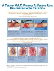
Across all follow-up visits, <strong>the</strong> patient’s rating <strong>of</strong> pain associated <strong>with</strong> <strong>the</strong> ulcer ranged from 0 to 2 on a 10-point scale. After 4<br />
weeks, ulcer size decreased to 0.6 cm x 0.9 cm, representing 64% closure (see Figure 1c). Endovenous laser treatment was<br />
per<strong>for</strong>med at week 5 and after 6 weeks ulcer size decreased to 0.4 cm x 0.5 cm (87% closure). By week 7, ulcer area<br />
decreased to 0.2 cm x 0.2 cm, <strong>the</strong> wound base had become superficial, and <strong>the</strong> wound was almost completely (97%) closed;<br />
thus, Oasis® treatment was discontinued (see Figure 1d). No adverse events were reported during Oasis ® treatment.<br />
Figure 1. Case 1: A 45-year-old man <strong>with</strong> a venous leg ulcer<br />
A B C D<br />
Figure 1A. At presentation Figure 1B. Oasis ® initiated Figure 1C. After 4 weeks <strong>of</strong> Oasis ® Figure 1D. Complete closure after 7<br />
weeks <strong>of</strong> Oasis ® treatment.<br />
Case 2<br />
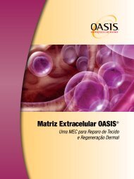
Figure 2. Case 2: An 86-year-old woman <strong>with</strong> a venous leg ulcer<br />
An 86-year-old woman <strong>with</strong> venous insufficiency, arterial insufficiency,<br />
myelodysplastic anemia, hypothyroidism, and a history<br />
<strong>of</strong> DVT presented <strong>with</strong> an ulcer on her right lateral ankle (see<br />
Figure 2a) and bilateral edema <strong>of</strong> <strong>the</strong> lower extremities. The<br />
ulcer had been present <strong>for</strong> 17 weeks, characterized by eschar,<br />
and fully granulated <strong>with</strong> a superficial wound base. At presentation,<br />
<strong>the</strong> ulcer measured 1.2 cm x 1.1 cm proximal and 0.4 cm A<br />
B<br />
x 0.3 cm distal and <strong>the</strong> patient rated pain associated <strong>with</strong> <strong>the</strong><br />
ulcer as 4 on a 10-point scale. The patient had been managed<br />
Figure 2A. At presentation Figure 2B. After 1 week <strong>of</strong> Oasis ®<br />
sequentially <strong>for</strong> a total <strong>of</strong> 13<br />
weeks <strong>with</strong> various treatments,<br />
including silver sulfadiazine <strong>for</strong> 3<br />
weeks, silver sulfadiazine/PUCCC<br />
<strong>for</strong> 6 weeks, PUCCC/gentamicin<br />
<strong>for</strong> 2 weeks, and a topical silver<br />
dressing <strong>for</strong> 2 weeks. At this C<br />
E<br />
D<br />
point, <strong>the</strong> silver dressing was<br />
Figure 2C. After 2 weeks <strong>of</strong> Oasis ® Figure 2D. After 3 weeks <strong>of</strong> Oasis ® Figure 2E. Completely epi<strong>the</strong>lized at 4<br />
weeks after presentation.
Case 2 continued<br />
discontinued and treatment <strong>with</strong> Oasis ® initiated. Oasis ® was applied once weekly and covered <strong>with</strong> a non-adherent<br />
and cadexamer iodine dressing that was changed two times per week. Visco/Cotton/Coban (3M Health Care)<br />
wraps were used <strong>for</strong> compression and <strong>the</strong> wound was excisionally debrided as needed. Pain and ulcer area were<br />
evaluated at 1-week intervals during follow-up.<br />
Across all follow-up visits, <strong>the</strong> patient’s rating <strong>of</strong> pain associated <strong>with</strong> <strong>the</strong> ulcer ranged from 0 to 1 on a 10-point<br />
scale. After 1 week, <strong>the</strong> ulcer size was 1.8 cm x 1.1 cm (see Figure 2b), and after 2 weeks, <strong>the</strong> ulcer size had<br />
decreased to 1.5 cm x 1.1 cm (see Figure 2c). Following 3 weeks <strong>of</strong> Oasis ® application, <strong>the</strong> size <strong>of</strong> <strong>the</strong> ulcer<br />
decreased to 0.2 cm x 0.2 cm and Oasis ® treatment was discontinued (see Figure 2d). By 4 weeks, <strong>the</strong> wound was<br />
completely epi<strong>the</strong>lized (see Figure 2e). No adverse events were reported during Oasis ® treatment.<br />
Case 3<br />
A 75-year-old woman <strong>with</strong> peripheral neuropathy and a history <strong>of</strong> cancer<br />
treated <strong>with</strong> surgery plus radiation <strong>the</strong>rapy presented <strong>with</strong> a diabetic<br />
ulcer on her left plantar foot (see Figure 3a). The ulcer had been<br />
present <strong>for</strong> 15 weeks, exhibited periwound callus, and measured 1.3<br />
cm x 0.8 cm at baseline. No pain was associated <strong>with</strong> <strong>the</strong> ulcer. The<br />
patient had initiated dry dressing <strong>for</strong> 2 weeks and had been treated<br />
<strong>with</strong> Silvadene (Medline) <strong>for</strong> 3 weeks. At this point, previous treatment<br />
was halted and treatment <strong>with</strong> Oasis ® initiated. Oasis ® was applied<br />
once weekly and covered <strong>with</strong> a non-adherent dressing. In addition, a<br />
cover dressing was applied. The patient was <strong>of</strong>floaded in a DH shoe<br />
(Royce Medical) and <strong>the</strong> wound periodically debrided excisionally to<br />
remove fibrin and periwound callus as needed. Pain and ulcer size<br />
were evaluated at 1-week intervals during follow-up.<br />
Figure 3. Case 3: A 75-year-old woman <strong>with</strong> a diabetic<br />
foot ulcer<br />
A<br />
Figure 3A. At presentation<br />
B<br />
Figure 3B. After 3 weeks<br />
<strong>of</strong> Oasis ®<br />
No pain was associated <strong>with</strong> <strong>the</strong> ulcer during<br />
follow-up. After 1 week <strong>of</strong> Oasis ® treatment,<br />
ulcer size decreased to 0.7 cm x 0.3 cm<br />
(80% closure). After 2 weeks, <strong>the</strong> size was 0.9<br />
cm x 0.3 x 0.2 cm (74% closure) and following<br />
3 weeks, <strong>the</strong> size was 0.8 cm x 0.3 cm<br />
(77% closure; see Figure 3b). The ulcer size<br />
continued to decrease over <strong>the</strong> next few<br />
weeks and after 8 weeks, <strong>the</strong> wound measured<br />
0.5 cm x 0.1 cm and Oasis ® treatment<br />
was discontinued (see Figures 3c, 3d). The<br />
wound was fully closed and healed when evaluated<br />
at 1 week post- Oasis ® discontinuation<br />
(see Figure 3e). No adverse events were reported<br />
during Oasis ® treatment.<br />
C D E<br />
Figure 3C. After 5 weeks<br />
<strong>of</strong> Oasis ®<br />
Figure 3D. After 8 weeks<br />
<strong>of</strong> Oasis ®<br />
Figure 3E. Ulcer closed.
Case 4<br />
A 71-year-old woman <strong>with</strong> coronary artery disease, diabetes mellitus, hypertension, hypercholesterolemia, and a<br />
history <strong>of</strong> cerebrovascular accident and transient ischemic attacks presented <strong>with</strong> a right plantar diabetic foot ulcer<br />
on <strong>the</strong> first metatarsal phalangeal joint (see Figure 4a). The ulcer had been present <strong>for</strong> 9 weeks and measured 1.6<br />
cm x 1.0 cm. The patient rated <strong>the</strong> pain associated <strong>with</strong> <strong>the</strong> ulcer as a 2 on a 10-point scale.<br />
The ulcer had shown signs <strong>of</strong> infection (ery<strong>the</strong>ma, drainage, and periwound warmth) 1 week earlier and <strong>the</strong> patient<br />
had been placed on lev<strong>of</strong>loxacin (Levaquin ® , Ortho-McNeil), 500 mg, <strong>for</strong> 7 days. At that time, treatment <strong>with</strong><br />
PUCCC and silver sulfadiazine was initiated. This treatment <strong>with</strong> PUCCC and silver sulfadiazine was subsequently<br />
halted and treatment <strong>with</strong> Oasis ® initiated. Oasis ® was applied once weekly, covered <strong>with</strong> a non-adherent dressing<br />
followed by a cover dressing. The patient was <strong>of</strong>floaded in a DH shoe (Royce Medical) and <strong>the</strong> periwound callus<br />
debrided as necessary.<br />
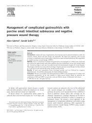
Ulcer size and pain were evaluated at 1-week intervals during follow-up. No pain was associated <strong>with</strong> <strong>the</strong> ulcer<br />
during follow-up. After 1 week, <strong>the</strong> ulcer size decreased to 1.0 cm x 0.8 cm (38% closure; see Figure 4b). The<br />
wound continued to decrease in size over <strong>the</strong> subsequent weeks (45% closure at 2 weeks; 88% closure at 4<br />
weeks). After 6 weeks, <strong>the</strong> size had decreased to 0.3 cm x 0.1 cm (98% closure) and Oasis ® treatment was discontinued<br />
see (Figure 4c). The wound was fully closed when evaluated 1 week after stopping Oasis® treatment.<br />
No adverse events were reported during Oasis ® treatment.<br />
Figure 4. Case 4: A 71-year-old woman <strong>with</strong> a diabetic foot ulcer<br />
A B C<br />
Figure 4A. At presentation Figure 4B. After 3 weeks <strong>of</strong> Oasis ® Figure 4C. Almost complete closure<br />
after 6 weeks <strong>of</strong> Oasis ® .<br />
For CE/CME accredited courses available 24/7,<br />
including more in<strong>for</strong>mation on extracellular matrix, check out<br />
<strong>the</strong> new course, The Biology <strong>of</strong> <strong>the</strong> Chronic <strong>Wound</strong>,<br />
now available at www.The<strong>Wound</strong>Institute.com ® .<br />
The<strong>Wound</strong>Institute.com ® , your source <strong>for</strong> interactive wound care education.
Discussion<br />
In all four cases, <strong>the</strong> venous or diabetic ulcers had persisted<br />
<strong>for</strong> at least 2 months be<strong>for</strong>e Oasis ® treatment and were<br />
chronic. In all cases, treatment <strong>with</strong> Oasis ® , combined <strong>with</strong><br />
standard care, resulted in wound closure <strong>with</strong>in 4 to 9<br />
weeks. <strong>Treatment</strong> <strong>with</strong> Oasis ® was discontinued approximately<br />
1 week be<strong>for</strong>e complete wound closure in each<br />
case. The two patients <strong>with</strong> venous ulcers and one <strong>of</strong> <strong>the</strong><br />
patients <strong>with</strong> a diabetic ulcer complained <strong>of</strong> pain be<strong>for</strong>e<br />
treatment <strong>with</strong> Oasis ® ; in all three cases, pain was reduced<br />
after initiating Oasis ® treatment. Fur<strong>the</strong>rmore, Oasis ® was<br />
well tolerated and no adverse events occurred in any <strong>of</strong> <strong>the</strong><br />
four patients.<br />
Deficits in <strong>the</strong> extracellular matrix may contribute to a failure<br />
<strong>of</strong> healing in patients <strong>with</strong> diabetes and/or venous<br />
insufficiency. Diabetes is associated <strong>with</strong> an increased glycosylation<br />
<strong>of</strong> collagen and fibronectin, which may interfere<br />
<strong>with</strong> attachment and migration <strong>of</strong> epi<strong>the</strong>lial cells. 9 Venous<br />
ulcers are characterized by fibrin cuffs (organized structures<br />
<strong>of</strong> extracellular matrix proteins around wound edges and<br />
trapped leukocytes and fibrin) as well as a lack <strong>of</strong><br />
fibronectin in <strong>the</strong> ulcer tissue (a glycoprotein that mediates<br />
cell attachment, proliferation, and migration). 10 Thus,<br />
attempting to replace extracellular matrix in <strong>the</strong>se conditions<br />
is a logical treatment strategy and one that proved<br />
successful in this series <strong>of</strong> patients.<br />
References<br />
1. Margolis DJ, Allen-Taylor L, H<strong>of</strong>fstad O, Berlin JA. Healing diabetic neuropathic<br />
foot ulcers: Are we getting better? Diabet Med.<br />
2005;22(2):172–176.<br />
2. Sheehan P, Jones P, Caselli A, Giurini JM, Veves A. Percent change in<br />
wound area <strong>of</strong> diabetic foot ulcers over a 4-week period is a robust predictor<br />
<strong>of</strong> complete healing in a 12-week prospective trial. Diabetes Care.<br />
2003;26(6):1879–1882.<br />
3. van Rijswijk L, Polansky M. Predictors <strong>of</strong> time to healing deep pressure<br />
ulcers. Ostomy <strong>Wound</strong> Manage. 1994;40(8):40–42,44,46–48 passim.<br />
4. Loots MAM, Lamme EN, Zeegelaar J, Mekkes JR, Box JD, Middlekoop<br />
E. Differences in cellular infiltrate and extracellular matrix <strong>of</strong> chronic diabetic<br />
venous ulcers versus acute wounds. J Invest<br />
Dermatol.1998;111(5):850–857.<br />
5. MacNeil S. What role does <strong>the</strong> extracellular matrix serve in skin grafting<br />
and wound healing? Burns. 1994;20(suppl 1):S607–S670.<br />
6. Brown-Etris M, Cutshall W, Hiles MC. A new biomaterial derived from<br />
small intestine submucosa and developed into a wound matrix device.<br />
WOUNDS. 2002;14(4):150–166.<br />
7. Hodde JP, Ernst DMJ, Hiles MC. Bioactivity <strong>of</strong> FGF-2 in OASIS wound<br />
matrix after prolonged storage. J <strong>Wound</strong> Care. 2005;14(1):23–25.<br />
8. Mostow EN, Haraway GD, Dalsing M, Hodde JP, King D. OASIS Venus<br />
Ulcer Study Group. Effectiveness <strong>of</strong> an extracellular matrix graft (OASIS<br />
<strong>Wound</strong> <strong>Matrix</strong>) in <strong>the</strong> treatment <strong>of</strong> chronic leg ulcers: a randomized clinical<br />
trial. J Vasc Surg. 2005;41(5):837–843.<br />
9. McDermott AM, Xiao TL, Kern TS, Murphy CJ. Non-enzymatic glycation<br />
in corneas from normal and diabetic donors and its effects on epi<strong>the</strong>lial<br />
cell attachment in vitro. Optometry. 2003;74(7):443–452.<br />
10. Herrick SE, Sloan P, McGurk M, Freak L, McCollum CN, Ferguson MW.<br />
Sequential changes in histologic pattern and extracellular matrix deposition<br />
during <strong>the</strong> healing <strong>of</strong> chronic venous ulcers. Am J Pathol.<br />
1992;141(5):1085–1095.<br />
11. Niezgoda JA, Van Gils CC, Frykberg RG, Hodde JP. Randomized clinical<br />
trial comparing OASIS <strong>Wound</strong> <strong>Matrix</strong> to Regranex Gel <strong>for</strong> diabetic ulcers.<br />
Adv Skin <strong>Wound</strong> Care. 2005;18(5Pt.1):258–266.<br />
One advantage <strong>of</strong> Oasis ® , in addition to its documented<br />
benefits in <strong>the</strong> treatment <strong>of</strong> venous and diabetic ulcers, 8,11 is<br />
its practicality. For instance, its 2-year shelf life and room<br />
temperature storage requirement facilitate routine use.<br />
Additionally, <strong>the</strong> cost <strong>of</strong> Oasis ® is reasonable based on several<br />
randomized trials. In <strong>the</strong>se studies, <strong>the</strong> average cost <strong>of</strong><br />
12 weeks <strong>of</strong> Oasis ® was $320 to treat venous leg ulcers<br />
and $250 to treat diabetic foot ulcers. 8,11 These clinical and<br />
practical features may make Oasis ® a logical next step in<br />
treatment <strong>for</strong> patients <strong>with</strong> non-responsive venous or diabetic<br />
ulcers.<br />
Conclusion<br />
The four cases described demonstrate <strong>the</strong> safe and effective<br />
use <strong>of</strong> Oasis ® <strong>for</strong> venous and diabetic ulcers that are at<br />
risk <strong>of</strong> becoming chronic wounds. Because venous and<br />
diabetic ulcers may have a defective extracellular matrix<br />
that contributes to delayed healing, replacing <strong>the</strong> extracellular<br />
matrix is a logical next step in <strong>the</strong>rapy when adequate<br />
progress is not made <strong>with</strong> standard care.
When wounds fail to progress after<br />
2–4 weeks <strong>with</strong> your standard <strong>of</strong> care…<br />
OASIS ® enables <strong>the</strong> body to<br />
get things moving again.<br />
Simple application, proven results 1<br />
Significantly improves wound management 1<br />
Supports <strong>the</strong> body’s natural wound response by<br />
replacing <strong>the</strong> missing extracellular matrix (ECM) 2<br />
An easy addition to your standard wound care<br />
In <strong>the</strong> <strong>of</strong>fice, <strong>of</strong>f <strong>the</strong> shelf <strong>for</strong> once-weekly application<br />
In a recent clinical study, <strong>the</strong> average cost <strong>of</strong> OASIS ®<br />
<strong>for</strong> 12 weeks in venous stasis ulcers was $320 1<br />
Has relevant HCPCS (J) and CPT ® codes<br />
INTRODUCE<br />
Sheet <strong>of</strong> OASIS ® <strong>Wound</strong> <strong>Matrix</strong><br />
Get things moving again<br />
1-800-441-8227<br />
www.healthpoint.com<br />
References: 1. Data on file. Healthpoint, Ltd, Fort Worth, TX 76107.<br />
2. Brown-Etris M, Cutshall WD, Hiles MC. A new biomaterial derived<br />
from small intestine submucosa and developed into a wound matrix device.<br />
<strong>Wound</strong>s. 2002;14:150–166.<br />
OASIS is a registered trademark <strong>of</strong> Cook Biotech, Inc.<br />
CPT is a registered trademark <strong>of</strong> <strong>the</strong> American Medical Association.<br />
© Copyright 2006, Healthpoint, Ltd. Printed in USA TM0684-0506<br />
137387-11/06