L - Technische Universität Braunschweig
L - Technische Universität Braunschweig
L - Technische Universität Braunschweig

Create successful ePaper yourself
Turn your PDF publications into a flip-book with our unique Google optimized e-Paper software.
2013<br />
Dr. Dina Grohmann<br />
Junior Research Group Leader / Akademischer Rat a.Z.<br />
Institute of Physical and Theoretical Chemistry<br />
- NanoBioSciences -<br />
<strong>Technische</strong> <strong>Universität</strong> <strong>Braunschweig</strong><br />
Hans-Sommer-Straße 10<br />
38106 <strong>Braunschweig</strong><br />
Tel: +49 (0)531 391 5395<br />
Fax: +49 (0)531 391 5334<br />
d.grohmann@tu-bs.de<br />
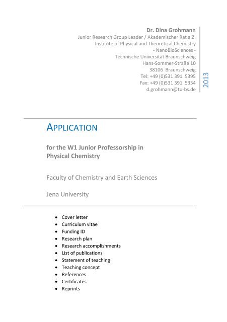
APPLICATION<br />
for the W1 Junior Professorship in<br />
Physical Chemistry<br />
Faculty of Chemistry and Earth Sciences<br />
Jena University<br />
Cover letter<br />
Curriculum vitae<br />
Funding ID<br />
Research plan<br />
Research accomplishments<br />
List of publications<br />
Statement of teaching<br />
Teaching concept<br />
References<br />
Certificates<br />
Reprints
2 APPLICATION DR. DINA GROHMANN<br />
Physikalische und Theoretische Chemie -NanoBioSciences<br />
Dr. Dina Grohmann<br />
An Herrn Prof. Dr. J. Popp<br />
Institut für Physikalische Chemie<br />
Helmholtzweg 4<br />
D-07743 Jena<br />
<strong>Technische</strong> <strong>Universität</strong> <strong>Braunschweig</strong><br />
Institut für Physikalische<br />
und Theoretische Chemie<br />
Abt. NanoBioSciences<br />
Hans-Sommer-Str. 10<br />
38106 <strong>Braunschweig</strong><br />
Dr. Dina Grohmann<br />
Tel. +49 (0) 531 391-5395<br />
Fax +49 (0) 531 391-5334<br />
d.grohmann@tu-braunschweig.de<br />
<strong>Braunschweig</strong>, 22.08.2013<br />
BEWERBUNG AUF DIE JUNIORPROFESSUR (W1) IN PHYSIKALISCHER CHEMIE<br />
Sehr geehrter Herr Professor Dr. Popp,<br />
Vielen Dank für Ihre persönliche Einladung, mich auf die Juniorprofessur in Physikalischer<br />
Chemie an der <strong>Universität</strong> Jena zu bewerben. Momentan leite ich eine interdisziplinäre<br />
Nachwuchsgruppe (1 Postdoktorandin, 4 Doktoranden, 1 Master- und 1 Bachelorstudent) am<br />
Institut für Physikalische und Theoretische Chemie / Abteilung NanoBioSciences an der<br />
<strong>Technische</strong>n <strong>Universität</strong> <strong>Braunschweig</strong>.<br />
Meine Arbeit gliedert sich in zwei Themenschwerpunkte: ein Teil meiner Forschung ist auf<br />
die Erforschung molekularer Mechanismen, die der Transkription und dem RNA-induzierten<br />
gene-silencing (RISC) zugrunde liegen, ausgerichtet. Auf der anderen Seite beschäftige ich<br />
mich in zunehmendem Maße mit der Entwicklung neuer Nanostrukturen, die als Biosensoren<br />
genutzt werden können. Diese Arbeiten schließen DNA Origami und Protein-basierte<br />
Nanostrukturen ein.<br />
In diesem Zusammenhang nutze ich v.a. fluoreszenzbasierte Einzelmolekülspektroskopie,<br />
um die Architektur von Transkriptionskomplexen zu studieren und<br />
Konformationsänderungen, die die katalytische Aktivität und intermolekulare Interaktionen<br />
begleiten, zeitaufgelöst zu verfolgen. Einen ähnlichen Ansatz verfolge ich für das Argonaute<br />
Protein und den RISC-Komplex, der jedoch nicht in vitro rekonstituiert werden kann. Daher<br />
setze ich modernste Techniken ein („single-molecule pulldown“), um den Komplex direkt aus<br />
der Zelle zu isolieren und für die Einzelmolekülmessung zugänglich zu machen oder direkt in<br />
lebenden Zellen zu verfolgen (TIRF-Mikroskopie). Für die fluoreszenzbasierte<br />
Einzelmolekülanalyse müssen die Biomoleküle ortsspezifisch und effizient mit
3 APPLICATION DR. DINA GROHMANN<br />
Fluoreszenzmarkierungen versehen werden. Daher habe ich ein umfassendes Repertoire an<br />
Markierungstechniken für die Markierung von Proteinen und Peptiden mit organischen<br />
Fluoreszenzfarbstoffen, Biotinen, Spinmarkierungen, über Aptamere und fluoreszierende<br />
Fusionsproteine, u.a. basierend auf modernen bioorthogonalen Strategien, erfolgreich<br />
realisiert. Diese Kopplungstechniken sind auch in der Entwicklung von Nanomaterialien<br />
vorteilhaft.<br />
Neben der Erforschung biologischer Maschinen beschäftigt sich ein Teil meiner Gruppe mit<br />
der Entwicklung neuer Nanomaterialien. Eine erste erfolgreiche Kombination von Biochemie<br />
und Nanotechnologie gelang mir in der Entwicklung von DNA Origami als neue<br />
biokompatible Oberfläche für Einzelmolekülmessungen. Ich strebe eine Weiterentwicklung<br />
der DNA- aber auch neuer Protein-basierter Nanostrukturen an, sodass diese als<br />
Biosensoren dienen können. Ziel ist es, Biomoleküle auf einer Oberfläche positionsgenau zu<br />
arrangieren und die Aktivität der Enzyme in Abhängigkeit von der Probenzusammensetzung<br />
fluoreszenzspektroskopisch auszulesen („Lab on a chip“).<br />
In meiner Laufbahn habe ich stets in einer interdisziplinären Umgebung gearbeitet (Max-<br />
Planck-Institut Dortmund, ISMB am University College London) und diese sehr geschätzt.<br />
Diese interdisziplinären Kontakte pflege ich auch als Mitglied der Forschungsverbünde in<br />
<strong>Braunschweig</strong> (z.B. BRICS, Graduiertenschule an der <strong>Universität</strong> <strong>Braunschweig</strong>). Ich freue<br />
mich darauf, meine Expertise im Bereich Aktivität und Struktur komplexer molekularer<br />
Maschinerien aber auch meine Kompetenz im Bereich NanoBioScience in Lehre und<br />
Forschung an der <strong>Universität</strong> Jena einzubringen. Beide Themenbereiche würden sich<br />
ausgezeichnet in die Forschungsinitiativen der <strong>Universität</strong> eingliedern (z.B. „Dynamik<br />
komplexer biologischer Systeme“, BMBF-Projekt „Jenaer Biochip-Initiative, Mikrobiologie<br />
und Biodervisität).<br />
Ich hoffe, dass meine wissenschaftliche Qualifikation überzeugt und freue mich auf die<br />
Gelegenheit, meine Forschung und Zukunftspläne persönlich vorzustellen.<br />
Mit freundlichen Grüßen,<br />
Dina Grohmann<br />
Anlagen:<br />
- Lebenslauf (und eingeworbene Drittmittel)<br />
- Forschungsplan<br />
- Wissenschaftlicher Werdegang<br />
- Publikationen<br />
- Lehrveranstaltungen und Lehrkonzept<br />
- Referenzen<br />
- Zeugnisse und Urkunden<br />
- Reprints
4 APPLICATION DR. DINA GROHMANN<br />
PERSONAL DETAILS<br />
Date of birth 25. Mai 1978<br />
Place of birth Stollberg / Germany<br />
Telefon +49 - 0531 – 391 5395<br />
Email<br />
d.grohmann@tu-bs.de<br />
Nationality<br />
German<br />
RESEARCH EXPERIENCE<br />
Apr/2011 - dato<br />
Jan/2007 – Mar/2011<br />
Jul-Aug 2009<br />
Aug/2006 – Oct/2006<br />
Sep/2002 – Jul/2006<br />
Oct/2001 – Jun/2002<br />
Junior Research Group Leader and Junior Lecturer (Akademischer Rat a.Z.),<br />
Institute of Physical and Theoretical Chemistry, Dpt. NanoBioSciences (Prof.<br />
Dr. P. Tinnefeld), <strong>Technische</strong> <strong>Universität</strong> <strong>Braunschweig</strong><br />
Postdoctoral Research Fellow, Department of Structural and Molecular<br />
Biology (Prof. Dr. F. Werner), University College London, UK, “Fluorescence<br />
mapping of archaeal transcription complexes”<br />
Visiting Researcher, Department of Chemistry and Biological Chemistry<br />
(Prof. Dr. R.H. Ebright), Rutgers University, USA, “Incorporation of unnatural<br />
amino acids into proteins and azide-specific fluorescent labelling of<br />
biomolecules by Staudinger-Bertozzi ligation”<br />
Postdoctoral Fellow, Institute of Molecular Medicine (Prof. Dr. T. Restle),<br />
University Lübeck, Germany<br />
PhD thesis, Max-Planck-Institute Dortmund and University Lübeck,<br />
Germany (Prof. Dr. T. Restle)<br />
“Development of antiviral strategies using HIV-1 reverse transcriptase as a<br />
target“<br />
Master thesis (Biology), University of Düsseldorf (Prof. Dr. P. Westhoff),<br />
Germany<br />
“Biochemical analysis of protein-protein-interactions of HCF-proteins in<br />
Arabidopsis thaliana”
5 APPLICATION DR. DINA GROHMANN<br />
UNIVERSITY EDUCATION<br />
Jul 2006<br />
Ph.D. degree (magna cum laude)<br />
2002 - 2006 Ph.D. Student in Physical Biochemistry<br />
Max-Planck Institute of Molecular Physiology, Dortmund (Dr. T. Restle /<br />
Prof. Dr. R. Goody) continued at the Institute of Molecular Medicine,<br />
<strong>Universität</strong> Lübeck (Prof. Dr. T. Restle), Germany<br />
1999 - 2002 M.S. (“Diplom”, degree: 1,3) in Biological Sciences from the University of<br />
Düsseldorf, Germany.<br />
Specialisation in Biophysics, Developmental Biology of Plants and Organic<br />
Chemistry<br />
1996 - 1999 B.S. (“Vordiplom”) in Biological Sciences from the University of Düsseldorf,<br />
Germany<br />
AWARDS<br />
2010 Award for the best presentation given by a young scientist at the 9th<br />
Annual UK Meeting on Genetics & Molecular Mechanisms in Archaea<br />
(Birmingham, UK)<br />
2009 Bogue Research Fellowship, The Bogue-Fellowship supports outstanding<br />
young Scientists enabling them to carry out research in laboratories in the<br />
USA and Canada in order to enrich their research experience and help<br />
develop the scientific career of the Fellow. This Fellowship allowed me to<br />
visit the laboratory of Prof. Dr. R.H. Ebright, Department of Chemistry and<br />
Biological Chemistry, Waksman Institute, Rutgers University, New Jersey,<br />
USA.<br />
2009 Wellcome Trust VIP (Value in People) Award, The aim of the Wellcome<br />
Trust Value in People award is to help retain excellent academic staff at the<br />
University by e.g. providing bridging support for postdoctoral<br />
researchers. This award covered my salary from Jan/2010 to Dec/2010.<br />
PATENTS<br />
2006 A.Mescalchin, W.Wünsche, S.Laufer, D.Grohmann, T.Restle, G.Sczakiel.<br />
Hexanucleotide-Wirkstoffe. Patentverwertungsagentur, Kiel, Schleswig-<br />
Holstein (PVA).
6 APPLICATION DR. DINA GROHMANN<br />
FUNDING ID<br />
Year Funding Body Project Title Time period Amount<br />
2009 Bogue Foundation (UK) Research Fellowship Jul – Aug 2012 4 500 £<br />
2010 Wellcome Trust (UK) Postdoctoral stipend 2011 - 2012 33 035 £<br />
2012 Deutsche<br />
Forschungsgemeinschaft<br />
(D)<br />
2013 German Israel<br />
Foundation (D)<br />
2013 Fonds der Chemischen<br />
Industrie (D)<br />
2013 Boehringer-Ingelheim-<br />
Stiftung<br />
Dynamics and Mechanisms of<br />
Argonaute proteins<br />
Single molecule analysis of<br />
transcription complex dynamics<br />
Sachkostenzuschuss für den<br />
Hochschullehrernachwuchs<br />
Protein-based scaffolding of<br />
nanostructures<br />
Apr 2012 - 2015 422 858 €<br />
Jan – Dec 2013 34 000 €<br />
2013 – 2016 10 000 €<br />
pending
7 APPLICATION DR. DINA GROHMANN<br />
RESEARCH OBJECTIVES<br />
Biological machines are multisubunit macromolecular complexes that drive cellular functions in a<br />
highly efficient and specific manner. Molecular machines are functional units that spontaneously<br />
form in a cellular environment with correct stoichiometry and a preserved architecture to ensure full<br />
activity. Over the last two decades – aiming to imitate efficient biological machineries – artificial<br />
nanostructures have been developed that form defined 2D- and 3D-shapes including carbonnanotubes,<br />
DNA Origami and cyclodextrins. Among these, DNA Origami found a remarkable rise of<br />
interest spurred by its exceptional properties to allow a precise and accurate arrangement of<br />
functional molecules like proteins, nanoparticles, DNA “docking stations” for biomolecular assays and<br />
fluorescent probes. These Origami structures are based on a ‘scaffold’ DNA strand (the singlestranded<br />
DNA genome of bacteriophage M13), which can be folded into pre-defined 2D and 3D<br />
assemblies at the nanometre scale with the help of hundreds of short oligonucleotides called ‘staple<br />
strands’. Research in my group exploits the knowledge we gain from studying biological<br />
machineries to build new functional nanostructures.<br />
The detailed characterization of complex biological systems is still a challenge and therefore I use an<br />
interdisciplinary approach to reach a comprehensive picture of cellular machineries. My research is<br />
focused on two cellular machineries involved in RNA production and posttranscriptional regulation,<br />
the transcriptional apparatus and the RNA induced silencing complex (RISC). My lab combines<br />
classical biochemical methods with sophisticated biophysical techniques (e.g. single molecule<br />
fluorescence spectroscopy and electron paramagnetic resonance spectroscopy) and chemical<br />
biology. I have a long-standing expertise in the chemical modification of biomolecules which allows<br />
me to site-specific incorporate labels like fluorescent dyes or spin labels into the biomolecules in<br />
order to allow biophysical studies and to build new functional materials based on these<br />
biomolecules. To this end, I work on the extension of the chemical toolkit to provide new labelling<br />
strategies and functional molecules. Biology, Biophysics, Chemical Biology and Nanotechnology are<br />
essential ingredients for my long-term project that aims to develop biosensors and nanomaterials<br />
(“lab on a chip”). Ideally, these sensors allow a sensitive measurement of cellular parameters that<br />
can be quantified via single molecule measurements.
8 APPLICATION DR. DINA GROHMANN<br />
I. Biology meets Nanotechnology<br />
Nanotechnology is aiming to create complex functional structures on the nanometre scale.<br />
Combining nanotechnology, biology and biophysics I envisage the development of biosensors and<br />
biomaterials. Biological systems are offering numerous advantages that recommend them for<br />
nanotechnological applications. Suitable model system can provide the following key features (i) the<br />
highly specific interaction between chosen molecules, (ii) reversible conformational changes in<br />
reaction to external stimuli and (iii) a precise molecular behaviour that is reflected in clear ON-OFF<br />
states. We already developed DNA origami as self-assembled platform on which for examples<br />
enzymes can be positioned with high accuracy retaining their catalytic activity. Using single-molecule<br />
fluorescence spectroscopy we showed that DNA origami can be used to match ensemble and singlemolecule<br />
measurements serving as a transport platform for a biomolecular assay and to prevent<br />
surface-induced artefacts often encountered in TIRF-microscopy. These platforms could potentially<br />
be used to screen for inhibitors or to quantify metabolic compounds as single molecule techniques<br />
allows the detection of molecules in picomolar range opening up the opportunity for highly sensitive<br />
detection in a high-throughput format.<br />
Additionally, modifying proteins rather than DNA to produce new nanometric structures endowed<br />
with novel properties is an innovative and exciting combination of bionanotechnology and synthetic<br />
biology. To this end, I am currently establishing nanostructures based on an extremely stable<br />
hexameric archaeal protein that has been purified in my group as building unit for protein-scaffolded<br />
nanostructures. Ultimately, the integration of the protein-based scaffold with DNA origami<br />
nanotechnology is envisaged to form higher-order assemblies. Combining nanotechnology, biology<br />
and biophysics I plan to establish the protein-based building block and to characterise the properties<br />
of protein-scaffolded nanostructures created in order to pave the way towards fully functional<br />
protein-DNA hybrid structures (Grant application for this project with the Boehringer Ingelheim<br />
Stiftung is currently pending). In conjugation with single DNA oligonucleotides or DNA origami the<br />
hexamer-based scaffold can form the foundation of a variety of different higher-order nanostructures<br />
and nanomachines. A site-specific introduction of modifications into the protein as well as the DNA<br />
opens up numerous possibilities to functionalise the nanostructure forming a material with new<br />
properties, among them i) biomimetic DNA-protein carpets and ii) nanospheres for drug delivery.<br />
Figure 2: Reactions on a heaxmeric protein-scaffold. A: Site-specific engineering of natural or unnatural amino acids with a<br />
unique reactive side group (e.g. a) thiol or f) azide) in MjHfq facilitates reactions that decorate the protein-hexamer with<br />
functional units. Shown is a selection of a wide range of modifications possible. Additionally, genetically encoded protein<br />
fusions (i.e. shown in g) GFP, not to scale) at the free N-terminus of the MjHfq subunit is conceivable. Conjugation of a DNA
9 APPLICATION DR. DINA GROHMANN<br />
oligonucleotide supports the formation of a biomimetic monolayer of DNA and protein (carpet), (B) or a nanosphere (C). D:<br />
More extended structures can be built when a DNA Origami like the six-helix bundle is coupled to the hexamer via a DNAlinkage.<br />
Linkage to the protein-scaffold can be followed by FRET between a donor (green) and acceptor dye (red) while<br />
super-resolution microscopy reports on the creation of the symmetric structure with sixfold architecture (visualised using<br />
the two red dyes).<br />
II. Development of functional reagents for site-specific labelling of<br />
biomolecules<br />
Many biophysical techniques require a modification of the biomolecule with a reporter group (e.g.<br />
fluorescent dye, spin label, biotin, etc). Currently we are aiming at a bi-funtional reagent that permits<br />
biotinylation and fluorescence-labelling in one step. In addition, in a collaborative effort with Prof. Dr.<br />
H.J. Steinhoff (University Osnabrück) I am working on the development of spin labels that react with<br />
the side chains of unnatural amino acids. So far, the introduction of spin labels for EPR measurements<br />
has been carried out via cysteines. Therefore, labelling cysteine-rich proteins has been complicated<br />
and in analogy to fluorescence-labelling via the Staudinger-Bertozzi Ligation the EPR field would<br />
benefit from new types of labels. We are able to synthesize unnatural amino acids which are not<br />
commercially available and test the labelling efficiency of newly developed labels on a wide range of<br />
model proteins.<br />
Currently, we also establish the labelling schemes for eukaryotic proteins based on the copper-free<br />
click-reaction. This approach enables the direct labelling of eukaryotic proteins without the need to<br />
overexpress them in a heterologous expression system which frequently fails and provides only<br />
inactive protein. Instead, we incorporate an unnatural amino acid containing a strained cyclic alkine<br />
in vivo. The strained alkine can readily react with the azide moiety of a fluorophore directly in the cell<br />
lysate. Selection of the labelled proteins species from cell lysates is planned to be achieved using the<br />
zero-mode waveguide technology. This completely new and unique combination of technologies<br />
opens up the possibility to study eukaryotic proteins hitherto not amenable to a detailed<br />
investigation as the labelling of native proteins can be achieved and no further purification and<br />
isolation step is required.<br />
III. Dynamics und mechanisms of molecular machines involved in<br />
transcriptional and posttranscriptional regulation<br />
The controlled and differential gene expression is of uppermost importance for every living organism<br />
and is executed – among others – at the transcriptional and posttranscriptional level. My work mainly<br />
focuses on two molecular machineries involved in transcriptional and posttranscriptional regulation,<br />
the transcriptional apparatus and the RNA induced silencing complex (RISC). The combination of<br />
chemical biology, sophisticated fluorescence-based single molecule analysis with biochemical<br />
methods in my laboratory is highly synergistic and enabled me to address open questions regarding<br />
the dynamic aspects of protein-nucleic acid complexes.<br />
Structural changes in the transcriptional machinery<br />
The organisation of transcription complexes and the structural arrangement of the RNAP itself are<br />
subject to constant change during the transcription cycle. For example, the transition into the<br />
elongation phase necessitates that a set of interactions is established (e.g. the interaction between
10 APPLICATION DR. DINA GROHMANN<br />
elongation factors and RNAP) while others have to be disrupted (contacts between RNAP and the<br />
promoter DNA and initiation factors). During my postdoctoral work I established a fluorescently<br />
labelled version of the previously developed wholly recombinant transcription system derived from<br />
the hyperthermophilic archaeal model organism Methanocaldococcus jannaschii. With the possibility<br />
to site-specifically introduce fluorescent probes into the twelve-subunit archaeal RNAP as well as into<br />
the basal transcription factors TBP and TFE fluorescence-based technologies are employed in my lab<br />
to gain a deeper understanding of the organisation of large transcription complexes. Single molecule<br />
fluorescence measurements are an excellent tool to analyse dynamic protein-nucleic acid complexes<br />
and we were just able to describe the dynamic behaviour of the RNAP clamp domain throughout the<br />
transcription cycle (manuscript in preparation). Furthermore, my group is focusing on the<br />
transcription initiation factor TBP which induces a severe bend in the promoter DNA that can be<br />
quantitatively described using a FRET-based bending assay. Even though the structures of TBP from<br />
different organisms is highly conserved we found that the kinetics and of the bending process is<br />
dramatically different in the archaeal and eukaryotic domain of life (manuscript submited). Our data<br />
suggest that the TBP-promoter DNA interaction is highly adapted to environmental conditions<br />
resulting in fundamentally different factor requirements.<br />
RNA-induced silencing complex (RISC)<br />
Targeted gene silencing by RNA interference (RNAi) represents a very critical mechanism to control<br />
cellular transcript and protein levels and is therefore involved in a multitude of important cellular<br />
functions. All RNA-silencing processes are based on large ribonucleoprotein assemblies, termed RNAinduced<br />
silencing complexes (RISCs). At the functional core of RNA-silencing pathways, every RISC<br />
contains a member of the Argonaute family (Ago). This key protein is bound to a small non-coding<br />
RNA and is responsible to direct RISC to the target mRNA which then leads to Ago-catalysed<br />
degradation of the mRNA or translational inhibition. Misregulated RNAi-based processes cause<br />
cancer and it is of special interest to gain a detailed understanding of the molecular mechanisms that<br />
drive RNAi in order to develop specific and effective therapeutics. A description of the dynamics of<br />
the mobile PAZ-domain of Ago is still missing and the mechanisms that underlie the transfer of the<br />
RNA between the RISC proteins are poorly understood. I started to use single-molecule methods in<br />
conjunction with biochemical approaches to unravel the conformational changes of site-specifically<br />
fluorescently labelled Ago proteins (manuscript summarising our first results is currently under<br />
review). Here, we succeeded in a stochastical labelling of a protein with a fluorescent donor and<br />
acceptor via two unnatural amino acids. Our in vitro data are currently complemented by ex situ<br />
experiments that allow the direct immobilization of the endogenous RISC complex from cellular<br />
extracts on a cover slip making it amenable to single molecule studies (single-molecule pulldown<br />
assay). As the RISC complex has not been successfully reconstituted in larger amounts so far<br />
structural information of the complete complex could just be obtained using cryo-electron<br />
microscopy. Our approach potentially allows us to gain more insights into the structural organization<br />
and the dynamics of the complex.
11 APPLICATION DR. DINA GROHMANN<br />
RESEARCH ACCOMPLISHMENTS<br />
Whether I worked on proteins that are involved in the assembly of the photosynthetic complex II, the<br />
HIV-1 Reverse Transcriptase or currently on the more complex system of multisubunit RNA<br />
polymerases and transcriptional regulation I have always been highly interested in the molecular<br />
mechanisms that govern the function of a protein/enzyme interacting with other proteins or nucleic<br />
acids. This included very often a detailed characterization of not just a single protein but a whole<br />
protein-nucleic acid interaction network and a dissection of the factors that influence the activity of<br />
an enzyme. Instrumental to the success of my work has been the development of cutting edge<br />
labelling strategies, e.g. the site-specific incorporation of fluorescent probes via unnatural amino<br />
acids into proteins.<br />
During my PhD thesis I developed alternative strategies for the inhibition of HIV focusing on the<br />
polymerase of the virus, the Reverse Transcriptase. With the emergence of drug-resistant HIV strains<br />
new inhibitory approaches are required and the HIV-1 Reverse Transcriptase is an ideal target protein<br />
as it is vital for viral replication copying the viral RNA into cDNA for subsequent integration into the<br />
host genome. I worked on the development of peptide, nucleic acid (aptamers, hexamers) and small<br />
molecule inhibitors that are targeted at the reverse transcriptase. Most notably, I characterized the<br />
first small molecule inhibitor that prevents the dimerization of the two Reverse Transcriptase<br />
subunits thereby inactivating the enzymatic function that is correlated with the dimeric form of the<br />
enzyme. Based on my mutational studies a structure-based ligand design combined with docking<br />
studies was carried out that led to the identification of several small molecules with the potential to<br />
disrupt RT dimerization. Using an in vitro dimerization assay I demonstrated that one of the selected<br />
molecules strongly reduced the association of the two RT subunits. Additionally, I showed that the<br />
compound simultaneously inhibited both the polymerase as well as the RNaseH activity of the<br />
enzyme. This study represented the first successful rational screen for a small molecule HIV RT<br />
dimerization inhibitor.<br />
Furthermore, I described the effect of an additional viral protein, the Nucleocapsid (NC)-protein on<br />
reverse transcription. NC is a so-called nucleic acid chaperon and is associated with the viral RNA and<br />
promotes various steps during reverse transcription. In order to describe in a quantitative fashion the<br />
effect of NC on reverse transcription I carried out single-turnover, single-nucleotide incorporation<br />
studies. I complemented these functional studies with time-resolved FRET experiments to determine<br />
the influence of NC on the dissociation of the RT-substrate complex. My studies revealed that NC<br />
considerably enhances the stability of RT-substrate complexes by reducing the observed dissociation<br />
rate constants, which more than compensates for the observed drop in the polymerization rate 1 .<br />
These data shed a new light on the activity spectrum of NC as it not only indirectly assists the reverse<br />
transcription process by its nucleic acid chaperoning activity but also positively affects the RTcatalysed<br />
nucleotide incorporation reaction by increasing polymerase processivity presumably via a<br />
physical interaction of the two viral proteins.<br />
During my work as Postdoctoral Fellow at the University College London I focused on the application<br />
of fluorescence techniques in combination with other biophysical methods to carry out structural and<br />
functional studies on one of the most complex cellular machines – the multi-subunit RNA
12 APPLICATION DR. DINA GROHMANN<br />
polymerase. This included work with proteins from the third domain of life, the Archaea, and the<br />
application of a broad range of biochemical techniques to investigate the molecular mechanisms of<br />
transcription initiation and elongation. I established assays that monitor the interaction of up to 17<br />
individual molecules. Additionally, I developed labelling and purification protocols for six out of the<br />
12 subunits of the archaeal RNA polymerase (RNAP) and for three additional transcription factors. In<br />
addition, in a collaborative effort with Chemists at UCL I worked on the expansion of the chemical<br />
toolkit for biological molecules, e.g. the reversible biotinylation of proteins. During my time as<br />
Postdoctoral Fellow I was awarded a Bogue Fellowship that allowed me to visit the laboratory of Prof.<br />
Dr. Richard Ebright (Ruttgers University, New Jersey) to learn how to incorporate unnatural amino<br />
acids into proteins for site-specific labelling with fluorophores. I was able to successfully establish this<br />
technique at UCL and to apply this to the transcription system. My work resulted in a unique fully<br />
recombinant transcription system that can be labelled with dyes, biotins or spin labels at any chosen<br />
position. This ultimately allowed the study of i) the interaction of isolated RNAP subunits with the<br />
RNAP core, (ii) the dynamics of isolated subunits upon binding to their nucleic acid interaction<br />
partner, (iii) the architecture and activity of initiation complexes and (iv) the dynamics of RNAP<br />
domains. The work included fluorescence measurements on the ensemble and the single molecule<br />
level and electron paramagnetic resonance spectroscopy (EPR) measurements. Bringing together the<br />
biochemical and the biophysical approach ultimately allowed a comprehensive study of the<br />
molecular mechanisms that govern transcription initiation and elongation which has been published<br />
in Molecular Cell. I precisely mapped the position of the transcription factor TFE in the transcriptional<br />
pre-iniation complex using the FRET-based nano-positioning system. Alternative structural methods<br />
have not been successful up to this date to capture this complex as the complex is too big for NMR<br />
and seems to be too flexible for X-ray studies. I furthermore showed that the binding sites for<br />
initiation and elongation factors are overlapping and that the binding of the factors to the RNAP is<br />
mutually exclusive proposing a factor swapping mechanism for archaeal-eukaryotic RNA polymerases<br />
that supports phase transition during the transcription cycle.<br />
Over the last 2 years I have been working as Junior Research Group Leader at the <strong>Technische</strong><br />
<strong>Universität</strong> <strong>Braunschweig</strong> heading a group of 4 PhD students and a Postdoctoral Fellow. During that<br />
time I expanded my methodological repertoire to Nanotechnology, more specifically the use of DNA<br />
origami as flexible three-dimensional structure and molecular breadboard. Again, using singlemolecule<br />
fluorescence spectroscopy I showed that DNA origami can be used to match ensemble and<br />
single-molecule measurements serving as a transport platform for a biomolecular assay and to<br />
prevent surface-induced artifacts often encountered in TIRF-microscopy. This work is currently<br />
extended by a completely new approach to build nanostructures. Counterbalancing my efforts in the<br />
nanotechnology sector are my interests in biological machineries, e.g. (i) the dynamics of the RISC<br />
complex and individual proteins that are part of RISC and (ii) the dynamics of transcription factors<br />
and the RNAP during the transcription cycle. These highly ambitious projects are financially<br />
supported by the Deutsche Forschungsgemeinschaft (DFG) and the German Israel Foundation and<br />
three manuscripts covering our recent results are in preparation already.
13 APPLICATION DR. DINA GROHMANN<br />
BOOK CHAPTER<br />
Finn Werner and Dina Grohmann (2011). Chapter 6: Structure, function and evolution of<br />
archaeo-eukaryotic RNA polymerases –gatekeeper of the genome. in Molecular Machines in<br />
Biology – Workshop of the Cell. Editor: Joachim Frank, Cambridge University Press.<br />
PUBLICATIONS<br />
1. Gietl A., Holzmeister P., Blombach F., Schulz S., Lamb D., Hahn S., Werner F., Tinnefeld<br />
P., Grohmann D. *corresponding author 2013). Single-molecule analysis reveals diverse<br />
pathways during early steps of transcription initiation. (submitted to Nat. Struct. Mol.<br />
Biol.)<br />
2. Zander A., Holzmeister P., Klose D. and Grohmann D. *corresponding author (2013).<br />
Fluorescent probes report on the structural dynamics of archaeal Argonaute. RNA<br />
Biology (under review)<br />
3. Holzmeister P., Acuna G.P., Grohmann D. and Tinnefeld P. (2013) Breaking the<br />
Concentration Limit of Optical Single-Molecule Detection. Chemical Society Reviews<br />
(accepted)<br />
4. Grohmann D. *corresponding author , Werner F. and Tinnefeld P. (2013). Making Connections<br />
– Strategies for Single Molecule Fluorescence Biophysics. Current Opinion in Chemical<br />
Biology. 17(4): 691-8.<br />
5. Gietl A. and Grohmann D. *corresponding author (2012). Modern biophysical approaches<br />
probe transcription factor induced DNA bending and looping. Biochem Soc Trans.<br />
41(1):368-73.<br />
6. Blombach F., Daviter T., Fielden D., Grohmann D., Smollett K. and Werner F. (2012).<br />
Archaeology of RNA polymerase – factor swapping during the transcription cycle.<br />
Biochem Soc Trans. 41(1):362-7.<br />
7. Klose D., Klare J., Grohmann D., Kay C.W.M., Werner F. and Steinhoff H-J. (2012).<br />
Simulation vs. reality: a comparison of in silico predictions with DEER and FRET<br />
measurements. PLOS one, 7(6):e39492.
14 APPLICATION DR. DINA GROHMANN<br />
8. Gietl A., Holzmeister P., Grohmann D. *shared corresponding author and Tinnefeld P. (2012).<br />
DNA origami as biocompatible surface to match single-molecule and ensemble<br />
experiments. Nucleic Acids Research, epub.<br />
9. Grohmann D., Nagy J., Chakraborty A., Klose D., Fielden D., Ebright R.H., Michaelis J.,<br />
Werner F. (2011). The Initiation Factor TFE and the Elongation Factor Spt4/5 Compete<br />
for the RNAP Clamp during Transcription Initiation and Elongation. Molecular Cell,<br />
43:263-74.<br />
10. Grohmann D. *corresponding author and Werner F. (2011) Recent advances in the<br />
understanding of archaeal transcription. Curr Opin Microbiol. 2011 May 17.<br />
11. Ryan C.P., Smith M.E., Schumacher F.F., Grohmann D., Papaioannou D., Waksman G.,<br />
Werner F., Baker J.R., Caddick S. (2011) Tunable reagents for multi-functional<br />
bioconjugation: reversible or permanent chemical modification of proteins and<br />
peptides by control of maleimide hydrolysis. Chem Commun. 47:5452-4.<br />
12. Grohmann D., Klose D., Fielden D., Werner F. (2011) FRET (fluorescence resonance<br />
energy transfer) sheds light on transcription. Biochem Soc Trans. 39:122-7.<br />
13. Werner F. and Grohmann D. (2011) Evolution of multisubunit RNA polymerases in the<br />
three domains of life. Nature Rev Microbiol.9:85-98.<br />
14. Grohmann D. and Werner F. (2011) Cycling through Transcription with the RNA<br />
polymerase F/E (RPB4/7) Complex - Structure, Function and Evolution of Archaeal RNA<br />
polymerase., Research in Microbiology 162:10-8.<br />
15. Grohmann D., Klose D., Klare J.P., Kay C.W., Steinhoff H.J., Werner F. (2010) RNAbinding<br />
to archaeal RNA polymerase subunits F/E: a DEER and FRET study. J Am Chem<br />
Soc., 132:5954-5.<br />
16. Hirtreiter A., Damsma G.E., Cheung A.C., Klose D., Grohmann D., Vojnic E., Martin A.C.,<br />
Cramer P., Werner F. (2010), Spt4/5 stimulates transcription elongation through the<br />
RNA polymerase clamp coiled-coil motif. Nucleic acids research 38:4040-51.<br />
17. Hirtreiter A., Grohmann D., Werner F. (2010) Molecular mechanisms of RNA<br />
polymerase – the F/E (Rpb4/7) complex is required for high processivity in vitro.<br />
Nucleic acids research, 38:585-96.<br />
18. Grohmann, D. & Werner, F. (2010) Hold on!: RNA polymerase interactions with the<br />
nascent RNA modulate transcription elongation and termination. RNA Biol, 7: 310-<br />
315.
15 APPLICATION DR. DINA GROHMANN<br />
19. Grohmann D., Hirtreiter A., Werner F. (2009), RNAP subunits F/E (Rpb4/7) are stably<br />
associated with archaeal RNA polymerase: using fluorescence anisotropy to monitor<br />
RNAP assembly in vitro. Biochem J, 421:339-43. (selected by the 'Faculty of 1000<br />
Biology' for its originality and important contribution to the field)<br />
20. Grohmann D., Hirtreiter A., Werner F. (2009), Molecular mechanisms of archaeal RNA<br />
polymerase. Biochem Soc Trans. 37:12-7.<br />
21. Grohmann D., Godet J., Mely Y., Darlix JL. and Restle, T. (2008). HIV-1 NC traps the<br />
reverse transcriptase on primer/template substrates. Biochemistry, 47:12230-40.<br />
22. Di Pasquale F, Fischer D, Grohmann D, Restle T, Geyer A, Marx A. (2008) Opposed<br />
steric constraints in human DNA polymerase beta and E.coli DNA polymerase I. J Am<br />
Chem Soc 130:10748-57.<br />
23. Grohmann D., Corradi V., Horenkamp F., Laufer, S.D., Manetti, F. Botta, M. and Restle,<br />
T. (2008). Small molecule inhibitors targeting HIV-1 dimerization. Chembiochem 9:916-<br />
22.<br />
24. Yamazaki S., Tan L., Mayer G., Hartig J.S., Song J.N., Reuter S., Restle T., Laufer S.D.,<br />
Grohmann D., Kräusslich H.G., Bajorath J., Famulok M. (2007). Aptamer displacement<br />
identifies alternative small-molecule target sites that escape viral resistance. Chem<br />
Biol.14:804-12.<br />
25. Mescalchin A., Wünsche W., Laufer S.D., Grohmann, D., Restle, T. and Sczakiel, G.<br />
(2006). Specific binding of a bioactive hexanucleotide to HIV-1 reverse transcriptase: a<br />
novel class of small oligomeric nucleic acid drugs. Nucleic acids research, 34, 5631–<br />
5637.<br />
26. Plücken H, Müller B., Grohmann D., Westhoff P., Eichacker LA. (2002). The HCF136<br />
protein is essential for assembly of the photosystem II reaction center in Arabidopsis<br />
thaliana. FEBS Letters 532:85-90.
16 APPLICATION DR. DINA GROHMANN<br />
STATEMENT OF TEACHING<br />
2012/2013 Lecture “Biophysical Chemistry” (english), 1 st and 2 nd year graduate<br />
students (Master Chemistry/Master Biotechnology), <strong>Technische</strong><br />
<strong>Universität</strong> <strong>Braunschweig</strong><br />
2011 - 2012 Seminar “Basics in Physical Chemistry”, 3rd year undergraduate<br />
students (Bachelor Chemistry), <strong>Technische</strong> <strong>Universität</strong> <strong>Braunschweig</strong><br />
2011/2012 Lecture and Seminar “NanoBioSciences” (englisch), 1 st and 2 nd year<br />
graduate students (Master Chemistry), <strong>Technische</strong> <strong>Universität</strong><br />
<strong>Braunschweig</strong><br />
2008 - 2010 Seminar “Advanced Biomolecular Mechanisms” (englisch),<br />
Department of Structural and Molecular Biology, 3rd year<br />
undergraduate students (Biochemistry), University College London,<br />
UK<br />
2003 – 2005 Seminar “Application of nucleic acid-based inhibitors”, 3 rd year<br />
undergraduate students (Bachelor Biotechnology), <strong>Universität</strong> Lübeck<br />
2003 – dato Supervision of<br />
<br />
various students for 1-2 month internships on a one to one<br />
basis<br />
final year projects of Diploma (1), Bachelor (7) and Master (2)<br />
students<br />
<br />
supervision of currently four PhD students working in my<br />
group<br />
Other relevant experiences<br />
2012 Supervision of the Fellows of the Fonds of the Chemical Industry<br />
(Fonds der Chemischen Industrie, Germany) – Supporting the<br />
education of women in science
17 APPLICATION DR. DINA GROHMANN<br />
TEACHING CONCEPT<br />
I am grateful to the University College London and the <strong>Technische</strong> <strong>Universität</strong> <strong>Braunschweig</strong> for the opportunity<br />
to teach undergraduate and graduate students. I am giving lectures and seminars on Biophysical Chemistry,<br />
NanoBioSciences and Biochemistry and enjoy teaching at the interface between Physics, Biology and Chemistry.<br />
I would like to pursue teaching these subjects in the future as they range from the fundamental nature of the<br />
biomolecules to current applications like single-molecule sequencing. It is a joy to see students realizing und<br />
understanding for the first time that the laws of the macroscopic world are not the same in the nanoscopic<br />
world. As I have taught Chemists, Biochemists, Biologist and Biotechnologists alike I had to adapt to the<br />
different levels of knowledge taking them from for example the discovery of the DNA double helix to the<br />
chemical fundament of DNA from which students can start to understand the nature of DNA and the<br />
information processing in cells. Personally, I think that good teaching is equipping students with a scientific<br />
fundament that enables them to follow their curiosity to discover the inner workings of life.<br />
I successfully used a variety of teaching methods ranging from direct discussion of difficult subjects with the<br />
students to exercises that accompany my lectures. I always seek to involve the students in the exploration of<br />
a subject starting sometimes from daily<br />
experiences and focusing more and more<br />
on the biological and chemical basis of<br />
these observations. I also incorporate<br />
example from the scientific frontiers to<br />
present current research questions<br />
pointing out the many open questions.<br />
Modern technology can help to include<br />
the students in a lecture. For example, I<br />
have been using the so-called “Eduvote”<br />
system. Here, students can chose an<br />
answer on questions that have been<br />
incorporated into a power point<br />
presentation using nothing more than a<br />
smart phone. This gives a fast feedback in<br />
real-time on how well the students<br />
understood the topic leaving the<br />
possibility to re-iterate difficult passages.<br />
In my seminars I often support students<br />
to work in small teams in order to include<br />
every single student encouraging them to<br />
discuss and present a topic guided by the<br />
figures of appropriate publications.<br />
Overall, I believe that teaching and research is about creativity, inspiration and productivity. Students need to<br />
be challenged intellectually but they also have to learn that creativity, ethics and perseverance is important in<br />
research. That is why I would like to realize a project I have been planning without the time to realize it yet. I<br />
called it “Scientists with a vision” (see flyer above) and it is designed to empower students to discover their<br />
personal vision in Science and to equip them with the skills needed to pursue a successful (scientific) career.
18 APPLICATION DR. DINA GROHMANN<br />
REFERENCES<br />
1. Prof. Dr. Philip Tinnefeld<br />
Institut für Physikalische und Theoretische Chemie<br />
Abteilung NanoBioSciences<br />
Fakultät für Lebenswissenschaften<br />
Hans-Sommer-Straße 10<br />
38106 <strong>Braunschweig</strong><br />
Tel: +49 (0)531 391 5330<br />
Fax: +49 (0)531 391 5334<br />
Email: p.tinnefeld@tu-bs.de<br />
2. Prof. Dr. Finn Werner<br />
Principal Investigator and Senior Lecturer<br />
Department of Structural and Molecular Biology<br />
University College London<br />
Room 308, Darwin Building<br />
Gower Street<br />
London, WC1E 6BT<br />
Tel: +44 (0)20 7679 0147<br />
Email: f.werner@ucl.ac.uk<br />
3. Prof. Dr. Tobias Restle<br />
Institut für Molekulare Medizin<br />
UK-SH Lübeck,<br />
Ratzeburger Allee 160<br />
23538 Lübeck<br />
Tel: +49 (0)451 500 2745<br />
Fax: +49 (0)451 500 2729<br />
Email: restle@imm.uni-luebeck.de<br />
4. Prof. Dr. Heinz-Jürgen Steinhoff<br />
Fachbereich Physik<br />
<strong>Universität</strong> Osnabrück<br />
Barbarastrasse 7<br />
D-49076 Osnabrück<br />
Tel: +49 541 969 2675 o. 2820<br />
Email: hsteinho@uni-osnabrueck.de<br />
5. Prof. Dr. Richard H. Ebright<br />
Howard Hughes Medical Institute<br />
Waksman Institute<br />
Rutgers University<br />
190 Frelinghuysen Road<br />
Piscataway, NJ 08854<br />
Tel: +1-848-445-5179<br />
Fax: +1-732-445-5735<br />
Email: ebright@waksman.rutgers.edu
Nucleic Acids Research Advance Access published April 20, 2012<br />
Nucleic Acids Research, 2012, 1–10<br />
doi:10.1093/nar/gks326<br />
DNA origami as biocompatible surface to match<br />
single-molecule and ensemble experiments<br />
Andreas Gietl, Phil Holzmeister, Dina Grohmann* and Philip Tinnefeld*<br />
Physikalische und Theoretische Chemie - NanoBioSciences, <strong>Technische</strong> <strong>Universität</strong> <strong>Braunschweig</strong>,<br />
Hans-Sommer-Strasse 10, 38106 <strong>Braunschweig</strong>, Germany<br />
Received February 2, 2012; Revised March 30, 2012; Accepted April 3, 2012<br />
ABSTRACT<br />
Single-molecule experiments on immobilized<br />
molecules allow unique insights into the dynamics of<br />
molecular machines and enzymes as well as their<br />
interactions. The immobilization, however, can<br />
invoke perturbation to the activity of biomolecules<br />
causing incongruities between single molecule and<br />
ensemble measurements. Here we introduce the<br />
recently developed DNA origami as a platform to<br />
transfer ensemble assays to the immobilized single<br />
molecule level without changing the nanoenvironment<br />
of the biomolecules. The idea is a<br />
stepwise transfer of common functional assays first<br />
to the surface of a DNA origami, which can be checked<br />
at the ensemble level, and then to the microscope<br />
glass slide for single-molecule inquiry using the DNA<br />
origami as a transfer platform. We studied the<br />
structural flexibility of a DNA Holliday junction and<br />
the TATA-binding protein (TBP)-induced bending of<br />
DNA both on freely diffusing molecules and attached<br />
to the origami structure by fluorescence resonance<br />
energy transfer. This resulted in highly congruent<br />
data sets demonstrating that the DNA origami does<br />
not influence the functionality of the biomolecule.<br />
Single-molecule data collected from surfaceimmobilized<br />
biomolecule-loaded DNA origami are in<br />
very good agreement with data from solution measurements<br />
supporting the fact that the DNA origami<br />
can be used as biocompatible surface in many<br />
fluorescence-based measurements.<br />
INTRODUCTION<br />
In recent years single-molecule experiments became a<br />
valuable tool to study dynamics of biomolecules on a<br />
molecular level (1,2). In particular Fluorescence (Fo¨ rster)-<br />
Resonance-Energy-Transfer (FRET)-based approaches<br />
can resolve conformational changes in the range of few<br />
nanometres and with a time-resolution of microseconds<br />
to minutes (3–6). To resolve such conformational<br />
changes, biomolecules are commonly immobilized to the<br />
surface of a cover slip. The surface, however, represents a<br />
potential perturbation that has to be carefully taken into<br />
account in each single-molecule experiment (7,8).<br />
Laborious control experiments have to be carried out to<br />
show that single-molecule experiments adequately reflect<br />
the ensemble solution experiment and often doubts<br />
remain whether obtained distributions of properties<br />
describe the heterogeneity of the system or that of the<br />
surface immobilization. Immobilization strategies have<br />
been an issue since single-molecule FRET has been established<br />
to study biomolecular dynamics more than a decade<br />
ago. Typical immobilization schemes include BSA<br />
passivated cover slips which have successfully been used<br />
for nucleic acid dynamics, polyethyleneglycol passivated<br />
glass slides and vesicle encapsulation (8). The encapsulation<br />
in immobilized unilamellar vesicles provides an environment<br />
for systems that do not interact with the membrane<br />
but the exchange of reagents remains challenging (9).<br />
A trustworthy immobilization strategy is so important<br />
because biomolecular reactions are usually characterized<br />
first in ensemble measurements on freely diffusing molecules<br />
and a reproduction of similar reaction conditions<br />
on the single molecule level is required for direct comparison.<br />
Groll et al.(6) could clearly demonstrate that RNAseH<br />
refolding after denaturation is prevented when immobilized<br />
on a PEO (polyethylene oxide) brush. Another example of<br />
surface-induced artefacts has been published by Talaga<br />
et al. Here, the direct immobilization of a peptide led to<br />
reduced conformational fluctuations (10). The continuous<br />
effort to find an universal and reliable method of<br />
immobilization is furthermore reflected in the incessant<br />
appearance of publications that primarily deal with the<br />
immobilization strategy of single molecules (5–9,11–17).<br />
Here, we present an immobilization scheme that allows<br />
matching of single-molecule and ensemble experiments<br />
(Figure 1). In contrast to previous approaches aiming at<br />
Downloaded from http://nar.oxfordjournals.org/ at Universitätsbibliothek <strong>Braunschweig</strong> on April 23, 2012<br />
*To whom correspondence should be addressed. Tel: +49 531 391 5330; Fax: +49 531 391 5334; Email: p.tinnefeld@tu-bs.de<br />
Correspondence may also be addressed to Dina Grohmann. Tel: +49 531 391 5395; Fax: +49 531 391 5334; Email: d.grohmann@tu-bs.de<br />
ß The Author(s) 2012. Published by Oxford University Press.<br />
This is an Open Access article distributed under the terms of the Creative Commons Attribution Non-Commercial License (http://creativecommons.org/licenses/<br />
by-nc/3.0), which permits unrestricted non-commercial use, distribution, and reproduction in any medium, provided the original work is properly cited.
2 Nucleic Acids Research, 2012<br />
improving the biocompatibility of surfaces, we focus on the<br />
convergence of conditions in ensemble and single-molecule<br />
experiments. By immobilizing the biomolecules of interests<br />
on a DNA origami, ensemble and single-molecule experiments<br />
can be carried out without changing the nanoenvironment.<br />
The DNA origami fulfils two functions: it<br />
serves as bio-compatible surface and represents a transportable<br />
entity for the biomolecular assay to passage it between<br />
fluorescence methods. For the single-molecule measurements,<br />
the DNA origami serves as an adapter between a<br />
glass slide and the biomolecular assay (Figure 1B). Since<br />
DNA origami is a promising scaffold for manifold applications<br />
including molecular computing, molecular assembly<br />
lines or nanorobots the biocompatibility of the DNA<br />
nanostructure is of particular importance (18–23).<br />
DNA origami structures are based on a ‘scaffold’ DNA<br />
strand (the single-stranded DNA genome of bacteriophage<br />
M13), which can be folded into 2D and 3D assemblies at the<br />
nanometre scale with the help of hundreds of short oligonucleotides<br />
called ‘staple strands’ (24). DNA origami represent<br />
a self-assembled system; the formation of the DNA<br />
nanostructure is achieved by a simple heat denaturation<br />
step followed by a slow cooling down of the scaffold<br />
DNA/staple strands mixture. Modifications (e.g. biotin)<br />
can be introduced into the DNA origami by replacing individual<br />
staple strands with a biotinylated version of the oligonucleotide.<br />
If biotins are positioned at one ‘face’ of a<br />
rectangular DNA origami an oriented immobilization of<br />
the rectangle on a streptavidin-covered glass surface<br />
becomes possible. Consequently, the opposite side of the<br />
DNA rectangle that faces away from the surface is available<br />
for the attachment of DNA sequences that mediate the<br />
specific interaction for biomolecule immobilization, e.g.<br />
via protein–DNA interactions. For this purpose, a single<br />
staple (‘anchor strand’) strand is extended by a sequence<br />
of interest that allows hybridization with complementary<br />
sequences to form a specific and functional DNA structure.<br />
The assembly of a DNA origami including modified staple<br />
strands, oligonucleotides complementary to the anchor<br />
staple strand and additional oligonucleotides required for<br />
the functional DNA entity follows the same convenient<br />
protocol of temperature-controlled self-assembly described.<br />
We studied the interconversion of the two conformational<br />
states of the well-described Holliday junction<br />
Downloaded from http://nar.oxfordjournals.org/ at Universitätsbibliothek <strong>Braunschweig</strong> on April 23, 2012<br />
Figure 1. Schematic drawing of the experimental strategy that allows a direct comparison of ensemble and single-molecule experiments as the DNA<br />
origami provides an identical nano-environment in all experiments. (A) DNA origami structures are based on a ‘scaffold’ DNA strand (the<br />
single-stranded DNA genome of bacteriophage M13), which can be folded into 2D and 3D assemblies at the nanometre scale with the help of<br />
hundreds of short oligonucleotides called ‘staple strands’ (24). DNA origami represent a self-assembled system; the formation of the DNA<br />
nanostructure is achieved by a simple heat denaturation step followed by slowly cooling down the scaffold DNA/staple strands mixture.<br />
Modifications (e.g. biotin, fluorophores) can be introduced into the DNA origami by replacing individual staple strands with a modified version<br />
of the oligonucleotide. (B) The decorated DNA origami represents a transportable pseudo-surface for a biomolecular reaction that can be used in a<br />
range of fluorescence-based methods. Importantly, the DNA origami ensures an identical nano-environment for the biomolecular assay (shown here<br />
is the fluorescently labelled double-stranded TATA–box containing oligonucleotide) leading to comparable reaction conditions. If biotins are positioned<br />
at one ‘face’ of a rectangular DNA origami (blue spheres) an oriented immobilization of the rectangle on a streptavidin-covered glass surface<br />
becomes possible. Consequently, the opposite side of the DNA rectangle that faces away from the surface is available for the DNA sequences that<br />
mediate the specific interaction for biomolecule immobilization, e.g. via protein–DNA interactions.
Nucleic Acids Research, 2012 3<br />
(HJ) and the interaction of the transcription factor TBP<br />
(TATA-binding protein) with a TATA–box containing<br />
DNA either as isolated biomolecules or attached to a<br />
rectangular DNA origami platform. In both cases, a<br />
FRET signal served as readout to distinguish between<br />
the alternative HJ conformations and to follow the<br />
TBP-induced bending of the TATA–DNA (25). Both molecular<br />
processes are not affected by the presence of the<br />
DNA origami and are in very good agreement to datasets<br />
collected on freely diffusing molecules either in ensemble<br />
or in a single-molecule setup. Therefore, we demonstrate<br />
that the DNA origami provides an ideal biocompatible<br />
surface which (i) closes the gap between ensemble measurements<br />
and single-molecule studies on surfaces as the<br />
ensemble solution can be directly used to immobilize<br />
biomolecule-decorated origami on a cover slide and (ii)<br />
does not influence the biomolecule under investigation<br />
but creates a biocompatible environment ideally suited<br />
to monitor conformational changes without the risk of<br />
significant loss of activity or flexibility due to surface<br />
interactions.<br />
MATERIALS AND METHODS<br />
Buffers<br />
All experiments were carried out at room temperature,<br />
unless indicated otherwise. DNA annealing buffer was<br />
1 TAE (40 mM Tris/Acetate pH 8.3, 2.5 mM EDTA)<br />
with 12.5 mM MgCl 2 . HJ experiments were carried out in<br />
1 PBS (8.06 mM Na 2 HPO 4 , 1.94 mM KH 2 PO 4 , 2.7 mM<br />
KCl and 137 mM NaCl, pH 7.4) with varying MgCl 2 concentrations.<br />
TBP titrations as well as the TBP singlemolecule<br />
experiments on surfaces were carried out in<br />
50 mM Tris–HCl pH 7.5, 1 M NaCl and 12.5 mM MgCl 2 .<br />
Immobilization of the TATA–DNA oligonucleotides<br />
(10 pM) and TATA-origami (100 pM) was realized in<br />
1 PBS, 12.5 mM MgCl 2 . In order to reduce blinking ON<br />
and OFF states of the fluorescent dyes 2 mM Trolox<br />
(dissolved in DMSO, 100 mM) was added in all experiments;<br />
oxygen was removed by the GOC (glucose oxidase<br />
catalase) oxygen scavenger system and 0.5% w/w glucose<br />
(26,27).<br />
Preparation of TATA–double-stranded DNA,<br />
HJ and DNA origami<br />
All DNA constructs were hybridized and folded in 1 TAE<br />
in the presence of 12.5 mM MgCl 2 in an Eppendorf<br />
Thermocycler. All modified DNA sequences were<br />
purchased from IBA (Go¨ ttingen). The single-stranded<br />
DNA origami scaffold (genomic DNA of bacteriophage<br />
M13mp18) was prepared as described in Douglas et al.<br />
(28) but can also be purchased from New England<br />
Biolabs. The complete set of staple strands need for the<br />
rectangular DNA origami was ordered from MWG [a<br />
complete list of staple strand sequences required for the<br />
rectangular origami can be found in Rothemund et al.(24)].<br />
The following DNA sequences were used for<br />
‘origami-free’ measurements: TATA–double-stranded<br />
DNA (dsDNA) 5 0 -Biotin-CGG ACC GAA AGC GCG<br />
ACC ATC GCC GGA GA (Cy3B) TGG AGT AAA<br />
GTT TAA ATA CTG-3 0 and 5 0 -ATTO647N-CAG TAT<br />
TTA AAC TTT ACT CCA ATC TCC GGC GAT GGT<br />
CGC GCT TTC GGT CCG-3 0 . Strands were hybridized at<br />
a final concentration of 25 mM for each strand. The HJ is<br />
composed of four strands called R, H, X and B (Figure 2A)<br />
with the following sequence: R: 5 0 -Biotin-CCC ACC GCT<br />
CGGC TCA ACT GGG-3 0 ,H:5 0 -Cy3-CCG TAG CAG<br />
CGCG AGC GGT GGG-3 0 ,X:5 0 -CCC AGT TGA GCG<br />
CTT GCT AGG G-3 0 ,B:5 0 -Cy5-CCC TAG CAA GCC<br />
GCT GCT AGG G-3 0 . The strands were annealed using a<br />
concentration ratio of 8.3 mM: 11.1 mM: 13.8 mM: 16.6 mM<br />
Figure 2. (A) The HJ is composed of four single-stranded DNAs<br />
(B, H, X, R) that can adopt two different conformations, iso I or<br />
iso II (B). The donor fluorophore Cy3 is attached to the 5 0 -end of<br />
strand H and the acceptor Cy5 is attached to the 5 0 end of strand B.<br />
Here we determined the kinetic properties of the interconversion<br />
between the two conformational states using a FRET signal between<br />
Cy3 and Cy5 as readout. The iso I conformation leads to a low FRET<br />
whereas the iso II state causes a high FRET signal. The kinetics have<br />
been determined for freely diffusing molecules or for HJs anchored to a<br />
rectangular origami that additionally can be immobilized to a quartz<br />
slide (Neutravidin–Biotin linkage) via strand R (B). (C) Single-molecule<br />
ALEX measurements on freely diffusing HJ molecules that are labelled<br />
with a Cy3–Cy5 FRET pair. FRET efficiency distributions together<br />
with Gaussian fits to the data are shown for the HJ (top) or a HJ<br />
attached to the origami (bottom). Measurements were carried out in<br />
the presence (right) or absence (left) of 100 mM MgCl 2 . At low magnesium<br />
concentrations the fast interconversion between the conformational<br />
states iso I and iso II cannot be resolved resulting in an<br />
averaged FRET efficiency of E = 0.53. The interconversion rate is<br />
reduced in the presence of 100 mM MgCl 2 and the low and high<br />
FRET states iso I and iso II can be detected (E iso I = 0.36 and<br />
E iso II = 0.74). An attachment of the HJ to the origami does not<br />
exert an influence on the conformational states of the HJ as judged<br />
by the distribution between iso I and iso II and the corresponding<br />
FRET efficiencies (E iso I = 0.35 and E iso II = 0.72).<br />
Downloaded from http://nar.oxfordjournals.org/ at Universitätsbibliothek <strong>Braunschweig</strong> on April 23, 2012
4 Nucleic Acids Research, 2012<br />
for R:H:X:B. Hybridization was carried out by a heating<br />
step (95 C for 30 s) and the sample was immediately cooled<br />
down to 20 C in 0.1 C steps every 6 s. A biotin modification<br />
at one of the HJ or TATA–oligonucleotide strands allowed<br />
a direct immobilization of the TATA–oligonucleotide or<br />
the HJ to a PEG–Biotin–Neutravidin glass surface as<br />
required for TIRF measurements.<br />
The rectangular DNA origami design from Rothemund’s<br />
original publication (24) was used for all experiments.<br />
Alternative DNA origami structures can be designed with<br />
the help of the freely available software ‘cadnano’ (http://<br />
cadnano.org) that also allows a visualization of the<br />
nanostructure. For folding of the DNA origami 3 nM<br />
single-stranded DNA (M13mp18), 30 nM unmodified<br />
staples and 300 nM labelled staples were used. In order to<br />
attach the TATA sequence or the HJ to the DNA origami<br />
the standard staple strand r3t12f [compare notation in (24)]<br />
has been replaced by an alternative version of the strand that<br />
has been extended either by one of the TATA–oligonucleotides<br />
(the 5 0 -to3 0 -strand) or strand X of the HJ (Table 1). In<br />
this case, none of the TATA–oligonucleotides or the HJ<br />
strands were modified with biotin. In addition, two of<br />
the standard staple strands (e.g. r-5t4f and r-5t6f) were<br />
replaced by a single staple strand that carries a<br />
biotin-modification at the 5 0 -end. This has been done for<br />
three sets of standard staple strand pairs to introduce<br />
three biotins at one ‘face’ of the rectangular DNA origami<br />
in order to allow an oriented immobilization of the<br />
decorated DNA origami to a streptavidin-coated glass<br />
surface. The assembly of the modified DNA origami<br />
including biotinylated staple strands, the modified strand<br />
r3t12f and additional oligonucleotides for the formation<br />
of the TATA–oligonucleotide (5 0 -ATTO647N-CAG TAT<br />
TTA AAC TTT ACT CCA ATC TCC GGC GAT GGT<br />
CGC GCT TTC GGT CCG-3 0 ) or the HJ (R: 5 0 –CCC ACC<br />
GCT CGGC TCA ACT GGG-3 0 ,H:5 0 -Cy3-CCG TAG<br />
CAG CGCG AGC GGT GGG-3 0 ,B:5 0 -Cy5-CCC TAG<br />
CAA GCC GCT GCT AGG G-3 0 ) follows the same<br />
protocol of temperature-controlled self-assembly (95 C<br />
for 30 s and slow cooling down to 20 C in 0.1 C steps<br />
every 6 s). Additional oligonucleotides that form the<br />
double-stranded TATA–oligonucleotide or the HJ have<br />
been added directly to the DNA origami mix (at a concentration<br />
of 300 nM each) and were therefore part of the<br />
self-assembly process.<br />
After folding the excess of staple strands was removed<br />
by filtration using Amicon Ultra-0.5 ml Centrifugal<br />
Filters (100 000 MWCO) according to manufacturer’s instructions.<br />
Choosing a filter with a cut-off of 100 000<br />
MWCO ensures that the DNA origami (4.6 MDa) is<br />
retained whereas the staple strands (10 kDa) and<br />
non-attached but properly folded individual HJs<br />
(28 kDa) and double-stranded TATA–oligonucleotides<br />
(32 kDa) are passing through the filters pores and can<br />
be washed away. The DNA origami solution has been<br />
washed three times with 1 TAE buffer and<br />
concentrated to a final volume of 20 ml. Filtering has<br />
turned out to be an efficient way to remove the excess<br />
of biotinylated strands (29), HJs and TATA–oligonucleotides<br />
that are not attached to the surface of the<br />
DNA origami. The wash fractions have been analysed<br />
by single molecule TIRF microscopy checking the<br />
amount of fluorescently labelled molecules left in the<br />
flow-through that can be immobilized on a<br />
PEG-surface via the Biotin-anchor. After four rounds<br />
of washing no fluorescence signal could be detected<br />
ensuring that biotinylated strands were completely<br />
removed and there were no competing biotinylated<br />
staple strands in addition to the DNA origami left.<br />
The DNA origami can be stored at 20 nM in 1 TAE<br />
and 12.5 mM MgCl 2 at 4 C for several days.<br />
Preparation of TBP<br />
TBP was expressed and purified as described earlier (30).<br />
Ensemble fluorescence measurements<br />
All fluorometer measurements were carried out on a<br />
temperature-controlled Cary Eclipse spectrometer in a<br />
fluorescence cuvette (50 ml volume, Hellma Germany).<br />
HJs oligo (10 nM) and HJ–origami (3 nM) experiments<br />
were executed at 25 Cin1 PBS with varying MgCl 2<br />
concentrations (Supplementary Figure S3). The TBP–<br />
TATA–DNA titration was performed at 55 C with a<br />
TATA–dsDNA oligo concentration of 10 nM as well as<br />
for the TATA–dsDNA on origami. Excitation was set to<br />
515 nm and the emission was recorded at 660 nm (slit<br />
width: 20 nm, detector voltage: medium, integration time:<br />
0.5 s). Data were evaluated using the program OriginPro.<br />
The relative fluorescence was plotted against the<br />
Downloaded from http://nar.oxfordjournals.org/ at Universitätsbibliothek <strong>Braunschweig</strong> on April 23, 2012<br />
Table 1. Modifications to the design are shown in the table below (Supplementary Figure S6)<br />
Unmodified strand<br />
r-5t4f, TTTCATGAAAATTGTGTCGAAATCTGTACAGA<br />
r-5t6f, CCAGGCGCTTAATCATTGTGAATTACAGGTAG<br />
r518f, GCGCAGAGATATCAAAATTATTTGACATTATC<br />
r5t20f, ATTTTGCGTCTTTAGGAGCACTAAGCAACAGT<br />
r-1t14f, AGGTAAAGAAATCACCATCAATATAATATTTT<br />
r-1t16f, GTTAAAATTTTAACCAATAGGAACCCGGCACC<br />
r3t12f, GGTATTAAGAACAAGAAAAATAATTAAAGCCA<br />
Modified strand<br />
Biotin-TTTTTCGAAATCTGTACAGACCAGGCGTTAATCAT<br />
Biotin-TTTTATTATTTGACATTATCATTTTGCGTCTTTAGG<br />
Biotin-TTTTCATCAATATAATATTTTGTAAAATTTTAACC<br />
HJ: GGTATTAAGAACAAGAAAAATAATTAAAGCCATTTCCCACCGCTC<br />
GGCTCAACTGGG<br />
TATA: GGTATTAAGAACAAGAAAAATAATTAAAGCCACGGACCGAAA<br />
GCGCGACCATCGCCGGAGA-Cy3b-TGGAGTAAAGTTTAAATACTG
Nucleic Acids Research, 2012 5<br />
corresponding TBP concentration and the dissociation<br />
constant was calculated using a quadratic equation with<br />
the error given as standard error.<br />
Surface preparation<br />
Studies on immobilized molecules using a widefield setup<br />
were carried out on a PEG surface attached to a flow<br />
chamber for custom built PRISM-based TIRF microscope.<br />
Quartz slides were thoroughly cleaned and dried<br />
with nitrogen. The quartz slides were first silanized and<br />
afterwards PEGylized according to Roy et al. (31)<br />
followed by washing with 1 PBS and Neutravidin<br />
(1 mg/ml) incubation for 10 min.<br />
Cover slides for solution measurements were rinsed with<br />
Acetone p.A., EtOH p.A. and deionized water. After a<br />
small chamber (Press-to-Seal 2.5 mm, Sigma Aldrich)<br />
had been glued to the cover slide, a solution of 5 mg/ml<br />
BSA in 1 PBS was incubated for 10 min. Excessive BSA<br />
was removed by washing with 1 PBS.<br />
Widefield single-molecule detection and analysis<br />
We used a homebuilt PRISM-TIRF setup based on an<br />
Olympus IX71 to perform widefield measurements.<br />
Fluorophores were excited with 532 nm (Coherent<br />
Sapphire, Clean-up filter 532/2 MaxLine Semrock, AHF<br />
Go¨ ttingen, circa 3 kW/cm 2 ) diode laser. The fluorescence<br />
was collected by an 60 Olympus 1.20 N.A. waterimmersion<br />
objective and split by wavelength with a<br />
dichroic mirror (640 DCXR, AHF) into two channels<br />
that were further narrowed by a bandpass filter Semrock<br />
BrightLine 582/75 in the green and a 633 nm RazorEdge<br />
longpass filter (Semrock, AHF) in the red detection range.<br />
Both detection channels were recorded by one EMCCD<br />
camera (Andor IXon 789DU, preGain 5.1, gain 1000,<br />
integration time 20 ms) in a dualview configuration and<br />
the videos were analysed by custom made software<br />
based on LabVIEW 2011 64 bit (National Instruments).<br />
The molecule spots were selected by an automated<br />
spotfinder and the resulting transients were filtered with<br />
the built in cubic filter of LabVIEW 2011. The fluorescent<br />
intensities were background corrected by subtracting the<br />
neighbour-pixels’ intensity. The transients were also corrected<br />
in leakage from the donor into the red detection<br />
channel and direct excitation of the acceptor by the<br />
532-nm laser excitation. The HJ transition states were<br />
analysed with the HaMMy software freely available at<br />
the TJ Ha group’s homepage. Statistical errors are given<br />
as standard deviation.<br />
Confocal measurements<br />
The concentrations of fluorescently labelled molecules<br />
were adjusted to an average of less than one molecule<br />
per confocal volume in order to identify bursts from<br />
single molecules, i.e. in the picomolar range.<br />
To study fluorescence and FRET on the level of single<br />
molecules, a custom built confocal microscope was used.<br />
The setup allowed alternating laser excitation of donor<br />
and acceptor fluorophores on diffusing molecules with<br />
separate donor and acceptor detection. Fluorophores were<br />
excited with continuous wave at 532 nm (TECGL-30,<br />
World Star Tech; 80 mW forCy3,60mW forCy3B)and<br />
with 80 MHz pulsed at 640 nm (LDH-D-C-640,<br />
Picoquant, 60 mW for Cy5, 30mW for ATTO647N).<br />
Alternation of both wavelengths with 100 -ms period was<br />
achieved by use of an acousto-optical tunable filter<br />
(AOTFnc-VIS, AA optoelectronic). The laser beam<br />
entered an inverse microscope and was coupled into an<br />
oil-immersion objective (TBP: 60X, NA 1.35, UPLSAPO<br />
60XO; HJ: 60X, NA 1.49, APON 60XO TIRFM, both<br />
Olympus) by a dual-band dichroic beam splitter<br />
(Dualband z532/633, AHF). The resulting fluorescence<br />
was collected by the same objective, focused onto a 50 mm<br />
pinhole, and split spectrally at 640 nm by a dichroic beam<br />
splitter (640DCXR, AHF). Two avalanche photodiodes<br />
(t-SPAD-100, Picoquant) detected the donor and acceptor<br />
fluorescence with appropriate spectral filtering (Brightline<br />
HC582/75, Bandpass ET 700/75 m, AHF). The detector<br />
signal was registered using a PC card for single-photon<br />
counting (SPC-830, Becker&Hickl) and evaluated using<br />
custom made LabVIEW (National Instruments) software.<br />
Data evaluation for ALEX measurements<br />
In solution measurements, fluorescence bursts from single<br />
molecules diffusing through the laser focus are identified<br />
by a burst search algorithm applied to the sum of donor<br />
and acceptor photons (parameters used for free HJ:<br />
T = 500 ms, M = 30, L = 60, for TBP oligo: T = 500 ms,<br />
M = 30, L = 50, for origami: T = 500 ms, M = 30,<br />
L = 100) (32). Molecules are alternately excited and the<br />
fluorescence of donor and acceptor is separately detected.<br />
This defines three different photon counts: donor emission<br />
due to donor excitationF D D , acceptor emission due to<br />
acceptor excitation F A A and acceptor emission due to<br />
donor excitation F D A . Upon correction of these values<br />
from background signals, the stoichiometry parameter S<br />
and the proximity ratio E are defined, where S describes<br />
the ratio between donor and acceptor dyes of the sample<br />
and E stands for the proximity ratio between the dyes in<br />
terms of energy-transfer efficiency (33). Statistical errors<br />
are given as standard deviation.<br />
FD A<br />
E ¼<br />
F D A +F D D<br />
FD A +FD D<br />
S ¼<br />
F D A +F D D +FA A<br />
RESULTS AND DISCUSSION<br />
DNA origami has emerged as a new way to build<br />
nanoarchitectures that combine the advantage of<br />
self-assembly with free addressability of the structure to<br />
add functionality (19,34). DNA origami are 3D folded<br />
DNA structures, highly ordered on the nanoscale that<br />
allow the site-directed tethering of DNA strands on its<br />
surface (24). DNA-nanostructures have been suggested<br />
to be a valuable tool for a multitude of future applications<br />
and initial work included (i) origami as scaffold for<br />
dye-based photonic wires (35), (ii) DNA nanostructures<br />
Downloaded from http://nar.oxfordjournals.org/ at Universitätsbibliothek <strong>Braunschweig</strong> on April 23, 2012
6 Nucleic Acids Research, 2012<br />
as host for macromolecular molecules e.g. proteins (36)<br />
and G-quadruplexes (37) and (iii) DNA origami as a<br />
molecular breadboard that allows the precise positioning<br />
of e.g. fluorescently labelled oligonucleotides (38) or<br />
nanotubes (39). Here, we add an alternative application<br />
to the DNA origami toolbox presenting origami as a<br />
bio-compatible surface that can match ensemble and<br />
single-molecule measurements.<br />
In order to evaluate whether DNA origami can serve as<br />
a transfer platform we chose two biological relevant<br />
reporter systems, the DNA HJ and the TBP-induced<br />
bending of DNA. For both model systems the conformational<br />
flexibility of the DNA component(s) engineered to<br />
the origami surface is mandatory for functionality and can<br />
be monitored via the change of a FRET signal. Thereby,<br />
employing ensemble and single-molecule fluorescence<br />
spectroscopy, we were able to assess in detail whether a<br />
biological assay can be established on an origami surface<br />
that can be directly used in both types of fluorescence<br />
measurements (Figure 1).<br />
Holliday junction<br />
The HJ is a mobile DNA junction composed of four individual<br />
DNA strands (Figure 2A). In presence of bivalent<br />
metal ions (here Mg 2+ ) the HJ switches between two<br />
conformational states iso I and iso II (Figure 2B). These<br />
states can be detected and distinguished when the<br />
FRET-efficiency of a Cy3–Cy5 donor-acceptor pair<br />
attached to strands H and B (Figure 2A) is monitored.<br />
The iso I state causes a low FRET signal whereas in the<br />
iso II state the fluorophores are in close proximity resulting<br />
in a high FRET signal. Bulk fluorescence measurements<br />
average the dynamic behaviour and the resulting FRET efficiency<br />
is independent of the MgCl 2 concentration<br />
(Supplementary Figures S2 and S3). In contrast, at the<br />
single-molecule level the fast switching between conformational<br />
states can be resolved. The fluorescence of freely<br />
diffusing HJ molecules can be monitored in a confocal<br />
fluorescence microscope equipped with alternating laser<br />
excitation (ALEX). Because of the fast transition rates at<br />
low magnesium concentrations the FRET efficiency of the<br />
two states cannot be resolved and leads to an averaged<br />
E-value of 0.5 (Figure 2C). In contrast, at higher magnesium<br />
concentrations of 100 mM the transition rate slows<br />
down so that the single molecule under investigation<br />
stays in either of the states while it is diffusing through the<br />
focus, resulting in two FRET populations with defined<br />
mean FRET efficiency values of E iso I = 0.36 (±0.10) and<br />
E iso II = 0.74 (±0.08) (Figure 2C). The kinetics can be<br />
monitored in real-time in a widefield setup with total<br />
internal reflection (TIR, Figure 3). Transition rates are<br />
calculated with the help of two-state hidden Markov<br />
modelling (40). Increasing the magnesium concentration<br />
from 40 to 400 mM leads to a 2.5-fold reduced transition<br />
Downloaded from http://nar.oxfordjournals.org/ at Universitätsbibliothek <strong>Braunschweig</strong> on April 23, 2012<br />
Figure 3. (A) Pseudo coloured wide-field image of HJ–origami immobilized on a PEG surface with 200 mM MgCl 2 added to the buffer, excited with<br />
532-nm laser light (scale bar = 10 mm). Green spots indicate donor (Cy3) only HJs, orange spots reveal FRET to the acceptor Cy5. (B) Fluorescence<br />
time transient of the circled HJ in (A) (upper trace). The Cy3 and Cy5 intensities show anti-correlated behaviour until the acceptor bleaches (at 9 s).<br />
The FRET efficiency changes rapidly between 0.2 (iso I) and 0.7 (iso II) (lower trace). Based on a two-state Hidden Markov Model (HMM) these<br />
changes are recognized and counted. Only FRET efficiencies between 0.15 and 0.8 are taken into account to rule out dye bleaching or dark states.<br />
(C) Transition rates in dependence of the magnesium concentration. All curves show decreasing rate values at higher Magnesium concentrations.
Nucleic Acids Research, 2012 7<br />
rate from 4 transitions/s to 1.5 transitions/s (Figure 3C).<br />
These data are in very good agreement with previously<br />
reported results (41,42). As judged from the FRET efficiency<br />
distributions and calculated transition rates, the<br />
kinetics of the transition and the spatial arrangement of<br />
HJ arms are not affected if the mobile HJ is attached to<br />
the origami. These results suggest that the HJ is able to<br />
freely undergo conformational switching without any<br />
effect of the DNA-nanostructure. An additional benefit of<br />
the origami pseudo-surface is the slower diffusion of the<br />
high-molecular weight origami (4.6 MDa) accompanied<br />
by an unusual form factor in solution that leads to a<br />
higher photon count per burst. It also increases the potential<br />
to study biomolecular dynamics in the lower millisecond<br />
timescale on diffusing molecules. Based on the Stokes–<br />
Einstein equation the difference in the diffusion constant<br />
can be calculated in relation to the molecular weight of molecules.<br />
The molecular weight of the DNA origami is 4600<br />
times higher as compared to a fluorescence dye (1 kDa).<br />
Since the cube of the diffusion constant scales approximately<br />
with the inverse of the molar mass the diffusion<br />
co-efficient should be of the order of 17-fold smaller than<br />
that of the single dye. For the TATA oligo (51 bp) we expect<br />
decrease of the diffusion constant by 5-fold.<br />
Experimentally, we found a decrease of the diffusion<br />
constant by a factor of 4 by burst length analysis.<br />
TBP induced bending of TATA–box DNA<br />
The TBP is one of the general transcription initiation<br />
factors found in Archaea and Eukaryotes (43).<br />
Recognition of the TATA–box motif upstream of the<br />
transcription start site by TBP allows the assembly of the<br />
transcription initiation complex at the promoter. Binding<br />
of TBP to the minor groove of the DNA is driven by hydrophobic<br />
interactions and leads to a significant bending of the<br />
DNA helix (Figure 4B) (44). We monitored the TBPinduced<br />
bending of DNA via a FRET signal between a<br />
donor (Cy3B) and acceptor (Atto647N) fluorophore<br />
attached to either end of a TATA–box containing<br />
dsDNA in free solution or attached to the origami surface<br />
(Figure 4A). The addition of TBP led to an increase in<br />
FRET, and fitting of the data yielded a dissociation<br />
constant of 21.5 (±1.1) nM (Figure 4C). The affinity<br />
between TBP and the TATA–box oligonucleotide<br />
attached to the origami is identical within experimental<br />
errors (K d = 21.7 ± 2.0 nM). The congruent affinities<br />
indicate that the four identical (and after assembly of the<br />
DNA origami double stranded) TATA–box sequence<br />
motifs (5 0 -TTTAAA-3 0 ) found in the M13mp18 scaffold<br />
are not accessible for TBP. Otherwise these motifs would<br />
act as competing TBP-binding sites and the K d would<br />
clearly deviate from the measurements with the isolated<br />
TATA–oligonucleotide. To further investigate this we<br />
carried out a competition experiment between a pre-formed<br />
labelled TATA–oligonucleotide/TBP complex at halfsaturation<br />
(50 nM TBP) and the DNA origami (Supplementary<br />
Figure S7). Addition of the origami did not<br />
result in any significant change in FRET efficiency<br />
providing clear evidence that the TATA–box motifs in<br />
the DNA origami are not available for TBP. Mei et al.<br />
Figure 4. (A) TATA–oligonucleotide used in this study showing the<br />
position of the donor fluorophore Cy3B and the acceptor Atto647N.<br />
The TATA–box sequence is highlighted (cyan box). Coupling of a<br />
biotin at the 5 0 -end of the upper strand allows direct immobilization<br />
of the TATA–oligo on a cover slide. For the attachment of the TATA–<br />
DNA to the origami surface the upper strand has been extended by the<br />
staple strand sequence (see ‘Materials and Methods’ section). (B)<br />
Addition of the TBP to the TATA–oligonucleotide induces a bending<br />
of the DNA. Bending of the DNA brings the fluorophores in close<br />
proximity which results in an increased FRET efficiency. (C)<br />
Ensemble fluorescence titration of 25 nM free TATA–oligonucleotide<br />
(filled circles) or 10 nM of origami-attached TATA–oligonucleotide<br />
(filled rectangle) was titrated with TBP while the FRET signal was<br />
observed (excitation at 515 nm, emission at 660 nm). The curve shows<br />
the best fit of the data to a quadratic equation yielding a dissociation<br />
constant (K d ) of 21.5 ± 1.1 (free oligonucleotide) and 21.7 ± 2.0 (oligo<br />
on DNA origami). The titration was carried out at 55 C. (D) Single<br />
molecule ALEX measurements on freely diffusing molecules using the<br />
Cy3B–Atto647N labelled TATA–DNA (top) or the labelled TATA–<br />
DNA attached to the origami (bottom). Measurements were carried<br />
out in the presence (right) or absence (left) of TBP. Upon addition<br />
of TBP the dsDNA bends and the donor and acceptor fluorophores<br />
get in close proximity. Consequently, in addition to the low FRET<br />
population of the straight dsDNA (E oligo = 0.29, E origami = 0.29) a<br />
high FRET population (E oligo = 0.47 and E origami = 0.51) can be<br />
detected in the presence of TBP (right). The emergence of the high<br />
FRET population is independent on whether or not the TATA–oligo<br />
is attached to the origami. Measurements were carried out at 25 C.<br />
showed that DNA origami are even stable in cell lysate<br />
making it suitable or for in vivo applications (45). It is noteworthy<br />
that these measurements were carried out at<br />
elevated temperatures (55 C) because the TBP protein<br />
used in this study originates from a hyperthermophilic<br />
organism (Methanocaldococcus jannaschii). Stability tests<br />
showed that the origami structure is stable and does not<br />
unfold when exposed to temperatures up to 55 C for at<br />
least up to 20 min (Supplementary Figure S1). Hence, the<br />
origami can additionally serve as firm platform to study<br />
Downloaded from http://nar.oxfordjournals.org/ at Universitätsbibliothek <strong>Braunschweig</strong> on April 23, 2012
8 Nucleic Acids Research, 2012<br />
Figure 5. (A) A transient of an immobilized origami-bound TATA–oligo visualizes the rapid binding and unbinding of TBP to the TATA–box (at<br />
35 C, 1 mM TBP). At 40 s the acceptor ATTO647N bleaches and the donor fluorophore Cy3B shows increased fluorescence. (B) The TBP-induced<br />
bending is shown in high-time resolution of 20 ms/frame (Seconds 2–5 of panel A). (C and D). FRET efficiency over time is changing between 0.19<br />
(low FRET of straight DNA) and 0.47 (TBP induced high FRET due to DNA bending).<br />
biomolecular reactions that require more extreme<br />
conditions. These data demonstrate that (i) the attached<br />
DNA is freely available for the TBP interaction and (ii)<br />
the TBP–TATA–box interaction occurs specifically<br />
between the exposed dsDNA that encodes the TATA–<br />
box but not between similar sequence motifs found in the<br />
M13 DNA sequence that builds up the origami structure.<br />
At the single-molecule level the TBP-induced increase in<br />
FRET was monitored employing either TIRF spectroscopy<br />
(immobilized molecules) or confocal fluorescence<br />
microscopy (freely diffusing molecules). In the absence of<br />
TBP a well-defined population at low FRET values<br />
(E = 0.28 ± 0.07) can be measured for the TATA–oligo<br />
(Figure 4C). The attachment of the TATA–DNA to the<br />
origami did not affect the FRET value (E = 0.29 ± 0.07).<br />
The addition of TBP leads to an increase in FRET<br />
efficiency as seen in ensemble measurements. Again, the<br />
presence of the origami does not interfere with the<br />
TBP–TATA–DNA interaction as the mean FRET values<br />
of the bent DNA are almost identical (TATA–DNA: E =<br />
0.47 ± 0.10, TATA–DNA/origami: E = 0.51 ± 0.08).<br />
Immobilization of the TATA–oligo decorated origami on<br />
a quartz slide makes it possible to study the TBP–TATA<br />
interaction with a time-resolution in the millisecond range<br />
(Figure 5). Hence, the association and dissociation of a<br />
nucleic acid-binding protein like TBP can be monitored in<br />
real-time adding another level of information, namely<br />
kinetic parameters like complex lifetime and transition rates<br />
that cannot be obtained from ensemble measurements.<br />
Taken together, we introduced the recently developed<br />
DNA origami as a biocompatible platform that paves the<br />
way for a reliable and direct transfer of a fluorescence-based<br />
experiment from the ensemble to the single-molecule level.<br />
Making use of the DNA origami platform, the conditions<br />
for a biomolecular reaction can be optimized first on the<br />
ensemble level and subsequently the very same experiment<br />
can be transferred to the single-molecule level. Here, the<br />
DNA origami provides an identical nano-environment<br />
that ensures highly congruent reaction conditions, which<br />
consequently leads to comparable results and adds all the<br />
benefits of the single-molecule technique.<br />
For optimal comparability of single-molecule and<br />
ensemble data we suggest the following procedure that<br />
can be applied to a wide range of studies on biomolecular<br />
mechanisms: (i) the functionality of the biomolecules is<br />
proven in an ensemble fluorescence assay using a cuvette<br />
and a spectrometer; (ii) the same assay is carried out with<br />
the biomolecules attached to the DNA origami and any<br />
effect of the origami can directly be detected. In case the<br />
reaction is affected by the DNA origami pseudo-surface<br />
the biocompatibility can be further improved e.g. by using<br />
pegylated oligonucleotides to convert the DNA origami<br />
into a PEG surface that can still be used to match<br />
single-molecule and ensemble measurements; and (iii) a<br />
transfer of the assay to the single-molecule setup is now<br />
possible as the DNA origami platform can be immobilized<br />
on the cover slide via its biotin-moieties. Following this<br />
scheme the researcher has a high confidence that surface<br />
induced artefacts can be excluded. The DNA origami<br />
approach is of special interest for studies that require<br />
immobilized nucleic acids as these can be easily introduced<br />
into the self-assembled DNA origami and error-prone<br />
immobilization reactions are avoided. Therefore,<br />
biomolecular reactions placed on the DNA origami<br />
surface can be easily studied on the single-molecule level<br />
and a deeper understanding of the system can be achieved<br />
as structurally different complexes can be identified and<br />
time-resolved data are obtained without the need to<br />
synchronize molecules. The DNA origami immobilization<br />
scheme is of interest for studies that are compatible with<br />
immobilized nucleic acids or immobilized proteins via<br />
linkers or tags. It is especially simple when the<br />
immobilization is straightforward such as for (i) nucleic<br />
acids that can adopt multiple structures dependent on<br />
co-factor, ligand or metal binding (e.g. Q-quadruplexes,<br />
ribozymes, complex RNA structures, DNA walkers) and<br />
Downloaded from http://nar.oxfordjournals.org/ at Universitätsbibliothek <strong>Braunschweig</strong> on April 23, 2012
Nucleic Acids Research, 2012 9<br />
(ii) protein–nucleic acids interactions (e.g. aptamer–ligand<br />
interaction, molecular motors acting on nucleic acids,<br />
transcription factors). We therefore suggest the origamiplatform<br />
to bridge the often encountered gap between<br />
ensemble and single-molecule methods.<br />
Finally, the demonstration of biocompatibility and<br />
unchanged kinetics in solution and on the DNA origami<br />
is fundamental for the design of new DNA origami applications<br />
that imply biomolecular dynamics including<br />
molecular computing, aritifical molecular machines, molecular<br />
assembly lines and nanorobots (23,46,47).<br />
SUPPLEMENTARY DATA<br />
Supplementary Data are available at NAR Online:<br />
Supplementary Figures S1–S7.<br />
ACKNOWLEDGEMENTS<br />
The authors like to thank Emmanuel Margeat for his<br />
advice and help in constructing a PRISM TIRF microscope<br />
as well as Ingo H. Stein for his support with<br />
respect to confocal solution measurements.<br />
FUNDING<br />
ERC starting grant (SiMBA), and a grant from the DFG<br />
[TI329/5-1]. Funding for open access charge: ERC starting<br />
grant [ERC-2010-StG-20091118].<br />
Conflict of interest statement. None declared.<br />
REFERENCES<br />
1. Myong,S., Stevens,B.C. and Ha,T. (2006) Bridging conformational<br />
dynamics and function using single-molecule spectroscopy.<br />
Structure, 14, 633–643.<br />
2. Tinnefeld,P. and Sauer,M. (2005) Branching out of<br />
single-molecule fluorescence spectroscopy: challenges for chemistry<br />
and influence on biology. Angew. Chem. Int. Ed. Engl., 44,<br />
2642–2671.<br />
3. Kim,H.D., Nienhaus,G.U., Ha,T., Orr,J.W., Williamson,J.R. and<br />
Chu,S. (2002) Mg2+-dependent conformational change of RNA<br />
studied by fluorescence correlation and FRET on immobilized<br />
single molecules. Proc. Natl Acad. Sci. USA, 99, 4284–4289.<br />
4. Schwille,P., Haupts,U., Maiti,S. and Webb,W.W. (1999)<br />
Molecular dynamics in living cells observed by fluorescence<br />
correlation spectroscopy with one- and two-photon excitation.<br />
Biophys. J., 77, 2251–2265.<br />
5. Amirgoulova,E.V., Groll,J., Heyes,C.D., Ameringer,T., Rocker,C.,<br />
Moller,M. and Nienhaus,G.U. (2004) Biofunctionalized polymer<br />
surfaces exhibiting minimal interaction towards immobilized<br />
proteins. Chemphyschem., 5, 552–555.<br />
6. Groll,J., Amirgoulova,E.V., Ameringer,T., Heyes,C.D., Rocker,C.,<br />
Nienhaus,G.U. and Moller,M. (2004) Biofunctionalized, ultrathin<br />
coatings of cross-linked star-shaped poly(ethylene oxide) allow<br />
reversible folding of immobilized proteins. J. Am. Chem. Soc.,<br />
126, 4234–4239.<br />
7. Okumus,B., Wilson,T.J., Lilley,D.M. and Ha,T. (2004) Vesicle<br />
encapsulation studies reveal that single molecule ribozyme<br />
heterogeneities are intrinsic. Biophys. J., 87, 2798–2806.<br />
8. Rasnik,I., McKinney,S.A. and Ha,T. (2005) Surfaces and<br />
orientations: much to FRET about? Acc. Chem. Res., 38,<br />
542–548.<br />
9. Okumus,B., Arslan,S., Fengler,S.M., Myong,S. and Ha,T. (2009)<br />
Single molecule nanocontainers made porous using a bacterial<br />
toxin. J. Am. Chem. Soc., 131, 14844–14849.<br />
10. Talaga,D.S., Lau,W.L., Roder,H., Tang,J., Jia,Y.,<br />
DeGrado,W.F. and Hochstrasser,R.M. (2000) Dynamics and<br />
folding of single two-stranded coiled-coil peptides studied by<br />
fluorescent energy transfer confocal microscopy. Proc. Natl<br />
Acad. Sci. USA, 97, 13021–13026.<br />
11. Cisse,I., Okumus,B., Joo,C. and Ha,T. (2007) Fueling protein<br />
DNA interactions inside porous nanocontainers. Proc. Natl Acad.<br />
Sci. USA, 104, 12646–12650.<br />
12. Dickson,R.M., Norris,D.J., Tzeng,Y.L. and Moerner,W.E. (1996)<br />
Three-dimensional imaging of single molecules solvated in pores<br />
of poly(acrylamide) gels. Science, 274, 966–969.<br />
13. Ishitsuka,Y., Okumus,B., Arslan,S., Chen,K.H. and Ha,T. (2010)<br />
Temperature-independent porous nanocontainers for<br />
single-molecule fluorescence studies. Anal. Chem., 82, 9694–9701.<br />
14. Koopmans,W.J., Schmidt,T. and van Noort,J. (2008) Nucleosome<br />
immobilization strategies for single-pair FRET microscopy.<br />
Chemphyschem., 9, 2002–2009.<br />
15. Kufer,S.K., Dietz,H., Albrecht,C., Blank,K., Kardinal,A., Rief,M.<br />
and Gaub,H.E. (2005) Covalent immobilization of recombinant<br />
fusion proteins with hAGT for single molecule force<br />
spectroscopy. Eur Biophys. J., 35, 72–78.<br />
16. Lee,J.Y., Okumus,B., Kim,D.S. and Ha,T. (2005) Extreme<br />
conformational diversity in human telomeric DNA. Proc. Natl<br />
Acad. Sci. USA, 102, 18938–18943.<br />
17. Wennmalm,S., Edman,L. and Rigler,R. (1997) Conformational<br />
fluctuations in single DNA molecules. Proc. Natl Acad. Sci. USA,<br />
94, 10641–10646.<br />
18. Seeman,N.C. (2010) Nanomaterials based on DNA. Annu. Rev.<br />
Biochem., 79, 65–87.<br />
19. Torring,T., Voigt,N.V., Nangreave,J., Yan,H. and Gothelf,K.V.<br />
(2011) DNA origami: a quantum leap for self-assembly of<br />
complex structures. Chem. Soc. Rev., 40, 5636–5646.<br />
20. Pinheiro,A.V., Han,D., Shih,W.M. and Yan,H. (2011) Challenges<br />
and opportunities for structural DNA nanotechnology. Nat.<br />
Nanotechnol., 6, 763–772.<br />
21. Rajendran,A., Endo,M. and Sugiyama,H. (2012) Single-molecule<br />
analysis using DNA origami. Angew. Chem. Int. Ed. Engl., 51,<br />
874–890.<br />
22. Jungmann,R., Scheible,M., Kuzyk,A., Pardatscher,G., Castro,C.E.<br />
and Simmel,F.C. (2011) DNA origami-based nanoribbons:<br />
assembly, length distribution, and twist. Nanotechnology, 22,<br />
275301.<br />
23. Simmel,F.C. (2012) DNA-based assembly lines and nanofactories.<br />
Curr. Opin. Biotechnol., In press.<br />
24. Rothemund,P.W. (2006) Folding DNA to create nanoscale shapes<br />
and patterns. Nature, 440, 297–302.<br />
25. Grohmann,D., Klose,D., Fielden,D. and Werner,F. (2011) FRET<br />
(fluorescence resonance energy transfer) sheds light on<br />
transcription. Biochem. Soc. Trans., 39, 122–127.<br />
26. Cordes,T., Vogelsang,J. and Tinnefeld,P. (2009) On the<br />
mechanism of Trolox as antiblinking and antibleaching reagent.<br />
J. Am. Chem. Soc., 131, 5018–5019.<br />
27. Rasnik,I., McKinney,S.A. and Ha,T. (2006) Nonblinking and<br />
long-lasting single-molecule fluorescence imaging. Nat. Methods, 3,<br />
891–893.<br />
28. Douglas,S.M., Dietz,H., Liedl,T., Hogberg,B., Graf,F. and<br />
Shih,W.M. (2009) Self-assembly of DNA into nanoscale<br />
three-dimensional shapes. Nature, 459, 414–418.<br />
29. Steinhauer,C., Jungmann,R., Sobey,T.L., Simmel,F.C. and<br />
Tinnefeld,P. (2009) DNA origami as a nanoscopic ruler for<br />
super-resolution microscopy. Angew. Chem. Int. Ed. Engl., 48,<br />
8870–8873.<br />
30. Werner,F. and Weinzierl,R.O. (2002) A recombinant RNA<br />
polymerase II-like enzyme capable of promoter-specific<br />
transcription. Mol. Cell, 10, 635–646.<br />
31. Roy,R., Hohng,S. and Ha,T. (2008) A practical guide to<br />
single-molecule FRET. Nat. Methods, 5, 507–516.<br />
32. Nir,E., Michalet,X., Hamadani,K.M., Laurence,T.A.,<br />
Neuhauser,D., Kovchegov,Y. and Weiss,S. (2006) Shot-noise<br />
limited single-molecule FRET histograms: comparison between<br />
theory and experiments. J. Phys. Chem. B., 110, 22103–22124.<br />
Downloaded from http://nar.oxfordjournals.org/ at Universitätsbibliothek <strong>Braunschweig</strong> on April 23, 2012
10 Nucleic Acids Research, 2012<br />
33. Kapanidis,A.N., Lee,N.K., Laurence,T.A., Doose,S., Margeat,E.<br />
and Weiss,S. (2004) Fluorescence-aided molecule sorting: analysis<br />
of structure and interactions by alternating-laser excitation of<br />
single molecules. Proc. Natl Acad. Sci. USA, 101, 8936–8941.<br />
34. Pinheiro,A.V., Han,D., Shih,W.M. and Yan,H. (2011) Challenges<br />
and opportunities for structural DNA nanotechnology. Nat.<br />
Nanotechnol., 6, 763–772.<br />
35. Stein,I.H., Steinhauer,C. and Tinnefeld,P. (2011) Single-molecule<br />
four-color FRET visualizes energy-transfer paths on DNA<br />
origami. J. Am. Chem. Soc., 133, 4193–4195.<br />
36. Berardi,M.J., Shih,W.M., Harrison,S.C. and Chou,J.J. (2011)<br />
Mitochondrial uncoupling protein 2 structure determined by<br />
NMR molecular fragment searching. Nature, 476, 109–113.<br />
37. Sannohe,Y., Endo,M., Katsuda,Y., Hidaka,K. and Sugiyama,H.<br />
(2010) Visualization of dynamic conformational switching of the<br />
G-quadruplex in a DNA nanostructure. J. Am. Chem. Soc., 132,<br />
16311–16313.<br />
38. Vogelsang,J., Steinhauer,C., Forthmann,C., Stein,I.H., Person-<br />
Skegro,B., Cordes,T. and Tinnefeld,P. (2010) Make them blink:<br />
probes for super-resolution microscopy. Chemphyschem, 11,<br />
2475–2490.<br />
39. Maune,H.T., Han,S.P., Barish,R.D., Bockrath,M., Iii,W.A.,<br />
Rothemund,P.W. and Winfree,E. (2010) Self-assembly of carbon<br />
nanotubes into two-dimensional geometries using DNA origami<br />
templates. Nat Nanotechnol., 5, 61–66.<br />
40. McKinney,S.A., Joo,C. and Ha,T. (2006) Analysis of<br />
single-molecule FRET trajectories using hidden Markov modeling.<br />
Biophys. J., 91, 1941–1951.<br />
41. Joo,C., McKinney,S.A., Lilley,D.M. and Ha,T. (2004) Exploring<br />
rare conformational species and ionic effects in DNA Holliday<br />
junctions using single-molecule spectroscopy. J. Mol. Biol., 341,<br />
739–751.<br />
42. McKinney,S.A., Declais,A.C., Lilley,D.M. and Ha,T. (2003)<br />
Structural dynamics of individual Holliday junctions. Nat. Struct.<br />
Biol., 10, 93–97.<br />
43. Werner,F. and Grohmann,D. (2011) Evolution of multisubunit<br />
RNA polymerases in the three domains of life. Nat. Rev.<br />
Microbiol, 9, 85–98.<br />
44. Masters,K.M., Parkhurst,K.M., Daugherty,M.A. and<br />
Parkhurst,L.J. (2003) Native human TATA-binding protein<br />
simultaneously binds and bends promoter DNA without a slow<br />
isomerization step or TFIIB requirement. J. Biol. Chem., 278,<br />
31685–31690.<br />
45. Mei,Q., Wei,X., Su,F., Liu,Y., Youngbull,C., Johnson,R.,<br />
Lindsay,S., Yan,H. and Meldrum,D. (2011) Stability of<br />
DNA origami nanoarrays in cell lysate. Nano Lett., 11,<br />
1477–1482.<br />
46. Gu,H., Chao,J., Xiao,S.-J. and Seeman,N.C. (2010) A<br />
proximity-based programmable DNA nanoscale assembly line.<br />
Nature, 465, 202–205.<br />
47. Jungmann,R., Steinhauer,C., Scheible,M., Kuzyk,A., Tinnefeld,P.<br />
and Simmel,F.C. (2010) Single-molecule kinetics and<br />
super-resolution microscopy by fluorescence imaging of transient<br />
binding on DNA origami. Nano Lett., 10, 4756–4761.<br />
Downloaded from http://nar.oxfordjournals.org/ at Universitätsbibliothek <strong>Braunschweig</strong> on April 23, 2012
Published on Web 04/12/2010<br />
RNA-Binding to Archaeal RNA Polymerase Subunits F/E: A DEER and FRET<br />
Study<br />
Dina Grohmann, † Daniel Klose, ‡ Johann P. Klare, ‡ Christopher W. M. Kay, † Heinz-Jürgen Steinhoff, ‡<br />
and Finn Werner* ,†<br />
Institute of Structural and Molecular Biology, UniVersity College London, Darwin Building, Gower Street, London<br />
WC1E 6BT, United Kingdom, and Department of Physics, UniVersity of Osnabrück, Barbarastrasse 7,<br />
49076 Osnabrück, Germany<br />
Received February 26, 2010; E-mail: werner@biochem.ucl.ac.uk<br />
DNA-dependent RNA polymerases (RNAPs) are complex multisubunit<br />
enzymes that facilitate transcription of all cellular genomes.<br />
The archaeal RNAP and eukaryotic RNAP are closely related in<br />
terms of subunit composition, ternary architecture, and requirements<br />
for basal transcription factors. Recently hyperthermophilic archaeal<br />
RNAPs have emerged as versatile model systems for the less<br />
tractable mesophilic eukaryotic RNAPII. 1,2 During transcription<br />
RNAP interacts with double-stranded DNA, a 9 base pair DNA/<br />
RNA hybrid, and the nascent RNA chain. Biochemical studies have<br />
shown that the eukaryotic RNAPII subunits RPB4/7 and their<br />
archaeal homologues F/E bind the emerging RNA transcript in a<br />
nonsequence-specific manner over a range of approximately 20<br />
nucleotides. 3,4 This interaction stimulates the processivity of the<br />
archaeal RNAP. 5 During transcription initiation, prior to RNA<br />
synthesis, F/E facilitates interaction with the basal transcription<br />
factor TFE and stimulates DNA-strand separation. 6,7 The molecular<br />
mechanisms that underlie the interplay between TFE, F/E and RNA<br />
during transcription are poorly understood but are likely to involve<br />
dynamic conformational changes of the RNAP-nucleic acid<br />
complex. 8 Subunits F/E form a stable heterodimer 9 that associates<br />
stably with the RNAP core 10 (Figure 1a). Structural and biophysical<br />
studies have provided insights into the topology of downstream<br />
and upstream DNA and the DNA/RNA hybrid in the context of<br />
the elongating RNAP. 11,12 However, the nascent transcript and its<br />
interaction with F/E-like subunits have not yet been captured.<br />
To characterize the interaction between subunits F/E and RNA<br />
in greater detail, we carried out an equilibrium fluorescence titration<br />
monitoring the fluorescence signal of a Cy3-labeled 27nt RNA upon<br />
addition of increasing amounts of subunits F/E (Figure 1b). The<br />
addition of F/E led to an increase of fluorescence, and fitting of<br />
the data yielded a dissociation constant of 0.34 ((0.06) µM, which<br />
is in good agreement with semiquantitative studies using electrophoretic<br />
mobility shift assays (0.54 µM). 3 The accurate determination<br />
of the affinity between RNA and F/E was essential to choose<br />
the appropriate conditions for the subsequent distance measurements<br />
in the presence and absence of RNA.<br />
In this study we have employed pulsed double electron-electron<br />
resonance (DEER) 13 and fluorescence resonance energy transfer<br />
(FRET) to probe for subtle alterations in the F/E structure upon binding<br />
of RNA. Both techniques report reliably on distances and changes<br />
thereof. 13-15 We introduced single cysteine residues at positions 36<br />
or 63 in subunit F and 49, 65, or 123 in subunit E, taking advantage<br />
of the fact that F and E can be expressed in a recombinant form,<br />
purified, and labeled individually prior to heterodimerization. These<br />
labeling positions were chosen because (i) they provide a good spatial<br />
† University College London.<br />
‡ University of Osnabrück.<br />
Figure 1. (a) Sketch of the transcription elongation complex. The archaeal<br />
RNAP is represented by the S. shibatae X-ray structure (PDB: 2WAQ, 8<br />
gray). RNAP subunits F and E are highlighted in magenta and blue. The<br />
path of the RNA transcript is indicated with a red dotted line, and the<br />
downstream duplex DNA binding channel is depicted as a cylinder. (b)<br />
Equilibrium titration of Cy3-labeled RNA (50 nM) with subunits F/E at 65<br />
°C. Analysis of the data using a single-site binding model yielded a K d of<br />
0.34 ((0.06) µM. (c) The probe derivatization sites are highlighted as red<br />
spheres in the crystal structure of the F/E complex 9 (PDB: 1GO3). The<br />
probe interactions are indicated with double headed arrows (Cβ-Cβ<br />
distances in angstrom). The location of the RNA binding interface is<br />
indicated with a yellow dashed circle.<br />
coverage along both vertical and horizontal axes of the F/E complex (see<br />
Figure 1c) so that possible conformational changes in either direction could<br />
be monitored and (ii) they do not obstruct RNA binding (Figure 1c). 3,5<br />
This approach allowed labeling with either nitroxide spin-label<br />
(MTSSL, the resulting spin label side chain is denoted R1) or the<br />
desired donor-acceptor fluorophor combination suitable for FRET at<br />
chosen sites in F or E. The structural and functional integrity of the labeled<br />
F/E complexes was ascertained in transcription elongation assays. 5<br />
Figure 2 illustrates the resulting DEER data for two double spin<br />
labeled F/E variants in the presence and absence of RNA. The<br />
chosen protein and RNA concentration ensured complete saturation<br />
5954 9 J. AM. CHEM. SOC. 2010, 132, 5954–5955<br />
10.1021/ja101663d<br />
© 2010 American Chemical Society
COMMUNICATIONS<br />
Figure 2. (a) Four-pulse DEER (16, 32 ns for π/2, π pulses and a frequency<br />
offset of 65 MHz pumping on the center of the nitroxide spectrum) form factors<br />
F/E ( RNA (blue and black, with the respective fits in green and red) after<br />
correction with a homogeneous 3D background, temperature T ) 50 K. (b)<br />
DEER interspin distance distributions obtained by Tikhonov Regularization<br />
of the traces in (a) for F/E ( RNA (blue and black, respectively).<br />
of F/E with RNA according to the affinity data derived from the<br />
equilibrium binding studies. The DEER distance distributions<br />
obtained by Tikhonov Regularization 13 result in a single interspin<br />
distance of 27 ( 3 Å for F C36R1 /E C123R1 and 36 ( 4 Å for F C36R1 /<br />
E C65R1 . These distances correspond well to the Cβ-Cβ distances<br />
in the F/E structure 9 [PDB: 1GO3] (27 Å for F C36 /E K123C and 34 Å<br />
for F C36 /E S65C ). The presence of RNA did not alter the interspin<br />
distance distributions significantly as judged by the form factors<br />
(Figure 2b). The result indicates that no gross structural rearrangement<br />
of F/E occurs upon RNA binding.<br />
Our model organism M. jannaschii is viable between 48 and 94<br />
°C. Therefore, we also performed FRET measurements of fluorescently<br />
labeled F/E complexes at the biologically relevant elevated temperature<br />
of 65 °C. F/E complexes F C63 /E C49 ,F C63 /E C65 , and F C63 /E C123 were<br />
labeled with the donor fluorophor Alexa350 in subunit F at position<br />
63 and with the acceptor fluorophor Alexa488 in subunit E at three<br />
different positions (49, 65, and 123). The emission spectra of all single<br />
and double labeled F/E complexes are shown in Figure 3. In the<br />
presence of the acceptor the donor fluorescence decreased and the<br />
acceptor emission increased accordingly indicating a high FRET<br />
efficiency. A distinct FRET signal is a prerequisite to monitor subtle<br />
changes in FRET efficiencies and thereby the distances between two<br />
probes. We calculated the interprobe distances from the decrease in<br />
donor emission using the Förster equation (F G63C -E VS65C , -E K123 ,<br />
-E V49C are 52 ( 2, 53 ( 5, and 46 ( 1 Å, respectively; X-ray [pdb<br />
1GO3 9 ]: 48.2, 52.8, and 27.4 Å). The discrepancy between the<br />
measured F G63C -E V49C distance and the X-ray structure is likely to<br />
be due to the relatively long Förster radius of the Alexa 350-488<br />
pair (R 0 ) 50 Å) compared to the distance between the probes (27.4<br />
Å). The addition of RNA at saturating concentrations (20 µM) to the<br />
donor-acceptor labeled F/E complexes had no influence on the relative<br />
donor and acceptor fluorescence intensities. Both FRET and DEER<br />
data are in good agreement and support the hypothesis that RNA<br />
binding has no influence on the overall structure of F/E.<br />
In summary, we have shown that interspin distance distributions<br />
within the archaeal F/E complex can be accurately determined by<br />
DEER. Likewise, FRET can provide information about the structural<br />
flexibility of a subcomplex of the RNAP machinery, or the lack of<br />
flexibility. Our results demonstrate that RNAP subunits F/E are<br />
conformationally stable upon RNA binding, which suggests that the<br />
F/E complex acts as a rigid guiding rail that directs the emerging RNA<br />
Figure 3. FRET within the F/E complex. (a-c) The fluorescence emission<br />
spectra are shown for donor only (F C63 /E C49*A350 , blue line), acceptor only<br />
(F C63*A488 /E C49 , red line), and donor-acceptor (DA) labeled F/E (black line)<br />
varying the position of the donor located in E (a: C49, b: C65, c: C123).<br />
(d-f) Fluorescence emission spectra of DA labeled F/E in the presence<br />
(red dashed line) or absence (gray line) of a 27nt RNA (20 µM). The protein<br />
concentration was 50 nM, and the excitation was carried out at 320 nm.<br />
away from the RNAP. This could possibly prevent the entanglement<br />
of the transcript with the DNA template during transcription. However,<br />
the results presented here do not rule out a movement of F/E as a<br />
rigid body relative to the RNAP core (Figure 1), which in turn could<br />
trigger a conformational change within the RNAP.<br />
Acknowledgment. Research at the ISMB was funded by project<br />
grants from the Wellcome Trust (079351/Z/06/Z) and BBSRC (BB/<br />
E008232/1) to Finn Werner. Work at the University of Osnabrück<br />
was funded by DFG SFB 431 P18.<br />
References<br />
(1) Werner, F. Mol. Microbiol. 2007, 65, 1395–404.<br />
(2) Werner, F.; Weinzierl, R. O. Mol. Cell 2002, 10, 635–46.<br />
(3) Meka, H.; Werner, F.; Cordell, S. C.; Onesti, S.; Brick, P. Nucleic Acids<br />
Res. 2005, 33, 6435–44.<br />
(4) Ujvari, A.; Luse, D. S. Nat. Struct. Mol. Biol. 2006, 13, 49–54.<br />
(5) Hirtreiter, A.; Grohmann, D.; Werner, F. Nucleic Acids Res. 2010, 38, 585–<br />
96.<br />
(6) Andrecka, J.; Lewis, R.; Bruckner, F.; Lehmann, E.; Cramer, P.; Michaelis,<br />
J. Proc. Natl. Acad. Sci. U.S.A. 2008, 105, 135–40.<br />
(7) Werner, F.; Weinzierl, R. O. Mol. Cell. Biol. 2005, 25, 8344–55.<br />
(8) Korkhin, Y.; Unligil, U. M.; Littlefield, O.; Nelson, P. J.; Stuart, D. I.;<br />
Sigler, P. B.; Bell, S. D.; Abrescia, N. G. PLoS Biol. 2009, 7, e102.<br />
(9) Todone, F.; Brick, P.; Werner, F.; Weinzierl, R. O.; Onesti, S. Mol. Cell<br />
2001, 8, 1137–43.<br />
(10) Grohmann, D.; Hirtreiter, A.; Werner, F. Biochem. J. 2009, 421, 339–43.<br />
(11) Kettenberger, H.; Armache, K. J.; Cramer, P. Mol. Cell 2004, 16, 955–65.<br />
(12) Andrecka, J.; Treutlein, B.; Arcusa, M. A.; Muschielok, A.; Lewis, R.; Cheung,<br />
A. C.; Cramer, P.; Michaelis, J. Nucleic Acids Res. 2009, 37, 5803–9.<br />
(13) Pannier, M.; Veit, S.; Godt, A.; Jeschke, G.; Spiess, H. W. J. Magn. Reson.<br />
2000, 142, 331–340.<br />
(14) Jeschke, G.; Chechik, V.; Ionita, P.; Godt, A.; Zimmermann, H.; Banham,<br />
J.; Timmel, C. R.; Hilger, D.; Jung, H. Appl. Magn. Reson. 2006, 30.<br />
(15) Heyduk, T. Curr. Opin. Biotechnol. 2002, 13, 292–6.<br />
JA101663D<br />
J. AM. CHEM. SOC. 9 VOL. 132, NO. 17, 2010 5955
Molecular Cell<br />
Article<br />
The Initiation Factor TFE and the Elongation<br />
Factor Spt4/5 Compete for the RNAP Clamp<br />
during Transcription Initiation and Elongation<br />
Dina Grohmann, 1,6 Julia Nagy, 2 Anirban Chakraborty, 3,4,5 Daniel Klose, 1 Daniel Fielden, 1 Richard H. Ebright, 3,4,5<br />
Jens Michaelis, 2 and Finn Werner 1, *<br />
1<br />
University College London, Institute for Structural and Molecular Biology, Division of Biosciences, Darwin Building, Gower Street,<br />
London WC1E 6BT, UK<br />
2<br />
Department of Chemistry and Center for Integrated Protein Science München, Ludwig-Maximilians-<strong>Universität</strong> München,<br />
Butenandtstrasse11, 81377 München, Germany<br />
3<br />
Howard Hughes Medical Institute<br />
4<br />
Waksman Institute<br />
5<br />
Department of Chemistry and Chemical Biology<br />
Rutgers University, Piscataway, NJ 08902, USA<br />
6<br />
Present address: Institute for Physical and Theoretical Chemistry, <strong>Braunschweig</strong> University of Technology, 38106 <strong>Braunschweig</strong>, Germany<br />
*Correspondence: werner@biochem.ucl.ac.uk<br />
DOI 10.1016/j.molcel.2011.05.030<br />
SUMMARY<br />
TFIIE and the archaeal homolog TFE enhance DNA<br />
strand separation of eukaryotic RNAPII and the<br />
archaeal RNAP during transcription initiation by an<br />
unknown mechanism. We have developed a fluorescently<br />
labeled recombinant M. jannaschii RNAP<br />
system to probe the archaeal transcription initiation<br />
complex, consisting of promoter DNA, TBP, TFB,<br />
TFE, and RNAP. We have localized the position of<br />
the TFE winged helix (WH) and Zinc ribbon (ZR)<br />
domains on the RNAP using single-molecule FRET.<br />
The interaction sites of the TFE WH domain and the<br />
transcription elongation factor Spt4/5 overlap, and<br />
both factors compete for RNAP binding. Binding of<br />
Spt4/5 to RNAP represses promoter-directed transcription<br />
in the absence of TFE, which alleviates<br />
this effect by displacing Spt4/5 from RNAP. During<br />
elongation, Spt4/5 can displace TFE from the RNAP<br />
elongation complex and stimulate processivity. Our<br />
results identify the RNAP ‘‘clamp’’ region as a regulatory<br />
hot spot for both transcription initiation and transcription<br />
elongation.<br />
INTRODUCTION<br />
RNA polymerases (RNAPs) are responsible for DNA-dependent<br />
transcription in all living organisms (Jun et al., 2011; Werner<br />
and Grohmann, 2011). In contrast to eukaryotes, who employ<br />
between three (animal) and five (plant) distinct nuclear RNAPs<br />
to transcribe distinct and nonoverlapping subsets of genes,<br />
archaea only have one RNAP. However, the subunit composition<br />
of the archaeal RNAP, its structure, and its requirements for<br />
general transcription factors bear close resemblance to those<br />
of eukaryotic RNAPII (Werner and Grohmann, 2011). The<br />
archaeal RNAP system offers substantial experimental advantages<br />
over the eukaryotic counterparts. Thus, it is possible to<br />
reconstitute an archaeal RNAP from its 12 individual recombinant<br />
subunits in vitro under defined conditions, a feat that has<br />
not been achieved in any eukaryotic system to date (Naji et al.,<br />
2007; Werner and Weinzierl, 2002). The ability to reconstitute<br />
archaeal RNAP in vitro has enabled us to site-specifically introduce<br />
molecular probes into separate RNAP subunits with the<br />
aim of characterizing dynamic properties of transcription<br />
complexes (Grohmann et al., 2010).<br />
In eukaryotes and archaea, TBP and TFIIB (TFB in archaea) are<br />
necessary and sufficient to direct transcription initiation from<br />
strong promoters in vitro (Parvin and Sharp, 1993; Qureshi<br />
et al., 1997; Werner and Weinzierl, 2002). A third evolutionary<br />
conserved factor, TFIIE (TFE in archaea), is not strictly required,<br />
but stimulates initiation by enhancing DNA strand separation<br />
(Forget et al., 2004; Naji et al., 2007) and in eukaryotes by aiding<br />
the recruitment of the RNAPII-specific transcription factor TFIIH<br />
(Holstege et al., 1995; Holstege et al., 1996). TFIIE (TFE) homologs<br />
can be found in several different RNAP systems. For<br />
example, eukaryotic RNAPIII includes two subunits, C82 and<br />
C34, that are homologous to TFIIEa and b, respectively (Geiger<br />
et al., 2010; Carter and Drouin, 2010). Archaeal TFE consists of<br />
two principal domains, a winged helix (WH) and a Zinc ribbon<br />
(ZR) domain, which together are homologous to the N-terminal<br />
part of the eukaryotic TFIIEa subunit (Bell et al., 2001). In yeast<br />
the corresponding region of the TFIIEa subunit is sufficient for<br />
TFIIE activity (Kuldell and Buratowski, 1997). While it has not<br />
been possible to determine the structure of the full-length<br />
factors, the structure of the archaeal WH domain from Sulfolobus<br />
shibatae has been determined by X-ray crystallography (Meinhart<br />
et al., 2003) and the structure of the ZR domain from human<br />
TFIIEa by NMR spectroscopy (Okuda et al., 2004). Recently, an<br />
archaeal homolog of the TFIIEb subunit was identified in a subset<br />
of archaeal genomes, but nothing is known about its function<br />
(Blombach et al., 2009). In the absence of complete structural<br />
Molecular Cell 43, 263–274, July 22, 2011 ª2011 Elsevier Inc. 263
Molecular Cell<br />
Mechanisms of TFE and Spt4/5<br />
information about TFE, mechanistic insights into its role in<br />
transcription initiation come from a variety of biochemical<br />
experiments. TFE enhances promoter DNA melting during the<br />
formation of the RNAP-promoter open complex, possibly by interacting<br />
directly with the DNA nontemplate strand (NTS), and it<br />
preferentially binds to transcription initiation complexes formed<br />
on artificially melted ‘‘heteroduplex’’ promoter variants (Naji<br />
et al., 2007; Werner and Weinzierl, 2005). This is corroborated<br />
by biochemical evidence from the RNAPII system, where TFIIE<br />
can be crosslinked to the promoter DNA in the transcription<br />
bubble (Kim et al., 2000). Using a recombinant in vitro reconstituted<br />
RNAP system, we have shown that the activity of TFE<br />
crucially depends on the RNAP ‘‘stalk’’ consisting of subunits<br />
Rpo4/7 (Todone et al., 2001), which suggested a functional<br />
and possibly physical interaction between the RNAP stalk and<br />
TFE (Ouhammouch et al., 2004; Werner and Weinzierl, 2005).<br />
In order to explore proximities between transcription factors<br />
and RNAPII in the eukaryotic PIC, Hahn and coworkers derivatized<br />
yeast RNAP subunits with a photoactivatable crosslinker<br />
inserted in RPB1 and 2 (corresponding to Rpo1 and 2 in the<br />
archaeal annotation) and showed that TFIIE could be crosslinked<br />
to the RNAP clamp motif (Chen et al., 2007). However, this work<br />
could not provide information on a possible proximity between<br />
the RNAP stalk and TFIIE. The Rpo4/7 stalk promotes DNA<br />
melting at suboptimal temperatures (Naji et al., 2007) and plays<br />
a pivotal role during transcription elongation by enhancing<br />
processivity in vitro and in vivo (Hirtreiter et al., 2010a; Runner<br />
et al., 2008). In addition to RNAP subunits Rpo4/7, which<br />
suppress pausing (Hirtreiter et al., 2010b), several transcription<br />
elongation factors can release paused transcription elongation<br />
complexes, among them Spt4/5 (eukaryotes and archaea) and<br />
NusG (the bacterial homolog of Spt5). Not all NusG homologs<br />
have the same effect on RNAP, e.g., T. thermophilus NusG has<br />
been shown to reduce transcription elongation rather than<br />
increasing it (Sevostyanova and Artsimovitch, 2010). Spt4/5<br />
and NusG associate with their cognate RNAPs by highly<br />
conserved interactions between the RNAP clamp coiled-coil<br />
motif and a hydrophobic depression in the Spt5 and NusG<br />
(NGN) domains (Hirtreiter et al., 2010a; Mooney et al., 2009b;<br />
Klein et al., 2011).<br />
While the last couple of years have seen some new structural<br />
information on the architecture of transcription initiation<br />
complexes (Kostrewa et al., 2009; Liu et al., 2010), the position<br />
and conformation of TFIIF and TFIIE in the complexes has<br />
remained covert. Protein crosslinking combined with mass<br />
spectrometry has been used to obtain information about the<br />
interactions between RNAPII and TFIIF (Chen et al., 2010). For<br />
complexes where structural information is difficult to obtain<br />
from standard methodologies, measurement of fluorescence<br />
resonance energy transfer (FRET) followed by triangulation has<br />
proven to be successful (Mekler et al., 2002). An extension of<br />
this technique to the level of single molecules (Joo et al., 2008)<br />
allows us to obtain information about dynamic aspects (Margittai<br />
et al., 2003; Rasnik et al., 2004). Triangulation of single-molecule<br />
FRET (smFRET) distance information, combined with structural<br />
information and rigorous statistical analysis referred to as nanopositioning<br />
system (NPS), has been used to study the position of<br />
the exiting RNA (Andrecka et al., 2008), the influence of transcription<br />
factor TFIIB on the position of the nascent RNA<br />
(Muschielok et al., 2008), and the position of nontemplate and<br />
upstream DNA (Andrecka et al., 2009) in yeast RNAPII transcription<br />
elongation complexes.<br />
Here we have used a recombinant in vitro transcription system<br />
based on the hyperthermophilic archaeon Methanocaldococcus<br />
jannaschii to investigate the structure and molecular mechanisms<br />
of the initiation and elongation factors TFE and Spt4/5,<br />
respectively. Using fluorescently labeled RNAP and TFE variants,<br />
we have applied the NPS to determine in solution the position<br />
of TFE in an archaeal preinitiation complex (PIC) consisting<br />
of RNAP, TBP, TFB, TFE, and promoter DNA. We find that the<br />
TFE WH domain binds to the RNAP clamp close to the clamp<br />
coiled-coil motif, and the TFE ZR domain binds at a position<br />
between the RNAP clamp and the RNAP stalk. Furthermore,<br />
using in-gel fluorescence quenching experiments, we have<br />
analyzed the spatial relationship between TFE domains and the<br />
DNA NTS. Since the binding site on RNAP for TFE identified in<br />
this work overlaps with the binding site on RNAP for Spt4/5<br />
identified in previous work (Hirtreiter et al., 2010a), we carried<br />
out binding competition experiments and compared effects of<br />
TFE and Spt4/5 on RNAP activity during the initiation and elongation<br />
phases of transcription. We find that TFE and Spt4/5<br />
compete for binding to RNAP and RNAP-containing complexes<br />
and that the relative binding affinities of TFE and Spt4/5 differ<br />
during initiation and elongation. During initiation, Spt4/5 can<br />
inhibit transcription, and TFE can efficiently displace Spt4/5<br />
and overcome this inhibition. In contrast, during elongation,<br />
Spt4/5 efficiently displaces TFE. Our results identify the RNAP<br />
clamp as an important interaction site and regulatory hotspot<br />
for both initiation and elongation factors. They suggest<br />
that structural differences between RNAP in the PIC and<br />
TEC—e.g., in the clamp and/or in the position of the NTS—alter<br />
the affinity for TFE and Spt4/5 in a way that is important for the<br />
molecular mechanisms of transcription initiation, promoter<br />
escape, and transcription elongation.<br />
RESULTS<br />
TFE Can Interact with Free RNAP, with RNAP<br />
in the PIC, and with RNAP in the TEC<br />
In order to characterize the binding of TFE to RNAP, we<br />
produced fluorescently labeled TFE variants and carried out<br />
native gel electrophoresis experiments. The structure of<br />
M. jannaschii TFE has not been solved yet. In order to illustrate<br />
the size of the two principal TFE domains and to highlight the<br />
probe incorporation sites, we built a homology model (Experimental<br />
Procedures) using structural information on the WH<br />
(Sulfolobus solfataricus TFE, PDB: 1Q1H) and the ZR domains<br />
(Homo sapiens TFIIEa, PDB: 1VD4) (Figure 1A) and approximating<br />
the conformations of the interdomain linker and<br />
the C-terminal tail using minimum-energy considerations. The<br />
models of the WH and ZR domains show a good overall<br />
structural alignment with their parental structures (Figure S1).<br />
Recombinant TFE variants containing p-azido phenylalanine<br />
at positions 44 (WH domain), 108 (interdomain linker), and<br />
133 (ZR domain) were produced, purified, and derivatized<br />
with the fluorescent probe DyLight 549 using Staudinger<br />
264 Molecular Cell 43, 263–274, July 22, 2011 ª2011 Elsevier Inc.
Molecular Cell<br />
Mechanisms of TFE and Spt4/5<br />
A<br />
B<br />
-<br />
G133<br />
C<br />
RNAP<br />
F108<br />
RNAP*TFE<br />
complex<br />
C<br />
G44<br />
N<br />
ZR<br />
WH<br />
Figure 1. TFE Can Interact Directly with RNAP as<br />
Component of the Transcription Preinitiation<br />
Complex and the Ternary Elongation Complex<br />
(A) A homology model of TFE from M. jannaschii. The<br />
winged helix domain is highlighted in purple-blue, the ZR<br />
domain in lemon green. Fluorophore attachment sites are<br />
shown in red.<br />
(B) RNAP-TFE complexes; EMSA using TFE 133 *DL549<br />
(0.74 mM) and RNAP (0.2, 0.4, 1, and 2 mM).<br />
(C) Fluorescence anisotropy using labeled TFE 44 *Cy3B<br />
(50 nM) and wild-type RNAP (red curve) or RNAPDRpo4/7<br />
(black curve). Direct fitting of the titration curves yields a<br />
K d of 0.2 ± 0.01 mM (wild-type RNAP) and 1.7 ± 0.15 mM<br />
(RNAPDRpo4/7).<br />
(D) Complete PICs. EMSA using TFE 133 *DL549 (0.74 mM),<br />
SSV T6 DNA (666 nM), TBP (8.7 mM), TFB (1 mM), and<br />
RNAP (1.2 mM).<br />
(E) TEC-TFE complexes. EMSA using TFE 133 *DL549<br />
(0.74 mM), TS DNA (15 mM), NTS DNA (20 mM), RNA<br />
(68 mM), and RNAP (1.2 mM).<br />
D<br />
TFE*<br />
E<br />
- + + + + RNAP<br />
- + + + + + RNAP<br />
- - + - + DNA<br />
- - - + + +<br />
-<br />
-<br />
-<br />
-<br />
-<br />
+<br />
+<br />
+<br />
+<br />
+<br />
TFB<br />
TBP<br />
-<br />
-<br />
-<br />
-<br />
-<br />
+<br />
-<br />
+<br />
+<br />
-<br />
+<br />
+<br />
PIC<br />
RNAP*TFE<br />
TFE*<br />
ligation (Chin et al., 2002) (Experimental Procedures). When<br />
labeled TFE was incubated with increasing amounts of RNAP,<br />
a species with lower electrophoretic mobility, corresponding<br />
to the RNAP-TFE complex, was formed in a concentrationdependent<br />
manner, indicating that TFE and RNAP can form<br />
a complex (Figure 1B). To confirm and quantify the interaction,<br />
we performed fluorescence-anisotropy experiments (Figure 1C).<br />
Upon addition of RNAP to fluorescently labeled TFE, fluorescence<br />
anisotropy increased in a concentration-dependent<br />
manner, with an apparent dissociation constant in the submM<br />
range (K d = 0.2 ± 0.01 mM). We next investigated the incorporation<br />
of TFE into the archaeal PIC. The PIC was assembled<br />
using SSV T6 promoter DNA oligonucleotides (Bell et al., 1999;<br />
Werner and Weinzierl, 2002), TBP, TFB, RNAP, and fluorescently<br />
labeled TFE. We utilized a promoter variant containing<br />
a 4 nucleotide (nt) heteroduplex region ( 3/+1), which previ-<br />
TS<br />
NTS<br />
RNA<br />
TEC<br />
RNAP*TFE<br />
TFE*<br />
ously has been shown to form very stable<br />
PICs in the open complex conformation (Figure<br />
S4) (Werner and Weinzierl, 2005). In the<br />
presence of all components, a species with<br />
lower electrophoretic mobility than the RNAP-<br />
TFE complex was observed, corresponding to<br />
the complete archaeal PIC (Figure 1D). The<br />
assembly of the PIC was absolutely dependent<br />
on TBP and TFB. In order to test whether TFE<br />
also could associate with RNAP during the<br />
elongation phase of transcription, we assayed<br />
the binding of fluorescently labeled TFE to an<br />
archaeal TEC. RNAP can be recruited in a<br />
promoter-independent manner to synthetic<br />
elongation scaffolds consisting of a DNA<br />
template strand (TS), a nontemplate strand<br />
(NTS), and a 14 nt RNA oligomer to form a catalytically<br />
competent TEC (Hirtreiter et al.,<br />
2010a). We find that fluorescently labeled TFE<br />
can be recruited to the TEC, resulting in the<br />
formation of a species with slightly but unambiguously<br />
decreased electrophoretic mobility in a manner<br />
dependent on the TS, the NTS, and RNA (Figure 1E).<br />
The Location of TFE within the Archaeal PIC Complex<br />
After we had established that TFE stably associates with RNAP,<br />
we sought to identify its precise binding site(s) on RNAP using<br />
NPS (Muschielok et al., 2008). In NPS, the location of a first entity<br />
(in this case TFE) relative to a second entity (in this case RNAP) is<br />
determined through the use of smFRET to obtain distance information<br />
for a fluorescent probe incorporated within the first entity<br />
and a set of complementary fluorescent probes incorporated at<br />
reference sites within the second entity. The use of Bayesian<br />
parameter estimation allows the computation of the most likely<br />
position and the three-dimensional uncertainty of the position<br />
of the fluorescent probe in the first entity (Figure S2). We incorporated<br />
a fluorescent probe at one site in each TFE domain (i.e.,<br />
Molecular Cell 43, 263–274, July 22, 2011 ª2011 Elsevier Inc. 265
Molecular Cell<br />
Mechanisms of TFE and Spt4/5<br />
Figure 2. The Two TFE Domains Interact<br />
with Distinct Sites of the RNAP Clamp<br />
(A) Inferred locations of a fluorescent probe<br />
attached to residue 44 in the TFE WH domain<br />
(purple volume) and a fluorescent probe attached<br />
to residue 133 in the ZR domain (green volume).<br />
The size of each surface corresponds to 68%<br />
credible volumes. The X-ray structure of the<br />
archaeal polymerase of S. solfataricus (Hirata<br />
et al., 2008) (PDB: 2PMZ) is represented as ribbon,<br />
and each subunit is color-coded according to the<br />
convention.<br />
(B) Histogram of 898 sp-FRET trajectories for<br />
the FRET pair TFE-Rpo2 00 (TFE 44APA *Cy3B and<br />
Rpo2 00373APA *DL649 ). The single peak can be fitted<br />
with a Gaussian distribution that is centered at<br />
E = 0.74.<br />
(C) Histogram of 197 sp-FRET trajectories for<br />
the FRET pair TFE-Rpo7 (TFE 44APA *Cy3B and<br />
Rpo7 S65C *A647 ), a main peak and a smaller side<br />
peak, which are fitted with Gaussian distributions<br />
centered at E = 0.29 and E = 0.56, respectively.<br />
residue 44 in the TFE WH domain and residue 133 in the TFE ZR<br />
domain), and we incorporated a complementary fluorescent<br />
probe at each of five reference sites in RNAP (i.e., residue 257<br />
of Rpo1 0 , residue 373 of Rpo2 00 , residue 11 of Rpo5, residue 49<br />
of Rpo7, and residue 65 of Rpo7). Archaeal PICs were formed<br />
by incubating the SSV T6 promoter DNA oligonucleotides with<br />
TBP, TFB, TFE, and RNAP. For each single-molecule measurement,<br />
complexes having a fluorescence donor molecule<br />
attached to a TFE domain and a fluorescent acceptor attached<br />
to one of the five reference sites on RNAP were prepared. The<br />
complexes were immobilized and measured in a homebuilt<br />
TIRF microscope (Experimental Procedures). At least three<br />
smFRET measurements were performed for each pair of labeling<br />
sites. The FRET efficiency from all molecules was plotted as<br />
histograms and fitted with one or two Gaussian functions to<br />
extract the mean FRET efficiency. Corresponding histograms<br />
are shown in Figures 2B and 2C. All other histograms are shown<br />
in Figure S3, and the extracted data are summarized in Tables S1<br />
and S2. For the NPS localization analysis of the position of<br />
the WH and the ZR domains of TFE in the PIC, first, the uncertainties<br />
due to the presence of flexible linkers between the probe<br />
and RNAP were computed (Figure S2), and the fluorescence<br />
anisotropies and the isotropic Förster radii were determined<br />
experimentally (Table S4). Three-dimensional probability densities<br />
were then calculated as in Andrecka et al., 2009 (Figure 2A<br />
and Table S3). The results indicate that the TFE WH domain<br />
interacts with RNAP in the PIC at or near the tip of the RNAP<br />
clamp coiled-coil motif (see purple volume in Figure 2A, denoting<br />
position of probe at TFE residue 44) and that the TFE ZR domain<br />
interacts with the RNAP within the PIC at<br />
or near the base of the RNAP clamp and<br />
the RNAP Rpo4/7 stalk (see green<br />
volume in Figure 2A, denoting position<br />
of probe at TFE residue 44). For each<br />
TFE domain, at least one smFRET histogram<br />
showed an additional minor subpopulation<br />
(%20% of molecules) (Figures 2C and S3). No<br />
dynamic switching between the major and minor subpopulations<br />
was observed. We infer that each TFE domain may have an alternative,<br />
less favorable, but long-lived binding position. The NPS<br />
results indicate that, for each TFE domain, the inferred alternative<br />
binding position is immediately adjacent to the inferred<br />
primary binding position (Figure S6).<br />
The WH Domain of TFE Is Located Proximal<br />
to the Upstream Edge of the Transcription Bubble<br />
In order to map the relative proximities of the two TFE domains<br />
and the interdomain linker to the NTS in the context of the PIC,<br />
we developed a fluorescence quenching assay by assembling<br />
PICs containing a fluorescence quencher (black hole quencher,<br />
BHQ-2) incorporated into the NTS at positions 21, 12, 1, +8,<br />
or +20 (Figure 3A). As in the above experiments, in order to<br />
ensure that the PIC was in the open complex conformation, we<br />
used a premelted heteroduplex promoter variant (Figure 3A).<br />
PICs were assembled with TFE fluorescently labeled at residue<br />
44 (WH), 108 (linker), or 133 (ZR) and BHQ-2 derivatized or<br />
wild-type promoter DNA. The complexes were separated on<br />
native gels, and the PIC TFE fluorescence signal was quantitated<br />
in situ (Figures 3B–3D). For a positive control, we used fluorescently<br />
labeled TBP, which exhibited maximal quenching (86%<br />
quenching efficiency) when BHQ-2 was incorporated at position<br />
21 just downstream of the TATA element (Figures 3 and S4).<br />
The TFE WH domain exhibited maximal quenching efficiency<br />
when BHQ-2 was incorporated at position 12 (76%), which is<br />
close to the upstream edge of the transcription bubble in the<br />
266 Molecular Cell 43, 263–274, July 22, 2011 ª2011 Elsevier Inc.
Molecular Cell<br />
Mechanisms of TFE and Spt4/5<br />
A<br />
B<br />
-21 -12 -1 +8 +20<br />
BHQ BHQ BHQ BHQ BHQ<br />
GATTGATAGA GTAAAGTTTAAATA CTTATATAGATAGAGTATAGATAGAGGGTTCAAAAAATGGTT<br />
CTAACTATCTCATTTCAAATTTAT GAATATATCTATCTCATAT TCGCCTCCCAAGTTTTTTACCAA<br />
BRE/TATA<br />
TSS<br />
-<br />
-<br />
-<br />
+<br />
-<br />
+<br />
+<br />
+<br />
+<br />
+<br />
+<br />
Q<br />
RNAP<br />
TBP/TFB<br />
DNA<br />
PIC<br />
C<br />
WH [G44]<br />
L [F108]<br />
ZR [G133]<br />
wt<br />
-21 -12 -1 +8 +20<br />
NTS<br />
TS<br />
Figure 3. Fluorescence Quenching between TFE<br />
and NTS<br />
(A) Sequence of the SSV T6 promoter (transcription start<br />
site, TSS) and the location of BH quenchers.<br />
(B) PIC EMSA using TFE DL549 (246 nM), RNAP (1.2 mM),<br />
TBP (8.7 mM), TFB (1 mM), and DNA (667 nM). The<br />
quencher (Q) incorporated into the DNA nontemplate<br />
strand reduces fluorescence emission of fluorophores<br />
incorporated into TFE (shown for TFE 44 *DL549 ).<br />
(C) PIC EMSAs (concentrations as in B) using individually<br />
labeled TFE domains (WH, winged helix; L, linker; ZR,<br />
Zinc ribbon) or labeled TBP (control). The promoter nontemplate<br />
strand DNA carried the BHQ-2 quencher molecule<br />
at positions 21, 12, 1, +8, or +20.<br />
(D) The fluorescence intensity of the PIC band was<br />
quantified and normalized to nonquenched wild-type (WT)<br />
PIC (based on at least three independent experiments).<br />
TFE*<br />
TBP<br />
D<br />
relative Quenching %<br />
BHQ-position Winged helix [G44] Linker [F108] Zn ribbon [G133]<br />
wt - - - -<br />
-21 54 ± 6 63 ± 9 25 ± 3 86 ± 0.3<br />
-12 76 ± 7 66 ± 12 26 ± 4 48 ± 21<br />
1 22 ± 6 26 ± 1 13 ± 5 17 ± 3<br />
8 18 ± 16 23 ± 5 4 ± 2 -7 ± 10<br />
20 25 ± 9 0 ± 18 -9 ± 12 -10 ± 18<br />
TEC (Andrecka et al., 2009). The TFE linker exhibited substantial<br />
quenching when BHQ-2 was incorporated at position 12 (66%)<br />
or position 21 (63%). The TFE ZR domain did not display<br />
substantial position-dependent differences in the fluorescence<br />
signal, suggesting that it is located approximately equidistant<br />
from the tested BHQ-2 incorporation positions in the NTS.<br />
The RNAP Clamp Coiled Coil and RNAP Stalk<br />
Are Required for TFE Binding and Activity<br />
In order to confirm the identified TFE domain binding sites, we<br />
made use of two previously described mutant variants of<br />
RNAP: a mutant in which ten residues of the tip of the RNAP<br />
clamp coiled-coil motif have been replaced by a tetra-glycine<br />
linker (the CC-Gly4 mutant) (Hirtreiter et al., 2010a) and a tensubunit<br />
RNAP subassembly lacking Rpo4/7 (RNAPDRpo4/7)<br />
(Hirtreiter et al., 2010a, 2010b; Ouhammouch et al., 2004;<br />
Werner and Weinzierl, 2005) . In electrophoretic mobility shift<br />
assays (EMSAs), the addition of wild-type RNAP to fluorescently<br />
labeled TFE yielded a fluorescently labeled species with lower<br />
electrophoretic mobility, corresponding to the RNAP-TFE<br />
complex (Figure 4A). In contrast, the addition of the mutant variants<br />
RNAP CC-Gly4 and RNAPDRpo4/7 failed to yield this<br />
species. We infer that the tip of the RNAP clamp coiled-coil motif<br />
and the Rpo4/7 stalk both are important for RNAP-TFE complex<br />
formation. Control experiments confirmed that both RNAP<br />
CC-Gly4 and DRpo4/7 are able to form stable PICs in a TBP/<br />
TFB-dependent fashion (Figure 4B). In order to quantify the<br />
contribution of the Rpo4/7 stalk to TFE binding, we repeated<br />
the fluorescence anisotropy experiments using RNAPDRpo4/7<br />
and found that the affinity for TFE was lower<br />
by approximately an order of magnitude (Figure<br />
1C) (K d = 1.7 ± 0.15 mM). We conclude that<br />
TBP<br />
the Rpo4/7 complex is important for the binding<br />
of TFE to RNAP. We infer that the Rpo4/7<br />
complex physically interacts with TFE, in agreement<br />
with the NPS localization of the ZR domain<br />
described above, and/or allosterically affects<br />
the conformation of the binding site for TFE.<br />
We directly observed TFE recruitment to PIC<br />
using fluorescently labeled TFE in EMSAs.<br />
Neither RNAP mutant variant was able to recruit TFE into the<br />
PIC (Figure 4C). In order to monitor the impact of TFE on transcription<br />
initiation, we developed a promoter-directed transcription<br />
runoff assay using the SSV T6 promoter. In the presence of<br />
TBP and TFB, RNAP initiates start-site-specific transcription<br />
from this strong viral promoter. The linearized plasmid template<br />
directs the synthesis of a 70 nt runoff transcript (Figure 4D). The<br />
addition of increasing amounts of TFE stimulates transcription<br />
without qualitatively altering the transcript pattern (Figure 4D).<br />
The TFE binding-deficient RNAP variants RNAP CC-Gly4 and<br />
RNAPDRpo4/7 were able to synthesize the runoff transcript,<br />
albeit at reduced levels (Figure 4D). However, while transcription<br />
by the wild-type RNAP was stimulated by TFE about 5-fold,<br />
neither of the mutant variants was able to respond to TFE to an<br />
extent comparable to the wild-type RNAP (Figure 4D).<br />
The Elongation Factor Spt4/5 Can Inhibit PIC Formation<br />
and Transcription Initiation<br />
Spt4/5 stimulates the processivity of RNAP (Hirtreiter et al.,<br />
2010a), and while the molecular mechanisms are still not<br />
completely understood, it is believed that Spt4/5 modulates<br />
the DNA binding properties of RNAP (Grohmann and Werner,<br />
2010). We tested the influence of Spt4/5 on the recruitment of<br />
RNAP to the PIC in EMSAs using fluorescently labeled DNA,<br />
TBP, and TFB. Interestingly, the addition of Spt4/5 prevented<br />
the formation of the minimal PIC in a dose-dependent manner<br />
(Figure 5A). The effect was specific. Thus, a mutant variant of<br />
Spt4/5 carrying a single substitution (A4R) in the Spt5 NGN<br />
domain that abrogates RNAP binding failed to exhibit this<br />
Molecular Cell 43, 263–274, July 22, 2011 ª2011 Elsevier Inc. 267
Molecular Cell<br />
Mechanisms of TFE and Spt4/5<br />
A<br />
B<br />
-<br />
wt Δ Rpo4/7 CC-Gly 4<br />
wt Δ Rpo4/7 CC-Gly<br />
- + + + + + + + + +<br />
-<br />
-<br />
-<br />
-<br />
-<br />
+<br />
+<br />
+<br />
-<br />
-<br />
-<br />
+<br />
+<br />
+<br />
-<br />
-<br />
-<br />
+<br />
+<br />
+<br />
4<br />
RNAP<br />
TFB<br />
TBP<br />
PIC<br />
DNA*<br />
RNAP*TFE<br />
TFE*<br />
TBP*DNA<br />
activity (Figure 5A) (Hirtreiter et al., 2010a). In order to determine<br />
whether this activity was also reflected in transcription initiation,<br />
we carried out promoter-directed runoff transcription assays.<br />
Consistent with the results of the recruitment experiments, the<br />
results of the transcription assays show that the addition of<br />
Spt4/5 to minimal transcription complexes consisting of DNA,<br />
TBP, TFB, and RNAP inhibited transcription (Figure 5B) (IC 50 =<br />
9.6 ± 5 mM) and that Spt4/5-A4R had no effect.<br />
TFE Efficiently Prevents Inhibition of Transcription<br />
Initiation by Spt4/5<br />
Our NPS results (Figure 2A) and our molecular genetics results<br />
with the RNAP CC-Gly4 mutant (Figure 4A) indicate that the<br />
binding site of the TFE maps to the same part of RNAP that<br />
previously has been shown to serve as the binding site for the<br />
Spt5 NGN domain, i.e., the tip of the RNAP clamp coiled-coil<br />
motif (Hirtreiter et al., 2010a). To determine whether the binding<br />
sites for TFE and Spt4/5 overlap, we performed binding competition<br />
experiments using fluorescently labeled RNAP-TFE<br />
complexes. The addition of Spt4/5 prevented the formation of<br />
RNAP-TFE complexes in a concentration-dependent fashion,<br />
indicating that Spt4/5 and TFE compete for binding to RNAP<br />
(Figure 5C). The RNAP binding-deficient mutant variant Spt4/5<br />
A4R had no effect on the RNAP-TFE complexes (Figure 5C).<br />
The IC 50 of Spt4/5 for the negative effect on the RNAP-TFE<br />
complex was 0.55 ± 0.14 mM (Figure S5).<br />
D<br />
C<br />
100<br />
90<br />
80<br />
70<br />
60<br />
50<br />
40<br />
wt<br />
Δ Rpo 4/7<br />
CC-Gly<br />
+<br />
4<br />
+ +<br />
+ + +<br />
+ + +<br />
wt<br />
RNAP<br />
DNA<br />
TFB<br />
TBP<br />
PIC<br />
RNAP*TFE<br />
TFE*<br />
Δ<br />
RNAP<br />
Rpo4/7 CC-Gly 4<br />
- - - TFE<br />
1 3.1 4.9 1 1.2 1.4 1 0.9 1.3<br />
run-off<br />
transcript<br />
Figure 4. Mutations in the RNAP Clamp<br />
Coiled Coil and the Rpo4/7 Stalk Complex<br />
Interfere with TFE Recruitment and Activity<br />
(A) The RNAP-TFE complex; EMSA of TFE-RNAP<br />
complexes using TFE 133 *DL549 (0.74 mM) and wildtype<br />
RNAP, RNAPDRpo4/7, or CC-Gly4 (0.5, 1,<br />
and 2 mM).<br />
(B) PIC EMSA using fluorescently labeled DNA<br />
(Alexa 555) as tracer (67 nM), RNAP (1.2 mM), TBP<br />
(8.7 mM), and TFB (1 mM).<br />
(C) PIC EMSA using fluorescently labeled TFE<br />
(TFE DL549 , 0.74 mM), RNAP (1.2 mM), TBP (8.7 mM),<br />
and TFB (1 mM).<br />
(D) Promoter-directed transcription assay using<br />
RNAP (1.2 mM), TBP (17.4 mM), TFB (2 mM), and<br />
TFE (0, 0.32, and 8 mM). The TFE stimulation is<br />
tabulated under the lanes.<br />
We subsequently investigated the<br />
combined effects of TFE and Spt4/5 on<br />
formation of the PIC. We assessed effects<br />
of Spt4/5 on the formation of the<br />
complete PIC (RNAP, TBP, TFB, TFE,<br />
and promoter DNA) in EMSAs using fluorescently<br />
labeled DNA as tracer and<br />
found that the presence of TFE prevented<br />
the inhibition of PIC formation by Spt4/5,<br />
reducing inhibition to the level observed<br />
with the mutant variant Spt4/5 A4R (Figure<br />
5A). We repeated the PIC EMSAs<br />
using fluorescently labeled TFE as tracer<br />
in order to test whether Spt4/5 could<br />
displace TFE from the PIC (Figure 5D). We found that Spt4/5<br />
could displace TFE from the PIC (Figure 5D), but that it could<br />
do so only very inefficiently, requiring a 50-fold higher concentration<br />
to displace TFE from the PIC than to displace TFE from<br />
RNAP-TFE (IC 50 =29±17mM versus IC 50 = 0.55 ± 0.14 mM)<br />
(Figures 5C and S5). We analyzed whether TFE could prevent<br />
the inhibition of transcription initiation by Spt4/5. The addition<br />
of TFE to minimal transcription reactions (DNA, TBP, TFB, and<br />
RNAP) increased the transcript synthesis by approximately<br />
5-fold, in agreement with previous observations (Bell et al.,<br />
2001; Naji et al., 2007; Werner and Weinzierl, 2005). In contrast,<br />
the addition of Spt4/5 inhibited transcript synthesis by more than<br />
10-fold (Figure 5E). The addition of TFE prevented the Spt4/5-<br />
dependent inhibition of transcription initiation by Spt4/5, leading<br />
to transcript levels identical to those in reactions in which Spt4/5<br />
was omitted (Figure 5E). For a negative control, we used the<br />
RNAPDRpo4/7 variant, which is defective in TFE binding (Figure<br />
3A). Under these conditions TFE stimulated transcription<br />
less than 1.2-fold, Spt4/5 repressed transcription similarly to<br />
the wild-type RNAP, and TFE only marginally compensated for<br />
this repression (Figure 5E).<br />
Spt4/5 Displaces TFE from the TEC<br />
Our data showed that relative affinities of TFE and Spt4/5<br />
to RNAP are context dependent: Spt4/5 efficiently displaces<br />
TFE from the RNAP-TFE complex (IC 50 = 0.55 ± 0.14 mM), but<br />
268 Molecular Cell 43, 263–274, July 22, 2011 ª2011 Elsevier Inc.
Molecular Cell<br />
Mechanisms of TFE and Spt4/5<br />
A<br />
-<br />
B<br />
100<br />
- TFE + TFE<br />
Spt 4/5 Spt 4/5 A4R Spt 4/5<br />
90<br />
80<br />
70<br />
60<br />
50<br />
-<br />
PIC<br />
DNA*TBP<br />
DNA*<br />
Spt4/5 Spt4/5 A4R run-off<br />
transcript<br />
D<br />
E<br />
100<br />
90<br />
80<br />
70<br />
60<br />
50<br />
+<br />
-<br />
+<br />
-<br />
-<br />
+<br />
-<br />
-<br />
+<br />
+<br />
+<br />
wt<br />
+ - +<br />
+<br />
-<br />
-<br />
TBP/TFB<br />
Spt 4/5<br />
PIC<br />
TFE*<br />
ΔRpo4/7<br />
- + - +<br />
-<br />
-<br />
+<br />
+<br />
RNAP<br />
TFE<br />
Spt 4/5<br />
run-off<br />
transcript<br />
Figure 5. Spt4/5 and TFE Compete for<br />
RNAP Binding during Transcription Initiation,<br />
and TFE Alleviates the Repression of<br />
Spt4/5<br />
(A) The PIC complex is destabilized by Spt4/5<br />
and rescued by TFE. Fluorescently labeled SSV<br />
T6 promoter DNA (67 nM) was incubated with<br />
1.2 mM RNAP, 8.7 mM TBP, and 1 mM TFB in the<br />
presence or absence of TFE (8 mM) and<br />
increasing amounts of WT Spt4/5 or the RNAP<br />
binding-deficient mutant Spt4/5 A4R (5, 18, 60, and<br />
147 mM).<br />
(B) Spt4/5 represses promoter-directed transcription<br />
in the absence of TFE. Reactions<br />
included RNAP (1.2 mM), TBP (17.4 mM), TFB<br />
(2 mM), TFE (0, 0.32, and 8 mM), and Spt4/5 or<br />
Spt4/5 A4R (5, 18, and 55 mM).<br />
(C) Spt4/5 displaces RNAP-bound TFE. Increasing<br />
amounts of Spt4/5 or Spt4/5 A4R (0.33, 1,<br />
7.5, and 25 mM) were added to a preformed<br />
RNAP * TFE 133 *DL549 (0.74 mM) complex.<br />
(D) Addition of increasing amounts of WT Spt4/5<br />
(0, 16, 32, and 137 mM) to preformed PICs using<br />
fluorescently labeled TFE (0.75 mM).<br />
(E) Promoter-directed transcription using either<br />
WT RNAP or RNAPDRpo4/7 (1.2 mM), TFE<br />
(8 mM), and Spt4/5 (55.2 mM). The effect of Spt4/<br />
5 and TFE on transcription is tabulated under<br />
the lanes.<br />
40<br />
40<br />
C<br />
-<br />
Spt 4/5 Spt 4/5 A4R TFE*<br />
RNAP*TFE<br />
only inefficiently displaces TFE from the PIC (IC 50 =29±17mM).<br />
In order to test the binding characteristics of TFE and Spt4/5<br />
during transcription elongation, we carried out binding and<br />
transcription assays using synthetic elongation scaffolds consisting<br />
of DNA TS, NTS, and a short RNA primer (RNA). As<br />
observed previously, TFE forms a complex with RNAP (Figure<br />
6A). In the presence of TS, NTS, and RNA, a species with<br />
lower electrophoretic mobility than that of RNAP-TFE appears,<br />
corresponding to the TEC-TFE complex (Figure 6A). The addition<br />
of increasing amounts of Spt4/5 efficiently prevented the<br />
formation of the TEC-TFE complex, with a half-maximal inhibitory<br />
concentration comparable to the RNAP-TFE complex:<br />
IC 50 = 0.79 ± 0.07 mM. For negative controls, we made use of<br />
the RNAP binding-deficient Spt4/5 A4R mutant, which had no<br />
effect on the TEC-TFE complex (Figure 6A). We complemented<br />
1 3.2 0.3 2.8 1 1.3 0.2 0.3<br />
the binding studies with transcription<br />
elongation assays using synthetic elongation<br />
scaffolds. RNAP can be recruited<br />
factor-independently to the scaffolds<br />
and upon NTP addition extends the<br />
14-mer RNA primer to form a 72 nt runoff<br />
transcript (Figure 6B). Whereas the addition<br />
of TFE has no substantial effect<br />
on elongation, the addition of Spt4/5<br />
stimulates the synthesis of the runoff<br />
transcript, as observed previously (Figure<br />
6B) (Hirtreiter et al., 2010a). Importantly,<br />
the Spt4/5 stimulation was not<br />
significantly reduced by the addition of<br />
TFE, indicating that Spt4/5 remains associated with the TEC in<br />
the presence of TFE (Figure 6B).<br />
DISCUSSION<br />
The FRET and mutational analyses presented here identify<br />
two discrete positions on the RNAP clamp as the binding site<br />
for TFE: the TFE WH domain interacts with the tip of the RNAP<br />
clamp coiled-coil motif (subunit Rpo1 0 ), and the TFE ZR domain<br />
interacts with base of the RNAP clamp (Rpo1 0 and Rpo2 00 ) and<br />
is in close proximity to the RNAP stalk (Rpo4/7) (Figure 2A).<br />
These binding sites provide a framework for understanding<br />
published results on TFE and, by inference, TFIIE. First, both<br />
TFE and TFB interact with the RNAP clamp coiled coil and<br />
possibly with each other, which may account for why TFE can<br />
Molecular Cell 43, 263–274, July 22, 2011 ª2011 Elsevier Inc. 269
Molecular Cell<br />
Mechanisms of TFE and Spt4/5<br />
A<br />
wt<br />
CC-Gly<br />
- + + + + + + + + + + +<br />
wt A4R wt<br />
- -<br />
-<br />
4<br />
RNAP<br />
DNA/RNA<br />
Spt4/5<br />
TEC<br />
RNAP*TFE<br />
TFE*<br />
B<br />
100<br />
90<br />
80<br />
70<br />
60<br />
50<br />
40<br />
-<br />
-<br />
+<br />
-<br />
- +<br />
+ +<br />
TFE<br />
Spt4/5<br />
time<br />
run-off<br />
transcript<br />
Figure 6. Spt4/5 Displaces TFE from<br />
Transcription Elongation Complexes<br />
(A) Spt4/5 efficiently competes for TFE binding in<br />
the TEC. EMSAs were conducted using WT RNAP<br />
or RNAP CC-Gly4 (0.21 mM), TEC (NTS, 20 mM; TS,<br />
15 mM; RNA, 68 mM), fluorescently labeled<br />
TFE G133 *DL549 (0.74 mM), and increasing amounts<br />
of Spt4/5 or Spt4/5 A4R (1, 2.5, and 15 mM).<br />
(B) Transcription elongation assay using WT RNAP<br />
(420 nM), TFE (2.5 mM), and Spt4/5 (10 mM). Spt4/5<br />
stimulates elongation in the presence of TFE.<br />
Reactions were stopped at 1.5, 3, and 10 min. The<br />
runoff transcript levels were quantified and are<br />
indicated under the lanes.<br />
complement mutations in the TFB linker region in RNAP recruitment<br />
and transcription assays (Werner and Weinzierl, 2005).<br />
Second, the proximity of the WH domain and the NTS at the<br />
upstream edge of the transcription bubble (Figure 3) accounts<br />
for the reported crosslinking between TFE and the NTS (Grünberg<br />
et al., 2007) and between eukaryotic TFIIE and promoter<br />
DNA in the transcription bubble (Kim et al., 2000). Third, the<br />
proximity between the TFE ZR domain and the RNAP stalk<br />
provide a rationale for the Rpo4/7 dependency of TFE activity<br />
(Naji et al., 2007; Werner and Weinzierl, 2005). The two binding<br />
sites on the archaeal RNAP for the TFE WH and ZR domains<br />
are in excellent agreement with the results obtained in the eukaryotic<br />
RNAPII system by Hahn and coworkers (Chen et al.,<br />
2007) and recent studies in the RNAPIII system. Yeast RNAPII<br />
subunits Rpb1 and 2 were derivatized with crosslinkers, and<br />
eukaryotic TFIIE could be crosslinked to residues RPB1<br />
His213 and 286 (corresponding to Lys186 and Gln259 in<br />
S. solfataricus) after formation of the PIC. Both residues reside<br />
in the RNAP clamp domain and are proximal to the location of<br />
the TFE WH domain identified with NPS (Figure S6). In the<br />
RNAPIII system, a subcomplex of subunits C82/C34/C31<br />
(C82/C34 are homologs of TFIIEa and TFIIEb) is stably associated<br />
with the RNAPIII core and essential for transcription initiation.<br />
A comparison between the crystal structure of yeast<br />
RNAPII and the cryo-EM surface envelope of RNAPIII has<br />
allowed the identification of additional densities that have<br />
been assigned to RNAPIII-specific subunits (Fernández-Tornero<br />
et al., 2007; Lefèvre et al., 2011). In congruence to our NPS data,<br />
both the C82/C34/C31 subcomplex as well as hRPC62 (a<br />
human ortholog of C82) have been assigned to densities next<br />
to the clamp and the stalk.<br />
Our NPS data have enabled us to position the two individual<br />
WH and ZR domains of archaeal TFE on discrete parts of the<br />
clamp motif. These two binding sites provide the basis to<br />
suggest specific structural hypotheses for the mechanism of<br />
action of TFE and, by inference, TFIIE. First, our results suggest<br />
that the WH and ZR domains both interact with the RNAP clamp.<br />
In principle, contacts of the two TFE domains with two different<br />
sites on the RNAP clamp might help ‘‘prise’’ the RNAP clamp into<br />
a specific open or closed conformation (Figure 7A). Second, our<br />
results suggest that the TFE ZR domain interacts with the base<br />
of the RNAP clamp close to the RNAP stalk. In principle, the<br />
TFE ZR domain could ‘‘wedge’’ between the RNAP clamp and<br />
the remainder of RNAP and/or between the RNAP clamp and<br />
the RNAP stalk, helping to ‘‘lock’’ the RNAP clamp in a specific<br />
open or closed conformation. By either of the above two hypotheses,<br />
TFE would induce or stabilize a conformational change that<br />
would affect the width of the DNA binding channel and thereby<br />
would affect loading of the template DNA. Our results also<br />
suggest that the TFE WH domain interacts with the NTS at the<br />
upstream edge of the transcription bubble. In principle, interactions<br />
of TFE with the NTS could help favor promoter melting and<br />
open complex formation.<br />
The eukaryotic and archaeal transcription elongation factor<br />
Spt4/5 and its bacterial counterpart NusG previously have<br />
been shown to interact with the tip of the RNAP clamp coiledcoil<br />
motif and to stimulate transcription elongation (Hirtreiter<br />
et al., 2010a). Here, we show that Spt4/5 additionally has an<br />
opposite effect on transcription initiation: Spt4/5 inhibits PIC<br />
formation and transcription initiation. Since the Spt4/5 (NusG)<br />
binding site is located on the tip of the RNAP clamp and is close<br />
to the RNAP DNA binding channel, it is likely that Spt4/5 (NusG)<br />
modulates the interaction of RNAP with the DNA and/or with the<br />
DNA-RNA hybrid (Grohmann and Werner, 2010). In principle,<br />
Spt4/5 (NusG) might allosterically favor closed conformational<br />
states of the RNAP clamp, thereby indirectly interfering with<br />
entry of DNA into the RNAP DNA binding channel and/or<br />
departure of DNA from the RNAP DNA binding channel. Our<br />
results provide two additional lines of support for this hypothesis.<br />
First, Spt4/5 inhibits formation of the PIC and inhibits promoterdependent<br />
transcription initiation. Second, Spt4/5 stimulates<br />
transcription elongation in assays that do not involve<br />
promoter-dependent transcription initiation but instead utilize<br />
linear DNA-RNA scaffolds. A low-resolution cryo-EM structure<br />
of the RNAP-Spt4/5 complex and a model based on an X-ray<br />
structure of a recombinant clamp-Spt4/5NGN complex (both<br />
from Pyrococcus furiosus) confirm our results (Klein et al.,<br />
2011; Martinez-Rucobo et al., 2011). A density corresponding<br />
to the Spt4/5NGN core closes the gap across the DNA binding<br />
channel; it could prevent entry to and release of template DNA<br />
from the RNAP.<br />
We discovered that TFE is able to overcome the inhibitory<br />
effects exerted by Spt4/5 during transcription initiation by virtue<br />
of competitive displacement of Spt4/5. Figure 7 illustrates<br />
a working model of TFE and Spt4/5 action during transcription<br />
initiation and elongation. In our experiments, TFE prevails over<br />
270 Molecular Cell 43, 263–274, July 22, 2011 ª2011 Elsevier Inc.
Molecular Cell<br />
Mechanisms of TFE and Spt4/5<br />
Figure 7. Molecular Mechanisms of TFE during<br />
Open Complex Formation<br />
(A) The TFE WH (highlighted in green) and ZR domains<br />
(purple blue) interact with the RNAP clamp on the tip of the<br />
Rpo1 coiled coil (gray spheres) and with Rpo2 (orange<br />
spheres) at the base of the Rpo4/7 stalk, respectively. The<br />
Spt5 NGN domain (red) interacts with the clamp coiled coil<br />
(gray spheres). RNAP structure representation is based on<br />
S. shibatae (PDB: 2WAQ), and TFE is a homology model of<br />
M. jannaschii TFE (Experimental Procedures). Rpo2 is<br />
highlighted in orange, Rpo4 in magenta, Rpo7 in sky blue,<br />
and Rpo1 and all other RNAP subunits in gray. We<br />
envisage that the bidentate RNAP-TFE interaction mode<br />
(indicated with gray block arrows) provides the necessary<br />
purchase for TFE to close/open the RNAP clamp (dashed<br />
black circle) and thereby alter the width of the DNA binding<br />
channel (red block arrow). The movement of the clamp<br />
(indicated with a spring) is likely to play an important role<br />
during DNA melting and the loading of the template strand<br />
into the active site.<br />
(B) Recruitment pathways during transcription elongation.<br />
The TATA/TBP/TFB platform can recruit the RNAP-TFE<br />
complex (1) or first RNAP and subsequently TFE (2) to<br />
form the preinitiation complex (PIC). Free RNAPs can<br />
associate with TFE or Spt4/5. The RNAP-Spt4/5 complex<br />
is barred from efficient recruitment (red cross) to the<br />
TATA-TBP-TFB platform, but TFE overcomes this<br />
impediment by displacing RNAP-bound Spt4/5 (3) to form<br />
RNAP-TFE complexes that are readily recruited to the<br />
promoter.<br />
(C) Recruitment of Spt4/5 during transcription elongation.<br />
Following promoter escape, TFE can remain associated<br />
with RNAP, forming a TEC-TFE complex. Spt4/5 can<br />
efficiently displace TFE from the TEC-TFE complex and<br />
stimulate processivity (4). Alternatively, Spt4/5 engages<br />
with the PIC at the transition of transcription initiation and<br />
elongation during promoter escape (5).<br />
Spt4/5 in the competition for binding to RNAP in the context of<br />
PIC (possibly due to contacts between TFE and TFB and/or<br />
possibly due to PIC-specific contacts between TFE and<br />
RNAP and/or between TFE and the NTS). Once RNAP has<br />
escaped the promoter, the relative affinities of Spt4/5 and<br />
TFE are reversed: in the context of the TEC, Spt4/5 prevails<br />
over TFE in the competition of binding to RNAP (Figure 7C). It<br />
is also possible that Spt4/5 displaces TFE from the PIC earlier,<br />
during promoter escape, and thus stimulates transcription at<br />
the transition between initiation and elongation (Figure 7C).<br />
Our data thus provide in vitro evidence for a mechanism of<br />
transcription initiation and elongation factors that compete for<br />
RNAP binding. The RNAP clamp coiled-coil motif is a conserved<br />
binding site for the initiation factors TFB and TFE (eukaryotes<br />
and archaea) and sigma70 (bacteria) and for<br />
the elongation factors Spt4/5 (eukaryotes and<br />
archaea) and NusG (bacteria) (Belogurov<br />
et al., 2007; Hirtreiter et al., 2010a; Kostrewa<br />
et al., 2009). Moreover, NusG and its paralog<br />
RfaH compete with sigma70 for binding to<br />
RNAP (Sevostyanova et al., 2008), highly reminiscent<br />
of Spt4/5 and TFE in the archaea and,<br />
by inference, in eukaryotes. NusG has pleiotropic effects on<br />
elongation; it is a positive elongation factor that increases<br />
processivity but enhances transcription termination in the<br />
context of rho (Mooney et al., 2009a). Similarly, Spt4/5 may<br />
modulate transcription in more than one way. Spt4/5 stimulates<br />
processivity (Hirtreiter et al., 2010a), and our results presented<br />
here demonstrate that it inhibits transcription initiation. The eukaryotic<br />
Spt4/5 complex has multiple KOW domains and<br />
C-terminal repeat regions and interacts with a plethora of<br />
factors involved in chromatin remodeling, RNA processing,<br />
and polyA site selection (Cui and Denis, 2003; Lindstrom<br />
et al., 2003; Schneider et al., 2006). In summary, Spt5-like transcription<br />
factors are not only universally conserved in evolution,<br />
but also highly versatile.<br />
Molecular Cell 43, 263–274, July 22, 2011 ª2011 Elsevier Inc. 271
Molecular Cell<br />
Mechanisms of TFE and Spt4/5<br />
EXPERIMENTAL PROCEDURES<br />
Recombinant Protein Production and Labeling<br />
Unlabeled transcription factors TBP, TFB, TFE, and Spt4/5 were produced as<br />
described previously (Hirtreiter et al., 2010a; Werner and Weinzierl, 2005).<br />
Recombinant RNAP was reconstituted as described previously (Werner and<br />
Weinzierl, 2002). Rpo5 and 7 were labeled with fluorescent probes as<br />
described previously (Grohmann et al., 2009). Rpo1 0 , Rpo2 00 , and TFE were<br />
labeled using a nonsense suppressor strategy (Chin et al., 2002) (Supplemental<br />
Information).<br />
Comparative Modeling<br />
The TFE WH domain was modeled based on the S. solfataricus TFE N-terminal<br />
domain crystal structure (PDB: 1Q1H, resolution 2.9 Å) (Meinhart et al., 2003),<br />
and the TFE ZR domain was modeled based on the human TFIIEa NMR<br />
structure ensemble (PDB: 1VD4) (Okuda et al., 2004), both using Modeler<br />
9.7 (build 6923) (Sali and Blundell, 1993). Stereochemistry was checked using<br />
Procheck V3.4 (Laskowski et al., 1993). The TM score for the WH domain<br />
model is 0.93 and the average rmsd 1.24 Å, and for the ZR domain model<br />
0.62 and 2.09 Å, respectively (Figure S1). The two domains were connected<br />
by an initially coiled linker of 12 aa missing in the templates and energy minimized<br />
in a 3 ns unconstrained molecular dynamics simulation (in explicit water<br />
with ions) using simulated annealing energy minimization with the force field<br />
Amber99 (Wang et al., 2000) as implemented in Yasara (Krieger et al., 2002).<br />
Fluorescence Anisotropy<br />
Fluorescence anisotropy of labeled TFE and TFE-RNAP complexes was<br />
recorded as previously described (Grohmann et al., 2009).<br />
PIC Preparation and NPS Experiments<br />
Nucleic acid scaffolds were used to assemble preinitiation complexes consisting<br />
of a 65 nt long double-stranded DNA with template and nontemplate DNA<br />
strands containing a 4 bp mismatch around the active site (m3 template<br />
[Werner and Weinzierl, 2005]). For surface immobilization of the complexes,<br />
the nontemplate DNA strand had Biotin attached at the 5 0 end via a C6-amino<br />
linker. The DNA strands were purchased from IBA (Göttingen, Germany). The<br />
DNA strands were annealed as described before (Andrecka et al., 2008). The<br />
PIC complexes were assembled by adding 1 ml each of nucleic acid scaffold<br />
(2 mM), TBP (10 mM), TFB (10 mM), RNAPDRpo4/7 (2 mM), and Rpo4/7<br />
(10 mM) to 10 ml HMNE buffer (40 mM HEPES [pH 7.3], 250 mM sodium<br />
chloride, 2.5 mM magnesium chloride, 0.1 mM EDTA, 5% glycerol and<br />
10 mM dithiothreitol). The mixture was then incubated at 55 C for 10 min,<br />
and complete PIC complexes were purified using Microcon-YM100 centrifugal<br />
filters (Millipore) against HMNE buffer. Then 1 ml TFE (12.4 mM) was added to<br />
the purified complexes and incubated for 10 min at 55 C.<br />
NPS was carried out as described previously (Andrecka et al., 2008, 2009;<br />
Muschielok et al., 2008). For a detailed description of NPS setup and calculations,<br />
refer to the Supplemental Information.<br />
Electrophoretic Mobility Shift Assays<br />
The reaction components indicated in the figure legends were combined on<br />
ice in 13 HNME buffer, incubated for 20 min at 65 C, and separated on<br />
10%–12% native Tris-glycine gels or 4%–12% Tris-glycine gradient gels<br />
(Bio-Rad and Invitrogen) at 200 V for 45 min at room temperature (Werner<br />
and Weinzierl, 2005). For PIC promoter templates and synthetic elongation<br />
scaffolds, complementary DNA strands and RNA were annealed by incubation<br />
for 5 min at 95 C and slowly cooled down to room temperature. The final<br />
concentrations were as follows: TFE, 740 nM; RNAP, 1.2 mM; TBP, 8.7 mM;<br />
TFB, 1 mM; TS, 667 nM; NTS, 667 nM; Heparin, 6.7 mg/ml. Fluorescently<br />
labeled TFE and TFE-containing complexes were visualized on a Fuji<br />
FLA2000 scanner, and signals were quantified using Image Gauge software<br />
(Fuji Science Lab 2003).<br />
Transcription Assays<br />
Promoter-directed transcription runoff assays were carried out by combining<br />
666 nM RNAP, 17.5 mM TBP, and 2 mM TFB with 1.5 mg pGEM-SSV T6 linearized<br />
with NcoI in a total volume of 15 ml (Werner and Weinzierl, 2002). All<br />
components were combined on ice, and transcription was initiated by the<br />
addition of 0.75 mM ATP, UTP, and GTP substrates containing 2 mM CTP<br />
and 75 pM [a- 32 P]CTP (0.3 ml of 3000 Ci/mmol, Perkin Elmer). Ten microliters<br />
of the reactions were stopped by the addition of 15 ml formamide loading<br />
buffer. The 32 P-labeled fragments were separated on 10% urea PAGE for<br />
80 min at 80 W and visualized using a Fuji FLA2000 scanner, and the signals<br />
were quantified using Image Gauge software (Fuji Science Lab). Transcription<br />
elongation assays using synthetic elongation scaffolds were carried out as<br />
previously described (Hirtreiter et al., 2010b).<br />
SUPPLEMENTAL INFORMATION<br />
Supplemental Information includes seven figures, four tables, Supplemental<br />
Experimental Procedures, and Supplemental References and can be found<br />
with this article online at doi:10.1016/j.molcel.2011.05.030.<br />
ACKNOWLEDGMENTS<br />
This work was supported by Wellcome Trust grant 079351/Z/06/Z and BBSRC<br />
grants BB/E008232/1 and BB/H019332/1 to F.W., DFG SFB 646 and additional<br />
financial support by Nanosystems Initiative Munich (NIM) to J.M., and<br />
by National Institutes of Health grant GM41376 and a Howard Hughes Medical<br />
Institute Investigatorship to R.H.E. D.K. received support from grant DFG SFB<br />
431 P18 to Heinz-Jürgen Steinhoff. We thank Peter Schultz for plasmids.<br />
Received: December 20, 2010<br />
Revised: March 9, 2011<br />
Accepted: May 24, 2011<br />
Published: July 21, 2011<br />
REFERENCES<br />
Andrecka, J., Lewis, R., Brückner, F., Lehmann, E., Cramer, P., and Michaelis,<br />
J. (2008). Single-molecule tracking of mRNA exiting from RNA polymerase II.<br />
Proc. Natl. Acad. Sci. USA 105, 135–140.<br />
Andrecka, J., Treutlein, B., Arcusa, M.A., Muschielok, A., Lewis, R., Cheung,<br />
A.C., Cramer, P., and Michaelis, J. (2009). Nano positioning system reveals<br />
the course of upstream and nontemplate DNA within the RNA polymerase II<br />
elongation complex. Nucleic Acids Res. 37, 5803–5809.<br />
Bell, S.D., Kosa, P.L., Sigler, P.B., and Jackson, S.P. (1999). Orientation of the<br />
transcription preinitiation complex in archaea. Proc. Natl. Acad. Sci. USA 96,<br />
13662–13667.<br />
Bell, S.D., Brinkman, A.B., van der Oost, J., and Jackson, S.P. (2001). The<br />
archaeal TFIIEalpha homologue facilitates transcription initiation by enhancing<br />
TATA-box recognition. EMBO Rep. 2, 133–138.<br />
Belogurov, G.A., Vassylyeva, M.N., Svetlov, V., Klyuyev, S., Grishin, N.V.,<br />
Vassylyev, D.G., and Artsimovitch, I. (2007). Structural basis for converting<br />
a general transcription factor into an operon-specific virulence regulator.<br />
Mol. Cell 26, 117–129.<br />
Blombach, F., Makarova, K.S., Marrero, J., Siebers, B., Koonin, E.V., and van<br />
der Oost, J. (2009). Identification of an ortholog of the eukaryotic RNA polymerase<br />
III subunit RPC34 in Crenarchaeota and Thaumarchaeota suggests<br />
specialization of RNA polymerases for coding and non-coding RNAs in<br />
Archaea. Biol. Direct 4, 39.<br />
Carter, R., and Drouin, G. (2010). The increase in the number of subunits in<br />
eukaryotic RNA polymerase III relative to RNA polymerase II is due to the<br />
permanent recruitment of general transcription factors. Mol. Biol. Evol. 27,<br />
1035–1043.<br />
Chen, H.T., Warfield, L., and Hahn, S. (2007). The positions of TFIIF and TFIIE in<br />
the RNA polymerase II transcription preinitiation complex. Nat. Struct. Mol.<br />
Biol. 14, 696–703.<br />
Chen, Z.A., Jawhari, A., Fischer, L., Buchen, C., Tahir, S., Kamenski, T.,<br />
Rasmussen, M., Lariviere, L., Bukowski-Wills, J.C., Nilges, M., et al. (2010).<br />
Architecture of the RNA polymerase II-TFIIF complex revealed by cross-linking<br />
and mass spectrometry. EMBO J. 29, 717–726.<br />
272 Molecular Cell 43, 263–274, July 22, 2011 ª2011 Elsevier Inc.
Molecular Cell<br />
Mechanisms of TFE and Spt4/5<br />
Chin, J.W., Santoro, S.W., Martin, A.B., King, D.S., Wang, L., and Schultz, P.G.<br />
(2002). Addition of p-azido-L-phenylalanine to the genetic code of Escherichia<br />
coli. J. Am. Chem. Soc. 124, 9026–9027.<br />
Cui, Y., and Denis, C.L. (2003). In vivo evidence that defects in the transcriptional<br />
elongation factors RPB2, TFIIS, and SPT5 enhance upstream poly(A)<br />
site utilization. Mol. Cell. Biol. 23, 7887–7901.<br />
Fernández-Tornero, C., Böttcher, B., Riva, M., Carles, C., Steuerwald, U.,<br />
Ruigrok, R.W., Sentenac, A., Müller, C.W., and Schoehn, G. (2007). Insights<br />
into transcription initiation and termination from the electron microscopy structure<br />
of yeast RNA polymerase III. Mol. Cell 25, 813–823.<br />
Forget, D., Langelier, M.F., Thérien, C., Trinh, V., and Coulombe, B. (2004).<br />
Photo-cross-linking of a purified preinitiation complex reveals central roles<br />
for the RNA polymerase II mobile clamp and TFIIE in initiation mechanisms.<br />
Mol. Cell. Biol. 24, 1122–1131.<br />
Geiger, S.R., Lorenzen, K., Schreieck, A., Hanecker, P., Kostrewa, D., Heck,<br />
A.J., and Cramer, P. (2010). RNA polymerase I contains a TFIIF-related<br />
DNA-binding subcomplex. Mol. Cell 39, 583–594.<br />
Grohmann, D., and Werner, F. (2010). Hold on!: RNA polymerase interactions<br />
with the nascent RNA modulate transcription elongation and termination. RNA<br />
Biol. 7, 310–315.<br />
Grohmann, D., Hirtreiter, A., and Werner, F. (2009). RNAP subunits F/E (RPB4/<br />
7) are stably associated with archaeal RNA polymerase: using fluorescence<br />
anisotropy to monitor RNAP assembly in vitro. Biochem. J. 421, 339–343.<br />
Grohmann, D., Klose, D., Klare, J.P., Kay, C.W., Steinhoff, H.J., and Werner, F.<br />
(2010). RNA-binding to archaeal RNA polymerase subunits F/E: a DEER and<br />
FRET study. J. Am. Chem. Soc. 132, 5954–5955.<br />
Grünberg, S., Bartlett, M.S., Naji, S., and Thomm, M. (2007). Transcription<br />
factor E is a part of transcription elongation complexes. J. Biol. Chem. 282,<br />
35482–35490.<br />
Hirata, A., Klein, B.J., and Murakami, K.S. (2008). The X-ray crystal structure of<br />
RNA polymerase from Archaea. Nature 451, 851–854.<br />
Hirtreiter, A., Damsma, G.E., Cheung, A.C., Klose, D., Grohmann, D., Vojnic,<br />
E., Martin, A.C., Cramer, P., and Werner, F. (2010a). Spt4/5 stimulates transcription<br />
elongation through the RNA polymerase clamp coiled-coil motif.<br />
Nucleic Acids Res. 38, 4040–4051.<br />
Hirtreiter, A., Grohmann, D., and Werner, F. (2010b). Molecular mechanisms of<br />
RNA polymerase—the F/E (RPB4/7) complex is required for high processivity<br />
in vitro. Nucleic Acids Res. 38, 585–596.<br />
Holstege, F.C., Tantin, D., Carey, M., van der Vliet, P.C., and Timmers, H.T.<br />
(1995). The requirement for the basal transcription factor IIE is determined<br />
by the helical stability of promoter DNA. EMBO J. 14, 810–819.<br />
Holstege, F.C., van der Vliet, P.C., and Timmers, H.T. (1996). Opening of an<br />
RNA polymerase II promoter occurs in two distinct steps and requires the<br />
basal transcription factors IIE and IIH. EMBO J. 15, 1666–1677.<br />
Joo, C., Balci, H., Ishitsuka, Y., Buranachai, C., and Ha, T. (2008). Advances in<br />
single-molecule fluorescence methods for molecular biology. Annu. Rev.<br />
Biochem. 77, 51–76.<br />
Jun, S.H., Reichlen, M.J., Tajiri, M., and Murakami, K.S. (2011). Archaeal RNA<br />
polymerase and transcription regulation. Crit. Rev. Biochem. Mol. Biol. 46,<br />
27–40.<br />
Kim, T.K., Ebright, R.H., and Reinberg, D. (2000). Mechanism of ATP-dependent<br />
promoter melting by transcription factor IIH. Science 288, 1418–1422.<br />
Klein, B.J., Bose, D., Baker, K.J., Yusoff, Z.M., Zhang, X., and Murakami, K.S.<br />
(2011). RNA polymerase and transcription elongation factor Spt4/5 complex<br />
structure. Proc. Natl. Acad. Sci. USA 108, 546–550.<br />
Kostrewa, D., Zeller, M.E., Armache, K.J., Seizl, M., Leike, K., Thomm, M., and<br />
Cramer, P. (2009). RNA polymerase II-TFIIB structure and mechanism of<br />
transcription initiation. Nature 462, 323–330.<br />
Krieger, E., Koraimann, G., and Vriend, G. (2002). Increasing the precision of<br />
comparative models with YASARA NOVA—a self-parameterizing force field.<br />
Proteins 47, 393–402.<br />
Kuldell, N.H., and Buratowski, S. (1997). Genetic analysis of the large subunit<br />
of yeast transcription factor IIE reveals two regions with distinct functions.<br />
Mol. Cell. Biol. 17, 5288–5298.<br />
Laskowski, R.A., MacArthur, M.W., Moss, D.S., and Thornton, M. (1993).<br />
PROCHECK: a program to check the stereochemical quality of protein structures.<br />
J. Appl. Cryst. 26, 283–291.<br />
Lefèvre, S., Dumay-Odelot, H., El-Ayoubi, L., Budd, A., Legrand, P., Pinaud,<br />
N., Teichmann, M., and Fribourg, S. (2011). Structure-function analysis of<br />
hRPC62 provides insights into RNA polymerase III transcription initiation.<br />
Nat. Struct. Mol. Biol. 18, 352–358.<br />
Lindstrom, D.L., Squazzo, S.L., Muster, N., Burckin, T.A., Wachter, K.C.,<br />
Emigh, C.A., McCleery, J.A., Yates, J.R., 3rd, and Hartzog, G.A. (2003). Dual<br />
roles for Spt5 in pre-mRNA processing and transcription elongation revealed<br />
by identification of Spt5-associated proteins. Mol. Cell. Biol. 23, 1368–1378.<br />
Liu, X., Bushnell, D.A., Wang, D., Calero, G., and Kornberg, R.D. (2010).<br />
Structure of an RNA polymerase II-TFIIB complex and the transcription initiation<br />
mechanism. Science 327, 206–209.<br />
Margittai, M., Widengren, J., Schweinberger, E., Schröder, G.F., Felekyan, S.,<br />
Haustein, E., König, M., Fasshauer, D., Grubmüller, H., Jahn, R., and Seidel,<br />
C.A. (2003). Single-molecule fluorescence resonance energy transfer reveals<br />
a dynamic equilibrium between closed and open conformations of syntaxin<br />
1. Proc. Natl. Acad. Sci. USA 100, 15516–15521.<br />
Martinez-Rucobo, F.W., Sainsbury, S., Cheung, A.C., and Cramer, P. (2011).<br />
Architecture of the RNA polymerase-Spt4/5 complex and basis of universal<br />
transcription processivity. EMBO J. 30, 1302–1310.<br />
Meinhart, A., Blobel, J., and Cramer, P. (2003). An extended winged helix<br />
domain in general transcription factor E/IIE alpha. J. Biol. Chem. 278,<br />
48267–48274.<br />
Mekler, V., Kortkhonjia, E., Mukhopadhyay, J., Knight, J., Revyakin, A.,<br />
Kapanidis, A.N., Niu, W., Ebright, Y.W., Levy, R., and Ebright, R.H. (2002).<br />
Structural organization of bacterial RNA polymerase holoenzyme and the<br />
RNA polymerase-promoter open complex. Cell 108, 599–614.<br />
Mooney, R.A., Davis, S.E., Peters, J.M., Rowland, J.L., Ansari, A.Z., and<br />
Landick, R. (2009a). Regulator trafficking on bacterial transcription units<br />
in vivo. Mol. Cell 33, 97–108.<br />
Mooney, R.A., Schweimer, K., Rösch, P., Gottesman, M., and Landick, R.<br />
(2009b). Two structurally independent domains of E. coli NusG create regulatory<br />
plasticity via distinct interactions with RNA polymerase and regulators.<br />
J. Mol. Biol. 391, 341–358.<br />
Muschielok, A., Andrecka, J., Jawhari, A., Brückner, F., Cramer, P., and<br />
Michaelis, J. (2008). A nano-positioning system for macromolecular structural<br />
analysis. Nat. Methods 5, 965–971.<br />
Naji, S., Grünberg, S., and Thomm, M. (2007). The RPB7 orthologue E 0 is<br />
required for transcriptional activity of a reconstituted archaeal core enzyme<br />
at low temperatures and stimulates open complex formation. J. Biol. Chem.<br />
282, 11047–11057.<br />
Okuda, M., Tanaka, A., Arai, Y., Satoh, M., Okamura, H., Nagadoi, A.,<br />
Hanaoka, F., Ohkuma, Y., and Nishimura, Y. (2004). A novel zinc finger<br />
structure in the large subunit of human general transcription factor TFIIE.<br />
J. Biol. Chem. 279, 51395–51403.<br />
Ouhammouch, M., Werner, F., Weinzierl, R.O., and Geiduschek, E.P. (2004). A<br />
fully recombinant system for activator-dependent archaeal transcription.<br />
J. Biol. Chem. 279, 51719–51721.<br />
Parvin, J.D., and Sharp, P.A. (1993). DNA topology and a minimal set of basal<br />
factors for transcription by RNA polymerase II. Cell 73, 533–540.<br />
Qureshi, S.A., Bell, S.D., and Jackson, S.P. (1997). Factor requirements for<br />
transcription in the Archaeon Sulfolobus shibatae. EMBO J. 16, 2927–2936.<br />
Rasnik, I., Myong, S., Cheng, W., Lohman, T.M., and Ha, T. (2004).<br />
DNA-binding orientation and domain conformation of the E. coli rep helicase<br />
monomer bound to a partial duplex junction: single-molecule studies of<br />
fluorescently labeled enzymes. J. Mol. Biol. 336, 395–408.<br />
Molecular Cell 43, 263–274, July 22, 2011 ª2011 Elsevier Inc. 273
Molecular Cell<br />
Mechanisms of TFE and Spt4/5<br />
Runner, V.M., Podolny, V., and Buratowski, S. (2008). The Rpb4 subunit of<br />
RNA polymerase II contributes to cotranscriptional recruitment of 3 0 processing<br />
factors. Mol. Cell. Biol. 28, 1883–1891.<br />
Sali, A., and Blundell, T.L. (1993). Comparative protein modelling by satisfaction<br />
of spatial restraints. J. Mol. Biol. 234, 779–815.<br />
Schneider, D.A., French, S.L., Osheim, Y.N., Bailey, A.O., Vu, L., Dodd, J.,<br />
Yates, J.R., Beyer, A.L., and Nomura, M. (2006). RNA polymerase II elongation<br />
factors Spt4p and Spt5p play roles in transcription elongation by RNA polymerase<br />
I and rRNA processing. Proc. Natl. Acad. Sci. USA 103, 12707–12712.<br />
Sevostyanova, A., and Artsimovitch, I. (2010). Functional analysis of Thermus<br />
thermophilus transcription factor NusG. Nucleic Acids Res. 38, 7432–7445.<br />
Sevostyanova, A., Svetlov, V., Vassylyev, D.G., and Artsimovitch, I. (2008).<br />
The elongation factor RfaH and the initiation factor sigma bind to the same<br />
site on the transcription elongation complex. Proc. Natl. Acad. Sci. USA 105,<br />
865–870.<br />
Todone, F., Brick, P., Werner, F., Weinzierl, R.O., and Onesti, S. (2001).<br />
Structure of an archaeal homolog of the eukaryotic RNA polymerase II<br />
RPB4/RPB7 complex. Mol. Cell 8, 1137–1143.<br />
Wang, J., Cieplak, P., and Kollman, P.A. (2000). How well does a restrained<br />
electrostatic potential (RESP) model perform in calculating conformational<br />
energies of organic and biological molecules? J. Comput. Chem. 21, 1049–<br />
1074.<br />
Werner, F., and Grohmann, D. (2011). Evolution of multisubunit RNA polymerases<br />
in the three domains of life. Nat. Rev. Microbiol. 9, 85–98.<br />
Werner, F., and Weinzierl, R.O. (2002). A recombinant RNA polymerase II-like<br />
enzyme capable of promoter-specific transcription. Mol. Cell 10, 635–646.<br />
Werner, F., and Weinzierl, R.O. (2005). Direct modulation of RNA polymerase<br />
core functions by basal transcription factors. Mol. Cell. Biol. 25, 8344–8355.<br />
274 Molecular Cell 43, 263–274, July 22, 2011 ª2011 Elsevier Inc.
REVIEWS<br />
Evolution of multisubunit RNA<br />
polymerases in the three domains of life<br />
Finn Werner and Dina Grohmann<br />
Abstract | RNA polymerases (RNAPs) carry out transcription in all living organisms. All<br />
multisubunit RNAPs are derived from a common ancestor, a fact that becomes apparent<br />
from their amino acid sequence, subunit composition, structure, function and molecular<br />
mechanisms. Despite the similarity of these complexes, the organisms that depend on them<br />
are extremely diverse, ranging from microorganisms to humans. Recent findings about the<br />
molecular and functional architecture of RNAPs has given us intriguing insights into their<br />
evolution and how their activities are harnessed by homologous and analogous basal factors<br />
during the transcription cycle. We provide an overview of the evolutionary conservation of<br />
and differences between the multisubunit polymerases in the three domains of life, and<br />
introduce the ‘elongation first’ hypothesis for the evolution of transcriptional regulation.<br />
Last universal common<br />
ancestor<br />
The last common ancestor of<br />
all three contemporary<br />
domains of life.<br />
Transcription factor<br />
A protein that transiently<br />
interacts with RNA polymerase,<br />
modulates its protein‐, DNAand<br />
RNA-binding or catalytic<br />
properties, and thereby<br />
regulates transcription.<br />
Convergent evolution<br />
The acquisition of the same<br />
trait in unrelated lineages.<br />
Homologous<br />
Pertaining to genes: derived<br />
from the same ancestor.<br />
RNA Polymerase Laboratory,<br />
Institute for Structural and<br />
Molecular Biology,<br />
Division of Biosciences,<br />
University College London,<br />
Darwin Building, Gower Street,<br />
London WC1E 6BT, UK.<br />
Correspondence to F.W.<br />
e-mail:<br />
werner@biochem.ucl.ac.uk<br />
doi:10.1038/nrmicro2507<br />
RNA plays key roles in the expression of genes in all<br />
living organisms. As RNA is omnipresent, it is not<br />
surprising that the enzymes that synthesize it — RNA<br />
polymerases (RNAPs) — are highly conserved in evolution<br />
1 . Even though the polymerization of RNA has probably<br />
been ‘invented’ several times during evolution, as<br />
judged by the five structurally discrete and evolutionarily<br />
unrelated folds of RNAP active sites, all multisubunit<br />
RNAPs responsible for the transcription of cellular<br />
genomes have a common structural framework and<br />
operate by closely related molecular mechanisms 2 , suggesting<br />
that the last universal common ancestor (LUCA) of<br />
the Archaea, Bacteria and Eukarya had an RNAP very<br />
similar to the simplest form of contemporary RNAPs<br />
found in the Bacteria. Whereas the Archaea and the<br />
Bacteria use a single type of RNAP to transcribe their<br />
entire gene repertoire, the Eukarya use several classes<br />
of RNAPs that are specialized to transcribe distinct<br />
and non-overlapping subsets of genes 3 . A plethora of<br />
transcription factors interact with their cognate RNAP to<br />
modulate its activities during the three distinct phases<br />
of the transcription cycle: initiation, elongation and termination<br />
(FIG. 1). The structure and function of some<br />
of these factors are conserved across the three domains,<br />
whereas some non-homologous factors show an intriguing<br />
level of structural and functional similarity, suggesting<br />
that convergent evolution has led to alternative means<br />
of facilitating the same process. Some homologous proteins<br />
that are permanently incorporated into RNAPs in<br />
one system are reversibly incorporated in another RNAP.<br />
Thus, the boundary between core RNAP subunits and<br />
associated transcription factors is diffuse. Therefore, an<br />
understanding of the evolution of transcription machineries<br />
requires an appreciation of both RNAP subunits and<br />
transcription factors, and for hypotheses to be rooted on<br />
our structural, functional and mechanistic knowledge of<br />
transcription. Our deep understanding of multisub unit<br />
RNAPs in all three domains of life allows us to explain<br />
the molecular mechanisms of RNAP and transcription<br />
in unprecedented detail. In this Review, we focus on the<br />
current understanding of the structure and function of<br />
RNAP in an evolutionary context, and propose the ‘elongation-first’<br />
hypothesis, according to which the RNAP of<br />
the LUCA was regulated during the elongation phase<br />
of transcription rather than the initiation phase.<br />
Molecular anatomy of RNAP<br />
Multisubunit RNAPs differ fundamentally from the<br />
single-subunit ‘right-handed’ RNAPs encoded by bacteriophages,<br />
such as T7 or SP6, which directly recognize<br />
promoter sequences without any requirements for accessory<br />
and regulatory factors 4,5 . All multisubunit RNAPs<br />
resemble a crab claw, the ‘jaws’ of which interact with<br />
the downstream duplex DNA template 6–10 (FIG. 2). In the<br />
transcribing RNAP, the downstream DNA projects<br />
along the floor of the major DNA-binding channel until<br />
it encounters the active centre at the RNAP ‘wall’. The<br />
DNA–RNA hybrid rises up from the active site perpendicular<br />
to the downstream duplex DNA, and the strands<br />
are separated by the RNAP ‘lid’ (REF. 7).The DNA–RNA<br />
hybrid and the DNA immediately downstream of the<br />
RNAP active centre are both secured by the ‘RNAP clamp’.<br />
nature reviews | Microbiology Volume 9 | february 2011 | 85<br />
© 2011 Macmillan Publishers Limited. All rights reserved
REVIEWS<br />
Rpo4–Rpo7<br />
RNAP<br />
TBP<br />
TFB<br />
TFE<br />
TFE<br />
TATA box<br />
Abortive<br />
RNAs<br />
Initiation<br />
(Recruitment)<br />
Initiation<br />
(Closed complex)<br />
Initiation<br />
(Open complex)<br />
Initiation<br />
(Abortive)<br />
In the archaeal and eukaryotic enzymes, the transcript is<br />
guided away from the elongating RNAP by interactions<br />
with the RNAP ‘stalk’ (REF. 11), whereas in the bacterial<br />
system, the flap tip helix might fulfil a similar function.<br />
The pore, or secondary channel, located under the active<br />
site, allows substrates and cleavage factors to access the<br />
active site, and allows extrusion of the transcript during<br />
backtracking 12 .<br />
All RNAP subunits can be divided into three overlapping<br />
functional classes 1,2 : assembly platform sub units,<br />
which nucleate RNAP assembly; catalytic subunits, which<br />
form the catalytic core that harbours the active site,<br />
including the Mg 2+ -chelating residues, the bridge and<br />
trigger helices, the downstream DNA-binding and<br />
DNA–RNA hybrid-binding sites, the secondary NTP<br />
entry channel, and the loop and switch regions that are<br />
instrumental for the handling of the nucleic acid scaffold<br />
(including DNA strand separation); and auxiliary<br />
subunits, which include the RNAP stalk. The combination<br />
of assembly platform and catalytic subunits is the<br />
minimal configuration of active RNAPs. The auxiliary<br />
RNAP subunits are not strictly required for basic RNAP<br />
operations, including promoter-directed transcription,<br />
but add interaction sites with basal transcription factors<br />
and/or nucleic acids.<br />
TATA box<br />
DNA<br />
U 5–8<br />
Nascent RNA<br />
Spt4–Spt5<br />
TFS<br />
Full-length RNA<br />
Rpo4–Rpo7<br />
Elongation<br />
Termination<br />
Structural and functional complexity of RNAP<br />
All multisubunit RNAPs in the three domains of life<br />
are evolutionary related and contain homologues<br />
of the bacterial RNAP β-, β’-, α- and ω-subunits<br />
(FIG. 3a; TABLE 1). The eukaryotic RNAPII subunits are<br />
called RPB1–RPB12 (numbered from largest to smallest<br />
polypeptide, based on the subunits in Saccharomyces<br />
cerevisiae), whereas the archaeal RNAP subunits are<br />
named Rpo1–Rpo13, although an alternative letter<br />
designation is often used in the literature (TABLE 1).<br />
The two largest RNAP subunits — the β-subunit and<br />
the β’-subunit in the Bacteria, RPB1 and RPB2 in the<br />
Eukarya, and Rpo1 (also known as RpoA) and Rpo2 (also<br />
known as RpoB) in the Archaea — contain two doublepsi<br />
β-barrel motifs that form the active site and are derived<br />
from a common ancestor, although it is difficult to recognize<br />
the structural similarity beyond the double-psi<br />
β-barrels owing to large insertions and deletions.<br />
RNAP clamp<br />
A flexible RNA polymerase<br />
(RNAP) domain that is<br />
predicted to move over the<br />
DNA-binding channel during<br />
open-complex formation.<br />
Bridge and trigger helices<br />
Flexible RNA polymerase<br />
motifs that unfold and refold<br />
repeatedly during nucleotide<br />
addition and translocation.<br />
RNAP in the RNA world. According to the ‘RNA world’<br />
Figure 1 | The transcription cycle Nature of the Reviews archaeal | Microbiology RNA<br />
polymerase. Multisubunit RNA polymerases (RNAPs) carry<br />
hypothesis, the primordial RNAP was a ribozyme. It has<br />
out transcription by repeatedly cycling through initiation, been suggested that the ancestor of the contemporary<br />
elongation and termination phases. RNAP activity is β-subunit and β’-subunit was a homodimeric RNAbinding<br />
dependent on, and modulated by, exogenous transcription<br />
protein without any catalytic activity 13 . After<br />
factors. In the Archaea, TATA box-binding protein (TBP), the emergence of protein synthesis, this homodimer<br />
transcription factor B (TFB) and TFE facilitate transcription could have acted as a chaperone by binding to and<br />
initiation, whereas TFS and Spt4–Spt5 regulate<br />
improving the fitness of a ribozyme RNAP. According to<br />
transcription elongation and Rpo4 and Rpo7 (also known this hypothesis, the homodimer subsequently evolved<br />
as RpoF and RpoE, respectively) increase the processivity<br />
into a heterodimer with useful chemical properties at<br />
of the RNAP. Some initiation factors (for example, TBP)<br />
its interface, and the active site was transferred from<br />
remain associated with the promoter ready for the<br />
recruitment of the next RNAP in the subsequent cycle,<br />
the RNA to the protein cofactor. Then, the functionally<br />
obsolete RNA component was lost and the pro<br />
whereas other factors (for example, TFE) remain associated<br />
with elongating RNAPs 120 . Transcription termination in the tein subunit complexity increased, resulting in the<br />
Archaea is facilitated by a poly(T) signal at the 3′ end of contemporary multisubunit RNAPs. However, this<br />
the template gene.<br />
hypothesis remains speculative without the existence<br />
86 | february 2011 | Volume 9 www.nature.com/reviews/micro<br />
© 2011 Macmillan Publishers Limited. All rights reserved
REVIEWS<br />
Assembly<br />
platform<br />
Wall<br />
Stalk<br />
Catalytic<br />
centre<br />
Clamp<br />
Figure 2 | Overall architecture of RNA polymerase. This Nature simplified Reviews diagram | Microbiology<br />
of RNA<br />
polymerase shows important structural and functional features discussed in the main<br />
text, including the assembly platform, the active site, the DNA-binding channel, the jaws<br />
and the wall, clamp and stalk domains. The two active-site Mg 2+ ions are indicated as<br />
magenta spheres. The structural information was obtained from eukaryotic RNAPII<br />
Protein Data Bank entry 1Y1W.<br />
Double-psi β-barrel motif<br />
A structural module in the two<br />
largest RNA polymerase<br />
subunits that reveals the<br />
common origin of these<br />
subunits and constitutes the<br />
active site.<br />
Ribozyme<br />
An enzyme made exclusively of<br />
RNA.<br />
RNAi RNAP<br />
An RNA polymerase (RNAP)<br />
involved in RNA interference<br />
(RNAi); it produces<br />
double-stranded RNA from<br />
aberrant single-stranded RNA<br />
templates to trigger RNAi.<br />
Jaw<br />
Jaw<br />
Duplex DNA-binding<br />
channel<br />
of experimental data or a naturally occurring ribozyme<br />
RNAP 14 . During evolution, the template specificity<br />
of RNAP gradually changed from RNA to DNA; bacterial<br />
RNAP and eukaryotic RNAPII still can use RNA templates<br />
in special circumstances, such as the regulation<br />
of transcription by 6S RNA 15 and the replication of the<br />
hepatitis delta virus genome 16 , respectively.<br />
The minimal core of RNAP. The two largest RNAP subunits<br />
are each encoded by one gene in most bacteria and<br />
eukaryotes, whereas many of the homologous archaeal<br />
subunits are split into two genes that are transcribed as<br />
one large polycistronic operon 2 . The split sites do not<br />
affect the structure or function of the subunits, as has<br />
been demonstrated by introducing the archaeal split<br />
sites into the bacterial RNAP 17 and by fusing the archaeal<br />
subunits (F.W., unpublished observations). In some<br />
bacterial lineages, including the genera Helicobacter<br />
and Campylobacter, the two large RNAP subunits are<br />
fused, presumably as a result of a frameshift in the intercistronic<br />
region 18 ; furthermore, a synthetic fusion protein<br />
of the two large Escherichia coli RNAP subunits can<br />
be assembled to yield a catalytically active enzyme 19 . The<br />
evolutionary conservation of the large subunits and the<br />
presence of highly conserved blocks of sequence can be<br />
easily recognized in all multisubunit RNAPs 20 , a fact that<br />
was further refined and elaborated in an analysis of multiple<br />
sequence alignments using a large sequence data<br />
set across all domains of life 21,22 . The availability of highresolution<br />
structural information about RNAPs allows<br />
the conserved sequence blocks to be mapped onto the<br />
three-dimensional structure of RNAP; these conserved<br />
regions correspond to mechanistically important motifs<br />
of RNAP, thereby linking evolution of sequence with<br />
RNAP function 21,22 . Such analyses make it possible to<br />
predict a hypothetical minimal RNAP core, which in<br />
essence is composed of the two double-psi β-barrels.<br />
Interestingly, two hypothetical proteins encoded by<br />
Corynebacterium glutamicum (ORF Cgl1702) and the<br />
yeast ‘killer’ plasmid pGKL2 correspond to this design 23 ,<br />
although no experimental data on the expression, structure<br />
or catalytic activity of these putative ultra-minimal<br />
RNAPs are available. Other examples of minimal RNAPs<br />
are RNAi RNAPs such as QDE1, which consist of two identical<br />
subunits with one active site in each subunit 24 . The<br />
active sites of QDE1 in Neurospora crassa have the same<br />
architecture as multisubunit DNA-dependent RNAPs,<br />
encompassing two double-psi β-barrels 25 .<br />
Despite a low sequence identity, the two main classes<br />
of multisubunit RNAP — from the Bacteria and from<br />
the Archaea and the Eukarya, respectively — share an<br />
extensive structural homology 8 (FIG. 3a–e). The majority<br />
of the strictly conserved residues cluster around the<br />
RNAP active site, form the pore and are involved in<br />
the handling of the template and non-template DNA<br />
strands and RNA strands, or form flexible motifs, including<br />
the RNAP clamp and the switch regions 23 . In addition to<br />
the well-characterized RNAP subunits that are conserved<br />
in the three domains of life, the archaeal and eukaryotic<br />
RNAPs share a complement of additional subunits 1<br />
(TABLE 1). The archaea–eukaryote-specific subunits<br />
interact with many of the universally conserved RNAP<br />
subunits (FIG. 3c–e) and are not clustered at one particular<br />
site of the enzyme. The following section discusses how<br />
these subunits contribute to RNAP function.<br />
Domain-specific RNAP subunits<br />
A subset of RNAP subunits emerged following the split<br />
of the bacterial and archaeal–eukaryotic branches of<br />
the universal tree of life. These subunits are present in<br />
archaeal and/or eukaryotic RNAPs but have no homologues<br />
in bacteria. Although most of them are essential<br />
for cell viability in yeast, they are not strictly required for<br />
RNA polymerization. They make important interactions<br />
with basal transcription factors and the DNA–RNA scaffold<br />
of transcription complexes. Some of these subunits<br />
facilitate interactions between distinct RNAP subunits<br />
and between RNAP and basal transcription factors.<br />
Others make important contacts with the DNA template<br />
or the transcript RNA, resulting in a modulation<br />
of RNAP function during the transcription cycle.<br />
The stalk. The most pronounced difference between<br />
the RNAPs of archaeal and eukaryotic cells and that<br />
of bacteria is the stalk (FIG. 2), which is located at the<br />
periphery of the archaeal and eukaryotic enzymes and<br />
comprises Rpo4–Rpo7 (also known as RpoF–RpoE) in<br />
archaea and RPB4–RBP7 in eukaryotes (FIG. 3b,d,e). The<br />
stalk is the most thoroughly characterised sub unit of the<br />
archaea–eukaryote-specific RNAP subunits. The two<br />
subunits form a stable complex that reversibly associates<br />
with RNAPII in S. cerevisiae 26 , but is stably (nonreversibly)<br />
incorporated into the archaeal RNAP 27 . The<br />
nature reviews | Microbiology Volume 9 | february 2011 | 87<br />
© 2011 Macmillan Publishers Limited. All rights reserved
REVIEWS<br />
a Bacteria<br />
RNAP clamp<br />
β′<br />
β<br />
α I<br />
α II<br />
ω<br />
b Archaea<br />
Rpo1<br />
Rpo2<br />
Rpo3<br />
Rpo11<br />
Rpo6<br />
Downstream<br />
DNA-binding site<br />
RNAP<br />
clamp<br />
Downstream<br />
DNA-binding site<br />
Rpo4<br />
Rpo5<br />
Rpo7<br />
Rpo8<br />
Rpo10<br />
Rpo12<br />
Rpo13<br />
Catalytic centre<br />
Catalytic centre<br />
c Bacteria d Archaea<br />
Rpo4–Rpo7 e Eukaryotes<br />
RPB4–RPB7<br />
Rpo13<br />
Rpo5<br />
RPB5<br />
Rpo10 and Rpo12<br />
RPB10 and RPB12<br />
RPB9<br />
Figure 3 | Important structural and functional features of multisubunit RNA polymerases. a,b | The structures of<br />
the bacterial RNA polymerase (RNAP) (part a; based on the Thermus aquaticus Protein Data Bank Nature entry Reviews 1I6V) | and Microbiology the<br />
archaeal RNAP (part b; based on the Sulfolobus shibatae Protein Data Bank entry 2WAQ) reveal an evolutionarily<br />
conserved architecture. Homologous RNAP subunits are colour coded according to the key. The Mg 2+ ion is highlighted in<br />
magenta, and the bridge helix is in orange. c–e | The locations of RNAP subunits that are absent in bacteria (part c) but<br />
present in archaea (part d) and eukaryotes (part e) are shown. Universally conserved RNAP subunits are blue, and those<br />
that are unique to the archaeal–eukaryotic lineage are magenta.<br />
Open-complex formation<br />
The structural transition of<br />
RNA polymerase concomitant<br />
with the melting of DNA<br />
strands during initiation.<br />
stalk has multiple roles during the transcription cycle 14,28 :<br />
it promotes open-complex formation during transcription<br />
initiation 29 and facilitates the action of the basal transcription<br />
factor TFE 30,31 . During elongation, Rpo4–Rpo7<br />
and RPB4–RPB7 increase the processivity of RNAP, and<br />
Rpo4–Rpo7 also augment transcription termination 11 .<br />
Assembly platform. The assembly of RNAPs requires an<br />
assembly platform 32 . In eukaryotes, RPB10 and RPB12<br />
(homologous to archaeal Rpo10 (also known as RpoN)<br />
and Rpo12 (also known as RpoP)) form a stable complex<br />
with RPB3–RPB11 (homologous to archaeal Rpo3–<br />
Rpo11 (also known as RpoD–RpoL)), and this complex<br />
is homologous to the bacterial α-subunit homodimer 33<br />
(FIG. 3a,b; TABLE 1). In contrast to the bacterial RNAP,<br />
which only requires the α-subunit homodimer (FIG. 3a),<br />
all four archaeal subunits are necessary for the efficient<br />
assembly and stability of archaeal RNAP 32 (FIG. 3b).<br />
Rpo10 and Rpo12, and RPB10 and RPB12, fill concave<br />
depressions in the second-largest RNAP sub unit (Rpo2<br />
and RPB2, respectively) and thereby act as structural<br />
adaptors between Rpo2 and Rpo3 or RPB2 and RPB3,<br />
respectively (FIG. 3d,e); this explains, at least in part, their<br />
role during RNAP assembly and stability. RPB10 and<br />
RPB12 are found in the three main classes of eukaryotic<br />
RNAPs, but their interaction partners differ in each class;<br />
the assembly platform subunits of RNAPI and RNAPIII<br />
(AC19–AC40 in both) are distinct from RPB3–RPB11,<br />
and the second-largest catalytic subunits of RNAPI and<br />
RNAPIII (A135 and C128, respectively) are distinct<br />
from RPB2, suggesting that RPB10 and RPB12 have<br />
additional functions beyond RNAP assembly. It is<br />
88 | february 2011 | Volume 9 www.nature.com/reviews/micro<br />
© 2011 Macmillan Publishers Limited. All rights reserved
REVIEWS<br />
Table 1 | Conserved RNA polymerase (RNAP) subunits and transcription factors*<br />
Bacteria Archaea Eukaryotes<br />
RNAP subunits<br />
RNAPII RNAPIII RNAPI Plant RNAPIV ‡ Plant RNAPV ‡<br />
β-subunit Rpo1 (RpoA) RPB1 C160 A190 NRPD1 NRPE1<br />
β-subunit Rpo2 (RpoB) RPB2 C128 A135 NRPD/E2 NRPD/E2<br />
a-subunit Rpo3 (RpoD) RPB3 AC40 AC40 RPB3 [1] RPB3 [1]<br />
a-subunit Rpo11 (RpoL) RPB11 AC19 AC19 RPB11 RPB11<br />
w-subunit Rpo6 (RpoK) RPB6 RPB6 RPB6 RPB6 [1] RPB6 [1]<br />
Rpo5 (RpoH) RPB5 RPB5 RPB5 RPB5 [3] NRPE5<br />
Rpo8 § (RpoG) RPB8 RPB8 RPB8 RPB8 [1] RPB8 [1]<br />
Rpo10 (RpoN) RPB10 RPB10 RPB10 RPB10 RPB10<br />
Rpo12 (RpoP) RPB12 RPB12 RPB12 RPB12 RPB12<br />
Rpo4 (RpoF) RPB4 C17 A14 NRPD/E4 NRPD/E4<br />
Rpo7 (RpoE) RPB7 C25 A43 NRPD7 [1] NRPE7<br />
Rpo13 §<br />
Transcription factors<br />
RPB9 C11 A12 NRPD9b RPB9<br />
TFIIFα (RAP74) C53 (C4) A49<br />
TFIIFβ (RAP30) C37(C5) A34.5<br />
TFEα TFIIEα C82<br />
TFEβ/C34 § TFIIEβ C34<br />
C31<br />
TBP TBP TBP TBP<br />
TFB TFIIB BRF1<br />
TFIIA<br />
TFIIH<br />
TFS TFIIS TFIIS<br />
Spt4 SPT4 SPT4<br />
NusG Spt5 SPT5 SPT5<br />
NusA<br />
Rho<br />
σ-factors<br />
NusA<br />
TBP, TATA box-binding protein. *Alternative names that are common in the literature are shown in brackets. ‡ The numbers in square<br />
brackets indicate the number of orthologues of RNAPIV and RNAPV subunits. § Found in some but not all archaeal species.<br />
TATA box-binding protein<br />
One of the two minimal<br />
transcription factors required<br />
for initiation by archaeal RNA<br />
polymerase (RNAP) and<br />
eukaryotic RNAPII. It binds to a<br />
sequence recognition motif in<br />
promoters that is called the<br />
TATA element or TATA box.<br />
TFIIB<br />
The second of the two minimal<br />
transcription factors required<br />
for initiation by archaeal RNA<br />
polymerase (RNAP) and<br />
eukaryotic RNAPII.<br />
possible that RPB10 and RPB12 are incorporated into<br />
multiple classes of RNAPs owing to functional and/or<br />
physical interactions with basal transcription factors<br />
that facilitate transcription of all eukaryotic and archaeal<br />
RNAPs, such as TATA box-binding protein (TBP). Indeed,<br />
RPB10 and RPB12 are localized in proximity to TBP in a<br />
structural model of the DNA–TBP–TFIIB–RNAPII transcription<br />
initiation complex 34 . In addition, the archaeal<br />
homologue Rpo12 plays a part during transcription initiation<br />
by promoting DNA melting and stabilizing the<br />
open complex 35 .<br />
DNA melting. Like RPB10 and RPB12, RPB5 is present<br />
in all eukaryotic RNAPs, and the archaeal homologue<br />
Rpo5 (also known as RpoH) plays a part in DNA melting<br />
and early transcription 36 . RPB5 consists of two discrete<br />
domains; the carboxy‐terminal domain, which corresponds<br />
to the full-length Rpo5, makes intricate contacts<br />
with the C terminus of the largest RNAP subunit<br />
(RPB1 and Rpo1; FIG. 3b). Similarly to RPB5 and RPB1 in<br />
eukaryotes, Rpo5 and a fragment of Rpo1 form the lower<br />
jaw domain of RNAP in archaea, which is more extended<br />
than its bacterial counterpart 8,9 (FIG. 3). The jaw interacts<br />
with the downstream duplex DNA and undergoes substantial<br />
conformational changes between the initiation<br />
and elongation phases of transcription 36,37 .<br />
RPB8 and Rpo8 are located at the underside of the<br />
RNAP between the assembly platform and the pore.<br />
These homologues contain a nucleic acid-binding<br />
OB‐fold 38 and could potentially interact with the 3′ end<br />
nature reviews | Microbiology Volume 9 | february 2011 | 89<br />
© 2011 Macmillan Publishers Limited. All rights reserved
REVIEWS<br />
s-factor<br />
A bacterial transcription<br />
initiation factor that binds<br />
specific sequences in the<br />
promoter. Each σ-factor<br />
regulates the transcription of<br />
a specific set of genes.<br />
–10 and –35 elements<br />
Key sequence motifs of<br />
bacterial promoters that are<br />
recognized by the transcription<br />
factor σ 70 . Promoter strength<br />
is in part regulated by the<br />
strength of the interaction<br />
between σ 70 and these<br />
elements.<br />
of the nascent transcript in backtracked elongation complexes,<br />
as this end is extruded through the pore 39 . Yeast<br />
Rpb8 is essential, but its precise function during transcription<br />
is not clear 40 . In archaea, Rpo8 is present only<br />
in the Crenarchaeota, one of two main archaeal phyla,<br />
reflecting the closer kinship of the Crenarchaeota to<br />
the Eukarya compared with the relationship between<br />
the Eukarya and the other main archaeal phylum, the<br />
Euryarchaeota 41 (TABLE 1).<br />
Domain-specific subunits. Some subunits are specific<br />
for a single domain. RPB9 is probably the only subunit<br />
found exclusively in eukaryotic RNAPs. RPB9 influences<br />
the interaction of RNAP with the basal factor TFIIF, and<br />
therefore transcription start site selection and transcription<br />
fidelity 42,43 . Rpo13 is the only archaea-specific subunit;<br />
its function is unknown, and it is only present in a<br />
subset of archaeal genomes 9 .<br />
Mechanisms of transcription initiation<br />
Promoter-directed transcription requires sequencespecific<br />
recruitment of RNAP to the promoter, initiation<br />
of RNA polymerization in a primer-independent manner<br />
and efficient escape from the promoter (FIG. 1), all of<br />
which are stimulated by evolutionarily unrelated basal<br />
initiation factors in the Bacteria and in the Eukarya and<br />
the Archaea. However, as the molecular mechanisms of<br />
initiation are the same in all three domains, the mechanisms<br />
of action of the non-homologous factors are<br />
closely related. They have similar structures, utilize the<br />
same RNAP-binding sites and carry out nearly identical<br />
functions. This is an important insight into the evolution<br />
of transcription machineries and provides an excellent<br />
example of convergent evolution.<br />
Bacteria. The bacterial core RNAP associates with a<br />
σ-factor to form ‘holo’-RNAP, which is directly recruited<br />
to the promoter in a sequence-specific manner (FIG. 4a,b).<br />
The canonical σ-factors consist of four conserved regions<br />
that contribute in distinct ways to transcription initiation<br />
44 . The σ-factors determine promoter specificity by<br />
binding directly to the –10 element in the promoter<br />
through region 2 and to the –35 element through region 4;<br />
they also interact with RNAP in a complex way to recruit<br />
the polymerase (predominantly through region 2 and<br />
region 4, but also through other regions), facilitate DNA<br />
melting and template strand loading during the closedto-open<br />
complex transition (through region 2), stabilize<br />
the binding of the initiating nucleotide substrate<br />
and affect abortive initiation (through region 3.2), and<br />
modulate promoter escape (through multiple regions) 45 .<br />
In addition to the ‘housekeeping’ factor σ 70 (also<br />
known as RpoD), numerous alternative σ-factors (up to<br />
60 in Streptomyces coelicolor), including σ S (also known<br />
as RpoS), σ 54 and σ 32 , direct RNAP to subsets of genes<br />
that are induced, for example, in stationary phase, under<br />
phage shock conditions and under heat shock conditions,<br />
respectively 46 . In Bacillus subtilis, one alternative<br />
σ-factor consists of two subunits, YvrHa (also known as<br />
RsoA) and YvrI (also known as SigO) 47,48 . YvrI binds to<br />
the RNAP flap motif and mediates promoter recognition<br />
of the –35 element, similarly to region 4 in σ 70 , whereas<br />
YvfHa interacts with the –10 element of the promoter<br />
and the coiled coil of the RNAP clamp, thereby stabilizing<br />
the open complex 47 , similarly to region 2 in σ 70<br />
(FIG. 4a,b).<br />
Whereas transcription initiation facilitated by the<br />
σ 70 holo-RNAP is a spontaneous, energy-independent<br />
process, transcription initiation by the σ 54 holo-RNAP<br />
requires an activator from the enhancer-binding protein<br />
family (which is part of the AAA+ family) and hydrolysis<br />
of ATP 49 . Transcription initiation complexes formed by<br />
σ 54 holo-RNAP are trapped in a conformation that is not<br />
conducive to DNA strand separation, and ATP hydrolysis<br />
mediated by the RNAP-bound enhancer-binding<br />
proteins induces a conformational change in the transcription<br />
initiation complex that overcomes this restriction<br />
and results in efficient induction of genes under the<br />
control of σ 54 promoters 49 . Conceptually, this mechanism<br />
is reminiscent of the RNAPII system, in which transcription<br />
initiation from many promoters is strongly enhanced<br />
by, if not dependent on, the hydrolysis of ATP by the<br />
basal transcription factor TFIIH, which facilitates DNA<br />
strand melting 50 . However, the underlying mechanisms<br />
are distinct and not conserved in evolution 49,50 .<br />
Archaea and eukaryotes. The archaeal RNAP and<br />
eukaryotic RNAPII have identical minimal requirements<br />
for homologous basal transcription initiation factors,<br />
and these factors operate via the same mechanisms<br />
(TABLE 1). Neither RNAP can be recruited to the promoter<br />
without the aid of additional factors; instead, the RNAP<br />
recognizes a complex of basal factors, including TBP and<br />
TFIIB, pre-assembled on the promoter 2 . Unlike bacterial<br />
σ-factors 51 , TBP and TFIIB can be recruited to the<br />
promoter independently of RNAP. TBP consists of a<br />
highly conserved DNA-binding core domain and a less<br />
conserved eukaryote-specific amino‐terminal domain 52 .<br />
The TBP core domain has a bipartite symmetrical structure<br />
encoded by a sequence repeat, which suggests that it<br />
evolved by a gene duplication event. However, no eukaryotic<br />
or archaeal protein with a single TBP repeat has been<br />
identified. Only one other protein, a bacterial RNAse H<br />
subtype (RNase HIII), contains a domain that is homologous<br />
to a single TBP repeat, and this domain probably<br />
facilitates interactions with its DNA–RNA substrate 52 .<br />
TFIIB consists of two discrete domains connected<br />
by a flexible linker. The C‐terminal core domain of<br />
TFIIB binds to the TATA box–TBP complex and makes<br />
contacts with the DNA both upstream and downstream<br />
of the TATA box. From a structural perspective,<br />
the TBP–TFIIB–TATA box complex is reminiscent of the<br />
complex formed by region 4 of σ 70 and the –35 element<br />
of bacterial promoters, which forms independently of<br />
RNAP 53 . Although the structure of the DNA–TBP–<br />
TFIIB–RNAPII complex has not been solved, partial<br />
crystal structures combined with cross-linking and<br />
cleavage experiments have allowed a model of the initiation<br />
complex to be generated 34,54,55 . In this model,<br />
eukaryotic and archaeal RNAPs make very few direct<br />
contacts with the DNA in the closed complex, similar to<br />
bacterial RNAP. Instead, archaeal and eukaryotic RNAPs<br />
90 | february 2011 | Volume 9 www.nature.com/reviews/micro<br />
© 2011 Macmillan Publishers Limited. All rights reserved
REVIEWS<br />
Bacteria<br />
a<br />
σ4<br />
σ3.2<br />
σ3<br />
σ2<br />
b<br />
σ3<br />
σ2<br />
β-flap<br />
σ3.2<br />
RNAP clamp<br />
coiled-coil<br />
motif<br />
RNAP clamp<br />
coiled-coil motif<br />
Eukaryotes<br />
c<br />
TFIIB<br />
Zn-ribbon<br />
RNAP dock<br />
TFIIB<br />
B-reader<br />
RNAP clamp<br />
coiled-coil<br />
motif<br />
d<br />
TFIIB<br />
B-reader<br />
TFIIB core N-terminal<br />
cyclin fold<br />
TFIIB<br />
B-linker<br />
RNAP clamp<br />
coiled-coil<br />
motif<br />
TFIIB<br />
B-linker<br />
TFIIB core N-terminal<br />
cyclin fold<br />
Figure 4 | Transcription initiation — holo-RNA polymerase structures from bacteria and eukaryotes. a,b | The<br />
bacterial core RNA polymerase (RNAP) forms a stable holoenzyme complex with the initiation Nature factor Reviews σ 70 (also | known Microbiology as<br />
RpoD) (based on the Thermus thermophilus Protein Data Bank entry 2A6E). The holoenzyme structure is shown in top<br />
(part a) and front (part b) views. The density of the bacterial β’-subunit downstream of region 2 is lineage specific and not<br />
representative of all bacterial RNAPs. c,d | Eukaryotic RNAPII forms a stable complex with basal transcription factor IIB<br />
(TFIIB) (based on the Saccharomyces cerevisiae Protein Data Bank entry 3K1F). The RNAPII–TFIIB structure is shown in top<br />
(part c) and front (part d) views. Important features in RNAP (the β-flap, RNAP dock and clamp coiled-coil motifs), TFIIB<br />
(the Zn-ribbon, the B‐reader, the B-linker and the core amino‐terminal cyclin repeats) and σ 70 (regions 2, 3, 3.2 and 4) are<br />
indicated. All RNAP subunits are colour coded as in FIG. 1.<br />
Zn-ribbon domain<br />
A domain found in many<br />
transcription factors, including<br />
TFIIB, TFIIS and TFIIE in<br />
eukaryotes (and TFB, TFS and<br />
TFE, respectively, in archaea).<br />
The amino‐terminal Zn-ribbon<br />
domain of TFIIB and TFB<br />
interacts with the RNA<br />
polymerase (RNAP) dock<br />
domain and is important for<br />
efficient RNAP recruitment.<br />
are anchored to the promoter via multiple interactions<br />
with TFIIB. The N‐terminal Zn-ribbon domain of TFIIB<br />
interacts with the dock domain of RNAP (FIG. 4c), and the<br />
C‐terminal core domain is positioned across the DNAbinding<br />
channel (FIG. 4c,d). The RNAP clamp coiled-coil<br />
motif, which is conserved across the three domains of<br />
life, is an important binding site for region 2 of σ 70 and<br />
for TFIIB 34,56 (FIG. 4). The highly flexible linker region<br />
that connects the TFIIB domains (consisting of the<br />
B-reader helix and the B‐linker) penetrates deep into<br />
the active centre of RNAP (FIG. 4c,d). The linker can be<br />
cross-linked to the template DNA strand 57 . The B‐reader<br />
is displaced by the growing RNA transcript (longer than<br />
5 nucleotides), whereas the B‐linker is displaced by the<br />
rewinding of upstream DNA during TFIIB release and<br />
promoter escape 58 . The position of the TFIIB Zn-ribbon<br />
and its interaction with RNAP, as well as the TFIIB core<br />
domain and the B‐reader, are reminiscent of region 4,<br />
region 3 and region 3.2 of σ 70 , respectively 34,59 (FIG. 4a,c).<br />
Bacterial, archaeal and eukaryotic RNAPs form<br />
a ‘composite’ active site that is complemented by the<br />
cognate initiation factor, resulting in a higher affinity<br />
nature reviews | Microbiology Volume 9 | february 2011 | 91<br />
© 2011 Macmillan Publishers Limited. All rights reserved
REVIEWS<br />
Paralogous<br />
Pertaining to genes: separated<br />
by a gene duplication event.<br />
for the initiating nucleotide substrate 60 and stimulating<br />
catalysis 31 . Thus, despite a lack of similarity between<br />
these transcription factors at the sequence level, the<br />
interaction networks between RNAP and σ-factors in<br />
bacteria and between RNAP and TFIIB in archaea and<br />
eukaryotes are strikingly similar.<br />
All RNAPs enter a non-productive phase of transcription<br />
following recruitment to the promoter called<br />
abortive cycling, during which the downstream DNA<br />
template is repeatedly reeled in 61 , and small transcripts<br />
of 3 to 9 nucleotides are synthesized and released without<br />
the RNAP disengaging from the promoter 62 . The exact<br />
mechanical nature of abortive initiation is still unclear,<br />
but it is likely to be caused by several factors. First, the<br />
linker regions of σ 70 (region 3.2) and TFIIB clash sterically<br />
with the growing RNA chain 34,63 . Second, the interactions<br />
within the initiation complex between RNAP and<br />
σ-factors or TFIIB need to be disrupted during promoter<br />
escape, and this presents a substantial energy barrier. In<br />
all initiation complexes, multiple low-affinity interactions<br />
between initiation factors and RNAP combine to form a<br />
stable complex 34,45,59 . These interactions guarantee efficient<br />
recruitment of RNAP to the promoter and simultaneously<br />
enable dissociation by small conformational<br />
changes of the complex, during which the individual<br />
contacts are broken in a stepwise manner 45 .<br />
In archaea and eukaryotes, additional basal factors<br />
contribute to transcription initiation, including TFE,<br />
TFIIE, TFIIF and TFIIH (TABLE 1). The interaction<br />
of TFIIE with the RNAP clamp is not strictly required for<br />
initiation but stimulates DNA melting and stabilizes the<br />
open complex 29,31,64 . The α-subunit of TFIIE shows very<br />
weak sequence homology to region 2 and region 4 of σ 70 ,<br />
and the β-subunit shows a weak homology to region 3<br />
(REFS 65,66). The eukaryote-specific factor TFIIF also<br />
displays very weak sequence similarities to σ 70 region 4<br />
(REF. 67). However, it is unlikely that these proteins are<br />
bona fide homologues, as there is no apparent structural<br />
homology or shared RNAP-binding sites between TFIIE,<br />
TFIIF and the σ-factors. TFIIF interacts with RNAPII<br />
during transcription initiation and elongation 68–70 .<br />
Paralogous TFIIF-like factors are involved in transcription<br />
in all three major eukaryotic RNAP systems: the<br />
general transcription factor TFIIF (the α-subunit and the<br />
β-subunit; also known as RAP74 and RAP30, respectively)<br />
in RNAPII, A49–A34.5 in RNAPI and C53–C37<br />
(also known as C4–C5) in RNAPIII 71 (TABLE 1).<br />
Modulation of elongation and termination<br />
Following promoter escape, RNAP enters the elongation<br />
phase of transcription, which is modulated by DNA<br />
and RNA sequences (including secondary structures) and<br />
RNAP subunits, and regulated by general and genespecific<br />
transcription factors 72,73 . Transcription elongation<br />
is intrinsically discontinuous and is interrupted by<br />
frequent pausing, stalling and arrest. The two important<br />
parameters that vary during elongation are the translocation<br />
rate (the average number of nucleotides polymerized<br />
per second) and the processivity (the number of nucleotides<br />
polymerized per initiation event). Transcription<br />
pausing can be instrumental for the regulation and<br />
timing of gene expression but can also decrease RNA<br />
synthesis, as pausing reduces processivity 74 . The catalytic<br />
cycle and the resulting mechanism of RNAP translocation<br />
along the DNA template involves alternative structures<br />
of the bridge and trigger helices in the active site 75 .<br />
In the ternary elongation complex (TEC), composed<br />
of RNAP–DNA–RNA (FIG. 5), RNAP interacts with the<br />
downstream duplex DNA template, the DNA–RNA<br />
hybrid and the nascent RNA transcript 76 . The interaction<br />
of the archaea–eukaryote-specific RNAP stalk with<br />
the RNA is dynamic, and the path of the transcript could<br />
not be resolved by X‐ray crystallography. Whereas the<br />
regions that interact with the duplex DNA and DNA–<br />
RNA hybrid are highly conserved in all RNAPs, the stalk<br />
(consisting of RNAP subunits RPB4–RPB7) (FIG. 5c,d) is<br />
not present in bacterial RNAP (FIG. 5a,b). In the bacterial<br />
TEC, the nascent transcript is secured by the RNAP flap<br />
domain (FIG. 5a), which also serves as the primary interaction<br />
site with the elongation termination factor NusA 77<br />
(TABLE 1). Both bacterial and archaeal NusA interact with<br />
RNA, but it is unclear whether archaeal NusA modulates<br />
transcription or indeed interacts with the RNAP, as it<br />
lacks the N‐terminal domain that is present in bacterial<br />
NusA and that facilitates recruitment to the RNAP 2,78,79 .<br />
Two lines of evidence demonstrate that the stalk is<br />
involved in elongation for eukaryotic and archaeal<br />
RNAP: recombinant archaeal RNAP lacking Rpo4–Rpo7<br />
has a substantially lowered processivity in vitro 11 , and<br />
S. cerevisiae RNAPII lacking RPB4 is depleted in the<br />
3′ regions of long ORFs in vivo 80 . Rpo4–Rpo7 modulates<br />
both transcription elongation and termination via<br />
two mechanisms: by interacting with the nascent transcript,<br />
and by an allosteric mechanism that probably<br />
involves a repositioning of the RNAP clamp 81 (FIG. 5c,d).<br />
As Rpo4–Rpo7‐like complexes increase the processivity<br />
of transcription, they may enable RNAPs to transcribe<br />
longer genes 14,28 . This probably applies only to eukaryotes,<br />
as eukaryotic genes are up to two orders of magnitude<br />
longer than bacterial and archaeal genes. However,<br />
it does not argue against the hypothesis that stalk-like<br />
complexes in ancestral eukaryotic RNAPs enabled the<br />
expansion of ORF size.<br />
Rescue of arrested elongation complexes<br />
Paused RNAPs have a tendency to move in a retrograde<br />
direction along the DNA template in vivo and<br />
in vitro. During this ‘backtracking’, the RNA 3′ end is<br />
extruded from the RNAP through the pore and RNA<br />
polymerization cannot occur. RNAPs can overcome<br />
this impediment by cleaving the transcript internally,<br />
releasing short (3 to 18 nucleotides) RNA 3′-cleavage<br />
products and creating a new 3′-OH on the RNA that<br />
is aligned in the active site and conducive to catalysis<br />
82,83 . This endonucleolytic cleavage activity of RNAP<br />
is prominent at elevated pH and stimulated by transcript<br />
cleavage factors under physiological conditions.<br />
In eukaryotes and archaea, TFIIS and TFS, respectively,<br />
stimulate transcript cleavage 84,85 , and the structure of the<br />
yeast RNAPII–TFIIS complex provides insights into its<br />
molecular mechanism 86 . TFIIS is recruited to the TEC<br />
via interactions between TFIIS domain II and the RNAP<br />
92 | february 2011 | Volume 9 www.nature.com/reviews/micro<br />
© 2011 Macmillan Publishers Limited. All rights reserved
REVIEWS<br />
Bacteria<br />
a<br />
RNAP flap<br />
RNAP clamp<br />
RNAP clamp<br />
coiled-coil motif<br />
b<br />
RNAP clamp<br />
coiled-coil<br />
motif<br />
RNAP clamp<br />
Downstream<br />
DNA<br />
Eukaryotes<br />
c<br />
RNA<br />
RNAP clamp<br />
RNAP clamp<br />
coiled-coil motif<br />
d<br />
RNAP clamp<br />
coiled-coil motif<br />
RNAP clamp<br />
Downstream<br />
DNA<br />
Figure 5 | The architecture of the transcription elongation complex. The ternary elongation complexes (TECs) of<br />
Thermus thermophilus RNA polymerase (RNAP) (parts a,b; Protein Data Bank entry 2O5I) and Nature Saccharomyces Reviews | cerevisiae Microbiology<br />
RNAPII (parts c,d; Protein Data Bank entry 1Y1W) are shown in top (parts a,c) and front (parts b,d) views. RPB4 is the<br />
magenta ribbon, and RPB7 is the blue ribbon. The RNAP clamp domain coiled-coil motif is the red ribbon, and the bridge<br />
helix is the orange ribbon. Template and non-template DNA strands are green, and the RNA transcript is red. The RNAP flap<br />
secures the nascent transcript in the bacterial TEC. The arrows indicates an inwards closing movement of the RNAP clamp<br />
over the DNA-binding channel in response to an association of RNAP with RPB4–RPB7 in eukaryotes, and possibly with the<br />
elongation factor NusG in bacteria, and with Spt4–Spt5 and SPT4–SPT5 in archaea and eukaryotes, respectively.<br />
jaw, while domain III is inserted into the active site<br />
through the pore (FIG. 6). Domain II forms a Zn-ribbon<br />
domain with a thin protruding β-hairpin, which complements<br />
the active site without either denying access of<br />
NTP substrates to the active site or blocking the extrusion<br />
of the transcript through the pore. Two invariant<br />
acidic residues on the tip of the hairpin are essential for<br />
the stimulatory effect of TFIIS on transcript cleavage 87<br />
(FIG. 6). They are brought into close proximity to the<br />
Mg 2+ ions in the active site and probably alter the binding<br />
characteristics of the metal ions and/or modulate<br />
the structure and location of the RNA or DNA–RNA<br />
hybrid in the active site in a manner that stimulates<br />
endonucleolysis 86 (FIG. 6). The archaeal TFS resembles<br />
both TFIIS and RPB9 at the sequence level; it has the<br />
domain structure of RPB9, but carries out the function<br />
of TFIIS 84 . Like TFIIS and TFS, the bacterial GreB<br />
stimulates transcript cleavage of its cognate RNAP and<br />
thereby augments transcription elongation 85,88 . Although<br />
GreB is not evolutionarily related to TFIIS in sequence<br />
or structure, their interactions with RNAP and their<br />
molecular mechanisms of action are very similar. GreB<br />
is recruited to the bacterial TEC by interacting with the<br />
RNAP jaw domain, while its N‐terminal coiled-coil<br />
domain is inserted into the active site through the pore.<br />
As in TFIIS, two acidic residues positioned at the tip of<br />
the coiled-coil domain are brought into close proximity<br />
to the active site Mg 2+ ions and are crucial for GreB<br />
activity 89 . As most if not all genes contain frequent<br />
pause sites, TFIIS and GreB regulate RNAP activity by<br />
increasing its processivity and thereby increasing the<br />
overall transcription elongation rate.<br />
nature reviews | Microbiology Volume 9 | february 2011 | 93<br />
© 2011 Macmillan Publishers Limited. All rights reserved
REVIEWS<br />
a<br />
TFIIS domain III<br />
TFIIS domain linker<br />
TFIIS domain II<br />
b<br />
Figure 6 | The transcript cleavage complex. The transcript cleavage factor TFIIS (red) forms a stable complex with RNA<br />
polymerase II (RNAPII) (based on the Saccharomyces cerevisiae Protein Data Bank entry 1Y1Y), Nature shown Reviews in bottom | Microbiology<br />
(part a)<br />
and front (part b) views. The TFIIS domain II interacts with the RNAPII jaw (resolved at lower resolution and shown as dots),<br />
while domain III penetrates deeply into the RNAPII active site through the pore. Two acidic residues (green) are located in<br />
close proximity to the Mg 2+ ion (magenta sphere). The RNAPII bridge helix and RNAP subunits RPB4 and RPB7 are orange,<br />
magenta and blue ribbons, respectively.<br />
The mechanisms of TFIIB, σ-factors, TFIIS and GreB<br />
bear some similarity, as all of these factors invade the<br />
catalytic centre of RNAP (either via the major DNAbinding<br />
channel or by the pore), complement the active<br />
site, alter the interactions with nucleic acids, Mg 2+ ions<br />
and NTP substrates, and thereby modulate the catalytic<br />
properties of RNAP. Notably, neither initiation factors<br />
(TFIIB and σ-factors) nor cleavage factors (TFIIS and<br />
GreB) are evolutionarily conserved between the Archaea<br />
and Eukarya and the Bacteria; instead, they have adapted<br />
a similar structure to interact with RNAP and execute<br />
the same function, by convergent evolution.<br />
Transcription elongation, NusG and Spt5<br />
Decreased processivity and transcription termination<br />
are widely used to regulate gene expression in bacteriophages,<br />
and in ribosomal (rrn) operons in bacteria 90 . The<br />
association of bacteriophage-encoded (such as phage λ<br />
anti-termination protein Q) and/or bacterial (such as<br />
NusA, NusB, NusE and NusG) elongation factors with<br />
RNAP converts the TEC into a termination-resistant<br />
anti-termination complex, which can transcribe genes<br />
beyond termination signals in the phage λ genome or<br />
downstream of the 5′ leader region of rrn operons 91–93 .<br />
Only NusG is universally conserved (FIG. 7; TABLE 1).<br />
NusG and its archaeal and eukaryotic homologues,<br />
Spt5 and SPT5, respectively, associate with their cognate<br />
RNAPs and enhance transcription elongation by<br />
stimulating processivity and possibly the elongation rate<br />
of RNAP. Archaeal Spt5 and bacterial NusG consist of<br />
an N‐terminal NusG (NGN) domain and a C‐terminal<br />
Kypridis–Ouzounis–Woese (KOW) domain (FIG. 7a). In<br />
contrast to the non-homologous basal factors that facilitate<br />
transcription initiation, the NGN domain structure,<br />
the NusG–RNAP interaction sites and the stimulatory<br />
activity on transcription elongation are conserved in<br />
archaeal Spt5 (REFS 94,95). Eukaryotic SPT5 is much<br />
larger owing to the presence of four to six copies of the<br />
KOW domain, and two heptad repeat motifs that regulate<br />
SPT5 through phosphorylation 96 .<br />
Archaeal and eukaryotic NGN domains form stable<br />
complexes with Spt4 and SPT4, respectively, a protein<br />
that is not conserved in bacteria (FIG. 7). The bacterial<br />
and archaeal NGN domains are sufficient for RNAP<br />
binding and stimulation of transcription elongation 94,95 .<br />
Archaeal Spt5 and bacterial NusG associate with RNAP<br />
via interactions between a hydrophobic cavity in the<br />
NGN domain (FIG. 7b,c) and the tip of the RNAP clamp<br />
coiled-coil domain, which protrudes from the RNAP<br />
surface 94,95 (FIG. 4). Interestingly, the clamp coiled-coil<br />
domain is also an important RNAP recruitment site<br />
during initiation in all RNAPs, through the interaction<br />
with region 4 of σ 70 and with TFIIB 34,56 . This overlapping<br />
binding site might have an important role during<br />
promoter escape or a stalled RNAP proximal to the promoter,<br />
where elongation factors (Spt5, SPT5, NusG or<br />
94 | february 2011 | Volume 9 www.nature.com/reviews/micro<br />
© 2011 Macmillan Publishers Limited. All rights reserved
REVIEWS<br />
a b c d<br />
NGN<br />
RNAP-binding site<br />
KOW<br />
Spt4 and SPT4<br />
Bacteria<br />
E. coli NusG NGN<br />
Archaea<br />
M. jannaschii Spt4–Spt5 NGN<br />
Eukaryotes<br />
S. cerevisiae SPT4–SPT5 NGN<br />
Figure 7 | Universal evolutionary conservation of the elongation factor Spt4–Spt5 and NusG. a | The X‐ray<br />
structure of full-length Pyrococcus furiosus Spt4–Spt5, containing the carboxy‐terminal Kypridis–Ouzounis–Woese<br />
Nature Reviews | Microbiology<br />
(KOW) domain (K. S. Murakami, personal communication). b | The bacterial amino-terminal NusG (NGN) domain (based<br />
on the Escherichia coli Protein Data Bank entry 2K06). c | The archaeal Spt5 NGN domain bound to Spt4 (based on the<br />
Methanocaldococcus jannaschii Protein Data Bank entry 3LPE). Note that the Spt5 NGN domain is homologous to NusG,<br />
whereas Spt4 does not have a bacterial homologue. d | The eukaryotic SPT5 NGN domain bound to SPT4 (based on the<br />
Saccharomyces cerevisiae Protein Data Bank entry 2EXU). The C‐terminal residue of the NGN domain that connects to<br />
the KOW domain is indicated with a dashed circle.<br />
the paralogue RfaH) could displace initiation factors<br />
(TFIIB and region 4 of σ 70 ) and thereby release RNAP<br />
into the elongation phase of transcription 56,97,98 . Efficient<br />
binding of SPT5 to RNAPII requires contacts with the<br />
RNA transcript in the eukaryotic system 99,100 .<br />
Thus, non-conserved RNAP subunits (Rpo4–Rpo7)<br />
and universally conserved transcription factors (Spt5,<br />
SPT5 and NusG) modulate RNAP elongation, with both<br />
the conserved factors and non-conserved factors interacting<br />
with the RNAP clamp. These interactions could<br />
lead to subtle alterations in the RNAP clamp that have<br />
the potential to alter the catalytic properties of RNAP<br />
directly via an allosteric signal to the bridge and trigger<br />
helices 94,101–103 . In addition, a closure of the clamp, which<br />
forms one side of the DNA-binding channel, is likely to<br />
affect the interactions of RNAP with the downstream<br />
template DNA or the DNA–RNA hybrid and thereby<br />
alter the ‘traction’ of the RNAP in the TEC 14,28 ; the<br />
interaction of Rpo4–Rpo7 and eukaryotic Spt5 with<br />
the nascent transcript could stabilize the TEC and result<br />
in increased processivity 94 .<br />
The ‘elongation-first hypothesis’<br />
Despite the high degree of homology between all<br />
RNAPs, the basal transcription factors that are required<br />
for transcription initiation in bacteria and in archaea and<br />
eukaryotes are not evolutionarily related. By contrast, the<br />
only RNAP-associated transcription factor that is universally<br />
conserved in evolution, the Spt5–SPT5–NusG<br />
family, controls the elongation phase of transcription.<br />
What is the significance of this observation, and what<br />
does it tell us about the regulation of the ancestral form of<br />
RNAPs in the LUCA? Owing to the complete absence<br />
of any bona fide σ-factor homologues in archaea and<br />
eukaryotes, and of any TBP or TFIIB homologues in<br />
bacteria, it is unlikely that the RNAP of the LUCA initiated<br />
transcription aided by σ‐like, TBP-like or TFIIB-like<br />
transcription factors. There are four possible scenarios<br />
for the emergence of ancestral forms of the transcription<br />
initiation factors in the three domains of life (FIG. 8). In<br />
the first scenario, RNAP in the LUCA did not use any<br />
transcription initiation factors, and ancestral variants<br />
of σ-factors and of TBP and TFIIB emerged independently<br />
in the bacterial and archaeal–eukaryotic lineages,<br />
following their split. In the second scenario, RNAP in<br />
the LUCA used both σ-factors and TBP and TFIIB<br />
factors, followed by loss of TBP and TFIIB in the bacterial<br />
lineage and loss of σ-factors in the archaeal–eukaryotic<br />
lineage. In the third scenario, RNAP in the LUCA used<br />
σ-factors, which were lost and replaced by TBP and<br />
TFIIB in the archaeal–eukaryotic lineage. In the fourth<br />
scenario, RNAP in the LUCA used TBP and TFIIB proteins,<br />
which were lost and replaced by σ-factors in the<br />
bacterial lineage. Based on parsimony, we favour the first<br />
scenario, as it requires only three independent evolutionary<br />
events, versus four events in the third scenario, five<br />
events in the fourth scenario and six events in second<br />
scenario (FIG. 8). Rather than regulating transcription by<br />
recruiting RNAPs to proto-promoters such as the TATA<br />
box or the –35 and –10 elements, RNAPs could have initiated<br />
transcription non-specifically by directly associating<br />
with the template DNA, without basal transcription<br />
factors. Suitable candidates for these RNAP ‘entry sites’<br />
are sequences that have a high A and T content, as they<br />
have a propensity to distort the DNA topology (that is,<br />
to cause DNA bending) 104 and they melt readily and thus<br />
allow loading of the template DNA strand into the RNAP<br />
active site. It may not be accidental, therefore, that both<br />
TATA boxes and –10 elements in contemporary promoters<br />
are rich in T and A residues. Auxiliary protein factors<br />
could have evolved independently in the bacterial and<br />
archaeal–eukaryotic lineages to enhance this process,<br />
and eventually these sequences could have co-evolved<br />
with their cognate factors, resulting in the TBP–TATA<br />
nature reviews | Microbiology Volume 9 | february 2011 | 95<br />
© 2011 Macmillan Publishers Limited. All rights reserved
REVIEWS<br />
a<br />
Bacteria<br />
c<br />
σ<br />
LUCA<br />
3 events<br />
Archaea<br />
Eukaryotes<br />
TBP and<br />
TFIIB<br />
4 events d<br />
5 events<br />
Figure 8 | Emergence of transcription initiation factors in the three domains of<br />
life. The bacterial RNA polymerase (RNAP) strictly depends Nature on σ-factors Reviews for | transcription<br />
Microbiology<br />
initiation, whereas archaeal and eukaryotic RNAPs require the initiation factors TATA<br />
box-binding protein (TBP) and transcription factor IIB (TFIIB). There are four potential<br />
scenarios for the evolution of these factors. As the first scenario requires the fewest<br />
independent evolutionary events, it is the simplest explanation for the occurrence of<br />
transcription initiation factors in all extant life. a | As there are no σ-factor homologues in<br />
archaea and eukaryotes, and no TBP and TFIIB homologues in bacteria, it is likely that the<br />
ancestral RNAP of the last universal common ancestor (LUCA) used neither σ-factors nor<br />
TBP or TFIIB factors, and that these factors evolved independently in the bacterial and<br />
archaeal–eukaryotic lineages, respectively, after their split. b | An alternative scenario is<br />
that the RNAP of the LUCA used both σ-factors and TBP and TFIIB factors in parallel, and<br />
then lost the relevant factors in each lineage. c | A third scenario is that the LUCA used<br />
σ-factors, and then the archaeal–eukaryotic lineage lost these factors are gained TBP and<br />
TFIIB factors. d | The final scenario is that the LUCA used TBP and TFIIB factors, and that<br />
these were then lost in the bacterial lineage and σ-factors were gained.<br />
b<br />
Bacteria<br />
TBP and<br />
TFIIB<br />
TBP and<br />
TFIIB<br />
6 events<br />
Archaea<br />
Eukaryotes<br />
Bacteria Archaea Eukaryotes Bacteria Archaea Eukaryotes<br />
LUCA<br />
σ<br />
σ<br />
TBP and<br />
TFIIB<br />
TBP and<br />
TFIIB<br />
σ<br />
TBP and<br />
TFIIB<br />
LUCA<br />
LUCA<br />
box ensemble of extant archaea and eukaryotes and the<br />
σ-factor–DNA (–35 and –10 elements) ensemble of<br />
extant bacteria. Interestingly, single-subunit RNAPs,<br />
such as the T7 RNAP, can initiate transcription at a<br />
promoter in a sequence-dependent manner, without<br />
additional factors 5 .<br />
Owing to the extensive structural and functional<br />
homology between bacterial NusG, archaeal Spt5 and<br />
eukaryotic SPT5, and the highly conserved binding sites<br />
on RNAP, it is almost certain that a NusG‐ and Spt5-like<br />
transcription factor associated with the RNAP in the<br />
LUCA, modulated its properties and possibly regulated<br />
gene expression. This hypothesis implies that the regulation<br />
of evolutionarily ancient RNAPs could predominantly<br />
have targeted the elongation phase of transcription<br />
instead of initiation. It is unclear to what extent the<br />
ancestral forms of NusG‐like factors regulated transcription<br />
per se. However, regulation could have occurred by<br />
counteracting sequence-specific pausing, by modulating<br />
σ<br />
σ<br />
the elongation rates, by affecting the likelihood of entering<br />
the paused state or by decreasing the pause duration.<br />
All of these phenomena have been observed with NusG<br />
in E. coli 105 . Alternatively or in addition to regulating<br />
transcription by RNAP in the LUCA in a gene-specific<br />
manner, the NusG‐like factor could have acted as a general<br />
processivity factor, possibly even by improving the<br />
poor utilization of RNA templates by RNAP before<br />
the emergence of DNA as the main coding molecule 16 .<br />
It was recently demonstrated that NusG plays a crucial<br />
part in coupling transcription and translation in vivo by<br />
connecting elongating RNAPs and ribosomes, and this<br />
further underpins the crucial role of NusG–Spt5–SPT5<br />
family factors in the regulation of gene expression and<br />
their possible very early origin 106,107 .<br />
Conclusions and questions<br />
RNAPs have a fundamental role in the biology of all<br />
living organisms. In recent years, the study of RNAPs<br />
has made tremendous progress, which is partially due<br />
to a number of technological breakthroughs. X‐ray<br />
crystallography of RNAPs and of complexes with basal<br />
transcription factors and nucleic acid scaffolds has given<br />
us structural information at the atomic level, which has<br />
helped to formulate theories on the molecular mechanisms<br />
of transcription 9,34,108 . Single-molecule studies<br />
using either fluorescence-based methods 61,109,110 or optical<br />
tweezers 105 have enabled a functional characterization<br />
of RNAP at the single-molecule level, which is essential<br />
if we are to characterize and refine our understanding<br />
of the mechanisms. Systems biology approaches including<br />
‘deep’ sequencing transcriptomics and ChIP–seq<br />
(chromatin immunoprecipitation followed by sequencing)<br />
whole-genome occupancy studies have reported on<br />
the composition of all transcripts synthesized by RNAPs<br />
(including their accurate 5′ and 3′ termini) 111 and the<br />
genomic locations of RNAP and transcription factors 112 ,<br />
respectively. The field of gene expression is currently in<br />
an exciting phase, during which it will be possible to collate<br />
all the available information — from the atomic scale<br />
to a global scale — in order to achieve a genuinely comprehensive<br />
understanding of transcription in all three<br />
domains of life. However, many important questions still<br />
remain unanswered. How is the subcellular organization<br />
of RNAP in ‘transcription factories or foci’ linked to the<br />
regulation of transcription 113 ? How do multiple RNAPs<br />
transcribing the same template affect each other 114,115 ?<br />
How does the coupling of transcription and translation<br />
regulate gene expression in the Archaea and the<br />
Bacteria 106,107 ? How are termination and initiation linked<br />
during re-initiation of the same RNAP on the same template<br />
116,117 ? Are the recently discovered plant complexes<br />
RNAPIV and RNAPV genuine RNA polymerases and, if<br />
so, what is the nature of their templates and which transcription<br />
factors regulate their activities 118 ? Not much is<br />
known about the unorthodox transcription machineries<br />
in ‘simple’ eukaryotes such as protists — how do they<br />
work, and what can they tell us about the evolution of<br />
transcription 119 ? Thus, even though parts of RNAPs are<br />
in some cases understood on an atomic level, there is still<br />
much to be learned about these important enzymes.<br />
96 | february 2011 | Volume 9 www.nature.com/reviews/micro<br />
© 2011 Macmillan Publishers Limited. All rights reserved
REVIEWS<br />
1. Werner, F. Structural evolution of multisubunit RNA<br />
polymerases. Trends Microbiol. 16, 247–250, (2008).<br />
2. Werner, F. Structure and function of archaeal RNA<br />
polymerases. Mol. Microbiol. 65, 1395–1404<br />
(2007).<br />
3. Cramer, P. et al. Structure of eukaryotic RNA<br />
polymerases. Annu. Rev. Biophys. 37, 337–352<br />
(2008).<br />
4. Cheetham, G. M. & Steitz, T. A. Insights into<br />
transcription: structure and function of single-subunit<br />
DNA-dependent RNA polymerases. Curr. Opin. Struct.<br />
Biol. 10, 117–123 (2000).<br />
5. Steitz, T. A. The structural changes of T7 RNA<br />
polymerase from transcription initiation to elongation.<br />
Curr. Opin. Struct. Biol. 19, 683–690 (2009).<br />
6. Cramer, P., Bushnell, D. A. & Kornberg, R. D.<br />
Structural basis of transcription: RNA polymerase II at<br />
2.8 Ångstrom resolution. Science 292, 1863–1876<br />
(2001).<br />
7. Gnatt, A. L., Cramer, P., Fu, J., Bushnell, D. A. &<br />
Kornberg, R. D. Structural basis of transcription: an<br />
RNA polymerase II elongation complex at 3.3 Å<br />
resolution. Science 292, 1876–1882 (2001).<br />
8. Hirata, A., Klein, B. J. & Murakami, K. S. The X‐ray<br />
crystal structure of RNA polymerase from Archaea.<br />
Nature 451, 851–854 (2008).<br />
9. Korkhin, Y. et al. Evolution of complex RNA<br />
polymerases: the complete archaeal RNA polymerase<br />
structure. PLoS Biol. 7, e102 (2009).<br />
10. Zhang, G. et al. Crystal structure of Thermus aquaticus<br />
core RNA polymerase at 3.3 Å resolution. Cell 98,<br />
811–824 (1999).<br />
11. Hirtreiter, A., Grohmann, D. & Werner, F. Molecular<br />
mechanisms of RNA polymerase — the F/E (RPB4/7)<br />
complex is required for high processivity in vitro.<br />
Nucleic Acids Res. 38, 585–596 (2010).<br />
12. Batada, N. N., Westover, K. D., Bushnell, D. A.,<br />
Levitt, M. & Kornberg, R. D. Diffusion of nucleoside<br />
triphosphates and role of the entry site to the RNA<br />
polymerase II active center. Proc. Natl Acad. Sci. USA<br />
101, 17361–17364 (2004).<br />
13. Iyer, L. M., Koonin, E. V. & Aravind, L. Evolutionary<br />
connection between the catalytic subunits of<br />
DNA‐dependent RNA polymerases and eukaryotic<br />
RNA-dependent RNA polymerases and the origin of<br />
RNA polymerases. BMC Struct. Biol. 3, 1 (2003).<br />
14. Grohmann, D., Werner, F. Cycling through<br />
transcription with the RNA polymerase F/E (RPB4/7)<br />
complex: structure, function and evolution of archaeal<br />
RNA polymerase. Res. Microbiol. 21 Sep 2010<br />
(doi:10.1016/j.resmic.2010.09.002).<br />
15. Wassarman, K. M. & Saecker, R. M. Synthesismediated<br />
release of a small RNA inhibitor of RNA<br />
polymerase. Science 314, 1601–1603 (2006).<br />
16. Lehmann, E., Brueckner, F. & Cramer, P. Molecular<br />
basis of RNA-dependent RNA polymerase II activity.<br />
Nature 450, 445–449 (2007).<br />
17. Severinov, K. et al. Structural modules of the large<br />
subunits of RNA polymerase. Introducing<br />
archaebacterial and chloroplast split sites in the β<br />
and β′ subunits of Escherichia coli RNA polymerase.<br />
J. Biol. Chem. 271, 27969–27974 (1996).<br />
18. Zakharova, N. et al. Fused and overlapping rpoB and<br />
rpoC genes in helicobacters, campylobacters, and<br />
related bacteria. J. Bacteriol. 181, 3857–3859<br />
(1999).<br />
19. Severinov, K., Mooney, R., Darst, S. A. & Landick, R.<br />
Tethering of the large subunits of Escherichia coli RNA<br />
polymerase. J. Biol. Chem. 272, 24137–24140<br />
(1997).<br />
20. Sweetser, D., Nonet, M. & Young, R. A. Prokaryotic<br />
and eukaryotic RNA polymerases have homologous<br />
core subunits. Proc. Natl Acad. Sci. USA 84,<br />
1192–1196 (1987).<br />
21. Lane, W. J. & Darst, S. A. Molecular evolution of<br />
multisubunit RNA polymerases: sequence analysis.<br />
J. Mol. Biol. 395, 671–685 (2010).<br />
22. Lane, W. J. & Darst, S. A. Molecular evolution of<br />
multisubunit RNA polymerases: structural analysis.<br />
J. Mol. Biol. 395, 686–704 (2010).<br />
23. Ruprich-Robert, G. & Thuriaux, P. Non-canonical DNA<br />
transcription enzymes and the conservation of twobarrel<br />
RNA polymerases. Nucleic Acids Res. 38,<br />
4559–4569 (2010).<br />
24. Makeyev, E. V. & Bamford, D. H. Cellular RNAdependent<br />
RNA polymerase involved in<br />
posttranscriptional gene silencing has two distinct<br />
activity modes. Mol. Cell 10, 1417–1427 (2002).<br />
25. Salgado, P. S. et al. The structure of an RNAi<br />
polymerase links RNA silencing and transcription.<br />
PLoS Biol. 4, e434 (2006).<br />
26. Orlicky, S. M., Tran, P. T., Sayre, M. H. & Edwards,<br />
A. M. Dissociable Rpb4-Rpb7 subassembly of RNA<br />
polymerase II binds to single-strand nucleic acid and<br />
mediates a post-recruitment step in transcription<br />
initiation. J. Biol. Chem. 276, 10097–10102 (2001).<br />
27. Grohmann, D., Hirtreiter, A. & Werner, F. RNAP<br />
subunits F/E (RPB4/7) are stably associated with<br />
archaeal RNA polymerase: using fluorescence<br />
anisotropy to monitor RNAP assembly in vitro.<br />
Biochem. J. 421, 339–343 (2009).<br />
28. Grohmann, D., Werner, F. Hold on! RNAP interactions<br />
with the nascent RNA modulate transcription<br />
elongation and termination. RNA Biol. 7, 310–315<br />
(2010).<br />
29. Naji, S., Grunberg, S. & Thomm, M. The RPB7<br />
orthologue E′ is required for transcriptional activity of<br />
a reconstituted archaeal core enzyme at low<br />
temperatures and stimulates open complex formation.<br />
J. Biol. Chem. 282, 11047–11057 (2007).<br />
30. Ouhammouch, M., Werner, F., Weinzierl, R. O. &<br />
Geiduschek, E. P. A fully recombinant system for<br />
activator-dependent archaeal transcription. J. Biol.<br />
Chem. 279, 51719–51721 (2004).<br />
31. Werner, F. & Weinzierl, R. O. Direct modulation of RNA<br />
polymerase core functions by basal transcription<br />
factors. Mol. Cell Biol. 25, 8344–8355 (2005).<br />
32. Werner, F. & Weinzierl, R. O. A recombinant RNA<br />
polymerase II‐like enzyme capable of promoterspecific<br />
transcription. Mol. Cell 10, 635–646 (2002).<br />
33. Werner, F., Eloranta, J. J. & Weinzierl, R. O. Archaeal<br />
RNA polymerase subunits F and P are bona fide<br />
homologs of eukaryotic RPB4 and RPB12. Nucleic<br />
Acids Res. 28, 4299–4305 (2000).<br />
34. Kostrewa, D. et al. RNA polymerase II–TFIIB structure<br />
and mechanism of transcription initiation. Nature<br />
462, 323–330 (2009).<br />
35. Reich, C. et al. The archaeal RNA polymerase<br />
subunit P and the eukaryotic polymerase subunit<br />
Rpb12 are interchangeable in vivo and in vitro. Mol.<br />
Microbiol. 71, 989–1002 (2009).<br />
36. Grunberg, S., Reich, C., Zeller, M. E., Bartlett, M. S. &<br />
Thomm, M. Rearrangement of the RNA polymerase<br />
subunit H and the lower jaw in archaeal elongation<br />
complexes. Nucleic Acids Res. 38, 1950–1963<br />
(2010).<br />
37. Bartlett, M. S., Thomm, M. & Geiduschek, E. P.<br />
Topography of the euryarchaeal transcription initiation<br />
complex. J. Biol. Chem. 279, 5894–5903 (2004).<br />
38. Kang, X. et al. Structural, biochemical, and dynamic<br />
characterizations of the hRPB8 subunit of human RNA<br />
polymerases. J. Biol. Chem. 281, 18216–18226<br />
(2006).<br />
39. Komissarova, N. & Kashlev, M. Functional topography<br />
of nascent RNA in elongation intermediates of RNA<br />
polymerase. Proc. Natl Acad. Sci. USA 95,<br />
14699–14704 (1998).<br />
40. Briand, J. F. et al. Partners of Rpb8p, a small subunit<br />
shared by yeast RNA polymerases I, II and III. Mol.<br />
Cell Biol. 21, 6056–6065 (2001).<br />
41. Cox, C. J., Foster, P. G., Hirt, R. P., Harris, S. R. &<br />
Embley, T. M. The archaebacterial origin of eukaryotes.<br />
Proc. Natl Acad. Sci. USA 105, 20356–20361<br />
(2008).<br />
42. Walmacq, C. et al. Rpb9 subunit controls transcription<br />
fidelity by delaying NTP sequestration in RNA<br />
polymerase II. J. Biol. Chem. 284, 19601–19612<br />
(2009).<br />
43. Ziegler, L. M., Khaperskyy, D. A., Ammerman, M. L. &<br />
Ponticelli, A. S. Yeast RNA polymerase II lacking the<br />
Rpb9 subunit is impaired for interaction with<br />
transcription factor IIF. J. Biol. Chem. 278,<br />
48950–48956 (2003).<br />
44. Campbell, E. A., Westblade, L. F. & Darst, S. A.<br />
Regulation of bacterial RNA polymerase σ factor<br />
activity: a structural perspective. Curr. Opin.<br />
Microbiol. 11, 121–127 (2008).<br />
45. Murakami, K. S. & Darst, S. A. Bacterial RNA<br />
polymerases: the wholo story. Curr. Opin. Struct. Biol.<br />
13, 31–39 (2003).<br />
46. Gruber, T. M. & Gross, C. A. Multiple sigma subunits<br />
and the partitioning of bacterial transcription space.<br />
Annu. Rev. Microbiol. 57, 441–466 (2003).<br />
47. MacLellan, S. R., Guariglia-Oropeza, V., Gaballa, A. &<br />
Helmann, J. D. A two-subunit bacterial σ-factor<br />
activates transcription in Bacillus subtilis. Proc. Natl<br />
Acad. Sci. USA 106, 21323–21328 (2009).<br />
48. MacLellan, S. R., Wecke, T. & Helmann, J. D. A<br />
previously unidentified σ factor and two accessory<br />
proteins regulate oxalate decarboxylase expression in<br />
Bacillus subtilis. Mol. Microbiol. 69, 954–967<br />
(2008).<br />
49. Wigneshweraraj, S. et al. Modus operandi of the<br />
bacterial RNA polymerase containing the σ 54<br />
promoter-specificity factor. Mol. Microbiol. 68,<br />
538–546 (2008).<br />
50. Kim, T. K., Ebright, R. H. & Reinberg, D. Mechanism<br />
of ATP-dependent promoter melting by transcription<br />
factor IIH. Science 288, 1418–1422 (2000).<br />
51. Schwartz, E. C. et al. A full-length group 1 bacterial<br />
sigma factor adopts a compact structure incompatible<br />
with DNA binding. Chem. Biol. 15, 1091–1103 (2008).<br />
52. Patikoglou, G. A. et al. TATA element recognition by<br />
the TATA box-binding protein has been conserved<br />
throughout evolution. Genes Dev. 13, 3217–3230<br />
(1999).<br />
53. Campbell, E. A. et al. Structure of the bacterial RNA<br />
polymerase promoter specificity σ subunit. Mol. Cell 9,<br />
527–539 (2002).<br />
54. Chen, H. T. & Hahn, S. Binding of TFIIB to RNA<br />
polymerase II: mapping the binding site for the TFIIB<br />
zinc ribbon domain within the preinitiation complex.<br />
Mol. Cell 12, 437–447 (2003).<br />
55. Chen, H. T. & Hahn, S. Mapping the location of TFIIB<br />
within the RNA polymerase II transcription preinitiation<br />
complex: a model for the structure of the PIC. Cell<br />
119, 169–180 (2004).<br />
56. Sevostyanova, A., Svetlov, V., Vassylyev, D. G. &<br />
Artsimovitch, I. The elongation factor RfaH and the<br />
initiation factor σ bind to the same site on the<br />
transcription elongation complex. Proc. Natl Acad.<br />
Sci. USA 105, 865–870 (2008).<br />
57. Renfrow, M. B. et al. Transcription factor B contacts<br />
promoter DNA near the transcription start site of the<br />
archaeal transcription initiation complex. J. Biol.<br />
Chem. 279, 2825–2831 (2004).<br />
58. Bushnell, D. A., Westover, K. D., Davis, R. E. &<br />
Kornberg, R. D. Structural basis of transcription: an<br />
RNA polymerase II‐TFIIB cocrystal at 4.5 Angstroms.<br />
Science 303, 983–988 (2004).<br />
59. Liu, X., Bushnell, D. A., Wang, D., Calero, G. &<br />
Kornberg, R. D. Structure of an RNA polymerase<br />
II‐TFIIB complex and the transcription initiation<br />
mechanism. Science 327, 206–209 (2010).<br />
60. Kulbachinskiy, A. & Mustaev, A. Region 3.2 of the σ<br />
subunit contributes to the binding of the 3′-initiating<br />
nucleotide in the RNA polymerase active center and<br />
facilitates promoter clearance during initiation. J. Biol.<br />
Chem. 281, 18273–18276 (2006).<br />
61. Kapanidis, A. N. et al. Initial transcription by RNA<br />
polymerase proceeds through a DNA-scrunching<br />
mechanism. Science 314, 1144–1147 (2006).<br />
62. Goldman, S. R., Ebright, R. H. & Nickels, B. E. Direct<br />
detection of abortive RNA transcripts in vivo. Science<br />
324, 927–928 (2009).<br />
63. Murakami, K. S., Masuda, S., Campbell, E. A.,<br />
Muzzin, O. & Darst, S. A. Structural basis of<br />
transcription initiation: an RNA polymerase<br />
holoenzyme-DNA complex. Science 296, 1285–1290<br />
(2002).<br />
64. Holstege, F. C., van der Vliet, P. C. & Timmers, H. T.<br />
Opening of an RNA polymerase II promoter occurs in<br />
two distinct steps and requires the basal transcription<br />
factors IIE and IIH. EMBO J. 15, 1666–1677 (1996).<br />
65. Ohkuma, Y. et al. Structural motifs and potential σ<br />
homologies in the large subunit of human general<br />
transcription factor TFIIE. Nature 354, 398–401<br />
(1991).<br />
66. Sumimoto, H. et al. Conserved sequence motifs in the<br />
small subunit of human general transcription factor<br />
TFIIE. Nature 354, 401–404 (1991).<br />
67. Garrett, K. P. et al. The carboxyl terminus of RAP30 is<br />
similar in sequence to region 4 of bacterial sigma<br />
factors and is required for function. J. Biol. Chem.<br />
267, 23942–23949 (1992).<br />
68. Flores, O. et al. The small subunit of transcription<br />
factor IIF recruits RNA polymerase II into the<br />
preinitiation complex. Proc. Natl Acad. Sci. USA 88,<br />
9999–10003 (1991).<br />
69. Yan, Q., Moreland, R. J., Conaway, J. W. & Conaway,<br />
R. C. Dual roles for transcription factor IIF in promoter<br />
escape by RNA polymerase II. J. Biol. Chem. 274,<br />
35668–35675 (1999).<br />
70. Eichner, J., Chen, H. T., Warfield, L. & Hahn, S.<br />
Position of the general transcription factor TFIIF within<br />
the RNA polymerase II transcription preinitiation<br />
complex. EMBO J. 29, 706–716 (2010).<br />
71. Geiger, S. R. et al. RNA polymerase I contains a<br />
TFIIF-related DNA-binding subcomplex. Mol. Cell 39,<br />
583–594 (2010).<br />
72. Selth, L. A., Sigurdsson, S. & Svejstrup, J. Q.<br />
Transcript elongation by RNA polymerase II. Annu.<br />
Rev. Biochem. 79, 271–293 (2010).<br />
nature reviews | Microbiology Volume 9 | february 2011 | 97<br />
© 2011 Macmillan Publishers Limited. All rights reserved
REVIEWS<br />
73. Vassylyev, D. G. Elongation by RNA polymerase: a race<br />
through roadblocks. Curr. Opin. Struct. Biol. 19,<br />
691–700 (2009).<br />
74. Landick, R. The regulatory roles and mechanism of<br />
transcriptional pausing. Biochem. Soc. Trans. 34,<br />
1062–1066 (2006).<br />
75. Brueckner, F., Ortiz, J. & Cramer, P. A movie of the<br />
RNA polymerase nucleotide addition cycle. Curr. Opin.<br />
Struct. Biol. 19, 294–299 (2009).<br />
76. Brueckner, F. et al. Structure–function studies of the<br />
RNA polymerase II elongation complex. Acta<br />
Crystallogr. D Biol. Crystallogr. 65, 112–120 (2009).<br />
77. Ha, K. S., Toulokhonov, I., Vassylyev, D. G. &<br />
Landick, R. The NusA N‐terminal domain is necessary<br />
and sufficient for enhancement of transcriptional<br />
pausing via interaction with the RNA exit channel of<br />
RNA polymerase. J. Mol. Biol. 401, 708–725 (2010).<br />
78. Shibata, R. et al. Crystal structure and RNA-binding<br />
analysis of the archaeal transcription factor NusA.<br />
Biochem. Biophys. Res. Commun. 355, 122–128<br />
(2007).<br />
79. Beuth, B., Pennell, S., Arnvig, K. B., Martin, S. R. &<br />
Taylor, I. A. Structure of a Mycobacterium tuberculosis<br />
NusA–RNA complex. EMBO J. 24, 3576–3587<br />
(2005).<br />
80. Runner, V. M., Podolny, V. & Buratowski, S. The Rpb4<br />
subunit of RNA polymerase II contributes to<br />
cotranscriptional recruitment of 3ʹ processing factors.<br />
Mol. Cell Biol. 28, 1883–1891 (2008).<br />
81. Armache, K. J., Mitterweger, S., Meinhart, A. &<br />
Cramer, P. Structures of complete RNA polymerase II<br />
and its subcomplex, Rpb4/7. J. Biol. Chem. 280,<br />
7131–7134 (2005).<br />
82. Deighan, P. & Hochschild, A. Conformational toggle<br />
triggers a modulator of RNA polymerase activity.<br />
Trends Biochem. Sci. 31, 424–426 (2006).<br />
83. Sigurdsson, S., Dirac-Svejstrup, A. B. & Svejstrup,<br />
J. Q. Evidence that transcript cleavage is essential for<br />
RNA polymerase II transcription and cell viability. Mol.<br />
Cell 38, 202–210 (2010).<br />
84. Hausner, W., Lange, U. & Musfeldt, M. Transcription<br />
factor S, a cleavage induction factor of the archaeal<br />
RNA polymerase. J. Biol. Chem. 275, 12393–12399<br />
(2000).<br />
85. Laptenko, O., Lee, J., Lomakin, I. & Borukhov, S.<br />
Transcript cleavage factors GreA and GreB act as<br />
transient catalytic components of RNA polymerase.<br />
EMBO J. 22, 6322–6334 (2003).<br />
86. Kettenberger, H., Armache, K. J. & Cramer, P.<br />
Complete RNA polymerase II elongation complex<br />
structure and its interactions with NTP and TFIIS. Mol.<br />
Cell 16, 955–965 (2004).<br />
87. Jeon, C., Yoon, H. & Agarwal, K. The transcription<br />
factor TFIIS zinc ribbon dipeptide Asp-Glu is critical for<br />
stimulation of elongation and RNA cleavage by RNA<br />
polymerase II. Proc. Natl Acad. Sci. USA 91,<br />
9106–9110 (1994).<br />
88. Borukhov, S., Lee, J. & Laptenko, O. Bacterial<br />
transcription elongation factors: new insights into<br />
molecular mechanism of action. Mol. Microbiol. 55,<br />
1315–1324 (2005).<br />
89. Opalka, N. et al. Structure and function of the<br />
transcription elongation factor GreB bound to<br />
bacterial RNA polymerase. Cell 114, 335–345<br />
(2003).<br />
90. Condon, C., Squires, C. & Squires, C. L. Control of<br />
rRNA transcription in Escherichia coli. Microbiol. Rev.<br />
59, 623–645 (1995).<br />
91. Arnvig, K. B. et al. Evolutionary comparison of<br />
ribosomal operon antitermination function.<br />
J. Bacteriol. 190, 7251–7257 (2008).<br />
92. Greenblatt, J., Nodwell, J. R. & Mason, S. W.<br />
Transcriptional antitermination. Nature 364,<br />
401–406 (1993).<br />
93. Dodd, I. B., Shearwin, K. E. & Egan, J. B. Revisited<br />
gene regulation in bacteriophage l. Curr. Opin. Genet.<br />
Dev. 15, 145–152 (2005).<br />
94. Hirtreiter, A. et al. Spt4/5 stimulates transcription<br />
elongation through the RNA polymerase clamp coiled<br />
coil motif. Nucleic Acids Res. 38, 4040–4051 (2010).<br />
95. Mooney, R. A., Schweimer, K., Roesch, P., Gottesman,<br />
M. & Landick, R. Two structurally independent<br />
domains of E. coli NusG create regulatory plasticity via<br />
distinct interactions with RNA polymerase and<br />
regulators. J. Mol. Biol. 2, 341–358 (2009).<br />
96. Zhou, K., Kuo, W. H., Fillingham, J. & Greenblatt, J. F.<br />
Control of transcriptional elongation and<br />
cotranscriptional histone modification by the yeast<br />
BUR kinase substrate Spt5. Proc. Natl Acad. Sci. USA<br />
106, 6956–6951 (2009).<br />
97. Nickels, B. E., Mukhopadhyay, J., Garrity, S. J.,<br />
Ebright, R. H. & Hochschild, A. The s 70 subunit of RNA<br />
polymerase mediates a promoter-proximal pause at<br />
the lac promoter. Nature Struct. Mol. Biol. 11,<br />
544–550 (2004).<br />
98. Artsimovitch, I. Post-initiation control by the initiation<br />
factor sigma. Mol. Microbiol. 68, 1–3 (2008).<br />
99. Missra, A. & Gilmour, D. S. Interactions between DSIF<br />
(DRB sensitivity inducing factor), NELF (negative<br />
elongation factor), and the Drosophila RNA<br />
polymerase II transcription elongation complex. Proc.<br />
Natl Acad. Sci. USA 107, 11301–11306 (2010).<br />
100. Cheng, B. & Price, D. H. Analysis of factor interactions<br />
with RNA polymerase II elongation complexes using a<br />
new electrophoretic mobility shift assay. Nucleic Acids<br />
Res. 36, e135 (2008).<br />
101. Svetlov, V., Belogurov, G. A., Shabrova, E., Vassylyev,<br />
D. G. & Artsimovitch, I. Allosteric control of the RNA<br />
polymerase by the elongation factor RfaH. Nucleic<br />
Acids Res. 35, 5694–5705 (2007).<br />
102. Belogurov, G. A. et al. Structural basis for converting a<br />
general transcription factor into an operon-specific<br />
virulence regulator. Mol. Cell 26, 117–129 (2007).<br />
103. Tan, L., Wiesler, S., Trzaska, D., Carney, H. C. &<br />
Weinzierl, R. O. Bridge helix and trigger loop<br />
perturbations generate superactive RNA polymerases.<br />
J. Biol. 7, 40 (2008).<br />
104. Peters, J. P. 3rd & Maher, L. J. DNA curvature and<br />
flexibility in vitro and in vivo. Q. Rev. Biophys. 43,<br />
23–63 (2010).<br />
105. Herbert, K. M. et al. E. coli NusG inhibits backtracking<br />
and accelerates pause-free transcription by promoting<br />
forward translocation of RNA polymerase. J. Mol. Biol.<br />
399, 17–30 (2010).<br />
106. Proshkin, S., Rahmouni, A. R., Mironov, A. &<br />
Nudler, E. Cooperation between translating ribosomes<br />
and RNA polymerase in transcription elongation.<br />
Science 328, 504–508 (2010).<br />
107. Burmann, B. M. et al. A NusE:NusG complex links<br />
transcription and translation. Science 328, 501–504<br />
(2010).<br />
108. Vassylyev, D. G., Vassylyeva, M. N., Perederina, A.,<br />
Tahirov, T. H. & Artsimovitch, I. Structural basis for<br />
transcription elongation by bacterial RNA polymerase.<br />
Nature 448, 157–162 (2007).<br />
109. Andrecka, J. et al. Nano positioning system reveals<br />
the course of upstream and nontemplate DNA within<br />
the RNA polymerase II elongation complex. Nucleic<br />
Acids Res. 37, 5803–5809 (2009).<br />
110. Greive, S. J. & von Hippel, P. H. Thinking quantitatively<br />
about transcriptional regulation. Nature Rev. Mol. Cell<br />
Biol. 6, 221–232 (2005).<br />
111. Wang, Z., Gerstein, M. & Snyder, M. RNA-Seq: a<br />
revolutionary tool for transcriptomics. Nature Rev.<br />
Genet. 10, 57–63 (2009).<br />
112. Mooney, R. A. et al. Regulator trafficking on bacterial<br />
transcription units in vivo. Mol. Cell 33, 97–108 (2009).<br />
113. Sutherland, H. & Bickmore, W. A. Transcription<br />
factories: gene expression in unions? Nature Rev.<br />
Genet. 10, 457–466 (2009).<br />
114. Epshtein, V., Toulme, F., Rahmouni, A. R., Borukhov, S.<br />
& Nudler, E. Transcription through the roadblocks: the<br />
role of RNA polymerase cooperation. EMBO J. 22,<br />
4719–4727 (2003).<br />
115. Saeki, H. & Svejstrup, J. Q. Stability, flexibility, and<br />
dynamic interactions of colliding RNA polymerase II<br />
elongation complexes. Mol. Cell 35, 191–205 (2009).<br />
116. Werner, M., Thuriaux, P. & Soutourina, J. Structure–<br />
function analysis of RNA polymerases I and III. Curr.<br />
Opin. Struct. Biol. 19, 740–745 (2009).<br />
117. Spitalny, P. & Thomm, M. A polymerase III-like<br />
reinitiation mechanism is operating in regulation of<br />
histone expression in archaea. Mol. Microbiol. 67,<br />
958–970 (2008).<br />
118. Landick, R. Functional divergence in the growing family<br />
of RNA polymerases. Structure 17, 323–325 (2009).<br />
119. Vanhamme, L. Trypanosome RNA polymerases and<br />
transcription factors: sensible trypanocidal drug<br />
targets? Curr. Drug Targets. 9, 979–996 (2008).<br />
120. Grunberg, S., Bartlett, M. S., Naji, S. & Thomm, M.<br />
Transcription factor E is a part of transcription<br />
elongation complexes. J. Biol. Chem. 282,<br />
35482–35490 (2007).<br />
Acknowledgements<br />
We thank S. Gribaldo from the Institute Pasteur, Paris,<br />
France, for stimulating discussions on evolution and the<br />
inspiration for figure 8. We also thank K. S. Murakami for<br />
sharing unpublished results.<br />
Competing interests statement<br />
The authors declare no competing financial interests.<br />
FURTHER INFORMATION<br />
Finn Werner’s homepage: http://www.smb.ucl.ac.uk/<br />
molecular-microbiology/dr-finn-werner.html<br />
All links are active in the online pdf<br />
98 | february 2011 | Volume 9 www.nature.com/reviews/micro<br />
© 2011 Macmillan Publishers Limited. All rights reserved