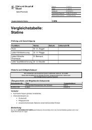
- Page 1 and 2: Basel Seminars in Pathology Postgra
- Page 3: UPDATE ON PATHOLOGY STANDARDS FOR B
- Page 7: HISTOLOGIC GRADE UROTHELIAL NEOPLAS
- Page 10 and 11: What’s New in the 2012 Consensus
- Page 12 and 13: Reporting of Bladder Cancer • Gui
- Page 14 and 15: • Seen with instrumentation with
- Page 16 and 17: Squamous Metaplasia • Should be r
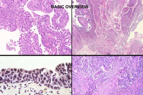
- Page 18 and 19: GRADING OF UROTHELIAL LESIONS • F
- Page 20 and 21: NORMAL
- Page 22 and 23: Urothelial Dysplasia • Overall fe
- Page 24 and 25: Urothelial Dysplasia • The diagno
- Page 28: ATYPIA OF UNKNOWN SIGNIFICANCE (WHO
- Page 31 and 32: Grading of Urothelial Carcinoma •
- Page 33 and 34: Papilloma
- Page 35 and 36: PUNLMP
- Page 37 and 38: LOW GRADE
- Page 39 and 40: HIGHGRADE
- Page 41 and 42: P A P I L O M A L M P L O W G R A D
- Page 43 and 44: Courtesy R. Montironi, Italy Exophy
- Page 45 and 46: Inverted High Grade without invasio
- Page 47 and 48: Inverted Papilloma
- Page 49 and 50: Inverted PUNLMP
- Page 51 and 52: Inverted LG
- Page 53 and 54: Inverted HG
- Page 55 and 56:
Inverted HG, Non Inv
- Page 57 and 58:
WHO (2004) /ISUP : Prognostic Signi
- Page 59 and 60:
Contributions of WHO (2004) /ISUP
- Page 61 and 62:
Handling grade heterogeneity in bla
- Page 63 and 64:
Grading Papillary Urothelial Neopla
- Page 65:
Papillary Hyperplasia with Cytologi
- Page 68 and 69:
Dysplasia with early papillary feat
- Page 70 and 71:
Grading Invasive Cancer • Practic
- Page 74 and 75:
• 2 types of muscle - awareness i
- Page 76 and 77:
Muscularis Mucosae Muscle • Hyper
- Page 78 and 79:
M. mucosae muscle patterns Typical
- Page 80 and 81:
M. mucosae muscle pattern Hypertrop
- Page 82 and 83:
Muscularis Propria • Several term
- Page 84 and 85:
Muscle Involved by UCa, Indetermina
- Page 88 and 89:
Assessment of pT2 vs. pT3 - cystect
- Page 91 and 92:
Microinvasive Urothelial Carcinoma
- Page 93 and 94:
Substratification or Substaging of
- Page 95 and 96:
Extensive invasion
- Page 97 and 98:
Retraction a mimic of vascular-lymp
- Page 99 and 100:
Vascular Lymphatic Invasion (LVI)
- Page 101 and 102:
2013 International Society of Urolo
- Page 103 and 104:
Lymph node involvement: • Approx.
- Page 105 and 106:
THE BEST BLADDER CANCER PATHOLOGY S
- Page 107 and 108:
PSEUDONEOPLASTIC MIMICS OF BLADDER
- Page 109 and 110:
EMPHYSEMATOUS & BULLOUS CYSTITIS
- Page 113 and 114:
AMYLOIDOSIS
- Page 115 and 116:
MALAKOPLAKIA
- Page 117:
NORMAL PARAGANGLIONIC TISSUE
- Page 123 and 124:
CIS
- Page 125:
REACTIVE ATYPIA History of stones,
- Page 130:
CIS REACTIVE ATYPIA
- Page 134 and 135:
RADIATION ATYPIA
- Page 136:
INTRAVESICAL THERAPY
- Page 141:
Polyoma Virus Infection • Usually
- Page 149 and 150:
Papillary tumor Micropapillary U Ca
- Page 151 and 152:
Low magnification distinction Broad
- Page 153:
Cystoscopic & microscopic mimic Pap
- Page 158 and 159:
Papillary Polypoid cystitis
- Page 161 and 162:
ALL THAT IS PAPILLARY BLADDER IS NO
- Page 165 and 166:
Bladder Biopsy Interpretation, 2nd
- Page 167 and 168:
TIGHTLY CLUSTERED UROTHELIUM
- Page 169 and 170:
TIGHTLY CLUSTERED UROTHELIUM
- Page 171 and 172:
U Ca. with small tubules Nephrogeni
- Page 173 and 174:
NEPHROGENIC ADENOMA - THE BIG MIMIC
- Page 175:
NEPHROGENIC ADENOMA
- Page 181:
NEPHROGENIC ADENOMA
- Page 185 and 186:
NEPHROGENIC ADENOMA Clues to benign
- Page 187 and 188:
FLORID REACTIVE PROLIFERATIONS norm
- Page 195:
POST-RADIATION PSEUDOCARCINOMATOUS
- Page 214:
LORID CYSTTTIS GLANDULARIS ITH MUCI
- Page 222:
MULLERIANOSIS • Endometriosis, en
- Page 233 and 234:
PSEUDOSARCOMATOUS MYOFIBROBLASTIC P
- Page 235:
Gross PMP • Exophyticpolypoid mas
- Page 246 and 247:
ROLE OF IMMUNOHISTOCHEMISTRY IN THE
- Page 248 and 249:
PROVING UROTHELIAL DIFFERENTIATION
- Page 250 and 251:
CA in the bladder, h.o of lung canc
- Page 252 and 253:
Paraganglioma Epith. LMS PEComa Mel
- Page 255 and 256:
p63
- Page 257 and 258:
Plasmacytoid U Ca
- Page 259 and 260:
• Markers of urothelium and uroth
- Page 261 and 262:
S100P S100P GATA3 GATA3 A-F: S100P,
- Page 263 and 264:
GATA 3 & S100 P: Diagnostic utility
- Page 265 and 266:
PARAGANGLIOMA
- Page 267:
Identification of Succinate Dehydro
- Page 270 and 271:
CIS REACTIVE
- Page 272 and 273:
NORMAL
- Page 274 and 275:
CK-20
- Page 276 and 277:
REACTIVE UROTHELIUM
- Page 278 and 279:
CK-20
- Page 280 and 281:
Reactive CD44
- Page 282 and 283:
CA-INSITU
- Page 284 and 285:
CK-20
- Page 286 and 287:
CIS CK20
- Page 288 and 289:
CK20 (+) CD44(-)
- Page 290 and 291:
p53
- Page 292 and 293:
CD44
- Page 294 and 295:
Reactive CIS
- Page 296 and 297:
Radiation-Reactive Radiation CIS
- Page 298 and 299:
AUA GUIDELINE 2007 UPDATE: • Stan
- Page 300 and 301:
Typical pattern: • Arranged in gr
- Page 302 and 303:
Muscularis mucosae or muscularis pr
- Page 304:
Am J Surg Pathol. 2009 ;33:91-8 SMO
- Page 307 and 308:
Smoothelin hyperplastic
- Page 309 and 310:
Smoothelin
- Page 311 and 312:
SMA - M. Mucosae
- Page 313 and 314:
Smoothelin
- Page 315 and 316:
CAUTERY: MINIMAL EFFECT ON IHC
- Page 318 and 319:
M. Mucosae vs. M. Propria in TURBT
- Page 321 and 322:
M. Mucosae vs. M. Propria in TURBT
- Page 323 and 324:
Smoothelin: Negative - Suggestive o
- Page 325:
Courtesy: Dr John Eble
- Page 329 and 330:
METASTASIS TO THE BLADDER Prostate
- Page 331 and 332:
?Urothelial Carcinoma vs. ?Prostati
- Page 333 and 334:
Concurrent PCa & UCa
- Page 335 and 336:
PCa PCa NKX3.1 P501S PSMA
- Page 338:
100% 100% 90% 88% 88% 88% 80% 70% 6
- Page 342 and 343:
Clear cell Ca Neph. adenoma
- Page 344 and 345:
U Ca. with small tubules Nephrogeni
- Page 346 and 347:
Nephrogenic adenoma Clear cell aden
- Page 348 and 349:
CCCa Pax 2 S100A1 Ki67
- Page 350 and 351:
P Ca A Pax 2 S100A1 Ki67
- Page 352 and 353:
PMP / Pseudotumors
- Page 354 and 355:
SMA
- Page 356:
Sarcomatoid urothelial carcinoma
- Page 361:
Leiomyosarcoma
- Page 365 and 366:
Spindle cell lesions Benign (PMP) v
- Page 367:
SARC CA p63 CK 5/6 or HMCK p63
- Page 370 and 371:
Session plan Renal Pathology for th
- Page 372 and 373:
Tubules - Normal
- Page 374 and 375:
Interstitial inflammation - tubuloi
- Page 376 and 377:
Tubulointerstitial nephritis Classi
- Page 378 and 379:
CKD, previous tuberculosis, HCV pos
- Page 380 and 381:
AKI, ANCA positive, cortex - TIN, g
- Page 382 and 383:
Male 59 years. Acute rise in creati
- Page 384 and 385:
35 patients with IgG4-TIN: 27 (77%)
- Page 386 and 387:
Male 70 years AKI Diagnosis?
- Page 388 and 389:
Hantavirus nephropathy
- Page 390 and 391:
Male, 57 years. Acute renal failure
- Page 392 and 393:
Ascending infection (pyelonephritis
- Page 394 and 395:
Tubulointerstitial infiltrates in t
- Page 396 and 397:
Tubulointerstitial infiltrates in t
- Page 398 and 399:
Things in tubules Casts, crystals,
- Page 400 and 401:
Acute phosphate nephropathy First c
- Page 402 and 403:
Diagnosis?
- Page 404 and 405:
Acute tubular injury Ischaemia, dru
- Page 406 and 407:
Light chain tubulopathy
- Page 408 and 409:
Acute tubular injury in the post mo
- Page 410 and 411:
Acute tubular injury in the post mo
- Page 412 and 413:
Drug-associated tubular injury Drug
- Page 414 and 415:
Gentamicin toxicity Diagnosis?
- Page 416 and 417:
Drug-associated tubular injury Mech
- Page 418 and 419:
Glomerular lesions Ian Roberts Oxfo
- Page 420 and 421:
Tumour nephrectomy specimens Things
- Page 422 and 423:
Glomeruli- Normal
- Page 424 and 425:
Nodular glomerulosclerosis Diabetic
- Page 426 and 427:
Diabetic nephropathy: thickened bas
- Page 428 and 429:
Diabetic nephropathy: Diffuse mesan
- Page 430 and 431:
Diabetic nephropathy: Insudative le
- Page 432 and 433:
Amyloidosis Congo red
- Page 434 and 435:
Amyloidosis
- Page 436 and 437:
Amyloidosis
- Page 438 and 439:
Immunotactoid Fibrillary GN 0.06% o
- Page 440 and 441:
Light chain deposition disease
- Page 442 and 443:
Focal segmental glomerulosclerosis
- Page 444 and 445:
Focal segmental glomerulosclerosis
- Page 446 and 447:
Focal segmental glomerulosclerosis
- Page 448 and 449:
Glomerular lesions: proliferation M
- Page 450 and 451:
Glomerular lesions: proliferation E
- Page 452 and 453:
The diagnosis of IgA nephropathy is
- Page 454 and 455:
The histology of IgA nephropathy is
- Page 456 and 457:
Mesangiocapillary GN = Membranoprol
- Page 458 and 459:
Mesangiocapillary GN, type I C3
- Page 460 and 461:
C3 glomerulopathy MPGN and C3 glome
- Page 462 and 463:
Dense deposit disease
- Page 464 and 465:
Glomerular lesions: necrosis Ruptur
- Page 466 and 467:
Renal vasculitis May be isolated re
- Page 468 and 469:
Renal vasculitis European vasculiti
- Page 470 and 471:
Anti-GBM disease Autoantibodies to
- Page 472 and 473:
Remember: multiple pathologies are
- Page 474 and 475:
Male 46 years. Suffered from diabet
- Page 476 and 477:
Diagnoses: Diabetic nephropathy Oxa
- Page 478 and 479:
Interactive Kidney Quiz Ian S. Robe
- Page 480 and 481:
Test vote How did you learn about t
- Page 482 and 483:
Set 1
- Page 484 and 485:
PathoPic B
- Page 486:
PathoPic D
- Page 490 and 491:
What is your main diagnosis? 1. chr
- Page 492 and 493:
What is your main diagnosis? 1. amy
- Page 494 and 495:
What is your main diagnosis? 1. no
- Page 496 and 497:
A PathoPic Dg: arteriolosclerosis,
- Page 498 and 499:
Dg: diabetic kidney C PathoPic
- Page 500 and 501:
Dg: malignant nephrosclerosis E
- Page 502 and 503:
RCPath “Minimum” dataset for tu
- Page 504 and 505:
Patterns of scarring Granular subca
- Page 506 and 507:
Patterns of scarring Granular subca
- Page 508 and 509:
Deep pitted cortical scars: Pattern
- Page 510 and 511:
Patterns of scarring Deep pitted co
- Page 512 and 513:
Patterns of scarring Segmental tran
- Page 514 and 515:
Malignant hypertension A cause of a
- Page 516 and 517:
Malignant hypertension A cause of a
- Page 518 and 519:
Malignant hypertension A cause of a
- Page 520 and 521:
Malignant vs benign vascular diseas
- Page 522 and 523:
PathoPic G
- Page 524:
PathoPic I
- Page 528 and 529:
What is your main diagnosis? 1. gou
- Page 530 and 531:
What is your main diagnosis? 1. met
- Page 532 and 533:
What is your main diagnosis? 1. ren
- Page 534 and 535:
Solutions (M.J. Mihatsch)
- Page 536 and 537:
Descripition Involvement of cortex
- Page 538 and 539:
Description Whitish discoloration a
- Page 540 and 541:
Description Massive enlargement of
- Page 542 and 543:
Description Brownish discoloration
- Page 544 and 545:
Papillary necrosis Pg: i.e. vascula
- Page 546 and 547:
Papillary necrosis Pg: i.e. vascula
- Page 548 and 549:
Types of papillary necrosis: Extend
- Page 550 and 551:
Black papillary necrosis Think of p
- Page 552 and 553:
Phenacetin kidney Compete necrosis
- Page 554 and 555:
Phenacetin kidney Note massive base
- Page 556 and 557:
Phenacetin kidney Note: Massive lip
- Page 558 and 559:
Typical examples of papillary necro
- Page 560 and 561:
White papillary necrosis Think of v
- Page 562 and 563:
Red papillary necrosis Think of vas
- Page 564 and 565:
I hope you will never overlook papi
- Page 566 and 567:
Challenges for Pathologists Renal M
- Page 568:
Do the morphotypes of RCC have prog
- Page 571 and 572:
Potential of Needle Biopsy • Diag
- Page 573 and 574:
Novel renal tumor types with clear
- Page 575 and 576:
Am J Surg Path 28, 2004 41 Carcinom
- Page 578 and 579:
Should Acquired Cystic Disease- RCC
- Page 580 and 581:
Clear cell papillary RCC has: 1. PO
- Page 582 and 583:
Which name should be used for 1. Cl
- Page 585 and 586:
1. Yes 2. No Should Thyroid-Like Fo
- Page 587 and 588:
If focal (
- Page 589 and 590:
Denote AMLs with epithelioid morpho
- Page 591:
Differential Diagnosis of Cystic Re
- Page 594 and 595:
smaller cysts with phyllodes glands
- Page 596 and 597:
Are Cystic Nephroma and Mixed Epith
- Page 598 and 599:
If you consider CN and MEST variati
- Page 600 and 601:
Should Tubulocystic-RCC be recogniz
- Page 602 and 603:
Prognosis • Type • Stage • Gr
- Page 604:
Papillary renal cell carcinoma Type
- Page 608 and 609:
Differential Diagnosis: metanephric
- Page 610 and 611:
The distinction between type 1 vers
- Page 612 and 613:
Should Oncocytic Papillary-RCC be r
- Page 614:
Leiomyomas of the Skin
- Page 618 and 619:
Fuhrman Grading in RCC Hong SK, BJU
- Page 620 and 621:
How should we grade clear cell RCC?
- Page 622:
How should we grade chromophobe RCC
- Page 625 and 626:
For Sarcomatoid Tumors Do You Repor
- Page 627 and 628:
Do You Consider a Tumor Area Sarcom
- Page 629 and 630:
Tumor Staging
- Page 631 and 632:
When invasion of the renal sinus is
- Page 633 and 634:
Should the presence or absence of t
- Page 635 and 636:
Should any assessment of amount (pe
- Page 637 and 638:
ISUP Consensus Conference Vancouver
- Page 639 and 640:
Molecular Pathways and Targeted The
- Page 641:
Chemokine receptor CXCR4 downregula
- Page 644 and 645:
Agents such as sunitinib act throug
- Page 646 and 647:
VHL-Mutation ↔ Gene expression Ba
- Page 648 and 649:
Driver and Passenger VHL Mutations
- Page 650:
Differential Diagnosis of Renal Cel
- Page 653:
Renal Tumors with Eosinophilic Cyto
- Page 658:
Birt Hogg Dubé syndrome
- Page 661 and 662:
Are the hybrid tumors associated wi
- Page 663 and 664:
For distinguishing clear cell RCC f
- Page 665 and 666:
Clear Cell Renal Cancer VHL Deletio
- Page 667 and 668:
Early on in VHL disease, the vast m
- Page 669 and 670:
Distinct tumor suppressor mechanism
- Page 671 and 672:
A Ksp-Cre - pos - pos - pos Vhlh fl
- Page 673 and 674:
pVHL localizes to primary cilia in
- Page 675 and 676:
a c b d Montani et al.: Am J Surg P
- Page 677 and 678:
Multilocular cystic renal cell carc
- Page 679 and 680:
What terminology for a multicystic
- Page 681 and 682:
In the setting of acquired cystic d
- Page 683 and 684:
„Translocation“ Type of Renal C
- Page 685 and 686:
„Translocation“ Type of Renal C
- Page 687:
t(X;1)
- Page 690 and 691:
Should t(6;11) RCC be Recognized as
- Page 693 and 694:
When a translocation carcinoma is s
- Page 696:
Leiomyomatous Renal Cell Carcinoma
- Page 699:
Edmunds W: Trans.Path.Soc.London 43
- Page 702 and 703:
Are Cystic Nephroma and Mixed Epith
- Page 704 and 705:
Renal Tumors with Papillary Archite
- Page 706 and 707:
In the absence of classical histolo
- Page 708 and 709:
Clin Cancer Res 2009;15:3297-3304 -
- Page 710 and 711:
A, Western blot analysis of HK-2 ce
- Page 712:
Tumors with Spindle Cell Morphology
- Page 716 and 717:
Which marker do you use most freque
- Page 719 and 720:
Primary Renal Synovial Sarcoma Immu
- Page 721:
SYT-SSX Fusion from translocation t
- Page 726:
Renal carcinoid from a horseshoe ki
- Page 729:
DD „Small round blue“ cell tumo
- Page 733 and 734:
Xu X. et al.L Cell 148, 886-895, 20
- Page 736 and 737:
Polybromo-1 (BAF180) in ccRCC ~40%
- Page 738 and 739:
wt versus VHL-/- cells VHL-loss der
- Page 740 and 741:
VHL and a Serum Screening Test for