altering vertical dimension in the perio-restorative patient - Kokich ...
altering vertical dimension in the perio-restorative patient - Kokich ...
altering vertical dimension in the perio-restorative patient - Kokich ...

You also want an ePaper? Increase the reach of your titles
YUMPU automatically turns print PDFs into web optimized ePapers that Google loves.
2<br />
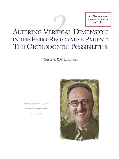
ALTERING VERTICAL DIMENSION<br />
IN THE PERIO-RESTORATIVE PATIENT:<br />
THE ORTHODONTIC POSSIBILITIES<br />
Au: Please answer<br />
queries on pages 4<br />
and 22.<br />
V<strong>in</strong>cent G. <strong>Kokich</strong>, DDS, MSD<br />
The location of <strong>the</strong> occlusal plane<br />
limits <strong>the</strong> scope of improvement<br />
<strong>in</strong> facial height.
2 ALTERING VERTICAL DIMENSION IN THE PERIO-RESTORATIVE PATIENT • <strong>Kokich</strong><br />
PHILOSOPHY AND BACKGROUND<br />
In <strong>the</strong> past decade, <strong>the</strong> number of adults seek<strong>in</strong>g orthodontic treatment has grown substantially.<br />
Today, <strong>in</strong> most orthodontic practices, 30% to 50% of <strong>patient</strong>s <strong>in</strong> active orthodontic treatment<br />
are adults. My practice is devoted completely to adults. Although adolescent and adult malocclusions<br />
are similar, adults usually have additional problems that make <strong>the</strong>ir treatment more challeng<strong>in</strong>g.<br />
One of <strong>the</strong> most destructive is anterior tooth wear comb<strong>in</strong>ed with compensatory<br />
eruption of <strong>the</strong> abraded teeth. When this type of <strong>patient</strong> is referred to me by a <strong>restorative</strong> dentist,<br />
<strong>the</strong> common request from <strong>the</strong> cl<strong>in</strong>ician is to “open <strong>the</strong> <strong>vertical</strong>” to allow adequate restoration<br />
of <strong>the</strong> teeth. What does “open <strong>the</strong> <strong>vertical</strong>” mean to you? What should it mean to <strong>the</strong><br />
orthodontist with whom you are collaborat<strong>in</strong>g? Actually, <strong>the</strong>re may be a dist<strong>in</strong>ct difference <strong>in</strong><br />
what is necessary and what is orthodontically possible <strong>in</strong> an adult <strong>patient</strong> with worn and/or<br />
abraded anterior teeth. Although some cl<strong>in</strong>icans believe that <strong>patient</strong>s with anterior tooth wear<br />
are “overclosed” and need to have <strong>the</strong>ir <strong>vertical</strong> <strong>dimension</strong> <strong>in</strong>creased, this diagnosis and <strong>the</strong><br />
result<strong>in</strong>g treatment plan are usually <strong>in</strong>appropriate. The problem typically is compensatory<br />
eruption of <strong>the</strong> anterior teeth, secondary to <strong>in</strong>cisal wear. The problem for <strong>the</strong> <strong>restorative</strong> dentist<br />
is <strong>in</strong>sufficient space to restore <strong>the</strong>se teeth without fur<strong>the</strong>r tooth reduction, possible crown<br />
leng<strong>the</strong>n<strong>in</strong>g, and perhaps root canal <strong>the</strong>rapy. However, orthodontists can <strong>in</strong>trude <strong>the</strong> maxillary<br />
<strong>in</strong>cisors, mandibular <strong>in</strong>cisors, or both <strong>in</strong> order to create <strong>restorative</strong> space, move <strong>the</strong> g<strong>in</strong>gival<br />
marg<strong>in</strong>s apically, and elim<strong>in</strong>ate <strong>the</strong> need for fur<strong>the</strong>r tooth reduction. The team of orthodontist<br />
and <strong>restorative</strong> dentist must decide which teeth are to be <strong>in</strong>truded, how long <strong>the</strong>y are to be held<br />
<strong>in</strong> position, and how this type of tooth movement will affect <strong>the</strong> <strong>patient</strong>’s es<strong>the</strong>tic appearance.<br />
2<br />
UUSING MAXILLARY AND MANDIBULAR<br />
INTRUSION TO CREATE RESTORATIVE SPACE:<br />
FOUR GUIDING PRINCIPLES<br />
One of <strong>the</strong> most perplex<strong>in</strong>g problems for general dentists is how to manage <strong>restorative</strong> treatment<br />
for <strong>the</strong> adult <strong>patient</strong> with a deep anterior overbite. In most cases, <strong>the</strong>re is little or no<br />
room to prepare <strong>the</strong> anterior teeth adequately, and creat<strong>in</strong>g space for <strong>restorative</strong> materials<br />
often requires significant reduction of <strong>the</strong> exist<strong>in</strong>g teeth. Moderate to extensive wear or abrasion<br />
of <strong>the</strong> maxillary and/or mandibular <strong>in</strong>cisors fur<strong>the</strong>r complicates <strong>the</strong> overbite problem.<br />
As <strong>the</strong>se teeth wear, <strong>the</strong>y usually cont<strong>in</strong>ue to erupt, br<strong>in</strong>g<strong>in</strong>g <strong>the</strong> bone and g<strong>in</strong>giva with <strong>the</strong>m.<br />
In order to restore <strong>the</strong>se short teeth, <strong>the</strong> <strong>restorative</strong> dentist typically considers two possible<br />
treatment plans. One option <strong>in</strong>volves crown leng<strong>the</strong>n<strong>in</strong>g of <strong>the</strong> short abraded teeth, followed<br />
by potential root canal <strong>the</strong>rapy, post and core buildups, and eventual restoration of <strong>the</strong> teeth.<br />
Ano<strong>the</strong>r option is to open <strong>the</strong> <strong>patient</strong>’s <strong>vertical</strong> <strong>dimension</strong> by restor<strong>in</strong>g most of <strong>the</strong> posterior<br />
teeth <strong>in</strong> order to ga<strong>in</strong> <strong>the</strong> space needed to restore <strong>the</strong> anterior teeth. However, <strong>the</strong>re is a third possibility—orthodontics<br />
and/or orthognathic surgery. Orthodontists can <strong>in</strong>trude maxillary and<br />
mandibular anterior teeth to create <strong>restorative</strong> space. In some situations, orthognathic surgery of<br />
<strong>the</strong> mandible and/or maxilla can open <strong>the</strong> <strong>patient</strong>’s anterior <strong>vertical</strong> <strong>dimension</strong> to improve facial<br />
es<strong>the</strong>tics and provide <strong>restorative</strong> space. This chapter presents and illustrates <strong>the</strong> govern<strong>in</strong>g pr<strong>in</strong>ciples<br />
and <strong>the</strong> diagnostic techniques required to correctly analyze and prescribe <strong>the</strong> appropriate<br />
treatment for <strong>the</strong>se types of problems.
USING MAXILLARY AND MANDIBULAR INTRUSION TO CREATE RESTORATIVE SPACE: FOUR GUIDING PRINCIPLES<br />
Advantages of growth<br />
In order to fully comprehend <strong>the</strong> orthodontic possibilities <strong>in</strong> resolv<strong>in</strong>g problems of <strong>vertical</strong><br />
<strong>dimension</strong>, <strong>the</strong> cl<strong>in</strong>ician must first understand <strong>the</strong> importance of facial growth. Most orthodontic<br />
<strong>patient</strong>s are treated dur<strong>in</strong>g childhood and adolescence. There are several reasons for correct<strong>in</strong>g<br />
a malocclusion early <strong>in</strong> life, but one of <strong>the</strong> most important is to take advantage of a<br />
<strong>patient</strong>’s facial growth potential.<br />
The face grows <strong>in</strong> two ways. The first is sutural growth, which occurs at <strong>the</strong> circummaxillary<br />
sutures that suspend <strong>the</strong> maxilla from <strong>the</strong> skull. But <strong>the</strong> major source of <strong>vertical</strong> change that<br />
occurs dur<strong>in</strong>g adolescent growth is found <strong>in</strong> <strong>the</strong> mandibular condyle. As <strong>the</strong> condyle grows, <strong>the</strong><br />
ramus of <strong>the</strong> mandible gets longer. Dur<strong>in</strong>g this process, <strong>the</strong> teeth erupt to fill <strong>the</strong> space created<br />
by ramus growth. This is advantageous <strong>in</strong> manag<strong>in</strong>g a deep overbite <strong>in</strong> an adolescent <strong>patient</strong>.<br />
Instead of <strong>in</strong>trud<strong>in</strong>g <strong>the</strong> teeth to resolve <strong>the</strong> overeruption of <strong>the</strong> maxillary and mandibular <strong>in</strong>cisors,<br />
ramus growth permits space for <strong>the</strong> posterior teeth to erupt. If <strong>the</strong> maxillary and/or<br />
mandibular <strong>in</strong>cisors are held <strong>in</strong> <strong>the</strong>ir orig<strong>in</strong>al <strong>vertical</strong> position dur<strong>in</strong>g this process, <strong>the</strong> deep<br />
overbite is corrected through posterior tooth eruption and relative <strong>in</strong>cisor <strong>in</strong>trusion. This possibility<br />
simplifies <strong>the</strong> management of a deep overbite.<br />
However, facial growth is essentially complete for females <strong>in</strong> <strong>the</strong> mid to late teenage years<br />
and for males by <strong>the</strong> early twenties. In an adult <strong>patient</strong> with a deep anterior overbite, growth is<br />
not a possibility. Therefore, <strong>the</strong> orthodontist must rely on <strong>in</strong>trusion of <strong>the</strong> maxillary <strong>in</strong>cisors,<br />
<strong>in</strong>trusion of <strong>the</strong> mandibular <strong>in</strong>cisors, or jaw surgery to correct deep overbites <strong>in</strong> adults. How do<br />
orthodontists diagnose which teeth to <strong>in</strong>trude and when to use orthognathic surgery to correct<br />
problems of <strong>vertical</strong> <strong>dimension</strong>? If <strong>the</strong> <strong>patient</strong>’s anterior teeth are worn and abraded, what<br />
is <strong>the</strong> <strong>restorative</strong> dentist’s role <strong>in</strong> <strong>the</strong> <strong>in</strong>terdiscipl<strong>in</strong>ary treatment plan for <strong>the</strong> restoration of<br />
<strong>the</strong>se teeth? In order to answer <strong>the</strong>se questions, I will expla<strong>in</strong> how <strong>the</strong> diagnostic process works<br />
and <strong>the</strong> pr<strong>in</strong>ciples I use to handle <strong>the</strong>se types of <strong>patient</strong>s.<br />
Pr<strong>in</strong>ciple 1<br />
The location of <strong>the</strong> occlusal plane limits <strong>the</strong> scope of improvement <strong>in</strong> facial height.<br />
When I am challenged to determ<strong>in</strong>e how to provide <strong>restorative</strong> space <strong>in</strong> <strong>the</strong> <strong>patient</strong> with a<br />
deep anterior overbite, my first step is to determ<strong>in</strong>e <strong>the</strong> <strong>patient</strong>’s correct occlusal plane us<strong>in</strong>g<br />
<strong>the</strong> fixed landmarks (ie, those that I cannot control) as my guide. To do this, I identify <strong>the</strong><br />
occlusal contact po<strong>in</strong>t between <strong>the</strong> maxillary and mandibular second molars. In an adult, I cannot<br />
change this relationship. Open<strong>in</strong>g <strong>the</strong> <strong>vertical</strong> <strong>dimension</strong> by extrud<strong>in</strong>g maxillary and/or<br />
mandibular molars is not stable <strong>in</strong> adults. S<strong>in</strong>ce adults are not grow<strong>in</strong>g, <strong>the</strong> muscles of mastication<br />
(primarily <strong>the</strong> masseter, medial pterygoid, and temporalis) do not have <strong>the</strong> capacity to<br />
stretch. Attempts to <strong>in</strong>crease posterior facial height with orthognathic surgery have consistently<br />
been unstable, and <strong>the</strong> <strong>vertical</strong> <strong>dimension</strong> usually reverts to its orig<strong>in</strong>al level. 1–3 So, us<strong>in</strong>g <strong>the</strong><br />
contact between <strong>the</strong> maxillary and mandibular second molars allows me to realistically determ<strong>in</strong>e<br />
<strong>the</strong> posterior limit of <strong>the</strong> occlusal plane.<br />
Next, I must identify a po<strong>in</strong>t anteriorly that will represent a realistic guide for <strong>the</strong> o<strong>the</strong>r end<br />
of <strong>the</strong> <strong>patient</strong>’s occlusal plane. For this I use <strong>the</strong> level of <strong>the</strong> <strong>patient</strong>’s upper lip at rest. Aga<strong>in</strong>, I<br />
cannot alter <strong>the</strong> position of <strong>the</strong> upper lip <strong>in</strong> a nongrow<strong>in</strong>g adult. In addition, if a deep overbite is<br />
corrected by <strong>in</strong>trud<strong>in</strong>g anterior teeth, <strong>the</strong>n it is important to <strong>in</strong>trude <strong>the</strong> correct teeth and provide<br />
<strong>the</strong> <strong>patient</strong> with a pleas<strong>in</strong>g es<strong>the</strong>tic appearance of <strong>the</strong> teeth relative to <strong>the</strong> upper lip after<br />
treatment. So, <strong>the</strong> occlusal plane connects <strong>the</strong> occlusal contact between <strong>the</strong> maxillary and mandibular<br />
second molars and <strong>the</strong> upper lip. This is <strong>the</strong> first step <strong>in</strong> <strong>the</strong> diagnostic process.<br />
3
2 ALTERING VERTICAL DIMENSION IN THE PERIO-RESTORATIVE PATIENT • <strong>Kokich</strong><br />
Pr<strong>in</strong>ciple 2<br />
The relationship between <strong>the</strong> <strong>in</strong>cisors and <strong>the</strong> occlusal plane reveals <strong>the</strong> cause of <strong>the</strong> overbite.<br />
The next step <strong>in</strong> <strong>the</strong> diagnostic process is to determ<strong>in</strong>e which <strong>in</strong>cisor, maxillary or mandibular,<br />
has “overerupted” and is caus<strong>in</strong>g <strong>the</strong> deep overbite. My procedure is to identify <strong>the</strong> <strong>in</strong>cisal edge of<br />
<strong>the</strong> maxillary central <strong>in</strong>cisor and measure its distance from <strong>the</strong> occlusal plane. This distance will<br />
vary with age. In a 30-year-old, <strong>the</strong> distance from <strong>the</strong> occlusal plane (upper lip) to <strong>the</strong> maxillary<br />
central <strong>in</strong>cisal edge at rest should be about 3 mm. However, studies of older adults have shown<br />
that this distance decreases with time, and <strong>in</strong> a 60-year-old it is not unusual for <strong>the</strong> distance from<br />
<strong>the</strong> <strong>in</strong>cisal edge to <strong>the</strong> upper lip to be 0 mm with <strong>the</strong> upper lip at rest.<br />
So, if <strong>the</strong> <strong>patient</strong> is 30 years of age, and <strong>the</strong> distance from <strong>the</strong> maxillary <strong>in</strong>cisor to <strong>the</strong><br />
occlusal plane is 5 mm, <strong>the</strong>n at least part of <strong>the</strong> deep overbite problem is due to overeruption<br />
of <strong>the</strong> maxillary <strong>in</strong>cisors. These teeth could be orthodontically <strong>in</strong>truded 2 mm to assist <strong>in</strong> correct<strong>in</strong>g<br />
<strong>the</strong> deep anterior overbite.<br />
Next, I identify <strong>the</strong> position of <strong>the</strong> mandibular <strong>in</strong>cisal edge and measure its distance to <strong>the</strong><br />
occlusal plane. The mandibular <strong>in</strong>cisal edge should be at or near <strong>the</strong> level of <strong>the</strong> occlusal plane.<br />
If <strong>the</strong>se teeth are 3 to 4 mm above <strong>the</strong> occlusal plane, <strong>the</strong>n <strong>the</strong> deep overbite is most likely due<br />
to overeruption of <strong>the</strong> mandibular <strong>in</strong>cisors. These teeth would need to be <strong>in</strong>truded 3 to 4 mm<br />
<strong>in</strong> order to place <strong>the</strong> <strong>in</strong>cisal edge at <strong>the</strong> occlusal plane, correct <strong>the</strong> deep overbite, and allow <strong>the</strong><br />
general dentist to complete <strong>the</strong> <strong>restorative</strong> treatment for <strong>the</strong> <strong>patient</strong>.<br />
However, <strong>the</strong> maxillary and/or mandibular <strong>in</strong>cisors could be abraded as a result of a brux<strong>in</strong>g<br />
habit. In <strong>the</strong>se cases, <strong>in</strong>trud<strong>in</strong>g <strong>the</strong> maxillary and/or mandibular <strong>in</strong>cisors would correct <strong>the</strong><br />
<strong>in</strong>cisal edge position of <strong>the</strong>se teeth, but <strong>the</strong> general dentist may still need space to restore <strong>the</strong>se<br />
teeth to a longer length. In o<strong>the</strong>r words, <strong>the</strong>se teeth may need to be <strong>in</strong>truded fur<strong>the</strong>r. The next<br />
step <strong>in</strong> <strong>the</strong> diagnostic process is to assess <strong>the</strong> g<strong>in</strong>gival levels and crown proportions of <strong>the</strong> maxillary<br />
and mandibular central <strong>in</strong>cisors relative to <strong>the</strong> lateral <strong>in</strong>cisors and can<strong>in</strong>es.<br />
Pr<strong>in</strong>ciple 3<br />
The correct g<strong>in</strong>gival position determ<strong>in</strong>es whe<strong>the</strong>r <strong>in</strong>trusion is <strong>the</strong> right solution, and crown<br />
proportions govern <strong>the</strong> amount of leng<strong>the</strong>n<strong>in</strong>g that is desirable.<br />
Once <strong>the</strong> <strong>in</strong>cisal edges of <strong>the</strong> maxillary and mandibular central and lateral <strong>in</strong>cisors have been<br />
positioned correctly relative to <strong>the</strong> occlusal plane, <strong>the</strong> next step is to verify <strong>the</strong>ir es<strong>the</strong>tic appearance<br />
(maxillary <strong>in</strong>cisors) and structural <strong>in</strong>tegrity (mandibular <strong>in</strong>cisors). If <strong>the</strong> g<strong>in</strong>gival marg<strong>in</strong>s of <strong>the</strong><br />
central <strong>in</strong>cisors are coronal to those of <strong>the</strong> can<strong>in</strong>es, this could <strong>in</strong>dicate ei<strong>the</strong>r that <strong>the</strong>se teeth are<br />
abraded or that <strong>the</strong> g<strong>in</strong>gival marg<strong>in</strong>s are positioned too far coronally. Sulcus depth helps to reveal<br />
<strong>the</strong> correct diagnosis; <strong>the</strong>refore, my next step is to measure it with a probe.<br />
If <strong>the</strong> sulcus depth is 1 mm [Au: or more?] and <strong>the</strong> cementoenamel junction is located at<br />
<strong>the</strong> bottom of <strong>the</strong> sulcus, <strong>the</strong>n <strong>the</strong> position of <strong>the</strong> g<strong>in</strong>gival marg<strong>in</strong> is correct. In this case, <strong>the</strong><br />
<strong>patient</strong> likely has <strong>in</strong>cisal wear. I confirm this suspicion by evaluat<strong>in</strong>g <strong>the</strong> maxillary and/or<br />
mandibular <strong>in</strong>cisal edges. If I see dent<strong>in</strong> at <strong>the</strong>se edges, it shows that <strong>the</strong> <strong>patient</strong> has abraded at<br />
least 2 mm and perhaps more from <strong>the</strong> <strong>in</strong>cisal edges of <strong>the</strong>se teeth. In this case I would <strong>in</strong>trude<br />
<strong>the</strong> maxillary and/or mandibular <strong>in</strong>cisors an additional 2 mm or more to position <strong>the</strong> g<strong>in</strong>gival<br />
marg<strong>in</strong> at <strong>the</strong> correct level relative to that of <strong>the</strong> maxillary and/or mandibular can<strong>in</strong>es. This<br />
permits leng<strong>the</strong>n<strong>in</strong>g of <strong>the</strong> maxillary and mandibular <strong>in</strong>cisors.<br />
4
USING MAXILLARY AND MANDIBULAR INTRUSION TO CREATE RESTORATIVE SPACE: FOUR GUIDING PRINCIPLES<br />
The amount of leng<strong>the</strong>n<strong>in</strong>g desired can be determ<strong>in</strong>ed by evaluat<strong>in</strong>g <strong>the</strong> width-to-length<br />
proportion of <strong>the</strong> maxillary and mandibular <strong>in</strong>cisors and select<strong>in</strong>g a proportion that fits <strong>the</strong><br />
<strong>patient</strong>’s es<strong>the</strong>tic and occlusal needs as well as <strong>the</strong> structural requirements for <strong>the</strong> eventual<br />
maxillary and/or mandibular restorations.<br />
Pr<strong>in</strong>ciple 4<br />
The need for jaw surgery depends on facial proportions.<br />
Most <strong>patient</strong>s with deep anterior overbites can be treated <strong>in</strong> <strong>the</strong> manner described above. However,<br />
if <strong>the</strong> overbite is severe and/or if <strong>the</strong> <strong>patient</strong>’s facial <strong>dimension</strong>s are extremely disproportionate,<br />
jaw surgery is ano<strong>the</strong>r possible option for decreas<strong>in</strong>g <strong>the</strong> deep overbite. In order to determ<strong>in</strong>e<br />
whe<strong>the</strong>r jaw surgery is necessary, I evaluate <strong>the</strong> <strong>patient</strong>’s facial proportions, specifically <strong>the</strong> ratio<br />
of upper to lower facial height. The upper facial height is measured from <strong>the</strong> glabella region at<br />
<strong>the</strong> base of <strong>the</strong> forehead to a po<strong>in</strong>t just beneath <strong>the</strong> nose; <strong>the</strong> lower facial height is <strong>the</strong> distance<br />
from <strong>the</strong> po<strong>in</strong>t beneath <strong>the</strong> nose to a po<strong>in</strong>t below <strong>the</strong> ch<strong>in</strong>. These measurements can be performed<br />
us<strong>in</strong>g a lateral facial photograph or a cephalometric radiograph.<br />
The normal proportion is around 45% upper to 55% lower facial height. However, if a<br />
<strong>patient</strong> presents with a deep overbite and <strong>the</strong> upper to lower facial height ratio is 55% to 45%,<br />
respectively, <strong>the</strong> <strong>patient</strong> has a significant shorten<strong>in</strong>g of <strong>the</strong> lower facial height. Although <strong>the</strong><br />
deep overbite could perhaps be treated with tooth <strong>in</strong>trusion, this solution may not enhance <strong>the</strong><br />
<strong>patient</strong>’s es<strong>the</strong>tic appearance. In <strong>the</strong>se situations, a mandibular sagittal split osteotomy and<br />
rotation of <strong>the</strong> mandible down <strong>in</strong> <strong>the</strong> ch<strong>in</strong> area will open <strong>the</strong> anterior <strong>vertical</strong> <strong>dimension</strong> while<br />
ma<strong>in</strong>ta<strong>in</strong><strong>in</strong>g <strong>the</strong> posterior contact between <strong>the</strong> maxillary and mandibular second molars,<br />
which will ma<strong>in</strong>ta<strong>in</strong> <strong>the</strong> posterior <strong>vertical</strong> <strong>dimension</strong>. After <strong>the</strong> surgery, an open bite is usually<br />
present between <strong>the</strong> maxillary and mandibular premolars. These teeth are <strong>the</strong>n erupted <strong>in</strong>to<br />
occlusion. This type of surgery reduces <strong>the</strong> complexity of <strong>the</strong> orthodontic treatment but still<br />
corrects <strong>the</strong> deep overbite and improves <strong>the</strong> facial proportions for <strong>the</strong> <strong>patient</strong>.<br />
In some extreme situations, both maxillary and mandibular surgery are necessary to correct<br />
a deep overbite and improve facial proportions. This treatment plan is appropriate for <strong>the</strong><br />
person who shows little or none of his or her maxillary <strong>in</strong>cisors at rest and when smil<strong>in</strong>g. If <strong>the</strong><br />
<strong>patient</strong> wants to show more of <strong>the</strong> maxillary <strong>in</strong>cisors, <strong>the</strong>n <strong>the</strong> only realistic and stable solution<br />
is to rotate both <strong>the</strong> maxilla and mandible downward anteriorly but ma<strong>in</strong>ta<strong>in</strong> <strong>the</strong> posterior <strong>vertical</strong><br />
<strong>dimension</strong> at <strong>the</strong> same level. This strategy does not alter <strong>the</strong> contact between <strong>the</strong> maxillary<br />
and mandibular second molars and <strong>the</strong>refore does not <strong>in</strong>crease posterior <strong>vertical</strong> <strong>dimension</strong>,<br />
hence reduc<strong>in</strong>g <strong>the</strong> potential for relapse.<br />
Cl<strong>in</strong>icians who apply <strong>the</strong>se pr<strong>in</strong>ciples to <strong>the</strong> diagnosis of <strong>patient</strong>s with deep anterior overbites<br />
and anterior tooth wear should be able to determ<strong>in</strong>e <strong>the</strong> appropriate treatment plan for<br />
restor<strong>in</strong>g <strong>the</strong> <strong>patient</strong>. To fur<strong>the</strong>r elucidate and demonstrate <strong>the</strong> application of <strong>the</strong>se pr<strong>in</strong>ciples,<br />
I will use <strong>the</strong> follow<strong>in</strong>g case reports to illustrate how <strong>the</strong> diagnostic process and result<strong>in</strong>g treatment<br />
should unfold.<br />
5
FIG 2-1 This woman had a deep anterior overbite (a), retrocl<strong>in</strong>ed maxillary and mandibular <strong>in</strong>cisors (b), and several unes<strong>the</strong>tic<br />
exist<strong>in</strong>g restorations (c). S<strong>in</strong>ce <strong>the</strong> cause of <strong>the</strong> deep overbite (d) was shown to be overeruption of <strong>the</strong> mandibular (e) and maxillary<br />
<strong>in</strong>cisors, <strong>the</strong>y were <strong>in</strong>truded orthodontically (f,g). As a result, <strong>the</strong> <strong>patient</strong>’s occlusion (h) and smile es<strong>the</strong>tics (i) were improved<br />
greatly (j).<br />
a b c<br />
APPLICATION OF THE PRINCIPLES<br />
Case 1: Deep anterior overbite<br />
This 32-year-old woman was concerned about <strong>the</strong> es<strong>the</strong>tic appearance of her maxillary anterior<br />
teeth and desired an “es<strong>the</strong>tic makeover.” She had several long-stand<strong>in</strong>g restorations <strong>in</strong> her maxillary<br />
anterior teeth (Fig 2-1a). Her anterior overbite was deep (Fig 2-1b), and <strong>the</strong> mandibular <strong>in</strong>cisors<br />
were imp<strong>in</strong>g<strong>in</strong>g on <strong>the</strong> palatal g<strong>in</strong>giva. She had a normal Class I posterior occlusion with<br />
good <strong>in</strong>terdigitation of her posterior teeth.<br />
I first established <strong>the</strong> occlusal plane between <strong>the</strong> occlusal surface of <strong>the</strong> second molar and<br />
her upper lip. The maxillary central <strong>in</strong>cisor was 4 mm, ra<strong>the</strong>r than <strong>the</strong> ideal 3 mm, below <strong>the</strong><br />
occlusal plane. The mandibular central <strong>in</strong>cisor, which should be at <strong>the</strong> level of <strong>the</strong> occlusal<br />
plane, was 3 mm above it. There was no <strong>in</strong>cisal wear on ei<strong>the</strong>r <strong>the</strong> maxillary or mandibular <strong>in</strong>cisors.<br />
The g<strong>in</strong>gival marg<strong>in</strong>s of both maxillary and mandibular central <strong>in</strong>cisors were coronal to<br />
those of <strong>the</strong> can<strong>in</strong>es (Fig 2-1c), <strong>in</strong>dicat<strong>in</strong>g that <strong>the</strong> anterior teeth had overerupted. Prior to <strong>the</strong><br />
<strong>patient</strong>’s <strong>restorative</strong> treatment, orthodontics was <strong>in</strong>itiated to <strong>in</strong>trude <strong>the</strong> mandibular <strong>in</strong>cisors<br />
by 3 mm and to <strong>in</strong>trude <strong>the</strong> maxillary <strong>in</strong>cisors by 1 mm (Figs 2-1d to 2-1f). As a result, <strong>the</strong> overbite<br />
was reduced by 4 mm, leav<strong>in</strong>g <strong>the</strong> <strong>patient</strong> with a good anterior occlusal relationship (Figs<br />
2-1g and 2-1h) and improved smile es<strong>the</strong>tics (Fig 2-1i). The result after <strong>restorative</strong> dentistry is<br />
shown <strong>in</strong> Fig 2-1j.<br />
6
d e f<br />
g<br />
h<br />
i<br />
j<br />
7
FIG 2-2 This man was unhappy with his short maxillary <strong>in</strong>cisors (a). The deep anterior overbite (b) prevented <strong>the</strong> dentist from<br />
leng<strong>the</strong>n<strong>in</strong>g <strong>the</strong>se teeth. The overbite was primarily due to overeruption of <strong>the</strong> maxillary <strong>in</strong>cisors and a protrusive brux<strong>in</strong>g habit<br />
(c) that resulted <strong>in</strong> significant wear (d). Orthodontic brackets were placed (e) to <strong>in</strong>trude <strong>the</strong> overerupted teeth (f) and level <strong>the</strong><br />
g<strong>in</strong>gival marg<strong>in</strong>s. The teeth were provisionally restored with composite (g,h), and eventually restored with porcela<strong>in</strong> veneers (i).<br />
a<br />
b<br />
Case 2: Deep anterior overbite and short maxillary <strong>in</strong>cisors<br />
This 42-year-old male firefighter was concerned about his short maxillary anterior teeth. His<br />
wife, a hygienist, encouraged him to see her employer about leng<strong>the</strong>n<strong>in</strong>g his maxillary <strong>in</strong>cisors.<br />
The <strong>patient</strong> had a healthy <strong>perio</strong>dontium and no temporomandibular jo<strong>in</strong>t symptoms.<br />
He had a well <strong>in</strong>terdigitated Class I posterior occlusion with a deep anterior overbite (Figs 2-2a<br />
and 2-2b). The challenge was giv<strong>in</strong>g <strong>the</strong> <strong>patient</strong> longer maxillary <strong>in</strong>cisors consider<strong>in</strong>g his deep<br />
anterior overbite. The first step was to determ<strong>in</strong>e <strong>the</strong> cause of <strong>the</strong> problem. In order to do this,<br />
I established <strong>the</strong> <strong>patient</strong>’s occlusal plane on <strong>the</strong> cephalometric radiograph between <strong>the</strong> second<br />
molars and <strong>the</strong> upper lip. The maxillary central <strong>in</strong>cisal edges were 3 mm below that level; at<br />
his age, <strong>the</strong>y could be 2 mm from <strong>the</strong> level of <strong>the</strong> upper lip. The mandibular central <strong>in</strong>cisal<br />
edges were 1 mm above <strong>the</strong> level of <strong>the</strong> occlusal plane. These f<strong>in</strong>d<strong>in</strong>gs <strong>in</strong>dicated that <strong>the</strong> deep<br />
overbite was due <strong>in</strong> m<strong>in</strong>or degree to <strong>the</strong> position of <strong>the</strong> mandibular <strong>in</strong>cisors but was more profoundly<br />
<strong>in</strong>fluenced by overeruption of <strong>the</strong> maxillary <strong>in</strong>cisors. However, <strong>the</strong> <strong>patient</strong> also had a<br />
severe protrusive brux<strong>in</strong>g habit (Fig 2-2c) and had worn his maxillary <strong>in</strong>cisal edges <strong>in</strong>to <strong>the</strong><br />
dent<strong>in</strong> (Fig 2-2d). In particular, about 3 mm of wear was observed at <strong>the</strong> <strong>in</strong>cisal edge of <strong>the</strong><br />
maxillary right central <strong>in</strong>cisor, with compensatory eruption of this tooth. Prior to <strong>in</strong>itiation<br />
of <strong>restorative</strong> dentistry, orthodontic brackets were placed on <strong>the</strong> teeth (Fig 2-2e) <strong>in</strong> an arrangement<br />
that would <strong>in</strong>trude <strong>the</strong> abraded teeth (Fig 2-2f), us<strong>in</strong>g <strong>the</strong> g<strong>in</strong>gival marg<strong>in</strong>s as a guide to<br />
<strong>the</strong> amount of tooth <strong>in</strong>trusion. After <strong>the</strong> g<strong>in</strong>gival marg<strong>in</strong>s had been leveled, <strong>the</strong> <strong>in</strong>cisal edges<br />
were provisionally restored with composite (Figs 2-2g and 2-2h). One year after orthodontics,<br />
<strong>the</strong> maxillary <strong>in</strong>cisors were restored with porcela<strong>in</strong> veneers (Fig 2-2i). Orthodontic <strong>in</strong>trusion<br />
of <strong>the</strong> maxillary <strong>in</strong>cisors not only provided <strong>restorative</strong> space but also helped reduce <strong>the</strong> deep<br />
overbite.<br />
8
c<br />
d<br />
e f g<br />
h<br />
i<br />
9
FIG 2-3 This man had a deep anterior overbite (a,b) due to wear and overeruption of both maxillary and mandibular <strong>in</strong>cisors<br />
(c,d). The mandibular central <strong>in</strong>cisors (e,f) and <strong>the</strong> maxillary right central <strong>in</strong>cisors (g,h) were <strong>in</strong>truded substantially to level <strong>the</strong><br />
g<strong>in</strong>gival marg<strong>in</strong>s and provide <strong>restorative</strong> space for <strong>the</strong> provisional composite restoration of <strong>the</strong>se teeth dur<strong>in</strong>g orthodontics.<br />
One year after orthodontic <strong>the</strong>rapy, porcela<strong>in</strong> veneers were placed on <strong>the</strong> maxillary and mandibular central <strong>in</strong>cisors (i).<br />
a<br />
b<br />
Case 3: Deep anterior overbite and abraded maxillary<br />
and mandibular <strong>in</strong>cisors<br />
This 55-year-old man had sensitivity <strong>in</strong> his mandibular <strong>in</strong>cisors due to <strong>the</strong>ir extensive wear. He<br />
was also concerned about <strong>the</strong> appearance of his maxillary anterior teeth. His mandibular <strong>in</strong>cisors<br />
needed restoration; however, <strong>the</strong>re was no space to restore <strong>the</strong>se teeth because of his deep<br />
anterior overbite (Figs 2-3a and 2-3b). His general dentist wanted to “open <strong>the</strong> <strong>vertical</strong>” <strong>in</strong> order<br />
to restore <strong>the</strong> maxillary and mandibular anterior teeth.<br />
Follow<strong>in</strong>g my standard procedure, I first established <strong>the</strong> <strong>patient</strong>’s occlusal plane between <strong>the</strong><br />
occlusal surface of <strong>the</strong> second molars and <strong>the</strong> upper lip. The maxillary <strong>in</strong>cisors were 2 mm coronal<br />
to this l<strong>in</strong>e, which is reasonably normal for his age. The mandibular <strong>in</strong>cisal edges, however,<br />
were 2 mm above <strong>the</strong> occlusal plane, when <strong>the</strong>y should be at <strong>the</strong> same level. Both maxillary and<br />
mandibular <strong>in</strong>cisors had been abraded over <strong>the</strong> years, and dent<strong>in</strong> was evident at <strong>the</strong> <strong>in</strong>cisal edges<br />
of <strong>the</strong> maxillary and mandibular central <strong>in</strong>cisors (Figs 2-3c and 2-3d). As <strong>the</strong> <strong>in</strong>cisal edges had<br />
worn, all of <strong>the</strong>se teeth had overerupted and become too short; <strong>the</strong>refore, <strong>the</strong>y needed to be<br />
leng<strong>the</strong>ned <strong>restorative</strong>ly. Pre<strong>restorative</strong> orthodontics was used to <strong>in</strong>trude <strong>the</strong> mandibular central<br />
<strong>in</strong>cisors (Fig 2-3e), which were <strong>the</strong>n provisionally restored with composite (Fig 2-3f). The<br />
maxillary right central <strong>in</strong>cisor was also <strong>in</strong>truded 2 mm (Fig 2-3g) and provisionally restored (Fig<br />
2-3h) <strong>in</strong> order to level <strong>the</strong> g<strong>in</strong>gival marg<strong>in</strong>s and provide <strong>the</strong> <strong>patient</strong> with maxillary central <strong>in</strong>cisors<br />
of more normal proportion. One year after orthodontic <strong>the</strong>rapy, <strong>the</strong> <strong>restorative</strong> dentist<br />
placed porcela<strong>in</strong> veneers on <strong>the</strong> maxillary and mandibular central <strong>in</strong>cisors to provide a more<br />
durable and es<strong>the</strong>tic restoration of <strong>the</strong>se teeth (Fig 2-3i).<br />
10
c<br />
d<br />
e<br />
f<br />
g<br />
h<br />
i<br />
11
FIG 2-4 This woman had a deep anterior overbite (a), and her maxillary <strong>in</strong>cisors were heavily restored with poor structural <strong>in</strong>tegrity<br />
beneath <strong>the</strong> exist<strong>in</strong>g restorations (b,c). The maxillary <strong>in</strong>cisors were extracted, and immediate implants were placed <strong>in</strong> <strong>the</strong> lateral <strong>in</strong>cisor<br />
sockets (d). After 5 months of heal<strong>in</strong>g (e), a four-unit implant-supported provisional partial denture was placed, and brackets were<br />
attached to facilitate orthodontics (f). S<strong>in</strong>ce <strong>the</strong> mandibular <strong>in</strong>cisors were worn and overerupted (g), <strong>the</strong>se teeth were <strong>in</strong>truded (h,i)<br />
and provisionalized with composite (j) dur<strong>in</strong>g orthodontics. After 1 year, porcela<strong>in</strong> veneers were placed on <strong>the</strong> mandibular <strong>in</strong>cisors<br />
(k), and a porcela<strong>in</strong>-fused-to-metal four-unit implant-supported partial denture replaced <strong>the</strong> maxillary <strong>in</strong>cisors (l).<br />
a b c<br />
Case 4: Unsalvageable maxillary <strong>in</strong>cisors and abraded<br />
mandibular <strong>in</strong>cisors<br />
This 48-year-old woman with a deep anterior overbite (Fig 2-4a) wanted to improve <strong>the</strong> es<strong>the</strong>tics<br />
of her smile. Her maxillary <strong>in</strong>cisors had previously been restored, and <strong>the</strong> right central and<br />
lateral <strong>in</strong>cisors had received root canal <strong>the</strong>rapy (Figs 2-4b and 2-4c). She had a long history of<br />
protrusive bruxism and, as a result, had abraded <strong>the</strong> palatal surfaces of <strong>the</strong> maxillary anterior<br />
teeth and had lost nearly half <strong>the</strong> length of her mandibular <strong>in</strong>cisors. The result<strong>in</strong>g overbite was<br />
deep, with <strong>the</strong> mandibular <strong>in</strong>cisors over-erupt<strong>in</strong>g and imp<strong>in</strong>g<strong>in</strong>g near <strong>the</strong> palatal g<strong>in</strong>gival marg<strong>in</strong>s<br />
of <strong>the</strong> maxillary <strong>in</strong>cisors. It was decided that <strong>the</strong> maxillary <strong>in</strong>cisors were structurally disadvantaged<br />
and could not be restored predictably. The <strong>restorative</strong> plan <strong>in</strong>volved extraction of<br />
all four maxillary <strong>in</strong>cisors and placement of immediate implants <strong>in</strong> <strong>the</strong> maxillary lateral <strong>in</strong>cisor<br />
sockets (Fig 2-4d) and, after 5 months of heal<strong>in</strong>g (Fig 2-4e), provisional restoration with a<br />
four-unit implant-supported partial denture (Fig 2-4f). However, <strong>the</strong> mandibular anterior teeth<br />
were severely worn (Fig 2-4g), and <strong>the</strong>re was <strong>in</strong>sufficient space to restore <strong>the</strong>se teeth without<br />
fur<strong>the</strong>r extensive tooth reduction.<br />
The <strong>in</strong>cisal edges of <strong>the</strong> maxillary and mandibular anterior teeth were appropriately related<br />
to <strong>the</strong> occlusal plane. The problem was overeruption and wear of <strong>the</strong> mandibular <strong>in</strong>cisors. The<br />
solution was <strong>in</strong>trusion of <strong>the</strong> mandibular <strong>in</strong>cisors, provisional restoration of <strong>the</strong> <strong>in</strong>cisal edges<br />
with composite, and replacement of <strong>the</strong> orthodontic brackets (Figs 2-4h to 2-4j) to stabilize <strong>the</strong><br />
position of <strong>the</strong>se teeth. One year after orthodontic treatment was completed, <strong>the</strong> mandibular<br />
<strong>in</strong>cisors were restored with porcela<strong>in</strong> veneers (Fig 2-4k), and <strong>the</strong> maxillary <strong>in</strong>cisors were restored<br />
with a four-unit implant-supported porcela<strong>in</strong>-fused-to-metal partial denture (Fig 2-4l).<br />
12
d e f<br />
g<br />
h<br />
i<br />
j<br />
k<br />
l<br />
13
FIG 2-5 This man had a deep anterior overbite (a), retrocl<strong>in</strong>ed maxillary and mandibular <strong>in</strong>cisors (b), and severe attrition of<br />
<strong>the</strong> labial and <strong>in</strong>cisal surfaces of <strong>the</strong> mandibular anterior teeth (c). The overbite was a result of wear and overeruption of <strong>the</strong><br />
mandibular <strong>in</strong>cisors. In this case, <strong>the</strong> <strong>in</strong>cisors were provisionalized before orthodontics (d), so that brackets could be placed<br />
(e) for <strong>in</strong>trusion of <strong>the</strong> mandibular <strong>in</strong>cisors (f,g). One year after orthodontic <strong>the</strong>rapy, porcela<strong>in</strong> veneers were placed on <strong>the</strong><br />
mandibular <strong>in</strong>cisors (h), and porcela<strong>in</strong> crowns were placed on <strong>the</strong> maxillary central <strong>in</strong>cisors (i).<br />
a<br />
b<br />
Case 5: L<strong>in</strong>gually <strong>in</strong>cl<strong>in</strong>ed maxillary <strong>in</strong>cisors and<br />
severely abraded mandibular <strong>in</strong>cisors<br />
This 56-year-old man had a very deep anterior overbite with l<strong>in</strong>gual <strong>in</strong>cl<strong>in</strong>ation of <strong>the</strong> maxillary<br />
<strong>in</strong>cisors (Figs 2-5a and 2-5b). Because of a <strong>vertical</strong> protrusive brux<strong>in</strong>g habit, he had severely<br />
abraded <strong>the</strong> labial surfaces of <strong>the</strong> mandibular <strong>in</strong>cisors and was <strong>in</strong>terested <strong>in</strong> hav<strong>in</strong>g <strong>the</strong>se teeth<br />
restored.<br />
Aga<strong>in</strong>, my first step was to determ<strong>in</strong>e which teeth were responsible for <strong>the</strong> <strong>patient</strong>’s deep<br />
anterior overbite by establish<strong>in</strong>g <strong>the</strong> occlusal plane between <strong>the</strong> mandibular second molars and<br />
<strong>the</strong> upper lip. The maxillary central <strong>in</strong>cisal edges were appropriately located at <strong>the</strong> level of <strong>the</strong><br />
upper lip, which <strong>in</strong>dicated that <strong>the</strong> deep overbite was not caused by <strong>the</strong> maxillary <strong>in</strong>cisors. In<br />
addition, <strong>the</strong> g<strong>in</strong>gival marg<strong>in</strong>s of <strong>the</strong> maxillary central <strong>in</strong>cisors were nearly even with those of<br />
<strong>the</strong> can<strong>in</strong>es. The <strong>in</strong>cisal edges of <strong>the</strong> mandibular <strong>in</strong>cisors were 2 mm above <strong>the</strong> occlusal plane.<br />
However, this <strong>patient</strong> had worn away an estimated 2 to 3 mm of <strong>the</strong> mandibular <strong>in</strong>cisal edges<br />
(Fig 2-5c). Consequently, it is likely that <strong>the</strong> mandibular <strong>in</strong>cisors had overerupted 4 to 5 mm.<br />
The plan was to <strong>in</strong>trude <strong>the</strong> mandibular <strong>in</strong>cisors about 4 mm to reduce <strong>the</strong> overbite. Composite<br />
was placed on <strong>the</strong> teeth first to simulate <strong>the</strong>ir pre-abraded length (Fig 2-5d). Follow<strong>in</strong>g<br />
bracket placement (Fig 2-5e), <strong>the</strong> teeth were <strong>in</strong>truded 4 mm (Fig 2-5f). After bracket removal<br />
(Fig 2-5g), <strong>the</strong> mandibular <strong>in</strong>cisal edges, as well as <strong>the</strong> g<strong>in</strong>gival marg<strong>in</strong>s, were <strong>in</strong> a more normal<br />
relationship. One year after orthodontics, <strong>the</strong> <strong>restorative</strong> dentist placed porcela<strong>in</strong> veneers on<br />
<strong>the</strong> maxillary and mandibular anterior teeth (Figs 2-5h and 2-5i).<br />
14
c d e<br />
f<br />
g<br />
h<br />
i<br />
15
FIG 2-6 This man had an imp<strong>in</strong>g<strong>in</strong>g anterior overbite (a), which had caused significant mandibular labial g<strong>in</strong>gival recession (b).<br />
The overbite, which measured 15 mm on <strong>the</strong> cephalometric radiograph (c), resulted from 2-mm overeruption of <strong>the</strong> maxillary<br />
<strong>in</strong>cisors (d) and 10-mm overeruption of <strong>the</strong> mandibular <strong>in</strong>cisors (e). The treatment <strong>in</strong>volved <strong>in</strong>trusion of <strong>the</strong> maxillary <strong>in</strong>cisors<br />
(2 mm), <strong>in</strong>trusion of <strong>the</strong> mandibular <strong>in</strong>cisors (3 mm), mandibular sagittal osteotomy with downward rotation (7 mm), and<br />
eruption of <strong>the</strong> posterior teeth to correct <strong>the</strong> mandibular curve of Spee (f,g). At <strong>the</strong> end of treatment, <strong>the</strong> result<strong>in</strong>g overbite was<br />
3 mm (h), and <strong>the</strong> occlusion was re-established at a new anterior <strong>vertical</strong> <strong>dimension</strong> (i).<br />
a<br />
b<br />
Case 6: Extremely deep anterior overbite and<br />
short lower facial height<br />
This 34-year-old man was concerned about g<strong>in</strong>gival discomfort <strong>in</strong> his palate result<strong>in</strong>g from an<br />
extremely deep anterior overbite (Fig 2-6a). In addition, he had significant recession labial to<br />
<strong>the</strong> mandibular <strong>in</strong>cisors (Fig 2-6b) because of <strong>the</strong> deep overbite. The cephalometric radiograph<br />
revealed a 15-mm <strong>vertical</strong> overlap of <strong>the</strong> maxillary and mandibular <strong>in</strong>cisors (Fig 2-6c).<br />
Once aga<strong>in</strong>, <strong>the</strong> first step was to establish <strong>the</strong> occlusal plane from <strong>the</strong> second molars to <strong>the</strong><br />
<strong>patient</strong>’s upper lip. The maxillary central <strong>in</strong>cisal edges were 5 mm, ra<strong>the</strong>r than <strong>the</strong> optimal 3<br />
mm, below <strong>the</strong> level of <strong>the</strong> occlusal plane. The mandibular <strong>in</strong>cisal edges were 10 mm above <strong>the</strong><br />
occlusal plane; <strong>the</strong>y should be at <strong>the</strong> same level. So it would seem that orthodontics would be<br />
necessary to <strong>in</strong>trude <strong>the</strong> mandibular <strong>in</strong>cisors 10 mm and to <strong>in</strong>trude <strong>the</strong> maxillary <strong>in</strong>cisors 2<br />
mm, leav<strong>in</strong>g an overbite of 3 mm. The goal for <strong>the</strong> maxillary tooth position would be reasonable<br />
(Fig 2-6d); however, <strong>the</strong> mandibular <strong>in</strong>cisors could be <strong>in</strong>truded only about 3 mm (Fig 2-6e).<br />
How could <strong>the</strong> rema<strong>in</strong><strong>in</strong>g 7 mm of overbite (Fig 2-6f) be corrected?<br />
The answer was found <strong>in</strong> <strong>the</strong> analysis of <strong>the</strong> <strong>patient</strong>’s facial proportions (see Fig 2-6c). The<br />
<strong>patient</strong>’s upper facial height was 55%, and <strong>the</strong> lower facial height was 45%, which <strong>in</strong>dicated that<br />
<strong>the</strong> lower facial height was dim<strong>in</strong>ished. Orthognathic surgery could be used <strong>in</strong> this <strong>patient</strong> to<br />
rotate <strong>the</strong> mandible open <strong>in</strong> <strong>the</strong> anterior without leng<strong>the</strong>n<strong>in</strong>g <strong>the</strong> posterior <strong>vertical</strong> <strong>dimension</strong>,<br />
<strong>the</strong>reby ma<strong>in</strong>ta<strong>in</strong><strong>in</strong>g posterior muscle length and avoid<strong>in</strong>g potential relapse. Therefore, <strong>in</strong> this<br />
<strong>patient</strong>, <strong>the</strong> maxillary <strong>in</strong>cisors were <strong>in</strong>truded 2 mm, <strong>the</strong> mandibular <strong>in</strong>cisors were <strong>in</strong>truded 3<br />
mm, <strong>the</strong> mandible was rotated downward 7 mm with jaw surgery, and <strong>the</strong> posterior teeth were<br />
erupted to correct <strong>the</strong> curve of Spee. This series of movements resulted <strong>in</strong> a 12-mm reduction<br />
<strong>in</strong> <strong>the</strong> overbite, which greatly improved <strong>the</strong> <strong>patient</strong>’s anterior occlusion and <strong>the</strong> health of <strong>the</strong><br />
g<strong>in</strong>giva (Figs 2-6g to 2-6i).<br />
16
c<br />
d e f<br />
g<br />
h<br />
i<br />
17
2 ALTERING VERTICAL DIMENSION IN THE PERIO-RESTORATIVE PATIENT • <strong>Kokich</strong><br />
Case 7: Deep anterior overbite and extremely short<br />
lower facial height<br />
This 59-year-old woman was unhappy with <strong>the</strong> es<strong>the</strong>tic appearance of her smile. Specifically,<br />
she showed very little of her maxillary <strong>in</strong>cisors upon smil<strong>in</strong>g. She had a heavily restored dentition<br />
with a deep anterior overbite of 7 mm (Figs 2-7a to 2-7d).<br />
The first step was to establish <strong>the</strong> occlusal plane between <strong>the</strong> second molars and <strong>the</strong> upper<br />
lip. The maxillary <strong>in</strong>cisal edges were 1 mm above, ra<strong>the</strong>r than 1 mm below, <strong>the</strong> level of <strong>the</strong><br />
occlusal plane. The mandibular <strong>in</strong>cisal edges, which should be at <strong>the</strong> level of <strong>the</strong> occlusal plane,<br />
were 5 mm above it. Intrud<strong>in</strong>g <strong>the</strong> mandibular <strong>in</strong>cisors and extrud<strong>in</strong>g <strong>the</strong> maxillary <strong>in</strong>cisors to<br />
show more of <strong>the</strong> maxillary anterior teeth would be difficult to accomplish orthodontically.<br />
However, <strong>the</strong> <strong>patient</strong>’s facial proportions, ie, her extremely short lower facial height, suggested<br />
that she could benefit from mandibular surgery to rotate <strong>the</strong> mandible downward <strong>in</strong> front<br />
comb<strong>in</strong>ed with maxillary surgery to rotate <strong>the</strong> maxilla downward <strong>in</strong> <strong>the</strong> anterior. Both of <strong>the</strong>se<br />
surgeries could be accomplished without open<strong>in</strong>g <strong>the</strong> posterior <strong>vertical</strong> <strong>dimension</strong> <strong>in</strong> order to<br />
avoid <strong>alter<strong>in</strong>g</strong> muscle length and to enhance stability (Fig 2-7e). The alteration <strong>in</strong> facial proportion<br />
<strong>in</strong> this <strong>patient</strong> after surgery vastly improved <strong>the</strong> es<strong>the</strong>tics of her smile (Figs 2-7f to 2-7h).<br />
IINCREASING THE VERTICAL DIMENSION<br />
ORTHODONTICALLY: BIOLOGIC<br />
CONSIDERATIONS<br />
Plann<strong>in</strong>g <strong>restorative</strong> rehabilitation for a <strong>patient</strong> with a deep anterior overbite and significant maxillary<br />
and mandibular anterior tooth wear is a challeng<strong>in</strong>g and often confus<strong>in</strong>g task for <strong>the</strong> <strong>restorative</strong><br />
dentist. The typical thought process would suggest that <strong>the</strong> <strong>vertical</strong> <strong>dimension</strong> should be<br />
<strong>in</strong>creased by <strong>restorative</strong>ly “open<strong>in</strong>g <strong>the</strong> bite,” <strong>the</strong>reby permitt<strong>in</strong>g space to leng<strong>the</strong>n <strong>the</strong> abraded<br />
teeth. However, if <strong>the</strong> tooth wear is limited to <strong>the</strong> mandibular <strong>in</strong>cisors, <strong>the</strong>n many nonabraded posterior<br />
teeth would have to be restored to open <strong>the</strong> <strong>patient</strong>’s <strong>vertical</strong> <strong>dimension</strong>. In <strong>the</strong>se situations,<br />
<strong>the</strong> <strong>restorative</strong> dentist must realize that when teeth wear as a result of a protrusive brux<strong>in</strong>g habit,<br />
<strong>the</strong>y cont<strong>in</strong>ue to erupt so as to ma<strong>in</strong>ta<strong>in</strong> occlusal contact. As <strong>the</strong>y erupt, <strong>the</strong>y br<strong>in</strong>g <strong>the</strong> g<strong>in</strong>giva and<br />
bone with <strong>the</strong>m. Therefore, <strong>the</strong> most logical method of correct<strong>in</strong>g this problem is to <strong>in</strong>trude <strong>the</strong><br />
abraded teeth so that <strong>the</strong> crowns can be restored to <strong>the</strong>ir orig<strong>in</strong>al length without fur<strong>the</strong>r tooth<br />
preparation. Adjunctive orthodontics is required to accomplish <strong>the</strong> <strong>in</strong>trusion.<br />
In some <strong>patient</strong>s with severe attrition of <strong>the</strong> mandibular anterior teeth, <strong>the</strong>re is <strong>in</strong>sufficient<br />
crown length ei<strong>the</strong>r to place orthodontic brackets or to permit adequate ferrule for tooth<br />
preparation, or both. In <strong>the</strong>se situations, some <strong>perio</strong>dontal surgery and crown leng<strong>the</strong>n<strong>in</strong>g<br />
prior to orthodontic <strong>in</strong>trusion may be appropriate. 4 The key is to carefully assess <strong>the</strong> exist<strong>in</strong>g<br />
crown length of <strong>the</strong> abraded teeth. Is <strong>the</strong>re sufficient <strong>in</strong>terproximal and labiol<strong>in</strong>gual tooth<br />
length to provide a m<strong>in</strong>imum of 1.5 to 2 mm of ferrule? If <strong>the</strong> answer is yes, <strong>the</strong>n orthodontic<br />
<strong>in</strong>trusion can be used to create <strong>the</strong> <strong>in</strong>terocclusal space, and <strong>the</strong> tooth preparation requirements<br />
will be acceptable. 5–10 However, if <strong>the</strong> exist<strong>in</strong>g crown length is <strong>in</strong>sufficient, <strong>the</strong>n crown leng<strong>the</strong>n<strong>in</strong>g<br />
surgery should be performed first to establish adequate ferrule, 11 <strong>the</strong>n <strong>the</strong> teeth should<br />
be <strong>in</strong>truded orthodontically to create <strong>the</strong> correct <strong>vertical</strong> position prior to restoration.<br />
18
FIG 2-7 This woman was unhappy with her smile because she did not show much of her maxillary anterior teeth (a,b,c). Her<br />
problem was caused by <strong>in</strong>sufficient <strong>vertical</strong> growth of <strong>the</strong> maxilla, leav<strong>in</strong>g <strong>the</strong> maxillary <strong>in</strong>cisors well above <strong>the</strong> level of <strong>the</strong> lip<br />
at rest (d). The solution to her problem required maxillary and mandibular jaw surgery to rotate <strong>the</strong>se bones downward <strong>in</strong> <strong>the</strong><br />
front, while ma<strong>in</strong>ta<strong>in</strong><strong>in</strong>g <strong>the</strong> posterior <strong>vertical</strong> <strong>dimension</strong> (e). The <strong>patient</strong> was pleased with <strong>the</strong> change <strong>in</strong> <strong>the</strong> <strong>vertical</strong> <strong>dimension</strong><br />
of her face (f,g) as well as <strong>the</strong> improvement <strong>in</strong> tooth display when smil<strong>in</strong>g (h).<br />
a b c<br />
d<br />
e<br />
f g h<br />
19
2 ALTERING VERTICAL DIMENSION IN THE PERIO-RESTORATIVE PATIENT • <strong>Kokich</strong><br />
What happens to <strong>the</strong> alveolar bone adjacent to a tooth when <strong>the</strong> root is <strong>in</strong>truded orthodontically?<br />
Although some have proposed that <strong>in</strong>trud<strong>in</strong>g a tooth will create new attachment, 12<br />
<strong>the</strong>re is little evidence to support this <strong>the</strong>ory. When teeth are <strong>in</strong>truded or extruded, <strong>the</strong> alveolar<br />
bone moves with <strong>the</strong> tooth, thus ma<strong>in</strong>ta<strong>in</strong><strong>in</strong>g <strong>the</strong> distance between <strong>the</strong> alveolar crest and<br />
<strong>the</strong> cementoenamel junction on <strong>the</strong> tooth. In o<strong>the</strong>r words, <strong>the</strong> <strong>patient</strong>’s biologic width stays<br />
about <strong>the</strong> same when <strong>the</strong> tooth is <strong>in</strong>truded or extruded. 13<br />
What happens to <strong>the</strong> g<strong>in</strong>gival marg<strong>in</strong> as teeth are <strong>in</strong>truded? Do <strong>the</strong> cl<strong>in</strong>ical crowns become<br />
shorter as <strong>the</strong> root is pushed back <strong>in</strong>to <strong>the</strong> bone, or does <strong>the</strong> g<strong>in</strong>gival marg<strong>in</strong> move with <strong>the</strong><br />
tooth? In my experience, when a tooth is <strong>in</strong>truded, <strong>the</strong> g<strong>in</strong>gival marg<strong>in</strong> moves about <strong>the</strong> same<br />
amount as <strong>the</strong> tooth. 14 Aga<strong>in</strong>, this <strong>in</strong>dicates that <strong>the</strong> <strong>patient</strong>’s biologic width is ma<strong>in</strong>ta<strong>in</strong>ed <strong>in</strong><br />
spite of extrusive or <strong>in</strong>trusive movements of <strong>the</strong> teeth. The exception to this rule has been when<br />
I have <strong>in</strong>truded teeth with exist<strong>in</strong>g porcela<strong>in</strong> or gold crowns. In some of <strong>the</strong>se situations, it<br />
appears that <strong>the</strong> bone moves to match <strong>the</strong> amount of root <strong>in</strong>trusion; however, <strong>the</strong> g<strong>in</strong>gival marg<strong>in</strong><br />
does not respond <strong>in</strong> <strong>the</strong> same way. In <strong>the</strong>se <strong>patient</strong>s, it appears that <strong>the</strong> crown is be<strong>in</strong>g<br />
pushed <strong>in</strong>to <strong>the</strong> g<strong>in</strong>gival tissue. In <strong>the</strong> <strong>patient</strong> reported <strong>in</strong> <strong>the</strong> follow<strong>in</strong>g case presentation, <strong>the</strong><br />
g<strong>in</strong>gival marg<strong>in</strong> moved apically as <strong>the</strong> mandibular <strong>in</strong>cisors were <strong>in</strong>truded, thus ma<strong>in</strong>ta<strong>in</strong><strong>in</strong>g <strong>the</strong><br />
<strong>patient</strong>’s cl<strong>in</strong>ical crown length.<br />
Does an <strong>in</strong>trusive force on <strong>the</strong> roots produce or exacerbate root shorten<strong>in</strong>g through root<br />
resorption? Previous research <strong>in</strong> monkeys 15 shows that significant <strong>in</strong>trusive force causes extensive<br />
root resorption. However, this side effect of tooth movement does not apply to all humans.<br />
The <strong>in</strong>cidence of moderate to severe root resorption <strong>in</strong> adults is about 4%. 16,17 If a person is susceptible<br />
to root resorption, ie, has <strong>the</strong> genetic predisposition that causes root shorten<strong>in</strong>g dur<strong>in</strong>g<br />
orthodontics, <strong>the</strong>n <strong>in</strong>trusive forces on <strong>the</strong> teeth would exacerbate that resorptive response.<br />
However, if <strong>the</strong> <strong>patient</strong> is not susceptible to root resorption, <strong>the</strong>n significant root shorten<strong>in</strong>g<br />
will not occur, regardless of <strong>the</strong> amount of tooth <strong>in</strong>trusion.<br />
Is root resorption progressive? Does it cont<strong>in</strong>ue after orthodontics <strong>in</strong> a susceptible <strong>patient</strong>?<br />
This question was answered <strong>in</strong> a study that evaluated root length <strong>in</strong> 100 <strong>patient</strong>s who had<br />
moderate to severe root resorption dur<strong>in</strong>g orthodontics. 18 This retrospective assessment, conducted<br />
14 years after orthodontic treatment, clearly showed that root shorten<strong>in</strong>g stops when<br />
<strong>the</strong> orthodontic force is term<strong>in</strong>ated, and no fur<strong>the</strong>r root resorption occurred long-term <strong>in</strong> <strong>the</strong>ir<br />
sample.<br />
20
CONCLUSION<br />
Why do <strong>the</strong> posterior teeth not extrude as <strong>the</strong> mandibular <strong>in</strong>cisors are <strong>in</strong>truded? Although<br />
it is much easier to extrude a tooth than to <strong>in</strong>trude it, <strong>the</strong> mandibular posterior teeth <strong>in</strong> an<br />
adult are prevented from erupt<strong>in</strong>g by <strong>the</strong> muscles of mastication, primarily <strong>the</strong> masseter, temporalis,<br />
and medial pterygoid. In an adult, it is difficult if not impossible to permanently stretch<br />
<strong>the</strong>se muscle fibers beyond <strong>the</strong>ir natural length. So, <strong>the</strong> <strong>patient</strong>’s <strong>vertical</strong> <strong>dimension</strong> stays <strong>the</strong><br />
same <strong>in</strong> spite of <strong>the</strong> extrusive force on <strong>the</strong> posterior teeth dur<strong>in</strong>g <strong>in</strong>cisor <strong>in</strong>trusion. In <strong>the</strong><br />
<strong>patient</strong> reported <strong>in</strong> <strong>the</strong> follow<strong>in</strong>g case presentation, <strong>the</strong> <strong>vertical</strong> <strong>dimension</strong> did not change, and<br />
<strong>the</strong> posterior teeth did not erupt, even though <strong>the</strong> <strong>in</strong>cisors were <strong>in</strong>truded 3 mm dur<strong>in</strong>g <strong>the</strong><br />
orthodontic treatment.<br />
Is <strong>the</strong> <strong>in</strong>trusion of anterior teeth stable long-term? The answer to this question is yes, if <strong>the</strong><br />
teeth are stabilized or reta<strong>in</strong>ed <strong>in</strong> <strong>the</strong> <strong>in</strong>truded position for a sufficient <strong>perio</strong>d of time. Experimental<br />
studies <strong>in</strong> laboratory animals 19,20 have shown that <strong>the</strong> pr<strong>in</strong>cipal fibers of <strong>the</strong> <strong>perio</strong>dontium<br />
(subcrestal collagen fibers connect<strong>in</strong>g root to alveolar socket) stretch and become<br />
obliquely oriented as a tooth is <strong>in</strong>truded or extruded. However, if <strong>the</strong> tooth is held <strong>in</strong> <strong>the</strong><br />
extruded or <strong>in</strong>truded position, <strong>the</strong> collagen fibers eventually reorient <strong>the</strong>mselves perpendicular<br />
to <strong>the</strong> tooth root and socket wall. In animal studies 19,20 this retention <strong>perio</strong>d was 28 days.<br />
However, <strong>in</strong> a human it would probably take a m<strong>in</strong>imum of 6 months’ stabilization to produce<br />
a similar reorientation of <strong>the</strong> pr<strong>in</strong>cipal fibers of <strong>the</strong> human <strong>perio</strong>dontium.<br />
How is this type of tooth correction reta<strong>in</strong>ed? After <strong>the</strong> orthodontist <strong>in</strong>trudes <strong>the</strong> teeth, <strong>the</strong><br />
<strong>restorative</strong> dentist should provisionalize <strong>the</strong> teeth with ei<strong>the</strong>r bonded composite or provisional<br />
acrylic crowns. Then <strong>the</strong> orthodontic brackets should be replaced to ma<strong>in</strong>ta<strong>in</strong> <strong>the</strong> <strong>in</strong>truded<br />
tooth position for at least 6 months, preferably longer. After orthodontic bracket removal, I<br />
recommend us<strong>in</strong>g a nightguard (ei<strong>the</strong>r maxillary or mandibular) to prevent fur<strong>the</strong>r tooth wear<br />
and to ma<strong>in</strong>ta<strong>in</strong> <strong>the</strong> <strong>vertical</strong> position of <strong>the</strong> <strong>in</strong>cisors long-term.<br />
CCONCLUSION<br />
This chapter discussed <strong>the</strong> advantages of us<strong>in</strong>g adjunctive orthodontics and/or orthognathic surgery<br />
to assist <strong>the</strong> dentist <strong>in</strong> restor<strong>in</strong>g <strong>the</strong> dentition of adult <strong>patient</strong>s with deep anterior overbites,<br />
severe wear, and/or overeruption of <strong>the</strong> maxillary and mandibular anterior teeth. It is hoped that<br />
<strong>the</strong> guidel<strong>in</strong>es and discussion presented <strong>in</strong> this section will help cl<strong>in</strong>ical teams provide <strong>the</strong> sequence<br />
of <strong>in</strong>terdiscipl<strong>in</strong>ary treatment that is necessary to successfully treat <strong>the</strong>se challeng<strong>in</strong>g situations.<br />
21
CASE PRESENTATION<br />
Treat<strong>in</strong>g cl<strong>in</strong>icians<br />
Orthodontist: V<strong>in</strong>cent G. <strong>Kokich</strong>, DDS, MSD<br />
Restorative dentist: Rhonda Savage, DDS<br />
CLINICAL TREATMENT PLANNING<br />
Age at <strong>in</strong>itial presentation: 58 years [Au: Is this correct?]<br />
Initial presentation: [Au: Please provide.]<br />
Active treatment completed: [Au: Please provide.]<br />
• Inadequate tooth display on smil<strong>in</strong>g<br />
• No maxillary tooth display at rest<br />
TMJ and mandibular range of motion<br />
Introduction and background<br />
This 58-year-old man was concerned about <strong>the</strong> appearance of<br />
his maxillary anterior teeth, particularly <strong>the</strong> wear of <strong>the</strong> maxillary<br />
and mandibular <strong>in</strong>cisors and <strong>the</strong> lack of tooth display<br />
when smil<strong>in</strong>g. He was an architect and had <strong>the</strong> f<strong>in</strong>ancial capability<br />
to have ideal treatment. His primary goals were to preserve<br />
his teeth and improve his smile.<br />
Medical history<br />
His medical history was noncontributory.<br />
Diagnostic f<strong>in</strong>d<strong>in</strong>gs<br />
Extraoral and facial f<strong>in</strong>d<strong>in</strong>gs<br />
• Relatively flat facial profile<br />
• Good frontal symmetry<br />
• No jo<strong>in</strong>t sounds<br />
• No history of lock<strong>in</strong>g or limited range of motion<br />
• No history of muscle pa<strong>in</strong><br />
Dental cast analysis<br />
• Angle Class II molar and can<strong>in</strong>e relationships<br />
• Excessive overbite<br />
• Both mandibular permanent first molars miss<strong>in</strong>g<br />
• Extreme wear of both maxillary and mandibular <strong>in</strong>cisors<br />
• History of protrusive brux<strong>in</strong>g habit<br />
Intraoral f<strong>in</strong>d<strong>in</strong>gs<br />
• M<strong>in</strong>imal prior restoration of teeth<br />
• Less-than-ideal spac<strong>in</strong>g <strong>in</strong> <strong>the</strong> mandibular anterior region<br />
• Relatively healthy g<strong>in</strong>gival support<br />
• Evidence of extreme wear on <strong>the</strong> maxillary and mandibular<br />
<strong>in</strong>cisors<br />
22
PRETREATMENT<br />
Lateral facial view. Frontal facial view. Frontal smile view.<br />
Right lateral <strong>in</strong>traoral view. Frontal <strong>in</strong>traoral view. Left lateral <strong>in</strong>traoral view.<br />
Mobilities<br />
Prob<strong>in</strong>gs<br />
323 323 323 323 323 323 323 323 323 323 323 323 423<br />
423<br />
5/2001<br />
FACIAL<br />
FACIAL<br />
RIGHT<br />
LEFT<br />
LINGUAL<br />
2<br />
3<br />
4<br />
5<br />
6 7 8 9 10 11<br />
12 13<br />
14<br />
15<br />
16<br />
LINGUAL<br />
Prob<strong>in</strong>gs<br />
4 33 333 323<br />
3 23<br />
3 23<br />
3 23<br />
3 23<br />
323<br />
3 23<br />
323<br />
323<br />
323<br />
333<br />
323<br />
5/2001<br />
Prob<strong>in</strong>gs<br />
323 323 323 323 322 222 222 222 222 223 323 323 333<br />
344<br />
5/2001<br />
32<br />
31 30 29 28 27 26 25 24 23 22 21 20 19 18 17<br />
LINGUAL<br />
LINGUAL<br />
RIGHT<br />
LEFT<br />
FACIAL<br />
FACIAL<br />
Prob<strong>in</strong>gs<br />
3 23<br />
323<br />
3 23<br />
3 23<br />
3 22<br />
2 22<br />
2 22<br />
2 22<br />
223<br />
3 23<br />
323<br />
323<br />
323<br />
424<br />
5/2001<br />
Mobilities<br />
23
PRETREATMENT<br />
Right lateral occlusion. Frontal occlusion. Left lateral occlusion.<br />
Maxillary anterior occlusal view.<br />
Mandibular anterior occlusal view.<br />
Radiographic f<strong>in</strong>d<strong>in</strong>gs<br />
• Normal bone levels <strong>in</strong> anterior and posterior regions<br />
• No evidence of decay<br />
• Mostly small restorations previously placed <strong>in</strong> occlusal surfaces<br />
• Extreme anterior tooth wear well <strong>in</strong>to dent<strong>in</strong><br />
Cephalometric analysis<br />
Summary of concerns<br />
1. How would we be able to effectively address <strong>the</strong> problem<br />
of severe anterior tooth wear?<br />
2. How would we be able to meet this <strong>patient</strong>’s es<strong>the</strong>tic<br />
enhancement expectations with <strong>the</strong> present lack of display<br />
of <strong>the</strong> maxillary anterior teeth and uneven tooth spac<strong>in</strong>g <strong>in</strong><br />
<strong>the</strong> mandibular anterior region?<br />
3. How will we be able to place anterior restorations with <strong>the</strong><br />
lack of tooth structure and occlusal space?<br />
• Class I skeletal pattern<br />
• Normal relationship between maxilla and mandible<br />
• Normal facial proportions<br />
• Maxillary central <strong>in</strong>cisal edge 1 mm above upper lip<br />
24
PRETREATMENT<br />
Maxillary anterior periapical radiographs.<br />
Right posterior bitew<strong>in</strong>g radiographs.<br />
Left posterior bitew<strong>in</strong>g radiographs.<br />
Mandibular anterior periapical radiographs.<br />
Lateral cephalometric radiograph.<br />
Lateral cephalometric trac<strong>in</strong>g.<br />
As a learn<strong>in</strong>g exercise, you may now outl<strong>in</strong>e goals/objectives of treatment and a treatment plan.<br />
25
PROPOSED TREATMENT PLAN<br />
Goals/objectives of treatment<br />
The orig<strong>in</strong>al objectives for treat<strong>in</strong>g this <strong>patient</strong> were to create<br />
<strong>restorative</strong> space for <strong>the</strong> dentist and to manage <strong>the</strong> posterior<br />
occlusion appropriately. In order to achieve <strong>the</strong>se objectives,<br />
<strong>the</strong> <strong>patient</strong> required a comb<strong>in</strong>ation of orthodontics and<br />
<strong>restorative</strong> dentistry. The key to <strong>the</strong> treatment plan was <strong>the</strong><br />
diagnostic waxup, which elucidated <strong>the</strong> amount of tooth<br />
movement that would be necessary to achieve <strong>the</strong> <strong>restorative</strong><br />
space and confirm <strong>the</strong> conservative management of <strong>the</strong><br />
<strong>patient</strong>’s exist<strong>in</strong>g posterior occlusion.<br />
Phase 1: Plann<strong>in</strong>g and preparation<br />
• Meet<strong>in</strong>g between orthodontist and <strong>restorative</strong> dentist to<br />
establish plan<br />
• Construction of diagnostic waxup to create a vision of<br />
<strong>the</strong> result<br />
• Patient consultations with orthodontist and <strong>restorative</strong><br />
dentist<br />
• Root plan<strong>in</strong>g and scal<strong>in</strong>g prior to orthodontics<br />
Phase 2: Initial orthodontic <strong>the</strong>rapy<br />
• Bracket placement on maxillary and mandibular teeth<br />
• Intrusion of maxillary and mandibular <strong>in</strong>cisors to level <strong>the</strong><br />
g<strong>in</strong>gival marg<strong>in</strong>s<br />
• Creation of anterior space for provisionalization of <strong>the</strong><br />
anterior teeth<br />
Phase 3: Provisionalization of<br />
anterior teeth<br />
• Removal of orthodontic brackets<br />
• Provisionalization of maxillary and mandibular <strong>in</strong>cisors<br />
with composite<br />
• Replacement of brackets to stabilize <strong>vertical</strong> correction<br />
of teeth<br />
Phase 4: Orthodontic f<strong>in</strong>ish<strong>in</strong>g<br />
• Establishment of alignment and occlusion with waxup<br />
• Use of maxillary Essix reta<strong>in</strong>er to reta<strong>in</strong> correction and protect<br />
provisionals<br />
• Placement of fixed bonded mandibular l<strong>in</strong>gual reta<strong>in</strong>er to<br />
ma<strong>in</strong>ta<strong>in</strong> alignment<br />
Phase 5: F<strong>in</strong>al restorations<br />
• After 1 year, placement of porcela<strong>in</strong> veneers and crowns<br />
on anterior teeth<br />
• Placement of heat-processed nightguard on maxillary teeth<br />
for wear<strong>in</strong>g at night<br />
• Rout<strong>in</strong>e <strong>perio</strong>dontal ma<strong>in</strong>tenance every 6 months<br />
26
ACTIVE CLINICAL TREATMENT<br />
Treatment progress<br />
Initially, <strong>the</strong> <strong>restorative</strong> dentist and orthodontist met to discuss<br />
<strong>the</strong> comb<strong>in</strong>ed treatment of this <strong>patient</strong>. The <strong>restorative</strong> dentist’s<br />
<strong>in</strong>itial comment was that <strong>the</strong> <strong>vertical</strong> <strong>dimension</strong> needed to<br />
be <strong>in</strong>creased <strong>in</strong> order to restore <strong>the</strong> <strong>patient</strong>’s abraded anterior<br />
teeth. However, it would have been impossible to <strong>in</strong>crease <strong>the</strong><br />
posterior <strong>vertical</strong> <strong>dimension</strong> with orthodontics alone, and <strong>the</strong><br />
<strong>patient</strong> did not need restoration of <strong>the</strong> posterior teeth. Therefore,<br />
it was decided to leave <strong>the</strong> posterior teeth <strong>in</strong> <strong>the</strong>ir orig<strong>in</strong>al<br />
position and to <strong>in</strong>trude <strong>the</strong> maxillary and mandibular <strong>in</strong>cisors<br />
<strong>in</strong> order to create <strong>restorative</strong> space.<br />
The orthodontist constructed a diagnostic waxup to simulate<br />
<strong>the</strong> amount of tooth movement that could be achieved.<br />
The <strong>restorative</strong> dentist wanted sufficient <strong>in</strong>terproximal space<br />
between <strong>the</strong> mandibular <strong>in</strong>cisors to restore <strong>the</strong>se anterior teeth.<br />
The waxup showed that <strong>the</strong> posterior occlusion could rema<strong>in</strong> <strong>in</strong><br />
an Angle Class II relationship, but <strong>the</strong> anterior relationship<br />
between <strong>the</strong> maxillary and mandibular anterior teeth would<br />
result <strong>in</strong> an acceptable overbite and overjet relationship. It was<br />
decided that this would be acceptable s<strong>in</strong>ce <strong>the</strong> <strong>patient</strong> had<br />
lived with this posterior occlusion for many years and had never<br />
experienced any temporomandibular jo<strong>in</strong>t symptoms.<br />
Right lateral view of diagnostic waxup.<br />
Frontal view of diagnostic waxup.<br />
Left lateral view of diagnostic waxup.<br />
The treatment began with <strong>in</strong>itial root plan<strong>in</strong>g and scal<strong>in</strong>g,<br />
followed by band<strong>in</strong>g and bracket<strong>in</strong>g of <strong>the</strong> maxillary and<br />
mandibular teeth. Round orthodontic archwires were used<br />
to <strong>in</strong>trude <strong>the</strong> maxillary and mandibular <strong>in</strong>cisors by plac<strong>in</strong>g<br />
stepwise bends <strong>in</strong> <strong>the</strong> archwires. The g<strong>in</strong>gival marg<strong>in</strong>s of <strong>the</strong><br />
maxillary and mandibular can<strong>in</strong>es were used as a guide to<br />
establish <strong>the</strong> correct amount of <strong>in</strong>trusion of <strong>the</strong> <strong>in</strong>cisors. The<br />
tooth <strong>in</strong>trusion took about 8 months to accomplish. At that<br />
time, <strong>the</strong> <strong>patient</strong> had an anterior open bite. In order to establish<br />
<strong>the</strong> proper functional relationships of <strong>the</strong> maxillary and<br />
mandibular anterior teeth dur<strong>in</strong>g orthodontic f<strong>in</strong>ish<strong>in</strong>g, <strong>the</strong><br />
orthodontist removed <strong>the</strong> <strong>in</strong>cisor brackets, and <strong>the</strong> <strong>restorative</strong><br />
dentist provisionally restored <strong>the</strong> maxillary and mandibular<br />
<strong>in</strong>cisors with composite.<br />
27
Intrusion of mandibular <strong>in</strong>cisors. Provisional restoration of mandibular <strong>in</strong>cisors. Stabilization of mandibular <strong>in</strong>cisors.<br />
Intrusion of maxillary <strong>in</strong>cisors. Provisional restoration of maxillary <strong>in</strong>cisors. Stabilization of maxillary <strong>in</strong>cisors.<br />
After provisionalization, brackets were aga<strong>in</strong> placed on<br />
<strong>the</strong> maxillary and mandibular <strong>in</strong>cisors to stabilize <strong>the</strong> teeth <strong>in</strong><br />
that position. A <strong>perio</strong>d of at least 6 months of stabilization<br />
is needed to prevent re-eruption of <strong>the</strong>se teeth. Once <strong>the</strong><br />
teeth had been stabilized, <strong>the</strong> f<strong>in</strong>ish<strong>in</strong>g procedures <strong>in</strong> orthodontics<br />
were accomplished. Dur<strong>in</strong>g that time, <strong>the</strong> diagnostic<br />
waxup was used as a guide to determ<strong>in</strong>e <strong>the</strong> f<strong>in</strong>al occlusal<br />
position<strong>in</strong>g that was <strong>in</strong>itially agreed upon by <strong>the</strong> orthodontist<br />
and <strong>restorative</strong> dentist.<br />
When <strong>the</strong> <strong>restorative</strong> dentist was satisfied with <strong>the</strong> tooth<br />
position<strong>in</strong>g, <strong>the</strong> orthodontic brackets were removed, and <strong>the</strong><br />
<strong>patient</strong>’s tooth position was stabilized with a mandibular<br />
fixed bonded reta<strong>in</strong>er and a maxillary Essix reta<strong>in</strong>er to help<br />
hold <strong>the</strong> <strong>vertical</strong> position of <strong>the</strong> <strong>in</strong>truded <strong>in</strong>cisors and to protect<br />
<strong>the</strong> composite restorations. The <strong>patient</strong> was ma<strong>in</strong>ta<strong>in</strong>ed<br />
with <strong>the</strong>se reta<strong>in</strong>ers for 1 year.<br />
28
POSTTREATMENT<br />
Right lateral <strong>in</strong>traoral view. Frontal <strong>in</strong>traoral view. Left lateral <strong>in</strong>traoral view.<br />
Mobilities<br />
Prob<strong>in</strong>gs<br />
323 323 323 323 212 212 212 212 212 213 323 323 323 323<br />
323 323 323 323 323 323 323 323 323 323 323 323 423 423<br />
4/2005<br />
5/2001<br />
FACIAL<br />
FACIAL<br />
RIGHT<br />
LEFT<br />
LINGUAL<br />
2<br />
3<br />
4<br />
5<br />
6 7 8 9 10 11<br />
12 13<br />
14<br />
15<br />
16<br />
LINGUAL<br />
Prob<strong>in</strong>gs<br />
4 33 333 323<br />
3 23<br />
3 23<br />
3 23<br />
3 23<br />
323<br />
3 23<br />
323<br />
323<br />
323<br />
333<br />
323<br />
323 323 323 323 323 323 323 323 323 323 323 323 423 423<br />
5/2001<br />
4/2005<br />
Prob<strong>in</strong>gs<br />
324 423 423 323 212 212 212 212 212 213 223 323 323 323<br />
323 323 323 323 322 222 222 222 222 223 323 323 333 344<br />
4/2005<br />
5/2001<br />
32<br />
31 30 29 28 27 26 25 24 23 22 21 20 19 18 17<br />
LINGUAL<br />
LINGUAL<br />
RIGHT<br />
LEFT<br />
FACIAL<br />
FACIAL<br />
Prob<strong>in</strong>gs<br />
Mobilities<br />
3 23<br />
323<br />
3 23<br />
3 23<br />
3 22<br />
2 22<br />
2 22<br />
2 22<br />
223<br />
3 23<br />
323<br />
323<br />
323<br />
424<br />
5/2001<br />
323 323 323 323 222 222 212 212 222 223 323 323 323 424 4/2005<br />
29
POSTTREATMENT<br />
Lateral cephalometric radiograph.<br />
Cephalometric superimposition of dental changes.<br />
Maxillary anterior periapical radiographs.<br />
Mandibular anterior periapical radiographs.<br />
After 1 year of orthodontic retention, <strong>the</strong> dentist placed<br />
porcela<strong>in</strong> veneers on <strong>the</strong> previously abraded maxillary and<br />
mandibular <strong>in</strong>cisors as well as on <strong>the</strong> can<strong>in</strong>es. Then <strong>the</strong><br />
<strong>restorative</strong> dentist constructed a maxillary nightguard made<br />
from heat-processed acrylic to prevent fur<strong>the</strong>r wear and abrasion<br />
and to hold <strong>the</strong> orthodontic correction of <strong>the</strong> teeth.<br />
Commentary<br />
The comb<strong>in</strong>ation of orthodontics and <strong>restorative</strong> dentistry <strong>in</strong><br />
<strong>the</strong> management of this <strong>patient</strong> was <strong>in</strong>dispensable. The orthodontic<br />
<strong>in</strong>trusion permitted space for restoration of <strong>the</strong> abraded<br />
<strong>in</strong>cisal edges without significant fur<strong>the</strong>r <strong>in</strong>cisal preparation.<br />
The f<strong>in</strong>al anterior periapical radiographs and <strong>in</strong>traoral photographs<br />
show that as <strong>the</strong> teeth were <strong>in</strong>truded, <strong>the</strong> g<strong>in</strong>giva and<br />
bone moved apically with <strong>the</strong> teeth. The f<strong>in</strong>al cephalometric<br />
radiographs and superimposition show that <strong>the</strong> <strong>patient</strong> now<br />
has <strong>the</strong> maxillary <strong>in</strong>cisal edge slightly below <strong>the</strong> level of <strong>the</strong><br />
upper lip at rest and <strong>the</strong> smile es<strong>the</strong>tics have been improved.<br />
Orthodontic tooth <strong>in</strong>trusion of severely abraded maxillary<br />
and mandibular <strong>in</strong>cisors is a viable, realistic, and appropriate<br />
method for reposition<strong>in</strong>g <strong>the</strong> maxillary and mandibular<br />
<strong>in</strong>cisors <strong>in</strong>to <strong>the</strong>ir orig<strong>in</strong>al position, so <strong>the</strong>y can be restored<br />
more conservatively by <strong>the</strong> <strong>restorative</strong> dentist. This is a perfect<br />
example of <strong>the</strong> value of <strong>in</strong>terdiscipl<strong>in</strong>ary treatment <strong>in</strong> a<br />
challeng<strong>in</strong>g adult <strong>restorative</strong> case.<br />
30
POSTTREATMENT<br />
Maxillary anterior <strong>in</strong>traoral view.<br />
Mandibular anterior <strong>in</strong>traoral view.<br />
Frontal view of smile.<br />
31
2 ALTERING VERTICAL DIMENSION IN THE PERIO-RESTORATIVE PATIENT • <strong>Kokich</strong><br />
REFERENCES<br />
1. De Mol van Otterloo JJ, Tu<strong>in</strong>z<strong>in</strong>g DB, Kostense P. Inferior position<strong>in</strong>g of <strong>the</strong> maxilla by a Le Fort I osteotomy:<br />
A review of 25 <strong>patient</strong>s with <strong>vertical</strong> maxillary deficiency. J Craniomaxillofac Surg 1996;24:69–77.<br />
2. Major PW, Phillippson GE, Glover KE, Grace MG. Stability of maxilla downgraft<strong>in</strong>g after rigid or wire fixation.<br />
J Oral Maxillofac Surg 1996;54:1287–1291.<br />
3. Costa F, Robiony M, Zerman N, Zorzan E, Politi M. Bone biological plate for stabilization of maxillary <strong>in</strong>ferior<br />
reposition<strong>in</strong>g. M<strong>in</strong>erva Stomatol 2005;54:227–236.<br />
4. Spear F, <strong>Kokich</strong> VG, Ma<strong>the</strong>ws D. Interdiscipl<strong>in</strong>ary management of anterior dental es<strong>the</strong>tics. J Am Dent Assoc<br />
2006;137:160–169.<br />
5. <strong>Kokich</strong> VG. Es<strong>the</strong>tics: The ortho-<strong>perio</strong>-<strong>restorative</strong> connection. Sem<strong>in</strong> Orthod 1996;2:21–30.<br />
6. <strong>Kokich</strong> VG, <strong>Kokich</strong> VO. Orthodontic <strong>the</strong>rapy for <strong>the</strong> <strong>perio</strong>dontal-<strong>restorative</strong> <strong>patient</strong>. In: Rose L, Mealey B, Genco R,<br />
Cohen D (eds). Periodontics: Medic<strong>in</strong>e, Surgery, and Implants. St Louis: Mosby, 2004:718–744.<br />
7. <strong>Kokich</strong> VG. Anterior dental es<strong>the</strong>tics: An orthodontic perspective. I: Crown length. J Es<strong>the</strong>t Dent 1993;5:19–23.<br />
8. <strong>Kokich</strong> VG. Es<strong>the</strong>tics and <strong>vertical</strong> tooth position: The orthodontic possibilities. Compend Cont<strong>in</strong> Educ Dent<br />
1997;18:1225–1231.<br />
9. <strong>Kokich</strong> VG. Manag<strong>in</strong>g orthodontic–<strong>restorative</strong> treatment for <strong>the</strong> adolescent <strong>patient</strong>. In: McNamara JA (ed).<br />
Orthodontics and Dentofacial Orthopedics. Ann Arbor: Needham, 2001:395–422.<br />
10. <strong>Kokich</strong> VG, <strong>Kokich</strong> VO. Interrelationship of orthodontics with <strong>perio</strong>dontics and <strong>restorative</strong> dentistry. In: Nanda R<br />
(ed). Biomechanics and Es<strong>the</strong>tic Strategies <strong>in</strong> Cl<strong>in</strong>ical Orthodontics. St Louis: Elsevier, 2005:348–373.<br />
11. <strong>Kokich</strong> V, Spear F, Ma<strong>the</strong>ws D. Inherit<strong>in</strong>g <strong>the</strong> unhappy <strong>patient</strong>: An <strong>in</strong>terdiscipl<strong>in</strong>ary case report. Adv Es<strong>the</strong>t<br />
Interdiscip Dent 2005;1:12–22.<br />
12. Melsen B, Agerbaeck N, Markenstam G. Intrusion of <strong>in</strong>cisors <strong>in</strong> adult <strong>patient</strong>s with marg<strong>in</strong>al bone loss. Am J<br />
Orthod Dentofacial Orthop 1989;96:232–241.<br />
13. <strong>Kokich</strong> VG, Spear FM, <strong>Kokich</strong> VO. Maximiz<strong>in</strong>g anterior es<strong>the</strong>tics: An <strong>in</strong>terdiscipl<strong>in</strong>ary approach. In: McNamara<br />
JA (ed). Frontiers <strong>in</strong> Dental and Facial Es<strong>the</strong>tics. Ann Arbor: Needham, 2001:1–18.<br />
14. <strong>Kokich</strong> VG, Spear F. Guidel<strong>in</strong>es for treat<strong>in</strong>g <strong>the</strong> orthodontic-<strong>restorative</strong> <strong>patient</strong>. Sem<strong>in</strong> Orthod 1997;3:3–20.<br />
15. Dell<strong>in</strong>ger EL. A histologic and cephalometric <strong>in</strong>vestigation of premolar <strong>in</strong>trusion <strong>in</strong> <strong>the</strong> Macaca speciosa monkey.<br />
Am J Orthod 1967;53:325–355.<br />
16. Mirabella AD, Artun J. Prevalence and severity of apical root resorption of maxillary anterior teeth <strong>in</strong> adult<br />
orthodontic <strong>patient</strong>s. Eur J Orthod 1995;17:93–99.<br />
17. Mirabella AD, Artun J. Risk factors for apical root resorption of maxillary anterior teeth <strong>in</strong> adult orthodontic<br />
<strong>patient</strong>s. Am J Orthod Dentofacial Orthop 1995;108:48–55.<br />
18. Rem<strong>in</strong>gton DN, Joondeph DR, Artun J, Riedel RA, Chapko MK. Long-term evaluation of root resorption occurr<strong>in</strong>g<br />
dur<strong>in</strong>g orthodontic treatment. Am J Orthod Dentofacial Orthop 1989;96:43–46.<br />
19. Reitan K. Cl<strong>in</strong>ical and histologic observations on tooth movement dur<strong>in</strong>g and after orthodontic treatment. Am<br />
J Orthod 1967;53:721–745.<br />
20. Reitan K. Pr<strong>in</strong>ciples of retention and avoidance of posttreatment relapse. Am J Orthod 1969;55:776–790.<br />
32