Evaluation of Oxidative Stress in Patients with Acute Ischemic Stroke
Evaluation of Oxidative Stress in Patients with Acute Ischemic Stroke
Evaluation of Oxidative Stress in Patients with Acute Ischemic Stroke

Create successful ePaper yourself
Turn your PDF publications into a flip-book with our unique Google optimized e-Paper software.
3 <strong>Oxidative</strong> stress <strong>in</strong> acute ischemic stroke ORIGINAL ARTICLES 97<br />
<strong>Evaluation</strong> <strong>of</strong> <strong>Oxidative</strong> <strong>Stress</strong> <strong>in</strong> <strong>Patients</strong> <strong>with</strong> <strong>Acute</strong> <strong>Ischemic</strong> <strong>Stroke</strong><br />
INIMIOARA MIHAELA COJOCARU 1,3 , M. COJOCARU 2 ,<br />
VIOLETA ŞAPIRA 3 , ANDREEA IONESCU 3<br />
1 “Carol Davila” University <strong>of</strong> Medic<strong>in</strong>e and Pharmacy, Bucharest<br />
2 “Titu Maiorescu” University, Faculty <strong>of</strong> Medic<strong>in</strong>e, Bucharest<br />
3“Colent<strong>in</strong>a” Cl<strong>in</strong>ical Hospital, Department <strong>of</strong> Neurology, Bucharest, Romania<br />
<strong>Oxidative</strong> stress is <strong>in</strong>volved <strong>in</strong> the pathogenesis <strong>of</strong> acute ischemic stroke. Antioxidants are<br />
consumed <strong>in</strong> the reaction <strong>with</strong> free radicals generated dur<strong>in</strong>g the oxidative stress. The aim <strong>of</strong> the study<br />
was the to evaluate the oxidative stress <strong>in</strong> patients <strong>with</strong> acute ischemic stroke. Malondialdehyde<br />
(MDA), plasma glutathione, plasma glutathione peroxidase (GPX), catalase (CAT), uric acid,<br />
bilirub<strong>in</strong>, plasma superoxide dismutase (SOD), red blood cells superoxide dismutase (RBS SOD)<br />
(spectrophotometric assay), total antioxidant capacity (TAC) (enhanced chemilum<strong>in</strong>escence),<br />
ceruloplasm<strong>in</strong>, C-reactive prote<strong>in</strong> (CRP), album<strong>in</strong>, transferr<strong>in</strong> (nephelometric assay) were performed<br />
<strong>in</strong> 57 patients (mean age 73.4±6.5 years) <strong>with</strong> acute ischemic stroke <strong>with</strong><strong>in</strong> 24 hours and at 7 days<br />
after stroke onset as compared to 51 age-and sex-matched controls. Significantly lower values <strong>in</strong> the<br />
first 24 hours were: plasma glutathione, CAT, plasma SOD, RBS SOD (p < 0.001), plasma GPX,<br />
TAC, transferr<strong>in</strong> (p < 0.05). Significantly higher values <strong>in</strong> the first 24 hours were: CRP (p < 0.001),<br />
MDA, uric acid (p < 0.05). Significantly lower values at 7 days were: TAC, album<strong>in</strong>, transferr<strong>in</strong> (p <<br />
0.001), plasma glutathione, plasma SOD, CAT (p < 0.05). Significantly higher values at 7 days were:<br />
MDA, plasma GPX, RBC SOD, CRP, uric acid, bilirub<strong>in</strong> (p < 0.001), ceruloplasm<strong>in</strong> (p<br />
150 µmol/L, gout or renal failure or tak<strong>in</strong>g <strong>of</strong><br />
antioxidant vitam<strong>in</strong>s.<br />
ANOVA test (repeated measures analyses <strong>of</strong><br />
variance) was used to test <strong>with</strong><strong>in</strong> and between<br />
subject differences and p < 0.05 was considered<br />
significant.<br />
RESULTS<br />
Forty <strong>of</strong> the stroke patients had large vessel<br />
disease and 17 had lacunar <strong>in</strong>farcts.<br />
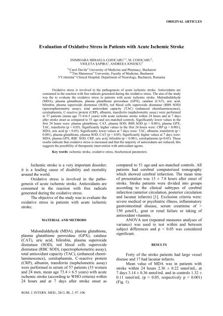
Mean value <strong>of</strong> MDA was <strong>in</strong> patients <strong>with</strong><br />
stroke <strong>with</strong><strong>in</strong> 24 hours 2.36 ± 0.22 nmol/mL, at<br />
7 days 3.14 ± 0.36 nmol/mL and <strong>in</strong> controls 1.32 ±<br />
0.11 nmol/mL (p < 0.05, respectively p < 0.001)<br />
(Fig. 1).<br />
ROM. J. INTERN. MED., 2013, 51, 2, 97–106
98<br />
Inimioara Mihaela Cojocaru et al. 2<br />
Mean value <strong>of</strong> plasma glutathione was <strong>in</strong><br />
patients <strong>with</strong> stroke <strong>with</strong><strong>in</strong> 24 hours 0.17 ±<br />
0.03 µmol/L, at 7 days 0.22 ± 0.04 µmol/L and <strong>in</strong><br />
controls 0.38 ± 0.07 µmol/L (p
3 <strong>Oxidative</strong> stress <strong>in</strong> acute ischemic stroke 99<br />
p < 0.001 p < 0.05<br />
Fig. 2. Mean value <strong>of</strong> plasma glutathione <strong>in</strong> patients <strong>with</strong> acute ischemic stroke <strong>with</strong><strong>in</strong> 24 hours,<br />
at 7 days, and <strong>in</strong> controls.<br />
p < 0.05 p < 0.001<br />
Fig. 3. Mean value <strong>of</strong> plasma glutathione peroxidase (GPX) <strong>in</strong> patients <strong>with</strong> acute ischemic stroke <strong>with</strong><strong>in</strong> 24 hours,<br />
at 7 days, and <strong>in</strong> controls.<br />
p < 0.001 p < 0.05<br />
Fig. 4. Mean value <strong>of</strong> catalase (CAT) <strong>in</strong> patients <strong>with</strong> acute ischemic stroke <strong>with</strong><strong>in</strong> 24 hours,<br />
at 7 days, and <strong>in</strong> controls.
100<br />
Inimioara Mihaela Cojocaru et al. 4<br />
p < 0.05 p < 0.001<br />
Fig. 5. Mean value <strong>of</strong> uric acid <strong>in</strong> patients <strong>with</strong> acute ischemic stroke <strong>with</strong><strong>in</strong> 24 hours, at 7 days, and <strong>in</strong> controls.<br />
p < 0.05 p < 0.001<br />
Fig. 6. Mean value <strong>of</strong> bilirub<strong>in</strong> <strong>in</strong> patients <strong>with</strong> acute ischemic stroke <strong>with</strong><strong>in</strong> 24 hours, at 7 days, and <strong>in</strong> controls.<br />
p < 0.001 p < 0.05<br />
Fig. 7. Mean value <strong>of</strong> plasma superoxide dismutase (SOD) <strong>in</strong> patients <strong>with</strong> acute ischemic stroke <strong>with</strong><strong>in</strong> 24 hours,<br />
at 7 days, and <strong>in</strong> controls.
5 <strong>Oxidative</strong> stress <strong>in</strong> acute ischemic stroke 101<br />
p < 0.001 p < 0.001<br />
Fig. 8. Mean value <strong>of</strong> red blood cells superoxide dismutase (RBC SOD) <strong>in</strong> patients<br />
<strong>with</strong> acute ischemic stroke <strong>with</strong><strong>in</strong> 24 hours, at 7 days, and <strong>in</strong> controls.<br />
p < 0.001 p < 0.001<br />
Fig. 9. Mean value <strong>of</strong> total antioxidant capacity (TAC) <strong>in</strong> patients <strong>with</strong> acute ischemic stroke<br />
<strong>with</strong><strong>in</strong> 24 hours, at 7 days, and <strong>in</strong> controls.<br />
p < 0.05 p < 0.05<br />
Fig. 10. Mean value <strong>of</strong> ceruloplasm<strong>in</strong> <strong>in</strong> patients <strong>with</strong> acute ischemic stroke <strong>with</strong><strong>in</strong> 24 hours, at 7 days, and <strong>in</strong> controls.
102<br />
Inimioara Mihaela Cojocaru et al. 6<br />
p < 0.001 p < 0.001<br />
Fig. 11. Mean value <strong>of</strong> C-reactive prote<strong>in</strong> (CRP) <strong>in</strong> patients <strong>with</strong> acute ischemic stroke <strong>with</strong><strong>in</strong> 24 hours,<br />
at 7 days, and <strong>in</strong> controls.<br />
p < 0.001 p < 0.001<br />
Fig. 12. Mean value <strong>of</strong> album<strong>in</strong> <strong>in</strong> patients <strong>with</strong> acute ischemic stroke <strong>with</strong><strong>in</strong> 24 hours, at 7 days, and <strong>in</strong> controls.<br />
p < 0.001 p < 0.001<br />
Fig. 13. Mean value <strong>of</strong> transferr<strong>in</strong> <strong>in</strong> patients <strong>with</strong> acute ischemic stroke <strong>with</strong><strong>in</strong> 24 hours,<br />
at 7 days, and <strong>in</strong> controls.
7 <strong>Oxidative</strong> stress <strong>in</strong> acute ischemic stroke 103<br />
DISCUSSION<br />
<strong>Oxidative</strong> stress is an <strong>in</strong>balance between<br />
oxidants and antioxidants <strong>in</strong> favor <strong>of</strong> the oxidants,<br />
potentially lead<strong>in</strong>g to damage. It has been proposed<br />
that antioxidant changes reflect an altered redox<br />
balance <strong>in</strong> several pathological states; antioxidants<br />
would be consumed <strong>in</strong> the reaction <strong>with</strong> free<br />
radicals.<br />
The <strong>in</strong>creased production <strong>of</strong> free radicals <strong>in</strong><br />
the sett<strong>in</strong>g <strong>of</strong> cerebral ischemia, <strong>with</strong> or <strong>with</strong>out<br />
reperfusion, can arise from several mechanisms:<br />
glutamate stimulation <strong>of</strong> NMDA receptors, mitochondrial<br />
dysfunction, activation <strong>of</strong> neuronal nitric<br />
oxide synthase, <strong>in</strong>duction <strong>of</strong> nitric oxide synthetase<br />
or cyclooxygenase 2, autooxidation <strong>of</strong> catecholam<strong>in</strong>es,<br />
metabolism <strong>of</strong> free fatty acids, particularly arachidonic<br />
acid, released dur<strong>in</strong>g ischemia, migration <strong>of</strong><br />
neutrophils and leukocytes able to generate superoxide<br />
anions, the conversion <strong>of</strong> xanth<strong>in</strong>e dehydrogenase<br />
to xanth<strong>in</strong>e oxidase [2–19].<br />
Lipid peroxidation, <strong>with</strong> accumulation <strong>of</strong><br />
both conjugated dienes and thiobarbiturate-reactive<br />
material, is consistently found when cerebral ischemia<br />
is followed by reperfusion. Hydroxyl radicals<br />
(formed from hydrogen peroxide), perox<strong>in</strong>itrite and<br />
superoxide are powerful radicals that can cause<br />
lipid peroxidation, a self propagat<strong>in</strong>g cha<strong>in</strong> reaction,<br />
that irreversibly damages plasma and mitochondrial<br />
membranes. Malondialdehyde product <strong>of</strong> lipid<br />
peroxidation, irreversibly disrupts enzymes, receptors<br />
and membrane transport mechanisms. The concentrations<br />
<strong>of</strong> MDA were significantly <strong>in</strong>creased <strong>in</strong><br />
stroke patients, aris<strong>in</strong>g from excess free radical<br />
activity [20–24].<br />
Uric acid is a powerful free radical scavenger<br />
<strong>in</strong> humans and these antioxidant properties could<br />
be expected to <strong>of</strong>fer a number <strong>of</strong> benefits <strong>with</strong><strong>in</strong> the<br />
cardiovascular system. No potential biological<br />
mechanisms are known by which raised UA concentrations<br />
could <strong>in</strong>fluence the development <strong>of</strong><br />
stroke. Therefore, it is unclear whether high UA<br />
concentrations promote or protect aga<strong>in</strong>st the<br />
development <strong>of</strong> cardio-vascular disease, or significant<br />
as a possible marker <strong>of</strong> <strong>in</strong>creased risk.<br />
There is some evidence that <strong>in</strong>creased<br />
oxidative stress is associated <strong>with</strong> high circulat<strong>in</strong>g<br />
uric acid levels and that uric acid may protect<br />
aga<strong>in</strong>st oxidative modification <strong>of</strong> endothelial enzymes<br />
and preserves the ability <strong>of</strong> endothelium to mediate<br />
vascular dilatation <strong>in</strong> the face <strong>of</strong> oxidative stress.<br />
Follow<strong>in</strong>g cerebral ischemia and reperfusion,<br />
the metabolism <strong>of</strong> nucleosides and pur<strong>in</strong>e bases to<br />
<strong>in</strong>os<strong>in</strong>e and hypoxanth<strong>in</strong>e via the xanth<strong>in</strong>e oxidase<br />
pathway, results <strong>in</strong> production <strong>of</strong> oxygen free<br />
radicals. Xanth<strong>in</strong>e oxidase is an enzyme that controls<br />
the formation <strong>of</strong> uric acid.<br />
Uric acid is the most abundant aqueus<br />
antioxidant <strong>in</strong> humans, and contributes as much as<br />
two-thirds <strong>of</strong> all free radical scaveng<strong>in</strong>g capacity <strong>in</strong><br />
the plasma. It is particularly effective <strong>in</strong> quench<strong>in</strong>g<br />
hydroxyl, superoxide and peroxynitrite, and may<br />
serve a protective physiological role by preserv<strong>in</strong>g<br />
lipid peroxidation [25–43].<br />
A number <strong>of</strong> components present <strong>in</strong> the serum<br />
have been shown to possess cha<strong>in</strong> break<strong>in</strong>g antioxidant<br />
capacity <strong>in</strong>clud<strong>in</strong>g album<strong>in</strong> and bilirub<strong>in</strong>.<br />
Enhanced total antioxidant capacity (TAC)<br />
after acute stroke may protect aga<strong>in</strong>st the adverse<br />
effects <strong>of</strong> free radicals production dur<strong>in</strong>g ischemia<br />
and reperfusion. The TAC <strong>of</strong> biological fluids is<br />
believed to be a useful measure <strong>of</strong> the ability <strong>of</strong><br />
antioxidant present <strong>in</strong> the fluids to protect aga<strong>in</strong>st<br />
oxidative damage to membranes and other cellular<br />
components [44–46].<br />
Both serum SOD and RBC SOD levels were<br />
lower <strong>in</strong> acute stroke patients. It is possible that the<br />
lower activity <strong>in</strong> the acute phase <strong>of</strong> stroke is a<br />
consequence <strong>of</strong> a higher oxidative damage [47–53].<br />
Mean serum levels <strong>of</strong> nonenzymatic antioxidants<br />
and antioxidant enzyme activities were<br />
lower <strong>in</strong> patients than <strong>in</strong> controls.<br />
Our study revealed that almost all antioxidants<br />
are reduced <strong>with</strong><strong>in</strong> 24 hours after an<br />
ischemic stroke and <strong>in</strong>crease over the follow<strong>in</strong>g<br />
days, suggest<strong>in</strong>g the presence <strong>of</strong> a condition <strong>of</strong><br />
oxidative stress <strong>in</strong> this disease [54–55].<br />
CONCLUSION<br />
These results <strong>in</strong>dicate that oxidative stress is<br />
<strong>in</strong>creased <strong>in</strong> acute ischemic stroke and that the<br />
majority <strong>of</strong> antioxidants are reduced, which<br />
suggests the possibility <strong>of</strong> therapeutic <strong>in</strong>tervention<br />
<strong>with</strong> antioxidant agents.<br />
Stresul oxidativ este implicat în patogenia stroke-ului ischemic acut.<br />
Antioxidanţii sunt consumaţi în reacţia cu radicalii liberi generaţi în cursul<br />
stresului oxidativ. Scopul studiului a fost evaluarea stresului oxidativ la pacienţii
104<br />
Inimioara Mihaela Cojocaru et al. 8<br />
cu stroke ischemic acut. Au fost <strong>in</strong>vestigate malondialdehida (MDA), glutationul<br />
plasmatic, glutation peroxidaza plasmatică (GPX), catalaza (CAT), acidul uric,<br />
bilirub<strong>in</strong>a, superoxid dismutaza plasmatică (SOD), superoxid dismutaza eritrocitară<br />
(RBS SOD) (colorimetrie), capacitatea antioxidantă totală (TAC) (chemilum<strong>in</strong>escenţă),<br />
ceruloplasm<strong>in</strong>a, prote<strong>in</strong>a C-reactivă (CRP), album<strong>in</strong>a, transfer<strong>in</strong>a<br />
(nefelometrie) la 57 pacienţi (vârsta medie 73,4 ± 6,5 ani) cu stroke ischemic acut în<br />
primele 24 ore şi la 7 zile de la debutul stroke-ului comparativ cu un lot martor de<br />
51 subiecţi cu vârste şi sex comparabile. Au prezentat valori semnificativ reduse în<br />
primele 24 ore: glutationul plasmatic, catalaza (CAT), superoxid dismutaza plasmatică<br />
(SOD), superoxid dismutaza eritrocitară (RBS SOD), (p
9 <strong>Oxidative</strong> stress <strong>in</strong> acute ischemic stroke 105<br />
14. WEI G., DAWSON V. L., ZWAEIER J. L. et al., Role <strong>of</strong> neuronal and endothelial nitric oxide synthase <strong>in</strong> nitric oxide<br />
generation <strong>in</strong> the bra<strong>in</strong> follow<strong>in</strong>g cerebral ischemia Biochim. Biophys. Acta. 1999; 1455: 23-4.<br />
15. SETHI S., SINGH M. P., DIKSHIT M. et al.Mechanisms <strong>in</strong>volved <strong>in</strong> the augmentation <strong>of</strong> archidonic acid-<strong>in</strong>duced free radical<br />
generation from rat neutrophils follow<strong>in</strong>g hypoxia-reoxygenation Thrombosis Res. 2000; 98: 445-50<br />
16. KANNO T., ABE K., YABUKI M. et al., Selective <strong>in</strong>hibition <strong>of</strong> formyl-methionyl-leucyl-phenylalan<strong>in</strong>e (fMLF)-dependent<br />
superoxide generation <strong>in</strong> neutrophils by pravastat<strong>in</strong>, an <strong>in</strong>hibitor <strong>of</strong> 3-hydroxy-3-methylglutaryl coenzyme A (HMG-CoA)<br />
reductase Biochem. Pharm. 1999; 58: 1975-80.<br />
17. GLUTHIKONDA S., SINKEY C., BARANZ T. et al., Xant<strong>in</strong>e oxidase <strong>in</strong>hibition reverses endothelial dysfunction <strong>in</strong> heavy<br />
smokers Circulation. 2003; 107: 416-21.<br />
18. LIU S., LIU M., PETERSON S. et al., Hydroxyl radical formation is greater <strong>in</strong> striatal core than <strong>in</strong> penumbra <strong>in</strong> a rat model <strong>of</strong><br />
ischemic stroke. J. Neurosci. Res. 2003; 71: 882-8.<br />
19. OHTSUBO T., ROVIRA I. I., STAROST M. F. et al., Xanth<strong>in</strong>e oxidoreductase is an endogenous regulator <strong>of</strong> cyclooxygenase-2.<br />
Circ. Res. 2004; 95: 1118-24<br />
20. SAKAMOTO A., OHNISHI S. T., OGAWA R. et al.Relationship between free radical production and lipid peroxidation<br />
dur<strong>in</strong>g ischemia/reperfusion <strong>in</strong>jury <strong>in</strong> rat bra<strong>in</strong>. Bra<strong>in</strong> Res. 1991; 554: 186-92.<br />
21. SHARPE P. C., MULHOLLAND C., TRINICEK T. et al., Ascorbate and malondialdehyde <strong>in</strong> stroke patients. Ir. J. Med. Sci.<br />
1994; 163: 488-91.<br />
22. RE G., AZZIMONDI G., LANZARINI C. et al., Plasma lipoperoxidative markers <strong>in</strong> ischemic stroke suggest bra<strong>in</strong> embolism.<br />
Eur. J. Emerg. Med. 1997; 4: 5-9.<br />
23. VAN KOOTEN F., CIABATTONI G., PATRONO C., et al., Platelet activation and lipid peroxidation <strong>in</strong> patients <strong>with</strong> acute<br />
ischemic stroke. <strong>Stroke</strong> 1997; 28:1557-63.<br />
24. McCRAKEN E., VALERIANI V., SIMPSON C. et al., The lipid peroxidation product 4-hydroxynonenal is toxic to axons and<br />
olygodendrocytes. J. Cereb. Blood Flow Metab. 2000; 20: 1529-36.<br />
25. LEHTO S., NISKANEN L., RONNENMAA T. et al., Serum uric acid is a strong predictor <strong>of</strong> stroke <strong>in</strong> patients <strong>with</strong> non<strong>in</strong>sul<strong>in</strong>-dependent<br />
diabetes mellitus. <strong>Stroke</strong> 1998; 29: 635-9.<br />
26. YU Z. F., BRUCE-KELLER A. J., GOODMANN Y., et al., Uric acid protects neurons aga<strong>in</strong>st focal ischemic bra<strong>in</strong> <strong>in</strong>jury <strong>in</strong><br />
vivo. J. Neurosci. Res. 1998; 33: 613-25.<br />
27. DOBSON A. Is raised uric acid a cause <strong>of</strong> cardiovascular disease or death? Lancet. 1999; 354: 1578-85.<br />
28. FRANSE L. V., PAHOR M., DI BARI M. et al., Serum uric acid, diuretic treatment and risk <strong>of</strong> cardiovascular events <strong>in</strong> the<br />
Systolic Hypertension <strong>in</strong> the Elderly. Program (SHEP). J. Hypertens. 2000; 18: 1149-5.<br />
29. WARING W. S. et al., Uric acid as a risk factor for cardiovascular disease. Q. J. Med. 2000; 93: 707-13.<br />
30. SQUADRITO G. L., CUETO R., SPLENSER A. E. et al., Reaction <strong>of</strong> uric acid <strong>with</strong> peroxynitrite and implications for the<br />
mechanism <strong>of</strong> neuroprotection by uric acid. Arch. Biochem. Biophys. 2000; 376: 333-7.<br />
31. NIETO F. J., IRRIBAREN C., GROSS M. D. et al., Uric acid and serum antioxidant capacity: a reaction to atherosclerosis?<br />
Atherosclerosis. 2000; 148: 131-9.<br />
32. JOHNSON R. J., KANG D. H., FEIG D. et al., Much ado about noth<strong>in</strong>g or much to do about someth<strong>in</strong>g? The cont<strong>in</strong>u<strong>in</strong>g<br />
controversy over the role <strong>of</strong> uric acid <strong>in</strong> cardiovascular disease. Hypertension. 2000; 35: e10-10.<br />
33. WARING W. S., Uric acid: an important antioxidant <strong>in</strong> acute ischaemic stroke. Q. J. Med. 2002; 95: 691-3.<br />
34. CHAMORRO A., OBACH V., CERVERA A. et al., Prognostic significance <strong>of</strong> uric acid serum concentration <strong>in</strong> patients <strong>with</strong><br />
acute ischemic stroke. <strong>Stroke</strong>. 2002; 33: 1048-52.<br />
35. SEGHIERI G., MORUZZO D., FASCETTI S. et al., Increase <strong>in</strong> serum uric acid is selectively associated <strong>with</strong> stroke <strong>in</strong> type 2<br />
diabetes Diabetes Care. 2002; 25: 1095.<br />
36. WARING W. S., WEBB D. J., MAXWELL S. R. et al., Systemic uric acid adm<strong>in</strong>istration <strong>in</strong>creases serum antioxidant capacity<br />
<strong>in</strong> healthy volunteers. J. Cardiovasc. Pharmacol. 2001; 38: 365-71.<br />
37. KANELLIS J., JONSON R. J. et al., Editorial comment-Elevated uric acid and ischemic stroke: accumulat<strong>in</strong>g evidence that it is<br />
<strong>in</strong>jurious and not neuroprotective. <strong>Stroke</strong>. 2003; 34: 1956-57<br />
38. WEIR C. J., MUIR S. W., WALTERS M. R. et al., Serum urate as an <strong>in</strong>dependent predictor <strong>of</strong> poor outcome and future<br />
vascular events after acute stroke. <strong>Stroke</strong>. 2003; 34: 1951-6.<br />
39. CHAMORRO A., PLANAS A. M., WEIR C. J. et al., Y<strong>in</strong> and Yang <strong>of</strong> uric acid <strong>in</strong> patients <strong>with</strong> stroke. <strong>Stroke</strong>. 2004; 35: e11-2.<br />
40. WARING W. S., ADWANI S. H., BREUKELS O. et al., Hyperuricaemia does not impair cardiovascular function <strong>in</strong> healthy<br />
adults. Heart. 2004; 90: 155-9.<br />
41. STINEFELT B., LEONARD S. S., BLEMINGS K. P. et al., Free radical scaveng<strong>in</strong>g, DNA protection, and <strong>in</strong>hibition <strong>of</strong> lipid<br />
peroxidation mediated by uric acid. Ann. Cl<strong>in</strong>. Lab . Sci. 2005; 35: 37-45.<br />
42. SUNDSTROM J., SULLIVAN L., D’AGOSTINO R. B. et al., Relation <strong>of</strong> serum uric acid to longitud<strong>in</strong>al blood pressure<br />
track<strong>in</strong>g and hypertension <strong>in</strong>cidence. Hypertension. 2005; 45: 28-33.<br />
43. ISHIZAKA M., ISHIZAKA Y., TODA E. I. et al., Association between serum uric acid, metabolic syndrome, and carotid<br />
atherosclerosis <strong>in</strong> Japanese <strong>in</strong>dividuals. Atherioscler. Thromb. Vasc. Biol. 2005; 25: 1038-44.<br />
44. MITREVSKI A., PEMOV P., POPOVSKI A. et al., Total antioxidant status and superoxide dismutase after cerebrovascular<br />
accident. Proc XVI ICC 1996; 68.<br />
45. RYAN M., GRAYSON L., CLARKE D. J. et al., The total antioxidant capacity <strong>of</strong> human serum measured us<strong>in</strong>g enhanced<br />
chemilum<strong>in</strong>escence is almost completely accounted for by urate. Ann. Cl<strong>in</strong>.Biochem. 1997; 34: 688-9.<br />
46. YOUNG I. S. Measurement <strong>of</strong> total antioxidant capacity. J. Cl<strong>in</strong>. Pathol. 2001; 54: 339-40.<br />
47. KINOUCHII H., EBSTEIN C. J., MIZUI T. et al., Attenuation <strong>of</strong> focal cerebral ischemic <strong>in</strong>jury <strong>in</strong> transgenic mice<br />
overexpress<strong>in</strong>g CuZn superoxide dismutase. Proc. Natl. Acad. Sci. USA 1991; 88: 11158-62.
106<br />
Inimioara Mihaela Cojocaru et al. 10<br />
48. GRUENER N., GROSS B., GOZBAN O. et al., Increase <strong>in</strong> superoxide dismutase after cerebral accident. Life Sci. 1994; 54:<br />
711-3.<br />
49. SPRANGER M., KREMPIEN S., SCHWAB S. et al., Superoxide dismutase activity <strong>in</strong> serum <strong>of</strong> patients <strong>with</strong> acute cerebral<br />
ischemic <strong>in</strong>jury. Correlation <strong>with</strong> cl<strong>in</strong>ical course and <strong>in</strong>farct size. <strong>Stroke</strong> 1997; 28: 2425-8<br />
50. BAKER K., BUCAY M. C., HUFFMAN K. et al., Synthetic comb<strong>in</strong>ed superoxide dismutase/catalase mimetics are protective as<br />
a delayed treatment <strong>in</strong> a rat stroke model: a key role for reactive oxygen species <strong>in</strong> ischemic bra<strong>in</strong>. J. Pharmacol. Exper. Therap.<br />
1998; 284: 215-24.<br />
51. FUJIMURA M., MORIRA-FUJIMURA Y., KAWASE M. et al., Manganese superoxide dismutase mediates the early release <strong>of</strong><br />
mitochondrial cytocrome C and subsequent DNA fragmentation after permanent focal cerebral ischemia <strong>in</strong> mice. J. Neurosci.<br />
1999; 19: 3414-22.<br />
52. HINK H. U., SANTANAM N., DIKALOV S. et al., Peroxidase properties <strong>of</strong> extracellular superoxide dismutase. Role <strong>of</strong> uric<br />
acid <strong>in</strong> modulat<strong>in</strong>g <strong>in</strong> vivo activity. Atherosclerosis, Thrombosis, and Vascular Biology. 2002; 222: 1402-8.<br />
53. CIRIOLO M. R., DE MARTINO A., LAFAVIA E. et al., CuZn-superoxide dismutase dependent apoptosis <strong>in</strong>duced by nitric<br />
oxide <strong>in</strong> neuronal cells. J. Biol.Chem. 2000; 275: 5065-72<br />
54. COOPER A. J. L., PULSINELLI W. A., DUFFY T. E. et al., Glutathione and ascorbate dur<strong>in</strong>g ischemia and post-ischemic<br />
reperfusion <strong>in</strong> rat bra<strong>in</strong>. J. Neurochem. 1980; 35: 1242-5.<br />
55. SCHNABEL R., LACEKNER K. J., RUPPRECHT H. J. et al., Glutathione peroxidase-1 and homocyste<strong>in</strong>e for cardiovascular<br />
risk prediction: results from the AtheroGene Study. J. Am. Coll. Cardiol.2005; 45: 1631-7.<br />
Received April 20, 2013