o_195effea7fovm0stco1dgrk2ta.pdf

Create successful ePaper yourself
Turn your PDF publications into a flip-book with our unique Google optimized e-Paper software.
Case Report<br />
DOI: 10.7241/ourd.20144.105<br />
IMMUNOREACTIVITY TO DERMAL VESSELS IN A<br />
PATIENT WITH PYODERMA GANDRENOSUM<br />
Ana Maria Abreu Velez 1 , Michael S. Howard 1 , Vickie M. Brown 2<br />
Source of Support:<br />
Georgia Dermatopathology<br />
Associates (AMAV, MSH), USA<br />
Competing Interests:<br />
None<br />
1<br />
Georgia Dermatopathology Associates, Atlanta, Georgia, USA<br />
2<br />
Family Dermatology, Milledgeville, Georgia, USA<br />
Corresponding author: Ana Maria Abreu Velez, M.D., Ph.D.,<br />
abreuvelez@yahoo.com<br />
Our Dermatol Online. 2014; 5(4): 419-422 Date of submission: 08.08.2014 / acceptance: 19.09.2014<br />
Abstract<br />
Introduction: Pyoderma gangrenosum (PG) represents a lesion with an elusive etiology associated with Crohn’s and/or arthritic diseases. A<br />
genuine associated vasculitis has not been proven; however, dilation of the dermal blood vessels the presence of neutrophilic and/or lymphocytic<br />
infiltrates in selected biopsies suggests that vessels are likely play a role in the pathogenesis of PG.<br />
Materials and Methods: A patient presented with a necrotic pustule or furuncle, evolving into a large necrotic ulcer with violaceous borders<br />
and surrounding erythema. A skin biopsy for hematoxylin and eosin (H&E) review and immunohistochemistry (IHC) stains was obtained. A<br />
second biopsy for direct immunofluorescence (DIF) was also taken.<br />
Results: H&E review demonstrated an ulcerated epidermis; within the ulcer base were numerous neutrophils, lymphocytes, histiocytes and<br />
fibrin. No vasculitis was present. DIF revealed strong deposits of FITC conjugated fibrinogen around superficial and the deep dermal vessels.<br />
FITC conjugated Complement/C1q and albumin conjugated were also seen between dermal extracellular matrix fibers. IHC showed that the<br />
dermal vessels (venules, arterioles and lymphatics) displayed dilation, and a loss of normal endothelial markers including von Willebrand<br />
factor and D2-40/podoplanin.<br />
Conclusions: Our case of PG shows that there seems to be an alteration of several skin structures, including dermal vessels. An alteration of<br />
the vascular fibrin-fibrinogen balance was also detected, causing some autoreactivity to fibrinogen and an immune response involving<br />
neutrophils and T lymphocytes. Our findings suggest that the etiology of PG is more complex than previously thought.<br />
Key words: Pyoderma gangrenosum; von Willebrand factor; vascular degeneration, autoimmunity; lymphatic vessels; CD99; D2-40<br />
Abbreviations and acronyms: Pyoderma gangrenosum (PG), immunohistochemistry (IHC), direct immunofluorescence (DIF), hematoxylin<br />
and eosin (H&E).<br />
Cite this article:<br />
Abreu Velez AM, Brown VM, Howard MS. Immunoreactivity to dermal vessels in a patient with pyoderma gandrenosum. Our Dermatol Online. 2014; 5(4): 419-<br />
422..<br />
Introduction<br />
Pyoderma gangrenosum (PG) is an uncommon cause<br />
of painful skin ulceration. It may affect any part of the skin;<br />
however, it mainly presents on the lower legs, and has an<br />
unknown etiology resembling a Class II Schwartzman-like<br />
hypersensitivity reaction. PGs usually initially present as a<br />
single pustule, progress to solitary or multiple erythematous<br />
macules and papules and then to ulcerated, painful nodules or<br />
plaques with undermined, dusky borders. Patients are often<br />
systemically ill, with associated symptoms such as fever,<br />
malaise, arthralgias, and myalgias. Lesional pain may be<br />
severe. When lesions heal, the scars are often cribriform. PG<br />
is sometimes classified as a neutrophilic dermatosis, and has<br />
often been considered a reaction to another internal disease<br />
such as Crohn’s disease, ulcerative colitis, polyarteritis nodosa<br />
or hematologic alterations. PG may affect males and females<br />
of any age, but is more common in people over 50 years [1-5].<br />
PG combined with acne and a pyogenic arthritis may represent<br />
a rare genetic disorder characterized by its effects on skin and<br />
joints (PAPA syndrome).<br />
Case Report<br />
A 58 year old female presented to her dermatologist for<br />
evaluation of a pustule or furuncle that was evolving into an<br />
ulcer. The patient denied any gastrointestinal, acne or joint<br />
problems concomitant with the presentation of the skin lesions.<br />
Physical exam revealed a large, necrotic ulcer with violaceous<br />
border and surrounding erythema.<br />
www.odermatol.com<br />
© Our Dermatol Online 4.2014 419
On examination, the ulcer was tender. Skin biopsies were obtained<br />
for hematoxylin and eosin (H&E) review, immunohistochemistry<br />
(IHC) staining and direct immunofluorescence (DIF) studies.<br />
Material and Methods<br />
For DIF, we incubated 4 micron glass slides with our<br />
secondary antibodies as previously described [6-9]. We<br />
utilized FITC conjugated rabbit anti-total IgG (Dako,<br />
Carpinteria, California, USA) at a 1:25 dilution. The samples<br />
were run with positive and negative controls. We also utilized<br />
FITC conjugated rabbit antisera to human IgG, IgA, IgM,<br />
Complement/C1q, Complement/C3, fibrinogen and albumin.<br />
Anti-human IgA antiserum (alpha chain) and anti-human IgM<br />
antiserum (mu chain) were obtained from Dako. Anti-human<br />
IgE antiserum (epsilon chain) was obtained from Vector<br />
Laboratories (Burlingame, California, USA). Anti-human IgD<br />
FITC-conjugated antibodies were obtained from Southern<br />
Biotechnology (Birmingham, Alabama, USA). The slides were<br />
counterstained with 4’,6-diamidino-2-phenylindole(DAPI)<br />
(Pierce, Rockford, Illinois, USA). A mouse anti-collagen IV<br />
monoclonal antibody (Invitrogen, Carlsbad, California, USA)<br />
was also utilized, with secondary donkey anti-mouse IgG<br />
antisera (heavy and light chains) conjugated with Alexa Fluor<br />
555 (Invitrogen).<br />
Immunohistochemistry (IHC)<br />
We performed IHC utilizing multiple monoclonal and<br />
polyclonal antibodies, all from Dako (Carpinteria, California,<br />
USA). For all our IHC testing, we used a dual endogenous<br />
peroxidase blockage, with the addition of an Envision dual<br />
link (to assist in chromogen attachment). We applied the<br />
chromogen 3,3-diaminobenzidine(DAB), and counterstained<br />
with hematoxylin. The samples were run in a Dako Autostainer<br />
Universal Staining System. Positive and negative controls were<br />
consistently performed. Our studies were specifically performed<br />
as previously described [6-9].The following antibodies were<br />
utilized: 1) monoclonal mouse anti-human CD99, MIC2 gene<br />
products, Ewing’s sarcoma marker Clone 12E7, monoclonal<br />
mouse anti-human, 2) von Willebrand Factor, Clone F8/86, 3)<br />
polyclonal rabbit anti-human myeloperoxidase, monoclonal<br />
mouse anti-human neutrophil elastase Clone NP57, 4) polyclonal<br />
rabbit anti-human somatostatin, 5) monoclonal mouse antihuman<br />
podoplanin, Clone D2-40, 6) monoclonal mouse antihuman<br />
CD4, Clone 4B12, 7) monoclonal mouse anti-human<br />
CD45, Leucocyte Common Antigen Clones 2B11 + PD7/26,<br />
8) monoclonal mouse anti-human CD8, Clone C8/144B, 9)<br />
monoclonal mouse anti-human IMP3 (Clone 69.1), (insulinlike<br />
growth factor II mRNA binding protein 3, 10) monoclonal<br />
mouse anti-human CD68 Clone EBM11, 11) monoclonal<br />
mouse anti-metallothionein Clone E9, 12) monoclonal mouse<br />
anti-human tissue inhibitor of metalloproteinases 1 and 13)<br />
polyclonal rabbit anti-human alpha-1-antitrypsin.<br />
Resulrs<br />
H&E tissue sections demonstrate an ulcerated epidermis<br />
(Fig. 1). Within the ulcer base were numerous neutrophils,<br />
lymphocytes and histiocytes; abundant fibrin was also present.<br />
Several vessels were dilated (Fig. 3). No vasculitis was noted; a<br />
dermal perivascular and interstitia infiltrate of lymphocytes and<br />
mononuclear cells was present. Fungal and bacterial stains were<br />
negative for organisms.<br />
Direct immunofluorescence (DIF)<br />
DIF displayed the following results: IgG (+, focal deep dermal<br />
perivascular); IgG3(-); IgA (++, focal deep dermal perivascular);<br />
IgM (+); IgD (-); IgE (-); Complement/C1q (++, focal deep<br />
dermal perivascular); complement/C3 (+, focal deep dermal<br />
perivascular); complement/C4(-); albumin (++, superficial<br />
dermal perivascular) and fibrinogen (++++, superficial and deep<br />
dermal perivascular, and around eccrine gland supply vessels).<br />
Fibrinogen was the strongest immunoreactant seen around<br />
the upper and deep dermal vessels, with Complement/C1q of<br />
second strongest intensity (Figs 1 - 4). In Figure 4, using Alexa<br />
555 conjugated anti-human IgG antibody (red staining) we show<br />
positive staining of possible damaged vessels, amalgamated<br />
with some dermal structures<br />
IHC staining<br />
CD99 staining was very positive on dermal vessels subjacent<br />
to and surrounding the ulcer (Fig. 2). Somatostatin was weakly<br />
positive, within the dermal extracellular matrix. Our most<br />
important finding was the presence of significantly dilated<br />
lymphatic vessels via D2-40 staining, especially surrounding<br />
and subjacent to the ulcer (Fig. 2) Myeloperoxidase staining was<br />
positive, and showed some cellular defragmentation around one<br />
edge of the ulcer. Neutrophil elastase, CD4 and CD8 staining<br />
were predominantly negative. CD45 staining was positive in<br />
only few focal perivascular areas, adjacent to the ulcer. Antiinsulin-like<br />
growth factor II/mRNA binding protein 3 staining<br />
was negative. Of note, many dermal vessels were involuted and<br />
significantly damaged, observed by staining alterations with the<br />
von Willebrand antibody (Fig. 3).<br />
Discussion<br />
PG onsets are rapid, often at the site of a minor injury. The<br />
lesion may begin as a small pustule, a red bump or a bloodblister,<br />
with these lesions subsequently progressing to an ulcer.<br />
The ulcer often grows rapidly; the edges are usually purple<br />
and very painful and tender. Multiple ulcers may progress at<br />
the same time [1-5]. An ulcer may spontaneously improve, and<br />
complete healing may take months. It is important to evaluate<br />
if concomitant local venous disease is present, because this<br />
may play a negative role in the healing process. The clinical<br />
pathergy test is frequently positive (a skin prick test causing<br />
a papule, pustule or ulcer). Peristomal PG occurs close to<br />
abdominal stomas, and comprises about 15% of all cases of<br />
PG [1-2]. Most of these patients have inflammatory bowel<br />
disease, but peristomal PG may also occur in patients who<br />
have had an ileostomy or colostomy for either malignancy or<br />
diverticular disease. However, about 50% of those affected by<br />
PG demonstrate none of these associated risk factors.<br />
Clinical differential diagnoses of PG lesions include<br />
antiphospholipid antibody syndrome, anthrax, Wegener’s<br />
granulomatosis, atypical mycobacterial infections, Sweet’s<br />
syndrome, venous or arterial disease, and Behçet’s disease [1-5].<br />
A final diagnosis of PG is based on the clinical appearance, and<br />
on ruling out histopathologic features of other disorders. There<br />
is no specific positive laboratory test for PG. However, it is<br />
recommended to swab PG ulcers for superinfecting pathogens.<br />
In our case, these studies were negative. It is also recommended<br />
to perform a chest radiogram. Angiography<br />
420 © Our Dermatol Online 4.2014
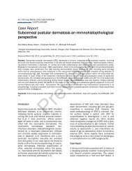
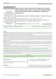
Figure 1. a, Clinical PG photograph, showing the PG ulcer. b,c.<br />
H&E photos at lower and higher magnification, respectively. In<br />
b, an ulcerated epidermis with numerous neutrophils,<br />
lymphocytes and histiocytes within the ulcer base; fibrin was also<br />
present. In c, we show several dilated dermal blood vessels. d-f.<br />
DIF. d-f. IgG conjugated with Alexa 555 (yellow staining) and<br />
FITC conjugated anti-fibrinogen(white arrows; green staining)<br />
on dermal lymphatic vessels.<br />
Figure 3. a-c. IHC staining, demonstrating damage to<br />
dermal vessels (showing degenerative changes and<br />
uneven staining with von Willebrand antibody (red<br />
arrows; light brown staining). d. Positive IHC staining,<br />
demonstrating a large, dilated lymphatic vessel via D2-40<br />
antibody (red arrow; brown staining).<br />
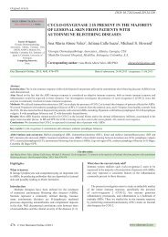
Figure 2. a, c, and f, CD99 positive IHC staining on<br />
dermal vessel endothelia(brown staining, black<br />
arrows). b. H & E staining, demonstrating dilated<br />
vessels in the dermis (400x) d. Positive IHC<br />
staining for myeloperoxidase (black arrows;<br />
brown staining). e. Positive IHC somatostatin<br />
staining in small dermal vessels of unknown<br />
identity (black arrows; light brown staining color).<br />
Figure 4. a. DIF, with Alexa 555 conjugated IgG (red<br />
staining) shows a tremendous deposition on some dermal<br />
structures (scattered red dots), and FITC conjugated<br />
fibrinogen (white arrow; green staining). Cell nuclei were<br />
counterstained with DAPI (blue). b. Positive D2-40 IHC<br />
staining documenting dilated dermal lymphatics (red<br />
arrow; brown staining) and in c, evidence of lymphatic<br />
vessel endothelial fragmentation(red arrow; brown<br />
staining). d. Positive DIF staining with Alexa 555<br />
conjugated IgG (red staining) showing possible damaged<br />
vessels amalgamated with unidentified dermal structures<br />
(white arrows). Cell nuclei were counterstained with<br />
DAPI (blue).<br />
© Our Dermatol Online 4.2014 421
Angiography or Doppler studies may be carried out in patients<br />
suspected of having arterial or venous insufficiency, and<br />
colonoscopy or other testing may be warranted to exclude<br />
associated inflammatory bowel disease in selected patients [1-<br />
5].<br />
PG treatment includes the gentle removal of necrotic tissue.<br />
Small ulcers may be treated with potent topical steroid<br />
creams, intralesional steroid injections, and special dressings<br />
including silver sulfadiazine cream, potassium iodide or<br />
hydrocolloids. In some instances, oral anti-inflammatory agents<br />
such as dapsone or minocycline may help. If tolerated, careful<br />
compression bandaging may be helpful for swollen legs. Some<br />
severe cases warrant extended, systemic immunosuppressive<br />
therapy. The focus of treatment in these patients is long-term<br />
immunosuppression, often with high doses of corticosteroids<br />
or low doses of cyclosporin [2,3]. Lately, good outcomes have<br />
been reported for treatments with anti-tumor necrosis factor α;<br />
infliximab has also proven effective in a randomized, controlled<br />
clinical trial.<br />
The etiopathology of PG is not clear. Some people believe<br />
that neutrophils play a critical role in this disease, because is<br />
common to see a neutrophilic dermal inflammatory infiltrate (as<br />
we found); however, this finding is not always present. Some<br />
patients may have positive ANCAs (antineutrophil cytoplasmic<br />
antibodies). In our case, these antibodies were negative. We<br />
found many defragmented neutrophils via positive staining for<br />
myeloperoxidase at the edges of the ulcer. Most T lymphocytic<br />
stains were negative (ie, CD4 and CD8; a few CD45 positive<br />
cells were noted). We found altered dermal vessels, and some<br />
degeneration of the vessels as demonstrated by altered staining<br />
of von Willebrand factor (especially under the ulcer and in close<br />
proximity). We also found many dermal vessels in these areas to<br />
be dilated, including the lymphatics; and we detected a immune<br />
response to these vessels, as demonstrated by positive CD99<br />
staining in many of them. In addition, strong staining with antifibrinogen<br />
against these vessels was clearly present. The specific<br />
etiologic role of this vascular immune response is not known.<br />
Other authors had reported clinical, histologic, and<br />
immunofluorescent findings in 68 PG cases. Notably, 78<br />
per cent had associated systemic diseases, with arthritis and<br />
inflammatory bowel disease being most common. These<br />
authors reported cutaneous histopathologic changes varied<br />
with the site of biopsy. Specifically, lymphocytic vasculitis was<br />
predominant in the zone of erythema peripheral to the ulcers,<br />
while neutrophilic infiltrates and abscess formation were more<br />
prominent in central ulcer areas. In most of the cases studied,<br />
DIF showed immunoglobulins and complement deposited in<br />
and around superficial and deep dermal vessels, as we also<br />
observed [10].<br />
Other authors reported the results of biopsies from ulcer<br />
edges in 8 PG patients. These biopsies were examined by DIF.<br />
Deposits of Complement/C3 were seen in the vessel walls of<br />
all samples, and IgM in three and IgA in one. Granular deposits<br />
of Complement/C3 were seen at the dermal-epidermal junction<br />
in 2 patients. Biopsies from clinically normal skin of 6 of the<br />
patients were DIF negative [11]. In agreement with our findings,<br />
these authors suggested that deposition of immune complexes in<br />
multiple dermal vessel walls may play a role in the pathogenesis<br />
of PG [11].<br />
Conclusion<br />
PG presents with multiple clinical appearances, and is easy<br />
to misdiagnose. The histopathology of PG is nonspecific; a<br />
biopsy is needed to exclude other diagnoses. Given our data,<br />
multiple dermal vessels including lymphatics, venules and<br />
arterioles seem to play a role in the immunopathology of PG.<br />
REFERENCES<br />
1. Weizman A, Huang B, Berel D, Targan SR, Dubinsky M, Fleshner<br />
P, et al. Clinical, Serologic, and Genetic Factors associated with<br />
pyoderma gangrenosum and erythema nodosum in inflammatory<br />
bowel disease patients. Inflamm Bowel Dis. 2014;20:525-33.<br />
2. Dantzig PI. Letter: Pyoderma gangrenosum. N Engl J Med.<br />
1975;292:47-8.<br />
3. Bernard P, Amici JM, Catanzano G, Cardinaud F, Fayol<br />
J, Bonnetblanc JM. Pyoderma gangrenosum and vasculitis.<br />
Pathogenic discussion apropos of 3 cases. Ann Dermatol Venereol.<br />
1987;114:1229-34.<br />
4. Su WP, Schroeter AL, Perry HO, Powell FC. Histopathologic and<br />
immunopathologic study of pyoderma gangrenosum. J Cutan Pathol.<br />
1986;13:323-30.<br />
5. Brooklyn T, Dunnill G. Diagnosis and treatment of pyoderma<br />
gangrenosum. BMJ. 2006;333:181–4.<br />
6. Abreu Velez AM, Howard MS, Grossniklaus HE, Gao W, Hashimoto<br />
T. Neural system antigens are recognized by autoantibodies from<br />
patients affected by a new variant of endemic pemphigus foliaceus in<br />
Colombia. Clin Immunol. 2011;31:356-68.<br />
7. Abreu Velez AM, Smith JG Jr, Howard MS. Neutrophil extracellular<br />
traps (NETS), IgD, myeloperoxidase (MPO) and antineutrophil<br />
cytoplasmic antibody (ANCA) associated vasculitides. North Am J<br />
Med Sci. 2009;1:309-13.<br />
8. Abreu Velez AM, Zhe J, Howard MS, Dudley SC. Cardiac<br />
immunoreactivity in patients affected by a new variant of endemic<br />
pemphigus foliaceus in El-Bagre, Colombia, South America. J Clin<br />
Immunol. 2011;31:985-97.<br />
9. Abreu Velez AM, Yi H, Googe PB Jr, Mihm MC Jr, Howard MS.<br />
Autoantibodies to melanocytes and characterization of melanophages<br />
in patients affected by a new variant of endemic pemphigus foliaceus.<br />
J Cutan Pathol. 2011;38:710-9.<br />
10. Powell FC, Schroeter AL, Perry HO, Su WP. Direct<br />
immunofluorescence in pyoderma gangrenosum. Br J Dermatol.<br />
1983;108:287-93.<br />
11. Ullman S, Halberg P, Howitz J.Deposits of complement and<br />
immunoglobulins in vessel walls in pyoderma gangrenosum. Acta<br />
Derm Venereol. 1982;62:340-1.<br />
Copyright by Ana Maria Abreu Velez, et al. This is an open access article distributed under the terms of the Creative Commons Attribution<br />
License, which permits unrestricted use, distribution, and reproduction in any medium, provided the original author and source are credited.<br />
422 © Our Dermatol Online 4.2014