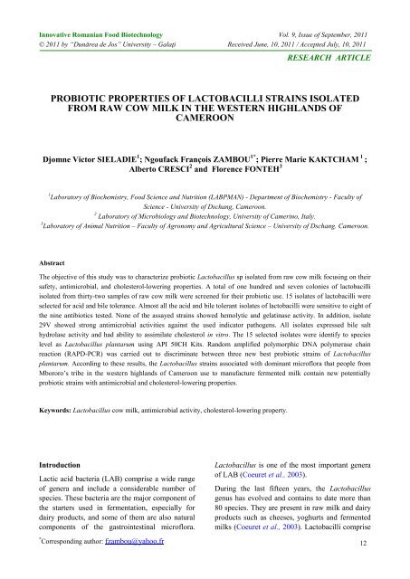
probiotic properties of lactobacilli strains isolated from ... - Bioaliment
probiotic properties of lactobacilli strains isolated from ... - Bioaliment
probiotic properties of lactobacilli strains isolated from ... - Bioaliment

Create successful ePaper yourself
Turn your PDF publications into a flip-book with our unique Google optimized e-Paper software.
Innovative Romanian Food Biotechnology Vol. 9, Issue <strong>of</strong> September, 2011<br />
© 2011 by “Dunărea de Jos” University – Galaţi Received June, 10, 2011 / Accepted July, 10, 2011<br />
RESEARCH ARTICLE<br />
PROBIOTIC PROPERTIES OF LACTOBACILLI STRAINS ISOLATED<br />
FROM RAW COW MILK IN THE WESTERN HIGHLANDS OF<br />
CAMEROON<br />
Djomne Victor SIELADIE 1 ; Ngoufack François ZAMBOU 1* ; Pierre Marie KAKTCHAM 1 ;<br />
Alberto CRESCI 2 and Florence FONTEH 3<br />
1 Laboratory <strong>of</strong> Biochemistry, Food Science and Nutrition (LABPMAN) - Department <strong>of</strong> Biochemistry - Faculty <strong>of</strong><br />
Science - University <strong>of</strong> Dschang, Cameroon.<br />
2 Laboratory <strong>of</strong> Microbiology and Biotechnology, University <strong>of</strong> Camerino, Italy.<br />
3 Laboratory <strong>of</strong> Animal Nutrition – Faculty <strong>of</strong> Agronomy and Agricultural Science – University <strong>of</strong> Dschang. Cameroon.<br />
Abstract<br />
The objective <strong>of</strong> this study was to characterize <strong>probiotic</strong> Lactobacillus sp <strong>isolated</strong> <strong>from</strong> raw cow milk focusing on their<br />
safety, antimicrobial, and cholesterol-lowering <strong>properties</strong>. A total <strong>of</strong> one hundred and seven colonies <strong>of</strong> <strong>lactobacilli</strong><br />
<strong>isolated</strong> <strong>from</strong> thirty-two samples <strong>of</strong> raw cow milk were screened for their <strong>probiotic</strong> use. 15 isolates <strong>of</strong> <strong>lactobacilli</strong> were<br />
selected for acid and bile tolerance. Almost all the acid and bile tolerant isolates <strong>of</strong> <strong>lactobacilli</strong> were sensitive to eight <strong>of</strong><br />
the nine antibiotics tested. None <strong>of</strong> the assayed <strong>strains</strong> showed hemolytic and gelatinase activity. In addition, isolate<br />
29V showed strong antimicrobial activities against the used indicator pathogens. All isolates expressed bile salt<br />
hydrolase activity and had ability to assimilate cholesterol in vitro. The 15 selected isolates were identify to species<br />
level as Lactobacillus plantarum using API 50CH Kits. Random amplified polymorphic DNA polymerase chain<br />
reaction (RAPD -PCR) was carried out to discriminate between three new best <strong>probiotic</strong> <strong>strains</strong> <strong>of</strong> Lactobacillus<br />
plantarum. According to these results, the Lactobacillus <strong>strains</strong> associated with dominant micr<strong>of</strong>lora that people <strong>from</strong><br />
Mbororo’s tribe in the western highlands <strong>of</strong> Cameroon use to manufacture fermented milk contain new potentially<br />
<strong>probiotic</strong> <strong>strains</strong> with antimicrobial and cholesterol-lowering <strong>properties</strong>.<br />
Keywords: Lactobacillus cow milk, antimicrobial activity, cholesterol-lowering property.<br />
Introduction<br />
Lactic acid bacteria (LAB) comprise a wide range<br />
<strong>of</strong> genera and include a considerable number <strong>of</strong><br />
species. These bacteria are the major component <strong>of</strong><br />
the starters used in fermentation, especially for<br />
dairy products, and some <strong>of</strong> them are also natural<br />
components <strong>of</strong> the gastrointestinal micr<strong>of</strong>lora.<br />
* Corresponding author: fzambou@yahoo.fr<br />
Lactobacillus is one <strong>of</strong> the most important genera<br />
<strong>of</strong> LAB (Coeuret et al., 2003).<br />
During the last fifteen years, the Lactobacillus<br />
genus has evolved and contains to date more than<br />
80 species. They are present in raw milk and dairy<br />
products such as cheeses, yoghurts and fermented<br />
milks (Coeuret et al., 2003). Lactobacilli comprise<br />
12
Sieladie, Zambou, Kaktcham, Cresci, Fonteh: Probiotic Innovative Romanian Food Biotechnology (2011) 9, 12-28<br />
<strong>properties</strong> <strong>of</strong> <strong>lactobacilli</strong> <strong>strains</strong> <strong>isolated</strong> <strong>from</strong> raw<br />
cow milk in the western highlands <strong>of</strong> Cameroon<br />
a large and diverse group <strong>of</strong> gram positive, nonspore<br />
forming, catalase negative rod bacteria, able<br />
to produce lactic acid as the main end-product <strong>of</strong><br />
the fermentation <strong>of</strong> carbohydrates (Pelinescu et al.,<br />
2009). They are considered as generally<br />
recognized as safe (GRAS) organisms and can be<br />
safely used as <strong>probiotic</strong>s for medical and<br />
veterinary applications (Fuller, 1989). Probiotics,<br />
as defined in a FAO/WHO (2002) report, are “live<br />
microorganisms which when administered in<br />
adequate amounts confer a health benefit on the<br />
host”.<br />
Probiotics are beneficial bacteria in that they<br />
favorably alter the intestinal micr<strong>of</strong>lora balance,<br />
inhibit the growth <strong>of</strong> harmful bacteria, promote<br />
good digestion, boost immune function and<br />
increase resistance to infection (Helland et al.,<br />
2004). Other physiological benefits <strong>of</strong> <strong>probiotic</strong>s<br />
include removal <strong>of</strong> carcinogens, lowering <strong>of</strong><br />
cholesterol, immunostimulating and allergy<br />
lowering effect, synthesis and enhancing the<br />
bioavailability <strong>of</strong> nutrients, alleviation <strong>of</strong> lactose<br />
intolerance (Parvez et al., 2006).<br />
In order to exert their beneficial effect, <strong>probiotic</strong>s<br />
must survive in the gastrointestinal (GI) tract,<br />
persist in the host, and prove safety for consumer<br />
(De-Vries et al., 2006). To survive in the gut, the<br />
organisms must be tolerant to low pH and bile<br />
toxicity prevalent in the upper digestive tract.<br />
Besides, quality assurance programmes associated<br />
with research, development, production and<br />
validation <strong>of</strong> the health benefits <strong>of</strong> these bacteria<br />
require their relevant characterization and<br />
identification.<br />
Over the world, the research <strong>of</strong> novel <strong>probiotic</strong><br />
<strong>strains</strong> is important in order to satisfy the<br />
increasing request <strong>of</strong> the market and to obtain new<br />
functional products. These new functional products<br />
must contain <strong>probiotic</strong> cultures more active and<br />
with better <strong>probiotic</strong> characteristics comparing to<br />
those already present on the market.<br />
In Cameroon as in many developing countries, the<br />
rural people still produce unpasteurized fermented<br />
milk by traditional methods. Such milk products<br />
are still consumed affectionately by rural<br />
population. There is a lack <strong>of</strong> information on the<br />
<strong>probiotic</strong> characteristics <strong>of</strong> traditional fermentative<br />
micr<strong>of</strong>lora that Mbororo’s tribes used to produce<br />
fermented milk in the western highlands <strong>of</strong><br />
Cameroon.<br />
The main goal <strong>of</strong> our study was the <strong>probiotic</strong><br />
characterization <strong>of</strong> Lactobacillus sp <strong>isolated</strong> <strong>from</strong><br />
raw cow milk in the western highlands <strong>of</strong><br />
Cameroon focusing on their safety, antimicrobial,<br />
and cholesterol-lowering <strong>properties</strong>.<br />
Material and Methods<br />
Isolation <strong>of</strong> bacteria<br />
Thirty-two raw cow milk samples were collected<br />
<strong>from</strong> fifteen farms in the western highlands <strong>of</strong><br />
Cameroon. Samples were incubated at 37°C until<br />
coagulation. Coagulated samples were then<br />
activated in MRS broth (Biolife, Italy) at 37°C for<br />
24h in order to obtain enriched cultures. These<br />
cultures were streaked on MRS agar medium and<br />
incubated under anaerobic condition using a candle<br />
extinction jar with a moistened filter paper to<br />
provide a CO 2 -enriched, water-vapor saturated<br />
atmosphere at 37°C for 48h. Single colonies<br />
picked <strong>of</strong>f the plates were sub cultured in MRS<br />
broth at 37°C for 24h before microscopic<br />
examination. The cultures <strong>of</strong> rod-shaped bacteria<br />
were streaked on MRS agar medium for<br />
purification. Purified <strong>strains</strong> were stores at -20°C<br />
in sterile MRS broth supplemented with 20%<br />
glycerol. Additionally, 0.05% cysteine was added<br />
to MRS to improve the specificity <strong>of</strong> this medium<br />
for isolation <strong>of</strong> Lactobacillus (Hartemink et al.,<br />
1997).<br />
Preliminary identification <strong>of</strong> the isolates<br />
Identification <strong>of</strong> the isolates at genus level was<br />
carried out following the criteria <strong>of</strong> Sharpe (1979)<br />
using morphological, phenotypic and biochemical<br />
methods. The cultures were examined<br />
microscopically for gram staining and catalase<br />
production (Harrigan and MacCance, 1976). In<br />
addition, all isolates were tested for growth at 10°C<br />
for 10 days, 45°C for 48h and CO 2 production<br />
<strong>from</strong> glucose.<br />
Acid tolerance<br />
Preliminary selection <strong>of</strong> acid tolerant <strong>lactobacilli</strong><br />
using rapid method was determined according to<br />
This paper is available on line at http://www.bioaliment.ugal.ro/ejournal.htm<br />
13
Sieladie, Zambou, Kaktcham, Cresci, Fonteh: Probiotic Innovative Romanian Food Biotechnology (2011) 9, 12-28<br />
<strong>properties</strong> <strong>of</strong> <strong>lactobacilli</strong> <strong>strains</strong> <strong>isolated</strong> <strong>from</strong> raw<br />
cow milk in the western highlands <strong>of</strong> Cameroon<br />
slightly modified methods as described by<br />
Pelinescu et al. (2009) to simulate gastric<br />
conditions. Tested <strong>lactobacilli</strong> isolate cultures were<br />
grown for 6h in MRS broth at 37°C. An aliquot <strong>of</strong><br />
1ml <strong>of</strong> the 6h old culture was inoculated into<br />
100ml MRS broth whose pH had been adjusted to<br />
2, 3 or 7 using 1N HCL or NaOH. Bacterial growth<br />
was monitored by determination <strong>of</strong> optical density<br />
at 620nm after 6 and 24h incubation period at<br />
37°C.<br />
The percent difference between the variation <strong>of</strong><br />
optical density (DO) at pH7 .0 (DO pH7 ) and the<br />
variation <strong>of</strong> optical density (DO) at pH2 or 3<br />
(DO pH2 or 3 ) would give an index <strong>of</strong> isolates<br />
surviving that can be expressed as follows:<br />
DOpH<br />
7<br />
DOpH<br />
2or3<br />
Surviving (%) <br />
100<br />
(1)<br />
DO<br />
pH 7<br />
Classification criteria included four arbitrary level<br />
<strong>of</strong> acid condition tolerance: excellent if the isolate<br />
survived at pH 2 after 24h; very good if the isolate<br />
survived at pH 2 after 6h but not after 24h; good if<br />
the isolate survived at pH 3 after 24h but not at<br />
pH2; poor if the isolate did not survive in any<br />
experimental condition.<br />
An isolate survived if it demonstrated a surviving<br />
percentage equal or greater than 50%.<br />
In the present study, pH 3 was used as a<br />
representative gastric pH value. Isolates were<br />
cultured on MRS agar medium for 24h at 37°C.<br />
Colonies <strong>of</strong> isolates were collected and suspended<br />
in 0.1M citrate buffer pH3, and the turbidity <strong>of</strong><br />
cells suspensions were compared to 4 Mc Farland<br />
(12X 10 8 cfu/ml) followed by serial dilution and<br />
plate counting (Verdenelli et al., 2009). At the time<br />
<strong>of</strong> 0 and 5h incubation at 37°C, each strain was<br />
cultured in MRS agar and was incubated<br />
anaerobically at 37°C for 48h. Results were<br />
expressed as the percent (log10cfu) <strong>of</strong> resistant<br />
cell.<br />
Bile salt tolerance<br />
The tolerance <strong>of</strong> <strong>lactobacilli</strong> to bile salts (BS) was<br />
evaluated in MRS supplemented with bile salts<br />
using a modified method described by Dora and<br />
Glenn (2002). Test <strong>lactobacilli</strong> isolates cultures<br />
were grown for 6h in MRS broth at 37°C. An<br />
aliquot <strong>of</strong> 1ml <strong>of</strong> the 6h old culture was inoculated<br />
This paper is available on line at http://www.bioaliment.ugal.ro/ejournal.htm<br />
into 100ml MRS broth with 0.2 or 0.4% (w/v) bile<br />
salts (Sigma, USA).<br />
Bacterial growth was monitored by determination<br />
<strong>of</strong> optical density at 650nm after 6 and 24h<br />
incubation period at 37°C.<br />
The percent difference between the variation <strong>of</strong><br />
optical density (DO) <strong>of</strong> culture without bile salts<br />
(DO 0%BS ) and the variation <strong>of</strong> optical density <strong>of</strong><br />
culture containing 0.2 or 0.4% bile salts (DO 0.2 or<br />
0.4%BS) would give an index <strong>of</strong> isolates surviving<br />
that can be expressed as follows:<br />
DO0%<br />
BS<br />
DO0.2or0.4%<br />
BS<br />
Surviving (%) <br />
100<br />
(2)<br />
DO<br />
0% BS<br />
Classification criteria included four arbitrary level<br />
<strong>of</strong> bile salt tolerance: excellent if the isolate<br />
survived at 0.4% bile salt after 24h; very good if<br />
the isolate survived at 0.4% bile salt after 6h but<br />
not after 24h; good if the isolate survived at 0.2%<br />
bile salt after 24h but not at 0.4% bile salt; poor if<br />
the isolate did not survive in any experimental<br />
condition.<br />
An isolate survived if it demonstrated a surviving<br />
percentage equal or greater than 50%.<br />
Resistance to antibiotics<br />
The antibiotic susceptibility <strong>of</strong> selected<br />
acidotolerant and bile tolerant isolates was<br />
determined towards nine antibiotics, namely,<br />
penicillin G (10 units), ampicillin (10µg),<br />
amoxicillin (10µg), erythromycin (15µg),<br />
tetracycline (30µg), chloramphenicol (30µg),<br />
Doxycycline (25µg), cotrimoxazole (25µg) and<br />
cipr<strong>of</strong>loxacine (5µg). Strains selection was based<br />
on their performance toward acid and bile salts.<br />
Antibiotic susceptibility was determined semiquantitatively<br />
using a modification <strong>of</strong> the agar<br />
overlay diffusion methods <strong>of</strong> the National<br />
Committee for Clinical Laboratory Standards<br />
NCCLS (1993).<br />
Diameters <strong>of</strong> inhibition zones were measured and<br />
results were expressed in terms <strong>of</strong> resistance (R),<br />
intermediate susceptibility (I), and susceptibility<br />
(S), according to cut <strong>of</strong>f levels proposed by<br />
Prescott et al. (1999), NCCLS (2002), Vlkova´ et<br />
al. (2006).<br />
14
Sieladie, Zambou, Kaktcham, Cresci, Fonteh: Probiotic Innovative Romanian Food Biotechnology (2011) 9, 12-28<br />
<strong>properties</strong> <strong>of</strong> <strong>lactobacilli</strong> <strong>strains</strong> <strong>isolated</strong> <strong>from</strong> raw<br />
cow milk in the western highlands <strong>of</strong> Cameroon<br />
Gelatinase activity<br />
Gelatinase activity <strong>of</strong> the most antibiotics sensitive<br />
isolates was investigated as described by Harrigan<br />
and McCance (1990). 2µl <strong>of</strong> a 6h old culture was<br />
spot-inoculated into nutrient gelatin agar (Oxoid ,<br />
Basingstoke, Hampshire, UK). The plates were<br />
incubated anaerobically for 48h at 37°C after<br />
which they were flooded with saturated ammonium<br />
sulfate solution and observed for clear zones<br />
surrounding colonies (posit ive reaction for gelatin<br />
hydrolysis). A strain <strong>of</strong> Staphylococcus aureus<br />
ATCC 25923 was used as positive control.<br />
Haemolysis activity<br />
Haemolysis activity <strong>of</strong> gelatinase negative isolates<br />
was investigated as described by Gerhardt et al.<br />
(1981). 2µl <strong>of</strong> a 6h old culture broth was spotinoculated<br />
into sterile blood agar. The blood agar<br />
was prepared by adding 7% sheep-blood, that had<br />
been preserved in ethylenediaminetetraacetic acid<br />
(EDTA), into sterile blood agar base at 45°C.<br />
Plates were incubated anaerobically at 37°C for<br />
48h after which they were observed for clear zones<br />
surrounding colonies (positive reaction for beta<br />
haemolysis). A strain <strong>of</strong> S. aureus ATCC 25923<br />
was used as positive control.<br />
Antimicrobial activity<br />
Antimicrobial activity <strong>of</strong> the selected <strong>probiotic</strong><br />
isolates was checked by using the agar-spot test<br />
(Mami et al., 2008). Isolates were screened for<br />
production <strong>of</strong> antimicrobial against Listeria<br />
innocua ATCC 33090, Staphylococcus aureus<br />
ATCC 25923, S. aureus ATCC 25922, S. aureus<br />
(MDR, clinical isolate), Streptococcus mutans<br />
DSM 20523, Enterococcus faecalis ATCC10541,<br />
Escherichia coli ATCC 13706, E. coli (MDR,<br />
clinical isolate), Salmonella typhi ATCC 6539,<br />
Pseudomonas aeruginosa ATCC 20027, P.<br />
aeruginosa ATCC 27853, Klebsiella pneumoniae<br />
(clinical isolate) as the indicator microorganisms.<br />
An aliquot <strong>of</strong> 2µl <strong>of</strong> a 6h old producer isolate<br />
culture was spotted on MRS agar and plates were<br />
incubated anaerobically at 37°C for 48h to allow<br />
exhibition <strong>of</strong> antimicrobial compounds.<br />
Cell suspensions <strong>of</strong> the indicator microorganisms<br />
were prepared as follows: each 24h old culture <strong>of</strong><br />
the indicator strain on Mueller Hinton Agar slant<br />
This paper is available on line at http://www.bioaliment.ugal.ro/ejournal.htm<br />
was suspended in sterile physiological saline<br />
solution (NaCl 0.9%) and the turbidity was<br />
compared to 0.5 Mc Farland (corresponding to<br />
10 8 cfu.ml -1 ). 50µl <strong>of</strong> the cell suspension was<br />
inoculated in 5ml <strong>of</strong> Plate Count S<strong>of</strong>t Agar and<br />
overlaid on colonies <strong>of</strong> producer isolates. After<br />
incubation at 37°C for 24h, plates were checked<br />
for zones <strong>of</strong> inhibition surrounding the producer<br />
colonies. Inhibition was recorded as positive if the<br />
width <strong>of</strong> the clear zone around the colonies <strong>of</strong> the<br />
producer was 2mm or larger.<br />
The agar well diffusion technique was also used to<br />
discriminate antimicrobial activity <strong>of</strong> the selected<br />
<strong>probiotic</strong> isolates due to organic acid production.<br />
The method <strong>of</strong> Mante et al. (2003) was adapted.<br />
Isolates were cultured overnight before assay.<br />
Bacterial cultures were prepared into cell<br />
supernatant pH 7.0. 50μl sterilized free-cell<br />
neutralized supernatant was filled into the well<br />
against target microorganisms. After 24h <strong>of</strong><br />
incubation time, the diameter <strong>of</strong> the inhibition zone<br />
was measured and scored. The representation <strong>of</strong><br />
inhibition zone were not included in 6mm diameter<br />
<strong>of</strong> well. The inhibition zone larger than 2mm was<br />
scored positive.<br />
Screening for bile-salt hydrolytic (BSH) activity<br />
The isolates were screened for BSH activity by<br />
spotting 10l aliquots <strong>of</strong> overnight cultures on<br />
MRS agar plates supplemented with 0.5% (w/v)<br />
sodium salt <strong>of</strong> taurodeoxycholic acid (TDCA;<br />
Sigma, USA) and 0.37g/l <strong>of</strong> CaCl 2 (Schillinger et<br />
al., 2005).<br />
Plates were incubated anaerobically at 37°C for<br />
72h. The precipitation zone surrounding colonies<br />
indicated the bile salt hydrolase activity <strong>of</strong><br />
bacteria.<br />
Isolates were grouped into one <strong>of</strong> the three<br />
arbitrary classes based on the diameter <strong>of</strong> the<br />
precipitation zones on BSH screening medium<br />
according to Mathara et al. (2008): low BSH<br />
activity if the isolate demonstrated precipitation<br />
zone up to 10mm; medium BSH activity if the<br />
isolate demonstrated precipitation zone <strong>of</strong> 11 to<br />
15mm; high BSH activity if the isolate<br />
demonstrated precipitation zone greater than<br />
16mm.<br />
15
Sieladie, Zambou, Kaktcham, Cresci, Fonteh: Probiotic Innovative Romanian Food Biotechnology (2011) 9, 12-28<br />
<strong>properties</strong> <strong>of</strong> <strong>lactobacilli</strong> <strong>strains</strong> <strong>isolated</strong> <strong>from</strong> raw<br />
cow milk in the western highlands <strong>of</strong> Cameroon<br />
In vitro cholesterol-lowering property<br />
The ability <strong>of</strong> isolates to assimilate cholesterol was<br />
determined by a modified method described by<br />
Dora and Glenn (2002). A 9.9 ml aliquot <strong>of</strong> MRS<br />
broth containing 0.4% bile salt (w/v) and 0.01%<br />
(w/v) cholesterol (polyoxyethanyl-cholesteryl<br />
Sebacate; Sigma) was inoculated separately with<br />
0.1ml overnight culture <strong>of</strong> each <strong>of</strong> the isolates. The<br />
inoculated bottles were incubated at 37°C under<br />
anaerobic conditions for 18h. The bacterial cells<br />
were removed <strong>from</strong> the culture broth by<br />
centrifugation at 4000rpm for 20min and the<br />
supernatant was used directly for measuring<br />
cholesterol. Total cholesterol was analyzed using<br />
an enzymatic procedure, which is a modification <strong>of</strong><br />
the method <strong>of</strong> Allain et al. (1974). The amount <strong>of</strong><br />
cholesterol removed <strong>from</strong> the growth medium was<br />
expressed as a percentage by the treatment<br />
compared with the control (MRS broth<br />
supplemented 0.4% bile salt) as follows: [1-<br />
(residual cholesterol in cell-free broth)/<br />
(cholesterol <strong>of</strong> control broth)] x 100. Cell pellet<br />
was dry at 80°C until constant weight. Cholesterol<br />
assimilation <strong>of</strong> the isolates was expressed as the<br />
amount <strong>of</strong> cholesterol consumed in milligram per<br />
gram <strong>of</strong> cells.<br />
Sugar fermentation pr<strong>of</strong>ile<br />
The carbohydrate fermentation pr<strong>of</strong>iles <strong>of</strong> purified<br />
isolates were determined using API 50 CH system<br />
(Biomérieux, Marcy l’étoile, France).<br />
Interpretation <strong>of</strong> these fermentation pr<strong>of</strong>iles were<br />
facilitated by systematically comparing all results<br />
obtained for the isolates studied with information<br />
<strong>from</strong> the computer-aid database Apiweb TM API 50<br />
CH V5.1 s<strong>of</strong>tware.<br />
Genotypic characterization<br />
DNA extraction<br />
The genomic DNA <strong>of</strong> each bacteria strain was<br />
extracted and purified using the NucleoSpin Tissue<br />
Kit or UltraClean MEGA SOIL DNA Isolation Kit.<br />
Purified DNA samples were then stored at -20°C.<br />
Reference <strong>strains</strong> were obtained <strong>from</strong> the<br />
University <strong>of</strong> Camerino (L. plantarum strain N°<br />
319, L. paracasei L.PRC502 and L. rhamnosus<br />
L.RHM501).<br />
RAPD-PCR analysis<br />
This paper is available on line at http://www.bioaliment.ugal.ro/ejournal.htm<br />
The primers M13 (5'-GAG GGT GGC GGT TCT-<br />
3'), RP (5’ -CAGCACCCAC-3’) and R5 (5’ -<br />
AACGCGCAAC-3’) were used for RAPD-PCR<br />
(Huey and Hall, 1989, Torriani et al., 1999).<br />
Conditions <strong>of</strong> PCR reactions and amplification<br />
were performed as described by Schillinger et al.<br />
(2003). Reactions were carried out in 25µl<br />
amplification mixtures with 12.5µl <strong>of</strong> 2x Master<br />
Mix (Fermentas, Burlington, Canada), 0.5µl <strong>of</strong><br />
primer, 1µl <strong>of</strong> total DNA and 11µl <strong>of</strong> water.<br />
The reaction mixtures with M13 primer, after<br />
incubation at 94°C for 2min, were cycled through<br />
the following temperature pr<strong>of</strong>ile: 40 cycles 94°C<br />
for 60s, 42°C for 20s and 72°C for 2min. Final<br />
extension was carried out at 72°C for 10min.<br />
The primer RP was used under the following<br />
amplification conditions: one cycle 94°C for 3min,<br />
45°C for 45s, 72°C for 1min; 30 cycles 94°C for<br />
45s, 45°C for 45s, 72°C for 1min; one cycle 94°C<br />
for 45s, 45°C for 45s, 72°C for 5min.<br />
The reaction mixtures with R5 primer, after<br />
incubation at 94°C for 5min, were cycled through<br />
the following temperature pr<strong>of</strong>ile: 40 cycles 94°C<br />
for 60s, 29°C for 90s and 72°C for 2min. Final<br />
extension was carried out at 74°C for 5min. The<br />
PCR was conducted in a Tpersonal Thermal Cycler<br />
(Biometra, Gottingen, Germany).<br />
Agarose gel electrophoresis<br />
Agarose at a concentration <strong>of</strong> 2% (w/v, Molecular<br />
Biology Certified) was used to separate PCR<br />
products. Electrophoresis was conducted in TAE<br />
50X Buffer (242g <strong>of</strong> Tris base, 57.1ml acetic acid<br />
glacial, 100ml EDTA 0.5M, pH 8, in 1 liter <strong>of</strong><br />
distilled water). DNA size marker (Boehringer<br />
XII) was used as standards. DNA bands were<br />
stained with ethidium bromide (GIBCO BRL<br />
Gaithersburg, USA) and then visualized and<br />
photographed under UV light using a Gel Doc EQ<br />
System (Biorad, Hercules, CA).<br />
Results and discussions<br />
Generic identification<br />
One hundred and seven rod-shape, gram-positive,<br />
and catalase-negative bacteria were <strong>isolated</strong> <strong>from</strong><br />
32 samples <strong>of</strong> raw cow milk.<br />
16
Sieladie, Zambou, Kaktcham, Cresci, Fonteh: Probiotic Innovative Romanian Food Biotechnology (2011) 9, 12-28<br />
<strong>properties</strong> <strong>of</strong> <strong>lactobacilli</strong> <strong>strains</strong> <strong>isolated</strong> <strong>from</strong> raw<br />
cow milk in the western highlands <strong>of</strong> Cameroon<br />
These isolates grew at 10°C and 45°C. None <strong>of</strong><br />
them produced CO 2 <strong>from</strong> glucose. These<br />
characteristics suggest their classification as<br />
facultative heter<strong>of</strong>ermentative <strong>lactobacilli</strong>.<br />
Acid tolerance<br />
Screening and selection <strong>of</strong> the 107 <strong>lactobacilli</strong><br />
isolates under the acidic conditions using rapid<br />
selective method resulted in four groups. Sixty-six<br />
isolates out <strong>of</strong> the 107 tested demonstrated poor<br />
tolerances to acidic condition, 34 isolates showed<br />
good tolerance, 1 isolate demonstrated very good<br />
tolerance and 6 isolates presented excellent<br />
tolerance.<br />
Table 1. Survival (log 10cfu ml −1 ) <strong>of</strong> selected <strong>lactobacilli</strong> isolates under acidic conditions after 5h <strong>of</strong> incubation in<br />
citric acid<br />
Isolates<br />
Initial count<br />
(log ufc /ml)<br />
Count after 5h<br />
(log ufc /ml)<br />
Surviving percentage<br />
(%)<br />
1RM 9.04 6.82 75.44<br />
8RM 9.08 6.84 75.38<br />
11RM 8.95 7.47 83.46<br />
13RM 9.17 5.60 61.06<br />
73RM 8.90 4.90 55.05<br />
66RM 9.30 0 0<br />
20RM 9.11 5.77 63.33<br />
48RM 9.04 5.30 56.38<br />
53RM 9.15 4,77 52.13<br />
102RM 9.15 5.60 61.20<br />
103RM 9.04 4.30 47.56<br />
105RM 9.11 6.14 67.39<br />
82RM 9.04 0 0<br />
89RM 9.08 6 60.07<br />
98RM 9.00 5.30 58.88<br />
3V 9.08 6 66,07<br />
18V 9.11 5.78 63.44<br />
29V 9.00 6.58 73.11<br />
Among the 41 <strong>lactobacilli</strong> isolates demonstrating at<br />
least good tolerance under the acidic conditions<br />
using rapid selective method, 18 best isolates were<br />
screened for their ability to tolerate acidic<br />
condition in citric acid, pH 3 after 5h (Table 1).<br />
Fifteen <strong>of</strong> these isolates demonstrated high<br />
tolerance to acidic conditions <strong>of</strong> pH 3 after 5h <strong>of</strong><br />
exposure in citric acid at 37°C by showing<br />
surviving percentage greater than 50%. The<br />
highest resistance to acidic conditions was<br />
observed in isolate 11RM with maintenance levels<br />
<strong>of</strong> 83.46 after exposure to pH 3. Surviving<br />
percentage <strong>of</strong> one isolate (103RM) was below 50%<br />
after 5h <strong>of</strong> exposure in pH3. Two isolates (66RM<br />
and 82RM) lost their viability under exposure in<br />
citric acid pH 3 after 5h at 37°C. Additionally, the<br />
two methods used to screen isolates based on their<br />
tolerance to acidic conditions showed almost<br />
similar results. The fifteen isolates demonstrating<br />
surviving percentage greater than 50% were<br />
selected for the further investigations.<br />
Bile salt tolerance<br />
After exposure to acidic conditions, 15 selected<br />
acidotolerant <strong>lactobacilli</strong> isolates were assayed for<br />
bile salt tolerance (Table 2). All isolates<br />
demonstrated good capacity to resist bile salts by<br />
presenting surviving percentage greater than 50%<br />
under exposure to 0.2% bile salts after 24h at<br />
37°C. These isolates were further investigated for<br />
their safety <strong>properties</strong> including sensitivity to<br />
antibiotic, haemolysis and gelatinase activity.<br />
This paper is available on line at http://www.bioaliment.ugal.ro/ejournal.htm<br />
17
Sieladie, Zambou, Kaktcham, Cresci, Fonteh: Probiotic Innovative Romanian Food Biotechnology (2011) 9, 12-28<br />
<strong>properties</strong> <strong>of</strong> <strong>lactobacilli</strong> <strong>strains</strong> <strong>isolated</strong> <strong>from</strong> raw<br />
cow milk in the western highlands <strong>of</strong> Cameroon<br />
Table 2. Surviving percentage <strong>of</strong> <strong>lactobacilli</strong> isolates in MRS broth supplemented with 0.2% or 0.4% bile salts after 6h<br />
and 24h at 37°C<br />
Isolates 0.2% bile salts 0.4% bile salts<br />
6h 24h 6h 24h<br />
1RM 50.73 57.88 51.67 49.61<br />
8RM 82.56 81.34 71.38 62.18<br />
11RM 66.06 54.01 59.38 40.44<br />
13RM 62.21 81.05 49.66 48.49<br />
20RM 102.98 105.46 54.62 79.03<br />
48RM 92.93 103.11 55.12 93.23<br />
53RM 96.19 98.96 77.29 89.26<br />
73RM 115.57 119.92 51.94 85.53<br />
89RM 72.43 95.98 11.15 62.94<br />
98RM 90.94 81.52 39.43 70.84<br />
102RM 63.13 67.68 54.63 55.60<br />
105RM 77.77 83.09 51.71 73.71<br />
3V 41.53 51.69 70.81 82.31<br />
18V 89.60 93.55 59.37 93.73<br />
29V 101.00 95.46 60.65 95.28<br />
Table 3. Susceptibility <strong>of</strong> potentially <strong>probiotic</strong> <strong>lactobacilli</strong> isolates to antibiotics using the disc diffusion method<br />
Isolates<br />
Diameter <strong>of</strong> inhibition zone in mm<br />
P 10 Ap 10 Am 10 T 30 D 25 Ch 30 E 15 C 5 B 25<br />
1RM 28 (S) 31 (S) 30 (S) 23 (S) 22 (S) 25 (S) 21 (S) 0 (R) 18 (S)<br />
8RM 26 (S) 28 (S) 32 (S) 22 (S) 19 (S) 29 (S) 22 (S) 0 (R) 17 (S)<br />
11RM 27 (S) 25 (S) 30 (S) 21 (S) 20 (S) 25 (S) 21 (S) 0 (R) 16 (S)<br />
13RM 27 (S) 26 (S) 32 (S) 21 (S) 20 (S) 24 (S) 24 (S) 0 (R) 17 (S)<br />
20RM 26 (S) 29 (S) 29 (S) 20 (S) 18 (S) 24 (S) 26 (S) 0 (R) 15 (I)<br />
48RM 30 (S) 29 (S) 31 (S) 25 (S) 24 (S) 26 (S) 25 (S) 0 (R) 13 (I)<br />
53RM 27 (S) 30 (S) 32 (S) 20 (S) 21 (S) 25 (S) 24 (S) 0 (R) 16 (I)<br />
73RM 32 (S) 34 (S) 37 (S) 25 (S) 26 (S) 32 (S) 33 (S) 0 (R) 24 (S)<br />
89RM 30 (S) 32 (S) 35 (S) 27 (S) 26 (S) 28 (S) 27 (S) 0 (R) 17 (S)<br />
98RM 25 (S) 27 (S) 33 (S) 22 (S) 22 (S) 25 (S) 25 (S) 0 (R) 17 (S)<br />
102RM 33 (S) 37 (S) 36 (S) 25 (S) 26 (S) 29 (S) 35 (S) 0 (R) 25 (S)<br />
105RM 38 (S) 36 (S) 38 (S) 24 (S) 24 (S) 27 (S) 32 (S) 0 (R) 18 (S)<br />
3V 31 (S) 32 (S) 30 (S) 27 (S) 25 (S) 30 (S) 26 (S) 0 (R) 19 (S)<br />
18V 27 (S) 34 (S) 30 (S) 22 (S) 17 (S) 25 (S) 24 (S) 0 (R) 17 (S)<br />
29V 30 (S) 32 (S) 27 (S) 20 (S) 28 (S) 30 (S) 30 (S) 0 (R) 21 (S)<br />
Antibiotics (Disk potency): P 10 : Penicillin G (10units) ; Ap 10 : Ampicillin (10µg); Am 10 : Amoxicilline (10µg); T 30 :<br />
Tetracycline (30µg); Ch 30 : Chloramphenicol (30µg); E 15 : Erythromycin (15µg); D 25 : Doxycycline (25µg); C 5 :<br />
Cipr<strong>of</strong>loxacine (5µg); B 25 : Cotrimoxazole (25µg). (S): sensitive; (R): resistant; (I): intermediate<br />
Resistance to antibiotics<br />
Fifteen potentially <strong>probiotic</strong> <strong>lactobacilli</strong> isolates<br />
were subjected to antibiotic susceptibility testing<br />
using the agar diffusion method (Table 3). All <strong>of</strong><br />
them were sensitive to penicillin, ampicillin,<br />
amoxicillin, erythromycin, tetracycline,<br />
This paper is available on line at http://www.bioaliment.ugal.ro/ejournal.htm<br />
chloramphenicol, and doxycycline. Three isolates<br />
(20RM, 48RM, 53RM) demonstrated intermediate<br />
resistance to cotrimoxazole. Notable observation is<br />
the resistance towards cipr<strong>of</strong>loxacin expressed by<br />
all isolates.<br />
18
Sieladie, Zambou, Kaktcham, Cresci, Fonteh: Probiotic Innovative Romanian Food Biotechnology (2011) 9, 12-28<br />
<strong>properties</strong> <strong>of</strong> <strong>lactobacilli</strong> <strong>strains</strong> <strong>isolated</strong> <strong>from</strong> raw<br />
cow milk in the western highlands <strong>of</strong> Cameroon<br />
Haemolysis and gelatinase activity<br />
positive haemolysis and gelatinase activity<br />
compared to the positive control strain <strong>of</strong> S. aureus<br />
The fifteen potentially <strong>probiotic</strong> <strong>lactobacilli</strong><br />
ATCC 25923.<br />
isolates were tested for their haemolysis and<br />
gelatinase activity. All the isolates showed no<br />
Table 4. Inhibitory activity <strong>of</strong> potentially <strong>probiotic</strong> <strong>lactobacilli</strong> isolates<br />
Isolates<br />
Indicator <strong>strains</strong><br />
LI SA1 SA2 SA3 EF SM EC1 EC2<br />
1RM + + + + + + + +<br />
8RM + + + + + + + +<br />
11RM + + + + + + + +<br />
13RM + + + + + + + +<br />
20RM + + + + + + + +<br />
48RM + + + + + + + +<br />
53RM + + + + + + + +<br />
73RM + + + + + + + +<br />
89RM + + + + + + + +<br />
98RM + + + + + + + +<br />
102RM + + + + + + + +<br />
105RM + + + + + + + +<br />
3V + + + + + + + +<br />
18V + + + + + + + +<br />
29V* + + + + + + + +<br />
+: Diameter <strong>of</strong> inhibition zone ≥ 2mm; - : No inhibition; MDR: Multi Drug Resistant.<br />
*Free-cell neutralized supernatant <strong>of</strong> the culture inhibited the growth <strong>of</strong> all indicator <strong>strains</strong><br />
LI: L. innocua ATCC 33090; SA1: S. aureus ATCC 25923; SA2: S. aureus ATCC 25922; SA3:S. aureus (MDR) Clinical isolate; SM:<br />
S. mutans DSM 20523; EF: E. faecalis ATCC10541; EC1: E. coli ATCC 13706; EC2: E. coli (MDR) Clinical isolate; ST: S. typhi<br />
ATCC 6539; PA1: P. aeruginosa ATCC 20027; PA2: P. aeruginosa ATCC 27853; KP: K. pneumoniae Clinical isolate.<br />
DSM: Deutsche Sammlung von Mikroorganismen und Zellkulturen GmbH, Braunschweig, Germany; ATCC: American Type<br />
Culture Collection, USA<br />
Antimicrobial activity<br />
Results for antimicrobial activity <strong>of</strong> fifteen safe<br />
<strong>probiotic</strong> <strong>lactobacilli</strong> isolates were as shown in<br />
Table 4. All isolates inhibited the growth <strong>of</strong> all<br />
pathogenic <strong>strains</strong> when agar spot method was<br />
used. The free-cell neutralized supernatant <strong>of</strong> 14<br />
out <strong>of</strong> the 15 <strong>lactobacilli</strong> tested did not inhibit the<br />
growth <strong>of</strong> the tested pathogenic indicators. It was<br />
also noticed that, the neutralized free-cell<br />
supernatant <strong>from</strong> the culture <strong>of</strong> the isolate 29V<br />
inhibited the growth <strong>of</strong> all pathogenic indicators.<br />
Bile salt hydrolase (BSH) activity<br />
All the 15 isolates inhibiting the growth <strong>of</strong><br />
pathogens, displayed BSH activity by providing<br />
the precipitation zone around colonies on plate<br />
assay. Four isolates exhibited medium BSH<br />
activity by demonstrating precipitation zone<br />
diameter <strong>of</strong> 12 to 15mm and eleven demonstrated<br />
high BSH activity by expressing precipitation zone<br />
diameter greater than 15mm.<br />
Cholesterol assimilation<br />
All the 15 isolates were tested for their ability to<br />
reduce cholesterol in-vitro in the presence <strong>of</strong> bile<br />
salts ( Table 5). The amount <strong>of</strong> cholesterol<br />
assimilated ranged <strong>from</strong> 56.52 to 95.65%.<br />
Cholesterol assimilation <strong>of</strong> the isolates ranged<br />
<strong>from</strong> 16.20 to 37.23 mg <strong>of</strong> cholesterol per g <strong>of</strong><br />
cells. The highest value <strong>of</strong> cholesterol assimilation<br />
was recorded in isolate 53RM.<br />
This paper is available on line at http://www.bioaliment.ugal.ro/ejournal.htm<br />
19
Sieladie, Zambou, Kaktcham, Cresci, Fonteh: Probiotic Innovative Romanian Food Biotechnology (2011) 9, 12-28<br />
<strong>properties</strong> <strong>of</strong> <strong>lactobacilli</strong> <strong>strains</strong> <strong>isolated</strong> <strong>from</strong> raw<br />
cow milk in the western highlands <strong>of</strong> Cameroon<br />
Table 5. Cholesterol assimilation <strong>of</strong> potentially <strong>probiotic</strong> <strong>lactobacilli</strong> isolates<br />
Isolates % <strong>of</strong> cholesterol assimilated mg <strong>of</strong> cholesterol assimilated per g <strong>of</strong> cells a<br />
1RM 56.52 18.29 ±0.50<br />
8RM 86.95 26.22 ±0,25<br />
11RM 86.95 30,56 ±0.33<br />
13RM 91.30 31.94 ±0.33<br />
20RM 78.26 30.34 ±0.33<br />
48RM 73.91 27.79 ±1.00<br />
53RM 86.95 34.41 ±0.14<br />
73RM 73.91 29.41 ±0.50<br />
89RM 56.52 16.20 ±0.50<br />
98RM 78.26 27.38 ±0.33<br />
102RM 93.48 33.64 ±1.00<br />
105RM 56.52 19.19 ±0.50<br />
3V 95.65 32.63 ±0.50<br />
18V 65.22 21.66 ±0.25<br />
29V 82.61 29.66 ±0.33<br />
a Results are presented as mean ± standard variation <strong>of</strong> triplicates<br />
Table 6: Origin and identification <strong>of</strong> <strong>lactobacilli</strong> isolates using API system<br />
Isolates Origin <strong>of</strong> the isolates API 5OCH Identification (% similarity)*<br />
1RM Raw cow milk <strong>from</strong> Santa Akum 1 L. plantarum 99.9%<br />
8RM Raw cow milk <strong>from</strong> Santa Akum3 L. plantarum 99.9%<br />
11RM Raw cow milk <strong>from</strong> Santa Akum4 L. plantarum 99.9%<br />
13RM Raw cow milk <strong>from</strong> Santa Akum 4 L. plantarum 99.9%<br />
20RM Raw cow milk <strong>from</strong> Bambui L. plantarum 99.9%<br />
48RM Raw cow milk <strong>from</strong> Foto1 L. plantarum 99.9%<br />
53RM Raw cow milk <strong>from</strong> Foto1 L. plantarum 99.9%<br />
73RM Raw cow milk <strong>from</strong> Fotouni L. plantarum 72.5%<br />
89RM Raw cow milk <strong>from</strong> Foto1 L. plantarum 99.9%<br />
98RM Raw cow milk <strong>from</strong> Fokoue2 L. plantarum 99.9%<br />
102RM Raw cow milk <strong>from</strong> Foto2 L. plantarum 99.9%<br />
105RM Raw cow milk <strong>from</strong> Foto2 L. plantarum 72.5%<br />
3V Raw cow milk <strong>from</strong> Fongo Tongo1 L. plantarum 99.9%<br />
18V Raw cow milk <strong>from</strong> Fongo Tongo2 L. plantarum 99.9%<br />
29V Raw cow milk <strong>from</strong> Fongo Tongo3 L. plantarum 99.9%<br />
*: The percentages following the scientific names <strong>of</strong> <strong>strains</strong> represent the similarities <strong>from</strong> the computer-aided database<br />
<strong>of</strong> the Apiweb TM API 50 CH V5.1 s<strong>of</strong>tware.<br />
Santa Akum1, Santa Akum3, Santa Akum4, Santa Akum4, Bambui, Foto1, Fotouni, Fokoue2, Foto2, Fongo Tongo3,<br />
Fongo Tongo2 and Fongo Tongo1 are the localities where samples <strong>of</strong> raw cow milk were collected.<br />
Phenotypic identification<br />
Fifteen isolates selected according to their good<br />
<strong>probiotic</strong> potential were identified at phenotypic<br />
level as L. plantarum using API 50 CHL technique<br />
(Table 6). These isolates were <strong>from</strong> samples <strong>of</strong><br />
various origins. Furthermore, a genotypic method<br />
was essential to discriminate between <strong>strains</strong>.<br />
This paper is available on line at http://www.bioaliment.ugal.ro/ejournal.htm<br />
20
Sieladie, Zambou, Kaktcham, Cresci, Fonteh: Probiotic Innovative Romanian Food Biotechnology (2011) 9, 12-28<br />
<strong>properties</strong> <strong>of</strong> <strong>lactobacilli</strong> <strong>strains</strong> <strong>isolated</strong> <strong>from</strong> raw<br />
cow milk in the western highlands <strong>of</strong> Cameroon<br />
Genotypic identification<br />
RAPD-PCR method was used to estimate the<br />
diversity between three potential <strong>probiotic</strong> <strong>strains</strong><br />
<strong>of</strong> L. plantarum including 1RM, 11RM and 29V<br />
(Figure 1). L. plantarum 1RM and 11RM were<br />
selected for their high acidotolerance. Selection <strong>of</strong><br />
L. plantarum 29V was based to its high<br />
acidotolerance and strong antimicrobial activity.<br />
Among the three primer used (M13, RP and R5),<br />
primer M13 was retained for its reproductivity.<br />
The pr<strong>of</strong>iles <strong>of</strong> RAPD-PCR products for primer<br />
M13 obtained <strong>from</strong> L. plantarum 1RM, 11RM and<br />
29V differed <strong>from</strong> each other and <strong>from</strong> those <strong>of</strong><br />
patented <strong>strains</strong> <strong>from</strong> Camerino University ( L.<br />
plantarum N° 319, L. rhamnosus L.RHM501 and<br />
L. paracasei L.PRC502). These pr<strong>of</strong>iles are little<br />
similar to the one <strong>of</strong> L. plantarum N° 319.<br />
Therefore, <strong>strains</strong> <strong>of</strong> L. plantarum 1RM, 11RM<br />
and 29V constitute different new potentially<br />
<strong>probiotic</strong> <strong>lactobacilli</strong>.<br />
Lactic acid bacteria have been consistently<br />
demonstrated to be responsible for the spontaneous<br />
lactic acid fermentation <strong>of</strong> homemade dairy<br />
products in Cameroon, where three genera have<br />
previously been identified: Lactobacillus,<br />
Enterococcus and Lactococcus (Zambou et al.,<br />
2004). Zambou et al. (2008) demonstrated that<br />
<strong>strains</strong> <strong>of</strong> Lactobacillus plantarum constituted the<br />
dominant species <strong>of</strong> <strong>lactobacilli</strong> present in dairy<br />
products <strong>from</strong> western highland region <strong>of</strong><br />
Cameroon. In our study, all the 107 isolates<br />
belonged to facultative hetero-fermentative group.<br />
Lactobacilli have the most claims to be selected<br />
among potential <strong>probiotic</strong>s (FAO/WHO, 2002).<br />
Probiotics are health-promoting microorganisms.<br />
The criteria used to select potential <strong>probiotic</strong>s are<br />
related to acid and bile tolerance, production <strong>of</strong><br />
antimicrobial substances, cholesterol metabolism,<br />
production <strong>of</strong> useful enzymes and safety for food<br />
and clinical use (Ouwehand et al., 1999).<br />
This paper is available on line at http://www.bioaliment.ugal.ro/ejournal.htm<br />
21
Sieladie, Zambou, Kaktcham, Cresci, Fonteh: Probiotic Innovative Romanian Food Biotechnology (2011) 9, 12-28<br />
<strong>properties</strong> <strong>of</strong> <strong>lactobacilli</strong> <strong>strains</strong> <strong>isolated</strong> <strong>from</strong> raw<br />
cow milk in the western highlands <strong>of</strong> Cameroon<br />
In vitro survival <strong>of</strong> bacterial <strong>strains</strong> in low pH is a<br />
more accurate indication <strong>of</strong> the ability <strong>of</strong> <strong>strains</strong> to<br />
survive passage through the stomach. The<br />
organisms taken orally have to face stresses <strong>from</strong><br />
the host which begin in the stomach, with pH<br />
between 1.5 and 3.0 (Corzo and Gilliland, 1999).<br />
The rapid and the classical screening techniques<br />
were the two methods used in this study to test the<br />
ability <strong>of</strong> Lactobacillus <strong>strains</strong> to tolerate gastric<br />
acidic conditions. Acid tolerance capacity <strong>of</strong> the<br />
selected isolates under these two conditions<br />
showed similar results indicating that the two<br />
techniques could be used individually in this<br />
purpose. Many scientific reviews demonstrated the<br />
capacity <strong>of</strong> L. plantarum <strong>strains</strong> to tolerate gastric<br />
acidic conditions. Kalui et al. (2009) demonstrated<br />
that all the 18 L. plantarum <strong>strains</strong> tested were<br />
tolerable to pH 2.5 after exposure for 3h and 10%<br />
<strong>of</strong> these <strong>strains</strong> could not at pH 2. Sirilun et al.<br />
(2010) reported that a viable rate <strong>of</strong> more than 90%<br />
<strong>of</strong> 43 out <strong>of</strong> 114 <strong>strains</strong> at pH 3 after 2h <strong>of</strong><br />
incubation was found; at the pH 2 a surviving<br />
percentage that was higher than 50% could be<br />
observed in 27 <strong>strains</strong>. Many studies reported that<br />
tolerance to acid and other gastrointestinal stresses<br />
is strain specific (Morelli, 2000; Huang and<br />
conditions with <strong>strains</strong> incorporated in the final<br />
product (e.g. yoghurt). There is therefore a need<br />
for consideration <strong>of</strong> inclusion <strong>of</strong> foods during in<br />
vitro characterizations for <strong>probiotic</strong> potential. In<br />
the present study, results suggest that these 41<br />
selected isolates could successfully transit the<br />
human stomach and may be capable <strong>of</strong> reaching<br />
the intestinal environment and functioning<br />
effectively there. Additionally, the tolerance to<br />
gastric transit was also observed to be variable<br />
among <strong>strains</strong> tested.<br />
Bile salt tolerance is the second selection criterion<br />
for <strong>probiotic</strong>s. Resistance to bile salts is generally<br />
considered as an essential property for <strong>probiotic</strong><br />
<strong>strains</strong> to survive the conditions in the small<br />
intestine. Bile salts are synthesized in the liver<br />
<strong>from</strong> cholesterol and are secreted <strong>from</strong> the gall<br />
bladder into the duodenum in the conjugated form<br />
in volumes ranging <strong>from</strong> 500 to 700ml per day<br />
(H<strong>of</strong>fman et al., 1983). The relevant physiological<br />
concentrations <strong>of</strong> human bile range <strong>from</strong> 0.1 to<br />
0.3% (Dunne et al., 2001) and 0.5% (Mathara et<br />
al., 2008). Thus, it is necessary that efficient<br />
<strong>probiotic</strong> bacteria should be able to grow in bile<br />
salt with concentration ranging <strong>from</strong> 0.15 - 0.30%<br />
(w/v) (Šuškovi et al., 2000). Kalui et al. (2009)<br />
reported that 18 <strong>of</strong> the 19 L. plantarum tested were<br />
able to grow in broth supplemented with 0.3% bile<br />
salts following exposure to pH 2.5. Bile salt<br />
hydrolytic (BSH) activity may contribute to<br />
resistance <strong>of</strong> <strong>lactobacilli</strong> to the toxicity <strong>of</strong><br />
conjugated bile salts in the duodenum and<br />
therefore is an important colonization factor (De<br />
Smet et al., 1995). The deconjugation activity may<br />
play a role in maintaining the equilibrium <strong>of</strong> the<br />
gut micr<strong>of</strong>lora (Taranto et al., 1996). It has been<br />
also suggested that bile BSH enzyme might be a<br />
detergent shock protein that enables <strong>lactobacilli</strong> to<br />
survive the intestinal bile stress (De Smet et al.,<br />
1995). Schillinger et al. (2005) demonstrated that<br />
among <strong>lactobacilli</strong> <strong>isolated</strong> <strong>from</strong> <strong>probiotic</strong><br />
yoghurts, BSH activity was only for the <strong>strains</strong> <strong>of</strong><br />
the L. acidophilus group and not for L. paracasei<br />
and L. rhamnosus. In a recent study, 17 <strong>strains</strong> <strong>of</strong><br />
L. casei (most probably L. paracasei) were found<br />
to grow in the presence <strong>of</strong> 0.5% bile without<br />
hydrolyzing the bile acids (Bertazzoni et al., 2004).<br />
Data <strong>from</strong> Moser and Savage (2001) indicate that<br />
Adams, 2004). For <strong>strains</strong> to survive and colonize<br />
the gastrointestinal tract, microorganisms should<br />
express tolerance to acid and bile salts (Gibson,<br />
1998). It has been suggested that food intake could<br />
protect bacteria during gastric passage<br />
(Charalampopoulos et al., 2002). The pH, physical<br />
and chemical characteristics <strong>of</strong> a food carrier in<br />
which potential <strong>probiotic</strong>s are relayed into the gut<br />
may have a buffering effect and significantly<br />
influence survival <strong>of</strong> the microorganisms (Patel et<br />
al., 2004).This may also explain why L.<br />
delbrueckii subsp. bulgaricus and S. thermophilus,<br />
known to exhibit poor survival when challenged in<br />
vitro to gastric acidity, showed high survival rates<br />
in the terminal ileum <strong>of</strong> fistulated minipigs fed<br />
with yoghurt (Lick et al., 2001). From these<br />
observations the question may arise whether<br />
resistance to gastric acidity in a ‘‘non-food’’ model<br />
is an obligatory prerequisite for a strain to survive<br />
the gastrointestinal transit, and what is the<br />
relevance <strong>of</strong> this functional property as undefined<br />
selection criterion for <strong>probiotic</strong> <strong>strains</strong>. It seems to<br />
make more sense to study the tolerance to gastric<br />
bile salt hydrolase activity and resistance to bile<br />
This paper is available on line at http://www.bioaliment.ugal.ro/ejournal.htm<br />
22
Sieladie, Zambou, Kaktcham, Cresci, Fonteh: Probiotic Innovative Romanian Food Biotechnology (2011) 9, 12-28<br />
<strong>properties</strong> <strong>of</strong> <strong>lactobacilli</strong> <strong>strains</strong> <strong>isolated</strong> <strong>from</strong> raw<br />
cow milk in the western highlands <strong>of</strong> Cameroon<br />
salts are unrelated in <strong>lactobacilli</strong>. In our<br />
investigations, high bile salt tolerance and BSH<br />
activity was observed for all L. plantarum <strong>strains</strong><br />
tested. For a strain to be considered as <strong>probiotic</strong>, it<br />
should be able to survive at pH 3 and in the<br />
presence <strong>of</strong> 0.1% bile salt ( Dunne et al., 2001).<br />
According to this statement, 18 out <strong>of</strong> the 107<br />
<strong>lactobacilli</strong> tested in this study could be considered<br />
as potentially <strong>probiotic</strong> <strong>strains</strong>.<br />
Antibiotic resistance <strong>of</strong> microorganisms used as<br />
<strong>probiotic</strong> agents is an area <strong>of</strong> growing concern. It is<br />
believed that antibiotic used for food-producing<br />
animals can promote the emergence <strong>of</strong> antibiotic<br />
resistance in bacteria present in the intestinal<br />
micr<strong>of</strong>lora. Then, the antibiotic-resistant bacteria<br />
can transfer the resistance factor to other<br />
pathogenic bacteria through the exchange <strong>of</strong><br />
genetic material (Mathur and Singh, 2005). One <strong>of</strong><br />
the safety considerations in <strong>probiotic</strong> studies is the<br />
verification that a potential <strong>probiotic</strong> strain does<br />
not contain transferable resistance genes. A recent<br />
study reported that the <strong>lactobacilli</strong> <strong>isolated</strong> <strong>from</strong><br />
commercial products in Europe comprised <strong>strains</strong><br />
resistant to tetracycline (29.5%), chloramphenicol<br />
(8.5%), and erythromycin (12%) and overall, more<br />
than 68% <strong>of</strong> the isolates exhibited resistance to two<br />
or more antibiotics (Temmerman et al., 2003).<br />
Rojo-Bezares et al. (2006) reported that the most<br />
resistant species to the tested antibiotics were L.<br />
plantarum and P. pentosaceus; these authors also<br />
demonstrated that, Lactobacillus <strong>strains</strong> showed<br />
high minimum inhibitory concentration ( MIC)<br />
values (indicating high resistance) to cipr<strong>of</strong>loxacin<br />
[MIC <strong>of</strong> 50 to 64 μg/ml]. Similar results were<br />
previously reported by Elkins and Mullis (2004).<br />
Glycopeptide, aminoglycoside and<br />
sulfamethoxazole resistance has been formerly<br />
described in LAB species (Mathur and Singh,<br />
2005), and in all cases it has been associated with<br />
their natural and intrinsic resistance, probably due<br />
to cell wall structure and membrane<br />
impermeability, complemented in some cases by<br />
potential efflux mechanisms (Elkins and Mullis,<br />
2004). Liasi et al. (2009) demonstrated that, the 3<br />
<strong>lactobacilli</strong> isolates tested (including one strain <strong>of</strong><br />
L. plantarum) were susceptible to -lactam group<br />
<strong>of</strong> antibiotic which include penicillin G,<br />
amoxicilline and ampicillin. The isolates were also<br />
susceptible to erythromycin, chloramphenicol and<br />
This paper is available on line at http://www.bioaliment.ugal.ro/ejournal.htm<br />
tetracycline. Furthermore, all isolates were also<br />
resistant to quinolones and sulfonamides. Herreros<br />
et al. (2005) demonstrated that in general, <strong>strains</strong><br />
<strong>of</strong> <strong>lactobacilli</strong> showed significant resistance to<br />
cipr<strong>of</strong>loxacin and trimethoprim. In our<br />
investigations, almost all tested isolates were<br />
sensitive to 8 <strong>of</strong> the 9 antibiotics used. This could<br />
be due to limited use <strong>of</strong> antibiotics among the<br />
Mbororo community on livestock husbandry.<br />
Resistance <strong>of</strong> all L. plantarum <strong>strains</strong> to<br />
cipr<strong>of</strong>loxacine could be due to their natural and<br />
intrinsic resistance, probably due to the cell wall<br />
structure and membrane impermeability,<br />
complemented in some cases by potential efflux<br />
mechanisms (Ammor et al., 2007).<br />
Safety is one <strong>of</strong> the recommended attributes in the<br />
FAO/ WHO (2002) guidelines on evaluation for<br />
<strong>probiotic</strong>s. The mucoid lining constitutes the target<br />
across which many physiological substances are<br />
exchanged. Haemolysis activity would break down<br />
the epithelial layer while gelatinase activity would<br />
derange the mucoid lining. These impairments<br />
interfere with the normal functioning <strong>of</strong> these very<br />
important linings and would cause pathways for<br />
infections. Absence <strong>of</strong> haemolytic and gelatinase<br />
activity is a selection criteria for <strong>probiotic</strong> <strong>strains</strong>,<br />
indicating that these bacteria are none virulent (De<br />
Vuyst et al., 2003). Kalui et al. (2009) reported the<br />
situation where all the L. plantarum <strong>strains</strong> tested<br />
were haemolytic-negative and gelatinase-negative.<br />
Similar results were published by Mami et al.<br />
(2008) for 8 <strong>strains</strong> <strong>of</strong> Lactobacillus. This study<br />
showed that L. plantarum <strong>isolated</strong> <strong>from</strong> raw cow<br />
milk in the western highlands <strong>of</strong> Cameroon were<br />
haemolytic-negative and gelatinase-negative. They<br />
may therefore be considered as safe with regard to<br />
these activities.<br />
Different reports showed that most <strong>lactobacilli</strong><br />
<strong>strains</strong> produce substances that inhibit pathogenic,<br />
non-pathogenic and spoilage organisms in<br />
fermenting foods and beverages. In general, the<br />
antimicrobial activity <strong>of</strong> <strong>lactobacilli</strong> may be due to<br />
organic acids, hydrogen peroxide, bacteriocins or<br />
other inhibitory substances <strong>from</strong> metabolites<br />
(Kuwaki et al., 2002; Testa et al., 2003; Todorov<br />
et al., 2010). In this study, observed growth<br />
inhibition on agar-spot plates indicates that the<br />
assayed <strong>lactobacilli</strong> produced antimicrobial<br />
23
Sieladie, Zambou, Kaktcham, Cresci, Fonteh: Probiotic Innovative Romanian Food Biotechnology (2011) 9, 12-28<br />
<strong>properties</strong> <strong>of</strong> <strong>lactobacilli</strong> <strong>strains</strong> <strong>isolated</strong> <strong>from</strong> raw<br />
cow milk in the western highlands <strong>of</strong> Cameroon<br />
products such as organic acids, hydrogen peroxide,<br />
diacetyl, inhibitory enzymes and bacteriocins that<br />
were able to inhibit growth <strong>of</strong> L. innocua, S.<br />
aureus, S. mutans, E. faecalis, E. coli, S. typhi, P.<br />
aeruginosa, K. pneumoniae, all <strong>of</strong> which are food<br />
contaminants and pathogens. Furthermore, the<br />
absence <strong>of</strong> antimicrobial activity in neutralized<br />
free-cell supernatant <strong>of</strong> 14 out <strong>of</strong> the 15 strain<br />
indicates that the growth inhibition observed on<br />
agar-spot plates was due to organic acid production<br />
by <strong>lactobacilli</strong> strain. The inhibitory effects <strong>of</strong> L.<br />
plantarum 29V, whose free-cell supernatant pH7<br />
showed activity could be due to other antimicrobial<br />
products except organic acid. Varadaraj and<br />
Manjrekar (1993) observed moderate inhibition on<br />
some food borne pathogens and other bacterial<br />
species by neutralized culture filtrates <strong>of</strong> LAB<br />
using a well diffusion assay. McLean and<br />
McGroarty (1996) also showed that about 60% <strong>of</strong><br />
the antimicrobial activity <strong>of</strong> culture filtrates <strong>of</strong><br />
LAB was lost when the filtrates were neutralized to<br />
pH 6.5 with NaOH. Herreros et al. (2005)<br />
demonstrated that none <strong>of</strong> the <strong>strains</strong> tested was<br />
active against L. monocytogenes CECT 4031, S.<br />
aureus CECT 240, E. faecalis CECT 481 or C.<br />
tyrobutyricum CECT 4011, in an agar well<br />
diffusion assay after prior treatment <strong>of</strong> the extracts<br />
with catalase. Todorov et al. (2010) reported that<br />
thirty colonies <strong>of</strong> L. plantarum inhibited the<br />
growth <strong>of</strong> E. faecium HKLHS imbedded in MRS<br />
agar. Sirilun et al. (2010) showed that 4 <strong>strains</strong> <strong>of</strong><br />
Lactobacillus sp. had ability against all the seven<br />
microbial indicators; the antagonistic activities <strong>of</strong><br />
these <strong>strains</strong> were effective in all fractions<br />
including normal supernatant, neutralized<br />
supernatant 7.0 and neutralized supernatant<br />
containing catalase. De Waard et al. (2002)<br />
reported that antimicrobial activities <strong>of</strong> the tested<br />
Lactobacillus spp. Had broad inhibitory spectrum,<br />
against yeast and bacteria both <strong>of</strong> gram-negative<br />
and gram-positive. Further studies should be done<br />
on the strain L. plantarum 29V in order to<br />
determine the nature <strong>of</strong> the antimicrobial<br />
compound other than organic acid produced by this<br />
strain.<br />
Hypercholesterolemia is considered as a major risk<br />
factor for the development <strong>of</strong> coronary heart<br />
disease (Pereira et al., 2003). Although therapeutic<br />
drugs are available to relieve this problem, they are<br />
This paper is available on line at http://www.bioaliment.ugal.ro/ejournal.htm<br />
<strong>of</strong>ten expensive and can have side effects. Several<br />
studies indicated that Lactobacillus species were<br />
able to reduce cholesterol via several mechanisms<br />
including bile salt deconjugation (Liong and Shah,<br />
2005).<br />
Another phenomenon related to the presence <strong>of</strong> the<br />
deconjugation activity is the reduction <strong>of</strong> serum<br />
cholesterol (Corzo and Gilliland, 1999).<br />
Dashkevicz and Feighner (1989) demonstrated that<br />
bile salt hydrolase activity was measured in 71% <strong>of</strong><br />
the Lactobacillus cultures tested, representing both<br />
homo- and heter<strong>of</strong>ermentative species. Ramasamy<br />
et al. (2009) reported that all the 12 Lactobacillus<br />
<strong>strains</strong> tested were able to deconjugate bile salt in<br />
vitro but the level <strong>of</strong> deconjugation differed<br />
significantly among the <strong>strains</strong>. Recently, Sirilun et<br />
al. (2010) demonstrated that among the 4<br />
<strong>lactobacilli</strong> <strong>strains</strong> tested, BSH activity was<br />
observed in 2 <strong>strains</strong> which were identified as L.<br />
plantarum. In this study, all the <strong>lactobacilli</strong> <strong>strains</strong><br />
tested expressed BSH activity.<br />
Other hypocholesterolemic mechanism(s) <strong>of</strong><br />
<strong>lactobacilli</strong> may be involved in the removal <strong>of</strong><br />
cholesterol <strong>from</strong> growth media. The removal <strong>of</strong><br />
cholesterol by <strong>lactobacilli</strong> in vitro could be due to<br />
an uptake or assimilation <strong>of</strong> cholesterol by<br />
bacterial <strong>strains</strong>. Liong and Shah (2005)<br />
demonstrated that a portion <strong>of</strong> the cholesterol<br />
assimilated by Lactobacillus <strong>strains</strong> was<br />
incorporated into the cellular membrane. Hyeong<br />
et al. (2004) reported that <strong>strains</strong> <strong>of</strong> <strong>lactobacilli</strong><br />
tested removed 31.5 to 58.5% cholesterol in the<br />
growth medium. Ramasamy et al. (2009) reported<br />
that all the 12 Lactobacillus <strong>strains</strong> tested were<br />
capable <strong>of</strong> removing cholesterol <strong>from</strong> the growth<br />
medium after 20h <strong>of</strong> incubation, but the percentage<br />
<strong>of</strong> cholesterol removal varied considerably among<br />
the <strong>strains</strong> (26.74 to 85.41% cholesterol). Sirilun et<br />
al. (2010) demonstrated that the four L. plantarum<br />
<strong>isolated</strong> <strong>from</strong> food origins were considered as the<br />
effective <strong>probiotic</strong>s with cholesterol-lowering<br />
property capable <strong>of</strong> reducing 25.41% to 81.46%<br />
<strong>from</strong> the growth medium after 24h <strong>of</strong> incubation.<br />
Dora and Glenn (2002) demonstrated that some<br />
<strong>strains</strong> <strong>of</strong> <strong>lactobacilli</strong> tested express cholesterol<br />
assimilation capacity <strong>of</strong> 0.09 to 29.73<br />
mg cholesterol/g cell. Cholesterol assimilation is<br />
widely influenced by the growth rate in the<br />
24
Sieladie, Zambou, Kaktcham, Cresci, Fonteh: Probiotic Innovative Romanian Food Biotechnology (2011) 9, 12-28<br />
<strong>properties</strong> <strong>of</strong> <strong>lactobacilli</strong> <strong>strains</strong> <strong>isolated</strong> <strong>from</strong> raw<br />
cow milk in the western highlands <strong>of</strong> Cameroon<br />
medium (Dora and Glenn, 2002). In our study, all<br />
the 15 tested <strong>strains</strong> <strong>of</strong> L. plantarum removed<br />
56.52 to 95.65% cholesterol <strong>from</strong> the growth<br />
medium after 18h <strong>of</strong> incubation and demonstrated<br />
cholesterol assimilation capacity <strong>of</strong> 16.20 to<br />
34.41mg <strong>of</strong> cholesterol per g <strong>of</strong> cells.<br />
Quality assurance programmes associated with<br />
research, development, production and validation<br />
<strong>of</strong> the health benefits <strong>of</strong> <strong>probiotic</strong> Lactobacillus<br />
require their relevant identification using modern<br />
techniques. Several combinations <strong>of</strong> tests and<br />
ready-to-inoculate identification kits such as API<br />
50 CH, enzymatic tests can be used for the rapid<br />
and theoretically reproducible phenotypic<br />
identification <strong>of</strong> pure cultures. They have been<br />
used for the characterization and identification <strong>of</strong><br />
<strong>lactobacilli</strong> in milks (IDF, 1995; Zambou et al.,<br />
2004). RAPD-PCR has been used to monitor<br />
population dynamics in food fermentation and to<br />
estimate the diversity <strong>of</strong> Lactobacillus <strong>strains</strong>. In<br />
this study, RAPD-PCR enables to distinguish<br />
between three potential <strong>probiotic</strong> <strong>strains</strong> <strong>of</strong> L.<br />
plantarum <strong>strains</strong> (1RM, 11RM and 29V) and<br />
patented <strong>lactobacilli</strong> <strong>strains</strong> (L. plantarum strain N°<br />
319, L. paracasei L.PRC502 and L. rhamnosus<br />
L.RHM501).<br />
Conclusion<br />
Consequently to our investigations, it could be<br />
noticed that, the Lactobacillus <strong>strains</strong> associated<br />
with dominant micr<strong>of</strong>lora that people <strong>from</strong><br />
Mbororo’s tribe in the western highlands <strong>of</strong><br />
Cameroon use to manufacture fermented milk<br />
contain new potentially safe <strong>probiotic</strong> <strong>strains</strong> with<br />
antimicrobial and cholesterol-lowering <strong>properties</strong>.<br />
References<br />
Allain C.C, Poon L.S, Chan C.S.G, Richmond W.,<br />
Fu P.C. (1974). Enzy matic determination <strong>of</strong> total<br />
serum cholesterol. Clinical Chemistry, 20, 470–<br />
475.<br />
Ammor M. S., Belén F. A. and Mayo B. (2007).<br />
Antibiotic resistance in non enterococcal lactic<br />
acid bacteria and bifidobacteria. Food<br />
Microbiology, 24, 559-570.<br />
This paper is available on line at http://www.bioaliment.ugal.ro/ejournal.htm<br />
Bertazzoni M.E., Benini A., Marzotto M., Sbarbati<br />
A., Ruzzenente O., Ferrario R., Hendriks H.,<br />
Charalampopoulos D., Pandiella S.S., Webb C.<br />
(2002). Evaluation <strong>of</strong> the effect <strong>of</strong> malt, wheat, and<br />
barley extracts on the viability <strong>of</strong> potentially<br />
<strong>probiotic</strong> lactic acid bacteria under acidic<br />
conditions. International Journal <strong>of</strong> Food<br />
Microbiology, 82, 133-141.<br />
Coeuret V., Dubernet S., Bernardeau M., Gueguen<br />
M., Vernoux J.P. (2003). Isolation, characterisation<br />
and identification <strong>of</strong> <strong>lactobacilli</strong> focusing mainly<br />
on cheeses and other dairy products. Lait, 83, 269–<br />
306.<br />
Corzo G., Gilliland S.E. (1999). Measurement <strong>of</strong><br />
bile salt hydrolase activity <strong>from</strong> Lactobacillus<br />
acidophilus based on disappearance <strong>of</strong> conjugated<br />
bile salts. Journal <strong>of</strong> Dairy Science, 82, 466-71.<br />
Dashkevicz M.P., Feighner S.D. (1989).<br />
Development <strong>of</strong> a differential medium for bile salt<br />
hydrolase-active Lactobacillus spp. Applied and<br />
Environmental Microbiology, 55, 11–16.<br />
De Man J. C., Rogosa M., Sharpe M. E. (1960).<br />
Medium <strong>of</strong> Lactobacilli. Journal <strong>of</strong> Applied<br />
Bacteriology, 23, 130 – 135.<br />
De Smet I., Van Hoorde L., Vande Woestyne M.,<br />
Christiaens H., Verstraete W. (1995). Significance<br />
<strong>of</strong> bile salt hydrolytic activities <strong>of</strong> <strong>lactobacilli</strong>.<br />
Journal <strong>of</strong> Applied Bacteriology, 79, 292-301.<br />
De Vries M.C., Vaughan E.E., Kleerebezem M., de<br />
Vos WM. (2006). Lactobacillus plantarumsurvival,<br />
functional and potential <strong>probiotic</strong><br />
<strong>properties</strong> in the human intestinal tract.<br />
International Dairy Journal, 16, 1018–1028.<br />
De Vuyst L., Foulquie M.R., H. Revets. (2003).<br />
Screening for enterocins and detection <strong>of</strong><br />
hemolysin and vancomycin resistance in<br />
Enterococci <strong>of</strong> different origins. International<br />
Journal <strong>of</strong> Food Microbiology, 84, 299-318.<br />
De Waard R., Garssen J., Bokken G.C., Vos J.G.<br />
(2002). Antagonistic activity <strong>of</strong> Lactobacillus casei<br />
strain shirota against gastrointestinal Listeria<br />
monocytogenes infection in rats. International<br />
Journal <strong>of</strong> Food Microbiology, 73, 93-100.<br />
Dora I.A.P., Glenn R.G. (2002). Cholesterol<br />
assimilation by lactic acid bacteria and<br />
25
Sieladie, Zambou, Kaktcham, Cresci, Fonteh: Probiotic Innovative Romanian Food Biotechnology (2011) 9, 12-28<br />
<strong>properties</strong> <strong>of</strong> <strong>lactobacilli</strong> <strong>strains</strong> <strong>isolated</strong> <strong>from</strong> raw<br />
cow milk in the western highlands <strong>of</strong> Cameroon<br />
bifidobacteria <strong>isolated</strong> <strong>from</strong> the human gut.<br />
Applied Environmental Microbiology, 68, 4689-<br />
4693.<br />
Dunne C.L., Mahony M., Thornton G., Morrisey<br />
D., Hallorans S., Feeney M., Flynn S., Kiely B.,<br />
Daly C., Collins K. (2001). In vitro selection<br />
criteria for <strong>probiotic</strong> bacteria <strong>of</strong> human origin:<br />
Correlation with in vivo findings. American<br />
Journal <strong>of</strong> Clinical Nutrition, 73, 386-392.<br />
Elkins C.A., Mullis L.B. (2004). Bile -mediated<br />
aminoglycoside sensibility in Lactobacillus species<br />
likely results <strong>from</strong> increased membrane<br />
permeability attributable to cholic acid. Applied<br />
and Environmental Microbiology, 70, 7200–7209.<br />
FAO/WHO Working Group Report. (2002).<br />
Guidelines for the evaluation <strong>of</strong> <strong>probiotic</strong>s in food.<br />
London, Ontario, Canada.<br />
Fuller R. (1989). Probiotic in man and animals. A<br />
Review Journal <strong>of</strong> Applied Bacteriology, 90, 3452-<br />
3453.<br />
Gerhardt P., Murray R.G.E., Costilow R.N., Nester<br />
E.W., Wood W.A., Krieg N.R., Phillips G.B.<br />
(1981). Manual <strong>of</strong> methos for general bacteriology.<br />
American society for Microbilogy, Washington,<br />
DC 20006.<br />
Gibson G.R. (1998). Dietary modulation <strong>of</strong> t he<br />
human gut micr<strong>of</strong>lora using <strong>probiotic</strong>s. Britain<br />
Journal <strong>of</strong> Nutrition, 80, S209-S212.<br />
Harrigan W. F., McCance M. E. (1976).<br />
Laboratory Methods. In Food and Dairy<br />
Microbiology. Eds Harrigan W. F. and Mc Cance<br />
M. E. Academic press. New York. USA, 12-15.<br />
Harrigan W.F., McCance M.E. (1990). Laboratory<br />
Methods in Food and Dairy Microbiology.<br />
Academic Press, London.<br />
Hartemink R., Domenech V.R., RomboutsF.M.<br />
(1997). LAMVAB – A new selective medium for<br />
the isolation <strong>of</strong> <strong>lactobacilli</strong> <strong>from</strong> faeces. Journal <strong>of</strong><br />
Microbiological Methods, 29, 77–84.<br />
Helland M.H., Wicklund T., Narvhus J.A. (2004).<br />
Growth and metabolism <strong>of</strong> selected <strong>strains</strong> <strong>of</strong><br />
<strong>probiotic</strong> bacteria in maize porridge with added<br />
malted barley. International Journal <strong>of</strong> Food<br />
Microbiology, 91, 305-313.<br />
Herreros M.A., Sandoval H., Gonzalez L., Castro<br />
J.M., Fresno J.M., Tornadijo M.E. (2005).<br />
Antimicrobial activity and antibiotic resistance <strong>of</strong><br />
lactic acid bacteria <strong>isolated</strong> <strong>from</strong> Armada cheese (a<br />
Spanish goats’ milk cheese). Food Microbiology,<br />
22, 455–459.<br />
Huang J., Adams M.C. (2004). In vitro assessment<br />
<strong>of</strong> the upper gastrointestinal tolerance <strong>of</strong> potential<br />
<strong>probiotic</strong> dairy Propionibacteria. International<br />
Journal <strong>of</strong> Food Microbiology, 91, 253-260.<br />
Huey B., Hall J., (1989). Hypervariable DNA<br />
fingerprinting in Escherichia coli minisatellite<br />
probe <strong>from</strong> bacteriophage M13. Journal <strong>of</strong><br />
Bacteriology, 171, 2528–2532.<br />
Hyeong-Jun L., So-Young K., Wan-Kyu L. (2004).<br />
Isolation <strong>of</strong> cholesterol-lowering lactic acid<br />
bacteria <strong>from</strong> human intestine for <strong>probiotic</strong> use.<br />
Journal <strong>of</strong> veterinary science, 5,391-395.<br />
IDF. (1995). Detection and enumeration <strong>of</strong><br />
Lactobacillus acidophilus, Standard 306,<br />
International Dairy Federation. Brussels,<br />
Belgium.<br />
Kalui C.M., Mathara J.M., Kutima P.M., Kiiyukia<br />
C., Wongo L. E. (2009). Functional characteristics<br />
<strong>of</strong> Lactobacillus plantarum and Lactobacillus<br />
rhamnosus <strong>from</strong> ikii, a Kenyan traditional<br />
fermented maize porridge. African Journal <strong>of</strong><br />
Biotechnology, 8, 4363-4373.<br />
Kuwaki S., Ohhira I., Takahata M., Murata Y.,<br />
Tada M. (2002). Antifungal activity <strong>of</strong> the<br />
fermentation product <strong>of</strong> herbs by lactic acid<br />
bacteria against tinea. Journal <strong>of</strong> Bioscience and<br />
Bioengineering, 94, 401-5.<br />
Liasi S. A., Azmi T. I., Hassan M. D., Shuhaimi<br />
M., Rosfarizan M., Ariff A. B. (2009).<br />
Antimicrobial activity and antibiotic sensitivity <strong>of</strong><br />
three isolates <strong>of</strong> lactic acid bacteria <strong>from</strong> fermented<br />
fish product, Budu. Malaysian Journal <strong>of</strong><br />
Microbiology, 5, 33-37.<br />
Lick S., Drescher K., Heller K. J. (2001). Survival<br />
<strong>of</strong> Lactobacillus delbrueckii subsp. bulgaricus and<br />
Streptococcus thermophilus in the terminal ileum<br />
<strong>of</strong> fistulated Go¨ ttingen minipigs. Applied and<br />
Environmental Microbiology, 67, 4137–4143.<br />
This paper is available on line at http://www.bioaliment.ugal.ro/ejournal.htm<br />
26
Sieladie, Zambou, Kaktcham, Cresci, Fonteh: Probiotic Innovative Romanian Food Biotechnology (2011) 9, 12-28<br />
<strong>properties</strong> <strong>of</strong> <strong>lactobacilli</strong> <strong>strains</strong> <strong>isolated</strong> <strong>from</strong> raw<br />
cow milk in the western highlands <strong>of</strong> Cameroon<br />
Liong M.T., Shah N.P. (2005). Acid and bile<br />
tolerance and cholesterol removal ability <strong>of</strong><br />
<strong>lactobacilli</strong> <strong>strains</strong>. Journal <strong>of</strong> Dairy Science, 88,<br />
55–66.<br />
Mami A., Henni J.E., Kihal M. (2008).<br />
Antimicrobial activity <strong>of</strong> Lactobacillus species<br />
<strong>isolated</strong> <strong>from</strong> Algerian Raw Goat’s milk against<br />
Staphylococcus aureus. World Journal <strong>of</strong> Dairy<br />
and Food Sciences, 3, 39-49<br />
Mante E.S., Sakyi-Dawson E., Amoa-Awua W.K.<br />
(2003). Antimicrobial interactions <strong>of</strong> microbial<br />
species involved in the fermentation <strong>of</strong> cassava<br />
dough into agbelima with particular reference to<br />
the inhibitory effect <strong>of</strong> lactic acid bacteria on<br />
enteric pathogens. International Journal <strong>of</strong> Food<br />
Microbiology, 89, 41-50.<br />
Mathara J. M., Schillinger U., Guigas C., Franz C.,<br />
Kutima P.M., Mbugua S. K., Shin H.-K., Holzapfel<br />
W. H. (2008). Functional characteristics <strong>of</strong><br />
Lactobacillus spp. <strong>from</strong> traditional Maasai<br />
fermented milk products in Kenya. International<br />
Journal <strong>of</strong> Food Microbiology, 126, 57–64.<br />
Mathur S., Singh.R. (2005). Antibiotic resistance<br />
in food lactic acid bacteria. International Journal<br />
<strong>of</strong> Food Microbiology, 105, 281-295.<br />
McLean N.W. McGroarty J.A. (1996). Growth<br />
inhibition <strong>of</strong> metronidazole susceptible and<br />
metronidazoleresistant <strong>strains</strong> <strong>of</strong> Gardnerella<br />
vaginalis by <strong>lactobacilli</strong> in vitro. Applied and<br />
Environmental Microbiology, 62, 1089-1092.<br />
Morelli L. (2000). In vitro selection <strong>of</strong> <strong>probiotic</strong><br />
<strong>lactobacilli</strong>: a critical appraisal. Intestinal<br />
Microbiology, 1, 59-67.<br />
Moser S.A., Savage D.C. (2001). Bile salt<br />
hydrolase activity and resistance to toxicity <strong>of</strong><br />
conjugated bile salts are unrelated <strong>properties</strong> in<br />
<strong>lactobacilli</strong>. Applied Environmental Microbiology,<br />
67, 3476-80.<br />
NCCLS (National Committee for Clinical and<br />
Laboratory Standard). (1993). Performance<br />
Standards for Antimicrobial Disc Susceptibility<br />
Testing. 13 th edition, Tentative Standards, M2-A5.<br />
NCCLS (National Committee for Clinical and<br />
Laboratory Standard). (2002). Performance<br />
standards for antimicrobial susceptibility testing,<br />
Twelfth Informational Supplement. M100-S12.<br />
Ouwehand A.C., Kirjavainen P.V., Shortt C.,<br />
Salminen S. (1999). Probiotics: mechanisms and<br />
established effects. International Dairy Journal, 9,<br />
43-52.<br />
Parvez S., Malik K.A., Ah Kang S., Kim H.Y.<br />
(2006). Probiotics and their fermented food<br />
products are beneficial for health. Journal <strong>of</strong><br />
Applied Microbiology, 100, 1171-1185.<br />
Patel H.M., Pandiella S.S., Wang R.H., Webb C.<br />
(2004). Influence <strong>of</strong> malt, wheat and barley<br />
extracts on the bile tolerance <strong>of</strong> selected <strong>strains</strong> <strong>of</strong><br />
<strong>lactobacilli</strong>. Food Microbiology, 21, 83-89.<br />
Pelinescu D.R., Sasarman E., Chifiriuc M.C.,<br />
Stoica I., Nohita A.M., Avram I., Serbancea F.,<br />
Dimov T.V. (2009). Isolation and identification <strong>of</strong><br />
some Lactobacillus and Enterococcus <strong>strains</strong> by a<br />
polyphasic taxonomical approach. Romanian<br />
Biotechnological Letters, 14, 4225-4233.<br />
Pereira D.I., McCartney A.L., Gibson G.R. (2003).<br />
An in vitro study <strong>of</strong> the <strong>probiotic</strong> potential <strong>of</strong> a<br />
bile-salt-hydrolyzing Lactobacillus fermentum<br />
strain, and determination <strong>of</strong> its cholesterollowering<br />
<strong>properties</strong>. Applied Environmental<br />
Microbiology, 69, 4743-52.<br />
Prescott M.L., Harley P.J., Klein A.D. (1999).<br />
Antimicrobial chemotherapy. In Microbiology<br />
Fourth Edition. ISBN.USA : 678-695.<br />
Ramasamy K., Abdullah N., Wong M.C.V.L.,<br />
Karuthand C., Y. W. Hob. (2009). Bile salt<br />
deconjugation and cholesterol removal <strong>from</strong>media<br />
by Lactobacillus <strong>strains</strong> used as <strong>probiotic</strong>s in<br />
chickens. Journal Science <strong>of</strong> Food Agriculture, 90,<br />
65–69.<br />
Rojo-Bezares B., Sáenz Y., Poeta P., Zarazaga M.,<br />
Ruiz-Larrea F., Torres C. (2006). Assessment <strong>of</strong><br />
antibiotic susceptibility within lactic acid bacteria<br />
<strong>strains</strong> <strong>isolated</strong> <strong>from</strong> wine. International Journal <strong>of</strong><br />
Food Microbiology, 111, 234–240.<br />
Schillinger U., Guigas C., Holzapfel W.H. (2005).<br />
In vitro adherence and other <strong>properties</strong> <strong>of</strong><br />
<strong>lactobacilli</strong> used in <strong>probiotic</strong> yoghurt like products.<br />
International Dairy Journal, 15, 1289–1297.<br />
This paper is available on line at http://www.bioaliment.ugal.ro/ejournal.htm<br />
27
Sieladie, Zambou, Kaktcham, Cresci, Fonteh: Probiotic Innovative Romanian Food Biotechnology (2011) 9, 12-28<br />
<strong>properties</strong> <strong>of</strong> <strong>lactobacilli</strong> <strong>strains</strong> <strong>isolated</strong> <strong>from</strong> raw<br />
cow milk in the western highlands <strong>of</strong> Cameroon<br />
Schillinger U., Lücke K. (1989). Animicrobial<br />
activity <strong>of</strong> Lactobacillus sake <strong>isolated</strong> <strong>from</strong><br />
meatus. Applied Environmental Microbiology, 55,<br />
1901-1906.<br />
Schillinger U., Yousif N.M.K., Sesar L., Franz<br />
C.M.A.P. (2003). Use <strong>of</strong> groupspecific and RAPD-<br />
PCR analyses for rapid differentiation <strong>of</strong><br />
Lactobacillus <strong>strains</strong> <strong>from</strong> <strong>probiotic</strong> yogurts.<br />
Current Microbiology, 47, 453–456.<br />
Scott K.P. (2002). The role <strong>of</strong> conjugative<br />
transposons in spreading antibiotic resistance<br />
between bacteria that inhabit the gastrointestinal<br />
tract. Cell. Molecular Life Science, 59, 2071–2082.<br />
Sharpe M.E. (1979). Identification <strong>of</strong> lactic acid<br />
bacteria, In: Skinner F.A., Lovelock D.W., (Ed),<br />
identification methods for microbiologists.<br />
Academic Press, London: 233-259.<br />
Sirilun S., Chaiyasut C., Kantachote D., Luxananil<br />
P. (2010). Characterization <strong>of</strong> non human origin<br />
<strong>probiotic</strong> Lactobacillus plantarum with<br />
cholesterol-lowering property. African Journal <strong>of</strong><br />
Microbiology Research, 4, 994-1000.<br />
Small, D. M. (1971). The physical chemistry <strong>of</strong><br />
cholanic acids. In P. P. Nair and D. Kritchevsky<br />
(ed.). The bile acids: chemistry, physiology and<br />
metabolism, vol. 1. Plenum Publishing Corp., New<br />
York. p. 249-356.<br />
Šuškovi B.K., Matošic S., Besendorfer V. (2000).<br />
World J. Microbiol. Biotechnol. 16: 673–678.<br />
Taranto M.P., Fernandez Murga M.L., Lorca G.,<br />
de Valdez G.F (2003). Bile salts and cholesterol<br />
induce changes in the lipid cell membrane <strong>of</strong><br />
Lactobacillus reuteri. Journal <strong>of</strong> Applied<br />
Microbiology, 95, 86-91.<br />
Temmerman R., Pot B., Huys G., Swings J. (2002).<br />
Identification and antibiotic susceptibility <strong>of</strong><br />
bacterial isolates <strong>from</strong> <strong>probiotic</strong> products.<br />
International Journal <strong>of</strong> Food Microbiology, 81, 1<br />
– 10.<br />
Testa M.M., de Valladares R.R., de Carnedas<br />
I.L.B. (2003). Antagonistic interactions among<br />
Fusobacterium intermedia with oral <strong>lactobacilli</strong>.<br />
Research Innovative Microbiology, 154, 669-675.<br />
Todorov S.D., Ho P., Vaz-Velho M., Dicks L.M.T.<br />
(2010) Characterization <strong>of</strong> bacteriocins produced<br />
by two <strong>strains</strong> <strong>of</strong> Lactobacillus plantarum <strong>isolated</strong><br />
<strong>from</strong> Beloura and Chouriço, traditional pork<br />
products <strong>from</strong> Portugal. Meat Science, 84, 334–<br />
343<br />
Torriani S., Zapparoli G., Dellaglio F. (1999). Use<br />
<strong>of</strong> PCR-based methods for rapid differentiation <strong>of</strong><br />
Lactobacillus delbrueckii subsp. Bulgaricus and L.<br />
delbrueckii subsp. Lactis. Applied Environmental<br />
Microbiology, 65, 4351–4356.<br />
Varadaraj, M.C., Devi, N., Keshava, N. Manjrekar,<br />
S.P. (1993). Antimicrobial activity <strong>of</strong> neutralized<br />
extracellular culture filtrates <strong>of</strong> lactic acid bacteria<br />
<strong>isolated</strong> <strong>from</strong> a cultured Indian milk product<br />
(‘dahi’). International Journal <strong>of</strong> Food<br />
Microbiology, 20, 259-267.<br />
Verdenelli M.C., Ghelfi F., Stefania S., Orpianesi<br />
C., Cecchini C., Cresci A. (2009). Probiotic<br />
<strong>properties</strong> <strong>of</strong> Lactobacillus rhamnosus and<br />
Lactobacillus paracasei <strong>isolated</strong> <strong>from</strong> human<br />
faeces. European Journal <strong>of</strong> Nutrition.<br />
Vlkova´ E., Rada V., Popela´rˇova´ P., Trojanova´<br />
I., Killer J. (2006). Antimicrobial susceptibility <strong>of</strong><br />
bifidobacteria <strong>isolated</strong> <strong>from</strong> gastrointestinal tract <strong>of</strong><br />
calves. Livestock Science, 105, 253– 259.<br />
Zambou N.F., EL-Dousouky Z., EL-Arazek S.A.,<br />
Mbiapo T.F. and EL-Soda M. (2004). Important<br />
technological <strong>properties</strong> <strong>of</strong> lactic acid bacteria<br />
<strong>isolated</strong> <strong>from</strong> milk and traditional dairy products.<br />
Egyptian Journal <strong>of</strong> Dairy Science, 32, 201-220.<br />
Zambou N. F., Sieladie D. V., Osman., G.,<br />
Moundipa F. P., Mbiapo T. F. and El -Soda M.<br />
(2008). Phenotypic Characteristics <strong>of</strong> Lactic Acid<br />
Bacteria Isolated <strong>from</strong> Cow’s Raw Milk <strong>of</strong><br />
“Bororo” Cattle Breeders in Western Highland<br />
Region <strong>of</strong> Cameroon. Research Journal <strong>of</strong><br />
Microbiology, 3, 447-456.<br />
This paper is available on line at http://www.bioaliment.ugal.ro/ejournal.htm<br />
28