2010 AMED Sponsors - Dental-Revue
2010 AMED Sponsors - Dental-Revue
2010 AMED Sponsors - Dental-Revue

You also want an ePaper? Increase the reach of your titles
YUMPU automatically turns print PDFs into web optimized ePapers that Google loves.
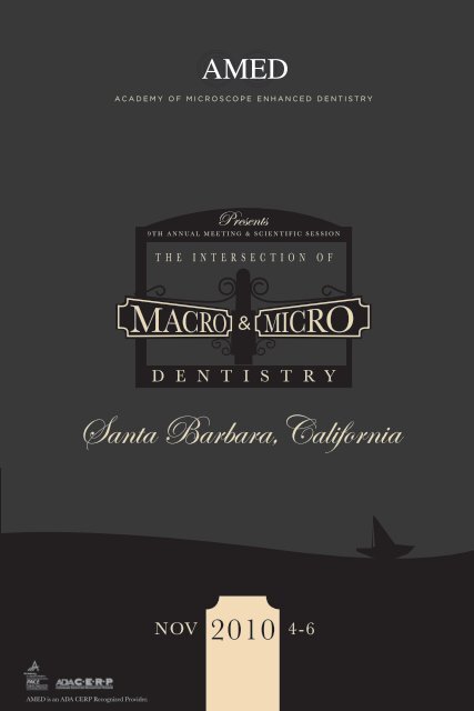
<strong>AMED</strong> is an ADA CERP Recognized Provider.<br />
NOV <strong>2010</strong> 4-6
“The Intersection of Macro and Micro<br />
Dentistry” will feature lectures from top<br />
clinicians in every discipline as well as master<br />
classes, corporate forums and pre and post<br />
session comprehensive “Hands-On” courses at<br />
education facilities along the Pacific coast.<br />
English to Japanese translation will be available.<br />
Program Co-Chairs:<br />
William Lannan, DDS<br />
John West, DDS, MSD<br />
<strong>2010</strong> <strong>AMED</strong> <strong>Sponsors</strong><br />
Platinum:<br />
Scientific Session Committee:<br />
Terrel Pannkuk, DDS, MScD<br />
Adriana McGregor, DDS<br />
Tetsuya Hirata, DDS, PhD<br />
Silver:
Featured<br />
<strong>2010</strong> Presenters<br />
All of our presenters have a microscope centered practice and are<br />
highly skilled in the application of this technology in clinical dentistry.<br />
Dr. Katsuhiko<br />
Akiyama<br />
Dr. Paul Anstey<br />
Dr. Stephane Browet<br />
Dr. Cami Ferris Dr. Eudes Gondim Dr. Jeffrey Hamilton<br />
Dr. Eric<br />
Herbranson<br />
Dr. Tetsuya Hirata Dr. Markus Hürzeler Dr. Peter Jannetta Dr. Kazuo Kurihara Dr. Te-Fu Li<br />
Dr. Kunio Matsumoto Dr. Adriana McGregor<br />
Dr. Jose Roberto<br />
Moura<br />
Dr. Carlos Alberto<br />
Ferreira Murgel<br />
Dr. Masahiro<br />
Nakazawa<br />
Dr. Morio Okaguchi<br />
Dr. Masayuki Okawa Dr. Junya Okawara Dr. Terrel Pannkuk Dr. Laurence Rifkin Dr. Cliff Ruddle Dr. Dennis Shanelec<br />
Dr. Cherylin Sheets<br />
Dr. Randy Shoup Dr. Malcolm Snead Dr. Glenn van As Dr. John West<br />
Dr. Claudia Cia<br />
Worschech
Schedule At-A-Glance<br />
Wednesday<br />
November 3rd<br />
10am - 4:30pm Santa Barbara Back<br />
Country Wine Tour**<br />
6pm - 9pm Registration Begins<br />
Thursday<br />
November 4th<br />
7am Registration & Exhibits Open<br />
9:30am - 1:30pm Spouse/Guest Event:<br />
Lotus land Garden Tour**<br />
8am - 12:45pm General Session<br />
(with 45min break)<br />
John West, DDS, MSD Presidents Welcome<br />
Cliff Ruddle, DDS My Endodontic Practice –<br />
A 35-Year Retrospective Analysis<br />
Terrel Pannkuk, DDS, MScD<br />
The Endo/Perio Differential Diagnosis<br />
Paul Anstey, DDS The Power of 3D<br />
Imaging in Endodontics and Beyond…<br />
Tetsuya Hirata, DDS, PhD What I Learned<br />
During Eight Years Research Study in<br />
Image Enhanced Dentistry<br />
Cherylin Sheets, DDS Quantitative<br />
Percussion Diagnostics and Magnification<br />
– A Synergistic Combination<br />
12:45pm - 1pm Members’ Business Meeting<br />
12:45pm - 2pm Luncheon Buffet<br />
2pm - 5pm Endo Master Class<br />
Carlos Murgel, DDS Small FOV CBCT for<br />
Endodontics: Another Gadget or a<br />
Paradigm Shift?<br />
Terrel Pannkuk, DDS, MScD Outcome<br />
Study Science and Art: Can the Value of<br />
an Endodontic Technique or Technology<br />
Be Adequately Assessed?<br />
Morio Okaguchi, DDS Microscope Assisted<br />
Precision Dentistry II<br />
Eudes Gondim, DDS, PhD Beyond the<br />
Microscope: What Else Can Make the<br />
Difference?<br />
John West, DDS, MSD<br />
Speakers’ Panel Moderator<br />
2pm - 5pm Perio Master Class<br />
Markus Hürzeler, DMD, PhD Minimally<br />
Invasive Implant Surgery Supported by<br />
Microsurgical Techniques<br />
Adriana McGregor, DDS The Hidden<br />
Secrets of Outstanding Results in Soft<br />
Tissue Management Around Implants:<br />
From Planning to Placement to Restoration<br />
Te-Fu Li, DDS Micro-invasive Treatment of<br />
Periodontal Pockets<br />
Katsuhiko Akiyama, DDS Papilla<br />
Reconstruction Using the Patch Technique<br />
Bryan Pearson, DDS, MS<br />
Speakers’ Panel Moderator<br />
2pm - 5pm Restorative Master Class<br />
Kunio Matsumoto, DDS Pre-Treatment in<br />
Esthetic Restoration<br />
Masayuki Okawa, DDS Minimally Invasive<br />
Interventions & Interdisciplinary Approach<br />
for Esthetic Dentistry<br />
José Roberto Moura, DDS Art and<br />
Precision with Direct Composites<br />
Claudia Cia Worschech, DDS, PhD<br />
Obtaining Clinical Success with Micro<br />
Laminate Porcelain Veneers<br />
Assad F. Mora, DDS, MSD, FACP<br />
Speakers’ Panel Moderator<br />
7pm - 11pm Social Event:<br />
Welcome Reception, Entertainment & Dance<br />
Friday<br />
November 5th<br />
8am Registration & Exhibits Open<br />
8am - 5pm “Test Drive” the Latest Technology<br />
9am - 12:45pm General Session<br />
Larry Rifkin, DDS Facial Esthetics<br />
Glenn vanAs, DDS Lasers and the Operating<br />
Microscope: Seeing the Light!<br />
Marc Alexander, DDS<br />
Treatment Planning for Esthetics<br />
Paul Piontkowski<br />
The Perfect CAD/CAM Restoration<br />
2pm - 5pm Short Presentations<br />
Randy Shoup, DDS Minimally Invasive<br />
Restorative Dentistry-Live Demonstration<br />
of Principals, Techniques, Equipment<br />
and Materials<br />
Junya Okawara, DDS Periodontal<br />
Microsurgery: Achieving Gingival Level<br />
Alignment with Connetive Tissue Graft<br />
Cami Ferris, DDS Heroic Endodontics in<br />
an Age of Implants<br />
Masahiro Nakazawa, DDS Utility of All-on-4<br />
with Socket Preservation<br />
Kazuo Kurihara, DDS Tissue Management<br />
Around Implants in Esthetic Zones<br />
Stephane Browet, DDS The Matrix Revisited<br />
2pm - 5pm Corporate Forums<br />
BioClear Composites Hands-on Course**<br />
Global Surgical<br />
AMD Lasers<br />
Crystal Mark & GC America: Air Abrasion<br />
2pm - 3:30pm Spouse/Guest Event:<br />
Land Shark Tour**<br />
7pm - 11pm Social Event:<br />
President’s Dinner and Awards<br />
(with 45min break)<br />
(30 min sessions)<br />
Saturday<br />
November 6th<br />
8am Registration & Exhibits Open<br />
9am - 1:30pm “Test Drive” the Latest Technology<br />
9am - 1:30pm General Session<br />
Dennis Shanelec, DDS A Retrospective of<br />
Clinical Periodontal Microsurgery<br />
Jeff Hamilton, DDS Oral Medicine and the<br />
Clinical Operating Microscope<br />
Eric Herbranson, DDS Latest in<br />
Photographic Documentation<br />
Peter J. Jannetta, MD<br />
Neurogenic Face Pain in the <strong>Dental</strong> Office<br />
Malcolm Snead, DDS, PhD<br />
Thinking the Unthinkable: Regenerating the<br />
Whole Tooth<br />
1:30pm Adjourn<br />
Off-Site Hands-on Courses<br />
Pre & Post Session “Hands-On” Courses<br />
will be held at:<br />
Microsurgery Training Institute<br />
www.microdentistry.com<br />
Newport Coast Oral Facial Institute<br />
www.ncofi.org<br />
Center for Endodontics<br />
www.centerforendodontics.com<br />
* Schedule subject to change, please see website<br />
for full course descriptions and details.<br />
** Additional cost.<br />
register online at:<br />
www.MicroscopeDentistry.com
Meeting Location & Accommodations<br />
Fess Parker’s DoubleTree Resort<br />
Tel: 1.800.564.4333 Fax: 1.800.564.4964<br />
633 East Cabrillo Boulevard, Santa Barbara, CA, USA 93103<br />
Santa Barbara, the Channel Islands and the nearby Santa Ynez<br />
Valley offer beautiful scenic opportunities and wonderful recreation for<br />
those with interest in exploring the area. The Double Tree is offering<br />
the convention room rate to our attendees the week before and the<br />
week after our meeting for those who wish to extend their trip in Santa<br />
Barbara.<br />
Social Events & Tours<br />
Group rate is $259.00 for Run of House/Mountain View rooms.<br />
Please refer to ‘<strong>AMED</strong>’ when calling in reservations. The cut-off date for<br />
making reservations at this special rate is October 4, <strong>2010</strong>.<br />
Fess Parker’s Doubletree Resort offers a complimentary shuttle to and<br />
from the Santa Barbara airport. Please call upon arrival (805-564-4333).<br />
Wednesday, Nov 3rd 10am - 4:30pm<br />
Santa Barbara Back-Country Wine Tour<br />
Enjoy lush valley views, breath-taking scenery and<br />
stops at 4 of the region’s best wineries for tastings,<br />
and a gourmet picnic lunch at one of the vineyards.<br />
Journey back to Santa Barbara through oak-shaded<br />
canyons and dirt trails, past the former Reagan Ranch<br />
and along the beautiful Pacific.… a visual feast!<br />
Thursday, Nov 4th 9:30am - 1:30pm<br />
Spouse/Guest Event:<br />
LotusLand Garden Tour with Lunch<br />
You are invited to visit Lotusland, a unique 37-acre<br />
estate and botanic garden situated in the foothills of<br />
Montecito to the east of the city of Santa Barbara.<br />
visit www.lotusland.org for more information.<br />
Thursday, Nov 4th 7pm - 11pm<br />
Welcome Reception, Entertainment &<br />
Dancing<br />
This memorable annual event is an opportunity for<br />
members and guests from around the world to meet<br />
face to face. Enjoy a Santa Barbara themed dinner,<br />
entertainment and dancing. California Casual Attire.<br />
Friday, Nov 5th 2pm - 3:30pm<br />
Spouse/Guest Event:<br />
Santa Barbara’s Land Shark Tour<br />
Climb aboard Santa Barbara’s original amphibious<br />
tour vehicle for a personally narrated 90 minute land<br />
and sea adventure. Enjoy exquisite views of the<br />
Santa Barbara coastline, the Riviera, and the Santa<br />
Ynez mountains as seen only from our boat at sea.<br />
Don’t miss this exciting and entertaining Land and<br />
Sea adventure to experience Santa Barbara.<br />
Friday, Nov 5th 7pm - 9:30pm<br />
President’s Dinner and Awards<br />
Enjoy an elegant evening during <strong>AMED</strong>’s annual<br />
President’s Dinner and short Awards presentation<br />
Ceremony. Semi-Formal Attire.<br />
register online at:<br />
www.MicroscopeDentistry.com
Registration<br />
Register Online at<br />
www.microscopedentistry.com<br />
SPOUSE/FAMILY GUEST EVENT REGISTRATION<br />
Name of Guest(s) for Badge<br />
Attendee Name<br />
Degree(s)<br />
Specialty License Number ADA/AGD#<br />
Email Address Phone #<br />
Address<br />
City/Province<br />
Postal Code<br />
Country<br />
Wednesday, Nov 3rd.......Santa Barbara Back Country Wine Tour........ ADD $130<br />
(per person)<br />
Thursday, Nov 4th...........LotusLand Garden Tour with Lunch............... ADD $100<br />
(per person)<br />
Thursday, Nov 4th...........Welcome Reception....................................... ADD $100<br />
(per person)<br />
Friday, Nov 5th................Meeting Luncheon......................................... ADD $45<br />
(per person)<br />
Friday, Nov 5th................Land Shark Tour............................................. ADD $30<br />
(per person)<br />
Friday, Nov 5th................President’s Dinner/Awards............................. ADD $100<br />
(per person)<br />
Subtotal<br />
Year Began Using Microscope<br />
Food Allergies/Preferences<br />
Discount Code and Amount<br />
Grand Total<br />
MEETING REGISTRATION<br />
AGENDA/EVENT ITEMS: Check all that you will attend<br />
Wednesday, Nov 3rd....... Santa Barbara Back Country Wine Tour....... ADD $130<br />
Thursday, Nov 4th........... Master Classes: Endo/Restorative/Perio (Circle one)<br />
Thursday, Nov 4th........... Welcome Reception<br />
Earn up to 17 CE credits<br />
INCLUDES: General Sessions, Master Classes, Short Presentations, Test Drive,<br />
Corporate Forums, Breaks, Buffet Luncheons, Welcome Reception and Presidents Dinner.<br />
Member<br />
$595<br />
Non-Member<br />
$1095<br />
Early Bird<br />
Registration ends August 1st<br />
<strong>Dental</strong> Auxiliary<br />
Must accompany<br />
$295 Doctor<br />
Subtotal<br />
Friday, Nov 5th................ BioClear/Composites Hands-on Courses..... ADD $295<br />
Friday, Nov 5th................ President’s Dinner/Awards<br />
Subtotal<br />
Student/Faculity<br />
Must Provide<br />
$295 proof<br />
(add $100 after August 1st)<br />
billing information<br />
Enter EXACTLY as appears on your billing statement<br />
Card Type: VISA | MASTER CARD | DISCOVER (Circle One)<br />
Card Holder’s Name<br />
Card Number<br />
Card Security Code on Back of Card Billing Phone #<br />
Billing Address<br />
City/Province<br />
Postal Code<br />
Register Online AT<br />
www.microscopedentistry.com<br />
Expiration Date (MM/YY) Security Code (3 digits on back)<br />
Country<br />
An official confirmation will be emailed to the email address provided on this form.<br />
Please mail to:<br />
The Academy of Microscope Enhanced Dentistry (<strong>AMED</strong>)<br />
PO BOX 15834, Fort Wayne, IN 46885<br />
Or send via fax 480-247-4006<br />
Questions? Please email: info@microscopedentistry.com or call 260-249-1028<br />
*An official confirmation will be emailed to the email address provided on this form.
Presentation Descriptions<br />
Thursday<br />
November 4th<br />
8am - 12:45pm General Session<br />
John West, DDS, MSD Presidents Welcome<br />
Cliff Ruddle, DDS My Endodontic Practice – A<br />
35-Year Retrospective Analysis<br />
There is an old expression that to see the future you only have to look<br />
at the past. A 35-year retrospective analysis of my clinical practice<br />
will provide a glimpse of those early years, the enormous technical<br />
changes that followed, and how these changes created both chaos yet<br />
enormous opportunities. This presentation will focus on the unforeseen<br />
and unanticipated benefits of utilizing the dental operating microscope<br />
in every day practice. Through this monologue, Dr. Ruddle will share his<br />
experiences and provide a brief look into the past, present, and future of<br />
Endodontics.<br />
Terrel Pannkuk, DDS, MScD The Endo/Perio<br />
Differential Diagnosis<br />
Endodontic diagnosis requires astute observation of signs, symptoms,<br />
tests, and radiographs. Distinguishing between artifacts and significant<br />
clinical entities can be very challenging. In this presentation Dr. Pannkuk<br />
will describe a logical approach to interpretation that can predictably and<br />
accurately distinguish between confusing presentations of periodontal<br />
and endodontic disease. He will show interesting and unique cases that<br />
highlight the clinical strategy.<br />
At the end of the presentation the audience will be able to:<br />
• recognize important subtle radiographic signs<br />
• determine prognosis<br />
• recognize the treatment implications of an endodontic or periodontal<br />
disease etiology<br />
Paul Anstey, DDS The Power of 3D Imaging in<br />
Endodontics and Beyond…<br />
We have been relying on radiographs to analyze dental disease and<br />
diagnose dental pathosis since the early 1900’s. This age-old 2D<br />
technology has obvious limitations. As a result, much of the true data<br />
has been hidden from us for decades leading to shortfalls in diagnosis,<br />
treatment planning and ultimately patient care.<br />
The time has finally arrived and we are now able to utilize 3D imaging<br />
to improve patient care dramatically. These are exciting times with the<br />
technologic breakthrough of High Resolution Cone Beam Computer<br />
Tomography (CBCT).<br />
The goal of this presentation is to highlight the newfound capabilities of<br />
this amazing “new and improved” CT technology. The multiple uses of<br />
CBCT in dental diagnosis and treatment planning will be demonstrated<br />
with correlation of the CT images to the areas explored and photographed<br />
through the Operating Microscope.<br />
After this presentation the audience will have a better understanding of:<br />
• How and why CBCT is truly a diagnostic breakthrough<br />
• What we have been missing all of these years<br />
• When and how to decide if we need a CBCT scan<br />
• How to read the scans and understand their value in everyday practice<br />
• How to detect classic artifacts and thereby prevent misdiagnosis<br />
associated with CBCT<br />
• What the challenges are with this new technology<br />
• What we can look forward to with the CBCT of the future<br />
Tetsuya Hirata, DDS, PhD What I Learned<br />
During Eight Years Research Study in iMage<br />
Enhanced Dentistry<br />
In this presentation Dr. Hirata will share his research study results in<br />
these 8 yrs and a research project’s result with Universities in Japan, in<br />
Europe, in Brazil and in the States in iMage Enhanced Dentistry. This<br />
iMage Enhanced Dentistry, like Image Enhanced Medicine in medical<br />
field, includes Microscopes, CT, MRI, MRA, Endoscopes, SEM, video<br />
systems and all other magnification systems. Topics will cover rapid<br />
healing, atraumatic soft tissue reaction, effect of hydration, effect of<br />
chemical substance, Implant rescue, treatment planning in Implantology,<br />
relationship between crack and soft tissue, precise tooth preparation,<br />
atraumatic tooth preparation, relationship between marginal fitness and<br />
leakage, retrospective research on old prosthesis, and more.<br />
Cherylin Sheets, DDS Quantitative Percussion<br />
Diagnostics and Magnification – A Synergistic<br />
Combination<br />
The introduction of magnification into dentistry revolutionized the ability of<br />
dentists to diagnose and treat abnormalities due to the increased size of<br />
objects and the flawless illumination provided. The microscope provided,<br />
regardless of specialty, more detailed visual information to the clinician in<br />
all phases of treatment.<br />
During the last decade, a new technology has been explored that will give<br />
additional diagnostic information to the clinician. Quantitative percussion<br />
testing allows a clinician to evaluate the structural stability of natural teeth<br />
or dental implants by measuring the way it responds to a light impact<br />
on the buccal surface. The energy that is returned to the handpiece is<br />
analyzed in a manner that provides two pieces of information—the loss<br />
coefficient (LC) and energy-time response (ETR) of the structure tested<br />
. These two pieces of information can give the clinician indications of<br />
how sound the tested structure is and whether there are problems such<br />
as dentinal cracks and fractures, microleakage, recurrent decay, loose<br />
post and cores and other structural defects. By having an indication as<br />
to how a tooth or implant responds to mechanical stress prior to starting<br />
restorative care, the microscopic enhanced clinician will be prepared to<br />
look for potential problems where abnormal ETR’s are observed. ETRs<br />
also help patients understand the bioengineering challenges that some<br />
teeth present due to crack propagation and other serious biomechanical<br />
problems. Combined with video documentation of the defect, it creates a<br />
powerful documentation and patient educational tool simultaneously.<br />
2pm - 5pm Endo Master Class<br />
Carlos Murgel, DDS Small FOV CBCT for<br />
Endodontics: Another Gadget or a Paradigm<br />
Shift?<br />
Just very recently small FOV (field of view) CBCT (cone beam computer<br />
tomography) unities became available for endodontics. These machines<br />
have lower radiation dosages, more resolution, are smaller and less<br />
expensive than the large FOV ones. There are several machines<br />
available on the market and it is very difficult to select one that is<br />
more suitable for our clinical needs. Also it is crucial to know what is<br />
this technology about it, when and why to use it, how to interpret its<br />
results and how to implement it into a busy practice. At the end of this<br />
presentation the participants will have a solid understanding about the<br />
small FOV CBCT unities and will be able to know why this technology<br />
represents a major paradigm shift in modern endodontics.<br />
register online at: www.MicroscopeDentistry.com<br />
Terrel Pannkuk, DDS, MScD Outcome<br />
Study Science and Art: Can the Value of an<br />
Endodontic Technique or Technology Be<br />
Adequately Assessed?<br />
The endodontic literature spans a wide spectrum of accepted levels<br />
of evidence. In this presentation Dr. Pannkuk will discuss the lowest<br />
forms of scientific evidence like opinion and single anecdotal case<br />
histories comparing them to less obtainable rigid CONSORT guidelines<br />
and Systematic Reviews as it relates to our understanding of clinical<br />
success and the value of clinical tools like the surgical operating<br />
microscope. Mounds of low level evidence are readily available yet<br />
high level evidence is not. Is this a double-edged sword of scientific<br />
knowledge? Is all available evidence valuable when looked at critically<br />
as a whole? Is a foggy picture better than no picture of reality?<br />
The philosophy of what we can and cannot test when it comes to a<br />
nuanced clinical art will be discussed. There will be a specific focus<br />
on the value of the microscope, single versus multivisit endodontic<br />
procedures, and implant versus root canal treatment success rates. Dr.<br />
Pannkuk will pose the questions:<br />
1. How does one achieve evidence-based clinical predictability?<br />
2. What is a definitive clinical outcome versus clinical success?<br />
3. What do we really understand about our clinical technique and<br />
choices?<br />
Morio Okaguchi, DDS Microscope Assisted<br />
Precision Dentistry II<br />
Recently attention has been focused on using <strong>Dental</strong> Microscopes.<br />
Many authorized medical doctors so called God-Hand in that field<br />
are definitely needed Microscopes and other up-to -date devices. In<br />
<strong>Dental</strong> field, more dentists require better and more excellent results<br />
in managing Minimal Intervention concepts, Esthetics, long term<br />
Prognosis and Occlusion, more precise procedure that surpass the<br />
limit in working under the naked eyes is required for those dentists.<br />
It has not been needed to state that high magnification and high<br />
resolution is essential to achieve fine dental treatment.<br />
High magnification and resolution is giving dentists more clear<br />
images of not only root canals but also apical foramen. This grows<br />
the possibility in removing infected structures around apical area<br />
and in other difficult area for naked eyes. As the result, increasing<br />
the possibility higher and higher in saving treated root canals and<br />
miserable tooth.<br />
<strong>Dental</strong> Microscopes may potentially guide all dentists to higher and<br />
higher level and finally may lead to be called God-Hand. In this<br />
presentation Dr. Okaguchi will share his concepts in Endodontically<br />
treated tooth through his clinical cases.<br />
Eudes Gondim, DDS, PhD Beyond the<br />
Microscope: What Else Can Make the<br />
Difference?<br />
A range of clinical techniques and applications will be reviewed and<br />
illustrated in an attempt to help clinicians ensure and rationalize<br />
necessary treatment objectives for success. Anatomic exploration,<br />
systematic disinfection using new irrigation techniques will be<br />
discussed. Currently, effective irrigant delivery is a prerequisite for<br />
successful endodontics and this lectures will present an overview<br />
recommendations for safe and effective irrigation.<br />
2pm - 5pm Perio Master Class<br />
Markus Hürzeler, DMD, PhD Minimally<br />
Invasive Implant Surgery Supported by<br />
Microsurgical Techniques<br />
The refinement of the surgical technique by means of a microsurgical<br />
approach is one of the most importaant determinants of successful<br />
esthetic results. A series of case reports in reconstructive implant<br />
dentistry and a controlled clinical study on periodontal regeneration<br />
and plastic reconstrcutive surgery demonstrated that a microsurgical<br />
approach is able to accelerate healing and improve the rate of primary<br />
soft tissue closure.<br />
In this presentation the microsurgical concept will be demonstrated<br />
which will allow the participants to fullfil the high esthetic demands<br />
from the patients in the daily practice on a more predictable base. The<br />
microsurgical concept includes magnification aids, new instruments,<br />
new suture material and new flap designs. Case presentations using<br />
the microsurgical concept will help the attendees to understand the<br />
idea behind the novel idea better<br />
Adriana McGregor, DDS The Hidden<br />
Secrets of Outstanding Results in Soft Tissue<br />
Management Around Implants: From Planning<br />
to Placement to Restoration<br />
Pre-surgical, tran-surgical and post-surgical soft tissue management<br />
are critical factors in determining the overall implant outcome.<br />
Implant success is currently measured not only by implant location,<br />
stability and lack of surrounding infection, but additionally by the<br />
adjacent soft tissue form, level, color, tone, health and support, with the<br />
aim of replicating or even improving upon the original tooth esthetics.<br />
This course will focus in specific microsurgical techniques to achieve<br />
unsurpassed soft tissue outcomes around implants.<br />
Te-Fu Li, DDS Micro-invasive Treatment of<br />
Periodontal Pockets<br />
During the past several years, tremendous changes have occurred in<br />
the practice of periodontics and implantology. As our patients are now<br />
getting more conscious about their physical as well as psychological<br />
well-being, a shift of paradigm in the therapeutic modality has thus<br />
undertaken. Pain-free procedures and minimized post-operative<br />
trauma have become patients’ primary concerns, and emphasis is<br />
now being placed on the aspect of non-surgical approach for treating<br />
periodontal disease as well as placing dental implants. In order to<br />
achieve these, microscopically enhanced therapies provide a new way<br />
for insight into the solutions. Under the microscope, “flapless approach”<br />
is no more equated with “blind treatment”. The utilization of this perfect<br />
visual tool offers an aid as an eye opener for the dental clinicians. With<br />
the high power magnification, dentist’s vision can actually pass through<br />
tiny openings such as pocket entrances and crestal gingival punched<br />
accesses. As a result, there would be no more need for traditional<br />
open flap surgery to create visibly large wounds. The advantage of<br />
doing so obviates the possibilities of relevant potential risks carried by<br />
surgeries, such as uncontrolled bleeding, post-operative swelling, pain<br />
and even neurosensory damage. Treatment is simplified with patient<br />
undergoing one single procedure. Without incision and tearing of<br />
periosteum, healing can be faster and more uneventful. So the whole<br />
therapeutic course can be a more cost effective one. Nevertheless<br />
in the meanwhile different solutions are offered to meet our patients’<br />
demands, questions have also arisen as to whether or not these<br />
“newly invented non-surgical methods” would violate against our basic<br />
biological principles that we have long followed and believed in. Five<br />
to ten years’ follow-up results from presenter’s private practice will be<br />
presented for open discussion and hopefully will unveil this myth.<br />
Katsuhiko Akiyama, DDS Papilla<br />
Reconstruction Using the Patch Technique<br />
Papilla reconstruction is a difficult procedure to perform with your<br />
naked eye. However, the combination of a <strong>Dental</strong> Operating<br />
Microscope and the Patch Technique makes it much easier. In this<br />
presentation Dr. Akiyama will share his technique.<br />
The Patch Technique is composed of the following operations under<br />
the Microscope: (1) Debridement, Desquamation, and Tension free<br />
incisions through the sulcus; (2) Harvesting connective tissue from the<br />
palate; (3) Supporting the epithelial flap with the harvested connective<br />
tissue patch; and (4) Lifting/Maintaining this combined flap to the<br />
coronal direction using a composite resin anchor on the tooth surface.<br />
This technique gives more effective result in papilla reconstruction<br />
of both natural teeth and Implants in conjunction with using dental<br />
operating microscopes.<br />
Dr. Akiyama will introduce several special instruments that provide<br />
more efficient performance with the Patch Technique under the<br />
Microscope.
Thursday<br />
November 4th contintued...<br />
Friday<br />
November 5th<br />
2pm - 5pm Restorative Master Class<br />
Kunio Matsumoto, DDS Pre-Treatment in<br />
Esthetic Restoration<br />
Prosthetic treatment in esthetic zone requires not only acquiring tooth<br />
morphology harmonizing with patients’ features but also gaining optimal<br />
continuous soft tissue lines. Dr. Matsumoto will share his clinical cases<br />
utilizing both Periodontal Microsurgery and Orthodontic treatment as<br />
a Restorative Pre -Treatment and discuss about its effectiveness and<br />
efficaciousness.<br />
Masayuki Okawa, DDS Minimally Invasive<br />
Interventions & Interdisciplinary Approach for<br />
Esthetic Dentistry<br />
Lately, favorable results are seen in many esthetic cases with minimally<br />
invasive techniques. This became possible due to the development<br />
of biomimetics, advancement in bonding technique, and treatment<br />
using the problem-based approach. In addition, the use of microscope<br />
has allowed us to obtain precise and predictable outcomes. Many<br />
treatments require the expertise of specialists to get the best esthetic<br />
results, therefore, team approach is integral and necessary in esthetic<br />
dentistry.<br />
To achieve optimal esthetics, it is imperative to use the team approach.<br />
This team should include the restorative dentist, periodontist,<br />
orthodontist and oral surgeon. As the demands for esthetic outcomes<br />
increases, communication and mutual understanding of treatment<br />
goals between different treatment providers become more and more<br />
important.<br />
Using my clinical cases, the use and effectiveness of the microscope<br />
in porcelain bonded restorations and management of interdisciplinary<br />
approach in esthetic dentistry will be discussed.<br />
José Roberto Moura, DDS Art and Precision<br />
with Direct Composites<br />
Direct composites are one of the best solutions to restore anterior and<br />
posterior teeth regarding preservation of sound dental structure, natural<br />
appearance, reasonable cost and longevity. However, techniques are<br />
critical and careful attention to every step is essential to achieve best<br />
results.<br />
We’ll go through every step of the restorative techniques, showing how<br />
high magnification can make a huge difference.<br />
Claudia Cia Worschech, DDS, PhD Obtaining<br />
Clinical Success with MicroLaminate Porcelain<br />
Veneers<br />
Ceramic Micro Laminate porcelain veneers are a sub category of<br />
conventional veneers. They are extremely thin and much smaller in<br />
size and can be placed with little or no tooth preparation.Their use in<br />
diastema closure, augmentation of tooth form and color conserves tooth<br />
structure and creates a natural esthetic result. Marginal fit, adaptation,<br />
and finishing are absolutely critical to the long term success of the<br />
laminate and will be the emphasis of this presentation.<br />
9am - 12:45pm General Session<br />
Larry Rifkin, DDS Facial Esthetics<br />
Glenn vanAs, DDS Lasers and the Operating<br />
Microscope: Seeing the Light!<br />
In this lecture and live demonstration, attendees will witness how hard<br />
and soft tissue lasers can impact daily dental practice to make clinical<br />
procedures simpler and more enjoyable. The operating microscope<br />
can help improve treatment outcomes in laser dentistry and this will be<br />
demonstrated in digitally photographed and videotaped clinical cases.<br />
Clinical objectives that attendees will learn:<br />
1. The four different wavelengths available at present for dental practice.<br />
2. How soft tissue lasers can be used to help improve treatment<br />
outcomes.<br />
3. The value of hard tissue lasers in clinical practice.<br />
4. Live demonstration of at least one wavelength of laser at the end of<br />
the lecture.<br />
Marc Alexander, DDS Treatment Planning for<br />
Esthetics<br />
Esthetic reconstruction has become more popular, with adult patients<br />
asking for this treatment. Very often the restorative dentist is challenged<br />
by tooth malpositions and tooth size discrepancies. Diagnostic and<br />
planning criteria will be outlined, to help the restorative dentist achieve<br />
the patients’ satisfaction and the operators’ goals.<br />
Paul Piontkowski The Perfect CAD/CAM<br />
Restoration<br />
If you are considering purchasing a Cerec to integrate into your practice,<br />
attendees will learn the pros and cons of CAD/CAM dentistry and if it<br />
is a good fit for your practice. Altered preparation techniques, esthetic<br />
consideration and improved Cerec techniques will be presented for the<br />
microscope user based office.<br />
2pm - 5pm Short Presentations<br />
Randy Shoup, DDS Minimally Invasive<br />
Restorative Dentistry-Live Demonstration<br />
of Principals, Techniques, Equipment and<br />
Materials<br />
Junya Okawara, DDS Periodontal<br />
Microsurgery: Achieving Gingival Level<br />
Alignment with Connetive Tissue Graft<br />
A harmonically aligned gingival level is a major factor in creating<br />
a beautiful smile. In esthetic dentistry, creating an aligned gingival<br />
level is of the utmost importance. At the same time, it is necessary<br />
to understand and consider each patient’s request during treatment<br />
planning and to perform surgery with absolute accuracy.<br />
This presentation will introduce the benefits of using a dental<br />
microscope in periodontal surgery with connective tissue graft for<br />
patients who desire esthetic dental treatment.<br />
Cami Ferris, DDS Heroic Endodontics in an<br />
Age of Implants<br />
Dr. Cami Ferris will present the clinical rationale for treating some<br />
cases that may otherwise be deemed “heroic” in today’s implantdriven<br />
practices. She will discuss evaluation and consultation with the<br />
patient, as well as the referring dentist. Decision points and treatment<br />
modalities of two cases will be reviewed, including non-surgical retrieval<br />
of amalgam retrofilling material and use of Mineral Trioxide Aggregate<br />
cement.<br />
Masahiro Nakazawa, DDS Utility of All-on-4 with<br />
Socket Preservation<br />
All-on-4 with a concept of immediate function, developed by Dr. Paulo<br />
Malo, is a medical treatment for patients with edentulous jaw. At Malo’s<br />
office, 80% of the cases who received All-on-4 were dentulous patients<br />
with severe gum disease. When All-on-4 treatment is given to dentulous<br />
patients, it is classified into two groups, immediate implant placement,<br />
and delayed implant placement.<br />
Malo recommends immediate implant placement; however, the teeth to<br />
be extracted have the primary diseases and there is an indication that<br />
the surrounding alveolar bone is damaged. In addition, the volume of<br />
the alveolar bone in Asians is sometimes more restricted than that of<br />
Westerners.<br />
When immediate implant placement is performed, the volume of the<br />
residual alveolar bone is restricted, due to flattening of the alveolar bone<br />
(the operative procedure of All-on-4), which is a series of procedures<br />
performed after tooth extraction. As a result, it is a concern that the size<br />
of the implant appropriate for immediate loading with All-on-4 treatment<br />
can not be determined.<br />
The case presented is based on the results of the CT scan taken prior<br />
to the procedure, proving that application of immediate loading is difficult<br />
when the implant is performed in immediate implant placement. In this<br />
case, we conducted tooth extractions and socket preservation using a<br />
microscope and therefore could preserve the alveolar bone and obtain<br />
sufficient amounts of keratinized gingival.<br />
Thereafter by using guided surgery, we succeeded in performing All-on-6<br />
treatment after only 2 months from the extraction of the tooth.<br />
Kazuo Kurihara, DDS Tissue Management<br />
Around Implants in Esthetic Zones<br />
It is well known that Implant procedure in Esthetic zone is effective, but<br />
is requiring complicated steps. Furthermore, it is also well known that<br />
both hard tissue and soft tissue management in that area is playing an<br />
important roll as a needed technique. Hard tissue management is usually<br />
performed before or at the same time placing Implants. On the other<br />
hand, Soft tissue management is able to perform after Implantation.<br />
That is the reason why Soft tissue management has been easy to<br />
prognoses the final result and been able to give long term stability with<br />
its appropriate technique and methods. In this presentation Dr. Kurihara<br />
will share his concept by showing his clinical cases grafting soft tissue<br />
around Implants after Implantation under the Microscope.<br />
Stephane Browet, DDS The Matrix Revisited<br />
This presentation focuses on problem solving with the use of different<br />
matrix systems for direct restorations, in both anterior and posterior teeth.<br />
Numerous clinical cases will help to illustrate the strategy to address difficult<br />
situations.<br />
2pm - 5pm Corporate Forums<br />
BioClear Composites Hands-on Course (additional cost)<br />
Direct Composites - Simplified, Beautified, Clarified. Participants will<br />
have the opportunity to perform modern composite dentistry utilizing<br />
the Bioclear Matrix Systems, Operating Microscopes, and flowable and<br />
paste composites.<br />
Participants will perform the following on simulation models: Prepare and<br />
restore Clark Class II preparations, peg lateral treatment, Restoratively<br />
driven papilla regeneration (“black triangle” elimination), and advanced<br />
composite preparations and matrixing for deep caries and diastema<br />
closure.<br />
Cracked Tooth diagnosis will be addressed and various techniques to<br />
prevent and treat cracked teeth with both the Cala Lilly and composite<br />
onlay will be performed. You will re-think the role of resins in their<br />
practice after seeing the possibilities in this renaissance of direct<br />
composites!<br />
At the end of this course participants will:<br />
• Achieve clinical proficiency in both diastema closure techniques and<br />
restoratively-driven papilla regeneration utilizing direct composites.<br />
• Understand and achieve confidence with minimally traumatic class I<br />
and class II composite preparations, including the Clark Class II.<br />
• Understand C-Factor, composite chemistry, and receive an update in<br />
dentin and composite bonding options.<br />
• Understand the strengths and weaknesses of flowable and past<br />
composites.<br />
• Receive a blueprint for the incorporation of flowable composites in all<br />
different types of composite preparations.<br />
• Have confidence in diagnosis, treatment and prevention of cracked<br />
teeth<br />
• Gain confidence in the use of the dental microscope for direct<br />
composites<br />
Global Surgical Forum<br />
Crystal Mark & GC America: Air Abrasion<br />
AMD Lasers Forum<br />
register online at: www.MicroscopeDentistry.com
Saturday<br />
November 6th<br />
9am - 1:30pm General Session<br />
Dennis Shanelec, DDS A Retrospective of<br />
Clinical Periodontal Microsurgery<br />
Twenty years ago, microscopy was introduced to clinical dentistry. In<br />
the ensuing years it has become an important modality for periodontal<br />
and dental implant treatment.<br />
What has been learned about microscopy and its importance in clinical<br />
practice and education? Dr. Shanelec will review his experience<br />
applying microsurgical principles to surgical practice as well as his<br />
progress in educating a new generation of microsurgical clinicians.<br />
This presentation will highlight a case series of 300 immediate dental<br />
implants in extraction sockets with provisionals to demonstrate the<br />
value of a microsurgical approach for most challenging clinical and<br />
esthetic situations.<br />
Jeff Hamilton, DDS Oral Medicine and the<br />
Clinical Operating Microscope<br />
The use of the clinical microscope in dentistry has proven to be an<br />
important adjunct to treatment in many fields including: restorative,<br />
endodontics, periodontics, and diagnosis. The diagnostic benefit of the<br />
clinical microscope in Oral Medicine, however, has not been as well<br />
developed as in the above dental disciplines.<br />
Although oral cavity (& pharynx) account for only 2.5 - 3% of new<br />
cases of all cancer projected in 2008 (SEER program, National Cancer<br />
Institute) and only 1.3% of total cancer deaths of the same period, early<br />
diagnosis still rests on the dental profession to lead the way in detecting<br />
these lesions. Detection of persistent “white” or “red” lesions may also<br />
lead to a broad diagnosis of lesions due to autoimmune, infectious, or<br />
drug related etiologies. The introduction to dentistry of narrow band<br />
imaging, notably the VelScope, increases the visibility of suspected<br />
lesions but has limited capabilities of definitive differential diagnosis.<br />
The use of magnification and light contrast techniques in oral medicine<br />
to narrow diagnosis, document lesion progression (assumed premalignant<br />
risk), and improve early detection as investigated at the<br />
University of Washington Department of Oral Medicine over the past<br />
year will be summarized.<br />
Eric Herbranson, DDS Latest in Photographic<br />
Documentation<br />
Digital still and video camera technology is rapidly changing and their<br />
capabilities are converging. Current HD video cameras can take good<br />
quality still images and high end SLR still cameras now do good quality<br />
HD video. Both types of cameras can be used on a microscope. It is<br />
time to review how these changes impact documentation through the<br />
surgical operating microscope. This lecture will review and compare<br />
the technologies and how they impact equipment decision-making in<br />
the microscope driven practice. For completeness, they will also be<br />
compared to intra-oral wand type cameras. We will also review the<br />
storage and processing software necessary for the cameras. The<br />
second part will review the basic fundamental skills necessary for good<br />
clinical imaging and briefly review photo editing.<br />
Peter J. Jannetta, MD Neurogenic Face Pain<br />
in the <strong>Dental</strong> Office<br />
Malcolm Snead, DDS, PhD Thinking the<br />
Unthinkable: Regenerating the Whole Tooth<br />
President Kennedy captured the American imagination in a speech<br />
made decades ago where he stated the unthinkable: “We choose to<br />
go to the moon in this decade and do the other things, not because<br />
they are easy, but because they are hard”. Our research team has<br />
been guided and inspired by the challenge of “doing what is hard”<br />
by embarking on a grand project to improve the oral health of the<br />
American people. We adopted the goal to regenerate a complete<br />
single rooted tooth. We sought to include the bioceramic portions<br />
and the tissues integrating the tooth to the bone using either a<br />
biological regeneration approach or a biomimetic recreation approach.<br />
A team of scientists was drawn from multiple universities and from<br />
multiple disciplines to be amalgamated as a trans-disciplinary,<br />
high-performance team dedicated to accomplishing this goal. We<br />
translated our broad goals into actionable organizational units, we<br />
identify three parallel interdisciplinary research groups that worked<br />
hand in hand with the others. One group involved the development<br />
of a “Blueprint” with structural and functional design specifications<br />
drawn from a human tooth, spanning gene expression to functional<br />
organization of the matrix. Members of our scientific team will mine,<br />
evaluate, and develop essential information needed for the Blueprint,<br />
while other members will create high-fidelity models intended to be<br />
so perfect that they might bypass wet laboratory tests in favor of<br />
computer generated outcomes. Knowledge from the Blueprint will<br />
be translated to stem cells or genetically engineered cells that, in<br />
conjunction with molecularly designed informational-rich extracellular<br />
matrices, will direct tissue regeneration. The Blueprint information<br />
is also used to guide the fabrication of nature-inspired biomimetic<br />
equivalents of tooth components using synthetic molecular systems<br />
and genetically engineered peptides.<br />
Our vision is defined by five prioritized goals:<br />
1) Regenerate functional tooth structures and supporting tissues;<br />
2) Develop intermediary devices that are used for repair of diseased<br />
oral tissues;<br />
3) Develop stem cell lines from single teeth applicable to all<br />
regenerative medicine strategies;<br />
4) Build a polydisciplinary team with renewable knowledge and<br />
techniques to achieve these goals;<br />
5) Translate this knowledge and technical capability into devices<br />
that improve health while creating: educational opportunities,<br />
new research pathways, new clinical applications and new jobs in<br />
technology-rich environments.<br />
At the conclusion the participants should be able to:<br />
• describe the functional and performance difference between digital<br />
video and digital still camera when applied to microscope photography,<br />
as well as how they compare to wand type intra-oral cameras.<br />
• make informed purchase decision based on their particular clinical<br />
imaging needs.<br />
• describe the basic skills necessary to produce good clinical images.<br />
register online at: www.MicroscopeDentistry.com
The Academy of Microscope Enhanced Dentistry (<strong>AMED</strong>)<br />
is an International Association of Restorative Microdentistry, Periodontal<br />
Microsurgery, Microendodontics, Microprosthodontics, Implant Microsurgery,<br />
and Associated Disciplines. We are dedicated to providing a framework for<br />
all microscope-centered dental practitioners, regardless of specialty, to share<br />
information in an inclusive, not exclusive environment.<br />
Restorative Endodontics Periodontics<br />
To register online please visit:<br />
www.microscopedentistry.com<br />
questions? contact:<br />
Email: info@microscopedentistry.com<br />
Phone: 260-249-1028 EST<br />
Cancellations and Refunds:<br />
All cancellations must be received by September 30, <strong>2010</strong>.<br />
Requests for refunds received by September 30, <strong>2010</strong><br />
will be granted in full, less a $75 administration fee. After<br />
September 30, <strong>2010</strong> NO registration refunds will be issued.<br />
Please call or email us to request a cancellation.<br />
<strong>AMED</strong> Central Office:<br />
PO BOX 15834<br />
Fort Wayne, IN 46885-5834<br />
Fax: 480-247-4006<br />
PRIVACY NOTICE:<br />
Unless otherwise requested, your contact information will<br />
be shared with our sponsors and exhibitors. Any other<br />
information you provide will be held with the utmost care and<br />
will not be used in ways that you have not consented to. If<br />
you have any questions, please feel free to call or email us.<br />
Course Space:<br />
Registration for courses is on a first come first serve basis<br />
and is only open to registered conference attendees.<br />
Please register for the Annual Meeting before registering for<br />
courses. You will receive confirmation once your registration<br />
has been accepted.