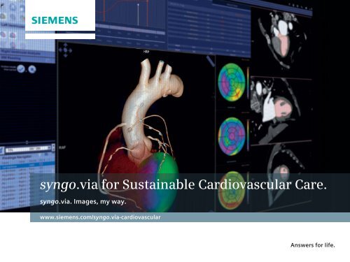
syngo. via for Sustainable Cardiovascular Care. - Siemens Healthcare
syngo. via for Sustainable Cardiovascular Care. - Siemens Healthcare
syngo. via for Sustainable Cardiovascular Care. - Siemens Healthcare

Create successful ePaper yourself
Turn your PDF publications into a flip-book with our unique Google optimized e-Paper software.
<strong>syngo</strong>. <strong>via</strong> <strong>for</strong> <strong>Sustainable</strong> <strong>Cardiovascular</strong> <strong>Care</strong>.<br />
<strong>syngo</strong>. <strong>via</strong>. Images, my way.<br />
www.siemens.com/<strong>syngo</strong>.<strong>via</strong>-cardiovascular<br />
Answers <strong>for</strong> life.
<strong>syngo</strong>®. <strong>via</strong> enables me<br />
to diagnose and share<br />
results, my way.<br />
My cases are ready <strong>for</strong> reading. <strong>syngo</strong>.<strong>via</strong> 1 helps to<br />
analyze the individual case, prepares images, suggests an optimized<br />
workflow, and offers guidance when needed.<br />
My images are networked. And so am I.<br />
<strong>syngo</strong>. <strong>via</strong> speeds the way I connect and share in<strong>for</strong>mation with<br />
my clinical partners and patients – even on the go 2 .<br />
My needs are anticipated. The <strong>syngo</strong>. <strong>via</strong> service<br />
offering helps me keep up with innovations, get integrated services,<br />
and rely on predictable costs now and in the future.<br />
1<br />
<strong>syngo</strong>. <strong>via</strong> can be used as a stand-alone device or<br />
together with a variety of <strong>syngo</strong>. <strong>via</strong>-based software<br />
options, which are medical devices in their own rights.<br />
2<br />
Prerequisites include: Internet connection to clinical<br />
network, DICOM compliance, meeting of minimum<br />
hardware requirements, and adherence to local data<br />
security regulations.<br />
2
Index<br />
Intro 04<br />
Coronary Artery Disease (CAD) 06<br />
Heart Failure 12<br />
Valvular Heart Disease 16<br />
Vascular Disease 18<br />
More <strong>syngo</strong>. <strong>via</strong> features 22<br />
<strong>syngo</strong>. <strong>via</strong> is more than processing images 24<br />
<strong>syngo</strong>. <strong>via</strong> <strong>Cardiovascular</strong> Portfolio 26<br />
<strong>Siemens</strong> IT <strong>Care</strong> Plan TOP <strong>for</strong> <strong>syngo</strong>. <strong>via</strong> 30<br />
3
We can help you provide<br />
high-quality, sustainable<br />
cardiovascular care.<br />
The number of chronic cardiovascular disease patients is rising and cardiovascular<br />
diseases are the leading cause of death worldwide. The consequence is an<br />
increasing demand <strong>for</strong> high-quality, sustainable care in the most effective and<br />
efficient way. As one of the leading medical technology and service providers,<br />
we offer you a comprehensive portfolio along the entire cardiovascular care<br />
continuum, from prevention, early diagnosis, and acute care to follow-up.<br />
<strong>Healthcare</strong> facilities worldwide are faced with increased pressures and challenges<br />
both financial and clinical. To help, we offer sustainable and af<strong>for</strong>dable solutions<br />
that present an intuitive, optimized workflow from scanning, to processing, to<br />
reading. We provide you with technology and innovations that can support your<br />
ability to make sounder decisions, per<strong>for</strong>m safer and in many cases less invasive<br />
procedures, operate with better efficiency, and invest resources wisely.<br />
<strong>Sustainable</strong> care delivery throughout this continuum is a multifaceted topic and<br />
expected by all parties: the patient, the care provider, and the healthcare system.<br />
4
<strong>syngo</strong>. <strong>via</strong> helps make advanced<br />
reading an integral part of<br />
clinical routine – efficiently.<br />
In recent years, many advancements have taken place in cardiovascular care.<br />
New imaging modalities like CT and MRI have entered the field of cardiovascular<br />
diagnostics. Innovative technologies and applications have been developed <strong>for</strong><br />
them as well as <strong>for</strong> well established modalities such as coronary angiography<br />
and nuclear cardiology.<br />
<strong>syngo</strong>. <strong>via</strong> 1 is one of these innovations. <strong>syngo</strong>. <strong>via</strong> can help you improve cardiovascular<br />
care as it equips you with comprehensive cardiovascular workflows and<br />
applications <strong>for</strong> evaluating images from multiple modalities. It provides you<br />
with a broad variety of automated tools and processes to enable more efficient<br />
reading in your clinical routine. And clinical workflows are tightly integrated<br />
with RIS/PACS, of course.<br />
On the following pages, you will see how <strong>syngo</strong>. <strong>via</strong> can help you deliver<br />
sustainable, high quality cardiovascular care – and provide answers to many<br />
clinical questions.<br />
Rely on <strong>syngo</strong>. <strong>via</strong> to help establish:<br />
• Accelerated and high-per<strong>for</strong>mance reading and diagnosis in cardiovascular<br />
cases.<br />
• Increased productivity and reduced costs.<br />
• Easy results communication with referring physicians and patients.<br />
1<br />
<strong>syngo</strong>.<strong>via</strong> can be used as a stand-alone device or together with a variety of <strong>syngo</strong>.<strong>via</strong>-based<br />
software options, which are medical devices in their own rights.<br />
5
<strong>syngo</strong>. <strong>via</strong> <strong>for</strong> sustainable care<br />
of Coronary Artery Disease (CAD)<br />
Stephan Achenbach, MD,<br />
University of Gießen,<br />
Germany<br />
“<br />
I have to say that<br />
the advanced<br />
functions,<br />
such as the<br />
curved planar<br />
reconstructions,<br />
are extremely<br />
stable and the<br />
results are really<br />
good and reliable.<br />
”<br />
In<strong>for</strong>mation on the coronary status is of greatest importance<br />
<strong>for</strong> the management of patients with suspected<br />
or diagnosed Coronary Artery Disease (CAD), the largest<br />
patient group in cardiology. When diagnosing a patient,<br />
you want to know:<br />
• What is the risk <strong>for</strong> a future myocardial infarction?<br />
• Is the acute chest pain caused by CAD?<br />
• What is the severity of disease?<br />
• Is there a myocardial infarction?<br />
• Which treatment is appropriate?<br />
<strong>syngo</strong>. <strong>via</strong> offers you a variety of tools and applications to<br />
efficiently identify the answers during related readings<br />
and evaluations.<br />
6
<strong>syngo</strong>. CT CaScoring<br />
Quick and confident risk assessment and<br />
coronary age calculation with CT or SPECT<br />
Assessment of coronary artery calcium burden can be<br />
used as a prognostic indicator of the patient’s risk of<br />
morbidity from atherosclerotic coronary heart disease.<br />
Recent studies have indicated that calcium scoring<br />
shows promise as a good risk-assessment tool and it<br />
may be a better predictor of coronary heart disease<br />
compared to more traditional cardiac risk evaluation<br />
methods. Calcium scoring can also be used in a presurgical<br />
workup be<strong>for</strong>e per<strong>for</strong>ming cardiothoracic<br />
surgery.<br />
<strong>syngo</strong>. <strong>via</strong> with <strong>syngo</strong>. CT CaScoring allows accurate<br />
visualization and quick quantification of calcified<br />
coronary lesions, including number of lesions,<br />
volume, calcium mass, and Agatston Score. Scoring<br />
is easily per<strong>for</strong>med <strong>for</strong> the main coronary arteries<br />
(RCA, LM, LAD, CX). To finally yield a refined risk<br />
stratification, the patient’s score is compared to a<br />
reference group, <strong>for</strong> example the MESA study.<br />
When combining SPECT with a diagnostic CT exam, a<br />
perfusion scan and calcium scoring can be per<strong>for</strong>med<br />
in one examination. <strong>syngo</strong>. <strong>via</strong> with <strong>syngo</strong>. SPECT<br />
Cardiology Engine 4DM 1 or <strong>syngo</strong>. SPECT Cardiology<br />
Engine Cedars 1 will allow the quantification of the<br />
myocardial perfusion examination and comprehensive<br />
calcium scoring. When combining SPECT perfusion<br />
with CT calcium scoring on a SPECT/CT scanner, the<br />
risk stratification and subsequent patient management<br />
can be even more differentiated.<br />
1<br />
The product is pending 510(k) clearance, and is not yet commercially available in the U.S.<br />
7
<strong>syngo</strong>. CT Coronary Analysis<br />
<strong>syngo</strong>. MR General Engine<br />
Rule-out of coronary artery disease<br />
in less than a minute<br />
In<strong>for</strong>mation about the coronary status is of greatest<br />
importance <strong>for</strong> the management of patients with<br />
coronary artery disease. Due to the Automatic Pre-<br />
Processing in <strong>syngo</strong>. <strong>via</strong> with <strong>syngo</strong>. CT Coronary<br />
Analysis, a cardiac CTA case is ready <strong>for</strong> review as<br />
soon as it is opened. The table and rib cage are automatically<br />
removed and the main coronary arteries,<br />
major coronary branches, and SV grafts are automatically<br />
segmented and labeled. The centerlines<br />
are automatically computed and the comprehensive<br />
layout <strong>for</strong> display of multiple CPRs permits the review<br />
of the coronary tree with the blink of an eye. The<br />
Single Click Stenosis function 1 gives you all relevant<br />
in<strong>for</strong>mation at a glance, such as the diameter and<br />
area of the stenosis. The profile curve helps to locate<br />
other possible stenoses and the plaque lens visualizes<br />
plaque composition. <strong>syngo</strong>. <strong>via</strong> with <strong>syngo</strong>. CT Coronary<br />
Analysis further provides the robust and intuitive<br />
VesselSURF tool, which allows <strong>for</strong> immediate 3D<br />
vessel assessment of axial slices, even without the<br />
existence of centerlines or in occluded vessels.<br />
All your findings and key images are collected on-thefly<br />
in the Findings Navigator as you read the case.<br />
Your result: Rule-out and reporting of coronary artery<br />
disease in less than a minute.<br />
Detect even minor abnormalities<br />
in MRI exams<br />
In MR exams, <strong>syngo</strong>.<strong>via</strong> combines images from functional<br />
data and dynamic signal assessment at rest and<br />
stress in the same view. When scrolling up and down<br />
within a series, you will see linked data from the<br />
other acquisitions displayed at the same anatomical<br />
position – making it easy to compare all available<br />
data and detect even minor abnormalities.<br />
1<br />
This functionality belongs to the <strong>syngo</strong>.<strong>via</strong>-based software application <strong>syngo</strong>.CT Coronary Analysis,<br />
which is a medical device in its own right.<br />
2<br />
The product is pending 510(k) clearance, and is not yet commercially available in the U.S.<br />
8
To take full advantage of the quantitative and dynamic<br />
imaging capabilities of PET, the <strong>syngo</strong>.PET Myocardial<br />
Blood Flow 2 application is available. It evaluates<br />
myocardial blood flow (MBF) during rest and peak<br />
stress (in ml/g/min) and calculates the resulting coronary<br />
flow reserve (CFR). This quantitative approach<br />
improves confidence in the evaluation of myocar<strong>syngo</strong>.PET<br />
Myocardial Blood Flow 2<br />
<strong>syngo</strong>. CT Cardiac Function<br />
Full spectrum of myocardial perfusion<br />
analysis in SPECT and PET exams<br />
In symptomatic patients, you need to determine<br />
whether a decrease in bloodflow is occurring at rest,<br />
stress, or both. Is there a perfusion defect? Is the<br />
damage reversible? <strong>syngo</strong>. <strong>via</strong> with <strong>syngo</strong>. SPECT<br />
Cardiology Engine 4DM 2 or <strong>syngo</strong>. SPECT Cardiology<br />
Engine Cedars 2 can answer these questions by automatically<br />
analyzing and quantifying the stress and<br />
rest myocardial perfusion and comparing the results<br />
to a normals database, also <strong>for</strong> gated datasets. Slice<br />
displays are customizable to provide visualization of<br />
the 2D cardiac slices in the desired layout. Polar plots<br />
(raw, normalized, blackout, severity) with segmental<br />
quantitative scores can also be displayed to help<br />
assess the severity of disease.<br />
Myocardial perfusion using PET has been shown to<br />
have high sensitivity and specificity <strong>for</strong> diagnosis<br />
and characterization of coronary artery disease. The<br />
<strong>syngo</strong>.mCT Cardiology Engine 4DM 2 and <strong>syngo</strong>.mCT<br />
Cardiology Engine Cedars 2 allow <strong>for</strong> comprehensive<br />
evaluation of PET perfusion and <strong>via</strong>bility studies.<br />
dial ischemia and functional estimation of coronary<br />
stenosis as well as the characterization of balanced or<br />
triple vessel disease. With <strong>syngo</strong>.PET Myocardial Blood<br />
Flow 2 , such quantitative evaluations can be per<strong>for</strong>med<br />
routinely as part of dynamic perfusion scans.<br />
9
<strong>syngo</strong>. MR General Engine<br />
<strong>syngo</strong>. Interventional QCA<br />
CT as a one-stop shop <strong>for</strong> assessment<br />
of myocardial perfusion<br />
In recent years, CT has been moving toward being a<br />
one-stop shop that allows <strong>for</strong> assessment of myocardial<br />
perfusion in addition to pure anatomy to analyze the<br />
hemodynamic relevance of a stenosis. <strong>syngo</strong>. <strong>via</strong> with<br />
<strong>syngo</strong>. CT Cardiac Function provides dedicated First<br />
Pass and Late Enhancement visualizations that facilitate<br />
the detection of myocardial perfusion defects.<br />
The Compare Layout easily allows users to differentiate<br />
between fixed and reversible defects by simultaneously<br />
displaying the rest against the stress scan.<br />
Alternatively, this mode may be used to distinguish<br />
scar tissue by loading a late enhancement scan.<br />
For dual energy scans, <strong>syngo</strong>.CT DE Heart PBV calculates<br />
material-specific iodine maps of the myocardium<br />
that also help in the detection and quantification<br />
of perfusion defects. Furthermore, <strong>syngo</strong>. <strong>via</strong> allows<br />
users to easily load dynamic myocardial perfusion<br />
data obtained with the SOMATOM® Definition Flash<br />
<strong>for</strong> the quantitative assessment of blood volume and<br />
blood flow.<br />
Simplified reading and analysis<br />
of cardiac MRI examinations<br />
Working with cardiac MRI data on a conventional<br />
workstation is challenging: Multiple applications are<br />
needed; synchronization and navigation capabilities<br />
may be insufficient. Cardiology-specific features may<br />
not be available. The <strong>syngo</strong>. <strong>via</strong>-based <strong>syngo</strong>. MR<br />
General Engine offers a rich suite of various functionalities<br />
to cover all of your routine reading and postprocessing<br />
needs. Be it managing images from different<br />
cardiac views, or phases or searching <strong>for</strong> useful<br />
in<strong>for</strong>mation from other acquisitions, the MR Cardiac<br />
Reading workflow of this Engine links all the images<br />
and in<strong>for</strong>mation to the series that is currently viewed.<br />
With its automatic layout <strong>for</strong>matting, the MR Cardiac<br />
Reading workflow ensures that a cardiac MR case is<br />
ready <strong>for</strong> review as soon as it is opened. Every detail is<br />
in the combined view. Images are presented by type<br />
or anatomical view. They are automatically recognized<br />
and sorted by order and location to provide a quick<br />
overview of even the tough cases. Through the guided<br />
approach, automated short and long axis contours are<br />
created and further parameter analysis is suggested.<br />
So your MR images are ready <strong>for</strong> reading.<br />
10
<strong>syngo</strong>. Interventional QCA Bifurcation<br />
<strong>syngo</strong>.Interventional IZ3D<br />
Optimal assistance <strong>for</strong> stent planning<br />
Once you detect a stenosis, imaging helps you plan<br />
the intervention. To close the gap between diagnosis<br />
and interventional procedure planning, <strong>syngo</strong>. <strong>via</strong><br />
with <strong>syngo</strong>. Interventional QCA supports you in the<br />
assessment of coronary artery dimensions. This<br />
scientifically validated 2D coronary quantification<br />
software provides quantitative coronary vessel analysis<br />
and is optimized <strong>for</strong> small vessels like coronary arteries.<br />
Featuring single vessel analysis including calibration,<br />
vessel border detection, and quantification of the<br />
severity of stenosis, it can assist you in the selection<br />
of the optimal balloon and stent. For CT datasets,<br />
<strong>syngo</strong>.<strong>via</strong> with <strong>syngo</strong>.CT Coronary Analysis assists<br />
you on the pre-procedural planning of the coronary<br />
stents as it provides the curved length, area, and<br />
diameter of a lesion <strong>for</strong> easy and efficient planning.<br />
Accurate analysis of bifurcation lesions<br />
A major challenge <strong>for</strong> interventional cardiologists is<br />
coronary artery bifurcation lesions. Using the conventional<br />
QCA without a dedicated bifurcation option<br />
<strong>for</strong> the evaluation of dedicated stenting devices has<br />
limitations due to vessel diameter changes over the<br />
bifurcation. Another complication lies in defining the<br />
true reference vessel size (healthy vessel) within the<br />
bifurcation area. <strong>syngo</strong>. <strong>via</strong> with <strong>syngo</strong>. Interventional<br />
QCA Bifurcation helps you to overcome these limitations<br />
and achieve the most accurate bifurcation<br />
analysis.<br />
3D reconstructions of the artery of interest help to<br />
eliminate <strong>for</strong>eshortening and out-of-plane magnification<br />
errors. <strong>syngo</strong>. <strong>via</strong> with <strong>syngo</strong>. Inter ventional<br />
IZ3D offers automated detection and 3D analysis of<br />
single and bifurcated coronary arteries from angiographic<br />
X-ray projection images. Multiple X-ray<br />
projections are used to calculate the true geometric<br />
shape in 3D space and create an interactive 3D view<br />
of the artery of interest. This way, it enables improved<br />
analysis results and a clear visualization of the<br />
geometry of difficult lesions and segment anatomy.<br />
Sharper images of calcified<br />
lesions and stents<br />
Even though a stenting procedure was successful,<br />
patients might get symptomatic again over time. So<br />
you need to follow-up on the patency of the stents.<br />
Evaluating stent patency or possible occlusions is<br />
facilitated by the Image Sharpening Tool of <strong>syngo</strong>. <strong>via</strong><br />
with <strong>syngo</strong>. CT Coronary Analysis. Blooming artifacts<br />
that often appear around small, high-contrast objects<br />
are reduced without the need <strong>for</strong> an additional sharp<br />
reconstruction. The image is displayed as if it was<br />
reconstructed with a sharp reconstruction kernel thus<br />
saving valuable time.<br />
11
Award-winning<br />
cardiac segmentation<br />
solution <strong>for</strong> MRI<br />
In 2009 and 2011, <strong>Siemens</strong> won<br />
a competition <strong>for</strong> segmentation<br />
algorithms arranged by the Medical<br />
Image Computing and Computer<br />
Assisted Intervention (MICCAI)<br />
Society. The society had invited<br />
universities and companies to<br />
compete in a challenge on segmentation<br />
algorithms.<br />
<strong>Siemens</strong> was able to achieve the<br />
best results with a registration-based<br />
segmentation technique to fully<br />
automated segment the left ventricle<br />
in cardiac cine MRI studies, in all<br />
slices and all phases of such a study.<br />
A derivation of this technique has<br />
been implemented in the <strong>syngo</strong>. <strong>via</strong>based<br />
software option <strong>syngo</strong>. MR<br />
Cardiac 4D Ventricular Function.<br />
<strong>syngo</strong>. <strong>via</strong> <strong>for</strong> sustainable<br />
care of Heart Failure<br />
Heart failure is one of the most common diseases in<br />
Western civilizations. It is a syndrome that may be caused<br />
by other heart diseases such as coronary artery disease<br />
or different <strong>for</strong>ms of cardiomyopathies. Its treatment<br />
depends on the underlying disease. There<strong>for</strong>e, finding<br />
the cause of heart failure and an exact diagnosis and<br />
follow-up are a must to assure optimized treatment.<br />
12
<strong>syngo</strong>. MR Cardiac 4D Ventricular Function<br />
Comprehensive functionalities <strong>for</strong><br />
ventricular analysis of MRI data<br />
Functional and volumetric evaluations are the cornerstone<br />
of every cardiac MRI examination in ischaemic<br />
heart disease as well as in cardiomyopathies and<br />
myocarditis. Precise analysis of all relevant volumetric<br />
parameters such as ejection fraction, stroke volume<br />
and segmental analysis of wall thickening needs to<br />
be per<strong>for</strong>med. <strong>syngo</strong>. <strong>via</strong> with <strong>syngo</strong>. MR Cardiac 4D<br />
Ventricular Function processes MR cine images of the<br />
heart and generates the required quantitative results<br />
<strong>for</strong> the diagnostic process. It loads the entire dataset<br />
and sorts it automatically, allowing you to see immediate<br />
results when you open the case. Additionally,<br />
the preprocessed segmentation of the left ventricle,<br />
which is based on an award-winning algorithm, is<br />
instantly displayed. Ventricular volumes, ejection<br />
fraction, and myocardial mass, as well as myocardial<br />
thickening <strong>for</strong> all myocardial segments are calculated<br />
automatically and displayed in standardized graphs<br />
and tables. All data are structured into workflow steps<br />
with dedicated reading layouts. These workflow steps<br />
also guide you through the MR cardiac function analysis<br />
<strong>for</strong> the left and right ventricle. As an example,<br />
<strong>syngo</strong>. MR Cardiac 4D Ventricular Function includes an<br />
interactive reporting template.<br />
Low-dose evaluation of ischemia or<br />
cardiomyopathy with CTA<br />
<strong>syngo</strong>. <strong>via</strong> with <strong>syngo</strong>. CT Cardiac Function allows you<br />
to read and diagnose CT angiography images of the<br />
left and right 1 heart <strong>for</strong> the evaluation of ischemia or<br />
cardiomyopathy. Usually the evaluation is based on<br />
ECG gated spiral CT data, which is relatively doseintensive.<br />
The smart segmentation algorithms of<br />
<strong>syngo</strong>. CT Cardiac Function can manage SOMATOM®<br />
Definition CT’s MinDose ECG data, which allow <strong>for</strong><br />
functional evaluation on top of the coronary assessment.<br />
This saves up to 50% of dose <strong>for</strong> full functional<br />
assessment, in other words it reduces the dose from<br />
8-12 mSv down to ~4 mSv. With the Automated Case<br />
1<br />
Optional feature. Available in <strong>syngo</strong>. CT Cardio-Vascular Engine Pro.<br />
13
<strong>syngo</strong>. CT Cardiac Function<br />
<strong>syngo</strong>. Interventional LVA<br />
Preparation, all the relevant data <strong>for</strong> cardiac function<br />
are presented when opening the case: Global functional<br />
parameters such as endsystolic and enddiastolic<br />
volume, myocardial mass, and ejection fraction are<br />
automatically calculated <strong>for</strong> both the left and the<br />
right 1 ventricle. Local left ventricular functional<br />
parameters such as wall motion, wall thickening,<br />
and wall thickness are displayed in easy to read AHA<br />
con<strong>for</strong>m 17 segment 2D polar plots.<br />
Fast and intuitive assessment of left<br />
ventricular function within XA studies<br />
You might need to validate the diagnosis that was<br />
done based on CT or MR data within an additional<br />
XA study, complemented by a hemodynamic measurement<br />
at this modality to evaluate the severity<br />
of CAD. The <strong>syngo</strong>. <strong>via</strong> client-server architecture<br />
helps to ensure that the previous images as well<br />
as measurements will be at hand in the lab where<br />
this XA exam ultimately takes place. <strong>syngo</strong>. <strong>via</strong> with<br />
<strong>syngo</strong>. Interventional LVA then offers fast and intuitive<br />
assessment of left ventricular function within the<br />
XA study. The software provides automated contour<br />
detection of the left ventricle, automatic end-diastole/<br />
end-systole detection, volume calculation, and wall<br />
motion analysis.<br />
1<br />
Optional feature. Available in <strong>syngo</strong>. CT Cardio-Vascular Engine Pro.<br />
2<br />
The product is pending 510(k) clearance, and is not yet commercially available in the U.S.<br />
14
<strong>syngo</strong>. SPECT Corridor4DM 2 <strong>syngo</strong>.PET Myocardial Bloodflow 2<br />
Revascularization decision based on<br />
PET myocardial <strong>via</strong>bility examinations<br />
A myocardial <strong>via</strong>bility examination helps determine if<br />
a patient could benefit from revascularization despite<br />
non reversible perfusion defects. An 18 F-FDG PET<br />
study helps differentiate hibernating from non-<strong>via</strong>ble<br />
myocardium by showing glucose metabolism of <strong>via</strong>ble<br />
tissue. Through the use of <strong>syngo</strong>. mCT Cardiology<br />
Engine 4DM 2 or <strong>syngo</strong>.mCT Cardiology Engine Cedars 2<br />
on <strong>syngo</strong>. <strong>via</strong>, the myocardial <strong>via</strong>bility exam can easily<br />
be visualized and immediately compared to the SPECT<br />
or PET myocardial perfusion examination.<br />
Other parameters that aid in the assessment of<br />
<strong>via</strong>bility and patient prognosis are ejection fraction,<br />
wall thickening, and wall motion, which are calculated<br />
in the <strong>syngo</strong>.mCT Cardiology Engine 4DM 2 and<br />
<strong>syngo</strong>.mCT Cardiology Engine Cedars 2 .<br />
15
“<br />
Tobias Pflederer, MD,<br />
University of Erlangen-Nuremberg,<br />
Germany<br />
<strong>syngo</strong>.<strong>via</strong><br />
combines all<br />
evaluation tools<br />
in one single<br />
workflow.<br />
This is a real<br />
ad vantage because<br />
we need less time<br />
to evaluate all<br />
anatomic structures<br />
relevant <strong>for</strong> the<br />
TAVI procedure.<br />
”<br />
<strong>syngo</strong>. <strong>via</strong> <strong>for</strong> sustainable<br />
care of Valvular Heart<br />
Disease<br />
Imaging is of key importance to assess suspicions of<br />
valvular heart disease such as valvular stenosis or<br />
valvular insufficiency, to grade the disease, to decide<br />
on the treatment of choice to prevent manifestations<br />
of heart failure, and <strong>for</strong> appropriate treatment planning<br />
and evaluation. <strong>syngo</strong>. <strong>via</strong> guides you through all these<br />
steps.<br />
16
<strong>syngo</strong>. CT Vascular Analysis <strong>syngo</strong>. MR Cardiac Flow 1<br />
Streamlined CT TAVI Planning<br />
<strong>syngo</strong>. <strong>via</strong>’s CT Cardio-Vascular Engine provides a<br />
dedicated workflow <strong>for</strong> CT TAVI Planning. In a first step,<br />
the smallest diameter of the iliac arteries is visualized<br />
with a single click so you are able to determine the<br />
access route <strong>for</strong> the catheter. The Single Energy Calcification<br />
Removal helps to visualize calcifications<br />
in the entire aorta. With <strong>syngo</strong>. CT Vascular Analysis,<br />
the curvature of the aortic arch is easily assessed.<br />
The short and long axis of the aortic annulus as well<br />
as its area are crucial to an optimal implant device<br />
size. <strong>syngo</strong>. CT Cardiac Function provides a single-click<br />
navigation to the aortic valve plane, which greatly<br />
facilitates the quantitative assessment of the annulus.<br />
The length of the device is determined by the distance<br />
of the coronary ostia, which is obtained in a matter<br />
of seconds. The sinotubular junction and the left ventricular<br />
outflow tract are also easily assessed. Finally,<br />
<strong>syngo</strong>. <strong>via</strong> automatically calculates the angulation <strong>for</strong><br />
the C-arm intervention to help save contrast agent in<br />
the cath lab. In addition, the angulation predicted can<br />
be transferred to guide the C-arm positioning.<br />
Quick and easy evaluation of<br />
blood flow dynamics with MRI<br />
Functional and flow evaluation are important keys<br />
of cardiac MR examination in valvular diseases as<br />
well as hydrocephalus. With the <strong>syngo</strong>. <strong>via</strong>-based<br />
application <strong>syngo</strong>. MR Cardiac Flow 1 , the evaluation<br />
of blood flow dynamics is quick and easy. It processes<br />
velocity-encoded MR images to evaluate<br />
blood flow dynamics, e.g., in the heart and the great<br />
vessels. The flow analysis determines the mean<br />
and maximum velocity of blood flow and the vessel<br />
cross-sections according to the trigger time, e.g., in<br />
various cardiac phases. Special features of <strong>syngo</strong>.MR<br />
Cardiac Flow 1 include the “one-click” vessel segmentation,<br />
background phase adjustment and VENC<br />
anti-aliasing.<br />
The detection and evaluation of vascular diseases<br />
such as aortic stenosis and aneurisms is possible.<br />
The application generates quantitative results <strong>for</strong><br />
physicians in the diagnostic process <strong>for</strong> example in<br />
the heart and the great vessels.<br />
An MR cardiac interactive reporting template is also<br />
included.<br />
1<br />
Under development. Not available <strong>for</strong> sale in the U.S.<br />
17
<strong>syngo</strong>. <strong>via</strong> <strong>for</strong> sustainable<br />
care of Vascular Disease<br />
“<br />
Roberto Maroldi, MD,<br />
University of Brescia,<br />
Italy<br />
<strong>syngo</strong>.<strong>via</strong> is an<br />
excellent tool<br />
<strong>for</strong> planning the<br />
placement of<br />
prosthesis <strong>for</strong><br />
abdominal<br />
aneurysms.<br />
”<br />
In addition to diseases of the heart, cardiovascular care<br />
also covers the management of diseases of the great<br />
vessels like aortic dissection or pulmonary embolism.<br />
Furthermore, Cerebrovascular Disease or Peripheral<br />
Artery Disease often manifest in CAD patients. For vascular<br />
reading, you need not only visualizations of the<br />
vessel lumen but also analysis of the vessel wall. And,<br />
you need accurate in<strong>for</strong>mation to guide the treatment<br />
decision and to evaluate its sustainable success.<br />
<strong>syngo</strong>. <strong>via</strong> provides powerful functionalities to help<br />
you leverage your potential.<br />
18
<strong>syngo</strong>. CT PE CAD 2<br />
<strong>syngo</strong>. CT Vascular Analysis<br />
Fast and comprehensive triple<br />
rule-out when every second counts<br />
Guidance <strong>for</strong> AAA stent planning<br />
<strong>syngo</strong>. <strong>via</strong> with the <strong>syngo</strong>. CT Acute <strong>Care</strong> Engine<br />
provides clinical functionality to deliver reliable and<br />
fast results in triple rule-out examinations. <strong>syngo</strong>. CT<br />
Coronary Analysis automatically segments and labels<br />
the main coronary arteries, major coronary branches,<br />
and SV grafts. It further provides the Single Click<br />
Stenosis function to rule-out coronary artery disease<br />
in less than a minute. Similarly, <strong>syngo</strong>. CT Vascular<br />
Analysis automatically removes the bone and displays<br />
the vessels in a dedicated layout <strong>for</strong> straight<strong>for</strong>ward<br />
diagnosis of aortic dissections. The Autotracer 1<br />
automatically calculates centerlines <strong>for</strong> the aorta,<br />
left and right run-offs, and renal arteries. In addition<br />
to an automatic calculation of CPRs, these vessels<br />
are automatically labeled. When ruling out pulmonary<br />
embolism, <strong>for</strong> example, in patients with acute chest<br />
pain, the optional feature <strong>syngo</strong>.CT PE CAD 2 helps as<br />
a second reader to detect segmental and subsegmental<br />
filling defects and it also provides an automatic<br />
lesion zoom view <strong>for</strong> easy lesion classification. Finally,<br />
<strong>syngo</strong>. CT Vascular Analysis provides the intuitive<br />
and robust VesselSURF function <strong>for</strong> the evaluation of<br />
peripheral vessels throughout the body. Herein vessel<br />
paths can be assessed along axial slices without centerlines<br />
even in case of a total occlusion. VesselSURF<br />
simultaneously presents you with the corresponding<br />
perpendicular and longitudinal cross sections. As you<br />
see, all these <strong>syngo</strong>. <strong>via</strong>-based workflows are designed<br />
to bring more efficiency into daily clinical routine.<br />
<strong>syngo</strong>. <strong>via</strong> with the <strong>syngo</strong>. CT Acute <strong>Care</strong> Engine or<br />
the <strong>syngo</strong>. CT Cardio-Vascular Engine also offers the<br />
means to assess abdominal aortic aneurysms (AAA).<br />
Due to several automatic pre-processing steps, like<br />
automatic bone and table removal, an immediate<br />
vascular-only view is provided. The aorta is displayed<br />
in a curved planar re<strong>for</strong>mation and the centerline<br />
is automatically created, providing the grounds <strong>for</strong><br />
important length measurements 1 . If Dual Energy data<br />
is available, <strong>syngo</strong>.CT Vascular Analysis also offers a<br />
Dual-Energy based bone and calcium removal, which<br />
provides even greater accuracy, especially in very<br />
small vessels and in vessels that are close to bones 3 .<br />
1<br />
Optional Feature. Available in the <strong>syngo</strong>.CT Cardio-Vascular Engine Pro and <strong>syngo</strong>. CT Acute <strong>Care</strong><br />
Engine Pro.<br />
2<br />
Not available <strong>for</strong> sale in the U.S.<br />
3<br />
Requires at least one user license of <strong>syngo</strong>.CT DE Direct Angio.<br />
19
<strong>syngo</strong>. CT Vascular Analysis <strong>syngo</strong>. CT Dynamic Angio 1<br />
Dynamic vessel evaluation, extended phase handling,<br />
and advanced bolus analysis<br />
Since 95% of aortic aneurysms are located below the<br />
ostia of the renal arteries, <strong>syngo</strong>. <strong>via</strong> with <strong>syngo</strong>. CT<br />
Vascular Analysis provides a dedicated stent planning<br />
template <strong>for</strong> these cases. This template guides you<br />
through all important measurements. In addition to<br />
the registration of all measurements in the Findings<br />
Navigator, all diameter measurements are automatically<br />
saved in the stent planning template. The automation<br />
greatly facilitates the workflow and allows <strong>for</strong> a reliable<br />
assessment of abdominal aortic stent parameters<br />
thus providing a sound basis <strong>for</strong> EVAR procedures.<br />
For patients showing TIA symptoms, <strong>syngo</strong>. <strong>via</strong> with<br />
<strong>syngo</strong>. CT Dynamic Angio 1 helps to inspect time-resolved<br />
CT images reconstructed from dynamic CT data. It<br />
supports the visualization of the vessel enhancement<br />
over time and can be used to create CT volumes of,<br />
e.g., arterial or venous phase. Additionally, it allows<br />
the evaluation of regions of interest and the visual<br />
inspection of time attenuation curves. As soon as<br />
the dataset is loaded, the case is ready <strong>for</strong> review:<br />
<strong>syngo</strong>. CT Dynamic Angio 1 automatically calculates a<br />
Temporal Maximum Intensity Projection (tMIP) and<br />
Temporal Average Volume (tAVG) <strong>for</strong> enhanced vessel<br />
and soft tissue visualization. For a phase-specific<br />
evaluation, <strong>for</strong> example of the arterial phase, the<br />
Twin Slider allows you to restrict the calculation<br />
of new CT volumes to any user-defined time range<br />
within the dynamic scan. For an evaluation of local<br />
vessel or tissue enhancement, <strong>syngo</strong>. CT Dynamic<br />
Angio 1 displays ROI-specific time attenuation curves,<br />
as well as curve and statistical parameters, <strong>for</strong><br />
example time-to-peak and peak enhancement.<br />
Finally, <strong>syngo</strong>. <strong>via</strong> with <strong>syngo</strong>. CT Neuro DSA and its<br />
guided workflow support the evaluation of complex<br />
intracranial vascular structures and delineation of<br />
aneurysms and other vascular diseases with zero<br />
delay. CT DSA data are immediately pre-processed<br />
and ready <strong>for</strong> evaluation. When opening a case, bones<br />
of the head and neck are automatically removed as<br />
low-dose native head CT scan and contrast-enhanced<br />
CTA data are automatically subtracted. Fast toggling<br />
between the bone removed and bony structures gives<br />
you a full and constant control of the data, allowing a<br />
separate view of bone or vessels, with all in<strong>for</strong>mation<br />
provided and presented in one place.<br />
1<br />
The product is pending 510(k) clearance, and is not yet commercially available in the U.S.<br />
20
<strong>syngo</strong>. MR General Engine<br />
<strong>syngo</strong>. MR General Engine<br />
Take full advantage of in<strong>for</strong>mation<br />
from MR angiography<br />
Whether it is the 3D Reference Point, the Auto zooming<br />
functionality in multi-stage exams, or subtraction,<br />
the <strong>syngo</strong>. MR General Engine acts as an extension<br />
<strong>for</strong> <strong>syngo</strong>. <strong>via</strong> by adding software <strong>for</strong> advanced and<br />
routine MR radiology usage. This rich suite of various<br />
functionalities includes workflows <strong>for</strong> dedicated MRI<br />
examinations that load and structure examination<br />
results automatically into meaningful layouts,<br />
including user support to make sure that no data is<br />
missed. The perfect match <strong>for</strong> applications such<br />
as <strong>syngo</strong> TWIST and TimCT Angio (Continuous Table<br />
move), the <strong>syngo</strong>. MR General Engine enables an<br />
optimized workflow from scanning, to processing,<br />
to reading.<br />
With the cardiovascular MR workflows on the<br />
<strong>syngo</strong>. MR General Engine – Single Station Angio,<br />
Multi Station Angio, Angio TimCT, and Angio TWIST –<br />
you can take full advantage of the in<strong>for</strong>mation<br />
generated in MR angiography. <strong>syngo</strong>.<strong>via</strong> automatically<br />
recognizes the acquisition method <strong>for</strong> TimCT,<br />
multi-station, or <strong>syngo</strong> TWIST and matches the layout.<br />
Images are organized automatically with MPR and 3D<br />
MIP views displayed next to each other, based on<br />
preferences the user can define, so that reading and<br />
evaluation begins as soon as the case is opened.<br />
Auto zooming on the region of interest across planes,<br />
rotating around the point of interest and a 3D reference<br />
point synchronized in all of the images, as<br />
well as easy drag-and-drop functionality enhance the<br />
reading experience.<br />
Accurate and reproducible vessel<br />
evaluation in XA studies<br />
XA studies offer diagnostic in<strong>for</strong>mation as well as<br />
interventional opportunities in vessel examinations.<br />
And, they can be used to validate the diagnosis that<br />
was done based on CT or MR data within an additional<br />
XA study. The <strong>syngo</strong>. <strong>via</strong> client-server architecture<br />
helps to ensure that the previous images as well as<br />
measurements will be at hand in the lab where the XA<br />
exam and XA-guided procedure ultimately take place.<br />
Furthermore, <strong>syngo</strong>. <strong>via</strong> with <strong>syngo</strong>. Interventional<br />
QVA (Quantitative Vascular Analysis) offers fast and<br />
intuitive analysis and 2D quantification of peripheral<br />
vessels such as the abdominal aorta and the carotid,<br />
renal, iliac, and femoral arteries. It features single<br />
vessel analysis including calibration, vessel border<br />
detection, and quantification of severity of stenosis.<br />
Its algorithm is validated <strong>for</strong> vessels from 5 mm to<br />
50 mm. The result? An objective, accurate, and reproducible<br />
vessel evaluation.<br />
21
More <strong>syngo</strong>. <strong>via</strong><br />
features <strong>for</strong> sustainable<br />
cardiovascular reading<br />
<strong>syngo</strong>. MR Cardiac 4D Ventricular Function<br />
Multimodality reading, immediate case<br />
review, and insightful disease orientation<br />
Since <strong>syngo</strong>. <strong>via</strong> provides multimodality reading, it can give you additional and<br />
potentially decisive diagnostic in<strong>for</strong>mation in your cardiovascular workup. Should<br />
any additional image data from, <strong>for</strong> example, MRI, Ultrasound, Angiography,<br />
Dual Energy or CT Perfusion be available <strong>for</strong> your patient, you can easily drag and<br />
drop the data into your cardiovascular reading layout. <strong>syngo</strong>. <strong>via</strong> preprocesses the<br />
images and lets you start reading your case immediately. Your cardiovascular case<br />
automatically loads into the appropriate reading environment, including the right<br />
application, layout, and tools. All user interface elements can be hidden so you<br />
can focus on the case. And when you need to change to a different tool, it’s where<br />
you need it: on the image, in the corner menus, and on your configurable context<br />
menu. Last but not least, with <strong>syngo</strong>. <strong>via</strong>, you can create disease-specific reports right<br />
away 1 . They visualize the diagnosis and provide important facts and results from<br />
your evaluation at a glance – including key images and findings details, such as<br />
calcium score, ejection fraction, stroke volume, and LV and RV volumes.<br />
On-the-fly collection of your<br />
findings and key images<br />
All your findings and key images are collected on-the-fly in <strong>syngo</strong>. <strong>via</strong>’s unique<br />
Findings Navigator while you read the case. You never need to repeat what<br />
you have already finished. With the Findings Navigator, retrieving and toggling<br />
between findings <strong>for</strong> detailed in<strong>for</strong>mation is as easy as a single click. It will always<br />
bring you back to the same context as when you first collected the finding: the<br />
same 2D or 3D image, the same layout, and the same window. The Findings<br />
Navigator facilitates accelerated reading and your list of findings can be elegantly<br />
used as bookmarks <strong>for</strong> easy sharing and presentation of your results during<br />
clinical demonstrations.<br />
1<br />
The disease-specific report created in <strong>syngo</strong>. <strong>via</strong> is not the final diagnostic report. The final<br />
diagnostic report is generated and signed off within the RIS. Archiving of diagnostic reports<br />
is the responsibility of the RIS.<br />
2<br />
<strong>syngo</strong>. <strong>via</strong> Mobile Applications, i.e., <strong>syngo</strong>. <strong>via</strong> WebViewer and <strong>syngo</strong>. <strong>via</strong> WebReport, are optional<br />
components of <strong>syngo</strong>. <strong>via</strong>. For further in<strong>for</strong>mation about availability or restrictions refer to the<br />
<strong>syngo</strong>. <strong>via</strong> Overview Brochure.<br />
22
Unique in<strong>for</strong>mation sharing and effective<br />
communication with colleagues and patients<br />
<strong>Cardiovascular</strong> imaging is a team ef<strong>for</strong>t. Whether it is in the 3D laboratories, at the<br />
scanner, or workspace in the cardiologist’s office, multiple users need to be able to<br />
access and manipulate cardiac datasets concurrently. For instance, when a cardiac<br />
CT scan is per<strong>for</strong>med, the technologist at the scanner can prepare the calcium<br />
score. At the same time, the reading physician can take care of the vascular and<br />
functional evaluation and can confidently confirm the findings. Furthermore,<br />
MR Cardiac data can be pulled up easily in order to support the diagnosis and to<br />
plan future treatment. Sharing results with colleagues and referrers is easy. The<br />
client server architecture enables real time second opinion and on-call support<br />
remotely. Using the Findings Navigator or the Collaboration Mode allows the<br />
physician the ability to discuss or present a case quickly and efficiently – and all<br />
of the in<strong>for</strong>mation is then automatically dropped into dedicated cardiac reporting<br />
templates. Finally, <strong>syngo</strong>.<strong>via</strong> Mobile Applications 2 allow new ways to discuss the<br />
case with colleagues, referrers, and even patients. For example, surgeons can<br />
plan an intervention by navigating through the case in 3D, instead of looking at<br />
static 2D images. Physicians and referrers within and outside the hospital can review<br />
findings, related images, and reports.<br />
For iPhone and iPad country specific laws may apply. Please refer to these laws be<strong>for</strong>e using <strong>for</strong><br />
diagnostic reading/viewing.<br />
23
<strong>syngo</strong>. <strong>via</strong> is more than<br />
processing images.<br />
Live access anytime, anywhere 1 <strong>via</strong> client-server architecture<br />
Innovative integration with modalities and RIS/PACS<br />
<strong>syngo</strong>. <strong>via</strong> is based on a client-server architecture and represents a complete departure<br />
from the workstation paradigm: The server processes and renders the data from<br />
the connected modalities. The client provides the user interface. Since the majority<br />
of the data processing is per<strong>for</strong>med by the server, the client can run on standard offthe-shelf<br />
computers 2 . This means you can access <strong>syngo</strong>. <strong>via</strong> from virtually anywhere<br />
within the network infrastructure and per<strong>for</strong>m your tasks. There<strong>for</strong>e, <strong>for</strong> example,<br />
pre-interventional 2D and 3D images as well as measurements and treatment planning<br />
data can be at hand in any 1 lab where an intervention ultimately takes place.<br />
Up to five concurrent readers can even use the same applications enabling fast parallel<br />
reading. Workplaces are not “bound” to a physical location, but virtually “follow”<br />
you. Whether at the reading physician’s office, at a workstation in a different department,<br />
reporting from PACS, at a case demonstration, or even at home 1 , you benefit<br />
from <strong>syngo</strong>. <strong>via</strong>’s client-server technology and get quick multimodality in<strong>for</strong>mation.<br />
By using common IT standards, <strong>syngo</strong>. <strong>via</strong> connects to RIS/PACS and modalities of<br />
all major vendors. Hence, you can freely navigate the entire dataset, use advanced<br />
segmentation and quantification tools, and document findings – right on the<br />
RIS/PACS reporting workplace. Cases can now be interpreted and reported with<br />
access to full clinical in<strong>for</strong>mation, without depending on secondary captures or<br />
switching between reporting and reading workplaces. Even greater synergies can<br />
be achieved by connecting <strong>syngo</strong>. <strong>via</strong> to RIS/PACS and modalities from <strong>Siemens</strong>:<br />
Instant data-streaming allows a faster modality connection, and thus faster availability<br />
of images. When using <strong>syngo</strong>. plaza (<strong>Siemens</strong> PACS), images are displayed<br />
simultaneously in <strong>syngo</strong>. <strong>via</strong> – without additional clicks. Last but not least,<br />
the unified <strong>syngo</strong> user interface makes it easy to work with <strong>Siemens</strong> systems.<br />
1<br />
Prerequisites include: Internet connection to clinical network, DICOM compliance, meeting<br />
of minimum hardware requirements, and adherence to local data security regulations.<br />
2<br />
If minimum hardware requirements are met.<br />
3<br />
This feature is only available in English; there<strong>for</strong>e it is only sold in countries/regions where<br />
the regulatory competent authorities allow <strong>for</strong> English language-based medical devices to be<br />
delivered to customers.<br />
4<br />
The <strong>syngo</strong>. <strong>via</strong> software applications are medical devices in their own rights.<br />
24
Capability to combine advanced visualization,<br />
quantification tools, and structured reporting<br />
The capability to combine <strong>syngo</strong>. <strong>via</strong> with <strong>syngo</strong> Dynamics <strong>Cardiovascular</strong> Imaging<br />
and In<strong>for</strong>mation Solution (CVIIS) continues the <strong>Siemens</strong> <strong>Healthcare</strong> philosophy<br />
to provide comprehensive and innovative solutions to cardiovascular specialists<br />
through a single point of control. <strong>syngo</strong>. <strong>via</strong> automatically prepares multimodality<br />
cases <strong>for</strong> reading, provides insightful disease-specific step-by-step guidance, and<br />
comes with advanced visualization tools. <strong>syngo</strong> Dynamics CVIIS is used <strong>for</strong> the<br />
acceptance, transfer, display, storage, archive, and manipulation of digital medical<br />
images from echocardiography, cardiac catheterization, nuclear medicine, OB,<br />
and ultrasound departments, as well as <strong>for</strong> quantification and report generation.<br />
It also provides the ability to manage patient in<strong>for</strong>mation, order entry, whiteboard<br />
scheduling and business analytics 3 . A personalized workspace offers a flexible<br />
monitor configuration with access to additional workflow-enhancing software<br />
and networked applications.<br />
The integration of both products thus offers cardiovascular specialists a natural<br />
transition between advanced visualization software and quantification tools combined<br />
with structured reporting in the department and enterprise environment.<br />
<strong>syngo</strong>. <strong>via</strong> – a modular, multimodality solution<br />
<strong>syngo</strong>. <strong>via</strong> is a modular solution combining client-server architecture and a variety of<br />
standard and optional software applications. Every <strong>syngo</strong>. <strong>via</strong> configuration includes<br />
applications that provide an extensive range of functions and tools required<br />
<strong>for</strong> a variety of clinical fields. <strong>syngo</strong>. <strong>via</strong> includes what you need to read and share<br />
images and results. Additionally, a broad variety of optional clinical applications 4<br />
<strong>for</strong> a higher level of automation and quantification is available <strong>for</strong> <strong>syngo</strong>. <strong>via</strong>.<br />
These applications are commercially available either as single applications or as<br />
Engines – sets of applications <strong>for</strong> the specific imaging technologies. There are two<br />
levels of Engines: Engine and Engine Pro. The Engine level addresses the needs<br />
of users who regularly work in a specific clinical field. The Engine Pro level can<br />
be ordered on top of the Engine level, and provides advanced functionality and<br />
automated workflows <strong>for</strong> high-end reading and processing. The result is a reading<br />
and evaluation solution tailored to your workflow requirements, installed modalities,<br />
and clinical focus. The powerful applications and engines can be purchased at<br />
any time – making <strong>syngo</strong>. <strong>via</strong> a modular, multimodality, client-server solution that<br />
can grow with you today, as well as tomorrow.<br />
25
<strong>syngo</strong>.<strong>via</strong> <strong>Cardiovascular</strong><br />
Portfolio<br />
A large selection of standard functionality, clinical applications<br />
and engines <strong>for</strong> diagnostic CT, MRI, XA, PET, and SPECT as well<br />
as interventional procedures is available to cover your basic and<br />
advanced reading needs <strong>for</strong> cardiovascular care.<br />
<strong>syngo</strong>.<strong>via</strong> Standard Functionality and Applications 1<br />
included with every <strong>syngo</strong>.<strong>via</strong> system<br />
<strong>syngo</strong>.<strong>via</strong> <strong>Cardiovascular</strong><br />
Applications<br />
<strong>syngo</strong>.CT CaScoring<br />
<strong>syngo</strong>.CT Cardiac Function<br />
<strong>syngo</strong>.CT Coronary Analysis<br />
<strong>syngo</strong>.CT Vascular Analysis<br />
<strong>syngo</strong>.CT Cardiac Function -<br />
Enhancement<br />
Features<br />
Total & relative Calcium Scoring with Coronary Age calculation based on trial data, e.g.,<br />
MESA Trial and more.<br />
Automatic Left Ventricular Analysis (LVA) <strong>for</strong> evaluation of global and local left ventricular<br />
function, visualization of left ventricular wall motion, thickness, and thickening in 17-segment<br />
polar plots, single-click display of the aortic valve plane, workflow CT TAVI Planning <strong>for</strong><br />
streamlined assessment of parameters necessary <strong>for</strong> implant device selection.<br />
Curved & cross-sectional ranges, AngioView, VesselSURF, automatic tracking and labeling (zero-click)<br />
of the main coronaries (RCA, LAD, CX), major coronary branches and SV grafts, single-click<br />
stenosis measurement, image sharpening <strong>for</strong> stent and calcified lesion evaluation, profile curve.<br />
Curved & cross-sectional ranges, VesselSURF and Best Plane, 2-click vessel tracking,<br />
measurement, and reporting tools <strong>for</strong> stent planning in case of AAA, single energy<br />
calcification removal, stenosis measurement, profile curve. Dual Energy bone & calcium<br />
removal. 3, 8<br />
Visualization of first pass and late enhancement myocardial perfusion based on single<br />
energy CT data.<br />
Multi-modality<br />
2D/3D Reading<br />
Reading 2D and 3D multimodality images,<br />
processing and evaluation of examinations.<br />
<strong>syngo</strong>.<strong>via</strong> supports: CT, MR, PET, CR, digital X-ray,<br />
X-ray angiographic 2 , X-ray radio-flouroscopic,<br />
ultra sound, and secondary capture images as<br />
well as encapsulated PDFs. Optimized and customizable<br />
reading layouts, multi-monitor reading<br />
(dual monitor), table removal, bone removal<br />
<strong>syngo</strong>.CT Cardiac Function -<br />
Right Ventricle<br />
<strong>syngo</strong>.CT Vascular Analysis -<br />
Autotracer<br />
<strong>syngo</strong> Volume Perfusion CT Body -<br />
Myocardium 4, 5<br />
<strong>syngo</strong>.CT PE CAD 6<br />
Automatic Right Ventricular Analysis (RVA) <strong>for</strong> evaluation of right ventricular function.<br />
Automatic tracking and labeling of main vessels (zero-click).<br />
Dynamic assessment of volumetric myocardial perfusion yielding quantitative values <strong>for</strong><br />
myocardial blood flow and blood volume.<br />
Automatic detection of filling defects, overview layout, automatic lesion center zoom,<br />
automatic transfer of confirmed findings to the Findings Navigator.<br />
MultiModality<br />
Compare<br />
<strong>syngo</strong>.<strong>via</strong><br />
Common<br />
Features<br />
<strong>syngo</strong>.CT<br />
Cardiac<br />
<strong>syngo</strong>.CT<br />
Vascular<br />
<strong>syngo</strong>.MR<br />
Reading<br />
<strong>syngo</strong>.CT Dual<br />
Energy<br />
Allows flexible comparison of 2D and 3D patient<br />
datasets (follow-up, multimodality data),<br />
synchronization of two phases<br />
AutoProcessing, AutoSorting, AutoLayouts,<br />
Case Navigator, Findings Navigator, contextspecific<br />
reports, customizable worklist<br />
Review marker, plaque visualization, heart<br />
isolation, movie (beating heart), manual<br />
coronary tracking, cardiac planes, curved &<br />
cross-section MPR, integrated reporting<br />
Bone removal, table removal, review marker,<br />
manual vessel tracking, MPR, thin MIP ranges,<br />
curved & cross-section MPR, integrated reporting<br />
Basic workflow, workflow customization,<br />
follow-up support, re-scan handling<br />
Preparing and viewing of Dual Energy data 3<br />
<strong>syngo</strong>.CT Dynamic Angio 9<br />
<strong>syngo</strong>.PET Myocardial Blood Flow 9<br />
<strong>syngo</strong>.PET Corridor4DM 9<br />
<strong>syngo</strong>.SPECT Corridor4DM 9<br />
<strong>syngo</strong>.CT Extension Corridor4DM 9<br />
Inspection of time-resolved CT images reconstructed from dynamic CT data, visualization of<br />
the vessel enhancement over time, creation of CT volumes of, e.g., arterial or venous phase,<br />
evaluation of regions of interest, and visual inspection of time attenuation curves.<br />
Enables computation of myocardial blood flow (MBF) during rest and peak stress<br />
(in terms of ml/g/min) and calculation of coronary flow reserve (CFR) , <strong>for</strong> both Ammonia<br />
and Rubidium scans. Validated on Biograph.<br />
Provides multiple 2D and 3D displays <strong>for</strong> assessment of myocardial perfusion. Polar plots (raw,<br />
normalized, blackout, severity) with segmental quan titative scores in 17, 19, or 20 segments<br />
are available to help assess the severity of disease. Functional evaluation provided with ejection<br />
fraction, wall thickening and wall motion is available. Normal databases <strong>for</strong> perfusion included.<br />
Provides multiple 2D and 3D displays <strong>for</strong> assessment of myocardial perfusion. Polar plots (raw,<br />
normalized, blackout, severity) with segmental quantitative scores in 17, 19, or 20 segments<br />
are available to help assess the severity of disease. Functional evaluation provided with ejection<br />
fraction, wall thickening and wall motion is available. Normal databases <strong>for</strong> perfusion included.<br />
Option available <strong>for</strong> Corridor4DM PET and SPECT. Volumetric registration of hybrid or stand-alone<br />
CT to PET and SPECT. Fusion allows <strong>for</strong> physician QC of attenuation correction. CT cardiac review,<br />
calcium scoring, and fusion display. Enables CT cardiac and further incidental finding reporting.<br />
26<br />
Annotations:<br />
Pro Engines are optional additions to Engines.
Available Engine Configurations<br />
<strong>syngo</strong>.CT<br />
Cardio-Vascular<br />
Engine<br />
<strong>syngo</strong>.CT<br />
Cardio-Vascular<br />
Engine Pro<br />
<strong>syngo</strong>.CT<br />
Acute <strong>Care</strong><br />
Engine<br />
<strong>syngo</strong>.CT<br />
Acute <strong>Care</strong><br />
Engine Pro<br />
<strong>syngo</strong>.mCT<br />
Cardiology<br />
Engine 4DM<br />
<strong>syngo</strong>.mCT<br />
Cardiology<br />
Engine Pro 4DM<br />
<strong>syngo</strong>.mCT<br />
Cardiology<br />
Engine Cedars<br />
<strong>syngo</strong>.mCT<br />
Cardiology<br />
Engine Pro Cedars<br />
<strong>syngo</strong>.SPECT<br />
Cardiology<br />
Engine 4DM<br />
<strong>syngo</strong>.SPECT<br />
Cardiology<br />
Engine Pro 4DM<br />
• • • • •<br />
• • • •<br />
• • • •<br />
• •<br />
• •<br />
• •<br />
• •<br />
•<br />
• •<br />
•<br />
•<br />
• •<br />
1<br />
Application features are part of medical products in their own right and fully compatible with <strong>syngo</strong>.<strong>via</strong>.<br />
2<br />
Only ready-processed DSA scenes can be reviewed.<br />
3<br />
For SOMATOM Definition and<br />
Definition Flash data.<br />
4<br />
For SOMATOM Definition Flash only.<br />
5<br />
Available as one user license on MMWP (MultiModality Workplace) 6 Not available <strong>for</strong> sale in the U.S.<br />
7<br />
The product is still under<br />
development and not commercially available yet. Its future availability cannot be ensured.<br />
8<br />
Requires at least one user license of <strong>syngo</strong>.CT DE Direct Angio.<br />
9<br />
The product is pending 510(k)<br />
clearance, and is not yet commercially available in the U.S.<br />
27
<strong>syngo</strong>.<strong>via</strong> <strong>Cardiovascular</strong><br />
Portfolio<br />
A large selection of standard functionality, clinical applications<br />
and engines <strong>for</strong> diagnostic CT, MRI, XA, PET, and SPECT as well<br />
as interventional procedures is available to cover your basic and<br />
advanced reading needs <strong>for</strong> cardiovascular care.<br />
<strong>syngo</strong>.<strong>via</strong> Standard Functionality and Applications 1<br />
included with every <strong>syngo</strong>.<strong>via</strong> system<br />
<strong>syngo</strong>.<strong>via</strong> <strong>Cardiovascular</strong><br />
Applications<br />
<strong>syngo</strong>.PET Corridor4DM CFR 9<br />
<strong>syngo</strong>.PET Cedars 9<br />
<strong>syngo</strong>.SPECT Cedars 9<br />
<strong>syngo</strong>.CT Extension Cedars 9<br />
<strong>syngo</strong>.Interventional Viewer<br />
<strong>syngo</strong>.Interventional QCA<br />
Features<br />
Enables computation of myocardial blood flow (MBF) during rest and peak stress<br />
(in terms of ml/g/min) and calculation of coronary flow reserve (CFR) <strong>for</strong> Rubidium studies.<br />
Provides multiple 2D and 3D displays <strong>for</strong> assessment of myocardial perfusion. Polar plots (raw,<br />
normalized, blackout, severity) with segmental quantitative scores in 17 and 19 segments are<br />
available to help assess the severity of disease. Functional evaluation provided with ejection<br />
fraction, wall thickening and wall motion is available. Normal databases <strong>for</strong> perfusion included.<br />
Volumetric registration of hybrid or stand alone CT to PET and SPECT. Fusion allows <strong>for</strong> physician QC<br />
of attenuation correction. CT cardiac review, calcium scoring, and fusion display. Enables CT cardiac<br />
and further incidental finding reporting.<br />
Software package consisting of DSA Angio Viewer <strong>for</strong> real-time display of native and subtracted<br />
angiography images.<br />
Scientific analysis of the coronary vessels with determination of degree of stenosis, distance<br />
measurement, and calibration.<br />
Multi-Modality<br />
2D/3D Reading<br />
MultiModality<br />
Compare<br />
<strong>syngo</strong>.<strong>via</strong><br />
Common<br />
Features<br />
<strong>syngo</strong>.CT<br />
Cardiac<br />
<strong>syngo</strong>.CT<br />
Vascular<br />
<strong>syngo</strong>.MR<br />
Reading<br />
<strong>syngo</strong>.CT Dual<br />
Energy<br />
28<br />
Reading 2D and 3D multimodality images,<br />
processing and evaluation of examinations.<br />
<strong>syngo</strong>.<strong>via</strong> supports: CT, MR, PET, CR, digital<br />
X-ray, X-ray angiographic 2 , X-ray radio-flouroscopic,<br />
ultra sound, and secondary capture images as<br />
well as encapsulated PDFs. Optimized and customizable<br />
reading layouts, multi-monitor reading<br />
(dual monitor), table removal, bone removal<br />
Allows flexible comparison of 2D and 3D patient<br />
datasets (follow-up, multimodality data),<br />
synchronization of two phases<br />
AutoProcessing, AutoSorting, AutoLayouts,<br />
Case Navigator, Findings Navigator, contextspecific<br />
reports, customizable worklist<br />
Review marker, plaque visualization, heart<br />
isolation, movie (beating heart), manual<br />
coronary tracking, cardiac planes, curved &<br />
cross-section MPR, integrated reporting<br />
Bone removal, table removal, review marker,<br />
manual vessel tracking, MPR, thin MIP ranges,<br />
curved & cross-section MPR, integrated reporting<br />
Basic workflow, workflow customization,<br />
follow-up support, re-scan handling<br />
Preparing and viewing of Dual Energy data 3<br />
<strong>syngo</strong>.Interventional QCA<br />
Bifurcation<br />
<strong>syngo</strong>.Interventional IZ3D<br />
<strong>syngo</strong>.Interventional LVA<br />
<strong>syngo</strong>.Interventional QVA<br />
<strong>syngo</strong>.MR Cardiac 4D Ventricular<br />
Function<br />
<strong>syngo</strong>.MR Cardiac Flow 6, 7<br />
<strong>syngo</strong>.MR Composing<br />
<strong>syngo</strong>.MR General Engine<br />
<strong>syngo</strong>.mMR General<br />
Annotations:<br />
Pro Engines are optional additions to Engines.<br />
Enhances the scientific cardiovascular analysis with an evaluation of bifurcations.<br />
3D analysis of single and bifurcated coronary arteries based on conventional X-ray acquisitions from<br />
two projections.<br />
Assessment of left ventricular function.<br />
Vessel analysis with determination of degree of stenosis, distance measurement, and calibration.<br />
4D ventricular function evaluations; completely automatic segmentation of LV, semiautomatic<br />
segmentation of RV. The automatic pre-processing allows to see immediate results when<br />
the case is opened. The step by step user interface allows intuitive editing as needed.<br />
Processing of velocity-encoded MR images to evaluate blood flow dynamics e.g., in the heart<br />
and the great vessels. Generation of quantitative results <strong>for</strong> physicians in the diagnostic<br />
process. Includes the MR cardiac interactive reporting template.<br />
Composing of images from different body regions to see the complete anatomy (also automated<br />
as Inline functionality). Dedicated offline application <strong>for</strong> creation of full-<strong>for</strong>mat images<br />
from overlapping MR volume datasets acquired at multiple stages. Can be used to compose<br />
images in any of the other <strong>syngo</strong>.<strong>via</strong> workflows.<br />
Software <strong>for</strong> professional and routine MR radiology usage. Includes workflows <strong>for</strong> dedicated MR<br />
examinations that load and structure examination results automatically into meaningful layouts<br />
including user support to make sure that no data is missed. Contains several MR Radiology workflows,<br />
cardiovascular workflows (angio reading, cardiac reading), and MR Evaluation features.<br />
Software <strong>for</strong> professional MR radiology and PET nuclear medicine usage. Includes workflows<br />
<strong>for</strong> dedicated MR-PET examinations that load and structure examination results into meaningful<br />
layouts including user support to make sure that no data is missed. Focuses also on lesion<br />
detection, staging, and follow-up by enabling the registration visualization and quantitative<br />
analysis of MR-PET studies acquired across multiple time points.
Available Engine Configurations<br />
<strong>syngo</strong>.mCT<br />
Cardiology<br />
Engine Cedars<br />
<strong>syngo</strong>.mCT<br />
Cardiology<br />
Engine Pro Cedars<br />
<strong>syngo</strong>.SPECT<br />
Cardiology<br />
Engine Cedars<br />
<strong>syngo</strong>.SPECT<br />
Cardiology<br />
Engine Pro Cedars<br />
<strong>syngo</strong>.MR<br />
Cardiology<br />
Engine<br />
<strong>syngo</strong>.Interventional<br />
Cardiology<br />
Engine<br />
<strong>syngo</strong>.Interventional<br />
Cardiology<br />
Engine Pro<br />
<strong>syngo</strong>.Interventional<br />
Radiology<br />
Engine<br />
•<br />
•<br />
• •<br />
• •<br />
•<br />
•<br />
•<br />
•<br />
•<br />
•<br />
•<br />
1<br />
Application features are part of medical products in their own right and fully compatible with <strong>syngo</strong>.<strong>via</strong>.<br />
2<br />
Only ready-processed DSA scenes can be reviewed.<br />
3<br />
For SOMATOM Definition and Definition Flash data.<br />
4<br />
For SOMATOM Definition Flash only.<br />
5<br />
Available as one user license on MMWP (MultiModality Workplace) 6 Not available <strong>for</strong> sale in the U.S.<br />
7<br />
The product is still under development and not commercially available yet. Its future availability cannot be ensured.<br />
8<br />
Requires at least one user license of <strong>syngo</strong>.CT DE Direct Angio.<br />
9<br />
The product is pending 510(k) clearance, and is not yet commercially available in the U.S.<br />
29
<strong>Siemens</strong> IT <strong>Care</strong> Plan <strong>for</strong><br />
<strong>syngo</strong>. <strong>via</strong> – taking care of your<br />
investment and innovations<br />
Whatever your application, workflow, or IT requirements<br />
are – with <strong>Siemens</strong> IT <strong>Care</strong> Plan <strong>for</strong> <strong>syngo</strong>. <strong>via</strong>, you receive<br />
service and support that mirrors your needs and workflows<br />
to help you tap the full potential of your <strong>syngo</strong>. <strong>via</strong>.<br />
You can benefit from services such as:<br />
Remote Assist<br />
• Let your experts talk to ours – and get high-quality service.<br />
• Our experts can answer even rarely occurring application questions and<br />
provide support <strong>via</strong> desktop sharing in real-time.<br />
• Your administrators receive training tools that are based on our experience<br />
and comprehensive expertise.<br />
Administrator Training<br />
• Web-based training with special administrator focus.<br />
• Knowledge-base access with in<strong>for</strong>mation regarding the collection of<br />
questions/answers.<br />
• Ask-the-expert teaching session, training on current topics provided by<br />
<strong>Siemens</strong> experts.<br />
30
<strong>Siemens</strong> IT <strong>Care</strong> Plan<br />
Remote Connection and Administrator are prerequisite<br />
TOP<br />
Standard<br />
Modules<br />
Hotline Service<br />
Software<br />
Updates<br />
Incident<br />
Application Support<br />
Software<br />
Upgrades<br />
Primary<br />
Contact<br />
System<br />
Monitoring<br />
Administrator<br />
Assistance<br />
Hardware Call<br />
Management<br />
Standard Service Level<br />
Principal Coverage Period<br />
Remote Response Time<br />
PRO<br />
Standard<br />
Modules<br />
Hotline Service<br />
Software<br />
Updates<br />
not part of the contract<br />
Administrator<br />
Assistance<br />
Hardware Call<br />
Management<br />
Standard Service Level<br />
Principal Coverage Period<br />
Remote Response Time<br />
Primary Contact<br />
• A primary contact who has in-depth knowledge of your installation and<br />
working environment.<br />
• Quick initiation of required measures thanks to direct contact.<br />
• A partnership based on certified processes in your sensitive patient data<br />
environment.<br />
System Monitoring<br />
• Proactively avoid malfunctions or unplanned system downtime.<br />
• Early recognition of potential system errors and unplanned system downtimes.<br />
• The ability to stay on schedule, as costly workflow interruptions are kept to a<br />
minimum.<br />
Software Updates and Upgrades<br />
• <strong>syngo</strong>. <strong>via</strong> is automatically kept up-to-date – future applications will<br />
smoothly integrate.<br />
• Delivery initiated by Customer Services (Regional Support Center – RSC)<br />
as push or pull service.<br />
• Latest image management and reporting software help to optimize your<br />
workflow.<br />
• Greater profitability, increased efficiency, and productivity.<br />
• Longer investment cycles – financial resources can be saved.<br />
Hardware Call Management<br />
• Management of OEM hardware service provider.<br />
• OEM support is engaged and dispatched on-site when required.<br />
Incident Management<br />
• Technical specialist support <strong>for</strong> incident and ticket management <strong>via</strong><br />
<strong>Siemens</strong> Remote Service (SRS).<br />
31
On account of certain regional limitations of sales<br />
rights and service availability, we cannot guarantee<br />
that all products included in this brochure are available<br />
through the <strong>Siemens</strong> sales organization worldwide.<br />
Availability and packaging may vary by country and are<br />
subject to change without prior notice. Some/all of<br />
the features and products described herein may not be<br />
available in the United States or Japan.<br />
The in<strong>for</strong>mation in this document contains general<br />
technical descriptions of specifications and options as<br />
well as standard and optional features which do not<br />
always have to be present in individual cases.<br />
<strong>Siemens</strong> reserves the right to modify the design,<br />
packaging, specifications, and options described herein<br />
without prior notice. Please contact your local <strong>Siemens</strong><br />
sales representative <strong>for</strong> the most current in<strong>for</strong>mation.<br />
All product names and company names are trademarks<br />
or registered trademarks of the corresponding<br />
companies.<br />
Note: Any technical data contained in this document<br />
may vary within defined tolerances. Original images<br />
always lose a certain amount of detail when reproduced.<br />
Global Business Unit<br />
<strong>Siemens</strong> AG<br />
Medical Solutions<br />
SYNGO<br />
Henkestr. 127<br />
DE-91052 Erlangen<br />
Germany<br />
Phone: +49 9131 84-0<br />
www.siemens.com/<strong>syngo</strong><br />
Global <strong>Siemens</strong> Headquarters<br />
<strong>Siemens</strong> AG<br />
Wittelsbacherplatz 2<br />
80333 Muenchen<br />
Germany<br />
Global <strong>Siemens</strong> <strong>Healthcare</strong><br />
Headquarters<br />
<strong>Siemens</strong> AG<br />
<strong>Healthcare</strong> Sector<br />
Henkestrasse 127<br />
91052 Erlangen<br />
Germany<br />
Phone: +49 9131 84-0<br />
www.siemens.com/healthcare<br />
Legal Manufacturer<br />
<strong>Siemens</strong> AG<br />
Wittelsbacherplatz 2<br />
DE-80333 Muenchen<br />
Germany<br />
A91001- M2040- G800-1-7600 | Printed in Germany | CC SY WS 11116. | © 11.2011, <strong>Siemens</strong> AG<br />
www.siemens.com/healthcare


![WalkAway plus Technical Specifications [41 KB] - Siemens Healthcare](https://img.yumpu.com/51018135/1/190x253/walkaway-plus-technical-specifications-41-kb-siemens-healthcare.jpg?quality=85)