FINAL PROGRAM - Imo
FINAL PROGRAM - Imo
FINAL PROGRAM - Imo

Create successful ePaper yourself
Turn your PDF publications into a flip-book with our unique Google optimized e-Paper software.
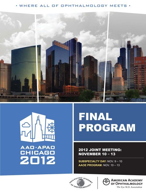
• WHERE ALL OF OPHTHALMOLOGY MEETS •<br />
<strong>FINAL</strong><br />
<strong>PROGRAM</strong><br />
2012 JOINT MEETING:<br />
NOVEMBER 10 – 13<br />
SUBSPECIALTY DAY: NOV. 9 – 10<br />
AAOE <strong>PROGRAM</strong>: NOV. 10 – 13
FALL INTO THE<br />
WINDY CITY<br />
Catch the leading experts in eye care at Allergan Booth #1408<br />
Saturday, November 10<br />
9:30 am<br />
Treatment of Macular Edema Due to<br />
Retinal Vein Occlusion<br />
Shree Kurup, MD<br />
10:00 am<br />
Treatment of Allergic Conjunctivitis<br />
Rajesh Rajpal, MD<br />
10:30 am<br />
Management of the Post-operative<br />
Cataract Surgery Patient<br />
Karl Stonecipher, MD<br />
11:00 am<br />
Treatment of Hypotrichosis<br />
Steve Yoelin, MD<br />
12:00 pm<br />
Detecting and Managing<br />
Glaucoma Progression<br />
Louis B. Cantor, MD<br />
12:30 pm<br />
RESCUE ME!—Interactive Cases<br />
Robert Osher, MD<br />
1:00 pm<br />
IOP Lowering: Options for Starting or<br />
Replacing Therapy<br />
Jonathan Myers, MD<br />
1:30 pm<br />
Conquering Capsule Complications—<br />
Strategies for Complicated Cataracts<br />
David Chang, MD<br />
2:00 pm<br />
Treatment of Macular Edema Due to<br />
Retinal Vein Occlusion<br />
Ron Gallemore, MD, PhD<br />
3:00 pm<br />
Focus on Dry Eye Disease<br />
Christopher Starr, MD, FACS<br />
3:30 pm<br />
Making Social Media “Work” for<br />
Your Practice<br />
Joe Casper, MBA, COE, OCS,<br />
Senior Eye Care Business Advisor,<br />
Allergan, Inc.<br />
Eric Abrantes, Marketing Director,<br />
Advanced Eye Centers<br />
Sunday, November 11<br />
9:30 am<br />
Management of the Small Pupil<br />
in Cataract Surgery<br />
Eric Donnenfeld, MD, FACS<br />
10:30 am<br />
REFRESH OPTIVE Advanced<br />
Marguerite McDonald, MD, FACS<br />
11:00 am<br />
IOP Reduction With Adjunctive Therapy<br />
Nathan Radcliffe, MD<br />
12:00 pm<br />
Treatment of Hypotrichosis<br />
Steve Yoelin, MD<br />
1:00 pm<br />
A Versatile Option in Adjunctive<br />
IOP Lowering<br />
E. Randy Craven, MD<br />
1:30 pm<br />
Treatment of Macular Edema Due to<br />
Retinal Vein Occlusion<br />
Michael Singer, MD<br />
2:00 pm<br />
Healthcare Reform: What Every<br />
Practice Should Know<br />
Mike Driscoll, OCS, Eye Care<br />
Business Advisor, Allergan, Inc.<br />
Jeffrey Lemay, Director, Healthcare<br />
Reform Initiative, Allergan, Inc.<br />
3:00 pm<br />
Adventures in Darkness<br />
Tom Sullivan<br />
Monday, November 12<br />
9:30 am<br />
Protecting Your Practice From Theft:<br />
Lessons Learned<br />
Jill Maher, MA, OCS, Eye Care Business<br />
Advisor, Allergan, Inc.<br />
11:00 am<br />
Successful Strategies for Effective<br />
EMR Implementation<br />
Sherri Boston, MBA, COE, OCS,<br />
Eye Care Business Advisor,<br />
Allergan, Inc.<br />
Jane T. Shuman, COT, COE, OCS,<br />
EyeTechs and eyebuzz ®<br />
Jeff Grant, President & Founder,<br />
Healthcare Management & Automation<br />
Systems, Inc.<br />
12:30 pm<br />
Why You Can’t Ignore Social Media: As<br />
Featured in Ophthalmology Management<br />
Greg Raeman, COE, CCOA, OCS, Eye<br />
Care Business Advisor, Allergan, Inc.<br />
2:00 pm<br />
Keys to Attracting & Managing<br />
Talented Employees<br />
Jim Rienzo, OCS, Senior Eye Care<br />
Business Advisor, Allergan, Inc.<br />
Tom Pannullo, COO, Ophthalmic<br />
Consultants of Long Island<br />
©2012 Allergan, Inc., Irvine, CA 92612 mark owned by Allergan, Inc. eyebuzz is a registered service mark owned by Eyetechs, Inc.<br />
www.allergan.com APC67SF12 122420 Presentation times and speakers are subject to change. This event is not affiliated with the official program of the 2012 Joint Meeting.
Table of Contents<br />
2012 Joint Meeting Highlights.......................................................ix<br />
2012 Board of Trustees.................................................................xiii<br />
2012 Committee of Secretaries.................................................... xix<br />
The Council...................................................................................xxv<br />
Meeting Overview.......................................................................xxix<br />
Meeting Directory.......................................................................xxxv<br />
Shuttle Schedule........................................................................xxxix<br />
Awards<br />
Academy Laureate...........................................................................1<br />
Special Awards................................................................................4<br />
Secretariat Award..........................................................................15<br />
Achievement Award Program........................................................17<br />
International Awards.....................................................................28<br />
Visionary Society, Corporate and Organizational Donors .............29<br />
Courses and Breakfasts<br />
Selection Committees....................................................................33<br />
Programs-by-Day............................................................................35<br />
Breakfast With the Experts............................................................51<br />
Instruction Course Program...........................................................59<br />
Skills Transfer Program<br />
Skills Transfer Program General Information...............................113<br />
Skills Transfer Course Contributors.............................................114<br />
Skills Transfer Program................................................................115<br />
Sessions and Symposia<br />
Opening Session & Business Meeting........................................129<br />
Academy Café..............................................................................130<br />
Academy Spotlight Sessions & Symposia...................................131<br />
Papers / Posters / Videos<br />
Original Papers.............................................................................153<br />
Scientific Posters.........................................................................173<br />
VIdeo Program..............................................................................246<br />
Special Meetings and Events<br />
Special Meetings & Events.........................................................257<br />
Learning Lounge...........................................................................261<br />
Technology Pavilion......................................................................265<br />
AAOE Program / Practice Management<br />
AAOE Program.............................................................................281<br />
Coding Sessions...........................................................................282<br />
AAOE Instruction Courses............................................................283<br />
AAOE Special Meetings & Events...............................................296<br />
Exhibit Hall, Indexes<br />
Informational Posters...................................................................305<br />
Available Academy Services in the Exhibit Hall..........................307<br />
Indexes.........................................................................................308<br />
Exhibitor List................................................................................309<br />
Product Index...............................................................................313<br />
Participant Index..........................................................................323<br />
Participant Financial Disclosure Index.........................................333<br />
CME & CE Credit..........................................................................349<br />
CME Credit Statement.................................................................351<br />
Future Annual Meeting Dates and Locations..............................352<br />
P.O. Box 7424, San Francisco, CA 94120-7424 | Tel: 415-561-8500 Fax: 415-561-8533<br />
© 2012 American Academy of Ophthalmology. All rights reserved.<br />
No portion may be reproduced without express consent of the American Academy of Ophthalmology.<br />
Academy staff are available at the Academy Resource Center (Booth 508) to answer any questions you may have.<br />
i
Living up to Life<br />
Visit Booth #2739 for our program:<br />
Innovations in Leica Microsystems’<br />
Surgical Microscopes & Video<br />
Saturday: 4:00 p.m.<br />
Sunday: 2:00 p.m.<br />
Monday: 4:15 p.m.<br />
• The New Leica M822 Surgical Microscope<br />
• Innovations in Ophthalmic Surgical Video<br />
Seenu M. Hariprasad, MD, University of Chicago<br />
• Surgical Video with TrueVision 3D<br />
Michael Saidel, MD, University of Chicago<br />
Meet us at AAO 2012 in Chicago, booth #2739 to Test Drive the Leica<br />
M822 Ultimate Red Reflex Surgical Microscope, or call 800-248-0123<br />
today to arrange your free demonstration.<br />
www.leica-microsystems.com/testdrive<br />
© 2012 Leica Microsystems, Inc. BGA#AAO
DURING THE AAO-APAO JOINT MEETING<br />
Here is just a sampling of the presentations that showcase the latest<br />
technologies in ophthalmology brought to you by Alcon, booth #2808.<br />
Saturday, Nov. 10<br />
9:30 AM<br />
Blepharitis: The New Consensus<br />
Stephen V. Scoper, MD<br />
11:00 AM<br />
The LenSx® Laser: Sphere and Cylinder<br />
Are Not Enough<br />
Paul Ernest, MD<br />
11:30 AM<br />
Alcon Advances for Today’s LASIK Surgery<br />
Sonny Goel, MD<br />
Charles Moore, MD<br />
12:30 PM<br />
Advanced Optical Biometry: Using the<br />
LENSTAR LS 900®* Optical Biometer with<br />
Toric IOLs, Strategies for Success<br />
Warren Hill, MD<br />
1:00 PM<br />
Methods to Manage Pre-Existing<br />
Corneal Astigmatism with Toric IOLs<br />
Edward J. Holland, MD<br />
Samuel Masket, MD<br />
1:30 PM<br />
Rethinking the Role of IOP in the Diagnosis and<br />
Management of Open-angle Glaucoma<br />
Matthew McMenemy, MD<br />
2:00 PM<br />
The LenSx® Laser: A New Cataract Procedure<br />
Stephen Lane, MD<br />
Satish Modi, MD<br />
Dan Tran, MD<br />
3:00 PM<br />
Multifocal IOLs: Setting Expectations for<br />
Presbyopic Patients<br />
Randy Epstein, MD<br />
Cathleen McCabe, MD<br />
3:30 PM<br />
Clinical Pearls to Adopting the<br />
Ex-PRESS®GFD<br />
Steve Vold, MD<br />
Sunday, Nov. 11<br />
11:00 AM<br />
Maximizing Success with the<br />
Ex-PRESS® Glaucoma Filtration Device<br />
Ike Ahmed, MD<br />
12:30 PM<br />
Multifocal IOLs: Setting Expectations for<br />
Presbyopic Patients<br />
William J. Lahners, MD<br />
Andrew Maxwell, MD<br />
1:00 PM<br />
Alcon Advances for Today’s<br />
LASIK Surgery<br />
Vance Thompson, MD<br />
1:30 PM<br />
Integrating the LenSx® Laser into<br />
Our Practice<br />
Michael P. Jones, MD<br />
Christa Garner, BA, CRC<br />
3:00 PM<br />
Methods to Manage Pre-Existing Corneal<br />
Astigmatism with Toric IOLs<br />
Gary Foster, MD<br />
Ehsan Sadri, MD<br />
3:30 PM<br />
Blepharitis: The New Consensus<br />
Stephen V. Scoper, MD<br />
Monday, Nov. 12<br />
10:00 AM<br />
The LenSx® Laser: A New Cataract Procedure<br />
Jerry Hu, MD<br />
Robert Lehmann, MD<br />
12:30 PM<br />
Alcon Advances for Today’s LASIK Surgery<br />
Joseph L. Parisi, MD<br />
1:30 PM<br />
My Experience with the Ex-PRESS® Glaucoma<br />
Filtration Device<br />
Jeff Goldberg, MD<br />
2:00 PM<br />
Optically Measured Lens Thickness in<br />
IOL Power Calculation<br />
Sheridan Lam, MD<br />
Presentations, presenters and<br />
times are subject to change.<br />
These presentations are not<br />
affiliated with the official<br />
program of the 2012 AAO-<br />
APAO Joint Meeting.<br />
For important safety information<br />
about the Alcon® products<br />
discussed in these presentations,<br />
please visit the Alcon booth.<br />
Scan for Alcon at the<br />
AAO Information<br />
*LENSTAR® is a registered trademark of Haag-Streit<br />
©2012 Novartis 9/12 MIX12422JAS
VISIT US AT<br />
ALLERGAN BOOTH #1408<br />
©2012 Allergan, Inc., Irvine, CA 92612<br />
®<br />
marks owned by Allergan, Inc. APC86OM12 122086<br />
restasisprofessional.com
Dedicated to<br />
advancing the<br />
treatment of eye<br />
diseases with<br />
unmet medical need<br />
Visit us at AAO/APAO Booth #1571<br />
ThromboGenics , a biopharmaceutical company focused on<br />
developing innovative ophthalmic medicines.<br />
11/12<br />
ThromboGenics, Inc. 101 Wood Avenue South, 6th Floor, Iselin, NJ 08830 - USA ©2012 ThromboGenics, Inc.<br />
All rights reserved. THROMBOGENICS and the THROMBOGENICS logo are trademarks or registered trademarks<br />
of ThromboGenics in the United States, European Union, Japan, and other countries.<br />
THRCOR002 A
800.787.5426<br />
haag-streit-usa.com<br />
Your Octopus<br />
data will connect<br />
with your satellite<br />
office 37,000<br />
times faster.<br />
haag-streit-usa.com<br />
Octopus connectivity launches a new era in perimetry. One<br />
where doctors are always linked in real time to crucial<br />
patient data. Wherever you are online, your expertise can<br />
go to work instantly. From a satellite office, exam room, or<br />
somewhere more far out.<br />
Call 1-800-787-5426 to schedule a demo.<br />
Powered by<br />
Visit us at AAO Booth #3808.<br />
The Superior Practice.<br />
© 2012 Haag-Streit USA. All Rights Reserved.
• 2012 Joint Meeting HIGHLIGHTS •<br />
Kick off the<br />
Joint Meeting<br />
The Opening Session<br />
Sunday, Nov. 11, 8:30 a.m. – 10 a.m.<br />
Stephen J Ryan MD will receive the 2012 Laureate<br />
Award for his distinguished career and contributions<br />
to ophthalmology. Dr. Ryan is currently the President of<br />
the Doheny Eye Institute and the Distinguished Grace<br />
and Emery Beardsley Professor at USC. Abraham Verghese<br />
MD MACP will deliver the Opening Session<br />
keynote address about the physician-patient relationship.<br />
Dr. Verghese is a renowned physician, bestselling<br />
author and professor for the theory and practice of<br />
medicine at Stanford University’s School of Medicine.<br />
Joan W Miller MD will give the Jackson Memorial<br />
Lecture: AMD Revisited—Piecing the Puzzle. Dr. Miller<br />
is the Henry Willard Williams Professor of Ophthalmology<br />
& Chair, Department of Ophthalmology, Harvard<br />
Medical School and the Chief of Ophthalmology, Massachusetts<br />
Eye and Ear Infirmary and Massachusetts<br />
General Hospital. She is preeminent in the field of<br />
ocular neovascularization.<br />
AAO-APAO Joint Symposia<br />
Join us for three joint symposia with the Asia-Pacific<br />
Academy of Ophthalmology (APAO):<br />
SYM05 Sunday, Nov. 11, 10:30 a.m. - 12:00 p.m.<br />
Corneal Stem Cell Advances in Clinical and Laboratory Research<br />
- international authorities will present on the topics of<br />
stem cells, the diagnosis of stem cell deficiency and interventions<br />
in clinical practice. Techniques currently emerging from<br />
the laboratory will be highlighted.<br />
SYM21 Monday, Nov. 12, 8:30 a.m. - 10:00 a.m.<br />
Ethnic Variations in Glaucoma Prevalence, Detection, and<br />
Treatment Outcomes – experts will discuss the role of race<br />
and ethnicity in glaucoma incidence, treatment and screening<br />
practices.<br />
SYM39 Tuesday, Nov. 13, 8:30 a.m. - 10:00 a.m.<br />
Management of Diabetic Retinopathy: East-West Perspectives<br />
– knowledgeable panelists will review new understanding of<br />
the epidemiology of, and trends in the management and clinical<br />
approaches to diabetic retinopathy, as well as how they differ<br />
between East and West.<br />
ix
• 2012 Joint Meeting HIGHLIGHTS •<br />
Turning the Spotlight On Four<br />
Hot Topics in Ophthalmology<br />
Be sure to make time for the four free Spotlight Sessions<br />
at the 2012 Joint Meeting:<br />
Spotlight on Innovation in Ophthalmology: From<br />
Theory to Therapy<br />
SPO1 Sunday, Nov. 11, 10:30 a.m. – 12:00 p.m., Grand<br />
Ballroom S100ab. Investigate the factors driving and influencing<br />
innovation in ophthalmology through six 10-minute<br />
presentations, followed by a Q&A session.<br />
Spotlight on Corneal Collagen Crosslinking<br />
SPO2 Sunday, Nov. 11, 2:00 p.m. – 3:30 p.m., Grand Ballroom<br />
S100ab. Discuss surgical treatment modalities utilized<br />
in US clinical trials and internationally, the clinical experience<br />
of basic science, indications, surgical technique and complications<br />
encountered with several CXL treatments for indications<br />
following refractive surgery. Then Continue the Conversation<br />
in the Learning Lounge, Booth 107 at 3:30 p.m. with R Doyle<br />
Stulting MD PhD and A John Kanellopoulos MD.<br />
Spotlight on Cataracts: Clinical Decision-making<br />
with Cataract Complications<br />
SPO3 Monday, Nov. 12, 8:15 a.m. – 12:15 p.m., North Hall<br />
B. Start off Cataract Monday by attending the presentation of<br />
seven video cases about the management of intra-operative<br />
challenges and complications. The session will conclude with<br />
the 8th annual Charles Kelman Lecture. Then Continue the<br />
Conversation in the Learning Lounge, Booth 107 at 1:45 p.m.<br />
where Douglas D Koch MD and Warren E Hill MD will moderate<br />
an interactive discussion.<br />
Spotlight on Pseudoexfoliation<br />
SPO4, Monday, Nov. 12, 4:15 p.m. – 5:30 p.m., North Hall<br />
B. Conclude Cataract Monday by exploring many of the challenges<br />
inherent to the exfoliation syndrome from the perspective<br />
of cataract and glaucoma management. This symposium<br />
will primarily examine important decision-making strategies<br />
pertaining to the timing of cataract surgery in patients with<br />
exfoliation. Clinical pearls to avoid and manage complications<br />
during cataract surgery will be discussed.<br />
x<br />
Not to Miss Returning Favorites<br />
The Great Debate<br />
SYM41 Monday, Nov. 12, 10:45 a.m. – 12:00 p.m., E450<br />
Five pairs of debators will argue the pros and cons of five controversial<br />
topics. Audience voting will determine which speakers<br />
were most effective.<br />
Late Breakers Symposium<br />
SYM25 Monday, Nov. 12, 2 – 3:30 p.m., Grand Ballroom S100c<br />
Topics will cover new technology and therapies as well as important<br />
issues and controversies that have come up within the last<br />
six months in the field of ophthalmology.<br />
Grand Rounds: Cases and Experts From Across the<br />
Nation<br />
SYM43 Monday, Nov. 12, 3:45 – 5:00 p.m., S406a<br />
Real residents from different academic programs present real<br />
cases from real department grand rounds. Cases are presented to<br />
a panel of experts followed by Q&A and discussion.<br />
Best of the Posterior Segment Specialty Meetings 2012<br />
SYM03 Sunday, Nov. 11 10:30 – 11:45 a.m., S406a<br />
This symposium will feature best papers focusing on the posterior<br />
segment from the major retina, neuro-ophthalmology, uveitis,<br />
oculoplastics and pediatric ophthalmology specialty meetings of<br />
2012. Continue the Conversation in the Learning Lounge, Booth<br />
107 at 12:00 p.m.<br />
Best of the Anterior Segment Specialty Meetings 2012<br />
SYM44 Tuesday, Nov 13 12:15 – 1:30 p.m., S406A<br />
This symposium will feature best papers focusing on the anterior<br />
segment from the major retina, neuro-ophthalmology, uveitis,<br />
oculoplastics and pediatric ophthalmology specialty meetings<br />
of 2012.<br />
Interactive Learning<br />
Learning Lounge, Booth 107<br />
All Joint Meeting attendees are welcome to gather at the Learning<br />
Lounge for informal, small group presentations and discussions<br />
on the hottest topics at the Joint Meeting. See page xx<br />
for a schedule, check the Mobile Meeting Guide, www.aao.org/<br />
mobile or stop by the Learning Lounge.
Laser Refractive Cataract Surgery is<br />
Now a Reality with Alcon’s LenSx ® Laser.<br />
Cataract Surgery Will Change<br />
in a Femtosecond.<br />
With Alcon’s LenSx ® Laser, the Possibilities Have Just Begun.<br />
Delivering the precision of a femtosecond laser to Refractive Cataract Surgery, the LenSx ® Laser is designed to reproducibly perform many of the<br />
most challenging aspects of traditional cataract surgery. Creating highly reproducible capsulotomy, lens fragmentation and all corneal incisions<br />
including arcuate incisions with image-guided surgeon control, Alcon’s LenSx ® Laser is Putting the Future in Motion.<br />
CAUTION: United States Federal Law restricts this device to sale and use by or on the order of a physician or licensed eye care practitioner.<br />
United States Federal Law restricts the use of this device to practitioners who have been trained in the operation of this device.<br />
Please see adjacent page for brief summary information.<br />
The LenSx® Laser is indicated for use in patients undergoing cataract surgery for removal of the crystalline lens.<br />
Intended uses in cataract surgery include anterior capsulotomy, phacofragmentation, and the creation of single<br />
plane and multiplane arc cuts/incisions in the cornea, each of which may be performed either individually or<br />
consecutively during the same procedure.<br />
For Important Safety Information and Full Directions for Use, please reference the LenSx® Laser Directions for Use.<br />
To learn more about LenSx ® Laser technology for Laser Refractive Cataract Surgery, visit lensxlasers.com.<br />
© 2011 Novartis 9/11 LSX11503JAD
2012 Board of Trustees<br />
Ruth D Williams MD<br />
Paul Sternberg Jr MD<br />
David W Parke II MD<br />
Richard L Abbott MD<br />
Laurie G Barber MD<br />
George B Bartley MD<br />
Cynthia A Bradford MD<br />
David A Durfee MD<br />
Alaa ElDanasoury MD<br />
Paul B Ginsburg PhD<br />
B Thomas Hutchinson MD<br />
J Antonio Roca MD<br />
Jonathan B Rubenstein MD<br />
Andrew P Schachat MD<br />
Gregory L Skuta MD<br />
John R Stechschulte MD<br />
Humphrey J F Taylor<br />
Linda M Tsai MD<br />
Russell N Van Gelder MD PhD<br />
Ann A Warn MD MBA<br />
George A Williams MD<br />
Charles M Zacks MD<br />
President<br />
President-Elect<br />
Executive Vice President/CEO<br />
Past President<br />
Trustee-at-Large<br />
Trustee-at-Large<br />
Senior Secretary for Advocacy<br />
Senior Secretary for Ophthalmic Practice<br />
International Trustee-at-Large<br />
Public Trustee<br />
Chair, FAAO Advisory Board<br />
International Advisor<br />
Secretary for Annual Meeting<br />
Editor, OPHTHALMOLOGY<br />
Senior Secretary for Clinical Education<br />
Trustee-at-Large<br />
Public Trustee<br />
Trustee-at-Large<br />
Chair, The Council<br />
Vice Chair, The Council<br />
Trustee-at-Large<br />
Trustee-at-Large<br />
P.O. Box 7424, San Francisco, CA 94120-7424 | Tel: 415-561-8500 Fax: 415-561-8533<br />
© 2012 American Academy of Ophthalmology. All rights reserved.<br />
No portion may be reproduced without express consent of the American Academy of Ophthalmology.<br />
Academy staff are available at the Academy Resource Center (Booth 508) to answer any questions you may have.<br />
xiii
See what we’re revealing.<br />
Introducing world-class visualization from the leader in cataract surgery.<br />
Experience Alcon’s latest commitment to you, the LuxOR Ophthalmic Microscope. It’s the only one of its kind to provide:<br />
• Superior red reflex stability 1<br />
• Greater depth of focus 1<br />
• An improved surgeon experience<br />
To see how Alcon is expanding its ophthalmic surgery expertise to microscope innovation, contact your sales representative<br />
today or visit AlconSurgical.com.<br />
1. Data on file, Alcon Laboratories, Inc.<br />
© 2012 Novartis 7/12 DIA12005JAD AlconSurgical.com
World Ophthalmology Congress ® 2014 Tokyo<br />
WOC2014<br />
Tokyo<br />
XXXIV International Congress of Ophthalmology<br />
in conjunction with<br />
29th Asia-Pacific Academy of Ophthalmology Congress<br />
118th Annual Meeting of the Japanese Ophthalmological Society<br />
April 2 6, 2014<br />
Venue: Tokyo International Forum / Imperial Hotel, Tokyo<br />
President<br />
Tetsuro Oshika, M.D.<br />
Professor and Chairman, Department of Ophthalmology, Faculty of Medicine, University of Tsukuba<br />
WOC2014<br />
Tokyo | April 2–6<br />
Secretariat: c/o Congress Corporation<br />
Kōsai-kaikan Bldg., 5-1 Kojimachi, Chiyoda-ku, Tokyo 102-8481, Japan<br />
Phone: +81-3-5216-5551 Fax: +81-3-5216-5552 E-mail: woc2014tokyo@congre.co.jp<br />
www.woc2014.org
Some surfaces are worth protecting<br />
THE OCULAR SURFACE IS ONE.<br />
© 2012 Novartis 2/12 SYS11179JAD<br />
Surface Protection and More<br />
References<br />
1. Christensen MT, Blackie CA, Korb DR, et al. An evaluation of the performance of a novel lubricant eye drop. Poster D692 presented at: The Association for Research in Vision and Ophthalmology Annual Meeting; May 2-6,<br />
2010; Fort Lauderdale, FL. 2. Lane S, Paugh JR, Webb JR, Christensen MT. An evaluation of the in vivo retention time of a novel artificial tear as compared to a placebo control. Poster D923 presented at: The Association for<br />
Research in Vision and Ophthalmology Annual Meeting; May 3-7, 2009; Fort Lauderdale, FL. 3. Davitt WF, Bloomenstein M, Christensen M, et al. Efficacy in patients with dry eye after treatment with a new lubricant eye drop<br />
formulation. J Ocul Pharmacol Ther. 2010;26(4):347-353. 4. Alejandro A. Efficacy of a Novel Lubricant Eye Drops in Reducing Squamous Metaplasia in Dry Eye Subjects. Presented at the 29th Pan-American Congress of<br />
Ophthalmology in Buenos Aires, Argentina, July 7-9, 2011. 5. Wojtowica JC., et al. Pilot, Prospective, Randomized, Double-masked, Placebo-controlled Clinical Trial of an Omega-3 Supplement for Dry Eye. Cornea 2011:30(3)<br />
308-314. 6. Geerling G., et al. The International Workshop on Meibomian Gland Dysfunction: Report of the Subcommittee on Management and Treatment of Meibomian Gland Dysfunction. IOVS 2011:52(4).
2012 Committee of Secretaries<br />
American Academy of Ophthalmology<br />
David W Parke II MD<br />
Cynthia A Bradford MD<br />
Daniel J Briceland MD<br />
Louis B Cantor MD<br />
Anne L Coleman MD PhD<br />
David A Durfee MD<br />
Tamara R Fountain MD<br />
Jeffrey S Heier MD<br />
Gregory P Kwasny MD<br />
Robert F Melendez MD MBA<br />
Richard P Mills MD MPH<br />
Jeffery A Nerad MD<br />
Michael X Repka MD<br />
William L Rich III MD<br />
Philip R Rizzuto MD<br />
Jonathan B Rubenstein MD<br />
Andrew P Schachat MD<br />
Gregory L Skuta MD<br />
Ronald E Smith MD<br />
Chair<br />
Senior Secretary for Advocacy<br />
Secretary for State Affairs<br />
Secretary for Ophthalmic Knowledge<br />
Secretary for Quality of Care<br />
Senior Secretary for Ophthalmic Practice<br />
Secretary for Member Services<br />
Secretary for Online Education/eLearning<br />
Secretary for Federal Affairs<br />
Editor-in-Chief, the ONE Network<br />
Chief Medical Editor, EyeNet Magazine<br />
Secretary for Knowledge Base Development<br />
AAO Medical Director for Governmental Affairs<br />
AAO Medical Director of Health Policy<br />
Secretary for Communications<br />
Secretary for Annual Meeting<br />
Editor, OPHTHALMOLOGY<br />
Senior Secretary for Clinical Education<br />
Secretary for Global Alliances<br />
P.O. Box 7424, San Francisco, CA 94120-7424 | Tel: 415-561-8500 Fax: 415-561-8533<br />
© 2012 American Academy of Ophthalmology. All rights reserved.<br />
No portion may be reproduced without express consent of the American Academy of Ophthalmology.<br />
Academy staff are available at the Academy Resource Center (Booth 508) to answer any questions you may have.<br />
xix
800.787.5426<br />
haag-streit-usa.com<br />
Imaging with Depth of Field. Just Dandy.<br />
HSImaging.com<br />
What if none of your slit images had blurry edges? You<br />
would have a Haag-Streit, of course. Invest in one and<br />
submerge yourself into our extreme world of depth of field.<br />
Visit HSImaging.com to learn about our entire range of slit<br />
lamps and imaging systems.<br />
LED Powered BQ 900 ® Slit Lamp with IM 900 ® Imaging System<br />
Visit us at AAO Booth #3808.<br />
The Superior Practice.<br />
© 2012 Haag-Streit USA. All Rights Reserved.
Anticipating every move.<br />
Now that’s smart.<br />
Experience the intuitively adaptive control of NEW OZil® IP.<br />
Featuring innovative OZil® Intelligent Phaco, the Infiniti® Vision System puts<br />
optimized OZil® torsional emulsification and dynamic fluidic management at<br />
your fingertips. With significantly enhanced capabilities, OZil® IP is always thinking<br />
one step ahead. For more information, contact your Alcon representative.<br />
© 2011 Novartis 12/11 INF546
Buy Reliance,<br />
and Help your Neighbor Weather the Storm.<br />
800.735.0357<br />
haag-streit-usa.com<br />
RelianceDurable.com<br />
In tough times, Americans pull together. And we become<br />
stronger, like Reliance equipment, manufactured in the USA<br />
since 1898. Yes, it’s patriotism. But our American made<br />
products are just better, too. More durable, and innovative.<br />
The qualities you value in exam chairs and stands. Are you in<br />
the market for new equipment? Then consider Reliance, and<br />
your 20,000 neighbors across the nation who help manufacture<br />
our products.<br />
Visit us at AAO Booth #3808.<br />
The Superior Practice.<br />
© 2012 Reliance Medical Products. All Rights Reserved.
Save the Date!<br />
There’s strength in numbers. Lobby on Capitol Hill for<br />
ophthalmology’s top legislative issues including advocating for<br />
fair Medicare physician payment, reducing regulatory burdens<br />
and vision research. Meet face-to-face with your Members<br />
of Congress and show the might of our members at this<br />
important opportunity. It’s the most effective way to protect<br />
the interests of our profession and our patients.<br />
Congressional<br />
Advocacy Day<br />
April 10 – 11, 2013<br />
Washington, DC<br />
“Join hundreds of your ophthalmology<br />
colleagues in Washington D.C. for<br />
Congressional Advocacy Day. We need your<br />
voice! Academy members advocate for our<br />
patients and our profession. Join me in<br />
Washington D.C. in 2013.”<br />
Ruth D. Williams, MD<br />
Academy President<br />
Registration opens in January 2013.<br />
Congressional Advocacy Day is open to all<br />
Academy members and registration is free.<br />
www.aao.org/myf
The Council<br />
The Council serves as the advisory body to the Board of Trustees. The Council was established in accordance with section 7.01 of the<br />
Bylaws of the American Academy of Ophthalmology.<br />
Russell N Van Gelder MD PhD<br />
Chair<br />
Ann A Warn MD MBA<br />
Vice Chair<br />
Councilors Representing State<br />
Societies<br />
Alabama Academy of<br />
Ophthalmology<br />
Wonsuck Kim DO<br />
Alaska Society of Eye Physicians<br />
and Surgeons<br />
Scott A Limstrom MD<br />
Arizona Ophthalmological Society<br />
Thomas J McPhee MD<br />
Arkansas Ophthalmological Society<br />
Morriss M Henry MD<br />
California Academy of Eye<br />
Physicians and Surgeons<br />
Kimberly Cockerham MD FACS<br />
Craig H Kliger MD<br />
Asa Dan Morton III MD<br />
Ronald Lee Morton MD FACS<br />
Colorado Society of Eye Physicians<br />
and Surgeons<br />
Robert A King MD<br />
Connecticut Society of Eye<br />
Physicians<br />
Jeffrey R Sandler MD<br />
Delaware Academy of<br />
Ophthalmology<br />
Odette V Callender MD<br />
Florida Society of Ophthalmology<br />
Gary B Schemmer MD<br />
Stephen G Schwartz MD MBA<br />
Michael W Stewart MD<br />
Georgia Society of Ophthalmology<br />
James Gerard Brooks Jr MD<br />
Hawaii Ophthalmological Society<br />
George Nardin MD<br />
Idaho Society of Ophthalmology<br />
Adam C Reynolds MD<br />
Illinois Association of<br />
Ophthalmology<br />
Richard A Quinones MD<br />
David K Yoo MD<br />
Indiana Academy of Ophthalmology<br />
Derek T Sprunger MD<br />
Iowa Academy of Ophthalmology<br />
Christopher L Haupert MD<br />
Kansas Society of Eye Physicians<br />
and Surgeons<br />
William S Clifford MD<br />
Kentucky Academy of Eye<br />
Physicians and Surgeons<br />
David E Jones MD<br />
Louisiana Ophthalmology<br />
Association<br />
Keith Kellum MD DVM<br />
Maine Society of Eye Physicians<br />
and Surgeons<br />
Cynthia A Self MD<br />
Maryland Society of Eye Physicians<br />
and Surgeons<br />
Sanjay D Goel MD<br />
John T Thompson MD<br />
Massachusetts Society of Eye<br />
Physicians and Surgeons<br />
Robert A Lytle MD<br />
Michael J Price MD<br />
Michigan Society of Eye Physicians<br />
and Surgeons<br />
Arezo Amirikia MD<br />
Robert Jay Granadier MD<br />
Minnesota Academy of<br />
Ophthalmology<br />
Eugene O Gullingsrud MD<br />
Mississippi Academy of Eye<br />
Physicians and Surgeons<br />
Curtis D Whittington Jr MD<br />
Missouri Society of Eye Physicians<br />
and Surgeons<br />
Melissa G Cable MD<br />
Montana Academy of<br />
Ophthalmology<br />
Brian D Sippy MD PhD<br />
Nebraska Academy of Eye<br />
Physicians and Surgeons<br />
David D Ingvoldstad MD<br />
Nevada Academy of Ophthalmology<br />
Steve M Friedlander MD FACS<br />
New Hampshire Society of Eye<br />
Physicians and Surgeons<br />
John J Dagianis MD<br />
New Jersey Academy of<br />
Ophthalmology<br />
Paul D Langer MD<br />
David M Ringel DO<br />
New Mexico Academy of<br />
Ophthalmology<br />
Ashok K Reddy MD<br />
New York State Ophthalmological<br />
Society<br />
Gary S Hirshfield MD<br />
Roger C Husted MD<br />
James A Kinsey MD<br />
Arnold S Prywes MD<br />
North Carolina Society of Eye<br />
Physicians and Surgeons<br />
Cynthia Hampton MD<br />
North Dakota Society of Eye<br />
Physicians and Surgeons<br />
Lance K Bergstrom MD<br />
Ohio Ophthalmological Society<br />
Anita Dash-Modi MD<br />
Mark S Law MD<br />
Oklahoma Academy of<br />
Ophthalmology<br />
Amalia Miranda MD<br />
Oregon Academy of Ophthalmology<br />
Mary P DeFrank MD<br />
Pennsylvania Academy of<br />
Ophthalmology<br />
James B Dickey MD<br />
Joanna M Fisher MD<br />
Karl R Olsen MD<br />
Puerto Rican Society of<br />
Ophthalmology<br />
Emilio A Arce-Lopez MD<br />
Rhode Island Society of Eye<br />
Physicians and Surgeons<br />
Robert H Janigian Jr MD<br />
South Carolina Society of<br />
Ophthalmology<br />
Kurt Frederick Heitman MD<br />
South Dakota Academy of<br />
Ophthalmology<br />
Monte Steven Dirks MD<br />
Tennessee Academy of<br />
Ophthalmology<br />
Erich Bryan Groos MD<br />
Texas Ophthalmological Association<br />
John R Fish MD<br />
Victor H Gonzalez MD<br />
James H Merritt MD<br />
Utah Ophthalmology Society<br />
Rachel Benator MD<br />
Vermont Ophthalmological Society<br />
Brian Y Kim MD<br />
Virginia Society of Eye Physicians<br />
and Surgeons<br />
Anthony J Viti MD<br />
Washington Academy of Eye<br />
Physicians and Surgeons<br />
Brian E Bowe MD<br />
Washington DC Metropolitan<br />
Ophthalmological Society<br />
Reshma Katira MD<br />
West Virginia Academy of Eye<br />
Physicians and Surgeons<br />
Mark D Mayle MD<br />
Wisconsin Academy of<br />
Ophthalmology<br />
Deborah W Bernstein MD<br />
Wyoming Ophthalmological Society<br />
Anne Elizabeth Miller MD<br />
Councilors representing<br />
Subspecialty and Specialized<br />
Interest Societies<br />
American Academy of Pediatrics,<br />
Section on Ophthalmology<br />
George S Ellis Jr MD<br />
American Association for Pediatric<br />
Ophthalmology and Strabismus<br />
Jane C Edmond MD<br />
American Association of<br />
Ophthalmic Oncologists and<br />
Pathologists<br />
Paul J Bryar MD<br />
American Board of Ophthalmology<br />
John E Sutphin MD<br />
American College of Surgeons,<br />
Advisory Council for Ophthalmic<br />
Surgery<br />
Vikram D Durairaj MD<br />
Sarwat Salim MD<br />
American Glaucoma Society<br />
James C Tsai MD MBA<br />
American Ophthalmological Society<br />
Thomas J Liesegang MD<br />
American Osteopathic Colleges of<br />
Ophthalmology and Otolaryngology<br />
David D Gossage DO<br />
American Society of Cataract and<br />
Refractive Surgery<br />
David A Goldman MD<br />
Thomas M Harvey MD<br />
American Society of Ocular Trauma<br />
Michael P Grant MD PhD<br />
American Society of Ophthalmic<br />
Plastic and Reconstructive Surgery<br />
Louise A Mawn MD<br />
American Society of Retina<br />
Specialists<br />
Peter K Kaiser MD<br />
Mathew W MacCumber MD PhD<br />
American Uveitis Society<br />
Justine R Smith MD<br />
Association for Research in Vision<br />
and Ophthalmology<br />
Robert B Nussenblatt MD<br />
Association of University Professors<br />
of Ophthalmology<br />
Joel S Schuman MD<br />
Association of Veterans Affairs<br />
Ophthalmologists<br />
Mary Gilbert Lawrence MD MPH<br />
Canadian Ophthalmological Society<br />
Paul E Rafuse MD PhD<br />
Contact Lens Association of<br />
Ophthalmologists<br />
Thomas L Steinemann MD<br />
Cornea Society<br />
Shahzad I Mian MD<br />
Eye Bank Association of America<br />
Alan Mark Kozarsky MD<br />
Macula Society<br />
Neil M Bressler MD<br />
National Medical Association,<br />
Ophthalmology Section<br />
Eydie G Miller-Ellis MD<br />
North American Neuro-<br />
Ophthalmology Society<br />
Matthew Dean Kay MD<br />
Ocular Microbiology and<br />
Immunology Group<br />
Bradley Dean Fouraker MD<br />
Outpatient Ophthalmic Surgery<br />
Society<br />
Y Ralph Chu MD<br />
Pan-American Association of<br />
Ophthalmology<br />
Stephanie Jones Marioneaux MD<br />
Retina Society<br />
Thomas M Aaberg Jr MD<br />
Society of Military Ophthalmologists<br />
Jonathan S Collins MD<br />
Women in Ophthalmology<br />
Laura J King MD MMM<br />
The Academy’s Council is integral to Academy relations with state, subspecialty and specialized interest ophthalmic societies. www.aao.org/council<br />
xxv
See what we’re revealing.<br />
Introducing world-class visualization from the leader in cataract surgery.<br />
Experience Alcon’s latest commitment to you, the LuxOR Ophthalmic Microscope. It’s the only one of its kind to provide:<br />
• Superior red reflex stability 1<br />
• Greater depth of focus 1<br />
• An improved surgeon experience<br />
To see how Alcon is expanding its ophthalmic surgery expertise to microscope innovation, contact your sales representative<br />
today or visit AlconSurgical.com.<br />
1. Data on file, Alcon Laboratories, Inc.<br />
© 2012 Novartis 7/12 DIA12005JAD AlconSurgical.com
Welcome to the<br />
S OE 2013 Congress<br />
in Copenhagen<br />
Photo: Morten Jerichau/Wonderful Copenhagen<br />
Photo: Cees van Roeden/Wonderful Copenhagen<br />
Online Registration and Abstract submission<br />
is open from 1 November 2012.<br />
Please stay updated on the SOE 2013 Congress website<br />
www.soe2013.org<br />
Photo: Shutterstock
888.myLENSTAR<br />
(888.695.3678)<br />
myLENSTAR.com<br />
myLENSTAR.com<br />
Breathe deeply. The unmatched<br />
accuracy of the LENSTAR ® Optical<br />
Biometer takes the stress out of<br />
selecting the correct toric IOL.<br />
Along with LENSTAR’s perfect<br />
Ks come superior outcomes<br />
for you and your patients. Call 888.myLENSTAR for a demo.<br />
Visit us at AAO Booth #3808.<br />
The Superior Practice.<br />
© 2012 Haag-Streit USA. All Rights Reserved.
Meeting Overview<br />
For location information see the program pages that follow or access the Mobile Meeting Guide, www.aao.org/mobile.<br />
Thursday, Nov. 8<br />
Event<br />
Time<br />
Registration Attendees 4:00 - 6:00 pm<br />
Exhibitors<br />
7:30 am - 6:00 pm<br />
Friday, Nov. 9<br />
Alumni & Related Group Functions<br />
Registration<br />
Special Meetings & Events<br />
Event<br />
Attendees<br />
Exhibitors<br />
Time<br />
All Day<br />
7:00 am - 5:00 pm<br />
7:30 am - 6:00 pm<br />
7:30 am - 3:00 pm<br />
Subspecialty Day Meetings Refractive Surgery 8:00 am - 5:15 pm<br />
Refractive Surgery<br />
E-posters<br />
Retina<br />
Retina Exhibits<br />
7:00 am - 5:30 pm<br />
8:00 am - 5:00 pm<br />
9:30 am - 3:30 pm<br />
Saturday, Nov. 10<br />
Event<br />
Time<br />
AAOE/Practice Management Coding SOS<br />
8:00 - 11:00 am<br />
AAOE/Practice Management Coding Camp<br />
12:30 - 3:30 pm<br />
AAOE/Practice Management Saturday Programs<br />
8:30 am - 4:30 pm<br />
Academy Café<br />
1:00 - 3:45 pm<br />
Alumni & Related Group Functions ><br />
Joint Meeting Exhibition<br />
9:00 am - 5:00 pm<br />
Learning Lounge<br />
12:00 - 5:00 pm<br />
Networking With the Experts: Knowledge and Tips for the Young Ophthalmologist<br />
12:00 - 1:30 pm<br />
Registration Attendees 7:00 am - 5:00 pm<br />
Exhibitors<br />
7:00 am - 5:00 pm<br />
Scientific Posters<br />
9:00 am - 5:00 pm<br />
Scientific Posters Online/Videos on Demand<br />
9:00 am - 5:00 pm<br />
Special Meetings & Events<br />
9:00 am - 4:00 pm<br />
Subspecialty Day Meetings Cornea 8:00 am - 5:30 pm<br />
Glaucoma<br />
8:00 am - 5:30 pm<br />
Oculofacial Plastic Surgery 8:00 am - 5:15 pm<br />
Pediatric Ophthalmology<br />
8:00 am - 5:15 pm<br />
Refractive Surgery<br />
8:00 am - 5:30 pm<br />
Refractive Surgery E-posters 7:00 am - 5:30 pm<br />
Retina<br />
8:00 am - 5:30 pm<br />
Uveitis<br />
7:50 am - 5:20 pm<br />
SYMPOSIA What is Global Ophthalmology? (Global Alliances) 2:30 - 4:00 pm<br />
Academy staff are available at the Academy Resource Center (Booth 508) to answer any questions you may have.<br />
xxix
Meeting Overview<br />
Sunday, Nov. 11<br />
AAOE/Practice Management Courses<br />
AAOE/Practice Management General Session<br />
Academy Business Meeting<br />
Academy Café<br />
Event<br />
Time<br />
2:00 pm - 5:30 pm<br />
10:00 am - 12:00 pm<br />
10:00 - 10:30 am<br />
10:30 am - 3:45 pm<br />
Alumni & Related Group Functions ><br />
Breakfast With the Experts<br />
Fall Council Meeting and Surgery by Surgeons Forum<br />
Instruction Courses<br />
Joint Meeting Exhibition<br />
Learning Lounge<br />
7:30 - 8:30 am<br />
11:30 am - 5:30 pm<br />
9:00 am - 5:30 pm<br />
9:00 am - 5:00 pm<br />
10:30 am - 5:00 pm<br />
2013 Medicare Update 12:15 - 1:45 pm<br />
OMIC Forum: Top Ten Indemnity Payments of 2011<br />
Opening Session<br />
Orbital Gala<br />
Original Paper Sessions<br />
2:00 - 3:30 pm<br />
8:30 - 10:00 am<br />
6:00 - 10:00 pm<br />
10:15 am - 5:30 pm<br />
Registration Attendees 8:00 am - 5:00 pm<br />
Run/Walk for Vision 5K Race<br />
Scientific Posters<br />
Scientific Posters Online/Videos on Demand<br />
Scientific Poster Tours<br />
Skills Transfer Courses<br />
Special Meetings & Events<br />
Young Ophthalmologist (YO) Program<br />
Spotlight on Innovation in Ophthalmology: From Theory to Therapy<br />
Spotlight on Corneal Collagen Crosslinking<br />
SYMPOSIA<br />
Introduction to Refractive Surgery for Residents (ISRS)<br />
Exhibitors<br />
Vision Rehabilitation Education: Effectively Transmitting the Need for Low Vision Services to the Ophthalmic<br />
Community (Vision Rehabilitation Committee)<br />
Best of the Posterior Segment Specialty Meetings 2012<br />
From Metal to Molecules: The Evolution of Oculofacial Plastic Surgery (ASOPRS)<br />
Corneal Stem Cell: Advances in Clinical and Laboratory Research (Joint Session with APAO)<br />
Contagion! Epidemics in Ophthalmic History (Museum of Vision)<br />
Cataract Surgery: The Cutting Edge<br />
Preferred Practice Pattern Guidelines ® : Adding Practical Value to Daily Practice (Preferred Practice Patterns<br />
Committee)<br />
Optimizing Optics: Perspectives From Contact Lens, Intraocular Lens, and Refractive Surgery (CLAO)<br />
How Does It Feel? An Insider’s Perspective on Living With Anophthalmia (ASO)<br />
Medical and Surgical Treatment of Macular Disease (The Retina Society)<br />
7:30 am - 5:00 pm<br />
6:30 - 8:00 am<br />
7:30 am - 5:00 pm<br />
7:30 am - 5:00 pm<br />
12:30 - 1:30 pm<br />
9:00 am - 5:30 pm<br />
10:00 am - 5:30 pm<br />
10:00 am - 2:00 pm<br />
10:30 am - 12:00 pm<br />
2:00 - 3:30 pm<br />
8:00 - 10:00 am<br />
10:00 am - 12:00 pm<br />
10:30 - 11:45 am<br />
10:30 am - 12:00 pm<br />
10:30 am - 12:00 pm<br />
12:15 - 1:45 pm<br />
12:15 - 1:45 pm<br />
12:45 - 1:45 pm<br />
2:00 - 3:30 pm<br />
2:00 - 3:30 pm<br />
2:00 - 3:30 pm<br />
xxx<br />
Academy staff are available at the Academy Resource Center (Booth 508) to answer any questions you may have.
Meeting Overview<br />
Sunday, Nov. 11 (cont.)<br />
SYMPOSIA<br />
Event<br />
Modern Technologies and Techniques for Young Ophthalmologists to Know (Young Ophthalmologist International<br />
Subcommittee and Young Ophthalmologists from SOE and APAO)<br />
Video Case Presentations of Rare Vitreoretinal Diseases (Eurolam)<br />
LASIK Is Safe : Prevention and Management of Laser Complications (ISRS)<br />
The Controversies and Dilemmas of Managing Ocular Infectious Diseases: Point-Counterpoint (OMIG)<br />
Controversies in Pediatric Ophthalmology and Orthoptics: A Point-Counterpoint Discussion (AOC/AACO)<br />
International Perspectives: Trauma of the Anterior Segment and Its Management (SCO and PAAO)<br />
International Opportunities for Young Ophthalmologists (Young Ophthalmologist International Subcommittee)<br />
Time<br />
2:30 - 4:00 pm<br />
3:45 - 5:15 pm<br />
3:45 - 5:15 pm<br />
3:45 - 5:15 pm<br />
3:45 - 5:15 pm<br />
3:45 - 5:15 pm<br />
4:15 - 5:15 pm<br />
Monday, Nov. 12<br />
Event<br />
Time<br />
AAOE/Practice Management Courses<br />
9:00 am - 5:30 pm<br />
Academy Café<br />
9:00 - 11:45 am<br />
Alumni & Related Group Functions ><br />
Breakfast With the Experts<br />
7:30 - 8:30 am<br />
Instruction Courses<br />
9:00 am - 5:30 pm<br />
Joint Meeting Exhibition<br />
9:00 am - 5:00 pm<br />
Learning Lounge<br />
9:00 am - 5:00 pm<br />
Original Paper Sessions<br />
8:30 am - 5:30 pm<br />
Registration Attendees 8:00 am - 5:00 pm<br />
Exhibitors<br />
7:30 am - 5:00 pm<br />
Scientific Posters<br />
7:30 am - 5:00 pm<br />
Scientific Posters Online/Videos on Demand<br />
7:30 am - 5:00 pm<br />
Scientific Poster Tours<br />
12:30 - 1:30 pm<br />
Senior Ophthalmologist (SO) Program<br />
2:30 - 5:00 pm<br />
Skills Transfer Courses<br />
7:30 am - 5:30 pm<br />
Special Meetings & Events<br />
8:30 am - 5:00 pm<br />
Spotlight on Cataracts: Clinical Decision-making With Cataract Complications<br />
8:15 am - 12:15 pm<br />
Spotlight on Pseudoexfoliation<br />
4:15 - 5:30 pm<br />
Ethnic Variations in Glaucoma Prevalence, Detection, and Treatment Outcomes (Joint Session with APAO)<br />
8:30 - 10:00 am<br />
SYMPOSIA<br />
Pediatric Corneal Disease and Treatment (AAPOS)<br />
8:30 - 10:00 am<br />
Re-engineering the U.S. Health Care System: The Impact on Ophthalmology (AMA Ophthalmology Section Council) 8:30 - 10:30 am<br />
What to Do When Your Patient Sees Nothing and You See Nothing: The Neuro-Ophthalmology Workup (NANOS)<br />
8:30 - 10:30 am<br />
2012 International Forum: Addressing Diabetic Blindness, Refractive Error and the Basic Eye Exam (Global Alliances) 8:30 - 11:00 am<br />
Advances in the Surgical Management of Glaucoma (Prevent Blindness America, Inc.)<br />
10:15 - 11:45 am<br />
Then and Now (Senior Ophthalmologist and Young Ophthalmologist Committees)<br />
10:15 - 11:45 am<br />
Update on Pediatric Ocular Trauma (ASOT)<br />
10:15 - 11:45 am<br />
The Great Debate<br />
10:45 am - 12:00 pm<br />
Making Electronic Health Records Meaningful and Useful in Your Practice (Committee on Medical Information<br />
12:45 - 1:45 pm<br />
Technology)<br />
Academy staff are available at the Academy Resource Center (Booth 508) to answer any questions you may have.<br />
xxxi
Meeting Overview<br />
Monday, Nov. 12 (cont.)<br />
SYMPOSIA<br />
Event<br />
The “Local” Challenges of International Ophthalmology (Women in Ophthalmology)<br />
Why Take the Risk? How to Create an Effective Risk Management Strategy With Patient Education and Informed<br />
Consent Documents (Patient Education Committee and OMIC)<br />
Crash Course in Teaching: A Primer for Faculty Development (AUPO)<br />
Advances in the Treatment of Diabetic Retinopathy (Macula Society)<br />
Late Breakers Symposium<br />
Femto Forum: Cataract, Cornea, Refractive, and Beyond (ASCRS)<br />
Non-bacterial Infectious Keratitis (Cornea Society)<br />
Vision Rehabilitation: What’s New for Patients With Low Vision (Vision Rehabilitation Committee)<br />
Quality Improvement: How Do We Improve Quality, Maintain Efficiency and Sustain the Physician-Patient Relationship?<br />
(Practice Improvement Committee)<br />
Grand Rounds: Cases and Experts From Across the Nation<br />
Time<br />
12:45 - 1:45 pm<br />
12:45 - 1:45 pm<br />
12:45 - 2:15 pm<br />
2:00 - 3:30 pm<br />
2:00 - 3:30 pm<br />
2:00 - 4:00 pm<br />
2:00 - 4:00 pm<br />
3:45 - 4:45 pm<br />
3:45 - 5:00pm<br />
3:45 - 5:00 pm<br />
Tuesday, Nov. 13<br />
AAOE/Practice Management Courses<br />
Academy Café<br />
Event<br />
Time<br />
9:00 am - 3:00 pm<br />
10:30 - 11:45 am<br />
Alumni & Related Group Functions ><br />
Breakfast With the Experts<br />
Instruction Courses<br />
Joint Meeting Exhibition<br />
Learning Lounge<br />
Original Paper Sessions<br />
7:30 - 8:30 am<br />
9:00 am - 5:30 pm<br />
9:00 am - 3:00 pm<br />
9:00 am - 12:00 pm<br />
8:30 am - 12:00 pm<br />
Registration Attendees 8:00 am - 3:00 pm<br />
Exhibitors<br />
7:30 am - 3:00 pm<br />
Scientific Posters<br />
7:30 am - 3:00 pm<br />
Scientific Posters Online/Videos on Demand<br />
7:30 am - 3:00 pm<br />
Skills Transfer Courses<br />
8:00 am - 5:30 pm<br />
Management of Diabetic Retinopathy: East-West Perspectives (Joint Session with APAO)<br />
8:30 - 10:00 am<br />
Ocular Tumors: Evidence-Based Rationale for Treatment (AAOOP)<br />
8:30 - 10:00 am<br />
Shifting Gears: Practical and Ethical Transitions to Retirement (Committee on Aging, Senior Ophthalmologist<br />
10:15 - 11:15 am<br />
Committee, Ethics Committee and the American Geriatrics Society)<br />
Workforce Issues in Ophthalmology: Eye Health Care for Baby Boomers and Beyond (NMA)<br />
8:30 - 10:00 am<br />
Clinical Applications of Ocular Imaging (ARVO)<br />
10:15 - 11:45 am<br />
Best of the Anterior Segment Specialty Meetings 2012<br />
12:15 - 1:30 pm<br />
SYMPOSIA<br />
Wednesday, Nov. 15<br />
Event<br />
Time<br />
26° Lo Mejor de la Academia en Español 2012 (The Best of the Academy in Spanish 2012) 7:00 am - 5:00 pm<br />
xxxii<br />
Academy staff are available at the Academy Resource Center (Booth 508) to answer any questions you may have.
Discover The FUTURE Of Diagnostic Imaging<br />
Advanced technology at the touch of a button<br />
Register To Attend a Special<br />
AAO Symposium<br />
New Advances in Multi-modality Imaging<br />
November 10, 2012 • 5:30pm–7:30pm<br />
Marriott Chicago Downtown<br />
www.HeidelbergEngineering.com/us/AAO2012<br />
SPECTRALIS is a registered trademark of Heidelberg Engineering, Inc.<br />
© 2012 Heidelberg Engineering, Inc. All rights reserved. 2660
Meeting Directory<br />
All rooms are in McCormick Place North Building (N), South Building (S), Lakeside Center (E).<br />
Exhibition is in South Building, Level 3, Hall A.<br />
AAO Meetings On Demand (Subspecialty Day<br />
and Joint Meeting Content)<br />
Grand Concourse and Booth 605<br />
AAOE Coding Sessions (Saturday)<br />
Room S105<br />
AAOE Member Lounge South, Level 5<br />
AAOE Program South, Level 5<br />
Academy Café<br />
Room S406b<br />
Academy Resource Center Booth 508<br />
Bags and Programs<br />
Hall A<br />
Bistro AAO Booth 2490<br />
Breakfast With the Experts<br />
Hall A<br />
Business Center South, Level 2.5<br />
Grand Concourse Level 2.5 &<br />
CME Reporting/Proof-of-Attendance<br />
Academy Resource Center,<br />
Booth 508<br />
Coat and Bag Check<br />
South, Level 1 Lobby<br />
Executive Offices<br />
Room S401<br />
Exhibitor Locator Booth 3500<br />
Exhibitor Lounge (Wi-Fi available) Booth 987<br />
Exhibitor Registration<br />
Hall A<br />
Exhibitor Service Center/Exhibitions Office Hall A<br />
First Aid South, Level 2.5<br />
Foundation of the American Academy of<br />
Ophthalmology (FAAO)<br />
Global Alliance Office<br />
Academy Resource Center,<br />
Booth 508<br />
Room S403a<br />
Hotel Assistance Grand Concourse, Level 2.5<br />
Informational Exhibits and Posters<br />
Hall A<br />
International Center Booth 4509<br />
Internet Access Grand Concourse & Booth 2987<br />
Learning Lounge Booth 107<br />
Lost and Found<br />
Room S402<br />
Meditation/Prayer Room<br />
Room SA1a<br />
Meeting Information<br />
Grand Concourse &<br />
South, Level 1 Lobby<br />
Meetings Office<br />
Room S402<br />
Mobile Device Charging Stations Booth 2485<br />
Mobile Meeting Guide Assistance Booth 2987<br />
Mobile Meeting Guide Download<br />
www.aao.org/mobile<br />
Museum of Vision Booth 704<br />
New Orleans 2013 Grand Concourse, Level 2.5<br />
Newsroom<br />
Room N426a<br />
Ophthalmic Mutual Insurance Company (OMIC) Booth 1104<br />
Ophthalmology Job Center<br />
Room N426c<br />
OPHTHPAC/Surgical Scope<br />
Grand Concourse<br />
Grand Concourse Level 2.5 &<br />
Proof-of-Attendance/CME Reporting<br />
Academy Resource Center,<br />
Booth 508<br />
Publishers’ Row<br />
Hall A<br />
Registration<br />
Hall A<br />
Rest Stop<br />
Booths 2485, 2490, 2787, 2981,<br />
2987<br />
Ribbons<br />
Bags & Programs, Hall A<br />
Scientific Posters<br />
Hall A<br />
Scientific Poster Tours<br />
Meeting Point, Hall A<br />
Scientific Posters Online/Videos on Demand Booth 165<br />
Seated Massage Stations Booth 2787<br />
Senior Ophthalmologist (SO) Lounge<br />
Grand Concourse Lobby<br />
Shuttle Bus Pick-up and Drop-off: Routes 1 - 8 South, Level 1 Lobby<br />
Shuttle Bus Pick-up and Drop-off: Routes 9 & 10 North, Level 1, Gate 26<br />
Speaker Ready Room<br />
Grand Concourse Lobby<br />
Subspecialty Day<br />
Cornea<br />
Glaucoma<br />
Oculofacial Plastic Surgery<br />
Pediatric Ophthalmology<br />
Refractive Surgery<br />
Refractive Surgery E-Posters<br />
Refractive Surgery Free Papers (Friday only)<br />
Retina<br />
Uveitis<br />
Grand Ballroom, S100ab<br />
Room E354<br />
Room S406a<br />
Grand Ballroom, S100c<br />
North, Hall B<br />
North, Hall B<br />
Grand Ballroom, S100ab<br />
Arie Crown Theater<br />
Room E450<br />
Technology Pavilion Booth 880<br />
The Electronic Office (IHE) Booth 114<br />
Ticketed Event and Tour Sales<br />
Hall A<br />
Tour Program Departures<br />
South, Level 1 Lobby<br />
Videos on Demand/ Scientific Posters Online Booth 165<br />
Wi-Fi Access Booth 2981<br />
Young Ophthalmologist (YO) Lounge<br />
Grand Concourse Lobby<br />
P.O. Box 7424, San Francisco, CA 94120-7424 | Tel: 415-561-8500 Fax: 415-561-8533<br />
© 2012 American Academy of Ophthalmology. All rights reserved.<br />
No portion may be reproduced without express consent of the American Academy of Ophthalmology.<br />
Academy staff are available at the Academy Resource Center (Booth 508) to answer any questions you may have.<br />
xxxv
More control at your fingertips.<br />
Now that’s confidence.<br />
Introducing automated lens placement<br />
with the new INTREPID ® AutoSert ® IOL Injector.<br />
Achieve smooth, streamlined IOL delivery for all standard incision sizes – that’s the<br />
power of the new INTREPID® AutoSert® IOL Injector. Optimize procedural control with<br />
surgeon-selectable parameters that offer greater versatility while helping to minimize<br />
wound disruption and trauma. 1,2<br />
To learn how this new innovation for the INFINITI® Vision System can help improve<br />
your surgical outcomes, visit infinitivision.com.<br />
IN<br />
Use your Smartphone to scan<br />
this QR code and learn more.<br />
NEW<br />
INTREPID ® AutoSert ® IOL Injector<br />
For important safety information, please see adjacent page.<br />
© 2012 Novartis 3/12 INF11708JADUS<br />
1. Johansson, C. Comparison of Motorized IOL Insertion to Traditional Manual IOL Delivery. ASCRS Presented Paper, 2011.<br />
2. Allen, D. Experience with Electro-Assisted IOL Injection Device. ASCRS Presented Paper, 2011.<br />
PANTONE<br />
2935<br />
Spot Reverse Version
Be part of an innovative and captivating debate on controversies in ophthalmology. Hear experts provide differing<br />
opinions and interpret clinical outcomes based on available knowledge. Vote for the best answers and winning argument.<br />
A Free CME Breakfast Symposium<br />
Monday, November 11, 7:00 a m–8:15 a m<br />
Hyatt Regency McCormick Place • Regency Ballroom C/D/E<br />
2233 South Martin L. King Drive • Chicago, IL 60616<br />
Faculty<br />
Edward J. Holland, MD<br />
Director, Cornea Services<br />
Cincinnati Eye Institute<br />
Professor of Ophthalmology<br />
University of Cincinnati<br />
Cincinnati, OH<br />
Marguerite B. McDonald, MD, FACS<br />
Clinical Professor of Ophthalmology<br />
NYU School of Medicine<br />
New York, NY<br />
Adjunct Clinical Professor of Ophthalmology<br />
Tulane University Health Sciences Center<br />
New Orleans. LA<br />
Jai G. Parekh, MD, MBA<br />
Managing Partner, Brar-Parekh Eye Associates<br />
Woodland Park / Edison, NJ<br />
Chief, Cornea & External Disease / Medical Director<br />
Research Institute at St. Joseph’s HealthCare System<br />
Paterson, NJ<br />
John D. Sheppard, MD, MMSc<br />
President, Virginia Eye Consultants<br />
Research Clinical Director<br />
Thomas R. Lee Center for Ocular Pharmacology<br />
Associate Professor of Ophthalmology &<br />
Microbiology/Molecular Cell Biology<br />
Eastern Virginia Medical School<br />
Norfolk, VA<br />
For more information and to register, please visit www.aao2012.strategicallyspeaking.com.<br />
Jointly sponsored by<br />
Office of Continuing Education and Strategically Speaking, Inc. Supported by an educational grant from Bausch & Lomb Incorporated.<br />
Presented for attendees of the American Academy of Ophthalmology 2012 Annual Meeting. This is not an official function/event of the American Academy of Ophthalmology.
Shuttle Schedule<br />
Continuous shuttle bus service will operate between most of the official Academy hotels and McCormick Place from Friday, Nov. 9 through<br />
Tuesday, Nov. 13, according to the schedule below. Times of operation and boarding locations are subject to change. Check the route list<br />
to determine which route serves your hotel. Routes 1-8 will drop off and pick up at South, Level 1, Transportation Lobby. Routes 9 & 10<br />
will drop off and pick up at North, Level 1, Gate 26. Additional shuttle information and updates will be available at your hotel and McCormick<br />
Place. Please note: Hotel rates include $8 to partially defray shuttle costs.<br />
6:30 AM<br />
7:00 AM<br />
7:30 AM<br />
8:00 AM<br />
8:30 AM<br />
9:00 AM<br />
9:30 AM<br />
10:00 AM<br />
10:30 AM<br />
11:00 AM<br />
11:30 AM<br />
12:00 PM<br />
12:30 PM<br />
1:00 PM<br />
1:30 PM<br />
2:00 PM<br />
2:30 PM<br />
3:00 PM<br />
3:30 PM<br />
4:00 PM<br />
4:30 PM<br />
5:00 PM<br />
5:30 PM<br />
6:00 PM<br />
6:30 PM<br />
Friday, Nov. 9<br />
Saturday, Nov. 10<br />
Sunday, Nov. 11<br />
Monday, Nov. 12<br />
Tuesday, Nov. 13<br />
During peak hours shuttles will run every 10 to 15 minutes.<br />
During non-peak hours shuttles will run every 20 to 25 minutes.<br />
Hotel Address in Chicago Phone Route Boarding Location<br />
ACME Hotel Company 15 East Ohio Street 312-894-0900 1 Embassy Suites Chicago Downtown<br />
Allerton Hotel Chicago 701 North Michigan Avenue 312-440-1500 7 Front Entrance on Huron Street<br />
Avenue Crowne Plaza Chicago 160 East Huron Street 312-787-2900 7 Allerton Hotel Chicago<br />
Best Western Grant Park 1100 South Michigan Avenue 312-922-2900 9 Curbside on 11th Street<br />
Chicago Marriott Downtown Magnificent Mile 540 North Michigan Avenue 312-836-0100 4 Corner of Ohio & Rush Streets<br />
Chicago’s Essex Inn 800 South Michigan Avenue 312-939-2800 9 Hilton Chicago<br />
Conrad Chicago 521 North Rush Street 312-645-1500 4 Corner of Ohio & Rush Streets<br />
Courtyard Chicago Magnificent Mile 165 East Ontario Street 312-573-0800 7 Fairfield Inn & Suites Chicago Downtown<br />
Courtyard Chicago River North 30 East Hubbard Street 312-329-2500 1 CTA stop SE corner Dearborn before Hubbard<br />
dana hotel & spa 660 North State Street 312-202-6000 1 Embassy Suites Chicago Downtown<br />
Doubletree Hotel Chicago Magnificent Mile 300 East Ohio Street 312-787-6100 6 Curbside on Fairbanks Court<br />
Drake Hotel 140 East Walton Place 312-787-2200 8 Curbside on Oak Street<br />
Embassy Suites Chicago Downtown 600 North State Street 312-943-3800 1 Front Entrance<br />
Embassy Suites Chicago Lakefront 511 North Columbus Drive 312-836-5900 6 Curbside on Columbus Drive<br />
Fairfield Inn & Suites Chicago Downtown 216 East Ontario Street 312-787-3777 7 Front Entrance<br />
Fairmont Chicago, Millennium Park 200 North Columbus Drive 312-565-8000 3 Curbside on Columbus Drive<br />
Four Points by Sheraton Chicago Magnificent<br />
Mile<br />
630 North Rush Street 312-981-6600 4 Corner of Ohio & Rush Streets<br />
Four Seasons Hotel Chicago 120 East Delaware Place 312-280-8800 8 Westin Michigan Avenue Chicago<br />
Hard Rock Hotel Chicago 230 North Michigan Avenue 312-345-1000 2 Curbside on Wacker Place<br />
Hilton Chicago 720 South Michigan Avenue 312-922-4400 9 Curbside on 8th Street<br />
Hilton Suites Chicago Magnificent Mile 198 East Delaware Place 312-664-1100 8 Westin Michigan Avenue Chicago<br />
Hotel 71 71 East Wacker Drive 312-346-7100 2 Hard Rock Hotel Chicago<br />
Hotel Cass – A Holiday Inn Express 640 North Wabash Avenue 312-787-4030 4 Corner of Ohio & Rush Streets<br />
Hotel Felix 111 West Huron Street 312-447-3440 1 Embassy Suites Chicago Downtown<br />
Hyatt Chicago Magnificent Mile 633 North Saint Clair Street 312-787-1234 7 Fairfield Inn & Suites Chicago<br />
Hyatt Regency Chicago 151 East Wacker Drive 312-565-1234 3 Curbside on Wacker Drive<br />
Hyatt Regency McCormick Place 2233 South Martin L King Drive 312-567-1234 walk N/A<br />
Academy staff are available at the Academy Resource Center (Booth 508) to answer any questions you may have.<br />
xxxix
Shuttle Schedule<br />
Hotel Address in Chicago Phone Route Boarding Location<br />
Inn of Chicago 162 East Ohio Street 312-787-3100 6 Curbside on Ohio Street, across street<br />
InterContinental Chicago Magnificent Mile 505 North Michigan Avenue 312-944-4100 4 Side Entrance on Upper Illinois Street<br />
James Chicago 55 East Ontario Street 312-337-1000 4 Corner of Ohio & Rush Streets<br />
JW Marriott Chicago 151 West Adams Street 312-660-8200 10 W Chicago City Center<br />
Millennium Knickerbocker Hotel Chicago 163 East Walton Place 312-751-8100 8 Westin Michigan Avenue Chicago<br />
Omni Chicago Hotel 676 North Michigan Avenue 312-944-6664 7 Allerton Hotel Chicago<br />
Palmer House Hilton 17 East Monroe Street 312-726-7500 10 Curbside on Wabash Avenue<br />
Peninsula Chicago 108 East Superior Street 312-337-2888 7 Allerton Hotel Chicago<br />
Radisson Blu Aqua Chicago Hotel 221 North Columbus Drive 312-565-5258 3 Fairmont Chicago, Millennium Park<br />
Renaissance Blackstone Chicago Hotel 636 South Michigan Avenue 312-447-0955 9 Hilton Chicago<br />
Renaissance Chicago Downtown Hotel 1 West Wacker Drive 312-372-7200 2 Curbside on Wacker Drive<br />
Residence Inn Chicago Magnificent Mile 201 East Walton Place 312-943-9800 8 Westin Michigan Avenue Chicago<br />
Residence Inn Chicago River North 410 North Dearborn Street 312-494-9301 1 CTA stop SE corner Dearborn before Hubbard<br />
Ritz-Carlton Chicago (A Four Seasons Hotel) 160 East Pearson Street 312-266-1000 8 Westin Michigan Avenue Chicago<br />
Sax Hotel Chicago 333 North Dearborn Street 312-245-0333 1 CTA stop SE corner Dearborn before Hubbard<br />
Sheraton Chicago Hotel & Towers 301 East North Water Street 312-464-1000 5 Curbside on Columbus Drive<br />
Silversmith Hotel & Suites 10 South Wabash Avenue 312-372-7696 10 Palmer House Hilton<br />
Sofitel Chicago Water Tower 20 East Chestnut Street 312-324-4000 8 Westin Michigan Avenue Chicago<br />
SpringHill Suites Chicago River North 410 North Dearborn Street 312-644-4071 1 CTA stop SE corner Dearborn before Hubbard<br />
Sutton Place Hotel Chicago 21 East Bellevue Place 312-266-2100 8 Westin Michigan Avenue Chicago<br />
Swissôtel Chicago 323 East Wacker Drive 312-565-0565 3 Hyatt Regency Chicago<br />
Trump International Hotel & Tower Chicago 401 North Wabash Avenue 312-588-8000 2 Hard Rock Hotel Chicago<br />
W Chicago City Center 172 West Adams Street 312-332-1200 10 Curbside on Adams Street<br />
W Chicago Lakeshore 644 North Lake Shore Drive 312-943-9200 6 Curbside on Inner Lake Shore Drive<br />
Westin Chicago River North 320 North Dearborn Street 312-744-1900 2 Curbside on Clark Street, across street<br />
Westin Michigan Avenue Chicago 909 North Michigan Avenue 312-943-7200 8 Front Entrance, Across St on Delaware Place<br />
xl<br />
Academy staff are available at the Academy Resource Center (Booth 508) to answer any questions you may have.
mobile meeting guide<br />
access the 2012 Mobile Meeting Guide<br />
at www.aao.org/mobile.<br />
The Mobile Meeting Guide contains:<br />
• Complete program content including<br />
abstracts, course handouts, evaluations,<br />
Technology Pavilion and Learning<br />
Lounge schedules<br />
• Meeting directory & information<br />
• Hotel & shuttle bus information<br />
• Exhibitor list & hall floor plans<br />
• Chicago attractions, restaurants &<br />
transportation information<br />
Access the Meeting Guide on your<br />
smartphone or tablet. The easy to<br />
use wireless Web App is available for<br />
any web-enabled mobile device. A<br />
downloadable version will be available<br />
for iOS and Android devices.<br />
For assistance onsite, visit the Internet<br />
Access Booth #2987.
Laureate Recognition Award<br />
In September 2002, the Board of Trustees approved an award<br />
program to induct individuals as Academy Laureates—outstanding<br />
ophthalmologists whose significant scientific contribution to<br />
the field has shaped the way modern ophthalmology is practiced.<br />
The Laureate award program recognizes individuals from around<br />
the world who have made exceptional scientific contributions to<br />
the betterment of eye care, leading to the prevention of blindness<br />
and the restoration of sight worldwide.<br />
The Laureate Recognition Award may be given to individuals who<br />
have:<br />
• Developed new techniques now accepted worldwide<br />
• Designed a seminal invention or an adaptation of previous<br />
technology<br />
• Developed a new treatment modality<br />
• Discovered the etiology of a disease state<br />
• Reassessed previous findings resulting in a significant shift in<br />
treatment<br />
• Established new standards of quality care in ophthalmology<br />
• Made a breakthrough in genetic understanding<br />
• Led primary research in new pharmacological products<br />
• Focused on eye care for people worldwide<br />
On behalf of the Board of Trustees, we are pleased to announce<br />
the 2012 Academy Laureate.<br />
P.O. Box 7424, San Francisco, CA 94120-7424 | Tel: 415-561-8500 Fax: 415-561-8533<br />
© 2012 American Academy of Ophthalmology. All rights reserved.<br />
No portion may be reproduced without express consent of the American Academy of Ophthalmology.<br />
Academy staff are available at the Academy Resource Center (Booth 508) to answer any questions you may have. 1
Laureate Award<br />
2012 Laureate Award<br />
Laureate Award<br />
Stephen J Ryan MD<br />
The American Academy of Ophthalmology takes special pride in honoring Stephen J.<br />
Ryan, MD, as recipient of the Laureate Recognition Award at its 116th Joint Meeting.<br />
While in medical school at Johns Hopkins, Dr. Ryan was introduced to vision research<br />
– stimulating his lifelong passion for ophthalmology and the excitement of the pursuit<br />
of new knowledge and innovation in our specialty. He credits A. Edward Maumenee,<br />
M.D. as his mentor and role model while a resident, chief resident, and assistant and<br />
associate professor at Wilmer.<br />
Dr. Ryan’s research contributions have helped to change how vitreoretinal disorders are<br />
now treated. For example, his early animal model of choroidal neovascularization has<br />
been widely used for decades for studying the mechanisms and treatment of choroidal<br />
neovascularization in preclinical studies. His posterior segment penetrating trauma<br />
model in primates and other animals led to a better understanding of the pathogenesis<br />
of traumatic traction retinal detachment which influenced the timing of vitrectomy in<br />
patients with vitreous hemorrhage after penetrating posterior segment injuries. His<br />
laboratory continues to study cellular proliferation in models to elucidate molecular<br />
mechanisms of diseases affecting the retina, especially the macula.<br />
He is currently the President of the Doheny Eye Institute and the Distinguished Grace<br />
and Emery Beardsley Professor at USC; but in 1974, he was the first and only full-time<br />
faculty member as well as founding Chair of Ophthalmology at the Keck School of Medicine<br />
of USC. Thirty eight years later, Doheny at USC is now a top ten ophthalmology<br />
program in U.S. News & World Report, Ophthalmology Times, and NEI funding rankings.<br />
Dr. Ryan was Dean of the Keck School of Medicine of USC and Senior Vice President for<br />
Medical Care from 1991 to 2004, a period characterized by institutional growth and a<br />
change in culture to a private research university medical school striving for excellence<br />
and following the proven successful model of Doheny at USC.<br />
Dr. Ryan is a longstanding member of the Institute of Medicine of the National Academy<br />
of Sciences and serves as the Home Secretary for the IOM. He has also been a leader<br />
of numerous ophthalmological organizations. He served as president of the Association<br />
of University Professors of Ophthalmology and the Macula Society. He is a member of<br />
the International Council of Ophthalmology. He is founding President of the National<br />
Alliance for Eye and Vision Research/Alliance for Eye and Vision Research established<br />
in 1993 by the AAO, ARVO, and the AUPO to educate opinion leaders in Washington and<br />
to advocate for NEI vision research to Congress.<br />
He has published almost 300 articles in the peer-reviewed literature and has delivered<br />
over 40 named lectures, including the Jackson Lecture. He is the author or editor of 9<br />
books including RETINA which is in its 5th Edition. Some of his awards include the AAO<br />
Senior Honor Award and Distinguished Service Award, the ARVO Kupfer Award for Distinguished<br />
Public Service, the American Ophthalmological Society Lucien Howe Medal,<br />
and the Johns Hopkins University Society of Scholars Award and the Distinguished<br />
Alumnus Award. He has Honorary Degrees from Providence College and the University<br />
of Leipzig. He is Chairman of the Board of the Arnold and Mabel Beckman Foundation<br />
and serves on the boards of Allergan, Inc., Johns Hopkins Medicine, and the W. M. Keck<br />
Foundation.<br />
Dr. Ryan resides in San Marino, California with his wife, Anne. They have one daughter.<br />
In addition to his laboratory colleagues at Doheny, he works with the Beckman Initiative<br />
in Macular Research which brings together leaders from around the world in different<br />
disciplines – clinician scientists and basic scientists -- to focus on atrophic macular<br />
degeneration in a collaborative effort to develop treatments of AMD.<br />
Dr. Ryan is honored at this 2012 Joint Meeting for his distinguished career and contributions<br />
to ophthalmology. It is with great pleasure that the Academy welcomes Dr.<br />
Stephen J. Ryan as the 2012 Academy Laureate.<br />
2<br />
Academy staff are available at the Academy Resource Center (Booth 508) to answer any questions you may have.
Laureate Award<br />
Laureate Recognition Awardees<br />
2011<br />
Alfred Sommer MD MHS<br />
Recognized for his remarkable career and lasting<br />
contributions to ophthalmology, epidemiology<br />
and public health worldwide through his<br />
discovery of the link between vitamin A deficiency<br />
and childhood blindness and mortality.<br />
2010<br />
Bradley R Straatsma MD JD<br />
Recognized for his unique contributions to the<br />
science of ophthalmology through innovative<br />
research in posterior segment pathology and<br />
his leadership in enhancing ophthalmic education<br />
institutions and organizations worldwide.<br />
2009<br />
Bernard Becker MD<br />
Recognized as a pioneer in ophthalmic research,<br />
clinical care, education and leadership.<br />
He is widely accepted as a world expert on the<br />
treatment of intraocular pressure regulation<br />
and neuroprotection of the optic nerve.<br />
2008<br />
Professor Alan C Bird MD<br />
Recognized as a pioneer in ophthalmic<br />
research, teaching and clinical medicine. He<br />
is one of the world’s experts on the treatment<br />
of retinal vascular disease and genetic and<br />
degenerative retinal disorder.<br />
2007<br />
Claes H Dohlman MD<br />
Recognized as the founder of modern corneal<br />
science and for his years of teaching and<br />
educating young ophthalmologists in the field<br />
of cornea.<br />
2006<br />
Lorenz E Zimmerman MD<br />
Widely recognized for his many contributions<br />
to our understanding of a broad spectrum<br />
of diseases of the eye and for his ability to<br />
clearly communicate his cogent observations<br />
to ophthalmologists in clinical practice through<br />
a series of seminal publications and lucid<br />
lectures.<br />
2005<br />
Arnall Patz MD<br />
A gifted teacher and clinician who saved the<br />
sight of an untold number of newborns when<br />
he suspected oxygen had a role in the alarming<br />
number of retrolental fibroplasia cases among<br />
premature infants.<br />
2004<br />
Danièle S Aron Rosa MD PhD<br />
Honorary Professor and Chair of Ophthalmology,<br />
Park University and Chair of Ophthalmology<br />
at Hospital Robert Debrè and Foundation<br />
Rothschild in Paris and inventor of the YAG<br />
Laser for the treatment of posterior capsule<br />
opacity (secondary cataract).<br />
J Donald M Gass MD<br />
Professor at both the University of Miami<br />
School of Medicine and Vanderbilt University<br />
in Nashville, well known for his research on<br />
diseases of the retina, macula and uvea, much<br />
of which was done in Miami.<br />
Marshall M Parks MD<br />
Almost single-handedly created the subspecialty<br />
field of pediatric ophthalmology and worldrenowned<br />
for his contributions to understanding<br />
and treating amblyopia and strabismus.<br />
2003<br />
Charles D Kelman MD<br />
New York Medical College clinical professor<br />
of ophthalmology and attending surgeon<br />
at the New York Eye and Ear Infirmary and<br />
the Manhattan Eye, Ear and Throat Hospital,<br />
pioneered phacoemulsification and invented<br />
the cryoprobe.<br />
Robert Machemer MD<br />
Former chairman of the Department of Ophthalmology,<br />
Duke University, Durham, N.C.,<br />
invented the vitreous infusion suction cutter,<br />
which allowed surgeons to more safely remove<br />
vitreous from the eye.<br />
Charles L Schepens MD<br />
Harvard Medical School clinical professor<br />
emeritus, and president of the Eye Research<br />
Institute of the Retina Foundation, is the father<br />
of modern retinal surgery.<br />
Laureate Award<br />
Academy staff are available at the Academy Resource Center (Booth 508) to answer any questions you may have.<br />
3
Special Awards<br />
Recipients of the following awards attend the Annual Meeting as guests of the president. The Academy formally presents them<br />
with an award during the opening ceremonies. Each award bestowed by the Academy is described below; however, not all<br />
awards are presented each year.<br />
Guests of Honor<br />
Each president has the privilege of selecting individuals<br />
as Guests of Honor at the Annual Meeting. Individuals are<br />
selected in recognition of their significant contribution to<br />
ophthalmology and to the Academy.<br />
Distinguished Service Award<br />
The Distinguished Service Award honors an individual or<br />
organization for ongoing notable service to ophthalmology and<br />
to the Academy. The president has the honor of selecting the<br />
recipient of this award, who also is approved by the Board of<br />
Trustees.<br />
Special Recognition Award<br />
First awarded in 1991, the Special Recognition Award is<br />
presented to an individual or organization for outstanding<br />
service in a specific effort or cause that improves the quality<br />
of eye care. The recipient need not be an ophthalmologist.<br />
The president has the honor of selecting the recipient of this<br />
award, who also is approved by the Board of Trustees.<br />
Straatsma Award for Excellence in Resident Education<br />
Established through the Academy, the Association of University<br />
Professors of Ophthalmology (AUPO), and private funds,<br />
the Straatsma Award for Excellence in Resident Education is<br />
given to a program director dedicated to the principles and<br />
significance of residency education.<br />
International Blindness Prevention Award<br />
Established in 1992, the International Blindness Prevention<br />
Award honors individuals who have made significant contributions<br />
to the prevention of blindness or the restoration of sight<br />
around the world.<br />
Outstanding Advocate Award<br />
The Outstanding Advocate award was established in 2008<br />
to recognize Academy members’ participation in advocacyrelated<br />
efforts at either the state and/or federal levels.<br />
Nominations are reviewed and a selection is made by the<br />
Academy’s Awards Committee with final approval by the Board<br />
of Trustees.<br />
Outstanding Humanitarian Service Award<br />
First awarded in 1992, the Outstanding Humanitarian Service<br />
Award was established to recognize the contributions of Academy<br />
Fellows and Members in selflessly providing medical eye<br />
care. Each year this award is presented to individuals for their<br />
participation in charitable activities, indigent care, community<br />
service, or other humanitarian activities. Nominations are<br />
reviewed and selections are made by the Academy’s Awards<br />
Committee. The Board of Trustees chooses from or approves<br />
the recipient(s) recommended.<br />
P.O. Box 7424, San Francisco, CA 94120-7424 | Tel: 415-561-8500 Fax: 415-561-8533<br />
© 2012 American Academy of Ophthalmology. All rights reserved.<br />
No portion may be reproduced without express consent of the American Academy of Ophthalmology.<br />
4<br />
Academy staff are available at the Academy Resource Center (Booth 508) to answer any questions you may have.
Special Awards<br />
Guest of Honor<br />
Emily Y Chew MD<br />
Emily Chew is the Deputy Director of the Division of Epidemiology and Clinical Applications<br />
(DECA), at the National Eye Institute, the National Institutes of Health in Bethesda,<br />
Maryland. She is also the Chief of the Clinical Trials Branch in the division. Emily received<br />
her medical degree and her ophthalmology training at the University of Toronto, Canada.<br />
She completed her fellowship in medical retina at the Wilmer Eye Institute, the Johns<br />
Hopkins Medical Institutes and the University of Nijmegen, the Netherlands.<br />
Her research interest includes phase I/II clinical trials and epidemiologic studies in retinovascular<br />
diseases such as age-related macular degeneration, diabetic retinopathy and<br />
other ocular diseases. She has worked extensively in large multi-centered trials headed<br />
by the staff from her division, including the Early Treatment Diabetic Retinopathy Study<br />
(ETDRS), the Age-Related Eye Disease Study (AREDS) and the Age-Related Eye Disease<br />
Study 2 (AREDS2), which she chairs. She works on other clinical trials in collaboration<br />
with other institutes within NIH such as the Actions to Control Cardiovascular Risk in<br />
Diabetes (ACCORD) Trial and she chairs the ACCORD Eye Study. She directs the clinical<br />
portion of the international study, Macular Telangiectasia Project.<br />
Emily is married to Robert Murphy, MD who is in practice with the Retina Group of Washington.<br />
They have three daughters.<br />
Special Awards<br />
Academy staff are available at the Academy Resource Center (Booth 508) to answer any questions you may have.<br />
5
Special Awards<br />
Guest of Honor<br />
Stephen C Gieser MD MPH<br />
Special Awards<br />
Stephen C. Gieser, MD, MPH is a glaucoma consultant at the Wheaton Eye Clinic. He is a<br />
third-generation ophthalmologist and a fourth-generation physician. Dr. Gieser received his<br />
undergraduate degree from Wheaton College and his medical degree from the University<br />
of Illinois in Chicago. Following his ophthalmology residency at Yale, Dr. Gieser completed<br />
a fellowship in International Blindness Prevention at the Dana Center for Preventive<br />
Ophthalmology of the Wilmer Institute and received an MPH from the Johns Hopkins<br />
Bloomberg School of Public Health. He subsequently went to India on a Fulbright Scholarship,<br />
where he taught and participated in research at C.M.C. Hospital in Tamil Nadu.<br />
Returning from India, Dr. Gieser completed a Heed fellowship in glaucoma at the Wilmer<br />
Institute. He has written numerous publications in ophthalmic journals and textbooks, and<br />
has given over 100 lectures and presentations on glaucoma pharmacology.<br />
He is a Fellow of the American Academy of Ophthalmology and a Diplomate of the American<br />
Board of Ophthalmology. Dr. Gieser currently serves as a trustee of the Morton Arboretum<br />
and is a member of the Collections & Research Committee of The Field Museum.<br />
A horticulturalist and beekeeper, Dr. Gieser lives on a farm in suburban Chicago with his<br />
wife, Dr. Ruth Williams and their three teenage children.<br />
6<br />
Academy staff are available at the Academy Resource Center (Booth 508) to answer any questions you may have.
Special Awards<br />
Guest of Honor<br />
H Dunbar Hoskins Jr MD<br />
H. Dunbar Hoskins Jr., MD is Executive Vice President Emeritus of the American Academy<br />
of Ophthalmology, a position he assumed January 1, 1993, after a distinguished career<br />
in private practice. He retired from that position in April 2009. As CEO of the Academy,<br />
he founded the ONE Network, initiated the Global Alliance Division and substantially<br />
increased the assets of the Foundation of the Academy. The American Academy of Ophthalmology<br />
is a leader in Subspecialty Societies in the area of outcomes analysis, patient<br />
advocacy, understanding health care delivery dilemmas, and educational and technological<br />
innovations<br />
Special Awards<br />
Dr. Hoskins is an internationally recognized glaucoma specialist, having authored or coauthored<br />
more than 70 publications and presented more than 300 invited lectures.<br />
He received an Academy Honor Award in 1979 and a Senior Honor Award in 1989 and<br />
Distinguished Service Award in 1999. He is also a member of the Alpha Omega Alpha<br />
Medical Society, the Sigma Zeta Honor Society, and was listed in the March 1991 issue of<br />
Good Housekeeping as one of “The 400 Best Doctors in America” and is listed annually in<br />
the “Best Doctors in America.”<br />
He has held numerous other positions in ophthalmology, medicine and business. Among<br />
the highlights: Chairman of St. Mary’s Hospital and Medical Center in San Francisco,<br />
Chairman of Mercy Services Corporation, founder and Chairman of Medem Corporation,<br />
Founding Director of the American Glaucoma Society, founder and Director of the Foundation<br />
for Glaucoma Research and Secretary-Treasurer of the Pan American Association of<br />
Ophthalmology and Chairman of the Advisory Committee to the International Council of<br />
Ophthalmology.<br />
He is a Clinical Professor of Ophthalmology at the University of California School Of Medicine,<br />
San Francisco.<br />
Born in Virginia, Dr. Hoskins, the son of an ophthalmologist, received his medical degree<br />
at the Medical College of Virginia where he did his internship and residency. He was certified<br />
by the American Board of Ophthalmology in 1970 and did his Glaucoma Fellowship<br />
at the University of California School San Francisco Medical Center. While serving in the<br />
United States Navy, he served as Chief of Ophthalmology at the Naval Hospital in Rhode<br />
Island in 1968.<br />
The father of three, he lives with his wife, Ann, in Belvedere, located in Northern California.<br />
Academy staff are available at the Academy Resource Center (Booth 508) to answer any questions you may have.<br />
7
Special Awards<br />
Distinguished Service Award<br />
Special Awards<br />
National Alliance for Eye And Vision Research<br />
NAEVR is a non-profit advocacy organization comprised of a coalition of 55 professional society (ophthalmology<br />
and optometry), patient and consumer, and industry organizations involved in eye and vision research. Founded<br />
in 1997 by the American Academy of Ophthalmology, the Association of University Professors of Ophthalmology<br />
(AUPO), and the Association for Research in Vision and Ophthalmology (ARVO), NAEVR serves as the<br />
“Friends of the National Eye Institute (NEI)” and advocates for increased federal funding for vision research at<br />
the NEI within the National Institutes of Health (NIH), the Department of Defense (DOD), and the Department<br />
of Veterans Affairs (VA). NAEVR has been commended for its advocacy by former NIH Director Elias Zerhouni,<br />
M.D. and, most recently in early 2012, by current NIH Director Francis Collins, M.D., Ph.D.<br />
NAEVR’s affiliated educational foundation, the Alliance for Eye and Vision Research (AEVR), is sponsoring The<br />
Decade of Vision 2010-2020 Initiative, a sustained awareness program about the power of federally funded<br />
vision research to benefit patients by increasing productivity, maintaining independence, and improving quality<br />
of life. In 2009, Congress recognized AEVR’s efforts in resolutions passed by both the House of Representatives<br />
and the Senate that designated 2010-2020 as the decade of vision.<br />
8<br />
Academy staff are available at the Academy Resource Center (Booth 508) to answer any questions you may have.
Special Awards<br />
Special Recognition Award<br />
Paul R Lichter MD<br />
Dr. Lichter served as Chair of the Department of Ophthalmology and Visual Sciences at<br />
the University of Michigan Medical School from 1978 to 2012 and is the founding Director<br />
of the W.K. Kellogg Eye Center, which opened in 1985. During his tenure, he expanded<br />
the scope of clinical practice of the Department; built on a legacy of innovation to grow a<br />
robust and vibrant vision research program; and brought together physicians and scientists<br />
to create what is today one of the most highly regarded and successful eye centers in the<br />
country. The center has top rankings in all areas of its mission: clinical care, research, and<br />
education.<br />
Special Awards<br />
Dr. Lichter is also a respected clinician, educator, and researcher whose interests involve<br />
glaucoma, genetics, quality of life, ethics and medical professionalism including physicianindustry<br />
conflict of interest. He has more than 200 publications to his credit, including<br />
scientific articles, editorials, and book chapters, and he has delivered 40 named lectures,<br />
including the 50th Jackson Memorial Lecture in 1993. In 2008, he received the prestigious<br />
Howe Medal from the American Ophthalmological Society.<br />
In addition to serving as the 100th President of the American Academy of Ophthalmology<br />
in 1996, Dr. Lichter is also a Past President of the Association of University Professors of<br />
Ophthalmology, the American Ophthalmological Society, and the Pan American Association<br />
of Ophthalmology as well as a Past Chair of the American Board of Ophthalmology.<br />
Until recently, he served as a Director of the Heed Ophthalmic Foundation and President of<br />
the Society of Heed Fellows. He is currently the President of Academia Ophthalmologica<br />
Internationalis. Dr. Lichter is a past Editor-in-Chief of the journal Ophthalmology and serves<br />
as an Associate Editor of the American Journal of Ophthalmology.<br />
A native of Detroit, Dr. Lichter received his undergraduate, medical school, and ophthalmology<br />
residency education at the University of Michigan and completed a glaucoma<br />
fellowship under the late Robert N. Shaffer, M.D. at the University of California, San<br />
Francisco. He is married to Carolyn Lichter and has a daughter, Laurie Lichter, and three<br />
grandchildren.<br />
Academy staff are available at the Academy Resource Center (Booth 508) to answer any questions you may have.<br />
9
Special Awards<br />
Outstanding Humanitarian Service<br />
Award<br />
Special Awards<br />
Timothy J Kietzman MD<br />
Timothy J. Kietzman, MD was nominated by the Wheaton Eye Clinic to receive this year’s<br />
Outstanding Humanitarian Service Award.<br />
Dr. Kietzman fell in love with ophthalmology as a child while observing his father, Dr.<br />
Benjamin Kietzman, who served as a missionary ophthalmologist in northern Nigeria. After<br />
Dr. Kietzman completed his training and military service commitment he joined a large group<br />
practice in Georgia as a comprehensive ophthalmologist. Dr. Kietzman, however, wanted<br />
more out of life, so he began seriously contemplating the possibility of also serving as a<br />
missionary ophthalmologist overseas. Through Interserve USA, he became aware of the<br />
desperate need for an ophthalmologist in Gilgit, a city in the northern mountains of Pakistan.<br />
One of Dr. Kietzman’s former classmates, a family practice doctor, had pioneered the Gilgit<br />
Eye Hospital project 5 years earlier. To provide needed specialty expertise, Dr. Kietzman<br />
visited the project, and then in March 2000 moved his family to Pakistan<br />
Dr. Kietzman brought western standard eye care to Gilgit. He treated a wide variety of eye ailments and introduced<br />
state of the art cataract surgery, glaucoma procedures and diabetic eye care. He was instrumental in raising funds<br />
to purchase equipment for the Gilgit Eye Hospital, including two phaco machines, a YAG laser, as well as other<br />
needed equipment. His desire to prevent the many injuries he was treating led him to produce a radio drama<br />
designed to raise public awareness about safety issues. He documented a 20% decline in open globe injuries in his<br />
last four years in Gilgit, which he partially attributes to his public health efforts.<br />
Due to the diligence and commitment of Dr. Kietzman, the Gilgit Eye Hospital set the standard for quality eye care<br />
that is still the primary source in Northern Pakistan within twenty hours of driving in any direction. The clinic saw<br />
over nine thousand patients in 2010 and performed fourteen to sixteen intraocular surgeries per week. Dr. Kietzman<br />
performed all of these services free of charge with the clinic only charging a minimal amount on a sliding scale to<br />
maintain its sustainability.<br />
Dr. Kietzman not only provided excellent eye care but actively taught both Pakistani ophthalmologists and ophthalmic<br />
technicians. He spent much of his last five years in Gilgit training a Pakistani ophthalmologist, as well as five<br />
ophthalmic physician assistants to be able to diagnose basic eye problems and triage as necessary. Dr. Kietzman’s<br />
hope is that education will create a more sustained health care resource for the rural Pakistani people.<br />
During his tenure, Dr. Kietzman, his wife and four sons, maintained strong passion for the Pakistani people despite<br />
many obstacles. First, Dr. Kietzman learned to speak Urdu as well as understand the rudiments of several local<br />
languages. Then there were the usual difficulties such as a non-western dress code, unreliable water, electricity,<br />
heating and medical supplies as well as the more unusual, like several robberies, personal health problems, floods,<br />
earthquakes, political & sectarian unrest and a terrorist attack on their children’s boarding school. Especially tough<br />
on Dr. Kietzman were prolonged separations from his family after the attack when his family was evacuated.<br />
Dr. Kietzman’s reputation in Pakistan and from those who have visited the Gilgit Eye Hospital is second to none.<br />
He is known throughout the country as an ophthalmologist who provided loving, caring and excellent patient care.<br />
Gilgit Eye Hospital is known by the same high standard, thanks to his medical care.<br />
In recognition of the sacrifice that he and his family have made to provide eye care for people in a very needy area<br />
of the world, the Academy is priviledged to honor Dr. Kietzman with this year’s Outstanding Humanitarian Service<br />
Award.<br />
10<br />
Academy staff are available at the Academy Resource Center (Booth 508) to answer any questions you may have.
Special Awards<br />
Outstanding Humanitarian Service<br />
Award<br />
P Namperumalsamy MD<br />
P. Namperumalsamy, MD was nominated by the All India Ophthalmology Society to receive<br />
this year’s Outstanding Humanitarian Service Award.<br />
Dr. P. Namperumalsamy is one of the founding members of the Aravind Eye Care System<br />
(AECS). AECS was established in 1976 as a modest eleven bed hospital and has grown to<br />
enormous proportions. The mission of AECS was to “eliminate needless blindness by providing<br />
appropriate, compassionate and high quality eye care to all”.<br />
Over the past three decades Aravind has restored or preserved sight in over 3.5 million<br />
people with over 70% of them receiving care either for free or at a subsidized rate. During<br />
2010-2011, Aravind performed over 300,000 surgeries and handled over 2.5 million outpatient<br />
visits. This makes Aravind the largest provider of eye care worldwide. Through its<br />
network of eight eye hospitals located in Tamilnadu and Pondicherry, Aravind contributes to<br />
35% of the cataract surgeries performed in the state of Tamilnadu and 5% of all national surgeries. They have been<br />
able to achieve this scale through the principle of providing large volume, high quality and affordable services in a<br />
sustainable manner.<br />
As one of the founding members of a team of three ophthalmologists, Dr. P. Namperumalsamy played an active<br />
role in developing Aravind’s outreach strategies. The cornerstone of Aravind’s high volume work is their outreach<br />
program, encouraging active involvement from the community. Working within a large community network Aravind<br />
conducts over two thousand eye camps and performs around 76,000 free cataract surgeries each year.<br />
An innovative initiative of Dr. Namperumalsamy to extend urban eye care amenities to people was setting up of<br />
permanent rural vision centers in the villages and providing quality care through an unique telemedicine system.<br />
This service to the underserved population was started in 2004 and extended to 41 centres at present.<br />
Having realized that India is having the largest diabetic population, next only to China, Dr. P. Namperumalsamy has<br />
launched programs to fight against diabetic retinopathy in concurrence with the directive from VISION 2020 – The<br />
Right to Sight initiative by WHO and IAPB. Special diabetic screening outreach camps are organized utilizing<br />
mobile vans equipped with modern imaging systems and satellite connectivity and developing software for reading<br />
and grading images.<br />
Dr. Namperumalsamy has received numerous awards, including the Padmashree Award by the Government of India<br />
(1997), the Lifetime Achievement Award by the All India Ophthalmological Society, the Achievement Award from<br />
the American Academy of Ophthalmology and the Dr. B.C. Roy National Award (2006) in the category of Eminent<br />
Medical Teacher. Time Magazine honored Dr. Namperumalsamy as one of the “100 Most Influential People in the<br />
World” in its May 2010 issue.<br />
Dr. Namperumalsamy sacrificed his personal life to serve the community and underserved populations in the<br />
prevention of blindness. Even from the beginning he drew a meager salary for himself in order to build Aravind, a<br />
nonprofit organization. He has been described as a tireless worker, dedicating six or more days a week to his various<br />
roles at Aravind for nearly 50 years. His natural people skills, innate intelligence, and tremendous leadership<br />
have enabled him to succeed at a level that could not have been imagined when he and others first started Aravind.<br />
For his dedication and commitment to provide high quality eye care and service regardless of economic status, the<br />
Academy is proud to announce Dr. P. Namperumalsamy as a recipient of the Outstanding Humanitarian Service<br />
Award.<br />
Special Awards<br />
Academy staff are available at the Academy Resource Center (Booth 508) to answer any questions you may have.<br />
11
Special Awards<br />
Outstanding Advocate Award<br />
S William Clark III MD<br />
Special Awards<br />
S. William Clark III, MD was nominated by the Georgia Society of Ophthalmology (GSO) to<br />
receive this year’s Outstanding Advocate Award.<br />
Dr. S. William Clark III, a solo general ophthalmologist in Waycross, Georgia, has been a<br />
committed advocate for ophthalmology and organized medicine at the state and federal<br />
levels since completing his fellowship in 1984. As a second-generation ophthalmologist, Dr.<br />
Clark learned the importance of being an effective political advocate from his father at an<br />
early age. His interest developed into a passion for patient and physician advocacy after he<br />
returned home to practice.<br />
Dr. Clark is Past President and Board member of the Medical Association of Georgia (MAG)<br />
and has served as Vice President and Academy Councilor representing the GSO. He has<br />
been active in the legislative and PAC committees of both organizations.<br />
Since 1988, he has served in the AMA House of Delegates, representing the AMA Young Physicians Section, AAO<br />
and MAG and is now Vice-Chair of the Georgia AMA Delegation. He was the AAO OphthPAC Chair from 2007 to<br />
2011.<br />
His academic appointments as Adjunct Associate Professor in Ophthalmology at Emory and Clinical Assistant<br />
Professor in Family Medicine at MCG have given him the platform to deliver advocacy training to residents there as<br />
well as at UF Shands and Yale.<br />
Emulating the Academy’s Leadership Development Program, the Georgia Physicians Leadership Academy (GPLA)<br />
was founded by Dr. Clark during his term as President of MAG as an initiative to involve more physicians in<br />
statewide advocacy and leadership efforts. GPLA is in its 5th year and was the driving force behind the 2012 GSO<br />
Advocacy Day, an inaugural program developed as his service project by GSO President and GPLA graduate, Dr.<br />
David Bogorad. Dr. Clark’s foresight in creating the GPLA will, without a doubt, continue to be a successful program<br />
and will serve as a breeding ground for state-level advocacy efforts across medical specialties, impacting all physicians<br />
in the state.<br />
During his tenure as OphthPAC Chair, our Academy achieved significant goals. The Congressional Advocacy Program<br />
successfully matched every member of Congress with a constituent AAO Congressional Advocate. The PAC<br />
first broke the two million dollar threshold and was nationally recognized for strategic expenditures. Ophthalmologists’<br />
incomes were increased and our patients were protected at the federal level.<br />
Dr. Clark is tireless in his efforts and loyalty to organized medicine, and specifically to ophthalmology. His level<br />
of commitment is to be admired by his colleagues and new generations of physicians to come. The scope of his<br />
advocacy efforts will continue through the GPLA program, participation with organized medicine and his unwavering<br />
dedication to his friends in Congress. For nearly thirty years, he has put forth tremendous time and energy to<br />
ensure the ophthalmologists’ point of view is heard and considered when relevant legislative issues arise. This<br />
steady involvement has forged personal relationships and mutual esteem with federal and state legislators, further<br />
expanding his scope of influence on key concerns for patients and physicians.<br />
As one colleague states-“Bill Clark has worked tirelessly for his entire career on behalf of our specialty. His efforts<br />
have been highly effective at organizing and inspiring other ophthalmologists in Georgia and around the country”.<br />
For his commitment and devotion to all his activities in the state and federal levels, the Academy is proud to honor<br />
Dr. Clark with this year’s Outstanding Advocate Award.<br />
12<br />
Academy staff are available at the Academy Resource Center (Booth 508) to answer any questions you may have.
Special Awards<br />
Straatsma Award for Excellence in<br />
Resident Education<br />
Thomas A Oetting MD<br />
Thomas Oetting, MD is the Program Director for the Ophthalmology Residency Program and<br />
Professor of Clinical Ophthalmology at the University of Iowa, Iowa City and the Chief of the<br />
VA Eye Service, Veterans Affairs Medical Center, Iowa City. He has worked tirelessly with<br />
residents and faculty to improve ophthalmic education.<br />
Dr. Oetting wishes to thank AAO/AUPO for the honor of being associated with Dr.<br />
Straatsma, who has been an inspiration to all in ophthalmic education. He accepts this<br />
award on behalf of the program at the University of Iowa and the dedicated faculty and<br />
staff who do not waiver in their support of resident education. In particular, Dr. Oetting<br />
would like to thank program coordinator Laura Pitlick, a compassionate leader with amazing<br />
organizational skills. Dr. Oetting feels fortunate to be able to work with the brilliant,<br />
hard working residents at Iowa.<br />
Dr. Oetting became Residency Director in 2006 when Keith Carter took on the role of Chairman. He feels fortunate<br />
for the active mentorship of Dr. Carter as well as the guidance and leadership of Andrew Lee. Dr. Oetting credits<br />
these two great mentors as he learned the ropes of being a program director and feels that their mentorship allowed<br />
him to move beyond the basics to strive for excellence in resident education.<br />
Dr. Oetting’s great interest is in surgical education. While Chief of the VA Eye Service in Iowa City for the past 15<br />
years he has taken on the initial education of residents as they enter the operating room. His goal has been to<br />
decrease the risk to patients as residents enter their surgical career and to increase resident competence at the<br />
end of the program through enhancements in surgical training. Dr. Oetting feels fortunate to have many like-minded<br />
colleagues at Iowa who have helped him to develop curriculum enhancements.<br />
Dr. Gina Rogers, Dr. Oetting and others recently published a summary of the downstream impact that the enhanced<br />
surgical education at Iowa has had on patient safety. Dr. Rogers reported that the enhanced surgical curriculum in<br />
the first and second year decreased the sentinel complications of the third-year residents. This study showed that<br />
the enhancements in Iowa’s educational program decreased the complication rate of posterior capsular tear and vitreous<br />
loss by about half. The curriculum enhancements included a structured wet-lab, a formative feedback process,<br />
backing-in process and deliberate practice of the capsulorhexis portion of the procedure.<br />
Dr. Oetting and his colleagues have been very involved in wet-lab and simulator work. One of the first projects Dr.<br />
Oetting took on when he came to Iowa was to develop a wet-lab at the VA Hospital in Iowa City. Over the years, this<br />
wet-lab has developed into a very active part of the curriculum. More recently, Dr. Oetting and Dr. Emily Greenlee<br />
developed a structured wet lab program that first-year residents use to learn the basic skills for ophthalmic surgery.<br />
In addition, residents use a commercial simulator. Dr. Oetting and Dr. Chris Watts have presented data demonstrating<br />
some benefit with simulation in the very early learning curve in residents at Iowa.<br />
Dr. Oetting is also interested in using Web-based resources for education. Dr. Andrew Doan, Dr. Oetting and others<br />
founded EyeRounds.org (http://www.eyerounds.org) to showcase interesting cases and tutorials from residents<br />
at Iowa. The EyeRounds Editorial Board includes elected faculty and residents who peer review the content. In<br />
addition, Dr. Oetting and his son Drew Oetting developed http://facebook.com/cataract.surgery as a virtual forum<br />
for interesting cases. This site has been a great way to learn and has essentially extended the Iowa faculty into the<br />
thousands as experienced surgeons from all over the world share tips.<br />
Dr. Oetting looks forward to continuing to shepherd innovations in ophthalmic education with his colleagues, and<br />
leading talented residents as they pass through Iowa City.<br />
Special Awards<br />
Academy staff are available at the Academy Resource Center (Booth 508) to answer any questions you may have.<br />
13
Special Awards<br />
International Blindness Prevention<br />
Award<br />
Special Awards<br />
Marilyn T Miller MD MS<br />
Marilyn T. Miller, MD, MS is a Professor of Ophthalmology at the University of Illinois at<br />
Chicago (UIC). She obtained her medical degree and clinical training in ophthalmology at UIC<br />
and has been on the faculty there since 1965.<br />
She has a longstanding participation in international ophthalmology, especially in educational<br />
activities in a number of developing countries including Nigeria and India, as well<br />
as several other Asian and South American countries. Dr. Miller has visited a clinic in rural<br />
Nigeria for over 25 years with a small non-governmental organization, FOCUS, Inc. Originally,<br />
their visits were focused on treating patients, but now the role is primarily in the area of<br />
teaching and lecturing. Currently, she is president of FOCUS, Inc. and continues to visit the<br />
eye clinic at Mercy Hospital Eye Center in Abak, Nigeria.<br />
Dr. Miller has been recognized for her many contributions to international service in ophthalmology, including the<br />
Humanitarian Award from the American Academy of Ophthalmology (AAO), and more recently the Howe Medal<br />
by the American Ophthalmological Society (AOS), the Park Silver Medal from the Children’s Eye Foundation of the<br />
American Association of Pediatric Ophthalmology and Strabismus (AAPOS) and the Dr. G. Venkataswamy Endowment<br />
Oration Award. The Howe Medal denotes distinguished service to ophthalmology and has had 75 recipients<br />
since first awarded in 1922. Dr. Miller is only the third woman to receive this honor. She also served on the AOS<br />
Council and served as its first woman president. Other organizations for which she has been president include the<br />
Chicago Ophthalmologic Society and AAPOS.<br />
Dr. Miller has served on a number of boards and advisory committees, including the Board of the AAO, Advisory<br />
Committee of the Foundation of the AAO, Medical Advisory Committee of the Division of Specialized Care for<br />
Children in Illinois, Smith Kettlewell Eye Research Institute, Bernadotte Foundation for Children’s Eyecare and the<br />
Advisory Committee of the World Health Organization. She has also participated in many of the international committees<br />
of the AAO chairing one of the first international committees, the Committee on International Ophthalmology.<br />
In addition, for many years she served as one of the AAO’s representatives to the International Agency for the<br />
Prevention of Blindness (IAPB).<br />
A current major area of interest for Dr. Miller is in training and education internationally in the field of pediatric<br />
ophthalmology and strabismus and she has been on the organizing committee for many international conferences<br />
and congresses for the pediatric ophthalmology and strabismus section. She has also been an invited lecturer<br />
at several national and international medical schools and hospitals. Dr. Miller has delivered a number of named<br />
lectures and received the Bock Award for a contribution in the field of neurodevelopment. Her research interest in<br />
congenital anomalies and teratogens led her to Sweden where years later she received an honorary degree from<br />
the University of Göteborg for her clinical research in thalidomide embryopathy.<br />
14<br />
Academy staff are available at the Academy Resource Center (Booth 508) to answer any questions you may have.
Secretariat Award<br />
The Secretariat Award recognizes ophthalmologists and non-ophthalmologists for special contributions<br />
to the Academy and ophthalmology. Recipients are selected by the senior secretaries<br />
and secretaries in their respective areas and approved by the Board of Trustees.<br />
This annual award was developed to increase opportunities for individuals to be recognized for<br />
contributions that are outside the scope of the current Achievement Award program.<br />
P.O. Box 7424, San Francisco, CA 94120-7424 | Tel: 415-561-8500 Fax: 415-561-8533<br />
© 2012 American Academy of Ophthalmology. All rights reserved.<br />
No portion may be reproduced without express consent of the American Academy of Ophthalmology.<br />
Academy staff are available at the Academy Resource Center (Booth 508) to answer any questions you may have. 15
Secretariat Awards<br />
Secretariat Awards<br />
Ann Louise Coleman MD PhD, Secretary for Quality<br />
Care, Jeffrey A Nerad MD, Secretary for Knowledge<br />
Base Development, Louis B Cantor MD, Secretary for<br />
Ophthalmic Knowledge, Jeffrey S Heier MD, Secretary<br />
for Online Education/eLearning, Robert F Melende<br />
MD, Gregory L Skuta MD, Senior Secretary for Clinical<br />
Education honor:<br />
Angela N Buffenn MD MPH Los Angeles, CA<br />
David F Chang MD<br />
Los Altos, CA<br />
Emily Y Chew MD<br />
Bethesda, MD<br />
George Cioffi MD<br />
Portland, OR<br />
Brad H Feldman MD<br />
Philadelphia, PA<br />
Mary Lou Jackson MD Boston, MA<br />
Andreas K Lauer MD<br />
Portland, OR<br />
Andrew G Lee MD<br />
Houston, TX<br />
Dale R Meyer MD<br />
Albany, NY<br />
Aaron M Miller MD<br />
Tomball, TX<br />
Kevin M Miller MD<br />
Los Angeles, CA<br />
Steven A Newman MD Charlottesville, VA<br />
Thomas A Oetting MD Iowa City, IA<br />
Mary A O’Hara MD<br />
Sacramento, CA<br />
Steven R Rusell MD<br />
Iowa City, IA<br />
Hermann D Schubert MD New York, NY<br />
Ingrid U Scott MD<br />
Hershey, PA<br />
Mark B Sherwood MD Gainesville, FL<br />
Carla J Siegfried MD<br />
St Louis, MO<br />
George A Stern MD<br />
Missoula, MT<br />
Gwen K Sterns MD<br />
Rochester, NY<br />
Tara A Uhler MD<br />
Philadelphia, PA<br />
Daniel J Briceland MD, Secretary for State Affairs,<br />
Gregory P Kwasny MD, Secretary for Federal Affairs,<br />
William L Rich III MD, AAO Medical Directory of<br />
Health Policy, Cynthia Ann Bradford MD, Senior<br />
Secretary for Advocacy honor:<br />
James S Brown III MD Starkville, MS<br />
David F Chang MD<br />
Los Altos, CA<br />
Robert A Copeland Jr MD Washington, DC<br />
William Ehlers MD<br />
Avon, CT<br />
Sanjay Goel MD<br />
Clarksville, MD<br />
John Haley MD<br />
Garland, TX<br />
Lawrence Halperin MD Boca Raton, FL<br />
John C Hoskins MD<br />
Knoxville, TN<br />
Suber S Huang MD<br />
Cleveland, OH<br />
Jeff S Maltzman MD<br />
Tucson, AZ<br />
Nancy McCann<br />
Fairfax, VA<br />
Phillip R Rizzuto MD, Secretary for Communications<br />
honors:<br />
Daniel J Briceland MD Carefree, AZ<br />
Ronald A Braswell MD Birmingham, AL<br />
Andrew G Iwach MD<br />
San Francisco, CA<br />
Rebecca J Taylor MD<br />
Nashville, TN<br />
Alan L Wagner MD<br />
Virginia Beach, VA<br />
Jonathan B Rubenstein MD, Secretary for Annual<br />
Meeting honors:<br />
Rosa Braga-Mele MD North York, Canada<br />
Christopher Rapuano MD Philadelphia, PA<br />
Andrew P Schachat MD, Ophthalmology Editor honors:<br />
Richard Y Hida MD<br />
Sao Paulo, Brazil<br />
Christopher Kai-shun Leung MD Hong Kong<br />
Gerald McGwin PhD<br />
Brimingham, AL<br />
Denis M O’Day MD<br />
Nashville, TN<br />
Joel Sugar MD<br />
Chicago, IL<br />
Tamara R Fountain MD, Secretary for Member<br />
Services honors:<br />
Brad H Feldman MD<br />
Philadelphia, PA<br />
Jeffrey L Marx MD<br />
Lexington, MA<br />
Christie Morse MD<br />
Concord, NH<br />
David W Parke Sr MD North Branford, CT<br />
Jean E Ramsey MD<br />
Boston, MA<br />
Richard P Mills MD MPH, Chief Medical Editor, EyeNet<br />
honors:<br />
Steven I Rosenfeld MD Delray Beach, FL<br />
Ronald E Smith MD, Secretary for Global Alliance<br />
honors:<br />
Alaa M El-Danasoury MD Jeddah, Saudi Arabia<br />
Gabriel Van Rij MD<br />
Rotterdam, Netherlands<br />
George O Waring III MD Atlanta, GA<br />
16<br />
Academy staff are available at the Academy Resource Center (Booth 508) to answer any questions you may have.
Achievement Award Program<br />
The Achievement Award program was originally developed to<br />
recognize individuals for their contributions to the Annual Meeting<br />
scientific program. In 1985 the program was expanded to include<br />
additional areas of contribution. In 1997 a new category, the Life<br />
Achievement Honor Award, was added. The Honor Award and the<br />
Senior Honor Award were renamed to Achievement Award and<br />
Senior Achievement Award in 1999.<br />
Categories of contribution for participation in the Annual Meeting<br />
include:<br />
• Instruction Course, Skills, or Breakfast With the Expert<br />
• Scientific E-Poster presentation<br />
• Film or Video Production<br />
• Scientific Exhibitor<br />
• Scientific Paper Presentation<br />
• Symposia<br />
• Scientific Poster Presentation<br />
• Subspecialty Day<br />
Informational Posters and Exhibits are not awarded points toward<br />
the Achievement Award programs.<br />
Other categories of contribution for Academy service include:<br />
• Committee Members<br />
• Trustees<br />
• State Society Presidents<br />
• Participants in the Academy’s Leadership Development Program<br />
• Representatives<br />
• Authors, Co-Authors, and Reviewers of Academy Educational<br />
Material<br />
• Councilors<br />
• Support of Advocacy Efforts<br />
The program is based on a cumulative point system; one point is<br />
awarded per category of contribution. Since 1998 an individual<br />
can earn a maximum of three points per year. For example, an<br />
individual serving on a committee and presenting two scientific<br />
papers, one instruction course, and one scientific poster<br />
during the Annual Meeting receives one point for committee<br />
participation, one point for the instruction course, and one point<br />
for the paper. The individual would not receive a fourth point for<br />
the poster or the additional paper.<br />
Individuals who earn 10 points receive the Achievement Award;<br />
individuals who earn 30 points receive the Senior Achievement<br />
Award; individuals who earn 60 points are eligible to receive the<br />
Life Achievement Honor Award. The Awards Committee reviews<br />
all nominees and submits their recommendations to the Board of<br />
Trustees for final approval.<br />
On behalf of the Board of Trustees, we are pleased to announce<br />
the recipients of the 2012 Achievement Award, Senior<br />
Achievement Award, and Life Achievement Honor Award for<br />
contributions to the Academy, its scientific and educational<br />
programs, and to the profession.<br />
The 2012 Awards Committee of the American Academy of<br />
Ophthalmology:<br />
Laurie Gray Barber MD – Chair<br />
Cynthia Ann Bradford MD<br />
Daniel J Briceland MD<br />
Mary G Lawrence MD<br />
Jonathan B Rubenstein MD<br />
Gregory L Skuta MD<br />
Derek T Sprunger MD<br />
P.O. Box 7424, San Francisco, CA 94120-7424 | Tel: 415-561-8500 Fax: 415-561-8533<br />
© 2012 American Academy of Ophthalmology. All rights reserved.<br />
No portion may be reproduced without express consent of the American Academy of Ophthalmology.<br />
Academy staff are available at the Academy Resource Center (Booth 508) to answer any questions you may have. 17
Life Achievement Honor Awards<br />
Life Achievement<br />
Honor Awards<br />
Susan H Day MD<br />
San Francisco, CA<br />
Yale L Fisher MD<br />
New York, NY<br />
Dale K Heuer MD<br />
Milwaukee, WI<br />
Edward J Holland<br />
Union, KY<br />
Thomas J Liesegang MD<br />
Ponte Vedra Beach, FL<br />
James G Ravin MD<br />
Toledo, OH<br />
Ivan R Schwab MD FACS<br />
Sacramento, CA<br />
George A Stern MD<br />
Missoula, MT<br />
Gwen K Sterns MD<br />
Rochester, NY<br />
18<br />
Academy staff are available at the Academy Resource Center (Booth 508) to answer any questions you may have.
Senior Achievement Awards<br />
Amar Agarwal MD<br />
Chennai, India<br />
Laurie Gray Barber MD<br />
Little Rock, AR<br />
David S Boyer MD<br />
Los Angeles, CA<br />
Alan N Carlson MD<br />
Durham, NC<br />
Suresh R Chandra MD<br />
Madison, WI<br />
Senior Achievement<br />
Awards<br />
James Chodosh MD MPH<br />
Boston, MA<br />
Y Ralph Chu MD<br />
Bloomington, MN<br />
Sophia Mihe Chung MD<br />
St Louis, MO<br />
Denise de Freitas MD<br />
Sao Paulo, Brazil<br />
John F Doane MD<br />
Leawood, KS<br />
James Philip Dunn Jr MD<br />
Baltimore, MD<br />
Jeffrey P Edelstein MD<br />
Chandler, AZ<br />
Deepak Paul Edward MD<br />
Baltimore, MD<br />
Philip J Ferrone MD<br />
Great Neck, NY<br />
Brian A Francis MD<br />
Los Angeles, CA<br />
Brenda L Gallie MD<br />
Toronto, Canada<br />
Jay M Galst MD<br />
New York, NY<br />
Steven J Gedde MD<br />
Miami, FL<br />
Christopher A Girkin MD<br />
Birmingham, AL<br />
John M Haley MD OCS<br />
Garland, TX<br />
Morris E Hartstein MD<br />
Raanana, Israel<br />
Lawrence W Hirst, MD MBBS<br />
Graceville, Australia<br />
Michael S Ip MD<br />
Madison, WI<br />
Deborah S Jacobs MD<br />
Needham, MA<br />
Anselm Kampik MD<br />
Munich, Germany<br />
Academy staff are available at the Academy Resource Center (Booth 508) to answer any questions you may have. 19
Senior Achievement Awards<br />
Senior Achievement<br />
Awards<br />
Jemshed A Khan MD<br />
Overland Park, KS<br />
Terry Kim MD<br />
Durham, NC<br />
Mark A Latina MD<br />
Reading, MA<br />
Michael A Lawless MD<br />
Chatswood, Australia<br />
Jacqueline A Leavitt MD<br />
Rochester, MN<br />
Mark J. Lucarelli, MD FACS<br />
Madison, WI<br />
David Baker Lyon MD<br />
Leawood, KS<br />
Geva E Mannor MD MPH<br />
La Jolla, CA<br />
Robert A Mazzoli MD<br />
Steilacoom, WA<br />
Marc A Michelson MD<br />
Birmingham, AL<br />
Quan Dong Nguyen MD<br />
Baltimore, MD<br />
Mildred M G Olivier MD<br />
Hoffman Estates, IL<br />
Silvia D Orengo-Nania MD<br />
Houston, TX<br />
Mark Packer MD<br />
Eugene, OR<br />
Jean E Ramsey MD MPH<br />
Boston, MA<br />
Jade S Schiffman MD<br />
Houston, TX<br />
Steven D Schwartz MD<br />
Los Angeles, CA<br />
Debra J Shetlar MD<br />
Houston, TX<br />
David G Shulman MD<br />
San Antonio, TX<br />
Rona Z Silkiss MD FACS<br />
Oakland, CA<br />
Samuel Solish MD<br />
Portland, ME<br />
Carole Gail Summers MD<br />
Minneapolis, MN<br />
Janet S Sunness MD<br />
Baltimore, MD<br />
Audrey R Talley-Rostov MD<br />
Seattle, WA<br />
Donald Tan MD FRCS FRCOphth<br />
Singapore, Singapore<br />
20<br />
Academy staff are available at the Academy Resource Center (Booth 508) to answer any questions you may have.
Senior Achievement Awards<br />
Vance Michael Thompson MD<br />
Sioux Falls, SD<br />
Russell N Van Gelder MD PhD<br />
Seattle, WA<br />
Michael S Vaphiades DO<br />
Birmingham, AL<br />
Abhay Raghukant Vasavada MBBS FRCS<br />
Ahmedabad, India<br />
Rudolph S Wagner MD<br />
Belleville, NJ<br />
Senior Achievement<br />
Awards<br />
David K Wallace MD MPH<br />
Chapel Hill, NC<br />
Scott M Whitcup MD<br />
Irvine, CA<br />
Alan H Zalta MD<br />
Cincinnati, OH<br />
Academy staff are available at the Academy Resource Center (Booth 508) to answer any questions you may have. 21
Achievement Awards<br />
Achievement Awards<br />
Madhu R Agarwal MD<br />
Redlands, CA<br />
Lama A Al-Aswad MD<br />
West New York, NJ<br />
Michael A Albert Jr MD<br />
Birmingham, AL<br />
Nitin Anand MD FRCS<br />
Halifax, England<br />
Donna M Applegate COT<br />
New Albany, IN<br />
Robert S Bailey Jr MD<br />
Wyndmoor, PA<br />
Roberto Bellucci MD<br />
Salo, Italy<br />
M Tariq Bhatti MD<br />
Durham, NC<br />
Carlos G Bianciotto MD<br />
Philadelphia, PA<br />
Michael V Boland MD PhD<br />
Baltimore, MD<br />
Navaneet S C Borisuth MD PhD<br />
Rock Island, IL<br />
Francois-Xavier Borruat MD<br />
Lausanne, Switzerland<br />
Erick D Bothun MD<br />
Minneapolis, MN<br />
Gagandeep S Brar MBBS<br />
Chandigarh, India<br />
Melissa G Cable MD<br />
Independence, MO<br />
Jorge G Camara MD<br />
Honolulu, HI<br />
Hilda Capo MD<br />
Miami, FL<br />
Albert Castillo BS<br />
San Antonio, TX<br />
Dean M Cestari MD<br />
Boston, MA<br />
Altug Cetinkaya MD<br />
Ankara, Turkey<br />
Meena Chakrabarti MBBS<br />
Trivandrum, India<br />
R V Paul Chan MD<br />
New York, NY<br />
Claus Cursiefen MD<br />
Koln, Germany<br />
Manuel B Datiles III MD<br />
Rockville, MD<br />
David A Della Rocca MD<br />
New York, NY<br />
22<br />
Academy staff are available at the Academy Resource Center (Booth 508) to answer any questions you may have.
Achievement Awards<br />
Hardeep S Dhindsa MD<br />
Reno, NV<br />
Sander Dubovy MD<br />
Miami, FL<br />
Ira Dunkel MD<br />
New York, NY<br />
Malvina B Eydelman MD<br />
North Potomac, MD<br />
Antonio Fea MD<br />
Torino, Italy<br />
Achievement Awards<br />
Paul J Foster FRCS<br />
London, United Kingdom<br />
Anne E Fung MD<br />
San Francisco, CA<br />
Ron P Gallemore MD PhD<br />
Torrance, CA<br />
Dasa Gangadhar MD<br />
Wichita, KS<br />
Julian Garcia-Feijoo MD PhD<br />
Madrid, Spain<br />
Thomas A Gardner MD<br />
Denver, CO<br />
Sunir J Garg MD FACS<br />
Philadelphia, PA<br />
Arthur Giebel, MD<br />
Walla Walla, WA<br />
Thomas A Graul MD<br />
Lincoln, NE<br />
Paul B Greenberg MD<br />
Providence, RI<br />
Satinder Pal Singh Grewal MD MBBS<br />
Chandigarh, India<br />
Dilraj Grewal MD<br />
Chicago, IL<br />
Erich Bryan Groos, MD<br />
Nashville, TN<br />
Eugene O Gullingsrud MD<br />
Edina, MN<br />
John R Guy MD<br />
Miami, FL<br />
Pedram Hamrah MD<br />
Cambridge, MA<br />
Richard A Harper MD<br />
Little Rock, AR<br />
Linda Harris<br />
Cincinnati, OH<br />
Thomas M Harvey MD<br />
Eau Claire, WI<br />
Seyed Javad Hashemian MD<br />
Tehran, Iran<br />
Academy staff are available at the Academy Resource Center (Booth 508) to answer any questions you may have.<br />
23
Achievement Awards<br />
Achievement Awards<br />
Natasha L Herz MD<br />
Rockville, MD<br />
R Nick Hogan MD PhD<br />
Dallas, TX<br />
J Jill Hopkins MD<br />
Los Angeles, CA<br />
Osama I Ibrahim MD PhD<br />
Alexandria, Egypt<br />
Tsutomu Inatomi MD PhD<br />
Kyoto, Japan<br />
Edward K Isbey III MD<br />
Asheville, NC<br />
Hiroshi Ishikawa MD<br />
Pittsburgh, PA<br />
Natalio J Izquierdo MD<br />
San Juan, PR<br />
Johanna Jensen MD<br />
Nampa, ID<br />
Peter E Jensen MD<br />
Nampa, ID<br />
Vishal Jhanji MD<br />
Kowloon, Hong Kong<br />
Douglas A Katsev MD<br />
Santa Barbara, CA<br />
Rohit C Khanna DO<br />
Hyderabad, India<br />
Rahul Khurana MD<br />
Los Altos, CA<br />
Yoon-Duck Kim MD<br />
Seoul, Korea<br />
Aaleya F Koreishi MD<br />
Colleyville, TX<br />
Bobby S Korn MD PhD FACS<br />
La Jolla, CA<br />
Jean-Francois Korobelnik MD<br />
Bordeaux, France<br />
Alan Kozarsky, MD<br />
Atlanta, GA<br />
Pavel Kuchynka MD<br />
Prague, 10, Czech Republic<br />
Oh Woong Kwon MD<br />
Seoul, Korea<br />
Steven M Kymes PhD<br />
St Louis, MO<br />
Warren E Laurita MBA<br />
Cleveland, OH<br />
Erik Letko MD<br />
Littleton, CO<br />
Michele C Lim MD<br />
Sacramento, CA<br />
24<br />
Academy staff are available at the Academy Resource Center (Booth 508) to answer any questions you may have.
Achievement Awards<br />
Anthony J Lombardo MD PhD<br />
Indianapolis, IN<br />
Mauricio Maia MD<br />
Assis, Brazil<br />
Marcus M Marcet MD<br />
Cyberport, Hong Kong<br />
Alexandre S Marcon MD<br />
Porto Alegre, Brazil<br />
Harry H Mark MD FACS<br />
North Haven, CT<br />
Achievement Awards<br />
Ian L McAllister MD<br />
Nedlands, Australia<br />
Peter J McCluskey MD<br />
Sydney, Australia<br />
Gregory J McCormick MD<br />
Hinesburg, VT<br />
Gerald McGwin PhD<br />
Birmingham, AL<br />
Erik L Mertens MD FEBOphthal<br />
Antwerp, Belgium<br />
No Photo<br />
Available<br />
Monte D Mills MD<br />
Philadelphia, PA<br />
Carlos A Moreira, MD PhD<br />
Curitiba, Brazil<br />
Susan K Mosier MD<br />
Manhattan, KS<br />
Raghu Mudumbai MD<br />
Seattle, WA<br />
Francis L Munier MD<br />
Lausanne, Switzerland<br />
Tanuj Nakra MD<br />
Austin, TX<br />
Aaron L Nathenson MD<br />
Minneapolis, MN<br />
Lisa Nijm MD JD<br />
Springfield, IL<br />
Rudy Nuijts MD<br />
Maastricht, Netherlands<br />
Rahul T Pandit MD<br />
Houston, TX<br />
Ki Ho Park MD PhD<br />
Seoul, Korea<br />
John D Peters MD<br />
Omaha, NE<br />
Paul M Phillips MD<br />
Sewickley, PA<br />
David E I Pyott CBE<br />
Irvine, CA<br />
Mujtaba A Qazi MD<br />
Chesterfield, MO<br />
Academy staff are available at the Academy Resource Center (Booth 508) to answer any questions you may have.<br />
25
Achievement Awards<br />
Achievement Awards<br />
Alireza Ramezani MD<br />
Tehran, Iran<br />
P Kumar Rao MD<br />
St Louis, MO<br />
Anthony D Realini MD<br />
Morgantown, WV<br />
Cynthia Roberts PhD<br />
Columbus, OH<br />
Karolinne M Rocha MD<br />
Shaker Heights, OH<br />
Reginald J Sanders MD<br />
Chevy Chase, MD<br />
Robert M Schwarcz MD<br />
New York, NY<br />
Vincenzo Scorcia MD<br />
Rome, Italy<br />
Stefan Seregard MD<br />
Stockholm, Sweden<br />
Diana J Shamis COMT<br />
Gainesville, FL<br />
Kimiya Shimizu MD<br />
Kanagawa, Japan<br />
Roni M Shtein MD<br />
Ann Arbor, MI<br />
Jonathan E Silbert MD<br />
Bethany, CT<br />
Annapurna Singh MD<br />
Cleveland, OH<br />
Arthur J Sit MD<br />
Rochester, MN<br />
Allan R Slomovic MD<br />
Toronto, Canada<br />
Lois E H Smith MD PhD<br />
Boston, MA<br />
R Theodore Smith MD<br />
New York, NY<br />
Sharon D Solomon MD<br />
Baltimore, MD<br />
Margaret Sophia Spencer MD<br />
Panorama City, CA<br />
Erin D Stahl MD<br />
Kansas City, MO<br />
Mitchell B Strominger MD<br />
Boston, MA<br />
Jeanine Suchecki MD<br />
Farmington, CT<br />
Kyung Rim Sung MD PhD<br />
Seoul, Korea<br />
Colin S Tan MBBS<br />
Singapore, Singapore<br />
26<br />
Academy staff are available at the Academy Resource Center (Booth 508) to answer any questions you may have.
Achievement Awards<br />
Shachar Tauber MD<br />
Springfield, MO<br />
Tongalp H Tezel MD<br />
Louisville, KY<br />
Christopher K Thiagarajah MD<br />
New York, NY<br />
Nancy A Tucker MD<br />
Toronto, Canada<br />
Harvey S Uy MD<br />
Makati City, Philippines<br />
Achievement Awards<br />
Pravin Vaddavalli MD<br />
Hyderabad, India<br />
Gregory P Van Stavern MD<br />
St Louis, MO<br />
Deborah K VanderVeen MD<br />
Boston, MA<br />
Jan A Venter MD<br />
London, England<br />
Steven D. Vold MD<br />
Fayetteville, AR<br />
Jonathan D Walker MD<br />
Fort Wayne, IN<br />
Robert E Wiggins MD MHA<br />
Asheville, NC<br />
Edward A Wylegala MD PhD<br />
Katowice, Poland<br />
Tetsuya Yamamoto MD<br />
Gifu, Japan<br />
Roger P Zelt MD FACS<br />
Pittsburgh, PA<br />
Academy staff are available at the Academy Resource Center (Booth 508) to answer any questions you may have.<br />
27
2012 International Awards<br />
International Awards<br />
International Ophthalmologist Education Award Recipients<br />
The International Ophthalmologist Education Award was developed to recognize international Academy members who pursue lifelong education<br />
in ophthalmology. To qualify, international members need to earn 90 Continuing Medical Education (CME) credits over a period of three<br />
years. Half of these credits (45) must be from Academy-sponsored CME activities. After applying for the award, members are responsible for<br />
recording their Academy and non-Academy credits on their online CME transcript.<br />
Hugh D McGowan MD (Canada)<br />
Czehong Low MBBS FRCS (Singapore)<br />
Ali A Alrajhi MD (Saudi Arabia)<br />
Nicanor F Tinageros MD (Peru)<br />
Armando J Rivas MD (Venezuela)<br />
Rosario Tapia-Ramirez MD (Mexico)<br />
Fabrizio I Camesasca MD (Italy)<br />
Jorge E Valdez MD (Mexico)<br />
Klaus N Ditzen MD (Germany)<br />
Emilio M Dodds MD (Argentina)<br />
Adolfo Guemes MD (Argentina)<br />
Olivia Early MBBCh (Northern Ireland)<br />
Anita Panda MD MRCOPHTH (India)<br />
Pierre George Mardelli MD (Lebanon)<br />
Sawsan R. Nowilaty MD (Saudi Arabia)<br />
Niro Kasahara MD (Brazil)<br />
Jeong-Min Hwang MD (Korea Republic of)<br />
Arif Omar Khan MD (Saudi Arabia)<br />
Daniel A Badoza MD (Argentina)<br />
Pavel Rozsival MD (Czech Republic)<br />
Sajjad Haider MBBS (United Kingdom)<br />
Abdul Hamid Awan MBBS (Pakistan)<br />
Hala Mohammed Nassim MBBS (Saudi Arabia)<br />
Sunil Shah MD (England)<br />
Ashok Sharma MD (India)<br />
Rafael A Macias MD (Colombia)<br />
Ahmed R Parkar MBChB (Kenya)<br />
Chee-Chew Yip MBBS (Singapore)<br />
Juliano V Coatti MD (Brazil)<br />
Nagpal Manish MS DO FRCS (India)<br />
Roberto Murillo Limongi S Carvalho MD (Brazil)<br />
Eduardo Jorge MD (Brazil)<br />
Carmen N Demetrio MD (Argentina)<br />
Benjamin F Mendoza Gonzales MD (Peru)<br />
Sergio Hernandez-Da Mota MD (Mexico)<br />
Hector J Barros Bermudez MD (Colombia)<br />
Sidney A Somoza Cienfuegos MD (El Salvador)<br />
Martin Baumeister MD (Germany)<br />
Jae Woo Jang MD PhD (Korea Republic of)<br />
Colin J Vize MBBS (England)<br />
Jodhbir S Mehta MBBS BSc (Singapore)<br />
Philippe M Desmangles MD (Switzerland)<br />
Shadrokh Nabili MD (United Kingdom)<br />
Thi Ha Chau Tran MD MBBS (France)<br />
Marc Salle MD (France)<br />
Milagros E Martinez MD (Dominican Republic)<br />
Masako Sugai MD (Japan)<br />
Paul E Tesha MBChB (United Kingdom)<br />
Roberto Gallego-Pinazo MD (Spain)<br />
Balakrishna V Kumar MBBS (England)<br />
Gernot Petzold MD (Germany)<br />
Gian P Giuliari MD (Venezuela)<br />
Anupama P Rao MBBS DO (United Arab Emirates)<br />
Marcus Montello Franca MD (Brazil)<br />
Emmanouil Gkogkos MD (Greece)<br />
Saiful I Bhuiyan MBBS (Bangladesh)<br />
Shivanand J Sheth MBBS (India)<br />
Marcos D Parra Orjuela MD (Colombia)<br />
Nam Soo Han MD (Korea Republic of)<br />
Tarek A M Abdel-Fattah FRCS (Saudi Arabia)<br />
Andreas Kotoulas MD (Greece)<br />
Norinobu Kaga MD (Japan)<br />
Anthi Bakouli MD (Greece)<br />
International Scholar Award Recipients<br />
The International Scholar Award honors international Academy members who have demonstrated their dedication to lifelong education in<br />
ophthalmology.<br />
To qualify, international members must have already received the International Ophthalmologist Education Award. They need to earn 60 Continuing<br />
Medical Education (CME) credits within two years of applying and half of the credits (30) must be from AAO sponsored CME activities. They must<br />
also successfully complete a timed, online self-assessment test.<br />
Paul A Adler MBBS (Australia)<br />
Francesco P Bernardini MD (Italy)<br />
Massimo Busin MD (Italy)<br />
David Choy MD (Mexico)<br />
Abali I Chuku MBBS (Nigeria)<br />
Javier Cordoba Umana MD (Costa Rica)<br />
Mohamed L Daramy MD (England)<br />
Carlo De Conciliis MD (Italy)<br />
Hosam Ibrahim El Sheha MD (Saudi Arabia)<br />
Hazem A El-Sabagh MBBS (Saudi Arabia)<br />
Arnaldo Espaillat MD (Dominican Republic)<br />
Antonio Ferreras MD PhD (Spain)<br />
Bruno M Fontes MD (Brazil)<br />
Graciela Garcia-Briones MD (Mexico)<br />
Jose A Gegundez Fernandez MD PhD (Spain)<br />
Hosam A Ibrahim Elzembely MD (Egypt)<br />
Hadi Kjaerbo MD (Denmark)<br />
Salvador Gerardo A. Lopez MD (El Salvador)<br />
Miguel J Maldonado MD PhD (Spain)<br />
Eduardo F Marques MD (Portugal)<br />
Marcia C Martins MD (Brazil)<br />
Prakash Mathew MBBS (England)<br />
Mehdi Modarres MD (Iran Islamic Republic of)<br />
Alejandro Navas MD (Mexico)<br />
Andres Perez Casas MD (Spain)<br />
Emeterio Pina-Hurtado MD PhD (Spain)<br />
Karen Salcedo MD (Venezuela)<br />
Juan Salinas MD (United Kingdom)<br />
Daniel Horacio Scorsetti MD PhD (Argentina)<br />
Tarun Sharma MBBS (India)<br />
Gopal Lal Verma MD (India)<br />
Ernesto S Yapur MD (Argentina)<br />
Yoram Zevnovaty-Braun MD (Mexico)<br />
28<br />
Academy staff are available at the Academy Resource Center (Booth 508) to answer any questions you may have.
Visionary Society, Corporate and Organizational Donors<br />
The Visionary Society<br />
The Academy greatly appreciates the generous support<br />
provided by the following corporations,<br />
organizations and individuals.<br />
2011 Corporate and<br />
Organizational Donors<br />
The Academy greatly appreciates the support provided<br />
by the following corporations and organizations to<br />
the Academy Foundation.*<br />
The Visionary Society recognizes those donors who have<br />
contributed $1 million or more to the Academy Foundation.<br />
Platinum Members<br />
(One-time contributions of $1 million<br />
or more in two or more fiscal years)<br />
Alcon, Inc.<br />
Pfizer Ophthalmics<br />
Gold Members<br />
(One-time contributions of $1 million<br />
or more in any one fiscal year)<br />
Alice R. McPherson, MD<br />
Dr. Charles R. and Judith G. Munnerlyn<br />
Stanley M. Truhlsen, MD<br />
Allergan, Inc.<br />
$250,000 to $499,999<br />
Genentech, Inc......................................................................$310,000<br />
Allergan, Inc..........................................................................$285,000<br />
Alcon, Inc..............................................................................$277,500<br />
Knights Templar Eye Foundation, Inc...................................$250,000<br />
$50,000 to $99,999<br />
Retina Research Foundation...................................................$50,000<br />
$25,000 to $49,999<br />
Merck & Co., Inc.....................................................................$40,000<br />
International Ophthalmic Industry Society.............................$36,500<br />
Terminal Varreux SA...............................................................$30,000<br />
Abbott Medical Optics............................................................$27,500<br />
Santen, Inc..............................................................................$27,500<br />
Visionary Society<br />
and Donors<br />
Members<br />
(Cumulative giving of $1 million or more)<br />
Knights Templar Eye Foundation, Inc.<br />
Abbott Medical Optics, Inc.<br />
Eli Lilly and Company<br />
Genentech, Inc.<br />
Merck & Co., Inc.<br />
Industry Support<br />
The Academy would like to thank the following companies<br />
for their generous support of the 2012 Joint Meeting<br />
and Subspecialty Day.<br />
Joint Meeting Gold Support Level<br />
Genentech, Inc.<br />
Joint Meeting Bronze Support Level<br />
Santen, Inc.<br />
Glaucoma Subspecialty Day<br />
Merck & Co., Inc.<br />
Retina Subspecialty Day<br />
Regeneron Pharmaceuticals, Inc.<br />
$10,000 to $24,999<br />
STAAR Surgical Company......................................................$19,500<br />
The Allergan Foundation........................................................$15,000<br />
AcuFocus, Inc..........................................................................$12,500<br />
Bausch + Lomb........................................................................$12,500<br />
Up to $10,000<br />
Avedro, Inc................................................................................$7,500<br />
Freeman Company....................................................................$7,500<br />
Oculus Optikgeraete GMBH.....................................................$5,000<br />
Schepens International Society...............................................$5,000<br />
Eli Lilly and Company................................................................$4,000<br />
Columbia Valley Daybreak Rotary Club ...................................$4,000<br />
Rotary Club of Birmingham.......................................................$4,000<br />
Sea Island Rotary Club.............................................................$4,000<br />
Roanoke Rotary Club................................................................$2,500<br />
Rotary Club of Alamance..........................................................$2,500<br />
Brainstormer LLC......................................................................$1,000<br />
Dorsey & Whitney....................................................................$1,000<br />
* This list recognizes gifts and pledges made from January 1 – December 31, 2011,<br />
with cumulative support of $1,000 or greater.<br />
Cataract Monday<br />
Alcon Laboratories, Inc.<br />
Bausch + Lomb<br />
Academy staff are available at the Academy Resource Center (Booth 508) to answer any questions you may have.<br />
29
Invest in Your<br />
Academy and<br />
the Future of<br />
Ophthalmology<br />
For more than 30 years, the Foundation of the American<br />
Academy of Ophthalmology has been committed to<br />
funding the Academy’s priority educational, quality of care<br />
research and service programs.<br />
These programs give the gift of sight, forge new paths in<br />
Make a gift today!<br />
www.faao.org/donate<br />
Questions?<br />
Contact Karen Duke<br />
at kduke@aao.org<br />
or 415.447.0356.<br />
www.faao.org<br />
education and health policy research, and build upon<br />
the distinguished legacy of our profession. The Academy<br />
supports its members from residency through retirement,<br />
providing the resources needed to administer the best<br />
possible patient care.<br />
Support from members like you plays a critical part<br />
in helping the Foundation to advance the Academy’s<br />
mission — dues alone are not enough. With your<br />
contribution, we can continue to truly make a difference<br />
in our profession and in our communities.
GetEyeSmart.org<br />
Educating the Public | Strengthening Our Profession<br />
Provided by the<br />
American Academy of Ophthalmology to:<br />
> Empower consumers to protect their eye health<br />
> Raise awareness of eye diseases and conditions<br />
> Promote ophthalmologists as the leaders in<br />
eye care<br />
EyeSmart features:<br />
> Comprehensive, reliable, easy-to-understand<br />
information on everything related to eye health<br />
– in English and Spanish (OjosSanos.org)<br />
> Timely tips and news articles to help people<br />
keep their eyes healthy<br />
> Educational videos, animations and images<br />
> Eye health Q&As that answer common<br />
questions about eye diseases and conditions<br />
Come to the Academy Resource Center, Booth 508, at the 2012 Joint Meeting in Chicago<br />
to learn more about how EyeSmart can benefit your practice and your patients.<br />
“Like” EyeSmart on Facebook to get<br />
the latest eye health news for your<br />
patients and details on important<br />
awareness campaigns.
Academy Online Community<br />
Where all of ophthalmology goes to connect<br />
Are you making the most of this Academy<br />
member benefit?<br />
The Academy’s online community is a secure,<br />
member-only forum where you can:<br />
• Consult with colleagues on difficult cases<br />
• Discuss clinical topics in your area of<br />
expertise<br />
• Get opinions on electronic health records<br />
• Connect with Academy leadership through<br />
blog posts<br />
• Reconnect with classmates and colleagues<br />
“The Academy online community is<br />
a perfect setting to get ideas from<br />
other practitioners on how to manage<br />
difficult cases. It is a very positive<br />
environment where I can share ideas<br />
and techniques on patient care, keep<br />
updated on the politics of medicine,<br />
and research practice management<br />
tools such as electronic medical<br />
records.”<br />
Connect with your colleagues today — it’s easy!<br />
• Scan this QR code with your smartphone or<br />
visit www.aao.org/community<br />
• Log in with your existing Academy<br />
username and password<br />
• Browse, comment and share<br />
– Chad R. Bouterse,<br />
DO, Clarkston, Wash.<br />
Join the conversation!<br />
www.aao.org/community
ALL NEW!<br />
See it at:<br />
AAO<br />
Chicago<br />
Booth 2344<br />
Intuitive,<br />
Inspired<br />
Pachymetry.<br />
With Bluetooth ® wireless connectivity, one-button navigation, rotating color LCD<br />
screen and rechargeable lithium ion battery, the Reichert ® iPac ® Pachymeter<br />
is the most advanced handheld pachymeter available.<br />
Reichert ® is Pachymetry. Learn more at www.reichert.com/ipac<br />
An American Company • Made in the USA<br />
Follow us:<br />
@ReichertTech<br />
© 2012 AMETEK, Inc. All rights reserved. 09/12 Reichert, Reichert Technologies and iPac are registered trademarks of Reichert, Inc.<br />
“Advancing Eye Care. Preserving Sight.” is a trademark of Reichert, Inc. AMETEK is a registered trademark of AMETEK, Inc. Bluetooth is a registered trademark of Bluetooth SIG, Inc.
No daily calibration required!<br />
Large easy to read<br />
LCD display on<br />
both sides.<br />
4X longer lasting<br />
battery. Over 2,500<br />
measurements.<br />
Easy to use push<br />
button activation.<br />
Actual size shown.<br />
Uses genuine<br />
OCU-FILM ® +<br />
tip covers, for<br />
the most accurate<br />
measurement.<br />
Cushioned grip<br />
to help prevent<br />
slippage.<br />
Award-winning,<br />
ergonomic design,<br />
for left or right<br />
handed use.<br />
Your wish is our design.<br />
TONO-PEN AVIA ® ...<br />
All the features you asked for, built on 30 years of<br />
TONO-PEN ® accuracy, reliability and ease of use.<br />
Visit us at:<br />
AAO<br />
Chicago<br />
Booth 2344<br />
Reichert ® is Tonometry. See more at www.reichert.com/tonopen<br />
An American Company • Made in the USA<br />
Follow us:<br />
@ReichertTech<br />
© 2012 AMETEK, Inc. All rights reserved. 09/12 Reichert, Reichert Technologies, TONO-PEN, TONO-PEN AVIA, and OCU-FILM are registered trademarks of Reichert, Inc.<br />
“Advancing Eye Care. Preserving Sight.” is a trademark of Reichert, Inc. AMETEK is a registered trademark of AMETEK, Inc. All other trademarks are properties of their respective owners.
Selection Committees<br />
Secretary for Annual Meeting<br />
Jonathan B Rubenstein MD*<br />
The Annual Meeting Program Committee is responsible for the<br />
Annual Meeting Scientific Program, including Breakfast With the<br />
Experts, Instruction Courses, Symposia, and Scientific Papers, Posters<br />
and Videos.<br />
Each subcommittee has five reviewers who grade the instruction<br />
courses and paper/poster abstracts, and three subcommittee members<br />
who review the grades and select the courses and abstracts for<br />
presentation. The chair of the subcommittee represents the group on<br />
the Annual Meeting Program Committee. This peer-review selection<br />
process ensures the quality and integrity of Academy educational<br />
programs and allows broader input from Academy Members.<br />
The Annual Meeting Program Committee monitors the scientific program<br />
by reviewing evaluation data, the post-Annual Meeting survey<br />
and reports from independent course/symposium monitors.<br />
Volunteer to be a reviewer by visiting Member Services on the<br />
Academy’s website and completing the online volunteer form. Volunteer<br />
to be a course/symposium monitor by e-mailing the Meetings<br />
Division at meetings@aao.org.<br />
Associate Secretary for the Annual Meeting Program<br />
Cynthia Mattox MD FACS*<br />
Annual Meeting Program Committee<br />
Keith D Carter MD FACS<br />
Kathryn A Colby MD PhD*<br />
William J Fishkind MD FACS*<br />
Jacqueline A Leavitt MD<br />
Parag A Majmudar MD*<br />
Carla J Siegfried MD*<br />
John T Thompson MD*<br />
Terri L Young MD*<br />
Subcommittee for: Cataract<br />
Chair<br />
William J Fishkind MD FACS*<br />
Subcommittee Members<br />
Steven Henry Dewey MD*<br />
Mitchell P Weikert MD*<br />
Subcommittee Reviewers<br />
George Beiko MD*<br />
Uday Devgan MD*<br />
Luther Fry MD*<br />
John A Hovanesian MD*<br />
Kevin M Miller MD*<br />
Subcommittee for: Cornea, External Disease<br />
Chair<br />
Kathryn A Colby MD PhD*<br />
Subcommittee Members<br />
Bennie H Jeng MD*<br />
Terry Kim MD*<br />
Subcommittee Reviewers<br />
Jessica B Ciralsky MD<br />
Shahzad I Mian MD*<br />
Robert L Schultze MD*<br />
Neda Shamie MD*<br />
Gary Alan Varley MD*<br />
Subcommittee for: Glaucoma<br />
Chair<br />
Carla J Siegfried MD*<br />
Subcommittee Members<br />
Jess Thomas Whitson MD FACS*<br />
Martha M Wright MD<br />
Subcommittee Reviewers<br />
Steven J Gedde MD*<br />
Malik Y Kahook MD*<br />
Martha Motuz Leen MD*<br />
Steven R Sarkisian MD*<br />
Molly Walsh MD*<br />
Subcommittee for: Neuro-Ophthalmology, Computers/<br />
Information Technology, Ethics, General Medical Care,<br />
International Ophthalmology, Medical Education, Ophthalmic<br />
History and Vision Rehabilitation<br />
Chair<br />
Jacqueline A Leavitt MD<br />
Subcommittee Members<br />
Sophia Mihe Chung MD*<br />
Michael S Lee MD*<br />
Subcommittee Reviewers<br />
Madhu R Agarwal MD<br />
John B Kerrison MD*<br />
Byron L Lam MD*<br />
Howard D Pomeranz MD PhD<br />
Prem S Subramanian MD PhD*<br />
Subcommittee for: Ocular Tumors, Pathology, and Orbit,<br />
Lacrimal Plastic Surgery<br />
Chair<br />
Keith D Carter MD FACS<br />
Subcommittee Members<br />
Don O Kikkawa MD<br />
Matthew W Wilson MD<br />
Subcommittee Reviewers<br />
Imtiaz A Chaudhry MD PhD<br />
Vikram D Durairaj MD*<br />
Bobby S Korn MD PhD FACS*<br />
Julian D Perry MD*<br />
Erin M Shriver MD<br />
Subcommittee for: Pediatric Ophthalmology, Strabismus<br />
Chair<br />
Terri L Young MD*<br />
Subcommittee Members<br />
Hilda Capo MD<br />
Michael F Chiang MD*<br />
Subcommittee Reviewers<br />
Arlene V Drack MD*<br />
Judith E Gurland MD<br />
Natalie C Kerr MD<br />
Selection Committees<br />
Academy staff are available at the Academy Resource Center (Booth 508) to answer any questions you may have.<br />
33
Selection Committees<br />
Selection Committees<br />
Edward L Raab MD<br />
Martha P Schatz MD<br />
Subcommittee for: Refractive Surgery and Optics, Refraction,<br />
Contact Lenses<br />
Chair<br />
Parag A Majmudar MD*<br />
Subcommittee Members<br />
Karl G Stonecipher MD*<br />
Sonia H Yoo MD*<br />
Subcommittee Reviewers<br />
David A Goldman MD*<br />
Rajesh K Rajpal MD*<br />
William B Trattler MD*<br />
John Allan Vukich MD*<br />
Elizabeth Yeu MD*<br />
Subcommittee for: Retina, Vitreous and Intraocular<br />
Inflammation, Uveitis<br />
Chair<br />
John T Thompson MD*<br />
Subcommittee Members<br />
Janet Louise Davis MD*<br />
Srinivas R Sadda MD*<br />
Subcommittee Reviewers<br />
Ron Afshari Adelman MD MPH*<br />
Rajendra S Apte MD PhD*<br />
J Fernando Arevalo MD FACS<br />
Sophie J Bakri MD*<br />
Christina J Flaxel MD<br />
Richard S Kaiser MD*<br />
Mathew W MacCumber MD PhD*<br />
Colin A McCannel MD*<br />
Dante Pieramici MD*<br />
Sharon D Solomon MD<br />
Special Projects Committee<br />
The Special Projects Committee is responsible for developing Annual<br />
Meeting programs to complement those that are submitted by<br />
members or societies.<br />
Associate Secretary<br />
Gary S Schwartz MD<br />
Committee Members<br />
Maria M Aaron MD<br />
Ali R Djalilian MD<br />
Jill S Melicher Larson MD*<br />
Scott C Oliver MD*<br />
Douglas J Rhee MD*<br />
R Michael Siatkowski MD*<br />
Skills Transfer Advisory Committee<br />
The Skills Transfer Advisory Committee is responsible for selecting<br />
the Skills Transfer courses and labs.<br />
Associate Secretary<br />
Thomas W Samuelson MD*<br />
Committee Members<br />
Iqbal K Ahmed MD*<br />
Susan R Carter MD<br />
Jack A Cohen MD FACS<br />
William Barry Lee MD*<br />
Kevin M Miller MD*<br />
Julie H Tsai MD<br />
Asia-Pacific Academy of Ophthalmology (APAO) Planning<br />
Committee<br />
Chair<br />
Clement C Y Tham MBBS*<br />
Committee Members<br />
Shigeru Kinoshita MD*<br />
Charles McGhee PhD FRCOphth FRANZCO*<br />
Tien Yin Wong MBBS*<br />
AAOE Program<br />
The AAOE Program Committee, with the AAOE Board of Directors,<br />
are responsible for selecting the AAOE Program and Practice Management<br />
Breakfast With the Experts roundtables.<br />
Chair<br />
Elise Levine MA CRC OCS*<br />
Board Members<br />
Nancy Baker<br />
Albert Castillo<br />
Tim D Couch<br />
Sandra Dixon Curd MBA COE COA OCS<br />
David A Durfee MD<br />
Rajiv R Rathod MD<br />
Robert E Wiggins Jr MD MHA*<br />
Committee Members<br />
Heather Hambrick Dunn COA<br />
Paulette Hottle<br />
Warren E Laurita MBA<br />
Susan Stratton OCS<br />
Subcommittee Members<br />
Laura G Hobbs<br />
Maryanne Inman<br />
Diane McVinney CPC OCS<br />
Denitra Michell Miller-Ballard OCS CPC<br />
Mark M Prussian<br />
Brittney Wachter<br />
Ivonne R Witt<br />
Academy Staff<br />
Julia Fennell<br />
Sangeeta Fernandes<br />
Brandi Garrigus<br />
Susan Oslar<br />
Melanie Rafaty<br />
Michael Rhea*<br />
Debra Rosencrance<br />
Elizabeth Washburn<br />
34<br />
Academy staff are available at the Academy Resource Center (Booth 508) to answer any questions you may have.
Programs-by-Day<br />
CAT - Cataract<br />
COM - Computers, Information Technology<br />
COR - Cornea, External Disease<br />
EHR - Electronic Health Records<br />
ETH - Ethics<br />
GEN - General Medical Care<br />
GLA - Glaucoma<br />
GO - Global Ophthalmology<br />
HIST - Ophthalmic History<br />
INTRA - Intraocular Inflammation, Uveitis<br />
MEDED - Medical Education<br />
Topic Key<br />
NEURO - Neuro-Ophthalmology<br />
NON - General Non-Medical<br />
OPTIC - Optics, Refraction, Contact Lenses<br />
PATH - Ocular Tumors, Pathology<br />
PEDS - Pediatric Ophthalmology, Strabismus<br />
PLAST - Orbit, Lacrimal, Plastic Surgery<br />
PRACTICE MANAGEMENT/AAOE<br />
PM-ASC - Ambulatory Surgery Centers<br />
PM-BUS - Business Operations & Finance<br />
PM-EHR - Electronic Health Records<br />
PM-HRM - Human Resources<br />
PM-IMT - Information Technology<br />
PM-MKT - Marketing & Business Development<br />
PM-OPT - Optical Dispensing<br />
PM-PROF - Professional Growth<br />
PM-REMB - Coding & Reimbursement<br />
PM-RISK - Compliance & Risk Management<br />
REF - Refractive Surgery<br />
RET - Retina, Vitreous<br />
VIS - Vision Rehabilitation<br />
Locations: All rooms are in McCormick Place, unless otherwise indicated.<br />
Courses designated with a plus (+) sign are part of the Academy Plus course pass. Courses designated with a dollar ($) sign are not<br />
included in the course pass and must be purchased separately. Sessions and events that are left blank are free. Content from courses<br />
with a diamond (u) will be captured and available to purchase on AAO Meetings On Demand. Product options include Subspecialty Day<br />
meetings and/or Joint Meeting. Courses are not sold individually.<br />
Friday, Nov. 9<br />
Time Type No. Title Topic $ Room<br />
On<br />
Demand<br />
7:30 AM - 3:00 PM SPE SPE01 DICOM Working Group 9 - Eye Care COM Hyatt Regency<br />
McCormick Place<br />
CC24ab<br />
8:00 AM - 5:00 PM SUB Retina 2012: The Winds of Change RET $ Arie Crown Theater<br />
(E)<br />
u<br />
SUB Refractive Surgery 2012: The Era of Lasers and Lenses REF $ Hall B (N) u<br />
Page<br />
258<br />
Saturday, Nov. 10 (cont.)<br />
Time Type No. Title Topic $ Room<br />
On<br />
Demand<br />
Page<br />
7:50 AM - 5:20 PM SUB Uveitis 2012: The Challenges Continue … But the Future Is Bright INTRA $ E450 u<br />
8:00 - 11:00 AM SOS Coding SOS PM-REMB $ S105 282<br />
8:00 AM - 12:00 PM AAOESP SPE02 The Profitable Practice: Managing Your Practice’s Billing Operation PM-BUS $ S504bc 296<br />
8:00 AM - 5:15 PM SUB Pediatric Ophthalmology 2012: Learning from Our Past, Looking to Our PED $ S100c<br />
Future<br />
u<br />
SUB Oculofacial Plastic Surgery 2012: Form, Function, Finesse PLAST $ S406a u<br />
8:00 AM - 5:30 PM SUB Retina 2012: The Winds of Change RET $ Arie Crown u<br />
Theater (E)<br />
SUB Refractive Surgery 2012: The Era of Lasers and Lenses REF $ Hall B (N) u<br />
SUB<br />
Cornea 2012: Pushing Surgical Boundaries, Professional Development, COR $ S100ab u<br />
and Popular Opinion<br />
SUB<br />
Glaucoma 2012: Managing Challenging Glaucoma Problems—Merging GLA $ E354 u<br />
Art and Science<br />
9:00 AM - 12:00 PM SPE SPE03 Selling and Purchasing on eBay, Craigslist, and other Mediums: How to COM $ N227a 258<br />
Clean your Office with Profit<br />
9:00 AM - 4:00 PM AAOESP SPE04 Harnessing the Effective Leader Within: Strategies to Improve Your PM-HRM $ S503ab 296<br />
Leadership Skills<br />
12:00 - 1:30 PM SPE SPE05 Networking with the Experts - Knowledge and Tips for the Young<br />
GEN $ S101ab 258<br />
Ophthalmologist<br />
12:00 - 3:00 PM AAOESP SPE06 Enhancing Quality, Productivity and Profits in the 21st Century Ophthalmic PM-ASC S501 296<br />
ASC<br />
12:00 - 5:00 PM LL Learning Lounge Booth 107 261<br />
12:30 - 3:30 PM CAMP Coding Camp PM-REMB $ S105 282<br />
12:30 - 4:30 PM AAOESP SPE07 Optical Dispensing Boot Camp PM-OPT $ S502a 296<br />
1:00 - 2:15 PM ACADCAFE SYM45 Academy Café: Uveitis INTRA S406b u 130<br />
1:00 - 4:00 PM SPE SPE08 Use Blogging & Social Networking to Super Charge Your Website &<br />
Internet Marketing with guest presenter: Randall Wong MD<br />
COM $ N227a 260<br />
Programs-by-Day<br />
Up-to-date information is available in the Program Search on the Academy’s website: www.aao.org/2012.<br />
35
Programs-by-Day<br />
Saturday, Nov. 10 (cont.)<br />
Time Type No. Title Topic $ Room<br />
On<br />
Demand<br />
Page<br />
2:30 - 3:45 PM ACADCAFE SYM46 Academy Café: Cataract CAT S406b u 130<br />
2:30 - 4:00 PM SYM SYM01 What is Global Ophthalmology? GO S405 u 132<br />
Programs-by-Day<br />
Sunday, Nov. 11 (cont.)<br />
Time Type No. Title Topic $ Room<br />
On<br />
Demand<br />
Page<br />
7:30 - 8:30 AM BWE Breakfast With the Experts Roundtables $ Hall A (S) 52<br />
8:00 - 10:00 AM SYM SYM53 Introduction to Refractive Surgery for Residents REF S503ab 132<br />
8:30 - 10:00 AM OPSESS SYM54 Opening Session GEN Hall B (N) u 129<br />
9:00 - 10:00 AM IC 148 Using an Add-On Mirror Telescopic Intraocular Implant to Assist AMD CAT + S106b 60<br />
Patients: Patient Selection, Surgical Technique, and Clinical Results<br />
IC 149 “Big Bubble” Technique of Deep Anterior Lamellar Keratoplasty: A<br />
COR + S106a 68<br />
Simplified Approach to Successful Surgery<br />
9:00 - 11:15 AM SKILLS LEC150/ Management of Vitreous for the Anterior Segment Surgeon CAT + S105a 115<br />
150<br />
SKILLS LEC151/ Phacoemulsification and Advanced Techniques CAT + N427bc u 115<br />
151<br />
SKILLS LEC152/ Computerized Scanning Imaging of the Optic Nerve and Retinal Nerve GLA + S105bc u 119<br />
152 Fiber Layer<br />
SKILLS LEC153/<br />
153<br />
Cosmetic Botulinum Toxin and Facial Fillers: An Introductory Course PLAST + S105d 122<br />
SKILLS<br />
LEC154/<br />
154<br />
Introduction to Aesthetic Facial Surgery by Fractional Lasers, Intense<br />
Pulsed Light, Radiofrequency, and Ultrasound Devices<br />
PLAST + S102d 123<br />
IC 155 Thyroid Eye Disease PLAST + S104b 93<br />
SKILLS LEC156/ ISRS Laser Refractive Surgery Course REF + S103bc 126<br />
156<br />
SKILLS LEC157/ Macular OCT: Mastering the Basics RET + S102abc u 127<br />
157<br />
9:00 - 11:30 AM SKILLS LAB158 Soft Contact Lens Fitting for the Practicing Ophthalmologist COR $ N227a 122<br />
SKILLS LAB159 Basic Oculoplastic Surgery PLAST $ N226 123<br />
9:30 - 11:00 AM SKILLS LAB432A 23-Gauge Vitrectomy: What, When, and How? Required: LEC432 RET $ N228 128<br />
9:30 - 11:30 AM SKILLS LAB160 Is It Time to Replace Gonioscopy by Anterior Segment Imaging in the GLA $ N231 120<br />
Diagnosis and Management of Angle-Closure Glaucoma?<br />
10:00 - 10:30 AM SPE SPE24 Annual Business Meeting NON Hall B (N) 258<br />
10:00 AM - 12:00 PM SYM SYM02 Vision Rehabilitation Education: Effectively Transmitting the Need for Low VIS S504d 132<br />
Vision Services to the Ophthalmic Community<br />
AAOESP SPE23 General Session: The 4 Disciplines of Execution PM-BUS S501 296<br />
10:00 AM - 2:00 PM SPE SPE09 2012 Young Ophthalmologist Program NON + S101ab 258<br />
10:15 - 11:15 AM IC 161 IOL Fixation With Fibrin Glue: Technique and Results CAT + N427a 60<br />
IC 162 Failed Graft: Never Say Die! COR + N140 68<br />
IC 163 Follow the Cornea: Do You Know Where Your Corneal Transplant Tissue COR + N139 68<br />
Comes From?<br />
10:15 AM - 12:00 PM OP OP01 Cataract Femtosecond Original Paper Session CAT Grand Bllrm u 154<br />
S100c<br />
OP OP02 Glaucoma Original Paper Session GLA S405 u 160<br />
10:15 AM - 12:30 PM IC 164 Advanced IOL Power Calculations for the Cataract and Refractive Surgeon CAT + E351 u 60<br />
IC 165 Learning Phaco Chop: Pearls and Pitfalls CAT + S404 u 60<br />
SKILLS LEC166/ Manual Extracapsular Cataract Extraction (ECCE/SICS) Surgery: Indications CAT + E352 116<br />
166 and Techniques<br />
IC 167 Phaco Pearls for the Beginner CAT + S505ab 61<br />
IC 168 Extreme Cornea: Diagnostic and Management Dilemmas in Your Practice COR + S103d 68<br />
IC 169 Herpes Simplex Keratitis: When Herpes Isn’t a Dendrite, and Vice Versa COR + N138 69<br />
IC 170 Interdisciplinary Approach to Kerato-prosthesis Surgery and Management, COR + N136 69<br />
From the Subspecialist’s Perspective<br />
36<br />
Type Key: AAOEIC = AAOE Instruction Course, AAOESP = AAOE Special Meeting, ACADCAFE = Academy Café, BWE = Breakfast with the Experts, IC = Instruction Course,<br />
LL = Learning Lounge, OP = Original Paper Session, OPSESS = Opening Session, PT = Poster Tour, SKILLS = Skills Transfer Course, SPE = Special Meeting,<br />
SPOTLIT = Spotlight Session, SUB = Subspecialty Day Meeting, SYM = Symposium
Programs-by-Day<br />
Sunday, Nov. 11 (cont.)<br />
Time Type No. Title Topic $ Room<br />
On<br />
Demand<br />
Page<br />
10:15 AM - 12:30 PM IC 171 Recent Developments in the Diagnosis and Management of Conjunctival COR + S403b 69<br />
Tumors<br />
IC 172 Combined Cataract Surgery With Trabeculectomy: Guidelines, Indications, GLA + S106b 80<br />
Methods, Techniques, and Postoperative Management<br />
IC 173 Tubes, Ties, and Videotape: Surgical Video of Baerveldt Glaucoma<br />
GLA + N135 80<br />
Implants and Managing Complications<br />
IC 174 Optic Neuropathy in Asia: Clinical Characteristics and Basic Research NEURO + N427d 89<br />
SKILLS LEC175/ Introduction to Nasal Anatomy and Rhinoplasty PLAST + S104a 123<br />
175<br />
IC 176 Common Asian Eyelid Surgery: A Video-Based Course PLAST + S106a 93<br />
IC 177 Advanced Vitreoretinal Surgical Techniques and Instrumentation RET + E350 104<br />
IC 178 Principles of Pediatric Retinal Surgery in Pediatric Retinal Diseases Other RET + S103a 104<br />
Than ROP<br />
IC 179 Eye Genetics for Ophthalmologists RET + N137 105<br />
10:30 AM - 5:00 PM LL Learning Lounge Booth 107<br />
10:30 - 11:45 AM SYM SYM03 Best of the Posterior Segment Specialty Meetings 2012 RET S406a u 132<br />
ACADCAFE SYM47 Academy Café: Combined Procedures GEN S406b u 130<br />
10:30 AM - 12:00 PM SPOTLIT SPO1 Spotlight on Innovation in Ophthalmology: From Theory to Therapy MEDED Grand Ballrm u 133<br />
S100ab<br />
SYM SYM04 From Metal to Molecules: The Evolution of Oculofacial Plastic Surgery PLAST E450 u 134<br />
SYM SYM05 Corneal Stem Cell Advances in Clinical and Laboratory Research COR Hall B (N) u 133<br />
11:30 AM - 12:30 PM IC 180 Anterior Segment OCT COR + N427bc u 69<br />
IC 181 Innovative Uses of Adhesives in Anterior Segment Surgery COR + N139 70<br />
IC 182 State-of-the-Art Use of the Femtosecond Laser for Keratoplasty, Cataract COR + N427a 69<br />
Surgery, and Astigmatic Incisions<br />
IC 183 Electronic Health Records: Compliance and Medicolegal Issues EHR + S105bc u 77<br />
IC 184 3-D Optic Disc Viewing: Top 10 Pitfalls in Identifying Glaucoma Damage GLA + S105a 80<br />
and Progression<br />
IC 185 Emergency Neuro-Ophthalmology: Diagnosis and Management NEURO + S102abc u 89<br />
IC 186 Avoiding and Managing Blepharoplasty Complications PLAST + S102d 93<br />
IC 187 Management of Strabismus in Thyroid Eye Disease PEDS + S103bc 98<br />
SKILLS LEC188/ New Techniques for Strabismus Surgery PEDS + N140 126<br />
188<br />
IC 189 Understanding Small Aperture Corneal Implant Technology: From<br />
REF + S104b 101<br />
Indications to Outcomes<br />
IC 190 Custom Ablation 2012: How to Start and Obtain the Best From Your REF + S105d 101<br />
Custom Ablation<br />
11:30 AM - 5:30 PM SPE SPE10 Fall Council Meeting and Surgery by Surgeons Forum GEN Fairmont Chicago<br />
259<br />
Imperial Ballrm<br />
12:00 - 1:30 PM SKILLS LAB150A Management of Vitreous for the Anterior Segment Surgeon<br />
CAT $ N228 115<br />
Prerequisite: LEC150<br />
12:15 - 1:45 PM SYM SYM06 Contagion! Epidemics in Ophthalmic History HIST S405 u 134<br />
SYM SYM07 Cataract Surgery: The Cutting Edge CAT Hall B (N) u 134<br />
SPE SPE11 2013 Medicare Update GEN Grand Ballrm u 259<br />
S100c<br />
12:30 -1:30 PM PT Poster Tour: Cataract CAT Meeting Pt<br />
174<br />
Hall A<br />
PT Poster Tour: Glaucoma GLA Meeting Pt<br />
197<br />
Hall A<br />
PT Poster Tour: Refractive Surgery REF Meeting Pt<br />
221<br />
Hall A<br />
PT Poster Tour: Retina, Vitreous RET Meeting Pt<br />
225<br />
Hall A<br />
12:30 - 2:00 PM SKILLS LAB157A Macular OCT: Small Group Instruction Prerequisite: LEC157 RET $ N231 128<br />
12:30 - 2:30 PM SKILLS LAB154A Introduction to Aesthetic Facial Surgery by Fractional Lasers, Intense PLAST $ N230 123<br />
Pulsed Light, Radiofrequency, and Ultrasound Devices<br />
Prerequisite: LEC154<br />
SKILLS LAB156A Laser Refractive Surgery Prerequisite: LEC156 REF $ N227b 127<br />
SKILLS LAB191 Enucleation and Evisceration: Theory and Practice PLAST $ N226 124<br />
Programs-by-Day<br />
Up-to-date information is available in the Program Search on the Academy’s website: www.aao.org/2012.<br />
37
Programs-by-Day<br />
Sunday, Nov. 11 (cont.)<br />
Time Type No. Title Topic $ Room<br />
On<br />
Demand<br />
Page<br />
12:45 - 1:45 PM SYM SYM08 Preferred Practice Pattern Guidelines: Adding Practical Value to Daily MEDED S403b 134<br />
Practice<br />
SPE SPE12 American Board of Ophthalmology: MOC Overview and Q&A with Board NON N136 259<br />
of Directors<br />
SPE SPE13 Dialogue With the FDA: Ophthalmic Initiatives Affecting You and Your NON N135 259<br />
Practice<br />
1:00 - 2:15 PM ACADCAFE SYM48 Academy Café: Oculoplastics PLAST S406b u 130<br />
1:30 - 1:45 PM SPE SPE14 OMIC Annual Members Meeting NON N137 259<br />
1:30 - 3:30 PM SKILLS LAB192A The iPhone for Ophthalmologists (Basic) COM $ N227a 117<br />
2:00 - 3:00 PM IC 193 Drop and Stop: The Practical Management of Fallen Fragments CAT + S403b 61<br />
IC 194 Resident Cataract Training Program Using the Kitaro DryLab and WetLab CAT + S104b 61<br />
Systems<br />
IC 195 Retina Pearls for the Anterior Segment Surgeon CAT + S106b 61<br />
IC 196 Diagnosis and Treatment Modalities in Cases of Moderate and<br />
COR + S106a 70<br />
Recalcitrant Fungal Keratitis<br />
IC 197 How to Avoid the Mistakes We Made in Starting Descemet-Stripping COR + S505ab 70<br />
Automated Endothelial Keratoplasty<br />
IC 198 Slay Your Dragon: Successful Implementation of Voice Recognition<br />
EHR + S105bc u 77<br />
Software Into Your Electronic Health Record<br />
IC 199 Medical Therapy for Open-Angle Glaucoma: A Complete Review of the GLA + N137 80<br />
Pharmacodynamics, Pharmacokinetics, and Toxicity of All Potentially<br />
Useful Drugs<br />
IC 200 Top 10 Pitfalls, Problem Solving, and Interpretive Strategy for Automated GLA + S105a 80<br />
Threshold Perimetry<br />
IC 201 The Vertical Diplopia Dilemma, Made Simple NEURO + N136 89<br />
IC 202 Current Controversies in Retinoblastoma Management PATH + N427a 91<br />
Programs-by-Day<br />
IC 203 A Natural Festival of Light and Color OPTIC + N135 92<br />
IC 204 Asian Blepharoplasty and the Eyelid Crease: Challenges and Solutions—A PLAST + N139 93<br />
Video-Complemented Course<br />
IC 205 Controversies and Advances in Pediatric Oculoplastic Surgery PLAST + S102d 93<br />
IC 206 Evaluation and Management of Orbital Cellulitis PLAST + N138 94<br />
IC 207 Update on the Current Diagnostic Workup for Infants and Children With PEDS + S105d 98<br />
Poor Vision<br />
AAOESP SPE22 The Four Disciplines of Execution: Breakout Session PM-PROF S501 296<br />
AAOEIC 208 Anatomy of an EHR Contract: Understanding and Negotiating the Best PM-EHR + S502a 290<br />
Terms<br />
AAOEIC 209 Diagnosing and Improving the Financial Health of Your Practice PM-BUS + S502b 283<br />
AAOEIC 210 Making the Most With Modifiers PM-REMB + S504d 288<br />
AAOEIC 211 MD/OD Relationships: Business and Compliance Concerns PM-RISK + S503ab 289<br />
AAOEIC 212 Ophthalmic Financial Policies for the Small Practice PM-BUS + S504a 283<br />
AAOEIC 213 Electronic Health Records Implementation: Overcoming Resistance to PM-EHR + S504bc 290<br />
Change<br />
IC 214 Advanced Corneal Topographic Analysis REF + E352 101<br />
IC 215 Update on Intrastromal Corneal Ring Segments REF + N427bc u 102<br />
IC 216 Medical and Surgical Therapy of Cystoid Macular Edema in Uveitis RET + N427d 105<br />
IC 217 Treatment of Retinal Detachment With Special Emphasis on Buckle RET + S103d 105<br />
Surgery<br />
2:00 - 3:30 PM SPOTLIT SPO2 Spotlight on Corneal Collagen Crosslinking COR Grand Ballrm u 135<br />
S100ab<br />
SYM SYM09 Optimizing Optics: Collagen Perspectives From Contact Lens, Intraocular OPTIC S406a u 136<br />
Lens, and Refractive Surgery<br />
SYM SYM10 How Does It Feel?- An Insider’s Perspective on Living With Anophthalmia PEDS Grand Ballrm u 135<br />
S100c<br />
SYM SYM11 Medical and Surgical Treatment of Macular Disease RET E450 u 135<br />
SPE SPE15 OMIC Forum: Top Ten Indemnity Payments NON Hall B (N) u 259<br />
SKILLS LAB151A Phacoemulsification and Advanced Techniques Prerequisite: LEC151 CAT $ N228 115<br />
2:00 - 3:40 PM OP OP03 Refractive Surgery Original Paper Session REF S405 u 168<br />
38<br />
Type Key: AAOEIC = AAOE Instruction Course, AAOESP = AAOE Special Meeting, ACADCAFE = Academy Café, BWE = Breakfast with the Experts, IC = Instruction Course,<br />
LL = Learning Lounge, OP = Original Paper Session, OPSESS = Opening Session, PT = Poster Tour, SKILLS = Skills Transfer Course, SPE = Special Meeting,<br />
SPOTLIT = Spotlight Session, SUB = Subspecialty Day Meeting, SYM = Symposium
Programs-by-Day<br />
Sunday, Nov. 11 (cont.)<br />
Time Type No. Title Topic $ Room<br />
On<br />
Demand<br />
Page<br />
2:00 - 4:15 PM IC 218 A Video Bouquet of Phaco Complications That Should Never Have<br />
CAT + S102abc u 62<br />
Occurred, With Tips on Damage Control and Prevention to Optimize<br />
Postoperative Outcome<br />
IC 219 Advanced Phacoemulsification: Maximizing Customized Cataract Surgery CAT + E351 u 62<br />
Outcomes<br />
IC 220 Clinical Decisions in the Management of Complications of Cataract and CAT + S103bc 62<br />
IOL Surgery<br />
IC 221 Best of the Best 2012: An Update in Cataract Surgery CAT + S404 u 62<br />
IC 223 Complications Following Glaucoma Filtering Surgery: Face Them Boldly, GLA + S103a 81<br />
Manage Them Efficiently<br />
IC 224 Prevention and Management of Complications in Oculoplastic Surgery PLAST + S104a 94<br />
IC 225 How to Interpret Fundus Fluorescein Angiography and Autofluorescence RET + E350 105<br />
2:30 - 3:45 PM ACADCAFE SYM49 Acadmey Café: Cornea, External Disease COR S406b u 130<br />
2:30 - 4:00 PM SYM SYM12 Modern Technologies and Techniques for Young Ophthalmologists to GO S101ab 136<br />
Know<br />
3:15 - 4:15 PM IC 222 The Management of Recurrent Pterygium COR + S105d 70<br />
AAOEIC 226 Going Lean: Tools for Efficiency and Effectiveness in the Ophthalmology PM-BUS + S504d 283<br />
Practice<br />
AAOEIC 227 How to Teach Your Staff to Get Along With Each Other PM-HRM + S504a 291<br />
AAOEIC 228 Marketing to Reach the Audience You Want! PM-MKT + S502b 293<br />
3:15 - 5:30 PM AAOEIC 229 Ophthalmology Practice Buy-In and Pay-Out Arrangements PM-BUS + S502a 284<br />
SKILLS<br />
SKILLS<br />
SKILLS<br />
LEC230/<br />
230<br />
LEC231/<br />
231<br />
LEC232/<br />
232<br />
Advanced Refractive Cataract Surgery and Anterior Segment<br />
Reconstruction<br />
CAT + N427bc u 116<br />
Microsurgical Suturing Techniques CAT + N140 116<br />
Anterior Lamellar Keratoplasty: Principles and Practice COR + S102d 118<br />
IC 233 Electronic Health Record Incentives: A Closer Look at Meaningful Use EHR + N427d 77<br />
IC 234 The Consequences of Poor Decision Making in Neuro-Ophthalmology ETH + S106a 78<br />
(Ethics)<br />
SKILLS LEC235/ Glaucoma Filtration Surgery GLA + S105bc u 120<br />
235<br />
SKILLS LEC236/ Implantation of Glaucoma Drainage Devices GLA + N138 120<br />
236<br />
IC 237 Improving Success in Filtration Surgery: Intraoperative Surgical Techniques GLA + S103d 81<br />
and Postoperative Management of the Failing Filter<br />
IC 238 Update Your Anterior Chamber Angle Skills: How to Best Examine, Grade, GLA + N137 81<br />
and Treat<br />
SKILLS LEC239/ Computerized Perimetry Lecture: Visual Field Testing and Interpretation, GLA + S105a 120<br />
239 Emphasizing Glaucoma<br />
IC 240 How to Evaluate a Patient With Uveitis INTRA + E352 88<br />
IC 241 Diagnostic and Therapeutic Dilemmas in Neuro-Ophthalmology NEURO + S505ab 89<br />
SKILLS LEC242/ Fundamental Face-Lifting Techniques PLAST + N139 124<br />
242<br />
IC 243 Difficult Strabismus Problems: Diagnosis and Management 2012 PEDS + S403b 98<br />
AAOEIC 244 Coding and Reimbursement for Ophthalmic Diagnostic Testing PM-REMB + S504bc 288<br />
AAOEIC 245 Deciphering Financial Reports PM-BUS + S503ab 284<br />
AAOEIC 246 Surgery Billing Made Easy For All Specialties PM-REMB + S501 288<br />
SKILLS LEC247/ Phakic IOLs REF + N136 127<br />
247<br />
IC 248 Diabetic Macular Edema: 2012 Update on Management RET + S106b 105<br />
IC 249 Diagnostic Ophthalmic Ultrasonography RET + N427a 106<br />
IC 250 Understanding Macular Disease Through Clinicopathologic Correlation RET + N135 106<br />
IC 251 Management of Vitreoretinal Diseases in Pathologic Myopia RET + S104b 106<br />
3:30 - 5:00 PM SKILLS LAB153A Cosmetic Botulinum Toxin and Facial Fillers: An Introductory Course<br />
Prerequisite: LEC153<br />
PLAST $ N229 122<br />
Programs-by-Day<br />
Up-to-date information is available in the Program Search on the Academy’s website: www.aao.org/2012.<br />
39
Programs-by-Day<br />
Sunday, Nov. 11 (cont.)<br />
Time Type No. Title Topic $ Room<br />
On<br />
Demand<br />
Page<br />
3:30 - 5:30 PM SKILLS LAB152A Computerized Scanning Imaging of the Optic Nerve and Retinal Nerve GLA $ N231 119<br />
Fiber Layer Prerequisite: LEC152<br />
SKILLS LAB156B Laser Refractive Surgery for Ophthalmology Residents<br />
REF $ N227b 127<br />
Prerequisite: LEC156<br />
SKILLS LAB188A New Techniques for Strabismus Surgery Prerequisite: LEC188 PEDS $ N230 126<br />
SKILLS LAB252 Oculoplastic Surgery: Anatomic Foundations, Surgical Techniques, and PLAST $ N226 124<br />
Enhanced Results<br />
3:45 - 5:15 PM SYM SYM13 Video Case Presentations of Rare Vitreoretinal Diseases RET E450 u 138<br />
SYM SYM14 LASIK Is Safe : Prevention and Management of Laser Complications REF Grand Ballrm u 137<br />
S100ab<br />
SYM SYM15 The Controversies and Dilemmas of Managing Ocular Infectious Diseases:<br />
Point–Counterpoint<br />
COR Hall B (N) u 138<br />
SYM SYM16 Controversies in Pediatric Ophthalmology and Orthoptics: A Point–<br />
Counterpoint Discussion<br />
SYM SYM17 International Perspectives: Trauma of the Anterior Segment and Its<br />
Management<br />
PEDS Grand Ballrm u 136<br />
S100C<br />
COR S406a u 137<br />
4:00 - 4:40 PM OP OP04 Ocular Tumors and Pathology Original Paper Session PATH S405 u 165<br />
4:00 - 5:30 PM SKILLS LAB166A Manual Extracapsular Cataract Extraction (ECCE/SICS) Surgery: Indications CAT $ N228 116<br />
and Techniques Prerequisite: LEC166<br />
4:15 - 5:15 PM SYM SYM18 International Opportunities for Young Ophthalmologists GO S101ab 138<br />
4:30 - 5:30 PM IC 253 Cliffhanger: Vitrectomy by the Anterior Segment Surgeon for the Broken CAT + S404 u 63<br />
Posterior Capsule, the Sinking Nucleus, and the Dangling IOL<br />
IC 254 Pterygium: The Outcome Measure Is Now Cosmesis, Not Recurrence COR + S103bc 70<br />
IC 255 Spectral Domain OCT From the Cornea to the Optic Nerve: A<br />
GEN + E350 79<br />
Comprehensive Overview<br />
IC 256 What’s Your Next Step? Case Studies in Glaucoma Management GLA + S102abc u 82<br />
IC 257 Case-Based Approach to Isolated Eye Pain NEURO + S105d 89<br />
AAOEIC 258 A Physician’s Guide to Avoiding Embezzlement PM-RISK + S502b 289<br />
AAOEIC 259 Accountable Care Organizations: A Primer for Ophthalmologists PM-BUS + S504d 284<br />
AAOEIC 260 EHR and Medical Professional Liability Risk PM-EHR + S504a 290<br />
IC 261 Children With Low Vision: Strategies and Interventions VIS + S104a 112<br />
4:40 - 5:30 PM OP OP05 Orbit, Lactimal, Plastic Surgery Original Paper Session PLAST S405 u 165<br />
Programs-by-Day<br />
Monday, Nov. 12 (cont.)<br />
Time Type No. Title Topic $ Room<br />
On<br />
Demand<br />
Page<br />
7:30 - 8:30 AM BWE Breakfast With the Experts Roundtables $ Hall A (S) 52<br />
7:30 - 9:00 AM SKILLS LAB306 Workshop in Techniques of Lacrimal Intubation PLAST $ N229 124<br />
8:00 - 9:00 AM SKILLS LAB307 An Innovative Approach to Iris Fixation of an IOL Without Capsular CAT $ N226 117<br />
Support: Hands-on and Practical<br />
8:00 - 9:30 AM SKILLS LAB235A Glaucoma Filtration Surgery Lab Prerequisite: LEC235 GLA $ N230 120<br />
SKILLS LAB247A Phakic IOLs Prerequisite: LEC247 REF $ N228 127<br />
8:00 - 10:00 AM SKILLS LAB232A Anterior Lamellar Keratoplasty: Principles and Practice<br />
COR $ N227b 118<br />
Prerequisite: LEC232<br />
8:00 - 11:00 AM SKILLS LAB239A Computerized Perimetry Lab: Visual Field Interpretation, Emphasizing<br />
Glaucoma Prerequisite: LEC239<br />
GLA $ N231 121<br />
8:15 AM - 12:15 PM SPOTLIT SPO3 Spotlight on Cataracts: Clinical Decision-making with Cataract<br />
Complications<br />
CAT<br />
Hall B<br />
(N)<br />
u 139<br />
8:30 - 10:00 AM SYM SYM19 Pediatric Corneal Disease and Treatment COR Grand Ballrm u 140<br />
S100c<br />
SYM SYM20 Re-engineering the U.S. Health Care System: The Impact on<br />
Ophthalmology<br />
GEN S406a u 140<br />
SYM SYM21 Ethnic Variations in Glaucoma Prevalence, Detection, and Treatment<br />
Outcomes<br />
GLA<br />
Grand Ballrm<br />
S100ab<br />
u 139<br />
8:30 - 10:10 AM OP OP06 Cornea, External Disease Original Paper Session, Part I COR S405 u 157<br />
40<br />
Type Key: AAOEIC = AAOE Instruction Course, AAOESP = AAOE Special Meeting, ACADCAFE = Academy Café, BWE = Breakfast with the Experts, IC = Instruction Course,<br />
LL = Learning Lounge, OP = Original Paper Session, OPSESS = Opening Session, PT = Poster Tour, SKILLS = Skills Transfer Course, SPE = Special Meeting,<br />
SPOTLIT = Spotlight Session, SUB = Subspecialty Day Meeting, SYM = Symposium
Programs-by-Day<br />
Monday, Nov. 12 (cont.)<br />
Time Type No. Title Topic $ Room<br />
On<br />
Demand<br />
Page<br />
8:30 - 10:30 AM SYM SYM40 What to Do When Your Patient Sees Nothing and You See Nothing: The NEURO E450 u 141<br />
Neuro-Ophthalmology Workup<br />
8:30 - 11:00 AM SYM SYM22 2012 International Forum: Addressing Diabetic Blindness, Refractive Error GO S101ab 141<br />
and the Basic Eye Exam<br />
9:00 - 10:00 AM IC 308 Surgical Management of Astigmatism in Cataract and Refractive Surgery CAT + E351 u 63<br />
IC 309 Zernike-Optimized Cataract Surgery: Pseudophakic Visual Quality Beyond CAT + S106b 63<br />
Emmetropia<br />
IC 310 Endothelial Keratoplasty in Challenging Cases COR + S403b 71<br />
IC 312 Medical Ethics in the Hot Seat: How Compliance With the Academy’s ETH + S106a 78<br />
Code of Ethics Can Turn a Good Litigation Defense Into a Great One<br />
IC 313 Drug-Related Adverse Effects of Clinical Importance to the<br />
GEN + S505ab 79<br />
Ophthalmologist<br />
IC 314 3-D Viewing: Nerve-Wracking Optic Nerves!! Is This Glaucoma?? GLA + S104b 82<br />
IC 316 The Glaucoma Horizon: Medications, Surgeries, Mechanisms, and GLA + S404 u 82<br />
Diagnostics<br />
IC 317 Top 10 Essentials of Gonioscopy: 3-D Viewing and Interpretation GLA + S105a 82<br />
IC 318 International (Dis)Agreement on Infectious Uveitis INTRA + S103bc 88<br />
IC 319 Pediatric Ocular Tumors PATH + S102d 91<br />
IC 320 Oculoplastic Office Procedures: Video Clips PLAST + E352 94<br />
IC 321 Oculoplastic Procedures for the General Ophthalmologist PLAST + E350 94<br />
AAOEIC 322 Can My ASC Accept that Reimbursement? A Financial Evaluation of PM-ASC + S501bc 283<br />
Payer Rates<br />
AAOEIC 323 Coding Odyssey PM-REMB + S502b 288<br />
AAOEIC 324 Defending the Small Practice Ophthalmologist in a Medical Malpractice PM-RISK + S504d 289<br />
Lawsuit<br />
AAOEIC 325 Human Resources and Your Practice: A Consultant’s Guide to Improved PM-HRM + S504bc 291<br />
Performance<br />
AAOEIC 326 Practice Growth and Profitability: Limiting Factors PM-BUS + S501d 284<br />
AAOEIC 327 Successfully Evaluating Practice Management Systems PM-EHR + S504a 290<br />
IC 328 Refractive Lensectomy: Indications, Lenses, Formulas, Outcomes REF + S103a 102<br />
IC 329 Controversies in the Management of Open-Globe Injuries Involving the RET + S103d 106<br />
Posterior Segment<br />
IC 330 Evaluation of Early-Onset Hereditary Retinal Degeneration in Infants and RET + N137 106<br />
Children<br />
9:00 - 10:15 AM ACADCAFE SYM50 Academy Café: Glaucoma GLA S406b u 130<br />
9:00 - 11:00 AM SKILLS LAB192B The iPhone for Ophthalmologists (Advanced) COM $ N227a 118<br />
9:00 - 11:15 AM IC 332 What’s New in Normal-Tension Glaucoma? GLA + N135 83<br />
IC 333 Dialog with the Editors of the Major General Ophthalmology Journals MEDED + N427a 88<br />
About the Peer Review Literature<br />
IC 334 Imaging Studies in Strabismus PEDS + N427d 98<br />
AAOEIC 335 Dissection of a Successful Web Design PM-MKT + S503ab 293<br />
AAOEIC 336 Medicare Postoperative Cataract Eyewear: Coding, Billing, and Supplier PM-OPT + S501a 293<br />
Compliance<br />
AAOEIC 337 Retina-Specific Panel Discussion: Ask Your Peers PM-BUS + S502a 284<br />
IC 338 A Step-by-Step Primer to Starting LASIK in 2012 REF + N427bc u 102<br />
IC 339 Diagnosis and Treatment of Central Serous Chorioretinopathy RET + S104a 107<br />
IC 544 OCT: Interpretation and Clinical Applications RET + S102abc u 90<br />
9:00 AM - 5:00 PM LL Learning Lounge Booth 107 261<br />
9:30 - 11:30 AM SKILLS LAB619 Workshop in Flap Techniques in Oculoplastic Surgery PLAST $ N226 126<br />
SKILLS LAB242A Fundamental Face-Lifting Techniques Prerequisite: LEC242 PLAST $ N229 124<br />
10:12 AM - 12:00 PM OP OP06 Cornea, External Disease Original Paper Session, Part II COR S405 u 158<br />
10:15 - 11:15 AM AAOEIC 341 Internal Controls for the Ophthalmic Practice PM-BUS + S501bc 284<br />
10:15 - 11:45 AM SYM SYM23 Update on Pediatric Ocular Trauma PEDS Grand Ballrm u 142<br />
S100c<br />
SYM SYM24 Advances in the Surgical Management of Glaucoma GLA Grand Ballrm u 141<br />
S100ab<br />
SYM SYM32 Then and Now HIST S406a u 142<br />
Programs-by-Day<br />
Up-to-date information is available in the Program Search on the Academy’s website: www.aao.org/2012.<br />
41
Programs-by-Day<br />
Programs-by-Day<br />
Monday, Nov. 12 (cont.)<br />
Time Type No. Title Topic $ Room<br />
On<br />
Demand<br />
Page<br />
10:15 AM - 12:30 PM IC 342 A Video Symposium of Challenging Cases and the Management of CAT + S404 u 63<br />
Intraoperative Complications During Cataract Surgery<br />
SKILLS LEC343/ Astigmatism in the Cataract Patient CAT + S103bc 117<br />
343<br />
IC 344 Comprehensive Strategy for Unplanned Vitrectomy Technique for the CAT + E351 u 64<br />
Anterior Segment Surgeon<br />
IC 345 Secure Posterior Chamber IOL Placement Without Adequate Capsular or CAT + E350 64<br />
Zonular Support<br />
IC 346 Advances in Treatment of Severe Ocular Surface Disease: Views From COR + N138 71<br />
Experts on the Front Lines<br />
IC 347 Complications in Collagen Crosslinking : Diagnosis, Management, and COR + E352 71<br />
Prevention<br />
IC 348 Deep Anterior Lamellar Keratoplasty Update COR + S106b 71<br />
IC 349 Descemet-Stripping Automated Endothelial Keratoplasty Cliffhangers COR + N140 71<br />
SKILLS<br />
LEC350/<br />
350<br />
Endothelial Keratoplasty Surgery: Comprehensive Overview and Surgical<br />
Pearls<br />
COR + S102d 118<br />
IC 351 Atypical Keratitis COR + S104b 72<br />
SKILLS LEC352/ Schlemm Canal Surgery GLA + N137 121<br />
352<br />
IC 353 Curbside Consultation in Neuro-Ophthalmology NEURO + S505ab 89<br />
SKILLS LEC354/ Blepharoplasty PLAST + S103a 124<br />
354<br />
AAOEIC 355 Content Marketing: Getting Your Web Page Ranked PM-MKT + S501d 293<br />
AAOEIC 356 Do I Look Like Your Mother?! Or, How to Manage the Difficult Employee PM-HRM + S502b 291<br />
AAOEIC 357 Health Care Fraud and Abuse Boot Camp: The Basics, Plus More PM-RISK + S504a 289<br />
AAOEIC 358 Let’s Make a (Practice) Deal: A Step-by-Step Approach PM-BUS + S504d 284<br />
AAOEIC 359 When to Use E&M and When to Use Eye Codes PM-REMB + S504bc 284<br />
SKILLS LEC360/ Diabetes 2012: Course on Diabetic Retinopathy RET + S105a 128<br />
360<br />
IC 361 Endoscopic-Assisted Ophthalmic Surgery: Anterior and Posterior Segment RET + S106a 107<br />
Techniques<br />
IC 362 Management of High-Risk ROP in the 21st Century: Thermal-Destructive RET + S403b 107<br />
vs. Pharmacologic Treatment<br />
IC 363 Ocular Ultrasound RET + S105d 107<br />
IC 364 Retinoblastoma 2012: They Live and See! RET + N136 108<br />
IC 365 State-of-the-Art Techniques and Technologies for Microincision<br />
RET + S105bc u 108<br />
Vitrectomy Surgery to Treat Complex Vitreoretinal Diseases<br />
IC 366 Systemic Therapeutic Agents and Retinal Toxicity RET + S103d 108<br />
10:30 - 11:45 PM ACADCAFE SYM51 Academy Café: Retina RET S406b u 130<br />
10:30 AM - 12:00 PM SKILLS LAB230A Advanced Refractive Cataract Surgery and Anterior Segment<br />
CAT $ N228 116<br />
Reconstruction Prerequisite: LEC230<br />
10:30 AM - 12:30 PM SKILLS LAB231A Microsurgical Suturing Techniques Prerequisite: LEC231 CAT $ N227b 117<br />
SKILLS LAB236A Implantation of Glaucoma Drainage Devices Prerequisite: LEC236 GLA $ N230 120<br />
10:45 AM - 12:00 PM SYM SYM41 The Great Debate RET E450 u 142<br />
11:30 AM - 12:30 PM IC 340 Argon Laser Peripheral Iridoplasty: All You Need to Know GLA + S102abc u 83<br />
IC 367 Vision and the Artist HIST + S104a 91<br />
IC 368 Pediatric Leukocoria: All You Need to Know PEDS + N427d 99<br />
IC 369 Pediatric Uveitis: What You Need to Know PEDS + N427bc u 99<br />
AAOEIC 370 Analyzing and Maximizing Return on Investment for Electronic Health PM-EHR + S503ab 290<br />
Records<br />
AAOEIC 371 Employment Law Basics for the Small Ophthalmology Practice PM-HRM + S501a 291<br />
AAOEIC 372 How to Create an Effective Dispensary Website Tab That Gets Results PM-OPT + S501bc 294<br />
AAOEIC 373 Managing Office Waiting Times PM-BUS + S502a 284<br />
IC 374 Presbyopia, the Corneal Approach: State of the Art REF + N139 102<br />
IC 375 Solving the High Myopia Problem With Phakic IOLs REF + N427a 102<br />
11:30 AM - 1:30 PM SKILLS LAB152B Computerized Scanning Imaging of the Optic Nerve and Retinal Nerve<br />
Fiber Layer Prerequisite: LEC152<br />
GLA $ N231 120<br />
42<br />
Type Key: AAOEIC = AAOE Instruction Course, AAOESP = AAOE Special Meeting, ACADCAFE = Academy Café, BWE = Breakfast with the Experts, IC = Instruction Course,<br />
LL = Learning Lounge, OP = Original Paper Session, OPSESS = Opening Session, PT = Poster Tour, SKILLS = Skills Transfer Course, SPE = Special Meeting,<br />
SPOTLIT = Spotlight Session, SUB = Subspecialty Day Meeting, SYM = Symposium
Programs-by-Day<br />
Monday, Nov. 12 (cont.)<br />
Time Type No. Title Topic $ Room<br />
On<br />
Demand<br />
Page<br />
12:30 - 1:45 PM SPE SPE16 Welcome to the Real World of Ophthalmology: Reality 101 for Residents GEN N135 260<br />
and Fellows<br />
12:30 - 1:30 PM PT Poster Tour: Cornea, External Disease COR Meeting Pt Hall A 190<br />
PT Poster Tour: Pediatric Ophthalmology, Strabismus PEDS Meeting Pt Hall A 219<br />
12:30 - 2:30 PM SKILLS LAB175A Introduction to Nasal Anatomy and Rhinoplasty Prerequisite: LEC175 PLAST $ N229 123<br />
12:45 - 1:45 PM SKILLS LAB376 Surgical Anatomy of the Eyelids: Cadaver Demonstration PLAST $ N226 125<br />
SPE SPE17 Tele-health in the VA, DOD and Indian Health Service: The Right Care in GEN N427a 259<br />
the Right Place at the Right Time<br />
SPE SPE18 The Resident Hub User Group Session GEN S103d 259<br />
SPE SPE19 Ergonomics/Musculoskeletal Disorders in Ophthalmologists GEN N427d 259<br />
SYM SYM27 Shifting Gears: Practical and Ethical Transitions to Retirement ETH S406a u 148<br />
SYM SYM28 The “Local” Challenges of International Ophthalmology GO S403b 143<br />
SYM SYM29 Making Electronic Health Records Meaningful and Useful in Your Practice COM Grand Ballrm u 143<br />
S100c<br />
SYM SYM30 Why Take the Risk? How to Create an Effective Risk Management GEN S505ab 144<br />
Strategy With Patient Education and Informed Consent Documents<br />
12:45 - 2:15 PM SYM SYM26 Crash Courses in Teaching: A Primer for Faculty Development MEDED S405 u 144<br />
1:00 - 2:30 PM SKILLS LAB151B Phacoemulsification and Advanced Techniques Prerequisite: LEC151 CAT $ N228 115<br />
1:30 - 3:00 PM SKILLS LAB235B Glaucoma Surgical Lab for Ophthalmology Residents<br />
GLA $ N230 120<br />
Prerequisite: LEC235<br />
1:30 - 3:30 PM SKILLS LAB350A Endothelial Keratoplasty Surgery: Comprehensive Overview and Surgical COR $ N227b 118<br />
Pearls Prerequisite: LEC350<br />
2:00 - 3:00 PM IC 311 Ocular Surface Diseases in Cancer Patients: Update on Clinical Spectrum COR + N136 72<br />
and Treatment<br />
IC 315 Glaucoma and Refractive Surgery GLA + S104a 83<br />
IC 377 Abandoned Phaco: Convert to No-Stitch Manual Small-Incision Cataract CAT + N427bc u 64<br />
Surgery<br />
IC 378 Complete and Early Vitrectomy for Postoperative Endophthalmitis (CEVE) CAT + N138 64<br />
IC 379 Understanding Ophthalmic Viscosurgical Devices to Optimize Their Use in CAT + N427a 64<br />
Cataract Surgery and Complications<br />
IC 380 Surgical Strategies for Recurrent Pterygium With or Without Motility COR + N135 72<br />
Restriction<br />
IC 381 Improving Patient Education and Adherence: Are You Up to Date? GLA + S104b 83<br />
IC 382 Understanding and Utilizing Glaucoma Imaging GLA + S106a 83<br />
IC 383 Needling Revision of Trabeculectomies GLA + N427d 84<br />
IC 384 Swollen Disks, Headache, and Vision Loss: A Case-Based Approach to NEURO + N140 90<br />
Pseudotumor Cerebri<br />
IC 385 Practical Ocular Oncology for the Comprehensive Ophthalmologist PATH + S105a 91<br />
IC 386 Diagnosis and Management of Essential Blepharospasm and Hemifacial PLAST + S105d 95<br />
Spasm<br />
IC 387 Enucleation, Evisceration, and Care of the Anophthalmic Socket PLAST + N137 95<br />
IC 388 Clinical Applications of New Concepts of Extraocular Muscle Function PEDS + S103d 99<br />
IC 389 Nightmares in Pediatric Cataract Surgery PEDS + S106b 99<br />
AAOEIC 390 A Practical Guide to Understanding and Negotiating Your First<br />
PM-BUS + S501d 285<br />
Employment Agreement<br />
AAOEIC 391 AAOE Book Club: Professional Growth and Development PM-PROF + S502a 294<br />
AAOEIC 392 Creating an Effective Lens Selection and Pricing System PM-OPT + S501bc 294<br />
AAOEIC 393 Electronic Health Record Conversion Challenges: Device Interfaces, PM-EHR + S503ab 290<br />
Existing Paper Charts, Transition Workflow, and More<br />
AAOEIC 394 Forecasting Staffing Needs: A Volume-Based Model PM-HRM + S504d 292<br />
AAOEIC 395 How to Develop a Successful In-House Technician Training Program PM-PROF + S502b 294<br />
AAOEIC 396 Benchmarks to Improve Your Practice PM-BUS + S504a 285<br />
AAOEIC 397 Social Media and the Workplace: Legal, Ethical, and Practical Issues PM-HRM + S501a 292<br />
Every Employer Should Consider<br />
IC 398 Engineering and Physics Principles: A Primer for the Vitreoretinal Surgeon RET + S102abc u 108<br />
IC 399 Epiretinal Membranes: Etiologies, Perioperative Management, Surgical<br />
Techniques, and Case Discussions<br />
RET + S102d 108<br />
Programs-by-Day<br />
Up-to-date information is available in the Program Search on the Academy’s website: www.aao.org/2012.<br />
43
Programs-by-Day<br />
Programs-by-Day<br />
Monday, Nov. 12 (cont.)<br />
Time Type No. Title Topic $ Room<br />
On<br />
Demand<br />
Page<br />
2:00 - 3:30 PM SYM SYM31 Advances in the Treatment of Diabetic Retinopathy RET S406a u 144<br />
SYM SYM25 Late Breakers Symposium GEN Grand Ballrm u 145<br />
S100c<br />
2:00 - 3:40 PM OP OP07 Retina, Vitreous Original Paper Session, Part I RET S406b u 169<br />
2:00 - 4:00 PM SYM SYM33 Femto Forum: Cataract, Cornea, Refractive, and Beyond CAT Hall B (N) u 145<br />
SYM SYM34 Non-bacterial Infectious Keratitis COR Grand Ballrm u 146<br />
S100ab<br />
2:00 - 4:15 PM SKILLS LEC400/ Glaucoma Laser Therapy: Innovations and Advice From the Experts GLA + S105bc u 121<br />
400<br />
AAOEIC 401 Social Networking: Balancing Risks and Rewards PM-RISK + S504bc 289<br />
IC 402 Danger Zone: Refractive Surgery Nightmares and Worst-Case Scenarios: REF + S505ab 103<br />
A Video-Based Course<br />
2:30 - 4:00 PM SKILLS LAB403 Enhancing Your Presentation Using Keynote by Apple Inc. for the<br />
COM $ N227a 117<br />
Intermediate User<br />
OP OP08 Neuro-Ophthalmology Original Paper Session NEURO S405 u 164<br />
2:30 - 5:00 PM SKILLS LAB360A Diabetes 2012: Workshop on Diabetic Retinopathy Prerequisite: LEC360 RET $ N231 128<br />
SPE SPE21 2012 Senior Ophthalmologist Program and Reception NON S101ab 260<br />
3:15 - 4:15 PM AAOEIC 404 Acquiring Practices: Buying Strangers, Friends, and Enemies PM-BUS + S504a 285<br />
AAOEIC 405 Building Patient Loyalty: How to Over-Deliver Service PM-MKT + S501a 293<br />
AAOEIC 406 Electronic Health Record Search and Negotiation: Keys to Finding the PM-EHR + S502b 291<br />
Right EHR for the Right Price With the Right Terms<br />
AAOEIC 587 How to Run a Lean, Mean Refractive Practice in Today’s Tough Economic PM-BUS + S501bc 285<br />
Times<br />
3:15 - 5:30 PM IC 407 Multifocal and Accommodative IOLs: Face the Challenge CAT + N139 64<br />
IC 408 Management of Difficult and Challenging Cases Presented in 3-D High CAT + S404 u 65<br />
Definition<br />
IC 409 One-Stop Chop Squad CAT + E351 u 65<br />
IC 410 Anterior Segment Reconstruction Following Trauma COR + S104a 72<br />
IC 411 Descemet’s Membrane Endothelial Keratoplasty and Related Advanced COR + S103a 72<br />
Endothelial Keratoplasty Techniques<br />
SKILLS LEC412/ Endothelial Keratoplasty Techniques COR + S105a 119<br />
412<br />
IC 413 Theory and Techniques of Corneal Surgery: An Interactive Course for COR + N427a 73<br />
Corneal Surgeons<br />
IC 414 Top 12 Corneal Surgical Tips for 2012 COR + S103d 73<br />
IC 415 Managing Your EHR After Implementation EHR + S104b 78<br />
SKILLS<br />
LEC416/<br />
416<br />
Trabeculotomy by Internal Approach (Trabectome) Surgery for Adult<br />
Open-Angle Glaucoma<br />
GLA + N138 122<br />
IC 417 Angle Closure and Angle-Closure Glaucoma GLA + S106a 84<br />
IC 418 Decoding the Uveitis Workup: Why, When, and What to Order INTRA + S103bc 88<br />
IC 419 What You Need to Know About Headache: A Pain for the Patient and a<br />
Pain for the Doctor<br />
NEURO + S105d 90<br />
44<br />
Type Key: AAOEIC = AAOE Instruction Course, AAOESP = AAOE Special Meeting, ACADCAFE = Academy Café, BWE = Breakfast with the Experts, IC = Instruction Course,<br />
LL = Learning Lounge, OP = Original Paper Session, OPSESS = Opening Session, PT = Poster Tour, SKILLS = Skills Transfer Course, SPE = Special Meeting,<br />
SPOTLIT = Spotlight Session, SUB = Subspecialty Day Meeting, SYM = Symposium
Programs-by-Day<br />
Monday, Nov. 12 (cont.)<br />
On<br />
Demand<br />
Page<br />
Endoscopic Forehead and Eyebrow Elevation PLAST + N140 125<br />
Time Type No. Title Topic $ Room<br />
3:15 - 5:30 PM SKILLS LEC420/<br />
420<br />
IC 421 Optimizing Outcomes and Minimizing Complications in Oculofacial Plastic PLAST + E352 95<br />
Procedures: A Case-Based Approach<br />
IC 422 Complete Guide to the Evaluation and Management of Ptosis PLAST + N427d 95<br />
IC 423 Lotions, Potions, Botulinum Toxin, and Fillers: Nonlaser Rejuvenation for PLAST + S106b 96<br />
the Ophthalmologist<br />
IC 424 Management of Orbital Tumors: Case Presentation and Discussion PLAST + N136 96<br />
IC 425 What’s New and Important in Pediatric Ophthalmology and Strabismus PEDS + S102d 99<br />
for 2012<br />
IC 426 Cataract Surgery in Children: Complicated Cases and Controversies PEDS + S403b 99<br />
AAOEIC 427 Compliance FAQs and NSFAQs (Not So Frequently Asked Questions) PM-RISK + S502a 289<br />
AAOEIC 428 Comprehensive Cataract Coding PM-REMB + S501d 288<br />
AAOEIC 429 Peer-to-Peer Problem Solving PM-PROF + S504d 294<br />
AAOEIC 430 Practice Valuations: What’s a Practice Worth Today? PM-BUS + S503ab 285<br />
SKILLS LEC431/ The Surgical Correction of Astigmatism REF + N135 127<br />
431<br />
SKILLS LEC432/ 23-Gauge Vitrectomy: What, When, and How? RET + S102abc u 128<br />
432<br />
IC 434 Retinal Pharmacotherapy RET + E350 109<br />
IC 435 Surgery of Retinal Detachment RET + N427bc u 109<br />
IC 436 Cutting-Edge Information on Pathologic Myopia RET + N137 109<br />
3:30 - 5:00 PM SKILLS LAB166B Manual Extracapsular Cataract Extraction (ECCE/SICS) Surgery:<br />
CAT $ N228 116<br />
Indications and Techniques Prerequisite: LEC166<br />
3:30 - 5:30 PM SKILLS LAB352A Schlemm Canal Surgery Prerequisite: LEC352 GLA $ N230 121<br />
SKILLS LAB354A Blepharoplasty Prerequisite: LEC354 PLAST $ N229 125<br />
SKILLS LAB437 Surgical Anatomy of the Orbit and Lacrimal System: Cadaver<br />
PLAST $ N226 125<br />
Demonstration<br />
3:42 - 5:35 PM OP OP07 Retina, Vitreous Original Paper Session, Part II RET S406b u 171<br />
3:45 - 4:45 PM SYM SYM35 Vision Rehabilitation: What’s New for Patients With Low Vision VIS E450 u 146<br />
3:45 - 5:00 PM SYM SYM36 Quality Improvement: How Do We Improve Quality, Maintain Efficiency<br />
and Sustain the Physician-Patient Relationship?<br />
GEN<br />
Grand Ballrm<br />
S100c<br />
u 146<br />
SYM SYM43 Grand Rounds: Cases and Experts From Across the Nation GEN S406a u 146<br />
4:00 - 5:15 PM OP OP09 Intraocular Inflammation, Uveitis Original Paper Session INTRA S405 u 162<br />
4:00 - 5:30 PM SKILLS LAB343A Astigmatism in the Cataract Patient Prerequisite: LEC343 CAT $ N227b 117<br />
4:15 - 5:30 PM SPOT SPO4 Spotlight on Pseudoexfoliation CAT Hall B (N) u 147<br />
4:30 - 5:30 PM IC 438 Ophthalmoscopic Evaluation of the Optic Disc and Retinal Nerve Fiber GLA + S105bc u 84<br />
Layer<br />
IC 439 Uveal Melanoma Therapies: Protons, Plaques and Eyewall Resection PATH + S505ab 91<br />
AAOEIC 440 ASC Guide to Managed Care Contracting PM-ASC + S504bc 283<br />
AAOEIC 441 Employee Physician Contract Review PM-HRM + S502b 292<br />
AAOEIC 442 How to Avoid an EHR Failure PM-EHR + S504a 291<br />
AAOEIC 443 Developing a Health Care Facility in the Aftermath of a Recession PM-BUS + S501a 285<br />
Programs-by-Day<br />
Up-to-date information is available in the Program Search on the Academy’s website: www.aao.org/2012.<br />
45
Programs-by-Day<br />
Programs-by-Day<br />
Tuesday, Nov. 13 (cont.)<br />
Time Type No. Title Topic $ Room<br />
On<br />
Demand<br />
Page<br />
7:30 - 8:30 AM BWE Breakfast With the Experts Roundtables $ Hall A (S) 51<br />
8:00 - 9:30 AM SKILLS LAB230B Advanced Refractive Cataract Surgery and Anterior Segment<br />
CAT $ N228 116<br />
Reconstruction Prerequisite: LEC230<br />
SKILLS LAB431A The Surgical Correction of Astigmatism Prerequisite: LEC431 REF $ N227b 127<br />
8:00 - 10:00 AM SKILLS LAB400A Glaucoma Laser Therapy: Innovations and Advice From the Experts GLA $ N230 121<br />
Prerequisite: LEC400<br />
8:00 - 10:30 AM SKILLS LAB420A Endoscopic Forehead and Eyebrow Elevation Prerequisite: LEC420 PLAST $ N229 125<br />
8:00 - 11:00 AM SKILLS LAB484 Orbital Fracture Repair: Plating Workshop PLAST $ N226 125<br />
8:30 - 10:00 AM SYM SYM37 Ocular Tumors: Evidence-Based Rationale for Treatment PATH S406a u 148<br />
SYM SYM38 Workforce Issues in Ophthalmology: Eye Health Care for Baby Boomers<br />
and Beyond<br />
GEN<br />
Grand Ballrm<br />
S100c<br />
u 148<br />
SYM SYM39 Management of Diabetic Retinopathy: East-West Perspectives RET Grand Ballrm u 147<br />
S100ab<br />
8:30 - 10:15 AM OP OP10 Cataract Original Paper Session CAT S406b u 154<br />
OP OP11 Pediatric Ophthalmology, Strabismus Original Paper Session PEDS S405 u 166<br />
8:30 - 10:30 AM SKILLS LAB485 Neuroimaging in Ophthalmology NEURO $ N231 122<br />
9:00 - 10:00 AM IC 486 Creating an Effective Cataract Surgery Training Curriculum for Residency CAT + S104b 65<br />
Programs<br />
IC 487 Intraoperative Floppy Iris Syndrome: Pearls for Management and<br />
CAT + N427bc u 65<br />
Prevention<br />
IC 488 Pearls for IOL Exchange CAT + E350 65<br />
IC 489 Allergic Eye Disease: An Enigma for Physicians COR + S105bc u 73<br />
IC 490 Anterior Segment Imaging: A Practical Guide for Ophthalmologists COR + S106a 74<br />
IC 491 Extreme Corneal Ectasia: Battle of the Bulge COR + S105a 74<br />
IC 492 Next-Generation Technologies for the Diagnosis and Treatment of Dry Eye COR + S103d 74<br />
and Meibomian Gland Dysfunction<br />
IC 493 Contact Lens in Ophthalmology Practice COR + N427a 74<br />
IC 494 Canal, Trab, or Tube: What Should I Do? GLA + S106b 85<br />
IC 495 Continuous 24-Hour IOP Monitoring for Glaucoma GLA + S101ab 85<br />
IC 496 Glaucoma Filtration Device Mini-Shunt: Friend or Foe? GLA + N139 85<br />
IC 497 Management of the Conjunctiva in Glaucoma Filtration Surgery GLA + N137 85<br />
IC 498 Manual Small-Incision Cataract Surgery: An Alternative to<br />
GO + S103bc 87<br />
Phacoemulsification<br />
IC 499 Management of Patients With Eyelid Tumors: From Clinic to Pathology PATH + N140 91<br />
IC 500 Glaucoma in Infants and Children: What We Don’t Know Can Hurt Them! PEDS + N138 100<br />
AAOEIC 501 Current Trends in New Associate Employment Contracts PM-BUS + S502b 285<br />
AAOEIC 502 Hanging a Shingle in the 21st Century: Is a Start-up Solo Practice Possible PM-BUS + S501d 285<br />
in 2012?<br />
AAOEIC 503 Hiring an Ophthalmologist for Your Practice PM-BUS + S502a 286<br />
AAOEIC 504 Managing Up! PM-HRM + S501a 292<br />
AAOEIC 505 Mastering Patient and Staff Efficiency and Satisfaction Though Facility PM-BUS + S504d 286<br />
Design<br />
AAOEIC 506 Social Media and Social Networking in the Physician Office PM-BUS + S504a 286<br />
AAOEIC 507 Implementing Electronic Health Records Into an Ambulatory Surgery PM-EHR + S501bc 291<br />
Center<br />
IC 508 Corneoplastique: The Art of Premium LASIK and Cataract Surgery REF + S104a 103<br />
IC 509 Diagnosis and Treatment of Polypoidal Choroidal Vasculopathy RET + S103a 109<br />
IC 510 Retinal OCT Interpretation 101 RET + S102abc u 110<br />
IC 511 The Evaluation and Management of Retinal Pigment Epithelial<br />
RET + E351 u 110<br />
Detachment in AMD<br />
SKILLS LEC513/<br />
513<br />
Endoscopic Transnasal Lacrimal Surgery: Principles and Practice PLAST + N136 125<br />
46<br />
Type Key: AAOEIC = AAOE Instruction Course, AAOESP = AAOE Special Meeting, ACADCAFE = Academy Café, BWE = Breakfast with the Experts, IC = Instruction Course,<br />
LL = Learning Lounge, OP = Original Paper Session, OPSESS = Opening Session, PT = Poster Tour, SKILLS = Skills Transfer Course, SPE = Special Meeting,<br />
SPOTLIT = Spotlight Session, SUB = Subspecialty Day Meeting, SYM = Symposium
Programs-by-Day<br />
Tuesday, Nov. 13 (cont.)<br />
On<br />
Demand<br />
Page<br />
Surgery for Severe Corneal and Ocular Surface Disease COR + S102d 119<br />
Time Type No. Title Topic $ Room<br />
9:00 - 11:15 AM SKILLS LEC512/<br />
512<br />
IC 514 Ophthalmic Dermatology: Periocular Skin Lesions PLAST + E352 96<br />
IC 515 Periocular Tumors and Techniques of Eyelid Reconstruction PLAST + S403b 96<br />
IC 516 Infantile Onset Esotropias PEDS + N427d 100<br />
IC 517 Management of Pediatric Cataracts PEDS + S505ab 100<br />
AAOEIC 518 CodeFest PM-REMB + S503ab 288<br />
AAOEIC 519 Optical Shops: Sales Strategies for Today’s Economy PM-OPT + S504bc 294<br />
IC 520 Update on Treatments for Diabetic Retinopathy: Clinically Relevant RET + S105d 110<br />
Results from the Diabetic Retinopathy Clinical Research Network<br />
9:00 AM - 12:00 PM LL Learning Lounge Booth 107 261<br />
10:00 - 11:30 AM SKILLS LAB150B Management of Vitreous for the Anterior Segment Surgeon<br />
CAT $ N228 115<br />
Prerequisite LEC150<br />
10:00 AM - 12:00 PM SKILLS LAB412A Endothelial Keratoplasty Techniques Prerequisite: LEC412 COR $ N227b 119<br />
10:15 - 11:15 AM IC 521 An Approach to Selecting Electronic Health Records in Your Practice EHR + S103a 78<br />
IC 522 Ophthalmologists’ Strategies for Enhancing Organizational Effectiveness GO + S103d 87<br />
in Developing Countries: Case Examples<br />
AAOEIC 523 ASC Benchmarking PM-ASC + S501a 283<br />
AAOEIC 524 Leading Change: Theory to Practice PM-PROF + S502a 294<br />
AAOEIC 525 Mitigating Legal Risks: Social Media and the Doctor’s Dilemma PM-RISK + S501d 289<br />
AAOEIC 526 Ophthalmic Coding Specialist (OCS) Exam Preparation PM-REMB + S504a 288<br />
AAOEIC 527 Selling Your Ophthalmology Practice in an Uncertain World: Nuts and PM-BUS + S504d 286<br />
Bolts for the Busy Practitioner<br />
AAOEIC 528 Smile ... Even If It Kills You: Training for Excellent Customer Service PM-MKT + S501bc 292<br />
10:15 - 11:45 AM SYM SYM42 Clinical Applications of Ocular Imaging RET S406a u 149<br />
10:15 AM - 12:30 PM IC 331 Cataract Surgery in the Setting of Ocular Comorbidities and High-Risk CAT + S102abc u 65<br />
Features for Intraoperative and Postoperative Complications<br />
IC 529 Conquering Capsule Complications: A Video Primer CAT + S404 u 66<br />
IC 531 Femtosecond Laser-Assisted Refractive Cataract Surgery CAT + S103bc 66<br />
IC 532 New and Emerging Technologies in Cataract and Refractive Surgery CAT + S106a 66<br />
IC 533 Tackling Weak Zonules and Using Capsular Tension Devices CAT + E350 66<br />
IC 534 The Surgical Management of the Malpositioned IOL CAT + E351 u 66<br />
IC 535 Current Topics in Cornea/ External Disease: Highlights of the Basic and COR + N139 75<br />
Clinical Science Course 8<br />
IC 536 Ocular Surface Disease Management: Moving From Adequate to Expert COR + N427bc u 75<br />
IC 537 Evidence-Based Guidelines in the Management of Glaucoma GLA + N140 85<br />
IC 538 Glaucoma Postop Care in the Office: A Video Compendium of<br />
GLA + N138 85<br />
Techniques—When and How to Intervene<br />
IC 539 Understanding Nystagmus: A Practical Approach for the Clinician NEURO + N137 90<br />
IC 540 Visual Fields in Neuro-Ophthalmology NEURO + N427a 90<br />
IC 541 Emerging Paradigms in Oculoplastics: The Asian Perspective PLAST + S105bc u 96<br />
AAOEIC 542 Financial Decision Making in the Eye Care Practice PM-BUS + S502b 286<br />
IC 543 Corneal Topographic Analysis and Anterior Segment Imaging: Pearls for REF + S104a 103<br />
Your Clinical Practice<br />
IC 545 Spectral Domain OCT Interpretation for the General Ophthalmologist RET + S106b 110<br />
IC 546 The Latest Advancements in Macular Surgery RET + S105a 110<br />
IC 547 Retinal and Choroidal Manifestations of Selected Systemic Diseases 2012 RET + S104b 111<br />
IC 548 Low Vision Rehabilitation for Ophthalmologists VIS + S101ab 112<br />
10:30 AM - 12:00 PM OP OP12 Glaucoma Original Paper Session GLA S405 u 160<br />
10:30 - 11:45 AM ACADCAFE SYM52 Academy Café: Cataract CAT S406b u 130<br />
11:00 AM - 12:00 PM SKILLS LAB416A Trabeculotomy by Internal Approach (Trabectome) Surgery for Adult Open-<br />
Angle Glaucoma Prerequisite: LEC416<br />
GLA $ N230 122<br />
Programs-by-Day<br />
Up-to-date information is available in the Program Search on the Academy’s website: www.aao.org/2012.<br />
47
Programs-by-Day<br />
Programs-by-Day<br />
Tuesday, Nov. 13 (cont.)<br />
Time Type No. Title Topic $ Room<br />
On<br />
Demand<br />
Page<br />
11:30 AM - 12:30 PM IC 549 IOL Power Calculation: Problems With LASIK Eyes CAT + N135 67<br />
IC 550 End-stage Ocular Surface Disorders: The Expert Opinion! COR + S505ab 75<br />
IC 551 Anterior Segment OCT and Confocal Imaging: A Practical Approach COR + S102d 75<br />
IC 552 An Approach to Implementing Electronic Health Records in Your Practice EHR + S103a 78<br />
IC 553 What the General Ophthalmologist Needs to Know About Cancer and Its GEN + S403b 79<br />
Complications: From Front to Back<br />
IC 554 Diagnosis, Optic Neuropathy: Now What? NEURO + S105d 90<br />
SKILLS LEC555/ Advanced Techniques in Orbital Decompression and Expansion PLAST + N136 126<br />
555<br />
IC 556 Approach to Genetic Eye Diseases for the Comprehensive Ophthalmologist PEDS + N427d 100<br />
IC 557 Overview of Utilization of Bevacizumab for ROP PEDS + S103d 100<br />
AAOEIC 558 How to Develop a Successful Ophthalmic Ambulatory Surgery Center PM-ASC + S504bc 283<br />
AAOEIC 559 Leveraging Relationships for Savings and Effectiveness in Technology PM-IMT + S501a 292<br />
AAOEIC 560 Customizing Patient Education Using a Patient Portal PM-IMT + S504d 292<br />
AAOEIC 561 Negotiating Clinical Trial Agreements—and Don’t Forget About<br />
PM-BUS + S503ab 286<br />
Compliance!<br />
11:30 AM - 1:45 PM AAOEIC 562 Buying or Selling a Practice:<br />
PM-BUS + S501bc 286<br />
Creating Win-Win Transactions<br />
AAOEIC 563 Detecting and Preventing Fraud and Abuse in the Optical Dispensary and PM-BUS + S501d 287<br />
Medical Practice<br />
AAOEIC 564 Steps for Successful Retina Coding PM-REMB + S504a 289<br />
AAOEIC 565 Website 101: Three Steps to Creating Your First Website PM-MKT + S502a 293<br />
11:30 AM - 2:00 PM SKILLS LAB513A Endoscopic Transnasal Lacrimal Surgery: Principles and Practice<br />
PLAST $ N229 126<br />
Prerequisite: LEC513<br />
12:15 - 1:30 PM SYM SYM44 Best of the Anterior Segment Specialty Meetings 2012 GEN S406a u 149<br />
12:30 - 1:30 PM SKILLS LAB416B Trabeculotomy by Internal Approach (Trabcetome) Surgery for Adult Open- GLA $ N230 122<br />
Angle Glaucoma Prerequisite: LEC416<br />
12:30 - 3:30 PM SKILLS LAB151C Phacoemulsification and Advanced Techniques Lab for Ophthalmology CAT $ N228 115<br />
Residents Prerequisite: LEC151<br />
12:45 - 1:45 PM AAOEIC 566 How to Benchmark for a Retina Practice PM-BUS + S501a 287<br />
AAOEIC 567 Starting Out on the Right Financial Foot: A Guide for Young<br />
PM-PROF + S502b 295<br />
Ophthalmologists<br />
12:45 - 3:00 PM AAOEIC 568 Three Areas of Succession Planning for Senior Physicians PM-BUS + S504bc 287<br />
1:00 - 2:30 PM SKILLS LAB343B Astigmatism in the Cataract Patient Prerequisite: LEC343 CAT $ N227b 117<br />
2:00 - 3:00 PM IC 569 Optimizing the Results of Presbyopic IOL Surgery CAT + S102abc u 67<br />
IC 570 Help! A Corneal Ulcer Just Walked In! What Do I Do Next? COR + E350 75<br />
IC 571 Pandora’s Box in Severe Ocular Chemical Injuries: The Entire Gamut of COR + S505ab 76<br />
Management<br />
IC 572 Use of Surgical Adhesives and Amniotic Membrane in Conjunctival, COR + S404 u 76<br />
Corneal, Cataract, and Refractive Surgery<br />
IC 573 Practical Use of Corneal Imaging COR + S104a 76<br />
IC 574 The Institutional Review Board Submission Process: Why Should I Care, ETH + S106a 79<br />
and What If I Don’t?<br />
IC 575 Cell Biology, Genetics, and Outflow Biochemistry of Glaucoma in 2012 and GLA + E352 86<br />
Beyond<br />
IC 576 Gonioscopy and Angle Imaging GLA + S104b 86<br />
IC 577 Help, the IOP Is 40! GLA + S105d 86<br />
IC 578 How to Manage Bleb Fibrosis Over the Plates of Glaucoma Drainage GLA + S103a 86<br />
Implants<br />
IC 579 Normal-Tension Glaucoma: Evaluation and Treatment GLA + E351 u 86<br />
IC 580 The Management of Neo-vascular Glaucoma in 2012 GLA + N136 86<br />
IC 581 Evolution’s Witness: How Eyes Evolved HIST + N137 92<br />
IC 582 Tips for Oculoplastic Surgery in Asian Patients PLAST + N427d 97<br />
IC 583 Oblique Muscle Surgery PEDS + N135 100<br />
AAOEIC 584 All I Want Is What’s Fair: Compensation Formulas for Shareholder<br />
Ophthalmologists<br />
PM-BUS + S501bc 287<br />
CANCELED<br />
48<br />
Type Key: AAOEIC = AAOE Instruction Course, AAOESP = AAOE Special Meeting, ACADCAFE = Academy Café, BWE = Breakfast with the Experts, IC = Instruction Course,<br />
LL = Learning Lounge, OP = Original Paper Session, OPSESS = Opening Session, PT = Poster Tour, SKILLS = Skills Transfer Course, SPE = Special Meeting,<br />
SPOTLIT = Spotlight Session, SUB = Subspecialty Day Meeting, SYM = Symposium
Programs-by-Day<br />
Tuesday, Nov. 13 (cont.)<br />
Time Type No. Title Topic $ Room<br />
On<br />
Demand<br />
Page<br />
2:00 - 3:00 PM AAOEIC 585 Essential Search Engine Optimization Tools for Your Website PM-IMT + S502b 293<br />
AAOEIC 586 How to Join a Group Practice: Introductory Concepts PM-PROF + S502a 295<br />
AAOEIC 588 Partnership Pitfalls: Preparing for the Unexpected PM-BUS + S503ab 287<br />
AAOEIC 589 Updating Your Partnership/ Shareholder/ Buy-Sell/New Employee PM-BUS + S501a 287<br />
Agreements<br />
IC 590 FemtoLASER: Diagnosis and Management of Intraoperative and<br />
REF + S101ab 103<br />
Postoperative Complications With the Use of a Femtosecond Laser for<br />
LASIK Surgery<br />
IC 591 Modern Management of the Irregular Cornea REF + N427a 103<br />
IC 592 Intravitreal Injection Techniques: Minimizing Discomfort and Potential RET + S102d 111<br />
Complications<br />
IC 593 The Crowded OR: Optimizing Surgical Strategies in Cataract, Glaucoma, RET + N138 111<br />
and Cornea Surgery in Combination With Vitreoretinal Surgery<br />
IC 594 Visual Electrophysiology Testing: Principles and Clinical Applications RET + N140 111<br />
2:00 - 4:15 PM IC 595 Code Red: Mastering Phaco Nightmares and Worst-Case Scenarios: A CAT + S103bc 67<br />
Video-Based Course<br />
IC 596 Endothelial Keratoplasty (DSEK/DSAEK/DMEK/ DMAEK): Current<br />
COR + N427bc u 76<br />
Strategies to Improve Results and Avoid Complications<br />
IC 597 Examining the Optic Nerve and Evaluating the Visual Field: The Five Rs GLA + S105a 87<br />
IC 598 Facial Fracture Repair: Better Outcomes and Improved Technique PLAST + S106b 97<br />
IC 599 Stereo Anatomy of the Orbit, Eyelid, and Lacrimal System: A Surgeon’s PLAST + S103d 97<br />
Perspective<br />
IC 600 Refractive Laser Surgery (Surface and LASIK) Following Previous Surgery: REF + S105bc u 104<br />
A Practical Approach to Indications, Diagnostic and Treatment Techniques,<br />
and Possible Complications and Their Management<br />
IC 601 Retinaws: A Comprehensive Video-Oriented Course on Challenging Retinal RET + S403b 111<br />
Cases in the Operating Room<br />
IC 602 Scleral Buckling and Vitrectomy for Rhegmatogenous Retinal Detachment: RET + N139 111<br />
How to Avoid and Deal With Recurrences?<br />
3:00 - 5:00 PM SKILLS LAB512A Surgery for Severe Corneal Ocular Surface Disease Prerequisite: LEC512 COR $ N227b 119<br />
3:00 - 5:30 PM SKILLS LAB555A Advanced Techniques in Orbital Decompression and Expansion<br />
PLAST $ N229 126<br />
Prerequisite: LEC555<br />
3:15 - 4:15 PM IC 603 Techniques and Devices for Surgical Reconstruction of Traumatic and CAT + N427d 67<br />
Developmental Iris Defects<br />
IC 604 Step-by-Step Deep Anterior Lamellar Keratoplasty Using the Big Bubble COR + S102d 76<br />
Technique<br />
IC 605 Current Techniques in Lacrimal Surgery PLAST + S105d 97<br />
IC 606 FemtoLASER: Current Anterior Segment Applications for a Femtosecond REF + S101ab 104<br />
Laser<br />
3:15 - 5:30 PM IC 607 Avoiding Complications in Lens Implant Surgery CAT + N427a 67<br />
IC 608 Step-by-Step, 3-D Instructional Course in Microcoaxial and Microbiaxial CAT + S404 u 68<br />
Cataract Surgery<br />
IC 609 The Boston Keratoprosthesis: Case-Based Presentations Highlighting the COR + S103a 77<br />
Essentials for Beginning and Experienced Surgeons<br />
IC 610 Descemet-Stripping Automated Endothelial Keratoplasty and New<br />
COR + S104b 77<br />
Refractive Surgeries<br />
IC 611 Managing Angle-Closure Glaucoma With Crystalline Lens Removal and GLA + N136 87<br />
Adjunctive Procedures<br />
IC 612 Glaucoma Drainage Device Surgery: All You Wanted to Know but Were GLA + N140 87<br />
Too Afraid to Ask<br />
IC 613 Medical and Surgical Therapy and Diagnosis of Uveitis INTRA + S505ab 88<br />
IC 614 Review of Clinical Optics OPTIC + N137 92<br />
IC 615 Basic Browlift: Principles and Techniques PLAST + N138 98<br />
IC 616 ROP Screening and Treatment: What You Wanted to Know but Were PEDS + S104a 101<br />
Afraid to Ask (Intermediate/Advanced)<br />
IC 617 Genetics and AMD: What Do I Need to Know Right Now? RET + S106a 112<br />
IC 618 Mystery Retina 2012: Interactive Discussion of Challenging Cases RET + S102abc u 112<br />
Programs-by-Day<br />
Up-to-date information is available in the Program Search on the Academy’s website: www.aao.org/2012.<br />
49
Programs-by-Day<br />
Wednesday, Nov. 14<br />
Time Type No. Title Topic $ Room<br />
On<br />
Demand<br />
Page<br />
7:00 AM - 5:00 PM SPE SPE20 26° Lo Mejor de la Academia en Español GEN $ Sheraton Chicago 260<br />
Programs-by-Day<br />
50<br />
Type Key: AAOEIC = AAOE Instruction Course, AAOESP = AAOE Special Meeting, ACADCAFE = Academy Café, BWE = Breakfast with the Experts, IC = Instruction Course,<br />
LL = Learning Lounge, OP = Original Paper Session, OPSESS = Opening Session, PT = Poster Tour, SKILLS = Skills Transfer Course, SPE = Special Meeting,<br />
SPOTLIT = Spotlight Session, SUB = Subspecialty Day Meeting, SYM = Symposium
Breakfast With the Experts<br />
Sunday – Tuesday, Nov. 11 - 13<br />
7:30 - 8:30 AM<br />
Hall A<br />
Roundtable discussions are moderated by leading experts in<br />
the field. Attendees are encouraged to bring their individual<br />
questions and cases for discussion.<br />
Registration fee is $40 onsite and includes a continental breakfast.<br />
Breakfast With the Experts are ticketed events; they are<br />
not included in the Academy Plus course pass.<br />
Selection Committees<br />
Practice Management roundtables were selected by the AAOE<br />
Program Committee.<br />
The Annual Meeting Program Committee selected all other<br />
roundtables.<br />
See page 33 for committee details.<br />
P.O. Box 7424, San Francisco, CA 94120-7424 | Tel: 415-561-8500 Fax: 415-561-8533<br />
© 2012 American Academy of Ophthalmology. All rights reserved.<br />
No portion may be reproduced without express consent of the American Academy of Ophthalmology.<br />
Academy staff are available at the Academy Resource Center (Booth 508) to answer any questions you may have. 51
Breakfast With the Experts<br />
Breakfast With<br />
the Experts<br />
Sunday, Nov. 11<br />
Cataract<br />
Jointly sponsored by the Academy’s Annual Meeting Program Committee and<br />
the American Society of Cataract and Refractive Surgery (ASCRS)<br />
Roundtable B100<br />
Clinical Pearls for Improving Outcomes With Presbyopia-<br />
Correcting IOLs<br />
Moderator: Bonnie A Henderson MD*<br />
Roundtable B101<br />
Femtosecond Laser Cataract Surgery<br />
Moderator: William J Fishkind MD FACS*<br />
Roundtable B102<br />
Financial Considerations in Purchasing a Femtosecond Laser<br />
for Refractive Cataract Surgery<br />
Moderator: Kevin M Miller MD*<br />
Roundtable B103<br />
Fuchs Endothelial Dystrophy: Preoperative Considerations for<br />
Cataract Surgery—Combined or Staged Surgery<br />
Moderator: Sumit Garg MD<br />
Roundtable B104<br />
Limbal Relaxing Incisions/Presbyopia IOLs<br />
Moderator: R Bruce Wallace MD*<br />
B Roundtable 105<br />
Pediatric Cataract and IOL Surgery<br />
Moderator: M Edward Wilson Jr MD*<br />
Roundtable B106<br />
Phacoemulsification Pearls<br />
Moderator: Stephen H Johnson MD<br />
Roundtable B107<br />
Surgical Management of Cataract and Glaucoma: So Many<br />
Options—Which One Should I Choose?<br />
Moderator: Bradford J Shingleton MD*<br />
Cornea, External Disease<br />
Jointly sponsored by the Academy’s Annual Meeting Program Committee and<br />
the Cornea Society<br />
Roundtable B108<br />
Acanthamoeba Keratitis<br />
Moderator: Kristin Hammersmith MD<br />
Roundtable B109<br />
Boston Keratoprosthesis<br />
Moderator: Peter Zloty MD*<br />
Roundtable B110<br />
Pterygium Surgery<br />
Moderator: Lawrence W Hirst MD MBBS MPH DO FRACO FRACS*<br />
Jointly sponsored by the Academy’s Annual Meeting Program Committee and<br />
the Pan-American Association of Ophthalmology (PAAO)<br />
Roundtable B111<br />
Lamellar Approaches in Keratoplasty: Back to the Past? (In<br />
Spanish)<br />
Moderator: Ashley Behrens MD<br />
Ethics<br />
Jointly sponsored by the Academy’s Annual Meeting Program Committee and<br />
the Ethics Committee<br />
Roundtable B112<br />
Ethical Relationships Between Physicians and Industry<br />
Moderator: Anthony J Aldave MD*<br />
Roundtable B113<br />
Ethical Expert Witness Testimony: What You Should Know<br />
Moderator: Ron W Pelton MD PhD<br />
Glaucoma<br />
Jointly sponsored by the Academy’s Annual Meeting Program Committee and<br />
the American Glaucoma Society (AGS)<br />
Roundtable B114<br />
Glaucoma Filtration Device Mini-shunt: Pearls and Pitfalls (Ex-<br />
PRESS)<br />
Moderator: Andrew George Iwach MD*<br />
Roundtable B115<br />
Glaucoma Surgery: Intraoperative Modifications for Improved<br />
Results<br />
Moderator: Samuel Solish MD<br />
Jointly sponsored by the Academy’s Annual Meeting Program Committee and<br />
Women in Ophthalmology (WIO)<br />
Roundtable B116<br />
Recognizing and Preventing Angle-Closure Glaucoma<br />
Moderator: Jody R Piltz-Seymour MD*<br />
Intraocular Inflammation, Uveitis<br />
Jointly sponsored by the Academy’s Annual Meeting Program Committee and<br />
the American Uveitis Society (AUS)<br />
Roundtable B117<br />
Workup in a Patient With Uveitis<br />
Moderator: John J Huang MD*<br />
Jointly sponsored by the Academy’s Annual Meeting Program Committee and<br />
the Retina Society<br />
Roundtable B118<br />
Posterior Uveitis: Workup and Management Pearls<br />
Moderator: David G Callanan MD*<br />
Medical Education<br />
Jointly sponsored by the Academy’s Annual Meeting Program Committee and<br />
the Pan-American Association of Ophthalmology (PAAO)<br />
Roundtable B119<br />
Como publicar en Inglés (How to Publish in English)<br />
Moderator: Lihteh Wu MD*<br />
Neuro-Ophthalmology<br />
Jointly sponsored by the Academy’s Annual Meeting Program Committee and<br />
the North American Neuro-Ophthalmology Society (NANOS)<br />
Roundtable B120<br />
Diagnosis and Management of Myasthenia Gravis<br />
Moderator: Mark L Moster MD*<br />
52<br />
* The presenter has a financial interest. ** The presenter has not submitted financial interest disclosure information as of press date.<br />
No asterisk indicates that the presenter has no financial interest.<br />
Up-to-date information is available in the Program Search on the Academy’s website: www.aao.org/2012.
Breakfast With the Experts<br />
Sunday, Nov. 11 (cont.)<br />
Roundtable B121<br />
What’s New in Giant Cell Arteritis?<br />
Moderator: Larry P Frohman MD<br />
Ocular Tumors and Pathology<br />
Jointly sponsored by the Academy’s Annual Meeting Program Committee and<br />
the Pan-American Association of Ophthalmology (PAAO)<br />
Roundtable B122<br />
Update on Retinoblastoma and Melanoma for the General<br />
Ophthalmologist<br />
Moderator: Miguel A Materin MD<br />
Orbit, Lacrimal, Plastic Surgery<br />
Jointly sponsored by the Academy’s Annual Meeting Program Committee<br />
and the American Society of Ophthalmic Plastic and Reconstructive Surgery<br />
(ASOPRS)<br />
Roundtable B123<br />
Challenging Coding Cases in Ophthalmic Plastic Surgery<br />
Moderator: L Neal Freeman MD MBA FACS*<br />
Roundtable B124<br />
Ptosis Repair: How to Achieve the Best Results<br />
Moderator: Jeremiah P Tao MD*<br />
Roundtable B125<br />
Workup and Management of Epiphora<br />
Moderator: Parag Gandhi MD*<br />
Pediatric Ophthalmology, Strabismus<br />
Jointly sponsored by the Academy’s Annual Meeting Program Committee<br />
and the American Association for Pediatric Ophthalmology and Strabismus<br />
(AAPOS)<br />
Roundtable B133<br />
Negotiating Your Buy-In<br />
Moderator: Mark D Abruzzo JD<br />
Roundtable B135<br />
Strategies to Survive and Thrive in Private Practice<br />
Moderator: Debra L Phairas<br />
Roundtable B136<br />
Tools and Tips to Build a Budget for a Dynamic Practice<br />
Moderator: Mark S O’Conner MBA<br />
Electronic Health Records<br />
Roundtable B130<br />
EHR Stage 2 of Meaningful Use<br />
Moderator: E Joy Woodke COE OCS*<br />
Roundtable B137<br />
When to Consider a New EHR System<br />
Moderator: Jeffrey Daigrepont<br />
Professional Growth<br />
Roundtable B131<br />
Hospital Employment of Ophthalmologists: The New Normal?<br />
Moderator: Lawrence Geller MBA*<br />
Roundtable B134<br />
Practical Aspects of Gearing Up for Retirement<br />
Moderator: Donna W Howell JD<br />
Jointly sponsored by the Academy’s Annual Meeting Program Committee and<br />
the Senior Ophthalmologist Committee (SO)<br />
SO Roundtable B138<br />
Transitions in Practice: Slowing Down and Its Implications<br />
Moderator: Allan D Jensen MD<br />
Jointly sponsored by the Academy’s Annual Meeting Program Committee and<br />
the Young Ophthalmologist Committee (YO)<br />
Roundtable B126<br />
Complications of Eye Muscle Surgery<br />
Moderator: John W Simon MD<br />
Roundtable B127<br />
Pediatric Pseudotumor Cerebri<br />
Moderator: Paul H Phillips MD<br />
Roundtable B128<br />
When and How to Prescribe Glasses for Children<br />
Moderator: W Walker Motley MD<br />
Practice Management /AAOE<br />
Sponsored by the American Academy of Ophthalmic Executives (AAOE)<br />
Business Operations & Finance<br />
Roundtable B129<br />
ACOs — What You Need to Know<br />
Moderator: Richard E Gable PhD MBA<br />
Roundtable B132<br />
How to Avoid Joining the Wrong Practice: Factors to Consider<br />
Before Accepting an Associate Position<br />
Moderator: Mark E Kropiewnicki JD LLM*<br />
YO Roundtable B139<br />
Pearls for Succeeding at Your First Job<br />
Moderator: Purnima S Patel MD<br />
Refractive Surgery<br />
Jointly sponsored by the Academy’s Annual Meeting Program Committee and<br />
the American Society of Cataract and Refractive Surgery (ASCRS)<br />
Roundtable B140<br />
OCT Anterior Segment Imaging in Clinical Practice<br />
Moderator: Amin Ashrafzadeh MD*<br />
Jointly sponsored by the Academy’s Annual Meeting Program Committee and<br />
the International Society of Refractive Surgery (ISRS) Executive Committee<br />
Roundtable B141<br />
Enhanced Screening for Ectasia: Why and How We Should Go<br />
Beyond Topography and Central Thickness<br />
Moderator: Renato Ambrosio Jr MD*<br />
Roundtable B142<br />
IOL Calculations After Refractive Surgery<br />
Moderator: Warren E Hill MD*<br />
Breakfast With<br />
the Experts<br />
* The presenter has a financial interest. ** The presenter has not submitted financial interest disclosure information as of press date.<br />
No asterisk indicates that the presenter has no financial interest.<br />
53
Breakfast With the Experts<br />
Sunday, Nov. 11 (cont.)<br />
Retina, Vitreous<br />
Jointly sponsored by the Academy’s Annual Meeting Program Committee and<br />
the American Society of Retina Specialists (ASRS)<br />
Roundtable B143<br />
How to Avoid Endophthalmitis After Anti-VEGF Injections<br />
Moderator: Thomas A Albini MD*<br />
Roundtable B144<br />
Tips and Traps in Surgery for Diabetic Retinopathy!<br />
Moderator: Carl D Regillo MD FACS*<br />
Jointly sponsored by the Academy’s Annual Meeting Program Committee and<br />
the Macula Society<br />
Roundtable B145<br />
Integrating Spectral Domain OCT Into a Busy Clinical Practice:<br />
Pearls and Pitfalls<br />
Moderator: Srinivas R Sadda MD*<br />
Roundtable B146<br />
New Paradigms in the Treatment of Wet AMD: From Naíve to<br />
Resistant Cases<br />
Moderator: Daniel B Roth MD*<br />
Jointly sponsored by the Academy’s Annual Meeting Program Committee and<br />
the Retina Society<br />
Roundtable B147<br />
Office-Based Vitrectomy: Experience and Advice<br />
Moderator: Scott J Westhouse DO<br />
Monday, Nov. 12<br />
Cornea, External Disease<br />
Jointly sponsored by the Academy’s Annual Meeting Program Committee and<br />
the Cornea Society<br />
Roundtable B266<br />
Deep Anterior Lamellar Keratoplasty<br />
Moderator: William Barry Lee MD*<br />
Roundtable B267<br />
Femtosecond-Assisted Keratoplasty<br />
Moderator: Luciene Sousa MD*<br />
Jointly sponsored by the Academy’s Annual Meeting Program Committee and<br />
Women in Ophthalmology (WIO)<br />
Roundtable B268<br />
Dos and Don’ts in Descemet-Stripping Endothelial Keratoplasty<br />
and Deep Anterior Lamellar Keratoplasty<br />
Moderator: Sophie X Deng MD PhD*<br />
Ethics<br />
Jointly sponsored by the Academy’s Annual Meeting Program Committee and<br />
the Ethics Committee<br />
Roundtable B269<br />
Ethical Dilemmas in Emergency Ophthalmic Care<br />
Moderator: Gregory J McCormick MD<br />
Roundtable B270<br />
Clinical Practice vs. Research: Ethical Distinctions<br />
Moderator: J Jill Hopkins MD*<br />
Roundtable B271<br />
Practical Aspects of Ethical Comanagement<br />
Moderator: Charles M Zacks MD<br />
Breakfast With<br />
the Experts<br />
Cataract<br />
Jointly sponsored by the Academy’s Annual Meeting Program Committee and<br />
the American Society of Cataract and Refractive Surgery (ASCRS)<br />
Roundtable B262<br />
Management of the Broken Posterior Capsule and Advanced<br />
Vitrectomy Technique<br />
Moderator: Louis D Skip Nichamin MD*<br />
Roundtable B263<br />
Strategies for Efficient Cataract Surgery<br />
Moderator: Priscilla Perry Arnold MD<br />
Roundtable B264<br />
Toxic Anterior Segment Syndrome Following Anterior Segment<br />
Surgery<br />
Moderator: Nick Mamalis MD*<br />
Jointly sponsored by the Academy’s Annual Meeting Program Committee and<br />
Women in Ophthalmology (WIO)<br />
Roundtable B265<br />
Posterior Capsule Management: How And Why<br />
Moderator: Lisa B Arbisser MD<br />
Glaucoma<br />
Jointly sponsored by the Academy’s Annual Meeting Program Committee and<br />
the American Glaucoma Society (AGS)<br />
Roundtable B272<br />
Importance of Ocular Blood Flow in Glaucoma<br />
Moderator: Alon Harris PhD*<br />
Roundtable B273<br />
Much More Than a Hole in the Iris: Management of Primary<br />
Angle-Closure Glaucoma<br />
Moderator: James A Savage MD*<br />
Roundtable B274<br />
Ocular Imaging: Can We Improve Glaucoma Diagnosis and<br />
Management With These Devices?<br />
Moderator: Gadi Wollstein MD*<br />
Global Ophthalmology<br />
Jointly sponsored by the Academy’s Annual Meeting Program Committee and<br />
the Global Outreach Committee<br />
GO Roundtable B275<br />
The Global Epidemic of Diabetic Eye Disease: What Is the<br />
Ophthalmologist’s Role?<br />
Moderator: Fernando Barria von Bischhoffsha<br />
54<br />
* The presenter has a financial interest. ** The presenter has not submitted financial interest disclosure information as of press date.<br />
No asterisk indicates that the presenter has no financial interest.<br />
Up-to-date information is available in the Program Search on the Academy’s website: www.aao.org/2012.
Breakfast With the Experts<br />
Monday, Nov. 12 (cont.)<br />
Intraocular Inflammation, Uveitis<br />
Jointly sponsored by the Academy’s Annual Meeting Program Committee and<br />
the American Uveitis Society (AUS)<br />
Roundtable B276<br />
Key Diagnostic Signs in Posterior Uveitis<br />
Moderator: Brian Berger MD*<br />
Roundtable B277<br />
Medical Management of Uveitis<br />
Moderator: C Stephen Foster MD*<br />
Neuro-Ophthalmology<br />
Jointly sponsored by the Academy’s Annual Meeting Program Committee and<br />
the North American Neuro-Ophthalmology Society (NANOS)<br />
Roundtable Roundtable B278<br />
I Think My Patient Has Anterior Ischemic Optic Neuropathy:<br />
What Do I Do Now?<br />
Moderator: Neil R Miller MD*<br />
Roundtable B279<br />
Idiopathic Intracranial Hypertension<br />
Moderator: Deborah I Friedman MD*<br />
Roundtable B280<br />
Pitfalls and Pearls: Radiology of Selected Orbital and<br />
Neurological Ophthalmic Lesions<br />
Moderator: Roger E Turbin MD*<br />
Orbit, Lacrimal, Plastic Surgery<br />
Jointly sponsored by the Academy’s Annual Meeting Program Committee<br />
and the American Society of Ophthalmic Plastic and Reconstructive Surgery<br />
(ASOPRS)<br />
B281Roundtable<br />
Aesthetic Skin Rejuvenation<br />
Moderator: Julie A Woodward MD*<br />
Roundtable B282<br />
Eyelid Reconstruction<br />
Moderator: Rona Z Silkiss MD FACS<br />
Roundtable B283<br />
How to Improve Your Blepharoplasty Technique<br />
Moderator: Denise D Garcia MD<br />
Pediatric Ophthalmology, Strabismus<br />
Jointly sponsored by the Academy’s Annual Meeting Program Committee<br />
and the American Association for Pediatric Ophthalmology and Strabismus<br />
(AAPOS)<br />
Roundtable B284<br />
Approach to Pediatric Cataracts: When and How to Operate<br />
Moderator: M Edward Wilson Jr MD*<br />
Roundtable B285<br />
Management of Brown and Duane Syndromes<br />
Moderator: Mohamad S Jaafar MD FACS<br />
Roundtable B286<br />
Tips for Examining Young Children in the Office<br />
Moderator: Gregg T Lueder MD<br />
Practice Management / AAOE<br />
Sponsored by the American Academy of Ophthalmic Executives (AAOE)<br />
Ambulatory Surgery Centers<br />
Roundtable B288<br />
ASC Benchmarking — The Path to Quality and Profit<br />
Management<br />
Moderator: Kent L Jackson*<br />
Business Operations & Finance<br />
Roundtable B287<br />
Are You Covered? What your Corporate Documents Need to Say<br />
about Governance, Buy-out, Termination, Compensation, and<br />
More<br />
Moderator: Daniel M Bernick JD*<br />
Roundtable B290<br />
Financial Decision-Making in the Eye Care Practice<br />
Moderator: Michael D Brown*<br />
Roundtable B291<br />
How to Value an Ophthalmology Practice<br />
Moderator: Mark E Kropiewnicki JD LLM*<br />
Roundtable B294<br />
The Seven Deadly Sins of Selling Your Solo Practice<br />
Moderator: Richard C Koval MPA CMPE*<br />
Coding & Reimbursement<br />
Jointly sponsored by the Academy’s Annual Meeting Program Committee and<br />
the Outpatient Ophthalmic Surgery Society (OOSS)<br />
Roundtable B296<br />
The Ophthalmic ASC: Payment, Regulation, Policy<br />
Moderator: Michael A Romansky JD<br />
Information Technology<br />
Roundtable B289<br />
EHR Stage 2 of Meaningful Use<br />
Moderator: Ronald Barry Sterling*<br />
Roundtable B295<br />
When to Consider a New EHR System<br />
Moderator: Jeffrey Daigrepont<br />
Professional Growth<br />
Roundtable B293<br />
The First Few Years of Practice: Some Key Concepts for Young<br />
Ophthalmologists<br />
Moderator: Lawrence Geller MBA*<br />
Jointly sponsored by the Academy’s Annual Meeting Program Committee and<br />
the Young Ophthalmologist Committee (YO)<br />
YO Roundtable B297<br />
Fellowship Match: Landing Your Top Choice<br />
Moderator: Azadeh Khatibi MD<br />
Breakfast With<br />
the Experts<br />
* The presenter has a financial interest. ** The presenter has not submitted financial interest disclosure information as of press date.<br />
No asterisk indicates that the presenter has no financial interest.<br />
55
Breakfast With the Experts<br />
Breakfast With<br />
the Experts<br />
Monday, Nov. 12 (cont.)<br />
YO Roundtable B298<br />
Go BIG! The Perks of Joining a Large Group<br />
Moderator: Robert F Melendez MD MBA*<br />
Roundtable B469<br />
Building A Successful Portfolio Using Alternative Investments<br />
Moderator: Donna W Howell JD<br />
Refractive Surgery<br />
Jointly sponsored by the Academy’s Annual Meeting Program Committee and<br />
the International Society of Refractive Surgery (ISRS) Executive Committee<br />
Roundtable B299<br />
Corneal Inlays for Presbyopia<br />
Moderator: Jorge L Alio MD PhD*<br />
Roundtable B300<br />
Management of Flap Complications<br />
Moderator: Ahmed N Sedky DO<br />
Retina, Vitreous<br />
Jointly sponsored by the Academy’s Annual Meeting Program Committee and<br />
the American Society of Retina Specialists (ASRS)<br />
Roundtable B301<br />
Diabetic Macular Edema Treatment: When to Use Anti-VEGF<br />
and When to Use Laser?<br />
Moderator: Andrew N Antoszyk MD*<br />
Jointly sponsored by the Academy’s Annual Meeting Program Committee and<br />
the Macula Society<br />
Roundtable B302<br />
Evidence-Based Approach to the Care of Neovascular AMD<br />
Patients<br />
Moderator: Jennifer Irene Lim MD*<br />
Jointly sponsored by the Academy’s Annual Meeting Program Committee and<br />
the Retina Society<br />
Roundtable B303<br />
Diabetic Retinopathy: Surgical Approaches in 2012<br />
Moderator: Thomas M Aaberg Jr MD*<br />
Roundtable B304<br />
Intellectual Property and the Retina Specialist<br />
Moderator: David A Saperstein MD*<br />
Vision Rehabilitation<br />
Jointly sponsored by the Academy’s Annual Meeting Program Committee and<br />
the Vision Rehabilitation Committee<br />
Roundtable B305<br />
“Doctor! I Can’t See _____!” Or, What to Do About Low Vision<br />
Moderator: Joseph L Fontenot MD<br />
Tuesday, Nov. 13<br />
Cataract<br />
Jointly sponsored by the Academy’s Annual Meeting Program Committee and<br />
the American Society of Cataract and Refractive Surgery (ASCRS)<br />
Roundtable B444<br />
A Systematic Approach to Resident Cataract Surgery<br />
Supervision<br />
Moderator: Aldo Fantin MD<br />
Roundtable B445<br />
Anterior Vitrectomy for the Cataract Surgeon<br />
Moderator: Lisa B Arbisser MD<br />
Roundtable B446<br />
Complex Axial Length Measurement and Unusual IOL Power<br />
Calculations<br />
Moderator: Warren E Hill MD*<br />
Roundtable B447<br />
Fixing the Iris: Repair and Prosthesis<br />
Moderator: Michael E Snyder MD*<br />
Roundtable B448<br />
Incorporation of High-Tech IOLs Into Your Practice<br />
Moderator: Marc A Michelson MD*<br />
Roundtable B449<br />
Pearls for Phaco Chop<br />
Moderator: Marjan Farid MD<br />
Cornea, External Disease<br />
Jointly sponsored by the Academy’s Annual Meeting Program Committee and<br />
the Cornea Society<br />
Roundtable B450<br />
Cataract Surgery and Fuchs Corneal Dystrophy Management:<br />
Considerations and Endothelial Keratoplasty Discussions<br />
Moderator: Kenneth M Goins MD<br />
Ethics<br />
Jointly sponsored by the Academy’s Annual Meeting Program Committee and<br />
the Ethics Committee<br />
Roundtable B451<br />
The Ethics of Informed Consent<br />
Moderator: Roberto Pineda II MD<br />
Glaucoma<br />
Jointly sponsored by the Academy’s Annual Meeting Program Committee and<br />
the American Glaucoma Society (AGS)<br />
Roundtable B452<br />
Combined Cataract, IOL Implant, and Glaucoma Filtering<br />
Surgery<br />
Moderator: Edward J Rockwood MD*<br />
Roundtable B453<br />
Getting the Best Results From Glaucoma Surgery<br />
Moderator: George L Spaeth MD FACS*<br />
56<br />
* The presenter has a financial interest. ** The presenter has not submitted financial interest disclosure information as of press date.<br />
No asterisk indicates that the presenter has no financial interest.<br />
Up-to-date information is available in the Program Search on the Academy’s website: www.aao.org/2012.
Breakfast With the Experts<br />
Tuesday, Nov. 13 (cont.)<br />
Roundtable B454<br />
Pediatric Glaucoma: Clinical Update<br />
Moderator: Sharon F Freedman MD*<br />
Global Ophthalmology<br />
Jointly sponsored by the Academy’s Annual Meeting Program Committee and<br />
the Global Outreach Committee<br />
YO GO Roundtable B455<br />
Start Now! Volunteering Abroad as a Young Ophthalmologist<br />
Moderator: Grace Sun MD<br />
Intraocular Inflammation, Uveitis<br />
Jointly sponsored by the Academy’s Annual Meeting Program Committee and<br />
the American Uveitis Society (AUS)<br />
Roundtable B456<br />
Uveitis for Dummies: Approach to the Patient With Uveitis<br />
Moderator: Russell W Read MD PhD*<br />
Roundtable B457<br />
When Is Ocular Imaging Useful in Uveitis?<br />
Moderator: Marc Dominique De Smet MD PHD FRCSC FRCOPHTH*<br />
Neuro-Ophthalmology<br />
Jointly sponsored by the Academy’s Annual Meeting Program Committee and<br />
the North American Neuro-Ophthalmology Society (NANOS)<br />
Roundtable B458<br />
Avoiding Malpractice Pitfalls When Caring for Neuro-<br />
Ophthalmology Patients<br />
Moderator: Walter M Jay MD<br />
Roundtable B459<br />
Cost-effective Approach to Diplopia<br />
Moderator: Steven A Newman MD<br />
P Roundtable B460<br />
The Patient With Eye Pain<br />
Moderator: Howard R Krauss MD*<br />
Ocular Tumors and Pathology<br />
Jointly sponsored by the Academy’s Annual Meeting Program Committee and<br />
Women in Ophthalmology (WIO)<br />
Roundtable B461<br />
When to Refer a Choroidal Nevus<br />
Moderator: Susanna S Park MD PhD*<br />
Orbit, Lacrimal, Plastic Surgery<br />
Jointly sponsored by the Academy’s Annual Meeting Program Committee<br />
and the American Society of Ophthalmic Plastic and Reconstructive Surgery<br />
(ASOPRS)<br />
Roundtable B462<br />
Fillers and Neurotoxins for Facial Rejuvenation<br />
Moderator: Wendy W Lee MD*<br />
Roundtable B463<br />
When and How to Lift the Brow?<br />
Moderator: Christine C Annunziata MD<br />
Pediatric Ophthalmology, Strabismus<br />
Jointly sponsored by the Academy’s Annual Meeting Program Committee<br />
and the American Association for Pediatric Ophthalmology and Strabismus<br />
(AAPOS)<br />
Roundtable B464<br />
Pediatric Optic Neuropathies<br />
Moderator: Mays A El-Dairi MD*<br />
Roundtable B465<br />
Reading Disorders in Children: What to Ask, What to Check,<br />
What to Tell Parents<br />
Moderator: William O Young MD<br />
Jointly sponsored by the Academy’s Annual Meeting Program Committee and<br />
the Pan-American Association of Ophthalmology (PAAO)<br />
Roundtable B466<br />
Catarata pediátrica (Pediatric Cataracts)<br />
Moderator: Tony Aguirre MD<br />
Practice Management/AAOE<br />
Sponsored by the American Academy of Ophthalmic Executives (AAOE)<br />
Ambulatory Surgery Centers<br />
Roundtable B473<br />
The Ophthalmic ASC: Payment, Regulation Policy<br />
Moderator: Michael A Romansky JD<br />
Business Operations & Finance<br />
Roundtable B467<br />
Associate Employment Contracts: How to Avoid Signing a<br />
Contract You Will Regret<br />
Moderator: Mark E Kropiewnicki JD LLM*<br />
Roundtable B471<br />
Improving Operational Efficiency using Lean Six Sigma Tools<br />
Moderator: Dennis G McCafferty COE MBA*<br />
Roundtable B472<br />
Partial Retirement In a Group Practice<br />
Moderator: Robert J Landau JD<br />
Coding & Reimbursement<br />
Jointly sponsored by the Academy’s Annual Meeting Program Committee and<br />
the Outpatient Ophthalmic Surgery Society (OOSS)<br />
Roundtable B474<br />
ASC Benchmarking: The Path to Quality and Profit Management<br />
Moderator: Kent L Jackson*<br />
Professional Growth<br />
Roundtable 292<br />
Savvy Medicare and Social Security Planning for “Baby<br />
Boomer” Physicians<br />
Moderator: Donna W Howell JD<br />
Breakfast With<br />
the Experts<br />
* The presenter has a financial interest. ** The presenter has not submitted financial interest disclosure information as of press date.<br />
No asterisk indicates that the presenter has no financial interest.<br />
57
Breakfast With the Experts<br />
Tuesday, Nov. 13 (cont.)<br />
Roundtable B468<br />
Big Practice Problems: What the Experts Recommend<br />
Moderator: Michael D Brown*<br />
Roundtable B470<br />
Charting Your Course to Financial Success in a Small<br />
Ophthalmology Practice<br />
Moderator: Lawrence Geller MBA*<br />
Jointly sponsored by the Academy’s Annual Meeting Program Committee and<br />
the Senior Ophthalmologist Committee (SO)<br />
Jointly sponsored by the Academy’s Annual Meeting Program Committee and<br />
the Retina Society<br />
Roundtable B483<br />
Satellite Office Models for the Retina Specialist<br />
Moderator: Dante Pieramici MD*<br />
SO Roundtable B475<br />
Stopping Surgery: When, Why, and What It Means to Your<br />
Practice<br />
Moderator: Michael W Brennan MD<br />
Jointly sponsored by the Academy’s Annual Meeting Program Committee and<br />
the Young Ophthalmologist Committee (YO)<br />
YO Roundtable B476<br />
Finding Your Dream Job<br />
Moderator: Michael E Summerfield MD<br />
Refractive Surgery<br />
Jointly sponsored by the Academy’s Annual Meeting Program Committee and<br />
the American Society of Cataract and Refractive Surgery (ASCRS)<br />
Roundtable B477<br />
Laser Vision Correction After Refractive IOL Surgery<br />
Moderator: Sonia H Yoo MD*<br />
Jointly sponsored by the Academy’s Annual Meeting Program Committee and<br />
the International Society of Refractive Surgery (ISRS) Executive Committee<br />
Roundtable B478<br />
Collagen Cross-Linking for Keratoconus and Ectasia<br />
Moderator: Joseph Colin MD*<br />
Roundtable B479<br />
Corneal Aberration: Evaluation and Treatment<br />
Moderator: Mark Packer MD*<br />
Retina, Vitreous<br />
Jointly sponsored by the Academy’s Annual Meeting Program Committee and<br />
the American Society of Retina Specialists (ASRS)<br />
Roundtable B480<br />
25-, 23-, 20-Gauge: Does Size Matter?<br />
Moderator: Kirk H Packo MD*<br />
Breakfast With<br />
the Experts<br />
Roundtable B481<br />
The Treatment of Choroidal Neovascularization in 2012: Which<br />
Drug and Why?<br />
Moderator: Jeffrey S Heier MD*<br />
Jointly sponsored by the Academy’s Annual Meeting Program Committee and<br />
the Macula Society<br />
Roundtable B482<br />
Controversies in the Management of Wet AMD<br />
Moderator: Judy E Kim MD*<br />
58<br />
* The presenter has a financial interest. ** The presenter has not submitted financial interest disclosure information as of press date.<br />
No asterisk indicates that the presenter has no financial interest.<br />
Up-to-date information is available in the Program Search on the Academy’s website: www.aao.org/2012.
Instruction Course Program<br />
Sunday – Tuesday, Nov. 11 – 13<br />
h<br />
NEW<br />
APAO<br />
EHR<br />
GO<br />
P<br />
SO<br />
YO<br />
Based on the 2011 Annual Meeting attendee evaluation<br />
data, a star indicates that the instruction course<br />
received an overall course grade within the top 10% of<br />
its subject area<br />
New course<br />
Asia-Pacific Academy of Ophthalmology<br />
Electronic Health Records<br />
Global Ophthalmology<br />
Eligible for Pain Management credit<br />
Endorsed by Senior Ophthalmalogist Committee<br />
Endorsed by Young Ophthalmalogist Committee<br />
Selection Committee<br />
The Annual Meeting Program Committee selected all instruction<br />
courses in this section.<br />
See page 33 for committee details.<br />
Academy Plus Course Pass<br />
All courses in the Instruction Course Program are part of the<br />
Academy Plus course pass.<br />
Academy Plus offers maximum convenience, with unlimited<br />
access to all Academy and AAOE instruction courses. Individual<br />
tickets for Academy and AAOE instruction courses will<br />
no longer be sold.<br />
Note: Due to Fire Marshal regulations, seating capacities are<br />
limited. Seating is available on a first-come basis, so please<br />
plan accordingly.<br />
P.O. Box 7424, San Francisco, CA 94120-7424 | Tel: 415-561-8500 Fax: 415-561-8533<br />
© 2012 American Academy of Ophthalmology. All rights reserved.<br />
No portion may be reproduced without express consent of the American Academy of Ophthalmology.<br />
Academy staff are available at the Academy Resource Center (Booth 508) to answer any questions you may have. 59
Instruction Courses<br />
Instruction Courses<br />
Cataract<br />
NEW Using an Add-On Mirror Telescopic Intraocular<br />
Implant to Assist AMD Patients: Patient Selection,<br />
Surgical Technique, and Clinical Results<br />
Course: 148<br />
Room: S106b<br />
Education Level: INT<br />
Sunday<br />
9:00 - 10:00 AM<br />
Target Audience: COMPSUB<br />
Synopsis: The mirror telescope can be used for phakic and pseudophakic eyes<br />
at any stages of the disease. Only the central field is magnified, enabling natural<br />
peripheral vision. Patient selection is performed by a unique patient selection<br />
program: surgical technique, clinical results and possible complications<br />
will be discussed.<br />
Objective: At the conclusion of this course, the attendee will be able to select<br />
patients suitable for implantation, use the special program for patient selection,<br />
understand the optical performance of the device, be able to demonstrate<br />
to patients the expected postop results, be familiar with the surgical technique,<br />
evaluate the clinical results being presented by 3 leading surgeons, and<br />
learn how to avoid possible complications.<br />
Senior Instructor(s): Isaac Lipshitz MD*<br />
Instructor(s): Amar Agarwal MD*, Sergio Benchimol MD, Juan-Carlos Abad MD<br />
Management of Vitreous for the Anterior Segment<br />
Surgeon<br />
Course: 150<br />
Room: S105a<br />
Education Level: INT<br />
Sunday<br />
9:00 - 11:15 AM<br />
Target Audience: COMPSUB<br />
Synopsis: This course will review the anatomy, physiology, and pathophysiology<br />
of the vitreous and will present vitrectomy techniques for challenging<br />
anterior segment surgery scenarios. Topics will include management of vitreous<br />
loss from the anterior and posterior approach, utilizing the vitrector for<br />
decompression in challenging cataract cases, and visualizing the vitreous with<br />
triamcinolone acetonide (Kenalog).<br />
Objective: This course is designed to enhance the anterior segment surgeon’s<br />
familiarity with anterior and posterior vitrectomy techniques for challenging<br />
anterior segment cases.<br />
Note: This is also the lecture portion of a Skills Transfer lab. To enroll in the lab,<br />
see the Skills Transfer section.<br />
Senior Instructor(s): Ron Afshari Adelman MD MPH**<br />
Instructor(s): Iqbal K Ahmed MD*, Rosa Braga-Mele MD*, Keith A Warren MD*,<br />
Baseer U Khan MD*, Thomas A Oetting MD<br />
Phacoemulsification and Advanced Techniques<br />
Course: 151<br />
Room: N427bc<br />
Education Level: ADV<br />
Sunday<br />
9:00 - 11:15 AM<br />
Target Audience: COMPSUB<br />
Synopsis: In the past few years noteworthy new phaco technologies have altered<br />
the way surgeons execute and analyze the phacoemulsification procedure.<br />
In this course, a faculty of experienced surgeons will present the latest<br />
phaco techniques and technologies that are applicable to ophthalmologists at<br />
every level of experience. This course will cover the steps of phacoemulsification,<br />
including scleral and corneal incisions, capsulorrhexis, hydrodissection,<br />
and various phaco techniques, with emphasis on machine principles including<br />
micropulse phaco, dual pump fluidics, and torsional phaco. Principles for<br />
safe cortex removal and implantation of IOLs, including foldable IOLs using<br />
forceps and injector-style delivery systems, will be demonstrated. Management<br />
of complications and challenging cases using capsular tension rings,<br />
pupil expanders, hooks, capsular staining, and related techniques will also<br />
be presented. This course will teach the most up-to-date phacoemulsification<br />
techniques to both individuals with little or no experience with the method and<br />
those wishing to refine or update their technique or transitioning from other<br />
cataract surgery methods.<br />
Objective: This course is designed to teach participants the principals and<br />
skills necessary to understand and perform phacoemulsification safely and<br />
efficiently.<br />
Note: Note: This is also the lecture portion of a Skills Transfer lab. To enroll in<br />
the lab, see the Skills Transfer section.<br />
Senior Instructor(s): Steven H Dewey MD*<br />
Instructor(s): Ricardo G Glikin MD, Nick Mamalis MD*, Anita Nevyas-Wallace<br />
MD*, Thomas A Oetting MD, Helen K Wu MD*<br />
IOL Fixation With Fibrin Glue: Technique and Results<br />
Course: 161<br />
Room: N427a<br />
Education Level: INT<br />
Sunday<br />
10:15 - 11:15 AM<br />
Target Audience: COMPSUB<br />
Synopsis: IOL implantation is difficult in patients with absent or deficient capsular<br />
support. Sutured scleral-fixated IOLs and iris-fixated lenses suffer from<br />
inherent disadvantages and are not suitable for all cases. IOL fixation with<br />
fibrin glue offers a stable and secure method of IOL implantation and is suitable<br />
for all patients, including those with multifocal IOLs. Advantages include<br />
shorter surgical time, well-centered IOL with no phacodonesis, and the ability<br />
to use the same IOL. Various surgeons will share their surgical steps and experience<br />
with glued IOLs and discuss the merits and demerits of this exciting<br />
new technique of IOL fixation.<br />
Objective: At the conclusion of this course, the attendee will be able to understand<br />
and implement the technique of IOL fixation using fibrin glue and will<br />
be able to implant a posterior chamber IOL in patients with loss of capsular<br />
support. They will become more efficient at managing intra- and postoperative<br />
complications due to capsular tears without the need for anterior chamber/<br />
iris-fixated suture-fixated IOLs, with better and faster visual recovery.<br />
Senior Instructor(s): Mahipal S Sachdev MBBS*<br />
Instructor(s): Amar Agarwal MD*, George Beiko MD*, Kenneth J Rosenthal MD<br />
FACS*, David T Vroman MD*<br />
H Advanced IOL Power Calculations for the Cataract<br />
and Refractive Surgeon<br />
Course: 164<br />
Room: E351<br />
Education Level: INT<br />
Sunday<br />
10:15 AM - 12:30 PM<br />
Target Audience: COMPSUB<br />
Synopsis: The indications for an IOL implantation following cataract or clear<br />
lensectomy have significantly increased. Techniques for determining the proper<br />
IOL and power will be presented.<br />
Objective: This course will provide clear methods and techniques for determining<br />
the proper IOL and power for complicated cases and will familiarize the<br />
clinician with indications and limitations of specialty lenses, such as multifocal<br />
and toric IOLs.<br />
Senior Instructor(s): Jack T Holladay MD MSEE FACS*<br />
Yo Learning Phaco Chop: Pearls and Pitfalls<br />
Course: 165<br />
Room: S404<br />
Education Level: INT<br />
Sunday<br />
10:15 AM - 12:30 PM<br />
Target Audience: COMP<br />
Synopsis: Phaco chop minimizes ultrasound time and zonular stress. Pearls and<br />
strategies for learning this technique will be presented, based upon the instructors’<br />
experience in teaching residents. Using a special projection system,<br />
the audience will don stereo glasses to view 3-D high-definition videos that<br />
60<br />
H Top 10% in subject area. NEW New Course. APAO Asia-Pacific Academy of Ophthalmology. Education Level Key: Bas = Basic, Int = Intermediate, Adv = Advanced<br />
Target Audience: COMP = Comprehensive Ophthalmologist, SUB = Subspecialist, COMPSUB = Comprehensive & Subspecialist.<br />
Up-to-date information is available in the Program Search on the Academy’s website: www.aao.org/2012.
Instruction Courses<br />
uniquely illustrate the angle and depth of the instrument tips for both horizontal<br />
(Nagahara) and vertical (quick chop) methods of chopping. A stepwise<br />
game plan for converting to phaco chop will be presented, along with phacodynamic<br />
principles for selecting machine parameters and instrumentation.<br />
Objective: This course will present both variations of the phaco chop technique,<br />
their advantages in complicated cases, the instrumentation, machine<br />
parameters, and transition steps involved, and common mistakes.<br />
Senior Instructor(s): David F Chang MD*<br />
Instructor(s): Randall J Olson MD, Louis D Skip Nichamin MD*, Barry S Seibel MD*<br />
Manual Extracapsular Cataract Extraction (ECCE/<br />
SICS) Surgery: Indications and Techniques<br />
Course: 166<br />
Room: E352<br />
Education Level: INT<br />
Sunday<br />
10:15 AM - 12:30 PM<br />
Target Audience: COMPSUB<br />
Synopsis: Although extracapsular cataract extractions by large incision (ECCE)<br />
and small incision (SICS) are still performed routinely throughout the world,<br />
phacoemulsification surgery has become the standard of care in many countries,<br />
and therefore ECCE/SICS is no longer being taught. However, understanding<br />
how to perform this surgery competently is still crucial when faced<br />
with complications during phacoemulsification surgery, or when an ECCE approach<br />
may be a better choice for the patient.<br />
Objective: By the conclusion of this course, the attendee will (1) have learned<br />
and be able to practice primary ECCE and SICS surgery, (2) have learned and<br />
be able to practice how to convert from a clear corneal phacoemulsification to<br />
either a ECCE or a SICS approach, and (3) better understand how to deal with<br />
complications of ECCE surgery.<br />
Note: This is also the lecture portion of a Skills Transfer lab. To enroll in the lab,<br />
see the Skills Transfer section.<br />
Senior Instructor(s): Bonnie A Henderson MD*<br />
Instructor(s): Thomas A Oetting MD, Geoffrey C Tabin MD, Maria Mendicino Aaron<br />
MD<br />
NEW Yo Phaco Pearls for the Beginner<br />
Course: 167<br />
Room: S505ab<br />
Education Level: BAS<br />
Sunday<br />
10:15 AM - 12:30 PM<br />
Target Audience: COMPSUB<br />
Synopsis: This video course will teach surgical pearls and recommend guidelines<br />
to fine-tune all steps in routine phaco for uncomplicated cataracts in a<br />
comprehensive manner. Emphasis will be on highlighting proper techniques,<br />
demonstrating specific strategies and maneuvers to optimize outcomes and<br />
to avoid iatrogenic complications. Adjunctive devices to stabilize the capsular<br />
bag and ensure pupillary dilatation will also be discussed. The technique of<br />
converting to a safer, nonphaco small-incision cataract surgery (SICS) in problematic<br />
situations like posterior chamber tear and impending nucleus drop will<br />
also be demonstrated.<br />
Objective: Attendees will be able to fine-tune their surgical strategy and optimize<br />
outcomes of all steps of phacoemulsification in uncomplicated scenarios<br />
on a consistent basis and will learn how to prevent common complications and<br />
successfully manage them should they still occur.<br />
Senior Instructor(s): Arup Chakrabarti MBBS<br />
Instructor(s): Thomas A Oetting MD, Kevin M Miller MD*, Abhay Raghukant<br />
Vasavada MBBS FRCS*, Warren E Hill MD*, Richard B Packard MD*, Iqbal K<br />
Ahmed MD*, Samar K Basak MD DNB MBBS*<br />
Drop and Stop: The Practical Management of Fallen<br />
Fragments<br />
Course: 193<br />
Room: S403b<br />
Education Level: INT<br />
Sunday<br />
2:00 - 3:00 PM<br />
Target Audience: COMPSUB<br />
Synopsis: This course will present a comprehensive overview of posterior dislocation<br />
of nuclear fragments during cataract surgery and its management. It<br />
is aimed at both anterior and posterior segment specialists, allowing them<br />
to understand a part of the patient pathway they are not usually involved<br />
with. Risk factors for posterior displacement of nucleus, tips for damage control<br />
(should this occur), and strategies for safe completion of surgery, with<br />
guidelines for referral to the vitreoretinal surgeon, will all be shown. Extensive<br />
video footage will be used to practically demonstrate the techniques used in<br />
anterior segment management and the strategies for pars plana vitrectomy for<br />
nucleus removal; newer techniques of nucleus removal allowing a fully sutureless<br />
small-gauge posterior vitrectomy method will also be shown. Outcomes,<br />
counselling, and patient expectations will be discussed.<br />
Objective: At the conclusion of the course, the attendee will have gained insight<br />
into the complete patient journey for patients who experience the complication<br />
of a posteriorly displaced nuclear fragment during cataract surgery.<br />
Attendees will be able to enhance safety in their part of the patient’s surgical<br />
care by optimizing strategies, and they will better understand the basis of<br />
management by the team dealing with the other side of the equation.<br />
Senior Instructor(s): Somdutt Prasad MBBS*<br />
Instructor(s): Manish Nagpal MD*, Brian C Little MD*<br />
NEW Resident Cataract Training Program Using the<br />
Kitaro DryLab and WetLab Systems<br />
Course: 194<br />
Room: S104b<br />
Education Level: BAS<br />
Sunday<br />
2:00 - 3:00 PM<br />
Target Audience: ALL<br />
Synopsis: The Kitaro DryLab and WetLab systems are new tools available for<br />
the realistic simulation of phacoemulsification cataract surgery in a wet laboratory.<br />
This course will outline the steps used to implement a new Resident<br />
Cataract Training Program using the Kitaro DryLab and WetLab systems at the<br />
Flaum Eye Institute. The dry lab curriculum uses the Kitaro DryLab system to<br />
teach and evaluate technical competence in four cataract surgical skills: continuous<br />
curvilinear capsulorrhexis, nuclear segmentation, IOL insertion, and<br />
wound construction. The wet lab curriculum uses the Kitaro WetLab system<br />
to teach and evaluate technical competence in five cataract surgical skills:<br />
wound construction, continuous curvilinear capsulorrhexis, nucleus disassembly,<br />
epinucleus removal, and IOL insertion.<br />
Objective: At the conclusion of this course, the attendee will be able to understand<br />
how to implement a systematic and structured approach to cataract<br />
training using the Kitaro DryLab and WetLab systems.<br />
Senior Instructor(s): Yousuf M Khalifa MD*<br />
Instructor(s): William G Gensheimer MD, Matthew D Gearinger MD*<br />
NEW Retina Pearls for the Anterior Segment Surgeon<br />
Course: 195<br />
Room: S106b<br />
Education Level: INT<br />
Sunday<br />
2:00 - 3:00 PM<br />
Target Audience: COMP<br />
Synopsis: Many aspects of anterior segment surgery potentially involve the<br />
retina. Through a combination of brief talks and cases followed by expert panel<br />
discussions, we will discuss cataract and anterior segment surgery and the<br />
potential impact on the posterior segment. The course will provide case-based<br />
examples including lens fragment removal, dislocated natural and artificial<br />
lens management, multifocal IOL impact on retina surgery, cataract surgery in<br />
the anti-VEGF era, and other interesting circumstances.<br />
Instruction Courses<br />
E Electronic Health Records GO Global Ophthalmology P Eligible for Pain Management credit. SO Endorsed by Senior Ophthalmologist Committee.<br />
YO Endorsed by Young Ophthalmologist committee. * The presenter has a financial interest. ** The presenter has not submitted financial interest disclosure information<br />
as of press date. No asterisk indicates that the presenter has no financial interest.<br />
61
Instruction Courses<br />
Instruction Courses<br />
Objective: At the conclusion of this course, the attendee will understand the<br />
impact of anterior segment surgery in relation to the retina, know the results<br />
of relevant retina literature, and understand how these studies apply<br />
to the anterior segment surgeon. The attendee will also better understand<br />
the perspective of the retina surgeon when preparing for surgery or managing<br />
complications. The goal is to foster discussion between anterior segment and<br />
retina surgeons in order to promote collegiality and to improve communication<br />
between specialties.<br />
Senior Instructor(s): Jeremy D Wolfe MD<br />
Instructor(s): George A Williams MD*, Kimberly A Drenser MD PhD*, Tarek S<br />
Hassan MD*, Maria A Woodward MD<br />
A Video Bouquet of Phaco Complications That Should<br />
Never Have Occurred, With Tips on Damage Control<br />
and Prevention to Optimize Postoperative Outcome<br />
Course: 218<br />
Room: S102abc<br />
Education Level: BAS<br />
Sunday<br />
2:00 - 4:15 PM<br />
Target Audience: COMPSUB<br />
Synopsis: This video course deals with the genesis, management, and prevention<br />
of unexpected surgeon- or technique-related complications in phacoemulsification<br />
in uncomplicated cataracts. The course will demonstrate complications<br />
that may be encountered during all steps of phaco (both uncomplicated<br />
and difficult cataracts) and will offer a stepwise strategy to prevent and manage<br />
them. Complications and remedial measures to be demonstrated include<br />
wound burns, wound length anomalies, capsulorrhexis extension and retrieval,<br />
two-stage rrhexis, use of microrrhexis forceps and scissors in tricky cases, incomplete/difficult<br />
hydrodissection, hurdles in phaco-chop, misplaced capsular<br />
tension ring, inappropriately used iris hook , how to convert to a safer, nonphaco<br />
technique in problem situations, and much more.<br />
Objective: At the end of the course, the attendee will have learned how to<br />
avoid and successfully manage certain intraoperative phaco complications<br />
that can not only mar the postoperative outcome in uncomplicated as well as<br />
complicated cataracts but can also lead to sight-threatening sequelae if not<br />
managed scientifically.<br />
Senior Instructor(s): Arup Chakrabarti MBBS<br />
Instructor(s): Thomas A Oetting MD, Amar Agarwal MD*, Kevin M Miller MD*,<br />
Abhay Raghukant Vasavada MBBS FRCS*, Samuel Masket MD*, Iqbal K Ahmed<br />
MD*, Khiun F Tjia MD*<br />
Advanced Phacoemulsification: Maximizing<br />
Customized Cataract Surgery Outcomes<br />
Course: 219<br />
Room: E351<br />
Education Level: INT<br />
Sunday<br />
2:00 - 4:15 PM<br />
Target Audience: COMPSUB<br />
Synopsis: A panel of experienced instructors will discuss recent data and their<br />
experience with advanced cataract surgery to assist attendees in understanding<br />
and mastering both routine and challenging cases. Pearls for wound construction,<br />
capsulorrhexis, surgical technique, and complication prevention will<br />
be shared. Advancements in ultrasound delivery, fluidic design, and procedure<br />
customization will be demonstrated.<br />
Objective: Attendees of this course will learn how to apply latest-generation<br />
phacoemulsification technology and advanced surgical technique to manage<br />
routine and challenging cataract surgery cases. This will be presented around<br />
common complicated scenarios.<br />
Senior Instructor(s): Randall J Olson MD<br />
Instructor(s): Roger F Steinert MD*, Y Ralph Chu MD*, Steven H Dewey MD*,<br />
William J Fishkind MD FACS*<br />
Clinical Decisions in the Management of<br />
Complications of Cataract and IOL Surgery<br />
Course: 220<br />
Room: S103Bc<br />
Education Level: ADV<br />
Sunday<br />
2:00 - 4:15 PM<br />
Target Audience: COMPSUB<br />
Synopsis: This program brings together 13 experts in cataract and IOL surgery<br />
to discuss common and rare problems. First, the most unusual complication<br />
of the year will be presented by expert ophthalmologists, who will describe<br />
it up to the point of the clinical decision. Then the panel and/or audience will<br />
discuss the problem, and finally the presenter will reveal the chosen management<br />
and resolution of the problem.<br />
Senior Instructor(s): Manus C Kraff MD*<br />
Instructor(s): Lisa B Arbisser MD, Alan S Crandall MD*, Kenneth J Hoffer MD<br />
FACS*, Sheri Rowen MD*, Geoffrey C Tabin MD, James P Gills MD*, Marc A<br />
Michelson MD*, Louis D Skip Nichamin MD*, Luther Fry MD FACS*, James A<br />
Davison MD*, Kenneth J Rosenthal MD FACS*<br />
Best of the Best 2012: An Update in Cataract Surgery<br />
Course: 221<br />
Room: S404<br />
Education Level: ADV<br />
Sunday<br />
2:00 - 4:15 PM<br />
Target Audience: COMPSUB<br />
Synopsis: This course will discuss how advances in technology, IOLs, phaco<br />
machines, and indications are making cataract surgery extremely similar to<br />
refractive surgery. The cataract surgeon must not only provide restoration of<br />
vision but also optimal vision quality; the ideal, and increasingly demanded,<br />
result of cataract surgery is plano correction. This requires accurate customization<br />
of IOL choice, obsessively accurate biometry, adoption of new technologies<br />
(microincision and premium IOLs: multifocal, toric, multifocal + toric,<br />
aspheric).<br />
Objective: This course is designed to provide attendees with key information<br />
in applying a refractive surgery approach to exploiting recent technological,<br />
surgical, organizational, and patient management advances.<br />
Senior Instructor(s): Matteo Piovella MD*<br />
Instructor(s): Fabrizio I Camesasca MD*, Richard L Lindstrom MD*, David F Chang<br />
MD*, Steven J Dell MD*, Roger F Steinert MD*, Stephen S Lane MD*, Steven C<br />
Schallhorn MD*<br />
Advanced Refractive Cataract Surgery and Anterior<br />
Segment Reconstruction<br />
Course: 230<br />
Room: N427bc<br />
Education Level: INT<br />
Sunday<br />
3:15 - 5:30 PM<br />
Target Audience: COMPSUB<br />
Synopsis: This course is designed for surgeons who (1) want to achieve a<br />
higher level of emmetropic results by addressing toricity through the use of<br />
limbal relaxing incisions, toric IOLs, and bioptics, and (2) want to expand their<br />
armamentarium for dealing with difficult cataract cases, dislocated IOLs, and<br />
traumatized eyes.<br />
Objective: This course will cover iris and scleral suture fixation techniques for<br />
IOLs, chopping techniques, capsular tension rings, artificial iris vs. primary closure<br />
for iris defects, pars plana vitrectomy, introduction to femtosecond cataract<br />
surgery, and strategies for dealing with challenging cases.<br />
Note: This is also the lecture portion of a Skills Transfer lab. To enroll in the<br />
lab, see the Skills Transfer section.<br />
Senior Instructor(s): Brock K Bakewell MD*<br />
Instructor(s): Louis D Skip Nichamin MD*, William J Fishkind MD FACS*, Samuel<br />
Masket MD*, Warren E Hill MD*, Steven H Dewey MD*, Lisa B Arbisser MD,<br />
Garry P Condon MD**, Alan S Crandall MD*, Richard S Hoffman MD*, Robert P<br />
Liss MD, Mark K Walsh MD<br />
62<br />
H Top 10% in subject area. NEW New Course. APAO Asia-Pacific Academy of Ophthalmology. Education Level Key: Bas = Basic, Int = Intermediate, Adv = Advanced<br />
Target Audience: COMP = Comprehensive Ophthalmologist, SUB = Subspecialist, COMPSUB = Comprehensive & Subspecialist.<br />
Up-to-date information is available in the Program Search on the Academy’s website: www.aao.org/2012.
Instruction Courses<br />
Microsurgical Suturing Techniques<br />
Course: 231<br />
Room: N140<br />
Education Level: BAS<br />
Sunday<br />
3:15 - 5:30 PM<br />
Target Audience: ALL<br />
Synopsis: This course will cover basic microsurgical suturing techniques. In<br />
addition, principles, theories, and practical instruction in corneal-scleral laceration<br />
and corneal wound repair, management of cataract wound problems,<br />
including wound burns, and extension of clear corneal incisions will be demonstrated.<br />
Objective: This course offers basic microsurgical suturing training that is applicable<br />
in the management of penetrating keratoplasty suturing, corneal lacerations,<br />
and cataract wound problems.<br />
Note: This is also the lecture portion of a Skills Transfer lab. To enroll in the lab,<br />
see the Skills Transfer section.<br />
Senior Instructor(s): Marian Sue Macsai-Kaplan MD*, Woodford S Van Meter<br />
MD FACS<br />
H Cliffhanger: Vitrectomy by the Anterior Segment<br />
Surgeon for the Broken Posterior Capsule, the<br />
Sinking Nucleus, and the Dangling IOL<br />
Course: 253<br />
Room: S404<br />
Education Level: INT<br />
Sunday<br />
4:30 - 5:30 PM<br />
Target Audience: COMPSUB<br />
Synopsis: Every anterior segment surgeon faces the problem of a broken<br />
posterior capsule at some time or another and should know how to perform<br />
vitrectomy. Also one should know how to manage a dropped nucleus or sinking<br />
fragments. Various techniques like the PAL technique (posterior-assisted<br />
levitation) and others will be taught, including how to perform bimanual vitrectomy.<br />
The dangling IOL and fixation of a posterior chamber IOL in eyes without<br />
capsule using the glued IOL technique will also be taught. The management of<br />
various challenges will be explained through videos.<br />
Objective: At the end of the course, the attendee will be able to fix an IOL in<br />
eyes without capsules, retrieve sinking lens fragments, and also do a thorough<br />
vitrectomy when required.<br />
Senior Instructor(s): Amar Agarwal MD*<br />
Instructor(s): David F Chang MD*, William F Mieler MD*<br />
Yo Surgical Management of Astigmatism in Cataract<br />
and Refractive Surgery<br />
Jointly Sponsored by the Academy’s Annual Meeting Program Committee<br />
and the International Society of Refractive Surgery (ISRS)<br />
Course: 308<br />
Monday<br />
Room: E351<br />
9:00 - 10:00 AM<br />
Education Level: INT<br />
Target Audience: ALL<br />
Synopsis: This course will teach strategies for minimizing surgically induced<br />
astigmatism, discuss surgical options for treating astigmatism during cataract<br />
and refractive surgery (including wound construction approaches, limbal relaxing<br />
incisions, toric IOLs, and corneal refractive surgical techniques), and demonstrate<br />
methods to treat postoperative astigmatism occurring after cataract<br />
and refractive surgery.<br />
Objective: By the conclusion of this course, the participants will be able to (1)<br />
identify regular and irregular astigmatism, (2) understand surgical strategies to<br />
minimize surgically induced astigmatism and determine treatment strategies<br />
for astigmatism, including complex refractive errors and eyes not amenable to<br />
surgical treatment, and (3) employ practical strategies for determining patient<br />
goals and desires for astigmatism correction.<br />
Senior Instructor(s): J Bradley Randleman MD<br />
Instructor(s): David T Vroman MD*<br />
NEW Zernike-Optimized Cataract Surgery:<br />
Pseudophakic Visual Quality Beyond Emmetropia<br />
Course: 309<br />
Room: S106b<br />
Education Level: INT<br />
Monday<br />
9:00 - 10:00 AM<br />
Target Audience: COMPSUB<br />
Synopsis: This course aims to provide a simplified, step-by-step approach enabling<br />
the cataract surgeon to understand the optics of the eye. We present<br />
an overview of the diagnostic technologies available to assess corneal and internal<br />
aberrations. We also discuss methods of quantifying the image formed<br />
at the macula and the impact on postoperative visual quality due to the interaction<br />
of the cornea with the IOL. We use this knowledge to offer the most<br />
appropriate advanced technology IOL to the patient. Finally we present cases<br />
where this knowledge has been useful in understanding the unhappy patient<br />
or where customized cataract surgery has been performed.<br />
Objective: At the conclusion of this course, the attendee will be able to use<br />
commercially available equipment to screen the visual optics of patients and<br />
offer the most suitable IOL technology to give good postoperative visual quality<br />
and improve patient satisfaction after cataract surgery.<br />
Senior Instructor(s): Mathew Kurian MD MBBS<br />
Instructor(s): Omar Awad MD, Rohit Shetty MD MBBS, Nermin Serbecic PhD ,<br />
Mukesh J Paryani MBBS, Kodatalu Puttaiah Narendra MD<br />
Yo A Video Symposium of Challenging Cases and the<br />
Management of Intraoperative Complications During<br />
Cataract Surgery<br />
Course: 342<br />
Room: S404<br />
Education Level: INT<br />
Monday<br />
10:15 AM - 12:30 PM<br />
Target Audience: COMPSUB<br />
Synopsis: This course will cover a spectrum of difficult cataract cases as well<br />
as a potpourri of intraoperative complications. The video content, which is<br />
updated yearly, will be discussed by the panel.<br />
Objective: This course will expose the surgeon to principles and techniques<br />
useful in operating difficult cases and in managing serious intraoperative complications.<br />
Senior Instructor(s): Robert H Osher MD*<br />
Astigmatism in the Cataract Patient<br />
Course: 343<br />
Room: S103bc<br />
Education Level: INT<br />
Monday<br />
10:15 AM - 12:30 PM<br />
Target Audience: COMPSUB<br />
Synopsis: This course will teach ways to control postoperative astigmatism.<br />
It will cover preoperative planning as well as intraoperative and postoperative<br />
surgical techniques used to control astigmatism. Intraoperative limbalrelaxing<br />
incisions will be discussed in detail, along with the use of toric IOLs<br />
and means by which to further enhance outcomes.<br />
Objective: At the conclusion of this course, participants will be able to plan and<br />
perform the techniques necessary to control postcataract astigmatism.<br />
Note: This is also the lecture portion of a Skills Transfer lab. To enroll in the lab,<br />
see the Skills Transfer section.<br />
Senior Instructor(s): Louis D “Skip” Nichamin MD*<br />
Instructor(s): Jonathan B Rubenstein MD*<br />
Instruction Courses<br />
E Electronic Health Records GO Global Ophthalmology P Eligible for Pain Management credit. SO Endorsed by Senior Ophthalmologist Committee.<br />
YO Endorsed by Young Ophthalmologist committee. * The presenter has a financial interest. ** The presenter has not submitted financial interest disclosure information<br />
as of press date. No asterisk indicates that the presenter has no financial interest.<br />
63
Instruction Courses<br />
Instruction Courses<br />
Yo Comprehensive Strategy for Unplanned<br />
Vitrectomy Technique for the Anterior Segment<br />
Surgeon<br />
Course: 344<br />
Room: E351<br />
Education Level: INT<br />
Monday<br />
10:15 AM - 12:30 PM<br />
Target Audience: COMPSUB<br />
Synopsis: This course will propose a strategy for prevention, early recognition,<br />
damage control, and specific plans for action to achieve an optimal outcome<br />
in cataract surgery complicated by vitreous presentation. Anterior and pars<br />
plana approaches will be detailed with ample video. The panel will include a<br />
retina-vitreous subspecialist.<br />
Objective: By the conclusion of this course, cataract surgeons of all levels of<br />
expertise will be able to describe a strategy for choosing the method and timing<br />
to remove residual lens material and appropriate management of vitreous,<br />
incision, and choice of IOL to achieve optimal outcomes in cataract surgery<br />
involving vitreous presentation.<br />
Senior Instructor(s): Lisa B Arbisser MD<br />
Instructor(s): Michael J Howcroft MD*<br />
Secure Posterior Chamber IOL Placement Without<br />
Adequate Capsular or Zonular Support<br />
Course: 345<br />
Room: E350<br />
Education Level: ADV<br />
Monday<br />
10:15 AM - 12:30 PM<br />
Target Audience: COMPSUB<br />
Synopsis: Loss of capsular support is often managed by placing an anterior<br />
chamber or iris-supported IOL. This is not always the best option, especially in<br />
eyes with compromised endothelium or angle damage. Video demonstration<br />
will be used to show a spectrum of techniques to securely place posterior<br />
chamber lenses when there is inadequate capsular support or decentered bag<br />
due to zonular inadequacy. Use in a “hot” setting, when unanticipated capsule<br />
loss occurs, and in a “cold” setting, when capsule or zonular deficiency<br />
are known preoperatively, will be covered. Techniques such as optic capture,<br />
scleral sutured rings and segments, scleral sutured IOLs, haptics glued under<br />
scleral flaps, and haptics placed in scleral tunnels will be shown in a step-bystep<br />
manner.<br />
Objective: At the end of the course, the attendee will have become familiar<br />
with a range of available techniques to allow secure posterior chamber IOL<br />
placement, even when there is inadequate capsular support or extensive zonular<br />
deficiency.<br />
Senior Instructor(s): Somdutt Prasad MBBS*<br />
Instructor(s): Amar Agarwal MD*, Kenneth J Rosenthal MD FACS*, Michael E<br />
Snyder MD*<br />
H Abandoned Phaco: Convert to No-Stitch Manual<br />
Small-Incision Cataract Surgery<br />
Course: 377<br />
Room: N427bc<br />
Education Level: BAS<br />
Monday<br />
2:00 - 3:00 PM<br />
Target Audience: COMP<br />
Synopsis: Complications do occur in phacoemulsification surgery, for many<br />
reasons. Sometimes, the surgeons convert phaco to extracapsular cataract<br />
extraction (ECCE) for the patient’s safety. The closed chamber situation is then<br />
compromised, and problems of suturing are also there. But if the surgeon converts<br />
it to no-stitch manual small-incision cataract surgery (SICS), the wound<br />
integrity will remain and rehabilitation will be much faster. This course will<br />
teach the basics of an effective no-stitch manual SICS, which is crucial when<br />
complications are faced during phacoemulsification surgery.<br />
Objective: At the conclusion of this video-based course, the cataract surgeon<br />
will be able to understand (1) the indications of primary no-stitch manual SICS<br />
technique in the most difficult situations, (2) how to perform no-stitch manual<br />
SICS, step by step, (3) conversion of abandoned phaco to no-stitch manual<br />
SICS, and (4) how to deal with complications of no-stitch manual SICS if they<br />
occur.<br />
Senior Instructor(s): Samar K Basak MD DNB MBBS*<br />
Instructor(s): Arup Chakrabarti MBBS, Santanu Mitra MBBS<br />
NEW Complete and Early Vitrectomy for<br />
Postoperative Endophthalmitis (CEVE)<br />
Course: 378<br />
Room: N138<br />
Education Level: BAS<br />
Monday<br />
2:00 - 3:00 PM<br />
Target Audience: COMPSUB<br />
Synopsis: Postoperative endophthalmitis remains a serious complication<br />
whose incidence, in the absence of intraocular antibiotic prophylaxis, is apparently<br />
increasing in the era of sutureless, clear-corneal-incision phacoemulsification.<br />
Currently, intravitreal antibiotics are the treatment of choice, with<br />
vitrectomy being recommended only if vision is reduced to light perception.<br />
However, complete and early vitrectomy has been found to statistically significantly<br />
improve the anatomical and functional prognosis over other treatment<br />
modalities, and the complication rate of such surgery is lower than that of<br />
other treatment options.<br />
Objective: To review, using intraoperative videotapes, the rationale for and<br />
techniques and results of timely vitrectomy for postoperative endophthalmitis.<br />
Senior Instructor(s): Ferenc P Kuhn MD PhD<br />
Instructor(s): Giampaolo Gini MD<br />
NEW Understanding Ophthalmic Viscosurgical<br />
Devices to Optimize Their Use in Cataract Surgery<br />
and Complications<br />
Course: 379<br />
Room: N427a<br />
Education Level: INT<br />
Monday<br />
2:00 - 3:00 PM<br />
Target Audience: COMPSUB<br />
Synopsis: This course will consist of a slide and video presentation of rheologic<br />
properties, classification, and varied utilization techniques for different ophthalmic<br />
viscosurgical device (OVD) types. New OVDs, recent discoveries, and<br />
principles and techniques to prevent and manage complications and postoperative<br />
IOP will be covered. Ample opportunity for discussion will be available.<br />
Objective: Attendees will gain insight into the rheological and surgical behavior,<br />
choice and optimal use of OVDs, which can enhance their skills in surgery<br />
and complications.<br />
Senior Instructor(s): Steve A Arshinoff MD*<br />
H Multifocal and Accommodative IOLs: Face the<br />
Challenge<br />
Course: 407<br />
Room: N139<br />
Education Level: ADV<br />
Monday<br />
3:15 - 5:30 PM<br />
Target Audience: COMP<br />
Synopsis: We will review the state-of-the-art knowledge on presently available<br />
multifocal and accommodative IOLs (MAI). Their features, indications and<br />
contraindications, matching possibilities, and clinical results, pursuing highly<br />
satisfactory uncorrected distance, intermediate, and near vision, will be examined.<br />
New multifocal (ie, torics) will be discussed.<br />
64<br />
H Top 10% in subject area. NEW New Course. APAO Asia-Pacific Academy of Ophthalmology. Education Level Key: Bas = Basic, Int = Intermediate, Adv = Advanced<br />
Target Audience: COMP = Comprehensive Ophthalmologist, SUB = Subspecialist, COMPSUB = Comprehensive & Subspecialist.<br />
Up-to-date information is available in the Program Search on the Academy’s website: www.aao.org/2012.
Instruction Courses<br />
Objective: Attendees will receive information on how to use MAI. A structured<br />
approach on patient and IOL selection based on accurate matching of IOL features<br />
with patient needs will be provided. IOL performance in terms of contrast<br />
sensitivity, light distribution and loss, and management of complications and<br />
visual complaints with available solutions will be discussed.<br />
Senior Instructor(s): Matteo Piovella MD*<br />
Instructor(s): David F Chang MD*, Richard L Lindstrom MD*, Jack T Holladay<br />
MD MSEE FACS*, Michael C Knorz MD*, Jay Stuart Pepose MD PhD*, Richard<br />
Tipperman MD*, Claudio Carbonara MD<br />
Management of Difficult and Challenging Cases<br />
Presented in 3-D High Definition<br />
Course: 408<br />
Room: S404<br />
Education Level: ADV<br />
Monday<br />
3:15 - 5:30 PM<br />
Target Audience: COMPSUB<br />
Synopsis: Management of difficult and challenging cases will be presented by<br />
the authors utilizing 3-D high-definition projection.<br />
Objective: Attendees will learn techniques for optimizing outcomes in difficult<br />
and challenging cataract cases, including intraoperative floppy iris syndrome,<br />
pseudoexfoliation syndrome, eyes status post RK, black and white cataracts,<br />
small pupil procedures, combined procedures, suture fixation of IOLs, refractive<br />
lens exchange, and piggyback IOLs. Attendees will enhance their surgical<br />
ability and increase their scope of practice.<br />
Senior Instructor(s): Mark Packer MD*<br />
Instructor(s): John A Hovanesian MD*, Douglas Katsev MD*, James A Katz MD*,<br />
Robert H Osher MD*, Christopher D Riemann MD*, Steven D Vold MD*, Robert J<br />
Weinstock MD*, Eric D Donnenfeld MD*, Neel R Desai MD**<br />
One-Stop Chop Squad<br />
Course: 409<br />
Room: E351<br />
Education Level: BAS<br />
Monday<br />
3:15 - 5:30 PM<br />
Target Audience: COMPSUB<br />
Synopsis: This course aims to provide a step-by-step learning plan for performing<br />
different phaco chop techniques. It will highlight their basic principles for<br />
effective, complete division, along with their rationale. The most effective<br />
parameters of ultrasound, vacuum, and aspiration flow rate for successful division<br />
will be presented. The course will encompass the application of these<br />
techniques in diverse grades of nuclear sclerosis.<br />
Objective: This course is designed to provide pearls and trouble shooters for<br />
performing different techniques of phaco chop and their applications in various<br />
case scenarios. The attendees will learn from the instructor’s demonstrations<br />
and from interactive discussion.<br />
Senior Instructor(s): Abhay Raghukant Vasavada MBBS FRCS*<br />
Instructor(s): Paul S Koch MD*, Alan S Crandall MD*, Robert H Osher MD*, Kevin<br />
M Miller MD*, Nick Mamalis MD*<br />
NEW Creating an Effective Cataract Surgery Training<br />
Curriculum for Residency Programs<br />
Course: 486<br />
Room: S104b<br />
Education Level: INT<br />
Tuesday<br />
9:00 - 10:00 AM<br />
Target Audience: COMPSUB<br />
Synopsis: This course will review basic principles in designing a surgical curriculum<br />
for residents and highlight effective teaching methods that can be<br />
incorporated into the curriculum.<br />
Objective: At the conclusion of this course, the attendee will be able to (1)<br />
identify basic principles in the design of a curriculum, (2) describe effective<br />
methods for preparing residents for cataract surgery, including wet-lab training,<br />
stepwise introduction of cataract surgery, and the Massachusetts Eye and<br />
Ear Cataract Master, and (3) incorporate these teaching strategies into an organized<br />
surgical curriculum.<br />
Senior Instructor(s): Carolyn Kloek MD<br />
Instructor(s): Bonnie A Henderson MD*, John I Loewenstein MD*<br />
Intraoperative Floppy Iris Syndrome: Pearls for<br />
Management and Prevention<br />
Course: 487<br />
Room: N427bc<br />
Education Level: INT<br />
Tuesday<br />
9:00 - 10:00 AM<br />
Target Audience: COMP<br />
Synopsis: Intraoperative floppy iris syndrome (IFIS) continues to challenge cataract<br />
surgeons. Multiple different surgical methods will be discussed in detail,<br />
with the goal of arming surgeons with a range of complimentary strategies.<br />
These include intracameral alpha agonists, ophthalmic viscosurgical device<br />
strategies, phaco techniques, iris retractors, and pupil expansion devices, including<br />
the Malyugin ring. This course will also review what is known about<br />
the pharmacologic basis and mechanism of IFIS.<br />
Objective: To update anterior segment surgeons on the latest clinical information<br />
about benign hyperstatic hypertrophy pharmacology, and the preoperative<br />
and intraoperative management of IFIS.<br />
Senior Instructor(s): David F Chang MD*<br />
Instructor(s): Steve A Arshinoff MD*, Allan J Flach MD<br />
Pearls for IOL Exchange<br />
Course: 488<br />
Room: E350<br />
Education Level: INT<br />
Tuesday<br />
9:00 - 10:00 AM<br />
Target Audience: ALL<br />
Synopsis: IOL exchange is becoming an increasingly popular procedure, perhaps<br />
because some patients are dissatisfied with their presbyopic IOL. Additionally,<br />
as our patient population is living longer, the prevalence of late-onset<br />
lens dislocation is also increasing. Thus a solid foundation in the techniques<br />
of lens exchange will help all ophthalmologists who perform cataract surgery.<br />
Objective: At the end of this course, participants will be familiar with the techniques<br />
to perform (1) lens exchange with or without an open posterior capsule,<br />
(2) lens exchange with anterior- or posterior-approach vitrectomy, (3) lens reposition<br />
surgery, and (4) suture fixation of IOLs.<br />
Senior Instructor(s): David A Goldman MD*<br />
Instructor(s): Sonia H Yoo MD*, Terrence P O’Brien MD*, John P Berdahl MD*,<br />
William Wiley MD*<br />
Cataract Surgery in the Setting of Ocular<br />
Comorbidities and High-Risk Features for<br />
Intraoperative and Postoperative Complications<br />
Course: 331<br />
Room: S102abc<br />
Education Level: INT<br />
Tuesday<br />
10:15 AM - 12:30 PM<br />
Target Audience: COMPSUB<br />
Synopsis: Ocular comorbidities and high-risk characteristics for intraoperative<br />
and postoperative complications occur with surprising regularity in cataract<br />
surgery patients. Ocular comorbidities often reduce visual potential. Systemic<br />
comorbidities and other characteristics of the eye or patient are often associated<br />
with a high risk of intraoperative and postoperative complications. In this<br />
course, a faculty of internationally recognized experts in cataract surgery will<br />
discuss a variety of common comorbidities and high-risk characteristics, the<br />
unique problems they present, and strategies for achieving successful visual<br />
and surgical outcomes.<br />
Instruction Courses<br />
E Electronic Health Records GO Global Ophthalmology P Eligible for Pain Management credit. SO Endorsed by Senior Ophthalmologist Committee.<br />
YO Endorsed by Young Ophthalmologist committee. * The presenter has a financial interest. ** The presenter has not submitted financial interest disclosure information<br />
as of press date. No asterisk indicates that the presenter has no financial interest.<br />
65
Instruction Courses<br />
Instruction Courses<br />
Objective: At the conclusion of this course, the attendee will be able to identify<br />
a variety of ocular comorbidities and high-risk eye and patient characteristics<br />
for surgical complications at the time of cataract surgery. The attendee will<br />
also be able to describe strategies for obtaining optimal outcomes under these<br />
conditions.<br />
Senior Instructor(s): Kevin M Miller MD*<br />
Instructor(s): Iqbal K Ahmed MD*, Arup Chakrabarti MBBS, Michael Colvard MD*,<br />
Alan S Crandall MD*, James Philip Dunn Jr MD, Bonnie A Henderson MD*, Terry<br />
Kim MD*, Douglas D Koch MD*, Nick Mamalis MD*, Samuel Masket MD*,<br />
Thomas A Oetting MD, Randall J Olson MD, Robert H Osher MD*, Mark Packer<br />
MD*, Walter J Stark MD*, Abhay Raghukant Vasavada MBBS FRCS*<br />
H Conquering Capsule Complications: A Video<br />
Primer<br />
Course: 529<br />
Room: S404<br />
Education Level: INT<br />
Tuesday<br />
10:15 AM - 12:30 PM<br />
Target Audience: COMP<br />
Synopsis: Video cases will illustrate a spectrum of techniques for anterior and<br />
posterior capsule complications: trypan blue dye, vitreous tap for crowded anterior<br />
chamber, torn continuous curvilinear capsulorrhexis (CCC) options, secondary<br />
CCC enlargement, flap tear-out rescue technique, posterior CCC, rrhexis<br />
capture of the optic, early recognition of posterior chamber rupture, conversion<br />
from top/clear corneal incision to extracapsular cataract extraction, posterior<br />
polar cataracts, IOL fixation with torn anterior or posterior capsule, capsular<br />
tension ring and capsule hooks for weak zonules, small pupil/floppy iris strategies<br />
(hooks, Malyugin and other expansion rings, epinephrine, Healon 5), pars<br />
plana bimanual anterior vitrectomy (± triamcinolone), and Viscoat posteriorassisted<br />
levitation (PAL) + trap for descending nuclei.<br />
Objective: To use video cases to systematically review techniques of preventing,<br />
recognizing, and handling complications with either the capsulorrhexis or<br />
posterior capsule.<br />
Senior Instructor(s): David F Chang MD*<br />
Instructor(s): Robert H Osher MD*<br />
Femtosecond Laser-Assisted Refractive Cataract<br />
Surgery<br />
Course: 531<br />
Room: S103bc<br />
Education Level: INT<br />
Tuesday<br />
10:15 AM - 12:30 PM<br />
Target Audience: COMPSUB<br />
Synopsis: This course will feature a panel discussion regarding the most promising<br />
femtosecond-laser cataract surgery platforms. We will discuss improved<br />
safety and efficacy, differences in image guidance between platforms, and optimizing<br />
incision creation, capsulotomy, nuclear fragmentation, and cortical removal.<br />
Incorporation of this new technology into your practice, as well as keys<br />
to success in patient selection and optimal IOL selection, will be discussed.<br />
Objective: Attendees will gain a clear understanding of the improved safety<br />
and efficacy of femtosecond laser-assisted cataract surgery.<br />
Senior Instructor(s): Richard M Awdeh MD*<br />
Instructor(s): William B Trattler MD*, William W Culbertson MD*, Stephen G Slade<br />
MD FACS*, Steven J Dell MD*, Ronald R Krueger MD*, Louis D Skip Nichamin<br />
MD*, Aylin Kilic MD<br />
New and Emerging Technologies in Cataract and<br />
Refractive Surgery<br />
Course: 532<br />
Room: S106a<br />
Education Level: INT<br />
Tuesday<br />
10:15 AM - 12:30 PM<br />
Target Audience: COMPSUB<br />
Synopsis: This course covers investigational and newly FDA-approved technologies<br />
and techniques: presbyopic and phakic IOLs, femtosecond cataract<br />
surgery, intraoperative wavefront measurements, 3-D imaging, femtosecond<br />
lasers for penetrating keratoplasty, endothelial and deep anterior lamellar<br />
keratoplasty, intracorneal lenses and multifocal excimer and intrastromal<br />
femtosecond laser treatments for presbyopia, transepithelial corneal collagen<br />
crosslinking, and minimally invasive glaucoma surgery 3-D visualization and<br />
guidance, new femtosecond lasers for refractive lenticule extraction and penetrating<br />
keratoplasty, endothelial and deep anterior lamellar keratoplasty, and<br />
intracorneal lenses and multifocal excimer and intrastromal femtosecond laser<br />
treatments for presbyopia, corneal crosslinking, and new stents for glaucoma.<br />
Objective: At the conclusion of this course, participants will be able to understand,<br />
discuss, evaluate, and anticipate emerging technologies in cataract and<br />
refractive surgery.<br />
Senior Instructor(s): Jonathan Davidorf MD*<br />
Instructor(s): Michael Colvard MD*, Iqbal K Ahmed MD*, Anthony J Aldave MD*,<br />
Daniel S Durrie MD*, Richard L Lindstrom MD*, John F Doane MD*, Mark Packer<br />
MD*, James J Salz MD*, Ganesha R Kandavel MD*<br />
Tackling Weak Zonules and Using Capsular Tension<br />
Devices<br />
Course: 533<br />
Room: E350<br />
Education Level: INT<br />
Tuesday<br />
10:15 AM - 12:30 PM<br />
Target Audience: COMP<br />
Synopsis: This course will cover techniques, indications, and pitfalls for the<br />
use of capsular tension devices (rings and segments), capsular retractors, and<br />
other surgical techniques in patients with capsular-zonular complex pathology.<br />
A strategy for preoperative evaluation and intraoperative clues to unstable<br />
or potentially unstable zonules will be presented, and surgical approach and<br />
selection of capsular device(s) and techniques for implantation will be discussed.<br />
Strategies for avoidance and management of complications will also<br />
be presented.<br />
Objective: Participants will gain an understanding of the premise of and indications<br />
for capsular tension devices, the use of adjunctive devices, specific<br />
techniques in implantation, and potential complications.<br />
Senior Instructor(s): Iqbal K Ahmed MD*<br />
Instructor(s): Robert J Cionni MD*, Alan S Crandall MD*, Samuel Masket MD*,<br />
Robert H Osher MD*, Kenneth J Rosenthal MD FACS*<br />
H The Surgical Management of the Malpositioned<br />
IOL<br />
Course: 534<br />
Room: E351<br />
Education Level: ADV<br />
Tuesday<br />
10:15 AM - 12:30 PM<br />
Target Audience: SUB<br />
Synopsis: This course will cover a myriad of causes for the malpositioned IOL<br />
and will demonstrate corrective surgical maneuvers, such as retrieving the<br />
IOL from the vitreous and late reopening of the capsular bag. Explantation<br />
techniques for soft IOLs will be emphasized, as will new suturing techniques<br />
for sulcus and iris fixation.<br />
66<br />
H Top 10% in subject area. NEW New Course. APAO Asia-Pacific Academy of Ophthalmology. Education Level Key: Bas = Basic, Int = Intermediate, Adv = Advanced<br />
Target Audience: COMP = Comprehensive Ophthalmologist, SUB = Subspecialist, COMPSUB = Comprehensive & Subspecialist.<br />
Up-to-date information is available in the Program Search on the Academy’s website: www.aao.org/2012.
Instruction Courses<br />
Objective: This course is designed to review the causes of the malpositioned<br />
IOL, with emphasis on the principles and techniques of repositioning, explantation,<br />
and lens exchange.<br />
Senior Instructor(s): Michael E Snyder MD*<br />
Instructor(s): Robert H Osher MD*, Alan S Crandall MD*, Christopher D Riemann<br />
MD*, Lisa B Arbisser MD<br />
IOL Power Calculation: Problems With LASIK Eyes<br />
Course: 549<br />
Room: N135<br />
Education Level: ADV<br />
Tuesday<br />
11:30 AM - 12:30 PM<br />
Target Audience: ALL<br />
Synopsis: This course will present the latest in IOL power calculation, from<br />
biometric measurements for axial length, corneal power, and anterior chamber<br />
depth, including the IOLMaster and LenStar, to formula usage and clinical decision<br />
making. Special attention will be paid to improving the accuracy of these<br />
parameters in clinical practice. Means for dealing with special cases, such<br />
as staphyloma, piggyback lenses, pediatric eyes, LASIK eyes, and silicone oil<br />
eyes, will be covered based on the presenter’s 38 years of experience in the<br />
field. How to handle power errors will also be presented.<br />
Objective: By the end of this course, attendees should obtain all the latest<br />
information to improve the accuracy of IOL power prediction for all patients<br />
receiving IOLs, either aphakic or phakic. They also should be able to handle<br />
especially difficult situations and to choose effective means to correct PO<br />
problems that occur.<br />
Senior Instructor(s): Kenneth J Hoffer MD FACS*<br />
Optimizing the Results of Presbyopic IOL Surgery<br />
Course: 569<br />
Room: S102Abc<br />
Education Level: INT<br />
Tuesday<br />
2:00 - 3:00 PM<br />
Target Audience: COMPSUB<br />
Synopsis: Appropriate patient selection for presbyopic IOL implantation is a<br />
major challenge. In this interactive panel discussion forum, we will cover the<br />
preoperative tests that can help improve a surgeon’s success rate. Challenging<br />
cases, such as patients with subtle topographic or OCT findings, will be<br />
discussed. In addition, pearls for handling the growing volume of patients with<br />
previous refractive surgery and/or dry eye will be presented, and strategies<br />
for troubleshooting the dissatisfied postoperative patient will be described.<br />
Objective: Attendees will interact with the faculty and improve their ability to<br />
recognize preoperative patient characteristics that reduce the success rate for<br />
presbyopic IOLs. Surgeons will also develop a systematic approach to treating<br />
the dissatisfied postoperative patient.<br />
Senior Instructor(s): William B Trattler MD*<br />
Instructor(s): Stephen G Slade MD FACS*, Steven J Dell MD*, John F Doane MD*,<br />
Mark Packer MD*, Y Ralph Chu MD*, Carlos Buznego MD*, Karl G Stonecipher<br />
MD*<br />
H Code Red: Mastering Phaco Nightmares and<br />
Worst-Case Scenarios: A Video-Based Course<br />
Course: 595<br />
Room: S103bc<br />
Education Level: INT<br />
Tuesday<br />
2:00 - 4:15 PM<br />
Target Audience: COMPSUB<br />
Synopsis: “Code red” signifies danger, and it’s best to be prepared for such<br />
cases. Endocapsular rings for subluxated cataracts, prosthetic irides, and pupil<br />
expanders are secrets for mastering phaco surgery’s worst-case scenarios. Intraoperative<br />
floppy iris syndrome, IOL implantation in eyes with deficient capsules,<br />
and glued IOLs will all be explained. Vitreous loss, torn rrhexis management,<br />
and other complication management will also be covered. Management<br />
of dropped lenses, hard cataracts, posterior capsular ruptures, and the like will<br />
all be explained with excellent videos. From basic complications to nightmare<br />
situations, all will be covered in this course. Questions from the audience will<br />
be discussed so that attendees will be able go back to their practices and<br />
manage any situation.<br />
Objective: At the conclusion of this course, the attendee will be able to master<br />
difficult phaco cases and manage various complications without developing a<br />
heart attack.<br />
Senior Instructor(s): Amar Agarwal MD*<br />
Instructor(s): David F Chang MD*, Robert H Osher MD*, Athiya Agarwal MD*,<br />
Iqbal K Ahmed MD*, Brian C Little MD*<br />
Techniques and Devices for Surgical Reconstruction<br />
of Traumatic and Developmental Iris Defects<br />
Course: 603<br />
Room: N427d<br />
Education Level: ADV<br />
Tuesday<br />
3:15 - 4:15 PM<br />
Target Audience: COMPSUB<br />
Synopsis: Iris reconstruction is required in cases of partial defects or total<br />
aniridia. This course will review all the currently available artificial iris implants<br />
in Europe and North America, including techniques and practical tips<br />
on implantation. Topics will include (1) preoperative assessment and surgical<br />
planning for iris defects, (2) systematic approach to iris reconstruction, (3)<br />
review of iris reconstruction implants currently available in the United States<br />
and Europe, (4) indications, (5) surgical techniques of each type (demonstrated<br />
with videos), and (6) complications and management. Surgical videos will be<br />
used extensively to demonstrate the techniques of each modality and the<br />
management of complications. A novel finding of multiple vacuolations in the<br />
optic of the Irismatch Aniridia Morcher 30B scleral sutured IOL will be also be<br />
discussed.<br />
Objective: After the course, the attendee will have gained a greater insight into<br />
the assessment, indications, and surgical strategies available for reconstructing<br />
the iris, together with management of complications in iris reconstruction.<br />
Senior Instructor(s): Sathish Srinivasan MBBS*<br />
Instructor(s): Somdutt Prasad MBBS*, Hans-Reinhard Koch MD**, Michael E<br />
Snyder MD*, Malhar Soni MD MS DNB FRCS<br />
Avoiding Complications in Lens Implant Surgery<br />
Course: 607<br />
Room: N427A<br />
Education Level: INT<br />
Tuesday<br />
3:15 - 5:30 PM<br />
Target Audience: ALL<br />
Synopsis: Six topics concerning lens implant surgery will be discussed: the feasibility<br />
of antibiotics and endophthalmitis prophylaxis, IOLs in the absence of<br />
capsular support, “fact or fiction” with new technology in IOLs, IOL explantation,<br />
refractive surgery, and IOL power calculations in special situations such<br />
as pediatric patients and patients with refractive surgery. The panel members<br />
will discuss methods to avoid complications and improve patient satisfaction<br />
with lens implant surgery.<br />
Objective: The panel will provide an overview of informed and management<br />
decisions in a wide variety of challenging implant situations to minimize complications,<br />
avoid pitfalls, and optimize results after cataract surgery. Audience<br />
questions and panel discussion will follow presentation of each topic.<br />
Senior Instructor(s): Woodford S Van Meter MD FACS<br />
Instructor(s): Robert S Feder MD*, M Bowes Hamill MD*, Cynthia Ann Bradford<br />
MD, Surendra Basti MBBS<br />
Instruction Courses<br />
E Electronic Health Records GO Global Ophthalmology P Eligible for Pain Management credit. SO Endorsed by Senior Ophthalmologist Committee.<br />
YO Endorsed by Young Ophthalmologist committee. * The presenter has a financial interest. ** The presenter has not submitted financial interest disclosure information<br />
as of press date. No asterisk indicates that the presenter has no financial interest.<br />
67
Instruction Courses<br />
Instruction Courses<br />
Step-by-Step, 3-D Instructional Course in<br />
Microcoaxial and Microbiaxial Cataract Surgery<br />
Course: 608<br />
Room: S404<br />
Education Level: INT<br />
Tuesday<br />
3:15 - 5:30 PM<br />
Target Audience: COMPSUB<br />
Synopsis: This course will focus on the latest instrumentation, machine settings,<br />
and techniques for performing microincisional cataract surgery. A stepby-step<br />
approach using 3-D videos will be employed to educate surgeons on<br />
how to do coaxial and bimanual sub-2-mm microincisional cataract surgery.<br />
The procedure will be broken down into individual steps to provide a comprehensive<br />
course on the subject. By observing microincisional surgery in 3-D, the<br />
attendees will gain a better appreciation of the techniques and skills required.<br />
Cases employing the femtosecond cataract laser will also be shown in 3-D<br />
and discussed.<br />
Objective: At the completion of this course, participants will have a thorough<br />
understanding of how to perform microincisional cataract surgery. By watching<br />
instructional videos in 3-D, they will learn how to create wounds, perform the<br />
capsulorrhexis, remove the cataract, and insert the implant with microincisional<br />
wounds and instruments. The participating surgeons will be able return<br />
to their practices with all the information and skills necessary to transition<br />
from standard 3-mm cataract surgery to sub-2-mm surgery.<br />
Senior Instructor(s): Robert J Weinstock MD*<br />
Instructor(s): Stephen M Weinstock MD**, Neel R Desai MD**, Mark Packer MD*<br />
Cornea, External Disease<br />
NEW “Big Bubble” Technique of Deep Anterior<br />
Lamellar Keratoplasty: A Simplified Approach to<br />
Successful Surgery<br />
Course: 149<br />
Room: S106a<br />
Education Level: INT<br />
Sunday<br />
9:00 - 10:00 AM<br />
Target Audience: SUB<br />
Synopsis: The big bubble technique for deep anterior lamellar keratoplasty<br />
(DALK) allows successful baring of the host Descemet membrane (DM). Visual<br />
results are comparable to penetrating keratoplasty, with no risk of endothelial<br />
rejection. This course will deal with basic surgical technique in detail, in a<br />
stepwise manner using surgical videos, along with instrumentation, patient<br />
selection, preoperative workup and investigations, and postoperative management.<br />
Both intraoperative and postoperative complications, along with their<br />
management, will be discussed. The role of the femtosecond laser will also<br />
be presented.<br />
Objective: At the end of the course, the attendee will have clear concept of<br />
case selection, surgical technique, postoperative care, and appropriate management<br />
of complications using the big bubble technique of DALK surgery.<br />
Senior Instructor(s): Rajesh Fogla MD FRCS<br />
Instructor(s): Mark A Terry MD*, David S Rootman MD*, Luigi Fontana MD PhD<br />
NEW Failed Graft: Never Say Die!<br />
Course: 162<br />
Room: N140<br />
Education Level: INT<br />
Sunday<br />
10:15 - 11:15 AM<br />
Target Audience: COMPSUB<br />
Synopsis: This course describes the selection of the appropriate surgical procedure<br />
for the high-risk failed graft such as repeat penetrating keratoplasty (PK),<br />
Descemet-stripping endothelial keratoplasty (DSEK), or keratoprosthesis. Even<br />
endothelial keratoplasty and deep anterior lamellar keratoplasty (DALK) may<br />
fail and a repeat surgery may be required. Keratoprosthesis may be an answer<br />
for multiple failed grafts and for conditions known to have poor outcomes with<br />
keratoplasty. Surgical techniques and modifications required for performing<br />
DSEK for failed PK, repeat DSEK, and repeat DALK, postoperative immunosuppressive<br />
regimen, and outcomes of such surgeries will be discussed. A panel<br />
discussion on expert consensus on controversial issues regarding Descemet<br />
scoring, graft sizing for DSEK for failed PK, and multiple repeat PK/ keratoprosthesis<br />
will conclude the course.<br />
Objective: By the conclusion of this course, attendees will be familiar with the<br />
indications and patient selection for regrafts, surgical techniques and modifications,<br />
follow-up for and risks of repeat graft (PK, DSEK, DALK, and keratoprosthesis)<br />
for failed grafts.<br />
Senior Instructor(s): Jatin Naresh Ashar MD<br />
Instructor(s): Sonia H Yoo MD*, David S Rootman MD*, Anthony J Aldave MD*,<br />
Mark A Terry MD*, Pravin K Vaddavalli MD*<br />
NEW Follow the Cornea: Do You Know Where Your<br />
Corneal Transplant Tissue Comes From?<br />
Course: 163<br />
Room: N139<br />
Education Level: BAS<br />
Sunday<br />
10:15 - 11:15 AM<br />
Target Audience: ALL<br />
Synopsis: The first steps of corneal transplant surgery take place in the eye<br />
bank. Through a combination of brief talks and expert panel discussions, we<br />
will inform the corneal surgeon about the process of eye tissue banking. We<br />
will (1) follow the path of tissue through recovery, evaluation, and processing<br />
to distribution for corneal transplantation, (2) provide the evidence-based<br />
standards for surgical tissue selection, (3) discuss current and upcoming trends<br />
in eye banking, and (4) inform corneal surgeons on ways to get involved in the<br />
process.<br />
Objective: At the conclusion of this course, the attendee will understand the<br />
intricacies of corneal donation and tissue processing. The attendee will know<br />
the results of the Cornea Donor Study and how they apply to surgeon screening<br />
of donor tissue. The attendee will appreciate the complexity of the work<br />
that is performed in the eye banks. The attendee will know ways to become<br />
involved locally and nationally with eye banks. The goal is to educate corneal<br />
surgeons in order to promote sustainability, to maintain availability of corneal<br />
tissue, and to broaden the surgeon’s knowledge of the vital resource of eye<br />
banking.<br />
Senior Instructor(s): Maria A Woodward MD<br />
Instructor(s): Bennie H Jeng MD*, Roni M Shtein MD*, Kristiana D Neff MD*,<br />
Mark J Mannis MD, Marian Sue Macsai-Kaplan MD*, David B Glasser MD, Monty<br />
Montoya<br />
Extreme Cornea: Diagnostic and Management<br />
Dilemmas in Your Practice<br />
Course: 168<br />
Room: S103d<br />
Education Level: INT<br />
Sunday<br />
10:15 AM - 12:30 PM<br />
Target Audience: ALL<br />
Synopsis: The faculty of the Massachusetts Eye & Ear Infirmary Cornea Service<br />
will present challenging clinical and surgical cases of corneal conditions that<br />
often pose a dilemma in both diagnostic and treatment decision making. The<br />
topics will include optimal management of corneal melting disorders, early<br />
and end-stage autoimmune diseases, irregular corneal astigmatism, and anterior<br />
segment trauma. Recognition of sentinel signs of ocular surface tumors,<br />
innovative uses of scleral lenses and keratoprostheses, and novel techniques<br />
of measuring IOP in severe corneal disease with alternative devices will be<br />
discussed.<br />
Objective: At the conclusion of this course, attendees will be able to recognize<br />
and use innovative strategies to manage commonly encountered yet complicated<br />
corneal and external disease conditions.<br />
Senior Instructor(s): Kathryn A Colby MD PhD*<br />
Instructor(s): James Chodosh MD MPH*, Deborah S Jacobs MD, Roberto Pineda II<br />
MD, Samir A Melki MD PhD*<br />
68<br />
H Top 10% in subject area. NEW New Course. APAO Asia-Pacific Academy of Ophthalmology. Education Level Key: Bas = Basic, Int = Intermediate, Adv = Advanced<br />
Target Audience: COMP = Comprehensive Ophthalmologist, SUB = Subspecialist, COMPSUB = Comprehensive & Subspecialist.<br />
Up-to-date information is available in the Program Search on the Academy’s website: www.aao.org/2012.
Instruction Courses<br />
Herpes Simplex Keratitis: When Herpes Isn’t a<br />
Dendrite, and Vice Versa<br />
Course: 169<br />
Room: N138<br />
Education Level: INT<br />
Sunday<br />
10:15 AM - 12:30 PM<br />
Target Audience: COMPSUB<br />
Synopsis: Herpes simplex keratitis (HSK) is the most common cause of corneal<br />
blindness in developed nations. Many cases are missed because ophthalmologists<br />
are unaware of the nondendritic manifestations of HSK, such as geographic,<br />
marginal, necrotizing, endotheliitis, and interstitial keratitis. Features<br />
and pathophysiology of the different forms of HSK and clues to their diagnosis<br />
will be presented. A logical plan for treatment will be presented, including<br />
when and how to use antivirals, steroids, and surgery. Major literature on herpes<br />
simplex keratitis will also be briefly discussed.<br />
Objective: At the conclusion of this course, the attendees will be able to (1)<br />
diagnose common as well as unusual forms of HSV keratitis, (2) formulate a<br />
logical treatment plan based on their understanding of the pathophysiology of<br />
the different manifestations of herpes, and (3) get a general understanding of<br />
the newer treatment modalities on the horizon.<br />
Senior Instructor(s): Sonal S Tuli MD<br />
Interdisciplinary Approach to Keratoprosthesis<br />
Surgery and Management, From the Subspecialist’s<br />
Perspective<br />
Course: 170<br />
Room: N136<br />
Education Level: INT<br />
Sunday<br />
10:15 AM - 12:30 PM<br />
Target Audience: SUB<br />
Synopsis: Successful keratoprosthesis surgery and long-term retention require<br />
a unique interaction between the corneal prosthesis and its surrounding environment.<br />
The requirements for success are not limited to the management of<br />
corneal disease but also include a team-focused effort between oculoplastic,<br />
vitreoretinal, and glaucoma specialists. Specific knowledge of the effect of<br />
keratoprosthesis management in these subspecialties makes all the difference<br />
in long-term visual rehabilitation.<br />
Objective: This course will guide the clinician in each different subspecialty<br />
toward a team approach for improved outcomes and will provide tips in avoiding<br />
complications within each field of expertise in regard to keratoprosthesis<br />
surgery and management.<br />
Senior Instructor(s): Jose J de la Cruz Napoli MD<br />
Instructor(s): Victor L Perez MD*, Eduardo C Alfonso MD*, Pete Setabutr MD,<br />
Audina Berrocal MD, Thasarat S Vajaranant MD*, Maria S Cortina MD<br />
H Recent Developments in the Diagnosis and<br />
Management of Conjunctival Tumors<br />
Course: 171<br />
Room: S403b<br />
Education Level: INT<br />
Sunday<br />
10:15 AM - 12:30 PM<br />
Target Audience: COMPSUB<br />
Synopsis: Benign and malignant tumors of the conjunctiva have a wide and<br />
varied spectrum of clinical manifestations. These are often misdiagnosed as<br />
simulating conditions, resulting in delayed, inappropriate, or inadequate treatment.<br />
Management without using standard treatment protocols may result in<br />
unacceptably high rates of local tumor recurrence. The aim of this course is to<br />
provide a systematic overview of the clinical manifestations of conjunctival<br />
tumors and to discuss recent concepts regarding their diagnosis, management,<br />
and prognosis. Typical and atypical manifestations of common and uncommon<br />
conjunctival tumors will be demonstrated with the help of well-documented<br />
clinical cases. Systemic associations will be highlighted and discussed. Advantages<br />
of anterior segment imaging techniques will be highlighted. Published<br />
literature elucidating clinical and histopathologic factors predictive of<br />
prognosis will be reviewed. Evidence-based standard treatment protocols will<br />
be detailed, and surgical procedures will be demonstrated with succinct video<br />
films. Discussion topics include indications and outcome of newer treatment<br />
modalities such as topical chemotherapy, and the utility of plaque brachytherapy<br />
when excision base has tumor infiltration. Useful techniques such as<br />
sutureless amniotic membrane transplantation for ocular surface reconstruction<br />
following tumor excision will be demonstrated.<br />
Objective: This course is designed to enable participants to accurately diagnose<br />
and appropriately manage common conjunctival tumors.<br />
Senior Instructor(s): Santosh G Honavar MD<br />
Instructor(s): Carol L Shields MD, Carol L Karp MD, Jerry A Shields MD,<br />
Mohammad Javed Ali MD<br />
NEW Anterior Segment OCT<br />
Course: 180<br />
Room: N427bc<br />
Education Level: INT<br />
Sunday<br />
11:30 AM - 12:30 PM<br />
Target Audience: COMPSUB<br />
Synopsis: Optical coherence tomography (OCT) can measure corneal and anterior<br />
segment anatomy with micron precision. Recent advances in speed and<br />
software for corneal power and pachymetry/epithelial thickness mapping<br />
makes this technology increasingly useful in a wide variety of diagnostic and<br />
surgical planning applications.<br />
Objective: By the conclusion of this course, the participants will be able to use<br />
OCT for (1) planning of LASIK other laser refractive and therapeutic procedures,<br />
(2) calculating and selecting IOL power for cataract surgery after previous laser<br />
vision correction, (3) detecting keratoconus, (4) fitting phakic IOL implants and<br />
following postoperative results, and (5) assessing risk for angle closure.<br />
Senior Instructor(s): David Huang MD PhD*<br />
Instructor(s): Georges D Baikoff MD*, Douglas D Koch MD*<br />
Innovative Uses of Adhesives in Anterior Segment<br />
Surgery<br />
Course: 181<br />
Room: N139<br />
Education Level: INT<br />
Sunday<br />
11:30 AM - 12:30 PM<br />
Target Audience: COMPSUB<br />
Synopsis: This course will present the use of cyanoacrylate, fibrin-based, and<br />
other new adhesives for corneal wounds, cataract surgery, pterygium surgery,<br />
LASIK complications, dislocated IOLs, limbal stem cell transplant, and glaucoma<br />
surgery.<br />
Objective: At the conclusion of this course, the attendee will be able to identify<br />
and describe the use of various adhesives for corneal disorders, cataract/<br />
corneal procedures, dislocated IOL procedures, LASIK complications, and glaucoma<br />
procedures.<br />
Senior Instructor(s): Terry Kim MD*<br />
Instructor(s): Amar Agarwal MD*, Sadeer B Hannush MD, David R Hardten MD*,<br />
Robert J Noecker MD*, Christopher Rapuano MD*, David C Ritterband MD*,<br />
Jonathan B Rubenstein MD*<br />
Instruction Courses<br />
E Electronic Health Records GO Global Ophthalmology P Eligible for Pain Management credit. SO Endorsed by Senior Ophthalmologist Committee.<br />
YO Endorsed by Young Ophthalmologist committee. * The presenter has a financial interest. ** The presenter has not submitted financial interest disclosure information<br />
as of press date. No asterisk indicates that the presenter has no financial interest.<br />
69
Instruction Courses<br />
Instruction Courses<br />
State-of-the-Art Use of the Femtosecond Laser for<br />
Keratoplasty, Cataract Surgery, and Astigmatic<br />
Incisions<br />
Course: 182<br />
Room: N427a<br />
Education Level: ADV<br />
Sunday<br />
11:30 AM - 12:30 PM<br />
Target Audience: ALL<br />
Synopsis: This course will present worldwide experts to discuss their femtosecond<br />
laser surgery experiences. Femtosecond laser surgery is a dramatic<br />
change from standard methods. Advantages, disadvantages, revolutionary<br />
changes in techniques, and different femtosecond laser surgery platforms will<br />
be presented.<br />
Senior Instructor(s): Francis W Price Jr MD*<br />
Instructor(s): Luciene Sousa MD*, Bernie Iliakis*, Ramon Naranjo-Tackman MD*,<br />
Rudy Nuijts MD*, Sonia H Yoo MD*, Roberto Zaldivar MD*, William W Culbertson<br />
MD*<br />
NEW Diagnosis and Treatment Modalities in Cases of<br />
Moderate and Recalcitrant Fungal Keratitis<br />
Course: 196<br />
Room: S106a<br />
Education Level: INT<br />
Sunday<br />
2:00 - 3:00 PM<br />
Target Audience: COMP<br />
Synopsis: The epidemiology, etiopathogenesis, laboratory diagnosis, and confocal<br />
microscopy in fungal keratitis will be discussed. Stepwise algorithm for<br />
using conventional and newer topical and systemic antifungal agents in mild<br />
and moderate cases and the role of intracameral and intrastromal injections<br />
with antifungal agents in recalcitrant fungal keratitis will be elucidated. The<br />
indications and the caveats in preoperative, intraoperative, and postoperative<br />
management in collagen crosslinking, therapeutic penetrating and deep anterior<br />
lamellar keratoplasty will be highlighted.<br />
Objective: At the end of the course, the attendee will have clarity about the<br />
management of not only mild and moderate but also recalcitrant cases of fungal<br />
keratitis using the stepwise algorithm.<br />
Senior Instructor(s): Namrata Sharma MD MBBS<br />
Instructor(s): Rasik B Vajpayee MD, Vishal Jhanji MBBS, Tushar Agarwal MD<br />
How to Avoid the Mistakes We Made in Starting<br />
Descemet-Stripping Automated Endothelial<br />
Keratoplasty<br />
Course: 197<br />
Room: S505ab<br />
Education Level: INT<br />
Sunday<br />
2:00 - 3:00 PM<br />
Target Audience: SUB<br />
Synopsis: This course will discuss the process of starting Descemet-stripping<br />
automated endothelial keratoplasty (DSAEK) and will illustrate how to avoid<br />
the most common pitfalls. Beginning the task of learning to do DSAEK is<br />
fraught with hidden dangers. The authors will point out their own mistakes<br />
and provide techniques for a smooth transition for the surgeon, whether you<br />
are just beginning or are an experienced corneal expert unfamiliar with DSAEK<br />
techniques. Each instructor will present his standard DSAEK technique with<br />
complete video. Clinical examples will illustrate points where simple steps<br />
may help avoid common pitfalls. Slides and videos of case studies will be<br />
shown, emphasizing dangers and solutions. The handout outline will include<br />
steps, with references, that will lead to fewer complications and a greatly<br />
shortened learning curve.<br />
Objective: This course is designed to enable participants to learn from our<br />
experience and avoid complications while shortening their learning curve for<br />
endothelial keratoplasty.<br />
Senior Instructor(s): Sadeer B Hannush MD<br />
Instructor(s): Anthony J Aldave MD*, Henry D Perry MD*<br />
NEW APAO The Management of Recurrent Pterygium<br />
Sponsored by the Asia-Pacific Academy of Ophthalmology (APAO)<br />
Course: 222<br />
Sunday<br />
Room: S105d<br />
3:15 - 4:15 PM<br />
Education Level: INT<br />
Target Audience: COMPSUB<br />
Synopsis: In order to properly manage recurrent pterygium surgically, we need<br />
to know several key “tips” related to the surgical procedures such as subconjunctival<br />
tissue dissection, treatment with Mitomycin C, amniotic membrane<br />
transplantation, etc. This course will cover the subjects mentioned in order to<br />
teach attendees how to perform the proper surgical techniques for reducing<br />
the recurrence rate to almost none using case presentations. The course also<br />
covers the treatment of primary pterygium and pseudo-pterygium.<br />
Objective: At the conclusion of this course, attendees will come to understand<br />
the key concepts related to the contemporary surgical management of recurrent<br />
pterygium.<br />
Senior Instructor(s): Shigeru Kinoshita MD*<br />
Instructor(s): Jodhbir S Mehta FRCS FRCOPHTH*, Vishal Jhanji MBBS, Tsutomu<br />
Inatomi MD PhD<br />
Anterior Lamellar Keratoplasty: Principles and<br />
Practice<br />
Course: 232<br />
Room: S102d<br />
Education Level: INT<br />
Sunday<br />
3:15 - 5:30 PM<br />
Target Audience: COMPSUB<br />
Synopsis: This course will cover current and evolving practice in anterior lamellar<br />
keratoplasty. Topics include evolving lamellar techniques, including the<br />
“big bubble,” modified Melles, viscodissection, Ferrara, and automated and<br />
femtosecond lamellar techniques. A series of didactic lectures will be provided,<br />
with technique pearls (and complications) supported by video presentations<br />
and handouts. The lecture portion will be a prerequisite for the wet lab,<br />
where candidates will be guided through many of the techniques.<br />
Objective: The participant should leave the course with an understanding of<br />
the various options for performing anterior lamellar keratoplasty. The participant<br />
will have a thorough understanding of the indications, advantages, and<br />
disadvantages of each of these techniques. Additional hands-on training on<br />
the use of some of these procedures will be provided in the associated laboratory.<br />
Note: This is also the lecture portion of a Skills Transfer lab. To enroll in the lab,<br />
see the Skills Transfer section.<br />
Senior Instructor(s): Sadeer B Hannush MD<br />
Instructor(s): Woodford S Van Meter MD FACS, William W Culbertson MD*, Luigi<br />
Fontana MD PhD, Shigeto Shimmura MD, Donald Tan MD FRCS FRCOphth*<br />
Pterygium: The Outcome Measure Is Now Cosmesis,<br />
Not Recurrence<br />
Course: 254<br />
Room: S103bc<br />
Education Level: BAS<br />
Sunday<br />
4:30 - 5:30 PM<br />
Target Audience: COMP<br />
Synopsis: The Perfect for Pterygium (Pterygium Extended Removal Followed by<br />
Extended Conjunctival Transplantation) procedure, which is a significant modification<br />
of autoconjunctival surgery, not only results in minimal recurrences (1<br />
recurrence in 1000 consecutive patients with primary and recurrent pterygia<br />
with 96% follow-up of more than 1 year) but also provides an excellent cosmetic<br />
result.<br />
Objective: At the end of the course, attendees will understand the differences<br />
between the Perfect for Pterygium surgical procedure and routine autoconjunctival<br />
surgery for pterygium. They will be prepared to treat pterygium as a<br />
significant disease, with detailed and “serious surgery” designed to achieve<br />
70<br />
H Top 10% in subject area. NEW New Course. APAO Asia-Pacific Academy of Ophthalmology. Education Level Key: Bas = Basic, Int = Intermediate, Adv = Advanced<br />
Target Audience: COMP = Comprehensive Ophthalmologist, SUB = Subspecialist, COMPSUB = Comprehensive & Subspecialist.<br />
Up-to-date information is available in the Program Search on the Academy’s website: www.aao.org/2012.
Instruction Courses<br />
a low recurrence rate and a cosmetic appearance at 1 year, when the site of<br />
surgery and pterygium will be undetectable. Attendees will be able to incorporate<br />
into their pterygium surgery any components of Perfect for Pterygium<br />
that they do not already use. They will understand the expected postoperative<br />
course and therapy and the complications of this surgery. Above all else, they<br />
will learn a new respect for this disease, which has so often been trivialized<br />
in the past.<br />
Senior Instructor(s): Lawrence W Hirst MD MBBS MPH DO FRACO FRACS*<br />
Instructor(s): Ivan R Schwab MD FACS, Linda Rose MD PhD*<br />
Endothelial Keratoplasty in Challenging Cases<br />
Course: 310<br />
Room: S403b<br />
Education Level: ADV<br />
Monday<br />
9:00 - 10:00 AM<br />
Target Audience: SUB<br />
Synopsis: The indications for endothelial keratoplasty have rapidly expanded,<br />
to the point that this surgery is now appropriate for endothelial failure of almost<br />
any etiology. In this course the instructors will use video and case presentations<br />
to explain the differing techniques for challenging cases such as<br />
post-penetrating keratoplasty, phakia, aphakia, buphthalmos, patients with<br />
anterior chamber IOLs, patients with filtering glaucoma procedures/devices,<br />
patients with long-standing/severe corneal edema, situations in which visualization<br />
is particularly poor, or in patients of pediatric age. Visual outcomes and<br />
complications will also be discussed in detail.<br />
Objective: Attendees will gain information that is instrumental in choosing the<br />
type of surgery appropriate for patients with endothelial failure and complicating<br />
ocular factors.<br />
Senior Instructor(s): Jacqueline E Beltz MBBS<br />
Instructor(s): Massimo Busin MD*, Amit K Patel MBbcH, Vincenzo Scorcia MD,<br />
Robert C Arffa MD<br />
NEW Advances in Treatment of Severe Ocular<br />
Surface Disease: Views From Experts on the Front<br />
Lines<br />
Course: 346<br />
Room: N138<br />
Education Level: INT<br />
Monday<br />
10:15 AM - 12:30 PM<br />
Target Audience: COMPSUB<br />
Synopsis: Exciting new treatment approaches to severe ocular surface diseases<br />
(Stevens-Johnson syndrome, graft versus host disease, chemical injuries,<br />
and others) have emerged in the past few years. In this newly developed<br />
course, leading experts in the field will present their own innovations as well<br />
as their perspectives on the latest developments. In Stevens-Johnson syndrome,<br />
for example, interventions in the acute disease phase, including topical<br />
and systemic medications, specialty contact lenses, and amniotic membrane<br />
application, have shown great promise in limiting devastating long-term ocular<br />
sequelae. For patients with chronic ocular findings, treatment with mucous<br />
membrane grafts, specialty devices such as prosthetic replacement of the<br />
ocular ecosystem (PROSE), and keratoprostheses constitute potential sightrestoring<br />
interventions.<br />
Objective: This course is designed to update general and subspecialty ophthalmologists<br />
on sophisticated novel treatment approaches to severe ocular<br />
surface disease.<br />
Senior Instructor(s): Jessica B Ciralsky MD<br />
Instructor(s): Kimberly C Sippel MD*, James Chodosh MD MPH*, C Stephen Foster<br />
MD*, Darren G Gregory MD*, Deborah S Jacobs MD, Stella K Kim MD*, Peter A<br />
D Rubin MD*<br />
Complications in Collagen Crosslinking : Diagnosis,<br />
Management, and Prevention<br />
Course: 347<br />
Room: E352<br />
Education Level: ADV<br />
Monday<br />
10:15 AM - 12:30 PM<br />
Target Audience: ALL<br />
Synopsis: This course will present a didactic approach to the clinical experience<br />
of complications encountered with several collagen crosslinking (CXL)<br />
treatments for indications such as (1) keratoconus, (2) ectasia following refractive<br />
surgery, (3) prophylactic CXL in LASIK and PRK, and (4) CXL in bullous<br />
keratopathy. Cornea scarring, infectious keratitis, delayed epithelial healing,<br />
regression, and endothelial decompensation were the most common complications<br />
encountered. Medical and surgical and treatment techniques of the<br />
above will be presented and discussed in detail.<br />
Objective: The participants will share our vast experience in CXL and the potential<br />
complications encountered in managing progressive keratoconus, post-<br />
LASIK ectasia, bullous keratopathy, prophylactic CXL in LASIK and PRK and<br />
lamellar grafts in order to stabilize and potentially visually rehabilitate these<br />
patients.<br />
Senior Instructor(s): A John Kanellopoulos MD*<br />
Instructor(s): Gregory Pamel MD**, Henry D Perry MD*, R Doyle Stulting MD<br />
PhD*, Eric D Donnenfeld MD*, Efekan Coskunseven MD**, Gaurav Prakash MBBS,<br />
Soosan Jacob FRCS<br />
H Deep Anterior Lamellar Keratoplasty Update<br />
Course: 348<br />
Room: S106b<br />
Education Level: INT<br />
Monday<br />
10:15 AM - 12:30 PM<br />
Target Audience: COMPSUB<br />
Synopsis: This course will explore the various surgical techniques of corneal<br />
tissue replacement through anterior lamellar keratoplasty, including deep lamellar<br />
dissections and automated lamellar resections, also announcing new<br />
applications of lamellar surgery in corneal stromal diseases with healthy endothelium.<br />
Each technique will be shown by video, and the current results<br />
supporting the technique will be discussed.<br />
Objective: At the conclusion of this course, attendees will recognize the indications<br />
for these new lamellar procedures and know the advantages and<br />
disadvantages of each technique.<br />
Senior Instructor(s): Vincenzo Sarnicola MD<br />
Instructor(s): Edward J Holland MD*, Sadeer B Hannush MD, Donald Tan MD FRCS<br />
FRCOphth*, Shigeto Shimmura MD, Rajesh Fogla MD FRCS<br />
NEW Descemet-Stripping Automated Endothelial<br />
Keratoplasty Cliffhangers<br />
Course: 349<br />
Room: N140<br />
Education Level: ADV<br />
Monday<br />
10:15 AM - 12:30 PM<br />
Target Audience: COMPSUB<br />
Synopsis: This course will be comprised of video- and case-based presentations<br />
of a number of difficult situations that may arise during Descemet-stripping<br />
automated endothelial keratoplasty (DSAEK), both during the learning<br />
curve and in experienced hands. Case scenarios will include difficulties with<br />
tissue dissection, surgical challenges such as recurrent iris prolapse and floppy<br />
iris, inverted graft, loosing orientation of the lenticule, managing extensive<br />
synechiae, and dealing with inadequate space in the anterior chamber. The<br />
course will also endeavor to focus on tips to overcoming poor visualization,<br />
doing post-keratoplasty DSAEK, DSAEK following trabeculectomy and tube<br />
shunts, DSAEK in phakic and aphakic eyes, and DSAEK in children. Management<br />
of postoperative complications including dislocated lenticules and repeat<br />
DSAEK will also be dealt with briefly. The course will end with an introduction<br />
to Descemet membrane endothelial keratoplasty and identify common<br />
problems faced by surgeons converting from DSAEK to DMEK.<br />
Instruction Courses<br />
E Electronic Health Records GO Global Ophthalmology P Eligible for Pain Management credit. SO Endorsed by Senior Ophthalmologist Committee.<br />
YO Endorsed by Young Ophthalmologist committee. * The presenter has a financial interest. ** The presenter has not submitted financial interest disclosure information<br />
as of press date. No asterisk indicates that the presenter has no financial interest.<br />
71
Instruction Courses<br />
Instruction Courses<br />
Objective: At the end of the course, attendees will be familiar with challenging<br />
situations that may arise during DSAEK and maneuvers to manage and<br />
prevent them.<br />
Senior Instructor(s): Pravin K Vaddavalli MD*<br />
Instructor(s): Sonia H Yoo MD*, Mark A Terry MD*, Anthony J Aldave MD*, David<br />
S Rootman MD*, Jatin Naresh Ashar MD<br />
NEW Endothelial Keratoplasty Surgery:<br />
Comprehensive Overview and Surgical Pearls<br />
Course: 350<br />
Room: S102d<br />
Education Level: INT<br />
Monday<br />
10:15 AM - 12:30 PM<br />
Target Audience:SUB<br />
Synopsis: Endothelial keratoplasty (EK) has become the standard of care for<br />
the surgical treatment of endothelial diseases of the cornea. This course will<br />
utilize international corneal experts to teach a comprehensive overview of a<br />
variety of surgical techniques and surgical pearls for EK. In addition, complication<br />
management and new Descemet-stripping EK and Descemet membrane<br />
EK techniques will be discussed.<br />
Objective: An international faculty will discuss the latest surgical techniques<br />
in endothelial keratoplasty surgery. The course will cover the most current<br />
DSEK and DMEK surgical methods including a variety of tissue preparation<br />
and insertion techniques. This course will also cover post operative management<br />
and complications of DSEK and DMEK surgery.<br />
Senior Instructor(s): Edward J Holland MD*<br />
Instructor(s): Francis W Price Jr MD*, Donald Tan MD FRCS FRCOphth*, Massimo<br />
Busin MD*, Mark J Mannis MD, David T Vroman MD*, William Barry Lee MD*,<br />
Keith A Walter MD*<br />
NEW APAO Atypical Keratitis<br />
Sponsored by the Asia-Pacific Academy of Ophthalmology (APAO)<br />
Course: 351<br />
Monday<br />
Room: S104b<br />
10:15 AM - 12:30 PM<br />
Education Level: ADV<br />
Target Audience: SUB<br />
Synopsis: Since many patients of corneal ulcer are managed empirically based<br />
on clinical features it is important to be familiar with not just the classical<br />
but also atypical clinical features. This course will present atypical features<br />
of common pathogens, clinical features of uncommon pathogens and special<br />
clinical situations such as infections after Laser in situ keratomileusis. The<br />
course will also provide pearls in establishing diagnosis of such atypical cases<br />
including the role of newer diagnostic modalities such as confocal microscopy<br />
and molecular methods.<br />
Objective: To make participants familiar with atypical keratitis cases and help<br />
acquire skills to diagnose and appropriately manage them.<br />
Note: This is also the lecture portion of a Skills Transfer lab. To enroll in the lab,<br />
see the Skills Transfer section.<br />
Senior Instructor(s): Prashant Garg MD*<br />
Instructor(s): Namrata Sharma MD MBBS, Francis S Mah MD*, M Srinivasan MD,<br />
Yoshitsugu Inoue MD PhD<br />
Ocular Surface Diseases in Cancer Patients: Update<br />
on Clinical Spectrum and Treatment<br />
Course: 311<br />
Room: N136<br />
Education Level: BAS<br />
Monday<br />
2:00 - 3:00 PM<br />
Target Audience: COMPSUB<br />
Synopsis: In this course the clinical spectrum and management of ocular surface<br />
diseases due to cancer treatment will be summarized. Update of new<br />
cancer treatments and their ocular side effects will be covered. Special surgical<br />
considerations in cancer patients will be discussed in an interactive format.<br />
Objective: This course is designed to provide an overview and update of ocular<br />
surface diseases that result from modern cancer treatments. At the conclusion<br />
of the course, the attendees will be able to recognize and treat the spectrum<br />
of the ocular surface diseases in cancer patients.<br />
Senior Instructor(s): Stella K Kim MD*<br />
Instructor(s): Kimberly C Sippel MD*<br />
Surgical Strategies for Recurrent Pterygium With or<br />
Without Motility Restriction<br />
Course: 380<br />
Room: N135<br />
Education Level: BAS<br />
Monday<br />
2:00 - 3:00 PM<br />
Target Audience: COMPSUB<br />
Synopsis: Although surgeries for primary pterygium have been successfully<br />
practiced by many general ophthalmologists, pterygium recurrence with or<br />
without motility restriction presents a puzzle and a great challenge.<br />
Objective: At the conclusion of this course, through interactive discussion,<br />
surgical video demonstration, and case presentation, the attendee will have<br />
learned the pathology that explains how recurrence occurs in pterygium, as<br />
well as the most up-to-date published surgical strategies —encompassing cicatrix<br />
lysis, amniotic membrane transplantation, intraoperative application of<br />
mitomycin C, muscle sheath recreation, and sealing the gap between conjunctiva<br />
and Tenon with anchoring sutures with or without additional conjunctival<br />
autograft or oral mucosal graft—to prevent recurrence and restore ocular motility.<br />
Furthermore, this new information can be used to correct other types of<br />
ocular surface diseases manifesting cicatricial complications with or without<br />
muscle involvement due to primary diseases or following surgeries for glaucoma,<br />
strabismus, and retinal problems.<br />
Senior Instructor(s): Scheffer C G Tseng MD PhD*<br />
NEW APAO Anterior Segment Reconstruction<br />
Following Trauma<br />
Sponsored by the Asia-Pacific Academy of Ophthalmology (APAO)<br />
Course: 410<br />
Monday<br />
Room: S104a<br />
3:15 - 5:30 PM<br />
Education Level: INT<br />
Target Audience: SUB<br />
Synopsis: This course will cover the presentation and the primary and secondary<br />
management of chemical and mechanical injury of the anterior segment<br />
including all aspects of anterior segment reconstruction.<br />
Objective: At the conclusion of this course, the attendee will be able to understand<br />
the key concepts in the contemporary management of anterior segment<br />
repair following severe trauma.<br />
Senior Instructor(s): Charles McGhee PhD FRCOphth FRANZCO*<br />
Instructor(s): Peter Zloty MD*, Alexandra Crawford MD**, Edward J Holland<br />
MD*, Sue E Ormonde MBChB, Michael W Belin MD*, Helen V Danesh-Meyer MD<br />
MBChB**, Donald Tan MD FRCS FRCOphth*<br />
Descemet’s Membrane Endothelial Keratoplasty<br />
and Related Advanced Endothelial Keratoplasty<br />
Techniques.<br />
Course: 411<br />
Room: S103a<br />
Education Level: ADV<br />
Monday<br />
3:15 - 5:30 PM<br />
Target Audience: ALL<br />
Synopsis: Descemet-stripping automated endothelial keratoplasty (DSAEK) is<br />
the treatment of choice for endothelial dysfunction, yet a significant proportion<br />
of patients without ocular comorbidity fail to achieve maximal visual potential.<br />
Transplantation of only the Descemet membrane and endothelium, a procedure<br />
known as Descemet membrane EK (DMEK), allows a significantly higher<br />
proportion of patients to achieve 20/25 or better vision. This course will cover<br />
indications and contraindications for DMEK, related techniques like Descemet<br />
72<br />
H Top 10% in subject area. NEW New Course. APAO Asia-Pacific Academy of Ophthalmology. Education Level Key: Bas = Basic, Int = Intermediate, Adv = Advanced<br />
Target Audience: COMP = Comprehensive Ophthalmologist, SUB = Subspecialist, COMPSUB = Comprehensive & Subspecialist.<br />
Up-to-date information is available in the Program Search on the Academy’s website: www.aao.org/2012.
Instruction Courses<br />
membrane automated endothelial keratoplasty, crescent DMEK , and thin cut<br />
DSAEK; describe the surgical techniques in detail; discuss intra- and postoperative<br />
complications and management; and present visual and refractive outcomes<br />
and endothelial cell losses.<br />
Objective: This course will enable participants to understand current concepts<br />
in DMEK and related techniques, including donor preparations, surgical implantation,<br />
and limitations.<br />
Senior Instructor(s): Francis W Price Jr MD*<br />
Instructor(s): Massimo Busin MD*, Art W Giebel MD, Friedrich E Kruse MD**,<br />
Amisha Patel MD**, Marianne O Price PhD*, Mark Soper*<br />
Endothelial Keratoplasty Techniques<br />
Course: 412<br />
Room: S105a<br />
Education Level: INT<br />
Monday<br />
3:15 - 5:30 PM<br />
Target Audience: COMPSUB<br />
Synopsis: This course will explore the various surgical techniques used for<br />
endothelial keratoplasty (EK): Descemet-stripping EK, Descemet-stripping<br />
automated EK, and Descemet membrane EK. Emphasis will be placed on basic<br />
techniques that minimize complications and maximize donor endothelial<br />
survival. Methods of donor tissue preparation, insertion, unfolding, and positioning<br />
will be discussed. Benefits and problems with tissue injectors will be<br />
presented. Techniques to promote donor tissue adhesion and to avoid primary<br />
graft failure will be emphasized. Detailed videos and discussion of EK in complex<br />
and combined cases will be presented.<br />
Objective: At the conclusion of the course, participants will understand the<br />
safest methods of EK to avoid dislocation, primary graft failure, and pupillary<br />
block, and how to enhance faster visual rehabilitation.<br />
Note: This is also the lecture portion of a Skills Transfer lab. To enroll in the lab,<br />
see the Skills Transfer section.<br />
Senior Instructor(s): Mark A Terry MD*, Kenneth M Goins MD, George O D<br />
Rosenwasser MD*<br />
NEW Theory and Techniques of Corneal Surgery: An<br />
Interactive Course for Corneal Surgeons<br />
Course: 413<br />
Room: N427a<br />
Education Level: INT<br />
Monday<br />
3:15 - 5:30 PM<br />
Target Audience: COMPSUB<br />
Synopsis: Corneal blindness is a global public health problem, second only to<br />
cataract in its importance. While medical treatments are emerging to restore<br />
sight in patients with corneal diseases, currently the treatment of choice is<br />
transplantation of the diseased tissue.<br />
Objective: This comprehensive course is intended to address various anterior<br />
segment problems involving the ocular surface, cornea, and lens. An interactive<br />
method will be utilized to transfer the skills of the participating faculty:<br />
first a patient case with a clinically relevant problem will be presented to the<br />
audience. The diagnosis, preoperative considerations, surgical techniques,<br />
detailed postoperative management, and handling of complications will be<br />
discussed through clinical pictures and videos. A detailed literature review<br />
will be presented regarding the recommendations of the faculty.<br />
Senior Instructor(s): Esen K Akpek MD*<br />
Instructor(s): Anthony J Aldave MD*, David R Hardten MD*, Albert S Jun MD,<br />
Roberto Pineda II MD, Kimberly C Sippel MD*<br />
Top 12 Corneal Surgical Tips for 2012<br />
Course: 414<br />
Room: S103d<br />
Education Level: INT<br />
Monday<br />
3:15 - 5:30 PM<br />
Target Audience: COMPSUB<br />
Synopsis: Four experienced corneal surgeons from across North America will<br />
share 12 of their most innovative, novel, and useful tips for obtaining superior<br />
results from corneal surgical procedures that vary from simple to complex.<br />
Of interest to corneal specialists as well as to any ophthalmologist with an<br />
interest in cornea, the course provides annually updated topics that range<br />
from office procedures (eg, anti-VEGF injection therapy, in-office rebubbling)<br />
and commonly performed operations (eg, fibrin glue amniotic membrane and<br />
pterygium surgery, repair of Descemet detachments) to more specialized surgical<br />
techniques (eg, exchange PK, rotational PK, complex endokeratoplasty,<br />
freehand lamellar keratoplasty, deep anterior lamellar keratoplasty, and keratoprosthesis).<br />
Each surgical pearl will be described in detail and has been<br />
selected for presentation based on novelty, clinical relevance, and practice<br />
impact. A rapid-fire format with expert panel commentary and audience Q&A<br />
will promote lively discussion, and annual refreshing of topics and a guest faculty<br />
slot ensure that material is up to date and of interest to repeat attendees.<br />
Objective: Through step-by-step instructions, surgical video, and detailed<br />
handouts, the practitioner will gain practical, specific, and immediately applicable<br />
knowledge of improved techniques and approaches for common and<br />
challenging corneal surgical problems.<br />
Senior Instructor(s): David G Hwang MD<br />
Instructor(s): Eduardo C Alfonso MD*, Sadeer B Hannush MD, Allan Slomovic MD*<br />
NEW Allergic Eye Disease: An Enigma for Physicians<br />
Course: 489<br />
Room: S105bc<br />
Education Level: INT<br />
Tuesday<br />
9:00 - 10:00 AM<br />
Target Audience: COMPSUB<br />
Synopsis: Ocular allergy is a common disorder, affecting up to 20% population.<br />
Allergic eye diseases are important due to their chronic nature and refractory<br />
behavior to most therapies. They affect overall quality of life. Because of their<br />
varied presentations, they are often missed or may mimic other ocular surface<br />
pathologies and sometimes even infection. This may delay their diagnosis and<br />
management. In this course, we will present a series of cases that would depict<br />
the various common and uncommon manifestations of different types of<br />
allergic eye diseases. Topics include seasonal, perennial, vernal, atopic, and<br />
toxic keratoconjunctivitis and their complications and sequelae. A stepwise<br />
algorithm approach will describe management for each, including the role of<br />
topical steroidal and nonsteroidal formulations, cyclosporine, mast cell stabilizers<br />
and antihistaminics, topical and oral anti-inflammatory and immunosuppressive<br />
drugs, and prevention and management of complications.<br />
Objective: At the end of the course, attendee will be able to differentiate various<br />
forms of ocular allergies and formulate a logical stepwise treatment plan<br />
for them.<br />
Senior Instructor(s): Virender S Sangwan MBBS<br />
Instructor(s): Jatin Naresh Ashar MD, Neal P Barney MD*, Victor L Perez MD*<br />
Instruction Courses<br />
E Electronic Health Records GO Global Ophthalmology P Eligible for Pain Management credit. SO Endorsed by Senior Ophthalmologist Committee.<br />
YO Endorsed by Young Ophthalmologist committee. * The presenter has a financial interest. ** The presenter has not submitted financial interest disclosure information<br />
as of press date. No asterisk indicates that the presenter has no financial interest.<br />
73
Instruction Courses<br />
Instruction Courses<br />
Anterior Segment Imaging: A Practical Guide for<br />
Ophthalmologists<br />
Course: 490<br />
Room: S106a<br />
Education Level: BAS<br />
Tuesday<br />
9:00 - 10:00 AM<br />
Target Audience: COMPSUB<br />
Synopsis: This course will provide a comprehensive review of established and<br />
newer anterior segment imaging instruments and their practical clinical uses<br />
for evaluation of pathology of the cornea and angle. Instructors will cover anterior<br />
segment OCT (AS-OCT), ultrasound biomicroscopy (UBM), in vivo confocal<br />
microscopy (IVCM), and corneal topography. Emphasis will be on casebased<br />
approach to choosing when to use each imaging modality, what it adds<br />
to clinical practice, and how to interpret the images.<br />
Objective: This course will provide a practical and comprehensive review of<br />
anterior segment imaging techniques for comprehensive ophthalmologists and<br />
anterior segment specialists. At the end of the course, attendees will be able<br />
to choose the appropriate imaging modality to use for individual patients in<br />
the clinical setting.<br />
Senior Instructor(s): Roni M Shtein MD*<br />
Instructor(s): Shahzad I Mian MD*, Sayoko E Moroi MD PhD*, Maria A Woodward<br />
MD<br />
NEW Extreme Corneal Ectasia: Battle of the Bulge<br />
Course: 491<br />
Room: S105a<br />
Education Level: ADV<br />
Tuesday<br />
9:00 - 10:00 AM<br />
Target Audience: SUB<br />
Synopsis: Ectatic disorders of the cornea in their advanced stage may be associated<br />
with extreme thinning and forward protrusion of cornea. Surgical<br />
intervention is often necessary to restore tectonic integrity and corneal anatomy,<br />
besides improving eyesight. Customized grafts—D-shaped, banana or<br />
crescent shaped, large diameter corneoscleral grafts, epikeratoplasty, lamellar<br />
grafts, wedge excision, and other techniques—are used in such situations.<br />
This course will discuss indications, preoperative evaluation, investigations,<br />
surgical planning and stepwise execution, managing intraop and postop complications,<br />
and postoperative care. A variety of clinical cases will be presented,<br />
along with videos, to enable discussion and audience interaction.<br />
Objective: At the end of the course, the attendee will have a clear understanding<br />
of the various surgical options available in the management of extreme<br />
corneal ectasia. Application of appropriate surgical techniques will help improve<br />
vision as well as corneal anatomy and shape<br />
Senior Instructor(s): Rajesh Fogla MD FRCS<br />
Instructor(s): Sheraz M Daya MD*, Rasik B Vajpayee MD, Pravin K Vaddavalli MD*,<br />
Rishi Swarup MBBS<br />
NEW Next-Generation Technologies for the<br />
Diagnosis and Treatment of Dry Eye and Meibomian<br />
Gland Dysfunction<br />
Course: 492<br />
Room: S103d<br />
Education Level: BAS<br />
Tuesday<br />
9:00 - 10:00 AM<br />
Target Audience: COMPSUB<br />
Synopsis: While diagnostics and treatments for dry eye are advancing, the majority<br />
of practitioners limit their interventions to giving out artificial tears. The<br />
panel of experts in this course will engage members of the audience to share<br />
their experiences with the presented technologies. The panel will discuss new<br />
diagnostic devices such as the InflammaDry Detector, the Lipiview interferometer,<br />
and tear osmolarity measurement, as well as the use of existing devices<br />
such as high-resolution OCT and topography to evaluate dry eye. Newer, as<br />
yet not widely used therapeutic strategies will be discussed including Lipiflow,<br />
intense pulsed light lasers, and Maskin Meibomian ProbesTM. Finally, emerging<br />
therapeutics on the horizon will be discussed.<br />
Objective: Attendees will collaborate with faculty to both summarize and disseminate<br />
our growing intuitions about how to approach and successfully treat<br />
one of the most common problems seen in the office daily.<br />
Senior Instructor(s): Linda Rose MD PhD*<br />
Instructor(s): William B Trattler MD*, Parag A Majmudar MD*, Marguerite B<br />
McDonald MD*, Penny Asbell MD FACS*, Robert Sambursky MD*, Mina Massaro-<br />
Giordano MD*, Esen K Akpek MD*<br />
Yo Contact Lens in Ophthalmology Practice<br />
Jointly Sponsored by the Academy’s Annual Meeting Program Committee<br />
and the Contact Lens Association of Ophthalmologists (CLAO)<br />
Course: 493<br />
Tuesday<br />
Room: N427a<br />
9:00 - 10:00 AM<br />
Education Level: BAS<br />
Target Audience: ALL<br />
Synopsis: In this course, the incentives, scope-of-practice options, and resource<br />
requirements for contact lens practice will be presented. Practitioners<br />
from various practice settings, including comprehensive solo practice, cornea<br />
practice, and multispecialty practice, will present their experiences in offering<br />
contact lens services to their patients. Both private and institutional practices<br />
will be represented. Useful resources will be reviewed.<br />
Objective: At the completion of this program, the participant will understand<br />
(1) the incentives for offering contact lens as part of comprehensive eye care,<br />
(2) options for offering a limited scope or full range of contact lens services,<br />
(3) resource requirements for a range of contact lens services and practice<br />
models, and (4) availability of resources for reference.<br />
Senior Instructor(s): Deborah S Jacobs MD<br />
Instructor(s): S Lance Forstot MD FACS*, Bruce Koffler MD*, Thomas L Steinemann<br />
MD*<br />
Surgery for Severe Corneal and Ocular Surface<br />
Disease<br />
Course: 512<br />
Room: S102d<br />
Education Level: ADV<br />
Tuesday<br />
9:00 - 11:15 AM<br />
Target Audience: SUB<br />
Synopsis: This course is intended for ophthalmologists who plan to expand<br />
their surgical skills in the management of severe corneal and ocular surface<br />
disease. The topics will include amniotic membrane transplantation, limbal<br />
stem cell transplantations, and keratoprosthesis.<br />
Objective: At the conclusion of this course, the attendee will be able to (1)<br />
describe the indications and apply the surgical techniques for amniotic membrane<br />
transplantation, (2) recognize limbal stem cell deficiency and effectively<br />
apply the various surgical techniques for limbal stem cell transplantation,<br />
(3) recognize and successfully prevent/treat limbal allograft rejection using<br />
systemic immunosuppression, and (4) describe the patient selection, surgical<br />
techniques, and postoperative management of patients with keratoprosthesis.<br />
Note: This is also the lecture portion of a Skills Transfer lab. To enroll in the lab,<br />
see the Skills Transfer section.<br />
Senior Instructor(s): Gunther Grabner MD*<br />
Instructor(s): Edward J Holland MD*, Scheffer C G Tseng MD PhD*, James<br />
Chodosh MD MPH*, Jose J de la Cruz Napoli MD, Ahmad Kheirkhah MD, Eduardo<br />
C Alfonso MD*<br />
74<br />
H Top 10% in subject area. NEW New Course. APAO Asia-Pacific Academy of Ophthalmology. Education Level Key: Bas = Basic, Int = Intermediate, Adv = Advanced<br />
Target Audience: COMP = Comprehensive Ophthalmologist, SUB = Subspecialist, COMPSUB = Comprehensive & Subspecialist.<br />
Up-to-date information is available in the Program Search on the Academy’s website: www.aao.org/2012.
Instruction Courses<br />
Current Topics in Cornea/External Disease:<br />
Highlights of the Basic and Clinical Science Course 8<br />
Course: 535<br />
Room: N139<br />
Education Level: INT<br />
Tuesday<br />
10:15 AM - 12:30 PM<br />
Target Audience: COMPSUB<br />
Synopsis: This course will provide an excellent review for the comprehensive<br />
ophthalmologist on a wide range of topics by the faculty of the Cornea and<br />
External Disease textbook in the Basic and Clinical Science Course series. The<br />
topics to be covered include the latest in diagnosis and treatment of ocular<br />
surface disorders, infectious disease, immune-related disorders, and surface<br />
neoplastic disorders. The new classification system for corneal dystrophies<br />
will be reviewed. The latest in surgical treatment for anterior segment disease<br />
will be shown, including indications and techniques for collagen crosslinking<br />
and corneal transplantation.<br />
Objective: At the conclusion of this course the participant should be able to<br />
diagnose and manage the patient with tear dysfunction. They should be able<br />
to recognize common infectious, neoplastic, and immune-related diseases and<br />
prescribe appropriate treatment. They should be able to differentiate the common<br />
corneal dystrophies. They should understand the role of collagen crosslinking,<br />
Descemet-stripping automated endothelial keratoplasty, Descemet<br />
membrane endothelial keratoplasty, deep anterior lamellar keratoplasty, and<br />
penetrating keratoplasty for the treatment of corneal disease.<br />
Senior Instructor(s): Robert W Weisenthal MD<br />
Instructor(s): Charles S Bouchard MD, David S Rootman MD*, Kathryn A Colby MD<br />
PhD*, Elmer Tu MD, Natalie A Afshari MD*, Denise de Freitas MD<br />
H Ocular Surface Disease Management: Moving<br />
From Adequate to Expert<br />
Course: 536<br />
Room: N427bc<br />
Education Level: INT<br />
Tuesday<br />
10:15 AM - 12:30 PM<br />
Target Audience: COMPSUB<br />
Synopsis: Ocular surface disease encompasses several entities, including keratoconjunctivitis<br />
sicca and meibomian gland dysfunction, that share a common<br />
denominator: an inflamed and desiccated ocular surface. In this course, the<br />
pathogenesis, signs and symptoms, and diagnosis of specific ocular surface<br />
disease entities will be discussed. Case presentations will be used to help<br />
guide discussion of management and treatment options. Guidelines for treatment<br />
and for developing a differential diagnosis of the irritated, inflamed red<br />
eye will be stressed.<br />
Objective: This course will help the clinician understand and differentiate<br />
the pathophysiology and management of ocular surface disease from other<br />
inflammatory ocular surface conditions. Attendees will (1) improve the diagnostic<br />
skills and therapeutic techniques used with keratoconjunctivitis sicca,<br />
blepharitis/meibomian gland dysfunction, and atypical conjunctival diseases,<br />
(2) understand the pathophysiology of ocular surface disease, (3) increase their<br />
knowledge of drugs available to treat ocular surface disease, including keratoconjunctivitis<br />
sicca and meibomian gland dysfunction, and (4) have a working<br />
differential diagnosis of the irritated and red eye.<br />
Senior Instructor(s): Gregg J Berdy MD*<br />
Instructor(s): Joseph Tauber MD*<br />
NEW End-stage Ocular Surface Disorders: The Expert<br />
Opinion!<br />
Course: 550<br />
Room: S505ab<br />
Education Level: INT<br />
Tuesday<br />
11:30 AM - 12:30 PM<br />
Target Audience: COMPSUB<br />
Synopsis: Ocular surface disorders are often recalcitrant to conventional<br />
therapies and are challenge for most ophthalmologists. Using video and<br />
case-based presentations, we will describe the physiology of the ocular surface<br />
(OS), pathophysiologic mechanisms of ocular surface disease, and OS<br />
reconstruction in extreme ocular surface disorders (OSD). Topics will include<br />
management options in acute and chronic phases of OSD, the role of aggressive<br />
immunosuppressive therapy, the scleral ocular prosthetic device, amniotic<br />
membrane and mucous membrane grafts, reconstruction of lids, fornices, and<br />
the OS, limbal transplantation, keratoplasty, keratoprosthesis (Boston and<br />
osteo-odonto), and other biological and biosynthetic substitutes and stem cell<br />
sources for end-stage ocular surface conditions such as dry eye, ocular burns,<br />
Stevens-Johnson syndrome, cicatricial disorders, and graft vs. host disease.<br />
Objective: At conclusion of this course, attendees will have a clear understanding<br />
of concepts and advances in OS reconstruction in end-stage OSD.<br />
Senior Instructor(s): Virender S Sangwan MBBS<br />
Instructor(s): Jatin Naresh Ashar MD, Santosh G Honavar MD, James Chodosh MD<br />
MPH*, Deborah S Jacobs MD, Sayan Basu MBBS<br />
NEW APAO Anterior Segment OCT and Confocal<br />
Imaging: A Practical Approach<br />
Sponsored by the Asia-Pacific Academy of Ophthalmology and the Asia<br />
Cornea Society<br />
Course: 551<br />
Tuesday<br />
Room: S102d<br />
11:30 AM - 12:30 PM<br />
Education Level: INT<br />
Target Audience: COMPSUB<br />
Synopsis: Anterior segment OCT (AS-OCT) and confocal microscopy are useful<br />
adjunctive tools in the diagnosis and medical and surgical treatment of<br />
simple and more complex corneal and anterior segment disorders. Confocal<br />
microscopy offers real-time cellular and structural imaging of the cornea,<br />
which is especially useful in the diagnosis of microbial infections. AS-OCT is<br />
now commonly used perioperatively in a variety of lamellar procedures such<br />
as deep anterior lamellar keratoplasty and endothelial keratoplasty, including<br />
intraoperative use. This course details many case examples of how both<br />
technologies can enhance diagnostic and therapeutic success in the clinic and<br />
operating theater.<br />
Objective: At the conclusion of the course, the attendee will be able to have a<br />
better understanding of the practical uses of AS-OCT and confocal imaging to<br />
enhance diagnostic and management strategies for a wide variety of corneal<br />
and anterior segment disorders.<br />
Senior Instructor(s): Donald Tan MD FRCS FRCOphth*<br />
Instructor(s): Jodhbir S Mehta FRCS FRCOPHTH*, Charles McGhee PhD FRCOphth<br />
FRANZCO*<br />
H Help! A Corneal Ulcer Just Walked In! What Do I<br />
Do Next?<br />
Course: 570<br />
Room: E350<br />
Education Level: INT<br />
Tuesday<br />
2:00 - 3:00 PM<br />
Target Audience: COMPSUB<br />
Synopsis: Ophthalmologists will invariably encounter corneal ulcers in practice.<br />
The knee-jerk response is to treat with fourth-generation fluoroquinolones.<br />
However, this may be ineffective and could actually be detrimental in<br />
autoimmune or noninfectious keratitis. Features that help in diagnosing and<br />
differentiating between the various types of corneal ulceration (infectious and<br />
noninfectious) will be presented. The various established and experimental<br />
Instruction Courses<br />
E Electronic Health Records GO Global Ophthalmology P Eligible for Pain Management credit. SO Endorsed by Senior Ophthalmologist Committee.<br />
YO Endorsed by Young Ophthalmologist committee. * The presenter has a financial interest. ** The presenter has not submitted financial interest disclosure information<br />
as of press date. No asterisk indicates that the presenter has no financial interest.<br />
75
Instruction Courses<br />
Instruction Courses<br />
medical and surgical therapies to treat corneal ulceration will be described,<br />
along with an explanation of which therapies may be useful for which types of<br />
ulcers. A flow chart for formulating a therapy plan for corneal ulceration will<br />
also be presented.<br />
Objective: At the conclusion of this course, the attendees will be able to (1)<br />
differentiate the various types of corneal ulceration, (2) determine which ulcers<br />
need emergent, urgent, or routine therapy, and (3) formulate a logical and<br />
stepwise medical and surgical treatment plan and decide when referral to a<br />
tertiary center is necessary.<br />
Senior Instructor(s): Sonal S Tuli MD<br />
NEW Pandora’s Box in Severe Ocular Chemical<br />
Injuries: The Entire Gamut of Management<br />
Course: 571<br />
Room: S505ab<br />
Education Level: INT<br />
Tuesday<br />
2:00 - 3:00 PM<br />
Target Audience: COMPSUB<br />
Synopsis: The final outcome of severe ocular chemical injuries depends on the<br />
management in the acute phase. Chronic phase management includes ocular<br />
surface reconstruction (amniotic membrane transplantation, mucus membrane<br />
grafting). Glaucoma is the most common comorbid condition that defeats successful<br />
rehabilitation. Limbal stem cell transplant and keratoprosthesis form<br />
the main options for visual recovery.<br />
Objective: By the end of the course, the attendee will have learned issues that<br />
cannot be neglected in the acute phase (the i’s and e’s), possible mechanisms<br />
of glaucoma and ways to beat it, technique of ocular surface reconstruction,<br />
preferred choice of treatment for visual rehabilitation, outcome of stem cell<br />
transplants, and pearls of keratoprosthesis use in chemical injuries.<br />
Senior Instructor(s): Geetha Iyer MBBCHIR FRCS<br />
Instructor(s): Claes H Dohlman MD PhD*, Scheffer C G Tseng MD PhD*, Virender S<br />
Sangwan MBBS<br />
Use of Surgical Adhesives and Amniotic Membrane<br />
in Conjunctival, Corneal, Cataract, and Refractive<br />
Surgery<br />
Course: 572<br />
Room: S404<br />
Education Level: INT<br />
Tuesday<br />
2:00 - 3:00 PM<br />
Target Audience: COMPSUB<br />
Synopsis: Recently updated, this course will teach technique for using both<br />
fibrin adhesives and newer synthetic adhesives in the following procedures:<br />
pterygium/autograft and pterygium/amnionic membrane, conjunctival chalasis<br />
treatment, LASIK epithelial ingrowth removal, cataract surgery, secondary<br />
posterior chamber IOL implantation, ocular surface reconstruction with<br />
amnionic membrane, conjunctival chalasis (conjunctivochalasis), and lamellar<br />
corneal transplants. Experienced instructors will review pearls, practical tips,<br />
and pitfall avoidance.<br />
Objective: The participant will gain knowledge needed to begin using surgical<br />
adhesives as useful tools.<br />
Senior Instructor(s): John A Hovanesian MD*<br />
Instructor(s): David R Hardten MD*, Stephen C Kaufman MD PhD*, Amar Agarwal<br />
MD*<br />
NEW<br />
Sponsored by the Asia-Pacific Academy of Ophthalmology (APAO)<br />
Course: 573<br />
Tuesday<br />
Room: S104a<br />
2:00 - 3:00 PM<br />
Education Level: INT<br />
Target Audience: COMP<br />
Synopsis: In this instruction course, the principles of Scheimpflug imaging,<br />
confocal microscope, and anterior segment OCT will be introduced. The indications<br />
and limitations of the instruments in both a refractive and corneal application<br />
will be discussed. The systematic interpretation of the images obtained<br />
using these devices will be explained for the diagnosis of corneal diseases and<br />
treatment planning by showing clinically important examples.<br />
Objective: At the conclusion of this course, the attendee will be able to understand<br />
how to use the presented corneal imaging devices as modalities<br />
complementary to the slitlamp examination.<br />
Senior Instructor(s): Naoyuki Maeda MD*<br />
Instructor(s): Michael W Belin MD*, Charles McGhee PhD FRCOphth FRANZCO*<br />
APAO Practical Use of Corneal Imaging<br />
Endothelial Keratoplasty (DSEK/DSAEK/DMEK/<br />
DMAEK): Current Strategies to Improve Results and<br />
Avoid Complications<br />
Course: 596<br />
Room: N427bc<br />
Education Level: INT<br />
Tuesday<br />
2:00 - 4:15 PM<br />
Target Audience: COMPSUB<br />
Synopsis: A video and slide presentation of the current surgical technique, instrumentation,<br />
and complications of endothelial keratoplasty (DSAEK, DMEK,<br />
and DMAEK) from the largest prospective series in the world (> 1500 cases)<br />
will be featured. Perspectives from “novice” EK surgeons on how to minimize<br />
the “learning curve” for both DSAEK and DMEK will be given. Current modifications<br />
of EK that avoid complications will be stressed. Various techniques for<br />
insertion (forceps, Busin glide, “pull through,” injectors) will be shown and correlated<br />
with their induced endothelial damage. Endothelial keratoplasty combined<br />
with vitrectomy, secondary IOL, and cataract surgery will be presented,<br />
including special care in the use of “precut” tissue. The course will emphasize<br />
an ethical, prospective approach to this new surgery and methods on how to<br />
avoid common surgical and postoperative pitfalls.<br />
Objective: At the conclusion of the course, the attendees will recognize the<br />
principles of EK surgical technique that produce low complications and excellent<br />
vision.<br />
Senior Instructor(s): Mark A Terry MD*<br />
Instructor(s): Michael D Straiko MD*, Neda Shamie MD*<br />
Step-by-Step Deep Anterior Lamellar Keratoplasty<br />
Using the Big Bubble Technique<br />
Course: 604<br />
Room: S102d<br />
Education Level: ADV<br />
Tuesday<br />
3:15 - 4:15 PM<br />
Target Audience: SUB<br />
Synopsis: Deep anterior lamellar keratoplasty (DALK) using the “big bubble”<br />
technique allows baring of the host cornea’s Descemet membrane. This course<br />
will discuss case selection and preoperative workup and investigations for<br />
performing DALK, the instrumentation required, surgical technique for baring<br />
the host Descemet membrane using the big bubble technique, management of<br />
intraoperative host Descemet membrane perforation, donor corneal preparation,<br />
suturing techniques, and postoperative management.<br />
Objective: At the end of the course, the attendee will be able to master case<br />
selection, principles of surgical technique, management of intraoperative perforation,<br />
and postoperative management of DALK.<br />
Senior Instructor(s): Rasik B Vajpayee MD<br />
Instructor(s): Namrata Sharma MD MBBS, Vishal Jhanji MBBS<br />
76<br />
H Top 10% in subject area. NEW New Course. APAO Asia-Pacific Academy of Ophthalmology. Education Level Key: Bas = Basic, Int = Intermediate, Adv = Advanced<br />
Target Audience: COMP = Comprehensive Ophthalmologist, SUB = Subspecialist, COMPSUB = Comprehensive & Subspecialist.<br />
Up-to-date information is available in the Program Search on the Academy’s website: www.aao.org/2012.
Instruction Courses<br />
H The Boston Keratoprosthesis: Case-Based<br />
Presentations Highlighting the Essentials for<br />
Beginning and Experienced Surgeons<br />
Course: 609<br />
Room: S103a<br />
Education Level: ADV<br />
Tuesday<br />
3:15 - 5:30 PM<br />
Target Audience: SUB<br />
Synopsis: While traditionally considered a procedure of last resort, keratoprosthesis<br />
(KPro) implantation is being performed with increasing frequency<br />
for an expanding variety of indications, including repeat corneal graft failure<br />
and corneal opacification combined with limbal stem cell failure. The design,<br />
indications and contraindications, surgical technique, and postoperative management<br />
of the Boston KPro will be presented.<br />
Objective: Attendees will learn to recognize patients in their practices who are<br />
good candidates for KPro implantation. Presentation of surgical videos and a<br />
detailed discussion of the postoperative management will familiarize attendees<br />
with KPro implantation, as well as with avoidance and management of<br />
postoperative complications.<br />
Senior Instructor(s): Anthony J Aldave MD*<br />
Instructor(s): Esen K Akpek MD*, James Aquavella MD*, Michael W Belin MD*,<br />
James Chodosh MD MPH*, Claes H Dohlman MD PhD*, Sadeer B Hannush MD,<br />
Kathryn A Colby MD PhD*<br />
NEW APAO Descemet-Stripping Automated<br />
Endothelial Keratoplasty and New Refractive<br />
Surgeries<br />
Sponsored by the Asia-Pacific Academy of Ophthalmology (APAO)<br />
Course: 610<br />
Tuesday<br />
Room: S104b<br />
3:15 - 5:30 PM<br />
Education Level: INT<br />
Target Audience: SUB<br />
Synopsis: This course will provide a comprehensive, contemporary overview<br />
of endothelial keratoplasty including indications, techniques, outcomes and<br />
complications of DSAEK and DMEK.<br />
Objective: At the conclusion of this course, the attendee will be able to understand<br />
the key concepts in the contemporary management of endothelial failure<br />
by endothelial keratoplasty.<br />
Senior Instructor(s): Charles McGhee PhD FRCOphth FRANZCO*<br />
Instructor(s): Alexandra Crawford MD**, Mark A Terry MD*, Donald Tan MD FRCS<br />
FRCOphth*, Sue E Ormonde MBChB, Shigeru Kinoshita MD*<br />
Electronic Health Records<br />
E Electronic Health Records: Compliance and<br />
Medicolegal Issues<br />
Jointly Sponsored by the Academy’s Annual Meeting Program Committee<br />
and the AAO Medical Information Technology Committee and<br />
AAOE EHR Subcommittee<br />
Course: 183<br />
Sunday<br />
Room: S105bc<br />
11:30 AM - 12:30 PM<br />
Education Level: INT<br />
Target Audience: ALL<br />
Synopsis: This course will demonstrate how electronic health records (EHRs),<br />
while improving the quality of medical records, can also create problems that<br />
can trigger serious medicolegal and compliance issues. This course will explore<br />
how EHRs can be used wisely or foolishly. Case studies will be presented<br />
to illustrate problems created by the use of EHRs.<br />
Objective: By the conclusion of this course, attendees will be able to (1) describe<br />
some strengths of EHR systems that can improve compliance with chart<br />
documentation requirements, (2) describe some “tricks” that could hurt documentation<br />
reliability and compliance, (3) identify features in EHRs that pose<br />
the greatest threats, and (4) modify EHR utilization to improve documentation<br />
reliability and compliance.<br />
Senior Instructor(s): David E Silverstone MD<br />
Instructor(s): Kevin J Corcoran*, Michele C Lim MD<br />
E Slay Your Dragon: Successful Implementation of<br />
Voice Recognition Software Into Your Electronic<br />
Health Record<br />
Course: 198<br />
Room: S105bc<br />
Education Level: BAS<br />
Sunday<br />
2:00 - 3:00 PM<br />
Target Audience: ALL<br />
Synopsis: You can talk, but you can’t type! With all of its promises, data input<br />
into an electronic health record (EHR) remains a difficult challenge. Data input<br />
into an EHR is frequently cumbersome and time intensive and often reduces<br />
practice efficiency. Voice recognition software can be your salvation if it is<br />
implemented correctly. Proper use of voice recognition requires more than plug<br />
and play, but it is achievable.<br />
Objective: At the end of this course participants will have learned how to<br />
effectively implement voice recognition software into an EHR in order to increase<br />
efficiency, reduce frustration, and increase profitability. Participants<br />
should be on their way to being able to document in an EHR without even<br />
touching the keyboard or mouse.<br />
Senior Instructor(s): David K Coats MD*<br />
SO E Electronic Health Record Incentives: A Closer<br />
Look at Meaningful Use<br />
Jointly Sponsored by the Academy’s Annual Meeting Program Committee<br />
and the AAO Medical Information Technology Committee and<br />
AAOE EHR Subcommittee<br />
Course: 233<br />
Sunday<br />
Room: N427d<br />
3:15 - 5:30 PM<br />
Education Level: BAS<br />
Target Audience: ALL<br />
Synopsis: The federal government is accelerating the adoption of electronic<br />
health records (EHRs) among hospitals and physicians, with significant financial<br />
incentives ($44,000 under Medicare and $63,750 under Medicaid) that will<br />
convert into penalties in 2015. The crux of successful participation is “meaningful<br />
use” of a federally certified EHR. This course will examine each of the<br />
meaningful use objectives and measures for Stage 1, offering specific tips for<br />
navigating the logistics of reporting. It will also offer a preview of Stage 2<br />
requirements. The panel will include practices and physicians who are participating<br />
in the EHR incentives and the Academy’s Health Policy Manager. Course<br />
attendees will have the opportunity to ask questions at the end of the session.<br />
Objective: At the conclusion of this course, attendees will have a detailed understanding<br />
of the meaningful use requirements for Stage 1, get a preview<br />
of the meaningful use requirements for Stage 2, and learn strategies for successful<br />
reporting.<br />
Senior Instructor(s): Julia Lee JD OCS<br />
Instructor(s): Michael F Chiang MD*, Kelsey Kurth<br />
Instruction Courses<br />
E Electronic Health Records GO Global Ophthalmology P Eligible for Pain Management credit. SO Endorsed by Senior Ophthalmologist Committee.<br />
YO Endorsed by Young Ophthalmologist committee. * The presenter has a financial interest. ** The presenter has not submitted financial interest disclosure information<br />
as of press date. No asterisk indicates that the presenter has no financial interest.<br />
77
Instruction Courses<br />
Instruction Courses<br />
NEW E Managing Your EHR After Implementation<br />
Jointly Sponsored by the Academy’s Annual Meeting Program Committee<br />
and the AAO Medical Information Technology Committee and<br />
AAOE EHR Subcommittee<br />
Course: 415<br />
Monday<br />
Room: S104b<br />
3:15 - 5:30 PM<br />
Education Level: BAS<br />
Target Audience: ALL<br />
Synopsis: Out-of-the-box electronic health record (EHR) software doesn’t work<br />
well for many physicians. It’s important to alter the EHR itself to make it work<br />
for each provider in your group and for the practice as a whole. This timely<br />
session will reveal tips and tricks to help your software work for you, whether<br />
you are a solo practice, a small group, or part of a large organization. This<br />
panel session will include academicians, a private practice physician, and a<br />
practice administrator to cover all aspects of how to get your system to work<br />
for you, not against you.<br />
Objective: Here are just a few things to expect from this dynamic session: You<br />
will (1) discover tips and tricks for EHR optimization, (2) review lessons learned<br />
though EHR implementation, (3) learn ways to customize the software to work<br />
for your charting style, (4) learn how to deal with unanticipated problems, (5)<br />
discover how to maintain rapport with your patients despite the necessary<br />
computer use, (6) discuss future health information technology trends to prepare<br />
for now. Anyone who has implemented an EHR system, or is in the beginning<br />
stages of implementation, should attend this course. If you do not have<br />
an EHR system, you will benefit from lessons learned and future expectations.<br />
Senior Instructor(s): K David Epley MD*<br />
Instructor(s): Michele C Lim MD, Michael V Boland MD PhD*<br />
E An Approach to Selecting Electronic Health<br />
Records in Your Practice<br />
Jointly Sponsored by the Academy’s Annual Meeting Program Committee<br />
and the AAO Medical Information Technology Committee and<br />
AAOE EHR Subcommittee<br />
Course: 521<br />
Tuesday<br />
Room: S103a<br />
10:15 - 11:15 AM<br />
Education Level: BAS<br />
Target Audience: ALL<br />
Synopsis: Electronic health records (EHRs) have potential to improve the quality,<br />
efficiency, and cost of health care. As a result, the federal government<br />
has established initiatives to promote their adoption. Beginning in 2011,<br />
physicians have been eligible to receive federal incentive payments ($44,000<br />
Medicare, $63,750 Medicaid) for meaningful use of certified EHR systems.<br />
Beginning in 2015, there will be financial penalties for failing to use EHRs.<br />
However, selection of systems is challenging, and includes clinical, administrative,<br />
technological, and financial components. This course is taught by a<br />
group of ophthalmologists and administrators, is sponsored by the Academy’s<br />
Medical Information Technology Committee and the American Academy of<br />
Ophthalmic Executives’ EHR Subcommittee, and is paired with a course on<br />
EHR implementation.<br />
Objective: At the conclusion of this course, the attendee will better understand<br />
key factors involving the entire EHR selection process: terminology, certification<br />
and meaningful use requirements, EHR workflow and clinical needs (e.g.,<br />
registration, clinical care, image management, billing), and establishing “requests<br />
for proposals” and contracts with vendors.<br />
Senior Instructor(s): Michael F Chiang MD*<br />
Instructor(s): Michael V Boland MD PhD*, Linda L Wedemeyer MD, Brittney<br />
Wachter, Flora Lum MD<br />
E An Approach to Implementing Electronic Health<br />
Records in Your Practice<br />
Jointly Sponsored by the Academy’s Annual Meeting Program Committee<br />
and the AAO Medical Information Technology Committee and<br />
AAOE EHR Subcommittee<br />
Course: 552<br />
Tuesday<br />
Room: S103a<br />
11:30 AM - 12:30 PM<br />
Education Level: INT<br />
Target Audience: ALL<br />
Synopsis: This course will present a framework for implementing an electronic<br />
health record (EHR) system in an ophthalmic practice. We will discuss issues<br />
related to computer hardware and software, integration of other electronic<br />
systems, maintenance of the system, and strategies for successful deployment.<br />
These issues will all be discussed in the context of federal incentives<br />
to use an EHR system in your practice. Attendees will also be made aware of<br />
relevant resources available from the Academy.<br />
Objective: At the conclusion of this course, attendees will be able to (1) describe<br />
important hardware and software decisions related to an EHR deployment,<br />
(2) understand how various electronic systems might be integrated together,<br />
(3) describe a plan for maintaining an EHR system, and (4) understand<br />
the key steps in an EHR deployment.<br />
Senior Instructor(s): Michael V Boland MD PhD*<br />
Instructor(s): Paulette Hottle, Brittney Wachter, Michele C Lim MD, Flora Lum MD<br />
Ethics<br />
The Consequences of Poor Decision Making in<br />
Neuro-Ophthalmology (Ethics)<br />
Course: 234<br />
Room: S106a<br />
Education Level: INT<br />
Sunday<br />
3:15 - 5:30 PM<br />
Target Audience: ALL<br />
Synopsis: Wrong decisions in neuro-ophthalmology can have unforgiving consequences.<br />
Both medical and ethical considerations help direct the approach<br />
to the complicated patient with tough questions. This course will present and<br />
discuss multiple litigated cases.<br />
Objective: This course will review the medical and ethical principles that are<br />
critical when dealing with difficult neuro-ophthalmologic problems.<br />
Senior Instructor(s): Bradley K Farris MD<br />
Instructor(s): Thomas J Whittaker MD<br />
NEW Medical Ethics in the Hot Seat: How<br />
Compliance With the Academy’s Code of Ethics Can<br />
Turn a Good Litigation Defense Into a Great One<br />
Course: 312<br />
Room: S106a<br />
Education Level: BAS<br />
Monday<br />
9:00 - 10:00 AM<br />
Target Audience: ALL<br />
Synopsis: By presenting a hypothetical malpractice litigation, this course will<br />
highlight the importance of ethical components of practice: adequate informed<br />
consent, adequate pretreatment assessment, appropriate postoperative care,<br />
and compliant advertising practices. The dramatization will demonstrate that<br />
compliance with the rules of the Academy’s Code of Ethics will benefit your<br />
patients and practice, and could prove advantageous or decisive in defense<br />
against allegations of negligence. As each ethical component is introduced<br />
by a plaintiff’s attorney in cross examination, “stop action” moments will allow<br />
attendees to offer their input on how testimony about each component<br />
may positively/negatively contribute to the case. Current federal regulatory<br />
guidelines and the Academy’s ethical guidelines will be referenced as part of<br />
each analysis.<br />
78<br />
H Top 10% in subject area. NEW New Course. APAO Asia-Pacific Academy of Ophthalmology. Education Level Key: Bas = Basic, Int = Intermediate, Adv = Advanced<br />
Target Audience: COMP = Comprehensive Ophthalmologist, SUB = Subspecialist, COMPSUB = Comprehensive & Subspecialist.<br />
Up-to-date information is available in the Program Search on the Academy’s website: www.aao.org/2012.
Instruction Courses<br />
Objective: At the conclusion of the course, attendees will be able to identify<br />
components of ethical practice and how these components affect not only the<br />
quality of care for each patient, but scrutiny of that care in the event of malpractice<br />
litigation.<br />
Senior Instructor(s): Christie L Morse MD*<br />
Instructor(s): Anthony J Aldave MD*, Gregory J McCormick MD, Ron W Pelton MD<br />
PhD, Roberto Pineda II MD, R V Paul Chan MD, Tamara R Fountain MD*<br />
NEW The Institutional Review Board Submission<br />
Process: Why Should I Care, and What If I Don’t?<br />
Course: 574<br />
Room: S106a<br />
Education Level: BAS<br />
Tuesday<br />
2:00 - 3:00 PM<br />
Target Audience: ALL<br />
Synopsis: This course will define ?research,? how/what to submit to IRBs,<br />
and applicability of research rules/regulations. Discussion topics will include<br />
existing guidelines?/ regulations for research in all settings, types of IRB review<br />
(full, expedited, exempt), institutional vs. private review boards, statutory<br />
authority of the Office for Human Research Protections (OHRP), specific<br />
regulations impacting all human research (prospective and retrospective), and<br />
special informed consent required by research. Via case studies, participants<br />
will discuss real-life obstacles in interactions with IRBs, including prolonged<br />
turnaround times impacting grant/study timelines, and multiple IRB submissions<br />
on one project with potentially different reviewing bodies. The potential<br />
consequences of not following ethical practices in IRB submission will be<br />
discussed.<br />
Objective: Attendees will be able to identify ethical dilemmas in human research,<br />
find the OHRP’s specific regulations, describe the special nature of<br />
research-based informed consent, resolve the identified ethical dilemmas, and<br />
ethically manage IRB interactions.<br />
Senior Instructor(s): Christie L Morse MD*<br />
Instructor(s): Anthony J Aldave MD*, Keith D Carter MD FACS, J Jill Hopkins MD*,<br />
Roberto Pineda II MD, R V Paul Chan MD, Carla J Siegfried MD*<br />
General Medical Care<br />
NEW Spectral Domain OCT From the Cornea to the<br />
Optic Nerve: A Comprehensive Overview<br />
Course: 255<br />
Room: E350<br />
Education Level: INT<br />
Sunday<br />
4:30 - 5:30 PM<br />
Target Audience: ALL<br />
Synopsis: First generations of optical coherence tomography (OCT) machines<br />
used in ophthalmology were designed to image the macular area and optic<br />
disc. The introduction of spectral domain OCT (SD-OCT) technology advanced<br />
the use of this diagnostic tool to different areas such as cornea, neuro-ophthalmology,<br />
glaucoma, vitreoretinal interface, and tumors. This course will provide<br />
knowledge on different applications of SD-OCT in ophthalmology.<br />
Objective: This course will serve as an overview of the applications of SD-OCT<br />
technology in different areas of ophthalmology. Topics of interest will be application<br />
of SD-OCT as an important tool (1) for cornea disease and refractive<br />
surgery, (2) to evaluate the anterior chamber configuration, (3) for ganglion cell<br />
layer and optic disk in glaucoma, (4) in diseases of the vitreoretinal interface,<br />
(5) in macular degeneration, macular edema, and other macular pathologies,<br />
(6) in a new application in neuro-ophthalmology for multiple sclerosis, and<br />
finally (7) in its applicability in ocular oncology. The attendee will be able to<br />
understand the indications, applications, and how to interpret SD-OCT in different<br />
fields of ophthalmology.<br />
Senior Instructor(s): Fernando M Penha MD*<br />
Instructor(s): Michel Eid Farah MD, Philip J Rosenfeld MD PhD*, William E Smiddy<br />
MD, Joshua Pasol MD, Andre Romano MD*, Mauro T Leite MD, Eduardo B<br />
Rodrigues MD, Caio V Regatieri MD, Carlos Alexandre Amorim Garcia Filho MD<br />
Drug-Related Adverse Effects of Clinical Importance<br />
to the Ophthalmologist<br />
Course: 313<br />
Room: S505ab<br />
Education Level: INT<br />
Monday<br />
9:00 - 10:00 AM<br />
Target Audience: ALL<br />
Synopsis: This course will describe adverse ocular reactions from topical<br />
ocular and systemic medications, with a focus on recently reported adverse<br />
events identified by the National Registry of Drug-Induced Ocular Side Effects<br />
(Portland, Oregon) and applying the WHO classification system.<br />
Objective: At the conclusion of this course, attendees will be able to recognize<br />
drug-related adverse ocular and systemic side effects when they occur in association<br />
with drugs commonly used by clinicians. Only those medications of<br />
clinical importance to ophthalmologists will be discussed.<br />
Senior Instructor(s): Rick W Fraunfelder MD<br />
Instructor(s): Frederick T Fraunfelder MD*<br />
What the General Ophthalmologist Needs to Know<br />
About Cancer and Its Complications: From Front to<br />
Back<br />
Course: 553<br />
Room: S403B<br />
Education Level: INT<br />
Tuesday<br />
11:30 AM - 12:30 PM<br />
Target Audience: ALL<br />
Synopsis: When patients with cancer present to the general ophthalmologist<br />
there is an extensive differential diagnosis of the underlying condition,<br />
whether tumor-related, concurrent infection, or the effect of treatment such as<br />
radiation and chemotherapy. This case-based course will review the scope of<br />
ocular manifestations related to cancer and illustrate how to sort these out in<br />
the office. The format will be both instructive and engaging.<br />
Objective: This course is designed to enable participants to acquire the knowledge<br />
and skills necessary for interpreting the eye findings seen in a cancer<br />
patient, using them in the differential diagnosis and management of these patients.<br />
At the conclusion of this course, the participants will be able to identify<br />
the major categories of underlying diagnoses, developing strategic management<br />
solutions to help their cancer patients.<br />
Senior Instructor(s): Jade Schiffman MD<br />
Instructor(s): Stella K Kim MD*, Dan S Gombos MD<br />
Glaucoma<br />
Computerized Scanning Imaging of the Optic Nerve<br />
and Retinal Nerve Fiber Layer<br />
Course: 152<br />
Room: S105bc<br />
Education Level: BAS<br />
Sunday<br />
9:00 - 11:15 AM<br />
Target Audience: COMP<br />
Synopsis: This lecture, required for the hands-on workshop, introduces the<br />
participant to the principles of computerized scanning imaging. This is a basic<br />
course featuring current technologies. The main emphasis in this course will<br />
be spectral (Fourier) domain OCT, although some discussion of scanning laser<br />
polarimetry and topography (Heidelberg Retinal Tomography) will be included.<br />
Objective: By the conclusion of this course, participants will be able to (1) understand<br />
the scientific basis for imaging, (2) understand how scanning imaging<br />
may be used in clinical practice, (3) learn how imaging can be applied<br />
to the optic nerve head, retinal nerve fiber layer, and macula, with emphasis<br />
Instruction Courses<br />
E Electronic Health Records GO Global Ophthalmology P Eligible for Pain Management credit. SO Endorsed by Senior Ophthalmologist Committee.<br />
YO Endorsed by Young Ophthalmologist committee. * The presenter has a financial interest. ** The presenter has not submitted financial interest disclosure information<br />
as of press date. No asterisk indicates that the presenter has no financial interest.<br />
79
Instruction Courses<br />
Instruction Courses<br />
on glaucoma, (4) understand the relationship between structure and function,<br />
and (5) differentiate normal from abnormal scans through appropriate clinical<br />
examples. A question-and-answer session will be held at the end of the<br />
presentations, during which time questions will be entertained by the faculty.<br />
Note: This is also the lecture portion of a Skills Transfer lab. To enroll in the lab,<br />
see the Skills Transfer section.<br />
Senior Instructor(s): Neil T Choplin MD*<br />
Instructor(s): E Randy Craven MD*, Howard Barnebey MD*<br />
Combined Cataract Surgery With Trabeculectomy:<br />
Guidelines, Indications, Methods, Techniques, and<br />
Postoperative Management<br />
Course: 172<br />
Room: S106b<br />
Education Level: ADV<br />
Sunday<br />
10:15 AM - 12:30 PM<br />
Target Audience: COMP<br />
Synopsis: This course will provide a comprehensive review of guidelines and<br />
indications for cataract surgery combined with trabeculectomy based on<br />
severity of glaucoma, IOP control, preoperative medications, extent of optic<br />
nerve damage, level of visual field loss, and type of glaucoma. Preoperative<br />
considerations, including determination of surgical site, antimetabolite use,<br />
poorly dilating pupil, shallow anterior chamber, and posterior synechiae, will<br />
be discussed. Surgical methods, including anesthesia, single- or two-site<br />
surgery, cataract surgery techniques, and conjunctival preservation, will be<br />
explained; trabeculectomy methods from conjunctival dissection and wound<br />
architecture to antimetabolite use will be stressed. Anticipation, avoidance,<br />
and management of surgical and postsurgical complications, plus postoperative<br />
management of glaucoma, will be explained.<br />
Objective: At the conclusion of this course, the attendee will be able to understand<br />
the guidelines and indications for cataract surgery combined with<br />
trabeculectomy, determine which surgical method to use, be able to avoid<br />
complications, and manage the postoperative glaucoma course.<br />
Senior Instructor(s): Leon G Partamian MD*<br />
Instructor(s): Eve J Higginbotham MD, Marlene R Moster MD*, Cynthia Mattox<br />
MD FACS*, David Lee MD*, Anastasios P Costarides MD PhD*, Thomas W<br />
Samuelson MD*<br />
H Tubes, Ties, and Videotape: Surgical Video<br />
of Baerveldt Glaucoma Implants and Managing<br />
Complications<br />
Course: 173<br />
Room: N135<br />
Education Level: INT<br />
Sunday<br />
10:15 AM - 12:30 PM<br />
Target Audience: ALL<br />
Synopsis: Clinical photos and history of several challenging glaucoma cases<br />
will be presented. Edited digital surgical videos will demonstrate surgical<br />
techniques and how to manage postoperative complications.<br />
Objective: The attendee will be able to formulate an appropriate surgical plan<br />
for a Baerveldt glaucoma implant and be better prepared to avoid and manage<br />
potential complications.<br />
Senior Instructor(s): Herbert P Fechter MD<br />
3-D Optic Disc Viewing: Top 10 Pitfalls in Identifying<br />
Glaucoma Damage and Progression<br />
Course: 184<br />
Room: S105a<br />
Education Level: INT<br />
Sunday<br />
11:30 AM - 12:30 PM<br />
Target Audience: COMPSUB<br />
Synopsis: Participants will wear red-blue glasses to view 3-D PowerPoint<br />
projections of stereoscopic optic disc images. The 10 most common pitfalls<br />
in identifying glaucomatous disc damage will be viewed, discussed, and correlated<br />
with visual field loss. Optic disc swelling, optic atrophy, and optic disc<br />
anomalies (disc proper, peripapillary, and vascular) may confound our assessment<br />
of the glaucomatous disc. Classic glaucomatous disc changes will be<br />
stressed, including the identification of splinter disk hemorrhages, nerve fiber<br />
layer defects, cupping asymmetry and vertical elongation, focal neural rim excavation,<br />
and diffuse cup enlargement. Special emphasis will be placed on<br />
simultaneous viewing of serial stereoscopic images that demonstrate progressive<br />
glaucomatous disc damage over time, including baring of circumlinear<br />
vessels, laminar dot sign, progressive neural rim notching, increase in parapapillary<br />
chorioretinal atrophy, vessel course changes, and peripheral vessel<br />
displacement with bayoneting.<br />
Objective: While viewing stereoscopic optic disc images, physicians will learn<br />
to appreciate (1) the complexities of optic disc analysis, (2) the subtleties in<br />
identifying glaucomatous disc damage and progression, and (3) the most common<br />
optic disc anomalies and pathologies that confound our assessment of<br />
the glaucomatous disc.<br />
Senior Instructor(s): Alan H Zalta MD<br />
Medical Therapy for Open-Angle Glaucoma: A<br />
Complete Review of the Pharmacodynamics,<br />
Pharmacokinetics, and Toxicity of All Potentially<br />
Useful Drugs<br />
Course: 199<br />
Room: N137<br />
Education Level: ADV<br />
Sunday<br />
2:00 - 3:00 PM<br />
Target Audience: COMPSUB<br />
Synopsis: This course will present the pharmacodynamics, pharmacokinetics,<br />
and toxicity of drugs potentially useful for open-angle glaucoma (OAG) treatment,<br />
including parasympathomimetics, sympathomimetics, sympatholytics,<br />
carbonic anhydrase inhibitors, prostaglandin analogs, osmotics, neuroprotectors,<br />
blood flow enhancers, marijuana, ginkgo biloba, and other alternative<br />
therapies.<br />
Objective: Participants will be able to treat OAG more effectively by enhancing<br />
compliance and risk-benefit ratios.<br />
Senior Instructor(s): Allan J Flach MD<br />
Top 10 Pitfalls, Problem Solving, and Interpretive<br />
Strategy for Automated Threshold Perimetry<br />
Course: 200<br />
Room: S105a<br />
Education Level: INT<br />
Sunday<br />
2:00 - 3:00 PM<br />
Target Audience: COMP<br />
Synopsis: This course will teach a step-by-step interpretive strategy for automated<br />
threshold perimetry. This course will also teach recognition of the most<br />
common pitfalls encountered in central threshold field testing and problem<br />
solving to avoid misinterpretation, underdiagnosis, and overdiagnosis.<br />
Objective: Participants will learn to (1) systematically interpret central 30-degree<br />
threshold visual fields, (2) recognize common pitfalls, including testing<br />
“legally blind” eyes, low reliability message, testing children, normal gray<br />
scale display in the presence of early scotomas, isolated peripheral nasal<br />
steps, severe visual field loss, limitations of computerized interpretation, SITA<br />
variability, and pseudo-scotomas or pseudo-progression due to artifact (mio-<br />
80<br />
H Top 10% in subject area. NEW New Course. APAO Asia-Pacific Academy of Ophthalmology. Education Level Key: Bas = Basic, Int = Intermediate, Adv = Advanced<br />
Target Audience: COMP = Comprehensive Ophthalmologist, SUB = Subspecialist, COMPSUB = Comprehensive & Subspecialist.<br />
Up-to-date information is available in the Program Search on the Academy’s website: www.aao.org/2012.
Instruction Courses<br />
sis, ptosis, lens rim), and (3) problem solve using nonstandard parameters (eg,<br />
size V stimulus) or alternate strategies (eg, central 10-degree or peripheral<br />
30/60 degree field). This course was designed to optimize visual field evaluation<br />
and monitoring and to eliminate common interpretational errors.<br />
Senior Instructor(s): Alan H Zalta MD<br />
Instructor(s): John S Cohen MD*<br />
Complications Following Glaucoma Filtering Surgery:<br />
Face Them Boldly, Manage Them Efficiently<br />
Course: 223<br />
Room: S103a<br />
Education Level: ADV<br />
Sunday<br />
2:00 - 4:15 PM<br />
Target Audience: SUB<br />
Synopsis: This course will review the early detection, prevention, and specific<br />
management strategy of the common complications that may be associated<br />
with glaucoma filtering surgery, such as hyphema, shallow to flat anterior<br />
chamber, early or late leaking bleb, encapsulated blebs, hypotony, the “wipeout”<br />
phenomenon, blebitis, bleb infection, and endophthalmitis.<br />
Objective: At the conclusion of the course, attendees will be able to face complications<br />
following glaucoma filtering surgery more boldly and manage them<br />
more efficiently.<br />
Senior Instructor(s): Anil K Mandal MD<br />
Instructor(s): Peter Andreas Netland MD PhD*, Dale K Heuer MD*, Eve J<br />
Higginbotham MD, Sriram Sonty MD FACS*<br />
Glaucoma Filtration Surgery<br />
Course: 235<br />
Room: S105bc<br />
Education Level: INT<br />
Sunday<br />
3:15 - 5:30 PM<br />
Target Audience: SUB<br />
Synopsis: This course provides a comprehensive review of the techniques and<br />
complications of glaucoma filtration surgery. Glaucoma surgical technique<br />
will be stressed in this update of filtration surgery, with a heavy emphasis<br />
on surgical video presentations. The course will also discuss novel surgical<br />
techniques used in filtration surgery, such as Ex-PRESS shunts, as compared<br />
to conventional surgery. A panel discussion with questions and answers will<br />
conclude the session.<br />
Objective: This course will review surgical anatomy, basic trabeculectomy<br />
techniques, antimetabolites (including 5-fluorouracil and mitomycin C), and<br />
postoperative management and complications.<br />
Note: This is also the lecture portion of a Skills Transfer lab. To enroll in the lab,<br />
see the Skills Transfer section.<br />
Senior Instructor(s): George A Cioffi MD*<br />
Instructor(s): Jeffrey M Liebmann MD*, F Jane Durcan MD, Robert D Fechtner MD<br />
FACS*, Kuldev Singh MD MPH*, Steven L Mansberger MD*, Celso Tello MD*<br />
Implantation of Glaucoma Drainage Devices<br />
Course: 236<br />
Room: N138<br />
Education Level: INT<br />
Sunday<br />
3:15 - 5:30 PM<br />
Target Audience: COMPSUB<br />
Synopsis: The first part of this course is didactic, covering the indications,<br />
pathophysiology, surgical technique, postoperative management, and complications<br />
of drainage devices (from anterior chamber tube to extraocular reservoir).<br />
The second part is hands-on, with implantation of Molteno, Krupin,<br />
Baerveldt, Ahmed, and Schocket devices on porcine eyes. Ex-PRESS shunt,<br />
Trabectome, and canaloplasty will not be covered.<br />
Objective: At the conclusion of this course, the attendee will be aware of principles<br />
and techniques of glaucoma drainage device implantation.<br />
Note: This is also the lecture portion of a Skills Transfer lab. To enroll in the lab,<br />
see the Skills Transfer section.<br />
Senior Instructor(s): Richard P Mills MD MPH<br />
Instructor(s): George Baerveldt MD*, Theodore Krupin MD, Anne Louise Coleman<br />
MD PhD, Frederick M Kapetansky MD, Donald L Budenz MD MPH*, Paul A Sidoti<br />
MD*<br />
Improving Success in Filtration Surgery:<br />
Intraoperative Surgical Techniques and<br />
Postoperative Management of the Failing Filter<br />
Course: 237<br />
Room: S103d<br />
Education Level: ADV<br />
Sunday<br />
3:15 - 5:30 PM<br />
Target Audience: COMP<br />
Synopsis: This course will describe techniques for successful performance of<br />
trabeculectomy and nonpenetrating glaucoma procedures and will comprehensively<br />
review postoperative management of the failing filter.<br />
Objective: At the end of this course, attendees will understand the available<br />
techniques for enhancing glaucoma surgery success, including antimetabolite<br />
administration, and will understand postoperative adjuncts for dealing with<br />
failing filtration characterized by either high IOP or low IOP.<br />
Senior Instructor(s): Bradford J Shingleton MD*<br />
Instructor(s): Husam Ansari MD PhD**<br />
Update Your Anterior Chamber Angle Skills: How to<br />
Best Examine, Grade, and Treat<br />
Course: 238<br />
Room: N137<br />
Education Level: INT<br />
Sunday<br />
3:15 - 5:30 PM<br />
Target Audience: COMP<br />
Synopsis: This course puts it all together for the comprehensive ophthalmologist:<br />
how to best look at the angle, distinguish normal from abnormal, indent<br />
crowded angles, record your findings, and treat the iris and angle with laser<br />
therapy. Studies show we look at the chamber angle in less than half of all initial<br />
glaucoma patient visits. Why? This course is about improving your chamber<br />
angle organizational skills to overcome chamber angle neglect.<br />
Objective: At the conclusion of the course, attendees will be able to better<br />
view the angle, improve their treatment of the angle, know when to perform<br />
and better record their gonioscopic findings, and be updated on the risks and<br />
benefits of laser peripheral iridotomy, selective laser trabeculoplasty, argon<br />
laser trabeculoplasty, and iridoplasty.<br />
Senior Instructor(s): Ronald Leigh Fellman MD OCS*<br />
Instructor(s): Ronald L Gross MD*, Silvia D Orengo-Nania MD*, Thomas W<br />
Samuelson MD*, Mark B Sherwood MD*, Steven T Simmons MD, George L<br />
Spaeth MD FACS*, Arvind Neelakantan MD*<br />
Computerized Perimetry Lecture: Visual Field Testing<br />
and Interpretation, Emphasizing Glaucoma<br />
Jointly Sponsored by the Academy’s Skills Transfer Advisory Committee<br />
and the American Glaucoma Society (AGS)<br />
Course: 239<br />
Sunday<br />
Room: S105a<br />
3:15 - 5:30 PM<br />
Education Level: BAS<br />
Target Audience: COMP<br />
Synopsis: This Academy/AGS lecture reviews computerized perimetry theory<br />
and application, emphasizing glaucoma and the Humphrey field analyzer as a<br />
basis for subsequent comparison with Octopus perimetry, which will be briefly<br />
introduced. Octopus perimetry then will be covered in more detail in the Skills<br />
Transfer lab. The stepwise interpretation of individual Humphrey visual fields<br />
Instruction Courses<br />
E Electronic Health Records GO Global Ophthalmology P Eligible for Pain Management credit. SO Endorsed by Senior Ophthalmologist Committee.<br />
YO Endorsed by Young Ophthalmologist committee. * The presenter has a financial interest. ** The presenter has not submitted financial interest disclosure information<br />
as of press date. No asterisk indicates that the presenter has no financial interest.<br />
81
Instruction Courses<br />
Instruction Courses<br />
(VFs) will be outlined, explaining the significance of each portion of the VF<br />
printout and the techniques for determining if the VF is reliable and if it is<br />
normal or abnormal. SITA, SWAP, frequency doubling perimetry, and the analysis<br />
of a series of VFs for progression (using event and trend methods) will be<br />
discussed. Tips for obtaining a better, more reliable VF test and for selecting<br />
the appropriate test for different patients will be summarized. Numerous<br />
case examples will be used. Most examples will be of glaucomatous VF abnormalities.<br />
Nonglaucomatous and hydroxychloroquine changes also will be<br />
briefly covered. Participants wanting (1) more information regarding Octopus<br />
perimetry and (2) practice reading Humphrey or Octopus VFs should take the<br />
supplemental lab.<br />
Objective: This course is designed to provide participants with the background<br />
knowledge necessary to be more comfortable with visual field interpretation<br />
and to be prepared for the separate laboratory session.<br />
Note: This is the lecture portion of a Skills Transfer lab. To enroll in the lab, see<br />
the Skills Transfer section.<br />
Senior Instructor(s): Julia Whiteside-de Vos MD MPH*<br />
What’s Your Next Step? Case Studies in Glaucoma<br />
Management<br />
Course: 256<br />
Room: S102abc<br />
Education Level: BAS<br />
Sunday<br />
4:30 - 5:30 PM<br />
Target Audience: COMP<br />
Synopsis: Both challenging and everyday glaucoma cases will be presented<br />
and discussed in a panel format. Visual fields and OCT, among other ancillary<br />
tests, will be used to demonstrate the case presentations. Audience participation<br />
will be encouraged.<br />
Objective: At the conclusion of the course, the participants will have a better<br />
understanding of common glaucoma cases with diagnostic or interventional<br />
dilemmas.<br />
Senior Instructor(s): Prithvi S Sankar MD<br />
Instructor(s): Eydie G Miller MD*, Cynthia L Grosskreutz MD PhD*, Sarwat Salim<br />
MD*<br />
NEW 3-D Viewing: Nerve-Wracking Optic Nerves!! Is<br />
This Glaucoma??<br />
Course: 314<br />
Room: S104b<br />
Education Level: BAS<br />
Monday<br />
9:00 - 10:00 AM<br />
Target Audience: COMP<br />
Synopsis: This updated course will use digital stereo projection technology as<br />
an instruction device to demonstrate the characteristics and variations of a<br />
normal optic nerve, a glaucomatous optic nerve, and other forms of optic neuropathy<br />
that mimic glaucoma.<br />
Objective: At the conclusion of this course, attendees will be able to (1) recognize<br />
characteristics of a normal optic nerve, (2) differentiate between the<br />
normal and glaucomatous nerves in a systematic manner, (3) recognize and<br />
distinguish key clinical features of congenital and acquired optic neuropathies<br />
that mimic glaucoma, and (4) distinguish and effectively apply various ancillary<br />
testing options in diagnosing glaucomatous optic neuropathy.<br />
Senior Instructor(s): Jose Morales MD<br />
Instructor(s): Deepak Paul Edward MD, Smajo Osmanovic MD PhD, Sami Al<br />
Shahwan MD<br />
The Glaucoma Horizon: Medications, Surgeries,<br />
Mechanisms, and Diagnostics<br />
Course: 316<br />
Room: S404<br />
Education Level: ADV<br />
Monday<br />
9:00 - 10:00 AM<br />
Target Audience: COMPSUB<br />
Synopsis: What principles and mechanisms of action govern the future development<br />
of glaucoma diagnostics and therapies? This course will present<br />
evidence-based insights into the causes of glaucoma, as well as principles<br />
applied in the development of emerging diagnostics and treatments, including<br />
medications, lasers, and surgeries. Applications, advantages, disadvantages,<br />
and limitations of these technologies will be discussed in the context of the<br />
practical clinical care of patients with glaucoma. Current and new tools used in<br />
the diagnosis and treatment of glaucoma will be highlighted from the perspective<br />
of the practicing ophthalmologist committed to delivering to patients the<br />
standard of care in a cost-effective manner today and within the next decade.<br />
Objective: At the conclusion of this course, the attendee will be able to have<br />
a solid understanding of the different causes of glaucoma, the principles and<br />
mechanisms of action of new diagnostics and therapies, and clinical pearls for<br />
applying them clinically to optimize the treatment of glaucoma now and into<br />
the next decade.<br />
Senior Instructor(s): David Lee MD*<br />
Instructor(s): John R Samples MD*, Shan C Lin MD*, Leonard A Levin MD PhD*<br />
Top 10 Essentials of Gonioscopy: 3-D Viewing and<br />
Interpretation<br />
Course: 317<br />
Room: S105a<br />
Education Level: INT<br />
Monday<br />
9:00 - 10:00 AM<br />
Target Audience: COMPSUB<br />
Synopsis: Participants will wear red-blue glasses to view 3-D PowerPoint<br />
projections of stereoscopic gonioscopy and slitlamp images. Identifying traditional<br />
gonioscopic anatomy, including normal angle vessels and iris processes,<br />
may be confounded by a variety of anterior chamber angle pathologies. The<br />
use of different gonioprisms and indispensable gonioscopy techniques (dynamic<br />
indentation and termination of the corneal light wedge) will be discussed.<br />
Special emphasis will be placed on the evaluation of narrow angles,<br />
decision making for the performance of laser iridotomy, and laser treatment<br />
of the angle and iris. Pathologic variations in overly deep anterior chambers,<br />
anomalous angle blood vessels and neovascularization, increased trabecular<br />
meshwork pigment and the Sampaolesi line, peripheral anterior synechiae due<br />
to anterior mechanisms with or without membranes and posterior mechanisms<br />
with or without pupillary block, and anterior chamber angle clefts will be presented.<br />
Objective: While viewing stereoscopic images, the attendee will learn essential<br />
gonioscopic anatomy, examination techniques, and categorization with<br />
variations in pathology.<br />
Senior Instructor(s): Alan H Zalta MD<br />
82<br />
H Top 10% in subject area. NEW New Course. APAO Asia-Pacific Academy of Ophthalmology. Education Level Key: Bas = Basic, Int = Intermediate, Adv = Advanced<br />
Target Audience: COMP = Comprehensive Ophthalmologist, SUB = Subspecialist, COMPSUB = Comprehensive & Subspecialist.<br />
Up-to-date information is available in the Program Search on the Academy’s website: www.aao.org/2012.
Instruction Courses<br />
NEW APAO What’s New in Normal-Tension<br />
Glaucoma?<br />
Sponsored by the Asia-Pacific Academy of Ophthalmology (APAO)<br />
Course: 332<br />
Monday<br />
Room: N135<br />
9:00 - 11:15 AM<br />
Education Level: INT<br />
Target Audience: COMPSUB<br />
Synopsis: This course covers recent advances in the pathogenesis, early diagnosis,<br />
risk factors, and treatment of normal-tension glaucoma (NTG). Based on<br />
rich clinical experience and proven study results, the authors provide current<br />
and new information on NTG. The recent advances in the structural and functional<br />
diagnostic technology, risk factors for the development and progression<br />
of NTG, neuroimaging issues, and current strategies for treating NTG will be<br />
shared with attendees.<br />
Objective: At the conclusion of this course, attendees will be able to assess<br />
and manage NTG patients with greater knowledge and confidence.<br />
Senior Instructor(s): Ki Ho Park MD*<br />
Instructor(s): Kazuhisa Sugiyama MD PHD, Makoto Aihara MD PhD*, Dexter Yu-<br />
Lung Leung DRCOPHTH FRCS, Tae-Woo Kim MD PHD*<br />
Schlemm Canal Surgery<br />
Course: 352<br />
Room: N137<br />
Education Level: INT<br />
Monday<br />
10:15 AM - 12:30 PM<br />
Target Audience: COMPSUB<br />
Synopsis: Early results of nonpenetrating glaucoma surgery, an alternative approach<br />
in glaucoma filtering surgery, have shown comparable results with a reduction<br />
in overall complications. Yet this technique has a steep learning curve,<br />
with its own set of unique challenges. This course will present a systematic,<br />
multimedia, video-assisted review of the anatomy, rationale, explanation, and<br />
illustration of canaloplasty.<br />
Objective: This course will explain (1) the procedure’s method of re-establishing<br />
aqueous outflow, (2) the procedure’s advantages, disadvantages, risks, and<br />
benefits, and (3) the identification of glaucomas where the procedure would<br />
have highest probability of success. Recent clinical results and innovations to<br />
enhance the procedure will also be discussed.<br />
Note: This is also the lecture portion of a Skills Transfer lab. To enroll in the lab,<br />
see the Skills Transfer section.<br />
Senior Instructor(s): Richard A Lehrer MD*, Iqbal K Ahmed MD*<br />
Instructor(s): Alan S Crandall MD*, Robert Stegmann MD**, Clive O Peckar MD*,<br />
Howard Barnebey MD*<br />
Argon Laser Peripheral Iridoplasty: All You Need to<br />
Know<br />
Course: 340<br />
Room: S102Abc<br />
Education Level: INT<br />
Monday<br />
11:30 AM - 12:30 PM<br />
Target Audience: COMPSUB<br />
Synopsis: This course will cover all you need to know about argon Laser peripheral<br />
iridoplasty (ALPI)—from indications, contraindications, techniques<br />
and pearls, and results to complications and their management. Slitlamp photographs<br />
and videos will be extensively used in the course, the handout, and<br />
the course DVD-ROM to illustrate the critical techniques.<br />
Objective: At the conclusion of this course, the attendee will be able to safely,<br />
effectively, and confidently perform ALPI in the appropriate patients.<br />
Senior Instructor(s): Clement C Y Tham MBBS*<br />
Instructor(s): Robert Ritch MD FACS*<br />
Glaucoma and Refractive Surgery<br />
Course: 315<br />
Room: S104A<br />
Education Level: INT<br />
Monday<br />
2:00 - 3:00 PM<br />
Target Audience: COMPSUB<br />
Synopsis: Refractive surgery is a rapidly growing sector of ophthalmology.<br />
Both glaucoma suspects and glaucoma patients require vigilant screening for<br />
suitable intervention and meticulous postoperative surveillance to circumvent<br />
adverse effects. Various types of refractive procedures and mechanisms of<br />
secondary glaucoma will be reviewed. Post-refractive surgery management<br />
will be discussed, including the use of different tonometric devices (in light of<br />
changes in central corneal thickness) and assessment of other parameters of<br />
glaucoma evaluation, such as stereoscopic optic nerve assessment, nerve fiber<br />
analysis, and visual field testing.<br />
Objective: At the conclusion of the course, the attendee will be knowledgeable<br />
about preoperative, intraoperative, and postoperative considerations in glaucoma<br />
suspects and glaucoma patients undergoing refractive surgery.<br />
Senior Instructor(s): Sarwat Salim MD*<br />
Instructor(s): Balamurali K Ambati MD, Raghu Mudumbai MD, Prithvi S Sankar MD<br />
NEW Improving Patient Education and Adherence:<br />
Are You Up to Date?<br />
Course: 381<br />
Room: S104b<br />
Education Level: BAS<br />
Monday<br />
2:00 - 3:00 PM<br />
Target Audience: ALL<br />
Synopsis: Patient understanding of glaucoma and the need for IOP-lowering<br />
medications is important in the treatment of glaucoma. We will provide a<br />
comprehensive review of patient educational materials available in various<br />
formats including brochures, videos, and online resources. We will review the<br />
advantages of each resource and how they can be used to help patients understand<br />
glaucoma and its treatment.<br />
Objective: At the conclusion of this course, the attendee will be able to (1)<br />
describe various educational resources available in treatment of glaucoma<br />
and (2) provide the patient with tools to understand glaucoma, the need for<br />
treatment, and methods to aid in applying and incorporating the drops into<br />
their daily routine.<br />
Senior Instructor(s): Babak Eliassi-Rad MD<br />
Instructor(s): Meenakshi Chaku MD, Manishi A Desai MD<br />
NEW Yo Understanding and Utilizing Glaucoma<br />
Imaging<br />
Course: 382<br />
Room: S106a<br />
Education Level: BAS<br />
Monday<br />
2:00 - 3:00 PM<br />
Target Audience: COMP<br />
Synopsis: Recent advances in imaging have led the way to better understanding<br />
of a variety of pathological mechanisms in glaucoma. We are now able to<br />
obtain high-resolution images of the anterior segment, iridocorneal angle, retinal<br />
nerve fiber layer, and optic nerve head. This has enabled the use of various<br />
types of imaging for the detection and monitoring of glaucoma progression,<br />
as well as aiding our diagnostic skills in assessing prognosis for interventions<br />
and when to intervene. This course will concentrate on the modalities of ultrasound<br />
biomicroscopy, anterior segment OCT, and a new angle imaging technique,<br />
Retcam. Optic nerve head evaluation techniques such as Heidelberg<br />
Retinal Tomography and retinal nerve fiber layer assessment by time domain<br />
OCT and spectral domain (high-definition) OCT will also be discussed.<br />
Instruction Courses<br />
E Electronic Health Records GO Global Ophthalmology P Eligible for Pain Management credit. SO Endorsed by Senior Ophthalmologist Committee.<br />
YO Endorsed by Young Ophthalmologist committee. * The presenter has a financial interest. ** The presenter has not submitted financial interest disclosure information<br />
as of press date. No asterisk indicates that the presenter has no financial interest.<br />
83
Instruction Courses<br />
Instruction Courses<br />
Objective: At the conclusion of this course, the attendee will be able to incorporate<br />
these insights into the diagnosis and management of glaucoma<br />
patients.<br />
Senior Instructor(s): Shamira Perera MBBS*<br />
Instructor(s): Tin Aung FRCS PhD*, Jocel L Chua MD**, David Goh MBBS<br />
NEW<br />
Sponsored by the Asia-Pacific Academy of Ophthalmology (APAO)<br />
Course: 383<br />
Monday<br />
Room: N427d<br />
2:00 - 3:00 PM<br />
Education Level: INT<br />
Target Audience: COMPSUB<br />
Synopsis: This course revisits all core issues of needling, from basic principles,<br />
preoperative preparations, technical tips for each step of the procedure, proper<br />
ways to prevent and manage complications, and finally to choosing the right<br />
adjunct.<br />
Objective: The course aims to help attendee to perform needling safely and<br />
with good results.<br />
Senior Instructor(s): Dennis S C Lam MD<br />
Instructor(s): Yolanda Yuen Ying Kwong MD, Chris Kai-shun Leung MD MBChB,<br />
Felix Chi Hung Li BMBS, Clement C Y Tham MBBS<br />
APAO Needling Revision of Trabeculectomies<br />
Glaucoma Laser Therapy: Innovations and Advice<br />
From the Experts<br />
Course: 400<br />
Room: S105bc<br />
Education Level: INT<br />
Monday<br />
2:00 - 4:15 PM<br />
Target Audience: COMPSUB<br />
Synopsis: This course will provide a comprehensive review of standard, new,<br />
and emerging laser procedures used in the treatment of glaucoma. Topics<br />
discussed will include argon laser trabeculoplasty (ALT), selective laser trabeculoplasty<br />
(SLT), micropulse laser trabeculoplasty (MLT), cyclophotocoagulation<br />
(both endoscopic and transscleral), iridotomy (Nd:YAG, argon, diode,<br />
and krypton), and iridoplasty. Laser enhancement of filtering procedures will<br />
be covered as well, including laser suture lysis and bleb revision. Indications,<br />
treatment techniques, and postoperative care will be discussed in detail during<br />
the didactic portion of the course. During the laboratory section, participants<br />
will have the opportunity to perform endoscopic cyclophotocoagulation,<br />
transscleral cyclophotocoagulation, SLT, ALT, MLT, and iridotomies under the<br />
supervision of the course instructors.<br />
Objective: At the conclusion of this course, attendees will be able to understand<br />
the indications and techniques for the various laser therapies used in the<br />
treatment of glaucoma. After the laboratory section, they will have hands-on<br />
experience using these modalities on animal eyes.<br />
Note: This is also the lecture portion of a Skills Transfer lab. To enroll in the lab,<br />
see the Skills Transfer section.<br />
Instructor(s): Joel S Schuman MD*, Jorge A Alvarado MD, Mark A Latina MD*,<br />
Malik Y Kahook MD*, Brian E Flowers MD*<br />
Trabeculotomy by Internal Approach (Trabectome)<br />
Surgery for Adult Open-Angle Glaucoma<br />
Course: 416<br />
Room: N138<br />
Education Level: INT<br />
Monday<br />
3:15 - 5:30 PM<br />
Target Audience: COMPSUB<br />
Synopsis: Angle-based surgery for glaucoma via an internal approach is covered<br />
in this course. The Trabectome, cleared for clinical use by the FDA, is a<br />
new instrument for surgical treatment of open-angle glaucoma. The didactic<br />
part of this course will describe accumulated data from an ongoing case series<br />
exceeding 2000 eyes, including combined cataract and glaucoma surgery. Published<br />
data, including case series, and comparative trials with cataract surgery<br />
alone, trabeculectomy, and aqueous tube shunt, will be presented. Indications,<br />
surgical technique, IOP outcomes, and complications will be described during<br />
a one-hour didactic course. Surgical tips including proper identification of<br />
angle structures using the gonioscopic approach will be emphasized. This may<br />
also be followed by a hands-on lab session using inverted corneal donor rings<br />
for practice.<br />
Objective: At the conclusion of the course, attendees will understand the indications,<br />
surgical technique, IOP outcomes, and complications associated with<br />
this device.<br />
Note: This is also the lecture portion of a Skills Transfer lab. To enroll in the lab,<br />
see the Skills Transfer section.<br />
Senior Instructor(s): Sameh Mosaed MD**<br />
Instructor(s): Nils A Loewen MD*, Iqbal K Ahmed MD*<br />
NEW APAO Angle Closure and Angle-Closure<br />
Glaucoma<br />
Sponsored by the Asia-Pacific Academy of Ophthalmology and the Asia-<br />
Pacific Glaucoma Society<br />
Course: 417<br />
Monday<br />
Room: S106a<br />
3:15 - 5:30 PM<br />
Education Level: INT<br />
Target Audience: COMPSUB<br />
Synopsis: Practically and logically, our course will define and classify these<br />
glaucomas, outline their prevalence and incidence, outline methods of assessment,<br />
and discuss pathophysiological mechanisms before covering medical,<br />
laser, and surgical strategies of prevention and treatment and current approaches<br />
to management of the acute angle closure crisis.<br />
Objective: After the course, participants will have a deeper and broader understanding<br />
of the burden and current classification of angle closure and angleclosure<br />
glaucoma, an awareness of pathophysiological principles underlying<br />
choices of new treatment strategies, and a detailed update on how best to<br />
prevent and to manage this spectrum of the glaucomas.<br />
Senior Instructor(s): Ivan Goldberg MBBS FRANZCO*<br />
Instructor(s): Clement C Y Tham MBBS*, Tetsuya Yamamoto MD*, Paul T K Chew<br />
MD, Tin Aung FRCS PhD*, Ki Ho Park MD*, Prin Rojanapongpun MD*, Ning Li<br />
Wang MD<br />
Ophthalmoscopic Evaluation of the Optic Disc and<br />
Retinal Nerve Fiber Layer<br />
Course: 438<br />
Room: S105bc<br />
Education Level: INT<br />
Monday<br />
4:30 - 5:30 PM<br />
Target Audience: COMP<br />
Synopsis: This course covers the ophthalmoscopic evaluation of optic disc,<br />
cup, neuroretinal rim, parapapillary atrophy, disc hemorrhages, retinal nerve<br />
fiber layer, and retinal vessel width.<br />
Objective: The course will help participants to (1) understand pseudoglaucomatous<br />
macrocupping and pseudonormal minicupping, (2) detect early glaucomatous<br />
changes, (3) differentiate glaucoma types by disc morphology, (4)<br />
distinguish glaucomatous optic neuropathy from nonglaucomatous optic nerve<br />
atrophy, (5) consider ethnic differences in the disc appearance, (6) discuss<br />
pathogenic implications of the disc morphology for glaucoma, (7) perform the<br />
dynamic optic disc assessment using modified ophthalmodynamometry for<br />
estimation of the central retinal artery and vein pressure, the orbital tissue<br />
pressure, and the brain pressure, and (8) include the results of the dynamic<br />
optic disc examinations for the diagnosis of optic nerve, orbital, and cerebral<br />
diseases.<br />
Senior Instructor(s): Jost B Jonas MD*<br />
84<br />
H Top 10% in subject area. NEW New Course. APAO Asia-Pacific Academy of Ophthalmology. Education Level Key: Bas = Basic, Int = Intermediate, Adv = Advanced<br />
Target Audience: COMP = Comprehensive Ophthalmologist, SUB = Subspecialist, COMPSUB = Comprehensive & Subspecialist.<br />
Up-to-date information is available in the Program Search on the Academy’s website: www.aao.org/2012.
Instruction Courses<br />
NEW Canal, Trab, or Tube: What Should I Do?<br />
Course: 494<br />
Room: S106b<br />
Education Level: INT<br />
Tuesday<br />
9:00 - 10:00 AM<br />
Target Audience: COMPSUB<br />
Synopsis: Surgical options for glaucoma today include filtering surgeries and<br />
newer minimally invasive technologies that increase outflow through physiologic<br />
channels. How does a glaucoma surgeon choose from all these available<br />
options? A case presentation format will highlight optimal clinical scenarios to<br />
help a surgeon pick cases appropriate for Schlemm canal surgery or traditional<br />
filtering surgery. Presentations will emphasize case selection, surgical pearls,<br />
potential pitfalls, and postop management.<br />
Objective: On completion of this course, the attendees will be able to tailor<br />
glaucoma surgery to their individual patient, offering a customized approach<br />
and integrating newer minimally invasive procedures into their surgical practice.<br />
Senior Instructor(s): Arvind Neelakantan MD*<br />
Instructor(s): Ronald Leigh Fellman MD OCS*, Quang H Nguyen MD*, Davinder S<br />
Grover MD*, Keith Barton MD*<br />
NEW Continuous 24-Hour IOP Monitoring for<br />
Glaucoma<br />
Course: 495<br />
Room: S101ab<br />
Education Level: BAS<br />
Tuesday<br />
9:00 - 10:00 AM<br />
Target Audience: COMPSUB<br />
Synopsis: Intraocular pressure (IOP), the only modifiable risk factor for glaucoma,<br />
is a dynamic parameter. Peak IOPs occurring outside clinic hours remain<br />
undetected in many glaucoma patients. Recent availability of telemetric<br />
devices for continuous 24-hour IOP monitoring has the potential to improve<br />
glaucoma care.<br />
Objective: This course will review (1) the nature of 24-hour IOP and its clinical<br />
significance and (2) the role of 24-hour IOP monitoring technologies in clinical<br />
practice. At the conclusion of this course, the attendees will understand the<br />
complexities of 24-hour IOP patterns and how to integrate current technologies<br />
for monitoring IOP into the clinical management of glaucoma patients.<br />
Senior Instructor(s): Kaweh Mansouri MD*<br />
Instructor(s): Syril Dorairaj MD, Christopher Kai-shun Leung MD MBChB*, Arthur J<br />
Sit MD*, Robert N Weinreb MD*<br />
Glaucoma Filitration Device Mini-shunt: Friend or<br />
Foe?<br />
Course: 496<br />
Room: N139<br />
Education Level: BAS<br />
Tuesday<br />
9:00 - 10:00 AM<br />
Target Audience: COMPSUB<br />
Synopsis: This course will provide practical tips in the use of Ex-PRESS shunts,<br />
including videos to demonstrate surgery to insert and remove the shunt if necessary.<br />
The management pearls for treatment of complications will be highlighted<br />
with illustrative cases.<br />
Objective: This course will review indications and contraindications for the<br />
use of Ex-PRESS shunts and practical tips for insertion. The course will also<br />
discuss prevention of complications and management of these, if they should<br />
occur.<br />
Senior Instructor(s): Annapurna Singh MD<br />
Instructor(s): Richard A Lehrer MD*<br />
H Management of the Conjunctiva in Glaucoma<br />
Filtration Surgery<br />
Course: 497<br />
Room: N137<br />
Education Level: INT<br />
Tuesday<br />
9:00 - 10:00 AM<br />
Target Audience: COMP<br />
Synopsis: Many of the intra- and postoperative problems in glaucoma filtration<br />
surgery are attributable to the conjunctiva. In this course, didactic and video<br />
presentations will discuss the preoperative preparation, incision and closure,<br />
and intra- and postoperative problems concerning the conjunctiva for safer and<br />
less frustrating glaucoma filtration surgery.<br />
Objective: Attendees will be given tools to help them minimize intra- and postoperative<br />
complications attributable to the conjunctiva to maximize the success<br />
of their glaucoma filtration operations.<br />
Senior Instructor(s): James A Savage MD*<br />
Evidence-Based Guidelines in the Management of<br />
Glaucoma<br />
Course: 537<br />
Room: N140<br />
Education Level: INT<br />
Tuesday<br />
10:15 AM - 12:30 PM<br />
Target Audience: COMPSUB<br />
Synopsis: Early detection and treatment of glaucoma are of paramount importance<br />
to reduce the burden of blindness and its economic impact on society.<br />
At present, our treatment strategies are directed at reducing IOP, either with<br />
medical therapy, laser surgery, or incisional surgery. Two important questions<br />
often confront a glaucoma specialist when initiating therapy: Who needs to<br />
be treated? And how should a patient be treated? This course will address the<br />
evidence-based guidelines for treating glaucoma and review the invaluable<br />
information from major clinical trials (Glaucoma Laser Trial, SLT/MED Study,<br />
Ocular Hypertension Treatment Study, European Glaucoma Prevention Study,<br />
Early Manifest Glaucoma Trial, Collaborative Initial Glaucoma Treatment<br />
Study, Advanced Glaucoma Intervention Study, etc.) that have enhanced our<br />
understanding of the risk factors and treatment strategies at various stages of<br />
the disease. Representative clinical cases will also be presented.<br />
Objective: At the conclusion of the course, the attendee will be knowledgeable<br />
about when and how to treat glaucoma patients based on evidence, including<br />
many of the major clinical trials that have guided clinical decision making in<br />
glaucoma practice.<br />
Senior Instructor(s): Sarwat Salim MD*<br />
Instructor(s): Malik Y Kahook MD*, Shan C Lin MD*, Raghu Mudumbai MD, Peter<br />
Andreas Netland MD PhD*, Quang H Nguyen MD*, Joel S Schuman MD*, Carla<br />
J Siegfried MD<br />
Glaucoma Postop Care in the Office: A Video<br />
Compendium of Techniques—When and How to<br />
Intervene<br />
Course: 538<br />
Room: N138<br />
Education Level: INT<br />
Tuesday<br />
10:15 AM - 12:30 PM<br />
Target Audience: COMPSUB<br />
Synopsis: This course will demonstrate strategies and techniques used during<br />
the postoperative period of trabeculectomy and glaucoma drainage implant<br />
procedures to increase surgical success.<br />
Objective: At the conclusion of this course, the attendee will be able to identify<br />
causes of surgical failure and early postoperative complications that require<br />
intervention. The strategies, timing, and techniques used to deal effectively<br />
with postoperative management of trabeculectomy and glaucoma implants<br />
will be discussed in detail and demonstrated with the use of slitlamp video<br />
Instruction Courses<br />
E Electronic Health Records GO Global Ophthalmology P Eligible for Pain Management credit. SO Endorsed by Senior Ophthalmologist Committee.<br />
YO Endorsed by Young Ophthalmologist committee. * The presenter has a financial interest. ** The presenter has not submitted financial interest disclosure information<br />
as of press date. No asterisk indicates that the presenter has no financial interest.<br />
85
Instruction Courses<br />
Instruction Courses<br />
footage to allow attendees to utilize these procedures in their own offices. The<br />
instructors, who have diverse training backgrounds, will show variations in<br />
technique that will allow even the seasoned glaucoma surgeon an opportunity<br />
for learning.<br />
Senior Instructor(s): Cynthia Mattox MD FACS*<br />
Instructor(s): Chandrasekharan Krishnan MD, Susan S Liang MD<br />
Cell Biology, Genetics, and Outflow Biochemistry of<br />
Glaucoma in 2012 and Beyond<br />
Course: 575<br />
Room: E352<br />
Education Level: INT<br />
Tuesday<br />
2:00 - 3:00 PM<br />
Target Audience: ALL<br />
Synopsis: This course will highlight what is known about the genes and the<br />
phenotypic differences between the open-angle glaucomas. An overview of<br />
cell biology-based theories relevant to glaucoma will be presented. A very<br />
brief summary of the discussions of the Trabecular Meshwork Society meeting<br />
in December 2011 will be presented, along with other recent biological<br />
advances pertaining to glaucoma. Specific theories about the biological etiology<br />
of open-angle glaucoma will be presented and discussed, especially as it<br />
pertains to matrix metalloproteinases. Uniquely, the course emphasizes the<br />
substantial relevance of these findings and theories to clinical practice.<br />
Objective: This course will give the attendee the tools to discuss the pros and<br />
cons of genetic testing in the future. The attendee will understand how specific<br />
concepts in cell biology, including the cytokine-modulated responses to<br />
trabeculoplasty, impact clinical care of the glaucoma patient. The attendee<br />
will be aware of some future prospects for glaucoma therapy. After the session,<br />
many attendees will engage in ongoing scientific dialog about specific<br />
aspects of glaucoma, conducted through e-mail. Handouts consist of numerous<br />
articles disseminated through a large file e-mail service (Yousendit) and<br />
will serve as a basis for these ongoing discussions.<br />
Senior Instructor(s): John R Samples MD*<br />
Instructor(s): Murray A Johnstone MD*<br />
NEW Gonioscopy and Angle Imaging<br />
Course: 576<br />
Room: S104b<br />
Education Level: BAS<br />
Tuesday<br />
2:00 - 3:00 PM<br />
Target Audience: COMP<br />
Synopsis: Gonioscopy, like any other skill, requires a learning curve. The training<br />
here will concentrate on when and how best to hone this skill. This course<br />
will educate the comprehensive ophthalmologist on different types of gonioscopy<br />
lenses and techniques, correlating the information with imaging studies,<br />
and the clinical implications of their findings. The different grading systems<br />
and methods of documentation will be covered, as well as advice on interpreting<br />
state-of-the-art imaging. The course will utilize several high-resolution<br />
images of the angle.<br />
Objective: At the conclusion of this course, attendees will be to incorporate<br />
this valuable skill into their practices for the benefit of patients with various<br />
angle abnormalities.<br />
Senior Instructor(s): Shamira Perera MBBS*<br />
Instructor(s): Tin Aung FRCS PhD*, David Goh MBBS, Jocel L Chua MD**<br />
Help, the IOP Is 40!<br />
Course: 577<br />
Room: S105d<br />
Education Level: BAS<br />
Tuesday<br />
2:00 - 3:00 PM<br />
Target Audience: COMPSUB<br />
Synopsis: This course will provide practical tips in rapid evaluation and urgent<br />
management of a patient with recently detected high IOP. The management<br />
pearls will be highlighted with illustrative cases.<br />
Objective: This course will review frequent etiopathogenetic mechanisms of<br />
acute rise of IOP and offer a practical approach to establishing the correct<br />
diagnosis and overall management of such cases.<br />
Senior Instructor(s): Annapurna Singh MD<br />
Instructor(s): Suber S Huang MD*<br />
NEW How to Manage Bleb Fibrosis Over the Plates of<br />
Glaucoma Drainage Implants<br />
Course: 578<br />
Room: S103a<br />
Education Level: INT<br />
Tuesday<br />
2:00 - 3:00 PM<br />
Target Audience: SUB<br />
Synopsis: This course will discuss capsule fibrosis over the plate, the most<br />
common reason for failure to control IOP in glaucoma drainage device (GDD)<br />
implants, together with methods to control this and improve success. Understanding<br />
capsule development and long-term physiological changes, and also<br />
how the aqueous components and the patient’s tissues can affect the success<br />
of GDD procedures will be reviewed. Methods to reduce bleb capsule fibrosis<br />
resulting in improved IOP control will be presented.<br />
Objective: By the conclusion of this course, the attendee will have gained<br />
knowledge regarding the reasons glaucoma implants can fail to control IOP,<br />
the importance of plate capsule formation, and how to manage this problem.<br />
Senior Instructor(s): Jeffrey Freedman MB BMBCH PHD*<br />
Instructor(s): Mark B Sherwood MD*<br />
Normal-Tension Glaucoma: Evaluation and Treatment<br />
Course: 579<br />
Room: E351<br />
Education Level: BAS<br />
Tuesday<br />
2:00 - 3:00 PM<br />
Target Audience: COMP<br />
Synopsis: A neuro-ophthalmologist will review neurologic conditions misdiagnosed<br />
as normal-tension glaucoma (NTG) and present the appropriate diagnostic<br />
evaluation. A glaucoma specialist will discuss the pathophysiology of<br />
NTG and appropriate medical and surgical therapies.<br />
Objective: This course will enable participants to identify NTG patients who<br />
deserve further neurologic evaluation and to become familiar with the appropriate<br />
treatment of patients with glaucoma and normal IOP.<br />
Senior Instructor(s): Mark L Moster MD*<br />
Instructor(s): Marlene R Moster MD*<br />
The Management of Neovascular Glaucoma in 2012<br />
Course: 580<br />
Room: N136<br />
Education Level: ADV<br />
CANCELED<br />
Tuesday<br />
2:00 - 3:00 PM<br />
Target Audience: SUB<br />
Synopsis: This course will present cases and data on various etiologies of neovascular<br />
glaucoma and will discuss different methods to eradicate or reduce<br />
rubeosis iridis/angle neovascularization and different management options<br />
including the use of trabeculectomy with mitomycin C, glaucoma drainage<br />
implants, and various cycloablative procedures. The role of intravitreal bevacizumab<br />
in the management of neovascular glaucoma will be stressed, and<br />
audience participation will be encouraged.<br />
Objective: At the conclusion of the course, the attendees will have gained confidence<br />
in managing patients with neovascular glaucoma of varied etiology.<br />
Senior Instructor(s): Anil K Mandal MD<br />
Instructor(s): Peter Andreas Netland MD PhD*, K V Chalam MD PhD<br />
86<br />
H Top 10% in subject area. NEW New Course. APAO Asia-Pacific Academy of Ophthalmology. Education Level Key: Bas = Basic, Int = Intermediate, Adv = Advanced<br />
Target Audience: COMP = Comprehensive Ophthalmologist, SUB = Subspecialist, COMPSUB = Comprehensive & Subspecialist.<br />
Up-to-date information is available in the Program Search on the Academy’s website: www.aao.org/2012.
Instruction Courses<br />
Examining the Optic Nerve and Evaluating the Visual<br />
Field: The Five Rs<br />
Course: 597<br />
Room: S105a<br />
Education Level: INT<br />
Tuesday<br />
2:00 - 4:15 PM<br />
Target Audience: COMP<br />
Synopsis: This course will provide a standardized process and technique, using<br />
five rules, for examination and documentation of the optic disc and retinal<br />
nerve fiber layer and for visual field evaluation to identify signs of glaucoma.<br />
Objective: At the end of the course, participants will be better able to systematically<br />
perform optic nerve examination and interpret visual fields to diagnose<br />
glaucoma earlier, correctly stage the disease, and detect glaucoma progression.<br />
Senior Instructor(s): Ronald L Gross MD*<br />
Instructor(s): George A Cioffi MD*<br />
Managing Angle-Closure Glaucoma With Crystalline<br />
Lens Removal and Adjunctive Procedures<br />
Course: 611<br />
Room: N136<br />
Education Level: INT<br />
Tuesday<br />
3:15 - 5:30 PM<br />
Target Audience: COMP<br />
Synopsis: Preoperative diagnostics and ample surgical video will be used to<br />
demonstrate a unified approach and specific techniques to safely perform<br />
phacoemulsification as a definitive treatment for angle-closure glaucoma. In<br />
addition, adjunctive procedures such as goniosynechialysis, endoscoplasty,<br />
pars plana vitreous tap, irido-zonulo-hyaloidectomy, and pupilloplasty will be<br />
demonstrated. Tools such as capsular tension rings, pupillary rings, iris hooks,<br />
and microinstrumentation to enhance surgical success will be presented.<br />
Objective: This course is designed to provide surgeons with an approach and<br />
tools for safely performing phacoemulsification in the management of angleclosure<br />
glaucoma and to introduce adjunctive procedures to further enhance<br />
outcomes in these cases.<br />
Senior Instructor(s): Devesh K Varma MD*<br />
Instructor(s): Iqbal K Ahmed MD*, Garry P Condon MD**, Sebastien Gagne MD**<br />
NEW APAo Glaucoma Drainage Device Surgery: All<br />
You Wanted to Know but Were Too Afraid to Ask<br />
Sponsored by the Asia-Pacific Academy of Ophthalmology (APAO)<br />
Course: 612<br />
Tuesday<br />
Room: N140<br />
3:15 - 5:30 PM<br />
Education Level: ADV<br />
Target Audience: SUB<br />
Synopsis: Topics of this course include indications of glaucoma drainage device<br />
(GDD) surgery, device differences, preventing early hypotony, postoperative<br />
care, management of encapsulation, tube outcomes, and GDD vs. trabeculectomy.<br />
Objective: At the conclusion of this course, the attendee will be able to (1)<br />
understand the role GDD plays in glaucoma management, (2) recognize problems<br />
encountered associated with the procedure, and (3) be able to institute<br />
appropriate measures to resolve them.<br />
Senior Instructor(s): Paul T K Chew MD<br />
Instructor(s): Clement C Y Tham MBBS*, Prin Rojanapongpun MD*, Ki Ho Park<br />
MD*, Ching Lin Ho FRCS MBBS*, Maria Cecilia D Aquino MD, Ivan Goldberg<br />
MBBS FRANZCO*, Keith Barton MD*<br />
Global Ophthalmology<br />
GO Manual Small-Incision Cataract Surgery: An<br />
Alternative to Phacoemulsification<br />
Course: 498<br />
Room: S103bc<br />
Education Level: INT<br />
Tuesday<br />
9:00 - 10:00 AM<br />
Target Audience: COMP<br />
Synopsis: Manual small-incision cataract surgery (SICS) is an established,<br />
safe, effective, and economical procedure of cataract extraction. The technique<br />
is wholly dependent on surgeon skill and experience for its outcome. The<br />
course will feature experts who will explain how to select patients and plan<br />
SICS. This will be followed by videos demonstrating tunnel construction and<br />
nucleus delivery. The instructors will also give an overview of SICS techniques<br />
and how to choose between them. This will be followed by ways to avoid and<br />
manage intraoperative and postoperative complications of SICS, and a discussion<br />
of how novice surgeons could master SICS. The four instructors have a<br />
combined surgical experience of >245,000 SICS/phaco surgeries.<br />
Objective: This course is designed to present the technique of manual SICS, an<br />
established, safe, effective, and economical procedure of cataract extraction.<br />
It would be of great use to ophthalmologists the world over, wherever phaco<br />
machines and/or surgeons are not available or affordable.<br />
Senior Instructor(s): Parikshit M Gogate MS FRCS(ED)<br />
Instructor(s): Tatyarao P Lahane MS**, Anil N Kulkarni MS, Ragini Parikh MD**<br />
GO Ophthalmologists’ Strategies for Enhancing<br />
Organizational Effectiveness in Developing<br />
Countries: Case Examples<br />
Course: 522<br />
Room: S103d<br />
Education Level: INT<br />
Tuesday<br />
10:15 - 11:15 AM<br />
Target Audience: ALL<br />
Synopsis: Eye care services in developing countries often lack the management<br />
capacity and financial sustainability needed to function at full potential.<br />
Yet today we have clinical interventions like manual sutureless small-incision<br />
cataract surgery (SICS), which offer excellent outcomes. These can be made<br />
very affordable through appropriate organizational practices that enhance the<br />
number of people reached, the efficiency with which they are treated, and the<br />
outcomes. This course will describe how ophthalmologists and their teams can<br />
play a role in this process of building clinical and organizational capacity to<br />
create sustainable and quality eye care services. A panel followed by audience<br />
discussion will explore the components of an effective service, including the<br />
use of various cataract surgical procedures such as SICS and phaco.<br />
Objective: Upon completion, participants will (1) understand the components<br />
of a viable, organizationally and financially sustainable eye care service, (2)<br />
recognize how a team approach leads to enhanced efficiency and effectiveness<br />
even in the short term, and (3) know how to influence constructive changes<br />
for sustainable eye hospitals.<br />
Senior Instructor(s): Maura Santangelo MD<br />
Instructor(s): Susan Lewallen MD, Raheem Rahmathullah BS*, Chundak Tenzing<br />
MD MPH, Mariano Yee Melgar MD<br />
Instruction Courses<br />
E Electronic Health Records GO Global Ophthalmology P Eligible for Pain Management credit. SO Endorsed by Senior Ophthalmologist Committee.<br />
YO Endorsed by Young Ophthalmologist committee. * The presenter has a financial interest. ** The presenter has not submitted financial interest disclosure information<br />
as of press date. No asterisk indicates that the presenter has no financial interest.<br />
87
Instruction Courses<br />
Instruction Courses<br />
Intraocular Inflammation, Uveitis<br />
How to Evaluate a Patient With Uveitis<br />
Jointly Sponsored by the Academy’s Annual Meeting Program Committee<br />
and the Sociedad Panamericana de Enfermedades Inflamatorias<br />
Oculares -PAAO<br />
Course: 240<br />
Sunday<br />
Room: E352<br />
3:15 - 5:30 PM<br />
Education Level: INT<br />
Target Audience: COMPSUB<br />
Synopsis: This course will focus on the step-by-step evaluation and differential<br />
diagnoses of patients with uveitis by using case presentations to illustrate<br />
specific uveitis entities. Each course instructor will present interesting cases<br />
as unknowns for discussion by the panel of instructors. Course attendees will<br />
also be invited to present their own mystery cases by submitting the cases in<br />
advance. The rationale for ordering laboratory evaluations/ancillary tests and<br />
for the treatment plan will be explained in detail.<br />
Objective: Attendees will have ample opportunity to participate in this interactive<br />
course and will experience how a uveitis specialist analyzes each patient.<br />
Presentations will include both infectious and noninfectious uveitis entities in<br />
patients with anterior, intermediate, posterior, and pan-uveitis. Special emphasis<br />
will be given to the newest treatment modalities. At the completion of<br />
this course, the attendee will be able to formulate a tailored laboratory evaluation<br />
for the entities presented.<br />
Senior Instructor(s): Careen Yen Lowder MD PhD*<br />
Instructor(s): Emilio M Dodds MD, Janet Louise Davis MD*, James Philip Dunn Jr<br />
MD, Debra A Goldstein MD*, Sunil K Srivastava MD*<br />
International (Dis)Agreement on Infectious Uveitis<br />
Jointly Sponsored by the Academy’s Annual Meeting Program Committee<br />
and the American Uveitis Society (AUS)<br />
Course: 318<br />
Monday<br />
Room: S103bc<br />
9:00 - 10:00 AM<br />
Education Level: INT<br />
Target Audience: COMPSUB<br />
Synopsis: Infectious uveitis is estimated to be responsible for 10% to 30% of<br />
all uveitis cases, depending on the geographic location. In some parts of the<br />
world toxoplasmosis alone is responsible for up to 50% of all posterior uveitis.<br />
Infectious uveitis poses unique diagnostic and therapeutic challenges. Uveitis<br />
resulting from tuberculosis, syphilis, herpes family viruses, or toxoplasmosis<br />
can each masquerade as noninfectious uveitis. In many cases, the diagnosis<br />
may require an anterior chamber or vitreous tap that can then be tested using<br />
simple stain, culture, polymerase chain reaction, or ELISA, depending on the<br />
organism suspected. While autoimmune uveitis requires long-term immunosuppressive<br />
therapy, most infectious uveitides can be treated with short-term<br />
antimicrobial therapy. It is therefore highly desirable to identify possible infectious<br />
etiology in idiopathic uveitis. The instructors will present a variety of<br />
challenging cases from around the world, with interactive discussion sessions.<br />
Objective: At the end of this course, the audience will be able to develop a<br />
stepwise approach to diagnosing and managing infectious uveitis entities.<br />
Senior Instructor(s): Rubens Belfort Jr MD PhD*<br />
Instructor(s): Hatice N Sen MD, Robert B Nussenblatt MD, Emilio M Dodds MD,<br />
Narsing A Rao MD, Khalid F Tabbara MD*, Careen Yen Lowder MD PhD*, Bahram<br />
Bodaghi MD PhD*, Cristina Muccioli MD, Ronald E Smith MD*, Thomas A Albini<br />
MD*, Soon-Phaik Chee MD*<br />
Decoding the Uveitis Workup: Why, When, and What<br />
to Order<br />
Jointly Sponsored by the Academy’s Annual Meeting Program Committee<br />
and the American Uveitis Society (AUS)<br />
Course: 418<br />
Monday<br />
Room: S103bc<br />
3:15 - 5:30 PM<br />
Education Level: BAS<br />
Target Audience: ALL<br />
Synopsis: This course will primarily focus on the stepwise, cost-effective uveitis<br />
workup utilizing the history and clinical presentation. Uveitis entities will<br />
be classified in a fashion that simplifies and optimizes the workup and reduces<br />
unnecessary testing. Clinical cases will highlight important features.<br />
Objective: Participants will be able to (1) selectively order appropriate diagnostic<br />
tests based on the clinical presentation of various uveitis entities in a<br />
cost-effective fashion and (2) interpret the results of diagnostic tests.<br />
Senior Instructor(s): Sarkis H Soukiasian MD<br />
Instructor(s): Michael E Zegans MD*, Russell N Van Gelder MD PhD*<br />
Medical and Surgical Therapy and Diagnosis of<br />
Uveitis<br />
Course: 613<br />
Room: S505AB<br />
Education Level: INT<br />
Tuesday<br />
3:15 - 5:30 PM<br />
Target Audience: ALL<br />
Synopsis: Recent advances have increased our ability to identify a specific etiology<br />
for many cases of uveitis. Medical and surgical therapeutic advances<br />
have also produced dramatic improvements in our ability to care for patients<br />
with uveitis. This course will present our system.<br />
Objective: This course will enable participants to recognize, investigate, and<br />
treat patients with uveitis.<br />
Senior Instructor(s): C Stephen Foster MD*<br />
Instructor(s): Emil Mitchel Opremcak MD, Albert T Vitale MD*<br />
Medical Education<br />
NEW Dialog with the Editors of the Major General<br />
Ophthalmology Journals About the Peer Review<br />
Literature<br />
Course: 333<br />
Room: N427A<br />
Education Level: INT<br />
Monday<br />
9:00 - 11:15 AM<br />
Target Audience: ALL<br />
Synopsis: Editors of the 3 major general ophthalmology journals will present<br />
hints on preparing research for publication, including the initial steps in research,<br />
preparation of manuscripts, and expectations of the peer review process.<br />
The audience will recount their experiences and concerns to the editors.<br />
Objective: At the conclusion of the course, the attendee will be able to understand<br />
how to select a journal to match a research study, identify the essential<br />
elements of a manuscript, and describe how the manuscript will be handled<br />
during the peer review process. Major portions of the course will be devoted<br />
to audience interaction with the editors concerning questions and concerns<br />
about the ophthalmic literature and the present peer review process.<br />
Senior Instructor(s): Thomas J Liesegang MD<br />
Instructor(s): Andrew P Schachat MD, Daniel M Albert MD FACS*<br />
88<br />
H Top 10% in subject area. NEW New Course. APAO Asia-Pacific Academy of Ophthalmology. Education Level Key: Bas = Basic, Int = Intermediate, Adv = Advanced<br />
Target Audience: COMP = Comprehensive Ophthalmologist, SUB = Subspecialist, COMPSUB = Comprehensive & Subspecialist.<br />
Up-to-date information is available in the Program Search on the Academy’s website: www.aao.org/2012.
Instruction Courses<br />
Neuro-Ophthalmology<br />
NEW apao Optic Neuropathy in Asia: Clinical<br />
Characteristics and Basic Research<br />
Jointly Sponsored by the Asia-Pacific Academy of Ophthalmology and the<br />
Asian Neuro-Ophthalmology Society (ASNOS)<br />
Course: 174<br />
Sunday<br />
Room: N427D<br />
10:15 AM - 12:30 PM<br />
Education Level: INT<br />
Target Audience: COMPSUB<br />
Synopsis: This course will highlight characteristic features of a variety of optic<br />
neuropathies in Asia and provide a different perspective on optic nerve disorders.<br />
For example, Leber hereditary optic neuropathy G11778A pedigrees<br />
in Thailand showed some characteristics different from those of Caucasians.<br />
Japanese optic neuritis is characterized by the lack of ocular pain and the presence<br />
of optic disc swelling, which may be associated with the low risk of MS.<br />
Recent studies on neuromyelitis optica (NMO) with aquaporin-4 antibodies in<br />
Japan showed the differential effects of interferon beta on MS (beneficial) and<br />
NMO (potentially harmful). Speakers will present and discuss the most up-todate<br />
clinical and basic research works on each entity.<br />
Objective: One of the main of this first Asian Neuro-Ophthalmology Society<br />
instruction course is to bring together neuro-ophthalmologists from different<br />
Asian countries and present the latest review and updated knowledge<br />
concerning characteristic optic neuropathies and their treatment options in<br />
each country. At the conclusion of this course, the attendee will be able to (1)<br />
recognize the varied presentations of optic nerve disorders, (2) describe characteristic<br />
features of main optic neuropathies in Asia, (3) discuss the currently<br />
available evidence about the treatment options for various optic neuropathies,<br />
and (4) take wide views and look at optic nerve disorders from a different<br />
perspective.<br />
Senior Instructor(s): Satoshi Kashii MD<br />
Instructor(s): Hyosook A Ahn MD PhD, Wanicha Chuenkongkaew MD, Jesus M<br />
Tamesis MD, An-Guor Wang MD, Xiaojun Zhang MD, Makoto Nakamura MD,<br />
May-Yung Yen MD, Mohamad Sidik MD, KY Goh FRCS(ED)<br />
Emergency Neuro-Ophthalmology: Diagnosis and<br />
Management<br />
Jointly Sponsored by the Academy’s Annual Meeting Program Committee<br />
and the North American Neuro-Ophthalmology Society (NANOS)<br />
Course: 185<br />
Sunday<br />
Room: S102Abc<br />
11:30 AM - 12:30 PM<br />
Education Level: BAS<br />
Target Audience: COMP<br />
Synopsis: This course will present a case-based review of the emergency management<br />
of acute vision loss, optic nerve swelling, pupillary disorders, and<br />
ocular motility defects.<br />
Objective: To teach participants to identify and manage patients with true<br />
neuro-ophthalmic emergencies, and to review the differential diagnosis and<br />
management of acute vision loss, anisocoria, and ophthalmoplegia.<br />
Senior Instructor(s): Nicholas J Volpe MD<br />
Instructor(s): Mark L Moster MD*<br />
NEW The Vertical Diplopia Dilemma, Made Simple<br />
Course: 201<br />
Room: N136<br />
Education Level: INT<br />
Sunday<br />
2:00 - 3:00 PM<br />
Target Audience: COMPSUB<br />
Synopsis: In the majority of patients, the etiology of vertical diplopia can be established<br />
by careful history taking and clinical examination. Illustrative cases<br />
and discussion will identify specific features of common causes of vertical<br />
diplopia such as cranial nerve palsy, thyroid orbitopathy, myasthenia gravis,<br />
and orbital and head trauma. A systematic approach toward evaluation and<br />
critical management pathways will be presented.<br />
Objective: At the conclusion of the course, the attendee will be able to diagnose<br />
common causes of vertical diplopia. A systematic approach toward<br />
management will aid the attendee in ordering appropriate tests, identifying<br />
patients requiring emergent management, and making appropriate referrals<br />
to specialists.<br />
Senior Instructor(s): Madhura A Tamhankar MD<br />
Instructor(s): Kenneth S Shindler MD PhD*, Nicholas J Volpe MD<br />
Diagnostic and Therapeutic Dilemmas in Neuro-<br />
Ophthalmology<br />
Course: 241<br />
Room: S505AB<br />
Education Level: ADV<br />
Sunday<br />
3:15 - 5:30 PM<br />
Target Audience: SUB<br />
Synopsis: In this course, actual diagnostic and therapeutic clinical dilemmas<br />
will be presented by a panel of university-based neuro-ophthalmologists, with<br />
members of the audience participating in the generation of differential diagnoses<br />
and therapeutic strategies.<br />
Objective: At the conclusion of the course, the attendee will be able to address<br />
difficult problems in the diagnosis and management of neuro-ophthalmic<br />
cases.<br />
Senior Instructor(s): Nancy J Newman MD<br />
Instructor(s): Neil R Miller MD*, Valerie Biousse MD, Steven L Galetta MD*,<br />
Andrew G Lee MD*<br />
P Case-Based Approach to Isolated Eye Pain<br />
Course: 257<br />
Room: S105d<br />
Education Level: BAS<br />
Sunday<br />
4:30 - 5:30 PM<br />
Target Audience: COMP<br />
Synopsis: Patients with eye pain and a normal ophthalmologic examination<br />
often present to the ophthalmologist. In this course, differential diagnosis,<br />
pathophysiology, management, and cases of isolated eye pain will be discussed<br />
in an interactive fashion.<br />
Objective: The attendee will be able to identify common causes of isolated eye<br />
pain and summarize current treatment options.<br />
Senior Instructor(s): Michael S Lee MD*<br />
Instructor(s): Gregory S Kosmorsky DO<br />
Curbside Consultation in Neuro-Ophthalmology<br />
Course: 353<br />
Room: S505ab<br />
Education Level: BAS<br />
Monday<br />
10:15 AM - 12:30 PM<br />
Target Audience: COMP<br />
Synopsis: This course offers brief, concise, and practical answers to those<br />
questions that are often left unanswered by traditional texts and references<br />
in neuro-ophthalmology. The presenters will use a case-based format to pose<br />
and answer questions of neuro-ophthalmic interest to the general ophthalmologist.<br />
The presentations will be given in a “curbside consult” format that<br />
will be educational and entertaining.<br />
Instruction Courses<br />
E Electronic Health Records GO Global Ophthalmology P Eligible for Pain Management credit. SO Endorsed by Senior Ophthalmologist Committee.<br />
YO Endorsed by Young Ophthalmologist committee. * The presenter has a financial interest. ** The presenter has not submitted financial interest disclosure information<br />
as of press date. No asterisk indicates that the presenter has no financial interest.<br />
89
Instruction Courses<br />
Instruction Courses<br />
Objective: At the conclusion of this course, attendees will be able to provide<br />
management solutions to common neuro-ophthalmic presentations, including<br />
papilledema, optic neuritis, neuroretinitis, anterior ischemic optic neuropathy,<br />
optic atrophy, common visual field defects, and ocular motor cranial neuropathies.<br />
Senior Instructor(s): Andrew G Lee MD*<br />
Instructor(s): Paul W Brazis MD<br />
Swollen Disks, Headache, and Vision Loss: A Case-<br />
Based Approach to Pseudotumor Cerebri<br />
Course: 384<br />
Room: N140<br />
Education Level: INT<br />
Monday<br />
2:00 - 3:00 PM<br />
Target Audience: COMPSUB<br />
Synopsis: What do you do when a patient with bilateral disc swelling shows<br />
up in your office? Pseudotumor cerebri (PTC) is a likely cause, but how do you<br />
decide the urgency of evaluation and make the right diagnosis? We will use a<br />
case-based approach to guide the clinician through the initial patient evaluation<br />
and workup. The key features of each case presentation that determine<br />
the severity of the disease and define the need for rapid referral and management<br />
will be emphasized. The instructors will demonstrate how to interpret<br />
complex MRI and CT images and the crucial sequences to evaluate for pathology.<br />
In-office technology (FA, visual fields, OCT) and its diagnostic role also<br />
will be covered, helping the clinician recognize the warning signs of sightthreatening<br />
disease. We will discuss new options for non-medical management,<br />
including venous sinus stenting and the long-term consequences of PTC.<br />
Objective: At the conclusion of this course, the attendee will be able to identify<br />
features of optic disc swelling and accompanying clinical symptoms that<br />
suggest sight-threatening disease is present and will understand the evaluation<br />
and referral process. Attendees will be able to interpret the results of<br />
diagnostic imaging techniques (OCT, CT, MRI) and recognize critical findings<br />
of venous sinus thrombosis. They will become familiar with current strategies<br />
for the treatment of patients with idiopathic intracranial hypertension and<br />
describe the indications for medical management, surgery, and interventional<br />
neuroradiological procedures.<br />
Senior Instructor(s): Prem S Subramanian MD PhD*<br />
Instructor(s): Vivek R Patel MD<br />
P What You Need to Know About Headache: A Pain<br />
for the Patient and a Pain for the Doctor<br />
Jointly Sponsored by the Academy’s Annual Meeting Program Committee<br />
and the North American Neuro-Ophthalmology Society (NANOS)<br />
Course: 419<br />
Monday<br />
Room: S105d<br />
3:15 - 5:30 PM<br />
Education Level: INT<br />
Target Audience: COMPSUB<br />
Synopsis: Patients often seen or are referred to the ophthalmologist because<br />
of headache, eye pain, or the elusive complaint of eye strain. These patients<br />
present a diagnostic and therapeutic challenge, as many will have no apparent<br />
ocular etiology for their pain. The purpose of this course is to present a simplified<br />
framework for diagnosis and management of these patients that will both<br />
streamline their evaluation and provide guidelines for potentially beneficial<br />
therapeutic interventions. This course will use a case-based approach to discuss<br />
various headache syndromes with ocular manifestations.<br />
Objective: At the conclusion of this course, the attendees will be able to identify<br />
the major categories of underlying diagnoses for headache and eye pain,<br />
develop a succinct strategy for history and clinical examination to confirm the<br />
suspected diagnosis, and develop treatment goals in order to help their patients.<br />
Senior Instructor(s): Peter A Quiros MD<br />
Instructor(s): Lynn K Gordon MD PhD*<br />
Understanding Nystagmus: A Practical Approach for<br />
the Clinician<br />
Course: 539<br />
Room: N137<br />
Education Level: BAS<br />
Tuesday<br />
10:15 AM - 12:30 PM<br />
Target Audience: COMPSUB<br />
Synopsis: This course will review the clinical features of different types of<br />
nystagmus. By using a systematic pathophysiologic approach, clinicians will<br />
develop a rational decision-making process in the diagnosis and management<br />
of nystagmus. Numerous videos will be presented.<br />
Objective: To help attendees understand nystagmus using a practical approach.<br />
At the conclusion of this course, attendees will be able to identify the salient<br />
features of different types of nystagmus, their etiology and pathophysiology,<br />
and important diagnostic and treatment issues. This course will aid attendees<br />
in improving their clinical management of nystagmus.<br />
Senior Instructor(s): Agnes M Wong MD<br />
Visual Fields in Neuro-Ophthalmology<br />
Jointly Sponsored by the Academy’s Annual Meeting Program Committee<br />
and the North American Neuro-Ophthalmology Society (NANOS)<br />
Course: 540<br />
Tuesday<br />
Room: N427A<br />
10:15 AM - 12:30 PM<br />
Education Level: BAS<br />
Target Audience: ALL<br />
Synopsis: This course will provide an overview of visual fields, with an emphasis<br />
on neuro-ophthalmic conditions. The role of visual fields in neuroophthalmology<br />
will be put in perspective. Guidelines will be suggested for<br />
choosing the best field technique. Some of the newest field techniques will<br />
be discussed. Anatomic correlations with visual fields will be comprehensively<br />
detailed. Reliability and artifacts in visual fields will be reviewed. Case examples<br />
of visual field defects will be presented, with audience participation.<br />
Objective: At the end of this course, participants should be able to maximize<br />
their use of visual fields as a component of the complete evaluation in assessment<br />
of neuro-ophthalmology patients.<br />
Senior Instructor(s): Jacqueline A Leavitt MD<br />
Instructor(s): Steven A Newman MD, Susan M Pepin MD<br />
Diagnosis, Optic Neuropathy: Now What?<br />
Course: 554<br />
Room: S105D<br />
Education Level: BAS<br />
Tuesday<br />
11:30 AM - 12:30 PM<br />
Target Audience: COMP<br />
Synopsis: Optic neuropathies demonstrate characteristic clinical features<br />
regardless of the underlying etiology. Illustrative cases and discussion will<br />
identify specific features of common optic neuropathies, including optic neuritis,<br />
arteritic and nonarteritic anterior ischemic optic neuropathy (AION and<br />
NAION), and compressive optic neuropathies. Current recommendations for<br />
evaluation and treatment will be presented.<br />
Objective: At the conclusion of this course, the attendee will be able to diagnose<br />
common optic neuropathies and distinguish them from less common<br />
etiologies. The attendee will know when and what studies, treatments, and<br />
referrals to initiate for distinct diagnoses.<br />
Senior Instructor(s): Kenneth S Shindler MD PhD*<br />
Instructor(s): Gabrielle R Bonhomme MD, Madhura A Tamhankar MD, Nicholas J<br />
Volpe MD<br />
90<br />
H Top 10% in subject area. NEW New Course. APAO Asia-Pacific Academy of Ophthalmology. Education Level Key: Bas = Basic, Int = Intermediate, Adv = Advanced<br />
Target Audience: COMP = Comprehensive Ophthalmologist, SUB = Subspecialist, COMPSUB = Comprehensive & Subspecialist.<br />
Up-to-date information is available in the Program Search on the Academy’s website: www.aao.org/2012.
Instruction Courses<br />
Ocular Tumors and Pathology<br />
Current Controversies in Retinoblastoma<br />
Management<br />
Course: 202<br />
Room: N427A<br />
Education Level: INT<br />
Sunday<br />
2:00 - 3:00 PM<br />
Target Audience: COMPSUB<br />
Synopsis: This course will present several controversial issues regarding diagnosis,<br />
management, prognosis, and follow-up of children with retinoblastoma.<br />
Objective: To discuss the role of selective ophthalmic artery infusion chemotherapy<br />
as treatment for intraocular retinoblastoma, the relative impacts of<br />
intravenous chemotherapy vs. external beam radiation therapy as initial treatment<br />
for bilateral retinoblastoma, the utility of screening for retinoblastoma in<br />
the general population, and the effectiveness of office ophthalmic examinations<br />
vs. periodic ophthalmic examinations under anesthesia for screening of<br />
children at risk for retinoblastoma.<br />
Senior Instructor(s): James Augsburger MD<br />
Instructor(s): Zelia M Correa MD, Miguel A Materin MD<br />
Pediatric Ocular Tumors<br />
Course: 319<br />
Room: S102D<br />
Education Level: INT<br />
Monday<br />
9:00 - 10:00 AM<br />
Target Audience: COMPSUB<br />
Synopsis: This course will cover the important ocular tumors affecting children.<br />
An organized presentation will be given on the recognition and management of<br />
tumors of the eyelid, conjunctiva, intraocular structures, and orbit. Retinoblastoma<br />
management will be summarized. Various cases will be presented, along<br />
with discussion of management.<br />
Objective: At the conclusion of this course, the attendee should be able to<br />
identify the most common ocular tumors of childhood and understand management<br />
strategies.<br />
Senior Instructor(s): Carol L Shields MD<br />
Instructor(s): Jerry A Shields MD<br />
NEW Practical Ocular Oncology for the<br />
Comprehensive Ophthalmologist<br />
Course: 385<br />
Room: S105A<br />
Education Level: BAS<br />
Monday<br />
2:00 - 3:00 PM<br />
Target Audience: COMP<br />
Synopsis: This course will consist of rapid-fire presentation and discussion of<br />
cases of classic but distinctive fundus lesions, including choroidal melanoma,<br />
retinoblastoma, metastatic carcinoma, intraocular lymphoma, and ocular surface<br />
neoplastic lesions. The presenters will emphasize the clinical differential<br />
diagnostic features and expected ancillary test findings of the various lesions<br />
and disorders.<br />
Objective: At the end of this course, attendees will be able to identify the classic<br />
lesions that have been presented, based on ophthalmoscopy, fluorescein<br />
angiography, ocular ultrasonography, and other studies where indicated.<br />
Senior Instructor(s): Miguel A Materin MD<br />
Instructor(s): Zelia M Correa MD, James Augsburger MD, Jose S Pulido MD MS,<br />
Brian P Marr MD, Hakan Demirci MD<br />
NEW Uveal Melanoma Therapies: Protons, Plaques<br />
and Eyewall Resection<br />
Course: 439<br />
Room: S505AB<br />
Education Level: ADV<br />
Monday<br />
4:30 - 5:30 PM<br />
Target Audience: SUB<br />
Synopsis: The course will overview the indications, contraindications, complications,<br />
and results with various therapies for uveal melanoma using an<br />
illustrative case approach with audience participation. The data set for the<br />
course is approximately 4000 cases managed by the author and others. Iris,<br />
ciliary body, and uveal tumors will be discussed and the advantages and disadvantages<br />
of various treatment modalities will be delineated, along with an<br />
approach for the surgeon on how to optimize their results.<br />
Objective: At the conclusion of the course, the participant should be able to<br />
select optimum therapy for each type of patient and determine the relative<br />
risks and successes of each procedure and determine how to minimize treatment<br />
complications.<br />
Senior Instructor(s): Devron H Char MD<br />
Instructor(s): Devron H Char MD<br />
Management of Patients With Eyelid Tumors: From<br />
Clinic to Pathology<br />
Course: 499<br />
Room: N140<br />
Education Level: INT<br />
Tuesday<br />
9:00 - 10:00 AM<br />
Target Audience: ALL<br />
Synopsis: This course will cover the common benign and malignant eyelid tumors,<br />
including inclusion papilloma, nevi, hidrocystoma, basal cell carcinoma,<br />
squamous cell carcinoma, malignant melanoma, and sebaceous carcinoma.<br />
Cases with various ophthalmic manifestations will be presented, and current<br />
management options will be discussed. The techniques used to biopsy and<br />
excise these lesions and methods of handling them in the pathology laboratory<br />
will be reviewed. This will be an interactive course with case presentations,<br />
including differential diagnosis and treatment.<br />
Objective: The attendees will learn the ophthalmic manifestations of common<br />
benign and malignant eyelid tumors and understand management strategies,<br />
including how to biopsy and excise these lesions. They will also learn how to<br />
avoid common mistakes made in submitting the specimen to the pathology<br />
laboratory.<br />
Senior Instructor(s): Hakan Demirci MD<br />
Instructor(s): Victor M Elner Ph D MD*, Jerry A Shields MD, Carol L Shields MD,<br />
Christine C Nelson MD, Brian P Marr MD<br />
NEW Vision and the Artist<br />
Course: 367<br />
Room: S104A<br />
Education Level: BAS<br />
Ophthalmic History<br />
Monday<br />
11:30 AM - 12:30 PM<br />
Target Audience: ALL<br />
Synopsis: This course demonstrates how art can teach ophthalmology, and<br />
how ophthalmology can enhance the appreciation of art. Light, dark, and contrast<br />
underlie vision and make representation in art possible. The diagnosis of<br />
eye disease from art is hazardous, but eye disease that is known to have afflicted<br />
an artist can reveal much about the art and about the functional effects<br />
of failing vision. Art from all ages will be shown, with special attention to the<br />
question of eye disease in Rembrandt, Cassatt, Degas, Monet, and O’Keeffe.<br />
Finally, simulations of the artist’s own view of their work will illustrate directly<br />
how different disabilities have affected different artists.<br />
Instruction Courses<br />
E Electronic Health Records GO Global Ophthalmology P Eligible for Pain Management credit. SO Endorsed by Senior Ophthalmologist Committee.<br />
YO Endorsed by Young Ophthalmologist committee. * The presenter has a financial interest. ** The presenter has not submitted financial interest disclosure information<br />
as of press date. No asterisk indicates that the presenter has no financial interest.<br />
91
Instruction Courses<br />
Instruction Courses<br />
Objective: Attendees should gain an understanding of how visual processes<br />
and eye disease affect their patients, and gain new appreciation of art.<br />
Senior Instructor(s): Michael F Marmor MD*<br />
Instructor(s): James G Ravin MD<br />
Evolution’s Witness: How Eyes Evolved<br />
Course: 581<br />
Room: N137<br />
Education Level: BAS<br />
Tuesday<br />
2:00 - 3:00 PM<br />
Target Audience: ALL<br />
Synopsis: With predation and carnivory as catalysts, the first known eye appeared<br />
in a trilobite during the Cambrian explosion approximately 543 million<br />
years ago. Although the journey to formed vision actually began billions<br />
of years before that, the Cambrian period teemed with anatomic creativity.<br />
This crucible of evolution spawned nearly all morphologic forms of the eye,<br />
followed by descent over hundreds of millions of years, providing an unimaginable<br />
variety of eyes with at least ten different designs. Some eyes display<br />
spectacular creativity, with mirror, scanning, or telephoto optics. Some of<br />
these ocular designs are merely curiosities, while others offer the finest visual<br />
potential packed into a small space, limited only by the laws of diffraction or<br />
physiological optics. For example, some spiders developed tiny, well-formed<br />
eyes with scanning optics and three visual pigments; scallops have 4000 eyes<br />
circling their mantle; and some birds have vision five times better than ours.<br />
Each animal alive today has an eye that fits its niche perfectly, demonstrating<br />
the intimacy of the evolutionary process as no other organ could. From initial<br />
photoreception 3.75 billion years ago to early spatial recognition in the first<br />
cupped eyespot in Euglena to fully formed camera-style eyes the size of beach<br />
balls in ichthyosaurs, animals have processed light to compete and survive<br />
in their respective niches. Vision is evolution’s greatest gift and its greatest<br />
triumph. This course will present the story of the evolution of eyes.<br />
Objective: This course is designed to stimulate curiosity about the novelties<br />
and the triumphs of the evolution of the eye.<br />
Senior Instructor(s): Ivan R Schwab MD FACS<br />
Optics, Refraction, Contact Lenses<br />
A Natural Festival of Light and Color<br />
Course: 203<br />
Room: N135<br />
Education Level: BAS<br />
Sunday<br />
2:00 - 3:00 PM<br />
Target Audience: ALL<br />
Synopsis: The subtle splendor of the green flash, the thrill of a rainbow with<br />
its accompanying Alexander’s phenomenon, the lunar corona, the Specter of<br />
the Brocken, the fog bow, the Bishop’s ring, and the surreal Fata Morgana are<br />
earthly examples of refraction, diffraction, and reflection. The quiet splendor<br />
of an ice crystal display (refraction and reflection) across the panorama of the<br />
skies has been a source of reverence, awe, wonderment, and even fear. Such<br />
common and diverse events as the twilight wedge, the aureole, the glory (diffraction),<br />
ice crystal displays (reflection and refraction), mirages, and eclipse<br />
phenomena are all displays that are subtle and often ethereal, yet stunning<br />
and magnificent in their presentation. Knowing what they look like, where<br />
and when to find them, and a bit about how they are formed will add to your<br />
enjoyment and understanding. These intriguing images are the product of the<br />
largest known terrestrial lens, our atmosphere. The optics of these events are<br />
fascinating, especially to those in our profession. These astonishing natural<br />
events are surprisingly common and will be revealed, explained, and admired.<br />
Objective: This course is designed to illustrate and explain the atmospheric<br />
optics of the events discussed above. The attendee will be able to recognize<br />
each of these and describe the optics involved in each display.<br />
Senior Instructor(s): Ivan R Schwab MD FACS<br />
Yo Review of Clinical Optics<br />
Course: 614<br />
Room: N137<br />
Education Level: BAS<br />
Tuesday<br />
3:15 - 5:30 PM<br />
Target Audience: COMP<br />
Synopsis: This review of clinical optics will prepare ophthalmology residents<br />
to take the Ophthalmic Knowledge Assessment Program examination and<br />
graduates of residency training programs to take the written qualifying examination<br />
of the American Board of Ophthalmology. It will also serve as a<br />
refresher course for those in practice. The course will be taught in a questionand-answer<br />
format, focusing on problem solving. Content and illustrations will<br />
be drawn from the American Academy of Ophthalmology’s Basic and Clinical<br />
Science Course book on Clinical Optics.<br />
Objective: At the conclusion of the course, participants will be better prepared<br />
to answer multiple-choice optics questions on written examinations.<br />
Senior Instructor(s): Kevin M Miller MD*<br />
Orbit, Lacrimal, Plastic Surgery<br />
Cosmetic Botulinum Toxin and Facial Fillers: An<br />
Introductory Course<br />
Course: 153<br />
Room: S105d<br />
Education Level: BAS<br />
Sunday<br />
9:00 - 11:15 AM<br />
Target Audience: COMP<br />
Synopsis: Principles of aesthetic facial injection of botulinum toxin and facial<br />
fillers will be presented in a didactic format.<br />
Objective: At the conclusion of this course, the attendee will better understand<br />
the salient anatomy, clinical techniques (including complication avoidance),<br />
and pharmacological principles pertaining to aesthetic injection of these substances.<br />
Note: This is also the lecture portion of a Skills Transfer lab. To enroll in the lab,<br />
see the Skills Transfer section.<br />
Senior Instructor(s): Michael S McCracken MD, Deborah D Sherman MD*,<br />
John Joseph Martin MD*<br />
Introduction to Aesthetic Facial Surgery by Fractional<br />
Lasers, Intense Pulsed Light, Radiofrequency, and<br />
Ultrasound Devices<br />
Course: 154<br />
Room: S102d<br />
Education Level: BAS<br />
Sunday<br />
9:00 - 11:15 AM<br />
Target Audience: ALL<br />
Synopsis: This course offers an introduction to the latest fractional resurfacing<br />
lasers, intense pulsed light (IPL), radiofrequency devices, and ultrasound “lift”<br />
(Ulthera) laser incisional surgery to rejuvenate skin. Laser liposuction will be<br />
introduced. Information on incorporating these techniques into a medical practice<br />
will also be introduced.<br />
Objective: By the conclusion of this course, attendees will be able to (1) compare<br />
and contrast the many different fractional laser devices on the market,<br />
(2) understand the principles of IPL, Thermage, incisional laser surgery, and<br />
ultrasound skin lifting, (3) have introductory knowledge of how to incorporate<br />
such new technologies into their practices, and (4) recognize and manage complications<br />
of these devices.<br />
Note: This is also the lecture portion of a Skills Transfer lab. To enroll in the lab,<br />
see the Skills Transfer section.<br />
Senior Instructor(s): Julie A Woodward MD*<br />
Instructor(s): Erin Holloman MD, Raminder K Saluja MD*, Usha P Reddy MD,<br />
Randal Pham MD FACS, Adam J Scheiner MD*, John Joseph Martin MD*, Ioannis<br />
P Glavas MD<br />
92<br />
H Top 10% in subject area. NEW New Course. APAO Asia-Pacific Academy of Ophthalmology. Education Level Key: Bas = Basic, Int = Intermediate, Adv = Advanced<br />
Target Audience: COMP = Comprehensive Ophthalmologist, SUB = Subspecialist, COMPSUB = Comprehensive & Subspecialist.<br />
Up-to-date information is available in the Program Search on the Academy’s website: www.aao.org/2012.
Instruction Courses<br />
Thyroid Eye Disease<br />
Jointly sponsored by the Academy’s Annual Meeting Program Committee<br />
and the International Thyroid Eye Disease Society (ITEDS)<br />
Course: 155<br />
Sunday<br />
Room: S104B<br />
9:00 - 11:15 AM<br />
Education Level: INT<br />
Target Audience: COMPSUB<br />
Synopsis: Thyroid eye disease (TED) presents with a variety of manifestations<br />
affecting the eye, adnexa, and orbit and can be challenging to evaluate and to<br />
manage. The purpose of this course is to provide a practical update to help the<br />
ophthalmologist evaluate and initiate management of the patient with TED.<br />
The International Thyroid Eye Disease Society (ITEDS) “VISA” standardized<br />
evaluation form will be utilized in the discussion of the clinical evaluation and<br />
management of this disorder. An overview of favored approaches for surgical<br />
management will also be provided.<br />
Objective: By the conclusion of this course, participants should understand (1)<br />
current concepts of pathogenesis of thyroid eye disease, (2) risk factors associated<br />
with disease and severity, (3) disease activity, progress, and severity, (4)<br />
the ITEDS-VISA classification, (5) the use of the ITEDS-VISA classification for<br />
clinical assessment, choice of investigations, and management planning, (6)<br />
management during the active phase, including conservative therapy, medical<br />
therapy, radiotherapy, and urgent surgery, and (7) management during the quiescent<br />
phase, including conservative therapy and surgical intervention.<br />
Senior Instructor(s): Mark J Lucarelli MD FACS<br />
Instructor(s): Kenneth V Cahill MD FACS, Peter J Dolman MD, Raymond Douglas<br />
MD PhD, Jonathan J Dutton MD PhD, Victor M Elner PhD MD*, Steven E Feldon<br />
MD*, Michael Kazim MD, Don Kikkawa MD, Jennifer A Sivak-Callcott MD<br />
Introduction to Nasal Anatomy and Rhinoplasty<br />
Course: 175<br />
Room: S104A<br />
Education Level: ADV<br />
Sunday<br />
10:15 AM - 12:30 PM<br />
Target Audience: SUB<br />
Synopsis: This introductory course will provide an overview of surgical nasal<br />
anatomy, preoperative rhinoplasty analysis, infiltrative anesthetic techniques,<br />
incisions, septoplasty, cartilage grafting, conchal cartilage harvesting, endonasal<br />
approach, external approach, removal of bony-cartilaginous hump, medial<br />
and lateral osteotomies, spreader grafts, placement of columellar struts,<br />
closure, intranasal packs, nasal splinting, and postoperative care.<br />
Objective: This introductory course will provide an overview of surgical nasal<br />
anatomy and basic rhinoplasty techniques.<br />
Note: This is also the lecture portion of a Skills Transfer lab. To enroll in the lab,<br />
see the Skills Transfer section.<br />
Senior Instructor(s): Robert M Schwarcz MD, Brett S Kotlus MD, William R<br />
Nunery MD, David E Holck MD, Joel Kopelman MD**<br />
NEW APAO Common Asian Eyelid Surgery: A Video-<br />
Based Course<br />
Sponsored by the Asia-Pacific Academy of Ophthalmology (APAO)<br />
Course: 176<br />
Sunday<br />
Room: S106A<br />
10:15 AM - 12:30 PM<br />
Education Level: INT<br />
Target Audience: COMPSUB<br />
Synopsis: This course is designed to provide the best surgical techniques for<br />
common eyelid surgery on Asian patients. Among the topics will be a review<br />
of eyelid anatomy, preoperative evaluation, and patient selection, followed by<br />
video presentations on anesthetic and surgical techniques for epicanthoplasty,<br />
epiblepharon, nonincisional (suture) and incisional upper lid blepharoplasty, levator<br />
surgery, frontalis suspension using silicone rods, and entropion. Notably,<br />
the course will place special emphasis on structural characteristics unique to<br />
Asian eyelids, as well as the prevention and treatment of complications.<br />
Objective: On the completion of this course, the attendee will have achieved<br />
several important learning outcomes: a better understanding of the surgical<br />
anatomy of Asians, a greater familiarity with the best and relevant surgical<br />
techniques, and greater competence in performing common eyelid procedures.<br />
Senior Instructor(s): Lay Leng Seah MBBS<br />
Instructor(s): Kelvin Kam Lung Chong MD, Kyung In Woo MD, Tomoyuki Kashima<br />
MD*, Jae Woo Jang MD PhD, Mohammad Moin MD, Chai-Teck Choo, Li Dongmei<br />
Avoiding and Managing Blepharoplasty<br />
Complications<br />
Course: 186<br />
Room: S102D<br />
Education Level: INT<br />
Sunday<br />
11:30 AM - 12:30 PM<br />
Target Audience: COMPSUB<br />
Synopsis: This course will discuss the management of upper and lower blepharoplasty<br />
complications, with particular emphasis on avoiding complications.<br />
Instead of presenting a list of complications and their remedies, this course<br />
will present a methodology that places complications in different categories.<br />
By understanding that complications can arise from technique errors or judgment<br />
errors, the blepharoplasty surgeon can better plan and execute more<br />
successful surgery. This course will teach the surgeon how to avoid common<br />
trouble spots and how to manage complications when they do arise.<br />
Objective: At the conclusion of this course, participants will understand why<br />
complications occur and will learn how to plan for successful procedures and<br />
how to manage blepharoplasty complications, including orbital hemorrhage,<br />
asymmetric lid creases, webbing, too much fat removed, lower lid retraction,<br />
rounded lateral canthus, and the unhappy blepharoplasty patient.<br />
Senior Instructor(s): Morris E Hartstein MD*<br />
Instructor(s): Don Kikkawa MD<br />
Asian Blepharoplasty and the Eyelid Crease:<br />
Challenges and Solutions—A Video-Complemented<br />
Course<br />
Course: 204<br />
Room: N139<br />
Education Level: BAS<br />
Sunday<br />
2:00 - 3:00 PM<br />
Target Audience: COMP<br />
Synopsis: This course will cover the basic surgical techniques used in creation<br />
of an upper eyelid crease in Asians. The fundamental parameters, proper techniques,<br />
pitfalls, and complications will be presented. Video will be used to<br />
illustrate the steps. A general handout will be available from each instructor.<br />
Objective: At the end of the course, the attendee will have an understanding<br />
of anatomy, specific terminology, and issues relating to primary as well as revisional<br />
cases, and will take away a working knowledge of the steps involved<br />
in Asian blepharoplasty, developed by the instructors.<br />
Senior Instructor(s): William Pai-Dei Chen MD<br />
Instructor(s): Don Liu MD<br />
Controversies and Advances in Pediatric<br />
Oculoplastic Surgery<br />
Course: 205<br />
Room: S102D<br />
Education Level: INT<br />
Sunday<br />
2:00 - 3:00 PM<br />
Target Audience: ALL<br />
Synopsis: Presented by a panel of experienced oculoplastic, pediatric, comprehensive,<br />
and pediatric oculoplastic surgeons, this popular course combines (1)<br />
short lectures on controversial topics, each presented by both a pediatric and<br />
oculoplastic surgeon for differing viewpoints, and (2) common and challenging<br />
case presentations with interactive discussion between the audience and<br />
panelists. Congenital ptosis is often more challenging than adult ptosis, with<br />
considerations such as amblyopia, strabismus, difficult exam, age at which<br />
Instruction Courses<br />
E Electronic Health Records GO Global Ophthalmology P Eligible for Pain Management credit. SO Endorsed by Senior Ophthalmologist Committee.<br />
YO Endorsed by Young Ophthalmologist committee. * The presenter has a financial interest. ** The presenter has not submitted financial interest disclosure information<br />
as of press date. No asterisk indicates that the presenter has no financial interest.<br />
93
Instruction Courses<br />
Instruction Courses<br />
surgery should be performed, and recurrence. Particular topics include congenital<br />
ptosis with poor levator function, timing of surgery, unilateral vs. bilateral<br />
repair, technique (frontalis sling, levator resection, posterior approach),<br />
and material choice. Marcus-Gunn jaw-winking ptosis also raises questions of<br />
which surgical procedure should be performed, whether unilateral or bilateral<br />
surgery is optimal, and whether levator extirpation is necessary. Other topics<br />
to be presented include congenital nasolacrimal duct obstruction; timing/<br />
order of repair; Pediatric Eye Disease Investigator Group (PEDIG) recommendations;<br />
balloon dacryoplasty as a subsequent or primary option; socket/fornix<br />
expansion for anophthalmia, including dermis fat grafting, static implants, and<br />
hydrogel dynamic expanders; periorbital hemangioma management; and advanced<br />
techniques.<br />
Objective: By the end of this course the attendee should be better equipped<br />
to make treatment recommendations and utilize techniques that incorporate<br />
the expertise of both specialties for these difficult but common pediatric oculoplastic<br />
problems.<br />
Senior Instructor(s): Cat Burkat MD<br />
Instructor(s): Francesco P Bernardini MD, Martin H Devoto MD, William R Katowitz<br />
MD, Morris E Hartstein MD*, Monte D Mills MD, Michael C Struck MD<br />
Evaluation and Management of Orbital Cellulitis<br />
Course: 206<br />
Room: N138<br />
Education Level: BAS<br />
Sunday<br />
2:00 - 3:00 PM<br />
Target Audience: COMPSUB<br />
Synopsis: Orbital cellulitis is a condition that may be associated with visionand<br />
life-threatening complications. Despite numerous medical advances<br />
in antimicrobial therapy, radiographic imaging, and surgical technique, the<br />
treatment of orbital cellulitis remains challenging. Successful management<br />
requires prompt diagnosis, aggressive medical therapy, and in some cases,<br />
timely surgical intervention. Through lectures, video, and panel discussions,<br />
participants will learn to manage these patients confidently.<br />
Objective: This course will familiarize participants with the clinical features,<br />
radiographic presentation, and current medical and surgical treatment options<br />
for orbital cellulitis.<br />
Senior Instructor(s): Michael T Yen MD<br />
Instructor(s): Thomas Edward Johnson MD<br />
NEW APAO Prevention and Management of<br />
Complications in Oculoplastic Surgery<br />
Sponsored by the Asia-Pacific Academy of Ophthalmology (APAO)<br />
Course: 224<br />
Sunday<br />
Room: S104a<br />
2:00 - 4:15 PM<br />
Education Level: INT<br />
Target Audience: COMPSUB<br />
Synopsis: This course will deal with various topics in oculoplastic surgery, such<br />
as blepharoplasty, Botox and fillers, ptosis surgery, lacrimal surgery, enucleation<br />
or evisceration, and orbital surgery. The first part of each topic will be<br />
dedicated how to prevent complications, and the second part will be focused<br />
on the management of complications in detail. Participants will learn how to<br />
manage challenging cases through lectures, video, and panel discussions.<br />
Objective: At the conclusion of this course, the attendee will be able to understand<br />
why complications occur and will learn how to plan for successful<br />
procedures and how to manage the complications effectively if they should<br />
occur when performing a variety of oculoplastic surgical procedures.<br />
Senior Instructor(s): Yoon-Duck Kim MD<br />
Instructor(s): Kyung In Woo MD, Timothy J Sullivan MBBS, Santosh G Honavar<br />
MD, Ashok K Grover MBBS, Chee-Chew Yip MBBS FRCS, Lay Leng Seah MBBS<br />
Fundamental Face-Lifting Techniques<br />
Jointly Sponsored by the Academy’s Skills Transfer Advisory Committee<br />
and the American Society of Ophthalmic Plastic & Reconstructive<br />
Surgery (ASOPRS)<br />
Course: 242<br />
Sunday<br />
Room: N139<br />
3:15 - 5:30 PM<br />
Education Level: INT<br />
Target Audience: SUB<br />
Synopsis: This basic course will consist of a step-by-step slide and video presentation<br />
of a preoperative assessment, surgical facial anatomy, fundamental<br />
face-lifting techniques, postoperative care, and potential complications. A<br />
downloadable course manual will be available to participants.<br />
Objective: Participants will acquire the ability to select appropriate patients,<br />
develop hands-on familiarity with facial surgical anatomy, perform safe surgical<br />
dissection, provide appropriate postoperative care, and integrate facelifting<br />
into their aesthetic oculofacial practice.<br />
Note: This is also the lecture portion of a Skills Transfer lab. To enroll in the lab,<br />
see the Skills Transfer section.<br />
Senior Instructor(s): Tanuj Nakra MD, Robert M Schwarcz MD, Bobby S Korn<br />
MD PhD FACS*<br />
Instructor(s): David E Holck MD, Joel Kopelman MD**, Allan E Wulc MD FACS*,<br />
Roger A Dailey MD*, Stuart R Seiff MD, Susan M Hughes MD FACS*, William R<br />
Nunery MD, Jill Annette Foster MD FACS*, John D Ng MD*<br />
Oculoplastic Office Procedures: Video Clips<br />
Course: 320<br />
Room: E352<br />
Education Level: INT<br />
Monday<br />
9:00 - 10:00 AM<br />
Target Audience: COMP<br />
Synopsis: Detailed instruction with emphasis on “pearls” of office eyelid and<br />
lacrimal surgeries will be presented with the use of video clips. The following<br />
procedures will be discussed: incision and drainage of a chalazion, lesion<br />
biopsy, lesion excisions, tarsorrhaphy (chemical and surgical), lacrimal probing<br />
and irrigation, punctoplasty, punctal cauterization, canaliculotomy, entropion<br />
repair, ectropion repair, and upper eyelid blepharoplasty.<br />
Objective: Participants will be shown the relevant anatomy and surgical steps<br />
in performing office eyelid and lacrimal procedures.<br />
Senior Instructor(s): Susan M Tucker MD<br />
Instructor(s): Nancy A Tucker MD<br />
Yo Oculoplastic Procedures for the General<br />
Ophthalmologist<br />
Course: 321<br />
Room: E350<br />
Education Level: BAS<br />
Monday<br />
9:00 - 10:00 AM<br />
Target Audience: COMP<br />
Synopsis: This course will describe basic and effective procedures for the<br />
treatment of involutional ectropion and entropion, tarsorrhaphy, marginal eyelid<br />
lesions, dermatochalasis, and aponeurogenic involutional ptosis.<br />
Objective: At the conclusion of this course, the attendee will be able to select<br />
and perform the appropriate surgical technique for the treatment of common<br />
eyelid problems encountered in a general ophthalmology practice.<br />
Senior Instructor(s): Robert C Kersten MD<br />
Instructor(s): Francesco P Bernardini MD, Carlo de Conciliis MD, Martin H Devoto<br />
MD, Jose R Montes MD<br />
94<br />
H Top 10% in subject area. NEW New Course. APAO Asia-Pacific Academy of Ophthalmology. Education Level Key: Bas = Basic, Int = Intermediate, Adv = Advanced<br />
Target Audience: COMP = Comprehensive Ophthalmologist, SUB = Subspecialist, COMPSUB = Comprehensive & Subspecialist.<br />
Up-to-date information is available in the Program Search on the Academy’s website: www.aao.org/2012.
Instruction Courses<br />
Blepharoplasty<br />
Course: 354<br />
Room: S103a<br />
Education Level: INT<br />
Monday<br />
10:15 AM - 12:30 PM<br />
Target Audience: COMPSUB<br />
Synopsis: This course will cover the latest techniques in upper and lower eyelid<br />
blepharoplasty. Basic and advanced surgical techniques, eyelid anatomy,<br />
patient selection, preoperative evaluation, and avoidance of complications<br />
will be discussed. A course handbook with illustrations outlining the surgical<br />
techniques will be provided.<br />
Objective: This course will provide participants with the techniques required to<br />
perform successful upper and lower eyelid blepharoplasties.<br />
Note: This is also the lecture portion of a Skills Transfer lab. To enroll in the lab,<br />
see the Skills Transfer section.<br />
Senior Instructor(s): Keith D Carter MD FACS<br />
Instructor(s): Mark A Alford MD, Richard C Allen MD PhD, Adam G de la Garza MD,<br />
Robert C Kersten MD, Jill S Melicher Larson MD*, Jeffrey A Nerad MD<br />
Diagnosis and Management of Essential<br />
Blepharospasm and Hemifacial Spasm<br />
Course: 386<br />
Room: S105d<br />
Education Level: BAS<br />
Monday<br />
2:00 - 3:00 PM<br />
Target Audience: COMPSUB<br />
Synopsis: Patients with facial spasms frequently present to ophthalmologists.<br />
The diagnosis and treatment of essential blepharospasm and hemifacial<br />
spasm is straightforward and gratifying. Through lectures, video, and panel<br />
discussions, participants will learn to manage these patients confidently.<br />
Objective: This course will familiarize participants with the diagnostic features<br />
and current treatment options for blepharospasm, hemifacial spasm, and related<br />
facial dystonias.<br />
Senior Instructor(s): Richard L Anderson MD FACS*<br />
Instructor(s): Michael T Yen MD<br />
Enucleation, Evisceration, and Care of the<br />
Anophthalmic Socket<br />
Course: 387<br />
Room: N137<br />
Education Level: INT<br />
Monday<br />
2:00 - 3:00 PM<br />
Target Audience: COMPSUB<br />
Synopsis: Enucleation and evisceration techniques will be reviewed along with<br />
the available implant materials. The indications and contraindications of each<br />
of the techniques and care of the subsequent anophthalmic socket will be<br />
discussed.<br />
Objective: At the conclusion of this course, the attendee will be able to discuss<br />
the appropriate circumstance when either an enucleation or evisceration<br />
would be performed, recite the surgical procedure for each of the techniques,<br />
and differentiate between the available implants and when each would be<br />
appropriately used. The attendee will also be able to formulate a plan for any<br />
early and late postoperative sequelae.<br />
Senior Instructor(s): Richard C Allen MD PhD<br />
Instructor(s): Erin Shriver MD, Jeffrey A Nerad MD<br />
Endoscopic Forehead and Eyebrow Elevation<br />
Course: 420<br />
Room: N140<br />
Education Level: INT<br />
Monday<br />
3:15 - 5:30 PM<br />
Target Audience: COMPSUB<br />
Synopsis: The aesthetic evaluation of the patient with eyebrow ptosis, the<br />
anatomy of the eyebrow and forehead, and a description of the endoscopic<br />
brow and forehead elevation procedure will be discussed. A course handbook<br />
will be provided, detailing the surgical technique.<br />
Objective: This course is designed to provide an understanding of the preoperative<br />
patient evaluation, pertinent surgical anatomy, and technique of endoscopic<br />
brow and forehead elevation.<br />
Note: This is also the lecture portion of a Skills Transfer lab. To enroll in the lab,<br />
see the Skills Transfer section.<br />
Senior Instructor(s): Susan R Carter MD<br />
Instructor(s): Louis Savar MD<br />
H Optimizing Outcomes and Minimizing<br />
Complications in Oculofacial Plastic Procedures: A<br />
Case-Based Approach<br />
Course: 421<br />
Room: E352<br />
Education Level: INT<br />
Monday<br />
3:15 - 5:30 PM<br />
Target Audience: COMPSUB<br />
Synopsis: This course will present a practical, case-based guide for the evaluation,<br />
planning, and surgical management of common oculofacial plastic procedures.<br />
The course will feature high-definition surgical videos of selected<br />
cases, including blepharoplasty, eyelid and brow ptosis repair, and nonsurgical<br />
procedures such as botulinum toxin and subcutaneous filler injections. Emphasis<br />
will be placed on optimizing surgical outcomes and minimizing and managing<br />
complications, using case-based vignettes.<br />
Objective: At the conclusion of this course, the attendee will be able to evaluate<br />
and manage common outpatient oculofacial plastic procedures in an efficient<br />
manner with optimal outcomes.<br />
Senior Instructor(s): Bobby S Korn MD PhD FACS*<br />
Instructor(s): Don Kikkawa MD, Michael S McCracken MD, Karim G Punja MD,<br />
Christine C Annunziata MD, Sang-Rog Oh MD**, Nattawut Wanumkarng MD<br />
Complete Guide to the Evaluation and Management<br />
of Ptosis<br />
Jointly Sponsored by the Academy’s Skills Transfer Advisory Committee<br />
and the American Society of Ophthalmic Plastic & Reconstructive<br />
Surgery (ASOPRS)<br />
Course: 422<br />
Monday<br />
Room: N427D<br />
3:15 - 5:30 PM<br />
Education Level: INT<br />
Target Audience: COMPSUB<br />
Synopsis: This course will provide a comprehensive understanding of ptosis<br />
syndromes and surgical treatments. Surgical techniques, including Müller<br />
muscle resection and levator and frontalis sling surgery, will be presented in<br />
detail.<br />
Objective: Participants will gain a thorough understanding of ptosis syndromes<br />
and their evaluation and management. Participants will evaluate, categorize,<br />
and surgically plan for the ptosis patient.<br />
Senior Instructor(s): Morris E Hartstein MD*<br />
Instructor(s): Adam G Buchanan MD, Gabriela Espinoza MD, Steven M Couch MD<br />
Instruction Courses<br />
E Electronic Health Records GO Global Ophthalmology P Eligible for Pain Management credit. SO Endorsed by Senior Ophthalmologist Committee.<br />
YO Endorsed by Young Ophthalmologist committee. * The presenter has a financial interest. ** The presenter has not submitted financial interest disclosure information<br />
as of press date. No asterisk indicates that the presenter has no financial interest.<br />
95
Instruction Courses<br />
Instruction Courses<br />
Lotions, Potions, Botulinum Toxin, and Fillers:<br />
Nonlaser Rejuvenation for the Ophthalmologist<br />
Jointly Sponsored by the Academy’s Skills Transfer Advisory Committee<br />
and the American Society of Ophthalmic Plastic & Reconstructive<br />
Surgery (ASOPRS)<br />
Course: 423<br />
Monday<br />
Room: S106B<br />
3:15 - 5:30 PM<br />
Education Level: BAS<br />
Target Audience: COMPSUB<br />
Synopsis: Cosmeceuticals, botulinum toxin, chemical peels, and soft tissue fillers<br />
all provide nonlaser options for minimally invasive facial rejuvenation. This<br />
course will review facial skin analysis and therapeutic options available to<br />
patients. Assessment, indications, and techniques for use of botulinum toxin,<br />
chemical peels, and soft tissue fillers will be discussed.<br />
Objective: At the end of this course, attendees will be familiar with cosmeceutical<br />
categories and applications and will understand the indications, techniques,<br />
and complications (management and avoidance) for botulinum toxin,<br />
soft tissue fillers, and chemical peels.<br />
Senior Instructor(s): Christopher M DeBacker MD<br />
Instructor(s): Scott M Goldstein MD, Femida Kherani MD, David E Holck MD, Jill<br />
Annette Foster MD FACS*, Wendy W Lee MD*<br />
H Management of Orbital Tumors: Case Presentation<br />
and Discussion<br />
Jointly Sponsored by the Academy’s Skills Transfer Advisory Committee<br />
and the American Society of Ophthalmic Plastic & Reconstructive<br />
Surgery (ASOPRS)<br />
Course: 424<br />
Monday<br />
Room: N136<br />
3:15 - 5:30 PM<br />
Education Level: ADV<br />
Target Audience: COMPSUB<br />
Synopsis: This course will focus on management of orbital tumors through representative<br />
cases from the instructors’ orbital practices. Emphasis will be on<br />
clinical pictures showing initial presentation, imaging characteristics of orbital<br />
lesions, surgical approach, and the role of ancillary treatment options such as<br />
radiotherapy and/or systemic chemotherapy.<br />
Objective: This course is designed to enable the orbital and oculoplastic specialist<br />
to appropriately diagnose orbital tumors and become familiar with the<br />
multidisciplinary management of orbital tumors using illustrative cases and<br />
the collective experience of the course instructors.<br />
Senior Instructor(s): Bita Esmaeli MD FACS<br />
Instructor(s): Geoffrey E Rose FRCOphth, Timothy J Sullivan MBBS, Dale R Meyer<br />
MD FACS, Jonathan J Dutton MD PhD<br />
Endoscopic Transnasal Lacrimal Surgery: Principles<br />
and Practice<br />
Course: 513<br />
Room: N136<br />
Education Level: INT<br />
Tuesday<br />
9:00 - 10:00 AM<br />
Target Audience: COMPSUB<br />
Synopsis: The transnasal diagnosis and treatment of lacrimal outflow disorders<br />
will be presented. Endoscopic nasal anatomy, transnasal dacryocystorhinostomy<br />
surgical techniques, endoscopic Jones tubes placement, and endoscopic<br />
diagnosis and management of lacrimal disorders will be covered.<br />
Objective: Participants will understand transnasal endoscopic diagnosis and<br />
treatment of lacrimal outflow disorders.<br />
Note: This is also the lecture portion of a Skills Transfer lab. To enroll in the lab,<br />
see the Skills Transfer section.<br />
Senior Instructor(s): Francois Codere MD, Bruce M Massaro MD<br />
Instructor(s): Jennifer A Sivak-Callcott MD<br />
H Ophthalmic Dermatology: Periocular Skin Lesions<br />
Course: 514<br />
Room: E352<br />
Education Level: INT<br />
Tuesday<br />
9:00 - 11:15 AM<br />
Target Audience: COMP<br />
Synopsis: This course is a comprehensive overview of skin anatomy, physiology,<br />
and pathology as it pertains to the periocular area. In particular, a full<br />
spectrum of pediatric and adult lesions will be covered, including inflammatory,<br />
infectious, infiltrative, and neoplastic etiologies. Individual cases will be<br />
presented in an interactive manner to highlight management options.<br />
Objective: By the end of this course, participants should be able to recognize,<br />
evaluate, and treat periocular cutaneous lesions in patients of all ages.<br />
Senior Instructor(s): Scott M Goldstein MD<br />
Instructor(s): Femida Kherani MD<br />
Periocular Tumors and Techniques of Eyelid<br />
Reconstruction<br />
Jointly Sponsored by the Academy’s Annual Meeting Program Committee<br />
and the American Society of Ophthalmic Plastic & Reconstructive<br />
Surgery (ASOPRS)<br />
Course: 515<br />
Tuesday<br />
Room: S403b<br />
9:00 - 11:15 AM<br />
Education Level: BAS<br />
Target Audience: COMPSUB<br />
Synopsis: This course will give an overview of the most common benign and<br />
malignant skin lesions that involve the periocular region, will highlight the<br />
general features of malignancy, will provide a detailed description of the most<br />
common eyelid neoplasms, will describe the different biopsy techniques, and<br />
will provide the requisites to reconstruct the upper and lower eyelid under any<br />
circumstance.<br />
Objective: At the conclusion of this course, the attendee will be able to recognize<br />
malignant and benign tumors, differentiate among the various types<br />
of malignant lesions, safely diagnose every suspicious periocular tumor, and<br />
master the most useful techniques for upper and lower eyelid reconstruction.<br />
Senior Instructor(s): Jeffrey A Nerad MD<br />
Instructor(s): Francesco P Bernardini MD, Martin H Devoto MD, Robert C Kersten<br />
MD, Carlo de Conciliis MD<br />
NEW APAO Emerging Paradigms in Oculoplastics:<br />
The Asian Perspective<br />
Sponsored by the Asia-Pacific Academy of Ophthalmology and the<br />
Asia-Pacific Society of Ophthalmic Plastic & Reconstructive Surgery<br />
(APSOPRS)<br />
Course: 541<br />
Tuesday<br />
Room: S105bc<br />
10:15 AM - 12:30 PM<br />
Education Level: INT<br />
Target Audience: COMPSUB<br />
Synopsis: This course gives an update on the diagnosis and treatment of common<br />
oculoplastic conditions in Asians. The upper face treatment modalities<br />
to be presented include nonendoscopic eyebrow lift and radio-frequency periorbital<br />
skin rejuvenation. The orbital topics will include the management of<br />
cavernous hemangioma and pediatric orbital tumors, complex orbital fractures<br />
management with a new navigational imaging system, and new options in<br />
correcting postoperative orbital fracture complications. Advances in the management<br />
of anophthalmic socket abnormalities will be taught. It is important<br />
to be familiar with these new trends to widen and refine one’s armamentarium<br />
of management options.<br />
96<br />
H Top 10% in subject area. NEW New Course. APAO Asia-Pacific Academy of Ophthalmology. Education Level Key: Bas = Basic, Int = Intermediate, Adv = Advanced<br />
Target Audience: COMP = Comprehensive Ophthalmologist, SUB = Subspecialist, COMPSUB = Comprehensive & Subspecialist.<br />
Up-to-date information is available in the Program Search on the Academy’s website: www.aao.org/2012.
Instruction Courses<br />
Objective: The attendee will be educated on emerging trends in the management<br />
of common oculoplastic conditions in Asians.<br />
Senior Instructor(s): Chee-Chew Yip MBBS FRCS<br />
Instructor(s): Yoon-Duck Kim MD, Ashok K Grover MBBS, Xianqun Fan, Reynaldo M<br />
Javate MD, Timothy J Sullivan MBBS, Nobutada Katori MD<br />
Advanced Techniques in Orbital Decompression and<br />
Expansion<br />
Course: 555<br />
Room: N136<br />
Education Level: INT<br />
Tuesday<br />
11:30 AM - 12:30 PM<br />
Target Audience: COMPSUB<br />
Synopsis: This course is designed to foster creative thinking in orbital decompression.<br />
Staging, planning, and techniques for orbital decompression will be<br />
reviewed, including deep lateral, fat removal, transcaruncular, and endoscopic.<br />
Surgical versatility and creative decision making will be stressed.<br />
Objective: This course is designed to acquaint participants with a variety of<br />
techniques for orbital decompression and to enable them to design an individualized<br />
approach based on the patient’s needs and goals.<br />
Note: This is also the lecture portion of a Skills Transfer lab. To enroll in the lab,<br />
see the Skills Transfer section.<br />
Instructor(s): Raymond Douglas MD PhD, Norman Shorr MD, Catherine Hwang<br />
MD, Joseph L Lin MD<br />
Tips for Oculoplastic Surgery in Asian Patients<br />
Course: 582<br />
Room: N427d<br />
Education Level: INT<br />
Tuesday<br />
2:00 - 3:00 PM<br />
Target Audience: COMPSUB<br />
Synopsis: Asian people have different anatomic characteristics from non-<br />
Asian people and different beauty standards as well. To obtain the satisfactory<br />
surgical outcome, it is important to understand the distinct points in anatomy<br />
and surgical techniques. In addition to detailed anatomic differences, surgical<br />
pearls on eyelid surgery such as epiblepharon repair, blepharoptosis surgery,<br />
blepharoplasty, and epicanthoplasty for Asian patients will be presented. Surgical<br />
approaches to lacrimal surgery focused on Asian surgical anatomy will<br />
also be introduced.<br />
Objective: At the conclusion of this course, the attendee will be familiar with<br />
different approaches and unique surgical techniques for Asian patients in performing<br />
various oculoplastic procedures.<br />
Senior Instructor(s): Yoon-Duck Kim MD<br />
Instructor(s): Don Kikkawa MD, Randal Pham MD FACS, Kyung In Woo MD<br />
NEW Facial Fracture Repair: Better Outcomes and<br />
Improved Technique<br />
Course: 598<br />
Room: S106b<br />
Education Level: ADV<br />
Tuesday<br />
2:00 - 4:15 PM<br />
Target Audience: SUB<br />
Synopsis: This course will provide a detailed discussion of valuable pearls for<br />
the reconstruction of common facial fractures that are often seen within the<br />
scope of an oculofacial practice, but not commonly taught during fellowship<br />
training. The content will include valuable insight regarding the management<br />
of zygomatic complex, naso-orbital-ethmoidal complex, and extensive multiple<br />
wall fractures of the orbit and roof and frontal sinus fractures.<br />
Objective: This course if designed to provide participants with a high-level understanding<br />
of the preoperative planning, nuances, and pitfalls of the most<br />
recent management and repair techniques of facial fractures from an orbitocentric<br />
perspective. In addition, participants should gain sufficient knowledge<br />
to improve surgical outcomes through the application of these techniques.<br />
Senior Instructor(s): Eli L Chang MD<br />
Instructor(s): Peter A D Rubin MD*, Jeremiah P Tao MD*, Vikram D Durairaj MD*,<br />
Hui Bae Harold Lee MD, William R Nunery MD, Alfio P Piva MD**<br />
Stereo Anatomy of the Orbit, Eyelid, and Lacrimal<br />
System: A Surgeon’s Perspective<br />
Course: 599<br />
Room: S103d<br />
Education Level: BAS<br />
Tuesday<br />
2:00 - 4:15 PM<br />
Target Audience: ALL<br />
Synopsis: This course will provide a comprehensive review of orbital, eyelid,<br />
and lacrimal system anatomy using projected stereo images of meticulous<br />
cadaveric dissections. Surgical approaches and complications will be emphasized.<br />
Objective: This course will reacquaint participants with the anatomy of the<br />
orbit/ocular adnexa and increase their confidence when operating in these<br />
areas.<br />
Senior Instructor(s): Asa Dan Morton III MD*<br />
Instructor(s): David Baker Lyon MD, Charles S Su MD, Victor M Elner PhD MD*<br />
Current Techniques in Lacrimal Surgery<br />
Course: 605<br />
Room: S105D<br />
Education Level: ADV<br />
Tuesday<br />
3:15 - 4:15 PM<br />
Target Audience: COMPSUB<br />
Synopsis: This course aims to make an overview of the present techniques in<br />
lacrimal surgery, presenting the advantages and disadvantages of each. Also<br />
the newest techniques will update the oculoplastic surgeon.<br />
Objective: The following subjects will be developed: probing in children with<br />
epiphora, balloon dacryoplasty, external dacryocystorhinostomy (DCR) techniques<br />
of bone removal and the role of mitomycin C, endoscopic DCR with<br />
sutured flaps, endoscopic radiofrequency-assisted DCR (ERA DCR), endonasal<br />
nonendoscopic DCR, transcanalicular diode laser DCR, management<br />
of canalicular obstructions and canalicular lacerations, Lester-Jones tubes<br />
techniques, septoplasty in endoscopic endonasal DCR, and endoscopic lacrimal<br />
duct recanalization using microendoscope. Challenging cases and their<br />
management will be presented. Videos of the most modern techniques will be<br />
presented, as well as management of complications. Indications and contraindications<br />
of each technique and pearls and pitfalls about the timing of the<br />
surgery and the steps of each technique will be revealed. By the end of the<br />
course, the attendee will be familiar with current techniques of lacrimal examination<br />
and diagnosis, including nasal endoscopy, and will have gained an<br />
understanding of the evaluation and management of tearing in the absence of<br />
anatomic obstruction. Attendees will also acquire an in-depth knowledge of all<br />
the current techniques of lacrimal surgery for congenital and acquired lacrimal<br />
outflow obstruction presented by some of the experts in this field.<br />
Senior Instructor(s): Nikolaos Trakos MD<br />
Instructor(s): Reynaldo M Javate MD, Don Kikkawa MD, Yasser A Khan MD**,<br />
Geoffrey E Rose FRCOphth<br />
Instruction Courses<br />
E Electronic Health Records GO Global Ophthalmology P Eligible for Pain Management credit. SO Endorsed by Senior Ophthalmologist Committee.<br />
YO Endorsed by Young Ophthalmologist committee. * The presenter has a financial interest. ** The presenter has not submitted financial interest disclosure information<br />
as of press date. No asterisk indicates that the presenter has no financial interest.<br />
97
Instruction Courses<br />
Instruction Courses<br />
Basic Browlift: Principles and Techniques<br />
Course: 615<br />
Room: N138<br />
Education Level: BAS<br />
Tuesday<br />
3:15 - 5:30 PM<br />
Target Audience: COMPSUB<br />
Synopsis: There are many brow elevation techniques, and in general, the procedure<br />
should be tailored to the specific patient. This course will review the<br />
anatomical foundations, evaluation, patient selection, and treatment of brow<br />
ptosis. The techniques reviewed will include direct, mid-forehead, pretrichial,<br />
coronal, limited incision, and browpexy.<br />
Objective: At the conclusion of the course, the attendee will be able to describe<br />
the anatomical foundations of brow ptosis, evaluate the patient with<br />
brow ptosis, list the different techniques available for brow elevation, and<br />
select which procedure is most appropriate for the particular patient.<br />
Senior Instructor(s): Richard C Allen MD PhD<br />
Instructor(s): Jill S Melicher Larson MD*, Keith D Carter MD FACS<br />
Pediatric Ophthalmology, Strabismus<br />
NEW Management of Strabismus in Thyroid Eye<br />
Disease<br />
Course: 187<br />
Room: S103bc<br />
Education Level: INT<br />
Sunday<br />
11:30 AM - 12:30 PM<br />
Target Audience: SUB<br />
Synopsis: The management of strabismus associated with thyroid eye disease<br />
is one of the more challenging types of ocular misalignments to correct. A<br />
variety of techniques have been described using adjustable as well as fixed sutures.<br />
This course will review the steps in the preoperative examination of the<br />
patient in the office, the gathering of necessary information to determine the<br />
muscles to be operated on, and the operative technique used by the instructors.<br />
Video clips will be used to partly transfer the necessary skills. A summary<br />
of the literature on the topic will also be given.<br />
Objective: Upon completion of the course, participants will be able to (1) list<br />
the clinical data necessary for management of the patient with TED and strabismus,<br />
(2) describe the operative steps involved in the surgical technique<br />
used to correct the ocular misalignment, and (3) understand the differences<br />
between the different surgical techniques for the correction of strabismus in<br />
TED and the pros and cons for each.<br />
Senior Instructor(s): Elias I Traboulsi MD*<br />
Instructor(s): Paul Joseph Rychwalski MD<br />
NEW New Techniques for Strabismus Surgery<br />
Course: 188<br />
Room: N140<br />
Education Level: INT<br />
Sunday<br />
11:30 AM - 12:30 PM<br />
Target Audience: COMPSUB<br />
Synopsis: Over the past several years, important surgical innovations have<br />
made strabismus surgery safer and more effective. This course will teach the<br />
use of novel strabismus surgical techniques, including grooved hook for suturing<br />
the muscle insertion, minimally invasive techniques (rectus central tenotomy<br />
and plication), and use of amniotic membrane transplant for restrictive<br />
strabismus. Video will be presented to help teach the techniques. Outcome<br />
studies of surgical procedures will be presented.<br />
Objective: At the conclusion of this course, the attendee will be able to use<br />
new strabismus surgical techniques to improve surgical outcomes and reduce<br />
complications.<br />
Note: This is also the lecture portion of a Skills Transfer lab. To enroll in the<br />
lab, see the Skills Transfer section.<br />
Senior Instructor(s): Kenneth W Wright MD*<br />
Instructor(s): Yi Ning Strube MD, Luke W Deitz MD<br />
NEW Update on the Current Diagnostic Workup for<br />
Infants and Children With Poor Vision<br />
Course: 207<br />
Room: S105D<br />
Education Level: BAS<br />
Sunday<br />
2:00 - 3:00 PM<br />
Target Audience: COMPSUB<br />
Synopsis: For children suspected of having inherited retinal disease there are<br />
new options for diagnosis, and a better outlook for emerging treatments. This<br />
course focuses on the current workup for diseases in children presenting with<br />
reduced vision, and sometimes nystagmus. Investigational treatments for<br />
these conditions will be discussed.<br />
Objective: Upon course completion, participants will be able to (1) recognize<br />
distinguishing signs and symptoms of inherited retinal diseases of children, (2)<br />
order appropriate genetic blood tests prior to/along with more traditional diagnostic<br />
tests such as electroretinography or fluorescein angiography, and (3)<br />
understand the current clinical trials for Leber congenital amaurosis, Stargardt<br />
disease, albinism, and Batten disease.<br />
Senior Instructor(s): Arlene V Drack MD*<br />
Instructor(s): Elias I Traboulsi MD*<br />
H Difficult Strabismus Problems: Diagnosis and<br />
Management 2012<br />
Course: 243<br />
Room: S403B<br />
Education Level: ADV<br />
Sunday<br />
3:15 - 5:30 PM<br />
Target Audience: SUB<br />
Synopsis: Case presentations of complex strabismus will form the basis for<br />
panel discussion and audience participation in this course on proper diagnosis<br />
and management. Diagnostic techniques such as orbital imaging, forced ductions,<br />
saccadic velocity, and active force generation testing will be discussed<br />
as they apply to specific cases. Reoperations, cranial nerve palsies, trauma,<br />
Duane syndrome, Brown syndrome, and thyroid ophthalmopathy are representative<br />
topics.<br />
Objective: To enhance participants’ ability to accurately diagnose and treat<br />
complicated strabismus patients. Surgical and nonsurgical options will be<br />
elaborated.<br />
Senior Instructor(s): Burton J Kushner MD<br />
Instructor(s): Edward G Buckley MD, David G Hunter MD PhD*<br />
NEW APAO Imaging Studies in Strabismus<br />
Sponsored by the Asia-Pacific Academy of Ophthalmology (APAO)<br />
Course: 334<br />
Monday<br />
Room: N427D<br />
9:00 - 11:15 AM<br />
Education Level: INT<br />
Target Audience: COMPSUB<br />
Synopsis: In order to recognize the mechanism of eye movement disorders,<br />
imaging studies of brain and orbit are essential. The most recent imaging<br />
studies show not only anatomical abnormalities but also functional abnormalities<br />
of extraocular muscles. Various types of strabismus cases are associated<br />
with abnormal findings of images, such as thyroid ophthalmopathy, esotropia<br />
with high myopia, consecutive exotropia, rupture of extraocular muscle after<br />
trauma, and special types of congenital strabismus. It is important for comprehensive<br />
ophthalmologists and strabismus specialists to be familiar with recent<br />
advances in imaging studies of strabismus.<br />
Objective: This course will show the findings of orbital and brain CT/MRI and<br />
strategies of the treatment of special types of strabismus. At the conclusion of<br />
this course, the attendee will be able to recognize the importance of imaging<br />
studies and the management of strabismus based on their findings.<br />
Senior Instructor(s): Miho Sato MD PhD<br />
Instructor(s): Joseph L Demer MD PhD*, Tsuranu Yokoyama, Jeong-Min Hwang<br />
MD*, Sonal R Farzavandi MBBS, Jonathan M Holmes MD*<br />
98<br />
H Top 10% in subject area. NEW New Course. APAO Asia-Pacific Academy of Ophthalmology. Education Level Key: Bas = Basic, Int = Intermediate, Adv = Advanced<br />
Target Audience: COMP = Comprehensive Ophthalmologist, SUB = Subspecialist, COMPSUB = Comprehensive & Subspecialist.<br />
Up-to-date information is available in the Program Search on the Academy’s website: www.aao.org/2012.
Instruction Courses<br />
Pediatric Leukocoria: All You Need to Know<br />
Course: 368<br />
Room: N427d<br />
Education Level: BAS<br />
Monday<br />
11:30 AM - 12:30 PM<br />
Target Audience: COMPSUB<br />
Synopsis: A panel of specialists, including a pediatric ophthalmologist, a vitreoretinal<br />
surgeon, and an ocular oncologist, will review the spectrum of diseases<br />
that can present as leukocoria in the first decade of life. Case studies<br />
will be used to highlight critical features helpful in distinguishing benign from<br />
malignant and ocular from systemic conditions, including ocular coloboma,<br />
retinoblastoma, Coats disease, persistent fetal vasculature, ROP, and more.<br />
Objective: At the conclusion of this course, the participant will be able to identify<br />
and distinguish the various and sometimes challenging ocular conditions<br />
that can present as pediatric leukocoria.<br />
Senior Instructor(s): Dan S Gombos MD<br />
Instructor(s): Evelyn A Paysse MD, G Baker Hubbard MD<br />
Pediatric Uveitis: What You Need to Know<br />
Jointly Sponsored by the Academy’s Annual Meeting Program Committee<br />
and the American Uveitis Society (AUS)<br />
Course: 369<br />
Monday<br />
Room: N427bc<br />
11:30 AM - 12:30 PM<br />
Education Level: INT<br />
Target Audience: COMPSUB<br />
Synopsis: This course will cover the challenging issue of pediatric uveitis<br />
through case presentation and interactive discussion with the audience. More<br />
than 10% of blindness due to uveitic conditions occurs in the pediatric population.<br />
Among etiologic entities, juvenile idiopathic arthritis, infectious diseases,<br />
and pars planitis remain predominant.<br />
Objective: At the conclusion of this course, the attendee will be able to diagnose<br />
common intraocular inflammatory conditions in children, evaluate the severity<br />
of disease, and propose a tailored workup. Therefore it will be possible<br />
to select the best therapeutic strategy in each situation and propose systemic<br />
immunosuppressors, when necessary.<br />
Senior Instructor(s): Janet Louise Davis MD*<br />
Instructor(s): Debra A Goldstein MD*, Bahram Bodaghi MD PhD*<br />
NEW Clinical Applications of New Concepts of<br />
Extraocular Muscle Function<br />
Course: 388<br />
Room: S103d<br />
Education Level: INT<br />
Monday<br />
2:00 - 3:00 PM<br />
Target Audience: COMPSUB<br />
Synopsis: Recent discoveries using functional imaging and animal models<br />
have revolutionized our understanding of disorders of ocular motility, adding<br />
novel concepts including orbital ligament degeneration, selective pathologies<br />
within particular layers of extraocular muscles (EOMs), EOM path abnormalities,<br />
and genetically programmed misinnervation. This course will review modern<br />
concepts of EOMs, motor nerves, connective tissues, and therapeutic and<br />
diagnostic implications for eye surgeons.<br />
Objective: Functional MRI and biomechanical, innervational, genetic, and<br />
physiologic findings will be related to strabismus diagnosis and treatment,<br />
including novel surgical procedures involving orbital connective tissues or portions<br />
of EOM tendons, in addition to classical recess, resect, and transpose.<br />
At the end of this course, the attendee will be able to (1) describe the modern<br />
compartmental concepts of EOMs and connective tissues and (2) diagnose and<br />
treat, using an expended surgical repertoire, newly recognized connective tissue<br />
disorders causing common forms of strabismus.<br />
Senior Instructor(s): Joseph L Demer MD PhD*<br />
NEW Nightmares in Pediatric Cataract Surgery<br />
Course: 389<br />
Room: S106b<br />
Education Level: ADV<br />
Monday<br />
2:00 - 3:00 PM<br />
Target Audience: COMPSUB<br />
Synopsis: In this video- and case-based presentation, cataract surgery in children<br />
with association of anterior segment trauma, anterior segment dysgenesis,<br />
microcornea, subluxation, posterior lenticonus, persistent fetal vasculature,<br />
coloboma, post keratoplasty (penetrating/endothelial), post glaucoma filtering<br />
surgery, uveitis, and extended anterior capsulorrhexis will be discussed.<br />
These cases are prone to develop intraoperative complications. Modifications<br />
in standard surgical technique will be discussed.<br />
Objective: At the end of this course, the attendee will be able to identify challenging<br />
situations that arise in complicated pediatric cataract surgery, including<br />
the surgical modifications required and the optimal outcomes of surgery.<br />
Senior Instructor(s): Ramesh Kekunnaya MBBS MD<br />
Instructor(s): Pravin K Vaddavalli MD*, Muralidhar Ramappa MS, Sunita Chaurasia<br />
MD, Jatin Naresh Ashar MD<br />
NEW What’s New and Important in Pediatric<br />
Ophthalmology and Strabismus for 2012<br />
Jointly Sponsored by the Academy’s Annual Meeting Program Committee<br />
and the American Association for Pediatric Ophthalmology and<br />
Strabismus (AAPOS)<br />
Course: 425<br />
Monday<br />
Room: S102d<br />
3:15 - 5:30 PM<br />
Education Level: INT<br />
Target Audience: COMPSUB<br />
Synopsis: Presented by the American Association for Pediatric Ophthalmology<br />
and Strabismus (AAPOS) Professional Education Committee, this course<br />
will provide a stimulating overview of important publications from a variety of<br />
peer-reviewed journals from 20011/2012 in the field of pediatric ophthalmology<br />
and strabismus.<br />
Objective: At the conclusion of this workshop, attendees will be able to (1)<br />
understand the latest research in ROP prevention and treatment as well as appropriate<br />
timing of screening in the NICU, (2) understand the causes of infantile<br />
esotropia and its cortical influences and consider reasons for implementing<br />
earlier surgery in order to improve cortical and motor outcomes, (3) be aware<br />
of the resources available for genetic testing and how to more wisely implement<br />
genetic testing into their practices, (4) understand trends in pediatric<br />
cataract and refractive surgery and implement this new information into their<br />
practice, and (5) understand the latest information on therapeutic treatment<br />
options for amblyopia.<br />
Senior Instructor(s): Jitka L Zobal-Ratner MD<br />
Instructor(s): Ajay Soni MD, Kyle A Arnoldi CO, Darron A Bacal MD*, Nisha R<br />
Krishan-Dave MD**, Arlene V Drack MD*, Patrick J Droste MD, Majida A Gaffar<br />
MD**, Ramesh Kekunnaya MBBS MD, Stacy L Pineles MD, Gill Roper-Hall CO,<br />
Kimberly G Yen MD, Terri L Young MD*<br />
Cataract Surgery in Children: Complicated Cases and<br />
Controversies<br />
Jointly Sponsored by the Academy’s Annual Meeting Program Committee<br />
and the American Society of Cataract and Refractive Surgery (ASCRS)<br />
Course: 426<br />
Monday<br />
Room: S403b<br />
3:15 - 5:30 PM<br />
Education Level: INT<br />
Target Audience: COMPSUB<br />
Synopsis: This video-based course will present a diverse group of pediatric<br />
cataract cases and involve panel and audience discussion as each case unfolds.<br />
It will also discuss the ongoing Infant Aphakia Treatment Study.<br />
Instruction Courses<br />
E Electronic Health Records GO Global Ophthalmology P Eligible for Pain Management credit. SO Endorsed by Senior Ophthalmologist Committee.<br />
YO Endorsed by Young Ophthalmologist committee. * The presenter has a financial interest. ** The presenter has not submitted financial interest disclosure information<br />
as of press date. No asterisk indicates that the presenter has no financial interest.<br />
99
Instruction Courses<br />
Instruction Courses<br />
Objective: At the end of this course, the participant will be able to describe the<br />
preferred steps in the surgical treatment of childhood cataract and describe<br />
solutions to many of the commonly encountered complications of surgery in<br />
childhood.<br />
Senior Instructor(s): M Edward Wilson Jr MD*<br />
Instructor(s): Erick D Bothun MD, Edward G Buckley MD, Scott R Lambert MD*,<br />
David A Plager MD*, Rupal H Trivedi MBBS MS*, Abhay Raghukant Vasavada<br />
MBBS FRCS*<br />
Glaucoma in Infants and Children: What We Don’t<br />
Know Can Hurt Them!<br />
Course: 500<br />
Room: N138<br />
Education Level: INT<br />
Tuesday<br />
9:00 - 10:00 AM<br />
Target Audience: COMPSUB<br />
Synopsis: This workshop will review basic principles in the diagnosis and treatment<br />
of childhood glaucoma. It will begin with an introduction of the etiologies<br />
of glaucoma in children. This will be followed by a discussion of approaches to<br />
diagnosing glaucoma in children, with attention to new devices for IOP measurement<br />
and updates on the utility of diagnostic and imaging technologies in<br />
children. Approaches to medical and surgical treatment will be discussed in<br />
detail, and key points will be reinforced by clinical cases and surgical video.<br />
Objective: At the conclusion of this workshop, the attendee will be able to<br />
identify primary and secondary pediatric glaucomas and to identify current<br />
methods for timely diagnosis and treatment.<br />
Senior Instructor(s): Sharon F Freedman MD*<br />
Instructor(s): Nandini G Gandhi MD<br />
NEW APAO Infantile Onset Esotropias<br />
Sponsored by the Asia-Pacific Academy of Ophthalmology<br />
Course: 516<br />
Tuesday<br />
Room: N427D<br />
9:00 - 11:15 AM<br />
Education Level: INT<br />
Target Audience: COMPSUB<br />
Synopsis: In order to manage the infant with crossed eyes, we need to consider<br />
the natural history of the development of ocular alignment, differential diagnosis,<br />
significance of preoperative instability and measurement uncertainty,<br />
nonsurgical management, timing of surgery, surgical management, possible<br />
role of Botulinum toxin, and short- and long-term postsurgical management.<br />
Objective: After attending this course, the attendee will be able to confidently<br />
diagnose infantile esotropia, determine a treatment plan, operate in a timely<br />
fashion with a high expectation of success, and manage the patient long term<br />
to visual maturity.<br />
Senior Instructor(s): Glen Anthony Gole MD FRANZCO<br />
Instructor(s): Sonal R Farzavandi MBBS, Malcolm R Ing MD**<br />
NEW Management of Pediatric Cataracts<br />
Jointly Sponsored by the Asia-Pacific Academy of Ophthalmology and the<br />
Asia-Pacific Strabismus and Pediatric Ophthalmology Society<br />
Course: 517<br />
Tuesday<br />
Room: S505AB<br />
9:00 - 11:15 AM<br />
Education Level: ADV<br />
Target Audience: COMPSUB<br />
Synopsis: This course highlights the paradigms for management of pediatric<br />
cataracts. Adhering to these paradigms will enhance intraoperative performance<br />
and ensure a good technical and functional outcome.<br />
Objective: At the conclusion of this course, the attendee will understand the<br />
clinical pearls and tips for enhancing outcomes following pediatric cataract<br />
surgery.<br />
Senior Instructor(s): Abhay Raghukant Vasavada MBBS FRCS*<br />
Instructor(s): Rupal H Trivedi MBBS MS*, M Edward Wilson Jr MD*, Kanwal K<br />
Nischal MBBS*, Michael O’Keeffe MD, Deborah K VanderVeen MD, Bharti Kishore<br />
Nihalani Gangwani MD<br />
H Approach to Genetic Eye Diseases for the<br />
Comprehensive Ophthalmologist<br />
Course: 556<br />
Room: N427D<br />
Education Level: BAS<br />
Tuesday<br />
11:30 AM - 12:30 PM<br />
Target Audience: COMP<br />
Synopsis: This course will focus on the comprehensive ophthalmologist’s role<br />
in the treatment of genetic eye diseases. The course will review the pertinent<br />
medical background, review the availability of diagnostic testing, including<br />
how to obtain them, and discuss nondirective counseling. Illustrative cases<br />
will be used to highlight pertinent aspects of the management of these patients.<br />
Objective: By the end of this course, participants will be able to understand (1)<br />
how to approach and evaluate a patient and family with a genetic disorder, (2)<br />
the principles of inheritance patterns, (3) where to find reliable information and<br />
laboratory diagnostics, including the eyeGENE network on genetic disorders,<br />
and (4) guidelines on genetic counseling.<br />
Senior Instructor(s): Johnny Tang MD<br />
Instructor(s): J Bronwyn Bateman MD, Wadih M Zein MD, Pamela C Sieving MA<br />
MS AHIP<br />
Overview of Utilization of Bevacizumab for ROP<br />
Course: 557<br />
Room: S103D<br />
Education Level: INT<br />
Tuesday<br />
11:30 AM - 12:30 PM<br />
Target Audience: SUB<br />
Synopsis: This course will emphasize the multiple uses of an anti-VEGF agent<br />
for ROP, specifically, bevacizumab (Avastin), identifying the risks and benefits<br />
of this therapeutic approach compared to confluent laser therapy.<br />
Objective: At the end of the course, participants will be expected to comprehend<br />
the pathogenesis of ROP as it relates to VEGF. They will understand the<br />
advantages of bevacizumab as monotherapy, the problem of delayed recurrence,<br />
and the special uses for bevacizumab: iris neovascularization, vitreous<br />
hemorrhage, following failed laser, and prior to vitrectomy. They will be aware<br />
of possible local and systemic complications.<br />
Senior Instructor(s): Helen Mintz-Hittner MD FACS<br />
Instructor(s): Michael Shapiro MD**, Mary Elizabeth Hartnett MD FACS*<br />
Oblique Muscle Surgery<br />
Course: 583<br />
Room: N135<br />
Education Level: INT<br />
Tuesday<br />
2:00 - 3:00 PM<br />
Target Audience: SUB<br />
Synopsis: Planning and performing surgery on the superior and inferior oblique<br />
muscles is often more challenging than rectus muscle surgery. We will provide<br />
a case-based approach to preoperative and intraoperative assessment<br />
of oblique function, and we will present practical tips for performing successful<br />
oblique muscle surgery. Case presentations will range from the relatively<br />
straightforward to complicated reoperations. Specific topics to be covered will<br />
include intraoperative traction testing and surgical techniques for strengthening<br />
and weakening the inferior and superior oblique. Surgical indications for<br />
each technique will be described, and audience participation will be encouraged.<br />
100<br />
H Top 10% in subject area. NEW New Course. APAO Asia-Pacific Academy of Ophthalmology. Education Level Key: Bas = Basic, Int = Intermediate, Adv = Advanced<br />
Target Audience: COMP = Comprehensive Ophthalmologist, SUB = Subspecialist, COMPSUB = Comprehensive & Subspecialist.<br />
Up-to-date information is available in the Program Search on the Academy’s website: www.aao.org/2012.
Instruction Courses<br />
Objective: At the completion of this course, the attendee should have a greater<br />
understanding of and confidence in assessing oblique muscle dysfunction and<br />
performing surgery on the oblique muscles.<br />
Senior Instructor(s): Jonathan M Holmes MD*<br />
Instructor(s): David A Plager MD*<br />
ROP Screening and Treatment: What You Wanted<br />
to Know but Were Afraid to Ask (Intermediate/<br />
Advanced)<br />
Course: 616<br />
Room: S104A<br />
Education Level: INT<br />
Tuesday<br />
3:15 - 5:30 PM<br />
Target Audience: COMPSUB<br />
Synopsis: ROP can be a difficult and challenging disease to manage. This<br />
course is intended for established (intermediate/advanced) screeners/treaters<br />
already familiar with ROP management who would like to enhance their proficiency.<br />
This course will provide practical advice on how to safely and effectively<br />
screen and treat children, with an emphasis on clinical pearls designed<br />
to improve the approach to this condition. Topics will include updated findings<br />
of the Early Treatment of ROP study, the accurate diagnosis of plus disease,<br />
pitfalls in managing aggressive posterior ROP, the impact of oxygen on ROP,<br />
the appropriate time to refer for vitreoretinal surgery, medicolegal issues,<br />
the role of telemedicine and photodocumentation, and the use of anti-VEGF<br />
therapy. This will be followed by a presentation of challenging cases and how<br />
they were managed.<br />
Objective: At the conclusion of this course, the attendee will be better able<br />
to recognize children progressing to treatment-requiring ROP and to more effectively<br />
deal with the challenges of laser treatment.<br />
Senior Instructor(s): Thomas Lee MD*<br />
Instructor(s): Michael F Chiang MD*, William V Good MD, Kenneth W Wright<br />
MD*, G Baker Hubbard MD, R V Paul Chan MD, Anne M Menke RN PhD, Maria<br />
Ana Martinez-Castellanos MD<br />
Refractive Surgery<br />
ISRS Laser Refractive Surgery Course<br />
Jointly Sponsored by the Academy’s Skills Transfer Advisory Committee<br />
and the International Society of Refractive Surgery (ISRS)<br />
Course: 156<br />
Sunday<br />
Room: S103bc<br />
9:00 - 11:15 AM<br />
Education Level: INT<br />
Target Audience: SUB<br />
Synopsis: This course will start with the basics of how conventional and laser<br />
microkeratomes work and what one needs to know before performing LASIK<br />
and surface ablation. It will move on to cover tips, step by step, with presentations<br />
discussing the newest applications and developments in LASIK and surface<br />
ablation. The prevention and treatment of complications will be covered<br />
in detail.<br />
Objective: This course is designed to give participants the information and<br />
skills needed for LASIK and surface ablation, including patient selection, basic<br />
principles, postoperative care, and management of complications.<br />
Note: This is also the lecture portion of a Skills Transfer lab. To enroll in the lab,<br />
see the Skills Transfer section.<br />
Senior Instructor(s): Jason E Stahl MD<br />
Instructor(s): Richard L Lindstrom MD*, Jack T Holladay MD MSEE FACS*<br />
Understanding Small Aperture Corneal Implant<br />
Technology: From Indications to Outcomes<br />
Course: 189<br />
Room: S104b<br />
Education Level: BAS<br />
Sunday<br />
11:30 AM - 12:30 PM<br />
Target Audience: COMPSUB<br />
Synopsis: Small aperture corneal inlays use the principle of small aperture<br />
optics to increase depth of field, improving near/intermediate vision without<br />
sacrificing distance vision. Indications, patient selection criteria, implant techniques<br />
and alignment, combination procedures, and patient outcomes will be<br />
presented.<br />
Objective: By the conclusion of this course, participants will be able to choose<br />
appropriate patients for small aperture corneal implants, have a basic understanding<br />
of the necessary surgical technique for inlay implantation, be aware<br />
of possible complications, and demonstrate knowledge of the patient outcomes<br />
that can be expected after the small aperture corneal inlay procedure.<br />
Senior Instructor(s): George O Waring MD*<br />
Instructor(s): Daniel S Durrie MD*, Gunther Grabner MD*, Minoru Tomita MD<br />
PhD*, John Allan Vukich MD*<br />
NEW Custom Ablation 2012: How to Start and Obtain<br />
the Best From Your Custom Ablation<br />
Course: 190<br />
Room: S105d<br />
Education Level: ADV<br />
Sunday<br />
11:30 AM - 12:30 PM<br />
Target Audience: COMPSUB<br />
Synopsis: Custom ablation (CA), either with surface ablation techniques or<br />
with LASIK, is part of the armamentarium of the refractive surgeon, but it remains<br />
controversial for extended use and may be hard to implement for the<br />
beginner. This course will discuss the principles of custom ablation, the use of<br />
aberrometers, the selection of patients who better benefit from CA, as well as<br />
the long-term evaluation of results. Special applications of CA, such as custom<br />
PTK, will also be discussed.<br />
Objective: At the end of the course the participants will be able to approach CA<br />
in an organized manner, and begin to improve their evaluation as well as their<br />
application of CA to refractive surgery patients.<br />
Senior Instructor(s): Paolo Vinciguerra MD*<br />
Instructor(s): Fabrizio I Camesasca MD*, Dan Z Reinstein MD*, Francesco Carones<br />
MD*, Damien Gatinel MD*, William B Trattler MD*<br />
H Advanced Corneal Topographic Analysis<br />
Jointly Sponsored by the Academy’s Skills Transfer Advisory Committee<br />
and the International Society of Refractive Surgery (ISRS)<br />
Course: 214<br />
Sunday<br />
Room: E352<br />
2:00 - 3:00 PM<br />
Education Level: INT<br />
Target Audience: ALL<br />
Synopsis: This course will discuss advanced topographic analysis in the patient<br />
evaluation process for the comprehensive clinician and refractive surgeon, focusing<br />
on understanding and recognizing normal and abnormal topographic<br />
patterns generated by multiple technologies, including standard Placido imaging,<br />
slitbeam-based imaging (Orbscan II), Scheimpflug imaging (Pentacam),<br />
and wavefront imaging.<br />
Objective: By the conclusion of this course, the participants will be able to (1)<br />
identify subtle abnormal topographic patterns that place patients at increased<br />
risk for postoperative complications, including ectasia, (2) differentiate truly<br />
abnormal preoperative topographies from artifactual images, and (3) effectively<br />
utilize topographic imaging techniques to demonstrate corneal pathology<br />
before keratorefractive refractive surgery.<br />
Senior Instructor(s): J Bradley Randleman MD<br />
Instructor(s): William J Dupps MD PhD*<br />
Instruction Courses<br />
E Electronic Health Records GO Global Ophthalmology P Eligible for Pain Management credit. SO Endorsed by Senior Ophthalmologist Committee.<br />
YO Endorsed by Young Ophthalmologist committee. * The presenter has a financial interest. ** The presenter has not submitted financial interest disclosure information<br />
as of press date. No asterisk indicates that the presenter has no financial interest.<br />
101
Instruction Courses<br />
Instruction Courses<br />
Update on Intrastromal Corneal Ring Segments<br />
Course: 215<br />
Room: N427bc<br />
Education Level: INT<br />
Sunday<br />
2:00 - 3:00 PM<br />
Target Audience: COMPSUB<br />
Synopsis: Intrastromal corneal ring segments are more than just a solution for<br />
low myopia; they can also correct keratoconus, corneal ectasia, and high astigmatism.<br />
With the aid of femtosecond lasers, the surgical technique is easier<br />
to perform and the use of intracorneal rings is increasing. Nowadays, the principles<br />
of its use, the indications, the surgical technique, and the management<br />
of complications must be known by all ophthalmologists since it is becoming a<br />
frequently used technique for refractive and corneal specialists.<br />
Objective: By the conclusion of this course, attendees will be able to learn the<br />
principles, when to indicate the technique, and how to diagnose and solve<br />
complications in patients with intracorneal rings. They will be able to participate<br />
in a surgical lab and learn how to implant the intracorneal rings.<br />
Senior Instructor(s): Salvador Garcia-Delpech MD<br />
Instructor(s): Paulo Ferrara MD**, Joseph Colin MD*, Maria T Iradier MD PhD,<br />
Rafael I Barraquer Compte MD*, Manuel Diaz Llopis MD PhD, David Salom MD,<br />
Patricia Udaondo MD, Maria Garcia Pous MD<br />
Phakic IOLs<br />
Course: 247<br />
Room: N136<br />
Education Level: INT<br />
Sunday<br />
3:15 - 5:30 PM<br />
Target Audience: COMPSUB<br />
Synopsis: This course will educate the participant on the use of phakic IOLs<br />
in refractive surgery. Topics of discussion will include patient selection and<br />
preoperative testing, IOL power calculations, postoperative care, complication<br />
management, surgical technique, and managing residual refractive error. Phakic<br />
IOLs currently available in the United States will be emphasized.<br />
Objective: At the course conclusion, the participant will understand indications<br />
for the use of phakic IOLs and key management aspects of phakic IOL<br />
candidates.<br />
Note: This is the lecture portion of a Skills Transfer lab. To enroll in the lab, see<br />
the Skills Transfer section.<br />
Senior Instructor(s): Sherman W Reeves MD MPH*<br />
Instructor(s): Elizabeth A Davis MD*, David R Hardten MD*, Stephen S Lane MD*,<br />
Jack T Holladay MD MSEE FACS*, Scott D Barnes MD*, Paul J Harton Jr MD*<br />
Refractive Lensectomy: Indications, Lenses,<br />
Formulas, Outcomes<br />
Course: 328<br />
Room: S103a<br />
Education Level: ADV<br />
Monday<br />
9:00 - 10:00 AM<br />
Target Audience: COMP<br />
Synopsis: This course presents different approaches using refractive lensectomy<br />
as a refractive surgical procedure to achieve spectacle independence for<br />
far and near. The indications, contraindications, IOL calculation and selection<br />
(monofocal, multifocal, accommodative), and patient selection criteria for successful<br />
outcomes will be shown and discussed in a didactic format.<br />
Objective: At the conclusion of this course, the attendee will be able to define<br />
the best indications and most frequent contraindications of refractive lens<br />
exchange, identify the best IOL calculation method, know how to manage<br />
astigmatism, select the best choice for a multifocal IOL and the best cases for<br />
accommodative IOLs, and understand practical tips to achieve a high patient<br />
satisfaction rate with refractive lens exchange.<br />
Senior Instructor(s): Jorge L Alio MD PhD*<br />
Instructor(s): Warren E Hill MD*, Michael C Knorz MD*, Mark Packer MD*, Eric D<br />
Donnenfeld MD*, Andrzej Grzybowski MD*<br />
H A Step-by-Step Primer to Starting LASIK in 2012<br />
Jointly Sponsored by the Academy’s Annual Meeting Program Committee<br />
and the International Society of Refractive Surgery (ISRS)<br />
Course: 338<br />
Monday<br />
Room: N427bc<br />
9:00 - 11:15 AM<br />
Education Level: BAS<br />
Target Audience: SUB<br />
Synopsis: LASIK has become the most commonly performed ophthalmic procedure<br />
in the world. The scientific basis for preoperative screening, surgical<br />
strategy, technology, and postoperative management of complications has<br />
progressed enormously in the last decade. This course will provide a solid<br />
overview of the principles underlying modern safe practice, as well as pearls<br />
of surgical technique to benefit the beginning LASIK surgeon. The technology<br />
overview will include wavefront sensing, excimer laser, microkeratome, femtosecond,<br />
and modern topographic technologies. The prevention and management<br />
of complications will be comprehensively covered.<br />
Objective: The attendee will acquire a broad understanding of the scientific<br />
principles underlying the various technologies involved in the practice of<br />
LASIK. The course will provide the foundations for patient selection, surgical<br />
planning, and operating technique, as well as a comprehensive overview of<br />
complication avoidance and management.<br />
Senior Instructor(s): Dan Z Reinstein MD*<br />
Instructor(s): Daniel S Durrie MD*, David R Hardten MD*, Jack T Holladay MD<br />
MSEE FACS*, Stephen G Slade MD FACS*, Gustavo E Tamayo MD*<br />
Presbyopia, the Corneal Approach: State of the Art<br />
Course: 374<br />
Room: N139<br />
Education Level: INT<br />
Monday<br />
11:30 AM - 12:30 PM<br />
Target Audience: SUB<br />
Synopsis: The course will discuss different corneal methods of correcting presbyopia,<br />
including Allegretto custom-Q, Visx presby-LASIK, Amaris PresbyMAX,<br />
Mel 80 laser blended vision, corneal inlays, and Femtec Intracor. Presbyopia<br />
and its adaptive optics will be demonstrated.<br />
Objective: Attendees will be able to design the best corneal treatment profile<br />
for each presbyopic patient according to age, error of refraction, and corneal<br />
measurements.<br />
Senior Instructor(s): Mounir A Khalifa MD*<br />
Instructor(s): Ronald R Krueger MD*, Gustavo E Tamayo MD*, Jorge L Alio MD<br />
PhD*, Dan Z Reinstein MD*, Tarek Abdel Wahab MD*<br />
Solving the High Myopia Problem With Phakic IOLs<br />
Course: 375<br />
Room: N427A<br />
Education Level: ADV<br />
Monday<br />
11:30 AM - 12:30 PM<br />
Target Audience: COMP<br />
Synopsis: This course will present a review of state-of-the-art knowledge on<br />
anterior and posterior chamber phakic IOLs, with information on patient, eye,<br />
and IOL selection, surgical techniques, and management of intra- and postoperative<br />
complications. Special attention will be dedicated to diagnostic instruments<br />
for patient selection and follow-up.<br />
Objective: Participants will be able to assess phakic IOLs as a valuable tool in<br />
refractive surgery. Experience, imaging, and long-term follow-up will provide a<br />
safer approach to phakic IOLs.<br />
Senior Instructor(s): Matteo Piovella MD*<br />
Instructor(s): Georges D Baikoff MD*, Dimitri Dementiev MD**, Luca Gualdi,<br />
David R Hardten MD*, Michael C Knorz MD*, Stephen G Slade MD FACS*, Paolo<br />
Vinciguerra MD*<br />
102<br />
H Top 10% in subject area. NEW New Course. APAO Asia-Pacific Academy of Ophthalmology. Education Level Key: Bas = Basic, Int = Intermediate, Adv = Advanced<br />
Target Audience: COMP = Comprehensive Ophthalmologist, SUB = Subspecialist, COMPSUB = Comprehensive & Subspecialist.<br />
Up-to-date information is available in the Program Search on the Academy’s website: www.aao.org/2012.
Instruction Courses<br />
Danger Zone: Refractive Surgery Nightmares and<br />
Worst-Case Scenarios: A Video-Based Course<br />
Jointly Sponsored by the Academy’s Annual Meeting Program Committee<br />
and the International Society of Refractive Surgery (ISRS)<br />
Course: 402<br />
Monday<br />
Room: S505AB<br />
2:00 - 4:15 PM<br />
Education Level: INT<br />
Target Audience: COMPSUB<br />
Synopsis: Refractive surgery has its own danger zones. Various refractive surgery<br />
techniques, from surface ablation to LASIK and phakic IOLs, can have disasters<br />
that have to be handled with care. In this course, collagen crosslinking<br />
for ectasia and other conditions will be taught. Visco cannula-assisted reinversion<br />
of an implantable contact lens will be shown. Topics like flap complications,<br />
decentered ablations, and iatrogenic ectasia will be explained in detail.<br />
Attendees will be taught how to manage eyes with previous LASIK flaps and<br />
how to address problems with Kerarings and other intrastromal ring segments.<br />
Femtosecond laser and its problems will be taught through videos. Suction<br />
loss, stuck microkeratomes, and previous radial keratotomy will all be shown<br />
in video format. Finally, miscellaneous topics like conductive keratoplasty and<br />
its nightmares will also be covered.<br />
Objective: At the conclusion of this course, the attendee will know how to<br />
manage refractive surgery catastrophes, perform crosslinking, and treat iatrogenic<br />
keratectasia, flap complications, and femtosecond problems. This course<br />
will help the refractive surgeon sleep peacefully.<br />
Senior Instructor(s): Amar Agarwal MD*<br />
Instructor(s): Ronald R Krueger MD*, Scott M MacRae MD*, Athiya Agarwal<br />
MD*, Jorge L Alio MD PhD*, Francesco Carones MD*, Alaa M Eldanasoury MD*,<br />
Marguerite B McDonald MD*, Gustavo E Tamayo MD*, Stephen G Slade MD<br />
FACS*, Sonia H Yoo MD*, Michael C Knorz MD*, A John Kanellopoulos MD*,<br />
Soosan Jacob FRCS, Theo Seiler MD PhD*<br />
The Surgical Correction of Astigmatism<br />
Course: 431<br />
Room: N135<br />
Education Level: INT<br />
Monday<br />
3:15 - 5:30 PM<br />
Target Audience: COMPSUB<br />
Synopsis: This course will supply participants with the necessary principles,<br />
theories, and practical instruction in the various forms of astigmatic keratotomy<br />
(PRI, limbal relaxing incisions) and nonincisional astigmatism correction<br />
(LASIK, toric IOLs).<br />
Objective: Attendees will gain an understanding of techniques to evaluate and<br />
manage astigmatism as a primary procedure and as an adjunct to lens surgery.<br />
Note: This is also the lecture portion of a Skills Transfer lab. To enroll in the lab,<br />
see the Skills Transfer section.<br />
Senior Instructor(s): Hamza N Khan MD FACS*<br />
Instructor(s): Jean-Luc Febbraro MD*, Hamza N Khan MD FACS*, Kurt A Buzard<br />
MD, Miles H Friedlander MD, Ronald N Gaster MD FACS*, David H Haight MD,<br />
Jack T Holladay MD MSEE FACS*, Douglas D Koch MD*, R Bruce Wallace MD*<br />
NEW Corneoplastique: The Art of Premium LASIK<br />
and Cataract Surgery<br />
Course: 508<br />
Room: S104A<br />
Education Level: INT<br />
Tuesday<br />
9:00 - 10:00 AM<br />
Target Audience: COMPSUB<br />
Synopsis: To discuss the application of Corneoplastique as an art of refractive<br />
surgery using the full spectrum of lens- and laser-based techniques, including<br />
combinations to manipulate the optics of every eye for resultant emmetropic<br />
vision.<br />
Objective: At the end of this course, the attendees will be able to identify the<br />
most well-suited refractive laser, multifocal lens, and enhancing techniques<br />
for every premium surgery patient.<br />
Senior Instructor(s): Arun C Gulani MD<br />
Instructor(s): Amar Agarwal MD*, Uday Devgan MD*, Iqbal K Ahmed MD*, Jorge<br />
L Alio MD PhD*<br />
Corneal Topographic Analysis and Anterior Segment<br />
Imaging: Pearls for Your Clinical Practice<br />
Course: 543<br />
Room: S104a<br />
Education Level: INT<br />
Tuesday<br />
10:15 AM - 12:30 PM<br />
Target Audience: COMPSUB<br />
Synopsis: This course will present a systematic approach to the interpretation<br />
of corneal topographies and anterior segment imaging studies using didactic<br />
instruction combined with numerous clinical examples. The course will<br />
highlight several imaging systems and technologies, including Placido-based<br />
topography, single and dual Scheimpflug imaging, OCT, and scanning slit beam<br />
imaging.<br />
Objective: At the conclusion of this course, the attendee will understand the<br />
different technologies used to image the anterior segment, be able to use<br />
the different maps and displays available on multiple devices, know the advantages<br />
and disadvantages of the most common topographers and imaging<br />
devices, and use the study results to manage common clinical situations in<br />
corneal, cataract, and refractive surgery.<br />
Senior Instructor(s): Mitchell P Weikert MD*<br />
Instructor(s): Douglas D Koch MD*, Thomas Kohnen MD*, Cynthia Roberts PhD*,<br />
Surendra Basti MBBS*, William J Dupps MD PhD*<br />
NEW FemtoLASER: Diagnosis and Management of<br />
Intraoperative and Postoperative Complications With<br />
the Use of a Femtosecond Laser for LASIK Surgery<br />
Course: 590<br />
Room: S101ab<br />
Education Level: INT<br />
Tuesday<br />
2:00 - 3:00 PM<br />
Target Audience: SUB<br />
Synopsis: We will discuss the surgical complications that occur with femtosecond<br />
laser flap creation: data entry errors, centration difficulties, suction loss,<br />
interface entry issues, vertical gas breakthrough, and flap tears. Also covered<br />
will be the diagnosis and management of postoperative complications: flap<br />
slippage, interface inflammation, how to distinguish these from diffuse lamellar<br />
keratitis, interface haze, transient light sensitivity, rainbow glare, etc. This<br />
course is recommended for those surgeons who have recently purchased or<br />
already use a femtosecond laser.<br />
Objective: This course will educate users of the potential risks associated with<br />
femtoLASIK flap creation.<br />
Senior Instructor(s): Christopher L Blanton MD*<br />
Instructor(s): Perry S Binder MD*, Jonathan H Talamo MD*, Kenneth A Greenberg<br />
MD**, Ronald R Krueger MD*, Steven E Wilson MD*, Dan B Tran MD*<br />
Modern Management of the Irregular Cornea<br />
Course: 591<br />
Room: N427a<br />
Education Level: INT<br />
Tuesday<br />
2:00 - 3:00 PM<br />
Target Audience: COMPSUB<br />
Synopsis: This clinical, experience-based course describes various diagnostic<br />
modalities of the highly aberrated cornea and its correlation with symptomatology,<br />
and how to create a plan for a treatment profile with different excimer<br />
laser platforms using topography, tomography, corneal wavefront- or ocular<br />
wavefront-guided techniques. Intracorneal ring segment (ICRS) implantation,<br />
corneal crosslinking , (CXL) and different techniques of keratoplasty will also<br />
be discussed.<br />
Instruction Courses<br />
E Electronic Health Records GO Global Ophthalmology P Eligible for Pain Management credit. SO Endorsed by Senior Ophthalmologist Committee.<br />
YO Endorsed by Young Ophthalmologist committee. * The presenter has a financial interest. ** The presenter has not submitted financial interest disclosure information<br />
as of press date. No asterisk indicates that the presenter has no financial interest.<br />
103
Instruction Courses<br />
Instruction Courses<br />
Objective: Attendees will be able to manage the irregular cornea using different<br />
diagnostic modalities and therapeutic techniques, including different<br />
excimer laser platforms, ICRS, CXL and lamellar corneal surgery.<br />
Senior Instructor(s): Mohamed Shafik Shaheen MD PhD<br />
Instructor(s): Sonia H Yoo MD*, Jose L Guell MD PhD*, Renato Ambrosio Jr MD*,<br />
Mounir A Khalifa MD*, Dan Z Reinstein MD*<br />
Refractive Laser Surgery (Surface and LASIK)<br />
Following Previous Surgery: A Practical Approach to<br />
Indications, Diagnostic and Treatment Techniques,<br />
and Possible Complications and Their Management<br />
Course: 600<br />
Room: S105bc<br />
Education Level: BAS<br />
Tuesday<br />
2:00 - 4:15 PM<br />
Target Audience: ALL<br />
Synopsis: This course will present a didactic approach to laser refractive surgery<br />
(surface and LASIK) in eyes following previous eye surgery. This growing<br />
subgroup of patients offers an entirely different challenge than do new refractive<br />
procedures. This group includes eyes following cataract surgery (especially<br />
the growing number of post-multifocal and accommodating IOL cases),<br />
penetrating keratoplasty, retina surgery, glaucoma surgery, radial and arcuate<br />
keratotomy, PRK, LASEK, epi-LASIK, phakic IOL implantation, and finally LASIK<br />
reoperations with and without a customized approach. The preop assessment<br />
and measurements, indications, methodology, and technique of alternative<br />
treatments, as well as clinical experience pearls with challenges and complications,<br />
will be presented.<br />
Objective: The participants will share our experience and pearls of indications,<br />
patient selection, surgical technique, and complication management for safe<br />
and effective results based on our clinical work in the United States and in<br />
Europe.<br />
Senior Instructor(s): A John Kanellopoulos MD*<br />
Instructor(s): Gregory Pamel MD**, Eric D Donnenfeld MD*, Alaa M Eldanasoury<br />
MD*<br />
NEW FemtoLASER: Current Anterior Segment<br />
Applications for a Femtosecond Laser<br />
Course: 606<br />
Room: S101ab<br />
Education Level: INT<br />
Tuesday<br />
3:15 - 4:15 PM<br />
Target Audience: SUB<br />
Synopsis: This course is designated for surgeons who use a femtosecond laser<br />
primarily for LASIK surgery, but who wish to expand its surgical applications.<br />
We will discuss indications and risks of full, deep, and partial corneal transplant<br />
techniques, intracorneal rings for keratoconus and postLASIK ectasia,<br />
lamellar corneal surgery, and penetrating arcuate incisions for astigmatism<br />
correction. This course is recommended for those surgeons who all ready use<br />
a femtosecond laser.<br />
Objective: This course will present many of the on-label applications of femtosecond<br />
lasers.<br />
Senior Instructor(s): Perry S Binder MD*<br />
Instructor(s): Kenneth A Greenberg MD**, Sonia H Yoo MD*, Yaron S Rabinowitz<br />
MD, Steven C Schallhorn MD*, Christopher L Blanton MD*<br />
Retina, Vitreous<br />
Macular OCT: Mastering the Basics<br />
Course: 157<br />
Room: S102abc<br />
Education Level: BAS<br />
Sunday<br />
9:00 - 11:15 AM<br />
Target Audience: COMP<br />
Synopsis: This course provides basic instruction on accurate interpretation of<br />
OCTs of common macular pathologies, as well as instruction on how to identify<br />
and prevent common OCT artifacts. The course also provides a clinically meaningful<br />
review of indications and limitations of OCT in the diagnosis and management<br />
of common macular diseases such as AMD, diabetic maculopathy,<br />
epiretinal membrane, macular holes, vitreomacular traction, central serous<br />
retinopathy, vascular occlusions, and postoperative cystoid macular edema.<br />
Objective: Upon completion of this course, participants should be able to (1)<br />
accurately interpret OCTs of common macular pathologies, (2) describe the indications<br />
for and limitations of OCT in the diagnosis and management of macular<br />
diseases, and (3) identify, interpret, and correct common OCT artifacts.<br />
Note: This is also the lecture portion of a Skills Transfer lab. To enroll in the<br />
lab, see the Skills Transfer section.<br />
Senior Instructor(s): John S Pollack MD*<br />
Instructor(s): Jay S Duker MD*, Cynthia A Toth MD*, Anat Loewenstein MD*,<br />
Dante Pieramici MD*<br />
H Advanced Vitreoretinal Surgical Techniques and<br />
Instrumentation<br />
Course: 177<br />
Room: E350<br />
Education Level: ADV<br />
Sunday<br />
10:15 AM - 12:30 PM<br />
Target Audience: SUB<br />
Synopsis: This interactive, case-based course will highlight the latest developments<br />
in vitreoretinal surgery, with emphasis on sutureless vitrectomy. The<br />
panel will make extensive use of videos to discuss the most advanced approaches<br />
to vitreoretinal surgery, including the advantages of newer instrumentation.<br />
Objective: At the end of this course, attendees will be familiar with the latest<br />
advances in surgical instrumentation and techniques used for the treatment of<br />
macular diseases, retinal detachment, ocular trauma, retained lens material,<br />
and diabetic retinopathy.<br />
Senior Instructor(s): Sunir J Garg MD*<br />
Instructor(s): Julia A Haller MD*, Tarek S Hassan MD*, Allen C Ho MD*, Mark<br />
W Johnson MD*, Carl D Regillo MD FACS*, George A Williams MD*, Steven D<br />
Schwartz MD*<br />
Principles of Pediatric Retinal Surgery in Pediatric<br />
Retinal Diseases Other Than ROP<br />
Course: 178<br />
Room: S103A<br />
Education Level: ADV<br />
Sunday<br />
10:15 AM - 12:30 PM<br />
Target Audience: COMPSUB<br />
Synopsis: This course presents the management of pediatric retinal diseases<br />
other than retinopathy of prematurity, which are seen frequently in practices<br />
that are focused on pediatric vitreoretinal surgery.<br />
Objective: This course will deal with the surgical management of pediatric vitreous<br />
hemorrhage, Coats disease, familial exudative vitreoretinopathy, persistent<br />
fetal vasculature syndrome, Norrie disease, and congenital retinoschisis.<br />
It includes timing of intervention as well as surgical intervention, utilization of<br />
genetic testing, and analysis of angiographic data to aid in the management<br />
and prevention of retinal detachment. Also covered will be the management<br />
104<br />
H Top 10% in subject area. NEW New Course. APAO Asia-Pacific Academy of Ophthalmology. Education Level Key: Bas = Basic, Int = Intermediate, Adv = Advanced<br />
Target Audience: COMP = Comprehensive Ophthalmologist, SUB = Subspecialist, COMPSUB = Comprehensive & Subspecialist.<br />
Up-to-date information is available in the Program Search on the Academy’s website: www.aao.org/2012.
Instruction Courses<br />
of retinal detachment in these diseases that are often accompanied by an<br />
exudative response. The use of pharmacologic therapy as well as surgical<br />
techniques will be discussed. In addition, instrumentation and timing of intervention<br />
will be emphasized.<br />
Senior Instructor(s): Michael T Trese MD*<br />
Instructor(s): Philip J Ferrone MD*, Antonio Capone Jr MD*, Kimberly A Drenser<br />
MD PhD*<br />
NEW APAO Eye Genetics for Ophthalmologists<br />
Jointly Sponsored by the Asia-Pacific Academy of Ophthalmology and the<br />
Asia-Pacific Society of Eye Genetics<br />
Course: 179<br />
Sunday<br />
Room: N137<br />
10:15 AM - 12:30 PM<br />
Education Level: INT<br />
Target Audience: COMP<br />
Synopsis: Genetics is rapidly growing in research, diagnostics, and practice<br />
in medicine. Human genetic diseases are scripted and packed in our genome<br />
of 23 pairs of chromosomes, being classified into chromosomal, single gene<br />
disorders, mitochondrial diseases, and complex (multifactorial) disorders. In<br />
complex disorders, genes interface with lifestyle behavior to clinically manifest<br />
diseases like diabetic retinopathy. Diseases like retinoblastoma could be<br />
influenced by factors other than genetic mechanisms like methylation, a phenomenon<br />
called epigenetics. Gene therapy in clinical trials has reversed vision<br />
in patients with Leber congenital amaurosis. Gene therapy trials are ongoing<br />
in X-linked retinoschisis, Stargardt disease, and AMD.<br />
Objective: This course provides a primer for young clinical ophthalmologists,<br />
as it will cover a broad spectrum of the science of genetics in ophthalmology,<br />
starting from the basics. It will also touch upon recent advances in the molecular<br />
basis of ophthalmic diseases, including disease mechanism and gene<br />
therapy. It should also kindle the interest of clinicians thinking of venturing<br />
into research. At the conclusion of this course, the attendee will be able to<br />
take home an understanding of the definition of human genetics, the advances<br />
currently made, and its progression in alleviating human diseases, particularly<br />
blindness.<br />
Senior Instructor(s): Calvin C Pang PhD**<br />
Instructor(s): Lijia Chen MD, David Ta Li Liu MBChB, Haoyu Chen MD<br />
Medical and Surgical Therapy of Cystoid Macular<br />
Edema in Uveitis<br />
Course: 216<br />
Room: N427D<br />
Education Level: INT<br />
Sunday<br />
2:00 - 3:00 PM<br />
Target Audience: COMPSUB<br />
Synopsis: The major cause of visual loss in uveitis, regardless of etiology, is<br />
cystoid macular edema (CME). In most patients, CME will resolve with control<br />
of inflammation through corticosteroid therapy administered topically, periocularly,<br />
intravitreally, and/or systemically. In patients who are intolerant or<br />
nonresponsive to corticosteroids, the use of corticosteroid-sparing agents (eg,<br />
cytotoxic or biologic drugs) may be useful. However, a subset of patients will<br />
have decreased vision secondary to CME regardless of the medical regimen<br />
employed. The role of an adherent posterior hyaloid over the macula in the<br />
persistence of CME in these cases has only recently been appreciated. In this<br />
course, the importance of OCT imaging in the diagnosis of vitreomacular traction<br />
and the therapeutic benefit of surgical removal of the posterior hyaloid in<br />
persistent CME associated with uveitis will be presented.<br />
Objective: At the conclusion of this course, the attendee will be able to develop<br />
a stepwise approach to the management of chronic CME in uveitis, including<br />
both medical and surgical intervention.<br />
Senior Instructor(s): Henry J Kaplan MD*<br />
Instructor(s): Tongalp H Tezel MD, Shlomit Schaal MD<br />
NEW Treatment of Retinal Detachment With Special<br />
Emphasis on Buckle Surgery<br />
Course: 217<br />
Room: S103D<br />
Education Level: INT<br />
Sunday<br />
2:00 - 3:00 PM<br />
Target Audience: SUB<br />
Synopsis: This course will discuss indications for vitreoretinal techniques in<br />
retinal detachment surgery, with special emphasis on buckle surgery. Tips,<br />
tricks, instrumentation, limitations, and pitfalls of buckle surgery in retinal<br />
detachment will be provided.<br />
Objective: At the conclusion of the course, the attendee will be familiar with<br />
the various aspects of timing in retinal detachment surgery, will know the indications<br />
and limitations of buckle surgery, will be able to apply the appropriate<br />
buckling techniques and select the suitable implants, and will be updated in<br />
postoperative management and indications for alternative vitreoretinal surgical<br />
options.<br />
Senior Instructor(s): Nikolaos E Bechrakis MD*<br />
Instructor(s): Heinrich Heimann MD*<br />
How to Interpret Fundus Fluorescein Angiography<br />
and Autofluorescence<br />
Course: 225<br />
Room: E350<br />
Education Level: INT<br />
Sunday<br />
2:00 - 4:15 PM<br />
Target Audience: COMPSUB<br />
Synopsis: This course teaches how to interpret fundus fluorescein angiography<br />
(FA) and fundus autofluorescence images (AF). A step-by-step guide will be<br />
used, supported by (1) targeted review of the retinochoroidal anatomy, (2) illustrations<br />
to acquire a “visual”understanding of fluorescence patterns, and (3)<br />
numerous FA and AF images correlating these patterns with fundus findings.<br />
The majority of vascular, degenerative, inflammatory, hereditary, and tumoral<br />
chorioretinal pathologies will be reviewed, and the audience will be taught<br />
how to recognize these diseases based on the imagery.<br />
Objective: The attendee will be empowered with the know-how to recognize<br />
and interpret the angiographic and autofluorescent features of the majority of<br />
chorioretinal pathologies and to recognize these disease entities based on the<br />
angiographic and autofluorescence findings.<br />
Senior Instructor(s): Sawsan R Nowilaty MD<br />
Instructor(s): Emad Bishara Abboud MD, Hardeep S Dhindsa MD, Albert T Vitale<br />
MD*<br />
Diabetic Macular Edema: 2012 Update on<br />
Management<br />
Course: 248<br />
Room: S106b<br />
Education Level: INT<br />
Sunday<br />
3:15 - 5:30 PM<br />
Target Audience: COMPSUB<br />
Synopsis: This course will summarize the molecular mechanisms, imaging<br />
studies, control of systemic factors, laser therapies, new pharmacotherapies,<br />
and surgical strategies in the management of diabetic macular edema (DME).<br />
Major clinical trials (Diabetes Control and Complications Trial, Epidemiology<br />
of Diabetes Interventions and Complications, UK Prospective Diabetes Study,<br />
Action to Control Cardiovascular Risk in Diabetes, Early Treatment Diabetic<br />
Retinopathy Study, Diabetic Retinopathy Study, and Diabetic Retinopathy<br />
Clinical Research) will be reviewed. There will be case presentations/discussion<br />
by a panel.<br />
Instruction Courses<br />
E Electronic Health Records GO Global Ophthalmology P Eligible for Pain Management credit. SO Endorsed by Senior Ophthalmologist Committee.<br />
YO Endorsed by Young Ophthalmologist committee. * The presenter has a financial interest. ** The presenter has not submitted financial interest disclosure information<br />
as of press date. No asterisk indicates that the presenter has no financial interest.<br />
105
Instruction Courses<br />
Instruction Courses<br />
Objective: At the conclusion of this course, the attendees will be able to (1)<br />
recognize the molecular mechanisms of DME, (2) review major clinical trials on<br />
DME, (3) understand the role of imaging studies, and (4) manage DME patients<br />
based on a combination approach of control of systemic factors, laser therapy,<br />
pharmacotherapy (anti-VEGF and steroids), and surgical therapies.<br />
Senior Instructor(s): Arup Das MD PhD*<br />
Instructor(s): Thomas R Friberg MD*, Robert N Frank MD, Jay S Duker MD*, Lloyd<br />
P Aiello MD PhD*, Michael S Ip MD*, George A Williams MD*<br />
H Diagnostic Ophthalmic Ultrasonography<br />
Course: 249<br />
Room: N427a<br />
Education Level: BAS<br />
Sunday<br />
3:15 - 5:30 PM<br />
Target Audience: ALL<br />
Synopsis: This course will present a slide and video demonstration of diagnostic<br />
ophthalmic ultrasonographic techniques. Several case presentations will<br />
be included to highlight the evaluation, interpretation, and differentiation of a<br />
wide variety of ophthalmic disorders. Ultrasonographic findings will be correlated<br />
with fundus photography, OCT, computed tomography, MRI, indocyanine<br />
green, fluorescein angiography, and histopathology.<br />
Objective: This course will review techniques of diagnostic ophthalmic ultrasonography,<br />
including B-scan, diagnostic A-scan, and ultrasound biomicroscopy.<br />
The course will highlight the interpretation and diagnostic features of ultrasonographic<br />
images using well-illustrated cases. Correlation with ancillary<br />
diagnostic tests will also be presented.<br />
Senior Instructor(s): Brandy C Hayden BS<br />
Instructor(s): Arun D Singh MD<br />
Understanding Macular Disease Through<br />
Clinicopathologic Correlation<br />
Course: 250<br />
Room: N135<br />
Education Level: ADV<br />
Sunday<br />
3:15 - 5:30 PM<br />
Target Audience: COMPSUB<br />
Synopsis: This course will correlate color fundus photography (FP), fluorescein<br />
angiography (FA), optical coherence tomography (OCT), and fundus autofluorescence<br />
(AF) with the histopathology of macular disease. Various diseases<br />
will be presented, including AMD, diabetic retinopathy and other retinal vascular<br />
diseases, hereditary retinal and macular dystrophies, and toxic maculopathy,<br />
and each will be extensively illustrated with the aforementioned imaging<br />
systems and further elucidated with the correlative histopathology.<br />
Objective: At the conclusion of the course, the participants will be able to identify<br />
the imaging and correlative histopathological characteristics of the normal<br />
and the diseased macula. Participants will be able to recognize and describe<br />
the various imaging characteristics of macular diseases such as AMD, diabetic<br />
retinopathy, and macular dystrophy and will be able to correlate the histopathological<br />
features of these diseases, which will enhance their understanding<br />
of the pathophysiology and the rationale for investigation and treatment<br />
of macular disease.<br />
Senior Instructor(s): David Sarraf MD*<br />
Instructor(s): Sander Dubovy MD, Deepak Paul Edward MD, Sandeep Grover MD*,<br />
Wico W Lai MD FACS*<br />
NEW APAO Management of Vitreoretinal Diseases in<br />
Pathologic Myopia<br />
Sponsored by the Asia-Pacific Academy of Ophthalmology and the Asia-<br />
Pacific Vitreo-Retina Society<br />
Course: 251<br />
Sunday<br />
Room: S104b<br />
3:15 - 5:30 PM<br />
Education Level: INT<br />
Target Audience: SUB<br />
Synopsis: This course will cover the epidemiology, diagnosis, and treatments<br />
of vitreoretinal diseases related with pathological myopia, including myopic<br />
foveoschisis, retinal detachment secondary to macular hole, and myopic<br />
choroidal neovascular membrane. Recommendations and techniques will be<br />
provided using slide and video presentation based on clinical data and the<br />
experience of the instructors. Interactive discussion between instructors and<br />
audiences will be encouraged.<br />
Objective: At the conclusion of the course, the attendee will be expected to<br />
have an understanding of the current concepts on pathogenesis and the management<br />
of various pathological conditions associated with high myopia.<br />
Senior Instructor(s): Masahito Ohji MD*<br />
Instructor(s): Tien Yin Wong MBBS*, Chi-Chun Lai MD*, Yasushi Ikuno MD, Xiaoxin<br />
Li MD*, Won Ki Lee MD*, Timothy Y Lai MBBS*, Andrew A Chang MBBS<br />
Controversies in the Management of Open-Globe<br />
Injuries Involving the Posterior Segment<br />
Course: 329<br />
Room: S103d<br />
Education Level: INT<br />
Monday<br />
9:00 - 10:00 AM<br />
Target Audience: SUB<br />
Synopsis: Controversies in the management of open-globe injuries involving<br />
the posterior segment will be presented and thoroughly discussed. An overview<br />
of the extent of the problem will be provided, followed by a detailed<br />
breakdown of the specific controversies for which controlled clinical data<br />
regarding management are incomplete or lacking. These areas include the<br />
timing of vitrectomy, use and route of administration for prophylactic antibiotics,<br />
use of prophylactic cryotherapy and scleral buckle, management of<br />
intraocular foreign bodies, use of silicone oil, concurrent placement of primary<br />
IOLs, management of hypotony, and surgery on NLP eyes. Recommendations<br />
for treatment will be provided, based on clinical data and the experience of<br />
the presenters. Several short videos will be presented, documenting select<br />
procedures and techniques.<br />
Objective: This course is designed to provide an update on the most appropriate<br />
clinical management of controversial issues in the setting of posterior segment<br />
open-globe injuries through the use of slide presentations, videotapes,<br />
and interactive panel discussions. A comprehensive handout will be provided.<br />
Senior Instructor(s): William F Mieler MD*<br />
Instructor(s): Ferenc P Kuhn MD PhD, Robert A Mittra MD<br />
Evaluation of Early-Onset Hereditary Retinal<br />
Degeneration in Infants and Children<br />
Course: 330<br />
Room: N137<br />
Education Level: INT<br />
Monday<br />
9:00 - 10:00 AM<br />
Target Audience: COMPSUB<br />
Synopsis: Infants and children with inherited retinal degenerations can present<br />
with typical retinal findings. However, sometimes they may present with<br />
normal or near normal retinal appearance. Although retinal function testing is<br />
sometimes required for a definitive diagnosis, the diagnosis is often suspected<br />
or confirmed by ocular history, family history, and clinical examination. This<br />
course will provide a step-by-step approach to the diagnosis of early-onset<br />
retinal degenerations, with discussions of when to utilize electrophysiological<br />
testing, OCT, and genetic testing. This course will also discuss resources available<br />
at various levels for parents of affected children and ophthalmologists.<br />
106<br />
H Top 10% in subject area. NEW New Course. APAO Asia-Pacific Academy of Ophthalmology. Education Level Key: Bas = Basic, Int = Intermediate, Adv = Advanced<br />
Target Audience: COMP = Comprehensive Ophthalmologist, SUB = Subspecialist, COMPSUB = Comprehensive & Subspecialist.<br />
Up-to-date information is available in the Program Search on the Academy’s website: www.aao.org/2012.
Instruction Courses<br />
Objective: At the conclusion of the course, the pediatric and comprehensive<br />
ophthalmologist will be able to better diagnose and differentiate among the<br />
early-onset inherited retinal degenerations.<br />
Senior Instructor(s): Sandeep Grover MD*<br />
Instructor(s): Byron L Lam MD*, Craig A McKeown MD<br />
NEW APAO Diagnosis and Treatment of Central<br />
Serous Chorioretinopathy<br />
Sponsored by the Asia-Pacific Academy of Ophthalmology (APAO)<br />
Course: 339<br />
Monday<br />
Room: S104A<br />
9:00 - 11:15 AM<br />
Education Level: ADV<br />
Target Audience: SUB<br />
Synopsis: This course will cover the epidemiology, risk factors, differential<br />
diagnoses, natural history, various diagnostic imaging investigations such as<br />
fluorescein angiography, indocyanine green angiography, fundus autofluorescence,<br />
optical coherence tomography, and different treatment options for CSC.<br />
It aims to provide an overview on the diagnosis and treatment of CSC through<br />
review of the currently available literature and illustrative case examples.<br />
Objective: Participants will understand the epidemiology and risk factors of<br />
CSC, differentiating CSC from other retinal conditions mimicking CSC, with<br />
the applications of various diagnostic tool, as well as the current available<br />
treatment options for CSC.<br />
Senior Instructor(s): Timothy Y Lai MBBS*<br />
Instructor(s): Gemmy Chui Ming Cheung MBBCHIR FRCOPHTH, Andrew Chungtin<br />
Fok MBChB, Kenneth C S Fong MD*, Won Ki Lee MD*, Fiona O Luk MBChB,<br />
Tomohiro Iida MD*, Seung Young Yu MD<br />
OCT: Interpretation and Clinical Applications<br />
Course: 544<br />
Room: S102Abc<br />
Education Level: INT<br />
Monday<br />
9:00 - 11:15 AM<br />
Target Audience: ALL<br />
Synopsis: Through a series of lectures and case presentations, participants<br />
will become familiar with the use of OCT for a large variety of posterior pole<br />
disorders/pathologies.<br />
Objective: At the conclusion of the course, the attendee will be able to interpret<br />
OCT images for the diagnosis and/or management of patients with<br />
posterior pole disorders.<br />
Senior Instructor(s): Michael S Ip MD*<br />
Instructor(s): Carmen A Puliafito MD MBA*, Richard F Spaide MD*, Caroline R<br />
Baumal MD, Joel S Schuman MD*, Cynthia A Toth MD*<br />
Diabetes 2012: Course on Diabetic Retinopathy<br />
Course: 360<br />
Room: S105A<br />
Education Level: INT<br />
Monday<br />
10:15 AM - 12:30 PM<br />
Target Audience: COMPSUB<br />
Synopsis: This course will present a rational approach to the diagnosis and<br />
treatment of diabetic retinopathy based on first understanding results and<br />
recommendations of the Diabetic Retinopathy Study and Early Treatment Diabetic<br />
Retinopathy Study and subsequently reviewing DRCR.net publications<br />
that may modify some of those recommendations. Case presentations to a<br />
panel of experts will illustrate the role of OCT and pharmacotherapy relative<br />
to laser therapy.<br />
Objective: By the conclusion of this course, participants will be able to understand<br />
(1) the clinical indications for laser treatment of diabetic retinopathy and<br />
(2) the role of pharmacotherapy and OCT in current management of diabetic<br />
retinopathy.<br />
Note: This is also the lecture portion of a Skills Transfer lab. To enroll in the lab,<br />
see the Skills Transfer section.<br />
Senior Instructor(s): Keye Luc Wong MD*<br />
Instructor(s): Abdhish R Bhavsar MD*, Alexander J Brucker MD*, Emily Y Chew<br />
MD, Matthew D Davis MD, Harry W Flynn MD*, Arthur D Fu MD*, Justin L<br />
Gottlieb MD, Sam Edward Mansour MD*<br />
NEW Endoscopic-Assisted Ophthalmic Surgery:<br />
Anterior and Posterior Segment Techniques<br />
Course: 361<br />
Room: S106A<br />
Education Level: ADV<br />
Monday<br />
10:15 AM - 12:30 PM<br />
Target Audience: COMPSUB<br />
Synopsis: This course will introduce the concept of endoscopic-assisted ophthalmic<br />
surgery and will showcase presentations with surgical videos of the<br />
various procedures that can be accomplished via this technique. These include<br />
cataract and IOL, glaucoma, anterior and posterior segment trauma, and retinal<br />
procedures (including retinal detachment in retinopathy of prematurity, familial<br />
exudative vitreoretinopathy and persistent fetal vasculature syndrome).<br />
The presenters are experienced anterior and posterior segment surgeons familiar<br />
with these techniques.<br />
Objective: To educate anterior and posterior segment surgeons on how intraocular<br />
endoscopy can assist them in complex surgical procedures.<br />
Senior Instructor(s): S Chien Wong MBBS FRCS<br />
Instructor(s): Brian A Francis MD*, Robert J Noecker MD*, Thomas Lee MD*<br />
Management of High-Risk ROP in the 21st Century:<br />
Thermal-Destructive vs. Pharmacologic Treatment<br />
Course: 362<br />
Room: S403B<br />
Education Level: ADV<br />
Monday<br />
10:15 AM - 12:30 PM<br />
Target Audience: COMPSUB<br />
Synopsis: This course will describe the identification of high-risk ROP eyes and<br />
will present the rationale for both thermal and pharmacologic treatment.<br />
Objective: By the conclusion of this course, attendees will be better able to<br />
(1) identify aggressive posterior ROP, (2) distinguish typical stage 3 ROP from<br />
flat stage 3 ROP, (3) understand the rationale for both thermal (laser) and pharmacologic<br />
(anti-VEGF) treatment, (4) be familiar with the technique of current<br />
thermal laser treatment for posterior retinal disease, specifically dealing with<br />
flat neovascularization, and (5) be familiar with the technique of off-label pharmacologic<br />
management using available FDA-approved anti-VEGF drugs.<br />
Senior Instructor(s): Michael T Trese MD*<br />
Instructor(s): Antonio Capone Jr MD*, Kimberly A Drenser MD PhD*, Lois E H<br />
Smith MD PhD**<br />
Ocular Ultrasound<br />
Course: 363<br />
Room: S105D<br />
Education Level: BAS<br />
Monday<br />
10:15 AM - 12:30 PM<br />
Target Audience: COMP<br />
Synopsis: This course will cover basic principles of standardized ocular echography,<br />
including ultrasound instrumentation utilizing A- and B-scan, color Doppler,<br />
and ultrasound biomicroscopy. The topics will include basic principles<br />
of echography, vitreoretinal disease, optic disc pathology, intraocular tumors,<br />
ocular trauma, and anterior segment.<br />
Instruction Courses<br />
E Electronic Health Records GO Global Ophthalmology P Eligible for Pain Management credit. SO Endorsed by Senior Ophthalmologist Committee.<br />
YO Endorsed by Young Ophthalmologist committee. * The presenter has a financial interest. ** The presenter has not submitted financial interest disclosure information<br />
as of press date. No asterisk indicates that the presenter has no financial interest.<br />
107
Instruction Courses<br />
Instruction Courses<br />
Objective: At the conclusion of the course, the attendee will be expected to<br />
have an understanding of basic echographic exam techniques and to know the<br />
indications for the performance of ultrasound in various pathologic conditions.<br />
Senior Instructor(s): Roger P Harrie MD<br />
Instructor(s): Ronald L Green MD<br />
H Retinoblastoma 2012: They Live and See!<br />
Course: 364<br />
Room: N136<br />
Education Level: INT<br />
Monday<br />
10:15 AM - 12:30 PM<br />
Target Audience: COMPSUB<br />
Synopsis: Recent advances in the diagnosis and management of retinoblastoma<br />
have significantly contributed to improved success in patient survival, eye<br />
salvage, and optimal vision salvage. The new systematic staging and grouping<br />
systems are now clinically validated and well accepted. The wide-field<br />
digital retinal camera has helped standardize documentation and accurate<br />
comparative assessment on follow-up visits. Modern management strategies<br />
such as transpupillary thermotherapy, plaque brachytherapy, chemoreduction,<br />
and periocular chemotherapy have been effective in improving eye and vision<br />
salvage. Selective intra-arterial chemotherapy has shown promise and<br />
is an exciting new development. Minimal manipulation enucleation has been<br />
further optimized to enable harvesting a long optic nerve stump and provide<br />
excellent cosmetic outcome. Adjuvant therapy in children with histopathologic<br />
high-risk characteristics identified following enucleation has reduced the risk<br />
of systemic metastasis. A multimodal protocol has been effective in salvaging<br />
patients with orbital retinoblastoma. Judicious use of intensity-modulated<br />
radiotherapy has helped salvage difficult situations and with minimal complications.<br />
Genetic studies now help in prenatal diagnosis and screening of<br />
siblings. The course aims to highlight practical aspects in the current standard<br />
of care for retinoblastoma and logically analyze recent developments.<br />
Objective: This course is designed to enable the participant to understand and<br />
incorporate into practice recent developments in the diagnosis and management<br />
of retinoblastoma.<br />
Senior Instructor(s): Santosh G Honavar MD<br />
Instructor(s): Ralph Eagle MD*, Brenda L Gallie MD*, Ashwin C Mallipatna MBBS,<br />
Carol L Shields MD, Jerry A Shields MD, Arun D Singh MD, Mohammad Javed Ali<br />
MD<br />
State-of-the-Art Techniques and Technologies for<br />
Microincision Vitrectomy Surgery to Treat Complex<br />
Vitreoretinal Diseases<br />
Course: 365<br />
Room: S105bc<br />
Education Level: INT<br />
Monday<br />
10:15 AM - 12:30 PM<br />
Target Audience: COMPSUB<br />
Synopsis: This interactive course will highlight state-of-the-art surgical techniques<br />
and technologies used in 23-, 25-, and 27-gauge sutureless vitrectomy<br />
to treat challenging vitreoretinal pathologies, such as advanced retinal detachment,<br />
proliferative vitreoretinopathy, proliferative diabetic retinopathy,<br />
subretinal hemorrhage, ocular trauma, and uveitis-related vitreoretinal complications.<br />
The instructors will make extensive use of videos to engage the<br />
attendees in the discussion of the most advanced techniques, newer instrumentation,<br />
and surgical adjuncts to facilitate safe and efficient approaches<br />
with minimizing surgical complications.<br />
Objective: At the end of this course, the attendees will not only be familiar<br />
with state-of-the-art microincision vitrectomy surgery but will also get a<br />
glimpse of future technological advances.<br />
Senior Instructor(s): Yusuke Oshima MD*<br />
Instructor(s): Pravin U Dugel MD*, Chi-Chun Lai MD*, Manish Nagpal MD*, Maria<br />
H Berrocal MD*, Carl D Regillo MD FACS*, George A Williams MD*<br />
Systemic Therapeutic Agents and Retinal Toxicity<br />
Course: 366<br />
Room: S103d<br />
Education Level: INT<br />
Monday<br />
10:15 AM - 12:30 PM<br />
Target Audience: ALL<br />
Synopsis: This course will provide a thorough review of systemic (and select<br />
topical/intravitreal) medications that are capable of causing various patterns<br />
of retinal toxicity. Examples of all of these specific types and patterns of toxicity<br />
will be presented, including disruption of the retinal pigment epithelium,<br />
retinal vascular occlusion, cystoid macular edema / retinal edema, crystalline<br />
deposition, uveitis, miscellaneous, and subjective visual symptoms. The<br />
mechanism of action, when known, will be discussed. Numerous examples<br />
of all conditions will be shown, and time will be allotted for questions and<br />
answers. A comprehensive, thoroughly referenced handout, complete with a<br />
variety of images, will be provided to all attendees.<br />
Objective: At the conclusion of the course, the attendee will be able to (1) recognize<br />
the various patterns of retinal toxicity induced by a variety of systemic,<br />
topical, and intravitreal medications, (2) recognize which specific medications<br />
are capable of causing subjective visual symptoms, and (3) better understand<br />
the situations and conditions in which retinal toxicity is most likely to occur.<br />
Senior Instructor(s): William F Mieler MD*<br />
Instructor(s): George A Williams MD*, David F Williams MD*, Scott R Sneed MD<br />
Engineering and Physics Principles: A Primer for the<br />
Vitreoretinal Surgeon<br />
Course: 398<br />
Room: S102abc<br />
Education Level: BAS<br />
Monday<br />
2:00 - 3:00 PM<br />
Target Audience: SUB<br />
Synopsis: A firm grasp of basic engineering and physics principles is necessary<br />
to develop solid ophthalmic surgical skills. This is especially true of vitreoretinal<br />
surgery and is independent of the specific surgical equipment being<br />
used. This course will introduce a practical, sensible, and interactive way to<br />
understand the basic physical principles at play in vitrectomy surgery, as well<br />
as why and how these principles can be leveraged to benefit the patient in<br />
the day-to-day practice of vitreoretinal surgery. Specific topics may include<br />
Ohm’s law, hydraulic capacitance, hydraulic inductance, vacuum vs. pressure,<br />
Laplace’s law, Bernoulli’s principle, Poiseuille’s law, pressure-driven systems<br />
(Venturi), flow-driven systems (peristaltic), viscosity, duty cycle, and Reynold’s<br />
number (laminar vs. turbulent flow). These intimidating-sounding concepts are<br />
really quite intuitive, and they can be easily applied for the benefit of the retinal<br />
patient.<br />
Objective: The participants will learn the basic engineering principles outlined<br />
above and how they relate to the practical aspects of safe and efficient vitreoretinal<br />
surgery.<br />
Senior Instructor(s): Christopher D Riemann MD*<br />
Instructor(s): Kirk H Packo MD*<br />
NEW Epiretinal Membranes: Etiologies, Perioperative<br />
Management, Surgical Techniques, and Case<br />
Discussions<br />
Course: 399<br />
Room: S102d<br />
Education Level: ADV<br />
Monday<br />
2:00 - 3:00 PM<br />
Target Audience: COMPSUB<br />
Synopsis: Epiretinal membranes (ERMs) are a common disorder affecting the<br />
aging eye. This course is organized into three presentations on the etiologies,<br />
perioperative management, and surgical techniques for ERMs: (1) etiologies<br />
of ERMs (eg, vitreous traction, posterior hyaloid remnants and internal limiting<br />
membrane rips), (2) the indications for surgery including functional impairments,<br />
progression, and anatomical damage to the retina, the pre-, intra-,<br />
and postoperative management concerns, including avoiding operating on<br />
108<br />
H Top 10% in subject area. NEW New Course. APAO Asia-Pacific Academy of Ophthalmology. Education Level Key: Bas = Basic, Int = Intermediate, Adv = Advanced<br />
Target Audience: COMP = Comprehensive Ophthalmologist, SUB = Subspecialist, COMPSUB = Comprehensive & Subspecialist.<br />
Up-to-date information is available in the Program Search on the Academy’s website: www.aao.org/2012.
Instruction Courses<br />
clinically insignificant ERMs and management of cataract, vitreous, peripheral<br />
retina, and recognition of specific problems that put patients at risk for<br />
less optimal outcomes, and (3) optimal surgical approaches and avoidance of<br />
complications. This will be followed by a question-and-answer session and<br />
case presentations.<br />
Objective: At the conclusion of this course, the attendee will know the etiologies<br />
and management implications of different types of ERMs, understand<br />
how to recognize and surgically manage the different symptoms caused by<br />
ERMs, and gain insight into surgical approaches to improve visual outcomes<br />
and reduce complications.<br />
Senior Instructor(s): Albert O Edwards MD PhD<br />
Instructor(s): Colin McCannel MD*, Mark W Johnson MD*<br />
23-Gauge Vitrectomy: What, When, and How?<br />
Course: 432<br />
Room: S102abc<br />
Education Level: INT<br />
Monday<br />
3:15 - 5:30 PM<br />
Target Audience: SUB<br />
Synopsis: This course will highlight the recent advances in small-incision vitrectomy<br />
surgery, with an emphasis on 23-gauge surgery. The specific technique<br />
(wound construction, entry, and exit) will be presented in a didactic and<br />
hands-on format. Tips for transitioning from 20- and 25-gauge surgery will be<br />
discussed. The experience of the faculty members will also be discussed, including<br />
new instrumentation and difficult cases, such as silicone oil. Videos<br />
demonstrating these principles will be shown during the lecture part of the<br />
course. One- and two-step systems will be on display to show differences in<br />
each technique. .<br />
Objective: This course will allow one to be familiar and comfortable with instrumentation<br />
for 23-gauge surgery.<br />
Note: This is also the lecture portion of a Skills Transfer lab. To enroll in the lab,<br />
see the Skills Transfer section.<br />
Senior Instructor(s): Gaurav K Shah MD*<br />
Instructor(s): Richard F Spaide MD*, Keith A Warren MD*, Jeffrey S Heier MD*,<br />
Richard G Lane MD, Kevin J Blinder MD*, Asheesh Tewari MD*, Philip J Ferrone<br />
MD*, Paul E Tornambe MD*<br />
Retinal Pharmacotherapy<br />
Course: 434<br />
Room: E350<br />
Education Level: INT<br />
Monday<br />
3:15 - 5:30 PM<br />
Target Audience: ALL<br />
Synopsis: The management of retinal diseases has changed in recent years, as<br />
the focus has moved into pharmacologic treatments. Several currently existing<br />
and upcoming drugs are being used to treat various retinal diseases. This<br />
course will provide an in-depth knowledge of the drugs in retinal pharmacotherapy.<br />
Objective: This course will serve as an overview of how various drugs may<br />
work in the retina. The session will present cutting-edge results of clinical<br />
trials, such as the Comparison of AMD Treatment Trial (CATT), as well as an<br />
overview of the techniques and complications in retinal pharmacotherapy. The<br />
most important drugs available in clinical practice, ranibizumab (Lucentis),<br />
bevacizumab (Avastin), and dexamethasone intravitreal implant (Ozurdex), will<br />
be presented in detail. At the conclusion, the attendee will be able to understand<br />
the indications, applications, and status of drugs available in retinal<br />
pharmacotherapy.<br />
Senior Instructor(s): Eduardo B Rodrigues MD<br />
Instructor(s): Andrew P Schachat MD, Michel Eid Farah MD, Quan Dong Nguyen<br />
MD*, Carsten H Meyer MD**, Philip J Rosenfeld MD PhD*, William F Mieler<br />
MD*, Fernando M Penha MD*<br />
Surgery of Retinal Detachment<br />
Course: 435<br />
Room: N427bc<br />
Education Level: ADV<br />
Monday<br />
3:15 - 5:30 PM<br />
Target Audience: SUB<br />
Synopsis: This course will review the principles involved in managing rhegmatogenous<br />
retinal detachment. We will provide a detailed, comprehensive,<br />
state-of-the-art approach to techniques of scleral buckling, primary vitrectomy,<br />
and pneumatic retinopexy for the management of selected detachments. Principles<br />
of buckle placement, selection of elements, drainage, and use of gases<br />
and other adjutants will be described.<br />
Objective: At the conclusion of this course, the attendee will be able to (1)<br />
discuss the advantages and disadvantages of scleral buckling, vitrectomy, and<br />
pneumatic retinopexy, (2) identify the indications and contraindications and<br />
technique of drainage of subretinal fluid, (3) select the appropriate gas for<br />
intraocular injections, (4) discuss postoperative management of retinal detachment<br />
patients, and (5) recognize alternate techniques for the management of<br />
different types of retinal detachments.<br />
Senior Instructor(s): Daniel P Joseph MD PhD<br />
Instructor(s): Harry W Flynn MD*, George A Williams MD*, Edwin Hurlbut Ryan<br />
MD*<br />
NEW APAO Cutting-Edge Information on Pathologic<br />
Myopia<br />
Sponsored by the Asia-Pacific Academy of Ophthalmology (APAO)<br />
Course: 436<br />
Monday<br />
Room: N137<br />
3:15 - 5:30 PM<br />
Education Level: INT<br />
Target Audience: COMPSUB<br />
Synopsis: This course will cover all you need to know about pathologic myopia,<br />
including the types and classification of myopic maculopathy, epidemiology,<br />
evidence-based management of various vision-threatening complications<br />
(mainly myopic CNV and myopic traction maculopathy), recent findings obtained<br />
by new imaging modalities, and newly identified macular pathologies,<br />
eg, dome-shaped macula.<br />
Objective: At the conclusion of this course, attendees will be able to understand<br />
the characteristics and natural progression of various lesions associated<br />
with myopic maculopathy. They will also be able to determine appropriate<br />
therapeutic strategies for managing patients with pathologic myopia and to<br />
describe the prognosis.<br />
Senior Instructor(s): Kyoko Ohno-Matsui MD<br />
Instructor(s): Yasushi Ikuno MD, Timothy Y Lai MBBS*, Gemmy Chui Ming Cheung<br />
MBBCHIR FRCOPHTH, Chi-Chun Lai MD*, Tomohiro Iida MD*, Oh Woong Kwon<br />
MD<br />
Diagnosis and Treatment of Polypoidal Choroidal<br />
Vasculopathy<br />
Course: 509<br />
Room: S103A<br />
Education Level: INT<br />
Tuesday<br />
9:00 - 10:00 AM<br />
Target Audience: COMPSUB<br />
Synopsis: Polypoidal choroidal vasculopathy (PCV) is a condition characterized<br />
by multiple, recurrent, serosanguineous pigment epithelial detachment and<br />
neurosensory retinal detachment due to abnormal choroidal polypoidal, aneurysmal<br />
lesions. PCV is particularly prevalent in Asians and occurs in up to 30%<br />
of cases presenting as neovascular AMD; it may pose a diagnostic challenge<br />
to ophthalmologists. This instruction course aims to provide an overview of<br />
the diagnosis and treatment of PCV through review of the currently available<br />
literature and illustrative case examples.<br />
Instruction Courses<br />
E Electronic Health Records GO Global Ophthalmology P Eligible for Pain Management credit. SO Endorsed by Senior Ophthalmologist Committee.<br />
YO Endorsed by Young Ophthalmologist committee. * The presenter has a financial interest. ** The presenter has not submitted financial interest disclosure information<br />
as of press date. No asterisk indicates that the presenter has no financial interest.<br />
109
Instruction Courses<br />
Instruction Courses<br />
Objective: By the completion of this course, participants will understand the<br />
epidemiology of PCV, the differences between PCV and AMD, the use of various<br />
diagnostic tools for PCV, the available treatment options for PCV, and the<br />
overall management strategy for PCV.<br />
Senior Instructor(s): Timothy Y Lai MBBS*<br />
Instructor(s): Fumi Gomi MD PhD*, Gregg T Kokame MD*, Adrian H Koh MD*, Won<br />
Ki Lee MD*<br />
H Retinal OCT Interpretation 101<br />
Course: 510<br />
Room: S102Abc<br />
Education Level: BAS<br />
Tuesday<br />
9:00 - 10:00 AM<br />
Target Audience: COMP<br />
Synopsis: OCT is increasingly being used to diagnose and manage retinal diseases.<br />
This is an introductory, basic-level course for those who are not retinal<br />
specialists and are interested in learning about OCT and interpretation of OCT<br />
images. In this course, we will review the fundamentals of OCT, with emphasis<br />
on interpreting and recognizing OCT images obtained from various retinal conditions<br />
through examples, to become more proficient at differentiating normal<br />
from pathologic findings.<br />
Objective: At the conclusion of this course, the participants will be able to (1)<br />
discuss how OCT works and list differences between time domain and spectral<br />
domain OCT, (2) understand potential sources of artifacts, (3) recognize OCT<br />
images of common retinal diseases, and (4) learn to incorporate OCT into a<br />
practice.<br />
Senior Instructor(s): Judy E Kim MD*<br />
Instructor(s): Jennifer Irene Lim MD*<br />
The Evaluation and Management of Retinal Pigment<br />
Epithelial Detachment in AMD<br />
Course: 511<br />
Room: E351<br />
Education Level: ADV<br />
Tuesday<br />
9:00 - 10:00 AM<br />
Target Audience: COMPSUB<br />
Synopsis: Retinal pigment epithelial detachments (PED) are the most common<br />
form of exudative AMD and may present with a wide spectrum of morphologies.<br />
This course will review the various types of PEDs in AMD and will update<br />
the classification of PEDs using the many innovative imaging systems of the<br />
retina. Histopathological classification of PED will also be presented, allowing<br />
insight into the pathogenesis of the different types of PED. The natural history<br />
of the various types of PED will be compared, and the wide-ranging therapeutic<br />
modalities (new and old) will be discussed, as well as the potential<br />
complications of treatment, including retinal pigment epithelium (RPE) tears.<br />
Objective: At the conclusion of the course, the participants will be able to classify<br />
the different types of PEDs associated with AMD using traditional and<br />
innovative imaging systems of the retina and will be able to correlate these<br />
clinical analyses with histopathological findings. Participants will be able to<br />
compare the natural history of PEDs, determine the best therapeutic modality<br />
available, and identify and manage potential complications of PEDs, such as<br />
RPE tears.<br />
Senior Instructor(s): David Sarraf MD*<br />
Instructor(s): Sander Dubovy MD, K Bailey Freund MD*, Richard F Spaide MD*<br />
Update on Treatments for Diabetic Retinopathy:<br />
Clinically Relevant Results from the Diabetic<br />
Retinopathy Clinical Research Network<br />
Course: 520<br />
Room: S105D<br />
Education Level: BAS<br />
Tuesday<br />
9:00 - 11:15 AM<br />
Target Audience: COMPSUB<br />
Synopsis: This course will present clinically relevant results of completed Diabetic<br />
Retinopathy Clinical Research Network (DRCR.net) protocols.<br />
Objective: By the conclusion of this course, the attendee will be able to describe<br />
the results of recently completed DRCR.net protocols. Specifically, the<br />
attendee will know whether intravitreal ranibizumab with or without laser is<br />
more beneficial in the long-term treatment of patients with diabetic macular<br />
edema (DME). The attendee will also know the conversion factor between<br />
time domain OCT and spectral domain OCT in patients with DME. The attendee<br />
will know the incidence of clinically significant macular edema and<br />
progression of edema in patients undergoing cataract surgery. The attendee<br />
will have learned whether intravitreal ranibizumab is beneficial in decreasing<br />
the number of vitrectomies in eyes with vitreous hemorrhage secondary to<br />
proliferative diabetic retinopathy. Finally, the attendee will be able to apply<br />
these results to clinically relevant situations in daily practice.<br />
Senior Instructor(s): Scott M Friedman MD*<br />
Instructor(s): Carl W Baker MD*, Abdhish R Bhavsar MD*, Neil M Bressler MD*,<br />
Susan B Bressler MD*, K V Chalam MD PhD<br />
NEW Yo Spectral Domain OCT Interpretation for the<br />
General Ophthalmologist<br />
Course: 545<br />
Room: S106B<br />
Education Level: BAS<br />
Tuesday<br />
10:15 AM - 12:30 PM<br />
Target Audience: COMP<br />
Synopsis: The purpose of this course is to provide general ophthalmologists<br />
with basic knowledge for incorporating spectral domain OCT (SD-OCT) into<br />
clinical practice. Attendees will be given an overview of scanning protocols<br />
on various SD-OCT systems on the market today. Lecturers will present an<br />
organized method for analysis of images and identification of pathology in the<br />
anterior segment, optic nerve, vitreous, macula, and choroid. The course will<br />
have a special focus on use of SD-OCT in patients with glaucoma and/or retinal<br />
disease.<br />
Objective: At the conclusion of this course, the attendee will be able select<br />
appropriate SD-OCT scanning protocols and analyze pathology in SD-OCTs of<br />
the anterior segment, optic nerve, and posterior segment, especially to detect<br />
pre-parametric glaucoma, progression of glaucoma, and the most common<br />
retinal pathology.<br />
Senior Instructor(s): Purnima S Patel MD<br />
Instructor(s): Vikas Chopra MD*, Rajeev Kumar R Pappuru MBBS MD, Srinivas R<br />
Sadda MD*, Alexander C Walsh MD*<br />
NEW APAO The Latest Advancements in Macular<br />
Surgery<br />
Sponsored by the Asia-Pacific Academy of Ophthalmology (APAO)<br />
Course: 546<br />
Tuesday<br />
Room: S105a<br />
10:15 AM - 12:30 PM<br />
Education Level: INT<br />
Target Audience: COMPSUB<br />
Synopsis: This course will highlight the latest diagnostic technologies and surgical<br />
techniques for a variety of macular diseases, such as macular pucker,<br />
macular hole, diabetic macular edema, submacular hemorrhage, and optic disc<br />
pit maculopathy. Current concepts and new insights in clinical application of<br />
OCT, microperimetry, and multifocal electroretinography for macular surgery<br />
will be fully updated. State-of-the-art surgical instrumentation, including<br />
small-gauge and intraoperative viewing systems for macular surgery, will also<br />
be introduced via video demonstrations.<br />
Objective: At the conclusion of this course, the attendee will be familiar with<br />
state-of-the-art diagnostic and surgical techniques for the management of<br />
macular diseases.<br />
Senior Instructor(s): Chi-Chun Lai MD*<br />
Instructor(s): Andrew A Chang MBBS, Yusuke Oshima MD*, Masahito Ohji MD*,<br />
Xiaoxin Li MD*, Timothy Y Lai MBBS*, Doric Wong FRCS(ED)*, Manish Nagpal<br />
MD*<br />
110<br />
H Top 10% in subject area. NEW New Course. APAO Asia-Pacific Academy of Ophthalmology. Education Level Key: Bas = Basic, Int = Intermediate, Adv = Advanced<br />
Target Audience: COMP = Comprehensive Ophthalmologist, SUB = Subspecialist, COMPSUB = Comprehensive & Subspecialist.<br />
Up-to-date information is available in the Program Search on the Academy’s website: www.aao.org/2012.
Instruction Courses<br />
Retinal and Choroidal Manifestations of Selected<br />
Systemic Diseases 2012<br />
Jointly Sponsored by the Academy’s Annual Meeting Program Committee<br />
and the Pan-American Retina & Vitreous Society<br />
Course: 547<br />
Tuesday<br />
Room: S104b<br />
10:15 AM - 12:30 PM<br />
Education Level: INT<br />
Target Audience: ALL<br />
Synopsis: This course will discuss the current state of retinal and choroidal<br />
manifestations of selected systemic diseases, including AIDS, gastrointestinal<br />
diseases, phacomatosis, lupus, diffuse unilateral subacute neuroretinitis,<br />
lymphoma, metastasis, tuberculosis, and retinal-choroidal toxicity of systemic<br />
drugs. Experts from the United States, Asia, and South America will discuss<br />
the impact that systemic diseases have had in the posterior pole of the eye<br />
worldwide. Topics are rotated every year to cover a wider variety of diseases.<br />
Objective: At the conclusion of this course, participants will understand the<br />
variety of retinal and choroidal manifestations of selected systemic diseases<br />
in and outside the United States.<br />
Senior Instructor(s): J Fernando Arevalo MD FACS<br />
Instructor(s): Rubens Belfort Jr MD PhD*, Carol L Shields MD, Jerry A Shields MD,<br />
William F Mieler MD*, Careen Yen Lowder MD PhD*, Lihteh Wu MD*, Francisco J<br />
Rodriguez MD*, Alay S Banker MD<br />
NEW Intravitreal Injection Techniques: Minimizing<br />
Discomfort and Potential Complications<br />
Course: 592<br />
Room: S102d<br />
Education Level: INT<br />
Tuesday<br />
2:00 - 3:00 PM<br />
Target Audience: COMPSUB<br />
Synopsis: This course will review a practical, evidence-based approach to administration<br />
of intravitreal (IV) injections, including anesthetic and antiseptic<br />
methods, mask and antibiotic use, IOP monitoring, and the like, with an emphasis<br />
on minimizing potential risks and improving patient safety. The impact<br />
of compounding pharmacies on the safety of IV injections and recent endophthalmitis<br />
outbreaks will also be discussed.<br />
Objective: By the conclusion of this course, the attendee will be able to discuss<br />
specific strategies to reduce risks and improve patient comfort. Moreover they<br />
will be able to describe current standards for sterile medication compounding<br />
and ways of ensuring patient safety if a compounding pharmacy is utilized.<br />
Senior Instructor(s): Harry W Flynn MD*<br />
Instructor(s): Ninel Z Gregori MD, Raquel Goldhardt MD<br />
NEW The Crowded OR: Optimizing Surgical<br />
Strategies in Cataract, Glaucoma, and Cornea<br />
Surgery in Combination With Vitreoretinal Surgery<br />
Course: 593<br />
Room: N138<br />
Education Level: ADV<br />
Tuesday<br />
2:00 - 3:00 PM<br />
Target Audience: COMPSUB<br />
Synopsis: Advancements in ophthalmic surgical procedures that address cataract,<br />
glaucoma, corneal, and vitreoretinal pathology have led to expanding<br />
indications, and consequently increased opportunity for combined surgery.<br />
While progress in surgical technique and technology is a cornerstone of improved<br />
outcomes and efficiency, such change may have significant implications<br />
in the context of combined procedures performed by one or more surgeons. A<br />
multidisciplinary panel will present recommendations for optimizing combined<br />
surgery, with an emphasis on microincisional vitrectomy surgery, current concepts<br />
in cataract extraction and IOLs, glaucoma drainage device implantation,<br />
corneal transplantation, and pediatric cataract and corneal surgery.<br />
Objective: At the conclusion of this course, the attendee will recognize the<br />
implications of recent surgical developments for related ophthalmic subspecialties<br />
and will be equipped with tactical strategies to optimize the execution<br />
of combined ophthalmic surgical procedures.<br />
Senior Instructor(s): Yannek I Leiderman MD PhD*<br />
Instructor(s): William F Mieler MD*, Elmer Tu MD, Aisha S Traish MD, Ahmad A<br />
Aref MD<br />
Visual Electrophysiology Testing: Principles and<br />
Clinical Applications<br />
Course: 594<br />
Room: N140<br />
Education Level: INT<br />
Tuesday<br />
2:00 - 3:00 PM<br />
Target Audience: COMPSUB<br />
Synopsis: Visual electrophysiologic tests are diagnostic tools that are helpful<br />
or essential in a variety of retinal and visual disorders. This course, approved<br />
by the International Society for Clinical Electrophysiology of Vision (ISCEV),<br />
provides a comprehensive overview of clinical electrophysiologic tests with a<br />
focus on basic principles (including ISCEV standards) and clinical applications.<br />
A variety of clinical cases will be presented to illustrate the appropriateness<br />
and utility of visual electrophysiology testing.<br />
Objective: At the conclusion of this course, the attendee will understand the<br />
basic principles and clinical applications of full-field electroretinogram (ERG),<br />
multifocal ERG, electro-oculogram, and pattern visual evoked potential, enabling<br />
the attendee to incorporate these tests effectively in clinical practice.<br />
Senior Instructor(s): Byron L Lam MD*<br />
Instructor(s): Michael F Marmor MD*, Mitchell Brigell PhD*<br />
Retinaws: A Comprehensive Video-Oriented Course<br />
on Challenging Retinal Cases in the Operating Room<br />
Course: 601<br />
Room: S403b<br />
Education Level: INT<br />
Tuesday<br />
2:00 - 4:15 PM<br />
Target Audience: SUB<br />
Synopsis: Retina specialists often work in a “shark-infested” world. Unexpected<br />
events are a fact of life, and learning how not to get bitten is an effective<br />
way to practice safely and efficiently. This course consists of video presentations<br />
describing challenging cases and unexpected events during retinal surgery.<br />
Faculty will share their experience in predicting, treating, and preventing<br />
unexpected outcomes during retinal detachment surgery, diabetic vitrectomy<br />
surgery, small-gauge surgery, and complex vitreoretinal surgery.<br />
Objective: At the conclusion of this course, attendees will be able to predict,<br />
treat, and prevent unexpected events during and after vitreoretinal surgery.<br />
Senior Instructor(s): Kourous Rezaei MD*<br />
Instructor(s): Kirk H Packo MD*, Alay S Banker MD, Sjakon G Tahija MD, Peter W<br />
Stalmans MD PhD*, Hugo Quiroz-Mercado MD*, Peter Wiedemann MD*<br />
NEW Scleral Buckling and Vitrectomy for<br />
Rhegmatogenous Retinal Detachment: How to Avoid<br />
and Deal With Recurrences?<br />
Course: 602<br />
Room: N139<br />
Education Level: ADV<br />
Tuesday<br />
2:00 - 4:15 PM<br />
Target Audience: SUB<br />
Synopsis: Despite rapidly evolving technical advances in retinal detachment<br />
(RD) surgery, the recurrence rate still ranges between 10% and 20%. The main<br />
causes of recurrences are missed breaks and proliferative vitreoretinopathy.<br />
This course will provide an overview on the different causes, prevention strategies,<br />
and management options for dealing with recurrent RD.<br />
Instruction Courses<br />
E Electronic Health Records GO Global Ophthalmology P Eligible for Pain Management credit. SO Endorsed by Senior Ophthalmologist Committee.<br />
YO Endorsed by Young Ophthalmologist committee. * The presenter has a financial interest. ** The presenter has not submitted financial interest disclosure information<br />
as of press date. No asterisk indicates that the presenter has no financial interest.<br />
111
Instruction Courses<br />
Instruction Courses<br />
Objective: This course will (1) review the different causes and risk factors for<br />
recurrence of RD, (2) provide an overview of the technical details of scleral<br />
buckling and vitrectomy procedures that could increase the success rate, and<br />
(3) cover the different approaches to dealing with recurrent RD, including rebuckling,<br />
combining scleral buckles with vitrectomy, performing retinectomy<br />
and using different tamponading agents. Audience: Residents, fellows, and<br />
established vitreoretinal surgeons.<br />
Senior Instructor(s): Mostafa A Elgohary MD FRCS(ED)<br />
Instructor(s): Louisa J Wickham MBBS*<br />
NEW SO Genetics and AMD: What Do I Need to<br />
Know Right Now?<br />
Course: 617<br />
Room: S106A<br />
Education Level: BAS<br />
Tuesday<br />
3:15 - 5:30 PM<br />
Target Audience: ALL<br />
Synopsis: This course will provide a concise review of the use of genetics in<br />
the clinical care of patients with age-related macular degeneration (AMD).<br />
Objective: By the conclusion of this course, participants will be able to (1) understand<br />
fundamental concepts in AMD genetics (including complex genetic<br />
disorders, AMD-associated polymorphisms, genome-wide association studies,<br />
and how to evaluate these reports), (2) evaluate and select among currently<br />
available commercial genetic tests (including their validity and integrating<br />
them into current clinical practice), (3) review the current literature regarding<br />
pharmacogenetic associations with various AMD treatments (including<br />
AREDS vitamins, photodynamic therapy, ranibizumab, bevacizumab, and intravitreal<br />
triamcinolone acetonide), and (4) discuss clinical case examples and<br />
how genetics may improve patient care (including AMD mimics, singleton vs.<br />
multiplex phenotypes, twin studies, and clinical sub-features of AMD).<br />
Senior Instructor(s): Stephen G Schwartz MD MBA*<br />
Instructor(s): Milam A Brantley Jr MD, Anita Agarwal MD*, Jaclyn L Kovach MD<br />
Mystery Retina 2012: Interactive Discussion of<br />
Challenging Cases<br />
Course: 618<br />
Room: S102Abc<br />
Education Level: ADV<br />
Tuesday<br />
3:15 - 5:30 PM<br />
Target Audience: SUB<br />
Synopsis: The instructors of this course will present 20 to 25 diagnostically<br />
challenging “mystery retina” cases. The majority of the presentations will<br />
include interpretation of color photographs, fluorescein angiograms (FA), and<br />
OCT imaging studies. When indicated, other studies will also be shown, including<br />
indocyanine green (ICG) angiography, echography, autofluorescence,<br />
enhanced depth imaging (EDI) of the choroid, infrared images, electrophysiologic<br />
studies, microperimetry, CT and/or MRI, and, occasionally, cytology<br />
and histopathology. The cases will be presented as unknowns, and audience<br />
participation will be encouraged. At the completion of the case presentations,<br />
the attendees will receive a handout summarizing the cases, complete with<br />
pertinent references and an image or two.<br />
Objective: At the conclusion of this course, attendees will be better able to<br />
assess and evaluate a variety of diagnostically challenging “mystery retina”<br />
cases. They should also be able to establish a more complete differential diagnosis,<br />
know when to make the best use of ancillary diagnostic tests, and<br />
formulate a definitive treatment plan.<br />
Senior Instructor(s): William F Mieler MD*<br />
Instructor(s): Lee M Jampol MD*, Jerry A Shields MD, Richard F Spaide MD*,<br />
Lawrence A Yannuzzi MD<br />
Vision Rehabilitation<br />
NEW Children With Low Vision: Strategies and<br />
Interventions<br />
Course: 261<br />
Room: S104A<br />
Education Level: BAS<br />
Sunday<br />
4:30 - 5:30 PM<br />
Target Audience: COMPSUB<br />
Synopsis: Ophthalmologists are often the first to identify a child with vision<br />
impairment. In the context of providing eye care, doctors are also asked to<br />
make recommendations regarding educational and classroom adaptations and<br />
driving despite lack of formal training in these areas.<br />
Objective: At the conclusion of this course, the attendee will be able to communicate<br />
about their patients with the use of standardized reporting templates,<br />
recommend current options for rehabilitation, and provide up-to-date<br />
information regarding education, technology, and driving for children and<br />
teens with low vision.<br />
Senior Instructor(s): Terry L Schwartz MD<br />
Instructor(s): Kelly Lusk**, Rebecca B Coakley<br />
Low Vision Rehabilitation for Ophthalmologists<br />
Course: 548<br />
Room: S101AB<br />
Education Level: INT<br />
Tuesday<br />
10:15 AM - 12:30 PM<br />
Target Audience: COMPSUB<br />
Synopsis: As an ophthalmologist, you have the skills necessary to help low<br />
vision patients with vision rehabilitation. These basic, informative low vision<br />
testing tools will help your patients achieve their visual potential.<br />
Objective: After completion of this course, you will know how to use the necessary<br />
tools as a resident, comprehensive ophthalmologist, or retinal specialist<br />
to incorporate low vision rehabilitation into your practice.<br />
Senior Instructor(s): Robert M Christiansen MD FACS<br />
Instructor(s): Paul Homer MD, Gwen K Sterns MD<br />
112<br />
H Top 10% in subject area. NEW New Course. APAO Asia-Pacific Academy of Ophthalmology. Education Level Key: Bas = Basic, Int = Intermediate, Adv = Advanced<br />
Target Audience: COMP = Comprehensive Ophthalmologist, SUB = Subspecialist, COMPSUB = Comprehensive & Subspecialist.<br />
Up-to-date information is available in the Program Search on the Academy’s website: www.aao.org/2012.
Only One<br />
System Delivers<br />
the Shield of<br />
Protection.<br />
DuoVisc ® Viscoelastic System offers both<br />
the endothelial protection of chondroitin<br />
sulfate in Viscoat ® OVD with the proven<br />
mechanical protection and space<br />
maintenance found in ProVisc ® OVD.<br />
One System. No Compromises.<br />
Brilliant from Beginning to End. <br />
Protection you can count on for every phase of cataract surgery.<br />
The first and only viscous dispersive, DisCoVisc® OVD provides the flexibility of both cohesive and<br />
dispersive properties in a single syringe. DisCoVisc® OVD combines the excellent endothelial protection of<br />
chondroitin sulfate with the mechanical protection of superior space maintenance and clarity.<br />
To see how DisCoVisc® OVD can help protect your outcomes, visit AlconOVD.com.<br />
Please refer to the important safety information on the adjacent page.<br />
© 2012 Novartis 8/12 VIS12711JAD
DUOVISC® Viscoelastic System is designed to give two viscoelastic materials with different physico-chemical properties that can be used differently and/or sequentially<br />
to perform specific tasks during a cataract procedure. DUOVISC® Viscoelastic System consists of VISCOAT® Ophthalmic Viscosurgical Device and PROVISC®<br />
Ophthalmic Viscosurgical Device.<br />
CAUTION: Federal law restricts this device to sale by or on the order of a physician.<br />
Viscoat® (Sodium Chondroitin Sulfate – Sodium Hyaluronate) Ophthalmic Viscosurgical Device<br />
Indications: Viscoat is indicated for use as an ophthalmic surgical aid in anterior segment procedures including cataract extraction and intraocular lens (IOL) implantation.<br />
Viscoat maintains a deep anterior chamber during anterior segment surgeries, enhances visualization during the surgical procedure, and protects the corneal<br />
endothelium and other ocular tissues. The viscoelasticity of the solution maintains the normal position of the vitreous face and prevents formation of a flat chamber<br />
during surgery.<br />
Warnings: Failure to follow assembly instructions or use of an alternate cannula may result in cannula detachment and potential patient injury.<br />
Precautions: Precautions are limited to those normally associated with the surgical procedure being performed. Although sodium hyaluronate and sodium chondroitin<br />
sulfate are highly purified biological polymers, the physician should be aware of the potential allergic risks inherent in the use of any biological material.<br />
Adverse Reactions: Viscoat has been extremely well tolerated in human and animal studies. A transient rise in intraocular pressure in the early postoperative period<br />
may be expected due to the presence of sodium hyaluronate, which has been shown to effect such a rise. It is therefore recommended that Viscoat be removed<br />
from the anterior chamber by thorough irrigation and/or aspiration at the end of surgery to minimize postoperative IOP increases. Do not overfill anterior chamber.<br />
ATTENTION: Reference the Physician Labeling/Directions for Use for a complete listing of indications, warnings and precautions.<br />
ProVisc® (Sodium Hyaluronate) Ophthalmic Viscosurgical Device<br />
Indications: ProVisc is indicated for use as an ophthalmic surgical aid in the anterior segment during cataract extraction and intraocular lens (IOL) implantation.<br />
Ophthalmic viscoelastics serve to maintain a deep anterior chamber during anterior segment surgery allowing reduced trauma to the corneal endothelium and surrounding<br />
ocular tissues. They help push back the vitreous face and prevent formation of a flat chamber during surgery.<br />
Precautions: Postoperative increases in intraocular pressure have been reported with sodium hyaluronate products. The IOP should be carefully monitored and<br />
appropriate therapy instituted if significant increases should occur. It is recommended that ProVisc® be removed by irrigation and/or aspiration at the close of surgery.<br />
Do not overfill anterior chamber. Although sodium hyaluronate is a highly purified biological polymer, the physician should be aware of the potential allergic risks<br />
inherent in the use of any biological material; care should be used in patients with hypersensitivity to any components in this material. Cannula assembly instructions<br />
should be followed to prevent patient injury.<br />
Adverse Reactions: Postoperative inflammatory reactions such as hypopyon and iritis have been reported with the use of ophthalmic viscoelastics, as well as incidents<br />
of corneal edema, corneal decompensation, and a transient rise in intraocular pressure.<br />
ATTENTION: Reference the Physician Labeling/Directions for Use for a complete listing of indications, warnings and precautions.<br />
DisCoVisc® Ophthalmic Viscosurgical Device (Sodium Chondroitin Sulfate – Sodium Hyaluronate).<br />
Description: DisCoVisc® Ophthalmic Viscosurgical Device has an intermediate cohesive/dispersive index (CDI) and can best be described as the first viscous dispersive<br />
viscoelastic and is optimized for the entire surgical procedure.<br />
Indications: DisCoVisc® Ophthalmic Viscosurgical Device is indicated for use during surgery in the anterior segment of the eye. It is designed to create and maintain<br />
space, to protect the corneal endothelium and other intraocular tissues and to manipulate tissues during surgery. It may also be used to coat intraocular lenses and<br />
instruments during cataract extraction and IOL insertion.<br />
Warnings: Failure to follow assembly instructions or use of an alternate cannula may result in cannula detachment and potential patient injury.<br />
Precautions: Precautions are limited to those normally associated with the surgical procedure being performed. Although sodium hyaluronate and sodium chondroitin<br />
sulfate are highly purified biological polymers, the physician should be aware of the potential allergic risks inherent in the use of any biological material.<br />
Adverse Reactions: DisCoVisc® Ophthalmic Viscosurgical Device was very well tolerated in nonclinical and clinical studies. A transient rise in intraocular pressure<br />
in the early postoperative period may be expected due to the presence of sodium hyaluronate, which has been shown to effect such a rise. It is therefore recommended<br />
that DisCoVisc be removed from the anterior chamber by thorough irrigation and/or aspiration at the end of surgery to minimize postoperative IOP increases.<br />
Do not overfill anterior chamber.<br />
ATTENTION: Reference the Physician Labeling/Directions for Use for a complete listing of indications, warnings and precautions.<br />
CAUTION: FEDERAL (USA) LAW RESTRICTS THIS DEVICE TO THE SALE BY OR ON THE ORDER OF A PHYSICIAN.<br />
U.S. Patent Nos. 5,273,056; 5,876,379 and 6,051,560.<br />
© 2012 Novartis 8/12 VIS12711JAD-PI
Skills Transfer Program<br />
Sunday – Tuesday, Nov. 11 - 13<br />
These hands-on courses offer intensive training in surgical<br />
and diagnostic techniques, with direct supervision and a low<br />
participant-to-instructor ratio. Attendance is limited to physicians<br />
only, except where noted in the course description. (The<br />
term physician refers to the definition in the Federal Register:<br />
“those individuals licensed to practice medicine and surgery or<br />
osteopathy.”)<br />
Skills Transfer courses are intended to provide instruction<br />
leading to new knowledge and/or skills. The Academy does<br />
not certify competence upon completion of Academy courses.<br />
Academy courses are not intended to serve as a basis for<br />
requesting new or expanded privileges.<br />
Academy Plus Course Pass<br />
Skills Transfer Didactic courses are included in the<br />
Academy Plus course pass. Academy Plus offers maximum<br />
convenience, with unlimited access to all Academy and AAOE<br />
instruction courses. Individual tickets for Academy and AAOE<br />
instruction courses will no longer be sold.<br />
Note: Due to Fire Marshal regulations, seating capacities are<br />
limited. Seating is available on a first-come basis, so please<br />
plan accordingly.<br />
Skills Transfer Labs are ticketed events - they are not<br />
included in the Academy Plus course pass and must be purchased<br />
separately.<br />
If you are registering for a Skills Transfer lab (LAB), and a<br />
prerequisite lecture (LEC) is indicated, you must attend both<br />
courses. The lecture is included with the lab - you do not<br />
need to purchase the Academy Plus course pass to attend the<br />
lecture.<br />
NEW<br />
R<br />
EQUIP<br />
W<br />
New course<br />
Lab is available to Residents only<br />
Participants are required to bring specific equipment<br />
to the course<br />
Participants are required to sign an infectious disease<br />
transmission waiver/release form<br />
Selection Committee<br />
The Skills Transfer Advisory Committee selected all Skills<br />
Transfer Courses.<br />
See page 33 for committee details.<br />
P.O. Box 7424, San Francisco, CA 94120-7424 | Tel: 415-561-8500 Fax: 415-561-8533<br />
© 2012 American Academy of Ophthalmology. All rights reserved.<br />
No portion may be reproduced without express consent of the American Academy of Ophthalmology.<br />
Academy staff are available at the Academy Resource Center (Booth 508) to answer any questions you may have. 113
Skills Transfer Course Contributors<br />
The Academy gratefully acknowledges the following companies for their generous support of equipment and supplies used during<br />
the Skills Transfer Course Program:<br />
Medical Optics<br />
We would also like to thank the following companies for their contributions in the Cataract, Refractive Surgery/Cornea, Glaucoma,<br />
Retina, and Plastics Skills Transfer Labs:<br />
Durable Equipment / Instruments:<br />
Accurate Surgical & Scientific Instruments Corporation<br />
Accutome, Inc.<br />
Addition Technology<br />
Anodyne Surgical<br />
Black & Black Surgical<br />
Buffalo Filter<br />
Crestpoint Management<br />
Dutch Ophthalmic, USA<br />
Ellman International Inc.<br />
Endo Optiks, Inc.<br />
Epsilon Eye Care<br />
IRIDEX<br />
iScience Interventional<br />
JEDMED Instrument Company<br />
Katena Products Inc.<br />
Lasering USA<br />
Lumenis, Inc.<br />
Mastel Inc.<br />
Microaire Surgical Instruments<br />
MST (MicroSurgical Technology)<br />
NeoMedix<br />
New World Medical, Inc.<br />
Rhein Medical<br />
Rumex International Co<br />
Sciton, Inc.<br />
Stryker Medical<br />
Ziemer Group<br />
Disposables:<br />
Beaver-Visitec International Inc<br />
Ethicon, Inc<br />
FCI Ophthalmics<br />
IOP Inc.<br />
Medicis<br />
STAAR Surgical Company<br />
Tissue:<br />
Bio-Tissue Inc.<br />
Vision Share Eyebank Network<br />
114
Skills Transfer Program<br />
Cataract<br />
Management of Vitreous for the Anterior Segment Surgeon<br />
Course Director(s): Natalie A Afshari MD*<br />
Prerequisite Didactic<br />
Course: LEC150<br />
Sunday, 9:00 - 11:15 AM<br />
Room: S105A<br />
Target Audience: COMPSUB<br />
EduLevel: INT<br />
Synopsis: This course will review the anatomy, physiology, and pathophysiology of the<br />
vitreous and will present vitrectomy techniques for challenging anterior segment surgery<br />
scenarios. Topics will include management of vitreous loss from the anterior and posterior<br />
approach, utilizing the vitrector for decompression in challenging cataract cases, and<br />
visualizing the vitreous with triamcinolone acetonide (Kenalog).<br />
Objective: This course is designed to enhance the anterior segment surgeon’s familiarity<br />
with anterior and posterior vitrectomy techniques for challenging anterior segment cases.<br />
Senior Instructor(s): Ron Afshari Adelman MD MPH**<br />
Instructor(s): Iqbal K Ahmed MD*, Rosa Braga-Mele MD*, Keith A Warren MD*, Baseer U<br />
Khan MD*, Thomas A Oetting MD<br />
Labs<br />
Synopsis: This Skills Transfer course will present management of vitreous during complicated<br />
anterior segment surgery. A variety of vitrectomy techniques will be discussed.<br />
Topics will include anterior vitrectomy, pars plana access to the anterior vitreous, and<br />
effective use of small-gauge instrumentation.<br />
Objective: This course is designed to enhance participants’ technical skills in handling<br />
vitreous during complicated anterior segment surgery.<br />
Select one of the following<br />
Course: LAB150A<br />
Sunday, 12:00 - 1:30 PM<br />
Room: N228<br />
Target Audience: COMPSUB<br />
Fee: $280.00<br />
EduLevel: INT<br />
Senior Instructor(s): Ron Afshari Adelman MD MPH**<br />
Instructor(s): Maria Mendicino Aaron MD, Sherleen Huang Chen MD*, Baseer U Khan<br />
MD*, Gary S Schwartz MD, Roberto Pineda II MD, Jack A Cohen MD FACS, Ali R Djalilian<br />
MD, Aryan Shayegani MD, Jerzy Nawrocki MD PhD, Francesco Boscia MD*, Shameema<br />
Sikder MD*, Robert B Bhisitkul MD**, Thomas A Oetting MD, Kenneth M Goins MD,<br />
John Kim MD, W Barry Lee MD, Shahzad Mian MD<br />
Course: LAB150B<br />
Tuesday, 10:00 - 11:30 AM<br />
Room: N228<br />
Target Audience: COMPSUB<br />
Fee: $280.00<br />
EduLevel: INT<br />
Senior Instructor(s): Ron Afshari Adelman MD MPH**<br />
Instructor(s): Maria Mendicino Aaron MD, Sherleen Huang Chen MD*, Baseer U Khan<br />
MD*, Gary S Schwartz MD, Roberto Pineda II MD, Jack A Cohen MD FACS, Aryan<br />
Shayegani MD, Jerzy Nawrocki MD PhD, Brad H Feldman MD, Shameema Sikder MD*,<br />
Robert B Bhisitkul MD**, Thomas A Oetting MD, Kenneth M Goins MD, John Kim MD, W<br />
Barry Lee MD, Shahzad Mian MD<br />
Phacoemulsification and Advanced Techniques<br />
Course Director(s): Steven H Dewey MD*<br />
Prerequisite Didactic<br />
Course: LEC151<br />
Sunday, 9:00 - 11:15 AM<br />
Room: N427BC<br />
Target Audience: COMPSUB<br />
EduLevel: ADV<br />
Synopsis: In the past few years noteworthy new phaco technologies have altered the<br />
way surgeons execute and analyze the phacoemulsification procedure. In this course, a<br />
faculty of experienced surgeons will present the latest phaco techniques and technologies<br />
that are applicable to ophthalmologists at every level of experience. This course will<br />
cover the steps of phacoemulsification, including scleral and corneal incisions, capsulorrhexis,<br />
hydrodissection, and various phaco techniques, with emphasis on machine principles<br />
including micropulse phaco, dual pump fluidics, and torsional phaco. Principles for<br />
safe cortex removal and implantation of IOLs, including foldable IOLs using forceps and<br />
injector-style delivery systems, will be demonstrated. Management of complications and<br />
challenging cases using capsular tension rings, pupil expanders, hooks, capsular staining,<br />
and related techniques will also be presented. This course will teach the most up-to-date<br />
phacoemulsification techniques to both individuals with little or no experience with the<br />
method and those wishing to refine or update their technique or transitioning from other<br />
cataract surgery methods.<br />
Objective: This course is designed to teach participants the principals and skills necessary<br />
to understand and perform phacoemulsification safely and efficiently.<br />
Instructor(s): Ricardo G Glikin MD, Nick Mamalis MD*, Anita Nevyas-Wallace MD*,<br />
Thomas A Oetting MD, Helen K Wu MD*, Mark H Blecher MD*<br />
Labs<br />
Synopsis: The past few years have brought the introduction of noteworthy new phaco<br />
technologies that have altered the way surgeons execute and analyze the procedure. In<br />
this course, the latest phaco techniques and technologies will be presented. The most<br />
topical techniques will be taught, including wound construction, capsulorrhexis, capsular<br />
staining, phaco chop techniques, techniques for operating the mature cataract, astigmatism<br />
management, capsular tension rings, and pupil expanders and hooks. New machine<br />
principles including micropulse phaco and torsional phaco will be assessed. Training will<br />
be one-on-one, allowing for customization of the techniques emphasized for each surgeon<br />
attendee.<br />
Objective: The surgeon attendee should attain the skills necessary for and understand<br />
the concepts influencing the latest phaco techniques and phaco machine technologies.<br />
Select one of the following<br />
Course: LAB151A<br />
Sunday, 2:00 - 3:30 PM<br />
Room: N228<br />
Target Audience: COMPSUB<br />
Fee: $280.00<br />
EduLevel: ADV<br />
Senior Instructor(s): Anita Nevyas-Wallace MD*, Gerald Roper MD*<br />
Instructor(s): Chi-Wah (Rudy) Yung MD, Donald J Doughman MD, Robert R Ditkoff MD,<br />
Linda M Tsai MD*, Marc A Michelson MD*, Steven R Sarkisian MD*, Audrey R Talley-<br />
Rostov MD*, David Anthony Belyea MD, John M Hahn MD, Herbert J Ingraham MD,<br />
Jimmy K Lee MD*, Anjali K Pathak MD*, Carolyn Y Shih MD MBA MPH, Roxana Ursea<br />
MD, Nan Wang MD PhD*<br />
Course: LAB151B<br />
Monday, 1:00 - 2:30 PM<br />
Room: N228<br />
Target Audience: COMPSUB<br />
Fee: $280.00<br />
EduLevel: ADV<br />
Senior Instructor(s): Anita Nevyas-Wallace MD*, Gerald Roper MD*<br />
Instructor(s): Nancy E Awender MD**, Bennett Chotiner MD*, Erik A Chotiner MD,<br />
Ricardo G Glikin MD, David R Hardten MD*, Michael Hater MD**, Rachel W Kuchtey MD<br />
PhD**, Herbert J Nevyas MD, Florentino E Palmon MD, Clark L Springs MD*, P Dee G<br />
Stephenson MD FACS*, Linda M Tsai MD*, Kevin M Miller MD*, James F Freeman MD*,<br />
Alan N Carlson MD*, Amy Lin MD, Asim R Piracha MD, Jeffrey Whitman MD OCS*,<br />
Robert P Rivera MD*, Daniel Tran**<br />
R Phacoemulsification and Advanced Techniques Lab for<br />
Ophthalmology Residents<br />
Course Director(s): Maria Mendicino Aaron MD<br />
Course: LAB151C<br />
Tuesday, 12:30 - 3:30 PM<br />
Room: N228<br />
Target Audience: COMP<br />
Fee: $175.00<br />
EduLevel: BAS<br />
Senior Instructor(s): Maria Mendicino Aaron MD<br />
Instructor(s): Natalie A Afshari MD*, Ron Afshari Adelman MD MPH**, David B Krebs<br />
MD, Nina A Goyal MD, Raj K Goyal MD MPH*, Hilary A Beaver MD*, Michael H Goldstein<br />
MD*, Bonnie A Henderson MD*, Robert S Bailey MD, Chi-Wah (Rudy) Yung MD, C<br />
Diane Song MD**, Thomas A Oetting MD, Nan Wang MD PhD*, Rahul T Pandit MD*, Jill<br />
R Wells MD, Joung Y Kim MD, Amy Grossman Coburn MD**, Paul B Pruett MD, Maria A<br />
Woodward MD, Clark L Springs MD*, Emily B Graubart MD**, Preston H Blomquist MD,<br />
Daniel H Chang MD*<br />
Note: MIT discount already applied. Participants will be sharing equipment.<br />
Skills Transfer Program<br />
* The presenter has a financial interest. ** The presenter has not submitted financial interest disclosure information as of press date. No asterisk indicates that the presenter has no financial interest.<br />
R Lab for Residents only. EQUIP = Participants are required to bring specific equipment to the course. W = Participants are required to sign an infectious disease<br />
transmission waiver/release form. Education Level Key: BAS = Basic, INT = Intermediate, ADV = Advanced. Target Audience: COMP = Comprehensive Ophthalmologist,<br />
SUB = Subspecialist, COMPSUB = Comprehensive & Subspecialist.<br />
115
Skills Transfer Program<br />
Skills Transfer Program<br />
Manual Extracapsular Cataract Extraction (ECCE/SICS)<br />
Surgery: Indications and Techniques<br />
Course Director(s): Bonnie A Henderson MD*<br />
Prerequisite Didactic<br />
Course: LEC166<br />
Sunday, 10:15 AM - 12:30 PM<br />
Room: E352<br />
Target Audience: COMPSUB<br />
EduLevel: INT<br />
Synopsis: Although extracapsular cataract extractions by large incision (ECCE) and small<br />
incision (SICS) are still performed routinely throughout the world, phacoemulsification<br />
surgery has become the standard of care in many countries, and therefore ECCE/SICS<br />
is no longer being taught. However, understanding how to perform this surgery competently<br />
is still crucial when faced with complications during phacoemulsification surgery,<br />
or when an ECCE approach may be a better choice for the patient.<br />
Objective: By the conclusion of this course, the attendee will (1) have learned and be<br />
able to practice primary ECCE and SICS surgery, (2) have learned and be able to practice<br />
how to convert from a clear corneal phacoemulsification to either a ECCE or a SICS approach,<br />
and (3) better understand how to deal with complications of ECCE surgery.<br />
Senior Instructor(s): Bonnie A Henderson MD*<br />
Instructor(s): Thomas A Oetting MD, Geoffrey C Tabin MD, Maria Mendicino Aaron MD<br />
Labs<br />
Synopsis: Although extracapsular cataract extractions by large incision (ECCE) and small<br />
incision (SICS) are still performed routinely throughout the world, phacoemulsification<br />
surgery has become the standard of care in many countries, and therefore ECCE/SICS is<br />
no longer being taught. However, understanding how to perform this surgery competently<br />
is still crucial when faced with complications during phacoemulsification surgery or when<br />
an ECCE approach may be a better choice for the patient.<br />
Objective: By the conclusion of this course, the attendee will (1) have learned and be<br />
able to practice primary ECCE and SICS surgery, (2) have learned and be able to practice<br />
how to convert from a clear corneal phacoemulsification to either a ECCE or a SICS approach,<br />
and (3) better understand how to deal with complications of ECCE surgery.<br />
Select one of the following<br />
Course: LAB166A<br />
Sunday, 4:00 - 5:30 PM<br />
Room: N228<br />
Target Audience: COMPSUB<br />
Fee: $280.00<br />
EduLevel: INT<br />
Senior Instructor(s): Bonnie A Henderson MD*<br />
Instructor(s): Thomas A Oetting MD, Eduardo C Alfonso MD*, Maria Mendicino Aaron<br />
MD, Geoffrey C Tabin MD, Preston H Blomquist MD, Abhay Raghukant Vasavada MBBS<br />
FRCS*, Amar Agarwal MD*, Dianna L Bordewick MD, Surendra Basti MBBS*, Samar<br />
K Basak MD DNB MBBS*, Adil Agarwal MBBS**, Ashvin Agarwal, Anosh Agarwal<br />
BMBS**, Athiya Agarwal MD*, Francisco J Gutierrez-Carmona MD, Jeff H Petty MD,<br />
Arup Chakrabati MBBS, Soosan Jacob FRCS, Neera Kanjani MBBS, Nilesh Kanjani MBBS<br />
Course: LAB166B<br />
Monday, 3:30 - 5:00 PM<br />
Room: N228<br />
Target Audience: COMPSUB<br />
Fee: $280.00<br />
EduLevel: INT<br />
Senior Instructor(s): Bonnie A Henderson MD*<br />
Instructor(s): Thomas A Oetting MD, Eduardo C Alfonso MD*, Maria Mendicino Aaron<br />
MD, Geoffrey C Tabin MD, Preston H Blomquist MD, Abhay Raghukant Vasavada MBBS<br />
FRCS*, Amar Agarwal MD*, Haripriya Aravind MBBS, Dianna L Bordewick MD, Surendra<br />
Basti MBBS*, Samar K Basak MD DNB MBBS*, Adil Agarwal MBBS**, Ashvin Agarwal,<br />
Anosh Agarwal BMBS**, Athiya Agarwal MD*, Francisco J Gutierrez-Carmona MD, Jeff<br />
H Petty MD, Arup Chakrabati MBBS, Soosan Jacob FRCS, Neera Kanjani MBBS, Nilesh<br />
Kanjani MBBS<br />
Advanced Refractive Cataract Surgery and Anterior<br />
Segment Reconstruction<br />
Course Director(s): Brock K Bakewell MD*<br />
Prerequisite Didactic<br />
Course: LEC230<br />
Sunday, 3:15 - 5:30 PM<br />
Room: N427BC<br />
Target Audience: COMPSUB<br />
EduLevel: INT<br />
Synopsis: This course is designed for surgeons who (1) want to achieve a higher level<br />
of emmetropic results by addressing toricity through the use of limbal relaxing incisions,<br />
toric IOLs, and bioptics, and (2) want to expand their armamentarium for dealing with<br />
difficult cataract cases, dislocated IOLs, and traumatized eyes.<br />
Objective: This course will cover iris and scleral suture fixation techniques for IOLs,<br />
chopping techniques, capsular tension rings, artificial iris vs. primary closure for iris defects,<br />
pars plana vitrectomy, introduction to femtosecond cataract surgery, and strategies<br />
for dealing with challenging cases. These techniques will be presented in the didactic<br />
course and will be practiced in the Skills Transfer lab.<br />
Senior Instructor(s): Brock K Bakewell MD*<br />
Instructor(s): Louis D “Skip” Nichamin MD*, William J Fishkind MD FACS*, Samuel<br />
Masket MD*, Warren E Hill MD*, Steven H Dewey MD*, Lisa B Arbisser MD, Garry P<br />
Condon MD**, Alan S Crandall MD*, Richard S Hoffman MD*, Robert P Liss MD, Mark<br />
K Walsh MD<br />
Labs<br />
Synopsis: This course is designed for surgeons who (1) want to achieve a higher level<br />
of emmetropic results by addressing toricity through the use of limbal relaxing incisions,<br />
toric IOLs, and bioptics, and (2) want to expand their armamentarium for dealing with<br />
difficult cataract cases, dislocated IOLs, and traumatized eyes.<br />
Objective: This course will cover iris and scleral suture fixation techniques for IOLs,<br />
chopping techniques, capsular tension rings, artificial iris vs. primary closure for iris defects,<br />
pars plana vitrectomy, introduction to femtosecond cataract surgery, and strategies<br />
for dealing with challenging cases.<br />
Select one of the following<br />
Course: LAB230A<br />
Monday, 10:30 AM - 12:00 PM<br />
Room: N228<br />
Target Audience: COMPSUB<br />
Fee: $280.00<br />
EduLevel: INT<br />
Senior Instructor(s): Robert P Liss MD<br />
Instructor(s): Louis D “Skip” Nichamin MD*, Warren E Hill MD*, Bonnie A Henderson<br />
MD*, Ayman Naseri MD*, Garry P Condon MD**, James A Bryan III MD, Michael G Orr<br />
MD*, Richard S Hoffman MD*, Roger C Furlong MD, Michele M Bloomer MD, Steven H<br />
Dewey MD*, Jeff S Maltzman MD, Corey A Miller MD, Bruce David Nichols MD, Lynn<br />
Polonski MD, Kevin Lee Waltz MD*, Christine S Ament MD**, M Todd Brown MD**<br />
Course: LAB230B<br />
Tuesday, 8:00 - 9:30 AM<br />
Room: N228<br />
Target Audience: COMPSUB<br />
Fee: $280.00<br />
EduLevel: ADV<br />
Senior Instructor(s): Steven H Dewey MD*<br />
Instructor(s): Donald Nixon MD*, Helen K Wu MD*, George Beiko MD*, William S Clifford<br />
MD*, Robert J Weinstock MD*, Kenneth J Rosenthal MD FACS*, Jason J Jones MD*,<br />
Tal Raviv MD*, Douglas Katsev MD*, Harold W Climenhaga MD, Lindsay Ong-Tone<br />
MD*, Robert A Kaufer MD*, Anthony J Aldave MD*, Balamurali K Ambati MD, Richard S<br />
Davidson MD*, James T Banta MD, Y Ralph Chu MD*, Pablo Miguel Arregui MD, Daniel<br />
H Chang MD*<br />
Microsurgical Suturing Techniques<br />
Course Director(s): Marian Sue Macsai-Kaplan MD*<br />
Prerequisite Didactic<br />
Course: LEC231<br />
Sunday, 3:15 - 5:30 PM<br />
Room: N140<br />
Target Audience: ALL<br />
EduLevel: BAS<br />
Synopsis: This course will cover basic microsurgical suturing techniques. In addition,<br />
principles, theories, and practical instruction in corneal-scleral laceration and corneal<br />
wound repair, management of cataract wound problems, including wound burns, and<br />
extension of clear corneal incisions will be demonstrated.<br />
* The presenter has a financial interest. ** The presenter has not submitted financial interest disclosure information as of press date. No asterisk indicates that the presenter has no financial interest.<br />
116<br />
R Lab for Residents only. EQUIP = Participants are required to bring specific equipment to the course. W = Participants are required to sign an infectious disease<br />
transmission waiver/release form. Education Level Key: BAS = Basic, INT = Intermediate, ADV = Advanced. Target Audience: COMP = Comprehensive Ophthalmologist,<br />
SUB = Subspecialist, COMPSUB = Comprehensive & Subspecialist.
Skills Transfer Program<br />
Objective: This course offers basic microsurgical suturing training that is applicable in<br />
the management of penetrating keratoplasty suturing, corneal lacerations, and cataract<br />
wound problems.<br />
Senior Instructor(s): Marian Sue Macsai-Kaplan MD*, Woodford S Van Meter MD FACS<br />
Lab<br />
Synopsis: This course will cover basic microsurgical suturing techniques. In addition,<br />
principles, theories, and practical instruction in corneal-scleral laceration and corneal<br />
wound repair, management of cataract wound problems, including wound burns, and<br />
extension of clear corneal incisions will be demonstrated.<br />
Objective: This course will offer basic microsurgical suturing training that is applicable<br />
in the management of penetrating keratoplasty suturing, corneal lacerations, and cataract<br />
wound problems.<br />
Note: MIT discount already applied.<br />
Course: LAB231A<br />
Monday, 10:30 AM - 12:30 PM<br />
Room: N227B<br />
Target Audience: ALL<br />
Fee: $140.00<br />
EduLevel: BAS<br />
Senior Instructor(s): Marian Sue Macsai-Kaplan MD*, Woodford S Van Meter MD FACS<br />
Instructor(s): Andrew J W Huang MD MPH*, Carol L Karp MD, Anthony J Lubniewski<br />
MD*, Joel Sugar MD, Angelo P Tanna MD*, Helen K Wu MD*, R Doyle Stulting MD<br />
PhD*, Anthony J Johnson MD, William Barry Lee MD*, Julie H Tsai MD, Charles S<br />
Bouchard MD, Denise de Freitas MD, Robert S Feder MD*, David B Glasser MD, Satya<br />
Reddy MD, Mark J Mannis MD, Aaleya F Koreishi MD*, Jennifer Y Li MD, Paras R Shah<br />
MD, Michael W Belin MD*, Luciene Sousa MD*, Mitul R Vakharia MD, Scott A Uttley<br />
MD, Andrea Kara-Jose MD, Helen K Wu MD*<br />
An Innovative Approach to Iris Fixation of an IOL Without<br />
Capsular Support: Hands-on and Practical<br />
Course Director(s): Paul R Rosenberg MD FACS<br />
Course: LAB307<br />
Monday, 8:00 - 9:00 AM<br />
Room: N226<br />
Target Audience: COMP<br />
Fee: $65.00<br />
EduLevel: INT<br />
Synopsis: This course will present an innovative variation of a McCannel/Siepser smallincision<br />
retrievable suture technique for peripheral iris fixation in the absence of capsule<br />
support for secondary foldable IOLs and dislocated IOLs. This technique will be demonstrated<br />
with video, animation, and hands-on practice with a model.<br />
Objective: Surgeons will learn the rationale for iris fixation of a foldable implant and<br />
will add this technique to their arsenal of management options for IOL dislocation and<br />
aphakia. The practical method of suturing will be cemented by a hands-on approach.<br />
Note: Limited to 36 participants.<br />
Instructor(s): Garry P Condon MD**<br />
Astigmatism in the Cataract Patient<br />
Course Director(s): Louis D “Skip” Nichamin MD*<br />
Prerequisite Didactic<br />
Course: LEC343<br />
Monday, 10:15 AM - 12:30 PM<br />
Room: S103BC<br />
Target Audience: COMPSUB<br />
EduLevel: INT<br />
Synopsis: This course will teach ways to control postoperative astigmatism. It will cover<br />
preoperative planning as well as intraoperative and postoperative surgical techniques<br />
used to control astigmatism. Intraoperative limbal-relaxing incisions will be discussed in<br />
detail, along with the use of toric IOLs and means by which to further enhance outcomes.<br />
Objective: At the conclusion of this course, participants will be able to plan and perform<br />
the techniques necessary to control postcataract astigmatism.<br />
Senior Instructor(s): Louis D “Skip” Nichamin MD*<br />
Instructor(s): Jonathan B Rubenstein MD*<br />
Labs<br />
Synopsis: This course will teach ways to control postoperative astigmatism. It will cover<br />
preoperative planning as well as intraoperative and postoperative surgical techniques<br />
used to control astigmatism. Intraoperative limbal-relaxing incisions will be discussed in<br />
detail, along with the use of toric IOLs and means by which to further enhance outcomes.<br />
Objective: At the conclusion of this course, participants will be able to plan and perform<br />
the techniques necessary to control postcataract astigmatism.<br />
Select one of the following<br />
Course: LAB343A<br />
Monday, 4:00 - 5:30 PM<br />
Room: N227B<br />
Target Audience: COMPSUB<br />
Fee: $240.00<br />
EduLevel: INT<br />
Senior Instructor(s): Louis D “Skip” Nichamin MD*<br />
Instructor(s): Brock K Bakewell MD*, Y Ralph Chu MD*, Jonathan Davidorf MD*, Steven<br />
J Dell MD*, Randy J Epstein MD*, William J Fishkind MD FACS*, Leslie Grosinger<br />
MD, James C Hays MD, Warren E Hill MD*, Richard S Hoffman MD*, Terry Kim MD*,<br />
Nick Mamalis MD*, Kevin M Miller MD*, Gerald Roper MD*, Kenneth J Rosenthal MD<br />
FACS*, Alan Victor Spigelman MD, David T Vroman MD*, R Bruce Wallace MD*, Robert<br />
J Weinstock MD*<br />
Course: LAB343B<br />
Tuesday, 1:00 - 2:30 PM<br />
Room: N227B<br />
Target Audience: COMPSUB<br />
Fee: $240.00<br />
EduLevel: INT<br />
Senior Instructor(s): Jonathan B Rubenstein MD*<br />
Instructor(s): Corey A Miller MD, Thomas A Deutsch MD FACS, Charles S Bouchard MD,<br />
Bruce David Nichols MD, Thomas D Lindquist MD PhD, Stephen S Lane MD*, Richard<br />
J Grostern MD, Richard F Dennis MD, Parag A Majmudar MD*, Marian Sue Macsai-<br />
Kaplan MD*, Robert S Feder MD*, Raj K Goyal MD MPH*, Kevin M Miller MD*, Brock K<br />
Bakewell MD*, Vanee V Virasch MD, James A Katz MD*<br />
Computers, Information Technology<br />
Computer Courses are open to all registrants.<br />
EQUIP Enhancing Your Presentation Using Keynote by Apple<br />
Inc. for the Intermediate User<br />
Course Director(s): Robert F Melendez MD MBA*<br />
Course: LAB403<br />
Monday, 2:30 - 4:00 PM<br />
Room: N227A<br />
Target Audience: ALL<br />
Fee: $100.00<br />
EduLevel: INT<br />
Synopsis: The purpose of this Skills Transfer course is to help enhance your keynote<br />
presentations. This course is intended for intermediate users who already know how to<br />
use Keynote software (Apple Inc.).<br />
Objective: At the conclusion of this course, attendees will be able to incorporate video,<br />
audio, and special graphics into their slide presentations. They will learn how to build<br />
3-D graphs and tables with special graphics, and how to build transition slides that flow<br />
nicely.<br />
Note: Each participant must bring their MAC laptops.<br />
Senior Instructor(s): Robert F Melendez MD MBA*, John W Kitchens MD*<br />
Instructor(s): Andrew A Moshfeghi MD*<br />
EQUIP The iPhone for Ophthalmologists (Basic)<br />
Course Director(s): Vinay A Shah MD<br />
Course: LAB192A<br />
Sunday, 1:30 - 3:30 PM<br />
Room: N227a<br />
Target Audience: ALL<br />
Fee: $130.00<br />
EduLevel: BAS<br />
Synopsis: Currently, about 80% of physicians utilize a smartphone, and this number is<br />
expected to rise. In fact, estimates have shown the iPhone as having 50% of the smartphone<br />
market. Because of the iPhone’s advanced technology, ease of use, and market<br />
penetration, there are now numerous applications to improve a physician’s practice efficiency<br />
and overall patient care and experience. This course will introduce the user to<br />
more intermediate applications of the iPhone in the following areas: (1) office management:<br />
calendar integration, ICD-9 codes, and patient scheduling, (2) ophthalmic exam<br />
tools for the adult and pediatric patient: near vision cards, Amsler grids, color plates, optokinetic<br />
nystagmus drums and pupil gauges, optotypes, pediatric attention getters, and<br />
tools for performing external slitlamp and fundus photography, (3) patient education with<br />
videos and diagrams, (4) electronic medical records and patient consents, (5) marketing<br />
and branding through social media and a personalized doctor application, and (6) physician<br />
education / reference material: eye atlas, study reference guides, pharmacopoeia,<br />
Skills Transfer Program<br />
* The presenter has a financial interest. ** The presenter has not submitted financial interest disclosure information as of press date. No asterisk indicates that the presenter has no financial interest.<br />
R Lab for Residents only. EQUIP = Participants are required to bring specific equipment to the course. W = Participants are required to sign an infectious disease<br />
transmission waiver/release form. Education Level Key: BAS = Basic, INT = Intermediate, ADV = Advanced. Target Audience: COMP = Comprehensive Ophthalmologist,<br />
SUB = Subspecialist, COMPSUB = Comprehensive & Subspecialist.<br />
117
Skills Transfer Program<br />
Skills Transfer Program<br />
and board review study material. The iPhone is a powerful electronic device with novel<br />
uses in ophthalmology, both in patient care and as a reference guide.<br />
Objective: By the conclusion of the course, attendees will understand and be able to use<br />
the iPhone not only as a powerful communication device and personal digital assistant<br />
but also for patient care, as a reference guide, for their own education, and to make their<br />
practices more efficient.<br />
Note: Each participant is required to bring their iPhone. This is the Basic<br />
session. For the Advanced session please see LAB192B. You do NOT need to<br />
attend both sessions as there will be overlap between the two labs.<br />
Senior Instructor(s): Rohit Krishna MD<br />
Instructor(s): Ron K Lord MD, Hemang K Pandya MD, Michael A Cassell MD, Theodore<br />
Leng MD, Robert T Chang MD, Nicholas R Binder MD, Peter N Lombard MD<br />
EQUIP LAB192B The iPhone for Ophthalmologists (Advanced)<br />
Course Director(s): Vinay A Shah MD, Ron K Lord MD<br />
Course: LAB192B<br />
Monday, 9:00 - 11:00 AM<br />
Room: Room: N227a<br />
Target Audience: ALL<br />
Fee: $130.00<br />
EduLevel: ADV<br />
Synopsis: About 80% of physicians utilize smartphones, and this number is expected<br />
to rise. Recently, the iPhone has covered about 50% of the smartphone market. This<br />
course will introduce the user to intermediate to advanced applications of the iPhone in<br />
following areas: (1) office management: calendar, ICD-9 codes, schedules, etc., (2) patient<br />
care tools: near vision card, Amsler grid, color plates, optokinetic nystagmus drum, pupil<br />
gauge, patient consents, and performing external, slitlamp, and fundus photography, (3)<br />
patient education material: diagrams and videos, (4) physician education / reference material:<br />
eye atlas, study reference guides, pharmacopoeia, and board review study material,<br />
(5) discussing marketing and branding of one’s practice and services through social<br />
media and a Personalized Doctor Application, (6) use of the iPhone for personal use will<br />
be discussed, from making a conference call to using various handy iPhone apps. The<br />
iPhone is a powerful electronic device with novel uses in ophthalmology in patient care<br />
and as a reference guide.<br />
Objective: At the conclusion of the course, the attendees will understand and be able<br />
to use the iPhone not only as a powerful communication device and personal digital assistant<br />
but also for patient care, as a reference guide, for their own education, and to<br />
make their practices more efficient.<br />
Note: Each participant is required to bring their iPhone. This is the Advanced<br />
session. For the Basic session see LAB192A. You do NOT need to<br />
attend both sessions as there will be overlap between the two labs. .<br />
Instructor(s): Peter N Lombard MD, Rohit Krishna MD, Robert T Chang MD, Michael A<br />
Cassell MD, Nicholas R Binder MD, Theodore Leng MD, Hemang K Pandya MD<br />
Cornea, External Disease<br />
Anterior Lamellar Keratoplasty: Principles and Practice<br />
Course Director(s): Sheraz M Daya MD*, Sadeer B Hannush MD<br />
Prerequisite Didactic<br />
Course: LEC232<br />
Sunday, 3:15 - 5:30 PM<br />
Room: S102D<br />
Target Audience: COMPSUB<br />
EduLevel: INT<br />
Synopsis: This course will cover current and evolving practice in anterior lamellar keratoplasty.<br />
Topics include evolving lamellar techniques, including the “big bubble,” modified<br />
Melles, viscodissection, Ferrara, and automated and femtosecond lamellar techniques.<br />
A series of didactic lectures will be provided, with technique pearls (and complications)<br />
supported by video presentations and handouts. The lecture portion will be a prerequisite<br />
for the wet lab, where candidates will be guided through many of the techniques.<br />
Objective: The participant should leave the course with an understanding of the various<br />
options for performing anterior lamellar keratoplasty. The participant will have a thorough<br />
understanding of the indications, advantages, and disadvantages of each of these<br />
techniques.<br />
Senior Instructor(s): Sheraz M Daya MD*, Sadeer B Hannush MD<br />
Instructor(s): Woodford S Van Meter MD FACS, William W Culbertson MD*, Luigi Fontana<br />
MD PhD, Shigeto Shimmura MD, Donald Tan MD FRCS FRCOphth*<br />
Lab<br />
Synopsis: This course will cover current and evolving practice in anterior lamellar<br />
keratoplasty. An international faculty will cover different lamellar dissection techniques<br />
and will include the “big bubble,” viscodissection, modified Melles, and automated and<br />
femtosecond lamellar deep anterior lamellar keratoplasty. Avoiding and managing complications<br />
will also be discussed. Candidates will be guided through these techniques<br />
by expert and experienced faculty, and there will be opportunity for in-depth discussion.<br />
Objective: The participant should leave the lab with hands-on training on the use of<br />
various options for performing anterior lamellar keratoplasty.<br />
Course: LAB232A<br />
Monday, 8:00 - 10:00 AM<br />
Room: N227B<br />
Target Audience: SUB<br />
Fee: $240.00<br />
EduLevel: INT<br />
Senior Instructor(s): Sheraz M Daya MD*, Sadeer B Hannush MD<br />
Instructor(s): Woodford S Van Meter MD FACS, William W Culbertson MD*, Luigi Fontana<br />
MD PhD, Shigeto Shimmura MD, Donald Tan MD FRCS FRCOphth*, Brandon Ayres MD*,<br />
Deepinder K Dhaliwal MD, Jose Gomes MD*, Nabil E Habib MD*, Samer Hamada MD,<br />
Damian Lake MBChB, Mayank A Nanavaty MD, Rebecca Papadopoulos MD, Irving M<br />
Raber MD, Konstantinos Samaras MD, Vincenzo Sarnicola MD, Mike Tappin FRCOphth**,<br />
Elmer Tu MD, Federico Badala MD, Rajesh Fogla MD FRCS<br />
NEW Endothelial Keratoplasty Surgery: Comprehensive<br />
Overview and Surgical Pearls<br />
Course Director(s): Edward J Holland MD*<br />
Prerequisite Didactic<br />
Course: LEC350<br />
Monday, 10:15 AM - 12:30 PM<br />
Room: S102D<br />
Target Audience:<br />
EduLevel:<br />
Synopsis: Endothelial keratoplasty (EK) has become the standard of care for the surgical<br />
treatment of endothelial diseases of the cornea. This course will utilize international<br />
corneal experts to teach a comprehensive overview of a variety of surgical techniques<br />
and surgical pearls for EK. In addition, complication management and new Descemetstripping<br />
EK and Descemet membrane EK techniques will be discussed.<br />
Objective: An international faculty will discuss the latest surgical techniques in endothelial<br />
keratoplasty surgery. The course will cover the most current DSEK and DMEK<br />
surgical methods including a variety of tissue preparation and insertion techniques. This<br />
course will also cover post operative management and complications of DSEK and DMEK<br />
surgery.<br />
Instructor(s): Francis W Price Jr MD*, Donald Tan MD FRCS FRCOphth*, Massimo Busin MD*,<br />
Mark J Mannis MD, David T Vroman MD*, William Barry Lee MD*, Keith A Walter MD*<br />
Lab<br />
Synopsis: This laboratory course will utilize international corneal experts to teach a<br />
comprehensive overview of a variety of surgical techniques and surgical pearls for endothelial<br />
keratoplasty (EK). Attendees will receive hands-on experience with all steps<br />
of Descemet-stripping EK and Descemet membrane EK (DMEK), including donor tissue<br />
preparation, endothelial tissue insertion techniques, and experience with thin EK and<br />
DMEK techniques.<br />
Objective: An international faculty will discuss the latest surgical techniques in endothelial<br />
keratoplasty surgery. The course will cover the most current DSEK and DMEK<br />
surgical methods including a variety of tissue preparation and insertion techniques. This<br />
course will also cover post operative management and complications of DSEK and DMEK<br />
surgery.<br />
Course: LAB350A<br />
Monday, 1:30 - 3:30 PM<br />
Room: N227B<br />
Target Audience:<br />
Fee: $240.00<br />
EduLevel:<br />
Senior Instructor(s): Edward J Holland MD*<br />
Instructor(s): Francis W Price Jr MD*, Friedrich E Kruse MD**, Donald Tan MD FRCS<br />
FRCOphth*, David B Glasser MD, Massimo Busin MD*, Mark J Mannis MD, David T Vroman<br />
MD*, William Barry Lee MD*, Keith A Walter MD*, David D Verdier MD, Shahzad I<br />
Mian MD*, Richard S Davidson MD*, Clara C Chan MD*, Andrea Y Ang MBBS, Kristiana<br />
D Neff MD*, Heather M Skeens MD**, Mahshad Darvish-Zargar MD, Jennifer Y Li MD, J<br />
Brian Foster MD**, Kenneth C Mathys MD, Joseph S Tims MD, Florentino E Palmon MD,<br />
Joseph M Biber MD, Michael J Taravella MD, Vipul C Shah MD<br />
* The presenter has a financial interest. ** The presenter has not submitted financial interest disclosure information as of press date. No asterisk indicates that the presenter has no financial interest.<br />
118<br />
R Lab for Residents only. EQUIP = Participants are required to bring specific equipment to the course. W = Participants are required to sign an infectious disease<br />
transmission waiver/release form. Education Level Key: BAS = Basic, INT = Intermediate, ADV = Advanced. Target Audience: COMP = Comprehensive Ophthalmologist,<br />
SUB = Subspecialist, COMPSUB = Comprehensive & Subspecialist.
Skills Transfer Program<br />
Endothelial Keratoplasty Techniques<br />
Course Director(s): Mark A Terry MD*<br />
Prerequisite Didactic<br />
Course: LEC412<br />
Monday, 3:15 - 5:30 PM<br />
Room: S105A<br />
Target Audience: COMPSUB<br />
EduLevel: INT<br />
Synopsis: This course will explore the various surgical techniques used for endothelial<br />
keratoplasty (EK): Descemet-stripping EK, Descemet-stripping automated EK, and Descemet<br />
membrane EK. Emphasis will be placed on basic techniques that minimize complications<br />
and maximize donor endothelial survival. Methods of donor tissue preparation,<br />
insertion, unfolding, and positioning will be discussed. Benefits and problems with tissue<br />
injectors will be presented. Techniques to promote donor tissue adhesion and to avoid<br />
primary graft failure will be emphasized. Detailed videos and discussion of EK in complex<br />
and combined cases will be presented.<br />
Objective: At the conclusion of the course, participants will understand the safest<br />
methods of EK to avoid dislocation, primary graft failure, and pupillary block, and how to<br />
enhance faster visual rehabilitation.<br />
Senior Instructor(s): Mark A Terry MD*, Kenneth M Goins MD, George O D Rosenwasser<br />
MD*<br />
Lab<br />
Synopsis: This laboratory course will allow participants to become familiar with the<br />
instrumentation and surgical techniques involved with various forms of endothelial keratoplasty<br />
(EK): Descemet-stripping EK, Descemet-stripping automated EK, and Descemet<br />
membrane EK. Use of the microkeratome and femtosecond laser for donor preparation<br />
may be available. Safe methods of donor insertion, unfolding, and positioning will be<br />
stressed. Techniques of promoting donor adherence and avoiding primary graft failure<br />
will be practiced.<br />
Objective: At the conclusion of this course, the participants will understand the complexity<br />
and required instrumentation for these various techniques of endothelial keratoplasty.<br />
Techniques to avoid complications and preserve donor endothelial cells will be<br />
understood.<br />
Course: LAB412A<br />
Tuesday, 10:00 AM - 12:00 PM<br />
Room: N227B<br />
Target Audience: COMPSUB<br />
Fee: $240.00<br />
EduLevel: INT<br />
Senior Instructor(s): Mark A Terry MD*, George O D Rosenwasser MD*, Kenneth M<br />
Goins MD<br />
Instructor(s): Neda Shamie MD*, Anand K Shah MD, Irving M Raber MD, Paul M Phillips<br />
MD, Karen L Hoar MD**, Alexandre S Marcon MD, David D Verdier MD, Mark S Gorovoy<br />
MD, Edwin S Chen MD, Thomas John MD*, Ann McColgin MD, Rajesh Fogla MD FRCS,<br />
Vincenzo Sarnicola MD, Robert L Schultze MD*, Luciene Sousa MD*, Michael D Straiko<br />
MD*, Anita Panda MD FAMS FICS FICO MRCOPHTH, Ashraf F Amayem MD, Colleen C<br />
Grace MD, Jeffrey M Goshe MD, Jennifer Y Li MD, Julia C Talajic MD<br />
w Surgery for Severe Corneal and Ocular Surface Disease<br />
Course Director(s): Ali R Djalilian MD, Gunther Grabner MD*<br />
Prerequisite Didactic<br />
Course: LEC512<br />
Tuesday, 9:00 - 11:15 AM<br />
Room: S102D<br />
Target Audience: SUB<br />
EduLevel: ADV<br />
Synopsis: This course is intended for ophthalmologists who plan to expand their surgical<br />
skills in the management of severe corneal and ocular surface disease. The topics<br />
will include amniotic membrane transplantation, limbal stem cell transplantations, and<br />
keratoprosthesis.<br />
Objective: At the conclusion of this course, the attendee will be able to (1) describe<br />
the indications and apply the surgical techniques for amniotic membrane transplantation,<br />
(2) recognize limbal stem cell deficiency and effectively apply the various surgical<br />
techniques for limbal stem cell transplantation, (3) recognize and successfully prevent/<br />
treat limbal allograft rejection using systemic immunosuppression, and (4) describe the<br />
patient selection, surgical techniques, and postoperative management of patients with<br />
keratoprosthesis.<br />
Instructor(s): Edward J Holland MD*, Scheffer C G Tseng MD PhD*, James Chodosh MD<br />
MPH*, Jose J de la Cruz Napoli MD, Ahmad Kheirkhah MD, Eduardo C Alfonso MD*<br />
Lab<br />
Synopsis: This course is intended for ophthalmologists who plan to expand their surgical<br />
skills in the management of severe corneal and ocular surface disease. The topics<br />
will include amniotic membrane transplantation, limbal stem cell transplantations, and<br />
keratoprosthesis.<br />
Objective: At the conclusion of this course, the attendee will be able to (1) describe the<br />
indications and apply the surgical techniques for amniotic membrane transplantation,<br />
(2) recognize limbal stem cell deficiency and effectively apply the various surgical techniques<br />
for limbal stem cell transplantation, (3) recognize and successfully prevent/treat<br />
limbal allograft rejection with the use of systemic immunosuppression, and (4) describe<br />
the patient selection, surgical techniques, and postoperative management of patients<br />
with keratoprosthesis.<br />
Course: LAB512A<br />
Tuesday, 3:00 - 5:00 PM<br />
Room: N227B<br />
Target Audience: COMPSUB<br />
Fee: $240.00<br />
EduLevel: INT<br />
Instructor(s): Michael Nordlund MD*, Andrew J W Huang MD MPH*, Jose Gomes MD*,<br />
Jose J de la Cruz Napoli MD, Joung Y Kim MD, David D Verdier MD, Daniel H Scorsetti<br />
MD, Julie H Tsai MD, Heather M Skeens MD**, Pedram Hamrah MD*, Vadrevu K Raju<br />
MD FRCS FACS, Anthony J Lubniewski MD*, Ahmad Kheirkhah MD, Kristiana D Neff<br />
MD*, Clara C Chan MD*, Maria S Cortina MD, Aisha S Traish MD, Pejman Bakhtiari MD<br />
Glaucoma<br />
Computerized Scanning Imaging of the Optic Nerve and<br />
Retinal Nerve Fiber Layer<br />
Course Director(s): Neil T Choplin MD*<br />
Prerequisite Didactic<br />
Course: LEC152<br />
Sunday, 9:00 - 11:15 AM<br />
Room: S105BC<br />
Target Audience: COMP<br />
EduLevel: BAS<br />
Synopsis: This lecture, required for the hands-on workshop, introduces the participant<br />
to the principles of computerized scanning imaging. This is a basic course featuring current<br />
technologies. The main emphasis in this course will be spectral (Fourier) domain<br />
OCT, although some discussion of scanning laser polarimetry and topography (Heidelberg<br />
Retinal Tomography) will be included.<br />
Objective: By the conclusion of this course, participants will be able to (1) understand<br />
the scientific basis for imaging, (2) understand how scanning imaging may be used in<br />
clinical practice, (3) learn how imaging can be applied to the optic nerve head, retinal<br />
nerve fiber layer, and macula, with emphasis on glaucoma, (4) understand the relationship<br />
between structure and function, and (5) differentiate normal from abnormal scans<br />
through appropriate clinical examples. A question-and-answer session will be held at the<br />
end of the presentations, during which time questions will be entertained by the faculty.<br />
Senior Instructor(s): Neil T Choplin MD*<br />
Instructor(s): E Randy Craven MD*, Howard Barnebey MD*<br />
Labs<br />
Synopsis: This workshop will familiarize participants with the clinical uses of computerized<br />
scanning imaging of the retina and optic nerve, with emphasis on spectral (Fourier)<br />
domain OCT. The main focus of the workshop will be interpretation of representative<br />
printouts from clinical examples. Instructors will be available to discuss scanning laser<br />
polarimetry (GDx) and confocal scanning tomography (Heidelberg Retinal Tomography)<br />
for interested participants.<br />
Objective: By the conclusion of this course, participants will be able to (1) understand<br />
image quality control and the indications for repeating images, (2) become familiar with<br />
the analysis software, and (3) practice interpretation of representative cases.<br />
Select one of the following<br />
Course: LAB152A<br />
Sunday, 3:30 - 5:30 PM<br />
Room: N231<br />
Target Audience: COMP<br />
Fee: $130.00<br />
EduLevel: BAS<br />
Senior Instructor(s): Neil T Choplin MD*<br />
Instructor(s): E Randy Craven MD*, Howard Barnebey MD*<br />
Skills Transfer Program<br />
* The presenter has a financial interest. ** The presenter has not submitted financial interest disclosure information as of press date. No asterisk indicates that the presenter has no financial interest.<br />
R Lab for Residents only. EQUIP = Participants are required to bring specific equipment to the course. W = Participants are required to sign an infectious disease<br />
transmission waiver/release form. Education Level Key: BAS = Basic, INT = Intermediate, ADV = Advanced. Target Audience: COMP = Comprehensive Ophthalmologist,<br />
SUB = Subspecialist, COMPSUB = Comprehensive & Subspecialist.<br />
119
Skills Transfer Program<br />
Skills Transfer Program<br />
Course: LAB152B<br />
Room: N231<br />
Fee: $130.00<br />
Senior Instructor(s): Neil T Choplin MD*<br />
Instructor(s): E Randy Craven MD*, Howard Barnebey MD*<br />
Monday, 11:30 AM - 1:30 PM<br />
Target Audience: COMP<br />
EduLevel: BAS<br />
NEW Is It Time to Replace Gonioscopy by Anterior Segment<br />
Imaging in the Diagnosis and Management of Angle-Closure<br />
Glaucoma?<br />
Course Director(s): Syril Dorairaj MD<br />
Course: LAB160<br />
Sunday, 9:30 - 11:30 AM<br />
Room: N231<br />
Target Audience: COMP<br />
Fee: $130.00<br />
EduLevel: BAS<br />
Synopsis: Angle-closure glaucomas (ACGs) are a group of disorders characterized by<br />
mechanical blockage of the trabecular meshwork by the peripheral iris, resulting from<br />
interactions between the structures and hydrodynamics inside the eye. Imaging of the<br />
anterior segment (AS) is an essential tool aiding the diagnosis and understanding the<br />
mechanism and management of ACG. Various imaging devices are now available, each<br />
one representing particular technical advantages/disadvantages over the others. Anterior<br />
segment imaging cannot replace the direct visualization of angle structures, but it can<br />
overcome some of the limitations of gonioscopy by providing a more objective means of<br />
obtaining a qualitative and quantitative evaluation of the angle. Recent advances in ocular<br />
imaging have included high-resolution techniques for visualizing the anterior segment<br />
and angle structures. These imaging techniques are enlightening clinicians and researchers<br />
about the importance of making an early diagnosis, establishing underlying causal<br />
mechanisms, and evaluating treatments.<br />
Instructor(s): Christopher Kai-shun Leung MD MBChB*, Vishal Jhanji MBBS , Paul F<br />
Palmberg MD, Clement C Y Tham MBBS, Tin Aung FRCS<br />
Glaucoma Filtration Surgery<br />
Course Director(s): George A Cioffi MD*<br />
Prerequisite Didactic<br />
Course: LEC235<br />
Sunday, 3:15 - 5:30 PM<br />
Room: S105BC<br />
Target Audience: SUB<br />
EduLevel: INT<br />
Synopsis: This course provides a comprehensive review of the techniques and complications<br />
of glaucoma filtration surgery. Glaucoma surgical technique will be stressed in this<br />
update of filtration surgery, with a heavy emphasis on surgical video presentations. The<br />
course will also discuss novel surgical techniques used in filtration surgery, such as Ex-<br />
PRESS shunts, as compared to conventional surgery. A panel discussion with questions<br />
and answers will conclude the session.<br />
Objective: This course will review surgical anatomy, basic trabeculectomy techniques,<br />
antimetabolites (including 5-fluorouracil and mitomycin C), and postoperative management<br />
and complications.<br />
Senior Instructor(s): George A Cioffi MD*<br />
Instructor(s): Jeffrey M Liebmann MD*, F Jane Durcan MD, Robert D Fechtner MD FACS*,<br />
Kuldev Singh MD MPH*, Steven L Mansberger MD*, Celso Tello MD*<br />
Lab<br />
Synopsis: This Skills Transfer course will offer hands-on instruction on glaucoma filtration<br />
surgery specifically for the general ophthalmologist. The lab session will provide<br />
experience with the techniques of trabeculectomy.<br />
Objective: Using porcine eyes and dissection microscopes (one per student), glaucoma<br />
surgical techniques will be performed. Surgical anatomy, scleral flap dissection, fistula<br />
creation, iridectomy, and closure techniques will be performed.<br />
Course: LAB235A<br />
Monday, 8:00 - 9:30 AM<br />
Room: N230<br />
Target Audience: SUB<br />
Fee: $190.00<br />
EduLevel: INT<br />
Senior Instructor(s): John C Morrison MD<br />
Instructor(s): Young H Kwon MD PhD*, Paul A Sidoti MD*, Frank J Mares MD, Michael R<br />
Banitt MD*, Christopher J Engelman MD, Kevin P Pikey DO**, Steven D Vold MD*, Tak<br />
Yee Tania Tai MD<br />
R w Glaucoma Surgical Lab for Ophthalmology Residents<br />
Course Director(s): Thomas W Samuelson MD*<br />
Course: LAB235B<br />
Monday, 1:30 - 3:00 PM<br />
Room: N230<br />
Target Audience: ALL<br />
Fee: $100.00<br />
EduLevel: INT<br />
Senior Instructor(s): Thomas W Samuelson MD*<br />
Instructor(s): Iqbal K Ahmed MD*, George L Spaeth MD FACS*, Mark B Sherwood MD*,<br />
Michael C Stiles MD*, Leslie S Jones MD, Jonathan S Myers MD*, James D Brandt<br />
MD*, Martha M Wright MD, Garry P Condon MD**, Julia Whiteside-de Vos MD MPH*,<br />
Richard Lee MD*, Steven Gedde MD*, J Brent Bond MD*, Robert P Liss MD, Patrick J<br />
Riedel MD, Husam Ansari MD PhD**, Amy W Gemperli MD, Davinder S Grover MD*<br />
Note: MIT discount already applied.<br />
Implantation of Glaucoma Drainage Devices<br />
Course Director(s): Richard P Mills MD MPH<br />
Prerequisite Didactic<br />
Course: LEC236<br />
Sunday, 3:15 - 5:30 PM<br />
Room: N138<br />
Target Audience: COMPSUB<br />
EduLevel: INT<br />
Synopsis: The first part of this course is didactic, covering the indications, pathophysiology,<br />
surgical technique, postoperative management, and complications of drainage<br />
devices (from anterior chamber tube to extraocular reservoir). The second part is handson,<br />
with implantation of Molteno, Krupin, Baerveldt, Ahmed, and Schocket devices on<br />
porcine eyes. Ex-PRESS shunt, Trabectome, and canaloplasty will not be covered.<br />
Objective: At the conclusion of this course, the attendee will be aware of principles and<br />
techniques of glaucoma drainage device implantation.<br />
Senior Instructor(s): Richard P Mills MD MPH<br />
Instructor(s): George Baerveldt MD*, Theodore Krupin MD, Anne Louise Coleman MD<br />
PhD, Frederick M Kapetansky MD, Donald L Budenz MD MPH*, Paul A Sidoti MD*,<br />
Ronald Leigh Fellman MD OCS*<br />
Lab<br />
Synopsis: The first part of this course is didactic, covering the indications, pathophysiology,<br />
surgical technique, postoperative management, and complications of drainage<br />
devices (from anterior chamber tube to extraocular reservoir). The second part is handson,<br />
with implantation of Molteno, Krupin, Baerveldt, Ahmed, and Schocket devices on<br />
porcine eyes. Ex-PRESS shunt, Trabectome, and canaloplasty will not be covered.<br />
Objective: By the conclusion of this course, attendees will be able to complete a drainage<br />
device implantation in a fresh tissue eye.<br />
Course: LAB236A<br />
Monday, 10:30 AM - 12:30 PM<br />
Room: N230<br />
Target Audience: COMPSUB<br />
Fee: $240.00<br />
EduLevel: INT<br />
Senior Instructor(s): Theodore Krupin MD<br />
Instructor(s): Mark S Juzych MD**, Paul A Sidoti MD*, Martha Motuz Leen MD*,<br />
Alfred M Solish MD*, Keith Barton MD*, Ronald Leigh Fellman MD OCS*, Frederick M<br />
Kapetansky MD, Howard Barnebey MD*, Eugenio J Maul MD, Margaret Sophia Spencer<br />
MD, Marianne E Feitl MD, Simon K Law MD*, Joern B Soltau MD*, Steven Gedde MD*,<br />
Sameh Mosaed MD**, JoAnn A Giaconi MD*<br />
Computerized Perimetry Lecture: Visual Field Testing and<br />
Interpretation, Emphasizing Glaucoma<br />
Jointly Sponsored by the Academy’s Skills Transfer Advisory Committee and<br />
the American Glaucoma Society (AGS)<br />
Course Director(s): Julia Whiteside-de Vos MD MPH*<br />
Prerequisite Didactic<br />
Course: LEC239<br />
Sunday, 3:15 - 5:30 PM<br />
Room: S105A<br />
Target Audience: COMP<br />
EduLevel: BAS<br />
Synopsis: This Academy/AGS lecture reviews computerized perimetry theory and application,<br />
emphasizing glaucoma and the Humphrey field analyzer as a basis for subsequent<br />
comparison with Octopus perimetry, which will be briefly introduced. Octopus perimetry<br />
then will be covered in more detail in the Skills Transfer lab. The stepwise interpretation<br />
* The presenter has a financial interest. ** The presenter has not submitted financial interest disclosure information as of press date. No asterisk indicates that the presenter has no financial interest.<br />
120<br />
R Lab for Residents only. EQUIP = Participants are required to bring specific equipment to the course. W = Participants are required to sign an infectious disease<br />
transmission waiver/release form. Education Level Key: BAS = Basic, INT = Intermediate, ADV = Advanced. Target Audience: COMP = Comprehensive Ophthalmologist,<br />
SUB = Subspecialist, COMPSUB = Comprehensive & Subspecialist.
Skills Transfer Program<br />
of individual Humphrey visual fields (VFs) will be outlined, explaining the significance of<br />
each portion of the VF printout and the techniques for determining if the VF is reliable and<br />
if it is normal or abnormal. SITA, SWAP, frequency doubling perimetry, and the analysis<br />
of a series of VFs for progression (using event and trend methods) will be discussed.<br />
Tips for obtaining a better, more reliable VF test and for selecting the appropriate test<br />
for different patients will be summarized. Numerous case examples will be used. Most<br />
examples will be of glaucomatous VF abnormalities. Nonglaucomatous and hydroxychloroquine<br />
changes also will be briefly covered. Participants wanting (1) more information<br />
regarding Octopus perimetry and (2) practice reading Humphrey or Octopus VFs should<br />
take the supplemental lab.<br />
Objective: This course is designed to provide participants with the background knowledge<br />
necessary to be more comfortable with visual field interpretation and to be prepared<br />
for the separate laboratory session.<br />
Senior Instructor(s): Julia Whiteside-de Vos MD MPH*<br />
Lab<br />
Synopsis: This Academy/AGS lab will prepare clinicians to interpret computerized visual<br />
fields (VFs) and use them more effectively in the diagnosis and management of glaucoma<br />
patients. The first hour of the lab will be devoted to a lecture format presentation of<br />
more detailed information regarding Octopus perimetry. Participants subsequently will<br />
be able to spend two hours practicing interpreting Humphrey or Octopus unknown visual<br />
fields. Emphasis will be on glaucomatous VF changes. Interpretation of individual visual<br />
fields and of series of VFs for progression evaluation will be covered, using event and<br />
trend progression analysis methods corresponding to the type of perimeter the participant<br />
has chosen for concentration. Instructors have 15 to 20 years’ experience teaching VF<br />
interpretation. Laboratory participation is strongly encouraged to maximize the learning<br />
experience of the two-part Skills Transfer Computerized Perimetry course.<br />
Objective: This course is designed to enable participants to master the skills necessary<br />
for interpreting automated VFs and for using them in the management of glaucoma<br />
patients.<br />
Course: LAB239A<br />
Monday, 8:00 - 11:00 AM<br />
Room: N231<br />
Target Audience: COMP<br />
Fee: $170.00<br />
EduLevel: BAS<br />
Senior Instructor(s): Julia Whiteside-de Vos MD MPH*, Jonathan S Myers MD*<br />
Instructor(s): Michael Berlin MD, E Randy Craven MD*, Marc Lieberman MD, Todd W<br />
Perkins MD*, Jeffrey S Schultz MD*, David E Silverstone MD, Martha M Wright MD,<br />
Anand V Mantravadi MD*<br />
w Schlemm Canal Surgery<br />
Course Director(s): Richard A Lehrer MD*<br />
Prerequisite Didactic<br />
Course: LEC352<br />
Monday, 10:15 AM - 12:30 PM<br />
Room: N137<br />
Target Audience: COMPSUB<br />
EduLevel: INT<br />
Synopsis: Early results of nonpenetrating glaucoma surgery, an alternative approach in<br />
glaucoma filtering surgery, have shown comparable results with a reduction in overall<br />
complications. Yet this technique has a steep learning curve, with its own set of unique<br />
challenges. This course will present a systematic, multimedia, video-assisted review of<br />
the anatomy, rationale, explanation, and illustration of canaloplasty.<br />
Objective: This course will explain (1) the procedure’s method of re-establishing aqueous<br />
outflow, (2) the procedure’s advantages, disadvantages, risks, and benefits, and (3)<br />
the identification of glaucomas where the procedure would have highest probability of<br />
success. Recent clinical results and innovations to enhance the procedure will also be<br />
discussed.<br />
Senior Instructor(s): Richard A Lehrer MD*, Iqbal K Ahmed MD*<br />
Instructor(s): Alan S Crandall MD*, Robert Stegmann MD**, Clive O Peckar MD*, Howard<br />
Barnebey MD*, John R Kearney MD<br />
Lab<br />
Synopsis: This course will demonstrate and train attendees in the procedure of viscodilation<br />
of the Schlemm canal. Attendees will be mentored in the procedure, with microscopic<br />
dissection and catheter and tension suture placement in the Schlemm canal.<br />
Objective: At the conclusion of this course, the attendees should have a good understanding<br />
of and hands-on ability to perform this procedure, from the insertion of the<br />
microcatheter through placement of the suture invaginating the trabecular meshwork.<br />
Note: Attendees should have experience with nonpenetrating glaucoma<br />
surgery.<br />
Course: LAB352A<br />
Monday, 3:30 - 5:30 PM<br />
Room: N230<br />
Target Audience: COMPSUB<br />
Fee: $240.00<br />
EduLevel: INT<br />
Senior Instructor(s): Richard A Lehrer MD*, Iqbal K Ahmed MD*<br />
Instructor(s): Alan S Crandall MD*, Clive O Peckar MD*, Robert Stegmann MD**, Howard<br />
Barnebey MD*, John R Kearney MD, Robert P Liss MD, Baseer U Khan MD*, Sebastien<br />
Gagne MD**<br />
Glaucoma Laser Therapy: Innovations and Advice From the<br />
Experts<br />
Course Director(s): Lisa S Gamell MD, Robert J Noecker MD*<br />
Prerequisite Didactic<br />
Course: LEC400<br />
Monday, 2:00 - 4:15 PM<br />
Room: S105BC<br />
Target Audience: COMPSUB<br />
EduLevel: INT<br />
Synopsis: This course will provide a comprehensive review of standard, new, and<br />
emerging laser procedures used in the treatment of glaucoma. Topics discussed will include<br />
argon laser trabeculoplasty (ALT), selective laser trabeculoplasty (SLT), micropulse<br />
laser trabeculoplasty (MLT), cyclophotocoagulation (both endoscopic and transscleral),<br />
iridotomy (Nd:YAG, argon, diode, and krypton), and iridoplasty. Laser enhancement of<br />
filtering procedures will be covered as well, including laser suture lysis and bleb revision.<br />
Indications, treatment techniques, and postoperative care will be discussed in detail during<br />
the didactic portion of the course. During the laboratory section, participants will have<br />
the opportunity to perform endoscopic cyclophotocoagulation, transscleral cyclophotocoagulation,<br />
SLT, ALT, MLT, and iridotomies under the supervision of the course instructors.<br />
Objective: At the conclusion of this course, attendees will be able to understand the<br />
indications and techniques for the various laser therapies used in the treatment of glaucoma.<br />
After the laboratory section, they will have hands-on experience using these modalities<br />
on animal eyes.<br />
Instructor(s): Joel S Schuman MD*, Jorge A Alvarado MD, Mark A Latina MD*, Malik Y<br />
Kahook MD*, Brian E Flowers MD*<br />
Lab<br />
Synopsis: This course provides a comprehensive review of standard, new, and emerging<br />
laser procedures used in the treatment of glaucoma. Topics discussed will include<br />
argon laser trabeculoplasty (ALT), selective laser trabeculoplasty (SLT), micropulse laser<br />
trabeculoplasty (MLT), cyclophotocoagulation (both endoscopic and transscleral), iridotomy<br />
(Nd:YAG, argon, diode, and krypton), and iridoplasty. Laser enhancement of filtering<br />
procedures will be covered as well, including laser suture lysis and bleb revision. During<br />
the laboratory section, participants will have the opportunity to perform endoscopic cyclophotocoagulation,<br />
transscleral cyclophotocoagulation, SLT, ALT, MLT, and iridotomies<br />
under the supervision of the course instructors.<br />
Objective: At the conclusion of this course, attendees will be able to understand the<br />
indications and techniques for the various laser therapies used in the treatment of glaucoma.<br />
After the laboratory section, they will have hands-on experience using these modalities<br />
on animal eyes.<br />
Note: Participants will be sharing equipment.<br />
Course: LAB400A<br />
Tuesday, 8:00 - 10:00 AM<br />
Room: N230<br />
Target Audience: COMPSUB<br />
Fee: $240.00<br />
EduLevel: INT<br />
Senior Instructor(s): Lisa S Gamell MD, Robert J Noecker MD*<br />
Instructor(s): Joel S Schuman MD*, Jorge A Alvarado MD, Mark A Latina MD*, Malik Y<br />
Kahook MD*, Brian E Flowers MD*, Martin Uram MD*<br />
Skills Transfer Program<br />
* The presenter has a financial interest. ** The presenter has not submitted financial interest disclosure information as of press date. No asterisk indicates that the presenter has no financial interest.<br />
R Lab for Residents only. EQUIP = Participants are required to bring specific equipment to the course. W = Participants are required to sign an infectious disease<br />
transmission waiver/release form. Education Level Key: BAS = Basic, INT = Intermediate, ADV = Advanced. Target Audience: COMP = Comprehensive Ophthalmologist,<br />
SUB = Subspecialist, COMPSUB = Comprehensive & Subspecialist.<br />
121
Skills Transfer Program<br />
Skills Transfer Program<br />
w Trabeculotomy by Internal Approach Surgery for Adult<br />
Open-Angle Glaucoma<br />
Course Director(s): Brian A Francis MD*<br />
Prerequisite Didactic<br />
Course: LEC416<br />
Monday, 3:15 - 5:30 PM<br />
Room: N138<br />
Target Audience: COMPSUB<br />
EduLevel: INT<br />
Synopsis: Angle-based surgery for glaucoma via an internal approach is covered in this<br />
course. The Trabectome, cleared for clinical use by the FDA, is a new instrument for surgical<br />
treatment of open-angle glaucoma. The didactic part of this course will describe accumulated<br />
data from an ongoing case series exceeding 2000 eyes, including combined cataract<br />
and glaucoma surgery. Published data, including case series, and comparative trials<br />
with cataract surgery alone, trabeculectomy, and aqueous tube shunt, will be presented.<br />
Indications, surgical technique, IOP outcomes, and complications will be described during<br />
a one-hour didactic course. Surgical tips including proper identification of angle structures<br />
using the gonioscopic approach will be emphasized. This may also be followed by a<br />
hands-on lab session using inverted corneal donor rings for practice.<br />
Objective: At the conclusion of the course, attendees will understand the indications,<br />
surgical technique, IOP outcomes, and complications associated with this device.<br />
Senior Instructor(s): Sameh Mosaed MD**<br />
Instructor(s): Nils A Loewen MD*, Iqbal K Ahmed MD*<br />
Labs<br />
Synopsis: During a one-hour lab session, attendees will practice with a Trabectome<br />
console and handpiece, with adjustable power level and foot-pedal control, and a tissue<br />
model consisting of an inverted human corneal ring, including the trabecular meshwork.<br />
The corneal donor ring will be pin-fixated to a rubber holding device. Practice surgery will<br />
be done via an operating microscope, enabling a realistic view of the instrument effects<br />
as the Schlemm canal is unroofed by electroablation. Simultaneous video illustrating the<br />
gonioscopic view and live surgeries will also be presented.<br />
Objective: At the course conclusion, the attendees will have had a realistic exposure to<br />
the surgical technique and one-on-one discussion of the advantages and disadvantages<br />
of the Trabectome procedure.<br />
Note: Participants will be sharing equipment.<br />
Select one of the following<br />
Course: LAB416A<br />
Tuesday, 11:00 AM - 12:00 PM<br />
Room: N230<br />
Target Audience: COMPSUB<br />
Fee: $165.00<br />
EduLevel: INT<br />
Senior Instructor(s): Donald S Minckler MD*, Sameh Mosaed MD**, George Baerveldt<br />
MD*<br />
Instructor(s): Quang H Nguyen MD*, Douglas J Rhee MD*, Paul A Sidoti MD*, Arthur J<br />
Sit MD*, Michael C Stiles MD*, Steven D Vold MD*, Martha Motuz Leen MD*, Marina<br />
A Ramirez MD**, Edward M Barnett MD PhD*, Troy M Tanji MD, Mitsunori Watanabe<br />
MD**, Masahiro Maeda MD<br />
Course: LAB416B<br />
Tuesday, 12:30 - 1:30 PM<br />
Room: N230<br />
Target Audience: COMPSUB<br />
Fee: $165.00<br />
EduLevel: INT<br />
Senior Instructor(s): George Baerveldt MD*, Donald S Minckler MD*, Sameh Mosaed<br />
MD**<br />
Instructor(s): Carla J Siegfried MD*, Douglas J Rhee MD*, Martha Motuz Leen MD*, Paul<br />
A Sidoti MD*, Quang H Nguyen MD*, Arthur J Sit MD*, Masahiro Maeda MD<br />
Neuro-Ophthalmology<br />
Neuroimaging in Ophthalmology<br />
Jointly Sponsored by the Academy’s Skills Transfer Advisory Committee and<br />
the North American Neuro-Ophthalmology Society (NANOS)<br />
Course Director(s): Christopher C Glisson DO*<br />
Course: LAB485<br />
Tuesday, 8:30 - 10:30 AM<br />
Room: N231<br />
Target Audience: ALL<br />
Fee: $130.00<br />
EduLevel: INT<br />
Synopsis: This review of CT and MRI scans of the head and orbit will improve participants’<br />
knowledge of anatomy and imaging analysis using illustrative cases. The course<br />
will be supervised by experienced neuro-ophthalmologists and will employ a hands-on<br />
format, and participants will gain knowledge to enhance skills used when ordering and<br />
evaluating imaging.<br />
Objective: Participants will recognize normal and pathologic structures on CT and MRI<br />
scans and become proficient at reviewing neuroimaging studies and recognizing the indications<br />
for ordering various types of imaging.<br />
Senior Instructor(s): Eric Eggenberger DO*, Eric L Berman MD<br />
Instructor(s): Jade Schiffman MD, Larry P Frohman MD, Jacqueline A Leavitt MD, Michael<br />
C Brodsky MD, Nicholas J Volpe MD, Michael S Vaphiades DO, Rosa A Tang MD*, Julie<br />
Falardeau MD, John Pula MD*, Melissa W Ko MD<br />
Optics, Refraction, Contact Lenses<br />
EQUIP Soft Contact Lens Fitting for the Practicing<br />
Ophthalmologist<br />
Jointly Sponsored by the Academy’s Skills Transfer Advisory Committee and<br />
the Contact Lens Association of Ophthalmologists (CLAO)<br />
Course Director(s): Peter C Donshik MD*, William H Ehlers MD<br />
Course: LAB158<br />
Sunday, 9:00 - 11:30 AM<br />
Room: N227A<br />
Target Audience: COMP<br />
Fee: $130.00<br />
EduLevel: BAS<br />
Synopsis: To appropriately utilize contact lenses in practice, the Eye M.D. needs to recognize<br />
suitable candidates, select the most appropriate lens, and know how to fit and<br />
evaluate the contact lens. This course is for Eye M.D.s who want to add or expand soft<br />
contact lens fitting in their practices. The learning format includes PowerPoint presentations,<br />
videos, and interactive computer simulations to demonstrate and teach soft contact<br />
lens fitting skills. The attendee will utilize computer simulation to place and evaluate<br />
multiple soft contact lens fittings. Individual assistance will be available to attendees<br />
during the computer simulation training.<br />
Objective: This course will teach the practitioner (1) recognition of suitable candidates,<br />
(2) appropriate soft lens selection, (3) soft spherical and toric lens fitting, and (4) patient<br />
and practice management.<br />
Note: Each participant must bring their PC laptops (No MACS). Must have<br />
ability to load CD’s. Need Windows XP, Windows Vista or Windows 7.<br />
Senior Instructor(s): Melvin I Freeman MD FACS*<br />
Instructor(s): William T Driebe MD, Zoraida Fiol-Silva MD*, Michael H Goldstein MD*,<br />
Jeanine Suchecki MD, Bruce Koffler MD*, Peter R Kastl MD PhD, John S Massare PhD<br />
Orbit, Lacrimal, Plastic Surgery<br />
W Cosmetic Botulinum Toxin and Facial Fillers: An<br />
Introductory Course<br />
Course Director(s): Kathleen M Duerksen MD<br />
Prerequisite Didactic<br />
Course: LEC153<br />
Sunday, 9:00 - 11:15 AM<br />
Room: S105D<br />
Target Audience: COMP<br />
EduLevel: BAS<br />
Synopsis: Principles of aesthetic facial injection of botulinum toxin and facial fillers will<br />
be presented in a didactic format.<br />
* The presenter has a financial interest. ** The presenter has not submitted financial interest disclosure information as of press date. No asterisk indicates that the presenter has no financial interest.<br />
122<br />
R Lab for Residents only. EQUIP = Participants are required to bring specific equipment to the course. W = Participants are required to sign an infectious disease<br />
transmission waiver/release form. Education Level Key: BAS = Basic, INT = Intermediate, ADV = Advanced. Target Audience: COMP = Comprehensive Ophthalmologist,<br />
SUB = Subspecialist, COMPSUB = Comprehensive & Subspecialist.
Skills Transfer Program<br />
Objective: At the conclusion of this course, the attendee will better understand the salient<br />
anatomy, clinical techniques (including complication avoidance), and pharmacological<br />
principles pertaining to aesthetic injection of these substances.<br />
Senior Instructor(s): Michael S McCracken MD, Deborah D Sherman MD*, John Joseph<br />
Martin MD*<br />
Lab<br />
Synopsis: Taught by experienced instructors, the lab provides hands-on, personalized<br />
instruction in the basics of botulinum toxin and facial filler injections utilizing cadaver<br />
heads.<br />
Objective: At the completion of the lab, the participant will be able to identify the proper<br />
sites, doses, and indications for injection of botulinum toxin and facial fillers. The participant<br />
will demonstrate proper injection technique with the goal of avoiding techniquerelated<br />
complications.<br />
Note: Participants are required to bring surgical loupes.<br />
Course: LAB153A<br />
Sunday, 3:30 - 5:00 PM<br />
Room: N229<br />
Target Audience: COMP<br />
Fee: $305.00<br />
EduLevel: BAS<br />
Senior Instructor(s): Michael S McCracken MD, Deborah D Sherman MD*<br />
Instructor(s): Cat Burkat MD, Christopher M DeBacker MD, Jeffrey P Edelstein MD*,<br />
Craig Erwin Geist MD, Ioannis P Glavas MD, Morris E Hartstein MD*, Ronald W Kristan<br />
MD, John Joseph Martin MD*, Dale R Meyer MD FACS, Asa Dan Morton III MD*, Gary<br />
Borodic MD**, Rona Z Silkiss MD FACS, Charles B Slonim MD FACS*, Fred S Bodker MD,<br />
Julie A Woodward MD*, Kenneth David Steinsapir MD*, Janet M Neigel MD*, Dawn C<br />
Buckingham MD<br />
Introduction to Aesthetic Facial Surgery by Fractional<br />
Lasers, Intense Pulsed Light, Radiofrequency, and<br />
Ultrasound Devices<br />
Course Director(s): Julie A Woodward MD*<br />
Prerequisite Didactic<br />
Course: LEC154<br />
Sunday, 9:00 - 11:15 AM<br />
Room: S102D<br />
Target Audience: ALL<br />
EduLevel: BAS<br />
Synopsis: This course will focus primarily on ablative fractional and non-fractional laser<br />
resurfacing and incisional blepharoplasty because this is the only rejuvenating technology<br />
limited to physicians. Pre-operative, intra-operative, and post-operative care as<br />
well as management of complications and marketing will be covered. There will also<br />
be introductions to the latest in non-ablative fractional resurfacing, radiofrequency, and<br />
micro-focused ultrasound.<br />
Objective: By the conclusion of this course, attendees will be able to (1) compare and<br />
contrast the many different devices on the market,(2) have introductory knowledge of<br />
how to incorporate such new technologies into their practices, and (3) recognize and<br />
manage complications of these devices.<br />
Senior Instructor(s): Julie A Woodward MD*<br />
Instructor(s): Erin Holloman MD, Raminder K Saluja MD*, Usha P Reddy MD, Randal Pham<br />
MD FACS, Adam J Scheiner MD*, John Joseph Martin MD*, Ioannis P Glavas MD<br />
Lab<br />
Synopsis: This course will focus primarily on ablative fractional and non-fractional laser<br />
resurfacing and incisional blepharoplasty because this is the only rejuvenating technology<br />
limited to physicians. Pre-operative, intra-operative, and post-operative care as<br />
well as management of complications and marketing will be covered. There will also<br />
be introductions to the latest in non-ablative fractional resurfacing, radiofrequency, and<br />
micro-focused ultrasound.<br />
Objective: At the completion of this course, participants will (1) understand safe application<br />
of rejuvenation technology, (2) learn how to manage complications, and (3) start to<br />
choose which devices and procedures to incorporate into their current practices.<br />
Note: Participants will be sharing equipment.<br />
Course: LAB154A<br />
Sunday, 12:30 - 2:30 PM<br />
Room: N230<br />
Target Audience: ALL<br />
Fee: $240.00<br />
EduLevel: BAS<br />
Senior Instructor(s): Julie A Woodward MD*<br />
Instructor(s): Erin Holloman MD, Randal Pham MD FACS, Adam J Scheiner MD*, John<br />
Joseph Martin MD*, Ioannis P Glavas MD<br />
Basic Oculoplastic Surgery<br />
Course Director(s): Eve E Moscato MD<br />
Course: LAB159<br />
Sunday, 9:00 - 11:30 AM<br />
Room: N226<br />
Target Audience: COMP<br />
Fee: $245.00<br />
EduLevel: BAS<br />
Synopsis: This course is designed to refresh the ophthalmologist’s basic oculoplastics<br />
skills in a laboratory setting (employing porcine specimens), while simultaneously reviewing<br />
the fundamentals of ophthalmic plastic and reconstructive surgery knowledge.<br />
The basic principles involved in choosing suture materials and types of suture needle for<br />
various oculoplastics procedures will be reviewed. Surgical positioning and instrumentation<br />
will be touched upon as the course participants perform various surgical techniques<br />
employed in ophthalmic plastic and reconstructive surgery, with instructor assistance.<br />
Objective: The objective of the course is to provide participants with an opportunity to<br />
hone surgical skills that are employed in basic oculoplastics procedures such as eyelid<br />
lesion removal, eyelid margin lesion resection and reconstruction, eyelid laceration repair,<br />
ectropion repair, and blepharoplasty. A very basic introduction to ptosis repair will also<br />
be presented.<br />
Senior Instructor(s): Laryssa R Dragan MD<br />
Instructor(s): Christopher I Zoumalan MD, Shoib Myint DO, Raymond I Cho MD, Parag<br />
Gandhi MD*, Jonathan E Silbert MD, Alice Song MD, Lauren A Eckstein MD PhD, Julia<br />
Song MD, Jennifer Trew Scruggs MD, Edward Wladis MD*, Harmeet S Gill MD, Keshini<br />
C Parbhu MD, Bryan J Winn MD, Sarit M Patel MD, Carrie L Morris MD, Amy M Fowler<br />
MD*, Louis Savar MD<br />
W Equip Introduction to Nasal Anatomy and Rhinoplasty<br />
Course Director(s): Tanuj Nakra MD<br />
Prerequisite Didactic<br />
Course: LEC175<br />
Sunday, 10:15 AM - 12:30 PM<br />
Room: S104A<br />
Target Audience: SUB<br />
EduLevel: ADV<br />
Synopsis: This introductory course will provide an overview of surgical nasal anatomy,<br />
preoperative rhinoplasty analysis, infiltrative anesthetic techniques, incisions, septoplasty,<br />
cartilage grafting, conchal cartilage harvesting, endonasal approach, external<br />
approach, removal of bony-cartilaginous hump, medial and lateral osteotomies, spreader<br />
grafts, placement of columellar struts, closure, intranasal packs, nasal splinting, and<br />
postoperative care.<br />
Objective: This introductory course will provide an overview of surgical nasal anatomy<br />
and basic rhinoplasty techniques.<br />
Senior Instructor(s): Robert M Schwarcz MD, Brett S Kotlus MD, William R Nunery MD,<br />
David E Holck MD, Joel Kopelman MD**<br />
Lab<br />
Synopsis: Through lecture and video presentations, participants will be introduced<br />
to nasal anatomy, preoperative analysis, and surgical procedures. Cadaver dissection<br />
of the nose will provide hands-on exposure to surgical anatomy and basic rhinoplasty<br />
techniques, including infiltrative anesthetic techniques, open rhinoplasty incisions, septoplasty,<br />
removal of the bony and cartilaginous hump, medial and lateral osteotomies,<br />
placement of spreader and columellar grafts, closure, and nasal splinting.<br />
Objective: This course is designed to provide a basic introduction to nasal surgical<br />
anatomy and rhinoplasty.<br />
Note: Participants are required to bring surgical loupes.<br />
Course: LAB175A<br />
Monday, 12:30 - 2:30 PM<br />
Room: N229<br />
Target Audience: SUB<br />
Fee: $410.00<br />
EduLevel: ADV<br />
Senior Instructor(s): Robert M Schwarcz MD, Brett S Kotlus MD, William R Nunery MD<br />
Instructor(s): Joel Kopelman MD**, Natalie A Stanciu MD, Sean Matthew Blaydon MD,<br />
Dustin M Heringer MD, Robert M Dryden MD<br />
Skills Transfer Program<br />
* The presenter has a financial interest. ** The presenter has not submitted financial interest disclosure information as of press date. No asterisk indicates that the presenter has no financial interest.<br />
R Lab for Residents only. EQUIP = Participants are required to bring specific equipment to the course. W = Participants are required to sign an infectious disease<br />
transmission waiver/release form. Education Level Key: BAS = Basic, INT = Intermediate, ADV = Advanced. Target Audience: COMP = Comprehensive Ophthalmologist,<br />
SUB = Subspecialist, COMPSUB = Comprehensive & Subspecialist.<br />
123
Skills Transfer Program<br />
Skills Transfer Program<br />
NEW Enucleation and Evisceration: Theory and Practice<br />
Course Director(s): Parag Gandhi MD*<br />
Course: LAB191<br />
Sunday, 12:30 - 2:30 PM<br />
Room: N226<br />
Target Audience: COMPSUB<br />
Fee: $200.00<br />
EduLevel: INT<br />
Synopsis: This course will cover theory and practice of enucleation and evisceration<br />
surgery: relevant preoperative considerations, detailed surgical steps, hands-on practical<br />
training, and video demonstrations. Implants will be available for attendees to handle<br />
and use on porcine orbits. Choice of procedure and relative merits of wrapping and pegging<br />
will be discussed, as well as postoperative complications and long-term care of the<br />
socket.<br />
Objective: By the conclusion of this course, participants will be able to (1) understand<br />
preoperative evaluation and imaging, (2) develop an instrument set and implant options<br />
before surgery, (3) understand and anticipate anesthesia requirements, (4) understand<br />
and perform steps of enucleation and evisceration surgeries, (5) develop and implement a<br />
framework for deciding which procedure to choose, and when, (6) understand the history<br />
of orbital implants; gain facility with implants currently available and appreciate their<br />
advantages; (7) understand the advantages and disadvantages of wrapping and pegging<br />
implants, (8) gain familiarity with management of early postoperative complications, and<br />
(9) describe and learn how to manage late complications and post-enucleation socket<br />
syndrome.<br />
Instructor(s): Mauricio Chavez MD, Lauren A Eckstein MD PhD, Maria Kirzhner MD, Gary<br />
J Lelli MD, Marcus M Marcet MD, Sarit M Patel MD, Erin Shriver MD, Manoj M Thakker<br />
MD, M Reza Vagefi MD, Eve E Moscato MD<br />
EQUIP W Fundamental Face-Lifting Techniques<br />
Jointly Sponsored by the Academy’s Skills Transfer Advisory Committee<br />
and the American Society of Ophthalmic Plastic & Reconstructive Surgery<br />
(ASOPRS)<br />
Course Director(s): Brett S Kotlus MD<br />
Prerequisite Didactic<br />
Course: LEC242<br />
Sunday, 3:15 - 5:30 PM<br />
Room: N139<br />
Target Audience: SUB<br />
EduLevel: INT<br />
Synopsis: This basic course will consist of a step-by-step slide and video presentation of<br />
a preoperative assessment, surgical facial anatomy, fundamental face-lifting techniques,<br />
postoperative care, and potential complications. A downloadable course manual will be<br />
available to participants.<br />
Objective: Participants will acquire the ability to select appropriate patients, develop<br />
hands-on familiarity with facial surgical anatomy, perform safe surgical dissection, provide<br />
appropriate postoperative care, and integrate face-lifting into their aesthetic oculofacial<br />
practice.<br />
Senior Instructor(s): Tanuj Nakra MD, Robert M Schwarcz MD, Bobby S Korn MD PhD<br />
FACS*<br />
Instructor(s): David E Holck MD, Joel Kopelman MD**, Allan E Wulc MD FACS*, Roger A<br />
Dailey MD*, Stuart R Seiff MD, Susan M Hughes MD FACS*, William R Nunery MD, Jill<br />
Annette Foster MD FACS*, John D Ng MD*<br />
Lab<br />
Synopsis: Step-by-step cadaver dissection will demonstrate surgical anatomy, safe surgical<br />
planes, and fundamental face-lifting techniques, including cervical-mental restoration,<br />
flap and skin redraping, drain placement, and skin closure.<br />
Objective: Participants will acquire hands-on familiarity with the surgical anatomy and<br />
basic face-lifting techniques so they will be able to incorporate face-lifting procedures<br />
into their aesthetic practices.<br />
Note: Participants are required to bring surgical loupes.<br />
Course: LAB242A<br />
Monday, 9:30 - 11:30 AM<br />
Room: N229<br />
Target Audience: SUB<br />
Fee: $410.00<br />
EduLevel: INT<br />
Senior Instructor(s): Bobby S Korn MD PhD FACS*, Robert M Schwarcz MD, Tanuj Nakra<br />
MD, David E Holck MD<br />
Instructor(s): Joel Kopelman MD**, Stuart R Seiff MD, Allan E Wulc MD FACS*, Roger A<br />
Dailey MD*, Kevin Ira Perman MD, Kenneth David Steinsapir MD*, Susan M Hughes MD<br />
FACS*, Kathleen M Duerksen MD, William R Nunery MD, Jill Annette Foster MD FACS*,<br />
Mikel W Lo MD, Bhupendra C Patel MD**, John D Ng MD*, Asa Dan Morton III MD*,<br />
Christopher M DeBacker MD, John Joseph Martin MD*, Rona Z Silkiss MD FACS, Kenneth<br />
E Morgenstern MD*, Adam S Hassan MD, Janet M Neigel MD*, Scott M Goldstein<br />
MD, Geva E Mannor MD MPH, Dustin M Heringer MD<br />
w Oculoplastic Surgery: Anatomic Foundations, Surgical<br />
Techniques, and Enhanced Results<br />
Course Director(s): Cat Burkat MD, Mark J Lucarelli MD FACS<br />
Course: LAB252<br />
Sunday, 3:30 - 5:30 PM<br />
Room: N226<br />
Target Audience: COMPSUB<br />
Fee: $130.00<br />
EduLevel: INT<br />
Synopsis: Anatomic principles provide the cornerstone of successful oculofacial surgery.<br />
This course will cover the anatomic foundations to be considered when performing surgical<br />
procedures of the eyebrow, forehead, upper and lower eyelids, midface, and lacrimal<br />
system. A series of short lectures will be followed by interactive demonstration of clinically<br />
relevant anatomy on various cadaver prosections.<br />
Objective: This course is designed to provide knowledge of anatomic foundations on<br />
which participants can build successful oculofacial surgical procedures.<br />
Instructor(s): Arash Jian-Amadi MD, Richard Angrist MD, Richard K Dortzbach MD, Roberta<br />
Gausas MD, Shubhra Goel MD, Gregory J Griepentrog MD, Alon Kahana MD PhD,<br />
Don Kikkawa MD, Nancy Kim MD, Bobby S Korn MD PhD FACS*, David Baker Lyon MD,<br />
John J McGetrick MD FACS, Karim G Punja MD, Nicholas A Ramey MD, John G Rose Jr<br />
MD, Joseph P Shovlin MD, Manoj M Thakker MD, Edward Wladis MD*<br />
W Equip Workshop in Techniques of Lacrimal Intubation<br />
Course Director(s): Arthur Perry MD*<br />
Course: LAB306<br />
Monday, 7:30 - 9:00 AM<br />
Room: N229<br />
Target Audience: COMPSUB<br />
Fee: $305.00<br />
EduLevel: INT<br />
Synopsis: This course will review indications for silicone intubation and will show different<br />
intubation systems and techniques. Participants will spend most of the course time<br />
actually performing the intubation procedure on cadavers.<br />
Objective: Participants should leave the course feeling confident that they can successfully<br />
intubate the lacrimal system and understand both the indications for this procedure<br />
and possible complications.<br />
Note: Participants are required to bring surgical loupes.<br />
Senior Instructor(s): Arthur Perry MD*<br />
Instructor(s): Bruce B Becker MD*, Jeffrey P Edelstein MD*, Andrew R Harrison MD*,<br />
Melissa L Meldrum-Aaberg MD, Bert Bowden MD, Karim G Punja MD, Scott M Goldstein<br />
MD, Christine C Annunziata MD, Adam S Hassan MD<br />
EQUIP W Blepharoplasty<br />
Course Director(s): Keith D Carter MD FACS<br />
Prerequisite Didactic<br />
Course: LEC354<br />
Monday, 10:15 AM - 12:30 PM<br />
Room: S103A<br />
Target Audience: COMPSUB<br />
EduLevel: INT<br />
Synopsis: This course will cover the latest techniques in upper and lower eyelid blepharoplasty.<br />
Basic and advanced surgical techniques, eyelid anatomy, patient selection,<br />
preoperative evaluation, and avoidance of complications will be discussed. A course<br />
handbook with illustrations outlining the surgical techniques will be provided.<br />
Objective: This course will provide participants with the techniques required to perform<br />
successful upper and lower eyelid blepharoplasties.<br />
Senior Instructor(s): Keith D Carter MD FACS<br />
Instructor(s): Mark A Alford MD, Richard C Allen MD PhD, Adam G de la Garza MD,<br />
Robert C Kersten MD, Jill S Melicher Larson MD*, Jeffrey A Nerad MD<br />
* The presenter has a financial interest. ** The presenter has not submitted financial interest disclosure information as of press date. No asterisk indicates that the presenter has no financial interest.<br />
124<br />
R Lab for Residents only. EQUIP = Participants are required to bring specific equipment to the course. W = Participants are required to sign an infectious disease<br />
transmission waiver/release form. Education Level Key: BAS = Basic, INT = Intermediate, ADV = Advanced. Target Audience: COMP = Comprehensive Ophthalmologist,<br />
SUB = Subspecialist, COMPSUB = Comprehensive & Subspecialist.
Skills Transfer Program<br />
Lab<br />
Synopsis: This course is designed to provide hands-on laboratory experience with the<br />
techniques used in upper and lower eyelid blepharoplasty. Videos of techniques will be<br />
presented, along with personal assistance with cadaver dissection.<br />
Objective: Participants will be shown the clinically relevant anatomy as it relates to<br />
performing upper and lower eyelid blepharoplasties.<br />
Note: Participants are required to bring surgical loupes.<br />
Course: LAB354A<br />
Monday, 3:30 - 5:30 PM<br />
Room: N229<br />
Target Audience: COMPSUB<br />
Fee: $410.00<br />
EduLevel: INT<br />
Senior Instructor(s): Keith D Carter MD FACS<br />
Instructor(s): Mark A Alford MD, Richard C Allen MD PhD, Francesco P Bernardini MD,<br />
Adam G de la Garza MD, Carlo de Conciliis MD, Martin H Devoto MD, Robert C Kersten<br />
MD, Jill S Melicher Larson MD*, Jeffrey A Nerad MD, Alice Song MD<br />
W Surgical Anatomy of the Eyelids: Cadaver Demonstration<br />
Course Director(s): Edward H Bedrossian MD FACS<br />
Course: LAB376<br />
Monday, 12:30 - 3:00 PM<br />
Room: N226<br />
Target Audience: COMPSUB<br />
Fee: $155.00<br />
EduLevel: INT<br />
Synopsis: Using slides, video, and cadaver prosections, this course will demonstrate<br />
landmarks important in the correction of brow ptosis, blepharoptosis, ectropion, entropion,<br />
and retraction. Included will be a precourse quiz and participant interaction.<br />
Objective: This course is designed to (1) review eyelid anatomy, (2) show structures in<br />
prosected cadavers, and (3) improve surgical technique.<br />
Senior Instructor(s): Edward H Bedrossian MD FACS<br />
Instructor(s): Robert C Della Rocca MD, Bryan Arthurs MD FRCSC<br />
EQUIP W Endoscopic Forehead and Eyebrow Elevation<br />
Course Director(s): Stuart R Seiff MD<br />
Prerequisite Didactic<br />
Course: LEC420<br />
Monday, 3:15 - 5:30 PM<br />
Room: N140<br />
Target Audience: COMPSUB<br />
EduLevel: INT<br />
Synopsis: The aesthetic evaluation of the patient with eyebrow ptosis, the anatomy<br />
of the eyebrow and forehead, and a description of the endoscopic brow and forehead<br />
elevation procedure will be discussed. A course handbook will be provided, detailing the<br />
surgical technique.<br />
Objective: This course is designed to provide an understanding of the preoperative<br />
patient evaluation, pertinent surgical anatomy, and technique of endoscopic brow and<br />
forehead elevation.<br />
Senior Instructor(s): Susan R Carter MD<br />
Instructor(s): Louis Savar MD<br />
Lab<br />
Synopsis: A brief lecture outlining the steps in endoscopic forehead and eyebrow elevation<br />
will be followed by step-by-step cadaver dissections for hands-on experience with<br />
the equipment and surgical technique.<br />
Objective: Participants will obtain a familiarity with the surgical anatomy of the brow<br />
and forehead, instrumentation, planes of dissection, and closure techniques in endoscopic<br />
forehead and eyebrow elevation.<br />
Note: Participants are required to bring surgical loupes. Participants will be<br />
sharing equipment.<br />
Course: LAB420A<br />
Tuesday, 8:00 - 10:30 AM<br />
Room: N229<br />
Target Audience: SUB<br />
Fee: $460.00<br />
EduLevel: ADV<br />
Senior Instructor(s): Susan R Carter MD<br />
Instructor(s): Kimberly Cockerham MD FACS*, Kathleen M Duerksen MD, Jill Annette<br />
Foster MD FACS*, David E Holck MD, John Bryan Holds MD*, Asa Dan Morton III MD*,<br />
Eve E Moscato MD, Rona Z Silkiss MD FACS, Harmeet S Gill MD, Morris E Hartstein<br />
MD*, Craig N Czyz DO, Louis Savar MD<br />
W Surgical Anatomy of the Orbit and Lacrimal System:<br />
Cadaver Demonstration<br />
Course Director(s): Robert C Della Rocca MD<br />
Course: LAB437<br />
Monday, 3:30 - 5:30 PM<br />
Room: N226<br />
Target Audience: COMPSUB<br />
Fee: $130.00<br />
EduLevel: INT<br />
Synopsis: In this course, instructors will address small groups of attendees (four to five<br />
persons). Surgical anatomy and clinical correlation will be emphasized. Several short didactic<br />
lectures with slide demonstrations will present nasolacrimal surgery, orbital fracture<br />
repair, and anterior and medial orbitotomy. Associated neuro-anatomy correlation<br />
will be included.<br />
Objective: This course is designed to familiarize participants with surgical anatomical<br />
landmarks encountered in orbital and lacrimal surgery. Specific surgical techniques will<br />
be demonstrated.<br />
Senior Instructor(s): Robert C Della Rocca MD, Edward H Bedrossian MD FACS<br />
Instructor(s): David A Della Rocca MD, Chaneve Jeanniton MD, Meredith A Abbott MD,<br />
Harsha Reddy MD<br />
Orbital Fracture Repair: Plating Workshop<br />
Course Director(s): Jeremiah P Tao MD*, Eli L Chang MD<br />
Course: LAB484<br />
Tuesday, 8:00 - 11:00 AM<br />
Room: N226<br />
Target Audience: COMPSUB<br />
Fee: $180.00<br />
EduLevel: BAS<br />
Synopsis: A discussion of indications and techniques for straightforward and complex<br />
fracture repair, including anatomy, approaches, complications, and secondary repairs, will<br />
be followed by a series of well-defined exercises that introduce plating systems and other<br />
alloplastic materials. Model skulls, sample plates, and tools will be provided.<br />
Objective: To provide participants with a high-level understanding of the most recent<br />
management and repair techniques for facial fractures from an orbitocentric perspective.<br />
In addition, participants should gain sufficient practical experience to ease the transition<br />
to intraoperative use of these techniques.<br />
Instructor(s): Peter A D Rubin MD*, Carlo R Bernardino MD, Patrick R Boulos MD, William<br />
R Nunery MD, Harold Lee MD<br />
EQUIP W Endoscopic Transnasal Lacrimal Surgery:<br />
Principles and Practice<br />
Course Director(s): Francois Codere MD<br />
Prerequisite Didactic<br />
Course: LEC513<br />
Tuesday, 9:00 - 10:00 AM<br />
Room: N136<br />
Target Audience: COMPSUB<br />
EduLevel: INT<br />
Synopsis: The transnasal diagnosis and treatment of lacrimal outflow disorders will be<br />
presented. Endoscopic nasal anatomy, transnasal dacryocystorhinostomy surgical techniques,<br />
endoscopic Jones tubes placement, and endoscopic diagnosis and management<br />
of lacrimal disorders will be covered.<br />
Objective: Participants will understand transnasal endoscopic diagnosis and treatment<br />
of lacrimal outflow disorders.<br />
Senior Instructor(s): Francois Codere MD, Bruce M Massaro MD<br />
Instructor(s): Jennifer A Sivak-Callcott MD<br />
Lab<br />
Synopsis: This course will present the transnasal diagnosis and treatment of lacrimal<br />
outflow disorders. Endoscopic nasal anatomy, transnasal dacryocystorhinostomy surgical<br />
techniques, endoscopic placement of Jones tubes, endoscope-assisted intubation,<br />
and use of the endoscope for diagnosis and management of lacrimal disorders will be<br />
covered.<br />
Objective: At the conclusion of this course, participants will understand transnasal endoscopic<br />
diagnosis and treatment of lacrimal outflow disorders.<br />
Note: Participants are required to bring surgical loupes.<br />
Skills Transfer Program<br />
* The presenter has a financial interest. ** The presenter has not submitted financial interest disclosure information as of press date. No asterisk indicates that the presenter has no financial interest.<br />
R Lab for Residents only. EQUIP = Participants are required to bring specific equipment to the course. W = Participants are required to sign an infectious disease<br />
transmission waiver/release form. Education Level Key: BAS = Basic, INT = Intermediate, ADV = Advanced. Target Audience: COMP = Comprehensive Ophthalmologist,<br />
SUB = Subspecialist, COMPSUB = Comprehensive & Subspecialist.<br />
125
Skills Transfer Program<br />
Skills Transfer Program<br />
Course: LAB513A<br />
Room: N229<br />
Fee: $460.00<br />
Senior Instructor(s): Francois Codere MD<br />
Tuesday, 11:30 AM - 2:00 PM<br />
Target Audience: COMPSUB<br />
EduLevel: ADV<br />
Instructor(s): Bruce M Massaro MD, Jennifer A Sivak-Callcott MD, Jorge Corona MD,<br />
Steven C Dresner MD, Angela M Dolmetsch MD, Isabel Bleyen MD, James H Merritt MD,<br />
Jamie Wong MD, John T Harvey MD, Liat Attas-Fox MD<br />
EQUIP W Advanced Techniques in Orbital Decompression<br />
and Expansion<br />
Course Director(s): Robert A Goldberg MD<br />
Prerequisite Didactic<br />
Course: LEC555<br />
Tuesday, 11:30 AM - 12:30 PM<br />
Room: N136<br />
Target Audience: COMPSUB<br />
EduLevel: INT<br />
Synopsis: This course is designed to foster creative thinking in orbital decompression.<br />
Staging, planning, and techniques for orbital decompression will be reviewed, including<br />
deep lateral, fat removal, transcaruncular, and endoscopic. Surgical versatility and<br />
creative decision making will be stressed.<br />
Objective: This course is designed to acquaint participants with a variety of techniques<br />
for orbital decompression and to enable them to design an individualized approach based<br />
on the patient’s needs and goals.<br />
Instructor(s): Raymond Douglas MD PhD, Norman Shorr MD, Catherine Hwang MD,<br />
Joseph L Lin MD<br />
Lab<br />
Synopsis: Through lectures, video presentations, and laboratory dissection, various<br />
techniques for orbital decompression will be reviewed, including deep lateral, fat removal,<br />
transcaruncular, and endoscopic. Surgical versatility and creative decision making<br />
will be stressed.<br />
Objective: This course is designed to acquaint participants with a variety of techniques<br />
for orbital decompression and to enable them to design an individualized approach based<br />
on the patient’s needs and goals.<br />
Note: Participants are required to bring surgical loupes and headlamps.<br />
Participants will be sharing equipment.<br />
Course: LAB555A<br />
Tuesday, 3:00 - 5:30 PM<br />
Room: N229<br />
Target Audience: SUB<br />
Fee: $460.00<br />
EduLevel: ADV<br />
Senior Instructor(s): Robert A Goldberg MD<br />
Instructor(s): Norman Shorr MD, Michael Kazim MD, Jonathan Kim MD, Raymond<br />
Douglas MD PhD, Chee-Chew Yip MBBS FRCS, Kenneth A Feldman MD, Raman Malhotra<br />
MBChB FRCOphth, Tanuj Nakra MD, Ronald Mancini MD, Mehryar Taban MD, Catherine<br />
Hwang MD, Shu-Hong Chang MD, Konstantinos Papageorgiou MD, David Khoramian<br />
Isaacs MD, Joseph L Lin MD, Daniel B Rootman MD MSc<br />
Workshop in Flap Techniques in Oculoplastic Surgery<br />
Course Director(s): Dawn C Buckingham MD<br />
Course: LAB619<br />
Monday, 9:30 - 11:30 AM<br />
Room: N226<br />
Target Audience: COMPSUB<br />
Fee: $130.00<br />
EduLevel: INT<br />
Synopsis: This course will begin with a discussion of various suturing materials and<br />
techniques. Basic flap techniques will then be discussed, including advancement, rotation,<br />
and bilobe and rhomboid flaps. Additionally, Z-plasty, Y-V plasty, and the management<br />
of dog ears will be covered. These techniques are crucial in achieving the best<br />
possible cosmetic result in periorbital reconstruction. The majority of the course will be a<br />
hands-on practicum, during which participants may perform these procedures under the<br />
supervision of experienced course instructors, utilizing pig’s feet.<br />
Objective: At the conclusion of this course, the participant will be more familiar with<br />
various suturing techniques useful in periorbital wound closure and will have a better<br />
understanding of the indications for and techniques utilized in flap reconstruction of periorbital<br />
cutaneous defects.<br />
Senior Instructor(s): George O Stasior MD, Suzanne K Freitag MD, Morris E Hartstein<br />
MD*, Cat Burkat MD, Nicholas Rameu MD<br />
Instructor(s): Junhee Lee MD, Sang H Hong MD<br />
Pediatric Ophthalmology, Strabismus<br />
NEW New Techniques for Strabismus Surgery<br />
Course Director(s): Kenneth W Wright MD*<br />
Prerequisite Didactic<br />
Course: LEC188<br />
Sunday, 11:30 AM - 12:30 PM<br />
Room: N140<br />
Target Audience: COMPSUB<br />
EduLevel: INT<br />
Synopsis: Over the past several years, important surgical innovations have made strabismus<br />
surgery safer and more effective. This course will teach the use of novel strabismus<br />
surgical techniques, including grooved hook for suturing the muscle insertion, minimally<br />
invasive techniques (rectus central tenotomy and plication), and use of amniotic<br />
membrane transplant for restrictive strabismus. Video will be presented to help teach the<br />
techniques. Outcome studies of surgical procedures will be presented.<br />
Objective: At the conclusion of this course, the attendee will be able to use new strabismus<br />
surgical techniques to improve surgical outcomes and reduce complications.<br />
Instructor(s): Yi Ning Strube MD, Luke W Deitz MD<br />
Lab<br />
Synopsis: This hands-on lab will transfer the surgical skills required for the use of new<br />
strabismus techniques. Attendees will actually perform the procedures. Specific techniques<br />
will include use of the grooved hook for safe suturing of tight muscles and suturing<br />
of muscles during topical anesthesia surgery. Techniques for amniotic membrane transplant<br />
useful in the treatment of restrictive strabismus will be taught. Minimally invasive<br />
techniques of partial rectus tenotomy and plication will be presented. The minimally invasive<br />
procedures have utility for the treatment of small-angle strabismus.<br />
Objective: At the conclusion of the course, the attendee will be able to perform new<br />
strabismus surgical techniques that will improve patient care and clinical outcomes.<br />
Course: LAB188A<br />
Sunday, 3:30 - 5:30 PM<br />
Room: N230<br />
Target Audience:<br />
Fee: $200.00<br />
EduLevel:<br />
Senior Instructor(s): Yi Ning Strube MD, Lisa S Thompson MD<br />
Instructor(s): Luke W Deitz MD, Rebecca S Leenheer MD<br />
Refractive Surgery<br />
ISRS Laser Refractive Surgery Course<br />
Jointly Sponsored by the Academy’s Skills Transfer Advisory Committee and<br />
the International Society of Refractive Surgery (ISRS)<br />
Course Director(s): Michael C Knorz MD*<br />
Prerequisite Didactic<br />
Course: LEC156<br />
Sunday, 9:00 - 11:15 AM<br />
Room: S103BC<br />
Target Audience: SUB<br />
EduLevel: INT<br />
Synopsis: This course will start with the basics of how conventional and laser microkeratomes<br />
work and what one needs to know before performing LASIK and surface ablation.<br />
It will move on to cover tips, step by step, with presentations discussing the newest applications<br />
and developments in LASIK and surface ablation. The prevention and treatment<br />
of complications will be covered in detail.<br />
Objective: This course is designed to give participants the information and skills needed<br />
for LASIK and surface ablation, including patient selection, basic principles, postoperative<br />
care, and management of complications.<br />
Senior Instructor(s): Jason E Stahl MD<br />
Instructor(s): Richard L Lindstrom MD*, Jack T Holladay MD MSEE FACS*<br />
* The presenter has a financial interest. ** The presenter has not submitted financial interest disclosure information as of press date. No asterisk indicates that the presenter has no financial interest.<br />
126<br />
R Lab for Residents only. EQUIP = Participants are required to bring specific equipment to the course. W = Participants are required to sign an infectious disease<br />
transmission waiver/release form. Education Level Key: BAS = Basic, INT = Intermediate, ADV = Advanced. Target Audience: COMP = Comprehensive Ophthalmologist,<br />
SUB = Subspecialist, COMPSUB = Comprehensive & Subspecialist.
Skills Transfer Program<br />
Laser Refractive Surgery<br />
Course Director(s): George O Waring IV MD*<br />
Labs<br />
Synopsis: Jointly Sponsored by the Academy’s Skills Transfer Advisory Committee and<br />
the International Society of Refractive Surgery (ISRS) The wet lab is designed for those<br />
interested in learning techniques in laser refractive surgery. Mechanical microkeratomes,<br />
femtosecond lasers, excimer lasers, and corneal inlays will be available in the lab, with<br />
experienced surgeons on hand to assist with questions and to demonstrate techniques.<br />
Objective: Participants will become acquainted with a broad array of both mechanical<br />
and laser microkeratomes and how they work with excimer lasers.<br />
Note: Participants will be sharing equipment.<br />
Select one of the following<br />
Course: LAB156A<br />
Sunday, 12:30 - 2:30 PM<br />
Room: N227B<br />
Target Audience: COMPSUB<br />
Fee: $240.00<br />
EduLevel: BAS<br />
Senior Instructor(s): Jason E Stahl MD<br />
Instructor(s): David R Hardten MD*, Vance Michael Thompson MD*, Wallace Chamon<br />
MD*, Minoru Tomita MD PhD*, Parag A Majmudar MD*, Karolinne M Rocha MD, Sherman<br />
W Reeves MD MPH*, Mihai Pop MD, Elizabeth Yeu MD*, Calvin G Eshbaugh MD**,<br />
Duane A Wiggins MD, Raj K Goyal MD MPH*, Paul C Kang MD*, William Wiley MD*,<br />
John J DeStafeno MD, Thomas M Harvey MD*, John P Berdahl MD*, Brandon Ayres<br />
MD*, Jodhbir S Mehta FRCS FRCOPHTH*, Suphi Taneri MD*<br />
R Laser Refractive Surgery for Ophthalmology Residents<br />
Course Director(s): George O Waring MD*<br />
Course: LAB156B<br />
Sunday, 3:30 - 5:30 PM<br />
Room: N227B<br />
Target Audience: COMPSUB<br />
Fee: $140.00<br />
EduLevel: BAS<br />
Senior Instructor(s): Jason E Stahl MD<br />
Instructor(s): Daniel S Durrie MD*, Shachar Tauber MD*, Scott D Barnes MD*, Renato<br />
Ambrosio Jr MD*, John A Hovanesian MD*, David A Goldman MD*, Michael Vrabec<br />
MD*, Erin D Stahl MD*, James C Loden MD*, Allen Boghossian DO, Michael J Collins<br />
MD FACS, Ryan T Smith MD, Lisa Nijm MD, Erik Letko MD, Jeremy Z Kieval MD*, Jessica<br />
B Ciralsky MD, Joel D Hunter MD**, Aylin Kilic MD<br />
Note: MIT discount already applied.<br />
Phakic IOLs<br />
Course Director(s): Thomas M Harvey MD*<br />
Prerequisite Didactic<br />
Course: LEC247<br />
Sunday, 3:15 - 5:30 PM<br />
Room: N136<br />
Target Audience: COMPSUB<br />
EduLevel: INT<br />
Synopsis: This course will educate the participant on the use of phakic IOLs in refractive<br />
surgery. Topics of discussion will include patient selection and preoperative testing, IOL<br />
power calculations, postoperative care, complication management, surgical technique,<br />
and managing residual refractive error. Phakic IOLs currently available in the United<br />
States will be emphasized.<br />
Objective: At the course conclusion, the participant will understand indications for the<br />
use of phakic IOLs and key management aspects of phakic IOL candidates.<br />
Senior Instructor(s): Sherman W Reeves MD MPH*<br />
Instructor(s): Elizabeth A Davis MD*, David R Hardten MD*, Stephen S Lane MD*, Jack T<br />
Holladay MD MSEE FACS*, Scott D Barnes MD*, Paul J Harton Jr MD*<br />
Lab<br />
Synopsis: Using pig eyes, the participant will have the opportunity to practice the insertion<br />
of phakic IOLs. Instructors will be available to assist and answer questions. FDAapproved<br />
phakic IOLs will be emphasized and available for implantation.<br />
Objective: This wet lab will enable the participant to practice the surgical technique of<br />
inserting phakic IOLs.<br />
Course: LAB247A<br />
Monday, 8:00 - 9:30 AM<br />
Room: N228<br />
Target Audience: COMPSUB<br />
Fee: $280.00<br />
EduLevel: INT<br />
Senior Instructor(s): David R Hardten MD*, Sherman W Reeves MD MPH*<br />
Instructor(s): D Rex Hamilton MD*, Scott D Barnes MD*, Thomas M Harvey MD*, Andrew<br />
J Velazquez MD, John P Berdahl MD*, Anthony J Lombardo MD PhD, William Wiley<br />
MD*, Preeya K Gupta MD*, George O Waring MD*, Gregory Pamel MD**, Andrew C<br />
Shatz MD*, Asim R Piracha MD, Michael Vrabec MD*, Paul J Dougherty MD*, Lisa Nijm<br />
MD, Paul J Harton Jr MD*, Gregory Parkhurst MD*, Scott M MacRae MD*<br />
The Surgical Correction of Astigmatism<br />
Course Director(s): Jean-Luc Febbraro MD*, Hamza N Khan MD FACS*<br />
Prerequisite Didactic<br />
Course: LEC431<br />
Monday, 3:15 - 5:30 PM<br />
Room: N135<br />
Target Audience: COMPSUB<br />
EduLevel: INT<br />
Synopsis: This course will supply participants with the necessary principles, theories,<br />
and practical instruction in the various forms of astigmatic keratotomy (PRI, limbal relaxing<br />
incisions) and nonincisional astigmatism correction (LASIK, toric IOLs).<br />
Objective: Attendees will gain an understanding of techniques to evaluate and manage<br />
astigmatism as a primary procedure and as an adjunct to lens surgery.<br />
Senior Instructor(s): Jean-Luc Febbraro MD*, Hamza N Khan MD FACS*<br />
Instructor(s): Kurt A Buzard MD, Miles H Friedlander MD, Ronald N Gaster MD FACS*,<br />
David H Haight MD, Jack T Holladay MD MSEE FACS*, Douglas D Koch MD*, R Bruce<br />
Wallace MD*<br />
Lab<br />
Synopsis: This practical, video-based course and hands-on lab allows participants to<br />
improve their management of astigmatism, identify appropriate surgical candidates for<br />
astigmatism correction, and select the best techniques for each case, either combined<br />
with lens surgery or alone. Participants will learn to perform toric IOLs and corneal incisions<br />
(peripheral corneal relaxing incisions or limbal relaxing incisions), and will learn<br />
pearls and common pitfalls of each. The impact of various cataract incisions on astigmatism<br />
(surgically induced astigmatism) and laser vision correction will be discussed. Attendees<br />
will also learn to perform analysis of astigmatism, using topography and clinical<br />
measures as well as aggregate analysis using vector software.<br />
Objective: Attendees will gain an understanding of techniques to evaluate and manage<br />
astigmatism as a primary procedure and as an adjunct to lens surgery.<br />
Course: LAB431A<br />
Tuesday, 8:00 - 9:30 AM<br />
Room: N227B<br />
Target Audience: COMPSUB<br />
Fee: $190.00<br />
EduLevel: INT<br />
Senior Instructor(s): Jean-Luc Febbraro MD*, Hamza N Khan MD FACS*<br />
Instructor(s): Kurt A Buzard MD, Miles H Friedlander MD, Ronald N Gaster MD FACS*,<br />
Paul Gilwit MD FACS, David H Haight MD, R Bruce Wallace MD*, Douglas D Koch MD*<br />
Retina, Vitreous<br />
Macular OCT: Mastering the Basics<br />
Course Director(s): John S Pollack MD*<br />
Prerequisite Didactic<br />
Course: LEC157<br />
Sunday, 9:00 - 11:15 AM<br />
Room: S102ABC<br />
Target Audience: COMP<br />
EduLevel: BAS<br />
Synopsis: This course provides basic instruction on accurate interpretation of OCTs of<br />
common macular pathologies, as well as instruction on how to identify and prevent common<br />
OCT artifacts. The course also provides a clinically meaningful review of indications<br />
and limitations of OCT in the diagnosis and management of common macular diseases<br />
such as AMD, diabetic maculopathy, epiretinal membrane, macular holes, vitreomacular<br />
traction, central serous retinopathy, vascular occlusions, and postoperative cystoid<br />
macular edema.<br />
Skills Transfer Program<br />
* The presenter has a financial interest. ** The presenter has not submitted financial interest disclosure information as of press date. No asterisk indicates that the presenter has no financial interest.<br />
R Lab for Residents only. EQUIP = Participants are required to bring specific equipment to the course. W = Participants are required to sign an infectious disease<br />
transmission waiver/release form. Education Level Key: BAS = Basic, INT = Intermediate, ADV = Advanced. Target Audience: COMP = Comprehensive Ophthalmologist,<br />
SUB = Subspecialist, COMPSUB = Comprehensive & Subspecialist.<br />
127
Skills Transfer Program<br />
Skills Transfer Program<br />
Objective: Upon completion of this course, participants should be able to (1) accurately<br />
interpret OCTs of common macular pathologies, (2) describe the indications for and limitations<br />
of OCT in the diagnosis and management of macular diseases, and (3) identify,<br />
interpret, and correct common OCT artifacts.<br />
Senior Instructor(s): John S Pollack MD*<br />
Instructor(s): Jay S Duker MD*, Cynthia A Toth MD*, Anat Loewenstein MD*, Dante<br />
Pieramici MD*<br />
Lab<br />
Synopsis: This course will provide attendees with a small-group interactive format<br />
(5-6 attendees per instructor) for hands-on experience reviewing and evaluating OCTs<br />
covering a wide range of common macular diseases, with emphasis on sharpening the<br />
OCT-interpretation skills of the attendees. Attendees will rotate with all instructors, covering<br />
a variety of topics, including but not limited to AMD, vascular occlusions, epiretinal<br />
membrane, vitreomacular traction, macular holes, diabetic retinopathy, and postoperative<br />
cystoid macular edema. Attendees are invited to bring one challenging case for discussion<br />
at the end of the lab, time permitting.<br />
Objective: Upon completion of this course, participants should be able to (1) identify the<br />
OCT features of normal and abnormal macular anatomy, (2) diagnose common macular<br />
pathologies based on specific OCT characteristics, and (3) identify, correct, and prevent<br />
common OCT artifacts.<br />
Course: LAB157A<br />
Sunday, 12:30 - 2:00 PM<br />
Room: N231<br />
Target Audience: COMP<br />
Fee: $100.00<br />
EduLevel: BAS<br />
Senior Instructor(s): John S Pollack MD*<br />
Instructor(s): Jack A Cohen MD FACS, Dafna Goldenberg*, Brandon G Busbee MD*,<br />
Sachin S Mudvari MD<br />
Diabetes 2012: Course on Diabetic Retinopathy<br />
Course Director(s): Keye Luc Wong MD*<br />
Prerequisite Didactic<br />
Course: LEC360<br />
Monday, 10:15 AM - 12:30 PM<br />
Room: S105A<br />
Target Audience: COMPSUB<br />
EduLevel: INT<br />
Synopsis: This course will present a rational approach to the diagnosis and treatment<br />
of diabetic retinopathy based on first understanding results and recommendations of<br />
the Diabetic Retinopathy Study and Early Treatment Diabetic Retinopathy Study and<br />
subsequently reviewing DRCR.net publications that may modify some of those recommendations.<br />
Case presentations to a panel of experts will illustrate the role of OCT and<br />
pharmacotherapy relative to laser therapy.<br />
Objective: By the conclusion of this course, participants will be able to understand (1)<br />
the clinical indications for laser treatment of diabetic retinopathy and (2) the role of pharmacotherapy<br />
and OCT in current management of diabetic retinopathy.<br />
Senior Instructor(s): Keye Luc Wong MD*<br />
Instructor(s): Abdhish R Bhavsar MD*, Alexander J Brucker MD*, Emily Y Chew MD,<br />
Matthew D Davis MD, Harry W Flynn MD*, Arthur D Fu MD*, Justin L Gottlieb MD, Sam<br />
Edward Mansour MD*<br />
Lab<br />
Synopsis: The latest in diagnostic methods and treatment techniques will be reviewed<br />
in small groups using stereofundus images of actual cases of diabetic retinopathy. Panel<br />
discussions will highlight current treatment controversies in which DRCR.net results may<br />
modify DRS and ETDRS recommendations.<br />
Objective: By the conclusion of this course, participants will be able to understand the<br />
clinical indications for laser treatment of diabetic retinopathy and to know the best treatment<br />
techniques based on Diabetic Retinopathy Study, Early Treatment Diabetic Retinopathy<br />
Study, and DRCR.net clinical trial results.<br />
Course: LAB360A<br />
Monday, 2:30 - 5:00 PM<br />
Room: N231<br />
Target Audience: COMP<br />
Fee: $150.00<br />
EduLevel: INT<br />
Senior Instructor(s): Keye Luc Wong MD*, Justin L Gottlieb MD<br />
Instructor(s): Gary W Abrams MD*, Michael M Altaweel MD*, Neal H Atebara MD,<br />
Maria H Berrocal MD*, Robert A Braunstein MD, Alexander J Brucker MD*, Suresh R<br />
Chandra MD, John H Drouilhet MD FACS*, Michael S Ip MD*, Richard H Johnston MD,<br />
James L Kinyoun MD<br />
23-Gauge Vitrectomy: What, When, and How?<br />
Course Director(s): Gaurav K Shah MD*<br />
Prerequisite Didactic<br />
Course: LEC432<br />
Monday, 3:15 - 5:30 PM<br />
Room: S102ABC<br />
Target Audience: SUB<br />
EduLevel: INT<br />
Synopsis: This course will highlight the recent advances in small-incision vitrectomy surgery,<br />
with an emphasis on 23-gauge surgery. The specific technique (wound construction,<br />
entry, and exit) will be presented in a didactic and hands-on format. Tips for transitioning<br />
from 20- and 25-gauge surgery will be discussed. The experience of the faculty members<br />
will also be discussed, including new instrumentation and difficult cases, such as silicone<br />
oil. Videos demonstrating these principles will be shown during the lecture part of the<br />
course. One- and two-step systems will be on display to show differences in each technique.<br />
Hands-on experience with the faculty members will be available during the wet<br />
lab portion of the course. Various vitreoretinal machines will be available for hands-on<br />
participation with cannulas, cutters, and light pipes. Pig eyes will be used for the wet lab<br />
portion of the course.<br />
Objective: This course will allow one to be familiar and comfortable with instrumentation<br />
for 23-gauge surgery.<br />
Senior Instructor(s): Gaurav K Shah MD*<br />
Instructor(s): Richard F Spaide MD*, Keith A Warren MD*, Jeffrey S Heier MD*, Richard<br />
G Lane MD, Kevin J Blinder MD*, Asheesh Tewari MD*, Philip J Ferrone MD*, Paul E<br />
Tornambe MD*<br />
Lab<br />
Synopsis: This course will highlight the recent advances in small-incision vitrectomy surgery,<br />
with an emphasis on 23-gauge surgery. The specific technique (wound construction,<br />
entry, and exit) will be presented in a didactic and hands-on format. Tips for transitioning<br />
from 20- and 25-gauge surgery will be discussed. The experience of the faculty members<br />
will also be discussed, including new instrumentation and difficult cases, such as silicone<br />
oil. Videos demonstrating these principles will be shown during the lecture part of the<br />
course. One- and two-step systems will be on display to show differences in each technique.<br />
Hands-on experience with the faculty members will be available during the wet<br />
lab portion of the course. Various vitreoretinal machines will be available for hands-on<br />
participation with cannulas, cutters, and light pipes. Pig eyes will be used for the wet lab<br />
portion of the course.<br />
Objective: This course will allow one to be familiar and comfortable with instrumentation<br />
for 23-gauge surgery.<br />
Note: Participants will be sharing equipment.<br />
Course: LAB432A<br />
Sunday, 9:30 - 11:00 AM<br />
Room: N228<br />
Target Audience: COMPSUB<br />
Fee: $280.00<br />
EduLevel: INT<br />
Senior Instructor(s): Gaurav K Shah MD*<br />
Instructor(s): Keith A Warren MD*, Jeffrey S Heier MD*, Kevin J Blinder MD*, Richard G<br />
Lane MD, Richard F Spaide MD*, Philip J Ferrone MD*, Asheesh Tewari MD*, Peter W<br />
Stalmans MD PhD*, Paul E Tornambe MD*<br />
* The presenter has a financial interest. ** The presenter has not submitted financial interest disclosure information as of press date. No asterisk indicates that the presenter has no financial interest.<br />
128<br />
R Lab for Residents only. EQUIP = Participants are required to bring specific equipment to the course. W = Participants are required to sign an infectious disease<br />
transmission waiver/release form. Education Level Key: BAS = Basic, INT = Intermediate, ADV = Advanced. Target Audience: COMP = Comprehensive Ophthalmologist,<br />
SUB = Subspecialist, COMPSUB = Comprehensive & Subspecialist.
Cover more ground<br />
in less time.<br />
{<br />
Leaping over 50 times its own length,<br />
the rocket frog can accelerate up to twice<br />
the speed of gravity.<br />
The Integrated WaveLight ® Refractive Suite<br />
The fastest refractive platform in the U.S. features:<br />
• Unrivaled 500 Hz Excimer Laser ablation times at just 1.4 seconds per diopter *<br />
• Precise 200 kHz Femtosecond Laser custom flap creation in 6 seconds *<br />
• A 1050 Hz-type Eye Tracker, synchronized at 500 Hz, with 2 millisecond latency time *<br />
• A broad range of customized, patient-specific treatments available<br />
Ask your Alcon Sales Representative for more information.<br />
WaveLight ® FS200<br />
Femtosecond Laser<br />
WaveLight ® EX500<br />
Excimer Laser<br />
© 2012 Novartis 8/12 ALL12695JADUS<br />
*Treatment times are approximate.<br />
For important safety information about this product, please refer to the adjacent page.
Health Care Professional Information Sheet-All WaveLight ® Allegretto Wave ®<br />
EX500 System Indications<br />
The WaveLight ® ALLEGRETTO WAVE ® / ALLEGRETTO WAVE ® Eye-Q Excimer Laser System<br />
Caution: Federal (USA) law restricts this device to sale by, or on the order<br />
of a physician.<br />
Statements regarding the potential benefits of wavefront-guided and Wavefront<br />
Optimized ® laser-assisted in-situ keratomileusis (LASIK) are based upon the results<br />
of clinical trials. These results are indicative of not ony the WaveLight ® ALLEGRETTO<br />
WAVE ® / ALLEGRETTO WAVE ® Eye-Q Excimer Laser System treatment but also the care<br />
of the clinical physicians, the control of the surgical environment by those physicians,<br />
the clinical trials’ treatment parameters and the clinical trials’ patient inclusion and<br />
exclusion criteria. Although many clinical trial patients after the wavefront-guided<br />
and Wavefront Optimized ® procedure saw 20/20 or better and/or had or reported<br />
having better vision during the day and at night, compared to their vision with<br />
glasses or contact lenses before the procedure, individual results may vary. You can<br />
find information about the clinical trials below and in the Procudure Manuals for the<br />
WaveLight ® ALLEGRETTO WAVE ® / ALLEGRETTO WAVE ® Eye-Q Excimer Laser System.<br />
As with any surgical procedure, there are risks associated with the wavefront-guided<br />
and Wavefront Optimized ® treatment. Before treating patients with these procedures,<br />
you should carefully review the Procedure Manuals, complete the Physician WaveLight ®<br />
System Certification Course, provide your patients with the Patient Information Booklet,<br />
and discuss the risks associated with this procedure and questions about the procedure<br />
with your patients.<br />
Indications: The WaveLight ® ALLEGRETTO WAVE ® / ALLEGRETTO WAVE ® Eye-Q Excimer<br />
Laser System is indicated to perform LASIK treatments in patients with documented<br />
evidence of a stable manifest refraction defined as less than or equal to 0.50 diopters<br />
(D) of preoperative spherical equivalent shift over one year prior to surgery, exclusive<br />
of changes due to unmasking latent hyperopia in patients 18 years of age or older: for<br />
the reduction or elimination of myopic refractive errors up to -12.0 D of sphere with and<br />
without astigmatic refractive errors up to<br />
-6.0 D; for the reduction or elimination of hyperopic refractive errors up to +6.0 D of<br />
sphere with and without astigmatic refractive errors up to 5.0 D at the spectacle plane,<br />
with a maximum manifest refraction spherical equivalent (MRSE) of +6.0 D; and in<br />
patients 21 years of age or older for the reduction or elimination of naturally occurring<br />
mixed astigmatism of up to 6.0 D at the spectacle plane.<br />
LASIK is an elective procedure with the alternatives including but not limited to<br />
eyeglasses, contact lenses, photorefractive keratectomy (PRK), and other refractive<br />
surgeries. Only practitioners who are experienced in the medical management and<br />
surgical treatment of the cornea, who have been trained in laser refractive surgery<br />
including laser system calibration and operation, may use the device as approved.<br />
Prospective patients, as soon as they express an interest in an indicated LASIK procedure<br />
and prior to undergoing surgery, must be given the WaveLight ® System Patient<br />
Information Booklet and must be informed of the alternatives for refractive correction<br />
including eyeglasses, contact lenses, PRK, and other refractive surgeries.<br />
Clinical Data Myopia: The WaveLight ® ALLEGRETTO WAVE ® / ALLEGRETTO WAVE ® Eye-Q<br />
Excimer Laser System for LASIK treatments of myopic refractive errors up to -12.0 D of<br />
sphere with and without astigmatic refractive errors up to -6.0 D at the spectacle plane<br />
was studied in clinical trials in the United States with 901 eyes treated, of which 813 of<br />
866 eligible eyes were followed for 12 months. Accountability at 3 months was 93.8%,<br />
at 6 months was 91.9%, and at 12 months was 93.9%.<br />
The studies found that of the 844 eyes eligible for the uncorrected visual acuity (UCVA)<br />
analysis of effectiveness at the 3-month stability time point, 98.0% were corrected to<br />
20/40 or better, and 84.4% were corrected to 20/20 or better without spectacles or<br />
contact lenses.<br />
The clinical trials showed that the following subjective patient adverse events were<br />
reported as moderate to severe at a level at least 1% higher than baseline of the<br />
subjects at 3 months post-treatment: visual fluctuations (12.8% at baseline versus<br />
28.6% at 3 months). Long term risks of LASIK for myopia with and without astigmatism<br />
beyond 12 months have not been studied.<br />
Clinical Data Hyperopia: The WaveLight ® ALLEGRETTO WAVE ® / ALLEGRETTO WAVE ®<br />
Eye-Q Excimer Laser System for LASIK treatments of hyperopic refractive errors up<br />
to +6.0 D of sphere with and without astigmatic refractive errors up to 5.0 D with<br />
a maximum MRSE of +6.0 D has been studied in clinical trials in the United States<br />
with 290 eyes treated, of which 100 of 290 eligible eyes were followed for 12 months.<br />
Accountability at 3 months was 95.2%, at 6 months was 93.9%, and at 12 months<br />
was 69.9%.<br />
The studies found that of the 212 eyes eligible for the UCVA analysis of effectiveness at<br />
the 6-month stability time point, 95.3% were corrected to 20/40 or better, and 67.5%<br />
were corrected to 20/20 or better without spectacles or contact lenses.<br />
The study showed that the following subjective patient adverse events were reported<br />
as much worse by at least 1% of the subjects (in order of increasing frequency) at<br />
6 months post final treatment: glare from bright lights (3.0%); night driving glare<br />
(4.2%); light sensitivity (4.9%); visual fluctuations (6.1%); and halos (6.4%). Long<br />
term risks of LASIK for hyperopia with and without astigmatism beyond 12 months<br />
have not been studied.<br />
Clinical Data Mixed Astigmatism: The WaveLight ® ALLEGRETTO WAVE ® / ALLEGRETTO<br />
WAVE ® Eye-Q Excimer Laser System for LASIK treatments of naturally occurring mixed<br />
astigmatism of up to 6.0 D at the spectacle plane has been studied in clinical trials in<br />
the United States with 162 eyes treated, of which 111 were eligible to be followed at<br />
6 months. Accountability at 1 month was 99.4%, at 3 months was 96.0%, and at 6<br />
months was 100.0%.<br />
The studies found that of the 142 eyes eligible for the UCVA analysis of effectiveness at<br />
the 3-month stability time point, 95.8% achieved acuity of 20/40 or better, and 67.6%<br />
achieved acuity of 20/20 or better without spectacles or contact lenses.<br />
The clinical trials showed that the following subjective patient adverse events were<br />
reported as moderate to severe at a level at least 1% higher than baseline of the<br />
subjects at 3 months post-treatment: sensitivity to light (43.3% at baseline versus<br />
52.9% at 3 months); visual fluctuations (32.1% at baseline versus 43.0% at 3 months);<br />
and halos (37.0% at baseline versus 42.3% at 3 months). Long term risks of LASIK for<br />
mixed astigmatism beyond 6 months have not been studied.<br />
Clinical Data Wavefront-guided Treatment of Myopia: The WaveLight ®<br />
ALLEGRETTO WAVE ® / ALLEGRETTO WAVE ® Eye-Q Excimer Laser System used in<br />
conjunction with the WaveLight ® ALLEGRO Analyzer ® device. The device uses a 6.5 mm<br />
optical zone, a 9.0 mm ablation/treatment zone, and is indicated for wavefront-guided<br />
LASIK: 1) for the reduction or elimination of up to -7.0 D of spherical equivalent myopia<br />
or myopia with astigmatism, with up to -7.0 D of spherical component and up to 3.0 D<br />
of astigmatic component at the spectacle plane; 2) in patients who are 18 years of age<br />
or older; and 3) in patients with documentation of a stable manifest refraction defined<br />
as ≤0.50 D of preoperative spherical equivalent shift over one year prior to surgery was<br />
studied in a randomized clinical trial in the United States with 374 eyes treated; 188<br />
with wavefront-guided LASIK (Study Cohort) and 186 with Wavefront Optimized ® LASIK<br />
(Control Cohort). 178 of the Study Cohort and 180 of the Control Cohort were eligible to<br />
be followed at 6 months. In the Study Cohort, accountability at 1 month was 96.8%, at<br />
3 months was 96.8%, and at 6 months was 93.3%. In the Control Cohort, accountability<br />
at 1 month was 94.6%, at 3 months was 94.6%, and at 6 months was 92.2%.<br />
The studies found that of the 180 eyes eligible for the UCVA analysis of effectiveness<br />
at the 6-month stability time point in the Study Cohort, 99.4% were corrected to<br />
20/40 or better, and 93.4% were corrected to 20/20 or better without spectacles or<br />
contact lenses. In the Control Cohort, of the 176 eyes eligible for the UCVA analysis<br />
of effectiveness at the 6-month stability time point, 99.4% were corrected to 20/40<br />
or better, and 92.8% were corrected to 20/20 or better without spectacles or contact<br />
lenses.<br />
The clinical trials showed that the following subjective patient adverse events were<br />
reported as moderate to severe at a level at least 1% higher than baseline of the<br />
subjects at 3 months post-treatment in the Study Cohort: light sensitivity (37.2% at<br />
baseline versus 47.8% at 3 months); and visual fluctuations (13.8% at baseline versus<br />
20.0% at 3 months). In the Control Cohort: halos (36.6% at baseline versus 45.4% at 3<br />
months); and visual fluctuations (18.3% at baseline versus 21.9% at 3 months). Long<br />
term risks of wavefront-guided LASIK for myopia with and without astigmatism beyond<br />
6 months have not been studied.<br />
Contraindications: LASIK treatments using the WaveLight ® ALLEGRETTO WAVE ®<br />
/ ALLEGRETTO WAVE ® Eye-Q Excimer Laser System are contraindicated if any of the<br />
following conditions exist. Potential contraindications are not limited to those included<br />
in this list: pregnant or nursing women; patients with a diagnosed collagen vascular,<br />
autoimmune or immunodeficiency disease; patients with diagnosed keratoconus or<br />
any clinical pictures suggestive of keratoconus; and patients who are taking one or both<br />
of the following medications: isotretinoin (Accutane ® 1), amiodarone hydrochloride<br />
(Cordarone ® 2).<br />
Warnings: Any LASIK treatment with the WaveLight ® ALLEGRETTO WAVE ® /<br />
ALLEGRETTO WAVE ® Eye-Q Excimer Laser System is not recommended in patients<br />
who have: systemic diseases likely to affect wound healing, such as connective tissue<br />
disease, insulin dependent diabetes, severe atopic disease or an immunocompromised<br />
status; a history of Herpes simplex or Herpes zoster keratitis; significant dry eye that<br />
is unresponsive to treatment; severe allergies; and unreliable preoperative wavefront<br />
examination that precludes wavefront-guided treatment. The wavefront-guided LASIK<br />
procedure requires accurate and reliable data from the wavefront examination. Every<br />
step of every wavefront measurement that may be used as the basis for a wavefrontguided<br />
LASIK procedure must be validated by the user. Inaccurate or unreliable data<br />
from the wavefront examination will lead to an inaccurate treatment.<br />
Precautions: Safety and effectiveness of the WaveLight ® ALLEGRETTO WAVE ® /<br />
ALLEGRETTO WAVE ® Eye-Q Excimer Laser System have not been established for patients<br />
with: progressive myopia, hyperopia, astigmatism and/or mixed astigmatism; ocular<br />
disease; previous corneal or intraocular surgery, or trauma in the ablation zone; corneal<br />
abnormalities including, but not limited to, scars, irregular astigmatism and corneal<br />
warpage; residual corneal thickness after ablation of less than 250 microns increasing<br />
the risk for corneal ectasia; pupil size below 7.0 mm after mydriatics where applied for<br />
wavefront-guided ablation planning; history of glaucoma or ocular hypertension of<br />
> 23 mmHg; taking the medication sumatriptan succinate (Imitrex ® 3); under 18 years<br />
(21 years for mixed astigmatism) of age; over the long term (more than 12 months<br />
after surgery); corneal, lens and/or vitreous opacities including, but not limited to,<br />
cataract; iris problems including, but not limited to, coloboma and previous iris surgery<br />
compromising proper eyetracking; taking medications likely to affect wound healing<br />
including, but not limited to, antimetabolites; treatments with an optical zone below<br />
6.0 mm or above 6.5 mm in diameter; treatment targets different from emmetropia<br />
(plano) in which the wavefront-calculated defocus (spherical term) has been adjusted;<br />
myopia greater than – 12.0 D or astigmatism greater than 6 D; hyperopia greater than<br />
+ 6.0 D or astigmatism greater than 5.0 D; mixed astigmatism greater than + 6.0 D;<br />
and in cylinder amounts > 4.0 to < 6.0 D.<br />
Due to the lack of large numbers of patients in the general population, there are few<br />
subjects with cylinder amounts in this range to be studied. Not all complications,<br />
adverse events, and levels of effectiveness may have been determined.<br />
Pupil sizes should be evaluated under mesopic illumination conditions. Effects of<br />
treatment on vision under poor illumination cannot be predicted prior to surgery. Some<br />
patients may find it more difficult to see in such conditions as very dim light, rain, fog,<br />
snow and glare from bright lights. This has been shown to occur more frequently in the<br />
presence of residual refractive error and perhaps in patients with pupil sizes larger than<br />
the optical zone size.<br />
The refraction is determined in the spectacle plane, but treated in the corneal plane.<br />
In order to determine the right treatment program to achieve the right correction,<br />
assessment of the vertex distance during refraction testing is recommended.<br />
Preoperative evaluation for dry eyes should be performed. Patients should be advised<br />
of the potential for dry eyes post LASIK and post wavefront-guided LASIK surgery. This<br />
treatment can only be provided by a licensed healthcare professional.<br />
Adverse Events and Complications for Myopia: Certain adverse events and<br />
complications occurred after the LASIK surgery. Two adverse events occurred during<br />
the postoperative period of the clinical study: 0.2% (2/876) had a lost, misplaced, or<br />
misaligned flap reported at the 1 month examination.<br />
The following adverse events did NOT occur: corneal infiltrate or ulcer requiring<br />
treatment, corneal edema at 1 month or later visible in the slit lamp exam; any<br />
complication leading to intraocular surgery; melting of the flap of >1 mm2; epithelium<br />
of >1 mm2 in the interface with loss of 2 lines or more of BSCVA; uncontrolled IOP<br />
rise with increase of >5 mmHg or any reading above 25 mmHg; retinal detachment<br />
or retinal vascular accident; and decrease in BSCVA of >10 letters not due to irregular<br />
astigmatism as shown by hard contact lens refraction.<br />
The following complications occurred 3 months after LASIK during this clinical trial:<br />
0.8% (7/844) of eyes had a corneal epithelial defect; 0.1% (1/844) had any epithelium<br />
in the interface; 0.1% (1/844) had foreign body sensation; 0.2% (2/844) had pain; and<br />
0.7% (6/844) had ghosting or double images in the operative eye.<br />
The following complications did NOT occur 3 months following LASIK in this clinical<br />
trial: corneal edema and need for lifting and/or reseating the flap/cap.<br />
Adverse Events and Complications for Hyperopia: Certain adverse events and<br />
complications occurred after the LASIK surgery. Only one adverse event occurred during<br />
the clinical study: one eye (0.4%) had a retinal detachment or retinal vascular accident<br />
reported at the 3 month examination.<br />
The following adverse events did NOT occur: corneal infiltrate or ulcer requiring<br />
treatment; lost, misplaced, or misaligned flap, or any flap/cap problems requiring<br />
surgical intervention beyond 1 month; corneal edema at 1 month or later visible in the<br />
slit lamp exam; any complication leading to intraocular surgery; melting of the flap of<br />
> 1 mm2; epithelium of > 1 mm2 in the interface with loss of 2 lines or more of BSCVA;<br />
uncontrolled IOP rise with increase of > 5 mmHg or any reading above 25 mmHg and<br />
decrease in BSCVA of > 10 letters not due to irregular astigmatism as shown by hard<br />
contact lens refraction.<br />
The following complications occurred 6 months after LASIK during this clinical trial:<br />
0.8% (2/262) of eyes had a corneal epithelial defect and 0.8% (2/262) had any<br />
epithelium in the interface.<br />
The following complications did NOT occur 6 months following LASIK in this clinical<br />
trial: corneal edema; foreign body sensation; pain, ghosting or double images; and<br />
need for lifting and/or reseating of the flap/cap.<br />
Adverse Events and Complications for Mixed Astigmatism: Certain adverse<br />
events and complications occurred after the LASIK surgery. No protocol defined adverse<br />
events occurred during the clinical study. However, two events occurred which were<br />
reported to the FDA as Adverse Events.<br />
The first event involved a patient who postoperatively was subject to blunt trauma to<br />
the treatment eye 6 days after surgery. The patient was found to have an intact globe<br />
with no rupture, inflammation or any dislodgement of the flap. The second event<br />
involved the treatment of an incorrect axis of astigmatism which required retreatment.<br />
The following adverse events did NOT occur: corneal infiltrate or ulcer requiring<br />
treatment; corneal epithelial defect involving the keratectomy at 1 month or later;<br />
corneal edema at 1 month or later visible in the slit lamp exam; epithelium of > 1 mm2<br />
in the interface with loss of 2 lines or more of BSCVA; lost, misplaced, or misaligned<br />
flap, or any flap/cap problems requiring surgical intervention beyond 1 month;<br />
decrease in BSCVA of > 10 letters not due to irregular astigmatism as shown by hard<br />
contact lens refraction; any complication leading to intraocular surgery; melting of<br />
the flap of > 1 mm2; uncontrolled IOP rise and retinal detachment or retinal vascular<br />
accident.<br />
None of the following complications occurred at 3 months after LASIK during this<br />
clinical trial: corneal edema; corneal epithelial defect; any epithelium in the interface;<br />
foreign body sensation, pain, ghosting or double images; and need for lifting and/or<br />
reseating of the flap/cap.<br />
Subjects were asked to complete a patient questionnaire preoperatively and at<br />
3-months, 6-months, and 1-year postoperatively.<br />
Adverse Events and Complications for Wavefront - guided Myopia: Certain<br />
adverse events and complications occurred after the wavefront-guided LASIK surgery.<br />
No adverse event occurred during wavefront-guided treatments during this clinical<br />
study.<br />
The following adverse events did NOT occur: corneal infiltrate or ulcer requiring<br />
treatment; lost, misplaced or misaligned flap or any flap/cap problems requiring<br />
surgical intervention beyond 1 month; corneal edema at 1 month or later visible in the<br />
slit lamp exam; any complication leading to intraocular surgery; melting of the flap of<br />
> 1 mm2; epithelium of > 1 mm² in the interface with loss of 2 lines or more of BSCVA;<br />
uncontrolled IOP rise with increase of > 5 mmHg or any reading above 25 mmHg; and<br />
decrease in BSCVA of > 10 letters not due to irregular astigmatism as shown by hard<br />
contact lens refraction.<br />
The following complications occurred 3 months after wavefront-guided LASIK during<br />
this clinical trial: corneal epithelial defect (0.6%); foreign body sensation (0.6%); and<br />
pain (0.6%).<br />
The following complications did NOT occur 3 months following wavefront-guided LASIK<br />
in this clinical trial: corneal edema; any epithelium in the interface; ghosting or double<br />
images; and need for lifting and/or reseating of the flap/cap.<br />
ATTENTION: The safety and effectiveness of LASIK surgery has ONLY been established<br />
with an optical zone of 6.0 – 6.5 mm and an ablation zone of 9.0 mm.<br />
Reference the Directions for Use labeling for a complete listing of indications, warnings<br />
and precautions.<br />
1. Accutane ® is a registered trademark of<br />
Hoffmann-La Roche Inc.<br />
2. Cordarone ® is a registered trademark of<br />
Sanofi S.A.<br />
3. Imitrex ® is a registered trademark of<br />
Glaxo Group Limited<br />
© 2012 Novartis 8/12 ALL12695JADUS-PI
Opening Session<br />
Sunday, Nov. 11<br />
8:30 – 10:00 AM<br />
North Building, Hall B<br />
Join the Academy as we open the 2012 Joint Meeting with the Asia-Pacific Academy of Ophthalmology. This exciting<br />
program includes:<br />
2012 Laureate Award: Stephen J Ryan MD<br />
Dr. Ryan is recognized by the Academy for his valuable and innumerable contributions to the field of vitreoretinal<br />
disease and ocular trauma. His leadership has made a key impact in major national and international organizations,<br />
reshaping not only ophthalmology but the entire field of medicine.<br />
Opening Session Keynote: Abraham Verghese MD<br />
Dr. Verghese is a renowned physician, bestselling author and Professor for the Theory and Practice of Medicine at<br />
Stanford University School of Medicine. He has garnered many accolades for his advocacy efforts on behalf of patients<br />
and his celebrated debut novel, Cutting for Stone. Join us for a book signing at the Resource Center following<br />
the Opening Session.<br />
Jackson Memorial Lecture: Joan W Miller MD<br />
Dr. Miller is preeminent in the field of ocular neovascularization and an internationally recognized expert in the field<br />
of macular degeneration. She is the Henry Willard Williams Professor of Ophthalmology & Chair, Department of<br />
Ophthalmology at the Harvard Medical School. She is also the Chief of Ophthalmology with the Massachusetts Eye<br />
and Ear Infirmary and Massachusetts General Hospital.<br />
TIME TOPIC SPEAKER<br />
8:30 AM Opening Remarks Jonathan B Rubenstein MD<br />
8:32 AM APAO President’s Address Frank J Martin MD<br />
8:38 AM Academy President’s Address Ruth D Williams MD<br />
8:44 AM Academy Awards<br />
8:56 AM Laureate Award: Stephen J Ryan MD Paul R Lichter MD<br />
8:59 AM Academy’s Chief Executive Officer’s Address David W Parke II MD<br />
9:05 AM Academy’s President-Elect’s Address Paul Sternberg Jr MD<br />
9:10 AM Keynote Speaker Abraham Verghese MD<br />
9:30 AM Introduction of the Jackson Memorial Lecture Jonathan B Rubenstein MD<br />
9:32 AM Jackson Memorial Lecture: AMD Revisited – Piecing the Puzzle Joan W Miller MD<br />
9:57 AM Jackson Memorial Lecture Award Presentation Thomas J Liesegang MD<br />
9:58 AM Concluding Remarks Jonathan B Rubenstein MD<br />
10:00 AM End of Session<br />
P.O. Box 7424, San Francisco, CA 94120-7424 | Tel: 415-561-8500 Fax: 415-561-8533<br />
© 2012 American Academy of Ophthalmology. All rights reserved.<br />
No portion may be reproduced without express consent of the American Academy of Ophthalmology.<br />
Academy staff are available at the Academy Resource Center (Booth 508) to answer any questions you may have. 129
Academy Café<br />
Room S406b<br />
Join us for free coffee and lively conversation in the Academy Café: a casual meeting place where you can benefit from discussions on<br />
various ophthalmologic topics. Eight moderated discussion sessions with expert panels are scheduled throughout the meeting. Bring your<br />
cell phone, smartphone or laptop and text message your questions to the panel.<br />
How to submit a question: Point the browser on your laptop or wireless mobile device to http://ai.acuport.org and login with your<br />
Badge ID and Password or send SMS text to 41411 with the keyword ACafe followed by a space and your question (example: “acafe Here<br />
is my question”). Note: For SMS, standard text messaging fees may apply based on your wireless plan.<br />
Saturday<br />
SYM45<br />
Uveitis<br />
1:00 - 2:15 PM<br />
Moderator: Justine R Smith MD<br />
Panel: Russell W Read MD PhD, Howard H Tessler MD,<br />
Jennifer E Thorne MD PhD, Daniel V Vasconcelos-Santos MD PhD<br />
SYM46<br />
Cataract<br />
2:30 - 3:45 PM<br />
Moderator: Bonnie A Henderson MD<br />
Panel: Robert J Cionni MD, Douglas J Rhee MD, Mark Packer MD,<br />
Susan M Pepin MD, John Allan Vukich MD<br />
Monday<br />
SYM50<br />
Glaucoma<br />
9:00 - 10:15 AM<br />
Moderator: Thomas W Samuelson MD<br />
Panel: Reay H Brown MD, Marlene Moster MD,<br />
Nathan M Radcliffe MD, Kuldev Singh MD MPH<br />
SYM41<br />
Retina<br />
10:30 - 11:45 AM<br />
Moderator: Thomas R Friberg MD<br />
Panel: Sophie J Bakri MD, Lawrence P Chong MD,<br />
Alan F Cruess MD, K Bailey Freund MD<br />
Sunday<br />
SYM47<br />
Combined Procedures<br />
10:30 - 11:45 AM<br />
Moderator: Stephen S Lane MD<br />
Panel: Douglas D Koch MD, Richard A Lewis MD, Keith A Warren<br />
MD<br />
SYM48<br />
Oculoplastics<br />
1:00 - 2:15 PM<br />
Moderator: Stuart R Seiff MD<br />
Panel: Suzanne K Freitag MD, Jeffrey A Nerad MD, John D Ng MD<br />
SYM49<br />
Cornea, External Disease<br />
2:30 - 3:45 PM<br />
Moderator: Edward J Holland MD<br />
Panel: Clara C Chan MD, Douglas A Katsev MD,<br />
William Barry Lee MD, David D Verdier MD<br />
Tuesday<br />
SYM52<br />
Cataract<br />
10:30 - 11:45 AM<br />
Moderator: Sonia H Yoo MD<br />
Panel: Richard S Davidson MD, Richard S Hoffman MD,<br />
Christopher E Starr MD, Richard Tipperman MD<br />
Free Coffee.<br />
Your Questions.<br />
Expert Panels.<br />
Lively Conversation.<br />
130<br />
Academy staff are available at the Academy Resource Center (Booth 508) to answer any questions you may have.
Academy Spotlight Sessions & Symposia<br />
Saturday - Tuesday, Nov. 10 - 13<br />
Spotlight Sessions and Symposia are free of charge and open to all attendees.<br />
APAO Sponsored by the Asia-Pacific Academy of Ophthalmology<br />
GO Global Ophthalmology<br />
SO Endorsed by Senior Ophthalmologist Committee<br />
YO Endorsed by Young Ophthalmologist Committee<br />
Selection Committee<br />
The Special Projects Committee developed the Spotlight on Innovation in Ophthalmology: From<br />
Theory to Therapy, the Spotlight on Corneal Collagen Crosslinking, the Spotlight on Pseudoexfoliation,<br />
the Great Debate, Best of Anterior Segment Specialty Meetings 2012, Best of Posterior<br />
Segment Specialty Meetings 2012, Grand Rounds: Cases and Experts From Across the Nation,<br />
International Perspectives: Trauma of the Anterior Segment-Evaluation and Management, Late<br />
Breakers Symposium, and the Academy Cafés.<br />
The American Academy of Ophthalmology and the Asia-Pacific Academy of Ophthalmology<br />
selected all Joint Sessions.<br />
The Annual Meeting Program Committee selected all other spotlight sessions and symposia.<br />
See page 33 for committee details.<br />
P.O. Box 7424, San Francisco, CA 94120-7424 | Tel: 415-561-8500 Fax: 415-561-8533<br />
© 2012 American Academy of Ophthalmology. All rights reserved.<br />
No portion may be reproduced without express consent of the American Academy of Ophthalmology.<br />
Academy staff are available at the Academy Resource Center (Booth 508) to answer any questions you may have. 131
Spotlight Sessions & Symposia<br />
Spotlight Sessions<br />
& Symposia<br />
Saturday, Nov. 10<br />
GO What is Global Ophthalmology?<br />
Event No: SYM01<br />
Room: S405<br />
2:30 - 4:00 PM<br />
Combined meeting with the Global Alliances Secretariat<br />
Chair(s): Linda M Lawrence MD, David H Cherwek MD*, Brad H Feldman<br />
MD<br />
This symposium on global ophthalmology will first seek to define this evolving<br />
subspecialty within ophthalmology, one that concerns eye health in its global<br />
context with particular emphasis on communities lacking or still developing<br />
mechanisms for medical and surgical eye care. The session will tackle the delicate<br />
ethical issues involved when crossing borders and cultures to offer one’s<br />
services abroad. Furthermore, it will address the many risks inherent in international<br />
medicine and how to minimize exposure to these risks.<br />
2:30 PM What is Global Ophthalmology?<br />
Linda M Lawrence MD<br />
2:40 PM The Ethical Role of Trainees and Medical Students in Global<br />
Ophthalmology and Education<br />
R V Paul Chan MD<br />
2:50 PM Developing and Maintaining a Successful, Ethical International<br />
Ophthalmology Program<br />
Anthony J Aldave MD*<br />
3:00 PM Panel Discussion<br />
3:10 PM Risk Management: What to Know Before You Go<br />
Brad H Feldman MD<br />
3:20 PM Risk Management: How to Thrive When You Arrive<br />
David H Cherwek MD*<br />
3:30 PM Panel Discussion<br />
3:40 PM How is Global Ophthalmology Evolving?<br />
Alfred Sommer MD MHS<br />
3:50 PM Q&A<br />
4:00 PM End of Session<br />
Sunday, Nov. 11<br />
Introduction to Refractive Surgery for Residents<br />
Event No: SYM53<br />
8:00 - 10:00 AM<br />
Room: S503ab<br />
Sponsored by the International Society of Refractive Surgery (ISRS)<br />
Chair(s): Ronald R Krueger MD*, J Bradley Randleman MD<br />
This course will provide an overview of the most relevant topics on refractive<br />
surgery, including patient evaluation, topographic evaluation, a step-by-step<br />
surgical explanation of LASIK and surface ablation procedures, and discussion<br />
of surgical complication recognition and management, providing a basis upon<br />
which these individuals in training may begin to grow their knowledge base and<br />
gain comfort in practicing independently after graduation. The course will be an<br />
interactive didactic session led by recognized experts and international leaders<br />
in the field of refractive surgery, but will be specifically directed at the resident<br />
education level, as opposed to many of the courses at AAO directed towards<br />
specialists already in practice. This focus should allow the resident to gain the<br />
optimal learning experience.<br />
8:00 AM Welcome and Opening Remarks<br />
J Bradley Randleman MD, Ronald R Krueger MD*<br />
8:02 AM Basic LASIK Patient Evaluation<br />
Ronald R Krueger MD*<br />
8:20 AM Topographic Evaluation<br />
J Bradley Randleman MD<br />
8:40 AM Step-by-Step PRK and LASIK<br />
Sonia H Yoo MD*<br />
9:00 AM Decision Tree: When PRK and When LASIK<br />
Marguerite B McDonald MD*<br />
9:20 AM Intraoperative Complications and Management<br />
Amar Agarwal MD*<br />
9:40 AM Postoperative Complications and Management<br />
David R Hardten MD*<br />
10:00 AM Closing Remarks<br />
Ronald R Krueger MD*, J Bradley Randleman MD<br />
10:00 AM End of Session<br />
Vision Rehabilitation Education: Effectively Transmitting the<br />
Need for Low Vision Services to the Ophthalmic Community<br />
Event No: SYM02<br />
10:00 AM - 12:00 PM<br />
Room: S504d<br />
Combined meeting with the Vision Rehabilitation Committee<br />
Chair(s): Janet S Sunness MD*, Mary Lou Jackson MD<br />
Patients with moderate visual loss, for example patients successfully treated by<br />
anti-VEGF agents, may still be unable to read because they require more than<br />
normal magnification or lighting. Few of these patients, and others with difficulties<br />
in daily visual tasks whom vision rehabilitation would greatly help, are being<br />
referred for low vision intervention. There is a critical need to improve awareness<br />
of the benefits vision rehabilitation can offer. This session is designed to<br />
explore ways to increase this awareness.<br />
10:00 AM Introduction<br />
10:05 AM General Methods for Increasing Referrals<br />
Sue J Vicchrilli COT OCS<br />
10:20 AM Increasing Referrals for Low Vision Rehabilitation- What Can Be<br />
Done?<br />
Joseph L Fontenot MD<br />
10:35 AM Microperimetry and Clinical Practice: The Additional Benefit of<br />
Microperimetry Results for Referring Physicians<br />
Samuel N Markowitz MD<br />
10:50 AM Resident Education: Conveying the Value of Vision Rehabilitation<br />
Mary Lou Jackson MD<br />
11:05 AM BREAK<br />
11:10 AM Occupational Therapists and Publicizing the Importance of Vision<br />
Rehabilitation<br />
Lylas G Mogk MD*<br />
11:25 AM Panel Discussion<br />
11:40 AM The Impact of Reaching Out<br />
Janet S Sunness MD*<br />
11:50 AM Conclusion<br />
12:00 PM End of Session<br />
Best of the Posterior Segment Specialty Meetings 2012<br />
Event No: SYM03<br />
10:30 - 11:45 AM<br />
Room: S406a<br />
Chair(s): R Michael Siatkowski MD*, Scott C Oliver MD*<br />
This symposium will feature best papers focusing on the posterior segment from<br />
the major retina, neuro-ophthalmology, uveitis, oculoplastics and pediatric ophthalmology<br />
specialty meetings of 2012.<br />
10:30 AM Introduction<br />
R Michael Siatkowski MD*<br />
132<br />
* The presenter has a financial interest. ** The presenter has not submitted financial interest disclosure information as of press date. No asterisk indicates that the<br />
presenter has no financial interest. Up-to-date information is available in the Mobile Meeting Guide: www.aao.org/mobile.
Spotlight Sessions & Symposia<br />
Sunday, Nov. 11 (con’t)<br />
10:32 AM Automated Quantification of Volumetric Optic Disc Swelling in<br />
Papilledema Using Spectral-Domain OCT<br />
Randy H Kardon MD PhD*<br />
10:39 AM Subretinal Fluid in Uveitic Macular Edema: Effect on Vision and<br />
Response to Therapy<br />
Brian P Lehpamer MD**<br />
10:46 AM 36-Month Efficacy and Safety Results of RISE and RIDE, 2 Phase 3<br />
Randomized Controlled Clinical Trials of Ranibizumab for Diabetic<br />
Macular Edema<br />
David M Brown MD*<br />
10:53 AM Two-Year Results From the Argus II Retinal Prosthesis System<br />
Clinical Trial<br />
Mark S Humayun MD PhD*<br />
11:00 AM Lean Processes in Action: From Toyota to Ophthalmic Practice<br />
Dennis P Han MD*<br />
11:07 AM Macular Edema Following Cataract Surgery in Diabetic Eyes Without<br />
Central-Involved Diabetic Macular Edema<br />
Stephen J Kim MD<br />
11:14 AM Percutaneous Drainage and Ablation as the First-Line Therapy for<br />
Macrocystic and Microcystic Orbital Lymphatic Malformations<br />
Kenneth V Cahill MD FACS<br />
11:21 AM Intravitreal Aflibercept Injection vs. Ranibizumab for Neovascular<br />
AMD: Two-year Results From the Phase 3 VIEW 1 and VIEW 2<br />
Studies<br />
Ursula M Schmidt-Erfurth MD*<br />
11:28 AM Two-Year Results From the Comparison of AMD Treatments Trials<br />
(CATT)<br />
Daniel F Martin MD<br />
11:40 AM Conclusion<br />
R Michael Siatkowski MD*<br />
11:45 AM End of Session<br />
Spotlight on Innovation in Ophthalmology: From Theory to<br />
Therapy<br />
Event No: SP01<br />
10:30 AM - 12:00 PM<br />
Room: Grand Ballroom S100ab<br />
Chair(s): Emmett T Cunningham Jr MD PhD MPH<br />
This session is intended to give clinical ophthalmologists an improved understanding<br />
of the factors driving and influencing innovation in ophthalmology. Following<br />
a brief welcome, six 10-minute presentations will be given, covering: 1)<br />
The role of physician innovators; 2) Innovating from Academia; 3) Innovating<br />
from private practice; 4) Funding innovation; 5) Team building; and 6) Mistakes<br />
to avoid. This will be followed by a 20-30 minute audience Q&A session with all<br />
participants. Participants should gain an improved understanding of the innovative<br />
process, including how to protect and advance their own innovations.<br />
10:30 AM Welcome<br />
Emmett T Cunningham Jr MD PhD MPH<br />
10:35 AM The Role of Physician Innovators<br />
Daniel M Schwartz MD<br />
10:45 AM Innovating From Academia<br />
Mark S Blumenkranz MD*<br />
10:55 AM Innovating From Private Practice<br />
Richard L Lindstrom MD*<br />
11:05 AM Funding Innovation<br />
Gilbert H Kliman MD**<br />
11:15 AM Building A Team<br />
William Link PhD*<br />
11:25 AM Mistakes I’ve Made (but Wish I Hadn’t)<br />
Eugene De Juan Jr MD*<br />
11:35 AM Panel Discussion/Q&A<br />
12:00 PM End of Session<br />
APAO Corneal Stem Cell: Advances in Clinical and<br />
Laboratory Research<br />
Event No: SYM05<br />
10:30 AM - 12:00 PM<br />
Room: North Hall B<br />
Joint Session with the Asia-Pacific Academy of Ophthalmology (APAO)<br />
Chair(s): Charles McGhee PhD FRCOphth FRANZCO*, Edward J Holland<br />
MD*<br />
The diagnosis and treatment of stem cell deficiency has been marked by limited<br />
therapeutic interventions in the past. Over the last decade we have rapidly<br />
gained a more detailed understanding of stem cells and stem cell-like cells in<br />
the epithelial, stromal and endothelial layers and advanced techniques to replenish<br />
these layers. Current surgical techniques include traditional limbal stem<br />
cell transplants for ocular surface disease and penetrating/lamellar corneal<br />
transplantation for cellular deficiencies, or abnormalities, in stromal and endothelial<br />
layers. In a series of six presentations by acknowledged international<br />
authorities in the field, the concepts of stem cells, the diagnosis of stem cell<br />
deficiency and common and advanced interventions in clinical practice will be<br />
discussed. Techniques currently emerging from the laboratory, that will influence<br />
stem cell transplantation techniques in the coming 5-10 years, will also be<br />
highlighted. These concepts will be illustrated by case examples. Sufficient time<br />
is allotted for discussion.<br />
10:30 AM Introduction<br />
10:31 AM Stem Cells: What Are They and Where Are They in the Cornea?<br />
Ali R Djalilian MD<br />
10:41 AM Panel Discussion<br />
10:46 AM Defining Corneal Epithelial Stem Cell Failure in the Clinic<br />
Clara C Chan MD*<br />
10:56 AM Panel Discussion<br />
11:01 AM Advances in Ocular Surface Stem Cell Transplantation<br />
Edward J Holland MD*<br />
11:11 AM Panel Discussion<br />
11:16 AM Transplanting Limbal Stem Cells: Ex-Vivo Expansion and Other<br />
Techniques<br />
Stephanie L Watson BS MBBS FRANZCO PHD<br />
11:26 AM Panel Discussion<br />
11:31 AM Will Corneal Stromal Keratocyte Transplantation Replace DALK in 10<br />
Years?<br />
Charles McGhee PhD FRCOphth FRANZCO*<br />
11:41 AM Panel Discussion<br />
11:46 AM Restoring or Replacing the Corneal Endothelium?<br />
Shigeru Kinoshita MD*<br />
11:56 AM Panel Discussion<br />
12:00 PM End of Session<br />
Spotlight Sessions<br />
& Symposia<br />
* The presenter has a financial interest. ** The presenter has not submitted financial interest disclosure information as of press date. No asterisk indicates that the<br />
presenter has no financial interest. Up-to-date information is available in the Mobile Meeting Guide: www.aao.org/mobile.<br />
133
Spotlight Sessions & Symposia<br />
Spotlight Sessions<br />
& Symposia<br />
Sunday, Nov. 11 (con’t)<br />
From Metal to Molecules: The Evolution of Oculofacial<br />
Plastic Surgery<br />
Event No: SYM04<br />
10:30 AM - 12:00 PM<br />
Room: E450<br />
Combined meeting with the American Society of Ophthalmic Plastic and<br />
Reconstructive Surgery (ASOPRS)<br />
Chair(s): Rona Z Silkiss MD FACS, Angela M Dolmetsch MD<br />
Oculofacial plastic surgery is evolving. As new technologies develop they are<br />
incorporated into the practice of oculofacial plastic surgery in innovative and<br />
meaningful ways. New discoveries in endoscopic and laser technology, molecular<br />
medicine, designer pharmaceuticals, nanotechnology and nanofabrication<br />
have and will continue to influence the practice of oculofacial plastic surgery.<br />
These innovations have had a significant impact on patient care, influencing<br />
outcomes in a positive way. This forum will look at several of these innovations<br />
and their impact on traditional oculofacial plastic surgery and patient outcomes.<br />
10:30 AM Introduction<br />
Rona Z Silkiss MD FACS<br />
10:32 AM Historical Overview<br />
Robert Graham Weatherhead MD<br />
10:40 AM From Selective Neurectomy to Targeted BotulinumTherapy: Toxin<br />
Comparison<br />
Jean D Carruthers MD**<br />
10:48 AM From Decompression to Rituximab: The Use of Novel Biologics<br />
Simeon A Lauer MD<br />
10:56 AM From Surgical Removal of Capillary Hemangiomas to Propranolol<br />
Francois Codere MD<br />
11:04 AM From External DCR to Endoscopic DCR<br />
Nancy A Tucker MD<br />
11:12 AM From Surgical Excision to Topical Therapy for Basal Cell Carcinoma<br />
Timothy J Sullivan MBBS<br />
11:20 AM From Blepharoplasty to Stem Cell Transfer: What the Future Holds<br />
for Cosmetic Surgery<br />
Robert A Goldberg MD<br />
WENDELL L HUGHES LECTURE<br />
11:28 AM Introduction of the Wendell L Hughes Lecture<br />
Ralph E Wesley MD FACS<br />
11:33 PM Wendell L Hughes Lecture: From Bench to Bedside: Genetics of<br />
Congenital Anophthalmia<br />
Christine C Nelson MD<br />
11:58 PM Presentation of Award<br />
12:00 PM End of Session<br />
Cataract Surgery: The Cutting Edge<br />
Event No: SYM07<br />
Room: North Hall B<br />
Chair(s): Robert H Osher MD*<br />
12:15 - 1:45 PM<br />
Panelists: Iqbal K Ahmed MD*, Burkhard Dick MD*, Abhay Raghukant Vasavada<br />
MBBS FRCS*, Boris Malyugin MD PhD*, Richard L Lindstrom MD*, David F Chang<br />
MD*<br />
To stimulate a panel discussion, this symposium will present short videos of<br />
novel ideas, techniques, and devices from cataract surgeons around the world.<br />
Expect controversy and spirited debate from the expert international panel.<br />
Guaranteed to keep you on the edge of your seats!<br />
Contagion! Epidemics in Ophthalmic History<br />
Event No: SYM06<br />
12:15 - 1:45 PM<br />
Room: S405<br />
Combined meeting with the Museum of Vision of the American Academy of<br />
Ophthalmology<br />
Chair(s): Michael F Marmor MD*<br />
Plague, pestilence and pandemic are words that have struck fear for centuries.<br />
Ophthalmology is not immune to their ravages and, indeed, has been at the forefront<br />
of the fight against some of their worst symptoms. Here we will discuss<br />
historic outbreaks, ophthalmic implications and the people that have worked to<br />
find the cause and cure of contagious diseases.<br />
12:15 PM Welcome<br />
Jenny Benjamin<br />
12:20 PM Contagion: Epidemics in Truth and Fiction<br />
Robin Cook MD**<br />
12:30 PM From Miasma to Antisepsis: The Quest for a Cure<br />
Ronald S Fishman MD<br />
12:40 PM The Universal Diseases: Tuberculosis and Syphilis<br />
Pamela S Chavis MD<br />
12:50 PM The Egyptian Ophthalmia: Trachoma<br />
Robert M Feibel MD<br />
1:00 PM Gonococcus: More Than an Epidemic<br />
James G Ravin MD<br />
1:10 PM Leprosy and Ethics: The Trial of Hansen<br />
Michael F Marmor MD*<br />
1:20 PM Parasites in the Eye: On the River and Elsewhere<br />
Hugh R Taylor MD FRACS*<br />
1:30 PM The Modern Plagues: AIDS and CMV<br />
William R Freeman MD*<br />
1:40 PM Closing Remarks/Questions<br />
Michael F Marmor MD*<br />
1:45 PM End of Session<br />
Preferred Practice Pattern Guidelines: Adding Practical<br />
Value to Daily Practice<br />
Event No: SYM08<br />
12:45 - 1:45 PM<br />
Room: S403b<br />
Combined meeting with the Preferred Practice Patterns Committee<br />
Chair(s): Christopher Rapuano MD*<br />
This symposium features the newly revised Preferred Practice Pattern ® (PPP)<br />
Guidelines on Refractive Errors & Refractive Surgery, Amblyopia, Esotropia &<br />
Exotropia, Pediatric Eye Evaluation, and Vision Rehabilitation for Adults. The<br />
refractive PPP includes up-to-date evidence-based treatment recommendations<br />
and options for patients with refractive error or presbyopia with an emphasis on<br />
refractive surgery and contact lenses. The pediatric ophthalmology titles provide<br />
up-to-date evidence-based management recommendations for amblyopia, esotropia,<br />
and exotropia and vision screening for children. The Vision Rehabilitation<br />
PPP includes evidence-based recommendations for adults with visual impairment.<br />
Attendees will receive a free CD-ROM containing the 18 PPP titles in PDF<br />
format.<br />
12:45 PM Preferred Practice Patterns Development Process<br />
Christopher Rapuano MD*<br />
134<br />
* The presenter has a financial interest. ** The presenter has not submitted financial interest disclosure information as of press date. No asterisk indicates that the<br />
presenter has no financial interest. Up-to-date information is available in the Mobile Meeting Guide: www.aao.org/mobile.
Spotlight Sessions & Symposia<br />
Sunday, Nov. 11 (con’t)<br />
12:55 PM Highlights of the Refractive Errors and Refractive Surgery PPP<br />
Stephen D McLeod MD<br />
1:05 PM Highlights of the Pediatric Eye Evaluations PPP<br />
C Gail Summers MD*<br />
1:15 PM Highlights of the Amblyopia PPP<br />
C Gail Summers MD*<br />
1:20 PM Highlights of the Esotropia and Exotropia PPP<br />
C Gail Summers MD*<br />
1:25 PM Highlights of the Vision Rehabilitation in Adults PPP<br />
Mary Lou Jackson MD<br />
1:35 PM Q&A<br />
1:45 PM End of Session<br />
Spotlight on Collagen Corneal Crosslinking<br />
Event No: SP02<br />
Room: Room: Grand Ballroom S100ab<br />
2:00 - 3:30 PM<br />
Chair(s): A John Kanellopoulos MD*, R Doyle Stulting MD PhD*<br />
This spotlight session will present a didactic approach to the clinical experience<br />
of basic science, indications, surgical technique and complications encountered<br />
with several CXL treatments for indications, such as keratoconus and ectasia<br />
following refractive surgery. Several surgical treatment modalities utilized in<br />
the US clinical trials and internationally will be presented. Long term clinical<br />
results and other potential indications such as infectious keratitis, LASIK and<br />
PRK prophylaxis, and bullous keratopathy will be presented.<br />
2:00 PM Introduction<br />
A John Kanellopoulos MD*<br />
2:02 PM Basic Science of Corneal Crosslinking<br />
Farhad Hafezi MD PhD*<br />
2:12 PM Clinical Indications and Patient Selection<br />
R Doyle Stulting MD PhD*<br />
2:22 PM Surgical Technique<br />
Theo Seiler MD PhD*<br />
2:32 PM Higher Fluence and Higher Riboflavin Concentration<br />
John Marshall PhD*<br />
2:42 PM Combined CXL and Other Techniques: Topography-Guided PRK,<br />
Intracorneal Ring Segments, and Phakic IOLs<br />
A John Kanellopoulos MD*<br />
2:52 PM Complications and Their Management<br />
Michelle Y Cho MD<br />
3:02 PM Growing Potential Indications: Infectious Keratitis, Prophylaxis in<br />
LASIK/PRK, and Bullous Keratopathy<br />
Eric D Donnenfeld MD*<br />
3:12 PM Panel Discussion/Q&A<br />
3:30 PM End of Session<br />
How Does It Feel?– An Insider’s Perspective on Living With<br />
Anophthalmia<br />
Event No: SYM10<br />
2:00 - 3:30 PM<br />
Room: Grand Ballroom S100c<br />
Combined meeting with the American Society of Ocularists (ASO)<br />
Chair(s): Brian J Miller BCO BADO, Susan Benes MD*<br />
Surgeons and ocularists share experience and wisdom with patients and their<br />
families about how to adapt visually, psychologically, emotionally, cosmetically,<br />
vocationally and ethically to their new anophthalmic status. Hearing from patients<br />
(or their parents) who have gone through the loss of an eye, and who<br />
are also professionals (ophthalmologists, orthoptists, ocularists), will enrich our<br />
understanding of this transition. These experts will help professionals who have<br />
not had such a loss personally to counsel their patients and ease their transitions.<br />
2:00 PM Introduction<br />
Susan Benes MD*<br />
2:02 PM Growing Up With Monocular Vision<br />
Antonio L Alcorta BCO BADO<br />
2:10 PM Mono Vision 101<br />
Anna O’Connor PhD<br />
2:18 PM Parenting a Child With a Prosthetic Eye<br />
Michael O’Neill Hughes Sr BA<br />
2:26 PM Informed Consent: The Patient and Enucleation<br />
David L Rogers MD<br />
2:34 PM Learning to Adapt to Monocular Vision<br />
Craig R Pataky BCO BA DO<br />
2:42 PM Performing Surgery With Monocular Vision<br />
George M Bohigian MD<br />
2:50 PM The Monocular Ocularist<br />
Walter J Johnson BCO BA DO<br />
2:58 PM The Phantom Limb, Problems After Enucleation<br />
Hui Bae Harold Lee MD<br />
RUEDEMANN LECTURE<br />
3:06 PM Introduction of the Ruedemann Lecturer<br />
Brian J Miller BCO BADO<br />
3:08 PM Ruedemann Lecture: How Does the Patient and/or Pediatric Parent<br />
Feel About Their Ocularist or Physician Experience?<br />
Kathy J Hetzler BCO<br />
3:28 PM Presentation of Award<br />
Brian J Miller BCO BADO<br />
3:30 PM End of Session<br />
Medical and Surgical Treatment of Macular Disease<br />
Event No: SYM11<br />
2:00 - 3:30 PM<br />
Room: E450<br />
Combined meeting with The Retina Society<br />
Chair(s): Charles C Barr MD FACS*, Annabelle A Okada MD*<br />
Treatment options for macular disease currently involve both medical and surgical<br />
approaches. Multicenter clinical trials, which have been completed or will be<br />
completed in the near future, give us some guidelines, but some diseases of the<br />
macula still require the practitioners to use best clinical judgment. Symposium<br />
speakers will discuss both conventional and alternative therapeutic strategies<br />
in treating a variety of macular diseases. They will focus on not only the visual<br />
benefits to the patient, but also the economic impact of different treatment<br />
strategies. Understanding the benefits, as well as the risks and limitations of<br />
these therapeutic approaches will be of value to both vitreoretinal specialists<br />
and comprehensive ophthalmologists.<br />
2:00 PM Introduction<br />
Annabelle A Okada MD*<br />
2:01 PM The Role of Genetic Testing in AMD<br />
Edwin M Stone MD PhD<br />
2:09 PM The Current Status of Nutritional Supplements in the Treatment of<br />
Macular Degeneration<br />
Emily Y Chew MD<br />
2:17 PM Results of the CATT Trial and Implications for Treatment of AMD<br />
Daniel F Martin MD<br />
2:25 PM Cost-Effectiveness of VEGF- Trap vs. Ranibizumab and Bevacizumab<br />
in Treatment of AMD<br />
David F Williams MD*<br />
Spotlight Sessions<br />
& Symposia<br />
* The presenter has a financial interest. ** The presenter has not submitted financial interest disclosure information as of press date. No asterisk indicates that the<br />
presenter has no financial interest. Up-to-date information is available in the Mobile Meeting Guide: www.aao.org/mobile.<br />
135
Spotlight Sessions & Symposia<br />
Spotlight Sessions<br />
& Symposia<br />
Sunday, Nov. 11 (con’t)<br />
2:33 PM Emerging Treatments for Dry AMD<br />
Philip J Rosenfeld MD PhD*<br />
2:41 PM Management of Extensive Submacular Hemorrhage in AMD<br />
Sophie Bakri MD*<br />
2:49 PM Therapeutic Options for Treatment of Posterior Polypoidal Choriodal<br />
Vasculopathy<br />
Adrian H Koh MD*<br />
2:57 PM Management of Myopic Macular Disease<br />
Timothy Y Lai MBBS*<br />
3:05 PM Myopic Traction Maculopathy<br />
Mark W Johnson MD*<br />
3:13 PM Ocriplasmin Manipulation of the Epimacular Vitreous and<br />
Membranes<br />
Michael T Trese MD*<br />
3:21 PM Macular Hole Surgery 2012<br />
John T Thompson MD*<br />
3:29 PM Closing Remarks<br />
Charles C Barr MD FACS*<br />
3:30 PM End of Session<br />
Optimizing Optics: Perspectives From Contact Lens,<br />
Intraocular Lens, and Refractive Surgery<br />
Event No: SYM09<br />
2:00 - 3:30 PM<br />
Room: S406a<br />
Combined meeting with the Contact Lens Association of Ophthalmologists<br />
(CLAO)<br />
Chair(s): Deborah S Jacobs MD, Warren R Fagadau MD, Michael H Goldstein<br />
MD*<br />
The goal of this symposium is to highlight advances in optics in several realms:<br />
contact lens, intra-ocular lens, cornea surgery, and refractive surgery. Advances<br />
for presbyopia and astigmatism will be covered, as well as advances related to<br />
the optics of IOL calculations, lamellar keratoplasty, dry eye, and treatment of<br />
keratoconus. Visual performance as a metric for optics will be presented. Dmitri<br />
Azar will give the invited Whitney Sampson Lecture on wavefront optimization<br />
in refractive surgery.<br />
2:00 PM Introduction<br />
2:02 PM Pearls and Pitfalls With Toric IOLs<br />
Bonnie A Henderson MD*<br />
2:10 PM Optics of Presbyopia-Correcting IOLs<br />
Scott M MacRae MD*<br />
2:18 PM IOL Calculation After Refractive Surgery<br />
David Huang MD PhD*<br />
2:26 PM Optical Aberrations and Visual Disturbances Associated With Dry<br />
Eye<br />
Kazuo Tsubota MD*<br />
2:34 PM Contact Lenses and the Correction of Higher Order Aberrations<br />
Deborah S Jacobs MD<br />
2:42 PM Corneal Collagen Crosslinking for Keratoconus: Impact on Optics<br />
Kathryn A Colby MD PhD*<br />
2:50 PM Femtosecond Laser-assisted Keratoplasty: Impact on Optics<br />
Amy Lin MD<br />
WHITNEY G SAMPSON LECTURE<br />
2:58 PM Introduction of the Whitney G Sampson Lecture<br />
3:00 PM Whitney G Sampson Lecture: Wavefront Optimization in Refractive<br />
Surgery<br />
Dimitri T Azar MD*<br />
3:25 PM Closing Remarks<br />
3:30 PM End of Session<br />
GO SO Modern Technologies and Techniques for Young<br />
Ophthalmologists to Know<br />
Event No: SYM12<br />
2:30 - 4:00 PM<br />
Room: S101ab<br />
Combined meeting with the Young Ophthalmologist International<br />
Subcommittee, and Young Ophthalmologists from the European Society<br />
of Ophthalmology (SOE) and the Asia-Pacific Academy of Ophthalmology<br />
(APAO)<br />
Chair(s): Lauren Patty Daskivich MD, Anthony P Khawaja MBBS<br />
The joint session with the young ophthalmologist (YO ) committees of the Academy,<br />
the European Society of Ophthalmology and the Asia-Pacific Academy of<br />
Ophthalmology will focus on particular strategies for the treatment of various<br />
ocular diseases that are applied internationally, but not yet universally practiced.<br />
Attendees will learn about clinical and surgical techniques that may be useful in<br />
their future practices. If applicable, appropriate disclosures will be made when<br />
discussing available, but non-US approved, technologies or techniques.<br />
2:30 PM The Expanding Indications of the Boston Keratoprosthesis Both in<br />
North America and Around the World<br />
Anthony J Aldave MD*<br />
2:40 PM Q&A<br />
2:45 PM Why DMEK Will Replace DSAEK<br />
Francis W Price Jr MD*<br />
2:55 PM Q&A<br />
3:00 PM Imaging of the Lamina Cribrosa: Insight Into Glaucoma Pathogenesis<br />
Nicholas G Strouthidis MD<br />
3:10 PM Q&A<br />
3:15 PM Stem Cell Transplantation to the Inner Retina: A Translational<br />
Approach<br />
Hari Jayaram BMBCh<br />
3:25 PM Q&A<br />
3:30 PM Visual Field Testing: Innovative Practices and Future Trends<br />
Elena Papageorgiou MD PHD<br />
3:40 PM Q&A<br />
3:45 PM Glued IOLs in Sticky Situations<br />
Amar Agarwal MD*<br />
3:55 PM Q&A<br />
4:00 PM End of Session<br />
Controversies in Pediatric Ophthalmology and Orthoptics: A<br />
Point–Counterpoint Discussion<br />
Event No: SYM16<br />
3:45 - 5:15 PM<br />
Room: Grand Ballroom S100c<br />
Combined meeting with the American Orthoptic Council and American<br />
Association of Certified Orthoptists (AACO)<br />
Chair(s): David G Morrison MD, Cheryl Lynn McCarus CO COMT<br />
The pediatric ophthalmic practice encompasses a wide range of disease processes<br />
and potential treatment modalities. Many clinicians have anecdotal evidence<br />
to support many current practice patterns. The emergence of evidencebased<br />
medicine and large clinical trials of common conditions have given additional<br />
weight to certain treatment modalities. However, a “one size fits all” approach<br />
may not be the best strategy for all patients. The goal of this symposium<br />
is to define several common clinical conditions ranging from strabismus and<br />
136<br />
* The presenter has a financial interest. ** The presenter has not submitted financial interest disclosure information as of press date. No asterisk indicates that the<br />
presenter has no financial interest. Up-to-date information is available in the Mobile Meeting Guide: www.aao.org/mobile.
Spotlight Sessions & Symposia<br />
Sunday, Nov. 11 (con’t)<br />
amblyopia to vision therapy, and infantile hemangiomas, with evidence to support<br />
varied treatment strategies. Evaluation of these conditions and treatment<br />
dilemmas will be discussed in a point-counterpoint format.<br />
3:45 PM Introduction<br />
Cheryl Lynn McCarus CO COMT<br />
3:50 PM Hemangioma Treated With Propranolol: Rewards Outweigh Risks<br />
Kathryn M Haider MD<br />
3:58 PM Hemangioma: Lesions Can Be Treated With Steroid Injection or<br />
Surgery<br />
Louise A Mawn MD*<br />
4:06 PM Rebuttal / Introduction of Next Topic<br />
4:08 PM Part-time vs. Full-time Occlusion for Amblyopia: Evidence for Parttime<br />
Patching<br />
Noelle S Matta COT<br />
4:16 PM Full-time Occlusion: Better Compliance and Outcomes<br />
Jessica Kane CO<br />
4:24 PM Rebuttal / Introduction of Next Topic<br />
4:26 PM Botulinum for Recurrent Esotropia: A New Treatment Option<br />
Scott R Lambert MD*<br />
4:34 PM Botulinum for Comitant or Recurrent Esotropia: Not Worth the Risk<br />
Sean P Donahue MD PhD*<br />
4:42 PM Rebuttal / Introduction of Next Topic<br />
4:44 PM Vision Therapy and Computer Orthoptics: Evidence-Based Approach<br />
to Use in Your Practice<br />
Jennifer Lambert<br />
4:52 PM Vision Therapy- Are You Kidding Me? Problems With Current Studies<br />
Sarah Whitecross OC<br />
5:00 PM Closing Remarks<br />
David G Morrison MD<br />
5:15 PM End of Session<br />
International Perspectives: Trauma of the Anterior<br />
Segment—Evaluation and Management<br />
Event No: SYM17<br />
3:45 - 5:15 PM<br />
Room: S406a<br />
Combined meeting with the Colombian Society of Ophthalmology (SCO) and<br />
the Pan-American Association of Ophthalmology (PAAO)<br />
Chair(s): Carlos A Restrepo Pelaez MD*, Mark J Mannis MD, M Bowes<br />
Hamill MD*<br />
This symposium will deal with the evaluation and management of anterior<br />
segment ophthalmic trauma. The goal of the symposium is to present to the<br />
attendees the most recent and practical approaches to the evaluation and<br />
management of anterior segment injuries. To accomplish this, experts from<br />
both the United States and Colombia, South America, have been selected to<br />
present topics of interest to the ophthalmologist faced with managing ocular<br />
injuries. Topics to be covered include evaluation of the injured anterior segment,<br />
management of injuries to the cornea, lens and iris, intraocular foreign bodies,<br />
pediatric eye trauma, and war trauma. The talks will stress practical and useful<br />
information that is geared to providing the comprehensive ophthalmologist with<br />
the best techniques and approaches to assist in management of these injuries.<br />
3:45 PM Introduction<br />
3:46 PM Evaluation of the Injured Anterior Segment<br />
Preston H Blomquist MD<br />
3:56 PM Foreign Bodies in the Anterior Segment<br />
Mauricio Velez Fernandez MD**<br />
4:06 PM Management of Lacerating Injuries of the Anterior Segment<br />
Malvin D Anders MD**<br />
4:16 PM Acute Management of the Injured Lens<br />
Surendar Dwarakanathan MD<br />
4:26 PM Management of the Traumatic Cataract<br />
Juan G Sanchez MD**<br />
4:36 PM Management of the Injured Iris<br />
M Bowes Hamill MD*<br />
4:46 PM Pediatric Eye Trauma<br />
Angela Maria Fernandez**<br />
4:56 PM War Trauma<br />
Carlos Alberto Restrepo Pelaez MD*<br />
5:06 PM Discussion<br />
5:15 PM End of Session<br />
LASIK Is Safe: Prevention and Management of Laser<br />
Complications<br />
Event No: SYM14<br />
3:45 - 5:15 PM<br />
Room: Grand Ballroom S100ab<br />
Combined meeting with the International Society of Refractive Surgery (ISRS)<br />
Chair(s): Sonia H Yoo MD*, Joseph Colin MD*<br />
LASIK is one of the most common ophthalmic procedures performed all over the<br />
world because of its high rate of predictability and excellent visual outcomes. As<br />
new technologies are introduced, outcomes have improved and complications<br />
have declined. This symposium will look at the history of LASIK as well as new<br />
innovations in the field and will focus on strategies to avoid complications and<br />
improve patient outcomes.<br />
3:45 PM Introduction<br />
Joseph Colin MD*<br />
3:47 PM The History of LASIK<br />
Vikentia Katsanevaki MD<br />
3:55 PM Comparison Between Surface and Intrastromal Ablations<br />
Julian D Stevens DO*<br />
4:03 PM Comparison of Microkeratome and Femtolaser Flap Dissection<br />
Perry S Binder MD*<br />
4:11 PM Biomechanical Properties of the LASIK-ed Cornea<br />
Damien Gatinel MD*<br />
4:19 PM Quality of Vision and Life Following LASIK<br />
Steven C Schallhorn MD*<br />
4:27 PM How to Prevent and to Treat Flap Complications<br />
R Doyle Stulting MD PhD*<br />
4:35 PM When Are Phakic IOLs Preferable to LASIK<br />
John Allan Vukich MD*<br />
BARRAQUER LECTURE<br />
4:43 PM Introduction of the Barraquer Lecturer<br />
Sonia H Yoo MD*<br />
4:48 PM Barraquer Lecture: 30 Years of Refractive Surgery — A Critical<br />
Analysis With an Optimistic Outlook<br />
Gunther Grabner MD*<br />
5:13 PM Presentation of Award<br />
Sonia H Yoo MD*, Joseph Colin MD*<br />
5:15 PM End of Session<br />
Spotlight Sessions<br />
& Symposia<br />
* The presenter has a financial interest. ** The presenter has not submitted financial interest disclosure information as of press date. No asterisk indicates that the<br />
presenter has no financial interest. Up-to-date information is available in the Mobile Meeting Guide: www.aao.org/mobile.<br />
137
Spotlight Sessions & Symposia<br />
Spotlight Sessions<br />
& Symposia<br />
Sunday, Nov. 11 (con’t)<br />
The Controversies and Dilemmas of Managing Ocular<br />
Infectious Diseases: Point–Counterpoint<br />
Event No: SYM15<br />
3:45 - 5:15 PM<br />
Room: North Hall B<br />
Combined meeting with the Ocular Microbiology and Immunology Group<br />
(OMIG)<br />
Chair(s): Darlene Miller MPH, Prashant Garg MD*<br />
In recent years several issues related to the management of ocular infections<br />
have either provoked controversies or generated new dilemmas. These include<br />
the efficacy of intracameral antibiotics for prophylaxis against endophthalmitis,<br />
of culture and corticosteroids in the management of bacterial keratitis, of oral<br />
antiviral therapy for viral infections of the eye, and the usefulness of advanced<br />
diagnostic techniques such as in vivo imaging and molecular diagnostic techniquest.<br />
The point-counterpoint format of this symposium will present to the audience<br />
the evidence for and against these controversial issues, debated by a panel<br />
of leading ocular disease experts, followed by a survey of audience opinion via<br />
invited participation and polling.<br />
3:45 PM Introduction<br />
Darlene Miller MPH<br />
3:48 PM Diagnosis and Treatment of Ocular Infections — Should Be Guided<br />
by Culture/Susceptibility Testing<br />
David G Hwang MD<br />
3:55 PM Diagnosis and Treatment of Ocular Infections — Culture/<br />
Susceptibility Testing Are Unnecessary<br />
Edward J Holland MD*<br />
4:02 PM Panel Discussion and Audience Poll<br />
4:05 PM Topical Corticosteroid Therapy in the Management of Bacterial<br />
Keratitis — Is of Great Value With Proven Benefit<br />
M Srinivasan MD<br />
4:12 PM Topical Corticosteroid Therapy in the Management of Bacterial<br />
Keratitis — Is Dangerous and Has No Proven Benefit<br />
Elisabeth J Cohen MD*<br />
4:19 PM Panel Discussion and Audience Poll<br />
4:22 PM Prophylaxis Against Endophthalmitis in Cataract Surgery —<br />
Intracameral Therapy Is the Way Forward<br />
Steve A Arshinoff MD*<br />
4:29 PM Prophylaxis Against Endophthalmitis in Cataract Surgery —<br />
Prophylactic Antibiotic Therapy Is Unnecessary<br />
Wiley Andrew Chambers MD<br />
4:36 PM Panel Discussion and Audience Poll<br />
4:39 PM Treatment of Herpes Simplex Virus Infection — Oral Antiviral<br />
Therapy Is the New Standard<br />
James Chodosh MD MPH*<br />
4:46 PM Treatment of Herpes Simplex Virus Infection — Topical Antiviral<br />
Therapy Remains the Standard<br />
Eric D Donnenfeld MD*<br />
4:53 PM Panel Discussion and Audience Poll<br />
4:56 PM Molecular Methods in Diagnosis of Ocular Infections — The New<br />
Paradigm<br />
Regis P Kowalski MS*<br />
5:03 PM Molecular Methods in Diagnosis of Ocular Infections — Have<br />
Limited Applications<br />
Todd P Margolis MD PhD<br />
5:10 PM Panel Discussion and Audience Poll<br />
5:13 PM Concluding Remarks<br />
Prashant Garg MD*<br />
5:15 PM End of Session<br />
Video Case Presentations of Rare Vitreoretinal Diseases<br />
Event No: SYM13<br />
3:45 - 5:15 PM<br />
Room: E450<br />
Combined meeting with the European Latin America Retina Specialist Society<br />
(Eurolam)<br />
Chair(s): Stanley Chang MD*, Gisbert W Richard MD<br />
Using intraoperative videotapes and case presentations, a panel of internationally<br />
recognized experts will demonstrate their approach to various types<br />
of vitreoretinal diseases. Clinical evolution and diagnosis options will also be<br />
analyzed and the different management options employed by the panellists will<br />
be reviewed.<br />
3:45 PM Introduction<br />
3:46 PM Preoperative Diagnosis of Posterior Vitreous Detachment: Really?<br />
Ferenc P Kuhn MD PhD<br />
3:54 PM Surgical Techniques for Special Cases of Vitreoretinal Interface<br />
Disorders<br />
Marcio B Nehemy MD*<br />
4:02 PM Update in Diagnosis and Treatment of Familial Exudative<br />
Vitreoretinopathy<br />
Borja F Corcostegui MD*<br />
4:10 PM The Current Challenge of the Giant Retinal Tear<br />
Federico A Graue-Wiechers MD<br />
4:18 PM An Atypical Case of an Optic Nerve Pit<br />
George A Williams MD*<br />
4:26 PM Treatment of Vasproliferative Tumors<br />
Alain Gaudric MD*<br />
4:34 PM Macular Edema, Dissociation Between OCT and Angiography<br />
David E Pelayes MD*<br />
4:42 PM Current Management of Norrie Disease<br />
Antonio Capone Jr MD*<br />
4:50 PM Intraocular Tumor: Will Tissue for Diagnosis Tell the Whole Story?<br />
Stefan Seregard MD<br />
4:58 PM Discussion<br />
Stanley Chang MD*, Gisbert W Richard MD<br />
5:15 PM End of Session<br />
GO YO International Opportunities for Young<br />
Ophthalmologists<br />
Event No: SYM18<br />
4:15 - 5:15 PM<br />
Room: S101ab<br />
Combined meeting with the Young Ophthalmologist International<br />
Subcommittee<br />
Chair(s): Grace Sun MD<br />
Today’s residents and young ophthalmologists (YOs) must become tomorrow’s<br />
leaders in the global fight against eye disease. This symposium will address<br />
the challenges of treating eye disease in underserved parts of the world and<br />
will identify the organizations working toward this goal and the opportunities<br />
available for resident and YO involvement. The symposium will also examine<br />
the unique challenges of incorporating volunteer service into one’s career by<br />
sharing the experiences of those who have found ways to balance work abroad<br />
with family, clinical practice, and financial concerns at home. At the conclusion<br />
of the course, the attendee will have an understanding of the global burden of<br />
eye disease, the organizations and strategies employed to address this burden,<br />
the role residents and YOs play in this work, and the challenges of balancing<br />
domestic life with international service.<br />
4:15 PM Overview on Ophthalmology and Global Health<br />
Grace Sun MD<br />
4:25 PM Involving Residents in Global Health<br />
Devin M Gattey MD<br />
138<br />
* The presenter has a financial interest. ** The presenter has not submitted financial interest disclosure information as of press date. No asterisk indicates that the<br />
presenter has no financial interest. Up-to-date information is available in the Mobile Meeting Guide: www.aao.org/mobile.
Spotlight Sessions & Symposia<br />
Sunday, Nov. 11 (con’t)<br />
4:35 PM YO Opportunities With the Himalayan Cataract Project<br />
Geoffrey C Tabin MD<br />
4:45 PM Building a Career in Global Volunteering as a YO<br />
James D Lehmann MD<br />
4:55 PM The Resident’s Perspective on Global Health Work<br />
Luxme Hariharan MD<br />
5:05 PM Q&A<br />
5:15 PM End of Session<br />
Monday, Nov. 12<br />
Spotlight on Cataracts: Clinical Decision-making With<br />
Cataract Complications<br />
Event No: SP03<br />
8:15 AM - 12:15 PM<br />
Room: North Hall B<br />
Chair(s): David F Chang MD*, William J Fishkind MD FACS*<br />
This case-based video session will focus on cataract surgical complications.<br />
A series of seven video cases will provide the framework for didactic lectures<br />
and panel discussion regarding the management of intraoperative challenges<br />
and complications. Topics will include IOL exchange, mature white and brown<br />
lenses, anterior and posterior capsular tears, zonulopathy, small pupils, and IFIS.<br />
The videos will be paused at different decision points for the audience to weighin<br />
using response pads, and for the panelists to comment. The session will conclude<br />
with the 8th annual Charles Kelman Lecture.<br />
8:15 AM Case 1: IOL Power Surprise s/p LASIK<br />
8:21 AM Post-refractive IOL Calculations: The Art and Science<br />
Douglas D Koch MD<br />
8:26 AM Intraoperative Aberrometry<br />
Mark Packer MD*<br />
8:31 AM Management of IOL Power Surprise<br />
Warren E Hill MD*<br />
8:36 AM Pearls for IOL Explantation<br />
Bonnie A Henderson MD*<br />
8:41 AM Pearls for Toric IOL Implantation<br />
Stephen S Lane MD*<br />
8:46 AM Panel<br />
Jose L Guell MD PhD*, R Bruce Wallace MD*<br />
8:53 AM Case 2: Unhappy Multifocal IOL Patient<br />
8:58 AM Premium IOL Patient Selection: Common Mistakes<br />
Edward J Holland MD*<br />
9:03 AM Unhappy Premium IOL Patients: Managing the Eye<br />
Eric D Donnenfeld MD*<br />
9:08 AM Unhappy Premium IOL Patients: Managing the Patient<br />
Richard Tipperman MD*<br />
9:13 AM Panel<br />
Thomas Kohnen MD PhD FEBO*, Kerry D Solomon MD*<br />
9:20 AM Case 3: White Lens + Uveitis<br />
9:25 AM Phaco in Uveitis Patients<br />
James Philip Dunn Jr MD*<br />
9:30 AM Femtosecond Laser for Complex Cases<br />
Robert J Cionni MD*<br />
9:35 AM White Cataract: Capsulorhexis Pearls<br />
Brian C Little MD*<br />
9:40 AM Panel<br />
Randall J Olson MD, Robert H Osher MD*<br />
9:47 AM Case 4: Rock Hard Nucleus + Fixed Small Pupil<br />
9:52 AM Surgical Timing for One-eyed Patients<br />
Kevin M Miller MD*<br />
9:57 AM Suprachoroidal Hemorrhage: Why? When? What now?<br />
Walter J Stark MD*<br />
10:02 AM Strategies for Small Pupils<br />
Terry Kim MD*<br />
10:07 AM Strategies for the Rock Hard Cataract<br />
Steve A Arshinoff MD*<br />
10:12 AM Panel<br />
Richard L Lindstrom MD*, Ravilla D Ravindran MD<br />
10:19 AM Case 5: PC Rupture with Descending Nucleus<br />
10:24 AM Management of Descending Nucleus: Do’s and Don’ts<br />
Lisa B Arbisser MD<br />
10:29 AM Pars Plana Anterior Vitrectomy: Do’s and Don’ts<br />
Steven T Charles MD*<br />
10:34 AM Non-capsular IOL Fixation<br />
Roger F Steinert MD*<br />
10:39 AM Panel<br />
Amar Agarwal MD*, Suber S Huang MD MBA*, Louis D “Skip”<br />
Nichamin MD*<br />
10:46 AM Case 6: Traumatic Cataract and Mydriasis<br />
10:51 AM Strategies for Iris Defects<br />
Kenneth J Rosenthal MD FACS*<br />
10:56 AM Stabilizing the Capsular Bag<br />
Iqbal K Ahmed MD*<br />
11:01 AM Panel<br />
Alan S Crandall MD*, Boris Malyugin MD PhD*<br />
11:08 AM Case 7: Severe IFIS<br />
11:13 AM IFIS: Clinical Update<br />
Nick Mamalis MD*<br />
11:18 AM Surgical Strategies for IFIS<br />
Thomas A Oetting MD<br />
11:23 AM Panel<br />
Rosa Braga-Mele MD*, Samuel Masket MD*<br />
CHARLES D KELMAN LECTURE<br />
11:30 AM Introducation of the Kelman Lecture<br />
David F Chang MD*<br />
11:35 AM Charles D Kelman Lecture: The Perfect IOL Calculation<br />
Jack T Holladay MD MSEE FACS*<br />
12:15 PM End of Session<br />
APAO Ethnic Variations in Glaucoma Prevalence, Detection,<br />
and Treatment Outcomes<br />
Event No: SYM21<br />
8:30 - 10:00 AM<br />
Room: Grand Ballroom S100ab<br />
Joint Session with the Asia-Pacific Academy of Ophthalmology (APAO)<br />
Chair(s): Prin Rojanapongpun MD*, David S Friedman MD MPH PhD*, Sarwat<br />
Salim MD*<br />
Glaucoma is the second leading cause of blindness worldwide, affecting all ethnic<br />
groups. There are known differences in the prevalence, incidence, and etiology<br />
of glaucoma among different populations. Furthermore, ethnic variations<br />
exist in several clinical parameters used for glaucoma diagnosis, in addition<br />
to treatment outcomes. This symposium will provide an overview of glaucoma<br />
prevalence, detection, and treatment outcomes among different ethnic groups.<br />
Spotlight Sessions<br />
& Symposia<br />
* The presenter has a financial interest. ** The presenter has not submitted financial interest disclosure information as of press date. No asterisk indicates that the<br />
presenter has no financial interest. Up-to-date information is available in the Mobile Meeting Guide: www.aao.org/mobile.<br />
139
Spotlight Sessions & Symposia<br />
Spotlight Sessions<br />
& Symposia<br />
Monday, Nov. 12 (con’t)<br />
Knowledge of these ethnic variations will help ophthalmologists to better customize<br />
their approach to individual patients and to enhance therapeutic outcomes.<br />
8:30 AM Introduction<br />
Sarwat Salim MD*, Prin Rojanapongpun MD*<br />
8:35 AM Are There Ethnic Differences in Angle Anatomy?<br />
Shan C Lin MD*<br />
8:45 AM Are There Genetic Explanations for Glacuoma That Vary By Race/<br />
Ethnic Group?<br />
Calvin C Pang PhD**<br />
8:55 AM Are There Optic Nerve Characteristics That Vary By Race/Ethnicity?<br />
Jost B Jonas MD*<br />
9:05 AM Who Is at Risk for Angle Closure Glaucoma?<br />
Tin Aung FRCS PhD*<br />
9:15 AM How Does Central Corneal Thickness Vary By Race/Etnicity, and How<br />
Does This Influence Management?<br />
James D Brandt MD*<br />
9:25 AM Are There Variations in Response to Medical and Surgical Therapy in<br />
Primary Open-Angle Glaucoma by Race/Ethnicity?<br />
Leon W Herndon MD*<br />
9:35 AM Should Screening for Primary Open-Angle Glaucoma be Conducted<br />
Differently in Different Racial/Ethnic Groups?<br />
Mingguang He MD PhD<br />
9:45 AM Discussion<br />
9:55 AM Conclusion<br />
David S Friedman MD MPH PhD*<br />
10:00 AM End of Session<br />
Pediatric Corneal Disease and Treatment<br />
Event No: SYM19<br />
8:30 - 10:00 AM<br />
Room: Grand Ballroom S100c<br />
Combined meeting with the American Association of Pediatric Ophthalmology<br />
and Strabismus (AAPOS)<br />
Chair(s): Stephen P Christiansen MD, K David Epley MD*<br />
In children, congenital, inherited, infectious, and allergic keratopathies all vary<br />
dramatically in both pathophysiology and presenting signs. Each can lead to<br />
significant ocular morbidity and vision loss. Treatment of corneal disease is<br />
often more complicated in children than in adults, and vision loss from visual<br />
axis opacities is compounded by amblyopia. In this symposium, diagnostic and<br />
therapeutic challenges in the care of children with a variety of corneal diseases<br />
will be discussed, and advances and alternatives in the treatment of corneal<br />
opacities will be reviewed.<br />
8:30 AM Introduction<br />
Stephen P Christiansen MD<br />
8:32 AM Pediatric Allergic Eye Disease: Have We Made Any Progress?<br />
Stephen C Pflugfelder MD*<br />
8:42 AM Corneal Disease in the Developing World<br />
Jeremy D Keenan MD MPH<br />
8:52 AM HSV in Children: A Case-Based Update<br />
Kathryn A Colby MD PhD*<br />
9:02 AM Point: Refractive Surgery for High Anisometropia<br />
Amy K Hutchinson MD<br />
9:07 AM Counterpoint: There Are Safe and Effective Alternatives to<br />
Refractive Surgery for High Anisometropia<br />
Sean P Donahue MD PhD*<br />
9:12 AM Corneal Surgery for Pediatric Cornea Opacities: PK Is Better<br />
Gerald W Zaidman MD FACS<br />
9:18 AM Corneal Surgery for Pediatric Cornea Opacities: Lamellar Surgery Is<br />
Better<br />
Edward J Holland MD*<br />
9:24 AM Corneal Surgery for Pediatric Cornea Opacities: Keratoprosthesis Is<br />
Better<br />
James Aquavella MD*<br />
MARSHALL M PARKS LECTURE<br />
9:30 AM Introduction of the Marshall M Parks Lecturer<br />
Leon-Paul Noel MD<br />
9:35 AM Marshall M Parks Lecture: Corneal Manifestations of Genetic<br />
Disease<br />
C Gail Summers MD*<br />
9:58 AM Presentation Ceremony<br />
Leon-Paul Noel MD<br />
10:00 AM End of Session<br />
Re-engineering the U.S. Health Care System: The Impact on<br />
Ophthalmology<br />
Event No: SYM20<br />
8:30 - 10:00 AM<br />
Room: S406a<br />
Combined meeting with the American Medical Association Ophthalmology<br />
Section Council<br />
Chair(s): Samuel Solish MD, Mathew W MacCumber MD PhD*, S William<br />
Clark MD<br />
With the ongoing implementation of the Affordable Care Act, many changes to<br />
the U.S. health care system are under way. New regulations will impact ophthalmologists<br />
in significant ways. We must be prepared to understand and react to<br />
the serious implications of these changes. In this symposium, thought leaders<br />
in ophthalmology will discuss the crucial issues and challenges of the evolving<br />
implementation of health system reform legislation.<br />
8:30 AM Introduction<br />
Mathew W MacCumber MD PhD*<br />
8:32 AM How Will Last Week’s Elections Affect Your Practice of<br />
Ophthalmology?<br />
Donald J Cinotti MD<br />
8:44 AM Re-engineering the US Health Care System: The Impact on<br />
Ophthalmology at the State Level<br />
Susan K Mosier MD<br />
8:56 AM Re-engineering the US Health Care System: The Impact on<br />
Ophthalmology at the Federal Level<br />
Nan A S Hayworth MD**<br />
9:08 AM The Academy’s Medical Director’s Plan of Action for the D.C. Office<br />
Michael X Repka MD MBA*<br />
PARKER HEATH LECTURE<br />
9:20 AM Introduction of the Parker Heath Lecturer<br />
Mathew W MacCumber MD PhD*<br />
9:22 AM Electronic Health Registries for Ophthalmology: The Big Picture<br />
William L Rich MD<br />
9:37 AM Questions and Comments of Panelists<br />
10:00 AM End of Session<br />
140<br />
* The presenter has a financial interest. ** The presenter has not submitted financial interest disclosure information as of press date. No asterisk indicates that the<br />
presenter has no financial interest. Up-to-date information is available in the Mobile Meeting Guide: www.aao.org/mobile.
Spotlight Sessions & Symposia<br />
Monday, Nov. 12 (con’t)<br />
What to Do When Your Patient Sees Nothing and You See<br />
Nothing: The Neuro-Ophthalmology Workup<br />
Event No: SYM40<br />
8:30 - 10:30 AM<br />
Room: E450<br />
Combined meeting with the North American Neuro-Ophthalmology Society<br />
(NANOS)<br />
Chair(s): Wayne T Cornblath MD, Michael S Lee MD*<br />
The presence of a visible ocular abnormality improves diagnostic acumen among<br />
patients with visual loss. However, visual loss with a grossly normal eye examination<br />
can result from nonorganic complaints or organic disorders anywhere<br />
from the retina to higher cortical function. This symposium will present strategies<br />
for approaching patients with unexplained visual loss and will address the<br />
core competencies of medical knowledge, patient care, and systems based care.<br />
8:30 AM Welcome<br />
Michael S Lee MD*<br />
8:32 AM Case Introduction<br />
8:34 AM It’s Gotta Be the Retina<br />
Peter A Quiros MD<br />
8:41 AM It’s Really Gotta Be the Retina<br />
Swaraj Bose MD<br />
8:48 AM Case Introduction<br />
8:50 AM It’s Gotta Be the Nerve<br />
Peter J Savino MD<br />
9:00 AM Case Introduction<br />
9:02 AM It’s Gotta Be the Chiasm<br />
Norah Lincoff MD<br />
9:12 AM Case Introduction<br />
9:14 AM It’s Gotta Be the Brain<br />
Deborah I Friedman MD*<br />
9:24 AM Case Introduction<br />
9:26 AM It’s Gotta Be a Functional Field<br />
Eric Eggenberger DO*<br />
9:33 AM Case Introduction<br />
9:35 AM It’s Gotta Be a Functional Acuity<br />
Christopher C Glisson DO*<br />
9:42 AM Case Introduction<br />
9:44 AM It’s None of the Above<br />
Janet C Rucker MD<br />
9:54 AM Conclusion<br />
Wayne T Cornblath MD<br />
WILLIAM F HOYT LECTURE<br />
9:56 AM Introduction of William F Hoyt Lecturer<br />
Steven E Feldon MD*<br />
10:01 AM William F Hoyt Lecture: Are We There Yet? Has Neuro-<br />
Ophthalmology Reached the Paradigm Shift?<br />
Alfredo A Sadun MD PhD*<br />
10:26 AM Presentation of Award<br />
10:30 AM End of Session<br />
GO 2012 International Forum: Addressing Diabetic<br />
Blindness, Refractive Error and the Basic Eye Exam<br />
Event No: SYM22<br />
8:30 - 11:00 AM<br />
Room: S101ab<br />
Combined meeting with the AAO Global Outreach Committee<br />
Chair(s): Zelia M Correa MD, Linda M Lawrence MD, Fernando Pena MD*,<br />
Victoria M Sheffield*, Timothy P Page MD*<br />
At the 2012 International Forum, a panel of international experts will present<br />
data on diabetic retinopathy worldwide, and on screening and treatment modalities<br />
in resource poor countries. A second panel will discuss refractive error<br />
and the barriers to accessing optical services and spectacles in developing<br />
countries. A third panel will discuss training in a comprehensive basic eye exam<br />
and screening strategies in developing countries. Audience participation in Q&A<br />
sessions with the panelists is planned, as well as a tea and cookies break for<br />
informal interaction.<br />
8:30 AM Welcome<br />
Zelia M Correa MD<br />
8:35 AM The Emerging Threat of Diabetic Blindness in Resource Poor<br />
Countries<br />
Linda M Lawrence MD<br />
8:35 AM Global Data on Diabetic Retinopathy<br />
Ivo Kocur MD<br />
8:40 AM Diabetic Retinopathy Services in Urban Settings and the Use of<br />
Registries<br />
Petja I Vassileva MD PhD<br />
8:50 AM Diabetic Retinopathy Services in Rural Settings and Criteria for<br />
Referral<br />
Ahmed Trabelsi MD<br />
9:00 AM Q&A<br />
9:20 AM Addressing Refractive Error in Countries With Poor Access to<br />
Services<br />
Fernando Pena MD*<br />
9:20 AM Global Data on Refractive Error<br />
Ivo Kocur MD<br />
9:25 AM Procurement and Pricing Structures for Services<br />
John M Barrows MPH*<br />
9:35 AM Screening in Urban, Rural and School Settings<br />
Juan F Batlle MD*<br />
9:45 AM Q&A<br />
10:05 AM The Comprehensive Basic Eye Exam and Screening Strategies<br />
Victoria M Sheffield*<br />
10:05 AM Experiences From the Field on What Is Missed in Eye Exams<br />
James Standefer MD<br />
10:10 AM Community Screening: Planning, Executing, Referral<br />
Fernando Barria von Bischhoffsha<br />
10:20 AM Q&A<br />
10:35 AM Summary<br />
Timothy P Page MD*<br />
10:40 AM Tea and Cookies<br />
11:00 AM End of Session<br />
Advances in the Surgical Management of Glaucoma<br />
Event No: SYM24<br />
10:15 - 11:45 AM<br />
Room: Grand Ballroom S100ab<br />
Combined meeting with Prevent Blindness America, Inc.<br />
Chair(s): Mildred M G Olivier MD*, Angelo P Tanna MD*<br />
Numerous novel surgical approaches have been developed to lower IOP in eyes<br />
with glaucoma. In this balanced symposium many of these surgical treatments<br />
will be addressed by experts who are implementing these procedures into their<br />
daily practices. Our goal is to focus on those procedures most likely to be per-<br />
Spotlight Sessions<br />
& Symposia<br />
* The presenter has a financial interest. ** The presenter has not submitted financial interest disclosure information as of press date. No asterisk indicates that the<br />
presenter has no financial interest. Up-to-date information is available in the Mobile Meeting Guide: www.aao.org/mobile.<br />
141
Spotlight Sessions & Symposia<br />
Spotlight Sessions<br />
& Symposia<br />
Monday, Nov. 12 (con’t)<br />
formed by comprehensive ophthalmologists. An important aspect of this symposium<br />
is the plan to have one highly experienced, respected speaker provide<br />
on overall counterpoint discussion on each of the different surgical modalities.<br />
10:15 AM Introduction<br />
Mildred M G Olivier MD*<br />
10:18 AM Trabectome<br />
Eydie G Miller MD*<br />
10:26 AM ExPRESS Shunt<br />
Marlene R Moster MD*<br />
10:34 AM Update on Tube Shunt Surgery (Trabeculectomy vs. Tube Shunt, With<br />
a Comparison of the Devices)<br />
Steven Gedde MD*<br />
10:42 AM iStent<br />
Thomas W Samuelson MD*<br />
10:50 AM Counterpoint: What Are the Limitations of the New Surgeries and<br />
Why is Trabeculectomy Better?<br />
Paul F Palmberg MD PhD*<br />
ROBERT N SHAFFER LECTURE<br />
10:59 AM Introduction of the Robert N Shaffer Lecturer<br />
Steven M Litinsky MD<br />
11:03 AM Robert N Shaffer Lecture: Today’s Glaucoma Challenges and What<br />
You Can Do About Them<br />
Robert L Stamper MD*<br />
11:28 AM Conclusion<br />
Angelo P Tanna MD*<br />
11:31 AM End of Session<br />
SO YO Then and Now<br />
Event No: SYM32<br />
10:15 - 11:45 AM<br />
Room: S406a<br />
Combined meeting with the Senior Ophthalmologist Committee and Young<br />
Ophthalmologist Committee<br />
Chair(s): David W Parke MD, Martin Wand MD, Robert F Melendez MD<br />
MBA* , Andrew P Doan MD PhD*<br />
The history of ophthalmology is comprised of many chapters of an exciting<br />
story. It is important that senior ophthalmologists reveal the chapters that have<br />
evolved in their lifetime so that young ophthalmologists can better recognize<br />
the basis for the changes that have resulted in the chapter of our history that<br />
they will write. In turn, the young ophthalmologists will reveal the basis for<br />
new preferred practice patterns and how they are implemented. We can share<br />
the differences in the ways in which the average ophthalmologist learned and<br />
practiced, then and now. The bureaucratization of medicine with increasingly<br />
complex rules, codes and algorithms promoted by legislation and administrative<br />
directives is leaving its mark on how we have to change performance patterns<br />
as individual practitioners.<br />
10:15 AM Welcome and Introduction to the Program—SO and YO Chairs<br />
David W Parke MD, Martin Wand MD , Robert F Melendez MD MBA*,<br />
Andrew P Doan MD PhD*<br />
10:20 AM Cataract Surgical Approaches (SO)<br />
Norman B Medow MD FACS<br />
10:30 AM Cataract Surgical Approaches (YO)<br />
Uday Devgan MD*<br />
10:40 AM Glaucoma Diagnosis and Treatment (SO)<br />
M Bruce Shields MD*<br />
10:50 AM Glaucoma Diagnosis and Treatment (YO)<br />
Kouros Nouri-Mahdavi MD<br />
11:00 AM Cornea Surgery (SO)<br />
Walter J Stark MD*<br />
11:10 AM Cornea Surgery (YO)<br />
Leejee H Suh MD*<br />
11:20 AM The Academy’s Dual Emphasis: Education and Advocacy (SO)<br />
Richard L Abbott MD<br />
11:25 AM The Academy’s Dual Emphasis: Education and Advocacy (YO)<br />
Joseph T Nezgoda MD MBA<br />
11:30 AM The Key to Practice Success: Five Top Things to Keep Doing (SO)<br />
David A Durfee MD<br />
11:35 AM The Key to Practice Success: Five Top Things to Keep Doing (YO)<br />
Andrew P Doan MD PhD*<br />
11:40 AM Q&A<br />
11:45 AM End of Session<br />
Update on Pediatric Ocular Trauma<br />
Event No: SYM23<br />
10:15 - 11:45 AM<br />
Room: Grand Ballroom S100c<br />
Combined meeting with the American Society of Ocular Trauma (ASOT)<br />
Chair(s): G Philip Matthews MD PhD<br />
The management of pediatric ocular trauma presents unique challenges. This<br />
symposium will examine the epidemiology and treatment of pediatric ocular<br />
trauma with a focus on animal bites, non-accidental trauma, IOL calculation/<br />
fixation following traumatic cataract, trauma prevention and protection from<br />
sports injuries using case-based presentations. Finally, the sixth Helen Keller<br />
lecture will be presented.<br />
10:15 AM Introduction<br />
G Philip Matthews MD PhD<br />
10:17 AM Epidemiology, Prevention, and Management Pearls in Pediatric<br />
Injuries<br />
Ferenc P Kuhn MD PhD<br />
10:27 AM Evaluation and Management of Animal Bites to Children<br />
Ron W Pelton MD PhD<br />
10:37 AM New Updates in the Evaluation and Management of Non-accidental<br />
Trauma (Shaken Baby Syndrome)<br />
Alex V Levin MD<br />
10:47 AM New Updates in IOL Calculation and Placement From Traumatic<br />
Cataract<br />
Edward G Buckley MD<br />
10:57 AM What’s New in Sports Trauma and Protection<br />
David B Granet MD*<br />
11:07 AM Questions and Answers<br />
HELEN KELLER LECTURE<br />
11:16 AM Introduction of the Helen Keller Lecturer<br />
Ferenc P Kuhn MD PhD<br />
11:19 AM Helen Keller Lecture: Primary, Secondary, and Tertiary Prevention in<br />
Ocular Trauma<br />
Wolfgang F Schrader MD*<br />
11:44 AM Presentation of Award<br />
G Philip Matthews MD PhD<br />
11:45 AM End of Session<br />
The Great Debate<br />
Event No: SYM41<br />
10:45 AM - 12:00 PM<br />
Room: E450<br />
Chair(s): Peter K Kaiser MD*<br />
This symposium will be structured as a lively, old-fashioned debate. Four groups<br />
of debaters will argue the pros and cons of controversial topics. Speakers will<br />
142<br />
* The presenter has a financial interest. ** The presenter has not submitted financial interest disclosure information as of press date. No asterisk indicates that the<br />
presenter has no financial interest. Up-to-date information is available in the Mobile Meeting Guide: www.aao.org/mobile.
Spotlight Sessions & Symposia<br />
Monday, Nov. 12 (con’t)<br />
have the opportunity to present prepared statements and then will have a<br />
chance to rebut those of their opponents. Audience voting will be used to determine<br />
which speakers were most effective in stating their arguments.<br />
10:45 AM Introduction<br />
Peter K Kaiser MD*<br />
What Is the Best Anti-VEGF Agent to Treat Wet AMD?<br />
10:50 AM Introduction of Question and Audience Voting<br />
Peter K Kaiser MD*<br />
10:52 AM Ranibizumab<br />
David M Brown MD*<br />
10:55 AM Bevacizumab<br />
Phillip J Rosenfeld MD PhD*<br />
10:58 AM Aflibercept<br />
Jeffrey S Heier MD*<br />
11:01 AM Ranibizumab Rebuttal<br />
David M Brown MD*<br />
11:02 AM Bevacizumab Rebuttal<br />
Phillip J Rosenfeld MD PhD*<br />
11:03 AM Aflibercept Rebuttal<br />
Jeffrey S Heier MD*<br />
11:04 AM Audience Voting<br />
What Is the Best Dosing Regimen for Anti-VEGF Therapy?<br />
11:05 AM Introduction of Question and Audience Voting<br />
Peter K Kaiser MD*<br />
11:07 AM Monthly<br />
Daniel F Martin MD<br />
11:10 AM PRN<br />
Anne E Fung MD*<br />
11:13 AM Treat and Extend<br />
Carl D Regillo MD FACS*<br />
11:16 AM Monthly Rebuttal<br />
Daniel F Martin MD<br />
11:17 AM PRN Rebuttal<br />
Anne E Fung MD*<br />
11:18 AM Treat and Extend Rebuttal<br />
Carl D Regillo MD FACS*<br />
11:19 AM Audience Voting<br />
Is a Fluorescein Angiography Necessary to Follow Patients with AMD?<br />
11:20 AM Introduction of Question and Audience Voting<br />
Peter K Kaiser MD*<br />
11:22 AM Yes<br />
Lee M Jampol MD*<br />
11:25 AM No, all I need is an OCT<br />
Jay S Duker MD*<br />
11:28 AM Yes Rebuttal<br />
Lee M Jampol MD*<br />
11:29 AM No Rebuttal<br />
Jay S Duker MD*<br />
11:30 AM Audience Voting<br />
What Is My Preferred Treatment for Diabetic Macular Edema?<br />
11:31 AM Introduction of Question and Audience Voting<br />
Peter K Kaiser MD*<br />
11:33 AM Anti-VEGF<br />
Neil M Bressler MD*<br />
11:36 AM Steroids<br />
Sophie J Bakri MD*<br />
11:39 AM Laser<br />
Darius M Moshfeghi MD*<br />
11:42 AM Anti-VEGF Rebuttal<br />
Neil M Bressler MD*<br />
11:43 AM Steroids Rebuttal<br />
Sophie J Bakri MD*<br />
11:44 AM Laser Rebuttal<br />
Darius M Moshfeghi MD*<br />
11:45 AM Audience Voting<br />
e Making Electronic Health Records Meaningful and<br />
Useful in Your Practice<br />
Event No: SYM29<br />
12:45 - 1:45 PM<br />
Room: Grand Ballroom S100c<br />
Combined meeting with the Committee on Medical Information Technology<br />
(CMIT)<br />
Chair(s): Michael F Chiang MD*<br />
Ophthalmology is unique in its clinical workflow. Ophthalmologists need systems<br />
that can be integrated into a busy workflow, provide clinical images and<br />
data frequently associated with patient visits and help them meet reporting<br />
and information exchange requirements. Payment incentives associated with<br />
federal “Meaningful Use” criteria are accelerating the adoption of electronic<br />
health records (EHRs), but it is necessary that these systems actually help ophthalmologists<br />
deliver care efficiently. This session will highlight the Meaningful<br />
Use provisions of Stage 2 and how ophthalmologists can meet these. Highlights<br />
of an Academy survey of members about EHR use will be presented. In 2011,<br />
the Academy was proactive in announcing requirements of EHR systems for<br />
ophthalmologists, and a similar effort to provide a list of critical features for<br />
PACS and imaging vendors in ophthalmology will be addressed. The culmination<br />
of standards and interoperability of EHRs and imaging devices, and their<br />
implementation into real-life clinic settings, will be described.<br />
12:45 PM Introduction<br />
12:46 PM Stage 2 Meaningful Use: How Can You Qualify for Meaningful Use<br />
Incentives?<br />
Michael X Repka MD MBA*<br />
12:56 PM Results of the Academy Survey on Electronic Health Records<br />
Michael V Boland MD PhD*<br />
1:06 PM Making Sure That Devices in Your Office Work With Your Electronic<br />
Health Records<br />
Michael F Chiang MD*<br />
1:16 PM Question and Answer Period and Panel Discussion<br />
1:45 PM End of Session<br />
go The “Local” Challenges of International Ophthalmology<br />
Event No: SYM28<br />
12:45 - 1:45 PM<br />
Room: S403b<br />
Combined meeting with Women in Ophthalmology (WIO)<br />
Chair(s): Sharon D Solomon MD, Susan M MacDonald MD*<br />
Despite the advances in the prevention and treatment of ocular disease, the<br />
worldwide social and economic burden of visual impairment remains a significant<br />
global public health concern. According to 2009 data from the World Health<br />
Organization, 314 million people worldwide are visually impaired and 45 million<br />
are blind. Most of the affected live in developing nations where cataract, uncorrected<br />
refractive error, glaucoma, and AMD, all treatable conditions in most<br />
developed countries, remain leading causes of blindness. Through its international<br />
membership of ophthalmic healthcare professionals, this Women in Ophthalmology<br />
symposium will discuss influential factors in ocular disease prevention<br />
and management on a global level, such as the development of affordable<br />
and specialized eye care centers, commitment to prevention and treatment by<br />
Spotlight Sessions<br />
& Symposia<br />
* The presenter has a financial interest. ** The presenter has not submitted financial interest disclosure information as of press date. No asterisk indicates that the<br />
presenter has no financial interest. Up-to-date information is available in the Mobile Meeting Guide: www.aao.org/mobile.<br />
143
Spotlight Sessions & Symposia<br />
Spotlight Sessions<br />
& Symposia<br />
Monday, Nov. 12 (con’t)<br />
national leaders, partnerships between medical professionals and corporations<br />
to deliver care, and implementation of effective eye care strategies abroad to<br />
heighten local awareness and to educate affected populations.<br />
12:45 PM Introduction<br />
Sharon D Solomon MD<br />
12:47 PM Changing the Face of Trachoma, One Lash at a Time<br />
Sheila W West PhD<br />
12:57 PM Q&A<br />
1:01 PM Disparities in Women’s Eye Health Around the World<br />
Manal Bouhaimed MBChB PhD**<br />
1:11 PM Q&A<br />
1:15 PM A Worldwide Curriculum for Ophthalmologists?<br />
Zelia M Correa MD<br />
1:25 PM Q&A<br />
1:29 PM Twinkling Little Eyes: A Paradigm for the Delivery of Compassionate<br />
Care<br />
Sundaram Natarajan MD<br />
1:39 PM Q&A<br />
1:43 PM Closing Remarks<br />
Susan M MacDonald MD*<br />
1:45 PM End of Session<br />
Why Take the Risk? How to Create an Effective Risk<br />
Management Strategy With Patient Education and Informed<br />
Consent Documents<br />
Event No: SYM30<br />
12:45 - 1:45 PM<br />
Room: S505ab<br />
Combined meeting with the Patient Education Committee and Ophthalmic<br />
Mutual Insurance Company (OMIC)<br />
Chair(s): Philip R Rizzuto MD FACS, Richard L Abbott MD<br />
Join this discussion about the critical role that patient education and informed<br />
consent documents play in an ophthalmic practice’s risk management strategy.<br />
Through analysis of specific medico-legal cases and a survey of Ophthalmic<br />
Mutual Insurance Company defense counsel, learn how to minimize your risk<br />
of malpractice lawsuits with the effective use of ophthalmic patient education<br />
and informed consent tools. Discover the vital steps you can take now to save<br />
yourself time and money in the future.<br />
12:45 PM Welcome / Introduction of Panel<br />
Philip R Rizzuto MD FACS<br />
12:50 PM A Medico-Legal Case Review<br />
Devin A Harrison MD<br />
1:00 PM Role of Effective Patient Communication in Practice Risk<br />
Management<br />
Richard L Abbott MD<br />
1:10 PM Making Patient Education Work for Your Practice<br />
Philip R Rizzuto MD FACS<br />
1:20 PM Q&A<br />
1:45 PM End of Session<br />
Crash Courses in Teaching: A Primer for Faculty<br />
Development<br />
Event No: SYM26<br />
12:45 - 2:15 PM<br />
Room: S405<br />
Combined meeting with the Association for University Professors of<br />
Ophthalmology (AUPO)<br />
Chair(s): Andreas Lauer MD*<br />
The ACGME Outcomes Project emphasized the assessment of each trainee’s<br />
performance in core competencies as an effort to help prepare tomorrow’s physicians<br />
to practice, learn and teach in an ever-changing clinical environment<br />
where patient safety, effective clinical outcomes, practice efficiency are prized.<br />
While training program directors and coordinators, subspecialty academies<br />
and specialty medical boards have promoted competency-based curricula and<br />
assessment, a gap remains in many programs where engaging in competency<br />
based teaching is not intuitive to faculty when they have not learned in such a<br />
system themselves. The purpose of this symposium is to familiarize teachers<br />
with competency based education and instructional faculty development. While<br />
promoting faculty development is critical in meeting accreditation requirements,<br />
it also helps teachers succeed as facilitators of effective and efficient learning<br />
and encourages faculty to pursue teaching as scholarship.<br />
12:45 PM Introduction<br />
Andreas Lauer MD*<br />
12:46 PM Online Teaching Resources<br />
Jean Hausheer MD<br />
12:54 PM International Council of Ophthalmology Webinars<br />
Eduardo P Mayorga MD<br />
1:02 PM Teaching in the Clinic and the Integral Role of Feedback<br />
Andrew G Lee MD*<br />
1:10 PM Teaching and Assessing Simultaneously<br />
Karl C Golnik MD*<br />
1:18 PM Inclusion, Control and Esteem<br />
Andreas Lauer MD*<br />
1:26 PM Q&A<br />
1:42 PM Conclusion<br />
Andreas Lauer MD*<br />
STRAATSMA LECTURE<br />
1:45 PM Introduction of the Straatsma Lecturer<br />
Bartly J Mondino MD<br />
1:50 PM Straatsma Lecture: My Three Epiphanies<br />
Thomas A Oetting MD<br />
2:14 PM Presentation of the Straatsma Award<br />
2:15 PM End of Session<br />
Advances in the Treatment of Diabetic Retinopathy<br />
Event No: SYM31<br />
2:00 - 3:30 PM<br />
Room: S406a<br />
Combined meeting with the Macula Society<br />
Chair(s): Emily Y Chew MD, Lawrence J Singerman MD*<br />
The incidence of diabetes has increased markedly throughout the world. The<br />
microvascular complication of diabetic retinopathy is one of the leading causes<br />
of blindness and has become a condition of significant importance in public<br />
health. The medical therapies, including intensive control of blood glucose and<br />
blood pressure and serum lipid management, remain important for preventing<br />
development and progression of diabetic retinopathy. The management of diabetic<br />
retinopathy has also changed in the era of intravitreal injections of antivascular<br />
endothelial growth factor (VEGF) therapies and corticosteroids. The<br />
updated understanding of the pathogenesis will help to provide rationale for<br />
144<br />
* The presenter has a financial interest. ** The presenter has not submitted financial interest disclosure information as of press date. No asterisk indicates that the<br />
presenter has no financial interest. Up-to-date information is available in the Mobile Meeting Guide: www.aao.org/mobile.
Spotlight Sessions & Symposia<br />
Monday, Nov. 12 (con’t)<br />
these therapies that target VEGF or other inflammatory components. The results<br />
of the controlled clinical trials for the treatment of diabetic macular edema and<br />
proliferative diabetic retinopathy will be presented.<br />
2:00 PM Introduction<br />
2:03 PM Mechanisms of Disease in Diabetic Retinopathy<br />
Lloyd P Aiello MD PhD*<br />
2:10 PM The Role of Fluorescein Angiography in Management of Diabetic<br />
Retinopathy<br />
Jennifer K Sun MD*<br />
2:17 PM Wide-Angle Fluorescein Angiography<br />
Fumio Shiraga MD<br />
2:24 PM The Role of OCT in Diagnosis and Treatment of Diabetic Macular<br />
Edema<br />
Richard Rosen MD*<br />
2:31 PM Medical Therapies for Diabetic Retinopathy; What Have We Learned<br />
from DCCT/EDIC and ACCORD?<br />
Robert N Frank MD<br />
2:38 PM Laser Photocoagulation for Diabetic Macular Edema<br />
Timothy Y Lai MBBS*<br />
2:45 PM Anti-VEGF Therapies for Diabetic Macular Edema<br />
Diana Do MD*<br />
2:52 PM Corticosteroids for the Treatment of Diabetic Macular Edema<br />
Mark S Blumenkranz MD*<br />
2:59 PM What Is the Optimum Combination Treatment for Diabetic Macular<br />
Edema?<br />
Julia A Haller MD*<br />
3:06 PM Scatter Laser Photocoagulation for Proliferative Diabetic Retinopathy<br />
Jennifer Irene Lim MD*<br />
3:13 PM The Role of Anti-VEGF Therapy in the Treatment of Proliferative<br />
Diabetic Retinopathy<br />
Judy E Kim MD*<br />
3:20 PM Vitreoretinal Surgery for Diabetic Retinopathy<br />
Timothy W Olsen MD*<br />
3:27 PM Closing Remarks<br />
Lawrence J Singerman MD*<br />
3:30 PM End of Session<br />
Late Breakers Symposium<br />
Event No: SYM25<br />
2:00 - 3:30 PM<br />
Room: Grand Ballroom S100c<br />
Chair(s): Gary S Schwartz MD<br />
Topics will cover new technology and therapies as well as important topics and<br />
controversies that have come up within the last six months in the field of ophthalmology.<br />
2:00 PM Introduction<br />
Gary S Schwartz MD<br />
2:05 PM Use of the iCare Tonometer in Children<br />
David A Plager MD*<br />
2:15 PM Distinguishing Retinal Nerve Fiber Layer Injury by Optical Imaging in<br />
Acute Optic Nerve Head Swelling<br />
Mark J Kupersmith MD**<br />
2:25 PM Silicon Intubation for Nasolacrimal Duct Obstruction<br />
Stuart R Seiff MD<br />
2:35 PM Implantable Miniature Telescope for AMD<br />
Stephen S Lane MD*<br />
2:45 PM Lifitigrast<br />
John D Sheppard MD<br />
2:55 PM Preservatives in Glaucoma Drops<br />
James C Tsai MD MBA*<br />
3:05 PM Retinal Tears for Oral Fluoroquinolone<br />
David A L Maberley MD**<br />
3:15 PM Lucentis for Diabetic Macular Edema<br />
David M Brown MD*<br />
3:25 PM Conclusion<br />
3:30 PM End of Session<br />
Femto Forum: Cataract, Cornea, Refractive, and Beyond<br />
Event No: SYM33<br />
2:00 - 4:00 PM<br />
Room: North Hall B<br />
Combined meeting with the American Society of Cataract and Refractive<br />
Surgery (ASCRS)<br />
Chair(s): Stephen S Lane MD*, Roger F Steinert MD*<br />
Femtosecond lasers are a current “hot topic,” generating both enthusiasm and<br />
controversy. This session will explore this new technology in depth, describing<br />
current applications in cataract surgery, refractive surgery, and glaucoma<br />
surgery. The forum will expand the physician’s understanding of the potential for<br />
ultrashort lasers in ophthalmology while avoiding marketing hype or unfounded<br />
enthusiasm.<br />
2:00 PM Introduction<br />
Stephen S Lane MD*<br />
2:05 PM Understanding Femtosecond Laser Technology: What Every Surgeon<br />
Should Know<br />
Roger F Steinert MD*<br />
2:13 PM Femtosecond Technology in LASIK<br />
Steven C Schallhorn MD*<br />
2:21 PM Intracorneal Inlays for Presbyopia<br />
Stephen G Slade MD FACS*<br />
2:29 PM Presbyopia-Correction Corneal Shaping<br />
Michael C Knorz MD*<br />
2:37 PM Intrastromal Astigmatic Keratotomy<br />
Roberto Zaldivar MD*<br />
2:45 PM Can Femtosecond Lasers Treat Glaucoma?<br />
Tibor Juhasz**<br />
2:53 PM Incisions for Cataract Surgery<br />
Perry S Binder MD*<br />
3:01 PM Anterior Capsulotomy<br />
Stephen S Lane MD*<br />
3:09 PM Strength of Femtosecond Anterior Capsulotomy<br />
Gerd U Auffarth MD*<br />
3:17 PM Nucleus Disassembly 1<br />
William J Fishkind MD FACS*<br />
3:25 PM Nucleus Disassembly 2<br />
William W Culbertson MD*<br />
3:33 PM Economic Considerations<br />
Kevin M Miller MD*<br />
3:41 PM Panel Discussion<br />
4:00 PM End of Session<br />
Spotlight Sessions<br />
& Symposia<br />
* The presenter has a financial interest. ** The presenter has not submitted financial interest disclosure information as of press date. No asterisk indicates that the<br />
presenter has no financial interest. Up-to-date information is available in the Mobile Meeting Guide: www.aao.org/mobile.<br />
145
Spotlight Sessions & Symposia<br />
Spotlight Sessions<br />
& Symposia<br />
Monday, Nov. 12 (con’t)<br />
Non-bacterial Infectious Keratitis<br />
Event No: SYM34<br />
Room: Grand Ballroom S100ab<br />
2:00 - 4:00 PM<br />
Combined meeting with the Cornea Society<br />
Chair(s): William Barry Lee MD*, Joel Sugar MD<br />
This symposium, geared toward both the general ophthalmologist and the anterior<br />
segment specialist, will feature management strategies for diverse, and<br />
often vexing, forms of keratitis. Advanced diagnostic techniques and treatment<br />
strategies for non-bacterial forms of infectious keratitis ranging from the very<br />
common (herpes simplex keratitis) to the newly described (interface infectious<br />
keratitis after lamellar keratoplasty) to the rare forms of keratitis (fungal and<br />
parasitic keratitis) will be covered. The symposium will also highlight treatment<br />
approaches for several challenging forms of non-infectious keratitis and<br />
will conclude with the Castroviejo Lecture on meibomian gland disease by Dr.<br />
James McCulley.<br />
2:02 PM Introduction<br />
William Barry Lee MD*<br />
2:04 PM Diagnostic Advances for Non-bacterial Keratitis<br />
Stephen C Kaufman MD PhD*<br />
2:14 PM A Microbial Sandwich: Interface Infectious Keratitis in Lamellar<br />
Keratoplasty<br />
Sadeer B Hannush MD<br />
2:24 PM Fungus Among Us? Update on Fungal Keratitis<br />
Donald Tan MD FRCS FRCOphth*<br />
2:34 PM Acanthamoeba Keratitis: Is It Safe to Go Back Into the Water?<br />
Elmer Tu MD<br />
2:44 PM Herpetic Simplex Keratitis: Pearls to Prevent Peril<br />
Ivan R Schwab MD FACS<br />
2:54 PM Eradication Strategies for Filamentary Keratitis<br />
Denise de Freitas MD<br />
3:04 PM Management Strategies for Neurotrophic Keratitis<br />
Richard S Davidson MD*<br />
3:14 PM Ocular Predictions: Systemic Disease Associations With Keratitis<br />
Vincent P De Luise MD FACS<br />
CASTROVIEJO LECTURE<br />
3:24 PM Introduction of Castroviejo Lecture<br />
Joel Sugar MD<br />
3:27 PM Castroviejo Lecture: Lids, Lipids, and Dry Eyes<br />
James P McCulley MD FACS FRCOphth*<br />
3:57 PM Conclusion<br />
William Barry Lee MD*<br />
4:00 PM End of Session<br />
Vision Rehabilitation: What’s New for Patients With Low<br />
Vision<br />
Event No: SYM35<br />
3:45 - 4:45 PM<br />
Room: E450<br />
Combined meeting with the Vision Rehabilitation Committee<br />
Chair(s): Mary Lou Jackson MD, Joseph L Fontenot MD<br />
Patients who lose vision, and their families, are challenged to adapt to vision<br />
loss, and they are encouraged by the development of new technology. This symposium<br />
will outline exciting technologies that vision rehabilitation can offer to<br />
patients with vision loss.<br />
3:45 PM Introduction<br />
3:46 PM The School-Aged Child With Vision Loss: Technology in the School<br />
Setting<br />
Gwen K Sterns MD<br />
3:56 PM Transition to Post-Secondary Education and Employment: How to<br />
Use Technology When You Have Severe Vision Loss<br />
Janet S Sunness MD*<br />
4:06 PM Working-Age Adults and Technology<br />
Donald Calvin Fletcher MD<br />
4:16 PM Retinal Prosthesis<br />
Mark S Humayun MD PhD*<br />
4:26 PM Patients with AMD: Technologies That Work<br />
Joseph L Fontenot MD<br />
4:35 PM The Oldest-Old patient- Will Granny Use a Computer?<br />
Mary Lou Jackson MD<br />
4:44 PM Summary: How to Identify Patients Who Will Benefit From Vision<br />
Rehabilitation and What to Offer Them<br />
Mary Lou Jackson MD<br />
4:45 PM End of Session<br />
Grand Rounds: Cases and Experts From Across the Nation<br />
Event No: SYM43<br />
3:45 - 5:00 PM<br />
Room: S406a<br />
Chair(s): Nicholas J Volpe MD<br />
Panelists: Alfredo A Sadun MD PhD*, Alexander J Brucker MD*, Lee M Jampol MD*,<br />
Raymond S Douglas MD PhD<br />
Real residents present real cases from real department grand rounds. Residents<br />
chosen from different academic programs will present cases to a panel of experts<br />
followed by Q&A and discussion by the panel.<br />
3:45 PM Introduction and Welcome Remarks<br />
Nicholas J Volpe MD<br />
3:47 PM Battle of the Two<br />
Michelle Y Wang MD**<br />
3:58 PM It Is Not Adding Up<br />
George N Magrath MD<br />
4:09 PM Itchy Eyes and Vision Loss<br />
Brian W Toussaint MD<br />
4:20 PM Retinal Hemorrhage<br />
John J Chen MD**<br />
4:31 PM Intraocular Foreign Body<br />
Crandall E Peeler MD**<br />
4:43 PM Can’t See<br />
Ehsan Rahimy MD<br />
4:55 PM Closing Remarks<br />
Nicholas J Volpe MD<br />
5:00 PM End of Session<br />
Quality Improvement: How Do We Improve Quality, Maintain<br />
Efficiency and Sustain the Physician-Patient Relationship?<br />
Event No: SYM36<br />
3:45 - 5:00 PM<br />
Room: Grand Ballroom S100c<br />
Combined meeting with the Committee on Practice Improvement<br />
Chair(s): Joseph Caprioli MD FACS*, Anne Louise Coleman MD PhD<br />
Health care has become increasingly complex, with multi-step processes, interdependence<br />
among clinicians, and an overwhelming amount of new research<br />
data to integrate into daily clinical decisionmaking. Amidst this increasing complexity,<br />
payers and policymakers are taking the driver’s seat in asking physicians<br />
for evidence of quality, including efficiency in the definition of quality. Quality<br />
health care is defined as “the degree to which health services for individuals<br />
146<br />
* The presenter has a financial interest. ** The presenter has not submitted financial interest disclosure information as of press date. No asterisk indicates that the<br />
presenter has no financial interest. Up-to-date information is available in the Mobile Meeting Guide: www.aao.org/mobile.
Spotlight Sessions & Symposia<br />
Monday, Nov. 12 (con’t)<br />
and populations increase the likelihood of desired health outcomes and are<br />
consistent with current professional knowledge,” and six aims of health care<br />
are defined as effective, safe, patient-centered, timely, efficient and equitable.<br />
Therefore, ophthalmologists will be increasingly asked to measure quality, evaluate<br />
their processes and make improvements. Speakers will describe quality<br />
improvement initiatives and lessons learned from fields such as hospital care<br />
and general and vascular surgery, as well as from the Royal College of Ophthalmologists<br />
in the United Kingdom. The need for effective communication and<br />
teamwork for quality patient care will be addressed. The Academy’s Committee<br />
on Practice Improvement is developing activities to help members increase efficiency<br />
and enhance the quality of care process. These shared experiences and<br />
best practices will help ophthalmologist to evaluate their own care processes<br />
and target improvement efforts.<br />
3:45 PM Introduction<br />
3:46 PM Maintaining Efficiency: Use of Physician Extenders<br />
David A Durfee MD<br />
3:56 PM Quality Improvement on a Large Scale: Lessons Learned from Legacy<br />
Health<br />
George A Cioffi MD*<br />
4:06 PM How the Academy Can Help Members: Activities of the Committee<br />
on Practice Improvement<br />
Joseph Caprioli MD FACS*<br />
4:16 PM Quality Assurance Self-Testing for Ophthalmic Services: A<br />
Perspective from the United Kingdom<br />
Simon P Kelly FRCOphth*<br />
4:26 PM How Registry Participation Improves Quality of Care: Results From<br />
the Society of Thoracic Surgeons<br />
Fred Edwards MD<br />
4:36 PM Questions and Answers and Panel Discussion<br />
5:00 PM End of Session<br />
Spotlight on Pseudoexfoliation<br />
Event No: SPO4<br />
4:15 - 5:30PM<br />
Room: North Hall B<br />
Chair(s): Thomas W Samuelson MD*, Marlene R Moster MD*<br />
This symposium will address many of the challenges inherent to the exfoliation<br />
syndrome from the perspective of cataract and glaucoma management. The<br />
pathophysiology of exfoliation and the affect on the outflow system of the eye<br />
will be examined. In addition, the clinical relevance of recent genetic and epidemiological<br />
studies will be addressed. However, this symposium will be primarily<br />
clinical and will examine important decision-making strategies pertaining to the<br />
timing of cataract surgery in patients with exfoliation. The effect of cataract<br />
surgery on IOP in exfoliative glaucoma will be reviewed. Clinical pearls to avoid<br />
and manage complications during cataract surgery will be discussed. A panel<br />
of experts will debate the role of capsular tension rings, the merits of sutured<br />
IOLs vs anterior chamber IOLs, the role of toric and multifocal IOLs in potentially<br />
compromised capsules, as well as other relevant surgical dilemmas. Finally, the<br />
role of recently approved micro-invasive glaucoma surgery (MIGS) will also be<br />
debated as well as options for combined cataract and glaucoma surgery in patients<br />
with exfoliation.<br />
4:15 PM Introduction<br />
Thomas W Samuelson MD*<br />
4:18 PM Epidemiology and Genetics of Exfoliation<br />
Douglas J Rhee MD*<br />
4:26 PM Does Exfoliation Affect the Timing of Cataract Surgery?<br />
Reay H Brown MD*<br />
4:34 PM Optimizing Outcomes With Phacoemulsification in Exfoliation<br />
Bradford J Shingleton MD*<br />
4:42 PM Panel<br />
Thomas W Samuelson MD*, Marlene R Moster MD*<br />
4:54 PM The Pathology of Exfoliation: Are the Canal and Collectors Affected?<br />
Nick Mamalis MD*<br />
5:02 PM MIGS Procedures in Exfoliation: Do They Work?<br />
Iqbal K Ahmed MD*<br />
5:10 PM Combined Procedures in Exfoiation: When? Which Procedure?<br />
Kuldev Singh MD MPH*<br />
5:18 PM Panel<br />
Thomas W Samuelson MD*, Marlene R Moster MD*<br />
5:30 PM End of Session<br />
Tuesday, Nov. 13<br />
APAO Management of Diabetic Retinopathy: East-West<br />
Perspectives<br />
Event No: SYM39<br />
8:30 - 10:00 AM<br />
Room: Grand Ballroom S100ab<br />
Joint Session with the Asia-Pacific Academy of Ophthalmology (APAO)<br />
Chair(s): Tien Yin Wong MBBS*, Barbara Ann Blodi MD<br />
Diabetic retinopathy is the leading cause of blindness among working adult<br />
people worldwide. There are now 300 million people worldwide with diabetes,<br />
and half of these live in the Asia-Pacific countries. In the last 5 years, the use<br />
of novel intraocular injections of anti-vascular endothelial growth factor (VEGF)<br />
agents has resulted in major paradigm shifts in in terms of prevention and reversal<br />
of vision loss. Although such novel treatments have started to revolutionize<br />
how diabetic retinopathy is managed, questions remain about appropriate<br />
patient selection and the long-term efficacy, safety and cost-effectiveness of<br />
these new treatments compared to laser. This symposium will cover new understandings<br />
of the epidemiology and trends in management, comprehensively<br />
discuss clinical approaches to diabetic retinopathy, compare laser to intraocular<br />
anti-VEGF injections, and consider how clinical management and the impact of<br />
diabetic retinopathy differ between East and West.<br />
8:30 AM Introduction<br />
8:35 AM Trends in Epidemiology of Diabetic Retinopathy in the United States:<br />
A Western Perspective<br />
Ingrid U Scott MD MPH*<br />
8:45 AM Trends in Epidemiology of Diabetic Retinopathy: An Asian<br />
Perspective<br />
Tien Yin Wong MBBS*<br />
8:55 AM Overview of Clinical Trials on Diabetic Macular Edema<br />
Justin L Gottlieb MD<br />
9:05 AM Current Anti-VEGF Treatment Paradigms for Diabetic Macular<br />
Edema, Proliferative and Nonproliferative Diabetic Retinopathy<br />
Lloyd P Aiello MD PhD*<br />
9:15 AM Laser Treatment and Surgical Approaches for Proliferative Diabetic<br />
Retinopathy<br />
Raj V Azad FRCS (ED)<br />
9:25 AM Are There East-West Differences in the Impact of Diabetic<br />
Retinopathy on Quality of Life?<br />
Ecosse Lamoureux PhD<br />
9:35 AM Guidelines for Management of Diabetic Macular Edema in Asia<br />
Paul Mitchell MD PhD*<br />
9:45 AM Panel Discussion<br />
9:55 AM Conclusion<br />
10:00 AM End of Session<br />
Spotlight Sessions<br />
& Symposia<br />
* The presenter has a financial interest. ** The presenter has not submitted financial interest disclosure information as of press date. No asterisk indicates that the<br />
presenter has no financial interest. Up-to-date information is available in the Mobile Meeting Guide: www.aao.org/mobile.<br />
147
Spotlight Sessions & Symposia<br />
Spotlight Sessions<br />
& Symposia<br />
Tuesday, Nov. 13 (con’t)<br />
Ocular Tumors: Evidence-Based Rationale for Treatment<br />
Event No: SYM37<br />
8:30 - 10:00 AM<br />
Room: S406a<br />
Combined meeting with the American Association of Ophthalmic Oncologists<br />
and Pathologists (AAOOP)<br />
Chair(s): Nasreen A Syed MD<br />
Ocular and ocular adnexal tumors are relatively rare tumors when compared<br />
with other types of cancer and their treatment often differs from their counterparts<br />
in other areas of the body. Despite the overall rarity of ocular and adnexal<br />
tumors, every ophthalmologist will encounter one or more of these tumors<br />
in their practice and will need to have some understanding of the treatment<br />
options. There are numerous publications in the literature describing various<br />
treatment options for tumors of the eye and ocular adnexa. The purpose of this<br />
symposium is to review the quality of the evidence in the medical literature<br />
as it applies to treatment of intraocular and ocular adnexal tumors using Evidence-based<br />
Medicine criteria. Tumors discussed will include posterior uveal<br />
melanoma, retinoblastoma, ocular surface squamous neoplasias, ocular adnexal<br />
lymphoma, and epithelial tumors of the lacrimal gland.<br />
8:30 AM Introduction<br />
Nasreen A Syed MD<br />
8:32 AM Introduction to Evidence-Based Medicine<br />
Patricia Chevez-Barrios MD<br />
8:37 AM The Evidence for Treatment of Posterior Uveal Melanoma<br />
Timothy G Murray MD MBA*<br />
8:45 AM The Evidence for Treatment of Retinoblastoma<br />
Matthew W Wilson MD<br />
8:53 AM The Evidence for Treatment of Ocular Surface Squamous Neoplasia<br />
Kathryn A Colby MD PhD*<br />
9:00 AM The Evidence for Treatment of Ocular Adnexal Lymphoma<br />
Dan S Gombos MD<br />
9:08 AM The Evidence for Treatment of Lacrimal Epithelial Tumors<br />
Ezekiel Weis MD<br />
ZIMMERMAN LECTURE<br />
9:16 AM Introduction of the Zimmerman Lecturer<br />
Daniel M Albert MD FACS*<br />
9:21 AM Zimmerman Lecture: Progression of Metastatic Ocular Melanoma to<br />
the Liver<br />
Hans E Grossniklaus MD<br />
9:58 AM Presentation of the Zimmerman Medal<br />
Deepak Paul Edward MD<br />
10:00 AM End of Session<br />
Workforce Issues in Ophthalmology: Eye Health Care for<br />
Baby Boomers and Beyond<br />
Event No: SYM38<br />
8:30 - 10:00 AM<br />
Room: Grand Ballroom S100c<br />
Combined meeting with the National Medical Association Ophthalmology<br />
Section<br />
Chair(s): Everton L Arrindell MD, Robert A Copeland MD<br />
A growing aging U.S. population is expected to create significant demands on<br />
general medical care delivery over the next few decades, with specialty areas<br />
including ophthalmology most heavily affected. A significant shortage of ophthalmologists<br />
in the United States is projected over the next 20 years. To adequately<br />
address this need, changes in medical education and health policy,<br />
including the allocation and delivery of health care resources, may be necessary.<br />
In this symposium we will discuss the role of major organizational stakeholders<br />
in addressing this anticipated need. The economic impact and role of health<br />
policy on eye health care delivery will be explored. Additionally, a comparative<br />
look at eye health care delivery globally will be examined.<br />
8:30 AM Introduction and Welcome<br />
Everton L Arrindell MD<br />
8:32 AM The Physician Shortage in America: How Did We Get Here? A<br />
Historical Perspective<br />
Paul P Lee MD JD*<br />
8:40 AM The Economic Impact of Eye Health Delivery and Health Policy on<br />
the Physician Shortage<br />
William L Rich MD<br />
8:48 AM Training of the 21st Century Ophthalmologist: Is Expanding<br />
Residency Slots an Option: Pros and Cons<br />
Claude L Cowan Jr MD<br />
8:56 AM Physician Shortage and Eye Health Disparities: How Do We Prevent<br />
a Widening of the Gap?<br />
Scarlette M Wilson MD<br />
9:04 AM The Role of the Academy in Addressing Ophthalmology Staffing<br />
Issues<br />
David W Parke II MD*<br />
9:12 AM What Is the Global Perspective on Meeting Physician Shortages?<br />
Alfred Sommer MD MHS<br />
9:20 AM The Role of Optometrists and Physician Extenders in Addressing the<br />
Impending Physician Shortage<br />
David A Durfee MD<br />
9:28 AM The Crystal Ball: What Will Ophthalmology Look Like in 20 to 30<br />
years?<br />
H Dunbar Hoskins Jr MD FACS*<br />
9:36 AM Q&A and Panel Discussion<br />
9:58 AM Closing Remarks<br />
Robert A Copeland MD<br />
10:00 AM End of Session<br />
so Shifting Gears: Practical and Ethical Transitions to<br />
Retirement<br />
Event No: SYM27<br />
10:15 - 11:15 AM<br />
Room: Grand Ballroom S100c<br />
Combined meeting with the Committee on Aging, Senior Ophthalmologist<br />
Committee, Ethics Committee, and the American Geriatrics Society<br />
Chair(s): Gwen K Sterns MD, Harry Zink MD*<br />
Many ophthalmologists decide to “slow down” as they transition their practice<br />
towards retirement. What are the options and choices for the experienced senior<br />
ophthalmologist in approaching this transition? This symposium will address the<br />
following questions: Is there a strategic plan for slowing down that can benefit<br />
both the practice and the practitioner? What are the ethical, economic, emotional<br />
and physical issues involved? What are the practical and ethical implications<br />
when a partner wants to give up surgery? What are the ethical responsibilities<br />
of the practice in evaluating the mental and physical competency of a partner’s<br />
ability to continue to practice? How have other practices solved these issues?<br />
The practical and ethical decisions involved with this process will be discussed.<br />
This symposium will include a panel of experts to address these issues.<br />
10:15 AM Introduction<br />
Gwen K Sterns MD<br />
10:18 AM The Current State of Affairs<br />
Tamara R Fountain MD*<br />
10:26 AM Macro-economics: Demographics and Human Resource Needs: A<br />
Role for the Transitioning Ophthalmologist<br />
Paul P Lee MD JD*<br />
148<br />
* The presenter has a financial interest. ** The presenter has not submitted financial interest disclosure information as of press date. No asterisk indicates that the<br />
presenter has no financial interest. Up-to-date information is available in the Mobile Meeting Guide: www.aao.org/mobile.
Spotlight Sessions & Symposia<br />
10:35 AM Stopping Surgery: When, Why, How, and What Are the Personal<br />
Implications?<br />
Harry Zink MD*<br />
10:43 AM Ethical Issues in Evaluating the Mental and Physical Competency of<br />
Yourself or Your Transitioning Partner<br />
Christie L Morse MD*<br />
10:52 AM Panel Discussion: How to Transition—Success and Consequences<br />
Trexler Topping MD*, Michael W Brennan MD, Allan D Jensen MD<br />
11:07 AM Questions and Summary<br />
Gwen K Sterns MD<br />
11:15 AM End of Session<br />
Clinical Applications of Ocular Imaging<br />
Event No: SYM42<br />
10:15 - 11:45 AM<br />
Room: S406a<br />
Combined meeting with the Association for Research in Vision and<br />
Ophthalmology (ARVO)<br />
Chair(s): Joel S Schuman MD*, Kyung Rim Sung MD PhD<br />
Ocular imaging has become integral to clinical practice in ophthalmology. From<br />
refractive surgery to glaucoma to retinal disease, ocular imaging helps to guide<br />
our practice. Imaging often provides a clear picture of the pathologies affecting<br />
our patients, and gives ophthalmologists objective, quantitative measures on<br />
which to base treatment. The field continues to evolve quickly. This symposium<br />
reviews the current state-of-the-art in clinical ocular imaging and looks ahead<br />
toward what the future might have in store.<br />
10:15 AM Introduction<br />
10:17 AM Ocular Imaging? So What?<br />
Anne Louise Coleman MD PhD<br />
10:25 AM Anterior Segment Imaging: Surgical Guidance<br />
David Huang MD PhD*<br />
10:33 AM Anterior Segment Imaging: Angle-Closure Glaucoma<br />
Tin Aung FRCS PhD*<br />
10:41 AM Structure and Function in Glaucoma<br />
Harry A Quigley MD*<br />
10:49 AM Imaging in Glaucoma: Disease Detection<br />
David F Garway-Heath MD FRCOphth*<br />
10:57 AM Identifying Progression in Glaucoma<br />
Gadi Wollstein MD*<br />
11:05 AM Retinal Imaging: Macular Degeneration<br />
Philip J Rosenfeld MD PhD*<br />
11:13 AM Retinal Imaging: Diabetic Retinopathy<br />
Peter K Kaiser MD*<br />
11:21 AM Retinal Imaging: The Vitreoretinal Interface<br />
Jay S Duker MD*<br />
11:29 AM Retinal Imaging in the Cataract Patient<br />
Carmen A Puliafito MD MBA*<br />
11:37 AM OCT Advances: The Next Five Years<br />
James Fujimoto PhD*<br />
11:45 AM End of Session<br />
Best of the Anterior Segment Specialty Meetings 2012<br />
Event No: SYM44<br />
12:15 - 1:30 PM<br />
Room: S406A<br />
Chair(s): Ali R Djalilian MD, Jill S Melicher Larson MD*<br />
This symposium will feature ten of the best papers from the major anterior segment<br />
specialty meetings of 2012. These papers will be in the major anterior<br />
segment specialty areas and are selected by the program committees of the<br />
respective societies from their annual specialty meetings.<br />
12:15 PM Introduction<br />
12:17 PM The Ahmed Versus Baerveldt Study: Three-Year Interim Results<br />
Panos George Christakis**<br />
12:24 PM Risk Factors for Incident Open-angle Glaucoma: The Los Angeles<br />
Latino Eye Study (LALES)<br />
Rohit Varma MD MPH*<br />
12:31 PM Ocular Surface Transplantation Rejection: Incidence, Characteristics,<br />
and Outcomes<br />
Andrea Y Ang MBBS<br />
12:38 PM The Relationship of Fuchs Dystrophy Severity to Glaucoma<br />
Mehul H Nagarsheth MD**<br />
12:45 PM Illuminated Microcatheter-Assisted 360 Trabeculotomy for Refractive<br />
Juvenile Open-Angle Glaucoma and Glaucoma Post-Childhood<br />
Cataract Removal<br />
Jennifer B Dao MD**<br />
12:52 PM A Controlled Study of Topical 0.25% Timolol Maleate Gel for the<br />
Treatment of Cutaneous Infantile Capillary Hemangiomas<br />
Christopher B Chambers MD**<br />
12:59 PM MICS in 3-D<br />
Jorge L Alio MD PhD*<br />
1:06 PM Crosslinking<br />
Theo Seiler MD PhD*<br />
1:13 PM Contribution of Posterior Corneal Astigmatism to Total Corneal<br />
Astigmatism<br />
Mitchell P Weikert MD*<br />
1:20 PM Corneal Wavefront Aberrations and Pseudoaccommodation With<br />
Aspheric Monofocal IOLs<br />
Rory A Myer MD**<br />
1:27 PM Conclusion<br />
1:30 PM End of Session<br />
Spotlight Sessions<br />
& Symposia<br />
* The presenter has a financial interest. ** The presenter has not submitted financial interest disclosure information as of press date. No asterisk indicates that the<br />
presenter has no financial interest. Up-to-date information is available in the Mobile Meeting Guide: www.aao.org/mobile.<br />
149
150<br />
Notes
Come see the latest products and services<br />
at the Academy Resource Center.<br />
Booth 508<br />
• Sample products for your continuing education,<br />
your practice and your patients<br />
• Get demos of the ONE ® Network, Practicing<br />
Ophthalmologists Learning System, EyeWiki ,<br />
Academy online community, and the EyeSmart ®<br />
and OjosSanos TM patient education websites<br />
• Renew your Academy, ISRS and AAOE<br />
membership – or join!<br />
• Personalize DVDs in the Video Production<br />
Services studio<br />
• Schedule free consultations with practice<br />
management experts<br />
• Get advice on coding, e-prescribing and PQRS<br />
• Learn about EyeCare America ® and the Academy<br />
Foundation<br />
• Report CME credits and print proof-of-attendance<br />
certificates<br />
Special Appearance – Sunday, Nov. 11<br />
Win an iPad!<br />
Meet keynote speaker Abraham<br />
Verghese, MD, MACP, after Sunday’s<br />
Opening Session. Dr. Verghese will be<br />
signing copies of his celebrated novel,<br />
Cutting for Stone.<br />
Get a demonstration of any product at the Academy<br />
Resource Center and earn entry into our iPad raffle.*<br />
One lucky person per day will win an iPad loaded with<br />
digital Academy products. Stop by each day and get<br />
four chances to win!<br />
Free Shipping!<br />
Get free shipping within the United States<br />
and Canada for product purchases at the<br />
Resource Center.<br />
* Void where prohibited by law. No purchase necessary to enter or win. Not open to<br />
employees, officers or trustees of the Academy and their immediate families.
Meeting<br />
Archives<br />
Materials from the Academy’s past<br />
Annual Meetings and Subspecialty Days<br />
are available to use as a resource.<br />
> Scientific Posters Online<br />
> Course Handouts<br />
> Videos on Demand<br />
> Program Searches<br />
> Subspecialty Day Syllabi<br />
> Links to order information<br />
presented during the<br />
Joint Meeting and Subspecialty Day<br />
www.aao.org/aao-archives<br />
152
Visit AAO Booth #3833 for 1 FREE Sample<br />
Precision Marking That Lasts<br />
NEW<br />
Wet-Field® ThermoDot Marker [Osher]<br />
The ThermoDot is an intraoperative bipolar instrument designed to create an ink-free, fine<br />
dot on the eye that can be used as a precise reference point for a variety of procedures,<br />
including toric intraocular lens implantation.<br />
Product Benefits<br />
• Ink-free, will not smear<br />
• Lasts the duration of surgery<br />
• Clearly visible, precise dot<br />
• Unique curve facilitates<br />
perpendicular approach<br />
• No additional patient<br />
discomfort<br />
ThermoDot Mark<br />
(precise dot)<br />
Ink Mark<br />
(smeared mark)<br />
While supplies last. Limited to Wet-Field® ThermoDot Marker.<br />
For more information, call 1.866.906.8080 or visit us at www.beaver-visitec.com<br />
Beaver-Visitec International, Inc. | 411 Waverley Oaks Road Waltham, MA 02452 USA | Tel: 866.906.8080 | www.beaver-visitec.com<br />
BVI, BVI Logo and all other trademarks (unless noted otherwise) are property of a Beaver-Visitec International (“BVI”) company © 2012 BVI
• Don’t miss the special attendee discounts<br />
• Over 200 hours of content captured<br />
• Multiple product options<br />
• Convenient online access for your PC, Mac<br />
and mobile device<br />
• USB Drive for offline access on select products<br />
AAO Subspecialty Day On Demand includes<br />
content presented in the following programs:<br />
Cornea 2012:<br />
Pushing Surgical Boundaries, Professional Development and Popular Opinion<br />
Glaucoma 2012:<br />
Managing Challenging Glaucoma Problems: Merging Art and Science<br />
Oculofacial Plastic Surgery 2012:<br />
Form, Function, Finesse<br />
Pediatric Ophthalmology 2012:<br />
Learning from Our Past, Looking to Our Future<br />
Refractive Surgery 2012:<br />
The Era of Lasers and Lenses<br />
Retina 2012:<br />
The Winds of Change<br />
Uveitis 2012:<br />
The Challenges Continue…But the Future Is Bright<br />
AAO Joint Meeting On Demand<br />
includes over 150 hours of content<br />
from the 2012 Joint Meeting<br />
Main Booth:<br />
Satellite Booth:<br />
Order Online:<br />
Order at the Meeting<br />
Grand Concourse ( Fri-Tues)<br />
Outside Arie Crown Theater, Lakeside ( Fri-Sat)<br />
Exhibit Hall Booth #605 (Sat-Tues)<br />
www.aao.org/ondemand
Original Papers<br />
Sunday – Tuesday, Nov. 11 - 13<br />
A panel discussion, with time for audience questions, will follow each paper presentation.<br />
At the end of each session, the panel will select the best paper from that session.<br />
Selection Committee<br />
The Annual Meeting Program Committee selected all Original Papers.<br />
See page 33 for committee details.<br />
P.O. Box 7424, San Francisco, CA 94120-7424 | Tel: 415-561-8500 Fax: 415-561-8533<br />
© 2012 American Academy of Ophthalmology. All rights reserved.<br />
No portion may be reproduced without express consent of the American Academy of Ophthalmology.<br />
Academy staff are available at the Academy Resource Center (Booth 508) to answer any questions you may have. 153
Original Papers<br />
Original Papers<br />
Cataract Femtosecond<br />
Moderator: William J Fishkind MD FACS<br />
Sunday, Nov. 11<br />
10:15 AM - 12:00 PM<br />
Room: Grand Ballroom S100c<br />
Panel: Samuel Masket MD, Zoltan Nagy MD, R Bruce Wallace III MD FACS<br />
APAO PA001<br />
10:15 AM<br />
The Effect of Centration and Circularity in Manual<br />
Capsulorrhexis on Refractive Outcome<br />
Presenting Author: Mali Okada MBBS<br />
Co-Author(s): Dov Hersh MBBS, David vanderStraaten FRANZO<br />
Purpose: To determine if refractive outcome, as spherical equivalent (SE),<br />
was dependent on the degree of centration or circularity of the manual<br />
capsulorrhexis. Methods: Prospective observational study of 101 patients<br />
undergoing conventional cataract surgery. Slitlamp anterior segment photos<br />
were taken 3 weeks postoperatively. Characteristics of the capsulorrhexis<br />
were analyzed using computer photographic software. Results: The mean<br />
circularity index and decentration of the capsulorrhexis was 0.831 ± 0.018<br />
and 0.30 ± 0.17 mm, respectively. There was no significant association between<br />
either circularity (P = .447) or decentration (P = .179) with deviation<br />
from target SE. Conclusion: Precise and centered manual capsulorrhexis<br />
does not significantly change target refractive outcome.<br />
10:22 AM<br />
Panel discussion of previous paper<br />
PA002<br />
10:27 AM<br />
Impact of Capsulorrhexis Morphology on the Predictability<br />
of IOL Power Calculations<br />
Presenting Author: Jonathan Davidorf MD*<br />
Purpose: To study the impact of capsulorrhexis (CCC) morphology on predictability<br />
of IOL calculations. Methods: 175 random cataract surgery videos<br />
(acrylic IOL) were reviewed (observer masked). CCC morphology in relation to<br />
the IOL was graded. Haigis and SRK-T formulae were used to determine IOL<br />
power. Prediction errors (1-month refraction) were calculated and variances<br />
analyzed. Results: Mean prediction errors were +0.24 ± 0.50 D (Haigis) and<br />
+0.25 ± 0.47D (SRK-T). The “worst” CCC group had the lowest mean prediction<br />
error (P = .01). No statistically significant difference in variance was identified.<br />
Conclusion: Within the range of CCC morphology examined, there<br />
appears to be no relationship between CCC morphology and IOL calculation<br />
predictability.<br />
10:34 AM<br />
Panel discussion of previous paper<br />
APAO PA003<br />
10:39 AM<br />
High-resolution Confocal Structured Images Guide<br />
Phacoemulsification Technique Selection During Laser-<br />
Assisted Cataract Surgery<br />
Presenting Author: Harvey S Uy MD*<br />
Co-Author(s): Ronald R Krueger MD*<br />
Purpose: To determine effectiveness of phacoemulsification (PE) technique<br />
selection based on high-resolution confocal structured (CS) images acquired<br />
during laser-assisted cataract surgery (LCS). Methods: Forty-eight eyes underwent<br />
LCS using a femtosecond laser (LensAR; Orlando, Flor., USA); 55<br />
control eyes underwent standard PE. CS images of cataract anatomy guided<br />
the surgeon in seleccting a nuclear dissassembly technique. Main outcome<br />
measures: Success of LCS and adverse events (AE). Results: All LCS eyes<br />
underwent successful laser capsulotomy and IOL implantation. No AE were<br />
observed in the LCS group, while 1 case each of a radial tear and posterior<br />
capsule tear developed in the control group. Conclusion: CS images may<br />
guide selection of surgical technique during PE.<br />
10:46 AM<br />
Panel discussion of previous paper<br />
PA004<br />
10:51 AM<br />
Learning Curve and Initial Results in Laser Refractive<br />
Cataract Surgery<br />
Presenting Author: Sunil Shah MD*<br />
Co-Author(s): Harvey S Uy MD*<br />
Purpose: Assessment of the precision and predictability of a novel refractive<br />
cataract laser system. Methods: First 200 patients treated. Assessment included<br />
time under suction, time to suction, ease of opening the capsulotomy,<br />
and reduction in ultrasound energy compared to controls. Results: Mean<br />
time under suction was 250 ± 151 SD secs. On a scale of 1-10 for ease (10<br />
being free cap) of opening the capsulotomy, 91% scored a 10. Use of ultrasound<br />
energy was reduced by 56% for Grade 4 cataracts, 67% for Grade 3,<br />
79% for Grade 2, and 100% for Grade 1. Conclusion: The novel laser system<br />
for refractive cataract surgery is easy and safe to use.<br />
10:58 AM<br />
Panel discussion of previous paper<br />
APAO PA005<br />
11:03 AM<br />
Safety and Efficacy of Laser-Assisted Cataract Surgery by a<br />
Single Surgeon<br />
Presenting Author: Michael A Lawless MD*<br />
Purpose: To assess safety and efficacy of a cohort undergoing laser-assisted<br />
cataract surgery (LCS). Methods: We retrospectively assessed the initial<br />
400 consecutive cases by a single surgeon across various safety parameters.<br />
Patients undergoing implantation of ReSTOR 3+ IOL were then compared to<br />
a control group with the same IOL implanted by manual technique. Results:<br />
No adverse complications were noted. The absolute mean difference from<br />
intended result was 0.28 ± 0.23 D and 0.25 ± 0.16 D for the LCS and manual<br />
groups (P = .216). 100% and 89.1% of patients achieved uncorrected distance<br />
visual acuity of 20/40 or better in the LCS and manual groups, respectively.<br />
Conclusion: Results indicate that LCS is a safe procedure. Comparison with<br />
a refined cohort of manual patients suggests that refractive results are similar.<br />
11:10 AM<br />
Panel discussion of previous paper<br />
PA006<br />
11:15 AM<br />
Early Experience With the First 400 Cases Using a<br />
Femtosecond Laser for Cataract Surgery<br />
Presenting Author: Burkhard Dick MD*<br />
Co-Author(s): Ina Conrad-Hengerer MD, Tim Schultz MD**<br />
Purpose: To evaluate early advantages and limitations of a femtosecond<br />
laser for challenging cataract surgery (LCS). Methods: First 400 cases of capsulotomy<br />
and lens fragmentation were documented by video in a controlled<br />
prospective study. Docking attempts and suction breaks were noted. Patients<br />
were evaluated for comorbidities, free capsulotomy, effective phaco time<br />
(EPT), and complications. Results: 203 of 387 cases had mature cataract,<br />
154<br />
* The presenter has a financial interest. ** The presenter has not submitted financial interest disclosure information as of press date.<br />
No asterisk indicates that the presenter has no financial interest.<br />
Up-to-date information is available in the Program Search on the Academy’s website: www.aao.org/2012.
Original Papers<br />
Fuchs dystrophy, cornea guttata, corneal opacity, pseudoexfoliation, glaucoma,<br />
intraoperative floppy iris syndrome, postvitrectomy, or small pupil. Capsulotomy<br />
was free in 98%, EPT was low. Two complications were observed.<br />
Mean docking attempt was 1.15, with 2 suction breaks. Conclusion: LCS<br />
provided high precision and enabled successful completion of challenging<br />
cases.<br />
11:22 AM<br />
Panel discussion of previous paper<br />
PA007<br />
11:27 AM<br />
Clinical Validation Study on Cataract Surgery Without<br />
Astigmatic Keratotomy: The Potential of Numerical<br />
Simulations in Femtosecond Surgery<br />
Presenting Author: Harald Patrik Studer PHD*<br />
Co-Author(s): Philippe Büechler PhD*, Samuel W Amstutz MD**, Anita<br />
Nevyas-Wallace MD*, Cynthia Roberts PhD*<br />
Purpose: To validate finite element (FE) simulations in cataract surgery with<br />
a clinical study. Methods: Topography data of 13 patients’ corneas were<br />
acquired pre- and 30-days postop with Pentacam HR. The data were used to<br />
create patient-specific FE models and to compare clinical to simulated surgical<br />
induced change. Results: The prediction error of induced sphere and<br />
cylinder was 0.19 D and 0.38 D, respectively. The prediction error in surface<br />
elevation and curvature was below the measurement device accuracy of ±5<br />
µm and ±0.25 D, respectively. Conclusion: FE simulations of surgical interventions<br />
can predict postop topography within device accuracy. Therefore,<br />
simulations may prove valuable when planning incisions in femtosecond<br />
cataract surgery.<br />
11:34 AM<br />
Panel discussion of previous paper<br />
PA008<br />
11:39 AM<br />
Arcuate Incision Optimization With a Femtosecond Laser for<br />
Cataract Surgery<br />
Presenting Author: William W Culbertson MD*<br />
Co-Author(s): Javier Gonzalez*, Raymond Woo*, Bruce Woodley PhD*<br />
Purpose: To optimize the pulse parameters of arcuate incisions in laser<br />
cataract surgery (LCS). Methods: Twenty-four porcine and 8 cadaver eyes<br />
underwent anterior penetrating and intrastromal arcuate incisions with an<br />
image-guided LCS system. Pulse parameters were varied, with energy of 3-10<br />
µJ and spot spacing of 5-50 µm. Incision location, geometry, and quality were<br />
assessed with OCT, microscopy, and/or blunt dissection. Endothelial cell loss<br />
was characterized with a dual staining technique. Results: All incisions were<br />
successful, at the correct location, and with the proper geometry. Anterior<br />
penetrating incisions were blunt dissectable. The optimized parameters resulted<br />
in successfully placed and precise cuts. Conclusion: OCT-guided LCS<br />
allows for the creation of precise arcuate incisions.<br />
11:46 AM<br />
Panel discussion of previous paper<br />
Tuesday, Nov. 13<br />
8:30 AM - 10:15 AM<br />
Room: S406b<br />
Moderator: Steven H Dewey MD<br />
Panel: David R Hardten MD, Warren E Hill MD, Liliana Werner MD PhD<br />
PA076<br />
8:30 AM<br />
Partial Coherence Interferometry Axial Length<br />
Measurements Are Less Reliable After Myopic Implantable<br />
Collamer Lens Surgery<br />
Presenting Author: Miguel J Maldonado MD PhD<br />
Co-Author(s): Alberto Lopez Miguel, Loreto Martínez-Almeida MS**, María<br />
Begoña Coco-Martín MS, Maria Eugenia Mateo, Juan C Nieto DO<br />
Purpose: To assess axial length (AL) with partial coherence interferometry<br />
(PCI) in myopic eyes receiving the implantable collamer lens (ICL). Methods:<br />
Readings were obtained in 39 eyes before and 2 months after surgery. Highly<br />
myopic nonsurgical controls (25) undertook the same exams. Results: Mean<br />
difference between the 2 exams was 0.03 mm (P = .18) and 0.005 mm (P = .95)<br />
in the ICL and control groups, respectively. However, the width of limits of<br />
agreement was 0.66 mm and 0.16 mm for ICL and control eyes, respectively.<br />
ICL eyes with AL ≥ 27mm showed greater absolute differences (P = .01). Conclusion:<br />
After ICL implantation, PCI AL measurements are more unreliable,<br />
leading to possible random post-cataract refractive errors exceeding 0.75 D.<br />
8:37 AM<br />
Panel discussion of previous paper<br />
PA077<br />
8:42 AM<br />
Main Causes of Explantation for Different Types of Phakic<br />
IOLs<br />
Presenting Author: Pilar Casas de Llera MD<br />
Co-Author(s): Bader T Toffaha MD, Pablo Peña MS<br />
Purpose: To define the main causes of explantation of phakic IOLs (P-IOLs).<br />
Methods: 243 eyes of 226 patients were studied and grouped according to<br />
their different anatomical positions (angle supported, iris fixated, and posterior<br />
chamber P-IOLs). Results: The main causes of explantation were cataract<br />
formation (53.1%), endothelial cell loss (10.7%), luxation (6.6%), endothelial<br />
decompensation (5.3%), and pupilar ovalization (3.7%). Conclusion: Cataract<br />
is the main cause for explantation of P-IOLs; endothelial cell loss is the<br />
second-most important cause.<br />
8:49 AM<br />
Panel discussion of previous paper<br />
PA078<br />
8:54 AM<br />
Pathologic Comparison of Asymmetric or Sulcus Fixation of<br />
3-Piece IOLs With Square vs. Round Anterior Optic Edges<br />
Presenting Author: Liliana Werner MD PhD*<br />
Co-Author(s): Andrew Ollerton, Susan Strenk PhD*, Lawrence Strenk PhD*,<br />
Lisa Leishman MD**, Zachary Modest Bodnar MD, Jennifer Michelson,<br />
Nick Mamalis MD*<br />
Purpose: To provide pathologic evidence of complications related to asymmetric<br />
or sulcus fixation of IOLs with square anterior optic edges. Methods:<br />
661 pseudophakic cadaver eyes underwent high-frequency ultrasound or MRI<br />
exam, gross evaluation after sectioning, and histopathology (selected eyes).<br />
Results: Twelve eyes had 3-piece hydrophobic acrylic IOLs with anterior and<br />
posterior square optic edges, and 14 eyes had 3-piece lenses with anterior<br />
Original Papers<br />
* The presenter has a financial interest. ** The presenter has not submitted financial interest disclosure information as of press date.<br />
No asterisk indicates that the presenter has no financial interest.<br />
Up-to-date information is available in the Program Search on the Academy’s website: www.aao.org/2012.<br />
155
Original Papers<br />
Original Papers<br />
round edges (13 silicone; 1 hydrophobic acrylic without symmetric in-the-bag<br />
fixation. Pathologic findings were more severe in the first group. Conclusion:<br />
IOLs with round anterior optic edges are more suitable for sulcus fixation.<br />
9:01 AM<br />
Panel discussion of previous paper<br />
PA079<br />
9:06 AM<br />
Is the Error From Intended Refraction in First Eye Cataract<br />
Surgery a Predictor for Error in the Second Eye?<br />
Presenting Author: Mark F Brower DO<br />
Co-Author(s): Ryan Brower MS<br />
Purpose: To review the 1 month refraction of the first cataract surgery for a<br />
patient, and if there is an error from intended refraction (EIR), to consider the<br />
error (E) in selecting the IOL for the second eye. Methods: EIR in the first and<br />
second eye in a 102-patient population was compared. Results: Average EIR<br />
for the refraction was -0.044 D for the first eye and -0.014 D in the second<br />
eye. The difference in E between the first and second eye showed 53/102<br />
within 0.25 D. Twenty-three first eyes had over 0.50 D of E. Conclusion: In<br />
patients over 0.50 D of E in the first eye, an adjustment of 0.25 D in reverse<br />
direction of the EIR reduced the E in 21/23 second eyes; 0.50 D reduced E in<br />
21/23 eyes also, but comparing the 0.25 D vs. 0.50 D adjustment, the 0.50 D<br />
adjustment resulted in a lower EIR in 20/23 second eyes.<br />
9:13 AM<br />
Panel discussion of previous paper<br />
PA080<br />
9:18 AM<br />
Visual Results 6 Months After Bilateral Implantation of a<br />
Toric Multifocal IOL<br />
Presenting Author: Thomas Kohnen MD*<br />
Purpose: To evaluate visual outcomes 6 months following bilateral implantation<br />
of the ReSTOR toric multifocal IOL (MIOL). Methods: A prospective,<br />
multicenter trial was conducted in 49 cataract or ametropic adult patients.<br />
Corneal astigmatism before surgery was > 0.75 D and < 2.5 D. Defocus curve,<br />
cylinder correction, and overall patient satisfaction were evaluated. Results:<br />
Defocused visual acuity peaked at 0 D and -2.5 D. Mean postoperative absolute<br />
cylinder was ≥ 0.5 D, while the corneal cylinder remained the same<br />
before and after surgery. Overall patient satisfaction increased after surgery.<br />
9:25 AM<br />
Panel discussion of previous paper<br />
PA081<br />
9:30 AM<br />
NEI-RQL-42 and SVI Quality of Life and Vision Metrics After<br />
Bilateral Implantation of 3 Presbyopia-Correcting IOLs<br />
Presenting Author: Richard C Chu DO*<br />
Co-Author(s): Jay Stuart Pepose MD PhD*, Mujtaba A Qazi MD*<br />
Purpose: To compare quality of life and vision surveys following implantation<br />
of presbyopia-correcting IOLs. Methods: Following randomized, masked,<br />
bilateral implantation of Crystalens AO (n = 19), ReSTOR +3 (n = 17), or Tecnis<br />
Multifocal (n = 16), the NEI-RQL-42 and the Subjective Vision Score (SVI)<br />
were administered at 1 and 3 months. Results: There were no statistically<br />
significant differences for the distance subscale (P > .14). Multifocal IOLs<br />
had the best spectacle independence at 1 month (P < .006) but showed no<br />
statistical significance by 3 months (P > .4). The Crystalens AO had the lowest<br />
glare responses (P < .016 ), with Tecnis MF mean scores > 2x. Conclusion:<br />
Patient subjective responses to these instruments highlight each IOL’s inherent<br />
strengths and weaknesses.<br />
9:37 AM<br />
Panel discussion of previous paper<br />
PA082<br />
9:42 AM<br />
Risk Factors for Toxic Anterior Segment Syndrome<br />
Presenting Author: Nick Mamalis MD*<br />
Co-Author(s): Zachary Modest Bodnar MD<br />
Purpose: To identify risk factors for toxic anterior segment syndrome (TASS).<br />
Methods: A questionnaire on ophthalmic instrument cleaning, products used<br />
during cataract surgery, and site visits were used to evaluate TASS. Results:<br />
Data from 130 questionnaires and 71 site visits revealed 1570 cases of TASS.<br />
Inadequate flushing of handpieces, ultrasonic baths, enzymatic cleaners and<br />
detergents, and preservative containing epinephrine increase the risk of<br />
TASS. There was a downward trend in the number of surveys submitted, inadequate<br />
handpiece flushing, and use of preserved intraocular medications.<br />
There was an increased use of enzymatic cleaners and ultrasound baths.<br />
Conclusion: We have identified practices associated with an increased risk<br />
of TASS.<br />
9:49 AM<br />
Panel discussion of previous paper<br />
PA083<br />
9:54 AM<br />
Does Phacoemulsification Worsen Neovascular AMD?<br />
Presenting Author: Andre Grixti<br />
Co-Author(s): Evangelia Papavasileiou MD, Thomas Salisbury, Balakrishna V<br />
Kumar MBBS*, Somdutt Prasad MBBS*<br />
Purpose: To determine the effect of phacoemulsification on wet<br />
AMD. Methods: Retrospective case series of 31 subjects. Results:<br />
Median (range) BCVA preoperatively, postoperatively, and endpoint<br />
was 0.66 (0.16 to 1.32), 0.58 (-0.04 to 1.32), and 0.54 (-0.04 to<br />
1.4) logMAR, respectively. Median frequency of anti-VEGF injections<br />
per month before and after surgery was 0.33 and 0.27, respectively<br />
(P = .068). Median central macular thickness (OCT) measured 198 µm preoperatively<br />
and 231 µm postoperatively (P = .44). Conclusion: Results suggest<br />
phacoemulsification is not contraindicated in wet AMD, with no increased<br />
need of anti-VEGF injections to keep macula dry postoperatively. It improves<br />
vision and facilitates monitoring of disease progression.<br />
10:01 AM<br />
Panel discussion of previous paper<br />
156<br />
* The presenter has a financial interest. ** The presenter has not submitted financial interest disclosure information as of press date.<br />
No asterisk indicates that the presenter has no financial interest.<br />
Up-to-date information is available in the Program Search on the Academy’s website: www.aao.org/2012.
Original Papers<br />
Cornea, External Disease<br />
Moderator: Bennie H Jeng MD<br />
Monday, Nov. 12<br />
8:30 - 10:10 AM<br />
Room: S405<br />
Panel: Bokkwan Jun MD PhD, Jennifer Y Li MD, Robert L Schultze MD<br />
PA031<br />
8:30 AM<br />
Visual Rehabilitation and Complications of the First 300<br />
Consecutive Descemet Membrane Endothelial Keratoplasty<br />
Cases<br />
Presenting Author: Fayyaz Musa MBBS FRCOPHTH<br />
Co-Author(s): Korine van Dijk OD, Lisanne Ham PhD, Isabel Dapena, Gerrit<br />
RJ Melles MD PhD*<br />
Purpose: To evaluate the visual rehabilitation and postoperative complications<br />
after Descemet membrane endothelial keratoplasty (DMEK). Methods:<br />
A prospective clinical study of 300 consecutive eyes with corneal endothelial<br />
failure. Results: A total of 79 eyes were excluded from BCVA analysis because<br />
of low visual potential, incomplete data, or nonfunctional or detached<br />
grafts. In the remaining 221 eyes, 87% reached ≥ 0.5 at 1 month and 96%<br />
at 3 months. BCVA was ≥ 0.8 in 71% at 3 months and in 79% at 6 months.<br />
Graft detachment occurred in 10% of eyes, ocular hypertension occurred in<br />
4%, and secondary cataract developed in 5% of phakic eyes. Conclusion:<br />
DMEK appears to be a safe and effective treatment, offering rapid and near<br />
complete visual rehabilitation in the management of corneal endothelial disorders.<br />
8:07 AM<br />
Panel discussion of previous paper<br />
PA032<br />
8:42 AM<br />
Intraocular Graft Unfolding Techniques in Descemet<br />
Membrane Endothelial Keratoplasty<br />
Presenting Author: Vasilios S Liarakos MD<br />
Co-Author(s): Martin Dirisamer MD, Isabel Dapena, Lisanne Ham PhD,<br />
Korine van Dijk OD, Gerrit RJ Melles MD PhD*<br />
Purpose: To evaluate various Descemet-graft unfolding techniques in Descemet<br />
membrane endothelial keratoplasty (DMEK). Methods: In 100 consecutive<br />
DMEK cases, 4 different Descemet-graft unfolding techniques were<br />
identified and evaluated with regression analysis regarding BCVA, endothelial<br />
cell density (ECD), and postoperative complications at 6 months. Results:<br />
All DMEK surgeries could be successfully completed using 1 or more of the 4<br />
described techniques. None of the techniques correlated with the BCVA, ECD,<br />
or postoperative complication rate (P > .05). Conclusion: DMEK may be further<br />
facilitated by controlled unfolding techniques, either as stand-alone or<br />
used in various combinations, without compromising the final outcome.<br />
8:19 AM<br />
Panel discussion of previous paper<br />
PA033<br />
8:54 AM<br />
Prevention and Management of Graft Detachment in<br />
Descemet Membrane Endothelial Keratoplasty<br />
Presenting Author: Martin Dirisamer MD<br />
Co-Author(s): Korine van Dijk OD, Gerrit RJ Melles MD PhD*<br />
Purpose: To describe the prevention and management of various types<br />
of graft detachment after Descemet membrane endothelial keratoplasty<br />
(DMEK). Methods: In 150 consecutive DMEK eyes, the incidence and type<br />
of graft detachment were studied. Four groups of detachments were identified:<br />
a partial detachment ≤ 1/3, ≥ 1/3, graft positioned upside-down and a<br />
“free-floating” Descemet-roll in the anterior chamber. Results: All 24 eyes<br />
with a partial detachment showed ?spontaneous? corneal clearance, and all<br />
but 6 of these eyes (75%) reached a visual acuity of ≥ 20/40. Conclusion:<br />
Awaiting “spontaneous” clearance may be advocated in eyes with a partial<br />
detachment. Minor adjustments in surgical protocol as well as careful patient<br />
selection may further reduce the incidence of graft detachment after DMEK<br />
to ≤ 4%.<br />
8:31 AM<br />
Panel discussion of previous paper<br />
PA034<br />
9:06 AM<br />
A Retrospective Study Comparing First-Episode<br />
Immunologic Graft Rejection After Descemet-Stripping<br />
Automated Endothelial Keratoplasty and Penetrating<br />
Keratoplasty in Patients With Endothelial Disease<br />
Presenting Author: Tushar N Suthar MD<br />
Co-Author(s): Isaac C Ezon MD, Carolyn Y Shih MD MBA MPH, Lisa M<br />
Rosen MS, Ira J Udell MD*<br />
Purpose: To compare the incidence and risk factors for first-episode immunologic<br />
graft rejection in Descemet-stripping automated endothelial keratoplasty<br />
(DSAEK) vs. penetrating keratoplasty (PK) patients who had endothelial<br />
disease. Methods: A retrospective review of 171 PK and 122 DSAEK<br />
cases. Results: Incidence of rejection was 13.4% (23/171) for PK and 3.3%<br />
for DSAEK (4/122) (P < .05). Phakic patients were more likely to reject than<br />
pseudophakic (anterior chamber IOL P < .03 and posterior chamber IOL P <<br />
.01). Prior glaucoma surgery imparted a 2.63 times greater risk of rejection.<br />
Conclusion: DSAEK had a lower rejection rate than PK. The risk of rejection<br />
in both DSAEK and PK was higher in phakic and prior glaucoma surgery<br />
patients.<br />
8:43 AM<br />
Panel discussion of previous paper<br />
PA035<br />
9:18 AM<br />
Theoretical, Experimental, and OCT Study of Factors<br />
Affecting Graft Apposition and Adhesion Strength in<br />
Descemet-Stripping Automated Endothelial Keratoplasty<br />
Presenting Author: Maninder Bhogal**<br />
Co-Author(s): Romesh I Angunawela MBBS, Emiliano Bilotti PhD**, Ian<br />
Eames PhD, Bruce Allan MD<br />
Purpose: To investigate the effect of air-fill pressure (AFP), duration, venting<br />
incisions (VI), and stromal roughening (SR) on interface fluid (IF) dispersion<br />
and donor adhesion in a model of Descemet-stripping automated endothelial<br />
keratoplasty (DSAEK). Methods: AFPs of 18 mmHg (n = 6) or 60 mmHg (n = 6)<br />
were used, and IF was measured by OCT before and after opening VIs (n = 6).<br />
Adhesion was measured by strain gauge at 60 mmHg/8 min, 60 mmHg/1 min,<br />
18 mmHg/8 min, 18 mmHg/1 min (n = 8 each) and after VI, and SR (n = 12).<br />
Results: IF diminished with time at all AFPs (P < .0001) with no difference in<br />
amount/rate of dispersion. VIs eliminated all IF. Adhesion was independent<br />
of AFP (P = .38). VIs increased adhesion (P = .0001). SR increased adhesion (P<br />
= .0034). Conclusion: VIs and SR aid adhesion in DSAEK. High-pressure AFP<br />
has no additional effect.<br />
8:55 AM<br />
Panel discussion of previous paper<br />
Original Papers<br />
* The presenter has a financial interest. ** The presenter has not submitted financial interest disclosure information as of press date.<br />
No asterisk indicates that the presenter has no financial interest.<br />
Up-to-date information is available in the Program Search on the Academy’s website: www.aao.org/2012.<br />
157
Original Papers<br />
Original Papers<br />
PA036<br />
9:30 AM<br />
Sutureless Femtosecond Laser-Assisted Anterior Lamellar<br />
Keratoplasty vs. Deep Anterior Lamellar Keratoplasty<br />
Presenting Author: Mohamed F Abou Shousha MBCHB<br />
Co-Author(s): Victor L Perez MD*, Mervat Salah Mourad, Osama I Ibrahim<br />
MD PhD*, Abdallah K Hassouna MD*, Hassan El-Sayed MD PhD**, Eduardo<br />
C Alfonso MD*, Sonia H Yoo MD*<br />
Purpose: To compare the 12 months results of femtosecond laser-assisted<br />
anterior lamellar keratoplasty (FALK) to deep anterior lamellar keratoplasty<br />
(DALK). Methods: Twenty-nine eyes with anterior corneal pathologies sparing<br />
the endothelium were recruited. Fifteen eyes underwent FALK and 14 underwent<br />
big bubble DALK. Patients were followed up for 12 months. Results:<br />
FALK patients gained a mean of 2.5, 4, and 5 lines of BCVA at the 2-, 6-, and<br />
12-month visits, respectively, whereas DALK patients gained 1.3, 2.7, and<br />
2.7 at the same visits. Mean BCVA post-FALK was significantly better than<br />
post-DALK at the 6- and 12-month visits (P = .05 and P = .03). FALK patients<br />
had statistically significantly less astigmatism compared to DALK patients at<br />
all visits. Conclusion: FALK yields better BCVA and less astigmatism than<br />
DALK over the 12-month follow-up period.<br />
9:37 AM<br />
Panel discussion of previous paper<br />
PA037<br />
9:42 AM<br />
Femtosecond Laser Pre-Descemet Deep Anterior Lamellar<br />
Dissection With a “Mini-Bubble” Technique<br />
Presenting Author: Roger F Steinert MD*<br />
Co-Author(s): Jaime Martiz MD, Zsolt Bor**<br />
Purpose: To develop a method of obtaining a smooth deep lamellar dissection.<br />
Methods: Variable laser parameters and pass patterns with a 150-kHz<br />
femtosecond laser were applied to human donor corneas mounted in an artificial<br />
anterior chamber. Results: A smooth lamellar dissection can be obtained<br />
utilizing very low pulse energy, wide spot spacing, and multiple raster<br />
pattern passes in different directions. Scanning electron microscopy showed<br />
no evidence of loss of endothelial cells. Conclusion: This technique, called<br />
“mini-bubble,” has the potential to provide a reliable method for donor preparation<br />
for ultrathin Descemet-stripping endothelial keratoplasty and for donor<br />
and host dissection in deep anterior lamellar keratoplasty.<br />
9:49 AM<br />
Panel discussion of previous paper<br />
APAO PA038<br />
9:56 AM<br />
The Groove & Peel: A New Technique of Deep Lamellar<br />
Keratoplasty<br />
Presenting Author: Rishi Swarup FRCS<br />
Co-Author(s): Pravin K Vaddavalli MD*<br />
Purpose: To study efficacy and safety of a new technique of deep anterior lamellar<br />
keratoplasty (DALK):- the Groove & Peel Technique. Methods: Retrospective<br />
review of case records of the first 100 cases that underwent surgery<br />
by this technique for demographics, indication, outcome, and complications.<br />
Results: The most common indication was corneal scar (57%). Intraoperatively,<br />
Descemet membrane perforation occurred in 3 cases. All perforation<br />
cases could be salvaged and completed as DALK. Ninety-five percent could<br />
reach Descemet level dissection, whereas 5% had pre-Descemetic DALK. Final<br />
postop BSCVA was 20/50 or better in 78% of cases. Conclusion: Groove<br />
& Peel is an effective and safe technique of DALK and can be used a primary<br />
technique for deep lamellar keratoplasty to deliver good optical and visual<br />
results.<br />
10:03 AM<br />
Panel discussion of previous paper<br />
Part II, 10:12 AM - 12:00 PM<br />
Moderator: Terry Kim MD<br />
Panel: Jessica B Ciralsky MD, William J Dupps MD PhD, Marjan Farid MD<br />
PA039<br />
10:12 AM<br />
Long-term Follow-up of the Athens Protocol: Combined<br />
Topography-Guided Partial PRK and Corneal Crosslinking in<br />
212 Keratoconus Eyes<br />
Presenting Author: A John Kanellopoulos MD*<br />
Purpose: To evaluate the safety and efficacy of the Athens Protocol (AP).<br />
Methods: 212 keratoconus (KCN) cases had the AP and were evaluated over<br />
a follow-up time of 6 to 48 months. Results: None of the cases showed<br />
further ectasia progression. Uncorrected distance VA (UDVA) improved from<br />
0.3 to 0.5, corrected distance VA (CDVA) from 0.5 to 0.8, attributed to mean<br />
improvement of the cornea index of height decentration (IHD) by 55%, SE<br />
from -3.8 to -1.6 D, mean K from 51.5 to 47.5. Fifty percent of cases gained<br />
2 lines of CDVA, 35% gained 3 lines, respectively. Minor complications were<br />
encountered in 12% of cases. Conclusion: The AP appeared to be safe and<br />
effective in halting ectasia progression and improving UDVA and CDVA by<br />
dramatically improving the IHD.<br />
10:19 AM<br />
Panel discussion of previous paper<br />
PA040<br />
10:24 AM<br />
Intracorneal Ring Segments for Treating Keratoconus Based<br />
on Visual Impairment<br />
Presenting Author: Alfredo Vega<br />
Co-Author(s): Jorge L Alio MD PhD*, Amr M El Aswad MD, Roberto<br />
Fernandez Buenaga<br />
Purpose: To analyze outcomes of intracorneal ring segments (ICRS) for treating<br />
keratoconus based on preoperative visual impairment. Methods: Multicenter<br />
study where 611 eyes were classified according to their preoperative<br />
corrected visual acuity (CDVA) into 5 different grades. Refractive, topographic,<br />
and aberrometric data were evaluated during 6 months. Results:<br />
Keratometry readings significantly decreased in all grades (P < .05). Corrected<br />
distance VA significantly decreased in patients with milder keratoconus (P <<br />
.01), but significantly increased in all other grades (P < .05). Conclusion:<br />
ICRS implantation provides significantly better results in patients with a severe<br />
form of the disease.<br />
10:31 AM<br />
Panel discussion of previous paper<br />
PA041<br />
10:36 AM<br />
AcrySof Cachet Phakic IOL in Patients With Intacs in<br />
Keratoconic Eyes: Analysis Over a 24-Month Period<br />
Presenting Author: Luis Alberto Rodriguez Torres MD<br />
Purpose: To evaluate outcome of AcrySof Cachet Phakic IOL (Cachet) in patients<br />
with intrastromal Corneal Ring (Intacs) implanted in stable keratoconus<br />
eyes. Methods: Study included 18 eyes. All eyes had stable refraction over<br />
12 months of follow-up. Cachet IOL was implanted to correct the sphere. All<br />
cases were followed up for more than 24 months. Results: Patients’ mean<br />
age was 28.2 ± 4.4 years (range: 22 to 34 years). Preoperative sphere was<br />
-8.7 D ± 3.3 D (-6.00 D to -14.50 D) and cylinder was -1.8 D ± 0.8 D.The BCVA<br />
was 0.3 ± 1.1. Postoperative UCVA was 4.3 ± 1.2. One hundred percent of<br />
158<br />
* The presenter has a financial interest. ** The presenter has not submitted financial interest disclosure information as of press date.<br />
No asterisk indicates that the presenter has no financial interest.<br />
Up-to-date information is available in the Program Search on the Academy’s website: www.aao.org/2012.
Original Papers<br />
cases gained 1 line or more. No single major complication was reported.<br />
Conclusion: Cachet IOL is a promising is a promising solution in refractive<br />
correction of keratoconus after stabilizing the conditions with Intacs.<br />
10:43 AM<br />
Panel discussion of previous paper<br />
PA042<br />
10:48 AM<br />
Toric IOLs in Cataract Surgery for Keratoconus<br />
Presenting Author: Pierre R Fournie MD<br />
Co-Author(s): Vincent Soler MD**, David Touboul MD, Jose L Guell MD<br />
PhD*, Joseph Colin MD*, Francois Malecaze MD**<br />
Purpose: To evaluate toric pseudophakic IOL implantation in patients with<br />
cataract and keratoconus. Methods: Twenty-nine eyes of 19 patients diagnosed<br />
with keratoconus and a cataract underwent toric pseudophakic IOL<br />
implantation with refractive outcomes analyzed primarily with at least 1-year<br />
follow-up. Results: The mean postoperative manifest refraction spherical<br />
equivalent was -0.3 ± 1.12 D. Forty-four percent of the eyes were within ±<br />
0.50 D of emmetropia, and 75% were within ± 1.00 D. Preoperative vectorial<br />
astigmatism change was 0.71 D, reduced to 0.12 D (P < .001). The magnitude<br />
of cylinder change was 0.85 D, and the axis of cylinder change was 61°.<br />
Conclusion: Implantation of a toric pseudophakic IOL is a therapeutic option<br />
in carefully selected patients with keratoconus and cataract.<br />
10:55 AM<br />
Panel discussion of previous paper<br />
PA043<br />
11:00 AM<br />
Report of the Eye Bank Association of America Medical<br />
Advisory Board Subcommittee on Fungal Infection<br />
Following Corneal Transplantation<br />
Presenting Author: Anthony J Aldave MD*<br />
Purpose: To investigate the incidence of fungal infections following corneal<br />
transplantation. Methods: Adverse reactions reported to the Eye Bank Association<br />
of America from 1/1/2007 to 12/31/2010 were reviewed. Results:<br />
Thirty-one cases of culture-proven fungal keratitis and endophthalmitis were<br />
reported (1.4 cases per 10,000 transplants). Fungal infections were more<br />
common after Descemet-stripping endothelial keratoplasty than penetrating<br />
keratoplasty but the difference was not statistically significant (P = .076).<br />
Seventy-three percent of the fungal cultures performed on the mate corneas<br />
were positive, with infection developing in 67% of recipient eyes. Conclusion:<br />
A nonsignificant increase in the rate of fungal infection has been observed<br />
over the last 6 years, insufficient to compel antifungal supplementation<br />
of donor media.<br />
11:07 AM<br />
Panel discussion of previous paper<br />
PA044<br />
11:12 AM<br />
A Randomized Double-Masked Placebo-Controlled<br />
Evaluation of Subconjunctival Bevacizumab for Recent<br />
Onset Corneal Neovascularization<br />
Presenting Author: Kamaljit S Balaggan MBBS<br />
Co-Author(s): Constantinos Petsoglou**, John K G Dart MD**, Catey Bunce<br />
MBBS PhD, Wen Xing, Robin Ali PhD**, Stephen J Tuft MD**<br />
Purpose: To clinically evaluate bevacizumab for recent onset corneal neovascularization<br />
(CoNV). Methods: Eyes were randomized to receive either 3<br />
monthly injections of 2.5-mg bevacizumab (n = 15) or 0.9% saline (n = 15) as<br />
part of a Phase 2b study. Standardized corneal photographs were obtained at<br />
each visit, and masked digital image processing was performed to quantify<br />
the change in CoNV area between the baseline and 3-month visits. Results:<br />
The mean area of CoNV was reduced by 36% in eyes receiving bevacizumab,<br />
compared with an increase of 90% in control eyes (P = .007). Conclusion:<br />
Bevacizumab appears more effective than placebo at inducing regression of<br />
CoNV. Our data suggest that a sample size of 40 patients per treatment group<br />
is required in a Phase 3 study to confirm this effect.<br />
11:19 AM<br />
Panel discussion of previous paper<br />
PA045<br />
11:24 AM<br />
Evaluation of Riboflavin, UVA, and Riboflavin/UVA Effect on<br />
Various Ocular Pathogens<br />
Presenting Author: Verinder S Nirankari MD<br />
Co-Author(s): Jagdish Chander MD, Takeo Fukuchi MD**, Jennifer M<br />
Nottage MD<br />
Purpose: To demonstrate the antimicrobial properties of riboflavin and<br />
ultraviolet-A (UVA) on various ocular pathogens. Methods: Four groups of<br />
organisms were tested methicillin-sensitive Staphylococcus aureus (SA),<br />
Pseudomonas aeruginosa (PA), Candida albicans (CA) and Acanthamoeba. All<br />
groups were tested 6 different times with UVA light alone, riboflavin alone,<br />
and riboflavin with UVA. The growth inhibition zone (GIZ) was measured. Results:<br />
The GIZ was greater after riboflavin/UVA for PA only. The GIZ was not<br />
increased after any treatments on SA, CA or Acanthamoeba. Conclusion:<br />
Riboflavin/UVA was found to be effective against PA but ineffective on SA,<br />
CA, or Acanthamoeba. UVA alone and riboflavin alone were ineffective on all<br />
tested organisms.<br />
11:31 AM<br />
Panel discussion of previous paper<br />
PA046<br />
11:36 AM<br />
Long-term Outcomes After Boston Type 1 Keratoprosthesis<br />
Presenting Author: Divya Srikumaran MD<br />
Co-Author(s): James Aquavella MD*, Sadeer B Hannush MD, Robert L<br />
Schultze MD*, Anthony J Aldave MD*, Esen K Akpek MD*<br />
Purpose: To assess visual and clinical outcomes of the Boston type 1 keratoprosthesis<br />
(KPro). Methods: Multicenter, retrospective review of patients<br />
who underwent surgery prior to 2007. Results: 160 eyes were operated on<br />
between January 2003 and December 2006. Ninety-four eyes had a minimum<br />
5 years of follow-up. Forty-one of 94 eyes had removal of the KPro/<br />
enucleation at a mean of 21 months (range: 0.9-60 months). Of the 53 eyes<br />
with a retained KPro, 17% had final BCVA ≥ 20/40; 40%, ≥ 20/70; and 57%,<br />
≥ 20/200. Postoperative complications included retroprosthetic membrane<br />
48%; retinal detachment, 22%; need for glaucoma surgery, 18%; endophthalmitis,<br />
13%; and sterile corneal melt, 12%. Conclusion: The overall retention<br />
rate of KPro is 56% at 5 years. Sixty-eight percent of these eyes had improved<br />
final BCVA compared to preoperatively.<br />
11:43 AM<br />
Panel discussion of previous paper<br />
APAO PA047<br />
11:48 AM<br />
Treatment of Bilateral Limbal Stem Cell Deficiency: Cell-<br />
Based Therapy vs. Keratoprosthesis<br />
Presenting Author: Sayan Basu MBBS<br />
Co-Author(s): Archana Singh**, Virender S Sangwan MBBS<br />
Purpose: To report the visual outcomes after cell-based therapies and Boston<br />
type 1 keratoprosthesis for bilateral limbal stem cell deficiency (LSCD).<br />
Methods: This was a retrospective analysis of 108 procedures performed by<br />
Original Papers<br />
* The presenter has a financial interest. ** The presenter has not submitted financial interest disclosure information as of press date.<br />
No asterisk indicates that the presenter has no financial interest.<br />
Up-to-date information is available in the Program Search on the Academy’s website: www.aao.org/2012.<br />
159
Original Papers<br />
Original Papers<br />
a single surgeon between 2001 and 2011. Results: Best postoperative visual<br />
acuity attained and duration for which it was maintained was significantly<br />
better (P < .001) in eyes undergoing Boston type 1 keratoprosthesis (n = 51)<br />
as compared to allogeneic limbal transplantation (n = 36) or oral mucosal<br />
epithelial transplantation (n = 21) with or without keratoplasty . Conclusion:<br />
Boston type 1 keratoprosthesis gives better and longer visual rehabilitation<br />
as compared to cell-based therapies for bilateral LSCD.<br />
11:55 AM<br />
Panel discussion of previous paper<br />
Glaucoma<br />
Sunday, Nov. 11<br />
10:15 AM - 12:00 PM<br />
Room: S405<br />
Moderator: Sarwat Salim MD<br />
Panel: Philip P Chen MD, Richard A Quinones MD, Janet B Serle MD<br />
APAO PA009<br />
10:15 AM<br />
Evaluation of a Sustained Release Travoprost Punctum Plug<br />
for IOP Reduction<br />
Presenting Author: Shamira Perera MBBS*<br />
Co-Author(s): Paul T K Chew MD, Tin Aung FRCS PhD*<br />
Purpose: To appraise a novel sustained-release travoprost punctum plug (TP)<br />
(Ocular Therapeutix; Mass., USA) for retention and IOP reduction in ocular<br />
hypertensive or glaucoma patients. Methods: Twenty patients (up to n = 40<br />
eyes) at 2 sites in Singapore were implanted with TP in either the upper or<br />
lower puncta and prospectively followed over 30 days. Results: The mean<br />
IOP reduction over 10 days was approximately 6 mmHg from 25.9 (± 2.1) to<br />
19.3 (± 3.4) for 7 subjects. The IOP reduction was sustained in the 2 subjects<br />
that exited the study after 30 days. The TP was easy to insert, and preliminary<br />
results suggest good retention. Conclusion: The TP achieves therapeutic<br />
delivery levels over 30 days. Circumventing poor compliance, the TP shows<br />
promise as an alternative to topical drops.<br />
10:22 AM<br />
Panel discussion of previous paper<br />
PA010<br />
10:27 AM<br />
Corneal Hysteresis as a Risk Factor for Glaucoma<br />
Presenting Author: Francis Carbonaro MD PHD FRCOPHTH<br />
Co-Author(s): Pirro Hysi PhD, Sam Fahy PhD, Diana Kozareva, Chris<br />
Hammond<br />
Purpose: To study whether corneal hysteresis (CH) is an independent risk<br />
factor for glaucoma, as has been suggested. Methods CH was measured in<br />
1907 population-based subjects (mean age 57 years, SD 11.8 years) from the<br />
TwinsUK cohort, and univariate and multivariate linear regressions were performed<br />
against glaucoma-related endophenotypes (IOP, cup:disc ratio [CDR],<br />
optic disc area). Results CH was not significantly associated with disc area<br />
(P = .18), or CDR (P = .32), though CH reduced with age (coefficient = -0.03,<br />
P < .00005) and IOP (coefficient = -0.3, P < .00005). Conclusion: We found<br />
no relationship between CH and quantitative measures of optic disc cupping,<br />
suggesting that CH is not an independent risk factor for glaucoma.<br />
10:34 AM<br />
Panel discussion of previous paper<br />
PA011<br />
10:39 AM<br />
Topical Microdroplet Administration vs. Eyedropper for<br />
Delivering Eye Medication<br />
Presenting Author: Tsontcho Ianchulev MD*<br />
Co-Author(s): Mark Packer MD*, Arturo S Chayet MD*<br />
Purpose: To compare efficacy of topical ocular medication by microdroplet<br />
fluid ejection device vs. eyedropper. Methods: 102 healthy volunteers received<br />
1 drop (26 µL) each of phenylephrine 2.5% and tropicamide 1% in one<br />
eye vs. µdroplet sprays of same dilating components in fellow eye. Three<br />
dose levels were used: 1.5 µL (n = 34), 6 µL (n = 33) and 2 X 3 µL (n = 35).<br />
Results: The 6 µL sprays achieved equivalent dilation to eyedropper at 10,<br />
20, and 60 minutes and were preferred by 90% of subjects. Dilation also<br />
trended faster; 70% reached 6-mm threshold at 10 minutes vs. 61% for the<br />
eyedropper. Conclusion: µdroplet administration achieved levels of mydriasis<br />
similar to conventional eyedropper at one-fourth the dosing volume of an<br />
eye drop.<br />
10:46 AM<br />
Panel discussion of previous paper<br />
PA012<br />
10:51 AM<br />
In Vivo Visualization of Novel Anterior Chamber Angle<br />
Structures With a Newly Identified Trabecular Meshwork<br />
“Scoop” Using Spectral Domain OCT<br />
Presenting Author: Vikas Chopra MD*<br />
Co-Author(s): Fernanda Dominguez, Zhouyuan Zhang, Humberto Ruiz-Garcia<br />
MD, Muneeswar G Nittala, Brian A Francis MD*, Xiaojing Pan MD, Srinivas<br />
R Sadda MD*<br />
Purpose: To use the high-definition of the 840-nm spectral domain OCT (SD-<br />
OCT) Zeiss Cirrus to measure novel anterior chamber angle (ACA) parameters<br />
using the Schwalbe line (SL), a more easily identifiable landmark compared to<br />
the scleral spur (SS). Methods: The inferior and temporal angles of 208 eyes<br />
of open-angle glaucoma patients were imaged with SD-OCT. Results: SL<br />
was identified in 94% of images with calculation of a new angle metric, SLangle<br />
opening distance (AOD). We also newly identified the filtering region<br />
of the angle just posterior to SL visualized as a “scoop” containing the TM.<br />
Conclusion: SD-OCT permits calculation of novel angle metrics (SL-AOD and<br />
SL-TISA) based on location of SL instead of SS, which may provide more<br />
meaningful clinical measurements for angle grading and screening.<br />
10:58 AM<br />
Panel discussion of previous paper<br />
PA013<br />
11:03 AM<br />
Definitive Surgical Repair of Traumatic Cyclodialysis Cleft<br />
With Sulcus-Fixated Modified Cionni Capsular Tension Ring<br />
Presenting Author: Shakeel R Shareef MD<br />
Co-Author(s): Alan S Crandall MD*, David A DiLoreto MD, Iqbal K Ahmed<br />
MD*<br />
Purpose: Several surgical procedures of varying difficulty have been described<br />
in closing large cyclodialysis cleft (CDC). However, there is no consensus,<br />
and many require more than one intervention to achieve closure.<br />
Methods: We describe definitive surgical repair of 5 cases of CDC 3 3 clock<br />
hours with chronic hypotony ranging from 6 to 45 months by suturing a modified<br />
Cionni capsular tension ring (MC-CTR) in the sulcus using an ab interno<br />
approach with a 2-year follow-up. Results: All cases had normalization of<br />
IOP from preop 0-7 mmHg to postop 12-18 mmHg, and improved visual acuity<br />
from preop count fingers - 20/200 to postop 20/40-20/60. Conclusion: We<br />
160<br />
* The presenter has a financial interest. ** The presenter has not submitted financial interest disclosure information as of press date.<br />
No asterisk indicates that the presenter has no financial interest.<br />
Up-to-date information is available in the Program Search on the Academy’s website: www.aao.org/2012.
Original Papers<br />
propose that use of a MC-CTR should be the procedure of choice in repairing<br />
large CDC, given a 360 centrifugal force that precludes need for additional<br />
surgery should multiple CDC be present.<br />
11:10 AM<br />
Panel discussion of previous paper<br />
PA014<br />
11:15 AM<br />
Efficacy and Safety of Deep Sclerectomy in Childhood<br />
Glaucoma in Saudi Arabia<br />
Presenting Author: Saleh Ali Alobeidan<br />
Co-Author(s): Essam E Osman MD FRCS, Ahmed Mousa AbdelRahim PHD,<br />
Ahmed S Dewedar MD FRCS, Philippe Kestelyn MD PhD**<br />
Purpose: To evaluate the efficacy and safety of deep sclerectomy in childhood<br />
glaucoma. Methods: A prospective cohort of children presenting with<br />
glaucoma to King Abdul Aziz University Hospital was subjected to nonpenetrating<br />
deep sclerectomy surgery (NPDS). Results: After follow-up of 35.8<br />
(34.5) months, NPDS procedure went smoothly in 74 eyes of 63 patients.<br />
The complete success rate was 79.7%, while the overall success rate was<br />
82.4%. Thirteen cases have failed. The probability to survive was 74.6% after<br />
the 12th month. The mean IOP went down to 11.5 ± 3.0 mmHg compared<br />
to 31.9 mmHg preoperatively. Conclusion: Deep sclerectomy in childhood<br />
glaucoma can effectively reduce the IOP, without the occurrence of serious<br />
complications that are commonly seen after trabeculotomy or combined<br />
trabeculotomy-trabeculectomy.<br />
11:22 AM<br />
Panel discussion of previous paper<br />
PA015<br />
11:27 AM<br />
Does Macular Retinal Nerve Fiber Layer Thickness<br />
Measurement Correlate to Changes in Visual Field<br />
Progression in Advanced Glaucoma?<br />
Presenting Author: Sadhana V Kulkarni MBBS<br />
Co-Author(s): John Hamilton**, Stuart Glenn Coupland<br />
Purpose: To correlate changes in Humphrey 10-2 and SLO-Microperimetry<br />
(H10, SLO-MP) over 3 years to baseline macular retinal nerve fiber layer thickness.<br />
Methods: Decrease in macular sensitivity of 18 eyes with advanced<br />
glaucoma was prospectively correlated to baseline retinal nerve fiber layer<br />
(RNFL) thickness (µ). Decline in SLO-MP and H10 sensitivities were analyzed<br />
by linear regression. Results: At 3 years, baseline macular RNFL thickness<br />
significantly correlated with H10 and SLO-MP (P = .03). Both tests showed<br />
comparable overall reduction in macular RNFL sensitivity (SLO-MP P < .0001,<br />
H10 P = .006) Conclusion: Macular RNFL thickness measured by SLO-OCT is<br />
a good predictor of 3-year reduction of retinal sensitivity by H10 and SLO-MP,<br />
with a trend toward higher correlation with SLO-MP.<br />
11:34 AM<br />
Panel discussion of previous paper<br />
Tuesday, Nov. 13<br />
10:30 AM - 12:00 PM<br />
Room: S405<br />
Moderator: Robert M Feldman MD<br />
Panel: E Randy Craven MD, Frank J Moya MD, Lisa Fran Rosenberg MD<br />
PA092<br />
10:30 AM<br />
Cost Comparison of the Trabectome, iStent, and Endoscopic<br />
Cyclophotocoagulation With Glaucoma Medication in the<br />
Ontario Health Insurance Plan<br />
Presenting Author: Yiannis Iordanous MD<br />
Co-Author(s): Jerrod S Kent MD, Cindy M Hutnik MD PhD*, Monali S<br />
Malvankar-Mehta PhD<br />
Purpose: To examine the projected 6-year cost of the iStent, Trabectome,<br />
and Endoscopic Cyclophotocoagulation (ECP) compared with topical glaucoma<br />
medications within the Ontario Health Insurance Plan. Methods: The<br />
average annual cost of glaucoma medications was determined based on<br />
published utilization rates and the Ontario Drug Benefit formulary. We contacted<br />
suppliers to determine the per-patient cost of the glaucoma devices.<br />
Results: At 6 years, Trabectome yielded a $242.68 cost savings over topical<br />
mono-therapy, ECP offered a $742.68 savings, while iStent was $57.32 more<br />
expensive. Conclusion: Over a projected period of 6 years, Trabectome, iStent<br />
and ECP may be cost-effective alternatives to glaucoma medications.<br />
10:37 AM<br />
Panel discussion of previous paper<br />
PA093<br />
10:42 AM<br />
Comparison of Ex-PRESS Miniature Glaucoma Device<br />
Implanted Under Scleral Flap With Trabeculectomy<br />
Presenting Author: Peter Andreas Netland MD PhD*<br />
Co-Author(s): Iqbal K Ahmed MD*, Garry P Condon MD**, Marlene R<br />
Moster MD*, Sarwat Salim MD*, Steven R Sarkisian MD*, Mark B<br />
Sherwood MD*, Carla J Siegfried MD*<br />
Purpose: To compare clinical outcomes after the Ex-PRESS miniature glaucoma<br />
device under scleral flap with trabeculectomy. Methods: Randomized,<br />
prospective, multicenter clinical trial of 118 eyes (59 Ex-PRESS and 59 trabeculectomy)<br />
with 2 years follow-up. Results: Average IOP was significantly<br />
reduced compared with baseline in both groups (P < .001), with mean IOP at 1<br />
year after surgery of 14.6 ± 5.7 mmHg and 13.1 ± 4.6 mmHg in the Ex-PRESS<br />
and trabeculectomy groups, respectively (P = .118). Average IOP, number of<br />
medications, and success were similar in both groups at 2 years after surgery.<br />
Conclusion: IOP control was similar at 1 and 2 years after the Ex-PRESS<br />
device and trabeculectomy.<br />
10:49 AM<br />
Panel discussion of previous paper<br />
PA094<br />
10:54 AM<br />
One-Year safety of Suprachoroidal Microstent Implantation<br />
to Treat Open-Angle Glaucoma in Conjunction With Cataract<br />
Surgery<br />
Presenting Author: Steven D Vold MD*<br />
Co-Author(s): E Randy Craven MD*, Iqbal K Ahmed MD*, David F Chang<br />
MD*, Tsontcho Ianchulev MD*<br />
Purpose: To assess safety of CyPass Micro-Stent (CM) implantation and<br />
cataract surgery. Methods: Prospective study of open-angle glaucoma (OAG)<br />
patients with CM implanted after cataract surgery. Outcomes include adverse<br />
Original Papers<br />
* The presenter has a financial interest. ** The presenter has not submitted financial interest disclosure information as of press date.<br />
No asterisk indicates that the presenter has no financial interest.<br />
Up-to-date information is available in the Program Search on the Academy’s website: www.aao.org/2012.<br />
161
Original Papers<br />
Original Papers<br />
complications and change in IOP from baseline during 12-month follow-up.<br />
Results: There were no major complications of endophthalmitis, suprachoroidal<br />
hemorrhage, retinal detachment, choroid detachment, or flat anterior<br />
chamber in the CM patients (n = 254). Mean IOP went down 33.4% (21 to<br />
16.2 mmHg) at 12 months in patients hypertensive at baseline. Conclusion:<br />
The CyPass Micro-Stent procedure appears effective in lowering IOP in OAG<br />
patients for at least 12 months. No significant adverse complications were<br />
observed.<br />
11:01 AM<br />
Panel discussion of previous paper<br />
PA095<br />
11:06 AM<br />
Global Safety and Efficacy Study of Suprachoroidal<br />
Microstent Implantation as a Stand-alone Treatment for<br />
Open-Angle Glaucoma<br />
Presenting Author: Quang H Nguyen MD*<br />
Co-Author(s): William Flynn MD*, Steven Lee MD, Arvind Neelakantan<br />
MD*, Carl Erb MD**, Tsontcho Ianchulev MD*<br />
Purpose: To evaluate safety and efficacy of CyPass Micro-Stent (CM) implanted<br />
for stand-alone treatment of open-angle glaucoma (OAG). Methods:<br />
Prospective study of OAG patients implanted with CM. Adverse complications<br />
and change in IOP from medicated baseline. Results: Mean baseline<br />
IOP was 24.1 ± 8.5 mmHg and 2.3 ± 1.3 glaucoma meds (n = 289). No significant<br />
peri- or postoperative adverse complications. IOP lowered 24.3% (28.3<br />
± 8.5 mmHg to 20.6 ± 8.8 mmHg) from baseline at 12 months in hypertensive<br />
patients, and medications decreased 2.2 ± 1.4 to 1.6 ± 1.5. Conclusion:<br />
Clinical experience with the CM shows a beneficial safety profile, sustained<br />
IOP-lowering effect, and reduction in medication use through 12 months of<br />
follow-up.<br />
11:13 AM<br />
Panel discussion of previous paper<br />
APAO PA096<br />
11:18 AM<br />
Safe Glaucoma Filtering Surgery Combined With Small-<br />
Incision Cataract Surgery in Primary Glaucoma With Hard<br />
Cataracts: Need of the Hour in Developing Countries<br />
Presenting Author: Rekha R Khandelwal MBBS FRCS<br />
Co-Author(s): Kanav Gupta MBBS, Dhananjay Raje PhD**<br />
Purpose: To evaluate surgical outcome of Moorefield Safe Glaucoma Filtering<br />
Surgery combined with Small Incision Cataract Surgery (SICS). Methods:<br />
SICS Trabeculectomy (n = 50) for primary glaucoma with hard cataract was<br />
compared with PhacoTrabeculectomy (n = 25) from January 2008 to December<br />
2009. Patients were followed up for 2 years. Result: Both groups were<br />
efficient in lowering IOP (P < .0001) and did not show major complications.<br />
Complete success (IOP < 21 mmHg without antiglaucoma medications) was<br />
observed in 66% in the study group compared to 52% in controls. Conclusion:<br />
Safe Glaucoma Filtering Surgery with SICS is easy, economical, and as<br />
effective as PhacoTrabeculectomy in lowering IOP.<br />
11:25 AM<br />
Panel discussion of previous paper<br />
PA097<br />
11:30 AM<br />
Innovative, Minimally Invasive Glaucoma Surgery<br />
Technique: First Results of the AqueSys Aquecentesis<br />
Procedure<br />
Presenting Author: Inga Susann Kersten MD<br />
Co-Author(s): Burkhard Dick MD*<br />
Purpose: This prospective, open-label clinical study evaluated safety and<br />
effectiveness of Aquecentesis procedures in reducing IOP in glaucoma-patients.<br />
Methods: Safety parameters were evaluated using IOP and visual<br />
acuity. Effectiveness was assessed by comparing IOP values pre- to postoperatively<br />
continuing until Year 3, also calculating the change of antiglaucomatous<br />
medications. Results: The mean decrease of IOP with a mean<br />
preoperative IOP of 21.3 mmHg (n = 15) was -6.3 at Month 1 and -6.3 at<br />
Month 12 (-29% drop). The eyedrops were reduced from 2 preoperatively to<br />
0.7 postoperatively. Conclusion: The new Aquecentesis procedure proved to<br />
be a minimally invasive surgical approach with reduction of IOP ( ~30 % drop<br />
) and the number of meds after 12 months.<br />
11:37 AM<br />
Panel discussion of previous paper<br />
PA098<br />
11:42 AM<br />
One-Year Results of an Intracanalicular Microstent for IOP<br />
Reduction in Primary Open-Angle Glaucoma<br />
Presenting Author: Thomas W Samuelson MD*<br />
Co-Author(s): Manfred Tetz MD**, Norbert Pfeiffer MD, Gabor Bernd<br />
Scharioth MD*, Salvatore Grisanti*, Clemens Vass Vass MD*, Marina A<br />
Ramirez MD**<br />
Purpose: To evaluate the ability of a nickel titanium microstent implanted ab<br />
interno in the Schlemm canal to lower IOP in open-angle glaucoma patients.<br />
Methods: Subjects diagnosed with mild to moderate open-angle glaucoma<br />
with visual field mean deviation ≥12dB were recruited from 6 centers in a<br />
prospective, international case series. Subjects were washed out of glaucoma<br />
medications prior to surgery and at 1 year follow up. Results: In 40<br />
treated subjects, mean (±SD) IOP was reduced from 23.8 ± 3.5 at baseline to<br />
18.0 ± 4.6 at 1 year (P < .05). Conclusion: An intracanalicular microstent may<br />
offer a durable alternative to medical therapy for IOP reduction.<br />
11:49 AM<br />
Panel discussion of previous paper<br />
Intraocular Inflammation, Uveitis<br />
Monday, Nov. 12<br />
4:00 PM - 5:15 PM<br />
Room: S405<br />
Moderator: John H Kempen MD<br />
Panel: James Philip Dunn Jr MD, Debra A Goldstein MD<br />
APAO PA070<br />
4:00 PM<br />
Changing Pattern of Infectious Posterior Uveitis in Eastern<br />
India<br />
Presenting Author: Santanu Mandal MBBS<br />
Co-Author(s): Samar K Basak MD DNB MBBS*, Santanu Mitra MBBS<br />
Purpose: To evaluate the pattern of infectious posterior uveitis in Eastern<br />
India. Methods: Retrospective, noncomparative, interventional case series<br />
from a tertiary center. Review of all diagnosed cases to have posterior uve-<br />
162<br />
* The presenter has a financial interest. ** The presenter has not submitted financial interest disclosure information as of press date.<br />
No asterisk indicates that the presenter has no financial interest.<br />
Up-to-date information is available in the Program Search on the Academy’s website: www.aao.org/2012.
Original Papers<br />
itis from April 2010 to March 2011. Cases with presumed infectious origin<br />
were further analyzed to find out the causative factors. Results: Out of 131<br />
new cases of posterior uveitis, 85 cases (64.9%) diagnosed to have proved<br />
infectious etiology. Tubercular origin was most common (59, 69.4%), with<br />
serpiginous-like choroiditis in 40.7% of cases. Toxoplasma being second in<br />
13 cases (15.3%); viral retinitis came in third with 12 cases (14.1%), parasite<br />
with a single case. Treatment modalities were also reviewed in all categories.<br />
Conclusion: Tuberculous posterior uveitis forms a major cause of all<br />
posterior uveitis in India.<br />
4:07 PM<br />
Panel discussion of previous paper<br />
APAO PA071<br />
4:12 PM<br />
Choroidal Thickness in Behçet Uveitis: An Enhanced Depth<br />
Imaging OCT Study<br />
Presenting Author: Min Kim MD<br />
Co-Author(s): Hee Jung Kwon MD, Christopher S Lee MD, Sung Soo Kim<br />
MD PhD**, Sung-Chul Lee MD<br />
Purpose: To evaluate the choroidal thickness change during active and quiescent<br />
phases of Behçet’s posterior uveitis using enhanced depth imaging<br />
OCT (EDI-OCT). Methods: Choroidal thickness of 30 eyes with Behçet’s posterior<br />
uveitis (mean age: 47.03 ± 11.01 years) during active and quiescent<br />
phases was retrospectively compared. Results: Subfoveal choroidal thickness<br />
in the acute phase was significantly greater than that in the quiescent<br />
phase (398.77 ± 155.59 µm vs. 356.72 ± 141.09 µm; P = .004). It was also<br />
significantly greater than that of the 30 age, sex, and spherical equivalentmatched<br />
normal eyes. (259.96 ± 65.16 µm; P < .0001). Conclusion: Choroidal<br />
thickness measurement by EDI-OCT may be a useful, noninvasive.<br />
4:19 PM<br />
Panel discussion of previous paper<br />
PA072<br />
4:24 PM<br />
Intravitreal and Subconjunctival Sirolimus in Patients With<br />
Noninfectious Uveitis: One-Year Outcome of the Sirolimus<br />
as Therapeutic Approach to Uveitis (SAVE) Study<br />
Presenting Author: Mohamed A Ibrahim Ahmed MBBCH<br />
Co-Author(s): Anthony Lamont Watters, Abeer Akhtar, Millena Gomes<br />
Bittencourt MD DOMS, Yasir Jamal Sepah MBBS, Joel Naor MD**, Naveed<br />
Shams MD PhD**, Ovais Shaikh, Henry Leder MD, James Philip Dunn Jr<br />
MD, Quan Dong Nguyen MD*<br />
Purpose: To evaluate safety and bioactivity of subconjunctival (SCJ) sirolimus<br />
and intravitreal (IVT) sirolimus in noninfectious uveitis. Methods: Thirty<br />
patients were randomized to receive sirolimus 352 µg IVT or 1320 µg SCJ.<br />
Sirolimus was administered at Days 0, 60, and 120, and then criteria-based<br />
retreatments were allowed up to 1 year. Results: At the end of 12 months,<br />
75% of subjects showed ≥ 2 steps or 1 step reduction to no VH (P < .05); 21%<br />
showed 1-step reduction in VH or no change. Forty percent gained ≥ 1 line<br />
of VA. Mean corticosteroid (CS) dose was reduced from 21 mg/day to 4 mg/<br />
day; 35% discontinued CS completely. No statistical differences were found<br />
between study groups. Conclusion: Repeated SCJ/IVT sirolimus administrations<br />
appear to be safe and tolerable. Sirolimus has shown bioactivity in<br />
controlling noninfectious uveitis.<br />
4:31 PM<br />
Panel discussion of previous paper<br />
PA073<br />
4:36 PM<br />
Vitreous Haze Grading: An Analysis of Outcomes in the<br />
Multicenter Uveitis Steroid Treatment Trial (MUST)<br />
Presenting Author: Janet Louise Davis MD*<br />
Co-Author(s): William J Feuer MS, Silvana A Madi MD, Michael M Altaweel<br />
MD*, John H Kempen MD*, Joyce C Schiffman MS*<br />
Purpose: To evaluate vitreous haze as an outcome marker in uveitis. Methods:<br />
Vitreous haze was graded clinically using the Nussenblatt scale or from<br />
fundus photographs. Results: There were 95 right eyes and 99 left eyes,<br />
with haze grading and baseline and 1-year acuity. Two-step improvement occurred<br />
in 33 eyes (17%) photographically and 26 (13%) clinically (kappa =<br />
.338). Two-step improvement photographically correlated with + 12.7 (±13.5)<br />
ETDRS letters vs. +10.3 (±10.3) letters clinically (P = .17); however, raw changes<br />
in photographic grades correlated better than clinical grades with vision<br />
improvement (P = .026). Conclusion: Two-step haze improvement occurred<br />
in few eyes and varied between photographic and clinical grading. Photographic<br />
grading correlated with improvements in vision.<br />
4:43 PM<br />
Panel discussion of previous paper<br />
PA074<br />
4:48 PM<br />
Factors Affecting the Long-term Visual Prognosis in<br />
Birdshot Chorioretinopathy<br />
Presenting Author: Oren Tomkins<br />
Co-Author(s): Simon RJ Taylor MBBChir*, Susan L Lightman MBBS*<br />
Purpose: To ascertain the long-term prognosis of patients with birdshot<br />
chorioretinopathy. Methods: We retrospectively examined data from 36 patients<br />
with birdshot chorioretinopathy who were HLA-A29+. Results: Average<br />
logMAR visual acuity did not change significantly throughout follow-up<br />
of up to 10 years (0.24 ± 0.04 at baseline vs. 0.27 ± 0.06 at 10 years), but the<br />
visual acuity of patients on second-line immunosupression (94% mycophenolate<br />
mofetil) was on average 0.14 better than that of other patients. This difference<br />
was significant at 6 months (P < .0001), with a trend toward a better<br />
outcome at 1, 2, and 10 years. Conclusion: Our results suggest that visual<br />
acuity can be maintained long term, but patients on immunosuppression appear<br />
to have an improved outcome.<br />
4:55 PM<br />
Panel discussion of previous paper<br />
PA075<br />
5:00 PM<br />
Autoimmune Retinopathy: Clinical Features and Prognosis<br />
Presenting Author: Monica D Dalal MD<br />
Co-Author(s): Lucia Lapenna MD, Yujuan Wang PhD, Robert B Nussenblatt<br />
MD, Chi-Chao Chan MD, Hatice N Sen MD<br />
Purpose: To delineate the clinical characteristics of autoimmune retinopathy<br />
(AIR). Methods: Retrospective review of 23 AIR cases with positive antiretinal<br />
antibody. Results: Mean follow-up was 30 months. Mean age was<br />
50 years old; 74% were female; 83% were white. Eighty-seven percent had<br />
bilateral disease; the most common symptom was decreased vision. All had<br />
reduced amplitudes on ERG, cones more than rods. Ninety-six percent demonstrated<br />
an abnormal visual field, 13% had macular edema, and 48% had a<br />
history of autoimmune disease. Sixty-five percent were treated with immunosuppressives,<br />
of which 67% remained stable and 27% worsened. Conclusion:<br />
Clinical features along with ERG and visual fields are helpful in diagnosing<br />
AIR. Immunosuppressive therapy can help stabilize visual function.<br />
Original Papers<br />
* The presenter has a financial interest. ** The presenter has not submitted financial interest disclosure information as of press date.<br />
No asterisk indicates that the presenter has no financial interest.<br />
Up-to-date information is available in the Program Search on the Academy’s website: www.aao.org/2012.<br />
163
Original Papers<br />
Original Papers<br />
5:07 PM<br />
Panel discussion of previous paper<br />
Neuro-Ophthalmology<br />
Moderator: Sophia Mihe Chung MD<br />
Monday, Nov. 12<br />
2:30 PM - 4:00 PM<br />
Room: S405<br />
Panel: Marilyn C Kay MD, Reid A Longmuir MD, Roger E Turbin MD<br />
PA065<br />
2:30 PM<br />
Evaluation of Macular Ganglion Cell Complex and Retinal<br />
Nerve Fiber Layer Thickness Measured with Fourier Domain<br />
OCT in Nonarteritic Anterior Ischemic Optic Neuropathy<br />
Presenting Author: Saban Gonul DOMS<br />
Co-Author(s): Sansal Gedik MD**, Bengu Ekinci Koktekir MD, Berker Bakbak<br />
MD<br />
Purpose: To evaluate macular ganglion cell complex (GCC) and retinal nerve<br />
fiber layer (NFL) thickness measured with Fourier domain OCT (FD-OCT) in patients<br />
with nonarteritic anterior ischemic optic neuropathy (NAION). Methods:<br />
Eleven eyes of 10 patients with NAION and 15 eyes of 15 age-matched<br />
healthy subjects were enrolled in to this study. Results: Differences in RNFL<br />
and GCC parameters between normal and NAION eyes were significant (for<br />
all comparisons, P < .001). Significant correlations were found between GCC<br />
(Average) and RNFL (Average) (r = 0.82; P < .0001). Conclusion: Macular GCC<br />
thickness was comparable to RNFL thickness in NAION.<br />
2:37 PM<br />
Panel discussion of previous paper<br />
PA066<br />
2:45 PM<br />
Recovery of Vision in Leber Hereditary Optic Neuropathy<br />
Despite Progressive Retinal Nerve Fiber Layer Loss<br />
Presenting Author: John R Guy MD<br />
Co-Author(s): Byron L Lam MD*, William J Feuer MS, Vittorio Porciatti, Ruth<br />
Vandenbroucke, Joyce C Schiffman MS*<br />
Purpose: To report 18-month serial data on 49 of 96 subjects with G11778A<br />
mtDNA in the LHON Gene Pretreatment Trial. Methods: Visual function, OCT,<br />
and pattern ERG were done every 6 months. Results: Acuity,fields, and pattern<br />
ERG remained markedly impaired in most affected subjects. In asymptomatic<br />
carriers only the pattern ERG dropped from 1.00 µV at entry to 0.79 µV<br />
(P = .001), with acuity reduced 1 line relative to unaffected eyes of the ONTT,<br />
P = .003. Acuity improved in 6 eyes of 5 affected subjects, with a mean 10<br />
ETDRS letters (20/640) baseline to 58 letters (20/70). One subject improved<br />
to > 20/40. Surprisingly, vision improved despite progressive retinal nerve<br />
fiber layer (RNFL) loss (61 µm). Conclusion: Rare spontaneous recovery in<br />
the face of progressive RGC loss provides hope for most affected subjects<br />
(mean RNFL = 54 µm, ETDRS = 10) without recovery that gene therapy may<br />
rescue vision.<br />
2:52 PM<br />
Panel discussion of previous paper<br />
PA067<br />
3:00 PM<br />
Five- to 7-Year Longitudinal Data for Vision and OCT Retinal<br />
Nerve Fiber Layer Thickness in MS<br />
Presenting Author: Stephen Moster<br />
Co-Author(s): James Wilson, Kristin Galetta, Reiko Sakai, Steven L Galetta<br />
MD*, Elliot Frohman MD**, Peter Calabresi MD*, Maureen G Maguire<br />
PhD*, Laura J Balcer MD MSC**<br />
Purpose: We examined peripapillary retinal nerve fiber layer (RNFL) thickness<br />
up to 7 years in a multicenter collaborative MS cohort. Methods: 196<br />
MS patients underwent Stratus OCT measurement of RNFL thickness, lowcontrast<br />
acuity (2.5%, 1.25%), and visual acuity assessments at baseline and<br />
6-12-month intervals. Results: In MS, progressive RNFL thinning continued<br />
through 6-7 years of follow-up. Average thinning was 1.9 microns per year of<br />
follow-up (P < .001). Eyes with clinically significant visual loss showed greater<br />
RNFL thinning over time compared to eyes with stable vision (P ≤ .001).<br />
Conclusion: Progressive RNFL thinning occurs during long-term follow-up of<br />
patients with MS. This is observed even in the absence of ON, and is associated<br />
with clinically significant visual loss.<br />
3:07 PM<br />
Panel discussion of previous paper<br />
APAO PA068<br />
3:15 PM<br />
Evaluation of Cortical Activation Patterns on Color Vision<br />
Using Functional MRI Among Individuals With Normal<br />
Vision<br />
Presenting Author: Sourabh Sharma Jr<br />
Co-Author(s): Shikha Gupta MBBS, Senthil S Kumaran MD PhD, Tanuj Dada<br />
MD**, Supriyo Ghose BMBS MD DOMS<br />
Purpose: To evaluate cortical activation patterns on color vision using functional<br />
MRI (fMRI) among normal individuals. Methods: Ten subjects normal<br />
on color vision testing were enrolled into the study. Visual stimulation (cardinal<br />
colors and Ishihara plates) was provided using 1.5 T MRI system. The<br />
outcome measures were visual functions and blood oxygen level dependent<br />
(BOLD) activation on fMRI. Results: Bilateral occipital BOLD activation with<br />
fusiform was observed on stimulation of either eye with cardinal colors.<br />
No activation was seen in normal subjects when they were shown Ishihara<br />
plates with respect to cardinal colors. Conclusion: Occipital lobes along<br />
with fusiform gyrus seem to be involved in color processing in normal individuals.<br />
3:22 PM<br />
Panel discussion of previous paper<br />
APAO PA069<br />
3:30 PM<br />
Optical Aberrations: A New Trigger for Migraine?<br />
Presenting Author: Kareeshma N Wadia MBBS<br />
Co-Author(s): Rohit Shetty MD MBBS, Sharon D’Souza MD MBBS**<br />
Purpose: Study of the role of optical aberrations as a possible trigger for<br />
migraine. Methods: Light is known to be a triggering factor for migraine. Optical<br />
aberrations of the eye may induce scattering of light, which in turn may<br />
trigger a migraine in predisposed patients. Optical aberrations of 30 such patients<br />
were studied using Optical Path Difference III Scan, and compared with<br />
corresponding values in a normal group. Patients were screened for migraine<br />
based on a questionnaire (National Institute of Health). Results: Patients in<br />
the migraine cohort had increased optical aberrations as compared to normal.<br />
Higher-order aberrations were greater, with 59% of these patients having<br />
trefoil as a dominant aberration. Conclusion: Increased optical aberrations<br />
could be a possible trigger for light-induced migraine.<br />
164<br />
* The presenter has a financial interest. ** The presenter has not submitted financial interest disclosure information as of press date.<br />
No asterisk indicates that the presenter has no financial interest.<br />
Up-to-date information is available in the Program Search on the Academy’s website: www.aao.org/2012.
Original Papers<br />
3:37 PM<br />
Panel discussion of previous paper<br />
Ocular Tumors and Pathology<br />
Moderator: Paul T Finger MD<br />
Sunday, Nov. 11<br />
4:00 - 4:40 PM<br />
Room: S405<br />
Panel: Shahar Frenkel MD, Jonathan Kim MD, Prithvi Mruthyunjaya MD<br />
PA024<br />
4:00 PM<br />
Long-term Follow-up of Patients Treated With Intra-arterial<br />
Cytoreductive Chemotherapy for Lacrimal Gland Adenoid<br />
Cystic Carcinoma<br />
Presenting Author: Andrea N Kossler MD<br />
Co-Author(s): Pasquale Benedetto MD**, William J Feuer MS, David T Tse<br />
MD FACS*<br />
Purpose: To evaluate the long-term success of intra-arterial cytoreductive<br />
chemotherapy (IACC) as an adjunct to surgery and chemoradiation. Methods:<br />
A retrospective interventional case series. Results: Twenty patients<br />
were treated with IACC, followed by exenteration and chemoradiotherapy,<br />
with a follow-up range of 17-284 months (mean: 97.75 months). The carcinoma<br />
cause-specific survival rate was 90%. Two patients died of disease;<br />
however, one of these patients did not complete the treatment protocol. An<br />
additional patient developed metastatic disease. The longest disease-free<br />
survival duration since the start of IACC is 23.7 years. Conclusion: IACC is<br />
effective in minimizing recurrence and improving overall disease-free survival<br />
in lacrimal gland ACC.<br />
4:07 PM<br />
Panel discussion of previous paper<br />
PA025<br />
4:12 PM<br />
Socioeconomic Impacts in Management of Advanced<br />
Intraocular Unilateral Retinoblastoma in a Developing<br />
Country<br />
Presenting Author: Sameh E Soliman MD<br />
Co-Author(s): Helen Dimiras MD**, Ahmed Souka MD, Brenda L Gallie MD*<br />
Purpose: To compare socioeconomic impacts (SIs) of trial ocular salvage for<br />
International Intraocular Retinoblastoma Classification (IIRC) Group C and D<br />
unilateral retinoblastoma (URB) to IIRC Group E eyes that are primarily enucleated.<br />
Methods: Records of 64 URB cases over 6 years were retrospectively<br />
reviewed, and data were collected on IIRC group, treatment course,<br />
and ocular outcome. A score was created using 6 SIs aspects (medical, financial<br />
burden, financial outcome, social risk, psychological risk, or cancer risk),<br />
and each aspect was given 0-3 levels to score. Results: Group E eyes had<br />
a mean score of 5/18 with 0% salvage (0/27) (P < .001). Group D eyes had a<br />
mean score higher than 12/18 and ocular salvage of 11.1% (3/27) (P < .001).<br />
Group C eyes had a mean score of 7/18 with 66.6% salvage (4/6). Conclusion:<br />
Primary enucleation has better SIs than trial salvage in Group D eyes.<br />
4:19 PM<br />
Panel discussion of previous paper<br />
PA026<br />
4:24 PM<br />
Diffuse vs. Nondiffuse, Small (≤ 3 Millimeters Thickness)<br />
Choroidal Melanoma: Comparative Analysis in 1751 Cases<br />
Presenting Author: Carol L Shields MD<br />
Co-Author(s): Swathi Kaliki MD, Minoru Furuta MD, Jerry A Shields MD<br />
Purpose: To determine prognosis of diffuse vs. nondiffuse melanoma.<br />
Methods: Chart review. Results: Of 1751 patients with small choroidal<br />
melanoma (< 3 mm thickness), 297 (17%) were diffuse and 1454 (83%), nondiffuse.<br />
Features with statistical differences (diffuse vs. nondiffuse) included<br />
mean distance to optic disc (3 mm vs. 4 mm), tumor base (12 mm vs. 8 mm),<br />
and tumor thickness (1.9 mm vs. 2.5 mm). Using Kaplan-Meier estimates,<br />
melanoma-related metastasis (diffuse vs. nondiffuse) was 8% vs. 4% at 5<br />
years, 17% vs. 10% at 10 years, and 19% vs. 16% at 15 years (P = .0344).<br />
Conclusion: Diffuse melanoma showed higher probability of metastasis and<br />
death than nondiffuse melanoma.<br />
4:31 PM<br />
Panel discussion of previous paper<br />
Orbit, Lacrimal, Plastic Surgery<br />
Sunday, Nov. 11<br />
4:40 PM - 5:30 PM<br />
Room: S405<br />
Moderator: Mark A Alford MD<br />
Panel: Erin M Shriver MD, Julie A Woodward MD<br />
PA027<br />
4:40 PM<br />
Prostaglandin Analogues Induce Specific Cellular Changes<br />
in Orbital Fat<br />
Presenting Author: Edward Wladis MD*<br />
Co-Author(s): Hung Ngoc Le**, Dorina Avram PhD**<br />
Purpose: To characterize the cellular signals by which prostaglandin analogues<br />
(Pas) induce orbital adipocyte (OA) atrophy, sulcus deepening, and enophthalmos.<br />
Methods: A cell-based reporter array measured the transcriptional<br />
activity of 45 signaling pathways in 100,000 control Oas and 100,000<br />
Oas that were treated with Pas. Results: Transcriptional activity increased<br />
in hedgehog, interferon regulation, hepatocyte nuclear factor-4, interferong,<br />
and notch pathways, and decreased in heavy metal stress, Kruppel-like<br />
factor-4, and c-Myc pathways. Conclusion: Pas induce complex signaling<br />
effects in Oas. These mechanisms are novel targets in the management of<br />
thyroid orbitopathy, cosmesis, and the avoidance of glaucoma medication<br />
side effects.<br />
4:47 PM<br />
Panel discussion of previous paper<br />
PA028<br />
4:52 PM<br />
Concomitant Autoimmune Diseases in Thyroid-Associated<br />
Ophthalmopathy<br />
Presenting Author: Katharina A Ponto MD<br />
Co-Author(s): Alireza Mirshahi MD*, Susanne Pitz MD**, Norbert Pfeiffer<br />
MD, George J Kahaly MD<br />
Purpose: To assess the prevalence of concomitant autoimmune diseases in<br />
patients with thyroid-associated ophthalmopathy (TAO). Methods: We retrospectively<br />
analyzed the medical records of 1091 patients with autoimmune<br />
thyroid diseases (AITD). Results: 334 patients (30.6%) had TAO (304 [91%]<br />
Original Papers<br />
* The presenter has a financial interest. ** The presenter has not submitted financial interest disclosure information as of press date.<br />
No asterisk indicates that the presenter has no financial interest.<br />
Up-to-date information is available in the Program Search on the Academy’s website: www.aao.org/2012.<br />
165
Original Papers<br />
Original Papers<br />
had Graves disease) and 757 (69.4%) had AITD only. Autoimmune gastritis<br />
and celiac disease were more prevalent in TAO than in AITD (44 [13.2%]<br />
vs. 11 [1.5%], P < .001; 32 [9.6%] vs. 23 [3.2%], P = .001). Conversely, type<br />
I diabetes, Addison disease, rheumatoid arthritis, and vitiligo were less frequent<br />
in patients with TAO than without (21 [6.3%] vs. 93 [12.6%], P = .002;<br />
1 [0.3%] vs. 21 [3%], P = .005; 0 vs. 26 [3.5%], P = .001; 0 vs. 37 [4.9%], P <<br />
.001). Conclusion: Screening for gastrointestinal autoimmune diseases is<br />
recommended in TAO.<br />
4:59 PM<br />
Panel discussion of previous paper<br />
PA029<br />
5:04 PM<br />
Long-term Success Rate of Endoscopic Laser-Assisted<br />
Dacryocystorhinostomy for Treatment of Nasolacrimal Duct<br />
Obstruction<br />
Presenting Author: Jorge G Camara MD*<br />
Co-Author(s): John Kenneth D Lagunzad MD, Orestes Rafael Lopez Santos<br />
MD<br />
Purpose: To report the long-term success rate of endoscopic laser-assisted<br />
dacryocystorhinostomy (ELA-DCR) in the treatment of primary acquired nasolacrimal<br />
duct obstruction. Methods: Retrospective, consecutive noncomparative<br />
case series of 2080 ELA-DCR procedures with follow-up period of<br />
at least 1 year. Success was defined as absence of epiphora and/or patency<br />
to lacrimal irrigation. Results: 2007 cases (96.5%) had absence of epiphora<br />
and were patent to lacrimal irrigation. Seventy-three cases (3.5%) had symptomatic<br />
tearing and/or lacked patency to irrigation. Conclusion: This large<br />
series with long-term follow-up showed a 96.5% success rate of ELA-DCR for<br />
the treatment of nasolacrimal duct obstruction.<br />
5:11 PM<br />
Panel discussion of previous paper<br />
PA030<br />
5:16 PM<br />
Surgical Outcomes of Box-Shaped Frontalis Sling With<br />
Prolene in French Canadians With Oculopharyngeal<br />
Muscular Dystrophy<br />
Presenting Author: Liat Attas-Fox MD<br />
Co-Author(s): Xi Huang MD, Evan Kalin-Hajdu MD, Francois Codere MD<br />
Purpose: Oculopharyngeal muscular dystrophy (OPMD) is common among<br />
French Canadians. Due to its progressive nature, surgical correction should<br />
address the pathophysiology of the disease. Methods: A retrospective chart<br />
review was conducted on patients who had a bilateral box-shaped frontalis<br />
sling with prolene for ptosis due to OPMD between 2006 and 2011. Results:<br />
Thirty-two patients had the procedure. The average length was 35 minutes,<br />
resulting in an average elevation of 3.1 mm and excellent contour. Five patients<br />
had minor lagophthalmos, 1 of which had severe keratitis and only 1<br />
required a revision for recurrent ptosis. Conclusion: This is a simple and<br />
effective primary technique that results in good eyelid height and contour,<br />
with minimal keratitis in OPMD patients.<br />
5:23 PM<br />
Panel discussion of previous paper<br />
Pediatric Ophthalmology,<br />
Strabismus<br />
Tuesday, Nov. 13<br />
8:30 - 10:15 AM<br />
Room: S405<br />
Moderator: Terri L Young MD<br />
Panel: Hilda Capo MD, Michael F Chiang MD, Kenneth J Hoffer MD FACS<br />
APAO PA084<br />
8:30 AM<br />
Safety, Efficacy, and Adverse Events of Intravitreal<br />
Bevacizumab Injection Without Laser for ROP: Six-year<br />
Follow-up Results With Anatomical, Functional, OCT, and<br />
Neurodevelopmental Analysis<br />
Presenting Author: Alay S Banker MD<br />
Co-Author(s): Deepa Banker**, Khushboo Patel<br />
Purpose: To evaluate 6-year safety and efficacy of intravitreal bevacizumab<br />
(IB) in retinopathy of prematurity (ROP). Methods: Prospective study of 137<br />
eyes with ROP treated with IB without laser. Results: ROP regression, OCT,<br />
functional evaluation (electroretinogram [ERG], visual evoked potential [VEP])<br />
and neurodevelopmental anlysis (developmental quotient [DQ] score) were<br />
performed at 6 years. Results: All eyes showed regression of ROP and normal<br />
macula on OCT. Eight of 137 (6%) had persistent peripheral avsacular<br />
retina. There was normal retinal function (normal-VEP and ERG) and normal<br />
neurodevelopment score (normal DQ). Conclusion: Our long-term follow-up<br />
study shows that IB seems to be safe and effective in treatment of ROP.<br />
8:37 AM<br />
Panel discussion of previous paper<br />
PA085<br />
8:42 AM<br />
Angiographic Sequence of Vascular Changes in<br />
Spontaneously Regressed ROP Stage 2<br />
Presenting Author: Maria Ana Martinez-Castellanos MD<br />
Co-Author(s): Gerardo Garcia-Aguirre MD, Fernando Schoonewolff III,<br />
Virgilio Morales-Canton MD*, Jose Dalma MD*<br />
Purpose: To describe our findings by fluorescein angiography (FA) in spontaneously-regressed<br />
stage 2 ROP. Methods: Retrospective case series of Fas<br />
of 20 eyes (RetCam II), and macular OCT (iVUE) images. Follow-up was every<br />
2 weeks until reaching 52 weeks corrected age. Results: In the demarcation<br />
line we observed leakage, capillary free areas, vessel dilatation, and<br />
irregular branching. As the ROP regressed, distal vessels had tortuosity and<br />
perivascular leakage. No OCT abnormalities were observed in zones of angiographic<br />
leakage. Once the vessels reached the ora, abnormalities regressed.<br />
Conclusion: We were able to distinguish angiographic features of ROP regression.<br />
Our findings may help further the understanding of the patterns of<br />
retinal vascular development.<br />
8:49 AM<br />
Panel discussion of previous paper<br />
166<br />
* The presenter has a financial interest. ** The presenter has not submitted financial interest disclosure information as of press date.<br />
No asterisk indicates that the presenter has no financial interest.<br />
Up-to-date information is available in the Program Search on the Academy’s website: www.aao.org/2012.
Original Papers<br />
APAO PA086<br />
8:54 AM<br />
Cut and Paste for Muscle Recession in Squint Surgery<br />
Presenting Author: Rakesh Kumar Bansal FRCS(ED)<br />
Co-Author(s): Pratik Topiwala MBBS**, Pratik Topiwala MBBS**, Arun K<br />
Jain MD<br />
Purpose: To study the efficacy and safety of fibrin glue for muscle recession<br />
in patients undergoing squint surgery. Methods: Fibrin Glue (Baxter) was<br />
used for muscle recession in patients of esotropia and exotropia. The muscle<br />
was held with forceps and kept in contact with the sclera until reattachment<br />
was achieved. Resection was done as usual. Conjunctiva was closed with<br />
the same fibrin glue. Results: There were a total of 27 recessions (including<br />
bimedial recessions) in 19 patients. Eleven patients had esotropia, and<br />
8 had exotropia. Average time for reattachment was 1.056 minutes. After<br />
a minimum follow-up of 3 months, 16 patients achieved orthotropia, 2 had<br />
residual esotropia, and 1 patient developed consecutive extropia. There was<br />
no complication from use of fibrin glue. Conclusion: Fibrin glue is safe and<br />
effective for muscle recession.<br />
9:01 AM<br />
Panel discussion of previous paper<br />
APAO PA087<br />
9:06 AM<br />
Secondary IOL Implantation in Children: A Decade Long<br />
Analysis of Safety and Visual Outcomes<br />
Presenting Author: Ramesh Kekunnaya MBBS MD<br />
Co-Author(s): Bhamy Hariprasad Shenoy, Amit Gupta MD, Virender<br />
Sachdeva MS DNB, Vaibhev Mittal MS<br />
Purpose: To study safety and visual outcome in children undergoing secondary<br />
IOL implantation. Methods: Retrospective, interventional case series<br />
including 174 eyes of 104 children. Results: 174 eyes of 104 children (M:F,<br />
68:36) underwent secondary IOL implantation at 6.08 ± 3.75 years, 5.6 + 2.8<br />
years after primary cataract surgery. Mean logMAR visual acuity improved<br />
from 1.08 ± 0.655 preoperatively to 0.55 ± 0.51 postoperatively (P < .0001).<br />
Complications included wound leak (2), hyphema (2), secondary membrane<br />
formation (15), glaucoma (11), IOL decenteration (10), and optic capture (15).<br />
Conclusion: Secondary IOL in children is a safe procedure and leads to significant<br />
improvement in visual acuity.<br />
9:13 AM<br />
Panel discussion of previous paper<br />
PA088<br />
9:18 AM<br />
Comparison of IOL Calculation Formulae for Eyes of Infants<br />
Enrolled in the Infant Aphakia Treatment Study<br />
Presenting Author: Deborah K VanderVeen MD<br />
Co-Author(s): Rupal H Trivedi MBBS MS*, Azhar Nizam*, Michael J Lynn*,<br />
Scott R Lambert MD*<br />
Purpose: To evaluate the accuracy of common IOL calculation formulas<br />
for infant eyes. Methods: Forty-three eyes with in-the-bag placement of<br />
a SN60AT IOL were studied by entering axial length, keratometry, and IOL<br />
power into Holladay 1, Holladay 2 (33 eyes), Hoffer Q, SRK II, and SRK T<br />
formulae. Prediction error (PE: predicted - actual spherical equivalent 1 month<br />
postoperative refraction) was analyzed based on patient age and eye characteristics,<br />
using generalized linear mixed models. Results: SRK T gave the<br />
lowest mean absolute PE (1.4 D ± 1.1 D) and mean PE (0.3 D ± 1.8 D) (P < .05<br />
in all pairwise comparisons). PE within 1.0 D was obtained most often with<br />
Holladay 1 (45%) and SRK T (44%). Conclusion: SRK T should be considered<br />
for IOL power calculation in infants.<br />
9:25 AM<br />
Panel discussion of previous paper<br />
PA089<br />
9:30 AM<br />
Recurrent Pediatric Retinal Detachment After Successful<br />
Initial Repair<br />
Presenting Author: Laura A Vickers MD<br />
Co-Author(s): Joseph N Martel MD, Sandra Stinnett, Prithvi Mruthyunjaya<br />
MD<br />
Purpose: To identify factors associated with redetachment after initial pediatric<br />
retinal detachment (RD) repair. Methods: Retrospective review of<br />
175 eyes of 161 pediatric patients with repair of retinal detachment (RD),<br />
excluding serous detachment and ROP. Results: 126, 26, and 23 eyes had<br />
rhegmatogenous (RRD), tractional (TRD), and combined (RRD/TRD) RD respectively.<br />
Repair with scleral buckle and vitrectomy (PPV) was performed in<br />
64, 15, and 57% of RRD, TRD, and RRD/TRD eyes. Seventy-one eyes (39%)<br />
re-detached at a mean 109 days. Redetachment occurred later in RRD and<br />
RRD/TRD, compared to TRD (P = .002). High rates of redetachment occurred<br />
with prior trauma or glaucoma surgery. Conclusion: Retinal redetachment in<br />
pediatric patients can occur later than reported in adults. RD type, trauma,<br />
and glaucoma surgery are risk factors.<br />
9:37 AM<br />
Panel discussion of previous paper<br />
PA090<br />
9:42 AM<br />
Magnetic Resonance Diagnosis of Hypopituitarism in<br />
Children With Optic Nerve Hypoplasia<br />
Presenting Author: Paul H Phillips MD<br />
Co-Author(s): Raghu H Ramakrishnaiah MD**, Julie B Shelton MD**<br />
Purpose: To determine reliability of neurohypophyseal abnormalities on<br />
magnetic resonance imaging (MRI) for detection of hypopituitarism in children<br />
with optic nerve hypoplasia (ONH). Methods: Retrospective analysis of<br />
102 children with ONH who had MRI and endocrinologic evaluation. Results:<br />
Neurohypophyseal abnormalities were detected in 26 of 27 children with hypopituitarism<br />
(sensitivity = 96%). A normal neurohypophysis occurred in 69 of<br />
72 children with normal endocrine function (specificity = 92%). Conclusion:<br />
Neurohypophyseal abnormalities on MRI are sensitive and specific indicators<br />
of hypopituitarism in children with ONH.<br />
9:49 AM<br />
Panel discussion of previous paper<br />
PA091<br />
9:54 AM<br />
Treatment of Group D Retinoblastoma With Intravenous<br />
Chemoreduction vs. Intra-arterial Chemotherapy<br />
Presenting Author: Enzo M Fulco MD<br />
Co-Author(s): Carolina Alarcon MD, Carlos G Bianciotto MD, Shripaad Y<br />
Shukla MD, Carol L Shields MD<br />
Purpose: To evaluate efficacy of systemic chemoreduction (CRD) vs. intraarterial<br />
chemotherapy (IAC) for Group D retinoblastoma. Methods: Retrospective<br />
study. Results: Fifty-eight patients were treated with CRD, while 8<br />
received IAC. Baseline features included (CRD vs. IAC): mean patient age in<br />
months (11.7 vs. 17.5), unilateral involvement (43.1% vs. 100%), mean tumor<br />
diameter in millimeters (15.3 vs. 13.1). Mean number of treatment cycles was<br />
6 vs. 2.6 (CRD vs. IAC). Tumor control was achieved in 94% of eyes in the CRD<br />
group (mean follow-up: 62 months) vs. 100% in the IAC group (mean follow<br />
Original Papers<br />
* The presenter has a financial interest. ** The presenter has not submitted financial interest disclosure information as of press date.<br />
No asterisk indicates that the presenter has no financial interest.<br />
Up-to-date information is available in the Program Search on the Academy’s website: www.aao.org/2012.<br />
167
Original Papers<br />
Original Papers<br />
up: 15 months). Conclusion: Tumor control rate was similar between the<br />
two groups; however, IAC required fewer treatment cycles. Longer follow-up<br />
for IAC is necessary to determine the treatment efficacy<br />
10:01 AM<br />
Panel discussion of previous paper<br />
Refractive Surgery<br />
Sunday, Nov. 11<br />
2:00 PM - 3:40 PM<br />
Room: S405<br />
Moderator: Karl G Stonecipher MD<br />
Panel: Rajesh K Rajpal MD, Elizabeth Yeu MD, Sonia H Yoo MD<br />
PA016<br />
2:00 PM<br />
Survey Study Comparing Satisfaction With LASIK and<br />
Contact Lenses for Vision Correction<br />
Presenting Author: Francis W Price Jr MD*<br />
Co-Author(s): Marianne O Price PhD*<br />
Purpose: To compare patient satisfaction with LASIK and contact lenses.<br />
Methods: This prospective, multicenter study surveys participants annually<br />
for 4 years to assess visual satisfaction, side effects, and complications.<br />
Baseline refraction and LASIK treatment data are recorded. Participants are<br />
18-60 years old, with no keratoconus, abnormal topography, or multifocal corrections.<br />
Results: To date, 653 subjects are enrolled in the LASIK arm and<br />
546 in the contact lens arm. Median age is 34 years, and median spherical<br />
equivalent is -3.5D (range: -11 D to +4 D). Conclusion: This study compares<br />
2 widely accepted methods of vision correction to set an appropriate benchmark<br />
for LASIK. Both treatments entail some risk but provide more functional<br />
and aesthetic visual correction than glasses.<br />
2:07 PM<br />
Panel discussion of previous paper<br />
APAO PA017<br />
2:12 PM<br />
Factors Related to the Late Myopic Regression After LASIK:<br />
A 5-10 Year Follow-up Study<br />
Presenting Author: Osamu Hieda MD<br />
Co-Author(s): Kanae Miyamoto MD**, Shigeru Kinoshita MD*<br />
Purpose: To analyze factors related to long-term refractive change after<br />
LASIK between 5-10 years postoperatively. Methods: This retrospective<br />
study included 61 eyes of 35 patients who underwent myopic LASIK. Multiple<br />
linear regression analysis was used to examine factors associated with<br />
late regression, in which the independent variables were the changes in axial<br />
length, central corneal power, and internal higher-order aberration, between<br />
5-10 years postoperatively. Results: Corneal power and internal higher-order<br />
aberration changes showed a significant negative correlation with late regression<br />
(P < .001). Conclusion: Late regression is influenced not only by<br />
corneal curvature but also by lens change.<br />
2:19 PM<br />
Panel discussion of previous paper<br />
PA018<br />
2:24 PM<br />
Screening for Ectasia Risk Using Placido and Tomographic<br />
Indices: Validation of an Artificial Intelligence Scoring<br />
System<br />
Presenting Author: Alain Saad MD*<br />
Co-Author(s): Damien Gatinel MD*<br />
Purpose: To evaluate the accuracy of a new objective method for the detection<br />
of forme frustre keratoconus (FFKC). Methods: 418 topographies were<br />
retrospectively classified as “Normal” or “FFKC” by a refractive surgeon, then<br />
by an objective automated system using discriminant analysis of selected<br />
Placido and tomography parameters. The concordance between the objective<br />
and the subjective classification was assessed by receiver operating<br />
characteristic curves. Results: The sensitivity and specificity of the scoring<br />
system were 96% and 97%, respectively. In 12 cases there was discordance<br />
between the 2 classifications. Conclusion The combination of Placido and<br />
tomography parameters can create an accurate artificial intelligence for the<br />
detection of FFKC.<br />
2:31 PM<br />
Panel discussion of previous paper<br />
PA019<br />
2:36 PM<br />
Incidence of Microbial Keratitis Following Refractive<br />
Surgery<br />
Presenting Author: Vasudha A Panday MD<br />
Co-Author(s): Daniel R Lamothe MD, James R Townley MD, Matthew C<br />
Caldwell MD**<br />
Purpose: To determine the incidence and to identify trends of post-refractive<br />
surgery microbial keratitis at a military (USAF) laser center over the last 7<br />
years. Methods: Retrospective review of all refractive surgery cases completed<br />
from January 2005 to December 2011. A total of 24,446 total procedures,<br />
including both PRK and LASIK. Any diagnosis of microbial keratitis was<br />
identified and a chart review completed. Results: One case of Gram-positive<br />
bacterial, with secondary fungal, infection was identified for an incidence of<br />
0.0041%. Conclusion: The rate of infection reported in the literature varies<br />
between 0.02% and 1.5%. The rate seen in this review is significantly lower.<br />
Refractive surgery, whether PRK or LASIK, is a safe means of correcting refractive<br />
error.<br />
2:43 PM<br />
Panel discussion of previous paper<br />
PA020<br />
2:48 PM<br />
Development of an Optimal Wavefront-Guided LASIK<br />
Nomogram for Myopia<br />
Presenting Author: Steven C Schallhorn MD*<br />
Co-Author(s): Jan A Venter MD<br />
Purpose: To develop an effective nomogram to improve outcomes. Methods:<br />
An iterative series of nomogram models was constructed with multiregression<br />
analysis from 61,543 consecutive procedures. Results: The<br />
most comprehensive model was estimated to improve refractive accuracy to<br />
87.5% (3-month MSE within 0.50 D). However, a much simpler model yielded<br />
similar results (87.4%) and consisted of (1) selecting a wavefront capture<br />
with the sphere within 0.75 D of manifest, (2) an adjustment to match the<br />
treatment sphere to manifest, and (3) a +5% boost. Conclusion: Through<br />
an iterative process using a substantial dataset of wavefront-guided procedures,<br />
a nomogram was developed that derived the greatest projected gain<br />
in outcomes with the least complicated approach.<br />
168<br />
* The presenter has a financial interest. ** The presenter has not submitted financial interest disclosure information as of press date.<br />
No asterisk indicates that the presenter has no financial interest.<br />
Up-to-date information is available in the Program Search on the Academy’s website: www.aao.org/2012.
Original Papers<br />
2:55 PM<br />
Panel discussion of previous paper<br />
PA021<br />
3:00 PM<br />
Better Than 20/20: A Comparison of the Speed of Visual<br />
Recovery Following LASIK With 5 Different Lasers<br />
Presenting Author: Daniel S Durrie MD*<br />
Co-Author(s): Jason P Brinton MD, Michele R Avila OD<br />
Purpose: To prospectively evaluate speed of visual recovery (SOVR) following<br />
LASIK. Methods: LASIK was performed on 104 eyes of 52 patients using<br />
femtosecond lasers from Ziemer, IntraLase, and Alcon and excimer lasers<br />
from AMO and Alcon. Monocular and binocular uncorrected distance visual<br />
acuity and contrast sensitivity (CSF) were measured immediately after surgery<br />
and at 30 minutes, 1, 2, and 4 hours, 1 day, and 1 month post-surgery.<br />
Results: At 1 day, 96% of eyes were 20/20 and 98% of patients were 20/16<br />
O.U. At 1 month, 100% of patients were 20/20, 92% were 20/16, and 66%<br />
were 20/12.5. At 1 month there was a significant improvement in CSF at<br />
all spatial frequencies and within each laser cohort. Conclusion: Modern<br />
LASIK offers rapid SOVR as well as improvement in CSF across a spectrum<br />
of laser platforms.<br />
3:07 PM<br />
Panel discussion of previous paper<br />
PA022<br />
3:12 PM<br />
The Use of Point Spread Function Technology in Subjective<br />
Visual Assessment<br />
Presenting Author: Alison N Gordon MD<br />
Co-Author(s): David I Geffen OD*<br />
Purpose: To assess the initial user experience of a new refraction system<br />
that utilizes point spread function (PSF) technology compared to refraction<br />
using a standard phoropter. Methods: Standard manifest refractions were<br />
performed using the PSF Refractor (Vmax Vision; Maitland, Flor., USA) and<br />
a phoropter at 5 clinical sites. Results: 900 patients were included in the<br />
analysis. When compared to a standard phoropter manifest refraction, the<br />
PSF refractor obtained equal or better visual acuity. Results: 63% and 28%,<br />
respectively (P < .001). Conclusion: This new tool for subjective refraction<br />
provides better or equal results in terms of accuracy and reliability in refractive<br />
end points.<br />
3:19 PM<br />
Panel discussion of previous paper<br />
APAO PA023<br />
3:24 PM<br />
Corneal Inlay Implantation for the Treatment of Presbyopia:<br />
5500 Cases<br />
Presenting Author: Minoru Tomita MD PhD*<br />
Co-Author(s): Toru Nakamura MD, Satoshi Yukawa MD<br />
Purpose: To evaluate use of a corneal inlay to treat presbyopia in ametropic<br />
and prior-LASIK patients. Methods: A corneal inlay was implanted monocularly<br />
in 5500 patients either with LASIK (SIM) or in a pocket interface<br />
below the prior LASIK flap (PL). Uncorrected (U) distance (D) and near (N)<br />
visual acuities (VA) were evaluated at 6 months. Results: At 6 months, mean<br />
UDVA and UNVA for the SIM group improved to 20/20 and J2 (P < .0001) from<br />
20/125 and J6 at preop. For the PL group, mean UDVA and UNVA changed<br />
from 20/16 and J9 to 20/20 and J2 (P < .0001) at 6 months. Conclusion:<br />
Implantation of a corneal inlay improves UNVA and maintains good UDVA in<br />
ametropic and post-LASIK presbyopes.<br />
3:31 PM<br />
Panel discussion of previous paper<br />
Retina, Vitreous<br />
Monday, Nov.12<br />
Part I, 2:00 PM - 3:40 PM<br />
Room: S406B<br />
Moderator: Srinivas R Sadda MD<br />
Panel: Sophie J Bakri MD, Mathew W MacCumber MD PhD, Carl D Regillo MD<br />
FACS<br />
PA048<br />
2:00 PM<br />
Intravitreal Aflibercept Injection for Macular Edema<br />
Secondary in Central Retinal Vein Occlusion: Results of the<br />
Phase 3 GALILEO Study<br />
Presenting Author: Jean-Francois Korobelnik MD*<br />
On behalf of GALILEO Investigators<br />
Purpose: To evaluate the efficacy and safety of intravitreal aflibercept (IAI)<br />
vs. sham. Methods: In this double-masked study, 177 patients were randomized<br />
to 2-mg IAI or sham every 4 weeks. Weeks 24 (primary analysis) to<br />
52, the IAI group received IAI or sham as needed; the sham group received<br />
sham every 4 weeks. Results: At Week 52, 60.2% of IAI patients vs. 32.4%<br />
of sham group patients gained ≥ 15 letters (P = .0004). IAI patients gained<br />
16.9 (mean) letters vs. 3.8 of sham group patients (P < .0001). Most common<br />
ocular adverse events were macular edema, increased IOP, and eye pain.<br />
Conclusion: Patients receiving IAI had considerable and sustained improvements<br />
in visual acuity after 1 year of treatment.<br />
2:07 PM<br />
Panel discussion of previous paper<br />
PA049<br />
2:12 PM<br />
One-Year Results of the Phase 3 COPERNICUS Study:<br />
Intravitreal Aflibercept Injection in Central Retinal Vein<br />
Occlusion<br />
Presenting Author: David Brown MD FACS*<br />
On behalf of COPERNICUS Investigators<br />
Purpose: To assess the efficacy and safety of intravitreal aflibercept (IAI).<br />
Methods: Patients received 6 monthly injections of 2-mg IAI (114) or sham<br />
(73). From Week 24, both groups received 2 mg IAI as needed. Results: At<br />
Week 24, 56.1% of IAI group patients gained ≥ 15 letters vs. 12.3% of sham<br />
group patients (P < .001). At Week 52, 55.3% of IAI group patients gained ≥<br />
15 letters vs. 30.1% of sham group patients (P < .001). At Week 52, IAI group<br />
gained 16.2 (mean) letters vs. 3.8 for sham (P < .001). Most common adverse<br />
events were conjunctival hemorrhage, visual acuity (VA) reduced, and eye<br />
pain. Conclusion: VA improvements at Week 24 for IAI group treated with<br />
2q4 were largely maintained at Week 52 with less frequent dosing.<br />
2:19 PM<br />
Panel discussion of previous paper<br />
Original Papers<br />
* The presenter has a financial interest. ** The presenter has not submitted financial interest disclosure information as of press date.<br />
No asterisk indicates that the presenter has no financial interest.<br />
Up-to-date information is available in the Program Search on the Academy’s website: www.aao.org/2012.<br />
169
Original Papers<br />
Original Papers<br />
PA050<br />
2:24 PM<br />
Integrin Peptide Therapy: The Latest Human Clinical Update<br />
Presenting Author: Baruch D Kuppermann MD PhD*<br />
Co-Author(s): David S Boyer MD*, Peter A Campochiaro MD*, Hugo Quiroz-<br />
Mercado MD*, Vicken H Karageozian MD*, Hampar Karageozian*, John<br />
Park, Lisa Karageozian*, Marc Kirshbaum JD*<br />
Purpose: ALG-1001 inhibits integrin receptors and arrests neovascularization<br />
meditated by avB3, avB5 and a5B1 integrin sites. The study objective<br />
was to evaluate the safety of intravitreal ALG-1001 in humans with diabetic<br />
macular edema (DME). Methods: Fifteen subjects with advanced DME completed<br />
this open label study. Three monthly injections of 2.5-mg ALG-1001<br />
were given; subjects were followed for an additional 3 months. Results:<br />
There were no serious adverse effects related to study drug. Eight of 15 subjects<br />
reported a ≥ 3 line increase in BCVA after receiving 3 injections, with up<br />
to 80% reduction in central macular thickness (CMT) on OCT. Conclusion:<br />
ALG-1001 was well tolerated and produced a clinically significant indicator of<br />
efficacy, with 53% of subjects showing signficant improvement in BCVA and<br />
OCT CMT that lasted 90 days past the last intravitreal treatment.<br />
2:31 PM<br />
Panel discussion of previous paper<br />
APAO PA051<br />
2:36 PM<br />
RESTORE 36-Month Extension Study: Impact of Ranibizumab<br />
on Patient-Reported Near and Distance Visual Function in<br />
Diabetic Macular Edema<br />
Presenting Author: Paul Mitchell MD PhD*<br />
Co-Author(s): Jennifer Petrillo PhD**, Cheryl Coon PHD*, Alberto Ferreira<br />
PhD*, Neil M Bressler MD*<br />
Purpose: To assess the impact of ranibizumab on patient-reported visual<br />
function in diabetic macular edema (DME) with visual impairment in RE-<br />
STORE up to 36 months. Methods: Mean National Eye Institute Visual<br />
Function Questionnaire (VFQ-25) change was evaluated after randomization<br />
to 0.5-mg ranibizumab monotherapy ® , combined 0.5-mg ranibizumab + laser<br />
(R+L), or laser monotherapy for 12 months followed by ranibizumab for all<br />
groups. Results: Month 12 improvements were maintained to Month 36 on<br />
subscales for near (M12, M36: 10.7, 12.2 ® , and 9.3, 7.8 [R+L]) and distance<br />
activities (M12, M36: 6.4, 2.6 ® , and 4.5, 4.1 [R+L]). Conclusion: Visual function<br />
gains from baseline to Month 12 were maintained at Month 36 in ranibizumab<br />
groups.<br />
2:43 PM<br />
Panel discussion of previous paper<br />
PA052<br />
2:48 PM<br />
12-Month Preliminary Results of the READ 3 Study:<br />
Ranibizumab for Edema of the Macula in Diabetes<br />
Presenting Author: Lawrence S Halperin MD FACS<br />
Co-Author(s): Afsheen Khwaja MD<br />
Purpose: To assess BCVA and central subfield thickness (CSFT) with 6-month<br />
follow-up in READ 3, a phase 2, randomized trial of 2.0-mg and 0.5-mg ranibizumab<br />
(RBZ) in diabetic macular edema (DME). Methods: 152 study eyes<br />
were randomized to 2.0-mg or 0.5-mg RBZ monthly. Results: The baseline<br />
mean BCVA was 20/63 in the 2.0-mg RBZ group and 20/80 in the 0.5-mg RBZ<br />
group. CSFT was 438µm and 441µm. At Month 12, mean change in BCVA was<br />
+7.39 ETDRS letters and + 10.88 letters (P = .03). CSFT decreased by -175.33<br />
µm and -168.09 µm for the 2.0-mg and 0.5-mg groups, respectively (P = .76).<br />
There were no serious adverse events. Conclusion: Treatment of DME with<br />
0.5-mg RBZ for 12 months improves BCVA more than 2.0-mg RBZ; reduction<br />
in CSFT is similar to 2.0-mg RBZ. Long-term analyses of READ 3 are needed<br />
to determine the role of 2.0-mg RBZ in DME.<br />
2:55 PM<br />
Panel discussion of previous paper<br />
PA053<br />
3:00 PM<br />
Ranibizumab for Diabetic Macular Edema: 36-Month Results<br />
From RISE and RIDE, 2 Phase III Randomized Trials<br />
Presenting Author: Leonard Feiner MD*<br />
Co-Author(s): Amy Rundle MA*, Jiameng Zhang PhD*, J Jill Hopkins MD*,<br />
Jason S Ehrlich MD*<br />
Purpose: To determine (1) if efficacy outcomes are maintained through<br />
Month 36 and (2) the consequences of delaying treatment. Methods: Diabetic<br />
macular edema patients (n = 759) were randomized to monthly 0.5-mg<br />
or 0.3-mg ranibizumab (RBZ) or sham. Patients in the sham group could cross<br />
over to 0.5-mg RBZ in Year 3. Macular laser was available to all. The primary<br />
efficacy outcome was the proportion of patients gaining ≥15 ETDRS letters in<br />
BCVA from baseline. Results: At Month 36 the primary outcome was met by<br />
20.6% in the sham/0.5-mg group, 44.0% in the 0.3-mg group, and 40.9% of<br />
patients in the 0.5-mg group. Ocular and systemic safety was generally consistent<br />
with the controlled Month 24 data. Conclusion: Efficacy outcomes<br />
with ranibizumab were sustained through Month 36. Lesser BCVA gains were<br />
seen with delayed treatment.<br />
3:07 PM<br />
Panel discussion of previous paper<br />
PA054<br />
3:12 PM<br />
Effects of Intravitreal Ranibizumab on Diabetic Retinopathy<br />
Severity: 36-Month Data from RISE and RIDE Trials<br />
Presenting Author: Michael S Ip MD*<br />
Co-Author(s): Amitha Domalpally MBBS, Pamela Wong MPH*, J Jill Hopkins<br />
MD*, Jason S Ehrlich MD*<br />
Purpose: To evaluate the effect of intravitreal ranibizumab (RBZ) on diabetic<br />
retinopathy (DR) severity through 36 months. Methods: Exploratory analysis<br />
of ≥ 2-step and ≥ 3-step change on the Early Treatment Diabetic Retinopathy<br />
Study severity scale in the study eye (n = 707) in RISE and RIDE, two Phase<br />
3 clinical trials of monthly RBZ (0.3 or 0.5 mg) vs. sham for diabetic macular<br />
edema (DME). In the third year, patients in the sham group were eligible to<br />
cross over to 0.5-mg RBZ. Results: At Month 36, compared to sham/crossover,<br />
greater proportions of RBZ-treated eyes had ≥ 2 or ≥ 3 step regression<br />
(improvement) and fewer RBZ-treated eyes had ≥ 2 or ≥ 3-step worsening<br />
of DR from baseline. Conclusion: Intravitreal RBZ reduced the rate of DR<br />
worsening and increased rates of DR improvement in eyes with DME through<br />
36 months.<br />
3:19 PM<br />
Panel discussion of previous paper<br />
PA055<br />
3:24 PM<br />
Lesions Simulating Retinoblastoma: A Retrospective Study<br />
of 604 cases<br />
Presenting Author: Shripaad Y Shukla MD<br />
Co-Author(s): Carol L Shields MD, Kristen M Kocher**, Elizabeth<br />
Schoenberg**, Swathi Kaliki MD, Jerry A Shields MD<br />
Purpose: To determine and classify lesions simulating retinoblastoma seen<br />
in a large, referral-based practice. Methods: Retrospective case series of<br />
604 patients. Results: Of 2775 patients referred with a diagnosis of sus-<br />
170<br />
* The presenter has a financial interest. ** The presenter has not submitted financial interest disclosure information as of press date.<br />
No asterisk indicates that the presenter has no financial interest.<br />
Up-to-date information is available in the Program Search on the Academy’s website: www.aao.org/2012.
Original Papers<br />
pected retinoblastoma between 1974 and 2011, 2171 patients (78%) had<br />
retinoblastoma and 604 patients (22%) had simulating lesions. Of these 604<br />
patients, there were 27 different etiologies. The 3 most common conditions<br />
were Coats disease (244, 40%), persistent fetal vasculature (PFV) (158, 28%),<br />
and vitreous hemorrhage (27, 5%). The remainder of the patients had less<br />
common simulating conditions. Conclusion: Coats disease, PFV, and vitreous<br />
hemorrhage are the most common simulators of retinoblastoma.<br />
3:31 PM<br />
Panel discussion of previous paper<br />
Part II, 3:42 PM - 5:35 PM<br />
Moderator: John T Thompson MD<br />
Panel: Dean Eliott MD, Harry W Flynn Jr MD, K Bailey Freund MD<br />
PA056<br />
3:42 PM<br />
Combining Genotype and Phenotype to Predict Progression<br />
to Choroidal Neovascularization<br />
Presenting Author: Lorah Perlee*<br />
Co-Author(s): Aruna T Bansal PhD*, Karen Marie Gehrs MD*, Jeffrey S<br />
Heier MD*, Karl G Csaky MD*, Rando Allikmets PhD**, Paul Oeth MS**,<br />
Toni Paladino PhD*, Daniel H Farkas PHD*, Lyle Rawlings*, Gregory S<br />
Hageman PhD*<br />
Purpose: Accuracy of prediction of CNV development was evaluated in<br />
patients with AMD using a model that combined baseline disease severity<br />
and genetic burden. Methods: An analysis was performed on 2414 AREDS<br />
subjects graded at baseline (AREDS simplified severity scale), surveyed<br />
across 13 disease-associated genetic variants and evaluated based on 10-<br />
year follow-up data. Results: Allowing for variation in baseline grade, the<br />
median predicted probability of progression was 0.2% (range: 0.04%-12%) in<br />
subjects with the lowest genetic load, compared to 57% (range: 26%-99%)<br />
in subjects with the highest genetic load. Conclusion: Evaluating patient<br />
genotype in combination with baseline disease improves the accuracy of CNV<br />
prediction.<br />
3:49 PM<br />
Panel discussion of previous paper<br />
PA057<br />
3:54 PM<br />
Integrated 96-Week Results From the VIEW 1 & VIEW 2<br />
Studies: Intravitreal Aflibercept Injection in Neovascular<br />
AMD<br />
Presenting Author: Peter K Kaiser MD*<br />
On behalf of VIEW1 and VIEW2 Investigators<br />
Purpose: To assess efficacy and safety of intravitreal aflibercept (IAI) vs.<br />
ranibizumab (RBZ). Methods: Patients were randomized to RBZ 0.5 mg<br />
monthly (Rq4); IAI 2 mg monthly (2q4), 0.5 mg monthly (0.5q4), or 2 mg every<br />
2 months (2q8) after 3 loading doses. Weeks 52 to 96 patients were treated<br />
with modified quarterly dosing. Results: At Week 96, visual acuity (VA) gains<br />
were 7.9, 7.6, 6.6, and 7.6 letters, respectively. Most frequent ocular adverse<br />
events were conjunctival hemorrhage, eye pain, retinal hemorrhage, and VA<br />
reduced. Conclusion: VA improvements with IAI and RBZ at Week 52 were<br />
largely maintained through Week 96; efficacy of 2q8 IAI was similar to Rq4.<br />
4:01 PM<br />
Panel discussion of previous paper<br />
PA058<br />
4:06 PM<br />
Anatomic Correlates of Long-term Visual Outcomes in the<br />
ANCHOR/MARINA Cohort of Ranibizumab-Treated AMD<br />
Patients<br />
Presenting Author: Soraya Rofagha MD<br />
Co-Author(s): Robert B Bhisitkul MD**, David S Boyer MD*, Kang Zhang MD<br />
PhD*, Srinivas R Sadda MD*<br />
Purpose: To report long-term visual and anatomic outcomes of ranibizumabtreated<br />
exudative AMD patients from the ANCHOR/MARINA trials with carryover<br />
into the HORIZON trial. Methods: Multicenter, uncontrolled, crosssectional<br />
study. Patients were evaluated with ETDRS visual acuity, biomicroscopy,<br />
retinal imaging, and genetic analysis. Results: Fifteen sites recruited<br />
65 patients with average follow-up of 7.3 years. Thirty-seven percent of eyes<br />
were 20/70 or better. On average, eyes lost 8.6 and 19.8 letters since entry<br />
and exit from MARINA/ANCHOR, respectively; 67% had CNV on fluorescein<br />
angiography (FA) and 68% had cystoid macular edema and/or SRF on OCT.<br />
Conclusion: A minority of eyes had good visual and anatomic outcomes.<br />
Most eyes were active by FA and OCT; visual correlates suggest that close<br />
follow-up and therapeutic vigilance is necessary.<br />
4:13 PM<br />
Panel discussion of previous paper<br />
APAO PA059<br />
4:18 PM<br />
Comparison of Ranibizumab (Lucentis) and Photodynamic<br />
Therapy on Polypoidal Choroidal Vasculopathy (LAPTOP)<br />
Study<br />
Presenting Author: Akio Oishi MD PHD<br />
Co-Author(s): Hiroshi Kojima MD PhD, Michiko Mandai*, Shigeru Honda<br />
MD PHD, Toshiyuki Matsuoka MD PhD, Hideyasu Oh MD PHD, Mihori Kita,<br />
Tomoko Nagai MD, Masashi Fujihara MD PhD, Masafumi Uematsu MD,<br />
Yasuo Kurimoto MD PhD*, Akira Negi MD<br />
Purpose: To compare visual outcome between intravitreal injection of ranibizumab<br />
(IVR) and photodynamic therapy (PDT) in patients with polypoidal<br />
choroidal vasculopathy (PCV). Methods: We randomly assigned 94 patients<br />
with PCV to IVR or PDT. After 1 year of treatment protocol, the rate of visual<br />
acuity improvement or worsening as judged by 0.2 logMAR units change was<br />
compared. Results: After the exclusion of 6 patients who had dropped out,<br />
IVR and PDT groups consisted of 44 and 44 patients, respectively. Visual outcome<br />
was superior in IVR (improved: unchanged: worsened = 27%:66%:7%<br />
in IVR group and 16%:59%:25% in PDT group, P = .046). Conclusion: In<br />
PCV patients, IVR retained visual acuity better than PDT after 1 year of each<br />
treatment option.<br />
4:25 PM<br />
Panel discussion of previous paper<br />
PA060<br />
4:30 PM<br />
The Mahalo Phase 1b Study: Multidose Safety and<br />
Tolerability of FCFD4514S (Antifactor D) in Geographic<br />
Atrophy<br />
Presenting Author: David F Williams MD*<br />
Co-Author(s): Erich C Strauss MD*, Michel Friesenhahn MD*, Alice Fong<br />
PharmD*<br />
Purpose: The Phase 1b safety run-in portion of the Mahalo study evaluated<br />
safety and tolerability of multiple monthly doses of a novel antibody fragment<br />
(FCFD4514S) in geographic atrophy patients prior to initiating the Mahalo<br />
Phase 2 study. Methods: Patients received a minimum of 3 monthly 10-mg<br />
Original Papers<br />
* The presenter has a financial interest. ** The presenter has not submitted financial interest disclosure information as of press date.<br />
No asterisk indicates that the presenter has no financial interest.<br />
Up-to-date information is available in the Program Search on the Academy’s website: www.aao.org/2012.<br />
171
Original Papers<br />
Original Papers<br />
intravitreal doses of anti-factor D; ocular and systemic safety and tolerability<br />
were evaluated. Results: Ten patients that met multidose safety run-in<br />
evaluable criteria were assessed; no study drug-related ocular or systemic<br />
adverse events were reported. Two withdrawals occurred and were unrelated<br />
to study drug. Conclusion: Anti-factor D was well tolerated in the<br />
Phase 1b study and is being evaluated further for safety and efficacy in the<br />
Phase 2 randomized study.<br />
4:37 PM<br />
Panel discussion of previous paper<br />
PA061<br />
4:42 PM<br />
Antibiotic Resistance of Ocular Surface Flora After<br />
Continued Use of Topical Antibiotics Following Intravitreal<br />
Injection<br />
Presenting Author: Vivian T Yin MD*<br />
Co-Author(s): Daniel Weisbrod MD FRCSC, Kenneth T Eng MD, Carol E<br />
Schwartz MD*, Radha Pyari Kohly MD, Efrem D Mandelcorn MD*, Wai-<br />
Ching Lam MD*, Nick Daneman MD FRCP, Andrew Simor MD FRCP, Peter J<br />
Kertes MD*<br />
Purpose: To determine whether antibiotic resistance of ocular surface flora<br />
changes with prophylactic use of antibiotics following intravitreal injection<br />
for AMD. Methods: The study group received topical moxifloxacin for 3 days<br />
after each monthly injection. Cultures were taken at baseline and monthly<br />
for 3 months. Results: In the study group (n = 84), the MIC50 level increased<br />
(0.66 to 3.44 µg/mL; P = .011), as did the culture positive rate (25.0% to<br />
41.1%; P = .01) over 3 months. There was no significant change in the MIC50<br />
level and culture-positive rate in the control group (n = 94). Conclusion:<br />
Repeated use of topical moxifloxacin following intravitreal injection significantly<br />
increases antibiotic resistance of ocular surface flora.<br />
4:49 PM<br />
Panel discussion of previous paper<br />
PA062<br />
4:54 PM<br />
Long-term Outcomes of a Streptococcus Endophthalmitis<br />
Outbreak After Intravitreal Injection of Bevacizumab<br />
Presenting Author: Roger A Goldberg MD<br />
Co-Author(s): Harry W Flynn MD*, Ryan F Isom MD, Darlene Miller MPH,<br />
Serafin Gonzalez PharmD, Sander Dubovy MD, Jared Matthews MD<br />
Purpose: To report the long-term outcomes and possible contamination<br />
sources of an endophthalmitis outbreak. Methods: A noncomparative case<br />
series; medical records and investigative reports were reviewed. Results:<br />
Twelve patients were affected by this outbreak: 8 (67%) underwent pars<br />
plana vitrectomy (PPV), and 7 (58%) underwent evisceration or enucleation.<br />
After 8 months, only 1 patient regained preinjection vision. DNA analysis<br />
confirmed microbiological testing to identify a common strain of Streptococcus.<br />
Issues with syringe preparation and environmental sterility were cited as<br />
sources of contamination at the compounding pharmacy. Conclusion: Visual<br />
outcomes were generally poor, and PPV did not improve outcomes. Contamination<br />
likely occurred at the compounding pharmacy.<br />
5:01 PM<br />
Panel discussion of previous paper<br />
PA063<br />
5:06 PM<br />
Ocriplasmin for the Resolution of Symptomatic<br />
Vitreomacular Adhesion Including Macular Hole: Results of<br />
the MIVI-TRUST Program<br />
Presenting Author: Baruch D Kuppermann MD PhD*<br />
Purpose: To determine if ocriplasmin can be used for the pharmacologic<br />
resolution of symptomatic vitreomacular adhesion (VMA), including fullthickness<br />
macular hole (FTMH). Methods: We hypothesized that ocriplasmin<br />
could induce VMA resolution and FTMH closure more frequently than placebo.<br />
652 patients were randomized in 2 Phase 3 trials to test this hypothesis.<br />
Results: Pharmacologic VMA resolution was 26.5% for ocriplasmin-treated<br />
patients and 10.1% for placebo-treated patients (P < .001). In patients with<br />
FTMH at baseline, 40.6% of ocriplasmin-treated patients demonstrated nonsurgical<br />
FTMH closure by Day 28 compared to 10.6% (P < .001) of placebotreated<br />
patients. Conclusion: Ocriplasmin resulted in higher rates of VMA<br />
resolution and FTMH closure compared to placebo.<br />
5:13 PM<br />
Panel discussion of previous paper<br />
PA064<br />
5:18 PM<br />
Sutureless, Small-Gauge Vitrectomy for Relief of<br />
Symptomatic Vitreous Floaters<br />
Presenting Author: John O Mason MD<br />
Co-Author(s): Michael G Neimkin MD, John O Mason MD, Tracy L Emond<br />
MS, Richard M Feist MD, Martin Lee Thomley MD, Michael A Albert MD,<br />
Jacob Yunker MD<br />
Purpose: To evaluate improvement in visual acuity and quality of life (QOL)<br />
following small-gauge pars plana vitrectomy (PPV) for symptomatic vitreous<br />
debris. Methods: Retrospective review of 175 eyes in 110 patients who underwent<br />
25-gauge PPV for symptomatic vitreous debris between 2008 and<br />
2010. Subjective QOL via a modified 9-question survey was measured preop<br />
and postop at 1-week and at 1-, 6-, and 12-month postop intervals. Results:<br />
Preoperatively, all patients complained of difficulty reading, computer use,<br />
or driving. Postop, 95% had stable or improved vision, 97% had complete<br />
resolution of floaters, and 98% had improved QOL. No major complications<br />
occurred. Conclusion: 25-gauge PPV improves QOL in patients with symptomatic<br />
vitreous debris and should be considered for patients with impaired<br />
QOL.<br />
5:25 PM<br />
Panel discussion of previous paper<br />
172<br />
* The presenter has a financial interest. ** The presenter has not submitted financial interest disclosure information as of press date.<br />
No asterisk indicates that the presenter has no financial interest.<br />
Up-to-date information is available in the Program Search on the Academy’s website: www.aao.org/2012.
Scientific Posters<br />
Saturday - Tuesday, Nov. 10 - 13<br />
Hall A<br />
There will be two sessions of scientific posters, displayed as follows:<br />
Session 1 Saturday, Nov. 10 9:00 AM – 5:00 PM<br />
Sunday, Nov. 11<br />
7:30 AM – 5:00 PM<br />
Session 2 Monday, Nov. 12 7:30 AM – 5:00 PM<br />
Tuesday, Nov. 13 7:30 AM – 3:00 PM<br />
Poster presenters will be at their displays during the time indicated at the beginning of each<br />
poster topic session. Individual hours will be indicated on a mock clock at each poster board.<br />
Posters indicated by a h received the highest grades by the Annual Meeting Program Committee<br />
and have been designated as Best Posters.<br />
APAO Sponsored by the Asia-Pacific Academy of Ophthalmology<br />
Onsite Poster Tours<br />
The “Meeting Point” near the Scientific Posters, Hall A<br />
Attend tours of highly rated posters, moderated by your mentors and colleagues.<br />
Subject<br />
Sunday, Nov. 11, 12:30-1:30 PM<br />
Cataract<br />
Glaucoma<br />
Refractive Surgery<br />
Retina, Vitreous<br />
Monday, Nov. 12, 12:30-1:30 PM<br />
Cornea, External Disease<br />
Pediatric Ophthalmology, Strabismus<br />
Tour Leaders<br />
Kevin M Miller MD, Mitchell P Weikert MD<br />
Peter Andreas Netland MD PhD, Robert L Stamper MD<br />
Damien Gatinel MD, A John Kanellopoulos MD<br />
Andrew A Moshfeghi MD MBA, Sharon D Solomon MD<br />
Kenneth R Kenyon MD, Jayne S Weiss MD<br />
Arlene V Drack M, K David Epley MD<br />
Scientific Posters Online<br />
Booth 165<br />
View scientific posters at your convenience at the Scientific Posters Online computer terminals.<br />
This service is also available on the Academy’s website, www.aao.org/2012.<br />
P.O. Box 7424, San Francisco, CA 94120-7424 | Tel: 415-561-8500 Fax: 415-561-8533<br />
© 2012 American Academy of Ophthalmology. All rights reserved.<br />
No portion may be reproduced without express consent of the American Academy of Ophthalmology.<br />
Academy staff are available at the Academy Resource Center (Booth 508) to answer any questions you may have. 173
Scientific Posters<br />
Scientific Posters<br />
Cataract<br />
Cataract Poster Tour<br />
Sunday, Nov. 11<br />
12:30 PM to 1:30 PM<br />
Session One<br />
Saturday and Sunday<br />
Presenters for Posters 1 through 39 will attend their posters on<br />
Sunday, Nov. 11, from 12:30 PM to 2:00 PM.<br />
Scientific Poster 1<br />
Evaluation of the EpiGlare Tester for Disability Glare<br />
Detection in Subjects With and Without Cataracts<br />
Presenting Author: Alice T Epitropoulos MD FACS*<br />
Purpose: To determine glare-induced changes during visual acuity testing in subjects<br />
with and without cataracts using the EpiGlare Tester, a new medical device for clinical<br />
documentation of glare disability. Methods: Prospective multicenter study: n = 66 subjects,<br />
26 with and 40 without cataracts. Primary endpoint was change in BCVA pre- and<br />
post-glare testing. Results: BCVA reduction was greater for cataract than noncataract<br />
group (P < .001). Seventy-six percent of patients in the cataract group had a > 4-line reduction<br />
in visual acuity with glare testing, compared with a 2% reduction in normal eyes.<br />
Conclusion: The EpiGlare Tester accurately and reliably detects change in BCVA due to<br />
glare in cataract patients.<br />
Scientific Poster 2<br />
H Bacterial Susceptibility Profiles in Pseudophakic<br />
Endophthalmitis<br />
Presenting Author: Duncan A Friedman MD<br />
Co-Author(s): Mark L Hill MD, Gerald McGwin PhD, Richard M Feist MD<br />
Purpose: To examine susceptibility profiles in pseudophakic endophthalmitis. Methods:<br />
This was a retrospective chart review of pseudophakic endophthalmitis between 2006 and<br />
2010. An antibiogram was constructed. Fisher exact testing compared relations between<br />
older and newer fluoroquinolones (FQs). Results: A total of 108 cases of endophthalmitis<br />
were evaluated. Bacteria grew in 69% of endophthalmitis cultures. Gram-positive cultures<br />
accounted for 97% of all samples. A statistically significant association between<br />
newer and older generation FQs exists. Conclusion: Given the reported susceptibilities<br />
of endophthalmitis, prophylactic treatment of patients with newer FQ medications is an<br />
unnecessary cost burden to our health care system.<br />
Scientific Poster 3<br />
H Are Topical Steroids Necessary Following<br />
Uncomplicated Cataract Surgery? A Prospective Evaluation<br />
Offering Potential Changes in the Treatment Paradigm<br />
Presenting Author: Hon-Vu Q Duong MD<br />
Co-Author(s): Kenneth C Westfield MD**, Isaac C Singleton OD FAAO**<br />
Purpose: To compare visual recovery, anterior chamber inflammation, and macular edema<br />
between 2 treatment modalities. Methods: Patients in Group I (113) received besifloxacin<br />
and bromfenac, while those in Group II (109) received besifloxacin and prednisolone<br />
acetate. Preoperative evaluation included a baseline macular OCT. Postoperative data collected<br />
included visual acuity and degrees of anterior segment inflammation, and macular<br />
OCTs were performed at 1 week, 1 month, and 2 months. Results: Visual recovery, anterior<br />
chamber inflammation, and foveal thickness were statistically insignificant with the P-<br />
value > .05 for all parameters in all studied periods. Conclusion: This study demonstrated<br />
efficacy between the 2 treatment modalities.<br />
Scientific Poster 4<br />
Racial Differences in Lens Opacity Incidence and<br />
Progression: The Salisbury Eye Evaluation (SEE) Study<br />
Presenting Author: Philip Storey MPH<br />
Co-Author(s): Beatriz Munoz MSC, David S Friedman MD MPH PhD*, Sheila W West<br />
PhD<br />
Purpose: To evaluate racial differences in lens opacity incidence and progression over<br />
2 years in an older American population. Methods: 2520 people in Salisbury, Maryland,<br />
aged 65 to 84 years, were surveyed. Lens photographs were taken at baseline and at 2<br />
years and graded using the Wilmer grading scheme. Results: African Americans had lower<br />
rates of nuclear opacity incidence (OR: 0.52; 95% CI, 0.35-0.76) and nuclear progression<br />
(OR: 0.60; 95% CI, 0.38-0.92) compared to whites. African Americans had higher rates of<br />
cortical opacity incidence (OR: 1.90; 95% CI, 1.21-2.98) and cortical progression (OR: 1.72;<br />
95% CI, 1.21-2.45). Conclusion: Differences by race in the type of cataract incidence and<br />
progression deserve further exploration.<br />
Scientific Poster 5<br />
Integrated Phase 3 Clinical Trials of Low-Concentration,<br />
Modified Bromfenac Ophthalmic Solution Dosed Once Daily<br />
for Cataract Surgery<br />
Presenting Author: Sharon M Klier MD*<br />
Co-Author(s): Damien Goldberg MD*, James H Peace MD**, Tom R Walters MD,<br />
Joseph P Gira MD*, James A Gow MD*, Timothy R McNamara PharmD<br />
Purpose:To evaluate the efficacy and safety of low-concentration, modified bromfenac<br />
solution dosed once daily (q.d.) for cataract surgery. Methods: Subjects received either<br />
bromfenac (n = 222) or placebo (n = 218) q.d. Dosing began 1 day before cataract surgery<br />
and continued through postsurgery Day 14. Primary efficacy endpoint was no ocular inflammation<br />
by Day 15; secondary efficacy endpoint was no ocular pain at Day 1. Results:<br />
Bromfenac was superior to placebo for primary and secondary efficacy endpoints (P <<br />
.0001). Compared to placebo, bromfenac had a lower incidence of ocular adverse events<br />
(P = .0001). Conclusion: Low-concentration, modified bromfenac solution dosed q.d. is<br />
safe and effective to treat the inflammation and pain associated with cataract surgery.<br />
Scientific Poster 6<br />
APAO Evaluation of Role of Topical Nepafenac 0.1% in<br />
the Prevention of Cystoid Macular Edema in Diabetic and<br />
Nondiabetic Patients<br />
Presenting Author: Jeewan S Titiyal MD<br />
Co-Author(s): Sana Ilyas MD, Namrata Sharma MD MBBS, Rajesh Sinha, Radhika<br />
Tandon MBBS, Raj V Azad MD FRCS(ED)**<br />
Purpose: To evaluate role of topical nepafenac 0.1%in prophylaxis of pseudophakic cystoid<br />
macular edema (CME). Methods: Of 265 eyes (143 nondiabetic, 122 diabetic) with<br />
uneventful phacoemulsification, 133 (62 diabetic,71 nondiabetic) were randomized to receive<br />
nepafenac for 3 months. Results: CME was present on OCT in 6.4% of eyes. Rate of<br />
CME was higher in diabetics (9% vs. 4.2%, P = .1). Eyes on nepafenac had less CME (3%<br />
vs. 8% P = .02). Six eyes (2.2%) had clinical CME, of which 5 (3.7%) did not receive nepafenac<br />
(P = .1). Clinical CME developed in 4 diabetic and 1 nondiabetic eye (P = .4). BCVA<br />
was better with treatment (P = .02). Prolonged diabetes and insulin treatment had more<br />
CME (P = .001). Conclusion: Topical nepafenac decreased the rate of pseudophakic CME.<br />
Scientific Poster 7<br />
APAO Sutureless 2-Piece, Capsular Tension Ring Segment: A<br />
New Design Demonstration on Cadaver Eye<br />
Presenting Author: Satish C Gupta MBBS<br />
Purpose: To demonstrate the advantages of the new design, 2-piece capsular tension ring<br />
(CTR) segment over the Ahmed capsular tension (Ahmed CT) Segment in an aniridia model<br />
of cadaver eye. Methods: A new design, 2-piece CTR segment was anchored (sutureless)<br />
to the sclera in an iridia model of a cadaver eye. An Ahmed CT segment was sutured in<br />
the other cadaver eye. The behavior of the two is demonstrated in the video. Result: In<br />
the new design 2-piece CTR segment, the scleral fixation arm is securely anchored to the<br />
sclera and hence supports the capsular bag better. It also eliminates the use of suture.<br />
Conclusion: The 2-piece CTR segment provided better support to the capsular bag in<br />
zonular dehiscence than the Ahmed CT segment. The use of suture is avoided along with<br />
the suture-related complications.<br />
174<br />
* The presenter has a financial interest. ** The presenter has not submitted financial interest disclosure information as of press date.<br />
No asterisk indicates that the presenter has no financial interest.<br />
Up-to-date information is available in the Program Search on the Academy’s website: www.aao.org/2012.
Scientific Posters<br />
Scientific Poster 8<br />
A Clinical Comparison of 2 Dispersive Viscoelastics<br />
Presenting Author: Kevin Lee Waltz MD*<br />
Co-Author(s): Linda Tsai MA*<br />
Purpose: To compare the performance of 2 dispersive ophthalmic viscosurgical devices<br />
(OVDs), Healon Endocoat (HE) and Viscoat (VC). Methods: This study is a multicenter,<br />
randomized trial with 400 eyes at 11 sites, with follow-up of 3 months postoperatively. The<br />
study endpoints were incidence of postop IOP spikes and postop loss of endothelial cells.<br />
Results: Cumulative IOP spikes as well as the mean percentage change in endothelial cell<br />
count for HE, from preoperative through 3 months postoperative, were noninferior to VC (P<br />
= .0003 and d = 13%; P < .0001 and d = 5%, respectively). The percentage of subjects with<br />
surgical complications was lower for the HE group (P = .0266). Visual acuity results were<br />
comparable. Conclusion: Healon EndoCoat was found to be noninferior to Viscoat under<br />
the controlled conditions of this study.<br />
Scientific Poster 9<br />
Accuracy of Intraoperative Wavefront Aberrometry IOL<br />
Power Adjustment at Cataract Surgery<br />
Presenting Author: Samuel Masket MD*<br />
Co-Author(s): Nicole R Fram MD, Basak Bostanci Ceran<br />
Purpose: To compare intraoperative aberrometry-adjusted IOL power with preoperative<br />
partial coherence interferometry (PCI) for IOL power prediction. Methods: Following<br />
phacoemulsification, 30 eyes had IOL power adjusted (from preop PCI) according to intraoperative<br />
wavefront aberrometry. Results: Twenty-one of 30 eyes had a power adjustment<br />
of ±0.5 D; 8 eyes required a ±1 D adjustment, and 1 required a 1.5 D change. Postoperatively,<br />
18 of 30 eyes were emmetropic, with 27 eyes within ±0.5 D of the intended<br />
optical outcome. Mean optical error was -0.02 D. Conclusion: Intraoperative aberrometry<br />
appears to be a promising tool to minimize “optical surprises” following cataract surgery.<br />
Scientific Poster 10<br />
Clinical Outcomes With Artificial Iris (HumanOptics)<br />
Prosthesis<br />
Presenting Author: Samuel Masket MD*<br />
Co-Author(s): Nicole R Fram MD<br />
Purpose: To assess safety and outcomes in patients with artificial iris (AI) implants. Methods:<br />
Nine eyes were studied retrospectively. Indications: congenital (n = 2) and acquired<br />
(n = 6) iris defects. Four patients had combined cataract surgery and AI. Six patients had<br />
secondary IOL and AI. One patient had AI alone. Postoperative aesthetics, glare, BCVA,<br />
IOP, corneal failure, and cystoid macular edema (CME) were evaluated. Results: Mean<br />
follow-up was 7.1 months (3-19). All patients had desired aesthetic result. Eight out of 9<br />
eyes had improvement in glare. BCVA improved in 6 eyes and was unchanged in 2 eyes.<br />
Ocular hypertension was noted in 6 eyes. Two eyes required glaucoma filtering surgery.<br />
Corneal failure occurred in 1 eye. No patients developed CME. Conclusion: AI appears<br />
to be safe and effective. However, monitoring of IOP and corneal clarity is warranted.<br />
Scientific Poster 11<br />
A Phase 3 Clinical Trial of the Drug Product OMS302<br />
Delivered Intracamerally in BSS During IOL Replacement<br />
Surgery<br />
Presenting Author: Steve Whitaker MD*<br />
Co-Author(s): Alan S Crandall MD*, Gregory Demopulos MD**, Edmund Ng PhD*,<br />
Mark I Rosenblatt MD PhD*<br />
Purpose: This study evaluated effects of OMS302 on intraoperative mydriasis and postoperative<br />
pain in subjects undergoing IOL replacement. Methods: This 405-subject study<br />
was randomized, double blind, and placebo controlled. All subjects received preop mydriatics<br />
and anesthetics. Mydriasis and pain were measured by video and visual analogue<br />
scale. Results: OMS302 was superior to placebo in maintaining mydriasis (P < .0001) and<br />
preventing pain (P < .0001). Fewer OMS302-treated subjects experienced a pupil diameter<br />
< 6 mm at cortical clean-up (P < .0001) or moderate-to-severe pain (P = .006), and more<br />
were pain-free (P = .011). Adverse events were similar between groups. Conclusion: In<br />
this study, OMS302 significantly maintained mydriasis, prevented miosis, and decreased<br />
postop pain.<br />
Scientific Poster 12<br />
TECMICS (Truly Endo-Capsular Micro-Incision Cataract<br />
Surgery)<br />
Presenting Author: Ahmed Mohamed Ghoneim MA**<br />
Co-Author(s): Hisham A Saad MD<br />
Purpose: Current phaco techniques cause turbulence in the entire anterior chamber<br />
(AC), damaging the corneal endothelium, and possibly inflaming other intraocular tissues.<br />
TECMICS, in theory, protects the anterior segment because the dynamic of fluidics<br />
is generated inside the capsular bag. Methods: Using 2 corneal incisions of 1.4 mm and<br />
2 microcapsulorrhexis of 1 to 1.5 mm, TECMICS was done in 30 cases before doing phacoemulsification<br />
as a new trend for microincision cataract surgery (MICS). Results: Changes<br />
in endothelial cell density and corneal K readings were statistically insignificant (P > .5) in<br />
all cases at 12 months after surgery. Conclusion: In standard coaxial phaco and MICS,<br />
turbulence in the AC can damage the corneal endothelium. TECMICS helps to achieve a<br />
less invasive surgery with prompt recovery and less surgically induced astigmatism.<br />
Scientific Poster 13<br />
Performance Comparison of 21-gauge Straight and Bent<br />
Phaco Tips In Vitro and In Vivo During Microimplantation<br />
Cataract Surgery<br />
Presenting Author: Donald Nixon MD*<br />
Purpose: To compare cavitation pattern and operative performance of 21-gauge straight<br />
(ST) and bent phaco tips (BT). Methods: High-speed video of ST and BT in vitro. In vivo<br />
cataract surgery with ST (101) or BT (60) using the Whitestar Signature Ellips FX handpiece.<br />
Operative measures: mean phaco power, time, and balanced salt solution (BSS)<br />
were used. Results: Cavitation bubbles traveled in a relatively straight line with the ST<br />
compared to the BT. At each cataract density (1-5) phaco power, time, and BSS used were<br />
significantly lower (P < .05) with the BT compared to the ST. Conclusion: Performance<br />
was excellent with both 21-gauge tips, but the BT had a more complex cavitation pattern<br />
and better operative performance.<br />
Scientific Poster 14<br />
Impact of Intelligent Phacoemulsification Software on<br />
Torsional Phacoemulsification Surgery<br />
Presenting Author: Silay Canturk Ugurbas MD<br />
Co-Author(s): Sinan Caliskan, Atilla Alpay MD**, Suat Ugurbas MD<br />
Purpose To study the effects of OZil intelligent phaco software(IP) on torsional phacoemulsification.<br />
Methods: 128 eyes were divided into 2 groups: those undergoing phacoemulsification<br />
using IP software (Group 1, n = 67) and those operated without IP (Group<br />
2, n = 61). The 2 groups were compared in terms of energy parameters, aspiration time,<br />
and amount of fluid used. Results: Ultrasound energy parameters were similar in the 2<br />
study groups. Aspiration time was shorter (P = .007), and total fluid used was less when IP<br />
software was used (P = .02). Conclusion: The new IP software did not cause difference in<br />
ultrasound energy levels. However, the new software was found to be advantageous with<br />
regards to fluid use and aspiration time.<br />
Scientific Poster 15<br />
Vector Analysis of Surgically Induced Astigmatism During<br />
Cataract Surgery With Different Wound Sizes<br />
Presenting Author: Sathish Srinivasan MBBS*<br />
Co-Author(s): Douglas A M Lyall MBCHB<br />
Purpose: To compare surgically induced astigmatism (SIA) following coaxial cataract surgery<br />
(CS) through 1.8-mm, 2.2-mm, and 3.0-mm clear corneal wounds. Methods: Prospective<br />
comparative case series. Patients were randomly allocated to 3 groups. Eighty-two<br />
eyes had CS through a 1.8-mm wound, 15 eyes through a 2.2-mm wound, and 32 eyes<br />
through a 3.0-mm wound. Pre- and postoperative keratometry (at 4 weeks) was performed<br />
on all subjects. Surgically induced astigmatism (SIA) was calculated by vector analysis.<br />
Results: There were no intraoperative complications. Mean SIA in the 1.8-mm group was<br />
0.26 D ± 0.30 D; 0.31 D ± 0.31 D in the 2.2-mm group; and 0.39 D ± 0.29 D in the 3.00-mm<br />
group. SIA in the 1.8-mm group was significantly less than the 3.0-mm group (P = .04).<br />
Conclusion: Microincision CS through a 1.8-mm incision induces less SIA compared to<br />
a 3.0-mm wound.<br />
Scientific Posters<br />
* The presenter has a financial interest. ** The presenter has not submitted financial interest disclosure information as of press date.<br />
No asterisk indicates that the presenter has no financial interest.<br />
Up-to-date information is available in the Program Search on the Academy’s website: www.aao.org/2012.<br />
175
Scientific Posters<br />
Scientific Posters<br />
Scientific Poster 16<br />
Visual and Refractive Outcomes Following Hydrophilic<br />
Acrylic Toric IOL Implantation in Eyes With High Corneal<br />
Astigmatism (> 3.00 D)<br />
Presenting Author: Sathish Srinivasan MBBS*<br />
Co-Author(s): Douglas A M Lyall MBCHB<br />
Purpose: To assess visual and refractive outcomes following toric IOL implantation during<br />
cataract surgery in eyes with corneal astigmatism greater than 3 D. Methods: Prospective<br />
interventional case series. Twenty-six eyes of 22 patients with more than 3 D of<br />
astigmatism underwent uncomplicated cataract surgery with implantation of a toric IOL.<br />
Results: Mean preoperative corneal astigmatism was 3.86 D ± 0.72 D. Mean postoperative<br />
refractive cylinder was 0.38 D ± 0.35 D. Uncorrected visual acuity improved from 0.69<br />
± 0.19 (logMAR) preoperatively to 0.11 ± 0.14 postoperatively. All IOLs remained stable.<br />
Conclusion: Use of toric IOLs during cataract surgery is effective in correcting high regular<br />
corneal astigmatism.<br />
Scientific Poster 17<br />
Tamsulosin vs. Alfuzosin: Comparative Incidence and<br />
Severity of Intraoperative Floppy Iris Syndrome<br />
Presenting Author: Cedric Schweitzer MD<br />
Co-Author(s): John R Campbell MD, Jean-Luc Febbraro MD*, Pascal Rozot MD*,<br />
Thierry Amzallag MD**, Jean-Philippe Theron MD, Joseph Colin MD*, David Touboul<br />
MD, Rudy Drillon MD**, David F Chang MD*<br />
Purpose: Unlike tamsulosin, alfuzosin is a uroselective a-blocker not selective for a-1A<br />
receptor. Methods: We prospectively enrolled 214 patients undergoing cataract surgery<br />
who were taking either tamsulosin or alfuzosin (113 eyes) and a control patient for each<br />
case (113 eyes). Surgeries were video recorded and analyzed by 2 experienced surgeons<br />
masked to patient groupings. Results: 34.3% (n = 24) of tamsulosin eyes had a grade 3 of<br />
intraoperative floppy iris syndrome (IFIS) compared to 16.3% (n = 7) of alfusozin eyes and<br />
4.4% (n = 5) of control eyes (P < .001). IFIS incidence was different between cases and<br />
controls (P < .001) but was not between tamsulosin and alfuzosin groups (60% and 69.8%,<br />
respectively). Conclusion: a-blockers have different risk of IFIS; it should be considered<br />
for patients who are likely to need cataract surgery<br />
Scientific Poster 18<br />
APAO Posterior Chamber Collagen Copolymer Phakic<br />
IOLs for Correction of Myopia, Hypermetropia and Regular<br />
Myopic Astigmatism<br />
Presenting Author: Sudarshan Khokhar<br />
Co-Author(s): Shikha Gupta MBBS, Animesh Jindal, Bhushan Ratansingh Wadekar<br />
MBBS**<br />
Purpose: To assess long-term results of posterior chamber collagen phakic IOLs (P-IOLs).<br />
Methods: Prospective assessment of uncorrected distance VA, corrected distance VA,<br />
refractive error, P-IOL vault, and endothelial cell density (ECD) in 3 groups: Group I, myopic<br />
(126 eyes); Group II, hyperopic (6); Group III, myopic astigmatism (28), over 3 years. Results:<br />
The mean preoperative error decreased from -11.55 ± 5.22 DS (Group I), +5 ± 2.51<br />
DS (Group II), 4.5 ± 2.07 DC (Group III) to -0.11 ± 0.02 DS (Group I), 0.1 ± 0.01 DS (Group<br />
II) and 0.53 ± 0.04 DC, respectively. There was no significant change in CDVA and mean<br />
vaulting at Day 1 (406.41 ± 133.44 µm) and 1 month (413.68 ± 158.4 µm). There was a<br />
mean decrease of 6.3% in ECD at 3 years. No patient developed cataract. Conclusion:<br />
P-IOL is safe for correction of all refractive errors.<br />
Scientific Poster 19<br />
Long-term Results of Lensectomy Combined With Iris<br />
Sutured Posterior Chamber IOL in Extremely Subluxated<br />
Lens of the Pediatric Population<br />
Presenting Author: Osman S Arslan MD<br />
Co-Author(s): Eray Atalay, Mehmet Serhat Mangan**, Mustafa Unal MD, Ceyhun Arici<br />
MD**, lker Toker MD**<br />
Purpose: To evaluate long-term outcomes of lensectomy combined with iris-sutured<br />
posterior chamber IOLs (PC IOLs) in subluxated lens of the pediatric population. Methods:<br />
In this retrospective case series, 21 pediatric patients (23 eyes) with subluxated lens<br />
between June 1, 2006, and Oct. 30, 2010, were studied. Outcome measures were visual<br />
acuity and intraoperative and postoperative complications. Results: No intraoperative<br />
complications were observed. In all patients with a follow-up of at least 2 years (range:<br />
2-6 years), the visual acuity compared with preoperative visual acuity improved in all eyes.<br />
Conclusion: Lensectomy combined with iris-sutured PC IOL surgery is a very successful<br />
and effective procedure and is an alternative to conventional lens surgeries.<br />
Scientific Poster 20<br />
APAO Indications and Outcomes of Descemetopexy With<br />
Isoexpansile Perfluoropropane After Cataract Surgery<br />
Presenting Author: Umang Mathur MD<br />
Co-Author(s): Jyoti Garg MS**<br />
Purpose: To review indications and outcomes of descemetopexy for Descemet membrane<br />
detachment (DMD) after cataract surgery. Methods: Sixty-seven eyes underwent descemetopexy<br />
by injecting isoexpansile perfluoropropane gas (14% C3F8) intracamerally. DMD<br />
was classified as planar / nonplanar. Results: Anatomical attachment was achieved in<br />
71.64% and improvement in visual acuity in 74.63% of eyes. 100% planar DMD showed<br />
complete attachment, and 46% of eyes with the nonplanar DMD showed partial or no<br />
attachment. Conclusion: Descemetopexy offers good chance of surgical outcome both<br />
in terms of visual acuity and resolution of corneal edema. The clinical type of DMD is an<br />
important factor influencing the surgical outcome.<br />
Scientific Poster 21<br />
Loteprednol Etabonate Gel 0.5% in the Treatment of Ocular<br />
Inflammation and Pain Following Cataract Surgery<br />
Presenting Author: Rajesh K Rajpal MD*<br />
Co-Author(s): Raphaele Siou-Mermet MD*, Tara Erb MS<br />
Purpose: To evaluate loteprednol etabonate (LE) gel 0.5% pooled across 2 prospective,<br />
multicenter, double-masked parallel group studies. Methods: Patients (n = 813) with anterior<br />
chamber cell (ACC) ≥Grade 2 on the day after cataract surgery were randomized to<br />
LE 0.5% or vehicle q.i.d. for 14 days. Results: At Day 8, 30.8% and 15.1% of patients in<br />
the LE and vehicle group had complete resolution of ACC, while 74.3% and 43.8%, respectively,<br />
had Grade 0 (no) pain (P < .001 for both). Fewer patients treated with LE required<br />
rescue medication (13.9% vs. 47.0% by Day 15, P < .001), and fewer had ocular adverse<br />
events (17.4% vs. 25.2%, P = .006). Three patients (LE n = 2; vehicle n = 1) exhibited an<br />
IOP increase ≥10 mmHg. Conclusion: LE gel 0.5% was efficacious and safe in treating<br />
postoperative inflammation and pain in these clinical studies.<br />
Scientific Poster 22<br />
Visual Outcomes and Foveal Thickness After Iris Suture<br />
Fixation IOL With No Capsular Support<br />
Presenting Author: Leonardo Garcia-Rojas Castillo MD<br />
Co-Author(s): Eduardo Chavez-Mondragon MD**, Juan Manuel Paulin Hueria MD**,<br />
Arturo J Ramirez-Miranda MD<br />
Purpose: To report visual outcomes and macular thickness by spectral domain OCT of<br />
iris suture fixation of foldable acrylic IOLs. Methods: We implanted an iris-fixated IOL in<br />
13 eyes lacking capsule support, and we report visual acuity and macular thickness. The<br />
mean follow-up was 4.86 months, with a minimum of 3 months. Results: Uncorrected distance<br />
VA and corrected distance VA significantly improved postoperatively (1.91 and 1.00<br />
vs. 0.66 and 0.30, respectively; P < .01). Seven cases had ocular hypertension after surgery<br />
due to the presence of viscoelastic in the anterior chamber. There were no cases of cystoid<br />
macular edema, chronic iridocyclitis, IOL subluxation, pigment dispersion, or glaucoma.<br />
Foveal thickness preop and postop values were 211 vs. 214 µm (P > .05). Conclusion: Iris<br />
fixation of 3 acrylic foldable IOLs appears to be a great option for the treatment of aphakia<br />
with no capsular support.<br />
Scientific Poster 23<br />
APAO Randomized Clinical Comparison Study of 3 IOLs:<br />
Crystalens AO, ReSTOR +3, and Tecnis Multifocals<br />
Presenting Author: Robert Edward T Ang MD*<br />
Purpose: To compare visual outcomes associated with 3 types of presbyopia-correcting<br />
IOLs. Methods: Prospective, randomized study in 78 cataract patients followed for 6<br />
months after bilateral implantation of Crystalens AO, ReSTOR +3, or Tecnis Multifocal<br />
IOLs.Results: Uncorrected intermediate VA was better with Crystalens (20/20) than Re-<br />
STOR (20/25) or Tecnis (20/25, P < .001). Contrast sensitivity without glare was better<br />
with Crystalens than Tecnis or ReSTOR at 3 c/deg (P ≤ .046). Halos and starbursts were<br />
reduced with Crystalens vs. the multifocal IOLs (P < .001). Conclusion: Uncorrected near<br />
and distance vision were similar among groups. Uncorrected intermediate vision, contrast<br />
sensitivity, and vision quality were better with Crystalens AO.<br />
176<br />
* The presenter has a financial interest. ** The presenter has not submitted financial interest disclosure information as of press date.<br />
No asterisk indicates that the presenter has no financial interest.<br />
Up-to-date information is available in the Program Search on the Academy’s website: www.aao.org/2012.
Scientific Posters<br />
Scientific Poster 24<br />
APAO Glued IOLs: The Quick Fix Solutions for Aphakic<br />
PatientsóOur Experience<br />
Presenting Author: Bina John MBBS<br />
Co-Author(s): Sujatha Mohan MBBS, Mohan Rajan MD MBBS, Harsha Malkani, Sneha<br />
K K JR **<br />
Purpose: To analyze the outcomes of glued posterior chamber IOL in patients with deficient<br />
capsular support. Methods: Forty-four patients with deficient capsule support had<br />
implantation of either a rigid or a foldable glued IOL. Preoperative visual acuity, intraoperative<br />
and postoperative complications and postoperative visual acuity were noted.<br />
Results: There was a statistically significant improvement in the visual acuity in most<br />
patients after the glued IOL. (P-value: 0.035). Intraoperative and postoperative complications<br />
included ciliary body bleeding, broken haptic, IOL tilt and vitreous hemorrhage.<br />
Conclusion: Glued IOLs are effective and safe for the surgical correction of aphakia.<br />
Scientific Poster 25<br />
H American Society of Cataract and Refractive Surgery /<br />
European Society of Cataract and Refractive Surgery Survey<br />
on Foldable IOLs Requiring Explantation or Secondary<br />
Intervention: 2011 Update<br />
Presenting Author: Nick Mamalis MD*<br />
Co-Author(s): Zachary Modest Bodnar MD, Lisa Leishman MD**, Andrew Ollerton<br />
Purpose: ASCRS/ESCRS survey regarding explantation of foldable IOLs. Methods: Type<br />
of IOL, materials, and complications requiring explantation were evaluated. Results: Dislocation<br />
/ decentration was the most common complication associated with the 1-piece<br />
and 3-piece silicone IOLs, as well as 1-piece and 3-piece acrylic IOLs. The most common<br />
reason for explantation of multifocal lenses was glare/optical aberrations. Accommodating<br />
IOLs were new to the survey and removed due to glare/optical aberrations. Conclusion:<br />
The most common complications with foldable IOLs were dislocation / decentration,<br />
glare / optical aberrations, and incorrect lens power.<br />
Scientific Poster 26<br />
Rotational Stability of a Glistening-Free One-Piece<br />
Hydrophobic Acrylic IOL<br />
Presenting Author: Louis D Skip Nichamin MD*<br />
Purpose: To evaluate the rotational stability of enVista One-Piece Hydrophobic Acrylic<br />
Intraocular Lens (IOL), Model MX60. Methods: Subjects receiving enVista following cataract<br />
surgery were randomized to one of 4 axis positions (45°, 90°, 135°, or 180°) to determine<br />
the rotational stability of the lens. Results:Rotational stability was evaluated on 113<br />
eyes. Rotation was 5° or less for 100% of the eyes, which exceeds the ANSI standard as<br />
well as FDA requirements of 90% subjects with 5° or less rotation at 2 consecutive visits<br />
3 months apart. Conclusion: enVista MX60 IOL has excellent rotational stability.<br />
Scientific Poster 27<br />
APAO Pars Plana Fixation of Black Diaphragm IOL in<br />
Traumatically Aniridic Eyes<br />
Presenting Author: Yuping Zou MD<br />
Co-Author(s): Zhe Xu, Xiulan Zou MD**, Chu Zhang MD**, Chenjin Jin MD<br />
Purpose: To prevent corneal decompensation after black diaphragm IOL (BD-IOL) implantation<br />
by deepening the anterior chamber. Methods: Twenty-six cases (27 eyes) of aniridic<br />
patients due to eye trauma had received pars plana fixation of BD-IOL. The fixation site<br />
was located according to surface anatomy in 18 cases (18 eyes) and by endoscope-guiding<br />
in 6 cases (7 eyes). Corneal endothelial cells was monitored and the position of the IOL<br />
was examined postoperatively. Results: No obvious decentration or tilt of IOL and corneal<br />
decompensation were detected. Corneal endothelial cells decreased 1.04% at an interval<br />
of 43.3 ± 5.67 months. Conclusion: Pars plana fixation of BD-IOL helps to prevent postoperative<br />
corneal decompensation by deepening the anterior chamber in traumatically<br />
aniridic eyes.<br />
Scientific Poster 28<br />
The Use of a Light-Adjustable IOL in Post-Corneal Refractive<br />
Surgery Cataract Patients<br />
Presenting Author: Lawrence A Brierley MD<br />
Purpose: To determine if the ability to postoperatively adjust the light adjustable lens<br />
(LAL) improves the refractive results in patients with previous refractive surgery. Methods:<br />
One-site retrospective study of 34 post-refractive patients scheduled for routine<br />
cataract surgery. All eyes received the standard adjustment and lock-in procedure for the<br />
LAL. Postoperatively, the final manifest refraction was then compared to the preop target<br />
refraction. Results: Manifest refractive spherical equivalent was within 0.25 D in 73.5%<br />
of eyes, within 0.50 D in 97.1% of eyes, and within 1.00 D in 100% of eyes. Mean absolute<br />
error was 0.19 D ± 0.20 D. Conclusion: Use of a light-adjustable IOL in post-corneal<br />
refractive surgery cataract patients is effective at reducing postoperative refractive errors.<br />
Scientific Poster 29<br />
APAO IOL Power Calculation After Myopic LASIK Using<br />
Scheimpflug-Based Corneal Topographer and Double K<br />
Formula<br />
Presenting Author: Shizuka Koh MD*<br />
Co-Author(s): Naoyuki Maeda MD*, Yusuke Takada COT**, Teruki Fukumoto MD,<br />
Takeshi Ide MD PHD, Kazunori Miyata MD**, Kohji Nishida MD<br />
Purpose: To study the accuracy of methods of calculating IOL power after LASIK. Methods:<br />
Twenty LASIK patients with a monofocal IOL were enrolled. The predictive refraction<br />
errors (PRE) were compared among 3 methods using K readings from automated keratometry<br />
for the SRK/T formula (AK-SRKT) or for double-K modification of the SRK/T formula<br />
(AK-DK) and using the total corneal refractive power (TCRP) measured by Scheimpflugbased<br />
corneal topography for double-K formula (TCRP-DK). Results: The percentages of<br />
eyes with PREs within ± 0.5 and ± 1.0 D with AK-SRKT, AK-DK, and TCRP-DK were 10%,<br />
45%, and 50% and 15%, 65%, and 90%, respectively. Conclusion: The double-K formula<br />
using TCRP improved the accuracy of IOL power calculations after LASIK.<br />
Scientific Poster 30<br />
Multifocal Progressive Diffractive Lens: Two-Year Followup<br />
Presenting Author: Barbara Kusa MD<br />
Purpose: OptiVis multifocal IOL combines benefits of both apodized diffractive bifocal<br />
and progressive refractive optics for near, distance, and intermediate vision. We present<br />
2-year clinical outcomes of 79 implants. Methods: OptiVis was implanted in 79 cataract<br />
eyes (38 bilateral) through a 2.8-mm incision. Mean age was 70.37 ± 6.25 years. Patient<br />
satisfaction surveys were administered to assess functional visual performance, spectacle<br />
independence, and unwanted visual images. Results: At 2-year follow-up, distance UCVA<br />
was 20/29.32 and BCVA was 20/21.84 with -0.19 ± 0.65 SE. Near binocular UCVA was<br />
20/38.42 at 40 cm, and 20/47.84 at 70 cm. Patients were satisfied with distance and near<br />
vision and referred functional intermediate vision. Conclusion: Implantation with OptiVis<br />
showed good functional results and patient satisfaction.<br />
Scientific Poster 31<br />
Comparison of Through-Focus Image Sharpness Across 5<br />
Presbyopia-Correcting IOLs<br />
Presenting Author: Jay Stuart Pepose MD PhD*<br />
Co-Author(s): Daozhi Wang PhD**, Griffith Altmann*<br />
Purpose: To assess through-focus image sharpness of 5 presbyopia-correcting IOLs<br />
through a range of object vergences and pupil diameters. Methods: A 1951 US Air Force<br />
target was imaged through Crystalens AO (AO), Crystalens HD (HD), aspheric ReSTOR 4<br />
(R4), aspheric ReSTOR 3 (R3), and Tecnis Multifocal (TMF) in a model eye. Sharpness of<br />
each digital image was scored by a 2-dimensional sharpness gradient. Results: AO had<br />
the best distance image sharpness for all pupil diameters. At a 5-mm pupil, R4 distance<br />
image sharpness was similar to HD and at 6 mm the TMF was superior to HD, R3, and<br />
R4. TMF showed the best near image sharpness. Conclusion: Differing IOL designs are<br />
associated with quantifiable differences in image sharpness at varying vergences and<br />
pupil sizes.<br />
Scientific Poster 32<br />
APAO Comparative Study on the Occurrence of Ptosis After<br />
Cataract Surgery Using Topical Anesthesia and Nadbath-<br />
Rehman-Ellis Akinesia<br />
Presenting Author: Jongwook Lee MD<br />
Co-Author(s): Sungjin Kim<br />
Purpose: To compare the effect of topical anesthesia and Nadbath-Rehman-Ellis akinesia<br />
on the occurrence of postoperative ptosis after cataract surgery. Methods: We examined<br />
marginal reflex distance 1 (MRD1) and levator function (LF) of the patients who underwent<br />
cataract surgery with topical anesthesia and Nadbath-Rehman-Ellis akinesia on each eye.<br />
Results: The mean MRD1 on eyes with topical anesthesia was significantly low 1 week<br />
Scientific Posters<br />
* The presenter has a financial interest. ** The presenter has not submitted financial interest disclosure information as of press date.<br />
No asterisk indicates that the presenter has no financial interest.<br />
Up-to-date information is available in the Program Search on the Academy’s website: www.aao.org/2012.<br />
177
Scientific Posters<br />
Scientific Posters<br />
and 4 weeks after surgery. Six months after surgery, the differences of MRD1 and LF<br />
between the 2 groups were not significant. Conclusion: In cataract surgery, Nadbath-<br />
Rehman-Ellis akinesia can reduce the damage of levator aponeurosis.<br />
Scientific Poster 33<br />
H Effect of Caffeine on Fine Motor Performance on<br />
Simulated Anterior Segment Surgical Skills<br />
Presenting Author: David S Chin Yee MD<br />
Co-Author(s): David J Goldman MD**, Aly Sheraly MD<br />
Purpose: To assess the effect of caffeine intake on fine motor performance of ophthalmology<br />
residents using the EYESi simulator. Methods: Ophthalmology residents at Henry<br />
Ford completed a survey on caffeine use, then performed a series of tasks on the simulator.<br />
Tasks were first completed without caffeine, then 1 hour after a preselected amount<br />
of caffeine. Task score, timing, errors, and economy of motion were recorded. Statistical<br />
analysis t-tests were performed. Results: Three tasks on the simulator compared performance<br />
of residents’ pre- and post-caffeine intake. Each task demonstrated no significant<br />
difference (P > .05). Conclusion: Participants felt that caffeine intake influenced their<br />
surgical performance. However, the study noted that a preselected amount of caffeine did<br />
not improve or worsen performance.<br />
Scientific Poster 34<br />
H Ophthalmic Surgical Developments of an Injectable<br />
Shape Memory Nitinol Suture<br />
Presenting Author: Michael Erlanger MD*<br />
Co-Author(s): Jeffrey L Olson MD*<br />
Purpose: Surgical uses of a prototype surgical device that allows the injection of a<br />
shape memory nitinol alloy suture were evaulated. Methods: The device was used on<br />
enucleated porcine or human eyes in a laboratory setting. The following procedures were<br />
performed: pupilloplasty, iris fixation of an IOL, scleral buckle fixation, Ahmed valve fixation,<br />
and Retisert fixation. Results: The pupilloplasty and IOL fixation were able to be<br />
performed 15 times faster than conventional suture techniques. The Retisert and scleral<br />
buckle were able to be fixated with preplaced sutures. Conclusion: The injectable nitinol<br />
suture is quicker than conventional suture techniques. The nitinol suture has a benefit of<br />
being able to be preplaced, and then the device can be positioned and fixated onto the<br />
suture.<br />
Scientific Poster 35<br />
H Effect of Acute Sleep Deprivation on Microsurgical<br />
Performance on Simulated Anterior Segment Surgery<br />
Presenting Author: Aly Sheraly MD<br />
Co-Author(s): David S Chin Yee MD, David J Goldman MD**<br />
Purpose: To determine the effect of acute sleep deprivation (ASD) on microsurgical performance<br />
in ophthalmology residents. Methods: Residents completed preselected tasks<br />
on the EYESi ophthalmosurgical simulator while well rested and then after ASD. Task<br />
scores, timing, and errors were recorded. Results: Residents felt that ASD on call reduced<br />
the quality and quantity of sleep. No statistically significant difference was demonstrated<br />
in overall performance when comparing well rested to ASD states. Conclusion: With<br />
the increasing focus on residency program duty hours, this study demonstrates that ASD<br />
has a perceived negative affect on performance but did not alter simulated microsurgical<br />
performance.<br />
Scientific Poster 36<br />
H Comparative Visual Outcomes Achieved With Single<br />
Optic and Dual Optic Models of Accommodative IOLs<br />
Presenting Author: Jorge L Alio MD PhD*<br />
Co-Author(s): Ana Belen Plaza MS, Raul Montalban MSC, Paula Ortega MS<br />
Purpose: To compare the visual outcomes of eyes implanted with a single-optic or a<br />
dual-optic accommodative IOLs (A-IOLs). Methods: Fifty-three eyes were divided into 2<br />
groups: Group A, 27 eyes with the single-optic A-IOL, and Group B, 26 eyes with the dualoptic<br />
A-IOL. Results: No differences between groups were found in near and intermediate<br />
visual outcomes (P ≥ .13). Ocular Strehl ratio and contrast sensitivity were better for Group<br />
B (P ≤ .04). Conclusion: Both IOLs have limitations in near visual outcomes. Eyes with the<br />
dual-optic IOL presented better ocular optical quality.<br />
Scientific Poster 37<br />
H Cataract Surgery Improves Quality of Life in Cancer<br />
Patients as Measured by National Eye Institute Visual<br />
Function Questionnaire-25<br />
Presenting Author: Stella K Kim MD*<br />
Co-Author(s): Janhavi M Modak MD**<br />
Purpose: To study quality of life (QOL) of cataract surgery (CS) in cancer patients by National<br />
Eye Institute Visual Function Questionnaire-25 (NEI-VFQ-25). Methods: Retrospective<br />
study of cancer patients who had CS between 12/2008 and 3/2011. Evaluation included<br />
demographics, cancer and ocular history, vision, symptoms scores, and NEI-VFQ-25,<br />
pre- and at 1 month postop. The Student t test, Spearman correlation, and Wilcoxon<br />
signed rank test were used. Results: Sixty-three CE in 54 patients showed stastistically<br />
significant improvement in visual acuity (P < .0001), in vision-related QOL (P < .0001), 10/12<br />
subcategories in NEI-VFQ-25, Ocular Surface Disease Index, and symptoms scores (P <<br />
.0001). Conclusion: Pilot study shows CS to have a statistically significant impact on the<br />
vision-related quality of life in cancer patients.<br />
Scientific Poster 38<br />
H The Effect of Dietary Lutein, Folate, and B Vitamins on<br />
Cataract Progression in the Age-Related Eye Disease Study<br />
(AREDS)<br />
Presenting Author: Grace Chia-Huei Shih<br />
Co-Author(s): Brian Toy MD, Divya Nigam, Elvira Agron MS, John Paul San Giovanni**,<br />
Emily Y Chew MD<br />
Purpose: To determine nutritional intake effects on cataract incidence and progression.<br />
Methods: 4757 persons (55-80 years) were assessed with lens photographs and a food<br />
frequency questionnaire, divided into nutrient intake quintiles (Q1-5), and followed for<br />
10 years. We used multivariate logistic regression models. Results: For Q5 vs. Q1 (P <<br />
.05), increased lutein consumption correlated with decreased cortical cataracts (OR 0.68).<br />
Nuclear cataracts decreased with increased intake of vitamins B2 (OR 0.59), B6 (OR 0.64),<br />
and B12 (OR 0.66). No associations were seen for vitamins B1, B3, and folate. Conclusion:<br />
Increased dietary intake of vitamins B2, B6, B12, and lutein was associated with<br />
decreased incidence of cataract development or progression.<br />
Scientific Poster 39<br />
Cataract and Nutritional Factors in a Nationally<br />
Representative Population Sample<br />
Presenting Author: Sophia Ying Wang*<br />
Co-Author(s): Kuldev Singh MD MPH*, Shan C Lin MD*<br />
Purpose: To investigate potential associations between a history of cataract surgery and<br />
supplementation with or serum levels of vitamins and minerals. Methods: In 6790 participants<br />
age ≥ 40 of the National Health and Nutrition Examination Survey, we studied the<br />
relationship between a history of cataract surgery and dietary supplement use (2007-2008)<br />
and serum levels (2005-2006) of various vitamins and minerals. Results: The highest quartile<br />
of supplementary zinc intake was associated with greater adjusted odds of cataract<br />
surgery, compared to no intake (OR 2.00, 95% CI, 1.38-2.88). Other vitamins or minerals<br />
were not associated with cataract surgery after Bonferroni correction. Conclusion: With<br />
the potential exception of zinc, we did not find compelling links between cataract and<br />
vitamins or minerals.<br />
Session Two<br />
Monday and Tuesday<br />
Presenters for Posters 282 through 320 will attend their posters<br />
on Monday, Nov. 12, from 2:00 PM to 3:30 PM.<br />
Scientific Poster 282<br />
APAO Biometry and Corneal Astigmatism Before<br />
Phacoemulsification in Southern China<br />
Presenting Author: Haike Guo MD<br />
Co-Author(s): Ying Cui MD, Qian-li Meng MD PhD<br />
Purpose: To present and analyze biometry data and corneal astigmatism before phacoemulsification<br />
in our hospital in southern China. Methods: All eyes were examined using<br />
the IOLMaster. The patients were divided into 5 groups according to age. Results: Of the<br />
4561 patients, the mean age of the patients was 70.7 ± 10.6 years. The mean values were<br />
as follows: AL, 24.15 ± 2.29 mm; corneal radius, 7.65 ± 0.28 mm; WTW 11.69 ± 0.46 mm;<br />
178<br />
* The presenter has a financial interest. ** The presenter has not submitted financial interest disclosure information as of press date.<br />
No asterisk indicates that the presenter has no financial interest.<br />
Up-to-date information is available in the Program Search on the Academy’s website: www.aao.org/2012.
Scientific Posters<br />
ACD, 2.96 ± 0.52 mm. AL and ACD decreased with age. Corneal astigmatism more than<br />
1.0 D was 44.7%. Astigmatism was with the rule (WTR) in 29.0% of eyes, against the rule<br />
(ATR) in 53.1%. The amount of corneal astigmatism increased and the axis turned to ATR<br />
direction with age. Conclusion: The study provides data on ocular biometry in an older<br />
Chinese population.<br />
Scientific Poster 283<br />
Does Anyone Really Know What the Immediate Postop<br />
Pressure Is?<br />
Presenting Author: Allison R Jarstad<br />
Co-Author(s): John Steven Jarstad MD<br />
Purpose: To evaluate the immediate postop IOP following microincisional cataract surgery<br />
(MICS) and determine the accuracy of immediate IOP estimation by current standards.<br />
Methods: IOP was estimated by palpation, then confirmed and adjusted in the O.R.<br />
with a Tono-Pen. Results: Immediate postoperative IOP measurements, which appeared<br />
to be in a safe range by palpation measured 9 mmHg to 89 mmHg. Following repeated<br />
verification with tonometry, surgeon accuracy improved. Conclusion: Current practice of<br />
checking immediate postoperative IOP by palpation can lead to underestimation or overestimation<br />
of actual IOP, leading to devastating retinal artery occlusion or cystoid macular<br />
edema. Consideration should be given to immediate postoperative IOP adjustment prior<br />
to case completion.<br />
Scientific Poster 284<br />
APAO Pseudoexfoliation as a Risk Factor for Intraoperative<br />
Complications During Cataract Surgery<br />
Presenting Author: Ravilla D Ravindran MD<br />
Co-Author(s): Haripriya Aravind MBBS, Rengaraj Venkatesh MBBS, Alan L Robin MD*<br />
Purpose: To compare the intraoperative complications of phacoemulsification in eyes<br />
with pseudoexfoliation (PXF) to eyes without PXF. Methods: We prospectively randomized<br />
1000 eyes with PXF without phacodonesis into 4 groups: 3-piece and 1-piece acrylic<br />
IOLs with and without capsular tension rings (CTRs). We compared these to 500 eyes with<br />
cataracts alone randomized to 1- and 3-piece IOLs. Results: There were no significant differences<br />
in zonulodialysis (0.8% vs. 0.6%, P = .67), dropped nuclei (0.2% vs. 0.2%, P = 1.0),<br />
or PC rents (1.4% vs. 0.8%, P = .31). Conclusion: The complication rate for experienced<br />
cataract surgeons is no different when operating on an eye with or without PXF.<br />
Scientific Poster 285<br />
APAO Sharps Injuries in Cataract Surgery: Safety and<br />
Efficacy of the Hands-Free Technique<br />
Presenting Author: Marcus M Marcet MD<br />
Co-Author(s): Ian Yu Loong Yeung MBBCHir, David SH Wong FRCOPHTH**, Keith S K<br />
Chan MBBS, Jimmy Shiu Ming Lai**<br />
Purpose: To evaluate the change in rates of sharps injuries in cataract surgery and the<br />
effect of the hands-free technique for instrument transfer. Methods: Comparative, retrospective<br />
analysis of 7723 cataract surgeries performed over a 21-month period at a multisurgeon<br />
center. In the last 12 months, sharp instruments were passed using the hands-free<br />
technique. Results: After the use of the hands-free technique, the rate of sharps injuries<br />
(incidents/1000 cases) decreased by a factor of 7, from 1.72 to 0.24 (P < .005; 95% CI).<br />
The rate of posterior capsular rupture (PCR) remained 1% during the 21-month period (P<br />
= .23). Conclusion: Use of the hands-free technique resulted in a 7-fold reduction in the<br />
rate of sharps injuries during cataract surgery. Adoption of the technique did not affect<br />
rates of PCR.<br />
Scientific Poster 286<br />
APAO To Evaluate a New Device: The Glued Endocapsular<br />
Ring for Sutureless Transscleral Capsular Bag Fixation in<br />
Traumatic Subluxations<br />
Presenting Author: Soosan Jacob FRCS<br />
Co-Author(s): Athiya Agarwal MD*<br />
Purpose: To evaluate a new device3/4glued endocapsular ring (ECR)3/4 for sutureless<br />
transscleral bag fixation in traumatic subluxations. Methods: Five patients with traumatic<br />
subluxation underwent in-the-bag IOL implantation after stabilizing bag with glued ECR.<br />
Intraoperative outcome and anatomical and functional results in the postoperative period<br />
were studied with 7 months of follow-up. Results: Glued ECR showed good bag stabilization<br />
to enable procedure completion and in-the-bag IOL implantation in all cases. All<br />
showed good anatomical and functional results in the postoperative period. Conclusion:<br />
Glued ECR is safe and effective for sutureless transscleral fixation of bag in traumatic<br />
subluxations. It gives vertical and horizontal stability and fornix expansion.<br />
Scientific Poster 287<br />
OCT Study of Clear Corneal Wound After Cataract Surgery<br />
Presenting Author: Saverio Vincenzo Luccarelli MD<br />
Co-Author(s): Sacchi Matteo, Edoardo Villani MD**, Francesco Bonsignore OD, Paolo<br />
Nucci MD<br />
Purpose: To evaluate OCT features and time-related changes of clear corneal wound<br />
after cataract surgery. Methods: Fifteen eyes were examined prospectively with anterior<br />
segment OCT (AS-OCT) 1 hour and 1, 7, and 30 days after cataract surgery. Results:<br />
One hour after surgery, we found epithelial gaping in 67% of cases, endothelial gaping<br />
in 100%, epithelial misalignment in 75%, endothelial misalignment in 100%, and local<br />
Descemet membrane detachment in 67%. At 30 days follow-up these abnormalities were<br />
found respectively in 0%, 92%, 0%, 75%, and 0% of cases. Conclusion: AS-OCT was<br />
demonstrated to be effective in providing detailed information about clear-corneal incision<br />
architecture and time-related changes after cataract surgery.<br />
Scientific Poster 288<br />
Paired-Eye Comparison of Torsional and Transversal<br />
Handpiece Ultrasound Systems in Phacoemulsification<br />
Cataract Surgery<br />
Presenting Author: Kerry Assil MD*<br />
Co-Author(s): Lindsay Harris OD**<br />
Purpose: To compare performance of transversal and torsional phacoemulsification systems.<br />
Methods: First eye of patients (n = 25) were randomized to transversal (Ellips FX,<br />
[FX]) or torsional (Ozil, [OZ]) systems. Fellow eyes used the alternate system. Follow up<br />
took place at 1 day, 1 week, and 1 and 3 months. Results: Mean phaco time (45.7 vs. 69.7<br />
sec; P = .0192), power (45.2 vs. 67.1 mJ; P = .05), and BSS used (313 vs. 350.9 cc; P = .0097)<br />
for the FX and OZ groups, respectively. Mean differences (OZ > FX, all P < .05): endothelial<br />
cell loss (1 month: 243 cells), corneal edema (1 day: 24.4 µm), and macular edema (1 week:<br />
38.5 µm). Conclusion: FX used less phaco time, power, and BSS, which was associated<br />
with less endothelial cell loss and corneal and macular edema when compared to the OZ<br />
system.<br />
Scientific Poster 289<br />
Refractive Outcomes in Combined Macula Surgery and<br />
Phacoemulsification vs. Phacoemulsification Alone<br />
Presenting Author: Elona Gavazi MD<br />
Co-Author(s): Amilia Schrier MD, Jessica Kerns, Stanley Chang MD*<br />
Purpose: To determine if refractive outcomes in combined phacoemulsification (phaco)<br />
and macula surgery are reliable. Methods: A retrospective chart review from 2004-2012<br />
was done: Group 1, 91 phaco eyes, and Group 2, 113 combined phaco and macula surgery<br />
eyes. Predicted and outcome refraction differences, mean change, and confidence<br />
intervals for the difference of means were calculated for each group. Results: Mean difference<br />
in predicted and outcome refractions was 0.05, standard deviation (SD) of 0.64,<br />
range (-1.5, 1.75) in Group 1 and 0.10, SD of 0.70, range (-2, 2.63) in Group 2. Difference<br />
in mean change for the 2 groups is 0.15 [0.05- (-0.10)], confidence interval (-0.03, 0.34),<br />
P-value < .001. Conclusion: Combined phaco and macula surgery yield reliable refractive<br />
results.<br />
Scientific Poster 290<br />
APAO Comparison of Different Laser Pulse Widths for<br />
Anterior Capsulotomy and Lens Fragmentation During Laser-<br />
Assisted Refractive Cataract Surgery<br />
Presenting Author: Harvey S Uy MD*<br />
Co-Author(s): Simone Schneider**, Keith Edwards*<br />
Purpose: To compare ease of capsule opening and utilized phacoemulsification (PE) energy<br />
in eyes that underwent laser-assisted cataract surgery (LCS) using either a 1500-femtosecond<br />
(Fs) or a 10-picosecond (Ps) laser. Methods: 373 eyes underwent LCS using a Ps<br />
(n = 298) or Fs (n = 75) laser followed by PE. Main outcome measures: (1) ease of capsular<br />
opening (1 = as difficult as manual rhexis, up to 10 = no difficulty) and (2) PE energy used<br />
(cumulative dissipated energy, CDE). Results: The mean (SD) ease of opening was 9.4<br />
(1.5) and 8.6 (2.1) for Fs and Ps lasers (P < .001). The mean (SD) CDE was 13.44 (16.79) and<br />
7.50 (11.16) for Ps and Fs groups (P = .004). Conclusion: Lasers with shorter pulse widths<br />
improve the ease of capsule opening and reduce used CDE during LACS.<br />
Scientific Posters<br />
* The presenter has a financial interest. ** The presenter has not submitted financial interest disclosure information as of press date.<br />
No asterisk indicates that the presenter has no financial interest.<br />
Up-to-date information is available in the Program Search on the Academy’s website: www.aao.org/2012.<br />
179
Scientific Posters<br />
Scientific Posters<br />
Scientific Poster 291<br />
APAO The Toric IOL in an Unusual Situation<br />
Presenting Author: Ritu Arora MD MBBS<br />
Co-Author(s): Gaurav Goyal MBBS**, Jawahar Lal Goyal MD**, Deepa Gupta MBBS,<br />
Aditi Abhay Manudhane MBBS, Yashpal Goel MBBS<br />
We hereby report 3 unusual conditions where toric IOL is helpful for better visual outcome.<br />
Case 1: Cataract extraction and toric IOL implantation was done in a patient of<br />
complicated cataract and 4 D of astigmatism secondary to twice-operated scleral patch<br />
graft for necrotizing scleritis. Case 2: C-shaped compression keratoplasty was performed<br />
followed by a penetrating keratoplasty after 8 weeks in a patient of pellucid marginal<br />
degeneration. Subsequent cataract and 6 D of astigmatism was managed with toric IOL<br />
implantation. Case 3: Toric IOL implantation for management of cataract and astigmatism<br />
in patients of operated pterygium. Postoperative unaided visual acuity was better than<br />
20/30 in all cases.<br />
Scientific Poster 292<br />
Anterior Segment Reconstruction After Extensive Fusarium<br />
Corneal Abscess 1 Month Following LASIK Surgery<br />
Presenting Author: Nada S Jabbur MD*<br />
Co-Author(s): Mohamad Bachir Abiad MD, Elias L Warrak MD<br />
A 25-year-old woman presented with bare light perception O.D. 1 month after LASIK<br />
surgery. Initially, she had a corneal abrasion followed by an infected LASIK flap which<br />
required amputation. Cultures were negative, and she did not respond to antimicrobials.<br />
Exam showed a full-thickness corneal abscess extending to sclera, and B-scan showed a<br />
flat retina. A full-thickness corneal abscess excision with scleral rim and extracapsular<br />
lens extraction was done due to corneolenticular adhesions. A donor corneoscleral rim<br />
was sutured in place. The patient remained aphakic and cultures grew Fusarium sensitive<br />
to voriconazole. Nine months postop, her BSCVA was 20/40. She had no signs of infection<br />
or rejection.<br />
Scientific Poster 293<br />
APAO Good Results in Posterior Capsule Rupture Cases<br />
Managed by a Combined Anterior-Posterior Segment<br />
Surgeon<br />
Presenting Author: Shishir Agrawal MS DNB FRCS<br />
Purpose: To report the results of posterior capsule-vitreous management and IOL placement<br />
in inadvertent posterior capsule rupture cases. Methods: Fifty referred cases of<br />
inadvertent posterior capsular rupture were managed meticulously by vitrectomy and IOL<br />
placement 3/4in the bag, sulcus, or anterior chamber3/4 by a combined anterior-posterior<br />
segment surgeon. Results: Good visual and anatomical results were attained in the majority<br />
of cases. Conclusion: A good management of posterior capsular tear cases by a<br />
posterior segment surgeon gives invariably a good postoperative result.<br />
Scientific Poster 294<br />
APAO Anatomical and Visual Outcomes of Descemetopexy<br />
in Post-Cataract Surgery Descemet Membrane Detachment<br />
Presenting Author: Rajat Jain MBBS MS<br />
Co-Author(s): Somasheila I Murthy MD<br />
Purpose:To study anatomical and visual outcomes of descemetopexy in post-cataract<br />
surgery Descemet detachment (PC-DMD). Methods: Retrospective review of 60 PC-DMD<br />
patients (2006-2011), grouped based on intervention (Air/C3F8 injection) and timing (≤ 8/><br />
8 days). Results: Groups were comparable in age, sex, cataract, DMD severity, and BCVA.<br />
On comparing time and mode of intervention at 1 month, results were comparable within<br />
subgroups for BCVA (P = .39, .24), DM attachment (P = .3, .2), BCVA 3 20/40 (P = .17, .13),<br />
and need for Descemet-stripping automated endothelial keratoplasty (P = .3, .44). Pupillary<br />
block seen in 19.5% with C3F8 and none in air (P = .02). Conclusion:Early or late reattachment<br />
of DMD with air and C3F8 is equally effective. However, air is safer, with fewer<br />
complications, such as pupillary block.<br />
Scientific Poster 295<br />
Iris Sutured IOL Repositioning of Dislocated IOLs<br />
Presenting Author: David R Lally MD<br />
Co-Author(s): Marc J Spirn MD, Richard S Kaiser MD*, Mitchell S Fineman MD*<br />
Purpose: To compare outcomes of iris-sutured IOL repositioning of dislocated IOLs to<br />
anterior chamber IOL (AC-IOL) exchange. Methods: Retrospective consecutive case series<br />
of 20 patients with iris-sutured IOL repositioning and 26 patients with AC-IOL exchange for<br />
dislocated IOLs by retina surgeons. Main outcomes: visual acuity (VA), intra/postoperative<br />
complications. Results: Paired t test analysis: Iris-sutured IOLs: mean VA prior to dislocation<br />
0.48 ± 0.52 (20/60) not statistically different to best mean VA post-surgery 0.50 ±<br />
0.58 (20/64) ( P = .96, Snellen logMAR equivalent). AC-IOL exchanges: mean VA prior to<br />
dislocation 0.57 ± 0.47 (20/76) statistically different to best mean VA post-surgery 0.72 ±<br />
0.56 (20/105) (P = .03). Mean follow-up was 267 and 63 days, respectively. Conclusion:<br />
Repositioning dislocated IOLs through iris suturing is an effective solution.<br />
Scientific Poster 296<br />
Role of Anterior Segment OCT in Diagnosis and<br />
Management of Descemet Detachment in Opaque Cornea<br />
Presenting Author: Ravikrishna Nrusimhadevara MBBS<br />
Co-Author(s): Vasudha Erraguntla MBBS**, Vikas Sharma MBBS<br />
Purpose: To highlight the importance of anterior segment OCT in diagnosing Descemet<br />
detachment in a case of opaque cornea. Methods: Seventy-one-year-old gentleman underwent<br />
cataract surgery for very dense cataract. Postoperatively he developed corneal<br />
edema, which failed to resolve in 2 weeks, resulting in vision of counting fingers at 1<br />
foot. Pseudophakic bullous keratopathy was suspected. Slitlamp examination showed<br />
edematous cornea with no view beyond stroma. Anterior segment OCT showed Descemet<br />
detachment. Patient underwent SF6 tamponade intracamerally. Immediately after, OCT<br />
confirmed apposition of Descemet membrane. Results: Vision improved to 20/30 with<br />
complete resolution of edema in 2 weeks. Conclusion: Anterior segment OCT can help<br />
identify Descemet detachment in opaque corneas.<br />
Scientific Poster 297<br />
Prognostic Factors for Visual Acuity Outcome Following<br />
Vitreous Loss During Cataract Surgery<br />
Presenting Author: Michal Schaap-Fogler MD<br />
Co-Author(s): Philip J Polkinghorne MD*, Nadeem Ahmad MBBS, Rita Ehrlich MD<br />
Purpose: To determine prognostic factors for visual outcome following vitreous loss<br />
during cataract surgery Methods: Retrospective study on vitreous loss during cataract<br />
surgery cases. Data collected included age, gender, preoperative and postoperative visual<br />
acuity, surgeon experience, complications, axial length, and comorbidity. Results: 179<br />
patients followed for 12 ± 13 months. Visual acuity worse than 6/12 was associated with<br />
postoperative complications (OR 6.25, 95% CI, 1.378-30.9) and presence of ocular comorbidities<br />
(OR 4.45, 95% CI, 1.1-18.00). Secondary vitrectomy and aged patients were not<br />
significantly associated (P ≥ .05) Conclusion: Poor visual acuity function was associated<br />
with postoperative complications and comorbidity.<br />
Scientific Poster 298<br />
Viscoelastic-Induced Interlamellar Stromal Keratopathy<br />
Presenting Author: Rosalind C Vo MD<br />
Co-Author(s): Julio Narvaez MD<br />
The authors report a case describing a new clinical entity: viscoelastic-induced interlamellar<br />
stromal keratopathy that developed during cataract surgery on a patient with a<br />
history of LASIK. The LASIK flap edge was lifted and the viscoelastic removed by interface<br />
irrigation. The keratopathy improved at the postoperative Day 1 visit with UCVA of 20/60<br />
from count fingers. BCVA 2 weeks later was 20/40. Interlamellar stromal keratopathy after<br />
LASIK has been described as a result of high IOP. We present the first case of viscoelasticinduced<br />
interlamellar stromal keratopathy (VISK) occurring during cataract surgery in a<br />
patient with a history of LASIK. This new entity requires recognition in order to undertake<br />
appropriate management.<br />
Scientific Poster 299<br />
Zonular Capture Haptic<br />
Presenting Author: Paul Beer MD*<br />
Co-Author(s): Paul L Kaufman MD*, Mary Ann Croft MS*, Alexander Katz MD**, Gregg<br />
Heatley MD, Jarred McDonald**<br />
Purpose: To test a zonular capture haptic (ZCH), a first stage for an “out of the bag”<br />
accommodative-disaccommodative IOL. Methods: A ZCH was implanted in a surgically<br />
aniridic eye of 2 rhesus monkeys (Macaca mulatta). Haptic activation surgery was completed<br />
5 weeks later. Fifty-six days following implantation, haptic function was assessed<br />
with goniovideography, ultrasound biomicroscopy, and a plano perfusion lens. Supramaximal<br />
accommodation was induced by stimulation with 40% carbachol. Results: Prior to<br />
surgery, supramaximal accommodation reduced the diameter of the ciliary ring and capsular<br />
bag 1.87 mm and 0.99 mm, respectively. Postimplantation ciliary ring diameter was<br />
180<br />
* The presenter has a financial interest. ** The presenter has not submitted financial interest disclosure information as of press date.<br />
No asterisk indicates that the presenter has no financial interest.<br />
Up-to-date information is available in the Program Search on the Academy’s website: www.aao.org/2012.
Scientific Posters<br />
reduced by 1.38 ± 0.085 mm, capsular bag diameter by 1.62 ± 0.1 mm, and the ZCH by 1.06<br />
± 0.57 mm. Conclusion: Both ZCHs were secure at 56 days and responded appropriately<br />
to zonular tension.<br />
Scientific Poster 300<br />
Impact of Microvacuoles on Optical Performance of<br />
Hydrophobic Acrylic IOLs<br />
Presenting Author: George Beiko MD*<br />
Purpose: To assess image quality in hydrophobic acrylic IOLs with microvacuoles. Methods:<br />
Microvacuole formation was induced using an in vitro process by immersion in saline.<br />
The microvacuoles were analyzed using dark field photos and a confocal microscope.<br />
Modulation transfer function (MTF) and straylight scatter were measured on an optical<br />
bench. Results: Confocal images will be presented. Microvacuoles had no effect on MTF.<br />
All lenses without microvacuoles have straylight levels lower than a 20-year-old healthy<br />
human lens; lenses with glistenings have straylight levels greater than a 20-year-old lens.<br />
Multifocal lenses with glistenings have straylight levels approaching that for a 70-year-old<br />
human lens. Conclusion: Microvacuoles in hydrophobic acrylic IOLs increase straylight<br />
levels and can impact adversely on visual performance.<br />
Scientific Poster 301<br />
Spherical Aberration at Different Pupil Diameters Before<br />
and After Aspheric IOL Implantation<br />
Presenting Author: Fabrizio I Camesasca MD*<br />
Co-Author(s): Massimo Vitali**, Mario R Romano MD*<br />
Purpose: To evaluate prospectively internal ocular spherical aberration (SA) at different<br />
pupil diameters before and after implantation of an aspheric IOL (SN60WF) with a mean<br />
Z(4,0) of -0.20 µ. Methods: Sixty-three cataract surgery eyes received an aspheric IOL<br />
(mean power +20.38 ± 4.03 D). SA was evaluated (Nidek OPD aberrometer) preoperatively<br />
and postoperatively for a 3-, 4-, and 5-mm pupil. Results: Mean follow-up was 19.90 ±<br />
9.40 days. Internal SA decreased significantly (P < .05) for a 3-mm pupil (from 0.04 ± 0.03 µ<br />
to 0.03 ± 0.02 µ), a 4-mm pupil (from 0.13 ± 0.16 µ to 0.08 ± 0.06 µ), and a 5-mm pupil (from<br />
0.28 ± 0.23 µ to 0.26 ± 0.24 µ). Conclusion: Aspheric IOL provided efficient reduction of<br />
internal SA at different pupil diameters.<br />
Scientific Poster 302<br />
Multifocal Toric IOL Compared to Multifocal IOL Combined<br />
With Limbal Relaxing Incisions for Correction of Moderate<br />
Astigmatism During Cataract Surgery<br />
Presenting Author: Vinod U Gangwani MBBS<br />
Co-Author(s): Nino Hirnschall MD, Oliver Findl MD*, Vincenzo Maurino MRCOPHTH*<br />
Purpose To compare the multifocal toric IOL with a multifocal lens combined with limbal<br />
relaxing incisions (LRI). Methods Thirty patients with corneal astigmatism between 1.0<br />
and 2.5 D received a multifocal-toric IOL in one eye and a standard multifocal IOL combined<br />
with LRI in the other eye. Results Preoperative astigmatism was 1.85 D in the toric<br />
IOL eyes and 1.67 D in the LRI eyes. At 3 months, the mean distance vision in the toric IOL<br />
and LRI eyes was 0.12 and 0.17 (P = .10), respectively, and the near vision was 0.41 and<br />
0.37 (P = .12), respectively. The mean manifest residual cylinder was 0.41 D and 0.79 D,<br />
respectively (P = .002). Conclusion:The multifocal-toric IOL had a better performance.<br />
Scientific Poster 303<br />
APAO Early Clinical Experience of a New Hydrophobic IOL<br />
Presenting Author: Peter C Heiner MBBS*<br />
Purpose: To evaluate the visual, refractive, and keratometric performance of the enVista<br />
IOL (B + L). Methods: Cataract surgery was performed on 46 eyes. Unaided vision (UVA),<br />
subjective refraction, corrected visual acuity (BCVA), and keratometry were recorded preand<br />
postoperatively to 3 months. Results: At 3 months postoperatively, 82% of eyes had<br />
a Snellen UVA of 6/9 or better and 100% of eyes had a BCVA of 6/9 or better. Mean<br />
spherical equivalent (SE) refraction at 3 months was -0.15 ± 0.51 (expected -0.31 ± 0.33)<br />
where 80% and 100% of eyes had a SE refraction within 0.50 D and 0.75 D, respectively.<br />
Mean surgically induced astigmatism was 0.39 D. Conclusion: Preliminary analysis of<br />
the enVista IOL shows promising results in terms of visual and refractive postoperative<br />
outcomes.<br />
Scientific Poster 304<br />
Comparison of the Efficacy and Rotational Stability of Alcon<br />
AcrySof Toric IOL and Abbott Tecnis Toric IOL<br />
Presenting Author: Sergio G Monteiro MD<br />
Co-Author(s): Mariana Sa Cardoso MD**, Joana Pires MD**, Manuel S Mariano MD**<br />
Purpose: To compare the efficacy and rotational stability of 2 toric IOLs, Alcon AcrySof<br />
Toric and Abbot Tecnis Toric. Methods: Thirty-two eyes with cataract and topographic<br />
corneal astigmatism higher than 1.00 D and lower than 3.00 D; 16 received an Alcon AcrySof<br />
Toric IOL and 16 received an Abbott Tecnis Toric IOL. Outcomes of visual acuity and<br />
IOL rotation were evaluated 1 day and 1, 3, and 6 months after surgery. Results: Results<br />
showed a UCVA of 20/30 or better in all patients, and the mean rotation was < 6° for both<br />
toric IOLs. Conclusion: Short-term follow-up showed a favorable efficacy, rotational stability,<br />
and safety results supporting the use of these toric IOLs for patients with cataracts<br />
and corneal astigmatism.<br />
Scientific Poster 305<br />
A New IOL for Microincision Cataract Surgery<br />
Presenting Author: Dimitra M Portaliou MD<br />
Co-Author(s): Ioannis G Pallikaris MD*<br />
Purpose: This in vitro study explores the possibility of insertion of a new foldable multicomponent<br />
IOL through incisions less than 1.8 mm. Methods: The IVO, MC-IOL allows<br />
surgeons to provide a customized adjustable refractive implant combining all degrees of<br />
sphere, cylinder, and multifocality with a small inventory of parts. Results: The IVO, MC-<br />
IOL lens, by dividing its total dioptric power, allows the development of thinner lenses that<br />
can be injected through smaller incisions, facilitating micro-incision surgery. The use of<br />
the IVO, MC-IOL could decrease the incision size to approximately 1.3 mm. Conclusion:<br />
The recent development of IOL technology can lead to smaller incision size.<br />
Scientific Poster 306<br />
Multifocal IOL Calculations in the Post-LASIK Cataract<br />
Eye: A Comparison of the American Society of Cataract and<br />
Refractive Surgery IOL Calculator and Holladay II<br />
Presenting Author: Navaneet S C Borisuth MD PhD*<br />
Co-Author(s): Darshan S Hullon, Cheryl L Swanson COA, Sahiba K Chailertborisuth**,<br />
Susan Eaker<br />
Purpose: To compare the accuracy of Holladay II and the ASCRS IOL Calculator for multifocal<br />
IOL (mIOL) power calculations in post-LASIK cataract eyes. Methods: Postoperative<br />
refractive data were used to compare back-calculated optimum IOL powers (BCI) to those<br />
predicted by Holladay II and the ASCRS IOL Calculator in 11 eyes of 10 patients undergoing<br />
diffractive mIOL implantation after myopic LASIK (n = 6) or hyperopic LASIK (n = 5). Results:<br />
BCI correlated highly with Masket (r = 0.972), Haigis-L (r = 0.967), ASCRS average<br />
IOL power (r = 0.963), modified Masket (r = 0.960), and Holladay II (r = 0.956). Conclusion:<br />
The Masket and Haigis-L formulae most effectively predicted postoperative refractive outcomes<br />
in mIOL implantation of post-LASIK cataract eyes.<br />
Scientific Poster 307<br />
Intraocular Surgical Options to Correct Postkeratoplastic<br />
Astigmatism<br />
Presenting Author: Patrizia Busatto MD<br />
Co-Author(s): Giobatta Driussi MD, Giorgio Beltrame MD**<br />
Forty eyes of 30 patients with penetrating keratoplasty (PK) and stable refraction following<br />
suture removal for almost 6 months underwent astigmatism correction using pseudophakic<br />
or phakic (T ICL or T Artiflex) toric lenses (T-IOL). We evaluated pre- and postop UCVA,<br />
BCVA, efficacy and safety indexes, complications, and changes from the expected axis<br />
of the lenses. The follow-up time was 6 to 18 months. In every group of T-IOL there was<br />
an improvement from pre- to postop visual acuity and good efficacy and safety index. No<br />
significative changes in lenses axis position were detected: 90% of T-IOL were within 5<br />
degrees from the expected position. In the T Artiflex group the endothelial cell loss was<br />
8.3% after 12 months of follow-up. Every one of the previous procedures is effective in<br />
correcting intraocular post-PK astigmatism and anisometropia.<br />
Scientific Posters<br />
* The presenter has a financial interest. ** The presenter has not submitted financial interest disclosure information as of press date.<br />
No asterisk indicates that the presenter has no financial interest.<br />
Up-to-date information is available in the Program Search on the Academy’s website: www.aao.org/2012.<br />
181
Scientific Posters<br />
Scientific Posters<br />
Scientific Poster 308<br />
APAO Higher-Order Aberrations Produced by Dual Aspheric<br />
IOL Complexes<br />
Presenting Author: YiWei Goh MBBS<br />
Co-Author(s): James McKelvie MD PHD, Brian McArdle, Ellen Wang, Charles McGhee<br />
PhD FRCOphth FRANZCO*<br />
Purpose: To characterize higher-order aberrations (HOA) resulting from combined primary<br />
aspheric and supplementary IOL complexes. Methods: HOAs were compared between 3<br />
aspheric primary IOL models alone and in combination with an aspheric supplementary<br />
IOL. HOAs were assessed by repeated measurements (n = 60) of multiple IOLs (n = 9)<br />
using an aberrometer and a model eye. Results: All IOLs demonstrated consistent HOAs<br />
individually and as a complex. Negative spherical aberration was less than manufacturerstated<br />
values for the primary IOLs and was positive for all supplementary IOLs. Conclusion:<br />
Dual IOL HOA characterization may assist in IOL selection to minimize postoperative<br />
total HOA and improve visual outcomes.<br />
Scientific Poster 309<br />
Clinical Outcomes of Tecnis Multifocal ZMB00 Implantation<br />
in Post-LASIK Cataract Eyes<br />
Presenting Author: Darshan S Hullon<br />
Co-Author(s): Cheryl L Swanson COA, Susan Eaker, Sahiba K Chailertborisuth**,<br />
Navaneet S C Borisuth MD PhD*<br />
Purpose: To evaluate refractive outcomes of post-LASIK cataract eyes undergoing phacoemulsification<br />
(PE) with Tecnis Multifocal ZMB00 implantation. Methods: Ten eyes of<br />
9 patients underwent PE with ZMB00 implantation using the ASCRS IOL calculator to<br />
optimize IOL powers. We analyzed the postoperative MRSE, uncorrected near VA (UNVA),<br />
and the rate of excimer laser enhancement (ELE) to obtain the best UNVA. Results: After<br />
PE, the MRSE was -0.05 ± 1.09 D. UNVA significantly improved from 0.50 ± 0.10 to 0.05 ±<br />
0.08 (P < .001). Forty percent of eyes were within ± 0.5 D of target outcomes, and 100%<br />
were within ± 1.5 D. ELE was performed in 5 eyes because of residual myopia (n = 3)<br />
or hyperopia (n = 2). Conclusion: Implantation of the Tecnis 1-piece multifocal lens in<br />
post-LASIK cataract eyes results in excellent UNVA but is associated with a high rate of<br />
excimer laser enhancement.<br />
Scientific Poster 310<br />
Visual Function and Mobility After Multifocal vs. Monofocal<br />
Lens Implantation<br />
Presenting Author: Mary Gerard Lynch MD<br />
Co-Author(s): Courtney D Hall PhD, Audrey H Gutherie PhD, Casey Bowden, Donna N<br />
Loupe CO COMT<br />
Purpose: To assess visual function and mobility in patients with monofocal IOLs vs. multifocal<br />
IOLs. Methods: Twenty adults with monofocal IOLs and 14 adults with multifocal<br />
IOLs were evaluated by fall history and confidence questionnaires, vision, contrast and<br />
glare sensitivities, depth perception, balance, dynamic gait index, gait speed, and way<br />
finding in a virtual environment. Results: Vision, depth perception, and way finding were<br />
similar between groups. Contrast sensitivity was better with monofocal IOLs and less<br />
affected by glare (P = .02). Multifocal patients had fewer falls and higher confidence (P <<br />
.004), better gait speed (P = .03) and dynamic gait index (P = .02). Conclusion: Multifocal<br />
IOLS may have some advantage over monofocal IOLS in terms of mobility and fall risk.<br />
Scientific Poster 311<br />
Optical Quality Analysis System Visual Quality After<br />
Implantation of Presbyopic IOLs<br />
Presenting Author: Mujtaba A Qazi MD*<br />
Co-Author(s): Richard C Chu DO*, Jay Stuart Pepose MD PhD*<br />
Purpose: To compare Optical Quality Analysis System (OQAS) visual quality metrics<br />
following bilateral implantation of 3 presbyopia-correcting IOLs. OQAS evaluation was<br />
performed 1 month postop in a prospective, randomized study of 3 presbyopia-correcting<br />
IOLs: Crystalens AO (n = 33), ReSTOR D1(n = 29), and Tecnis ZMA00 (n = 29).The mean Objective<br />
Scatter Index for the Crystalens AO (1.1) was about 50% less (P < .03) than for both<br />
Tecnis (1.8) and ReSTOR (1.8). This correlated with a narrower PSF for Crystalens AO than<br />
ReSTOR. The MTF cut-off of the Tecnis was not statistically different from the Crystalens<br />
AO, but greater than for the ReSTOR (P = .04).The OQAS provides objective measurement<br />
of visual quality after presbyopia-correcting IOL implantation, with the Crystalens<br />
AO showing the least scatter and ReSTOR the lowest MTF and Strehl.<br />
Scientific Poster 312<br />
APAO Effect of Intravenous Sedation on Visual Experience<br />
and Fear in Patients Undergoing Phacoemulsification: A<br />
Randomized Controlled Trial<br />
Presenting Author: Tiakumzuk Sangtam MBBS<br />
Co-Author(s): Kah Guan Au Eong MBBS FRCS**, Joselo Delute Macachor MBBS FRCS,<br />
Chee-Chew Yip MBBS FRCS, Benjamin C M Chang MBBCh, Uma Shridhar Iyer MBBS<br />
FRCS, Kwong Fah Koh MBBS FRCS<br />
Purpose: To study the effect of intravenous midazolam on visual experience and fear<br />
during phacoemulsification under local anesthesia. Methods: 209 patients were randomized<br />
to midazolam (MZ = 109) or normal saline (NS = 100) groups. Postoperative interview<br />
with a standardized questionnaire about their intraoperative visual experiences, including<br />
perception of light, colors, movements, and fear, was performed. Results: Patients from<br />
both the groups reported perception of light (MZ = 51.03%, NS = 48.97%), colors (MZ =<br />
51.63%, NS = 48.37%), and movements (MZ = 46.79%, NS = 59%). Mean fear score was<br />
0.83 and 1.56 for MZ and NS group, respectively (P = .003). Conclusion: Results suggest<br />
that intravenous midazolam reduces fear.<br />
Scientific Poster 313<br />
APAO The Effect of Binaural Beat Audio on Operative<br />
Anxiety in Patients Undergoing Local Anesthesia for<br />
Ophthalmic Surgery<br />
Presenting Author: Pornpattana Vichitvejpaisal MD<br />
Purpose: To investigate anxiolytic effects of binaural beat (BB) and musical interventions<br />
(MI) during cataract surgery. Methods: Prospective randomized controlled study. Randomly<br />
assigned to BB, MI, or Control group. Vital sign measurement and anxiety level assessment.<br />
Results: Anxiety level showed statistically significant decreased in MI (P < .001)<br />
and BB (P < .001) groups and decreased systolic blood pressure in MI (P = .04) and BB (P<br />
< .043) groups when compared to Control group. Conclusion: BB and MI during cataract<br />
surgery could significantly reduce patient anxiety levels and lower systolic blood pressure.<br />
Scientific Poster 314<br />
A Novel Classification for Grading Ectopia Lentis<br />
Presenting Author: Aman Chandra MRCSED FRCOPHTH<br />
Co-Author(s): Philip James Banerjee BMBS FRCOPHTH, David G Charteris MD MBChB<br />
Purpose: To devise and validate a novel grading system for ectopia lentis (EL). Methods:<br />
The GEL system describes the direction and extent of lens displacement. Fifty-five images<br />
of EL were graded independently by 2 ophthalmologists and 1 physician and repeated<br />
after 8 weeks. Five patients (11%) were assessed clinically in addition to their images.<br />
Cohen Kappa coefficient was calculated. Results: Inter-ophthalmologist agreement was<br />
high at both assessments (k = 0.91 and 0.93, P < .0001). There was significant intra-rater<br />
agreement (k = 0.89, 0.82, P < .0001). Analysis between ophthalmologist and physician<br />
revealed k = 0.77 (P < .0001). Agreement between clinical appearances and photographs<br />
was complete (k = 1). Conclusion: This novel grading system is reproducible and simple<br />
and may have widespread application among those who manage and research EL.<br />
Scientific Poster 315<br />
Efficacy of Toric IOL in Cataract With Cornea Astigmatism:<br />
Resident Experience<br />
Presenting Author: Dongmei Chen MD<br />
Co-Author(s): Lingo Y Lai MD, Jason Ou MD**, Amy M Waddell MD, Jennifer L<br />
Eikenberry MD, Yang Sun MD, Chi-Wah (Rudy) Yung MD<br />
Purpose: To evaluate the outcomes of toric IOL in cataract with cornea astigmatism,<br />
performed by residents in Indiana University. Methods: Retrospective consecutive case<br />
series. Patients with visually significant cataract, ≥ 1.00 D regular corneal astigmatism,<br />
uncomplicated phaco, and IOL implant. Toric IOL group: 13 patients, 16 eyes, AcrySof Toric<br />
SN6AT3/4/5. Control group: 28 patients, 30 eyes, AcrySof SN60WF IOL. Results: (1) 53%<br />
and 87% of patients achieved ≤ 0.50 D or ≤ 1.00 D of residual cylinder in Toric group,<br />
compared to 40% and 20%, respectively, in Control group. (2) 46% and 100% of Toric<br />
patients achieved UCVA of ≥ 20/25, ≥ 20/40. (3) 69% of Toric lens were within 10 degrees<br />
of intended axis. Conclusion: Toric IOL performed by PGY-4 residents effectively improve<br />
visual outcome, comparable to Alcon clinical data.<br />
182<br />
* The presenter has a financial interest. ** The presenter has not submitted financial interest disclosure information as of press date.<br />
No asterisk indicates that the presenter has no financial interest.<br />
Up-to-date information is available in the Program Search on the Academy’s website: www.aao.org/2012.
Scientific Posters<br />
Scientific Poster 316<br />
Unpredictability of Femtosecond LASIK Enhancement<br />
Following Femtosecond Intrastromal Arcuate Incision With<br />
Cataract Surgery<br />
Presenting Author: James C Loden MD*<br />
Purpose: To report adverse event of iatrogenic astigmatism induction of over 6 D after<br />
uncomplicated femtosecond LASIK. Methods: Retrospective case review with video of<br />
LASIK enhancement procedure and intrastromal incision gape. Result: LASIK enhancement<br />
resulted in iatrogenic astigmatism induction of greater than 6 D with the axis being<br />
flipped and a loss of 1 line of BCVA. Conclusion: This case suggests PRK may be preferable<br />
to LASIK enhancements after intrastromal arcuate incisions as the overlying intact<br />
Bowman membrane provides significant incision stability.<br />
Scientific Poster 317<br />
eCME and Ophthalmology: The Toronto Visiting Professors<br />
Rounds Series<br />
Presenting Author: Zaid Mammo MD*<br />
Co-Author(s): Wai-Ching Lam MD*<br />
Purpose: To assess how the Toronto Visiting Professors Rounds Series (TVPRS) influences<br />
the knowledge, perceptions, and practice patterns of Canadian ophthalmologists. Methods:<br />
Online surveys, utilizing multiple choice and reflection questions, were administered<br />
before and after online viewing of the University of Toronto Ophthalmology grand rounds<br />
as screencasts. Results: At 6 months, 124 users registered and watched 429 screencasts.<br />
Most participants found TVPRS to be organized and user friendly. Mean pre-screencast<br />
knowledge score was 65% vs. 89% post-screencast (P = .002). Post-screencast, 73% replied<br />
in favor of changing future practice. Conclusion: TVPRS was well received, with<br />
demonstrated knowledge gain and practice change. The long-term applicability of the<br />
results requires more research.<br />
Scientific Poster 318<br />
APAO Central Retinal Thickness and Refractive Power After<br />
Epiretinal Membrane Surgery<br />
Presenting Author: Mao Kusano MD PHD<br />
Co-Author(s): Kiyoshi Suzuma MD, Eiko Tsuiki DOMS, Masafumi Uematsu MD, Takashi<br />
Kitaoka MD**<br />
Purpose: To evaluate central retinal thickness (CRT) and refractive power following<br />
epiretinal membrane (ERM) surgery. Methods: In this prospective study, CRT and refractive<br />
power were evaluated in 20 eyes after cataract surgery with simultaneous vitreous<br />
surgery for ERM. Results: The mean CRT before surgery was 410.5 ± 80.9 µm and decreased<br />
after surgery. The mean difference between the predicted and actual postoperative<br />
refractive power within 2 weeks after surgery was -0.17 ± 0.55 D, and a gradual<br />
decrease in myopic shift was observed. A positive correlation was observed between CRT<br />
before surgery and postoperative refractive power within 2 weeks after surgery (P < .05).<br />
Conclusion: These findings suggest that CRT before surgery is a useful prognostic indicator<br />
for refractive power after ERM surgery.<br />
Scientific Poster 319<br />
APAO Anterior Vitrectomy at the Time of Cataract<br />
Surgery: A Whole-Population Study of the Incidence and<br />
Consequences<br />
Presenting Author: Jonathon Q Ng MBBS*<br />
Co-Author(s): Antony Clark MBBS, Nigel Morlet MBBS*<br />
Purpose: To determine the incidence and long-term outcomes of anterior vitrectomy at<br />
the time of cataract surgery. Methods: Data linkage identified all cataract operations requiring<br />
anterior vitrectomy and occurrence of sight-threatening complications. Cases were<br />
validated using hospital medical record. Results: Of 129,982 cataract operations, 1342<br />
(10%) required anterior vitrectomy. There were 2 peaks in incidence, the early 1980s and<br />
early 1990s. A sight-threatening complication occurred in 11% of cataract operations requiring<br />
anterior vitrectomy. Conclusion: Sight-threatening complications are more likely<br />
after operations requiring anterior vitrectomy. Anterior vitrectomy rates mirror learning<br />
curves associated with the adoption of new surgical techniques.<br />
Scientific Poster 320<br />
Calculation of Intraocular Lens Power After Laser Vision<br />
Correction Using a New Clinical Method Compared to Other<br />
No-History Methods<br />
Presenting Author: Andrea I Zambrano MD<br />
Co-Author(s): John G Ladas MD, Alisa Kim MD, Kimberly Pratzer COT, Oliver Douglas<br />
Schein MD*, Kyle Huyn, Beatriz Munoz MSC, Walter J Stark MD*, John D Gottsch<br />
MD, Yassine J Daoud MD<br />
Purpose: Evaluation of a new method for IOL power calculation after LASIK/PRK for<br />
myopia in the absence of clinical history. Methods: Seventy-eight eyes of 60 patients<br />
with cataract surgery between 2002 and 2011. Flattest Km and a target refraction of -1.2<br />
D were used for IOL calculation. Mean predicted refractive error (MPRE) and mean predicted<br />
IOL power error (MPIPE) were calculated and compared to Shammas and Haigis-L.<br />
Results: MPRE for the Flattest Km methods was -0.06 ± 1.2; Haigis-L, 0.3 ± 0.9; and<br />
Shammas, -0.01 ± 0.9 (P < .001). MPIPE were -0.1 ± 1.5, 0.5 ± 1.2, and 0.08 ± 1.2 (P <<br />
.001), respectively. Conclusion: Mean refractive errors were within 1 D. The flattest Km<br />
methods resulted in less risk of hyperopia; hence it provides a possible alternative to the<br />
existing formulas.<br />
Computers, Information Technology<br />
Session One<br />
Saturday and Sunday<br />
Presenters for Posters 40 and 41 will attend their posters on<br />
Sunday, Nov. 11, from 11:00 AM to 12:30 PM.<br />
Scientific Poster 40<br />
H Digital Devices Are Not Always Helpful Tools<br />
Presenting Author: Javier A Jardon MD<br />
Co-Author(s): Maria C Fernandez, Omar García MBBS, Luis A Serrano MD<br />
Purpose: We evaluated the accuracy and precision of cell phone digital color tests.<br />
Methods: Fifty eyes with vision of 20/200 or better were tested using Ishihara plates<br />
from Ishihara’s Test for Colour Deficiency, concise edition, 2001, and 2 digital media applications<br />
(appA and appB) installed to an iPhone4G. Results: When the book was compared<br />
to the digital appA no differences were found among answers (P = .315); in contrast,<br />
differences were found when the book was compared to the appB (P = .0026). When both<br />
digital tests were compared, differences among answers were found to be statistically<br />
significant (P = .027). Conclusion: Our study showed that appA has accuracy and precision<br />
comparable to the book, but appB was neither accurate nor precise. Portable devices<br />
are not always reliable methods of testing color vision.<br />
Scientific Poster 41<br />
APAO Application of Computational Means to Develop<br />
Acoustic Approach to Aid the Visually Impaired<br />
Presenting Author: Pavan Kumar MS<br />
Co-Author(s): Durgesh Kumar MBBS, Pankhuri Johari MBBS MS<br />
Purpose: To develop an algorithm-based acoustic system. Methods: To utilize the stored<br />
information pattern of an event stored in destination neuron of brain. A head-mounted<br />
stereo camera, radar based technique was employed to identify moving and static objects.<br />
The common objects identified by interpreter and other things made their sense via characteristic<br />
acoustic signal. Multistaged information processing was done by artificial intelligence,<br />
ANN, and GA. A temporal distribution of audio bit pattern is to support stereoacoustic<br />
approach. Results: Stereo-acoustic imaging approach is capable of reproducing<br />
3-D stereo-acoustic frame. Conclusion: Two-way object recognition-interpretation via<br />
speaking device and acoustic signal provide firm steps to user.<br />
Scientific Posters<br />
* The presenter has a financial interest. ** The presenter has not submitted financial interest disclosure information as of press date.<br />
No asterisk indicates that the presenter has no financial interest.<br />
Up-to-date information is available in the Program Search on the Academy’s website: www.aao.org/2012.<br />
183
Scientific Posters<br />
Scientific Posters<br />
Session Two<br />
Monday and Tuesday<br />
Presenters for Posters 321 and 322 will attend their posters on<br />
Monday, Nov. 12, from 11:00 AM to 12:30 PM.<br />
Scientific Poster 321<br />
Implementation of Electronic Health Record Systems in<br />
Ophthalmology: Impact on Clinical Documentation<br />
Presenting Author: David Sebastian Sanders<br />
Co-Author(s): Sarah Read-Brown, Daniel C Tu MD*, Thomas Hwang MD, John C<br />
Morrison MD, Thomas Yackel MD MPH, Michael F Chiang MD*<br />
Purpose: To evaluate differences in ophthalmology documentation between paper vs.<br />
EHR systems in an institution that implemented an EHR (Epic; Madison, Wisc., USA) in<br />
2006. Methods: Database queries were performed to identify and compare 50 charts with<br />
the same clinical findings documented by the same providers in the same patients, using<br />
both paper and EHR on different dates. Results: In paper charts, 41/50 included drawings<br />
and 43/50 utilized checkbox templates. All EHR charts included text-based interpretations<br />
of clinical findings, but none had drawings. All EHR notes were longer than corresponding<br />
paper notes. Example cases will be presented. Conclusion: There were fundamental differences<br />
in the nature of paper vs. EHR documentation in these cases.<br />
Scientific Poster 322<br />
APAO A Novel, Integrated Computer Tool for Identifying<br />
Digital Fundus Images With Diabetic Retinopathy<br />
Presenting Author: Augustinus Laude MBChB<br />
Co-Author(s): Muthu Krishnan**, Chua Kuang Chua PhD**, Lim Choo Min PhD**, Eddie<br />
Ng, Rajendra U Acharya PhD**<br />
Purpose: To describe an automated classifier of normal and abnormal diabetic retinopathy<br />
(DR) digital fundal images. Methods: Using local binary pattern, entropies, and invariant<br />
moments to extract the salient features (eg, exudates, hemorrhages) within a digital<br />
image, we propose a novel integrated index called the diabetic retinopathy index (DRI)<br />
as a measure of texture variation. Results: Using 50 normal and 120 images with DR<br />
graded by experienced ophthalmologists, we successfully categorized the images and<br />
found significant difference in the DRI of normal 3.27 ± 0.000157 vs/ images with DR 2.94<br />
± 0.000559 (P < .0001). Conclusion: The DRI derived from our tool can aid in identifying<br />
normal and images with DR that may have application in fundus photography-based<br />
screening programs.<br />
Cornea, External Disease<br />
Session One<br />
Saturday and Sunday<br />
Presenters for Posters 42 through 91 will attend their posters on<br />
Sunday, Nov. 11, from 2:00 PM to 3:30 PM.<br />
Scientific Poster 42<br />
APAO Visual Rehabilitation with Rose-K2 IC and Rose-K2<br />
Post Graft Contact Lenses in Irregular Corneas<br />
Presenting Author: Umang Mathur MD<br />
Co-Author(s): Manisha Acharya MS**, Shyama Devi OD**, Abhilekh Arneja OD**<br />
Purpose: To evaluate the role of Rose K2 irregular cornea (IC) and post-graft (PG) contact<br />
lenses in improving the visual outcome in patients with irregular corneas. Methods:<br />
Forty-four eyes were fitted with rose K2 IC and PG contact lenses, and vision obtained<br />
was compared with spectacles. Results: BCVA of 20/60 or better was achieved in 4 eyes<br />
(10.52%) with spectacles and in 37 eyes (97.36%) with rose K2 IC and PG contact lenses,<br />
with minimum improvement of 3 lines over spectacles. Conclusion: Rose K2 IC and PG<br />
contact lenses, with their unique design, provide improved visual function in irregular corneal<br />
profiles of myriad etiologies.<br />
Scientific Poster 43<br />
Intravenous Immunoglobulin in the Treatment of Refractory<br />
Ocular Cicatricial Pemphigoid<br />
Presenting Author: Peter Y Chang MD<br />
Co-Author(s): Sana S Siddique MD, Siddharth S Pujari MBBS**, Ujwala Baheti<br />
MBBS, John Mauro DO, Shigeyasu Kazama MD**, C Michael A Samson MD*, Abdul<br />
Razzaque Ahmed MD**, C Stephen Foster MD*<br />
Purpose: To examine the treatment outcomes of intravenous immunoglobulin (IVIg) in<br />
patients with ocular cicatricial pemphigoid (OCP) refractory to other immunomodulatory<br />
therapies. Methods: Retrospective case series. Results: A total of 21 patients with refractory<br />
OCP were included. Mean age of disease onset was 67 years. Mean duration of<br />
IVIg was 34 months. Mean follow-up time was 58 months. Nine patients (43%) achieved<br />
disease control and maintained vision while continuing IVIg at last follow-up visit. Six<br />
patients (29%) remained in remission after stopping IVIg. IVIg was aborted in 1 patient<br />
(4.5%) due to severe pancytopenia. Five patients (24%) failed to respond to IVIg: 3 had<br />
disease progression and became blind in both eyes, while 2 others required addition of<br />
rituximab for OCP control. Conclusion: IVIg therapy may be considered in refractory OCP.<br />
Scientific Poster 44<br />
Topical Treatment With Tacrolimus 0.03% Ointment<br />
for Subepithelial Infiltrates Secondary to Adenoviral<br />
Keratoconjunctivitis<br />
Presenting Author: Eliya S Levinger MD<br />
Purpose: To evaluate treatment with topical tacrolimus 0.03% ointment in patients with<br />
subepithelial corneal infiltrates (SEIs). Methods: A prospective case study. All patients<br />
were treated with topical corticosteroids previously or stopped the medication secondary<br />
to IOP elevation. Results: Eleven eyes were included. Mean follow-up was 6 ± 2 months.<br />
The mean BCVA improved statistically significantly after treatment from 0.67 ± 0.29 to<br />
0.98 ± 0.20. Seventy-seven percent of patients reported no foreign body sensation, glare,<br />
or side effects with treatment. Conclusion: Tacrolimus ointment is good alternative treatment<br />
in patients who do not respond or have unwanted side effects from topical steroids.<br />
Scientific Poster 45<br />
APAO Mooren Ulcer: Two Decades of Experience at a<br />
Tertiary Care Center in South India<br />
Presenting Author: Anurag Mathur MBBS<br />
Co-Author(s): Jatin Naresh Ashar MD, Virender S Sangwan MBBS<br />
Purpose: To describe clinicoepidemiological features of Mooren ulcer (MU). Methods:<br />
Retrospective analysis (1987 to 2009). Results: 145 patients of MU (M:F, 125:20; 51.7 +<br />
20.3 years) had unilateral (UL) (66, 43.7 + 20.9 years) bilateral (BL) (79, 58.3 + 17.3 years;<br />
P = .001) disease of 1 to 156 weeks. Prior trauma, 33, and cataract surgery, 21. Visual<br />
acuity: no light perception to 20/20. > 6 hrs of peripheral involvement, 118 (UL, 6.6 + 2.8<br />
hrs, and BL, 7.9 + 2.7 hrs, P = .01). Mean stromal thinning: 75.5%. Mean follow-up: 52.8<br />
weeks. Therapy was topical steroids, oral steroids, 66; IV methyl prednisolone, and cyclophosphamide,<br />
16; conjunctival resection, 104; AMG, 1; keratoplasty, 19. Complications:<br />
infection, 26; reactivation, 16. Seventy-nine healed. Conclusion: MU needs aggressive<br />
immunosuppression for healing.<br />
Scientific Poster 46<br />
Keratitis in Autoimmune Polyglandular Syndrome Type 1<br />
Presenting Author: Khalid F Tabbara MD*<br />
Co-Author(s): Nazri Omar MD, Frida Dalin MD, Olle Kampe MD PhD<br />
Purpose: The main objective of this study is to report tyrosine hydroxylase (TH) autoantibodies<br />
in cases of severe keratitis in patients with autoimmune polyglandular syndrome<br />
type 1 (APS-1). Methods: Sixteen patients with APS-1 were included. All cases underwent<br />
biomicroscopy and blood samples were tested for a panel of autoantibodies and<br />
AIRE gene mutations. Results: There were 10 females and 6 males, with an age range of<br />
8-42 years and a mean age of 17.5 years. Six out of 7 patients (86%) with severe keratitis<br />
had TH autoantibodies compared to none (0%) of 9 cases with mild keratitis (P < .05). Conclusion:<br />
TH autoantibodies are detected in severe keratitis with APS-1. TH is localized in<br />
the corneal nerves and may explain the photophobia in patients with APS-1.<br />
184<br />
* The presenter has a financial interest. ** The presenter has not submitted financial interest disclosure information as of press date.<br />
No asterisk indicates that the presenter has no financial interest.<br />
Up-to-date information is available in the Program Search on the Academy’s website: www.aao.org/2012.
Scientific Posters<br />
Scientific Poster 47<br />
Infectious Keratitis Progressing to Endophthalmitis: A<br />
15-Year-Study of Microbiology, Risk Factors, and Clinical<br />
Outcomes<br />
Presenting Author: Christopher R Henry MD<br />
Co-Author(s): Harry W Flynn MD*, Darlene Miller MPH, Richard K Forster MD, Eduardo<br />
C Alfonso MD*<br />
Purpose: To describe the incidence, risk factors, and outcomes of patients with infectious<br />
keratitis progressing to endophthalmitis. Methods: Retrospective case series of patients<br />
with positive corneal and intraocular cultures between 1/1/95 and 12/31/09. Results:<br />
9934 corneal cultures were performed for keratitis. Forty-nine eyes (0.5%) progressed to<br />
endophthalmitis. Risk factors included topical steroids (37/49 [76%]), prior surgery (30/49<br />
[61%]), and fungal etiology (26/49 [53%]). Vision ≥ 20/50 was achieved in 7/49 patients<br />
(14%). Fifteen of 49 patients (31%) underwent enucleation. Conclusion: Progression of<br />
keratitis to endophthalmitis is uncommon but is associated with poor visual acuity outcomes<br />
and a high rate of enucleation.<br />
Scientific Poster 48<br />
Intrastromal Injection of Voriconazole for the Management<br />
of Refractory Fungal Keratitis<br />
Presenting Author: Mehdi Mazloumi MD<br />
Co-Author(s): Pejman Bakhtiari MD, Reza Ghaffary MD**, Hamid Riaziesfahani,<br />
Fatemeh Bazvand**<br />
Purpose: To study intrastroml voriconazole injection in patients with recalcitrant fungal<br />
keratitis. Methods: Twenty-four patients with fungal keratitis who failed to respond to<br />
the topical voriconazole and amphotericin B underwent intrastromal voriconazole injection<br />
at the concentration of 100 micrograms in 0.1 ml. Results: Dramatic therapeutic response<br />
was observed in 17 eyes (71%) after single intrastromal injection, in terms of decrease in<br />
size of infiltration. Seven unresponsive eyes (29%) underwent second intrastromal injection<br />
7 days after injection; 5 of these eyes underwent therapeutic penetrating keratoplasty.<br />
Conclusion: Intrastromal injection of voriconazole was shown to be an effective<br />
and safe option for the treatment of refractory fungal keratitis.<br />
Scientific Poster 49<br />
APAO Ocular Manifestations of Herpes Zoster Ophthalmicus:<br />
Prospective Evaluation at a Tertiary Care Center<br />
Presenting Author: Shivani Pahuja MBBS<br />
Co-Author(s): Prashant Garg MD*<br />
Purpose: To describe clinical manifestations of herpes zoster ophthalmicus (HZO) at a<br />
tertiary care center. Methods: Prospective evaluation of HZO patients (May 2010 - December<br />
2011) for epidemiological features, clinical manifestations, and outcome on medical<br />
/ surgical therapy. Results: Seventy-nine patients; M:F 62:17; 44.88 + 17.41 (6-73)<br />
years; 48 acute, 31 chronic; 19 HIV positive. Keratouveitis (38%), punctate keratitis (35%),<br />
anterior stromal infiltrates (15%), neurotrophic keratitis (14%), and microbial keratitis (6%)<br />
seen in acute phase, and vascularized corneal scar (10%) seen in chronic phase. Tarsorrhaphy<br />
most common intervention (38% patients). Conclusion: Keratouveitis and punctate<br />
keratitis in the acute phase and corneal scar in the chronic phase are the most common<br />
presentations in HZO.<br />
Scientific Poster 50<br />
Multidrug Resistance in MRSA/MRSE Colonization of<br />
Refractive Surgery Seeking Patients<br />
Presenting Author: Edward W Trudo MD<br />
Purpose: To report coexisting drug resistance of MRSA and MRSE organisms that were<br />
found to be colonized in refractive surgery-seeking patients. Methods: Retrospective review<br />
of records of 99 patients who underwent ocular and nasal cultures to detect MRSA<br />
or MRSE carrier status. Results: Seven nasal cultures and 1 eyelid culture were positive<br />
for either MRSA or MRSE. Resistance was seen to ciproflxicin (4/8 cultures), levofloxacin<br />
(3/8), moxifloxicin (2/6), and trimethoprim/sulfamethoxazole (1/8). Conclusion: MRSA<br />
and MRSE exhibit resistance to many currently prescribed ophthalmic and systemic medications<br />
in addition to methicillin.<br />
Scientific Poster 51<br />
APAO Mucosal Complications Following Osteo-Odonto<br />
Keratoprosthesis Surgery in Stevens-Johnson Syndrome<br />
Presenting Author: Sayan Basu MBBS<br />
Co-Author(s): Vinay S Pillai MS, Virender S Sangwan MBBS<br />
Purpose: To report the outcomes of surgical management of mucosal complications<br />
following osteo-odonto keratoprosthesis (OOKP). Methods: This was a retrospective<br />
study of 25 eyes undergoing OOKP surgery between 2009 and 2012 for bilateral corneal<br />
blindness following Stevens-Johnson syndrome (SJS). Results: Mucosal necrosis with<br />
exposure of OOKP lamina occurred in 11 eyes. This was non-vision threatening and successfully<br />
treated by tarso-conjunctival flaps in 9 eyes. Free mucosal grafts failed in all but<br />
2 eyes. Recurrent mucosal overgrowth with transient visual diminution occurred in 3 eyes,<br />
needing repeated mucosal debulking. Conclusion: Mucosal complications after OOKP in<br />
SJS are common but treatable and infrequently lead to visual problems.<br />
Scientific Poster 52<br />
Lipid Layer Thickness and Its Correlation With Osmolarity<br />
and Tear Film Stability<br />
Presenting Author: Richard C Chu DO*<br />
Co-Author(s): Jay Stuart Pepose MD PhD*, Mujtaba A Qazi MD*<br />
Purpose: To study the relationship of lipid layer thickness measurements with osmolarity<br />
and objective tear film stability over time. Methods: Ocular Surface Disease Index (OSDI),<br />
Standard Patient Evaluation of Eye Dryness (SPEED), tear interferometer (ICU), tear osmolarity<br />
(OSM), and optical scatter over time (OSI-T) were measured (n = 52), then analyzed<br />
for correlation. Results: High correlation was found between OSDI and SPEED scores (r<br />
= 0.79). Weak correlations were found between OSM and OSI-T and SPEED (r^2 = 0.2).<br />
OSM showed no correlation with ICU in all stratification analysis. Conclusion: Lipid layer<br />
thickness appears to be independent of OSM and tear film quality dynamics in both dry<br />
eye and non-dry eye patients.<br />
Scientific Poster 53<br />
Depression, Post Traumatic Stress Disorder, and Dry Eye<br />
Syndrome<br />
Presenting Author: Anat Galor MD*<br />
Co-Author(s): William J Feuer MS, David J Lee PhD, Victor L Perez MD*<br />
Purpose: To evaluate the relationship between psychiatric diagnoses and dry eye syndrome<br />
(DES). Methods: Patients seen in a Veterans Affairs (VA) eye clinic (2006-2011).<br />
Case-control study. Results: 2,454,458 patients were identified as a DES case (n =<br />
462,641) or control (n = 1,991,817). Nineteen percent of males and 22% of females had<br />
a diagnosis of DES. Conditions found to increase DES risk included PTSD (OR 1.92) and<br />
depression (OR 1.92). Systemic medications found to increase DES risk included antidepressants<br />
(OR 1.97) and anxiolitics (OR 1.74). Multivariable analysis revealed that both<br />
the diagnosis and medications remained significant risk factors when considered concomitantly,<br />
although the magnitude of each association decreased. Conclusion: DES is<br />
associated with depression and PTSD and is prevalent among men and women veterans<br />
receiving eye care services.<br />
Scientific Poster 54<br />
Topical Amnion Membrane Extract for the Treatment<br />
of Corneal Persistent Epithelial Defects: Results of the<br />
European Clinical Trial<br />
Presenting Author: Kenneth Kenyon MD*<br />
Co-Author(s): Emiliano Ghinelli MD**<br />
Purpose: To evaluate topical application of lyophylized extract of amnion membrane extract<br />
(AMX) for treatment of corneal persistent epithelial defects (PEDs). Methods: Following<br />
the Treviso (Italy) Tissue Bank protocol, 42 eyes of 37 patients with PEDs from 6<br />
European eye clinics underwent open label treatment with AMX, applied every 2 hours<br />
while awake for 4 weeks or until PED healed. Results: In all eyes, no adverse reactions<br />
occurred. Symptoms improved in 40/42 eyes (95%). Conjunctival inflammation decreased<br />
in 41/42 (98%). PED healing was complete in 40/42 (95%), averaging 21 days (range:<br />
7-28 days). Conclusion: Topically applied AMX demonstrated safety (100%) and efficacy<br />
(95%) in promoting the healing of corneal PEDs.<br />
Scientific Posters<br />
* The presenter has a financial interest. ** The presenter has not submitted financial interest disclosure information as of press date.<br />
No asterisk indicates that the presenter has no financial interest.<br />
Up-to-date information is available in the Program Search on the Academy’s website: www.aao.org/2012.<br />
185
Scientific Posters<br />
Scientific Posters<br />
Scientific Poster 55<br />
Donor Site Complications in Autolimbal and Living-Related<br />
Allolimbal Transplantation<br />
Presenting Author: Ammar Miri MRCS<br />
Co-Author(s): Dalia Said**, Harminder S Dua MBBS**<br />
Purpose: To study the long-term changes of donor eyes used for harvesting tissue for<br />
limbal transplants. Methods: Fifty donor sites of limbal tissue were examined. The eyes<br />
were examined by slitlamp biomicroscopy and in vivo confocal microscopy (IVCM). Results:<br />
Mean follow-up was 41 ± 38 months. Observed complications were filamentary<br />
keratitis and subconjunctival hemorrhage in 4 eyes. IVCM confirmed that the central corneal<br />
epithelium remained normal in all eyes. The re-epithelialized donor site was covered<br />
with conjunctival epithelium in 17 sites and with corneal epithelium in 7 sites. Conclusion:<br />
Limbal donation of 2 clock-hours of the superior and inferior limbus with 3×3 mm of<br />
adjacent conjunctiva was a safe procedure in this group of patients during the follow-up<br />
period.<br />
Scientific Poster 56<br />
Symptomatic, Clinical, and Imaging Response Following<br />
Anti-inflammatory Therapy for Meibomian Gland<br />
Dysfunction<br />
Presenting Author: Yureeda Qazi MBBS<br />
Co-Author(s): Bernardo Menelau Cavalcanti MD, Andrea C Cruzat MD, Monique<br />
Trinidad**, Candice Williams**, Caroline Blackie OD, Donald K Korb OD, Pedram<br />
Hamrah MD*<br />
Purpose: To compare the effects of anti-inflammatory treatment (AIT) on signs and symptoms<br />
in meibomian gland dysfunction (MGD). Methods: Twenty-two eyes with MGD and<br />
10 healthy eyes were assessed using the Ocular Surface Disease Index (OSDI) questionnaire,<br />
ocular surface examination, and in vivo confocal microscopy. Results: At the first<br />
follow-up, tear breakup time improved from 5 ± 2 sec. to 8 ± 1 sec. (P = .01), with 72%<br />
reduction in conjunctival epithelial inflammation (P = .003), comparable to normal eyes (P =<br />
.46). OSDI improved from 40 ± 13 to 34 ± 27 (first follow-up) to 28 ± 22 (second follow-up).<br />
Conclusion: There was prompt and significant improvement in clinical and imaging parameters,<br />
but patient-reported symptomatic relief lagged, indicating early sign-symptom<br />
disconnect and need for patient counseling to ensure compliance.<br />
Scientific Poster 57<br />
APAO Simple Limbal Epithelial Transplantation for Unilateral<br />
Limbal Stem Cell Deficiency<br />
Presenting Author: Virender S Sangwan MBBS<br />
Co-Author(s): Sayan Basu MBBS<br />
Purpose: To report the results of a novel and simple technique of limbal transplantation.<br />
Methods: In this retrospective study, 15 eyes each underwent either autologous simple<br />
limbal epithelial transplantation (SLET) or ex vivo cultivated limbal epithelial transplantation<br />
(CLET) between 2010 and 2011 for the treatment of unilateral limbal stem cell deficiency<br />
(LSCD). In SLET a 2-mm strip of donor limbus was divided and directly transplanted<br />
onto the recipient ocular surface.Results: At 1 year, successful restoration of the ocular<br />
surface was seen in 66% of eyes after CLET and 75% of eyes after SLET (P = .23). Visual<br />
recovery > 20/60 (P = .27) and graft survival rate (P = .1) were similar in both groups.<br />
Conclusion: SLET is an easier and equally viable alternative to CLET for the treatment<br />
of unilateral LSCD.<br />
Scientific Poster 58<br />
Anterior Segment OCT for Imaging of Ocular Surface<br />
Lesions<br />
Presenting Author: Fouad El Sayyad MBBch<br />
Co-Author(s): Carol L Karp MD, Patrick Rafael Oellers MD**, Leejee H Suh MD*,<br />
Sander Dubovy MD, Richard Lee MD*<br />
Purpose: To assess the feasibility of a commercially available OCT as an optical biopsy.<br />
Methods: OCT images were captured of various ocular surface lesions using the RTVue<br />
spectral domain OCT. Fifty-five ocular surface lesions were imaged: 20 ocular surface<br />
squamous neoplasia, 12 pigmented conjuctival lesions, 2 lymphomas, 10 corneal dystrophies,<br />
10 pterygia, and a case with ocular amyloidosis. These scans were then correlated<br />
to clinical photos and available histopatholology. Results: The OCT images provided a<br />
high correlation with the clinical images and histopathology. The RTVue OCT images provided<br />
optical signs, which were helpful in the differential diagnosis of lesions. It is also<br />
useful in following tumor resolution with topical chemotherapy. Conclusion: OCT is a<br />
useful, noninvasive technique for diagnosing ocular surface lesions.<br />
Scientific Poster 59<br />
Ocular Graft vs. Host Disease Onset in Allogeneic Unrelated<br />
and Related Matched Donor Hematopoietic Cell Transplant<br />
Recipients<br />
Presenting Author: Hasanain T Shikari MD*<br />
Co-Author(s): Francisco Amparo MD*, Reza Dana MD MSc MPH*<br />
Purpose: To assess time to onset of ocular graft vs. host disease (oGVHD) in recipients<br />
of allogeneic hematopoietic cell transplant (HCT) from matched donors (unrelated vs.<br />
related). Methods: Retrospective review of 111 patients with dry eye presenting after<br />
HCT. Results: Of 86 patients with allogeneic transplants, 60% received unrelated and<br />
40% received related HCT. Post-HCT, oGVHD developed after 550 days in unrelated and<br />
after 955.8 days in related transplant recipients (P = .04). Conclusion: Matched unrelated<br />
donor recipients present with oGVHD considerably earlier than related recipients. Patients<br />
undergoing HCT, particularly from unrelated donors, will benefit from focused ocular exam<br />
and strict follow-up pre- and post-HCT.<br />
Scientific Poster 60<br />
APAO Etiologic Features of Stevens-Johnson Syndrome and<br />
Toxic Epidermal Necrolysis With Ocular Involvement<br />
Presenting Author: Chie Sotozono MD<br />
Co-Author(s): Mayumi Ueta MD PhD, Shigeru Kinoshita MD*, Amane Kitami MD PhD,<br />
Masafumi Iijima MD PhD, Michiko Aihara MD PhD, Zenro Ikezawa MD PhD, Yoko<br />
Kano MD PhD, Tetsuo Shiohara MD PhD, Yuji Shirakata MD PhD, Satomi Sakabayashi,<br />
Yoshihiro Matsubara PhD, Koji Hashimoto MD PhD<br />
Purpose: To identify the correlation of ocular severity to specific etiologic factors of<br />
Stevens-Johnson syndrome (SJS) and toxic epidermal necrolysis (TEN). Methods: This<br />
study involved 135 cases (87 SJS, 48 TEN) obtained from a nationwide epidemiological<br />
study in Japan. Ocular severity at disease onset was compared to the patient’s age, sex,<br />
causative drugs, and systemic findings. Results: The patient’s age and nonsteroidal antiinflammatory<br />
drugs (NSAIDs) as the causative drug were significantly related to ocular<br />
severity (logistic regression analysis: P = .0151 and P = .0126, respectively). Conclusion:<br />
At the onset of SJS/TEN, strict attention should be paid to ocular involvement in young<br />
patients when NSAIDs are the causative drugs.<br />
Scientific Poster 61<br />
Amniotic Membrane Transplantation: Long-term Experience<br />
Presenting Author: Dorota Tarnawska MD<br />
Co-Author(s): Ewa Wroblewska-Czajka, Adrian P Smedowski MD<br />
Purpose: To present the outcomes of 10-year experience of amniotic membrane transplantation<br />
(AMT) in own material. Material: 1110 eyes. The indication for AMT: persistent<br />
ulceration, 42.2%; painful bullous keratopathy, 20.2%; pterygium, 16.8%; burn,<br />
10.4%; corneal perforation, 5.5%; band-shaped keratopathy, 3.5%; acute corneal hydrops<br />
in Down syndrome 0.5%; fornix reconstruction, 0.45%. Methods: The retrospective<br />
analysis of the percentage of resurgery earlier than 1 year after AMT. Results: Resurgery<br />
was required in cases of persistent corneal ulceration, 71.8%; painful bullous keratopathy,<br />
46.6%; pterygium, 36.8%; eye burns, 91.0%; perforation of the cornea, 83.8%; band<br />
keratopathy, 30.7%; acute corneal hydrops in Down syndrome, 16%; fornix reconstruction,<br />
60%. Conclusion: AMT is a useful adjunctive surgical procedure for selected diseases<br />
of the ocular surface.<br />
Scientific Poster 62<br />
APAO Histopathological Evaluation of Corneas With<br />
Pseudophakic Bullous Keratopathy Following Corneal<br />
Collagen Crosslinking<br />
Presenting Author: Aditi Abhay Manudhane MBBS<br />
Co-Author(s): Yashpal Goel MBBS, Ritu Arora MD MBBS, Ravindra Kumar Saran MD,<br />
Jawahar Lal Goyal MD**, Deepa Gupta MBBS, Gaurav Goyal MBBS**<br />
Purpose: To evaluate the histopathological effect of corneal collagen crosslinking (CXL)<br />
in pseudophakic bullous keratopathy (PBK). Methods: Twelve patients underwent keratoplasty<br />
at 1 month post-CXL, while another 12 patients underwent keratoplasty 3 months<br />
post-CXL. The corneal buttons were evaluated for stromal organization using anti-type1<br />
collagen antibody and chromogen tagged secondary antibody. Results: Anterior stromal<br />
compaction was seen in 7 out of 12 patients (58.3%) 1 month post-CXL and in 5 out of 12<br />
patients (41.6%) 3 months post-CXL. All patients with moderate disease showed stromal<br />
compaction. The effect was less evident at 3 months post-CXL. Conclusion: CXL induces<br />
anterior stromal compaction and reduces corneal edema in PBK. The effect depends upon<br />
disease severity and decreases with time.<br />
186<br />
* The presenter has a financial interest. ** The presenter has not submitted financial interest disclosure information as of press date.<br />
No asterisk indicates that the presenter has no financial interest.<br />
Up-to-date information is available in the Program Search on the Academy’s website: www.aao.org/2012.
Scientific Posters<br />
Scientific Poster 63<br />
Prospective Evaluation of the International Boston<br />
Keratoprosthesis Project in Ethiopia<br />
Presenting Author: Andrea C Cruzat MD<br />
Co-Author(s): Roberto Pineda II MD, Irmgard Behlau MD, Yonas Tilahun MD**, Jared<br />
Ament**, Claes H Dohlman MD PhD*<br />
Purpose: To evaluate the use of the Type I Boston keratoprosthesis (KPro) in Ethiopia.<br />
Methods: Twenty patients underwent surgery with the Boston KPro. Locally available<br />
topical antibiotics were used. Results: Fresh allografts, gamma-irradiated allografts, or<br />
autologous corneas were used as KPro carriers. After a follow-up time of 19 ± 14 months,<br />
from a baseline of light perception or hand motion (HM), all patients had improved visual<br />
acuity ranging from HM to 20/40. Conclusion: The Boston KPro may be a viable ophthalmic<br />
device in the developing world; however, widespread medical expertise and improved<br />
patient compliance is needed in this setting.<br />
Scientific Poster 64<br />
APAO Outcomes of Keratoplasty by Using Pediatric Donor<br />
Cornea Less Than 3 Years of Age<br />
Presenting Author: Magie Mathew MD FRCS<br />
Co-Author(s): Muralidhar Ramappa MS<br />
Purpose: To report the outcomes of keratoplasty using pediatric donor cornea. Methods:<br />
Medical records of 21 eyes were analyzed in two groups: Group 1, penetrating keratoplasty<br />
(PK), and Group 2, endothelial keratoplasty (EK). In all, donor corneas of less than<br />
3 years of age were used. Results: Mean recipient age was 16.86 ± 22.85 years; mean<br />
follow-up was 15.26 ± 11.77 months. Indication for EK was pseudophakic bullous keratoplasty<br />
(57%), and for PK, Peters anomaly (35%). Mean donor age was 23.63 ± 13.98<br />
months. Mean endothelial cell density was 4104.42 ± 1242.58 cells/mm2.The cumulative<br />
probability of graft survival in PK was 25% at 2 years and 10% at 5 years. Conclusion:<br />
Pediatric donor cornea can be considered for keratoplasty. Poor postoperative follow-up<br />
and pre-existing ocular comorbidities contribute to poorer visual outcomes.<br />
Scientific Poster 65<br />
Acute Endophthalmitis Following Penetrating Keratoplasty<br />
Presenting Author: Sathish Srinivasan MBBS*<br />
Co-Author(s): Mark Jones, W John Armitage PhD, Stephen B Kaye MBBCH MD**<br />
Purpose: To analyze the incidence of acute endophthalmitis (AE) following penetrating<br />
keratoplasty (PK) in the United Kingdom (UK). Methods: Patients registered on the UK<br />
National Transplant Database (NTD) undergoing PK between 1999 and 2006 were included.<br />
Reported cases of AE were collected for up to 5 years, including data on organism<br />
isolated, treatment, and outcome. Logistic regression analysis was used to identify risk<br />
factors. Results: Of 11,320 primary PKs registered with NTD, 0.67% (76) were treated<br />
as presumed endophthalmitis, and 0.14% (16) as confirmed (organism isolated). Cause<br />
of death (infection), high risk (P < .01) and nonvisual indication (P < .01) were significant<br />
risk factors. Conclusion: Prophylactic measures need to be considered in the presence of<br />
identified AE-associated risk factors.<br />
Scientific Poster 66<br />
Femtosecond Laser-Assisted Penetrating Keratoplasty<br />
Results in Low Astigmatism in Keratoconus Patients<br />
Presenting Author: Dariusz Dobrowolski MD<br />
Co-Author(s): Edward A Wylegala MD PhD, Michal Pawel Milka MD<br />
Purpose: To compare efficacy of femtosecond laser-assisted 110°-cut penetrating keratoplasty<br />
(FLPK) with conventional penetrating keratoplasty (PK). Methods: Sixty-two keratoconus<br />
patients were divided into 2 groups: FLPK, 32 patients, and PK, 30 patients. Postoperative<br />
astigmatism, BCVA, and endothelial cell loss were evaluated. Results: Follow-up<br />
was at least 6 months. Postoperative astigmatism was lower in the FLPK group (2.1 ± 0.5<br />
D vs. 4.19 ± 1.7 D in the PK group, P < .05). BCVA was, respectively, 0.59 ± 0.1 and 0.55 ±<br />
0.1; and endothelial cell loss was 30.4 ± 8.3% and 34.1 ± 7.8%, respectively. Conclusion:<br />
Femtosecond laser-assisted surgery results in lower astigmatism levels.<br />
Scientific Poster 67<br />
Lamellar vs. Penetrating Keratoplasty in the Same Patient:<br />
Series of 22 Eyes, 7 Years’ Follow-Up<br />
Presenting Author: Arturo I Maldonado-Junyent MD**<br />
Co-Author(s): Ana G Maldonado-Junyent MD<br />
Purpose: To compare endothelial cell loss (ECL) and BCVA after penetrating keratoplasty<br />
(PK) and deep anterior lamellar keratoplasty (DALK). Methods: In 11 patients with keratoconus,<br />
PK was performed in one eye and DALK in the other. Follow-up was 9 years in<br />
the PK group and 7 years in the DALK group. Endothelial loss and BCVA were studied.<br />
Results: Average endothelial cell count was 1263 in PK, while it was 2158 in DALK. ECL<br />
was remarkably higher (41.48%) after PK compared to DALK. There were no significant<br />
differences in BCVA: PK, 0.5; DALK, 0.48. Conclusion: DALK provided better endothelial<br />
cell preservation in the same patient (ie, with the same immunity).<br />
Scientific Poster 68<br />
APAO Intraoperative High-Resolution OCT during Anterior<br />
Lamellar Corneal Surgery<br />
Presenting Author: Jodhbir S Mehta FRCS FRCOPHTH*<br />
Co-Author(s): Laura de Benito-Llopis MD, Romesh I Angunawela MBBS, Han Nian<br />
Marcus Ang MBBS, Donald Tan MD FRCS FRCOphth*<br />
Purpose: To assess usefulness of anterior segment OCT (AS-OCT) during deep anterior<br />
lamellar keratoplasty (DALK). Methods: The high-definition AS-OCT iVue 100-2 (Optovue;<br />
Fremont, Calif., USA) was used during 8 DALK cases. Results: Images showed the<br />
trephination depth, the depth of different layers of manual dissection, the big bubble, a<br />
small localized bubble in a case with no big bubble achieved, an inadvertent intrastromal<br />
viscodissection, and an intrastromal retention of fluid not identified under the operating<br />
microscope due to diffuse scarring. Conclusion: AS-OCT obtained good quality images<br />
of the cornea during DALK and proved extremely useful in helping the surgeon decide on<br />
various surgical steps.<br />
Scientific Poster 69<br />
APAO Deep Anterior Lamellar Keratoplasty in Children of<br />
Amblyogenic Age Group: Indications, Surgical Challenges,<br />
and Outcomes<br />
Presenting Author: Sunita Chaurasia MD<br />
Co-Author(s): Muralidhar Ramappa MS<br />
Purpose: To determine functional success of deep anterior lamellar keratoplasty (DALK)<br />
in children under 8 years of age. Methods: Retrospective case series of 22 eyes of 22<br />
children with anterior stromal pathologies that underwent DALK in 2007-2012. Results:<br />
Median age, 3 years (1-9 years), M:F = 11:11, corneal opacity due to keratitis (7), trauma<br />
(4), unknown (3), keloid (5),storage disorder (1), and dermoids (2) underwent DALK using<br />
either big bubble or manual dissection. Followed up for 30 days to 5 years. BCVA ranged<br />
from counting fingers to 20/20. Complications were graft infiltrate (2), HSV recurrence<br />
(1), graft dehiscence (3), and graft failure due to Descemet detachment (1). Conclusion:<br />
DALK is a viable option in children with nonectatic stromal corneal pathology, with good<br />
visual outcomes.<br />
Scientific Poster 70<br />
Descemet-Stripping Automated Endothelial Keratoplasty in<br />
Aphakic Eyes<br />
Presenting Author: Cristina Bovone MD<br />
Co-Author(s): Leila Mattioli MD**, Lucia Lapenna MD<br />
Purpose: To evaluate the outcomes of Descemet-stripping automated endothelial keratoplasty<br />
(DSAEK) performed in aphakic eyes with endothelial decompensation. Methods:<br />
DSAEK with venting incisions was performed in 41 aphakic eyes (transcorneal fixation<br />
suture in 14 eyes). Before, as well as 1, 6, 12, and 24 months after DSAEK, BCVA, manifest<br />
refraction, and endothelial cell count were assessed in all eyes. Results: Comorbidities<br />
were present in 36 of 41 eyes. With an average follow-up of 10 ± 8.7 months, postoperative<br />
BCVA was ≥ 20/200 in 34 (84%) and ≥ 20/40 in 11 (27%) eyes. Complications<br />
included graft detachment (n = 3) and graft failure (primary, n = 1; late, n = 1). Conclusion:<br />
DSAEK can be successfully performed in aphakic eyes, although comorbidities can limit<br />
final BCVA.<br />
Scientific Posters<br />
* The presenter has a financial interest. ** The presenter has not submitted financial interest disclosure information as of press date.<br />
No asterisk indicates that the presenter has no financial interest.<br />
Up-to-date information is available in the Program Search on the Academy’s website: www.aao.org/2012.<br />
187
Scientific Posters<br />
Scientific Posters<br />
Scientific Poster 71<br />
In Vitro Effect of Massage and Venting Incisions on<br />
Donor Adhesion During Descemet-Stripping Automated<br />
Endothelial Keratoplasty<br />
Presenting Author: Ana P Fraga Santini Canto MD<br />
Co-Author(s): Pravin K Vaddavalli MD*, Sonia H Yoo MD*, Michael R Banitt MD*,<br />
Marco Ruggeri PhD*, Jean-Marie Parel PhD*<br />
Purpose: In vitro estimation of the effect of corneal massage and venting incisions on<br />
donor adhesion during Descemet-stripping automated endothelial keratoplasty (DSAEK).<br />
Methods: Serial high-resolution spectral domain OCT imaging was performed before and<br />
after corneal massage and venting incisions in 10 in vitro DSAEK surgeries, and areas of<br />
donor nonadhesion were measured. Results: Corneal surface massage did not seem to<br />
improve adhesion, but venting incisions followed by surface massage resulted in better<br />
donor adhesion. Discussion: Corneal massage might be useful in improving donor adhesion<br />
only after venting incisions and may not have a role by itself.<br />
Scientific Poster 72<br />
APAO Three-Year Incidence and Risk Factors for Ocular<br />
Hypertension Following Descemet-Stripping Automated<br />
Endothelial Keratoplasty<br />
Presenting Author: Errol W Chan MBBS MMED<br />
Co-Author(s): Tina T Wong PHD FRCOPHTH, Hla Myint Htoon PhD, Ching Lin Ho FRCS<br />
MBBS*, Donald Tan MD FRCS FRCOphth*, Jodhbir S Mehta FRCS FRCOPHTH*<br />
Purpose: To evaluate the 3-year incidence and risk factors of post- Descemet-stripping<br />
automated endothelial keratoplasty (DSAEK) ocular hypertension (OHT). Methods: In 107<br />
Fuchs dystrophy and pseudophakic bullous keratopathy eyes without pre-existing glaucoma<br />
and ≥3 years follow-up, Kaplan-Meier survival and multivariate Cox proportional<br />
hazards analyses were performed. Results: Cumulative OHT incidence rates at 1, 2, and<br />
3 years were 36.1%, 47.2%, and 47.2%. Risk factors were age < 60 years (HR 2.4, P =<br />
.016), fellow eye glaucoma (HR 3.2, P = .004), concurrent goniosynechiolysis (HR 3.3, P =<br />
.021), and post-DSAEK complications or procedures (HR 2.9, P = .006). Conclusion: OHT<br />
is a significant post-DSAEK complication, and its development plateaus after the first 2<br />
post-DSAEK years. Predictors of OHT may be useful to identify high-risk eyes for closer<br />
monitoring.<br />
Scientific Poster 73<br />
Cost Comparison of Surgeon-Cut vs. Eye Bank-Cut Donor<br />
Corneal Tissue for Endothelial Keratoplasty<br />
Presenting Author: Michael C Chen MD<br />
Co-Author(s): Bennie H Jeng MD*<br />
Purpose: To determine under which circumstances it would be more cost-effective for<br />
the surgeon or the eye-bank to cut donor tissue. Methods: A model was constructed<br />
to calculate the cost of surgeon-cut tissue, for comparison with the cost of eye bank-cut<br />
tissue. Results: The cost per surgeon-cut donor cornea is $301.90, $376.90, $526.90, and<br />
$826.90 if the number of cases performed per year are 104, 52, 26, and 13, respectively.<br />
The processing fee of an eye bank to cut the donor tissue is approximately $750. Conclusion:<br />
Cost per surgeon-cut donor corneal tissue decreases as the number of cases performed<br />
increases. The eye bank processing fee is equivalent to the cost associated with a<br />
surgeon-cut cornea if the surgeon were to perform 15 cases per year.<br />
Scientific Poster 74<br />
The Risk of Allograft Immunologic Rejection After Ultrathin<br />
Descemet-Stripping Automated Endothelial Keratoplasty<br />
Presenting Author: Massimo Busin MD*<br />
Co-Author(s): Silvana A Madi MD, Paolo Santorum MD, Cataldo Russo**<br />
Purpose: To report the incidence of immunologic rejection after ultrathin Descemetstripping<br />
automated endothelial keratoplasty (UT-DSAEK). Methods: To determine the<br />
cumulative probability of a rejection episode 1 and 2 years after surgery, we performed<br />
Kaplan-Meier survival analysis for 162 eyes that had undergone UT-DSAEK. Results: The<br />
risk for immunologic rejection was low in 140 eyes and high in 22 eyes. Endothelial rejection<br />
was documented in 4 eyes (2.47%), of which only 1 was a high-risk eye. All rejection<br />
episodes were reversed with corticosteroids. Kaplan-Meier cumulative probability of a rejection<br />
episode at Year 1 and 2 was 3% and 3%, respectively. Conclusion: Immunologic<br />
rejection is very rare after UT-DSAEK.<br />
Scientific Poster 75<br />
Standardized “No-Touch” Preparation for Deep Anterior<br />
Lamellar Keratoplasty and Descemet Membrane Endothelial<br />
Keratoplasty: Creating 2 Transplants From 1 Donor Cornea<br />
Presenting Author: John Parker MD<br />
Co-Author(s): Esther Groeneveld MS, Jessica Lie PhD, Jacqueline van der Wees PhD,<br />
Marieke Bruinsma PhD, Gerrit RJ Melles MD PhD*<br />
Purpose: To describe a standardized “no-touch” technique whereby one donor cornea<br />
may be sectioned into anterior and posterior grafts for deep anterior lamellar keratoplasty<br />
(DALK) and Descemet membrane endothelial keratoplasty (DMEK). Methods: We compared<br />
the endothelial cell loss following Descemet membrane (DM) stripping with our<br />
new technique (n = 31) against that of our former harvesting methods (n = 31). Results:<br />
Cell loss was not significantly different between the two techniques (P = .61). All 62 DM<br />
and 31 anterior grafts were functional at 6 months postoperatively. Conclusion: The new<br />
no-touch technique succeeds at producing undamaged DMEK and DALK grafts, effectively<br />
doubling the pool of transplantable tissue.<br />
Scientific Poster 76<br />
Are We Overestimating the Endothelial Cell “Loss” After<br />
Descemet Membrane Endothelial Keratoplasty?<br />
Presenting Author: John Parker MD<br />
Co-Author(s): Mary Ruth Macaraeg Quilendrino, Helmut Höhn, Isabel Dapena, Lisanne<br />
Ham PhD, Silke Oellerich PhD, Gerrit RJ Melles MD PhD*<br />
Purpose: To evaluate the margin of error in in vivo endothelial cell density (ECD) measurements<br />
after Descemet membrane endothelial keratoplasty (DMEK). Methods: In 25<br />
DMEK eyes, central corneal thickness (CCT) and ECD were measured. We used a mathematical<br />
model to estimate “apparent” cell loss attributed to morphometric changes due to<br />
corneal dehydration. Results: While the mean ECD decreased 34% (±17%), the mean CCT<br />
decreased 267 (±39) µm, which induced an 8.6% increase in total posterior corneal surface<br />
area. The associated stretching of the endothelial cell layer may result in an apparent cell<br />
“loss” that may explain ± 25% of the reported cell loss. Conclusion: The observed decrease<br />
in ECD after DMEK may be overestimated by about 8% due to corneal deswelling.<br />
Scientific Poster 77<br />
APAO Comparative Outcomes of Endothelial Keratoplasty<br />
With or Without Stripping Descemet Membrane<br />
Presenting Author: Anil Raj MD**<br />
Co-Author(s): Magie Mathew MD FRCS<br />
Purpose: To compare outcomes of non-Descemet-stripping endothelial keratoplasty<br />
(nDSEK) with Descemet-stripping endothelial keratoplasty (DSEK) in cases of pseudophakic<br />
bullous keratopathy (PBK). Methods: We prospectively analyzed 20 eyes of EK subjects<br />
divided into 2 groups based on surgical technique: Group 1 includes DSEK whereas<br />
Group 2 constitutes nDSEK. Results: The BCVA in Group 1 was 0.538 logMAR and in<br />
Group 2 was 0.513 (P = .96), statistically not significant, at a mean follow-up of 7.5 months.<br />
During the early postoperative period, 1 patient in each group had lenticule detachment<br />
managed by rebubbling. All grafts were remained clear in either group when last seen.<br />
Conclusion: Outcomes of nDSEK and DSEK in PBK are comparable.<br />
Scientific Poster 78<br />
Vital Dye Staining of Endothelial Cells Following Ultrathin<br />
Graft Preparation for Descemet-Stripping Endothelial<br />
Keratoplasty<br />
Presenting Author: Duna A Raoof-Daneshvar MD<br />
Co-Author(s): Maria A Woodward MD, Victor M Elner Ph D MD*, Roni M Shtein MD*<br />
Purpose: To quantify endothelial cell integrity in ultrathin tissue prepared for Descemetstripping<br />
endothelial keratoplasty (DSEK) over time. Methods: Following double-pass<br />
automated microkeratome sectioning, 8 corneas were stained with 0.2% alizarin red after<br />
12, 24, 48, or 72 hours of storage in Optisol. The percentages of cells exhibiting polygonal<br />
alizarin red staining patterns were calculated using Adobe Photoshop software. Results:<br />
The mean percentages of endothelial cells with normal polygonal morphology were<br />
78.14% at 12 hours, 80.44% at 24 hours, 84.07% at 48 hours, and 85.32% at 72 hours.<br />
Conclusion: Ultrathin corneal grafts prepared for DSEK may be stored up to 72 hours with<br />
no substantial change in endothelial cell morphology.<br />
188<br />
* The presenter has a financial interest. ** The presenter has not submitted financial interest disclosure information as of press date.<br />
No asterisk indicates that the presenter has no financial interest.<br />
Up-to-date information is available in the Program Search on the Academy’s website: www.aao.org/2012.
Scientific Posters<br />
Scientific Poster 79<br />
Replacement of Descemet-Stripping Automated Endothelial<br />
Keratoplasty Grafts for Late Endothelial Failure<br />
Presenting Author: Michael D Straiko MD*<br />
Co-Author(s): Mark A Terry MD*, Mark Greiner MD, Julia C Talajic MD<br />
Purpose: To examine causes of late endothelial failure (LEF) in Descemet-stripping automated<br />
endothelial keratoplasty (DSAEK). Methods: 758 DSAEK surgeries with at least<br />
6 months of follow-up were reviewed for the preoperative diagnosis. Results: Twentyone<br />
DSAEK surgeries were completed for LEF. Of the 21 eyes, 8 had active treatment of<br />
glaucoma (1 medical, 7 surgical). Of the remaining 13, 4 had known surgical trauma at the<br />
time of the initial surgery, and 1 had a history of both surgical trauma and a diagnosis of<br />
glaucoma. None of the replaced grafts experienced a dislocation or primary graft failure<br />
(PGF). Conclusion: A history of surgical trauma at the time of initial DSAEK surgery or<br />
a history of glaucoma surgery are frequently associated with LEF. Grafts with LEF can be<br />
replaced and have a low rate of dislocation or PGF with our technique.<br />
Scientific Poster 80<br />
Factors Influencing the Stability of Keraflex Treatments for<br />
Keratoconus<br />
Presenting Author: Arthur B Cummings MD*<br />
Purpose: To study the role of timing of corneal crosslinking (CXL) following Keraflex.<br />
Methods: Twenty-four eyes underwent Keraflex followed by CXL or accelerated crosslinking<br />
(KXL). Four groups were determined depending on the timing of CXL. Group I had<br />
CXL within 4-7 days, Group II had simultaneous KXL, Group III had KXL 4-5 hours later, and<br />
Group IV had KXL or CXL at any interval that was deemed to be ideal. Results: Keraflex<br />
flattens the cornea. Rebound of the corneal shape then occurs. CXL applied after Keraflex<br />
had a stabilizing effect if it was administered more than 4-5 hours later. The simultaneous<br />
treatments (Group II) failed at 3 months while the other groups retained their corneal<br />
shape better, although there was a general trend toward corneal steepening with time.<br />
Conclusion: The timing of CXL impacts the stability of the Keraflex results.<br />
Scientific Poster 81<br />
Long-term Results of Corneal Collagen Crosslinking for<br />
Progressive Keratoconus<br />
Presenting Author: Maddalena De Bernardo MD<br />
Co-Author(s): Nicola Rosa MD, Michele Lanza MD, Luigi Capasso MD, Achille Tortori<br />
MD**<br />
Purpose: To evaluate long-term keratoconus stability after corneal crosslinking with riboflavin<br />
(CXL). Methods: Thirty-eight eyes with progressive keratoconus were examined<br />
with a Pentacam before and during a 24-month follow-up after CXL. Results: Twenty-four<br />
months after CXL, corneal pachymetry at the thinnest point decreased in 22 eyes (58%),<br />
was stable in 15 eyes (39%), and increased in 1 eye (3%). Keratometry at the keratoconus<br />
apex decreased in 28 eyes (74%), was stable in 8 eyes (21%), and increased in 2 eyes<br />
(5%). Conclusion: According to our results CXL not only stabilizes most of the patients<br />
with progressive keratoconus but could improve some of them, suggesting the possibility<br />
to treat also patients with nonprogressive keratoconus.<br />
Scientific Poster 82<br />
APAO Is Accelerated Corneal Collagen Crosslinking a Safe<br />
Procedure? Early Results<br />
Presenting Author: Sharon D’Souza MD MBBS**<br />
Co-Author(s): Rohit Shetty MD MBBS, Himanshu Matalia MD, Kareeshma N Wadia<br />
MBBS<br />
Purpose: Accelerated collagen crosslinking (ACXL) using the Avedro, Inc.’s KXL system<br />
reduces UVA radiation exposure time to 3 minutes but increases power to 30 mW/cm2.<br />
This study evaluates the safety profile of ACXL. Methods: Twenty-five eyes of 25 patients<br />
underwent ACXL. Visual acuity, corneal endothelium (specular microscopy), stromal scarring,<br />
and corneal biomechanical changes on the Ocular Response Analyzer and CorVis ST<br />
were measured. Results: No statistically significant change in corneal endothelium was<br />
noted. Stromal haze was initially high but reduced by 1 month post-procedure. No intra- or<br />
postoperative complications were noted. Conclusion: ACXL appears to be a safe procedure,<br />
but larger sample size and longer follow-up are needed to confirm these results.<br />
Scientific Poster 83<br />
Topometric and Tomographic Parameters for the Diagnosis<br />
of Keratoconus<br />
Presenting Author: Fernando Faria Correia MD<br />
Co-Author(s): Isaac O Ramos MD, Bernardo Lopes**, Marcella Quaresma Salomao MD,<br />
Allan Luz MD, Rosane De Oliveira Correa, Renato Ambrosio Jr MD*<br />
Purpose: To compare topometric (front surface curvature) and tomographic (3-D corneal<br />
shape) indices for diagnosing keratoconus. Methods: Pentacam data from 1 eye randomly<br />
selected of 200 normal subjects (N) and 177 keratoconus subjects (KC) were analyzed.<br />
Tomographic and topometric indices were tested. ROC curves were calculated, along with<br />
pairwise comparisons. Results: All tested variables had significant differences among N<br />
and KC (Mann-Whitney, P < .001). Most accurate tomographic indices had higher area under<br />
the curve (AUC) than best topometric ones (DeLong, P < .05). Belin-Ambrosio D (BAD-D)<br />
had AUC of 1.00 (sensitivity 100.0%; specificity 99.5%). Conclusion: Tomographic data<br />
was superior to topometric data in detecting keratoconus. The BAD-D was an enhanced<br />
approach for detecting keratoconus.<br />
Scientific Poster 84<br />
Combinations of Parameters From Corneal Tomography and<br />
Biomechanics for Enhanced Ectasia Detection<br />
Presenting Author: Fernando Faria Correia MD<br />
Co-Author(s): Isaac O Ramos MD, Bernardo Lopes**, Allan Luz MD, João Marcelo Lyra<br />
MD PhD**, Aydano Machado PhD, Renato Ambrosio Jr MD*<br />
Purpose: To combine corneal tomography and biomechanics to distinguish normal from<br />
ectatic corneas. Methods: Pentacam HR and Corvis ST (Oculus; Germany) data from 1<br />
eye randomly selected from 119 normal subjects (N) and 59 keratoconus subjects(KC), and<br />
from 15 eyes with normal topography (FFKC) of patients with keratoconus in the fellow<br />
eye. Fisher discriminant analysis was performed. Results: Discriminant analysis of Pentacam<br />
data for detecting KC and FFKC had area under the curve (AUC) of 1.000 and 0.946,<br />
respectively. Combination of Pentacam and Corvis data for detecting KC and FFKC had<br />
AUC of 1 and 0.985, respectively (sensitivity 100%/93.33%; specificity 100%/92.44%).<br />
Conclusion: A combination of data from Scheimpflug-based corneal tomography and<br />
biomechanics enhances the detection of mild forms of ectasia, namely FFKC cases.<br />
Scientific Poster 85<br />
Continuous Corneal Intrastromal Ring Implantation for<br />
Treatment of Keratoconus<br />
Presenting Author: Mahmoud Jabbarvand Behrooz MD<br />
Co-Author(s): Hesam Hashemian MD, Ahmad Salamat Rad**, Mehdi Khodaparast<br />
MD**, Hamid Riaziesfahani, Elias Khalilipour MD<br />
Purpose: To evaluate the efficacy and safety of mechanical implantation of continuous<br />
intrastromal ring in keratoconus. Methods: Ninety-five keratoconic eyes were implanted<br />
by the MyoRing (Dioptex GmbH) with follow-up of at least 12 months. Results: A significant<br />
improvement in uncorrected distance VA was observed 1 month after surgery, which<br />
was consistent with the significant reduction in sphere (5.74 D) and cylinder (3.02 D). No<br />
significant changes were detected in these parameters afterward. Furthermore, a significant<br />
corneal flattening of a mean value of 9.78 D was found. Both spherical myopia and<br />
astigmatism underwent reduction, but reduction in myopia was greater than astigmatism.<br />
Conclusion: MyoRing implantation is a safe and effective treatment for keratoconus.<br />
Scientific Poster 86<br />
APAO Corneal Topographic Changes After Corneal Collagen<br />
Crosslinking for Keratoconus<br />
Presenting Author: Anurag Mathur MBBS<br />
Co-Author(s): Jatin Naresh Ashar MD, Mukesh Taneja DO MBBS, Pravin K Vaddavalli<br />
MD*, Somasheila I Murthy MD, Varsha Rathi DO**, Virender S Sangwan MBBS<br />
Purpose: To evaluate the topographic changes after corneal collagen crosslinking (CXL)<br />
for keratoconus. Methods: Thirty-one eyes / 22 progressive keratoconus (>0.5 D sim K<br />
change/6 months; age 18.90 + 5.03 years) patients had standard CXL. Orbscan was evaluated<br />
at 1 week, 3 months, 6 months, and 1 year after surgery. Results: In the first week<br />
after collagen crosslinking, there was worsening of all the parameters on Orbscan, followed<br />
by nonstatistically significant decrease in the sim K value and 3- and 5-mm zone<br />
irregularity, which continued for 1 year. Conclusion: The immediate post-CXL topographic<br />
picture should not be confused with progression. Stabilization of central astigmatism, decrease<br />
in irregularity in paracental area, and regularization of the corneal shape continues<br />
until 1year after CXL.<br />
Scientific Posters<br />
* The presenter has a financial interest. ** The presenter has not submitted financial interest disclosure information as of press date.<br />
No asterisk indicates that the presenter has no financial interest.<br />
Up-to-date information is available in the Program Search on the Academy’s website: www.aao.org/2012.<br />
189
Scientific Posters<br />
Scientific Posters<br />
Scientific Poster 87<br />
Anterior Segment OCT as a Tool for Boston Keratoprosthesis<br />
Serial Assessment<br />
Presenting Author: Norma Allemann MD*<br />
Co-Author(s): Joann J Kang MD, Jose J de la Cruz Napoli MD, Maria S Cortina MD<br />
Purpose: To identify critical anterior segment OCT (AS-OCT) findings in eyes with keratoprosthesis.<br />
Methods: Visante-OCT was performed in 54 eyes with Boston KPro type 1:<br />
44 aphakic, 10 pseudophakic; 20 primary, 34 secondary; follow-up = 19.6 months. Most<br />
common preoperative diagnoses were previous penetrating keratoplasty failure (35) and<br />
chemical burn (6). Evaluation included anatomical and functional anterior chamber depth<br />
(ACD), synechiae, retroprosthetic membranes (RPM), corneal melt. Results: Mean anatomical<br />
ACD = 2.00 mm; functional ACD = 0.23 mm; 6.74 clock-hours of angle closure;<br />
significant RPM (> 0.30 mm) 16 eyes; corneal melt, 7. Conclusion: Detailed anatomy in<br />
KPro eyes with AS-OCT allowed identification of the need for intervention.<br />
Scientific Poster 88<br />
Scleral Pressure Measurement Pre- and Post-<br />
Keratoprosthesis Implantation in Cadaver Eyes<br />
Presenting Author: Aiyin Chen MD<br />
Co-Author(s): Charles C Lin MD**, Michael C Chen MD, Bennie H Jeng MD*, Yvonne<br />
Ou MD*, Ying Han MD PhD<br />
Purpose: To correlate scleral pressure to IOP using pneumotonometry in cadaver eyes<br />
before and after Boston type I keratoprosthesis (KPro). Methods: Central corneal IOP and<br />
scleral IOP were measured using pneumotonometry in 6 cadaver eyes cannulated with an<br />
infusion line with IOP held at 20, 30, 40, and 50 mmHg. Measurements were repeated after<br />
the KPro was implanted. Results: Scleral IOP has a positive and linear correlation with<br />
assigned IOP before KPro (P < .00001), and this correlation is highly preserved after KPro<br />
implantation (P < .00001). Conclusion: Scleral IOP by pneumotonometry may be used to<br />
estimate IOP in cadaver eyes with and without keratoprosthesis.<br />
Scientific Poster 89<br />
Corneal Pathology in the Boston Type 1 Keratoprosthesis via<br />
High-resolution Anterior Segment OCT<br />
Presenting Author: Dennis E Cortes MD<br />
Co-Author(s): Mark J Mannis MD, Brett L Shapiro MD, Eric Chin MD, Jennifer Y Li MD,<br />
John S Werner PhD, Ellen Fiona Redenbo<br />
Purpose: To report our experience using high-resolution, anterior segment OCT (AS-OCT)<br />
in patients who had previously undergone Boston Type 1 keratoprosthesis (KPro) surgery.<br />
Methods: A retrospective, comparative study of patients in whom we implanted the KPro.<br />
Twenty-six eyes of 23 patients were examined with the Spectralis AS-OCT. Results: We<br />
identified epithelial ingrowth behind the front plate in 65%, periprosthetic gaps and melting<br />
in the carrier corneal graft in 31%, and retroprosthetic membranes in 77% of KPro<br />
eyes. Conclusion: AS-OCT is a useful modality in patients with a KPro and provides the<br />
ability to identify changes that are difficult to appreciate by routine clinical evaluation.<br />
Scientific Poster 90<br />
APAO Study of Corneal Biomechanics in Normal and<br />
Keratoconic Eyes: Comparison Between CorVis ST and<br />
Ocular Response Analyzer<br />
Presenting Author: Sharon D’Souza MD MBBS**<br />
Co-Author(s): Rohit Shetty MD MBBS, Sarika Ramachandran MBBS, Himanshu Matalia<br />
MD<br />
Purpose: The Ocular Response Analyzer (ORA) and Corneal Visualization with Scheimpflug<br />
Technology (CorVis ST) both measure corneal biomechanics. This study compares the<br />
2 methods in normal and keratoconic eyes. Methods: Measures of corneal biomechanics;<br />
corneal hysteresis (CH), and corneal resistance factor (CRF) measured on ORA were<br />
compared to deformation amplitude (Def amp) on CorVis ST in 60 normal eyes and 60<br />
keratoconic eyes. IOP from the 2 machines was also compared. Results: Corneal biomechanics<br />
and IOP measured in both groups showed no correlation between the machines.<br />
Conclusion: CorVis ST and ORA do not correlate in measurement of corneal biomechanics<br />
or IOP; hence, they cannot be used interchangeably.<br />
Scientific Poster 91<br />
Boston Keratoprosthesis in Egypt: Two-Year Follow-up<br />
Presenting Author: Ihab S Othman MD<br />
Purpose: To review 2-year results of Boston type 1 keratoprosthesis (KPro) in Egypt.<br />
Methods: Twenty-eight patients managed by the Boston KPro since October 2010. Results:<br />
Diagnosis included repeated graft failure in 18 cases, severe dry eye in 4 cases, and<br />
autoimmune disorders in 6 cases. Age range was 12-72 years. KPro insertion was associated<br />
with cataract extraction, IOL removal in 18 cases, pars plana vitrectomy in 6 cases,<br />
and anterior segment reconstruction in 10 cases. Vision improved in 22 cases. Repeated<br />
intervention was necessary to manage retroprosthetic membrane in 6 cases, secondary<br />
glaucoma in 4 cases, and corneal melting in 3 cases. Conclusion: Boston keratoprosthesis<br />
is a solution for high-risk corneal graft failure. Of particular use is the recurrent graft<br />
failure. Autoimmune disorders carry a worse prognosis.<br />
Cornea, External Disease Poster Tour<br />
Monday, Nov. 12<br />
12:30 PM to 1:30 PM<br />
Session Two<br />
Monday and Tuesday<br />
Presenters for Posters 323 through 371 will attend their posters<br />
on Monday, Nov. 12, from 12:30 PM to 2:00 PM.<br />
Scientific Poster 323<br />
APAO Rose-K vs. Soper Contact Lens in Keratoconus: A<br />
Randomized Comparative Trial<br />
Presenting Author: Rajesh Sinha<br />
Co-Author(s): Raghav Gupta MD**, Radhika Tandon MBBS, Jeewan S Titiyal MD<br />
Purpose: A comparative evaluation of Rose K and Soper contact lenses in keratoconus.<br />
Methods: A randomized comparative clinical trial was performed in which Rose K (Group<br />
1; n = 60) and Soper (Group 2; n = 60) contact lenses were fitted in keratoconus, and visual<br />
parameters and comfort score were evaluated. Results: The improvement in visual acuity<br />
and glare acuity were comparable. Contrast sensitivity improved significantly better in<br />
Group 1 at 1 and 3 months (P = .001 and .002). Comfort score was significantly better<br />
in Group 1 at 1 and 3 months (P = .006 and P < .001). The trial lenses required for fitting<br />
Rose K were significantly less (P < .001). Conclusion: Although visual improvement is<br />
comparable, Rose K provides more comfort and better quality of vision and requires less<br />
chair time and hence may have greater acceptability.<br />
Scientific Poster 324<br />
APAO Immunosuppression for Mooren Ulcer: Topical, Oral,<br />
and Intravenous Step-Ladder Approach<br />
Presenting Author: Jatin Naresh Ashar MD<br />
Co-Author(s): Anurag Mathur MBBS, Virender S Sangwan MBBS<br />
Purpose: To evaluate a severity-based approach for immunosuppression for Mooren<br />
ulcer (MU). Methods: Retrospective analysis of records of MU for outcomes of immunosuppression<br />
regimen, 2000-2010. Results: 148 cases of MU, 51.1 ± 20.5 years; 68 were<br />
unilateral (UL) and 80 were bilateral (BL); > 2 quadrant (Q) thinning seen in 42.5%, > 50%<br />
corneal thinning in 64.8%. Topical steroid given to all. < 2 Q/UL, only topical steroids (n =<br />
62), 76% healed. BL/> 2 Q, oral steroids (n = 66), 78% healed. < 50 years/BL/> 2 Q, oral<br />
methotrexate (n = 14), 78.5% healed. BL/> 3 Q/one eyed, intravenous pulse methyl prednisolone<br />
(IVMP) (n = 7), 71.4% healed. BL/perforation/> 3 Q, IVMP+ pulse cyclophosphamide<br />
(n = 15), 73.3% healed. Conclusion: Severity-based immunosuppression regimen<br />
has good outcomes in MU.<br />
Scientific Poster 325<br />
Acanthamoeba Keratitis: A Case-Control Investigation, 2008-<br />
2011<br />
Presenting Author: Allison Cory Brown PhD<br />
Co-Author(s): Jonathan Ross MD, Dan B Jones MD, Tracy Ayers MS, Jonathan Yoder<br />
MSW, Sharon L Roy MD MPH, Michael Beach PhD<br />
Purpose: Despite recall of an implicated lens solution after a 2007 outbreak, diagnosed<br />
Acanthamoeba keratitis (AK) cases did not decrease. We conducted a case-control investigation<br />
to identify risk factors contributing to persistence of AK. Methods: Cases<br />
190<br />
* The presenter has a financial interest. ** The presenter has not submitted financial interest disclosure information as of press date.<br />
No asterisk indicates that the presenter has no financial interest.<br />
Up-to-date information is available in the Program Search on the Academy’s website: www.aao.org/2012.
Scientific Posters<br />
were US residents with a laboratory diagnosis of AK after January 1, 2008. Controls were<br />
contact lens (CL) wearers with no history of AK. All were interviewed using a standardized<br />
questionnaire and matched by state and CL wear status at time of interest. Results: CL<br />
hygiene behaviors, including topping off (mOR 4.54; CI, 2.27-9.80), recent CL adoption<br />
(3.22; 1.60-6.81), water storage (5.37; 1.91-18.78), and handling with wet hands (2.17;<br />
1.10-4.52), were significant risk factors. Conclusion: To prevent infection, CL wearers<br />
should observe recommended lens care practices.<br />
Scientific Poster 326<br />
Vancomycin MIC Creep Among Ocular Staphylococcus<br />
aureus Isolates<br />
Presenting Author: Ana Hofling-Lima MD MBA<br />
Co-Author(s): Antonio Carlos Campos Pignatari MD PhD**, Paulo José Martins Bispo<br />
MS<br />
Purpose: To determine the emergence of S. aureus with reduced vancomycin susceptibility<br />
over time (MIC creep) isolates from keratitis and endophthalmitis. Methods: Vancomycin<br />
minimal inhibitory concentration (MIC) was determined using reference broth<br />
microdilution for 186 S. aureus isolates from 2002 to 2010. Results: The percentage of<br />
isolates inhibited at vancomycin MIC of ≤ 0.5, 1, and 2 µg/ml was 20.5%, 65.6%, and<br />
5.9%, respectively. From 2007 to 2010 there was a linear increase in the isolates with MIC<br />
= 1 µg/ml (43.6% to 81%) and 2 µg/ml (7.7% to 14.3%). Consequently, isolates presenting<br />
MIC ≤ 0.5 µg/ml decreased substantially from 2007 (48.7%) to 2010 (4.7%). Conclusion:<br />
Vancomycin MIC creep was found among ocular S. aureus isolates over a 4-year period.<br />
Scientific Poster 327<br />
Multidrug Resistance in Bacterial Conjunctivitis Studies<br />
With Besifloxacin Ophthalmic Suspension, 0.6%<br />
Presenting Author: Marguerite B McDonald MD*<br />
Co-Author(s): Lynne S Gearinger**, Timothy W Morris*<br />
Purpose: To evaluate the prevalence and susceptibility of multidrug resistant (MDR)<br />
strains among 1820 ocular surface isolates from 4 clinical studies. Methods: Species<br />
were identified at a central laboratory, and minimum inhibitory concentrations (MICs) were<br />
determined for various antibiotics including B-lactams, fluoroquinolones, and macrolides.<br />
Results: 9.4% of S. aureus isolates and 17.9 % of S. epidermidis isolates were both ciprofloxacin<br />
and methicillin resistant. Besifloxacin MIC90 values against these MDR strains<br />
were 8- to 128-fold lower (better) than comparator fluoroquinolones and similar to vancomycin.<br />
Conclusion: The prevalence of MDR staphylococci was significant. Besifloxacin<br />
had potent activity against these isolates, comparable to that of vancomycin.<br />
Scientific Poster 328<br />
APAO Evolution of Treatment of Resistant Deep<br />
Keratomycosis: A 7-Year Perspective<br />
Presenting Author: Ashok Sharma MD<br />
Co-Author(s): Jennifer M Nottage MD, Verinder S Nirankari MD<br />
Purpose: To describe the evolution of intrastromal injections of amphotericin B or voriconazole<br />
in treatment of resistant deep keratomycosis over 7 years. Methods: Intrastromal<br />
amphotericin B or voriconazole was given to patients with keratomycosis not responding<br />
to topical and systemic antifungals. Results: The addition of intrastromal amphotericin<br />
B was successful in 10/12 eyes (83%), and voriconazole was successful in 12/14 eyes<br />
(86%) with resolution of infection and scar formation. Two eyes in each group failed treatment<br />
and had therapeutic penetrating keratoplasty. The only complications were stromal<br />
haze and vascularization. Conclusion: The addition of intrastromal amphotericin B or<br />
voriconazole should be considered in resistant deep keratomycosis before resorting to<br />
surgical therapy.<br />
Scientific Poster 329<br />
APAO Scleral Patch-Augmented Cyanoacrylate Tissue<br />
Adhesive for the Treatment of Corneal Perforations Greater<br />
Than 3.0 mm<br />
Presenting Author: Ashok Sharma MD<br />
Co-Author(s): Jennifer M Nottage MD, Verinder S Nirankari MD<br />
Purpose: To describe a technique of scleral tissue-augmented cyanoacrylate tissue adhesive<br />
(CTA) application and to evaluate its efficacy in sealing corneal perforations of<br />
3-4.5 mm. Methods: Prospective study of corneal perforation greater than 3 mm that<br />
were treated with a partial-thickness scleral patch and CTA. Sealing of the perforation<br />
and complete healing were evaluated. Results: Sixteen eyes were treated. The corneal<br />
perforations healed in 14 eyes (87.5%) in a mean of 5.65 weeks (range: 5-7), but 3 eyes<br />
(18.8%) required reapplication. It was unsuccessful in 2 eyes (12.5%) that then underwent<br />
tectonic penetrating keratoplasty. Conclusion: Scleral patch-augmented CTA is a successful<br />
alternative treatment method for corneal perforations greater than 3 mm.<br />
Scientific Poster 330<br />
Efficacy of Alcaftadine 0.25% and Olopatadine 0.2% in<br />
Prevention of Ocular Itching at 16 and 24 Hours Post-Dosing<br />
in a CAC Model<br />
Presenting Author: Stacey L Ackerman MD*<br />
Co-Author(s): Francis D’Ambrosio Jr MD, Jack Volker Greiner DO PhD, Linda Villanueva<br />
COT*, David Hollander MD*<br />
Purpose: To evaluate q.d. dosed ocular antiallergics. Methods: A 3-arm (alcaftadine<br />
0.25%, olopatadine 0.2%, placebo) study utilizing the standard Conjunctival Allergen<br />
Challenge (CAC) model. The primary efficacy measure was subject-evaluated ocular itching<br />
at 3, 5, and 7 minutes post-challenge at 16 and 24 hours post-dosing. Results: 127<br />
subjects were enrolled and 115 completed. Both actives exhibited significantly lower<br />
mean ocular itching vs. placebo at 16 and 24 hours post-dosing (P < .0001). Alcaftadine<br />
achieved statistically significant lower mean itching vs. olopatadine at 3 minutes (16<br />
hours) (P = .0263). Conclusion: Both actives were effective at 16 and 24 hours vs. placebo.<br />
Alcaftadine demonstrated greater efficacy compared to olopatadine in the earliest<br />
time point measured in a CAC model.<br />
Scientific Poster 331<br />
APAO Use of Autologous Serum Eye Drops Combined With<br />
Silicone Hydrogen Contact Lenses for the Treatment of<br />
Persistent Epithelial Defects<br />
Presenting Author: Yan-Ming Chen MD<br />
Co-Author(s): Wei-Li Chen MD**, Fung-Rong Hu MD, Hsiu-Fen Lin MD, Huan-Chen Hsu<br />
MD**<br />
Purpose: To evaluate the effect of combination of 20% autologous serum eye drops and<br />
a silicone-hydrogel contact lens (CL) in the treatment of recalcitrant persistent epithelial<br />
defects (PEDs). Methods: Ten eyes of 10 patients were treated with serum combined<br />
with CLs. Patients were divided into 2 groups: continued use or non-use of serum for 2<br />
weeks after total re-epithelialization and CL removal. Results: The PEDs all healed within<br />
3 weeks. However, recurrence of epithelial defect was noted in Group 2 without continued<br />
use of serum (40%). No recurrence of epithelial breakdown was noted in Group 1 during<br />
the 6-month follow-up. Conclusion: Combination of CLs and autologous serum with continued<br />
use of serum can be successfully used in the treatment of PEDs.<br />
Scientific Poster 332<br />
Cultivating Oral Mucosa Epithelial Transplantation<br />
Followed by Penetrating Keratoplasty improves Vision in<br />
Ocular Burns<br />
Presenting Author: Dariusz Dobrowolski MD<br />
Co-Author(s): Bogumil Wowra, Boguslawa Orzechowska-Wylegala MD PhD, Edward A<br />
Wylegala MD PhD<br />
Purpose: Efficacy of cultivating oral mucosa epithelial transplantation (COMET) and<br />
subsequent penetrating keratoplasty (PK) in patients with limbal insufficiency combined<br />
with stromal corneal haze. Methods: Fifteen patients (16 eyes) with vascular conjunctival<br />
pannus and stromal involvement underwent autologous cultivated oral mucosa epithelial<br />
transplantation. Cultures were carried out on denuded amniotic membrane in the presence<br />
of 3T3 fibroblasts. At least 12 months after epithelium restoration, patients underwent<br />
PK. Corneal surface and visual acuity were evaluated. Results: Six months after surgery,<br />
68.7% of eyes had transparent cornea without revascularization, 31.2% of eyes developed<br />
vascular pannus again. In 62.5% of eyes, visual acuity increased from mean 0.01 to 0.3.<br />
Conclusion: COMET combined with PK effectively improves vision.<br />
Scientific Poster 333<br />
Corneal Stromal Stem Cells: A Mesenchymal Epithelial<br />
Transition<br />
Presenting Author: Khurram Hashmani MBBS<br />
Co-Author(s): Matthew James Branch**, Permesh Dhillon, Andrew Hopkinson PhD**,<br />
Harminder S Dua MBBS**<br />
Purpose: To investigate the stem cell properties of corneal stromal cells (CSCs). Methods:<br />
CSCs were assessed against criteria set by the International Society for Cellular<br />
Therapy (ISCT). These cells were sorted based on their marker profiles and were also as-<br />
Scientific Posters<br />
* The presenter has a financial interest. ** The presenter has not submitted financial interest disclosure information as of press date.<br />
No asterisk indicates that the presenter has no financial interest.<br />
Up-to-date information is available in the Program Search on the Academy’s website: www.aao.org/2012.<br />
191
Scientific Posters<br />
Scientific Posters<br />
sessed. Sorted populations were also analyzed for their potential to differentiate into corneal<br />
epithelial cells (CECs). Results: CSCs fulfilled the ISCT criteria. Three subpopulations<br />
were isolated and demonstrated significant differences in their expression and trilineage<br />
potential. They also demonstrated the capacity to differentiate into CECs and progenitor<br />
lineages. Conclusion: This is the first study to show mesenchymal-epithelial transition in<br />
ophthalmology. CSCs have potential of being prelimbal stem cell and can be a source for<br />
generating CECs for various ocular diseases.<br />
Scientific Poster 334<br />
APAO Reverse Conjunctival Autograft: Adieu to Recurrence<br />
Presenting Author: Somen Ghosh MBBS<br />
Co-Author(s): R K Ghosh<br />
Purpose: To evaluate the efficacy and safety of a reverse conjunctival autograft after<br />
excision of primary and recurrent pterygium. Methods: 200 eyes of 158 patients with<br />
primary and recurrent pterygium were divided into 2 equal groups. Group I had reverse<br />
graft - grafts placed with epithelial side down. Fine fenestrations were made in the graft<br />
for better and quicker fixation to the sclera. Group II had regular autografts. Results:<br />
Postoperative complications were mainly conjunctival congestion (83%), and loose autograft<br />
was seen more in Group I (7%). Mean follow-up period was 16 months. Group<br />
I showed recurrences in 2 eyes (failure rate 2%), while in Group II, failure rate was 8%.<br />
Conclusion: Reverse conjunctival autograft resulted in better success rates than normal<br />
autograft and is a cost-effective technique.<br />
Scientific Poster 335<br />
H Long-term (3 Year) Effects of a Single LipiFlow Thermal<br />
Pulsation System Treatment on Meibomian Gland Function<br />
and Dry Eye Symptoms<br />
Presenting Author: Jack Volker Greiner DO PhD<br />
Purpose: To determine long-term effects of a LipiFlow (LTPS) treatment on dry eye patients<br />
with meibomian gland dysfunction (MGD). Methods: Meibomian gland scores<br />
(MGS), tear breakup time (TBUT), Ocular Surface Disease Index (OSDI), and Standard<br />
Patient Evaluation for Eye Dryness (SPEED) questionnaires were measured in 18 patients<br />
at baseline (BL), 1 and 9 months, and 1 and 3 years post-LTPS treatment. Results: MGS<br />
increased from BL (4.8 ± 3.7) to 1 month (11.4 ± 4.6; P < .001) and persisted at 9 months<br />
without reduction at 1 and 3 years. TBUT increased from BL (4.1 ± 1.9) to 1 month (8.3<br />
± 6.7; P < .012) and persisted at 9 months (P < .05), returning to BL at 1 year. SPEED<br />
decreased from BL (13.1 ± 4.6) to 1 month (6.2 ± 5.5; P < .001) and persisted at 3 years.<br />
Conclusion: A single LTPS treatment of MGD patients improves MGS and SPEED scores<br />
for 3 years, a long-term result surpassing all current dry eye treatments.<br />
Scientific Poster 336<br />
Long-term Outcome of Autologous Serum Tears for<br />
Treatment of Dry Eye Disease<br />
Presenting Author: Munira T Hussain MS<br />
Co-Author(s): Roni M Shtein MD*, Alan Sugar MD, H Kaz Soong MD, Maria A<br />
Woodward MD, Shahzad I Mian MD*<br />
Purpose: To evaluate the safety and efficacy of long-term use of autologous serum (AS)<br />
for treatment of dry eye disease. Methods: Chart review of 97 eyes of 49 patients (mean<br />
age: 59 ± 11 years) treated with 50% AS eye drops q.i.d. was conducted. Indications<br />
included dry eye disease (61%), graft-vs.-host disease (28%), Sjúgren syndrome (7%),<br />
and Stevens-Johnson syndrome (4%). Clinical history, fluorescein staining, Schirmer, and<br />
Ocular Surface Disease Index (OSDI) were reviewed at baseline, 1 month, and then every<br />
3 months. Results: The mean follow-up was 8.7 (± 9.7, range: 1 to 43) months. OSDI<br />
improved at 3, 6, and 12 months (P = .0004, .0052, and .0009, respectively). Fluorescein<br />
staining improved at 1 and 6 months (P = .0092 and .0008, respectively). No serious adverse<br />
events were observed in any of the patients. Conclusion: AS eye drops are safe<br />
and effective for long-term treatment of dry eye disease.<br />
Scientific Poster 337<br />
In Vivo Confocal Evaluation of Corneal Nerves in Patients<br />
With Diabetes Mellitus<br />
Presenting Author: Munira T Hussain MS<br />
Co-Author(s): Nilesh Raval, Rodica Pop-Busui MD, Roni M Shtein MD*<br />
Purpose: To compare the sub-basal corneal nerves in patients with diabetic neuropathy<br />
(DN) and healthy controls (HC). Methods: In vivo confocal microscopy images of 34<br />
subjects: 10 with severe DN, 15 with mild or no DN, and 9 HC, were analyzed. Results:<br />
Patients with severe DN had fewer nerves (P < .001) and less branching (P < .001) than<br />
HC. They also had fewer nerves (P < .001) and less branching than those with diabetes<br />
with mild or no neuropathy (P = .004). There was a trend toward reduced corneal sensation<br />
in patients with severe DN compared to HC (P = .052). Further, there was a moderate<br />
correlation (r = 0.45) between the number of primary nerve trunks and corneal sensation.<br />
Conclusion: Patients with severe DN have fewer nerves and less branching than those<br />
with diabetes with mild or no neuropathy and HC.<br />
Scientific Poster 338<br />
APAO Keratoprosthesis: The Journey After ...! Analysis of<br />
Surgical Interventions Following Keratoprosthesis<br />
Presenting Author: Geetha Iyer MBBCHIR FRCS<br />
Co-Author(s): Bhaskar Srinivasan MBBS, Dr Shweta Agarwal, Tarun Sharma MBBS,<br />
Pukhraj Rishi**<br />
Purpose: To review the need for subsequent surgical interventions following keratoprosthesis<br />
surgery (Modified osteo-odonto keratoprosthesis, MOOKP) 80 eyes and Boston type<br />
1 KPro, 28 eyes) for functional / anatomic reasons. Methods: Retrospective review since<br />
March 2003. Results: A total of 77 procedures (intra- and extraocular) were performed<br />
ranging from 2 weeks to 5 years following keratoprosthesis surgery for mucus membrane,<br />
corneal graft, and lamina-related complications as well as for vitreoretinal procedures,<br />
glaucoma, and endophthalmitis. Vision could be maintained in all but 13 eyes. Conclusion:<br />
Keratoprosthesis surgery by itself is not the last step to visual rehabilitation. Further<br />
surgical interventions are required to maintain visual and anatomical outcome. Lifelong<br />
follow-up is mandatory.<br />
Scientific Poster 339<br />
APAO Is Ocular Demodicosis a Potential Cause of<br />
Chalazion?<br />
Presenting Author: Lingyi Liang MD<br />
Co-Author(s): Scheffer C G Tseng MD PhD*<br />
Purpose: To report Demodex infestation in chalazion. Methods: Demodex counts of 47<br />
eyes of 47 pediatric chalazia cases and 44 eyes of 44 adult chalazia cases were compared<br />
with pediatric and adult controls. Eight multiple recurrent chalazion pediatric cases,<br />
whose previous recurrences occurred 4 (1-7) months postoperatively, received tea tree<br />
oil (TTO) lid hygiene. Results: Demodex mite infestation was higher in chalazia than in<br />
controls (70.2% vs. 10% in pediatric, 68% vs. 27% in adult, both P = 0.00). Demodex count<br />
was higher in multiple than in single chalazia (P = .03). During 6 (4-12) months of follow-up,<br />
2 out of 8 cases had 1 recurrence each after TTO treatment. Conclusion: Ocular demodicosis<br />
may be a potential cause of chalazion.<br />
Scientific Poster 340<br />
Role of Transdermal Androgen Patches in Evaporative Dry<br />
Eye Syndrome: Case Series<br />
Presenting Author: Mayank A Nanavaty MD<br />
Co-Author(s): Matthew Gerard Long MBBS MD, Raman Malhotra MBChB FRCOphth<br />
Purpose: To report the effect of transdermal androgen patch therapy as an adjunct to<br />
conventional therapy for evaporative dry eye (EDE) and low androgen levels. Methods:<br />
Fourteen females with EDE and low serum testosterone were included. The Ocular Surface<br />
Disease Index (OSDI) questionnaire, tear film breakup time (TFBUT), Schirmer test,<br />
serum testosterone, and sex-hormone binding globulin (SHBG) were assessed at baseline<br />
and post-patching. Results: Post-patching, TFBUT, Schirmer test, and total OSDI scores<br />
improved significantly (P < .05) and increased after patch-free interval (P < .05). Conclusion:<br />
In patients with androgen deficiency, transdermal androgen patches appears to be<br />
effective in subjective and objective improvement of evaporative dry eye syndrome as an<br />
adjunct to conventional therapy.<br />
Scientific Poster 341<br />
Treatment of Ocular Graft-vs.-Host Disease With Topical<br />
Loteprednol Etabonate 0.5%<br />
Presenting Author: Duna A Raoof-Daneshvar MD<br />
Co-Author(s): Munira T Hussain MS, Maria A Woodward MD, Roni M Shtein MD*, H<br />
Kaz Soong MD, Alan Sugar MD, Shahzad I Mian MD*<br />
Purpose: To evaluate the safety and efficacy of topical loteprednol etabonate 0.5% (LE)<br />
compared to cyclosporine-A 0.05% (CsA) for dry eye after bone marrow transplant (BMT).<br />
Methods: Ninety-six eyes of 48 patients were randomized to LE or CsA pre-BMT. Lissamine<br />
green and fluorescein staining, tear osmolarity, Schirmer test (Sch), and Ocular<br />
Surface Disease Index were obtained at screen and 3 months post-BMT. Results: At 3<br />
months, there was no change in any parameter compared to pre-BMT. There was a trend<br />
toward reduced ocular surface staining, higher Sch, and global physician assessment<br />
192<br />
* The presenter has a financial interest. ** The presenter has not submitted financial interest disclosure information as of press date.<br />
No asterisk indicates that the presenter has no financial interest.<br />
Up-to-date information is available in the Program Search on the Academy’s website: www.aao.org/2012.
Scientific Posters<br />
scores in the LE group compared to the CsA group. No patients in the LE group developed<br />
ocular hypertension. Conclusion: Incidence of dry eye disease is low early after BMT.<br />
Pre-BMT initiation of LE or CsA is safe and may reduce risk of dry eye after BMT.<br />
Scientific Poster 342<br />
APAO The Safety of Long-term Topical Tacrolimus Treatment<br />
in Severe Ocular Allergic Disease<br />
Presenting Author: Yoshiyuki Satake MD PHD<br />
Co-Author(s): Murato Dogru MD, Kazumi Fukagawa MD PhD, Jun Shimazaki MD,<br />
Hiroshi Fujishima MD*<br />
Purpose: To assess the safety of long-term continuous use of topical tacrolimus. Methods:<br />
We investigated the adverse events associated with the continuous use of topical tacrolimus<br />
for more than 2 years in 72 eyes of 36 cases with vernal/atopic keratoconjunctivitis.<br />
Results: The associated ocular comorbidities in tacrolimus users were steroid-related<br />
IOP elevation and chalazion. IOP elevation improved rapidly after tapering or stopping<br />
topical steroids. Chalazia resolved completely during topical tacrolimus use. Conclusion:<br />
Long-term tacrolimus use was not associated with any adverse events in severe ocular<br />
allergic disease.<br />
Scientific Poster 343<br />
Primary Type I Boston Keratoprosthesis in Nonautoimmune<br />
Corneal Diseases<br />
Presenting Author: Han-Ying Peggy Chang MD<br />
Co-Author(s): Zhonghui Luo MD, James Chodosh MD MPH*, Claes H Dohlman MD<br />
PhD*, Kathryn A Colby MD PhD*<br />
Purpose: To report outcomes of Type I Boston KPro as primary corneal surgery in nonautoimmune<br />
corneal blindness. Methods: Retrospective, observational, large single-center<br />
series. Results: Forty-five eyes (39 patients) were followed for an average of 38 months<br />
(2 months to 6 years). Vision was ≥ 20/200 at 1 year in 75% of eyes. Complications included<br />
corneal melt (15%), glaucoma progression (42%), retroprosthetic membrane formation<br />
(51%), and sterile vitritis (11%). Conclusion: In a large series with long follow-up,<br />
primary Boston KPro effectively restored vision. Close follow-up is needed to manage the<br />
known complications following Boston KPro.<br />
Scientific Poster 344<br />
Penetrating Keratoplasty vs. Lamellar Corneal Grafts: Is PK<br />
Really Doing Worse?<br />
Presenting Author: Laura de Benito-Llopis MD<br />
Co-Author(s): Jodhbir S Mehta FRCS FRCOPHTH*, Stephanie S Lang MS**, Donald Tan<br />
MD FRCS FRCOphth*<br />
Purpose: To compare penetrating keratoplasty (PK) and lamellar surgeries. Methods: We<br />
reviewed all primary keratoplasties performed in 2000-2009, and compared PK, anterior<br />
lamellar (ALK), and endothelial keratoplasties (Descemet-stripping automated endothelial<br />
keratoplasty). Results: 933 grafts were analyzed. Survival after DSAEK for pseudophakic<br />
bullous keratopathy (PBK) was significantly better than after PK in low-risk cases. There<br />
was no difference for Fuchs dystrophy, or between PK and ALK for keratoconus and corneal<br />
scarring. Incidence of rejection was highest in PK, lowest after ALK. Conclusion: PK<br />
achieved similar 5-year survival rates for Fuchs dystrophy, keratoconus, and corneal scarring<br />
than lamellar techniques, but these carried a lower incidence of rejection.<br />
Scientific Poster 345<br />
Surgeon Characteristics of Eye Bank Distributed Corneal<br />
Tissue<br />
Presenting Author: Shahzad I Mian MD*<br />
Co-Author(s): Kevin W Ross**, Bradley Bruce Tennant, Roni M Shtein MD*, Maria A<br />
Woodward MD, Munira T Hussain MS, Alan Sugar MD<br />
Purpose: To assess surgeon characteristics of eye bank distribution of tissue for corneal<br />
transplantation between 2007 and 2012. Methods: Review of tissue distribution by<br />
Mid West Eye Banks by surgeon, type of surgery, and donor tissue parameters. Results:<br />
20,594 corneal tissues were distributed to 206 surgeons between April 2007 and January<br />
2012. The most common procedure was penetrating keratoplasty (66%), followed by<br />
endothelial keratoplasty (33%), which increased from 26.9% in 2007 to 36.4% in 2011.<br />
The mean number of grafts per surgeon was 14.0 ± 25.5 per year, while the median was<br />
4.3 per year. Twelve surgeons (5.8%) accounted for 41.2% of all corneal grafts performed<br />
annually. Conclusion: Corneal transplants are performed by a large number of surgeons;<br />
however, a few surgeons account for the majority of cases being performed.<br />
Scientific Poster 346<br />
APAO Long-term Outcomes of Deep Anterior Lamellar vs.<br />
Penetrating Keratoplasty in Australian Keratoconus Patients<br />
Presenting Author: Sing-Pey Chow MBBS<br />
Co-Author(s): Ross B MacIntyre MD**, Elsie Chan MBBS, Alexander C Poon MBBS<br />
FRACS FRACO<br />
Purpose: To compare long-term visual outcomes following deep anterior lamellar keratoplasty<br />
(DALK) and penetrating keratoplasty (PK) for keratoconus. Methods: Singlesurgeon<br />
retrospective review. Results: Thirty-one DALK and 42 PK were included; mean<br />
follow-up was 3.9 years (SD 2.9) and 4.4 years (2.5), respectively. DALK vs. PK cases with<br />
postoperative suture-out logMAR BCVA 0.2 or better was not significantly different (P =<br />
.16), but PK cases were significantly more likely to achieve BCVA 0.0 (P < .05). Cases with<br />
postop astigmatism ≥ 5 D were similar (P = .24). Complications (rejection, cataract, glaucoma)<br />
were significantly higher in PK cases (P < .05). Conclusion: Visual outcomes for<br />
DALK vs. PK were not significantly different, but complications were significantly higher<br />
in the PK group.<br />
Scientific Poster 347<br />
Gamma-Irradiated Donor Corneas for Therapeutic<br />
Penetrating Keratoplasty, Anterior Lamellar Keratectomy,<br />
and Glaucoma Patch Surgery<br />
Presenting Author: Jennifer M Nottage MD<br />
Co-Author(s): Takeo Fukuchi MD**, Verinder S Nirankari MD<br />
Purpose: To report the use of gamma-irradiated donor corneas (GIDC) for therapeutic<br />
penetrating keratoplasty (TPK), deep anterior lamellar keratectomy (DALK), and glaucoma<br />
patch graft surgery (GPG). Methods: A prospective noncomparative study of GIDC. Epithelialization,<br />
tissue clarity, corneal melt, rejection, infection, and adverse effects were evaluated.<br />
Results: Twenty-six eyes had GIDC (11 TPK, 9 DALK, 6 GPG). Mean follow-up was<br />
5.2 ± 3.8 months. Corneal epithelium healed in 5.4 ± 2.8 days. Graft clarity was achieved<br />
in all DALK eyes. Corneal melt occurred in 1 eye. There were no incidences of rejection<br />
or infection. Conclusion: GIDC is a promising treatment option for TPK, DALK, and GPG,<br />
with good epithelialization time and tissue clarity.<br />
Scientific Poster 348<br />
Outcomes of Penetrating Keratoplasty and Deep Anterior<br />
Lamellar Keratoplasty in Patients With Keratoconus<br />
Presenting Author: Alireza Ziaei MD<br />
Co-Author(s): Mohammad R Sedghipour MD, Behzad Fallahi MD<br />
Purpose: To compare the outcomes of penetrating keratoplasty (PK) and deep anterior<br />
lamellar keratoplasty (DALK). Methods: 106 eyes of 106 patients with keratoconus who<br />
underwent PK or DALK were evaluated. Main outcome measures were BCVA, refraction,<br />
and contrast sensitivity. Results: Mean postoperative logMAR BCVAs were 0.28 ± 0.04<br />
and 0.03 ± 0.05 in PK and DALK groups, respectively (P = .99). Mean postoperative spherical<br />
equivalent refraction was -2.74 ± 0.58 D in the PK group and 3.46 ± 0.52 D in the DALK<br />
group (P = .36). Contrast sensitivity was significantly lower in the DALK group than in the<br />
PK group in all frequencies. Conclusion: Both techniques of keratoplasty (PK and DALK)<br />
are effective in patients with keratoconus.<br />
Scientific Poster 349<br />
Endothelial Keratoplasty: The Relationship Between<br />
Recipient Anterior Chamber Depth and Donor Endothelial<br />
Survival<br />
Presenting Author: Asem A Alqudah MD<br />
Co-Author(s): Mark A Terry MD*, Mark Greiner MD, Michael D Straiko MD*, David Lee<br />
Davis-Boozer MPH*<br />
Purpose: To determine the relationship between anterior chamber depth (ACD) and percent<br />
endothelial cell loss (ECL) after endothelial keratoplasty (EK). Methods: In 76 eyes<br />
receiving EK, ACD was measured at preop with IOLMaster and ECL was calculated at 1,<br />
2, 3, and 4 years with specular microscopy. ACD and ECL were compared using correlation<br />
analysis and t-test. Results: Mean ACD was 2.95 ± 0.42 mm. Mean ECL was 27.7%,<br />
29.1%, 32.5%, and 37.2% at 1, 2, 3, and 4 years. No significant correlation was observed<br />
at any time point (P > .05). At 4 years, mean ECL was 43.0 ± 23.2 in eyes with ACD < 3<br />
mm and 29.6 ± 18.2 in eyes with ACD ≥ 3 mm (P = .008). Conclusion: ACD and ECL were<br />
not directly correlated; however, there may be a threshold ACD above which long-term<br />
ECL is reduced.<br />
Scientific Posters<br />
* The presenter has a financial interest. ** The presenter has not submitted financial interest disclosure information as of press date.<br />
No asterisk indicates that the presenter has no financial interest.<br />
Up-to-date information is available in the Program Search on the Academy’s website: www.aao.org/2012.<br />
193
Scientific Posters<br />
Scientific Posters<br />
Scientific Poster 350<br />
Corneal Flattening Following Descemet-Stripping<br />
Automated Endothelial Keratoplasty for Congenital<br />
Hereditary Endothelial Keratoplasty<br />
Presenting Author: Ali A AlRajhi MD<br />
Purpose: To report cornea topographic changes following Descemet-stripping automated<br />
endothelial keratoplasty (DSAEK) for congenital hereditary endothelial keratoplasty<br />
(CHED). Methods: A total of 24 eyes of 17 patients (age range: 3-25 years) with CHED<br />
underwent DSAEK, and 20 eyes had preoperative and postoperative corneal topography<br />
and corneal thickness measurements. Results: There was a clear flattening of the cornea<br />
following DSAEK in CHED with a mean of preoperative keratometric reading (KR) from<br />
43.4 to a mean of postoperative KR 35.7 during the follow-up period of 2 years. Conclusion:<br />
DSAEK in CHED is associated with corneal flattening effect as evidenced by<br />
topographic changes.<br />
Scientific Poster 351<br />
H APAO One-Year Outcomes of Pediatric Lamellar<br />
Keratoplasty: Endothelial and Deep Anterior Lamellar<br />
Keratoplasty—A Comparison With Penetrating Keratoplasty<br />
Presenting Author: Jatin Naresh Ashar MD<br />
Co-Author(s): Shivani Pahuja MBBS, Pravin K Vaddavalli MD*, Muralidhar Ramappa<br />
MS, Sunita Chaurasia MD<br />
Purpose: To analyze outcome of pediatric endothelial keratoplasty (EK) and deep anterior<br />
lamellar keratoplasty (DALK) and to compare with historical penetrating keratoplasty (PK)<br />
data. Methods: Retrospective analysis of indication, visual acuity (VA), refraction, graft<br />
survival of pediatric EK and DALK at 1 year was done and compared to PK. Results:<br />
Fifteen eyes (7.48 + 4.59 years, VA < 20/400) had EK (most common failed graft 5, CHED<br />
5). Fourteen (8.13 + 4.72 years, VA < 20/400) had DALK (most common keratoconus 5,<br />
scar- viral 6, non viral 2). One year VA > 20/80 was in 33%, 50%, 36%; refractive error<br />
was -0.7 D, 3.4 D, 4.75 D; graft survival was 86%, 57%, 62% in EK, DALK, and PK. Most<br />
common cause of DALK and PK failure was infection. Conclusion: Despite similar visual<br />
outcomes, EK has highest graft survival rates of these 3 pediatric keratoplasty techniques.<br />
Scientific Poster 352<br />
H First Series of Descemet Membrane Endothelial Transfer<br />
Presenting Author: Martin Dirisamer MD<br />
Co-Author(s): Ru-Yin Yeh, Gerrit RJ Melles MD PhD*<br />
Purpose: To describe corneal clearance after re-endothelialization of the recipient posterior<br />
stroma, through Descemet membrane endothelial transferral (DMET), ie, a “freefloating”<br />
donor Descemet-graft in the recipient anterior chamber after descemetorrhexis.<br />
Methods: Twelve eyes enrolled in our study (7 suffering from Fuchs endothelial dystrophy;<br />
5 had bullous keratopathy). Results: All eyes operated on for Fuchs endothelial dystrophy<br />
showed corneal clearance with re-endothelialization of the denuded recipient posterior<br />
stroma, with an average endothelial cell density of 797 (±743) cells/mm2 6 months after<br />
surgery. In contrast, none of the bullous keratopathy eyes showed any improvement<br />
throughout the follow-up period. Conclusion: DMET may be effective in the management<br />
of Fuchs endothelial dystrophy.<br />
Scientific Poster 353<br />
H A Multicenter Study of the Incidence of Descemet-<br />
Stripping Endothelial Keratoplasty Rejection<br />
Presenting Author: Christina Giannikas MD<br />
Co-Author(s): Ira J Udell MD*, Carolyn Y Shih MD MBA MPH, George J Florakis MD,<br />
Sadeer B Hannush MD, Christopher Rapuano MD*, Mark S Gorovoy MD, Robert W<br />
Weisenthal MD, Gerald W Zaidman MD FACS, George O D Rosenwasser MD*, Lara<br />
Rosenwasser, Michael B Raizman MD*, David D Verdier MD, Stephen E Orlin MD*,<br />
Sudeep Pramanik MD MBA<br />
Purpose: To determine the incidence of Descemet-stripping endothelial keratoplasty<br />
(DSEK) rejection at ≥ 2 years in a large surgical cohort from 13 surgeons. Methods: Retrospective<br />
review of all DSEK surgeries performed by 13 surgeons. More than 2 years<br />
follow-up was required. Results: The rejection rate was 2% (surgeons’ range: 1.3%-<br />
9.5%) in a total of 2454 surgeries. Eighty-seven percent of the rejections were reversed<br />
with topical steroids. Eight grafts (13%) failed following rejection. Conclusion: In this<br />
case series, where most patients were maintained on low-dose topical steroids for years<br />
after DSEK, the incidence of rejection at ≥ 2 years was 2%, lower than has been reported<br />
by other major studies. Most rejections were reversed with increased steroid dosages.<br />
Scientific Poster 354<br />
Diffuse Interface Reticular Turbidity (DIRT) Following<br />
Descemet-Stripping Automated Endothelial Keratoplasty<br />
Presenting Author: Keunsoo Kim MD PhD<br />
Co-Author(s): Kim Jiramongkolchai MD**, Alan N Carlson MD*, Natalie A Afshari<br />
MD*, Anthony N Kuo MD*, Brian D Alder MD, Terry Kim MD*<br />
Purpose:To describe 7 cases of diffuse interface reticular haze (DIRH) following Descemet-stripping<br />
automated endothelial keratoplasty (DSAEK). Methods: Pre- and postoperative<br />
visual acuity, donor information, surgical technique, pathology, anterior OCT, and<br />
outcomes were reviewed retrospectively. Results: The occurrence of DIRH ranged from<br />
postoperative Day 1 to 7 weeks. Four cases improved spontaneously, with BCVA better<br />
than 20/40, and 2 cases required repeat DSAEK for persistent decreased vision worse<br />
than 20/50 and nonclearing DIRH. Histopathology showed no inflammation, deposits, or<br />
fibrosis. Conclusion: Etiologic factors for the occurrence of DIRH are not clear, and repeat<br />
DSAEK may be an effective treatment option for DIRH.<br />
Scientific Poster 355<br />
H Endothelial Cell Density After Descemet Membrane<br />
Endothelial Keratoplasty: Five-Year Follow-up<br />
Presenting Author: Fayyaz Musa MBBS FRCOPHTH<br />
Co-Author(s): Lisanne Ham PhD, John Parker MD, Martin Dirisamer MD, Miguel L<br />
Naveiras Torres-Quiroga MD*, Korine van Dijk OD, Gerrit RJ Melles MD PhD*<br />
Purpose: To determine the rate of decline in endothelial cell density (ECD) up to 5 years<br />
after Descemet membrane endothelial keratoplasty (DMEK). Methods: In a group of 300<br />
consecutive patients who underwent DMEK for Fuchs endothelial dystrophy or pseudophakic<br />
bullous keratopathy, ECD measurements were taken at 6, 12, 24, 36, 48, and 60<br />
months. Results: The ECD averaged 2560 (± 200) cells/mm2 before surgery and 1670 (±<br />
520) cells/mm2, 1580 (± 540) cells/mm2, 1470 (± 500) cells/mm2, 1390 (± 530) cells/mm2,<br />
1300 (± 470) cells/mm2 and 1250 (± 390) cells/mm2 at 6, 12, 24, 36, 48, and 60 months, respectively,<br />
postoperatively. Conclusion: The rate of endothelial cell loss in patients up to<br />
5 years after DMEK closely resembles that after earlier types of endothelial keratoplasty.<br />
Scientific Poster 356<br />
H Five-Year Endothelial Cell Density Loss After Descemet-<br />
Stripping Automated Endothelial Keratoplasty: Comparison<br />
of Fellow vs. Attending Surgery<br />
Presenting Author: Julia C Talajic MD<br />
Co-Author(s): Mark Greiner MD, Mark A Terry MD*, Asem A Alqudah MD, David Lee<br />
Davis-Boozer MPH*<br />
Purpose: To compare long-term endothelial cell density (ECD) loss of fellow-performed<br />
Descemet-stripping automated endothelial keratoplasty (DSAEK) to one experienced surgeon.<br />
Methods: Retrospective review of 589 eyes (7 fellows = 265 eyes, 1 attending =<br />
324 eyes) with Fuchs dystrophy that underwent DSAEK using the same technique (5-mm<br />
scleral tunnel, forceps insertion). Results: Fellow vs. attending ECD losses at 6 months<br />
(23.8% vs. 23.5%, P = .812), 1 year (24.7% vs. 24.4%, P = .882), 2 years (25.8% vs. 26.0%,<br />
P = .935), 3 years (28.7% vs. 32.4%, P = .151), 4 years (33.9% vs. 38.7%, P = .251) and 5<br />
years (35.6% vs. 49.6%, P = .063) were compared. Conclusion: No significant difference<br />
in long-term ECD loss exists between fellow- and attending-performed DSAEK surgery,<br />
indicating a robust technique that achieves comparable results for novice and expert surgeons.<br />
Scientific Poster 357<br />
Descemet-Stripping Automated Endothelial Keratoplasty:<br />
Endothelial Cell Loss Over 5 Years in a Large Prospective<br />
Study Using the Same Technique<br />
Presenting Author: Mark A Terry MD*<br />
Co-Author(s): Julia C Talajic MD, Michael D Straiko MD*, Asem A Alqudah MD, David<br />
Lee Davis-Boozer MPH*<br />
Purpose: To document cell loss over 5 years with Descemet-stripping automated endothelial<br />
keratoplasty (DSAEK) using a single technique. Methods: DSAEK was performed<br />
with forceps insertion in 673 eyes with Fuchs dystrophy or pseudophakic bullous keratopathy.<br />
Results: Endothelial cell loss at 6 months, 1, 2, 3, 4, and 5 years was 25% (n<br />
= 673), 25% (n = 575), 27% (n = 407), 34% (n = 271), 38% (n = 139), and 45% (n = 47),<br />
respectively. Grafts with both 1-year and 5-year data (n = 41) had only a 12% further cell<br />
loss from 1 year to 5 years, which was significant (P = .001). Conclusion: After 6 months,<br />
194<br />
* The presenter has a financial interest. ** The presenter has not submitted financial interest disclosure information as of press date.<br />
No asterisk indicates that the presenter has no financial interest.<br />
Up-to-date information is available in the Program Search on the Academy’s website: www.aao.org/2012.
Scientific Posters<br />
the progressive cell loss with forceps-insertion DSAEK is mild and better than PK. The cell<br />
loss from DSAEK in this study at 5 years is less than that of PK and less than all previous<br />
DSAEK studies.<br />
Scientific Poster 358<br />
H APAO In Vitro Estimation of Optimal IOP and Duration<br />
Required for Donor Adhesion During Descemet-Stripping<br />
Automated Endothelial Keratoplasty<br />
Presenting Author: Pravin K Vaddavalli MD*<br />
Co-Author(s): Ana P Fraga Santini Canto MD, Sonia H Yoo MD*, Michael R Banitt MD*,<br />
Marco Ruggeri PhD*, Jean-Marie Parel PhD*<br />
Purpose: In vitro estimation of the minimum IOP required for optimal donor adhesion during<br />
Descemet-stripping automated endothelial keratoplasty (DSAEK). Methods: Graded<br />
increase in IOP with an air bubble was performed in 10 in vitro DSAEK surgeries. Areas<br />
of donor nonadhesion were measured on serial high-resolution spectral domain OCT. Results:<br />
An air bubble with IOP at 30 mmHg for 5 minutes achieved optimal adhesion, which<br />
improved over an hour, compared to reducing the pressure to 10 mmHg or by an air-fluid<br />
exchange. Discussion: IOP of 30 mmHg with a full chamber air bubble for an hour resulted<br />
in the least areas of nonadhesion and optimal donor adhesion in DSAEK.<br />
Scientific Poster 359<br />
APAO Evaluation of Clinical Outcome of Corneal Collagen<br />
Crosslinking in Pseudophakic Bullous Keratopathy<br />
Presenting Author: Yashpal Goel MBBS<br />
Co-Author(s): Jawahar Lal Goyal MD**, Ritu Arora MD MBBS, Aditi Abhay Manudhane<br />
MBBS, Gaurav Goyal MBBS**, Deepa Gupta MBBS, Ravindra Kumar Saran MD<br />
Purpose: To assess the clinical effect of corneal collagen crosslinking (CXL) in pseudophakic<br />
bullous keratopathy (PBK). Methods: Twenty-four patients of PBK underwent CXL.<br />
Twelve patients were followed up for 1 month, and 12 patients were followed up for 3<br />
months after CXL. Visual acuity (VA), ocular discomfort on the visual analogue scale (VAS),<br />
and central corneal thickness (CCT) were recorded serially. Results: Mean VA improved<br />
from 1.92 to 1.70 at 1 month in 24 patients, with a fall to 1.75 at 3 months in 12 patients.<br />
Mean CCT reduced from 846.46 µ to 781 µ at 1 month and increased to 805.08 µ at 3<br />
months. VAS score decreased from 4.75 to 2 at 1 month and 2.67 at 3 months. CCT and<br />
VAS score improved significantly at 1 month, with a worsening trend at 3 months. Conclusion:<br />
CXL reduces corneal edema and gives symptomatic relief in PBK. The effect is<br />
temporary and decreases with time.<br />
Scientific Poster 360<br />
Topography-Guided Photorefractive Keratectomy for<br />
Keratoconus Combined With Collagen Crosslinking<br />
Presenting Author: Simon P Holland MD*<br />
Co-Author(s): David Lin MD<br />
Purpose: To evaluate safety and efficacy of simultaneous topography-guided photorefractive<br />
keratectomy (TG-PRK) with collagen crosslinking (CXL). Methods: 112 eyes of<br />
73 patients with KC underwent TG-PRK with simultaneous crosslinking using Allegretto<br />
WaveLight laser. Symptoms, UCVA, and BSCVA were evaluated at 1 year. Results: Fiftyseven<br />
eyes completed 1 year of follow-up. Symptoms improved in 52, 28 (49%) had UCVA<br />
of ≥ 20/40, 31 improved BSCVA, 3 lost > 2 lines, astigmatism decreased 2.7 D to 1.1.<br />
Conclusion: Simultaneous TG-PRK with CXL improved symptoms, vision, and both UCVA<br />
and BSCVA in most keratoconus patients, with reasonable efficacy, safety, and stability<br />
at 1 year.<br />
Scientific Poster 361<br />
Topography-guided Photorefractive Keratectomy With<br />
Collagen Crosslinking for Post-LASIK Ectasia<br />
Presenting Author: Simon P Holland MD*<br />
Co-Author(s): David Lin MD<br />
Purpose: To evaluate topography-guided photorefractive keratectomy (TG-PRK) with collagen<br />
crosslinking (CXL) in post-LASIK ectasia. Methods: Evaluation at 1 year after TG-<br />
PRK with CXL using the Allegretto WaveLight laser of symptoms, UCVA, BSCVA, manifest<br />
refraction (MR), predictability, and safety. Results: UCVA improved to ≥ 20/40 in 14/24,<br />
compared to 4/24 before. Most (13/24) gained ≥ 2 lines of BSCVA. Mean reduction in<br />
astigmatism (MR) was 1.9 D. All but 2 patients had improved symptoms. Conclusion:<br />
Early results demonstrate that TG-PRK with CXL may be effective and safe for post-LASIK<br />
ectasia. Most patients had improved symptoms and uncorrected and corrected vision.<br />
Scientific Poster 362<br />
Combined Transepithelial Phototherapeutic Keratectomy<br />
and Corneal Crosslinking for Progressive Keratoconus<br />
Presenting Author: George D Kymionis MD PhD<br />
Co-Author(s): Michael Grentzelos MD, George A Kounis, Vasilios F Diakonis MD, Aliki<br />
N Limnopoulou MD, Sophia Panagopoulou PhD, Ioannis G Pallikaris MD*<br />
Purpose: To compare the outcomes of corneal crosslinking (CXL) using 2 different techniques<br />
for epithelial removal: transepithelial phototherapeutic keratectomy (t-PTK) and<br />
mechanical epithelial debridement. Methods: In this prospective, comparative study, 34<br />
patients (38 eyes) with progressive keratoconus underwent uneventful CXL; 16 patients<br />
(19 eyes) underwent epithelial removal with t-PTK (Group 1) and 18 patients (19 eyes)<br />
underwent mechanical epithelial debridement (Group 2). Results: In Group 1, mean uncorrected<br />
distance VA (UDVA) and mean corrected distance VA (CDVA) improved from 0.99<br />
± 0.71 and 0.30 ± 0.26 preop to 0.63 ± 0.42 (P = .02) and 0.19 ± 0.18 (P = .008) 12 months<br />
postop, respectively. In Group 2, neither mean UDVA nor mean CDVA demonstrated a<br />
significant improvement 12 months postop (P > .05). Conclusion: T-PTK during CXL results<br />
in better visual and refractive outcomes in comparison with mechanical epithelial<br />
debridement.<br />
Scientific Poster 363<br />
H Evaluation of Epithelial-On Corneal Collagen<br />
Crosslinking<br />
Presenting Author: Roy Scott Rubinfeld MD*<br />
Co-Author(s): William B Trattler MD*, S Lance Forstot MD FACS*, Neil F Martin MD,<br />
Charles J Kaiser MD**, Jonathan H Talamo MD*<br />
Purpose: To evaluate the safety, efficacy, and outcomes of epithelial-on corneal collagen<br />
crosslinking (CXL) in patients diagnosed with ectatic corneal disease. Methods: Eyes with<br />
preop diagnoses of corneal ectatic disease that had epithelial-on CXL were evaluated.<br />
Outcomes included UCVA, BSCVA, astigmatism measures, and Pentacam and topography<br />
data. Results: 155 eyes were evaluated. Forty-four percent and 49% of eyes had improvements<br />
in UCVA and BSCVA, respectively. Sixty-eight percent, 61%, and 69% of eyes had<br />
a reduction of 0.5 D or more in astigmatism, spherical equivalent, and 2-mm astigmatism,<br />
respectively; 43% and 56% of eyes experienced reduction in Holladay mean K and Magellan<br />
mean K, respectively. Conclusion: Epithelial-on CXL is proven safe and effective for<br />
patients diagnosed with corneal ectatic disease.<br />
Scientific Poster 364<br />
H Comparing Conventional and Transepithelial Corneal<br />
Collagen Crosslinking in Keratoconus<br />
Presenting Author: David Touboul MD<br />
Co-Author(s): Antoine Robinet-perrin MD, Nicolas Mesplie MD, Delphine Praud,<br />
Caroline Garra**, Florence E Malet MD, Joseph Colin MD*<br />
Purpose: To compare transepithelial collagen corneal crosslinking (TE-CXL), a new alternative<br />
for keratoconus management, with conventional epithelium-off corneal crosslinking<br />
(C-CXL). Methods: Two groups of progressive keratoconus cases, of 50 eyes each with<br />
mild severity, were retrospectively included in a comparative study. Keratometric readings,<br />
visual outcomes, and safety concerns were observed at 1 year of follow-up. Results: At 1<br />
year, progressive cases were still 11% in the C-CXL group and 23% in the TE-CXL group.<br />
Adverse corneal effects were reported in 3.5% in the C-CXL group and not a case in the<br />
TE-CXL group. Conclusion: TE-CXL has shown more safety but less efficacy than C-CXL<br />
1 year after the surgery.<br />
Scientific Poster 365<br />
Comparison of Epithelial-On Crosslinking for the Treatment<br />
of Mild, Moderate, and Severe Keratoconus<br />
Presenting Author: William B Trattler MD*<br />
Co-Author(s): Gaston O Lacayo MD, Gabriela Perez, Sandy T Feldman MD**, Parag A<br />
Majmudar MD*, Ranjan P Malhotra MD*<br />
Purpose: To evaluate the efficacy of corneal collagen crosslinking (CXL) in patients with<br />
varying grades of ectatic corneal disease. Methods: Patients that received epithelial-on<br />
CXL were evaluated and classified into mild, moderate, or severe keratoconus based on<br />
preop steep K values. Outcome measures included UCVA, BSCVA, astigmatism, steep K,<br />
and spherical equivalent. Results: 149 eyes met inclusion criteria. Forty-five percent of<br />
moderate eyes and 31% of severe eyes improved in UCVA, while 48% of mild eyes, 60%<br />
of moderate eyes, and 37% of severe eyes improved in BSCVA. Twenty-six percent of mild<br />
Scientific Posters<br />
* The presenter has a financial interest. ** The presenter has not submitted financial interest disclosure information as of press date.<br />
No asterisk indicates that the presenter has no financial interest.<br />
Up-to-date information is available in the Program Search on the Academy’s website: www.aao.org/2012.<br />
195
Scientific Posters<br />
Scientific Posters<br />
eyes, 51% of moderate eyes, and 45% of severe eyes experienced 1 D or more reduction<br />
in steep K. Conclusion: Epithelial-on CXL is proven safe and effective for patients classified<br />
as having mild, moderate, or severe corneal ectatic diseases.<br />
Scientific Poster 366<br />
Evaluation of Patients With a Strong Response to Epithelial-<br />
On Corneal Collagen Crosslinking<br />
Presenting Author: William B Trattler MD*<br />
Co-Author(s): Carlos Buznego MD*, Jodi Luchs MD*, Marwa Adi MD FACS**, Roy<br />
Scott Rubinfeld MD*, Gregg J Berdy MD*, Gabriela Perez<br />
Purpose: To evaluate patients that experienced a strong response to epithelial-on corneal<br />
collagen crosslinking (CXL). Methods: Patients who underwent epithelial-on CXL for corneal<br />
ectatic disease. Eyes considered to have a strong response experienced 2+ lines of<br />
improvement in UCVA and/or BSCVA postoperatively. Results: 211 eyes underwent epithelial-on<br />
CXL. Fifty-three percent at 1 week, 47% at 3 months, and 44% at 6 months were<br />
strong responders. Three or more lines of improvement in UCVA was seen in 18% of eyes,<br />
13% of eyes, and 11% of eyes at 1 week, 3 months, and 6 months, respectively. Three or<br />
more lines of improvement in BSCVA was seen in 10% eyes and 8% at 3 and 6 months,<br />
respectively. Conclusion: Epithelial-on CXL can result in significant visual improvement<br />
as well as improvement in corneal shape and corneal strength in some patients.<br />
Scientific Poster 367<br />
H Early Complications After Crosslinking for Keratoconus<br />
Presenting Author: Denise Wajnsztajn MD<br />
Co-Author(s): Shahar Frenkel MD, Joseph Frucht-Pery MD**<br />
Purpose:To report early complications post crosslinking (CXL) treatment for keratoconus<br />
(KC). Methods: Retrospective study (2007 to 2012) of ocular complications, within first<br />
month of CXL, in 206 eyes (180 patients), using Dresden protocol and JMP software 7.0<br />
(SAS; Carry, NC, USA). Results: Twenty-three eyes (11.2%) of 22 patients had 28 ocular<br />
complications, including delay of epithelial healing up to 30 days in 4 eyes, hypertrophic<br />
epithelial healing in 4 eyes, marked superficial punctate keratopathies (> 30 days) in 11<br />
eyes, corneal sterile infiltrates in 4 eyes, microbial keratitis in 4 eyes (positive cultures in<br />
2 eyes), and marked corneal edema with scarring in 1 eye. More complications occurred in<br />
thinner corneas (P = .002, Pearson test) and steeper Kmax (P < .0001, ANOVA). BCVA loss<br />
of 3 2 lines (3 eyes). Conclusion: Remarkable early complications may occur following<br />
CXL, more frequently in advanced KC.<br />
Scientific Poster 368<br />
Crosslinking in Advanced Keratoconus With Thin Corneas<br />
Presenting Author: Denise Wajnsztajn MD<br />
Co-Author(s): Eyal Strassman MD**, Shahar Frenkel MD, David Landau MD, Joseph<br />
Frucht-Pery MD**<br />
Purpose: To report 1-year outcome following corneal collagen crosslinking (CXL) treatment<br />
for very advanced keratoconus (KC). Methods: Retrospective study (2007 to 2012) of<br />
CXL in 25 eyes (26 patients) with corneas thinner than 400 µm, using hypotonic riboflavin<br />
(Dresden protocol) and matched pairs t-test. Results: Mean pachymetry was 371.9 ±<br />
19.7 µm (332 to 399 µm). After using hypotonic riboflavin, mean pachymetry increased to<br />
438.7 ± 38 µm (380 to 521 µm). After 1 year the Kmax decreased from 62.8 ± 7 D to 58.5<br />
± 5.4 D (P = .013), but BCVA of 0.59 ± 0.24 (Snellen) didn’t change and was 0.56 ± 0.18<br />
(P = .658). Complications included delay of epithelial healing in 5 eyes, sterile infiltrate<br />
and microbial keratitis (1 eye each), and stromal haze in 2 eyes. Three eyes lost 2 lines of<br />
BCVA. Conclusion: In advanced KC with thin corneas, CXL is an efficacious treatment but<br />
complications are common.<br />
Scientific Poster 369<br />
Reliability of Ocular Response Analyzer Corneal<br />
Biomechanical Readings After the Instillation of Topical<br />
Anesthetics<br />
Presenting Author: Juan C Nieto DO<br />
Co-Author(s): Miguel J Maldonado MD PhD, Alberto Lopez Miguel, Maria E Correa<br />
Perez MD<br />
Purpose: To analyze the repeatability of the ORA corneal resistance factor (CRF) and hysteresis<br />
(CH) after the instillation of different topical anesthetics (TAs). Methods: Normal<br />
eyes (60) received in a random order fashion 1 drop of tetracaine 0.5% (T), proparacaine<br />
0.5% (P), and oxibuprocaine 0.4% (O), and the fellow eyes balanced-salt solution (BSS).<br />
Consecutive ORA readings (3) were obtained 5 minutes after instillation. Results: Repeatability<br />
and intraclass correlation coefficients were as follows—CRF: 2.365 and 0.797 (O),<br />
2.364 and 0.825 (P), 2.011 and 0.844 (T), 2.097 and 0.829 (BSS); CH: 2.872 and 0.656 (O),<br />
2.738 and 0.619 (P), 2.454 and 0.697 (T), 2.064 and 0.782 (BSS). Conclusion: CRF and CH<br />
ORA readings should be obtained before the instillation of some TAs, particularly O and P.<br />
Scientific Poster 370<br />
Foldable Artificial Cornea for Treatment of Cornea Blindness<br />
Presenting Author: Jose M Vargas MD*<br />
Co-Author(s): Yichieh Shiuey MD*<br />
Purpose: To evaluate the safety and effectiveness of a foldable artificial cornea for the<br />
treatment of corneal blindness. Methods: Nineteen patients with corneal blindness due<br />
to failed grafts, burns, scars, dystrophies, and keratoconus had KeraKlear artificial corneas<br />
implanted into a corneal pocket using a nonpenetrating technique. Results: 100%<br />
of these patients had improvement in vision. Preop vision ranged from 20/200 to handmotions.<br />
Postop vision ranged from 20/30 to 20/100. These results remained stable between<br />
9 and 19 months of follow-up. There were no significant complications. Conclusion: The<br />
initial results of the KeraKlear artificial cornea show that this device can improve vision<br />
rapidly in cornea blind patients and may be used as an alternative to penetrating keratoplasty<br />
as a primary procedure.<br />
Scientific Poster 371<br />
A Review of Corneal Manifestations and Surgical Outcomes<br />
in Iridocorneal Endothelial Syndrome<br />
Presenting Author: Monisha Mandalaywala Vora MD<br />
Co-Author(s): Carolyn Y Shih MD MBA MPH, Khurram Mahmood Chaudhary MD, Ira J<br />
Udell MD*<br />
Purpose: To describe corneal findings and surgical outcomes of patients with iridocorneal<br />
endothelial syndrome (ICE). Methods: Retrospective case review of 12 ICE patients seen<br />
from 2002 to 2012 at North Shore-LIJ. Results: Corneal findings included persistent epithelial<br />
defect (17%), band keratopathy (17%), stromal scarring (17%), and corneal edema<br />
(75%). Sixty-six percent of patients had peripheral anterior synechiae, 25% had corectopia.<br />
Four of 12 patients had penetrating keratoplasty (PK), 2/12 had Descemet-stripping<br />
automated endothelial keratoplasty (DSAEK). Five of 6 patients had an average gain of 7<br />
Snellen lines over 41.8 months post-transplant follow-up. One of the 4 PKs had nonimmunologic<br />
graft failure 9 months postop; the other 3 have remained clear an average of<br />
59 months postop. Two of the 2 DSAEK grafts were clear an average of 16 months postop.<br />
Conclusion: ICE patients with corneal scarring/edema may benefit from PK or DSAEK.<br />
General Medical Care<br />
Session One<br />
Saturday and Sunday<br />
Presenters for Posters 92 and 93 will attend their posters on<br />
Sunday, Nov. 11, from 11:00 AM to 12:30 PM.<br />
Scientific Poster 92<br />
H APAO Study of Oculodepressor Reflex in Humans<br />
Presenting Author: Indu Khurana MD MBBS**<br />
Co-Author(s): Aruj Kumar Khurana**, Ashok Kumar Khurana MBBS MD<br />
Purpose: To demonstrate the occurrence of oculodepressor reflex (ODR) in human beings.<br />
Methods: In 20 eyes of 20 patients undergoing squint surgery under general anesthesia,<br />
the effects of mechanical traction of the medial rectus muscle on heart rate (HR) and mean<br />
arterial pressure (MAP) were recorded. Changes were compared after giving intravenous<br />
atropine (Group 1) or retrobulbar anesthesia (Group 2) in 10 patients each. Results: MAP<br />
fall was 7.11% and 5.14% and HR fall was 18.28% and22.26% in Group 1 and Group<br />
2, respectively. Prior intravenous atropine could abolish fall in HR but not in MAP (P <<br />
.01), whereas retrobulbar anesthesia abolished the fall in both HR and MAP. Conclusion:<br />
This confirms the presence of ODR in human beings and shows that it is independent of<br />
oculocardiac reflex (OCR).<br />
196<br />
* The presenter has a financial interest. ** The presenter has not submitted financial interest disclosure information as of press date.<br />
No asterisk indicates that the presenter has no financial interest.<br />
Up-to-date information is available in the Program Search on the Academy’s website: www.aao.org/2012.
Scientific Posters<br />
Scientific Poster 93<br />
APAO Characteristic Changes of Spontaneous Blinks in<br />
Patients With Parkinson Disease<br />
Presenting Author: Naoko Kimura MD<br />
Co-Author(s): Akihide Watanabe MD, Kazutaka Suzuki, Haruyoshi Toyoda PhD,<br />
Naotoshi Hakamata**, Shigeru Kinoshita MD*<br />
Purpose: To investigate the characteristic changes in Parkinson disease (PD)-related<br />
spontaneous blinks. Methods: Spontaneous blinks of 23 PD patients and 18 age-matched<br />
healthy controls were measured by use of an intelligent vision sensor camera prototype.<br />
Results: Five PD patients (22%) exhibited an extremely high (more than 75 times/80 sec)<br />
spontaneous blink frequency and 3 PD patients (17%) exhibited an extremely low (less<br />
than 3 times/80 sec) frequency. Small blink-waves (100-200 millisecond) prior to blink onset<br />
existed in 43% of the PD patients and in 0% of the controls. Conclusion: Our findings<br />
showed the existence of characteristic changes in PD-related spontaneous blinks, which<br />
might possibly prove useful for the diagnosis of PD.<br />
Session Two<br />
Sunday and Monday<br />
Presenters for Posters 372 and 373 will attend their posters on<br />
Monday, Nov. 12, from 11:00 AM to 12:30 PM.<br />
Scientific Poster 372<br />
Red Eyes and Primary Polycythemia<br />
Presenting Author: David Y Poon MD<br />
Co-Author(s): David J Ramsey MD, Richard J Kolker MD, Robert S Weinberg MD<br />
Purpose: To describe the presenting ocular signs of primary polycythemia. Methods:<br />
Three patients presented with bilateral eye redness and were initially diagnosed and<br />
treated for dry eye, blepharitis, and rosacea. Despite treatment and unremarkable past<br />
medical histories, the patients continued to have a plethoric complexion, conjunctival<br />
hyperemia, and persistent redness: signs suggestive of a possible systemic etiology. Results:<br />
When initial treatment failed, each patient had a hematocrit checked that was<br />
compatible with primary polycythemia (54.5 ± 2.1%). A hematologist treated the patients<br />
for primary polycythemia with periodic phlebotomy, which subsequently eliminated the<br />
eye redness. Conclusion: When managing a patient with recalcitrant eye redness, ophthalmologists<br />
should consider obtaining a hematocrit to rule out primary polycythemia.<br />
Scientific Poster 373<br />
APAO Antibiotic Susceptibility of Ocular Bacterial Isolates<br />
in an Asian Tertiary Eye Center: A 5-year Retrospective<br />
Review<br />
Presenting Author: Yanping Xu MBBS<br />
Co-Author(s): Tat-Keong Chan MD FRCS FRCOphth*, Eunice Tse Ching Loh RN<br />
Purpose: To investigate the microbial spectrum and antimicrobial susceptibilities of<br />
ocular isolates. Methods: Retrospective review of 596 ocular bacterial isolates from<br />
2004-2008 at a tertiary eye center. Data included origin of ocular isolates and sensitivities<br />
to commonly used antibiotics. Results: Sensitivities of gram-positive bacteria were:<br />
cephalothin (58.7%), tobramycin (59.4%), ciprofloxacin (70%), levofloxacin (73.6%), and<br />
chloramphenicol (86.1%). Sensitivities of gram-negative bacteria were: chloramphenicol<br />
(32.4%), tobramycin (74.1%), levofloxacin (79.7%), and ciprofloxacin (86.2%). Conclusion:<br />
A large proportion of organisms tested were sensitive to the commercially available<br />
antibiotics levofloxacin, ciprofloxacin, and tobramycin.<br />
Glaucoma<br />
Glaucoma Poster Tour<br />
Sunday, Nov. 11<br />
12:30 PM to 1:30 PM<br />
Session One<br />
Saturday and Sunday<br />
Presenters for Posters 94 through 129 will attend their posters on<br />
Sunday, Nov. 11, from 12:30 PM to 2:00 PM.<br />
Scientific Poster 94<br />
Long-term Evaluation of Efficacy and Safety of Deep<br />
Sclerectomy in Uveitic Glaucoma<br />
Presenting Author: Saleh Ali Alobeidan<br />
Co-Author(s): Essam E Osman MD FRCS, Ahmed Mousa AbdelRahim PHD,<br />
Abdulrahman M Al Muammar MBBS, Ahmed M Abu El-Asrar MD**<br />
Purpose: To investigate the efficacy and safety of nonpenetrating glaucoma surgery in<br />
controlling IOP in uveitic glaucoma. Methods: Patients with uveitic glaucoma underwent<br />
nonpenetrating deep sclerectomy. All patients received anti-inflammatory medications<br />
before and after surgery. Results: The mean (SD) follow-up was 33.21 (±19.83). IOP<br />
was reduced from 37.66 to 15.07 mmHg (P < .0001). Complete success was obtained in<br />
20/29 eyes (69%); qualified success was achieved in 7 eyes (24.14%), while 2 eyes (6.9%)<br />
failed. Conclusion: Nonpenetrating deep sclerectomy is an effective and safe procedure<br />
in uveitic glaucoma with a relatively reasonable success rate and considerably low rate of<br />
complications. Goniopuncture increases the success rate.<br />
Scientific Poster 95<br />
Ab Interno Trabeculectomy Using a Novel Investigational<br />
Device<br />
Presenting Author: Jeffrey R SooHoo MD<br />
Co-Author(s): Leonard K Seibold MD, David Ammar*, Malik Y Kahook MD*<br />
Purpose: To evaluate the effects of a novel ab interno trabeculectomy device on human<br />
trabecular meshwork (TM). Methods: This was a preclinical study using human cadaveric<br />
corneal rim tissue. The TM was incised using 3 instruments: (1) a novel dual blade<br />
device, (2) MVR blade, and (3) the Trabectome. Results: The dual blade device achieved<br />
complete removal of TM without injury to surrounding tissues. Tissue cut with the MVR<br />
blade exhibited minimal removal of TM and obvious injury to the adjacent sclera. The<br />
Trabectome removed a large portion of the central TM, but leaflets of residual tissue remained<br />
and thermal injury was noted in all samples. Conclusion: The novel dual blade<br />
device demonstrated complete removal of TM without residual TM leaflets or damage to<br />
surrounding tissues.<br />
Scientific Poster 96<br />
APAO Bleb Excision and Free Conjunctival Autografts for<br />
Leaking and Overfiltering Blebs<br />
Presenting Author: Manish Panday MBBS<br />
Co-Author(s): Shantha Acharya MS**, Ronnie Jacob George MD*, Vijaya Lingam MD<br />
MBBS**, Lingam Gopal MBBS<br />
Purpose: Outcomes of free autologous conjunctival patch graft for bleb leak and overfiltration<br />
following trabeculectomy. Methods: Retrospective case series (January 1996-December<br />
2011). Hypotony was defined as IOP < 6 mmHg. Results: Sixty-four eyes were<br />
included (bleb leaks, 56; hypotony, 8). Median follow-up was 72 weeks. IOP (mmHg, median)<br />
increased from 4 at baseline to 11.5 at 6 weeks (P < .001) and 13 at final follow-up<br />
(P < .001). Visual acuity (logMAR, median) increased from 0.60 at baseline to 0.30 at 6<br />
weeks (P < .001) and 0.48 at final follow-up (P = .902). Failures (12 eyes) included 2 repeat<br />
patch grafts, 1 repeat trabeculecomy, and 1 case of endophthalmitis. Conclusion: Free<br />
patch graft is a safe and effective means of restoring bleb function and improving visual<br />
outcomes.<br />
Scientific Poster 97<br />
APAO Six-Year Incidence of Ocular Hypertension in a<br />
Population-Based Study<br />
Presenting Author: Manish Panday MBBS<br />
Co-Author(s): Rashima Asokan MD, Ronnie Jacob George MD*, Lingam Gopal MBBS,<br />
Vijaya Lingam MD MBBS**<br />
Purpose: To report the 6-year incidence and risk factors for ocular hypertension (OHT) in<br />
a population-based study. Methods: Prospective cohort study. Inclusion criteria: phakics,<br />
≥ 40 years with baseline IOP < 22 mmHg and no evidence of glaucoma. OHT was defined<br />
as an IOP ≥ 22 mmHg at the 6-year follow-up with no evidence of glaucoma and without<br />
disc or field changes. Results: Incidence of OHT over 6 years seen in 2.53% (74/2924<br />
Scientific Posters<br />
* The presenter has a financial interest. ** The presenter has not submitted financial interest disclosure information as of press date.<br />
No asterisk indicates that the presenter has no financial interest.<br />
Up-to-date information is available in the Program Search on the Academy’s website: www.aao.org/2012.<br />
197
Scientific Posters<br />
Scientific Posters<br />
participants; 95% CI, 1.96-3.09). Higher baseline IOP and increasing age were associated<br />
with conversion. Conclusion: A significant proportion of this normal population converted<br />
to ocular hypertension.<br />
Scientific Poster 98<br />
Laser and Incisional Surgery for Patients with Newly<br />
Diagnosed Open-Angle Glaucoma<br />
Presenting Author: Joshua D Stein MD MS<br />
Co-Author(s): Steven M Kymes PhD*, Leslie M Niziol MS, Colleen Peters MS*, Paul P<br />
Lee MD JD*, David C Musch PhD*<br />
Purpose: To understand the frequency, timing, risk factors, and costs associated with<br />
laser trabeculoplasty (LTP) and trabeculectomy for patients with incident open-angle glaucoma<br />
(OAG). Methods: A nationwide cohort of managed care enrollees with incident OAG<br />
were followed up to 8 years (2001-2009) in the i3 InVision DataMart database to identify<br />
need for LTP and trabeculectomy. Results: Among the 17,071 incident OAG patients, 1959<br />
(11.5%) had ≥ 1 LTP and 251 (1.5%) had ≥ 1 trabeculectomy. The median timing to first<br />
LTP and to first trabeculectomy was 0.96 and 1.36 years after diagnosis, respectively. The<br />
median cost for an episode involving LTP or trabeculectomy was $2797 and $10,128, respectively.<br />
Discussion: A small subset of patients with incident OAG require surgery, but<br />
for those who do, it is very costly.<br />
Scientific Poster 99<br />
H One-year Follow-up of a Novel, Minimally Invasive<br />
Drainage Implant: MIDI Arrow<br />
Presenting Author: Juan F Batlle MD*<br />
Co-Author(s): Francisco E Fantes MD**, Rachel Alburquerque Montes De Oca MD,<br />
Adalgisa Corona MD, Richard Parrish II MD*, Jean-Marie Parel PhD*<br />
Purpose: To evaluate the IOP-lowering effects of the MIDI Arrow used either alone or<br />
in combination with phacoemulsification. Methods: Single-site, prospective, nonrandomized<br />
study of 23 eyes in 23 patients; 9 MIDI Arrows alone and 14 combined with<br />
phacoemulsification. All eyes received intraoperative mitomycin C. Results: The mean IOP<br />
decreased 50% to 11.1 ± 3.0 mmHg (MIDI Arrow alone) and 60% to 9.6 ± 2.6 mmHg (MIDI<br />
Arrow with phacoemulsification) at 1 year. Two patients developed transient choroidal<br />
effusions. All eyes with lowered IOP had filtering blebs. No serious adverse events occurred.<br />
Conclusion: The MIDI Arrow effectively lowered IOP when implanted alone or in<br />
combination with phacoemulsification without serious long-term adverse events.<br />
Scientific Poster 100<br />
Retrobulbar Extension Implant for Encapsulated Tube<br />
Shunts<br />
Presenting Author: William Eric Sponsel MD*<br />
Co-Author(s): Sylvia Groth<br />
Purpose: To evaluate efficacy of a new retrobulbar seton for redirecting aqueous from encapsulated<br />
blebs. Methods: Nine eyes with bleb encapsulation underwent implantation<br />
of an retrobulbar extension implant (REI) silicone retrobulbar seton. IOP and antiglaucoma<br />
medications were compared with preop levels 0.2, 1, 2, 4, 6, 8, 10, 12, 14, and16 months<br />
postop. Results: 6 M/3 F; 57 ± 6.5 years; mean preop IOP 31.8 ± 3.3 mmHg on 2.6 ± 0.3<br />
meds. Mean IOP at the 10 postop intervals was 12.8, 15.0, 15.3, 17.7, 14.8, 15.7, 13.3,<br />
15.2, 16.0, and 13.2 (mean : -14.9 mmHg (-47%); P < .0001) using 0, 0, 0, 0.1, 0.6, 0.1, 0.9,<br />
1.3, 0.8, and 0.5 meds (mean : -2.2 meds (-85%); P < .0001). Viscoelastic reformations<br />
and tube flush maneuvers were required in several eyes. Conclusion: With appropriate<br />
postoperative measures the REI was highly effective in reducing IOP and medications.<br />
Scientific Poster 101<br />
Hypertensive Phase and Early Complications After Ahmed<br />
Glaucoma Valve Implantation With and Without Subtenon<br />
Triamcinolone Acetonide<br />
Presenting Author: Angela V Turalba MD<br />
Co-Author(s): Louis R Pasquale MD*<br />
Purpose: To evaluate subtenon triamcinolone acetonide (TA) as an adjunct to Ahmed<br />
glaucoma valve (AGV) implantation. Methods: To test our hypothesis that TA improves the<br />
hypertensive phase (HP) after AGV implantation, we retrospectively reviewed 42 consecutive<br />
adult cases of AGV with and without subtenon TA injection and analyzed 6-month<br />
postoperative data. Results: Five out of 19 TA cases (26%), and 12 out of 23 controls<br />
(52%) developed a HP (P = .08). Early tube erosion and bacterial endophthalmitis were<br />
noted with TA, but not with controls. Conclusion: Subtenon TA injection during AGV<br />
implantation may decrease occurrence of the HP but may pose increased risk of serious<br />
complications within the first 6 months of surgery.<br />
Scientific Poster 102<br />
H Lens Surgery in Patients at Risk for Narrow-Angle<br />
Glaucoma: Three-Year Follow-up<br />
Presenting Author: Barbara Kusa MD<br />
Purpose: To evaluate inclusion criteria and long-term results of lens removal for refractive<br />
purposes and glaucoma prevention. Methods: We performed anterior chamber (AC) OCT<br />
on 74 eyes with AC depth ≤ 2.4 mm, AC angle ≤ 15° (age above 50 years) before and 3<br />
years after lens extraction and IOL implantation. Results: Preoperative SE refraction was<br />
+2.59 D ± 2.84 D; IOP, 16.79 ± 5.17 mmHg; AC depth, 2.18 ± 0.18 mm; angle width, 12.84° ±<br />
5.79°. Surgery was uncomplicated. Three years postoperatively, IOP decreased by 5.38%,<br />
AC depth increased by 70.18%, endothelial cell count decreased by 16.02%, and AC angle<br />
increased by 204.36%. No eye showed angle-closure glaucoma. Conclusion: Three-year<br />
data show that early lens extraction in eyes with shallow AC depth and narrow angle apparently<br />
provides efficient glaucoma prevention and fewer surgical complications.<br />
Scientific Poster 103<br />
APAO Trabeculotomy From Below for Exfoliation Glaucoma<br />
Presenting Author: Mineo Ozaki MD<br />
Co-Author(s): Noriko Toyokawa MD, Atsuko Fukumoto MD, Takanori Mizoguchi MD,<br />
Shigeyasu Kazama MD**, Shinichiro Kuroda MD<br />
Purpose: To evaluate the surgical outcomes of phacotrabeculotomy (PLOT) from below<br />
for exfoliation glaucoma (XFG). Methods: 140 eyes with XFG and 183 eyes with primary<br />
open-angle glaucoma that underwent trabeculotomy alone or PLOT from either above or<br />
below from 1998 to 2010 were retrospectively analyzed. Surgical failure was defined as<br />
IOP ≥ 18 mmHg. The follow-up period was from 12 to 60 months. Results: IOP was reduced<br />
from 24.8 ± 6 to 15.3 ± 3.2 mmHg at 60 months in XFG with PLOT from below. Success<br />
rate at 5 years for PLOT from above and from below for XFG was 76.5% and 92.3%,<br />
respectively (P = .4690 ). Conclusion: Phacotrabeculotomy from below for XFG appears to<br />
be effective, safe, and preserves upper conjunctiva.<br />
Scientific Poster 104<br />
H One-Year Results of an Intracanalicular Microstent<br />
Combined With Cataract Surgery for IOP Reduction<br />
Presenting Author: Thomas W Samuelson MD*<br />
Co-Author(s): Manfred Tetz MD**, Norbert Pfeiffer MD, Gabor Bernd Scharioth MD*,<br />
Salvatore Grisanti*, Clemens Vass Vass MD*, Marina A Ramirez MD**<br />
Purpose:To evaluate the ability of the Hydrus microstent to lower intraocular pressure<br />
(IOP) in POAG patients when combined with cataract surgery. Methods: Subjects diagnosed<br />
with open angle glaucoma with visual field mean deviation ≥-12dB and an age<br />
related cataract were recruited from 6 centers in a prospective case series. Subjects were<br />
washed out of glaucoma medications prior to surgery and at 1 year. Microstent implantation<br />
in Schlemm’s canal was done following cataract surgery. Results: In 29 treated<br />
subjects, medication-free mean (± SDI) IOP was reduced from 24.6 ± 5.3 at baseline to<br />
15.5 ± 3.3 at 1 year (P < 0.05). Conclusion: An intracanalicular microstent may offer a<br />
continuous, durable alternative to medical therapy for IOP reduction.<br />
Scientific Poster 105<br />
H Baseline Predictive Factors Affecting Efficacy of<br />
Selective Laser Trabeculoplasty<br />
Presenting Author: James Lin<br />
Co-Author(s): Larissa Habib MD, Tamara Berezina, Barry A Maltzman MD, Robert D<br />
Fechtner MD FACS*, Albert S Khouri MD*<br />
Purpose: To study predictors of selective laser trabeculoplasty (SLT) efficacy. Methods:<br />
Review of patients with 360° SLT for IOP reduction was conducted. Data at 1, 4, 8, and 12<br />
months were collected. Multivariate logistic regression was performed using covariates<br />
(baseline IOP, diabetes (DM), and class of topical glaucoma medication), with an endpoint<br />
of > 20% IOP decrease from baseline. Results: Sixty-three patients (93 eyes) were included.<br />
Only positive predictor was baseline IOP > 20 mmHg (at 8 months: odds ratio (OR)<br />
= 13.2, P < .001; at 12 months: OR = 3.8, P = .03). Negative predictors were DM (8 months:<br />
OR = 0.21, P = .03), and topical carbonic anhydrase inhibitors (TCAI) (8 months: OR = 0.15,<br />
P = .01). Conclusion: Higher baseline IOP positively predicted SLT efficacy, while negative<br />
predictors included DM and TCAI.<br />
198<br />
* The presenter has a financial interest. ** The presenter has not submitted financial interest disclosure information as of press date.<br />
No asterisk indicates that the presenter has no financial interest.<br />
Up-to-date information is available in the Program Search on the Academy’s website: www.aao.org/2012.
Scientific Posters<br />
Scientific Poster 106<br />
Does Prostaglandin Use Limit Selective Laser<br />
Trabeculoplasty Efficacy?<br />
Presenting Author: James Lin<br />
Co-Author(s): Larissa Habib MD, Tamara Berezina, Barry A Maltzman MD, Robert D<br />
Fechtner MD FACS*, Albert S Khouri MD*<br />
Purpose: To determine how baseline topical prostaglandin (PG) use affects IOP reduction<br />
after SLT. Methods: Review of subjects with 360° SLT for IOP reduction was conducted.<br />
Data at 1, 4, 8, and 12 months were collected. Chi-squared analysis used to compare<br />
proportion of > 20% IOP decrease from baseline between PG and non-PG treated patients.<br />
Results: Fifty-three patients (74 eyes) were included (46 PG and 28 non-PG). At baseline,<br />
there were no differences in mean IOP (PG = 19.3 mmHg, non-PG = 20.5 mmHg, P = .21) or<br />
number of medications (P = .07) between groups. Differences in achieving IOP decrease ><br />
20% between PG vs. non-PG treated groups were insignificant at all follow-up times (P =<br />
0.1 at 1 month; 0.7 at 4 months; 0.7 at 8 months; 0.7 at 12 months). Conclusion: Treatment<br />
using PG does not influence the ability of SLT to lower IOP.<br />
Scientific Poster 107<br />
The Incidence and Amount of Anterior Chamber Bleeding<br />
After Laser Peripheral Iridotomy<br />
Presenting Author: Gabi Shemesh DOMS<br />
Co-Author(s): Shimon Kurtz MD**, Shani Golan<br />
Purpose: To determine the incidence of anterior chamber bleeding after laser peripheral<br />
iridotomy (LPI) in patients with antiplatelet/anticoagulant treatment. Methods: The right<br />
eye underwent LPI without stopping their treatment. The medications were discontinued<br />
for 2 weeks before the fellow left eye underwent LPI. The LPI-associated complications<br />
were evaluated. Results: A total 216 eyes were included. The bleeding did not differ<br />
significantly when the patient was on and off antiplatelet / anticoagulant treatment. The<br />
type of antiplatelet/anticoagulants, the laser power, age, gender, or iris color were not<br />
risk factors for increased bleeding. Conclusion: There is no indication for discontinuing<br />
antiplatelet / anticoagulant medications before an LPI.<br />
Scientific Poster 108<br />
Prostaglandin-Associated Periorbitopathy in Patients Using<br />
Bimatoprost, Latanoprost, and Travoprost<br />
Presenting Author: Atilla Bayer MD<br />
Co-Author(s): Murat Kucukevcilioglu, Yusuf Uysal MD**, Halil Ibrahim Altinsoy**<br />
Purpose: To investigate whether rates of prostaglandin-associated periorbitopathy (PAP)<br />
differ between bimatoprost (B), latanoprost (L), and travoprost (T) users. Methods: Sixty<br />
consecutive cases with glaucoma using one of the prostaglandins in one eye were evaluated<br />
for presence of components of PAP (enophthalmos, ptosis, upper eyelid sulcus deepening,<br />
involution of dermatochalasis, loss of periorbital fat pad). The contralateral eye<br />
was used as a control. Results: PAP was seen in 29 cases (48%). It was most frequent<br />
in patients using B (9/10), followed by T (9/12), and was less common in patients using<br />
L (11/38) (P = .00). Conclusion: PAP is more common in cases using B and T. This side<br />
effect seems to be worth considering, and patients should be informed when these drugs<br />
are prescribed.<br />
Scientific Poster 109<br />
A Cross-Sectional Survey of the Association Between<br />
Bilateral Topical Prostaglandin Analogue Use and Ocular<br />
Adnexal Features<br />
Presenting Author: Mamta Vinit Shah<br />
Co-Author(s): Grace Lee MD**, Daniel R Lefebvre MD, Benjamin P Kronberg MD,<br />
Stephanie J Loomis MPH*, Stacey C Brauner MD, Angela V Turalba MD, Douglas J<br />
Rhee MD*, Suzanne K Freitag MD, Louis R Pasquale MD*<br />
Purpose: We studied the relationship between prostaglandin analogue (PGA) use and<br />
ocular adnexal features. Methods: 343 patients without prior conditions affecting ocular<br />
adnexal anatomy underwent external digital photography. Masked readers assessed<br />
photos for upper lid dermatochalasis (D) and lower lid steatochalasis (S). Another masked<br />
observer assessed upper lid ptosis (P) and levator muscle dysfunction (L). We used unpaired<br />
t-tests to analyze the data. Results: There was less D (P = 5.89E-05) and less S (P<br />
= 4.35E-07) among current bimatoprost users vs. nonusers. All the PGAs were significantly<br />
associated with P and L. Conclusion: PGA use has distinct associations with ocular adnexal<br />
features.<br />
Scientific Poster 110<br />
Comparison of Haag-Streit AT900 Mechanical Applanation<br />
Tonometer and Huvitz HT5000 Electronic Applanation<br />
Tonometer<br />
Presenting Author: Assaf Kratz MD*<br />
Co-Author(s): Ahed Amtirat MD, Ronit Yagev MD**, Jaime Levy MD, Tova Lifshitz MD<br />
Purpose: To study the relationship between IOP measurements by the gold standard<br />
Goldmannn mechanical applanation tonometer and a recently introduced Huvitz HT5000<br />
digital applanation tonometer. Methods: Twenty-eight patients underwent complete<br />
ocular examination including IOP measurements with both Haag-Streit AT900 mechanical<br />
applanation tonometer and the recently introduced Huvitz HT5000 digital applanation tonometer.<br />
Results: The mean IOP for AT900 and HT5000 tonometers was similar (P = .47).<br />
A strong, significant correlation was found for paired measurements. No proportional bias<br />
was observed. Conclusion: IOP measurements by GAT AT900 and HT5000 tonometers<br />
are comparable and interchangeable.<br />
Scientific Poster 111<br />
Comparison of 3 Different Methods of IOP Measurements:<br />
Goldmann Applanation Tonometer, Ultrahigh-Speed<br />
Scheimpflug Technology, and Rebound Tonometer<br />
Presenting Author: Adrian P Smedowski MD<br />
Co-Author(s): Beata Weglarz MD, Edward A Wylegala MD PhD<br />
Purpose: A comparative trial to check different IOP measurement techniques. Methods:<br />
We made IOP and corneal thickness (CT) measurements of 192 eyes (63 females, 33<br />
males; mean age: 59.3 ± 19.9). Measurements were carried out using 3 methods: Goldmann<br />
Applanantion Tonometer (GAT), ultrahigh-speed Scheimpflug technology (UHS ST),<br />
and a rebound tonometer (RT). Results: There was no significance difference between<br />
values measured with GAT and RT but there was a difference between AT, RT, and UHS<br />
ST (P < .0001). Conclusion: The applanation tonometer and rebound tonometer can be<br />
equal methods of IOP measurement, in contrast to ultrahigh-speed Scheimpflug technology,<br />
which gives significantly different values.<br />
Scientific Poster 112<br />
Regression Modeling for Pointwise Visual Field Analysis in<br />
Glaucoma<br />
Presenting Author: Parham Azarbod MBBS<br />
Co-Author(s): Fei Yu PhD, Esteban Morales, Abdelmonem Afifi PhD, Elena Bitrian MD,<br />
Dennis Mock MS**, Kouros Nouri-Mahdavi MD, Anne Louise Coleman MD PhD,<br />
Joseph Caprioli MD FACS*<br />
Purpose: To compare the accuracy of 4 pointwise regression models to measure the rate<br />
of glaucomatous visual field worsening. Methods: The regression models compared by<br />
using the Akaike information criteria were (1) linear, (2) non-decay exponential (y = B0 +<br />
B1ex), (3) decay exponential (y = eB0 + B1x), and (4) Tobit linear. Results: A total of 798<br />
open-angle glaucoma eyes (MD range: +2 to -27 dB) were divided into 10 groups of severity<br />
and analyzed. Overall and within each group, compared to the other models, the decay<br />
exponential regression provided the best fit (88.1% overall) for serial pointwise decay<br />
graphs. Conclusion: The pointwise decay exponential regression was found to be the<br />
most suitable method for serial threshold sensitivity analysis.<br />
Scientific Poster 113<br />
H APAO Investigation of the Relationship Between Car<br />
Accidents Caused by Glaucoma Drivers and Their Visual<br />
Field Loss<br />
Presenting Author: Shiho Kunimatsu MD<br />
Co-Author(s): Yuki Aoki MD**, Takeshi Hara MD**, Hidetoshi Kawashima MD PhD,<br />
Aiko Iwase MD PhD, Toru Nakazawa MD, Hiroshi Ono**, Makoto Araie MD**<br />
Purpose: To investigate correlation between car accidents caused by glaucoma drivers<br />
and visual field loss. Methods: Thirty-six patients with mean deviation < -12 dB in both<br />
eyes with Humphrey visual fields analyzer 24-2 SITA-S program (HFA24-2) and 36 agematched<br />
normals received a driving simulator (Honda Motor Co.; Tokyo) where people/<br />
vehicles suddenly appear in front of testee’s car under controlled conditions. HFA24-2,<br />
binocular integrated-visual field (IVF) and Esterman visual field testing was studied in relationship<br />
to accident frequency. Results: Number of accidents correlated with sensitivities<br />
in fovea, 10° lower hemifield in IVF and Esterman score (P < .01). Conclusion: The lower<br />
10° hemifield is crucial for glaucoma drivers to avoid these accidents.<br />
Scientific Posters<br />
* The presenter has a financial interest. ** The presenter has not submitted financial interest disclosure information as of press date.<br />
No asterisk indicates that the presenter has no financial interest.<br />
Up-to-date information is available in the Program Search on the Academy’s website: www.aao.org/2012.<br />
199
Scientific Posters<br />
Scientific Posters<br />
Scientific Poster 114<br />
APAO Glaucoma Detection Ability of Ganglion Cell-Inner<br />
Plexiform Layer and Retinal Nerve Fiber Layer Thickness in<br />
High Myopia<br />
Presenting Author: Yun Jeong Choi MD<br />
Co-Author(s): MiJeung Kim MD, Kyoungnam Kim MD, Jin Wook Jeoung MD, Joon Mo<br />
Kim MD, Dong Myung Kim MD, Ki Ho Park MD*<br />
Purpose: To compare the glaucoma detection ability of ganglion cell-inner plexiform layer<br />
(GCIPL) thickness with that of retinal nerve fiber layer (RNFL) thickness in high myopia.<br />
Methods: In 46 high myopic and 58 non-high myopic patients along with 48 healthy myopic<br />
subjects, spectral domain OCT imaging was done. Results: In the high myopic group,<br />
the area under the receiver operating characteristic curve of the average GCIPL thickness<br />
did not differ significantly from that of the average RNFL thickness (0.772 vs. 0.873; P =<br />
.28). The best parameters were the inferior RNFL (0.884) and inferotemporal GCIPL (0.883)<br />
thickeness. Conclusion: In high myopia, the glaucoma detection ability of GCIPL thickness<br />
was comparable to that of RNFL thickness.<br />
Scientific Poster 115<br />
APAO Rates of Macular and Retinal Nerve Fiber Layer<br />
Progression Determined by OCT in Glaucomatous Eyes<br />
Presenting Author: Sophia Kim MD<br />
Co-Author(s): Jung Hwa Na MD, Seung-Hee Baek PhD, Kyung Rim Sung MD PhD<br />
Purpose: To evaluate macular (MA) and circumpapillary retinal nerve fiber layer (cRNFL)<br />
progression rate using spectral domain OCT in preperimetric (PPG, 87 eyes) and perimetric<br />
glaucomatous eyes (PG, 40 eyes). Methods: Linear mixed effects models were used to<br />
evaluate the rates of changes in 2 parameters (mean follow-up: 2.5 years). Results: Sixo’clock<br />
(-2.33 µm/year, cRNFL) and inferior outer sector (-2.88 µm/year, MA) showed the<br />
highest progression rates. PG eyes showed higher progression rates in the fovea and the<br />
inferior inner sector of the MA than PPG eyes did. None of the progression rates of the<br />
cRNFL parameter differed between the 2 groups. Conclusion: PG and PPG eyes showed<br />
different progression rates in MA thickness.<br />
Scientific Poster 116<br />
APAO Determination of Reference Standard for OCT<br />
Progression Detection in Glaucoma<br />
Presenting Author: Jongrak Lee MD<br />
Co-Author(s): Seung-Hee Baek PhD, Jung Hwa Na MD, Kyung Rim Sung MD PhD<br />
Purpose: To determine the preferred methods for detecting spectral domain OCT progression<br />
in glaucoma using various criteria. Methods: 280 glaucomatous eyes with at least<br />
4 OCT exams (mean follow up: 2.9 years) were enrolled. Rim area (RA), retinal nerve fiber<br />
layer thickness (RNFLT) slope (RNFLS), change of RNFLT normative classification (RNFLN),<br />
macular thickness (MT), slope (MS), and change of MT normative classification (MN) were<br />
compared by latent class regression model. Results: Sensitivity and specificity were RA:<br />
0.35, 0.92; RNFLS: 0.41, 0.85; RNFLN: 0.25, 0.98; MS: 0.60, 0.84; MN: 28, 0.93. Conclusion:<br />
Five methods showed decent glaucoma detection capability, while MS was the most<br />
sensitive method.<br />
Scientific Poster 117<br />
APAO Glaucoma Progression in Eyes With a Previous<br />
History of Refractive Corneal Surgery<br />
Presenting Author: Jongrak Lee MD<br />
Co-Author(s): Yoon Jeon Kim MD**, Sung-Cheol Yun DO PhD**, Jung Hwa Na MD,<br />
Kyung Rim Sung MD PhD<br />
Purpose: To evaluate the glaucoma progression of patients with a history of refractive<br />
corneal surgery (RCS). Methods: Progression was determined either by experts’ serial<br />
optic disc/retinal nerve fiber layer assessment or visual field data analysis in both RCS (34<br />
eyes) and non-RCS groups (279 eyes; mean follow-up: 2.7 years). Cox proportional hazards<br />
models were used to determine the associations between risk factors and progression.<br />
Results: Ten eyes (29.4%) of the RCS group and 77 eyes (27.6%) of the non-RCS group<br />
showed glaucoma progression. The prevalence was not different between the 2 groups<br />
(P = .48). RCS history was not a significant risk factor for glaucoma progression (P = .45).<br />
Conclusion: RCS history may not be related to glaucoma progression.<br />
CANCELED<br />
Scientific Poster 118<br />
APAO Macular Diagnostic Abilities of RTVue-100 and Cirrus<br />
HD Spectral Domain OCT in Early Glaucoma<br />
Presenting Author: Jongrak Lee MD<br />
Co-Author(s): Kyoungsub Lee MD**, Young Rok Lee MD**, Michael Scott Kook MD**<br />
Purpose: To compare glaucoma diagnostic abilities between RTVue-100 inner macular<br />
thickness (IMT) and Cirrus HD OCT full macular thickness (FMT). Methods: 119 glaucoma<br />
subjects and 51 healthy subjects were assessed by RTVue-100 and Cirrus HD OCT. Areas<br />
under receiver operating characteristic curves (AUCs) of retinal nerve fiber layer thickness<br />
(RNFLT), IMT, and FMT were compared between 2 OCTs in any stage of glaucoma. Results:<br />
Overall, RTVue-100 OCT did not show better glaucoma diagnostic ability than Cirrus<br />
HD OCT in RNFLT (AUC = 0.974 vs. 0.967, P = .37), but IMT demonstrated significantly higher<br />
AUCs than FMT in early glaucoma (AUC = 0.925 vs. 0.851, P = .02). Conclusion: In early<br />
glaucoma, RTVue-100 OCT showed better macular diagnostic ability than Cirrus HD OCT.<br />
Scientific Poster 119<br />
APAO Relationship Among Nocturnal Dip, Disc Area, and<br />
Visual Field Progression in Open-Angle Glaucoma<br />
Presenting Author: Sae Heun Rho MD<br />
Co-Author(s): Hong Ryung Seo MD, Ji Young Suh**, Hye Mi Jung MD<br />
Purpose: To investigate the relationship among nocturnal dip, disc area, and visual field<br />
progression in open-angle glaucoma (OAG). Methods: A total of 190 OAG patients (380<br />
eyes) were recruited. Twenty-four-hour blood pressure monitoring, disc spectral domain<br />
OCT, and visual field test were performed and their correlations were analyzed. Results:<br />
In OAG patients with over-dip (nocturnal dip over 10%) and small disc size, the probability<br />
of visual field progression was higher than in those with a larger disc size. Also, when<br />
patients were subdivided into normal-tension glaucoma (NTG) and primary OAG, this was<br />
true only in the NTG group. Conclusion: In OAG, especially in NTG, small disc area is<br />
thought to contribute to visual field progression in patients with over-dip.<br />
Scientific Poster 120<br />
APAO Analysis of Retinal Nerve Fiber Layer Thickness in<br />
Keratoconus: Impact of Rigid Gas Permeable Contact Lenses<br />
Presenting Author: Vishal Jhanji MBBS<br />
Co-Author(s): Elaine To, Marco Yu, Gilda Lai**, Cong Ye MBBS, Christopher Kai-shun<br />
Leung MD MBChB*<br />
Purpose: To measure intraclass correlation coefficient (ICC) for retinal nerve fiber layer<br />
(RNFL) analysis with and without rigid gas permeable (RGP) contact lenses in keratoconus<br />
patients. Methods: Cirrus HD-OCT imaging was performed in 14 keratoconus eyes.<br />
Results: Adequate reliability was found in cup/disc ratio and cup volume measurements<br />
(ICC ≥ 0.990) and in temporal and superior quadrant RNFL thickness (ICC ≥ 0.932). Good<br />
reliability was observed in rim area, disc area, and average RNFL thickness (ICC ≥ 0.845).<br />
Signal strength had poor reliability (ICC = 0.341). Conclusion: Use of RGP lenses during<br />
RNFL analysis in keratoconus patients may not have significant impact on the reliability<br />
of measurements.<br />
Scientific Poster 121<br />
Effect of Silicone Oil on the Nerve Fiber Layer Thickness in<br />
Vitrectomized Eyes<br />
Presenting Author: Mia Zoric Geber MD*<br />
Co-Author(s): Goran Bencic*, Zoran Vatavuk MD**, Renata Ivekovic MD PhD**,<br />
Thomas R Friberg MD*<br />
Purpose: To evaluate the effect of silicone oil on retinal nerve fiber layer thickness (RNFL).<br />
Methods: Sixty patients with silicone oil tamponade were prospectivelly enrolled and<br />
divided into the Normal and Raised IOP subgroups (IOP > 22 mmHg). RNFL thickness was<br />
measured with a Cirrus HD-OCT at 7, 30, 90, and 180 days postoperatively. Results: Significant<br />
thickening of RNFL was recorded at follow-up visits (χ2 = 12.20, P = .007). No<br />
significant difference in average RNFL thickness was found between subgroups. There<br />
was a statistically significant increase of RNFL thickening in Normal IOP subgroup over<br />
the course of the study (χ2 = 12.14, P = .007), as opposed to the Raised IOP subgroup (χ2<br />
= 2.69, P = .441). Conclusion: Silicone oil is associated with RNFL thickening, which is<br />
dependent on duration of the silicone oil tamponade.<br />
200<br />
* The presenter has a financial interest. ** The presenter has not submitted financial interest disclosure information as of press date.<br />
No asterisk indicates that the presenter has no financial interest.<br />
Up-to-date information is available in the Program Search on the Academy’s website: www.aao.org/2012.
Scientific Posters<br />
Scientific Poster 122<br />
APAO Comparison of Retinal Vascular Changes in<br />
Glaucomatous and Nonglaucomatous Eyes With Unilateral<br />
Retinal Vein Occlusion in the Fellow Eye<br />
Presenting Author: Sophia Kim MD<br />
Co-Author(s): Kyung Rim Sung MD PhD, Jee-Taek Kim MD, Dong Hoon Lee MD**, Soo<br />
Geun Joe, Joo Yong Lee MD*, June-Gone Kim MD PHD, Young Hee Yoon MD*<br />
Purpose: To evaluate and compare the retinal vascular changes in glaucomatous (GL,<br />
26 eyes) and nonglaucomatous eyes (NGL, 25 eyes) with unilateral retinal vein occlusion<br />
(RVO) in the fellow eye. Methods: Fluorescein angiographic images were independently<br />
assessed in both GL and NGL groups with unilateral RVO in the fellow eye by 3 retina<br />
specialists.Results: Nine eyes in NGL and 11 eyes in GL groups showed focal leakage (P<br />
= .041). Nonperfusion and collateral vessels were found only in GL groups (3 and 4 eyes,<br />
respectively). Conclusion: GL eyes with unilateral RVO in the fellow eye showed vascular<br />
abnormalities more frequently than NGL eyes did, which may suggest common underlying<br />
vascular pathogenesis between the 2 diseases, glaucoma and RVO.<br />
Scientific Poster 123<br />
APAO Risk Factors for Normal-Tension Glaucoma in a Young<br />
Population<br />
Presenting Author: Moonjung Kim MD<br />
Co-Author(s): MiJeung Kim MD, Ho Soong Kim**, Ki Ho Park MD*<br />
Purpose: To identify risk factors associated with normal-tension glaucoma (NTG) in a<br />
young Korean population. Methods: A population-based, cross-sectional study on Koreans<br />
aged 19 to 39 years was conducted from July 2008 to December 2010. Eighty (80) NTG<br />
patients and 4006 controls were included in the analysis. Results: Multivariate analysis<br />
with logistic regression demonstrated that negative refractive error (OR, 0.87 [95% CI,<br />
0.81-0.94]), fasting capillary blood glucose (FCG) level ≥ 200 mmol/L (OR, 12.41 [95% CI,<br />
2.84-54.35]), and lower HDL cholesterol level (OR, 0.97 [95% CI, 0.94-0.99]) were associated<br />
with an increased risk of NTG. Conclusion: Myopic refractive error, FCG level ≥ 200<br />
mmol/L and low HDL cholesterol level were significant risk factors for NTG in a young<br />
population.<br />
Scientific Poster 124<br />
APAO Increased Iris Thickness and Association With<br />
Appositional Closure in Japanese Subjects With Angle<br />
Closure<br />
Presenting Author: Takanori Mizoguchi MD<br />
Co-Author(s): Mineo Ozaki MD, Harumi Wakiyama MD**, Nobuchika Ogino MD PhD**<br />
Purpose: To investigate the relationship between the iris parameters and appositional<br />
closure (AC) in Japanese subjects with PAC and PACG after laser peripheral iridotomy<br />
(LPI). Methods: Thirty-three eyes with AC in ≥ 2 quadrants (AC+ Group) and 46 eyes with<br />
AC in ≤ 1 quadrant (AC- Group) were prospectively recruited. IT500, IT750, maximum IT,<br />
and iris-area were measured using anterior segment OCT.Results: IT500 and IT750 were<br />
significantly greater in the AC+ Group than in the AC- Group (P < .05). After multivariate<br />
analysis adjusted for age, gender, ACD, and pupil size, increased IT 500 and 750 were significantly<br />
associated with AC (OR, 1.9; 95% CI, 1.0-3.3, 2.0; 95% CI, 1.1-3.6, respectively),<br />
but no association was found for maximum IT or iris-area. Conclusion: Increased iris<br />
thickness is associated with AC after LPI.<br />
Scientific Poster 125<br />
Prognostic Significance of b-Zone Parapapillary Atrophy<br />
During the Ocular Hypertension Treatment Study (OHTS)<br />
Presenting Author: Eleonore Savatovsky MD<br />
Co-Author(s): Donald L Budenz MD MPH*, Jean-Claude Mwanza MD, Douglas R<br />
Anderson MD*, Joyce C Schiffman MS*, William J Feuer MS, Ruth Vandenbroucke,<br />
Jagadeesh Bandi MD MPH**<br />
Purpose: To determine if enlargement of parapapillary atrophy (PPA) predicts the development<br />
of primary open-angle glaucoma (POAG). Methods: 275 case/control pairs matched<br />
on age and follow-up time had initial and last disc photos analyzed. Disc, a and b-zone<br />
PPA were traced by 2 trained readers and areas were measured. Results: Readers had<br />
good agreement on disc area (ICC = 0.97) and b-zone (ICC = 0.82), but not a-zone (ICC<br />
= 0.48). The b-zone, as a percent of disc area, increased in size (P < .001) in eyes with<br />
incident POAG (mean = 10.6%, SD = 22.6%) and controls (mean = 10.1%, SD = 33.9),<br />
over follow-up (mean = 12.3 years). The increase in size did not differ between cases and<br />
controls (P = .82). Conclusion: b-zone enlarges both in case and control subjects during<br />
follow-up, but this was not associated with the onset of POAG.<br />
Scientific Poster 126<br />
H Glaucoma and Intake of the Oxidants Calcium and Iron<br />
in a U.S. Population Sample<br />
Presenting Author: Sophia Ying Wang*<br />
Co-Author(s): Kuldev Singh MD MPH*, Shan C Lin MD*<br />
Purpose: To investigate the relationship between glaucoma and total intake of calcium<br />
and iron. Methods: This cross-sectional study included 3598 participants in the 2007-<br />
2008 National Health and Nutrition Examination Survey, age ≥ 40 years, who self-reported<br />
glaucoma status. Usual total intakes of calcium and iron were modeled from two 24-<br />
hour recall interviews. Results: Adjusted odds of glaucoma increased with higher quintile<br />
(Q) intakes of calcium (P-trend < .0001; Q3: OR 1.58, 95% CI, 1.32-1.89; Q4: 1.21, 1.03-1.43)<br />
and iron (P-trend < .0001; Q4: 1.58, 1.36-1.83; Q5: 2.95, 2.52-3.45) compared with the lowest<br />
quintile intake. Conclusion: Higher total intake of calcium and iron may be associated<br />
with increased presence of glaucoma.<br />
Scientific Poster 127<br />
APAO Evaluation of Optic Disc Morphology With HRT-II of<br />
Middle-Age or Older Japanese Subjects With Normal Vision<br />
Presenting Author: Yoko Ikeda MD<br />
Co-Author(s): Kazuhiko Mori MD*, Morio Ueno MD, Haruna Yoshikawa MD, Yuko<br />
Maruyama MD, Shigeru Kinoshita MD*<br />
Purpose: To evaluate optic disc morphology of normal Japanese subjects. Methods: Of<br />
1861 Japanese volunteers > 40 years of age, we enrolled 838 subjects diagnosed as normal<br />
by glaucoma specialists and performed a Heidelberg Retina Tomograph II (HRT-II) for<br />
both eyes. Their refractive error was within ±12 D, and HRT-II SD was less than 50. They<br />
were divided into 4 age groups (40s, 50s, 60s, and over 70s), and right-eye disc area (DA)<br />
and rim area (RA) data were then compared. Results: In the 4 age groups, DA/RA was<br />
1.9 ± 0.5/1.4 ± 0.3, 1.9 ± 0.4/1.4 ± 0.3, 1.9 ± 0.4/1.4 ± 0.3, and 1.9 ± 0.4/1.5 ± 0.3 mm2,<br />
respectively, and no significant difference was found among generations. Conclusion:<br />
In normal subjects, glaucoma prevalence increased by age, yet DA/RA remained stable.<br />
Scientific Poster 128<br />
CYP1B1, MYOC, and LTBP2 Mutations in Primary Congenital<br />
Glaucoma Patients in the United States<br />
Presenting Author: SingHui Lim<br />
Co-Author(s): Khanh-Nhat Tran-Viet MHA, Tammy L Yanovitch MD, Sharon F Freedman<br />
MD*, Thomas Klemm MS, Whitney Call, Caldwell Powell, Ajay Ravichandran,<br />
Ravikanth Metlapally PhD, Erica Nading MS, Steve Rozen PhD, Terri L Young MD*<br />
Purpose: To screen primary congenital glaucoma (PCG) patient DNA samples for mutations<br />
in known PCG genes CYP1B1, LTBP2, and MYOC. Methods: Whole exome sequencing<br />
(WES) was performed on affected members of 3 families, and variants were confirmed<br />
by Sanger sequencing. Sanger sequencing was performed on additional 44 PCG families.<br />
Results: Twelve patients (21.1%) carried CYP1B1 mutations: 1 nonsense and 6 compound<br />
heterozygous mutations. There were no pathogenic LTBP2 and MYOC mutations. Conclusion:<br />
This is the largest U.S. PCG cohort study analyzing genetic mutations using WES and<br />
Sanger sequencing. Five compound heterozygous CYP1B1 mutations were novel. The low<br />
percentage mutation rate in the 3 genes indicates other genes are implicated with PCG.<br />
WES may be a powerful tool to identify novel PCG genes.<br />
Scientific Poster 129<br />
Comparing Visual and IOP Outcomes Between Descemet-<br />
Stripping Automated Endothelial Keratoplasty and<br />
Penetrating Keratoplasty<br />
Presenting Author: Meenakshi Chaku MD<br />
Co-Author(s): Sophia K Mirza MD, Peter Andreas Netland MD PhD*, Leslie A<br />
Olsakovsky MD<br />
Purpose: To compare visual acuity (VA) and IOP after Descemet-stripping automated<br />
endothelial keratoplasty (DSAEK) and penetrating keratoplasty (PK). Methods: Retrospective<br />
comparative study of 42 DSAEK and 30 PK eyes. Results: Visual acuity at 1 year<br />
was significantly better after DSAEK (20/40) than PK (20/80), P < .001. Initial IOP was not<br />
significantly different between DSAEK (16.5 mmHg) and PK (16.3 mmHg). Both groups had<br />
significant increased IOP at 6 months compared with baseline (P < .001). IOP at 1 year was<br />
Scientific Posters<br />
* The presenter has a financial interest. ** The presenter has not submitted financial interest disclosure information as of press date.<br />
No asterisk indicates that the presenter has no financial interest.<br />
Up-to-date information is available in the Program Search on the Academy’s website: www.aao.org/2012.<br />
201
Scientific Posters<br />
Scientific Posters<br />
not significantly different between DSAEK (17.8 mmHg) and PK (16.9 mmHg). Conclusion:<br />
Visual outcomes were significantly better after DSAEK than PK. There was not a<br />
significant difference in IOP between DSAEK and PK.<br />
Session Two<br />
Sunday and Monday<br />
Presenters for Posters 374 through 408 will attend their posters<br />
on Monday, Nov. 12, from 11:00 AM to 12:30 PM.<br />
Scientific Poster 374<br />
Excimer Laser Trabeculostomy Combined With Phaco + IOL<br />
Implantation: Five-Year Postop Observations<br />
Presenting Author: Michael Berlin MD<br />
Co-Author(s): Ulrich F Giers MD**, Lea Kleineberg MD**, Richard Stodtmeister MD,<br />
Lutz E Pillunat MD**<br />
Purpose: To evaluate the long-term IOP-lowering efficacy of excimer laser trabeculostomy<br />
(ELT). Methods: Ab interno ELT (fiberoptic delivery 308 nm) was used to ablate 10<br />
openings in the trabecular meshwork (TM) and inner wall of Schlemm canal following<br />
Phaco+IOL. Thirty-seven eyes of 37 patients with open-angle glaucoma participated in this<br />
prospective study. IOP was measured preoperatively and 1 day and 1- 60 months postop.<br />
Procedure: Following Phaco+IOL, the ELT probe traverses the anterior chamber to contact<br />
TM and excise ten 200-µm openings into the Schlemm canal in the lower nasal quadrant.<br />
Results: IOP—preop: 23.3 ± 5.9 mmHg; 60 months: 14.3 ± 2.6 mmHg. At each time point<br />
the IOP readings were significantly lower than preop (a < 0.001). Conclusion: Significant<br />
IOP reduction following ELT has been documented to remain stable in these patients for<br />
at least 5 years postop.<br />
Scientific Poster 375<br />
Primary Deep Sclerectomy Augmented With Bevacizumab:<br />
A Comparative Case Control Study<br />
Presenting Author: Juan Carlos Mesa Gutierrez MD<br />
Purpose: To assess the comparative efficacy of enhancing primary deep sclerectomy<br />
(DS) with subconjunctival bevacizumab or intraoperative mitomycin C (MMC) application.<br />
Methods: Retrospective comparative case-control study (53 eyes): 27 eyes with MMC<br />
and 26 eyes with bevacizumab. IOP changes after DS were compared with ANOVA for<br />
repeated measures, and survival curves were calculated for every group. Mean follow-up<br />
was 60 months. Results: Complete success and partial success rates were 70% and 83%<br />
at 12 months in the bevacizumab group and 70% and 77% in the MMC group. Conclusion:<br />
Augmentation of primary DS with a single intraoperative subconjunctival injection<br />
of bevacizumab may be as effective in lowering IOP as intraoperative MMC augmentation.<br />
Scientific Poster 376<br />
Differential Inner Macular Thinning in Glaucoma With<br />
Hemifield Visual Loss, a Fourier-Domain OCT Study<br />
Presenting Author: Juan Carlos Mesa Gutierrez MD<br />
Purpose: To correlate changes in Fourier domain OCT (FD-OCT) inner macular layer thinning<br />
to peripapillary nerve fiber layer (PNFL) thinning in eyes with glaucoma and visual<br />
field (VF) loss limited to one hemisphere. Methods: Case-control study. Sixty-four eyes<br />
with VF loss on perimetry were examined with FD-OCT. PNFL and inner macular (ganglion<br />
cell complex [GCC]) thicknesses were assessed. PNFL and GCC thickness were compared<br />
by Student t-test with noninvolved hemisphere. Results: GCC was thinner in the hemisphere<br />
corresponding to VF loss in 63 eyes (98.4%) and PNFL in 60 eyes (93.8%) (P < .001).<br />
Conclusion: In eyes with VF loss restricted to one hemisphere there is a significant correlation<br />
of VF loss to PNFL and GCC thinning in that hemisphere.<br />
Scientific Poster 377<br />
Outcomes With the Ex-PRESS Mini Shunt Using Mitomycin<br />
C, Ologen With 5-Fluorouracil, and Ologen Alone<br />
Presenting Author: Shivali Menda MD<br />
Co-Author(s): Robert L Stamper MD*, Ayman Naseri MD*, Michel Risnic Rubin MD,<br />
Ying Han MD PhD<br />
Purpose: To compare the efficacy of the Ex-PRESS with mitomycin C (MMC), ologen with<br />
5-fluorouracil (5-FU), or ologen collagen matrix alone. Methods: All uncontrolled glaucoma<br />
patients who had the Ex-PRESS from 2009 to 2011, for a total of 38 eyes of 31 patients,<br />
were studied. Results: At 6 months, IOPs were 14.7 mmHg, 13.4 mmHg, and 21.2 mmHg<br />
in the MMC, ologen with 5-FU, and ologen-only groups, respectively (P = .05). At 1 year,<br />
IOPs were 12.0 mmHg in the MMC group, 12.2 mmHg in the ologen with 5-FU group, and<br />
19.4 mmHg in the ologen-only group (P = .012). Endophthalmitis only occurred in the MMC<br />
group. Conclusion: Ologen may reduce severe complications but appears less effective<br />
at lowering the IOP when used as an adjunctive agent alone with the Ex-PRESS.<br />
Scientific Poster 378<br />
Use of Vascular Endothelial Growth Factor Inhibitor,<br />
Bevacizumab, in Trabeculectomy Surgery<br />
Presenting Author: Anuradha Mishra MD<br />
Co-Author(s): Majed M Alotaibi MD**, Marcelo T Nicolela MD*, Paul D Artes PhD,<br />
Miriam Stanfield**, Lesya M Shuba MD*<br />
Purpose: To investigate the effects of intraoperative injection of bevacizumab on the<br />
outcomes of trabeculectomy. Methods: A prospective, randomized trial comparing the<br />
outcomes of trabeculectomy with mitomycin C 0.4 mg/ml (1) without (n = 14, IOP 21 ± 7.1<br />
mmHg ) and (2) with intraoperative subtenon injection of 1.25-mg bevacizumab (n = 18,<br />
IOP 19.5 ± 6.2 mmHg). The primary outcome was IOP reduction of ≥ 30% at 12 months<br />
without glaucoma medications. Results: At 12 months, 5/14 patients (36%) in the control<br />
group and 13/18 patients (72%) in the bevacizumab group had a ≥ 30% reduction from<br />
preoperative IOP (P = .039) without glaucoma medications. There were no major ocular or<br />
systemic complications in either group. Conclusion: Intraoperative bevacizumab appears<br />
to improve the outcomes of trabeculectomy.<br />
Scientific Poster 379<br />
Implantation of Glaucoma Drainage Devices via a Modified<br />
Scleral Tunnel Technique Yields Reduced Rates of<br />
Postoperative Tube Exposure<br />
Presenting Author: Brian E Flowers MD*<br />
Co-Author(s): Unni K Nair MD<br />
Purpose: To compare postoperative tube exposure rates in glaucoma drainage devices<br />
(GDD) where the tube was covered by a patch graft or with a modified scleral tunnel<br />
technique. Methods: Retrospective chart review was performed on 156 patients (189<br />
eyes) who underwent GDD implantation between September 1998 and December 2011.<br />
Demographic and outcome measures were analyzed. Results: Tube exposure occurred in<br />
9.5% (4/42) of GDD procedures in which patch grafts were used vs. 0.7% of procedures<br />
(1/147) where the tube was covered by a modified scleral tunnel (P = .0015, two sample<br />
test of proportions). Mean follow-up time was 39.1 months. Conclusion: A scleral tunnel<br />
technique had lower rates of tube exposure than patch grafts for GDD implantation.<br />
Scientific Poster 380<br />
APAO Architectural Analysis of Post-trabeculectomy Blebs<br />
Using Spectral Domain OCT<br />
Presenting Author: Sonu Goel MBBS<br />
Co-Author(s): nabanita barua Jr MBBS DMS**, Chitra Sitaraman SR MD**, Sonai<br />
Mmukherje JR MBBS<br />
Purpose: A prospective and descriptive study analyzing 60 eyes that had earlier undergone<br />
trabeculectomy. Methods: Bleb was assessed by slitlamp biomicroscopy and spectral<br />
domain OCT. The time span was between first day and 1 year. No resurgery or previous<br />
conjunctival disease. Results: The IOPs were significantly correlated with the bleb height,<br />
wall thickness, internal cavity height. Internal cavity parameters and bleb reflectivity correlated<br />
well with the IOP of a functional bleb. Conclusion: The in vivo architecture of<br />
filtering blebs can be objectively described using OCT according to quantitative and qualitative<br />
parameters.<br />
Scientific Poster 381<br />
Phacoemulsification and Endoscopic<br />
Cyclophotocoagulation in Advanced Glaucoma: One-Year<br />
Results<br />
Presenting Author: Sami Al Odhayb MD<br />
Co-Author(s): Sami Al Shahwan MD, Deepak Paul Edward MD, Massaoud Al Qahtani<br />
MD, Faisal Al Mobarak MD, Jose Morales MD<br />
Purpose: To determine IOP outcomes following phacoemulsification and endoscopic<br />
cyclophotocoagulation (Ph/ECP) in advanced glaucoma. Methods: 109 patients with advanced<br />
glaucomatous disc damage underwent Ph/ECP (180-270 degrees) with minimum<br />
1 year follow-up. IOP-related outcome measures: complete success (IOP ≤ 15 without<br />
medications), qualified success (IOP ≤ 15 with medications), and failure (IOP > 15 with<br />
medications). Results: IOP decreased from mean of 18.3 (± 7.2) to 15.2 (± 4.6) mmHg at 1<br />
year and number of medications from mean of 3.0 (± 1.0) to 2.0 (± 1.2). At 1 year, survival<br />
analysis demonstrated 21% complete success, 52% qualified success, and 29% failure.<br />
202<br />
* The presenter has a financial interest. ** The presenter has not submitted financial interest disclosure information as of press date.<br />
No asterisk indicates that the presenter has no financial interest.<br />
Up-to-date information is available in the Program Search on the Academy’s website: www.aao.org/2012.
Scientific Posters<br />
No catastrophic complications were noted. Conclusion: In advanced glaucoma, Ph/ECP<br />
provided reasonable IOP control at 1 year with medications, but a noticeable failure rate<br />
was noted.<br />
Scientific Poster 382<br />
APAO Effects of Phacoemulsification vs. Trabeculectomy<br />
and Adjunctive Mitomycin C Chemotherapy on Drainage<br />
Angle Status in Medically Uncontrolled Chronic Angle<br />
Closure Glaucoma Without Visually Significant Cataract<br />
Presenting Author: Xiaofei Man MD<br />
Co-Author(s): Clement C Y Tham MBBS*<br />
Purpose: To document anatomical effects of phacoemulsification vs. trabeculectomy on<br />
drainage angle status in chronic angle-closure glaucoma (CACG) without cataract. Methods:<br />
Indentation gonioscopy and ultrasound biomicroscopy were performed preoperatively,<br />
and then postoperatively at 1 year. Results: Fifty CACG eyes of 50 patients were<br />
included in this study. Phacoemulsification resulted in significantly greater postoperative<br />
AOD500 (P < .001), TCPD (P < .001), and ACD (P < .001) than trabeculectomy. Conclusion:<br />
Phacoemulsification resulted in greater opening of drainage angle and greater deepening<br />
of anterior chamber than trabeculectomy postoperatively in CACG eyes.<br />
Scientific Poster 383<br />
APAO Comparing IOPs Between Both Eyes in Patients With<br />
Sequential Cataract Extractions<br />
Presenting Author: Wei Kiong Ngo MBBS<br />
Co-Author(s): Colin S Tan MBBS<br />
Purpose: To evaluate the effects of cataract extraction on IOP. Methods: We performed<br />
matched comparisons of IOP in a review of 128 patients with sequential cataract extractions<br />
in both eyes before and up to 4 years after surgery. Results: IOP (preoperative mean:<br />
15.3 mmHg) decreased at all postoperative periods up to 2 years (range: 12.8-13.8 mmHg,<br />
P ≤ .001). IOP in the first operated eye was lower than IOP in the contralateral nonoperated<br />
eye (P < .05). After surgery to the second eye, its IOP decreased to a value similar to the<br />
first operated eye. There was no difference in the IOP-lowering effect of phacoemulsification<br />
compared to extracapsular cataract extraction. Conclusion: Cataract extraction<br />
causes IOP decrease via local mechanism.<br />
Scientific Poster 384<br />
Comparison of Dorzolamide/ Timolol vs. Brimonidine/<br />
Timolol Fixed Combination Therapy in the Management of<br />
Steroid-Induced Ocular Hypertension<br />
Presenting Author: Goktug Seymenoglu MD<br />
Co-Author(s): Esin Fatma Baser MD, Bilge Öztürk MD**, Ceren Gülhan MD**<br />
Purpose: To compare efficacy of dorzolamide/timolol (FCDT) and brimonidine/timolol<br />
(FCBT) in patients with IOP elevations after intravitreal triamcinolone (IVTA) injections.<br />
Methods: Patients who had IOP of 24 mmHg or higher after IVTA were included. Sixty<br />
patients were randomized to receive FCBT or FCDT. Mean diurnal IOP (MDIOP) was measured<br />
on Week 4 and 12. Results: Postinjection MDIOP was 31.9 ± 7.3 mmHg and 29.8 ±<br />
5.1 mmHg in FCBT and FCDT groups, respectively (P = .23). After 4 weeks, MDIOP was 17.1<br />
± 3.6 mmHg in FCBT and 18.9 ± 3.3 mmHg in FCDT groups (P = .63). After 12 weeks, these<br />
values were 16.3 ± 2.7 mmHg and 18.4 ± 2.8 mmHg, respectively (P = .01). Conclusion:<br />
FCBT is superior to FCDT in reducing IOP after IVTA injections.<br />
Scientific Poster 385<br />
Intravitreal Bevacizumab vs. Ranibizumab for Rubeosis<br />
Iridis<br />
Presenting Author: Joao J Nassaralla MD PhD<br />
Co-Author(s): Belquiz A Nassaralla MD PhD<br />
Purpose: Bevacizumab (B) and ranibizumab (R) are anti-VEGF recombinant humanized<br />
monoclonal IgG1 antibodies. To determine and compare whether B or R decreases rubeosis<br />
iridis (RI). Methods: The study included 72 eyes with secondary neovascular glaucoma.<br />
All patients received an intraocular injection of B or R, which was performed in<br />
the anterior chamber or intravitreally. RI was investigated prospectively by biomicroscopy.<br />
Results: RI decreased significantly (P < .01) within 1 week after application of both drugs.<br />
Was maintained for at least 4 weeks. The inflammatory response was observed in 3 eyes.<br />
The cost differences for those receiving treatment are major at about $40 (B) and $2,000<br />
(R). Conclusion: The 2 drugs are about equal in their effectiveness. B or R provide an<br />
additional strategy to the treatment of RI.<br />
Scientific Poster 386<br />
APAO Effect of Lateral Decubitus Position on IOP in Normal-<br />
Tension Glaucoma Patients With Asymmetric Visual Field<br />
Loss<br />
Presenting Author: Kyoungnam Kim MD<br />
Co-Author(s): Yun Jeong Choi MD, Ho Soong Kim**, Jin Wook Jeoung MD, Dong<br />
Myung Kim MD, Ki Ho Park MD*<br />
Purpose: To investigate the effect of lateral decubitus position (LDP) on IOP in normaltension<br />
glaucoma (NTG) with asymmetric visual field (VF) loss. Methods: In 36 eyes of<br />
18 consecutive NTG patients with asymmetric VF by mean deviation (-12.62 vs. -3.07 dB;<br />
P = 0.000), IOP was measured by rebound tonometer in sitting, supine, and both LDPs.<br />
Results: IOP was higher in supine position than in sitting position (16.41 vs. 13.75 mmHg;<br />
P = 0.000). In the LDP with the worse eye on the lower side, IOP of the worse eye was<br />
higher than IOP of the better eye (19.82 vs. 16.57 mmHg; P = 0.000) and IOP of the worse<br />
eye in supine position (17.67 mmHg; P = .006). Conclusion: Our results suggest that the<br />
IOP discrepancy in LDPs may be associated with asymmetric optic nerve damage in NTG.<br />
Scientific Poster 387<br />
APAO Improving Multifocal Objective Pupillographic<br />
Perimetry for Glaucoma<br />
Presenting Author: Ted Maddess PhD*<br />
Co-Author(s): Maria Kolic*, Corinne Francis Carle PhD, Rohan W Essex MBBS, Andrew<br />
Charles James PhD*<br />
Purpose: To compare 2 new variants of noncontact objective visual field tests with the<br />
current best-performing multifocal pupillographic objective perimetry (mfPOP) methods for<br />
the management of glaucoma. Methods: The 2 newer methods added of red/green contrast<br />
(R/G) and or a new test sequence. Twenty-four normal controls and 22 open-angle<br />
glaucoma patients (age- and sex-matched) were tested twice, 2 weeks apart. Results:<br />
The new luminance methods areas under ROC plots for the new luminance methods were<br />
best at 0.89 ± 0.33 for moderate and severe fields combined, and 1.0 ± 0.00 for severe<br />
fields. The R/G was best for mild fields (0.68 ± 0.07). Reproducibility was between that<br />
for the HFA and Matrix perimeters. Conclusion: Performance was excellent for a test<br />
measuring both eyes concurrently in under 7 minutes. The new methods improve on the<br />
previous FDA-cleared methods.<br />
Scientific Poster 388<br />
24-Hour IOP Pattern in Ocular Hypertensive patients Who<br />
Converted to Glaucoma vs. Nonconverters<br />
Presenting Author: Nazlee Zebardast MS<br />
Co-Author(s): John HK Liu PhD, Tomas M Grippo MD, Taylor B Arnold MS, Grant H<br />
Moore MD, Robert N Weinreb MD*<br />
Purpose: To characterize the 24-hour IOP pattern in ocular hypertension (OHTN) glaucoma<br />
converters (Cs) and nonconverters (NCs) and compare with healthy (HC) and glaucoma (GL)<br />
groups. Methods: 15 OHTN patients who underwent a baseline 24-hour IOP study were<br />
followed for progression to glaucoma. We compared the 24-hour IOP pattern of Cs and<br />
NCs to HC and GL groups. Results: Thirty-three percent of OHTNs developed glaucoma<br />
during a mean of 4.3 ± 3.8 years. Similar to the GL group, IOP curve phase delay, IOP<br />
variation, diurnal to nocturnal IOP fluctuation and IOP change upon awakening of the Cs<br />
were significantly different from HC (P < .05). There were no differences between NCs and<br />
other groups. Conclusion: Contrary to the NCs, the 24-hour IOP pattern of Cs was similar<br />
to that of GL patients. This may help identify OHTN patients at higher risk of developing<br />
glaucoma.<br />
Scientific Poster 389<br />
Relationship Between Heidelberg Edge Perimetry and<br />
Heidelberg Retina Tomograph in Glaucoma Patients<br />
Presenting Author: Blanca Monsalve MD<br />
Co-Author(s): Antonio Ferreras MD PhD*, Ana Belen Pajarin MD PhD**, Sofia Otin MD,<br />
Pilar Calvo MD PHD, Jose L Urcelay MD**, Luis E Pablo MD<br />
Purpose: To evaluate the association between Heidelberg Edge perimetry (HEP) and optic<br />
nerve head parameters measured by Heidelberg Retina Tomograph (HRT). Methods: Seventy-eight<br />
eyes with glaucomatous optic neuropathy underwent imaging with the HRT3<br />
and HEP (24-2 ASTA Standard). Pearson correlations were calculated between HRT and<br />
HEP parameters. Results: Mean deviation (MD) of HEP was -5.96 ± 5.5 dB. Mild to moderate<br />
correlations were observed for some HRT parameters. The strongest correlations were<br />
found between MD of HEP and rim area (0.455) and rim volume (0.440). Conclusion: HEP<br />
indices correlated moderately well with HRT3 global stereometric parameters.<br />
Scientific Posters<br />
* The presenter has a financial interest. ** The presenter has not submitted financial interest disclosure information as of press date.<br />
No asterisk indicates that the presenter has no financial interest.<br />
Up-to-date information is available in the Program Search on the Academy’s website: www.aao.org/2012.<br />
203
Scientific Posters<br />
Scientific Posters<br />
Scientific Poster 390<br />
Reproducibility of the Spaeth/ Richman Contrast Sensitivity<br />
Test<br />
Presenting Author: George L Spaeth MD FACS*<br />
Co-Author(s): Jesse Richman MD*<br />
Purpose: To present the reproducibility of a new method of testing contrast sensitivity.<br />
Methods: We selected 136 eyes of 91 patients with glaucoma or ocular hypertension<br />
from the Wills Eye Institute. Patients were tested with the Spaeth/Richman contrast sensitivity<br />
test (SPARCS) and the Pelli-Robson contrast sensitivity chart. Monocular testing<br />
was performed on each test twice. Test-retest repeatability of SPARCS and Pelli-Robson<br />
was assessed by the intraclass correlation coefficient (ICC) and the coefficient of repeatability<br />
(COR). Results: The ICC was 0.97 for SPARCS and 0.98 for Pelli-Robson. The COR<br />
for SPARCS was ± 7.13% of its score range, while the COR for Pelli-Robson was ± 6.36%<br />
of its score range. Conclusion: The reliability of both SPARCS and Pelli-Robson was excellent.<br />
Scientific Poster 391<br />
Glaucoma and the Whole Patient<br />
Presenting Author: George L Spaeth MD FACS*<br />
Co-Author(s): Alice Williams MD, John Lackey MD, Srinivas Gatla, Sheryl S Wizov<br />
COA, Robert C Sergott MD*, Thomas Tao Ming Chia, Song Lai MD**<br />
Purpose: To present findings showing that glaucoma is associated with changes in<br />
brain morphology and to put this in the context of the “eye-body” relationship. Methods:<br />
Twenty patients with varying stages of glaucoma and matched controls had MRI<br />
brain scans with automated voxel-based morphometry. A search of literature related to<br />
the relationship between the eye and the body was performed. Results: Glaucoma is<br />
associated with a decrease of brain volume in localized areas in the occipital lobe and<br />
increases in brain volume in selected other areas. There have been many other reports on<br />
relationships between eye diseases and other illnesses; few withstand scientific scrutiny.<br />
Conclusion: Duke Elder’s comment that “glaucoma is a sick eye in a sick body” is often<br />
true. The implications of this for patients and those wishing to understand the causes of<br />
disease are important.<br />
Scientific Poster 392<br />
Artificial Neural Network for Glaucoma Diagnosis Using<br />
Spectral Domain OCT<br />
Presenting Author: Antonio Ferreras MD PhD*<br />
Co-Author(s): Ana Belen Pajarin MD PhD**, Blanca Monsalve MD, Luis E Pablo MD,<br />
Paolo Fogagnolo**, Michele Figus MD, Paolo Frezzotti MD, Michele Lester MD*<br />
Purpose: To design and evaluate the diagnostic performance of a neural network model<br />
based on spectral domain OCT parameters for discriminating glaucoma patients. Methods:<br />
196 controls and 154 glaucoma patients underwent imaging with Cirrus OCT. Retinal<br />
nerve fiber layer and optic nerve head parameters were included in the neural network<br />
model. Results: In the validating set, the neural network yielded a 95% sensitivity and<br />
98.3% specificity. Compared with OCT-provided parameters, the neural network had the<br />
largest area under the ROC curve (0.995). Conclusion: The artificial neural network improved<br />
the diagnostic ability of isolated OCT parameters.<br />
Scientific Poster 393<br />
APAO Focal Relationship Between Structure and Function<br />
Within Central 10 Degrees in Glaucoma<br />
Presenting Author: Shinji Ohkubo MD PhD*<br />
Co-Author(s): Tomomi Higashide MD PhD, Sachiko Udagawa**, Kazuhisa Sugiyama<br />
MD PHD, Masanori Hangai MD*, Chihiro Mayama MD PhD, Atsuo Tomidokoro MD**,<br />
Makoto Araie MD**, Aiko Iwase MD PhD, Takashi Fujimura**<br />
Purpose: To correlate visual sensitivity and each macular layer thickness by OCT. Methods:<br />
To compare sensitivity of each test point of 10-2 automated perimetry with corresponding<br />
thickness of nerve fiber layer (NFL), ganglion cell layer (GCL), GCL+ inner plexiform<br />
layer (IPL), and NFL+GCL+IPL (GCC) for 60 glaucoma patients with adjusting for retinal<br />
ganglion cell (RGC) displacement. Results: RGC displacement significantly improved the<br />
correlation between sensitivity and GCL, GCL+IPL, and GCC in central 4 points (r = from<br />
0.19 to 0.44, P < .01). All 68 point sensitivities of 10-2 significantly correlated with corresponding<br />
GCC adjusting for RGC displacement (r = 0.3-0.7, P < .05). Conclusion: GCC is<br />
the most useful to evaluate structure and function.<br />
Scientific Poster 394<br />
Assessment of Optic Nerve Head Drusen Using Enhanced<br />
Depth Imaging OCT<br />
Presenting Author: Timothy Sullivan MD<br />
Co-Author(s): Daniel Su, Rudrani Banik MD, Christopher C Teng MD, Jeffrey M<br />
Liebmann MD*, Robert Ritch MD FACS*<br />
Purpose: To assess enhanced depth imaging OCT (EDI-OCT) for evaluating optic nerve<br />
head drusen (ONHD) compared to conventional tests. Methods: EDI-OCT, spectral domain<br />
OCT, ultrasonography, disc photography, and perimetry were performed in patients with<br />
and without ONHD. Results: Based on disc photography, 68 eyes (34 ONHD patients)<br />
were classified as definite ONHD (30), suspected ONHD (27), and normal-appearing fellow<br />
eyes (11). EDI-OCT showed ONHD with greater detail than other tests, as hyper-reflective<br />
bands or signal-poor regions surrounded by the bands. No such signs were found in 80<br />
control eyes. ONHD detection rate by EDI-OCT was better than other tests in suspected<br />
ONHD eyes (67% vs. 7%-33%) and normal-appearing fellow eyes (45% vs. 9%-18%).<br />
Conclusion: EDI-OCT is more useful in detecting and monitoring ONHD than conventional<br />
tests.<br />
Scientific Poster 395<br />
APAO Pointwise Relationship Between Visual Field<br />
Sensitivity and Macular Thickness in Various Locations<br />
Determined by Spectral Domain OCT<br />
Presenting Author: Youngcheol Yoo MD<br />
Co-Author(s): Joon Mo Kim MD, Kyung Rim Sung MD PhD<br />
Purpose: To evaluate relationship between the visual field sensitivity (VFS) and the<br />
macular thickness in glaucoma patients. Methods: 106 subjects had standard automated<br />
perimetry and spectral domain OCT (SD-OCT) scans. VFS of 16 central test points were<br />
recorded. Macular thickness values of 4 square cells were averaged on a mean retinal<br />
thickness (MRT) value. In each poInt, linear regression analyses of the VFS and the MRT<br />
were performed. Results: The VFS of each of the 16 test points showed a significant relationship<br />
with the corresponding MRT values (r = 0.364 to 0.619, all P < .001). Conclusion:<br />
Although the VFS showed a significant relationship with MRT in each test poInt, the level<br />
of relationship was variable among different locations.<br />
CANCELED<br />
Scientific Poster 396<br />
APAO Macular Ganglion Cell-Inner Plexiform Layer<br />
Measurements by Spectral Domain OCT in Healthy Asian<br />
Eyes<br />
Presenting Author: Changho Yoon MBCB<br />
Co-Author(s): Jin Wook Jeoung MD, MiJeung Kim MD, Yun Jeong Choi MD,<br />
Kyoungnam Kim MD, Dong Myung Kim MD<br />
Purpose: To evaluate the profiles of the ganglion cell-inner plexiform layer (GC-IPL) thickness<br />
measured with spectral domain OCT (SD-OCT) in Asian healthy eyes. Methods:<br />
Ninety eyes of 90 healthy Asian subjects underwent macular and optic disc scanning with<br />
Cirrus SD-OCT. Results: The mean and minimum GC-IPL were 81.6 ± 5.8 µm and 79.0 ± 6.7<br />
µm, respectively. The mean GC-IPL was correlated with age (b = -0.102, P = .047), mean<br />
retinal nerve fiber layer (RNFL) thickness (b = 0.430, P < .001), and rim area (b = 12.516, P <<br />
.001). Conclusion: The independent factors associated with thinner GC-IPL include older<br />
age, thinner RNFL, and smaller rim area. These variables should be taken into account<br />
when evaluating and following-up glaucoma patients.<br />
Scientific Poster 397<br />
APAO Clinical Profile of Normotensive Pseudoexfoliation<br />
Glaucoma<br />
Presenting Author: Susanta Shatterjee Sr DMS FRCS MBBS<br />
Co-Author(s): Mahasweta Chowdhury MBBS, Amarendra Amar MD**, Aparna Rao<br />
DOMS<br />
Purpose: To evaluate the clinical profile of patients with a new phenotype of pseudoexfoliation<br />
glaucoma. Methods: Patients with pseudoexfoliation over iris and/or lens, glaucomatous<br />
optic nerve, and visual field changes and IOP < 21 mm Hg and < 24 mmHg at any<br />
visit were evaluated. Results: The mean diurnal minimum and maximum IOP ranged from<br />
12-24 mmHg and 10-21 mmHg in 38 eyes of 20 patients with a mean corneal thickness of<br />
506 ± 34.9 microns and mean deviation of -15 ± 8.4 dB. Linear regression failed to demonstrate<br />
the influence of IOP on the mean deviation. Conclusion: Optic nerve damage in<br />
normotensive pseudoexfoliative glaucoma was found to be IOP independent.<br />
204<br />
* The presenter has a financial interest. ** The presenter has not submitted financial interest disclosure information as of press date.<br />
No asterisk indicates that the presenter has no financial interest.<br />
Up-to-date information is available in the Program Search on the Academy’s website: www.aao.org/2012.
Scientific Posters<br />
Scientific Poster 398<br />
Hypothyroidism and Glaucoma in the United States<br />
Presenting Author: Toshimitsu Kasuga MD<br />
Co-Author(s): Sophia Ying Wang*, Kuldev Singh MD MPH*, Shan C Lin MD*<br />
Purpose: To investigate the association between hypothyroidism and glaucomatous disease.<br />
Methods: All subjects above the age of 40 from 2 nationwide surveys: the 2008 National<br />
Health Interview Survey (NHIS) and the National Health and Nutrition Examination<br />
Survey (NHANES) for 2007 and 2008 including 13,599 and 3752 participants, respectively,<br />
were ascertained for the self-diagnoses of glaucoma and hypothyroidism. Results: Multivariate<br />
logistic regression analysis adjusted for age, gender, and race showed no association<br />
between self-reported glaucoma and hypothyroidism in either survey (P = .503 for<br />
NHIS, and P = .663 for NHANES). Conclusion: A previously reported association between<br />
hypothyroidism and glaucomatous disease was not confirmed.<br />
Scientific Poster 399<br />
APAO Diagnostic Accuracy of Ganglion Cell Analysis<br />
Algorithm With Spectral Domain OCT in Preperimetric<br />
Glaucoma<br />
Presenting Author: MiJeung Kim MD<br />
Co-Author(s): Jin Wook Jeoung MD, Ki Ho Park MD*, Joon Mo Kim MD, Yun Jeong<br />
Choi MD, Kyoungnam Kim MD, Dong Myung Kim MD<br />
Purpose: To compare the diagnostic accuracies of ganglion cell-inner plexiform layer (GC-<br />
IPL), retinal nerve fiber layer (RNFL), and optic nerve head (ONH) parameters in preperimetric<br />
glaucoma. Methods: The GC-IPL, RNFL, and ONH parameters were measured by Cirrus<br />
HD-OCT in 92 eyes with preperimetric glaucoma and 92 age-matched normal control eyes.<br />
Results: There were no significant differences in the AUROCs for the best parameters of<br />
GC-IPL (inferotemporal, 0.823), RNFL (7 o’clock sector, 0.764) and ONH (rim area, 0.767) (all<br />
P > .05). With a criterion of abnormal at the < 5% level, the sensitivity of GC-IPL parameters<br />
ranged from 27.9% to 79.3% (specificity: 84.8%-100%). Conclusion: The GCIPL,<br />
RNFL, and ONH parameters have similar diagnostic power in preperimetric glaucoma.<br />
Scientific Poster 400<br />
A Comparison of Glaucoma Patients With and Without<br />
Obstructive Sleep Apnea<br />
Presenting Author: Shaam Mahasneh*<br />
Co-Author(s): Jacob K Thomas MD, Jess Thomas Whitson MD FACS*<br />
Purpose: To compare the features of primary open-angle glaucoma (POAG) and glaucoma<br />
suspects (GS) among obstructive sleep apnea (OSA) patients. Methods: Retrospective<br />
review of 28 POAG and 68 GS eyes of patients with OSA. Compared with matched control<br />
groups with POAG and GS without OSA. Statistical analysis included the t-test and<br />
Fisher exact test. Results: Greater diabetes prevalence among OSA patients with POAG<br />
(P = .0001) and GS (P = .04). Greater pseudophakia among POAG patients with OSA (P =<br />
.040). Greater pattern standard deviations (PSD) among those with OSA and GS (P = .024).<br />
Conclusion: OSA patients had an increased prevalence of diabetes, pseudophakia, and<br />
PSD defects.<br />
Scientific Poster 401<br />
The Association Between Hormone Replacement Therapy<br />
and Open-Angle Glaucoma<br />
Presenting Author: Paula Anne Newman-Casey MD<br />
Co-Author(s): Nidhi Talwar MS, Bin Nan PhD, David C Musch PhD*, Louis R Pasquale<br />
MD*, Joshua D Stein MD MS<br />
Purpose: To determine whether the use of hormone replacement therapy (HRT) affects<br />
the risk of developing open-angle glaucoma (OAG). Methods: Managed care claims data<br />
from 2001-2009 were used to identify and quantify exposure to HRT. Adjusted multivariable<br />
Cox regression assessed whether exposure to HRT over 4 years affected the risk of<br />
development of OAG. Results: Of 153,293 women included, the rate of OAG among women<br />
using HRT (2.98%) was significantly lower compared to those never on HRT (3.66%), P<br />
< .0001. Adjusted multivariable analysis showed no significant association between HRT<br />
use duration and OAG. Conclusion: After accounting for ocular and medical comorbidities<br />
and sociodemographics, there was no association between HRT use and OAG risk.<br />
Scientific Poster 402<br />
Glaucoma Severity and Medication Adherence in a County<br />
Hospital Population<br />
Presenting Author: Cindy Ung<br />
Co-Author(s): Yohko Murakami MD, Elisa Zhang, Tatyana Alfaro, Michael I Seider MD,<br />
Kuldev Singh MD MPH*, Shan C Lin MD*<br />
Purpose: To assess the association between adherence with glaucoma medications and<br />
disease severity in a county hospital population. Methods: Pharmacy refill data were<br />
obtained for 126 subjects who had completed an oral questionnaire regarding adherence<br />
with glaucoma therapy. The medication possession ratio was measured using this refill<br />
data. Glaucoma severity was classified according to American Academy of Ophthalmology<br />
Preferred Practice Pattern guidelines. Results: Subjects with mild/moderate glaucoma<br />
were more likely to be nonadherent to their prescribed glaucoma medications than those<br />
with severe disease (adj. OR, 1.54 [1.03-2.31]; P = .04). Conclusion: Patients with more<br />
severe glaucoma were more likely to be adherent to their medication regimen than those<br />
patients with less severe glaucoma.<br />
Scientific Poster 403<br />
Assessment of Eye Drop Administration in Patients With<br />
Glaucoma or Ocular Hypertension<br />
Presenting Author: Gail F Schwartz MD*<br />
Co-Author(s): Julia M Williams*, David Hollander MD*<br />
Purpose: To evaluate patients’ ability to administer drops properly. Methods: Sixteencenter<br />
study, drop administration observed in 159 patients while instilling artificial tears<br />
at 2 visits (Baseline, Month 3). Outcomes: Difficulty with administration defined as bottle<br />
touching eye/adnexa, drop missing, or excess drops. Results: Prior use of IOP-lowering<br />
meds was > 1 year for 80.7% of patients. At baseline, 88.6% of patients denied difficulty<br />
with drop administration. Difficulty instilling drops was observed in 42.1% of patients on<br />
at least 1 visit. Most common difficulty was bottle touching eye/adnexa (18% baseline,<br />
18% Month 3). Conclusion: Even patients chronically using glaucoma meds have difficulty<br />
with administration. Patients enrolled in trials may represent best scenario in drop<br />
technique.<br />
Scientific Poster 404<br />
APAO Primary Acute Angle Closure: A 10-Year Visual,<br />
Pressure, and Treatment Trend in Hong Kong Chinese<br />
Presenting Author: Jacky W Y Lee MBBS<br />
Co-Author(s): Jimmy S M Lai MD**, Doris Wai Fong Yick MBChB, Yat Hin Ian Wong<br />
MBBS*, Can Y F Yuen MD<br />
Purpose: To investigate long-term visual acuity (VA), IOP, and treatment trends in acute<br />
angle closure (AAC). Methods: Ten-year retrospective review of AAC in Hong Kong to<br />
document demographics, treatments, and pre- and post-AAC IOP and VA. The year of<br />
attack was correlated with timing of laser; last VA and IOP; and glaucoma eye drops.<br />
Results: In 210 eyes (200 patients), 10% had a simultaneous bilateral AAC; 68.6% of<br />
eyes had improved VA, with 11.4% blinded. At 3.7 ± 2.4 years, 49.5% had IOP < 21 mmHg,<br />
41.9% needed glaucoma eye drops, and 13.8% had trabeculectomy. The older the attack,<br />
the poorer the VA (P = .03) and the longer the laser wait time (P < .0001). Conclusion:<br />
VA and laser promptness in AAC has improved. At 4 years, 50% had normal IOP, 69% had<br />
improved VA, but 11% were blinded.<br />
Scientific Poster 405<br />
APAO Secondary Glaucoma Following Descemet-Stripping<br />
Endothelial Keratoplasty<br />
Presenting Author: Samar K Basak MD DNB MBBS*<br />
Co-Author(s): Sumit Chowdhury, Suchanda Sar MS**<br />
Purpose: To report the causal factors for secondary glaucoma following Descemetstripping<br />
endothelial keratoplasty (DSEK) with management. Methods: Retrospective<br />
review of 520 eyes in last 6 years performed by single surgeon. Patients with secondary<br />
glaucoma were divided into early (< 3 weeks) and late postoperative groups. Results:<br />
Causes in early period: pupillary block, 18 (3.5%); and toxic anterior segment syndrome,<br />
4 eyes. In late period: steroid responders, 44 cases (8.6%); previous glaucoma, 17 (3.7%);<br />
synechial closure, 6; and known iridocorneal endothelial syndrome, 4 eyes. Cases were<br />
managed medically; or surgically: trabeculectomy with MMC, 7; drainage valve, 2; and<br />
cyclocryopexy, 2 eyes. Re-DSEK required in 6 eyes (1.2%). Conclusion: Secondary glaucoma<br />
following DSEK is a significant problem and most of the cases can be managed. A<br />
close postoperative IOP monitoring is warranted in all DSEK eyes.<br />
Scientific Posters<br />
* The presenter has a financial interest. ** The presenter has not submitted financial interest disclosure information as of press date.<br />
No asterisk indicates that the presenter has no financial interest.<br />
Up-to-date information is available in the Program Search on the Academy’s website: www.aao.org/2012.<br />
205
Scientific Posters<br />
Scientific Posters<br />
Scientific Poster 406<br />
APAO Comparison of Cytomegalovirus-Positive and<br />
Negative Eyes in Posner-Schlossman Syndrome<br />
Presenting Author: Miho Nozaki MD<br />
Co-Author(s): Munenori Yoshida MD, Yuichiro Ogura MD PhD*<br />
Purpose: To distinguish the characteristics of cytomegalovirus (CMV)-positive eyes with<br />
presumed Posner-Schlossman syndrome (PSS). Methods: Retrospective interventional<br />
case series. Twelve eyes of 12 patients with presumed PSS, seen at Nagoya City University<br />
Hospital from 2009 to 2011, underwent aqueous analysis for CMV by PCR. Results:<br />
Twelve eyes with presumed PSS were tapped, of which 6 (50%) were CMV-positive. Significant<br />
corneal endothelial cell loss was noted in CMV-positive patients (P < .001). Conclusion:<br />
There is a significant correlation between CMV infection and corneal endothelial<br />
cell loss in presumed PSS. From our results, marked corneal endothelial cell loss might<br />
indicate CMV infection in presumed PSS patients.<br />
Scientific Poster 407<br />
APAO Diabetes Mellitus and Systemic Associations as Risk<br />
Factors for Glaucoma<br />
Presenting Author: Vidya J Chelerkar MBBS<br />
Co-Author(s): Rahul D Deshpande MBBS**, Shilpa A Joshi MBBS, Kavita Lohiya<br />
MBBS, Madan Deshpande MD**<br />
Purpose: To evaluate glaucoma in patients with diabetes mellitus (DM) and other associated<br />
systemic risk factors. Methods: 849 patients with DM were examined and followed<br />
up for 2 years for presence of glaucoma. Results: 120/849 patients were found to have<br />
glaucoma (14.11%). Important associations on univariate analysis were age > 65 years (P<br />
= .0057), duration of DM > 5 years (P = .0036), uncontrolled DM (P = .0225), serum cholesterol<br />
(P = 0.0000), serum triglycerides (P = 0.0000), and serum low density lipids (LDL) (P<br />
= 0.0000). Multiple logistic regression analysis showed total cholesterol and serum LDL<br />
as independent risk factors for glaucoma. Conclusion: Increasing age, longer duration of<br />
diabetes, high blood sugar, and lipid levels were found to be risk factors for glaucoma in<br />
diabetic patients.<br />
Scientific Poster 408<br />
The Gold Shunt: An Alternative to Trabeculectomy<br />
Presenting Author: Robert J Mitchell MD<br />
Purpose: To evaluate the safety and efficacy of the Gold Shunt as a potential alternative<br />
to trabeculectomy. Methods: Sixteen patients with glaucoma underwent surgical<br />
implantation of a Gold Shunt in the affected eye. The pre- and postoperative state of each<br />
eye were compared over 1 year. Measurements of visual acuity, IOP, visual fields, and<br />
changes to glaucoma medications were tracked. Results: The surgery proved to be no<br />
more difficult than trabeculectomy. There were few transient postoperative complications.<br />
At 1 year postoperatively IOP was reduced by 7.8 mmHg on average (15.6 vs. 23.4) and<br />
patients required 0.6 fewer glaucoma medications (1.8 vs. 2.4). Conclusion: The Gold<br />
Shunt proved to be safe and efficacious in both refractory and nonrefractory glaucoma<br />
patients, making it an excellent alternative to trabeculectomy.<br />
Global Ophthalmology<br />
Session One<br />
Saturday and Sunday<br />
Presenters for Posters 130 and 131 will attend their posters on<br />
Sunday, Nov. 11, from 11:00 AM to 12:30 PM.<br />
Scientific Poster 130<br />
APAO Epidemiology of Open Globe Injuries in Hong Kong: An<br />
Asian Perspective<br />
Presenting Author: Man Kit Tong MBBS<br />
Co-Author(s): Ka Hung Lau MBBS, Kenneth K W Li MBCHB<br />
Purpose: To evaluate the characteristics of all open globe injuries presented to a tertiary<br />
eye centre in Hong Kong. Methods: Retrospective study of all consecutive cases over a<br />
10-year period was conducted. Results: 102 cases were included. The mean age was<br />
51.7. Work-related (32.3%) and projectiles (46.5%) were the major causes. Linear regression<br />
analysis showed an increase of male-related injuries over the study period. The final<br />
visual acuity was worse than 0.1 in 52 cases (51%). No cases of sympathetic ophthalmia<br />
were encountered. Conclusion: The most prevalent trauma etiology was work-related<br />
projectiles. A bimodal distribution of open globe injury was observed, with a lower rate of<br />
endophthalmitis and sympathetic ophthalmia compared to other studies.<br />
Scientific Poster 131<br />
H APAO Demographic and Clinical Profile of Ocular<br />
Chemical Injuries in Children<br />
Presenting Author: Rasik B Vajpayee MD<br />
Co-Author(s): Himanshu Shekhar, Namrata Sharma MD MBBS<br />
Purpose: To review the pattern, risk factors, and visual outcomes of pediatric chemical<br />
eye injuries. Methods: Retrospective study of pediatric patients with ocular burns over<br />
a 5-year period at a tertiary eye center. Results: Of the 134 children, the majority were<br />
male (63.3%). The mean age at time of injury was 8.95 ± 4.89 years. Severe ocular burns<br />
(grade 4 to 6) were seen in 70% of children, with bilateral involvement seen in 18% of the<br />
cases. The majority of these were caused by lime kept at home for use as an additive by<br />
an adult tobacco chewer. Surgical intervention was required in 85% of eyes. Overall visual<br />
outcome was poor, and median visual acuity at final follow-up (mean: 537 ± 354 days)<br />
was 3/60. Conclusion: Tobacco chewing in adults poses a major ocular health hazard for<br />
children resulting in significant ocular morbidity.<br />
Session Two<br />
Sunday and Monday<br />
Presenters for Posters 409 through 411 will attend their posters<br />
on Monday, Nov. 12, from 11:00 AM to 12:30 PM.<br />
Scientific Poster 409<br />
APAO Barriers to Follow-up for Pediatric Cataract Surgery in<br />
Maharashtra, India: How Regular Follow-up Is Important for<br />
Good Outcome<br />
Presenting Author: Parikshit M Gogate MS FRCS(ED)<br />
Co-Author(s): Anil N Kulkarni MS, Shailbala Patil MD, Ashok Mahadik MD**, Joan<br />
McLeod PhD MBA<br />
Purpose: To study the regularity of follow-up after cataract surgery in children. Methods:<br />
Demographic data were collected, visual acuity was estimated, and ocular examination<br />
was performed. Results: 262 children (393 cataracts) who underwent cataract surgery in<br />
2004-2008 were traced and re-examined in 2010-2011. Only 53 (20.6%) had been regularly<br />
following up with any hospital. Follow-up was poor with older age group (P < .001), less<br />
education of mother (P = .012), father’s occupation (P = .031), money spent on travel (P =<br />
.033), and if it was free surgery (P = .001). The average visual acuity improved in 150 eyes<br />
(38.8%) > 1 line with regular follow-up. Conclusion: Regular follow-up is important and<br />
improves vision; special efforts are needed to ensure better follow-up.<br />
Scientific Poster 410<br />
APAO Hospital Cornea Retrieval Program: A Noble Concept<br />
in Eye Banking in India<br />
Presenting Author: Samar K Basak MD DNB MBBS*<br />
Co-Author(s): Santanu Mandal MBBS, Bani Biswas DO, Santanu Mitra MBBS, Soham<br />
Basak<br />
Purpose: To share the paradigm shift in eye banking through a hospital cornea retrieval<br />
program (HCRP) in India. Methods: Retrospective review of eye bank data from a tertiary<br />
eye hospital over last 7 years. Collection, donor age, tissue quality, and utilization data via<br />
HCRP were compared with voluntary donation data. Results: Since starting of HCRP in<br />
2005, total collection increased by 612%, from 156 in 2004 to 1112 in 2011 (P < .0001), and<br />
utilization percentage increased from 30 in 2004 to 80.5% in 2011 (P < .005). In the last 7<br />
years, donor age ≤ 60 years in HCRP was 31.8% vs. 11.3% in voluntary (P < .01). Quality<br />
of tissue was significantly higher in HCRP collection (P < .01). Conclusion: HCRP plays<br />
a crucial role in eye banking in India. It helps both in quantitative and qualitative cornea<br />
collection, with maximum utilization of tissue.<br />
206<br />
* The presenter has a financial interest. ** The presenter has not submitted financial interest disclosure information as of press date.<br />
No asterisk indicates that the presenter has no financial interest.<br />
Up-to-date information is available in the Program Search on the Academy’s website: www.aao.org/2012.
Scientific Posters<br />
Scientific Poster 411<br />
APAO Setting Priorities for Cataract Surgery: The Swedish<br />
Experience<br />
Presenting Author: Jonathon Q Ng MBBS*<br />
Co-Author(s): Mats H Lundstrom MD**<br />
Purpose: To examine changes in waiting times and outcomes from cataract surgery following<br />
adoption of national indications (NIKE) for surgery. Methods: The Swedish National<br />
Cataract Surgery Register was used to examine waiting times and quality-of-life<br />
changes for all cataract surgeries in Sweden (2009-2011). Results: There were 141,070<br />
operations. Waiting times fell 23% for those most needing cataract surgery (NIKE Group<br />
1). These patients were 1.7 times more likely to have surgery within the 3-month Government<br />
Guarantee period than patients not given an indication group. Postoperatively all<br />
patients achieved the same level of quality of life, but NIKE 1 patients had the greatest<br />
improvement. Conclusion: Patients with the most need for surgery are waiting less and<br />
benefiting more from cataract surgery.<br />
Intraocular Inflammation, Uveitis<br />
Session One<br />
Saturday and Sunday<br />
Presenters for Posters 132 through 139 will attend their posters<br />
on Sunday, Nov. 11, from 12:30 PM to 2:00 PM.<br />
Scientific Poster 132<br />
H Effects of Benzalkonium Chloride on the Blood-Aqueous<br />
and Blood-Retinal Barriers of Pseudophakic Eyes<br />
Presenting Author: Ricardo Abe MD<br />
Co-Author(s): Rafael S Zacchia MD, Priscila Santana, Vital Paulino Costa MD*<br />
Purpose: To evaluate the effects of benzalkonium chloride (BAK) on the blood-aqueous<br />
barrier (BAB) and blood-retinal barrier (BRB) of pseudophakic eyes. Methods: Forty-four<br />
eyes of 44 pseudophakic patients were randomized to the use of artificial tears with or<br />
without BAK q.i.d. for 30 days. Laser flare meter (LFM) and OCT measurements of macular<br />
thickness were made by a masked observer at baseline and 15 and 30 days after<br />
randomization. Results: BAK-preserved artificial tears (n = 22) resulted in a statistically<br />
significant increase in flare measurements on Days 15 (P = .017) and 30 (P = .043). No<br />
change was observed in the group using nonpreserved artificial tears (n = 22, P > .05). No<br />
significant differences were found in OCT measurements. Conclusion: Short-term use of<br />
BAK-preserved drops may cause disruption of the BAB in pseudophakic eyes.<br />
Scientific Poster 133<br />
A Comparison Between the Fluocinolone Acetonide<br />
(Retisert) and Dexamethasone (Ozurdex) Implants in Uveitis<br />
Presenting Author: Cheryl Myla A Acinue MD<br />
Co-Author(s): Olga M Ceron MD, C Stephen Foster MD*<br />
Purpose: Comparison of efficacy and safety of fluocinolone acetonide (FA) and dexamethasone<br />
(Dexa) implants in uveitis. Methods: Chart review of 27 eyes with FA (n = 16)<br />
or Dexa (n = 11) implant with a 2-year follow-up. Primary outcome was recurrence rate.<br />
Results: No significant differences in recurrence rate and improvement in inflammation<br />
and BCVA were found between the 2 groups. FA-implanted eyes had a higher rate of<br />
needing more glaucoma medications, surgery and/or laser (P = .02) and were 4.7 times<br />
more at risk of cataract progression (P = .04). Conclusion: Dexa and FA seem comparable<br />
in preventing uveitis recurrence and in improving inflammation and BCVA. However, the<br />
FA implant had higher rates of cataract progression and need for glaucoma medications,<br />
laser, and/or surgery.<br />
Scientific Poster 134<br />
Clinical Outcomes of Vogt-Koyanagi-Harada Disease at a<br />
Tertiary Center in 194 patients: A Uveitis Survey Study Group<br />
Presenting Author: J Fernando Arevalo MD FACS<br />
Co-Author(s): Andrés Francisco Lasave MD, Hanan Nasser Al Shamsi, Sulaiman<br />
Mohammad Alsulaiman MBBCHIR, Abdul Aziz Al Rushood MD, Yahya A Al-Zahrani,<br />
Hassan A Al-Dhibi MD<br />
Purpose: To describe ocular clinical characteristics, complications, management, and<br />
surgical outcomes among patients with Vogt-Koyanagi-Harada (VKH) disease. Methods:<br />
We retrospectively analyzed 194 patients (382 eyes). Results: The most common form of<br />
presentation was panuveitis in 151 eyes (77.8%). Exudative retinal detachment (ERD) was<br />
diagnosed in 143 eyes (87.2%). More common complications were glaucoma in 135 eyes<br />
(35.3%), cataract in 25 eyes (6.5%), and choroidal neovascularization in 21 eyes (5.5%).<br />
Conclusion: Bilateral panuveitis is the most common ocular manifestation of VKH in<br />
Saudi Arabia. At presentation, ERD is present in 87.2% of eyes. More than 60% of eyes<br />
maintain a visual acuity of 20/50 or better.<br />
Scientific Poster 135<br />
APAO Dexamethasone Sustained-Release Intravitreal<br />
Implant in Noninfectious Uveitis<br />
Presenting Author: Alay S Banker MD<br />
Purpose: To study efficacy of the dexamethasone sustained-release intravitreal implant<br />
(Ozurdex) implant in noninfectious uveitis. Methods: Prospective, noncontrolled case series<br />
of 14 eyes injected with Ozurdex implant (10 pars planitis, 2 serpiginous choroiditis,<br />
2 Vogt-Koyanagi-Harada syndrome). Vitreous haze,visual acuity, OCT, and adverse events<br />
were assessed. Results: All eyes showed reduction in vitreous haze, reduction of macular<br />
edema (347.50 µ vs. 396.125 µ at baseline, P < .05) up to 24 weeks in all but 3 eyes<br />
which required a second implant at end of 4 months. Mean visual acuity improved to 0.550<br />
logMAR from 0.575 at baseline. No significant increase in IOP was observed (17.42 vs.<br />
20.66). Conclusion: In patients with noninfectious uveitis, the Ozurdex implant reduces<br />
vitreous inflammation and macular edema, improves visual acuity, and reduces the need<br />
for systemic therapy.<br />
Scientific Poster 136<br />
APAO Polymerase Chain Reaction Outcomes in Clinically<br />
Predicted Tubercular Uveitis<br />
Presenting Author: Soumyava Basu MS<br />
Co-Author(s): Praveen Kumar Balne**, Savitri Sharma MD, Tapas Ranjan Padhi**,<br />
Taraprasad Das MD<br />
Purpose: To validate ocular signs predictive of tubercular uveitis by polymerase chain reaction<br />
(PCR). Methods: We tested aqueous humor of 64 patients with clinically suspected<br />
tubercular uveitis and one or more of following clinical signs: broad-based anterior synechiae,<br />
retinal vasculitis with/without choroiditis and serpiginous-like choroiditis, by multiplex<br />
PCR for Mycobacterium tuberculosis. Thirty postoperative endophthalmitis samples<br />
were used as controls. Results: Ninety-two percent (92%; n = 59) of tested samples were<br />
PCR-positive compared to 7% (n = 2) controls (P < .0005). Conclusion: PCR validated the<br />
role of clinical signs in diagnosis of tubercular uveitis.<br />
Scientific Poster 137<br />
Prevalence of Pediatric Noninfectious Uveitis in the United<br />
Kingdom and the United States<br />
Presenting Author: Oscar E Cuzzani MD DSC*<br />
Co-Author(s): Avani D Joshi PhD, Sumati A Rao PhD, Mary A Cifaldi PhD**, Oscar A<br />
Hayes PhD**, Shivaji Manthena PhD**, Andrew D Dick MD*<br />
Purpose: To estimate the prevalence of pediatric noninfectious uveitis in the United Kingdom<br />
(UK) and the United States (USA). Methods: Retrospective (1 Jan. – 31 Dec. 2010)<br />
analysis of pediatric uveitis patients (< 18 years). In the UK, General Practice Research<br />
Database (GPRD) comprises medical records of general practitioners. In the USA, MarketScan<br />
database captures patients treated by specialists (rheumatologists/ophthalmologists).<br />
Results: Sixty-three uveitis cases in GPRD; estimated prevalence of noninfectious<br />
uveitis (n = 47) was 4.4 per 100,000. 1513 uveitis cases in MarketScan; estimated prevalence<br />
of noninfectious uveitis (n = 1266) was 60.0 per 100,000. Conclusion: The UK GPRD<br />
may underestimate the prevalence of pediatric noninfectious uveitis, as it does not include<br />
patients treated by specialists.<br />
Scientific Poster 138<br />
The Efficacy and Safety of Adalimumab in Ocular<br />
Inflammation<br />
Presenting Author: Khayyam Durrani MD<br />
Co-Author(s): Pichaporn Artornsombudh MD, Cheryl Myla A Acinue MD, Alaa E<br />
Radwan FRCS DO, Ravi Parikh MD**, Ana Marina Suelves-Cogollos MD, Sana S<br />
Siddique MD, Ian Chang MD**, Charles Stephen Foster MD**<br />
Purpose: To evaluate the efficacy and safety of adalimumab in the management of ocular<br />
inflammation at our institution. Methods: We performed a review of all patients with active<br />
ocular inflammation treated with adalimumab at our institution. Results: Sixty-eight<br />
eyes of 48 patients were reviewed. The mean duration of follow-up was 19.6 months.<br />
At 1 year of follow-up, adalimumab was effective in achieving quiescence in 33 eyes<br />
(48.5%). The most common side effects were injection-site reactions, arthralgias, and<br />
Scientific Posters<br />
* The presenter has a financial interest. ** The presenter has not submitted financial interest disclosure information as of press date.<br />
No asterisk indicates that the presenter has no financial interest.<br />
Up-to-date information is available in the Program Search on the Academy’s website: www.aao.org/2012.<br />
207
Scientific Posters<br />
Scientific Posters<br />
nausea, occurring in 2 patients each. Adalimumab was discontinued due to side effects in<br />
12 patients. Conclusion: These results suggest that adalimumab is a moderately effective<br />
and safe therapeutic modality in ocular inflammation.<br />
Scientific Poster 139<br />
APAO Ocular Manifestations of Rickettsial Disease in India<br />
Presenting Author: Kanav Gupta MBBS<br />
Co-Author(s): Padmamalini Mahendradas MBBS DO DNB, Kavitha Avadhani, Naresh<br />
Kumar Yadav MBBS, Rekha R Khandelwal MBBS FRCS<br />
Purpose: To report ocular manifestations of Rickettsial retinitis in India. Methods: Retrospective<br />
observational case series of 6 patients, with history of fever and rash. They<br />
underwent detailed systemic and ophthalmic evaluation. Results: Anterior segment was<br />
normal. Fundus revealed whitish retinal lesions, retinal hemorrhages, and multiple cotton<br />
wool spots in both eyes. Weil Felix test was positive in all cases. All were treated<br />
with Tab. doxycycline and systemic steroids, which resulted in resolution of retinitis with<br />
improvement in visual acuity. Conclusion: Rickettsial retinitis is to be considered when<br />
patients present with fever and rash associated with retinitis in India. Pupillary miosis<br />
occurred during fundus fluorescein angiography due to Type I hypersensitivity reaction<br />
in 1 patient.<br />
Session Two<br />
Monday and Tuesday<br />
Presenters for Posters 412 through 419 will attend their posters<br />
on Monday, Nov. 12, from 11:00 AM to 12:30 PM.<br />
Scientific Poster 412<br />
APAO A Case Series of Scleral Tuberculosis Resistant to<br />
Conventional Treatment<br />
Presenting Author: Yashpal Goel MBBS<br />
Co-Author(s): Ritu Arora MD MBBS, Aditi Abhay Manudhane MBBS, G R Sethi MD**,<br />
Gaurav Goyal MBBS**, Deepa Gupta MBBS, Arushi Garg MBBS<br />
Purpose: To report cases of resistant scleral tuberculosis (TB). Methods: Five patients in<br />
the age group of 7-50 years presented with scleral nodules / diffuse scleritis / scleral perforation.<br />
Immunological testing for collagen vascular diseases was negative. All patients<br />
had a past history of receiving antituberculosis treatment (ATT) for systemic TB. Patients<br />
were given a trial of ATT. Results: Scleral lesions did not respond to the 4-drug regimen<br />
(earlier called Category 1). Patients were shifted to the 5-drug regimen (Category 2), which<br />
included streptomycin. The lesions resolved, and 1 patient underwent scleral graft. Conclusion:<br />
Resistant tuberculosis should be considered as an important etiological factor<br />
while treating patients with scleritis.<br />
Scientific Poster 413<br />
APAO Phacoemulsification Cataract Extraction and IOL<br />
Implantation in Patients With Uveitis<br />
Presenting Author: Takatoshi Kobayashi MD<br />
Co-Author(s): Koichi Maruyama MD**, Nanae Takai MD**, Rei Tada MD**, Sayako<br />
Takeda MD PhD**, Tsunehiko Ikeda MD<br />
Purpose: To analyze the outcomes of phacoemulsification cataract extraction and IOL<br />
implantation in patients with uveitis. Methods:The records of 79 patients (110 eyes) with<br />
uveitis who underwent surgery between January 2005 and March 2012 were retrospectively<br />
examined. Their postoperative visual outcomes and complications were analyzed.<br />
Results: At the final follow-up examination, 87 eyes (79.1%) showed improved visual<br />
acuity. In 26 eyes (23.6%), relapse of intraocular inflammation occurred within 6 months<br />
after surgery. In cases with a quiescent period of less than 3 months, the relapse rate<br />
was significantly higher. Conclusion: The outcomes of cataract surgery in patients with<br />
uveitis were satisfactory.<br />
Scientific Poster 414<br />
Characteristics, Complications, and Surgical Outcomes of<br />
Panuveitis in 400 patients Over 25 Years: A Uveitis Survey<br />
Study Group<br />
Presenting Author: Andrés Francisco Lasave MD<br />
Co-Author(s): Hanan Nasser Al Shamsi, Sulaiman Mohammad Alsulaiman MBBCHIR,<br />
Abdul Aziz Al Rushood MD, Yahya A Al-Zahrani, Hassan A Al-Dhibi MD, J Fernando<br />
Arevalo MD FACS<br />
Purpose: To describe clinical characteristics, complications, and surgical outcomes of<br />
panuveitis in a tertiary center over a 25-year period. Methods: Retrospective chart review<br />
study. Four-hundred patients (727 eyes) with panuveitis were evaluated. Results: Clinical<br />
diagnosis included Vogt-Koyanagi-Harada syndrome (VKH) in 151 patients (37.8%), Behcet<br />
disease in 104 patients (26%), presumed-intraocular tuberculosis in 21 patients (5.3%),<br />
and chronic postoperative panuveitis in 12 patients (3%). 181 patients (45.3%) underwent<br />
surgical procedures secondary to complications. Mean final visit logMAR BCVA in<br />
both eyes was 20/100 (0.7 ± 0.92). Conclusion: VHK and Behcet disease are the leading<br />
causes of panuveitis in Saudi Arabia.<br />
Scientific Poster 415<br />
Antitumor Necrosis Factor Agents in the Treatment of<br />
Noninfectious Uveitis in a Single Institution<br />
Presenting Author: Michelle P Lin MPH<br />
Co-Author(s): Maria Choudhary MD**, Rula Hajj-Ali MD, Sunil K Srivastava MD*,<br />
Careen Yen Lowder MD PhD*<br />
Purpose: To evaluate safety and efficacy of anti-tumor necrosis factor agents (anti-TNFs)<br />
in noninfectious uveitis. Methods: Retrospective review. Results: 161 patients (70%<br />
female; 80% Caucasian) of median age 38 years (3-79) and follow-up of 40 months (0.5-<br />
119) were included. Thirty-four percent had juvenile idiopathic arthritis; 19%, spondylitis;<br />
11%, sarcoidosis; 11%, idiopathic; 8%, inflammatory bowel disease; 17%, others. Fortyeight<br />
percent received infliximab; 26%, adalimumab; 26%, etanercept. Eighty-four percent<br />
achieved steroid-sparing remission (12% per person-year relapse). Complications: 14%,<br />
infusion-reaction; 12%, infection; 4.4%, malignancy; 3.8%, demyelination. Conclusion:<br />
Anti-TNFs are effective in the treatment of noninfectious uveitis but are associated with<br />
complications.<br />
Scientific Poster 416<br />
Uveitic Glaucoma at a Tertiary Care Center in the Middle<br />
East (KKESH)<br />
Presenting Author: Jose Morales MD<br />
Co-Author(s): Sami Al Odhayb MD, Dhafer Saad Alqahtani Sr, Hassan A Al-Dhibi MD,<br />
Sami Al Shahwan MD, Ibrahim A Aljadaan MD, Deepak Paul Edward MD<br />
Purpose: To determine the incidence of uveitic glaucoma, uveitic etiology, and risk factors<br />
at KKESH. Methods: Review of 888 uveitis patients. Incidence and association of uveitis<br />
type with IOP elevation (≥ 25 mmHg) and possible risk factors were evaluated. Results:<br />
IOP elevation occurred in 28%; 18% required medical treatment, and 9% required glaucoma<br />
surgery. Most common uveitic etiologies: Vogt-Koyanagi-Harada syndrome (VKH)<br />
(23%), and Behcet (16 %). Fuchs cyclitis, herpes and VKH were most likely to develop glaucoma<br />
(Odds ratio [OR] 2.5; 1.7; and 1.3 respectively). Fuchs and VKH patients were most<br />
likely to undergo glaucoma surgery [OR] 6.7 and 2.4. Conclusion: IOP elevation etiology<br />
differed from that reported. Fuchs and herpes had the highest risk of developing glaucoma,<br />
and Fuchs and VKH had the highest risk of requiring glaucoma surgery.<br />
Scientific Poster 417<br />
Retinal Angiography and Color Doppler of Retrobulbar<br />
Vessels in Takayasu’ Arteritis<br />
Presenting Author: Cristina Muccioli MD<br />
Co-Author(s): Tiago E Arantes MD, Alexandre Wagner Silva Souza MD**, Maria<br />
Helena M Carvalhaes**, Norma Allemann MD*<br />
Purpose: To evaluate and describe the ocular manifestations of Takayasu asteritis.<br />
Methods: Twenty-one patients were evaluated by fluorescein, indocyanine green angiography,<br />
and color Doppler ultrasound. Results: A statistical correlation was found between<br />
peak systolic velocity in the ophthalmic artery (OA) and Heidelberg retina angiograph exam<br />
(P = .006), and resistivity index (RI) in the OA was abnormal in patients with long vs. short<br />
onset time of the disease (P = .035). RI in the central retinal artery (CRA) was abnormal in<br />
patients with a long time of disease onset vs. those with a short time of onset (P = .006).<br />
208<br />
* The presenter has a financial interest. ** The presenter has not submitted financial interest disclosure information as of press date.<br />
No asterisk indicates that the presenter has no financial interest.<br />
Up-to-date information is available in the Program Search on the Academy’s website: www.aao.org/2012.
Scientific Posters<br />
Conclusion: The longer the onset of the disease, the more commonly the RI showed<br />
changes in the OA and CRA. Doppler blood flow evaluation and HRA angiography should<br />
be performed in all patients with Takayasu asteritis as part of monitoring.<br />
Scientific Poster 418<br />
APAO Loop-Mediated Isothermal Amplification for Rapid<br />
Diagnosis of Intraocular Tuberculosis<br />
Presenting Author: Kusum Sharma MD<br />
Co-Author(s): Reema Bansal MBBS, Aman Sharma MD, Vishali Gupta MBBS, Amod K<br />
Gupta MBBS<br />
Purpose: To report loop-mediated isothermal amplification (LAMP) in presumed intraocular<br />
tuberculosis (PIOTB). Methods: LAMP using 6 primers specific for Mycobacterium tuberculosis<br />
was performed on intraocular fluid samples of PIOTB (8), disease controls (10),<br />
and nonuveitic vitrectomy samples (10). Results: LAMP was positive in 6 of 8 samples<br />
from PIOTB and negative in all normal and disease controls. LAMP had sensitivity and<br />
specificity of 75% and 100%, respectively. Conclusion: LAMP is a simple, sensitive, and<br />
specific test for rapid diagnosis of intraocular tuberculosis.<br />
Scientific Poster 419<br />
APAO Therapy of Non-necrotizing Scleritis With Retrobulbar<br />
Injection of Triamcinolone<br />
Presenting Author: Yukako Taketani MD<br />
Co-Author(s): Kazuya Inamochi MD**, Kiyoto Totsuka MD**, Hirobumi Shibui MD**,<br />
Yuichi Fukuzawa MD**, Makoto Aihara MD PhD*, Yohei Nomoto MD**<br />
Purpose: To evaluate the efficacy and adverse effects of retrobulbar injection of triamcinolone<br />
(RIT) in treating nonnecrotizing scleritis. Methods: Retrospective review of all<br />
patients who came to our clinic from 2007 to 2011 who did not response to topical corticosteroid<br />
and were treated with RIT for nonnecrotizing scleritis. All cases were observed<br />
for over 3 months after RIT. Results: All patients (6 cases) had relief of their symptoms<br />
with clinical improvement in inflammation. Average period from first RIT to remission was<br />
10.3 ± 3.5 days. During the observation, no complication was seen. Conclusion: RIT is<br />
highly effective for nonnecrotizing scleritis. It should be considered as alternative, valid<br />
treatment for eyes that fail topical corticosteroid and patients intolerant of systemic corticosteroid<br />
use.<br />
Medical Education<br />
Session Two<br />
Monday and Tuesday<br />
Presenters for Posters 420 through 421 will attend their posters<br />
on Monday, Nov. 12, from 11:00 AM to 12:30 PM.<br />
Scientific Poster 420<br />
Implementation of a Structured Semiannual Review in an<br />
Ophthalmology Residency Program: Does It Alter Resident<br />
Development?<br />
Presenting Author: Katie Marie Keck<br />
Co-Author(s): Andreas Lauer MD*<br />
Purpose: Residency programs seek to develop well-rounded residents in the nonclinical<br />
areas of research, teaching, and service. Methods: The files of 40 graduates of one<br />
ophthalmology residency program were retrospectively reviewed to determine the number<br />
of research, teaching, and service activities reported. We compared data before (2002-<br />
2007) and after (2008-2011) implementation of a structured semiannual review. Results:<br />
Residents completing the semiannual review reported a mean of 20.7 activities, compared<br />
with a mean of 6.9 activities among those finishing residency prior to its implementation (P<br />
= .0001). Conclusion: A structured semiannual review was significantly associated with<br />
increased participation in resident development activities.<br />
Scientific Poster 421<br />
Research Experience of U.S. Medical Students Interested in<br />
Ophthalmology<br />
Presenting Author: Lauren Sielert**<br />
Co-Author(s): Evan Allan MD, Yoonsang Kim PhD**, Vinay A Shah MD*<br />
Purpose: To determine the amount and type of research that medical students (MS) with<br />
an interest in ophthalmology perform. Methods: E-mail survey to all US MS. Results: Of<br />
1444 respondents, 399 MS-III and 53 MS-IVs were interested in ophthalmology. Fifty-eight<br />
percent of interested MS-IIIs and 81% of MS-IVs had opportunities for ophthalmology<br />
research; 39% of MS-IIIs and 15% of MS-IVs did not know if such opportunities existed.<br />
Eighty-seven percent of MSI-IIIs and 38% of MS-IVs had not initiated ophthalmology research;<br />
19% of MS-IVs had published/presented ophthalmology research; 43% of interested<br />
MS-IVs chose clinical research and 25% chose basic science. Conclusion: The<br />
majority of MSI-III students interested in ophthalmology have not performed research.<br />
Only a portion of MS-IVs have published and presented research, while the majority of<br />
MSIIIs have not performed research of any kind. Research should be initiated early in US<br />
medical schools.<br />
Neuro-Ophthalmology<br />
Session One<br />
Saturday and Sunday<br />
Presenters for Posters 140 and 146 will attend their posters on<br />
Sunday, Nov. 11, from 2:00 PM to 3:30 PM.<br />
Scientific Poster 140<br />
Intravitreal Injection of a Rho-kinase Inhibitor (Fasudil)<br />
for Recent Onset Nonarteritic Anterior Ischemic Optic<br />
Neuropathy<br />
Presenting Author: Hamid Ahmadieh MD<br />
Co-Author(s): Mohammad Pakravan MD, Ali Hafezi-Moghadam MD PhD, Ramin<br />
Nourinia MD, Nasrin Sanjari, Souska Zandi MD PhD, Shintaro Nakao MD PhD**,<br />
Mohammadhassan Shah-Heidari, Mehdi Yaseri MD<br />
Purpose: To evaluate the effect of intravitreal injection of a rho-kinase inhibitor, Fasudil,<br />
in nonarteritic anterior ischemic optic neuropathy (NAION). Methods: In this pilot comparative<br />
study, intravitreal injection of Fasudil (0.025 mg/0.05 ml) was performed within<br />
2 weeks of the insult (6 eyes) and compared with placebo in control eyes (21) of another<br />
parallel study. Results: Visual improvement was significant 1 month after intravitreal Fasudil<br />
(P = .04) and was more significant than controls (P = .01). Mean deviation changes in<br />
perimetry were not significantly different between the groups (P = .70). Conclusion: Intravitreal<br />
Fasudil demonstrated short-term efficacy in management of recent-onset NAION,<br />
warranting larger trials with longer follow-up.<br />
Scientific Poster 141<br />
Combination of Lateral Tarsal Strip and Gold Weight Implant<br />
in the Treatment of Ocular Complications of Facial Nerve<br />
Paralysis<br />
Presenting Author: Imtiaz Chaudhry MD PhD<br />
Co-Author(s): Osama H Al-Sheikh MD, Waleed A Al Rashed, Farrukh A Shamsi PhD,<br />
Yonca O Arat MD**, Milton Boniuk MD**<br />
Purpose: To study the effectiveness of lateral tarsal strip (LTS) along with gold weight<br />
implant (GWI) in the management of ocular complications (OC) of facial nerve paralysis<br />
(FNP). Methods: Noncomparative, case series of FNP patients requiring surgical or nonsurgical<br />
intervention studied for the effectiveness of LTS+GWI and compared with LTS<br />
or GWI. Results: Among the 160 FNP patients (112 male, 48 female; average age: 62.4<br />
years), 36 required LTS along with GWI, 44 required GWI alone, and 42 required LTS alone.<br />
Patients having LTS and GWI had faster recovery and experienced fewer ocular complications<br />
compared with LTS, GWI, or no treatment (P < .05). Conclusion: LTS along with GWI<br />
may be more effective in the management of OC associated with FNP.<br />
Scientific Poster 142<br />
H Recovery of Ocular Motor Cranial Nerve Palsy<br />
Following Herpes Zoster Trigeminal Nerve Involvement<br />
Presenting Author: Manpreet S Chhabra MD<br />
Co-Author(s): Karl C Golnik MD*<br />
Purpose: To report the course of ocular motor cranial nerve palsy due to herpes zoster.<br />
Methods: A retrospective chart review identified patients with ocular motor cranial nerve<br />
palsy occurring at the time of herpes zoster trigeminal nerve involvement. Patients were<br />
seen by a single neuro-ophthalmologist from 1994 to 2012. Results: Twenty-one patients<br />
were identified; 3 were excluded because of incomplete follow-up. Nine (50%) had complete<br />
recovery and 8 (44%) had partial recovery but no diplopia in primary gaze (mean<br />
time = 10 weeks). One patient with complete ophthalmoplegia had persistent diplopia in<br />
primary position. Conclusion: Ophthalmoplegia secondary to herpes zoster has excellent<br />
long-term prognosis.<br />
Scientific Posters<br />
* The presenter has a financial interest. ** The presenter has not submitted financial interest disclosure information as of press date.<br />
No asterisk indicates that the presenter has no financial interest.<br />
Up-to-date information is available in the Program Search on the Academy’s website: www.aao.org/2012.<br />
209
Scientific Posters<br />
Scientific Posters<br />
Scientific Poster 143<br />
Fundus Autofluorescence and OCT Changes in Multiple<br />
Sclerosis Patients<br />
Presenting Author: Krystian J Kisza MD<br />
Co-Author(s): Elizabeth L Affel MS, Robert C Sergott MD*<br />
Purpose: To characterize fundus autofluorescence (FAF) and OCT findings in patients with<br />
primary progressive (PPMS) and relapsing/remitting (RRMS) multiple sclerosis. Methods:<br />
Fifty eyes of MS patients underwent FAF and OCT imaging with Spectralis spectral domain<br />
OCT. Results: The following patterns were noted: normal foveal dimensions with<br />
increased hyperfluorescence, enlarged foveola area with a combined hypo- and hyperflourescence,<br />
and smaller than normal hypofluorescent macular area with more hyperfluorescence,<br />
especially in the temporal extramacular region. Conclusion: MS patients<br />
demonstrated abnormal FAF patterns within the fovea, macula, and extramacular area.<br />
The FAF abnormalities appear to be independent of intrinsic changes within the retina. The<br />
RRMS patients had normal and PPMS patients had abnormal macular OCT.<br />
Scientific Poster 144<br />
Retinal Nerve Fiber Layer and Brain Atrophy in Relapsing<br />
Remitting Multiple Sclerosis: A Longitudinal Correlation<br />
Study<br />
Presenting Author: Michele Lanza MD<br />
Co-Author(s): Antonio Gallo MD*, Rosaria Sacco MD, Alvino Bisecco MD**, Luigi<br />
Lavorgna MD**, Mario Cirillo MD, Simona Bonavita MD, Gioacchino Tedeschi MD<br />
Purpose: To assess the longitudinal correlation between brain atrophy (BA) and retinal<br />
nerve fiber layer thickness (RNFLt) in relapsing remitting multiple sclerosis (RRMS). Methods:<br />
Whole brain (WB) and grey matter (GM) volumes have been measured with 3T-MRI,<br />
and RNFLt has been evaluated with scanning laser polarimeter (SLP) in 19 RRMS. All<br />
measurements were obtained at baseline and after a mean follow-up of 20 ± 5 months. A<br />
correlation analysis was run using the Pearson index. Results: Nasal RNFLt differences<br />
showed a significant correlation with both WB (R = 0.46, P < .05) and GM (R = 0.37, P < .05)<br />
progressive volume reductions. Conclusion: Nasal RNFLt change detected by SLP seems<br />
to be longitudinally correlated to BA evolution in a midterm follow-up in RRMS.<br />
Scientific Poster 145<br />
APAO Retinal Ganglion Cell Function in Neuromyelitis<br />
Optica-IgG Positive and Negative Optic Neuritis Patients:<br />
Correlation With Retinal Structure<br />
Presenting Author: Yoshimichi Matsunaga MD<br />
Co-Author(s): Takeshi Kezuka MD PhD, Yoshihiko Usui MD, Saori Numata MD**,<br />
Hiroshi Goto MD<br />
Purpose: Photopic negative response (PhNR) gained from focal macular electroretinogram<br />
reflects retinal ganglion cell (RGC) function. We assessed the relationship of PhNR<br />
and retinal structure in neuromyelitis optica (NMO)-IgG positive and negative optic neuritis<br />
patients. Methods: Retrospective chart review was done for 23 eyes, 14 cases of optic<br />
neuritis patients. Retinal thickness from OCT and PhNR was evaluated. Results: Negative<br />
correlation between PhNR response and average macular retinal thickness (Spearman r =<br />
-0.8117, P < .0031) in NMO IgG-positive group was obtained. Conclusion: In NMO IgGpositive<br />
optic neuritis, RGC layer damage may be strongly associated with retinal thinning.<br />
Scientific Poster 146<br />
King-Devick Test of Rapid Eye Movements: Correlate of<br />
Disability and Quality of Life in Multiple Sclerosis<br />
Presenting Author: Stephen Moster<br />
Co-Author(s): Steven L Galetta MD*, Maureen G Maguire PhD*, Laura J Balcer MD<br />
MSC**<br />
Purpose: To examine the potential for the King-Devick (K-D) test as a measure in MS.<br />
Methods: Forty MS patients and 20 controls completed the K-D test at a single study<br />
visit. Patients were also evaluated with low (2.5%, 1.25%) and high-contrast visual acuity,<br />
spectral domain OCT, and National Eye Institute Visual Function Questionnaire (NEI-<br />
VFQ-25), and MS Functional Composite. Results: MS cohort scores were higher than<br />
controls (55.3 ± 17.8 vs. 41.2 ± 7.2 seconds, P = .008). Higher K-D scores were associated<br />
with worse scores on NEI-VFQ-25 (P = .001), binocular low-contrast acuity (2.5%, P = .02),<br />
and high-contrast visual acuity (P = .03). Conclusion: The K-D test correlates well with<br />
binocular visual function, disability, and vision-specific QOL in MS. The K-D test should be<br />
considered for inclusion in MS trials as a rapid efferent visual function test.<br />
Session Two<br />
Monday and Tuesday<br />
Presenters for Posters 422 through 429 will attend their posters<br />
on Monday, Nov. 12, from 12:30 PM to 2:00 PM.<br />
Scientific Poster 422<br />
APAO Prevalence of Congenital Optic Disc Anomalies in the<br />
South Indian Population<br />
Presenting Author: Shikha Talwar Bassi MBBS<br />
Co-Author(s): Lingam Vajaya MD, Lingam Gopal MBBS, Swarnali Sen**, Ronnie Jacob<br />
George MD*<br />
Purpose: To determine the prevalence of congenital optic disc anomalies in the rural and<br />
urban South Indian population. Methods: This is a population-based cross-sectional study<br />
of 6013 subjects (12,026 eyes). Result: Optic disc anomalies were found in 81 eyes of 66<br />
subjects (1.09%). The prevalence of each anomaly was myelinated nerve fiber (0.28%),<br />
tilted disc (0.09%), hypoplastic disc (0.04%), Bergmisters papilla (0.03%), optic disc pit<br />
(0.04%), optic disc coloboma (0.08%), optic disc pit and coloboma (0.01%), optic disc pigmentation<br />
(0.03%), optic nerve head drusen (0.03%), anomalous disc vessels (0.16%), and<br />
glial tissue on the optic disc (0.28%). Conclusion: The prevalence of congenital optic<br />
disc anomalies is 1.09% in the South Indian population.<br />
Scientific Poster 423<br />
Evolution of Visual Function and Retinal Nerve Fiber Layer<br />
in Neuromyelitis Optica: A Longitudinal Comparative Study<br />
Presenting Author: Rabih Hage SR MD<br />
Co-Author(s): Philippe Cabre MD**, Severine Jeannin MD**, Raymond Richer MD,<br />
Angelique Donnio, Harold R Merle MD<br />
Purpose: To determine how visual function evolves in the absence of optic neuritis in<br />
neuromyelitis optica (NMO) and to assess its relation with retinal nerve fiber layer (RNFL)<br />
thickness. Methods: Patients underwent low-contrast visual acuity (LCVA) and measurement<br />
of RNFL and visual field by frequency doubling technology (FDT) at baseline and after<br />
2 years of follow-up. Results: Sixteen patients with NMO and 27 patients with multiple<br />
sclerosis (MS) were included. Over a relapse-free period of 2 years, RNFL decreased in the<br />
MS group (-4.26 µm; P < .0001), while RNFL in NMO did not change (+0.845; P = .63). The<br />
same significative results were found in LCVA and FDT. Conclusion: Results suggest that<br />
there is no subclinical axonal loss in NMO.<br />
Scientific Poster 424<br />
Chromatic Pupillometry to Quantify Rod and Cone Function<br />
in Retinal Dystrophies<br />
Presenting Author: Birgit Lorenz MD PhD<br />
Co-Author(s): Elisabeth Strohmayr DVM**, Steffen Zahn MS, Christoph Friedburg MD,<br />
Markus Preising PhD**, Knut Stieger<br />
Purpose: To quantify rod and cone function in patients with retinal dystrophies using<br />
chromatic pupillometry as objective testing. Methods. Twenty patients with retinal<br />
dystrophies and 32 healthy probands were tested using a custom-made binocular pupillometer.<br />
Results were correlated with visual field (VF), BCVA, and full-field stimulus test<br />
(FST). Results: Significant rod- or cone-weighted pupil response reduction (P < .05) corresponded<br />
well with clinical parameters. Conclusion: Chromatic pupillometry represents<br />
a highly sensitive objective test to quantify rod and cone function in retinal dystrophies.<br />
Scientific Poster 425<br />
Clinical Outcomes of Transverse Venous Sinus Stenting in<br />
Idiopathic Intracranial Hypertension<br />
Presenting Author: George N Magrath MD<br />
Co-Author(s): Stephen Potter, Pamela S Chavis MD, Vibhor Krishna MD, Imran<br />
Chaudry*, Aquilla Turk MD**, Raymond Turner MD*<br />
Purpose: To present the first description of detailed ophthalmic and clinical outcomes in<br />
idiopathic intracranial hypertension (IIH) patients treated with transverse venous sinus<br />
stenting (TVSS) for sinus stenosis. Methods: Nine IIH patients received TVSS if there was<br />
a significant pressure gradient across the stenosis. Retrospective chart review analyzed vision,<br />
diuretic use, headache severity, and concomitant weight loss and papilledema grade.<br />
Results: Nine females ranged from 28-49 years (mean: 38); follow-up ranged from 4-24<br />
months (mean: 10.6; 1 lost to follow-up); papilledema and headaches improved in 5; 7<br />
210<br />
* The presenter has a financial interest. ** The presenter has not submitted financial interest disclosure information as of press date.<br />
No asterisk indicates that the presenter has no financial interest.<br />
Up-to-date information is available in the Program Search on the Academy’s website: www.aao.org/2012.
Scientific Posters<br />
were able to maintain or decrease diuretic use. Timing of resolution of papilledema to<br />
stenting was observed. No patients had worsening of papilledema / headaches. Conclusion:<br />
Some patients with IIH can be adequately managed with TVSS.<br />
Scientific Poster 426<br />
Campimetric Lesions Characteristics During Neuromyelitis<br />
Optica<br />
Presenting Author: Harold R Merle MD<br />
Co-Author(s): Stephane Olindo MD, Rabih Hage SR MD, Angelique Donnio, Raymond<br />
Richer MD, Philippe Cabre MD**<br />
Purpose: To analyze the visual field (VF) anomalies observed throughout the neuromyelitis<br />
optica (NMO). Methods: Twenty-seven cases of NMO and 27 cases of multiple sclerosis<br />
were selected. Visual field anomalies were classified using the Optic Neuritis Treatment<br />
Trial system. Results: After an optic neuritis (ON), 40% of the NMO group’s eyes showed<br />
a total loss of the VF (P = .01) and 27% fascicular anomalies, among which 12% were<br />
altitudinal. In presence of total VF loss, the positive predictive value in favor of NMO was<br />
92.8% and the negative was 47.3%. Conclusion: Visual field alterations of the NMO are<br />
different from the multiple sclerosis. An ON, blinding from the first attack, must suggest<br />
an NMO. Altitudinal deficits suggest a vascular mechanism.<br />
Scientific Poster 427<br />
Perimetry Before and After Cerebral Shunting in Patients<br />
With Idiopathic Intracranial Hypertension<br />
Presenting Author: Jennifer L Rizzo MD<br />
Co-Author(s): Khoa Lam, Michael Wall MD, Machelle D Wilson PhD, John L Keltner<br />
MD<br />
Purpose: Patients with uncontrolled idiopathic intracranial hypertension (IIH) are treated<br />
with cerebral shunting to alleviate symptoms and improve visual function. Methods: We<br />
retrospectively reviewed all cases of IIH with cerebral shunting at our institution between<br />
2005 and 2011. Perimetry was performed before and after surgery in 19 patients. Results:<br />
Eighteen of the IIH patients were female and 1 male; their age was 33.9 ± 12.7 years.<br />
Independent analyses of the right and left eyes before and after shunting showed significant<br />
improvement O.D. of 4.6 dB (P < .001) and of 6.1 dB O.S. (P < .001). Conclusion: Our<br />
results suggest that cerebral shunting results in significant improvement in visual field<br />
function in patients with IIH.<br />
Scientific Poster 428<br />
H Neural Canal Crowding at the Lamina Cribrosa in<br />
Nonarteritic Anterior Ischemic Optic Neuropathy<br />
Presenting Author: Kevin I Rosenberg MD<br />
Co-Author(s): Daniel Su, Rudrani Banik MD, Jeffrey M Liebmann MD*, Robert Ritch<br />
MD FACS*<br />
Purpose: To compare neural canal dimensions between nonarteritic anterior ischemic optic<br />
neuropathy (NAION) and healthy subjects using enhanced depth imaging OCT. Methods:<br />
Horizontal and vertical diameters of the optic disc, Bruch membrane opening, lamina<br />
cribrosa, and narrowest neural canal opening were compared between the 2 groups. Results:<br />
Twenty-seven NAION eyes (27 subjects) and 43 healthy eyes (43 controls) were<br />
included. Mean horizontal (1575 vs. 1854 µm) and vertical (1666 vs. 1932 µm) diameters<br />
of the lamina cribrosa were significantly smaller in NAION eyes than in healthy eyes,<br />
both before and after controlling for age and gender (all P < .001). The other parameters<br />
were similar between the 2 groups (all P > .1). Conclusion: Neural canal crowding at the<br />
lamina cribrosa level may predispose to NAION.<br />
Scientific Poster 429<br />
APAO Methyl Alcohol Poisoning: Laboratory Tests as<br />
Predictors of Visual Outcomes<br />
Presenting Author: Tejas H Desai MBBS<br />
Co-Author(s): Aditya A Sudhalkar MS, Bakulesh Mafatlal Khamar<br />
Purpose: To determine if biochemical analysis at presentation can predict final visual acuity<br />
(VA) in methyl alcohol poisoning. Methods: Retrospective analysis of 122 methyl alcohol<br />
poisoning patients of a cluster outbreak in July 2009. Data collected included demographics,<br />
complete ocular and systemic examination, biochemical analysis, and treatment<br />
records. Appropriate statistical analysis was done. Results: Final VA correlated inversely<br />
with pH (r = -0.59), anion (r = -0.62), osmolal gap (r = -0.68), and methanol levels (r = -0.50)<br />
and positively with bicarbonate (r = 0.37), time to presentation (r = 0.67), and potassium (r<br />
= 0.29). Only pH was found to have significant influence (P < .001) on VA (multiple linear regression<br />
analysis). Conclusion: Acidosis affects final VA adversely despite early therapy.<br />
Ocular Tumors and Pathology<br />
Session One<br />
Saturday and Sunday<br />
Presenters for Posters 147 through 152 will attend their posters<br />
on Sunday, Nov. 11, from 2:00 PM to 3:30 PM.<br />
Scientific Poster 147<br />
Choroidal Evaluation Using Ultrawide-Field Enhanced<br />
Depth Imaging Spectral Domain OCT in Choroidal Tumors<br />
Presenting Author: Juan David Arias MD<br />
Co-Author(s): Sandor Ferenczy JR CRA, Sarah A Harmon CRA, Enzo M Fulco MD,<br />
Carolina Alarcon MD, Jerry A Shields MD, Carol L Shields MD<br />
Purpose: To describe a technique that permits ultrawide-field enhanced depth imagingoptical<br />
coherence tomography (EDI-OCT). Methods: Eight eyes with choroidal tumors<br />
were imaged with ultrawide-field EDI-OCT. Results: To achieve wide-field imaging, we<br />
captured several EDI-OCTs in sequential order, which together provided a field of 9 to 14<br />
mm at the posterior segment. Motion artifacts image were minimalized. The cross correlation<br />
between adjacent 2-D resolution artifacts of displacement were resolved. We present<br />
unique wide-field images of retinal and choroidal anatomic structure and intralesional information<br />
of various choroidal tumors. Conclusion: Ultrawide-field EDI-OCT can provide<br />
panoramic anatomic cross-sectional viewing of choroidal tumors.<br />
Scientific Poster 148<br />
Timing of Intraocular Blood Breakdown<br />
Presenting Author: Amanda C Maltry MD<br />
Co-Author(s): Nasreen A Syed MD<br />
Purpose: To determine the natural history of blood breakdown in human ocular tissues<br />
using hemosiderin as a marker, potentially applicable in pediatric cases of nonaccidental<br />
trauma. Methods: We reviewed the pathology of 49 eyes enucleated due to recent traumatic<br />
injury and known timing from injury to enucleation. Iron staining was performed on<br />
archived tissue. Results: Time from injury to enucleation ranged 0 to 64 days. Hemosiderin<br />
was detected in 42 of 49 eyes, earliest at 4 days. Three of 8 eyes (38%) were positive<br />
within 4 days of injury, 14 of 21 eyes (66%) within 7 days, and all eyes within 8 to 64 days.<br />
Conclusion: Hemosiderin can be identified as early as 4 days after traumatic intraocular<br />
hemorrhage. Its presence can establish only a minimum age of the hemorrhage.<br />
Scientific Poster 149<br />
APAO Prognostic Biomarkers in Adenoid Cystic Carcinoma<br />
of the Lacrimal Gland<br />
Presenting Author: Kaustubh Bharat Mulay BMBS MD<br />
Co-Author(s): Fairooz Puthiyapurayil Manjandavida MBBS, Santosh G Honavar MD<br />
Purpose: To identify the potential role of biomarkers as prognostic indicators of adenoid<br />
cystic carcinoma (ACC) and to study the potential role of hormonal therapy. Methods: IHC<br />
staining was performed for Ki67, Survivin, p53, and Bcl2 on 55 cases of ACC of the lacrimal<br />
gland. ER and PR staining was performed on every third case. Results: Thirteen cases<br />
(26%) had a significant basaloid pattern. p53 (median 79%) and bcl2 (median 94%) were<br />
expressed in all cases. Ki 67 was low (median 22.9%). Thirty-three cases (60%) expressed<br />
nuclear Survivin, while 43 cases (78.1%) expressed cytoplasmic Survivin. None of the 18<br />
cases expressed ER and PR. Conclusion: Ki67, Bcl2, p53, and nuclear Survivin expression<br />
have a prognostic impact in ACCs of the lacrimal gland. ER and PR nonexpression make<br />
the role of hormonal therapy doubtful.<br />
Scientific Poster 150<br />
Clinical Spectrum and Prognosis of Uveal Melanoma Based<br />
on Age at Presentation in 8033 Cases<br />
Presenting Author: Carol L Shields MD<br />
Co-Author(s): Swathi Kaliki MD, Minoru Furuta MD, Arman Mashayekhi MD, Jerry A<br />
Shields MD<br />
Purpose: To evaluate prognosis of uveal melanoma based on age. Methods: Chart review.<br />
Results: Of 8033 patients with uveal melanoma, 106 were young (≤ 20 years), 4287<br />
in mid-adults (21 to 60 years), and 3640 in older adults (beyond 60 years). Based on age<br />
(young, mid-adults, older adults), tumor locations was iris (21%, 4%, 2%), tumor diameter<br />
(10.2, 10.8, 11.5 mm) and thickness (5.0, 5.3, 5.7 mm) increased (P < .0001). Kaplan-Meier<br />
Scientific Posters<br />
* The presenter has a financial interest. ** The presenter has not submitted financial interest disclosure information as of press date.<br />
No asterisk indicates that the presenter has no financial interest.<br />
Up-to-date information is available in the Program Search on the Academy’s website: www.aao.org/2012.<br />
211
Scientific Posters<br />
Scientific Posters<br />
metastasis at 10 and 20 years were 9% and 20% in young (P < .011); 23% and 34% in<br />
mid-adults (P < .0001); and 28% and 39% in older adults. Conclusion: Young patients<br />
showed lower melanoma metastasis.<br />
Scientific Poster 151<br />
Choroidal Lymphoid Neoplasia: Association With Systemic<br />
Lymphoma<br />
Presenting Author: Shripaad Y Shukla MD<br />
Co-Author(s): Arman Mashayekhi MD, Jerry A Shields MD, Carol L Shields MD<br />
Purpose: To determine the association of choroidal lymphoid neoplasia (CLN) with systemic<br />
lymphoma. Methods: Noncomparative series of 68 eyes of 55 patients. Results:<br />
Twelve patients (22%) had systemic non-Hodgkins lymphoma (SNHL) at presentation. Over<br />
a mean follow-up of 19 months, 3 additional patients (5%) developed SNHL. All SNHLs<br />
were low-grade, except 3 patients that had aggressive types (2 with diffuse large B-cell<br />
lymphoma and 1 with plasmablastic lymphoma). All 3 cases of aggressive SNHL were<br />
associated with aggressive types of CLN and required enucleation for management. Conclusion:<br />
Most SNHLs associated with CLN were low-grade, but 5% were aggressive.<br />
CLN associated with aggressive systemic NHL had a poor prognosis.<br />
Scientific Poster 152<br />
APAO Novel Immunophenotypic Profiles for Distinguishing<br />
Orbital MALT Lymphoma From Benign Orbital<br />
Lymphoproliferative Tumors<br />
Presenting Author: Yoshihiko Usui MD<br />
Co-Author(s): Shunichiro Ueda MD, Hiroshi Goto MD<br />
Purpose: To evaluate the immunophenotypic profiles of orbital lymphoproliferative tumors<br />
as an aid in distinguishing orbital mucosa-associated lymphoid tissue lymphoma<br />
(MALT) from benign tumors such as IgG4-related lymphoproliferative disease (IgG4) or reactive<br />
lymphoid hyperplasia (RLH). Methods: The immunophenotypic profiles of 40 orbital<br />
tumors comprising MALT (n = 20), IgG4 (n = 14), and RLH (n = 6) were analyzed.Results:<br />
Expression of CD25 was higher in patients with MALT compared to IgG4 or RLH (P < .001).<br />
In contrast, expression of CD23 was higher in patients with orbital IgG4 or RLH compared<br />
to MALT (P < .001). Conclusion: These results demonstrate the applicability of CD23 and<br />
CD25 as useful markers in distinguishing MALT from IgG4 or RLH.<br />
Session Two<br />
Monday and Tuesday<br />
Presenters for Posters 430 through 435 will attend their posters<br />
on Monday, Nov. 12, from 2:00 PM to 3:30 PM.<br />
Scientific Poster 430<br />
H Hemodynamic Modeling of Super Selective<br />
Intraophthalmic Artery Chemotherapy for the Treatment of<br />
Retinoblastoma<br />
Presenting Author: Lauren C Ditta MD<br />
Co-Author(s): Eric Chin MD, Asim F Choudhri MD**, Matthew W Wilson MD<br />
Purpose: Super selective intraophthalmic artery chemotherapy (SSIOAC) is currently being<br />
used to treat retinoblastoma. We performed computer modeling of ophthalmic artery<br />
(OA) flow to determine the impact of cannulation. Methods: MATLAB computer simulation<br />
of flow was created with input functions as OA diameter, endovascular catheter<br />
diameter, distance of catheter tip from ostium, mean arterial pressure, and central venous<br />
pressure. Results: OA flow decreases with degree of cannulation in a nonlinear manner.<br />
A 1.5 French microcatheter reduces flow by 15%-30% when the catheter tip is between<br />
2 mm and 4 mm from OA ostium, independent from displacement by infusant. Conclusion:<br />
SSIOAC has potential to significantly impair OA blood flow due to luminal occlusion.<br />
Scientific Poster 431<br />
Indocyanine Green-Enhanced Transpupillary Thermotherapy<br />
for Retinoblastoma<br />
Presenting Author: Jasmine H Francis MD<br />
Co-Author(s): Brian P Marr MD, Scott Brodie MD PhD, David Abramson MD FACS<br />
Purpose: To review the experience in the use of indocyanine green (ICG)-guided transpupillary<br />
thermotherapy (TTT) in retinoblastomas so thick or devoid of pigment that TTT alone<br />
is usually ineffective. Methods: Retrospective single-center experience; retinoblastoma<br />
patients treated with TTT via indirect ophthalmoscope: 22 treatments of 16 eyes, with<br />
a mean follow-up of 12.1 months (range: 3-35 months). Results: Treatment resulted in<br />
significant tumor regression, with a mean reduction in apical height of 2.9 mm (± 2.3 mm,<br />
P = .02) in those measurable by ultrasonography. Conclusion: ICG-enhanced TTT can<br />
effectively treat retinoblastoma devoid of pigment and too thick for standard TTT alone.<br />
Scientific Poster 432<br />
Clinical and Ultrasound Biomicroscopy Features Associated<br />
With Growth in Iris Melanocytic Lesions<br />
Presenting Author: Gian P Giuliari MD<br />
Co-Author(s): Hatem A Krema MBBS**, Hugh D McGowan MD, Charles J Pavlin MD<br />
FRCS, E Rand Simpson MD**<br />
Purpose: To determine the clinical and ultrasound biomicroscopic (UBM) features associated<br />
with growth in iris melanocytic lesions (IML). Methods: Clinical features such as iris<br />
color, largest base diameter (LBD), radial location, pigmentation, intrinsic vascularity, and<br />
the impact on the pupil were analyzed. UBM features included thickness, surface plaque,<br />
internal structure, and internal reflectivity. Results: Twenty-three percent of the 44 IML<br />
included showed growth. Clinical features associated with growth were a large LBD at<br />
baseline and an inferior location of the lesion. UBM features associated with growth were<br />
a greater thickness and an irregular internal structure. Conclusion: Presence of these<br />
features should alert the physician to a possibly more aggressive course that would require<br />
closer monitoring.<br />
Scientific Poster 433<br />
Ocular Adnexal Lymphoma Staging and Treatment:<br />
American Joint Committee on Cancer vs. Ann Arbor<br />
Presenting Author: Gerardo F Graue-Moreno MD<br />
Co-Author(s): Paul Finger MD*, Tatyana Milman MD, Elizabeth A Maher MD, David A<br />
Della Rocca MD, Robert C Della Rocca MD, Gary J Lelli MD<br />
Purpose: To evaluate the prognostic utility of the American Joint Committee on Cancer<br />
(AJCC) staging system for Ocular Adnexal Lymphoma (OAL). Methods: Retrospective observational<br />
case series of 83 patients. Results: Ann Arbor clinical stages were IE (76%),<br />
IIE (17%), and IIIE (7%). Pathology upstaged 13 to IV. AJCC clinical stages were cT1NOMO<br />
(21.7%), cT2NOMO (44.6%), cT3N0M0 (5%), and cT4NOMO (2.4%). Local control was<br />
achieved in 75% of treated patients. There were 19 local recurrences, 14 belonging to<br />
nonradiation treatment groups. Lower-risk groups had longer disease-free survival than<br />
higher-risk groups. Overall mean follow-up was 43.3 months. Conclusion: Recurrence<br />
and disease-free survival were related to treatment and histopathology rather than tumor<br />
size or site-specific location.<br />
Scientific Poster 434<br />
H Prediction of High-risk Retinoblastoma Based on<br />
International Classification of Retinoblastoma<br />
Presenting Author: Swathi Kaliki MD<br />
Co-Author(s): Duangnate Rojanaporn MD, Saad Abdullah Al-Dahmash MBBS, Ralph<br />
Eagle MD*, Carol L Shields MD<br />
Purpose: To determine the correlation of International Classification of Retinoblastoma<br />
(ICRB) and histopathologic high-risk retinoblastoma (RB). Methods: Retrospective study.<br />
Results: Of 519 primarily enucleated eyes, 17% were group D and 83% were group E<br />
eyes based on ICRB. High-risk RB was identified in 23% of enucleated eyes, including<br />
17% of group D and 24% of group E. On logistic regression analysis, massive posterior<br />
uveal invasion > 3 mm was more common in group D eyes (P = .0442), and postlaminar<br />
optic nerve invasion was more common in group E eyes (P = .0390). Conclusion: The<br />
International Classification of Retinoblastoma can predict high-risk retinoblastoma. In this<br />
study, high-risk retinoblastoma was noted in 17% of group D and 24% of group E eyes.<br />
Scientific Poster 435<br />
H Histopathology of Streptococcal Endophthalmitis<br />
Following Intravitreal Injection of Bevacizumab: A Series of<br />
Seven Cases<br />
Presenting Author: Jared Matthews MD<br />
Co-Author(s): Sander Dubovy MD, Roger A Goldberg MD, Harry W Flynn MD*<br />
Purpose: To report the histopathologic findings from an outbreak of streptococcal endophthalmitis<br />
after intravitreal injection of bevacizumab from a single compounding pharmacy.<br />
Methods: Retrospective review of medical records and histopathologic specimens.<br />
Results: Seven of 12 cases of S. mitis/oralis endophthalmitis resulted in globe loss via<br />
enucleation (5) or evisceration (2). All globes displayed retinal detachment, cyclitic membrane<br />
formation, rubeosis iridis, and secondary angle closure. All specimens displayed<br />
212<br />
* The presenter has a financial interest. ** The presenter has not submitted financial interest disclosure information as of press date.<br />
No asterisk indicates that the presenter has no financial interest.<br />
Up-to-date information is available in the Program Search on the Academy’s website: www.aao.org/2012.
Scientific Posters<br />
choroidal inflammation, and 6 of 7 had foci of retinal necrosis. Conclusion: Streptococcal<br />
endophthalmitis is a devastating complication of intravitreal bevacizumab associated with<br />
severe pathologic tissue changes and a high rate of globe loss.<br />
Ophthalmic History<br />
Session Two<br />
Monday and Tuesday<br />
Presenters for Posters 436 and 437 will attend their posters on<br />
Monday, Nov. 12, from 11:00 AM to 12:30 PM.<br />
Scientific Poster 436<br />
APAO DA VINCI Code<br />
Presenting Author: Somen Ghosh MBBS<br />
Co-Author(s): R K Ghosh<br />
Leonardo Da Vinci was one of the greatest inventor-scientists of recorded history. His<br />
genius was unbounded by time and technology. His works in ophthalmology have been<br />
colossus, a fact which mostly was not appreciated during his era and thus was lost to<br />
history. My researches of his notebook have revealed his immense contribution to ophthalmology.<br />
To name a few: He was the first to compare the eye with a pin-hole camera<br />
(camera obscura); he invented a new embedding technique to dissect the eye without<br />
spilling its contents; stereopsis; accommodation; correction of presbyopia; the optic pathway;<br />
pupillary reflex; night vision; the contact lens3/4and the list goes on. This poster is<br />
a humble attempt to give our tribute to the maestro, maybe the Greatest (FORGOTTEN)<br />
contributor in ophthalmology.<br />
Scientific Poster 437<br />
H Ophthalmic Procedures in the Early Ottoman Period<br />
Presenting Author: Halit Oguz MD<br />
In 1465 Serefeddin Sabuncuoglu wrote a book entitled Cerrahiyyetü’ l-Haniyye, which<br />
means “Imperial Surgery. The book consists of all the fields of surgery, including ophthalmology.<br />
Cerrahiyyetü’ l-Haniyye is concerned with the ophthalmologic procedures that<br />
were studied at the time. Treatment methods are mentioned in the text, with previous<br />
distinguished scientists. Sabuncuoglu himself illustrated in colored miniatures nearly all<br />
of the surgical procedures and instruments pictured in the book. He combined Greek, Roman,<br />
Arabic, and Turkish surgery and influenced the development of European surgery. A<br />
number of Sabuncuoglu’s procedures regarding the eye are still compatible with modern<br />
ophthalmology. The fine natures of the instruments that are illustrated in the ophthalmologic<br />
sections of this book are remarkable.<br />
Optics, Refraction, Contact Lenses<br />
Session One<br />
Saturday and Sunday<br />
Presenters for Posters 153 through 155 will attend their posters<br />
on Sunday, Nov. 11, from 12:30 PM to 2:00 PM<br />
Scientific Poster 153<br />
Orientation of Astigmatism in the Refractive Surgery<br />
Population<br />
Presenting Author: Louis Probst MD*<br />
Co-Author(s): Jack T Holladay MD MSEE FACS*<br />
Purpose: To evaluate astigmatism in the refractive surgery population. Methods: A<br />
retrospective analysis of 183,917 evaluations performed at a corporate LASIK provider<br />
between 2000 and 2009. Results: As the spherical equivalent (SE) myopia increased from<br />
-1 D to -10 D, the amount of astigmatism increased for with-the-rule (WTR) and oblique<br />
(0.42 to 1.29 D, 0.79 to 1.08 D, respectively) and percentage orientation of the astigmatism<br />
increased for WTR (29.7% to 55.1%) and decreased for against-the-rule (ATR) (44.7% to<br />
12.7%). As the magnitude of the astigmatism increased from 1 to 5 D, the percentage of<br />
WTR almost doubled, while ATR and oblique astigmatism decreased (37% to 66%, 21%<br />
to 12%, 42% to 23%, respectively). Conclusion: WTR and oblique astigmatism increased<br />
with increasing myopia. WTR astigmatism increased with increasing astigmatism.<br />
Scientific Poster 154<br />
H Myopia and Level of Education<br />
Presenting Author: Alireza Mirshahi MD*<br />
Co-Author(s): Katharina A Ponto MD, Rene Hoehn MD, Isabella Zwiener PhD**,<br />
Dagmar Laubert-Reh PhD, Norbert Pfeiffer MD<br />
Purpose: To assess the association between myopia and education level. Methods: We<br />
analyzed refractive data and levels of school and vocational training of 5000 Caucasian<br />
participants in a population-based study. Results: Myopia (mean ±SD of spherical equivalent<br />
[SE]) was higher and more prevalent in individuals having left school after 13 years<br />
(-1.08 ± 2.5; 60.3% SE ≤-0.13 and 19.2% SE ≤ -3) than those who left after 9 years (0.24<br />
± 2.3, 37.5% ≤ -0.13 and 6.7% ≤ -3; P < .001, respectively). The same applies to university<br />
graduates (-1.19 ± 2.6; 61.9% ≤ -0.13 and 20.6% ≤ -3) vs. secondary (-0.34 ± 2.3; 45.7%<br />
≤ -0.13 and 11.3% ≤ -3) and primary vocational school degree holders (0.16 ± 2.5; 46.4%<br />
≤ -0.13 and 9.9% ≤ -3), P < .001, respectively. Conclusion: Prevalence and magnitude of<br />
myopia increase with the levels of school and vocational education.<br />
Scientific Poster 155<br />
H Perceptual Training on Mobile Devices Is Effective for<br />
Overcoming the Effects of Aging on the Human Eye<br />
Presenting Author: Tova Ma-Naim MD<br />
Co-Author(s): Oren Yehezkel MS, Anna Sterkin PhD, Maria Lev MS**, Uri Polat PHD*<br />
Purpose: In presbyopia, uncorrected near visual acuity (UCNVA) results in blurred image.<br />
We showed that perceptual training (on PC, 30 min/session) improved the UCNVA<br />
(81%, 2.57 ETDRS lines) and enabled glasses-free reading without optical changes. Here<br />
we tested the training efficacy on mobile devices (Mob). Methods: Two groups (young,<br />
presbyopes) were trained on Mob (iPhone, etc.) from a distance of 40 cm (only 15 min/session,<br />
3 times/week). Results: The improvement of the UCNVA of presbyopes on Mob was<br />
similar to the results on PC (P > .05), whereas their contrast sensitivity was better on Mob<br />
(P < .01). Conclusion: Perceptual training on Mob is an effective solution for improving<br />
near vision in presbyopia by enhancing image processing in the brain.<br />
Session Two<br />
Monday and Tuesday<br />
Presenters for Posters 438 and 439 will attend their posters on<br />
Monday, Nov. 12, from 11:00 AM to 12:30 PM.<br />
Scientific Poster 438<br />
APAO Spatial Resolution to Moving Optotypes as a Sensitive<br />
Parameter to Functional Outcome After Cataract Surgery<br />
Presenting Author: Mingxin Ao MBCB<br />
Co-Author(s): Wei Wang MD, Xuemin Li MD PhD**, Zhiqiang Hou MD**, Chen Huang<br />
MD**<br />
Purpose: Complete evaluation of functional vision after cataract surgery demands assessment<br />
of resolution to moving targets. Methods: Postoperative resolution to moving<br />
targets might increase more obviously. Binocular visual acuity to static and moving optotypes<br />
at 4 speed levels was tested before and 1 month after surgery in 21 senile cataract<br />
patients. Results: Increment of visual acuity was 0.274 ± 0.207 at a speed of 15 degrees<br />
per second (dps), 0.330 ± 0.204 at 30 dps, 0.408 ± 0.190 at 60 dps, and 0.414 ± 0.172 at 90<br />
dps, all significantly higher than 0.164 ± 0.241 under static conditions (P ≤ .003). Conclusion:<br />
Postoperative outcome in terms of resolution to moving optotypes was more obvious<br />
in senile cataract patients.<br />
Scientific Poster 439<br />
APAO The Reproducibility of Optical Quality Parameters<br />
Measured at Objective and Subjective Best Focuses in a<br />
Double-Pass System<br />
Presenting Author: Liya Qiao OCS<br />
Co-Author(s): Ye Zhang MD**, Xiao gu Cai Sr MD, Jiaxuan Tan**, Lei Li**, Xiuhua Wan<br />
MD PhD**, Ning Li Wang MD<br />
Purpose: To evaluate repeatability and reproducibility of Optical Quality Analysis System<br />
(OQAS) parameters measured at objective and subjective best focuses. Methods:<br />
Thirty randomizing eyes of 30 subjects were enrolled. Three consecutive measurements<br />
were made at objective and subjective best focus respectively. MTFcutoff, Strehl2D ratio,<br />
OV100%, 20%, 9%, and objective scatter index (OSI) were analyzed. Results: The PLs and<br />
CORs of 2 conditions were 8.4%~15.1% and 10.8%~16.3%, 8.3%~14.8%, and 10.6%~<br />
Scientific Posters<br />
* The presenter has a financial interest. ** The presenter has not submitted financial interest disclosure information as of press date.<br />
No asterisk indicates that the presenter has no financial interest.<br />
Up-to-date information is available in the Program Search on the Academy’s website: www.aao.org/2012.<br />
213
Scientific Posters<br />
Scientific Posters<br />
15.9%. ICC and Bland-Altman plots showed a poor agreement of 2 conditions. Conclusion:<br />
Measurements provided by OQAS with either method showed a good repeatability.<br />
It might be best to use the best focus as chosen automatically by the instrument.<br />
Orbit, Lacrimal, Plastic Surgery<br />
Session One<br />
Saturday and Sunday<br />
Presenters for Posters 156 through 167 will attend their posters<br />
on Sunday, Nov. 11, from 2:00 PM to 3:30 PM.<br />
Scientific Poster 156<br />
Nontuberculous Mycobacterial Infection as a Cause of<br />
Chronic Socket Discharge and Orbital Implant Exposure<br />
Presenting Author: Waleed A Al Rashed<br />
Co-Author(s): Imtiaz Chaudhry MD PhD, Hind M Alkatan MBBS**, Osama H Al-Sheikh<br />
MD, Yonca O Arat MD**<br />
Purpose: To report nontuberculous mycobacterial (NTM) infection as the cause of anophthalmic<br />
socket complications. Methods: A noncomparative, interventional, retrospective<br />
case series of 20 patients with NTM-caused socket infection studied over a 2-year<br />
period for their complications. Results: Among the 20 patients (12 male; average age,<br />
42.2 years), all had chronic discharge from their sockets, and despite therapy, 12 showed<br />
evidence of orbital implant exposures. Presence of NTM was confirmed by culture results.<br />
All patients required treatment with antibiotics, and 10 required surgical intervention in<br />
the form of implant removal, exchange, or dermis fat graft. Conclusion: NTM infection<br />
should be considered in patients with chronic discharge and exposed/extruded orbital<br />
implants.<br />
Scientific Poster 157<br />
Orbital Exenteration Under Local Anesthesia in Patients<br />
With Significant Contraindications for General Anesthesia<br />
Presenting Author: Imtiaz Chaudhry MD PhD<br />
Co-Author(s): Massa Mateger MD, Abdul-Zahoor Khan MD, Nauman Ahmad MD,<br />
Osama H Al-Sheikh MD, Ayman Al-Ayoubi MD**, Waleed A Al Rashed, Yonca O Arat<br />
MD**, Milton Boniuk MD**<br />
Purpose: To report our experience with orbital exenteration (OE) for periocular malignant<br />
tumors under local anesthesia (LA). Methods: Over a 2-year period, 12 patients (8 male;<br />
average age, 77.6 years (range: 57 to 95) with significant medical conditions contraindicated<br />
for general anesthesia underwent OE for malignant tumors under local anesthesia<br />
(LA) at King Khaled Eye Specialist Hospital. Results: Average surgical time, 113 minutes<br />
(range: 50 to 300), estimated blood loss, 256 ml (range: 50 to 1200), and the amount of<br />
LA used was 15.6 ml (range: 6.5 to 24). All patients tolerated the procedure well and<br />
recovered completely from their surgery. Conclusion: Orbital exenteration can be accomplished<br />
safely under LA in patients with significant cardiac or medical risks contraindicated<br />
for general anesthesia.<br />
Scientific Poster 158<br />
H The Use of Hyaluronic Acid Gel Plugs in the Treatment<br />
of Dry Eyes<br />
Presenting Author: John P Fezza MD*<br />
Purpose: There are potential complications from current punctal plugs such as extrusion,<br />
rubbing, submersion, or infection. Hyaluronic acid (HA) is a natural component in<br />
our bodies and is present in the tear film, joints, and skin. A new HA gel canalicular plug<br />
was assessed for dry eye therapy. Methods: IRB approval was achieved to place the<br />
HA plug in the canaliculus of 80 patients. The gel plug is inserted into the canaliculus<br />
with syringe and lacrimal irrigator. Patients were followed for 6 months. Results: The HA<br />
plug was easy to insert, and a majority of patients had a significant improvement in dry<br />
eye symptoms. Slitlamp confirmed improvement of corneal lubrication, and Schirmer tests<br />
demonstrated improved wetness. No complications were noted. Conclusion: This HA<br />
plug appears to be a new, safe, and effective method to treat dry eyes.<br />
Scientific Poster 159<br />
APAO The Impact of Levator Aponeurosis Advancement on<br />
Spontaneous Blinks and Tear Volume in Blepharoptosis<br />
Presenting Author: Akihide Watanabe MD<br />
Co-Author(s): Naoko Kimura MD, Norihiko Yokoi MD PhD*, Shigeru Kinoshita MD*<br />
Purpose: To investigate the impact of blepharoptosis surgery on spontaneous blinks and<br />
tear volume. Methods: Twenty-five sides of 17 patients underwent levator aponeurosis<br />
advancement, and we examined pre- and 1.5-month postoperative spontaneous blinks by<br />
an intelligent vision system camera and assessed tear volume by measuring the tear meniscus<br />
radius (R) by meniscometry. Results: Postoperative up- and down-phase upper lid<br />
movement distance and up-phase maximum velocities were statistically superior. Mean<br />
preoperative R (0.26 mm) was significantly decreased (0.19). Higher R with longer upper<br />
lid down-phase movement distance tended to decrease. Conclusion: Blepharoptosis surgery<br />
affects spontaneous blinks, thus changing tear volume.<br />
Scientific Poster 160<br />
Persistence of Enophthalmos and Diplopia After<br />
Transconjunctival vs. Endonasal Repair of Orbital Blowout<br />
Fractures<br />
Presenting Author: Hajirah N Saeed MD<br />
Co-Author(s): Shivani S Kamat MD, Bruce I Gaynes OD PHARMD**, Geeta N Belsare<br />
MD<br />
Purpose: To compare the postoperative persistence of enophthalmos and diplopia<br />
in transconjunctival (TC) vs. endonasal (EN) repairs of orbital blowout fractures (OBF).<br />
Methods: Meta-analysis of 13 retrospective studies describing postoperative diplopia or<br />
enophthalmos following EN or TC repair. Results: The odds of persistent enophthalmos<br />
were 0.162 and 0.189 for TC and EN repairs, respectively. The difference between the 2<br />
approaches was not statistically significant (Fisher exact test, P = .795). However, the odds<br />
of persistent diplopia were 0.052 and 0.169 for TC and EN repairs, respectively, and was<br />
statistically significant (P = .005). Conclusion: Transconjunctival repair of OBF appears<br />
to result in statistically significant reduced odds of persistent diplopia as compared to<br />
endonasal repair.<br />
Scientific Poster 161<br />
Is Orbital Decompression in Thyroid Orbitopathy<br />
Predictable? Experience With 154 Orbits<br />
Presenting Author: Barbara Berasategui Fernandez MD<br />
Co-Author(s): Roberto F Hermida MD PhD, miriam duque MD**, Ana Orive DOMS,<br />
Begona Arana MBCB CMPE, Nerea Martinez Alday MD<br />
Purpose: To examine the asymmetry of exophthalmos before and after orbital decompression<br />
(OD) and its influence on the onset of consecutive diplopia in Graves orbitopathy<br />
(GO). Methods: A retrospective case series of 92 patients and 154 orbits that underwent<br />
OD for GO between January 1999 and March 2012. Results: The average preoperative<br />
asymmetry was 1.91 mm (0-8 mm), and 0.66 mm (0-2 mm) postoperatively. The mean<br />
exophthalmos reduction was 2.38 mm per wall. Only 2 patients (2.2%) suffered from new<br />
onset primary-gaze diplopia. One resolved spontaneously. Conclusion: OD developed by<br />
a skilled surgeon is a reliable and reproducible technique to correct exophthalmos caused<br />
by thyroid eye disease, with good symmetric results.<br />
Scientific Poster 162<br />
Lid Contour Abnormalities After Graves Upper Eyelid<br />
Retraction Surgery<br />
Presenting Author: Sara Filipa Teixeira Ribeiro MD<br />
Co-Author(s): Gherusa Helena Milbratz, Denny Marcos Garcia MD**, Martin H Devoto<br />
MD, Guilherme Herzog Neto , Ricardo Morschbacher MD, Filipe Jose Pereira**,<br />
Antonio A V Cruz MD PhD<br />
Purpose: To analyze mathematically the upper eyelid contour abnormalities of Graves<br />
patients who had undergone lid surgery for retraction correction. Methods: The pre- and<br />
postoperative upper lid contour of 62 eyes of 43 patients was quantified with a custommade<br />
software which measured multiple midpupil lid distances (MPLDs) in the temporal<br />
and nasal sectors of the palpebral fissure. Results: The mean ratio between the temporal<br />
and nasal MPLDs of the lids with postoperative good contour was 1.10. Lids were found to<br />
be unpleasant when there was a marked deviation of this pattern, usually due to a central<br />
or medial overcorrection. Conclusion: Postoperative surgical contour abnormality can be<br />
objectively measured by comparing the temporal and nasal MPLDs.<br />
214<br />
* The presenter has a financial interest. ** The presenter has not submitted financial interest disclosure information as of press date.<br />
No asterisk indicates that the presenter has no financial interest.<br />
Up-to-date information is available in the Program Search on the Academy’s website: www.aao.org/2012.
Scientific Posters<br />
Scientific Poster 163<br />
Alleviating Pain in Oculoplastic Procedures by Reducing<br />
the Rate of Injection of Local Anesthetic<br />
Presenting Author: Aditi Gupta MBBS<br />
Co-Author(s): Paul J Tomlins MBChB, Tristan T Reuser MD<br />
Purpose: To investigate whether rate of infiltration of local anesthetic influences pain or<br />
efficacy of local anesthesia in oculoplastic surgery. Methods: Prospective observational<br />
study. An observer recorded the rate of injection of local anesthetic during each procedure.<br />
Patients rated pain of both injection and surgery using a visual analog score scale (VAS).<br />
Chi-square was used to test for significance. Results: Fifty-seven consecutive patients<br />
were observed. Statistically significant correlation was found between rate of injection<br />
and VAS score from both injection (P < .0001) and procedure (P = .0014). Significant correlation<br />
was found between injection VAS score and procedure VAS score (P < .0001).<br />
Conclusion: Slowing the rate of injection is an effective way of alleviating pain.<br />
Scientific Poster 164<br />
Sinus Mucoceles: Is Endoscopic Sinus Surgery Enough?<br />
Presenting Author: Ami Ashvin Shah MD<br />
Co-Author(s): Mark R Melson MD<br />
Purpose: To clarify the surgical role of the ophthalmologist, determine whether primary<br />
orbital reconstruction is advantageous, and review the surgical outcomes of endoscopic<br />
sinus surgery (ESS) on patients with intraorbital extension (IOE) from sinus mucoceles.<br />
Methods: Retrospective review of all adults found with mucocele IOE from January 2000<br />
to January 2011. Results: Ten patients underwent operative repair to address mucoceles<br />
with IOE. Four patients had combined ESS and orbitotomy; 1 required primary orbital reconstruction;<br />
4 recurred. Conclusion: Mucoceles recur more often than currently quoted<br />
in the literature, and this has specific implications on patient expectations and surgical<br />
outcomes. Orbital reconstruction may be delayed in most cases. Orbital surgeons should<br />
be readily available for the operating room.<br />
Scientific Poster 165<br />
H Risk of Ocular Blood Splatter During Oculoplastic<br />
Surgery<br />
Presenting Author: Andrew W Stacey MD<br />
Co-Author(s): Craig N Czyz DO, Srinivas Sai Kondapalli MD, Robert H Hill MD**, Kelly R<br />
Everman MD, Kenneth V Cahill MD FACS, Jill Annette Foster MD FACS*<br />
Purpose: To assess intraoperative blood splatter to ocular surface and adnexa. Methods:<br />
Five surgeons and multiple assistants wore 183 eye shields during 72 surgeries. Postoperatively,<br />
a luminol blood detection system was used to identify blood splatter. Results:<br />
Blood was detected on 55% of eye shields. Surgeons (59%) were splattered more often<br />
than resident/fellows (55%) and scrubs (51%). Shields worn during orbitotomy were most<br />
likely to be splattered (82%). Surgeon ability was “negatively affected” by shields in 42%<br />
of cases. Conclusion: Mucocutaneous transmission of HIV and hepatitis has been documented<br />
in the literature. These results suggest that oculoplastic surgeons should consider<br />
eye protection with patients having known blood-borne disease.<br />
Scientific Poster 166<br />
Methicillin Resistant Staphylococcus Aureus Orbital<br />
Cellulitis Less Likely Associated With Sinusitis<br />
Presenting Author: Daniel G Straka MD<br />
Co-Author(s): Ami Ashvin Shah MD, Ruth Hill Yeilding MD, Chun Li PhD, Louise A<br />
Mawn MD*<br />
Purpose: To determine if sinusitis is associated with MRSA orbital cellulitis. Methods:<br />
Retrospective review of 274 children presenting from July 1, 1999, to June 30, 2009, with<br />
orbital or preseptal cellulitis. Results: 101 cases of pediatric orbital cellulitis were identified.<br />
Thirty-four patients had pathogens identified; 28 of these were non-MRSA infections.<br />
All of the non-MRSA infections had traditional historical features such as sinus disease<br />
(26) or trauma (2). One of the 6 MRSA infections was associated with sinusitis; however,<br />
the abscess was not (P < .000022). Conclusion: Results suggest the absence of adjacent<br />
sinusitis in a child with orbital cellulitis should alert the ophthalmologist to the possibility<br />
of MRSA.<br />
Scientific Poster 167<br />
H Acute Idiopathic Orbital Inflammation: A Superantigen<br />
Response?<br />
Presenting Author: Omaya H Youssef MD<br />
Co-Author(s): Derron Alves DVM**, Teresa Krakauer PhD<br />
Purpose: To identify whether the bacterial superantigen (SAg) Staphylococcus aureus<br />
toxic shock syndrome toxin-1 (TSST-1) produces clinical and histopathological features<br />
of acute idiopathic orbital inflammation (IOI). Methods: New Zealand white rabbits received<br />
TSST-1 or lipopolysaccharide (LPS) injections in specific orbital tissues. Results:<br />
Gross orbital inflammation was present in all rabbits. Histologically, the tissue response<br />
in TSST-1 was primarily lymphocytic with fewer neutrophils. The LPS-induced inflammatory<br />
response was composed of neutrophils with fewer lymphocytes, plasma cells, and<br />
macrophages. Conclusion: TSST-1 and LPS produced clinically observable orbital inflammation,<br />
but only S aureus SAg TSST-1 produced histolopathologic features seen in the<br />
acute stage of IOI.<br />
Session Two<br />
Monday and Tuesday<br />
Presenters for Posters 440 through 450 will attend their posters<br />
on Monday, Nov. 12, from 2:00 PM to 3:30 PM.<br />
Scientific Poster 440<br />
Comparison of Long-term Outcomes in 1185 Hydroxyapatite<br />
Orbital Implants Based on Wrap or Coating<br />
Presenting Author: Sanket Shah MD<br />
Co-Author(s): Arthur Au MD**, Noel Perez Soto MD, Masood Naseripour MD**, Raza<br />
M Shah MD, Sara E Lally MD, Carol L Shields MD<br />
Purpose: To compare hydroxyapatite orbital implants (HOI) based on wrap or coating.<br />
Methods: Retrospective study of 1185 unpegged HOIs over 20 years. Results: Three<br />
groups included A: uncoated unwrapped (7%), B: uncoated sclera-wrapped (63%) and C:<br />
polymer coated unwrapped (30%). Mean follow-up was 46 months. Rates of exposure<br />
(1%), conjunctival thinning (< 1%) and infection (< 1%) were similar in the 3 groups. Ptosis<br />
(P < .001), superior sulcus defect (P < .001), and conjunctival cyst (P = .002) were more<br />
frequent in Group A. Large angle prosthesis motility was better (P < .001) in Group B.<br />
Conclusion: Rates of most outcomes in HOIs are independent of wrap or coating.<br />
Scientific Poster 441<br />
APAO 18-gauge Needle: A Simple, Inexpensive Tool for<br />
Frontalis Surgery<br />
Presenting Author: Shubhra Goel MD<br />
Co-Author(s): Cat Burkat MD<br />
Purpose: To introduce the 18-gauge hypodermic needle as a tool in frontalis sling surgery.<br />
Methods: Case series: 64 patients (82 sides) with poor levator function congenital ptosis<br />
underwent frontalis closed-incision pentagon surgery with silicone rod inserted via 1.5-<br />
inch 18-gauge needle guide. Results: Fifty-six of 64 patients (88%) were age 1-18 years.<br />
Seventy-two percent had unilateral surgery. In 2.4%, the sharp needle tip cut the silicone<br />
during passage. Surgical time was 15 mins. for unilateral (30 mins = bilateral) cases. Main<br />
advantages: small incisions allowed for less operative time/bleeding, easy maneuverability<br />
and needle control during tissue passage, smooth threading of the silicone, and large<br />
decrease in material cost. Conclusion: The 18-gauge needle is a simple, cost-effective<br />
alternative needle for frontalis surgery, particularly useful in large medical eye camps.<br />
Scientific Poster 442<br />
APAO Evaluation of Different Suspensory Materials in<br />
Frontalis Suspension for Congenital Ptosis<br />
Presenting Author: Kengo Hayashi MD PHD<br />
Co-Author(s): Nobutada Katori MD, Ken-ichiro Kasai MD**, Taro Kamisasanuki MD<br />
Purpose: To compare nylon suture with polytetrafluoroethylene (PTFE: Gore-tex) sheet<br />
that was used in frontalis suspension for correction of congenital ptosis. Methods: The<br />
medical records of patients with a minimum follow-up of 1 year after the surgery were<br />
retrospectively examined. Results: Eighteen eyelids of 26 patients with nylon, and 34<br />
eyelids of 33 patients with PTFE were evaluated. The recurrence rate was 73% in nylon,<br />
3% in PTFE (P < .001). There were no complications using nylon. Infection and granuloma<br />
formation occurred in 6% using PTFE. Conclusion: PTFE sheet significantly achieved a<br />
lower percentage of recurrence than nylon suture.<br />
Scientific Posters<br />
* The presenter has a financial interest. ** The presenter has not submitted financial interest disclosure information as of press date.<br />
No asterisk indicates that the presenter has no financial interest.<br />
Up-to-date information is available in the Program Search on the Academy’s website: www.aao.org/2012.<br />
215
Scientific Posters<br />
Scientific Posters<br />
Scientific Poster 443<br />
APAO The Effect of Applying Temperature-Sensitive Guardix<br />
SG With Adhesiolysis in Experimentally Induced Lid<br />
Adhesion in Rabbits<br />
Presenting Author: Hye Mi Jung MD<br />
Co-Author(s): Hee Bae Ahn MD PhD, Hong Ryung Seo MD<br />
Purpose: To evaluate the effect of Guardix SG following adhesiolysis for preventing postoperative<br />
adhesion. Methods: The rabbits were categorized into 2 groups: adhesiolysis<br />
alone in left eyelids (Control Group) and adhesiolysis with Guardix SG in right eyelids<br />
(Guardix SG Group). The experimental lid adhesion was achieved by blepharoplasty with<br />
NaOH. Then adhesiolysis was done with or without Guardix SG. The degree of adhesion<br />
was evaluated microscopically. Results: Within 2 weeks after surgery, the shape of the<br />
eyelids had recovered completely in both groups. The Guardix SG Group showed less inflammation<br />
and fibrosis than the Control Group, microscopically. Conclusion: Guardix SG<br />
is an effective adjunctive method for reducing postoperative adhesion.<br />
Scientific Poster 444<br />
APAO Classification and Surgical Outcome of Asian<br />
Blepharoptosis<br />
Presenting Author: Byeong Jae Son MD<br />
Co-Author(s): Young Ji Lee MD, Chang Yeom Kim MD, Jin S Yoon MD, Sae Heun Rho<br />
MD, Sang Y Lee MD<br />
Purpose: To classify blepharoptosis in Asian patients and investigate the type and outcome<br />
of surgery used. Methods: A total of 1930 patients who underwent surgery for<br />
blepharoptosis from 1991 to 2011 were investigated. Results: 1449 patients (75%) were<br />
congenital type, and 1580 (81.9%) were myogenic. 55.2% of eyes underwent frontalis<br />
suspension. 57.2% of congenital type were poor levator function, and 9.8% of acquired<br />
type. The incidences of poor surgical outcome in congenital type (17.8%), neurogenic type<br />
(31.2%), and frontalis suspension (17.7%) were higher than the others (P < .05). Conclusion:<br />
In this study, some values were different from Western studies. This is probably due<br />
to difference of anatomical structure and the influence of socioeconomic levels.<br />
Scientific Poster 445<br />
Crossed Lateral Tarsal Strip: A Modified Technique for<br />
Paralytic Ectropion<br />
Presenting Author: Juan Carlos Mesa Gutierrez MD<br />
Purpose: To present a technique that combines properties of lateral tarsal strip (lid shortening)<br />
to those of tarsorrhaphy (lasting lift) for the treatment of paralytic ectropion. Methods:<br />
Sixty patients (noncomparative case series) were operated on with a combination of<br />
tarsorrhaphy and strip: a tarsal strip shortens margin and gives temporary lateral static<br />
lift, whereas a minitarsorrhaphy transfers dynamic upper lid lift. Mean follow-up was 60<br />
months. Results: Outcomes were “excellent” in 56 patients (93.3%), “good” in 2 patients<br />
(3.3%), and 2 failed (3.3%) (P < .0001). Complications were 1 suture granuloma and 1 fistula<br />
at lateral canthus. Conclusion: Crossed lateral tarsal strip offers lid shortening, lasting<br />
dynamic lift, and intact lateral canthal tendon, which should be respected as irreplaceable.<br />
Scientific Poster 446<br />
Mandible Fractures in Oculofacial Plastic Trauma: A Case<br />
Series of Type and Management<br />
Presenting Author: Mithra O Gonzalez MD<br />
Co-Author(s): Brian J Willoughby MD**<br />
Purpose: To present mandible fractures primarily managed by an oculofacial plastics service.<br />
Methods: Retrospective consecutive case series. Results: Twenty-five mandible<br />
fractures were identified. Mechanism of injury included intentional violence, falls, motor<br />
vehicle accidents, sporting accidents, and other. Types of fractures encountered include<br />
symphysis, ramus, condylar, angle, dentoalveolar, and body. Concurrent orbital fractures<br />
were seen in 12% of patients. Management included open reduction internal fixation,<br />
maxillomandibular fixation (MMF)/functioning stabilization, open-reduction and rigid internal<br />
fixation (ORIF) + MMF. A total of 8% had complications that required additional<br />
surgery. Conclusion: This case series demonstrates that mandible fractures may be successfully<br />
managed primarily by oculofacial plastic surgeons.<br />
Scientific Poster 447<br />
APAO Histopathologic Findings of Unilateral Idiopathic<br />
Persistent Eyelid Swelling<br />
Presenting Author: Junghoon Kim MD<br />
Co-Author(s): Yeon-lim Suh MD PhD**, Yoon-Duck Kim MD, Kyung In Woo MD<br />
Purpose: To evaluate histopathologic properties in persistent upper eyelid swelling.<br />
Methods: This is a retrospective study of 6 patients (6 males; mean age: 50.8 years) with<br />
unilateral idiopathic eyelid swelling who did not respond to steroid. In a control group, the<br />
specimens were collected from 5 patients with dermatochalasis. They were examined<br />
with light microscopy, immunohistochemistry with lymphatic marker D2-40. Results: The<br />
specimens of eyelid swelling showed increased lymphatic density (lymphatics/100x field),<br />
angulation, aggregation, and papillary projection of lymphatics (P < .05). Conclusion:<br />
Abnormal configuration of lymphatics in unilateral eyelid swelling patients implies that<br />
changes in lymphatics remain the primary predisposition of unilateral persistent eyelid<br />
swelling.<br />
Scientific Poster 448<br />
APAO Imaging Analysis of Orbital Cavernous Hemangioma<br />
Presenting Author: Jung Hye Lee MD<br />
Co-Author(s): Yoon-Duck Kim MD, Kyung In Woo MD<br />
Purpose: To describe radiologic features of orbital cavernous hemangioma (CH) on contrast<br />
CT/Gd-enhanced MRI. Methods: Retrospective review of CT/MRI findings in 66<br />
patients diagnosed as CH. Results: CT showed late inhomogenous contrast filling (85%),<br />
started from one, small point (58%) in early phase. On MRI, enhancement started from<br />
multiple (92%), patchy (96%) areas, and progressed to late homogenous filling (50%). Two<br />
cases with rapid blood circulation revealed early diffuse enhancement on MRI, whereas<br />
they showed early focal enhancement on CT. Conclusion: Progressive contrast filling is<br />
a pathognomonic sign of CH. However, its hemodynamic difference results in variable enhancement<br />
pattern. Since time consumption of CT scan is shorter than MRI, CT can detect<br />
typical progressive filling of CH with rapid blood flow from an earlier phase.<br />
Scientific Poster 449<br />
APAO Neoadjuvant Chemotherapy in Advanced Sebaceous<br />
Gland Carcinoma<br />
Presenting Author: Fairooz Puthiyapurayil Manjandavida MBBS<br />
Co-Author(s): Santosh G Honavar MD, Milind N Naik MBBS**, Mohammad Javed Ali<br />
MD, Vijay Anand P Reddy MD<br />
Purpose: To present outcome in advanced sebaceous gland carcinoma (SGC) following<br />
neoadjuvant chemotherapy. Methods: Consecutive case series including 13 patients with<br />
advanced SGC. Results: Four were diffuse but preseptal, while 9 had orbital invasion; 3<br />
had regional lymph node metastasis. TNM staging was T3a N0 M0 in 3, T3a N1 M0 in 4,<br />
and T3b N1 M0 in 6. Neoadjuvant chemotherapy (cisplatin + 5-fluorouracil) was followed<br />
by surgery in all (local tumor excision in 7, orbital exenteration in 6), radical neck dissection<br />
in 3, and external beam radiotherapy in 7. At a median follow-up of 18 months (range: 15-<br />
60), none had local or regional tumor recurrence, while 2 developed systemic metastasis.<br />
Conclusion: Neoadjuvant chemotherapy in advanced sebaceous gland carcinoma can<br />
augment local-regional tumor control and reduce the risk of systemic metastasis.<br />
Scientific Poster 450<br />
The Value of Optic Nerve Sheath Biopsy at the Time of<br />
Fenestration<br />
Presenting Author: Rachel K Sobel MD<br />
Co-Author(s): Nasreen A Syed MD, Keith D Carter MD FACS, Richard C Allen MD PhD<br />
Purpose: To investigate the findings of optic nerve sheath biopsy at the time of fenestration.<br />
Methods: A retrospective review was performed of patients who had optic<br />
nerve sheath fenestrations at a tertiary care center from 1998 to 2011. Results: Sixtythree<br />
patients underwent fenestration, for a total of 75 biopsies. Forty-nine patients were<br />
diagnosed with idiopathic intracranial hypertension (IIH) preoperatively. The rest carried<br />
other diagnoses. There was no unexpected diagnostic abnormality found on any biopsy.<br />
Conclusion: Optic nerve sheath biopsy has limited diagnostic value. In the absence of<br />
any evidence to suggest a diagnosis other than IIH, the usefulness of biopsy during fenestration<br />
is low. This has implications for preferred surgical approach.<br />
216<br />
* The presenter has a financial interest. ** The presenter has not submitted financial interest disclosure information as of press date.<br />
No asterisk indicates that the presenter has no financial interest.<br />
Up-to-date information is available in the Program Search on the Academy’s website: www.aao.org/2012.
Scientific Posters<br />
Pediatric Ophthalmology, Strabismus<br />
Session One<br />
Saturday and Sunday<br />
Presenters for Posters 168 through 186 will attend their posters<br />
on Sunday, Nov. 11, from 11:00 AM to 12:30 PM.<br />
Scientific Poster 168<br />
APAO Macular and Nerve Fiber Layer Thickness in<br />
Deprivational Amblyopia<br />
Presenting Author: Yong-Woo Kim MD<br />
Co-Author(s): Seong-Joon Kim MD PHD**, Young Suk Yu MD<br />
Purpose: To evaluate the difference of macular and retinal nerve fiber layer (RNFL) thickness<br />
between amblyopic and the fellow eye in patients with deprivational amblyopia.<br />
Methods: Seventeen patients who underwent IOL implantation by a single surgeon after<br />
the diagnosis of congenital cataract were recruited for OCT scans performed by Cirrus HD-<br />
OCT (Carl-Zeiss Meditec; Dublin, Calif., USA). Results: No interocular differences were<br />
found in the final spherical equivalents and the axial lengths. The macular thickness was<br />
significantly greater in amblyopic eyes (P < .05), but the RNFL thickness was not. Conclusion:<br />
Results suggest that the light deprivation may influence on the macular thickness.<br />
Scientific Poster 169<br />
Comparison of the iScreen to the MTI Photoscreener for the<br />
Detection of Amblyopia Risk Factors in Children<br />
Presenting Author: Noelle S Matta COT<br />
Co-Author(s): David I Silbert MD*<br />
Purpose: To evaluate the iScreen (IP) vs. the MTI photoscreener in detecting American<br />
Association for Pediatric Ophthalmology and Strabismus (AAPOS)amblyopia risk factors<br />
in children under age 7. Methods: Retrospective chart review of children with IP, MTI,<br />
acuity, alignment, and cycloplegic refraction performed by one pediatric ophthalmologist.<br />
Results: Of 150 children, 60% were found to have AAPOS amblyopia risk factors. Sensitivity<br />
and specificity of IP were 84% and 78%; of MTI, were 78% and 78%. With newly<br />
proposed revisions to AAPOS referral criteria, IP sensitivity and specificity were 86% and<br />
75%; of MTI, were 80% and 80%. Conclusion: Both the iScreen and MTI are accurate in<br />
the detection of amblyopia risk factors in children under the age of 7 and perform similarly.<br />
Both perform well with proposed revisions to the AAPOS referral criteria.<br />
Scientific Poster 170<br />
APAO Outcomes of Secondary IOL Implantation in Aphakic<br />
Children: A 5-Year Experience at a Tertiary Eye Care Center<br />
Presenting Author: Muralidhar Ramappa MS<br />
Co-Author(s): Archana Singh**, Sunita Chaurasia MD<br />
Purpose: To report the results of secondary IOL implantation in eyes that underwent prior<br />
cataract surgery during early infancy. Methods: Retrospectively analyzed 121 eyes of 71<br />
children who had secondary IOL implantation between 2005 and 2011. Results: 67.63 ±<br />
36.18 months; M:F: 47:24. Mean follow-up, 23 months; BCVA of ≥20/40 in 28% of cases,<br />
≥ 20/41 and ≤ 20/100 in 31%, > 20/100 and ≤ 20/200 in 12%; better than 20/200 and ≤<br />
20/400 in 6%; < 20/400 in 23%. Major complications included endophthalmitis in 1 case<br />
and glaucoma in 3 eyes. Conclusion: Secondary IOL implantation is a safe and effective<br />
means of rehabilitating pediatric aphakia.<br />
Scientific Poster 171<br />
APAO Longitudinal Assessment of Central Corneal<br />
Thickness in Aphakic and Pseudophakic Pediatric Eyes<br />
Under 2 Years of Age: Randomized Trial<br />
Presenting Author: Abhay Raghukant Vasavada MBBS FRCS*<br />
Co-Author(s): Mamidipudi Praveen DO**, Sajani K Shah MD, Vaishali Abhay Vasavada<br />
MS**, Viraj A Vasavada MD*, Mayuri Khamar**, Rupal H Trivedi MBBS MS*<br />
Purpose: Comparative evaluation of central corneal thickness (CCT) with/without IOL implantation<br />
after bilateral cataract surgery at 4 years follow-up. Methods: A randomized<br />
clinical trial of 60 children assigned to either contact lens/spectacle correction (aphakia, n<br />
= 30) or IOL implantation (pseudophakia, n = 30) to assess CCT. Results: Mean CCT with<br />
aphakia was significantly thicker than pseudophakia at 4 years (P < .001). In presence of<br />
glaucoma, mean CCT in aphakia was significantly thicker (P < .01) while there was no<br />
difference in pseudophakia at 4 years (P = .155). Conclusion: Eyes remaining aphakic<br />
showed a larger increase in CCT than those receiving a primary IOL after cataract removal.<br />
Scientific Poster 172<br />
Comparison of Postoperative Complications in Infants<br />
Undergoing Bilateral Simultaneous, Bilateral Sequential,<br />
and Unilateral Intraocular Surgery<br />
Presenting Author: Michael B Yang MD<br />
Co-Author(s): Sheela L Masifi MS, Elizabeth D Agabegi MD<br />
Purpose: We compared the incidence of postoperative complications after bilateral simultaneous<br />
(BSI), bilateral sequential (BSEQ), or unilateral (UNI) intraocular surgery in<br />
infants < 12 months of age. Methods: Twenty-one infants (42 eyes), 10 infants (20 eyes),<br />
and 19 infants (19 eyes) had BSI, BSEQ, and UNI intraocular surgery, respectively. Results:<br />
No eyes developed endophthalmitis. Both eyes of a single BSI cataract patient, no eyes<br />
of any BSEQ patients, and 1 eye each of 2 UNI cataract patients developed pupillary block<br />
glaucoma (P = .59). Retinal detachment occurred in the second eye of a BSI glaucoma<br />
patient (P = .20). Conclusion: Bilateral PBG is a serious complication that should be considered<br />
when contemplating BSI surgery.<br />
Scientific Poster 173<br />
The Predictive Value of Clinical Risk Index for Babies<br />
Illness Severity Score for Severe ROP in Different Gender<br />
and Racial Populations<br />
Presenting Author: Michael B Yang MD<br />
Purpose: We previously showed that race, gender, and Clinical Risk Index for Babies<br />
subscore (CRIBSUB), in addition to birth weight, gestational age, and multiple birth status,<br />
were independent predictors for ROP warranting surgery (ROPWS). Methods: We hypothesized<br />
that CRIBSUB would continue as an independent predictor of ROPWS when analyzed<br />
in subcategories of male-, female-, black-, or non-black-only infants and performed<br />
multiple logistic regression on 357 infants. Results: CRIBSUB was an independent predictor<br />
for ROPWS in eyes of non-black or male infants but not for black or female infants.<br />
Conclusion: Black or female infants may have protective factors that lessen the impact<br />
of illness severity on the development of ROPWS.<br />
Scientific Poster 174<br />
Long-term Outcome of Ahmed Glaucoma Valve Implantation<br />
in Primary Congenital Glaucoma<br />
Presenting Author: Walid Barakat MD**<br />
Co-Author(s): Deepak Paul Edward MD, Sami Al Shahwan MD<br />
Purpose:To evaluate long-term IOP reduction following Ahmed glaucoma valve implantation<br />
(AGVI) in primary congenital glaucoma (PCG). Methods: Retrospective review. Primary<br />
outcome measure was cumulative probability of success defined as IOP > 5 mmHg and<br />
< 23 mmHg without serious complications and no additional glaucoma surgery. Results:<br />
Fifty-eight eyes with PCG with previous glaucoma surgery underwent AGVI at 3.98 ± 3.12<br />
years, with a preoperative IOP of 33 ± 5.63 mmHg, and follow-up of 62.5 ± 53.0 months.<br />
The cumulative probability of success was 69% at 6 months, 62% at 1 year, and 56% at<br />
2 years. Medication use remained unchanged (3.3 preoop to 2.56 at 2 years). Major complications<br />
included encapsulation (10.3%), tube-cornea issues (10%), and hypotony (7%).<br />
Conclusion: AGVI in PCG showed decreasing success over time with need for glaucoma<br />
medications.<br />
Scientific Poster 175<br />
Quality of Life in Children With Glaucoma<br />
Presenting Author: Benjamin L Freedman<br />
Co-Author(s): Alice A Lin MD, Sarah K Jones, Sandra Stinnett, Kelly Walton Muir MD<br />
Purpose: The evaluation of pediatric glaucoma therapies requires an understanding of<br />
the factors influencing vision-related quality of life (VRQoL) as reported by children with<br />
glaucoma. Methods: Fifty children with glaucoma were prospectively enrolled in an observational<br />
study including VRQoL assessment. Results: Seven patients with mental disabilities<br />
were unable to complete the VRQoL; of 43 children aged 5-17 years, mean VRQoL<br />
was 24.9 ± 12.9 (scale 0-96, lower=better VRQoL) and was associated with visual acuity<br />
in the better seeing eye (P = .03), not number of surgeries, number of eye drops, or specific<br />
diagnosis (P = 0.6-0.9). The association was consistent across age groups. Conclusion:<br />
VRQoL as reported by the child can be assessed across a wide age range of most children<br />
with glaucoma and is influenced by visual acuity in the better-seeing eye.<br />
Scientific Posters<br />
* The presenter has a financial interest. ** The presenter has not submitted financial interest disclosure information as of press date.<br />
No asterisk indicates that the presenter has no financial interest.<br />
Up-to-date information is available in the Program Search on the Academy’s website: www.aao.org/2012.<br />
217
Scientific Posters<br />
Scientific Posters<br />
Scientific Poster 176<br />
APAO Visual Field Defects in Children With Primary<br />
Congenital Glaucoma<br />
Presenting Author: Anil K Mandal MD<br />
Co-Author(s): Paaraj Rajendra Dave, Harsha BL Rao MD*<br />
Purpose: To characterize the visual field (VF) defects in children operated for primary<br />
congenital glaucoma (PCG). Methods: VFs (24-2 SITA standard program) of 33 eyes of 22<br />
children with PCG were retrospectively reviewed. Results: The mean age at VF examination<br />
was 11 ± 3.8 years. The most common field defect was the arcuate defect (65% eyes),<br />
followed by paracentral scotoma (15% eyes). Twenty percent of the eyes had advanced<br />
loss with just the central island remaining. Sixty-five percent of the defects involved both<br />
the superior and the inferior hemifields, while 35% involved the superior hemifield only.<br />
Conclusion: VF defects in children with PCG resemble those of adult patients.<br />
Scientific Poster 177<br />
High Magnitude With-the-Rule Astigmatism Is Associated<br />
With Higher Myopic Refractive Change in Children<br />
Presenting Author: Ta Chen Chang MD<br />
Co-Author(s): Wei Shi MS, William J Feuer MS, Joyce C Schiffman MS*, Sean P<br />
Donahue MD PhD*<br />
Purpose: To determine if high magnitude with-the-rule astigmatism (HWTRA, 3 2 D cyl)<br />
is a risk factor for myopic progression in children. Methods: We performed a retrospective<br />
chart review. Patients with a history of ocular pathologies or procedures, presence of<br />
pathologic myopia (magnitude 3 8 D) were excluded. Results: 202 eyes of 101 patients<br />
were enrolled; 24 eyes (11.9%) had initial refraction with HWTRA, which had a significantly<br />
higher rate of myopic change (-0.24 ± 0.21 D/year) than the rest of the cohort (-0.13<br />
± 0.13 D/year, P = .027) when followed > 5 years. Conclusion: HWTRA may be a risk<br />
factor for myopic progression in children.<br />
Scientific Poster 178<br />
APAO Diminished Ciliary Body Movement on<br />
Accommodation in Myopia<br />
Presenting Author: Sohee Jeon MD<br />
Co-Author(s): Hakyoung H Kim MD, Won Ki Lee MD*<br />
Purpose: To evaluate the relationship between ciliary body (CB) characteristics and<br />
myopia. Methods: The relationship between axial length and CB parameters detected<br />
by ultrasound biomicroscope were evaluated in 31 young volunteers. Results: A positive<br />
correlation was found between axial length and CB length at scleral side, CB thickness<br />
at 2 and 3 mm (P = .037, .048, .044), while changes in thickness at the thickest part, and<br />
apical angle were inversely related (P = .006, .008). Changes in thickness at the thickest<br />
part (P = .006) was independently associated with axial length. Conclusion: Diminished<br />
CB movement during accommodation and myopia were related.<br />
Scientific Poster 179<br />
APAO Changes in Refractive Errors Related to Spectacle<br />
Correction of Hyperopia<br />
Presenting Author: Jeong-Min Hwang MD*<br />
Co-Author(s): Hee Kyung Yang MD, Jungyeon Choi MD<br />
Purpose: To evaluate the effect of spectacle correction on the change in hyperopia.<br />
Methods: The following 4 groups of 120 children under 12 years of age were matched<br />
by age and refractive errors: esotropia with full correction of hyperopia, exotropia with<br />
undercorrection of hyperopia, orthotropia with full correction of hyperopia, and orthotropia<br />
with undercorrection of hyperopia. Changes in refractive errors were determined after 2<br />
years. Results: The decrease in hyperopia was more rapid in patients receiving undercorrection<br />
compared to full correction of hyperopia, regardless of the presence of strabismus.<br />
There was a positive correlation between the amount of undercorrection and change in<br />
hyperopia. Conclusion: Full correction of hyperopia may inhibit hyperopic reduction during<br />
childhood.<br />
Scientific Poster 180<br />
APAO Comparison of Outcomes of Vitrectomy for Stage 4A<br />
ROP With or Without Preoperative Bevacizumab<br />
Presenting Author: Shunji Kusaka MD*<br />
Co-Author(s): Kazuki Kuniyoshi MD, Chiharu Shima MD, Kosuke Abe MD, Yoshikazu<br />
Shimomura MD<br />
Purpose: To evaluate the effect of preoperative intravitreal bevacizumab (IVB) on the<br />
visual outcomes for stage 4A retinopathy of prematurity (ROP). Methods: The postoperative<br />
retinal status and the frequency of reoperations due to vitreous hemorrhage and/or<br />
reproliferation were compared between 25 consecutive eyes with IVB and 21 consecutive<br />
eyes without IVB. Results: The retinas were reattached in 100% of the eyes with IVB and<br />
95.2% without IVB. Reoperations were performed due to vitreous hemorrhage in 24.0%<br />
of the eyes with IVB and 19.0% without IVB. Reproliferation developed in 0% of the eyes<br />
with IVB and 19.0% without IVB (P = .037). Conclusion: Preoperative IVB can reduce<br />
postoperative reproliferation following vitrectomy for stage 4A ROP.<br />
Scientific Poster 181<br />
APAO Comparative Analysis of 4 Horizontal Muscle<br />
Recession vs. 4 Muscle Tenotomy and Reattachment<br />
Procedures in Patients With Infantile Nystagmus Syndrome<br />
Presenting Author: Professor Kamlesh MS**<br />
Co-Author(s): Ashraya Nayaka T E MBBS, Monika Kapoor MBBS, Shilpa Goel MBBS**,<br />
Yuvika Bansal SR**<br />
Purpose: To study results of tenotomy and reattachment (T&R) in comparison with horizontal<br />
muscle recession in terms of improvement of postoperative nystagmus waveform<br />
and visual acuity. Methods: Rotational chair testing (RCT) on 20 patients (10 each in<br />
Group A and B) of infantile nystagmus syndrome (INS) without AHP abnormal head posture<br />
(or < 10° AHP). An 8-mm medial rectus recession and 10-mm lateral rectus recession was<br />
done in Group A, and Group B underwent disinsertion and reattachment at the same site.<br />
Results: Visual acuity improvement was seen in a range of 0%-50%. However, difference<br />
between the groups was not statistically significant, either in terms of waveform and<br />
visual acuity improvement. Conclusion: T&R may be an upcoming procedure to dampen<br />
nystagmus in patients of INS, considering the ease of the procedure and the decreased<br />
chance of postoperative heterotropia.<br />
Scientific Poster 182<br />
An Effective and Novel Modification of Rectus Muscle<br />
Tucking Surgery<br />
Presenting Author: Birsen Gokyigit MD<br />
Co-Author(s): Serpil Akar MD, Ebru Demet Aygt MD**, Ahmet Demirok MD<br />
Purpose: To introduce the modified technique of rectus muscle tucking operation and<br />
its effectiveness. Methods: The technique was performed in 8 eyes. Tucking between<br />
4 and 14 mm was applied on muscles of 4 lateral, 3 medial, and 1 inferior rectus. During<br />
the surgery, the desired amount of the muscle folded under the remaining portion of the<br />
rectus muscle and 5/0 nonabsorbable suture was used for the operation. There were no<br />
visible parts of muscle beyond the insertion. Results: Our results shows us median 2.9<br />
PD (between 2 and 5 PD) decrease of squint per mm of plicated muscle in the third month<br />
of follow-up. There is no visible swelling in any of the cases due to tucking under the<br />
conjunctiva. Conclusion: This modified tucking operation is an effective procedure for<br />
preserving the blood supply and having cosmetically good results.<br />
Scientific Poster 183<br />
APAO Comparison of the Surgical Response to Medial<br />
Rectus Recession in Esotropes With and Without Cerebral<br />
Palsy<br />
Presenting Author: Jeong-Min Hwang MD*<br />
Co-Author(s): DaeJoong Ma MD, Hee Kyung Yang MD<br />
Purpose: To compare the surgical outcomes of medial rectus muscle recession in esotropes<br />
with and without cerebral palsy (CP). Methods: Thirty esotropes with CP and 60<br />
age-matched esotropes without CP who underwent unilateral / bilateral medial rectus<br />
muscle recession were included. Success rates and factors affecting surgical responses<br />
were evaluated. Results: The rate of early and late overcorrection was significantly higher<br />
in patients with CP (P = .002). Patients with CP showed a larger surgical response than<br />
patients without CP, about 1 prism diopter per millimeter of recession (P < .001). Conclu-<br />
218<br />
* The presenter has a financial interest. ** The presenter has not submitted financial interest disclosure information as of press date.<br />
No asterisk indicates that the presenter has no financial interest.<br />
Up-to-date information is available in the Program Search on the Academy’s website: www.aao.org/2012.
Scientific Posters<br />
sion: Patients with CP showed a larger surgical response than patients without CP. The<br />
surgical dosage of medial rectus muscle recession should be reduced to prevent overcorrection.<br />
Scientific Poster 184<br />
Preoperative Assessment of Strabismus Surgery: A Tool for<br />
the Evaluation of Trainees and Surgeons<br />
Presenting Author: Ian M MacDonald MD*<br />
Co-Author(s): Heather K Burnett MD, Garry T Drummond MD<br />
Purpose: To present a tool for the assessment of competency in strabismus surgical planning.<br />
Methods: A daily operative card (DOC) was developed after consulting experienced<br />
strabismus surgeons and resident trainees, then validated for use. Preoperative choices<br />
were made after review of patients’ charts, recorded prior to surgery and then reviewed<br />
on the day of surgery. Results: The DOC allowed review of choices of particular tendons,<br />
unilateral vs. bilateral surgery, adjustable surgery, tendon offset, advancement of tendons,<br />
and others. Conclusion: A standardized DOC provides the qualitative assessment<br />
of preoperative strabismus surgery decision making and an opportunity for discussion and<br />
formative evaluation of trainees and surgeons.<br />
Scientific Poster 185<br />
APAO Bilateral Harada Ito Procedure for Extorsion and V<br />
Eso Shift in Traumatic Acquired Bilateral Superior Oblique<br />
Palsies<br />
Co-Author(s): Bhawna Khurana MBBS, Anupam Singh Sr DOMS**, Sanjeev Sharma**,<br />
Meenakshi Chandel MBBS**<br />
Purpose: To report the effect of bilateral Harada Ito (BLHI) sugery on extorsion > 10°<br />
and esotropia in down gaze in traumatic bilateral superior oblique (SO) palsies (BLSOP).<br />
Methods: Five patients of BLSOP class IV were evaluated for ocular deviation and subjective<br />
and objective extorsion. BLHI procedure (Fell’s modification) was done. Anterior<br />
one-third of SO tendon was put 6-8 mm posteriorly along the superior border of lateral<br />
rectus insertion as per the eso shift. Results: Preoperative extorsion ranged from 10-22°,<br />
eso in down gaze ranged from 12-28 PD. At 6 weeks postoperatively, subjective extorsion<br />
of 4° and 6° were seen in 2. Eso of 5 and 8 PD were seen in 2. Conclusion: Modified<br />
BLHI procedure is effective in simultaneously treating extorsion >10° and eso shift >10<br />
PD in traumatic BLSOP.<br />
Scientific Poster 186<br />
APAO Oral Propranolol for Treatment of Infantile Orbital<br />
Capillary Hemangiomas<br />
Presenting Author: Vishaal R Bhambhwani MBBS<br />
Co-Author(s): Usha K Raina MD**, Neha Goel MBBS, Anisha Seth MS**, Basudeb<br />
Ghosh**<br />
Purpose: To evaluate safety and efficacy of oral propranolol in infantile orbital capillary<br />
hemangiomas. Methods: Six patients received oral propranolol 0.5 mg/kg for 1 week, 1<br />
mg/kg for 1 week, and 2 mg/kg for 4 weeks after comprehensive evaluation by a multidisciplinary<br />
team. Pre- and post-treatment contrast enhanced CT scan was performed with<br />
refraction at each visit. Results: Reduction in volume of hemangiomas clinically and on<br />
imaging was noted in all eyes in 6 weeks, with reduction in astigmatism due to globe<br />
displacement. Mean reduction in volume and astigmatism was statistically significant (P<br />
< .01). There were no side effects of treatment. Conclusion: Oral propranolol in a gradually<br />
escalating dose proved safe and effective for the treatment of orbital hemangiomas,<br />
which can cause amblyopia.<br />
Pediatric Ophthalmology, Strabismus Poster Tour<br />
Monday, Nov. 12<br />
12:30 PM to 1:30 PM<br />
Session Two<br />
Monday and Tuesday<br />
Presenters for Posters 451 through 469 will attend their posters<br />
on Monday, Nov. 12, from 12:30 PM to 2:00 PM.<br />
Scientific Poster 451<br />
A Fresh Look at the PlusoptiX Photoscreener Utilizing<br />
the Newly Proposed Revised American Association for<br />
Pediatric Ophthalmology and Strabismus Referral Criteria<br />
Presenting Author: Prucilla Shady<br />
Co-Author(s): Noelle S Matta COT, David I Silbert MD*, Eric L Singman MD PhD<br />
Purpose: To evaluate the Plusoptix photoscreener (PP) in detecting amblyopia risk factors<br />
in children using the current and proposed AAPOS referral criteria. Methods: Retrospective<br />
chart review of children evaluated by the PP and for acuity, alignment, and refractive<br />
error by a pediatric ophthalmologist. Results: 109 children were examined. Under the<br />
current guidelines, the PP had a sensitivity and specificity of 98% and 88%, respectively.<br />
Using the proposed AAPOS referral criteria, the sensitivity and specificity were 98% and<br />
93%, respectively. Conclusion: The PP is a fast, user-friendly, automated, and portable<br />
tool for the reliable detection of amblyopia risk factors in children. Its sensitivity and specificity<br />
improve using the newly revised AAPOS referral criteria.<br />
Scientific Poster 452<br />
Performance of the Spot Vision Screener for the Detection<br />
of Amblyopia Risk Factors in Children<br />
Presenting Author: David I Silbert MD*<br />
Co-Author(s): Noelle S Matta COT<br />
Purpose: To evaluate the Spot photoscreener (SP) in detecting amblyopia risk factors<br />
in children using the American Association for Pediatric Ophthalmology and Strabismus<br />
(AAPOS) referral criteria and recent revision to these criteria. Methods: Retrospective<br />
chart review; SP acuity, alignment, and cycloplegic refraction was performed by 1 pediatric<br />
ophthalmologist. Results: 105 children were examined; 70% were found to have<br />
amblyopia risk factors based on current AAPOS guidelines. The SP had a sensitivity and<br />
specificity of 80% and 74%. With the proposed revisions to the AAPOS referral criteria,<br />
the sensitivity and specificity were 82% and 70%. Conclusion: The SP is an accurate,<br />
fast, user-friendly, and portable tool for the detection of amblyopia risk factors in children<br />
and performs similarly to other photoscreening devices.<br />
Scientific Poster 453<br />
APAO Long-term Outcomes of Bilateral Congenital and<br />
Developmental Cataracts Operated in Maharashtra, India<br />
Presenting Author: Parikshit M Gogate MS FRCS(ED)<br />
Co-Author(s): Anil N Kulkarni MS, Shailbala Patil MD, Mitali Shah MD**, Mohini<br />
Sahasrabudhe MD**, Rupal H Trivedi MBBS MS*<br />
Purpose: To study 3-7 year outcomes of bilateral congenital and developmental cataract<br />
surgery. Methods: 258 pediatric cataract-operated eyes of 129 children who underwent<br />
pediatric cataract surgery in 2004-2008 were traced and examined prospectively in 2010-<br />
2011. Results: 109 (42.2%) had BCVA 3 6/18, while 149 (57.8%) had BCVA < 6/18. 157<br />
(60.9%) had BCVA 3 6/60, while 101 (39.1%) had BCVA < 6/60. Visual outcome (as a<br />
cut-off point of 6/18) depended on type of cataract (P = .004), type of cataract surgery<br />
(P < .001), type of intraocular lens (P = .05), age at surgery (P = .003), absence of postoperative<br />
uveitis (P = .041), and preoperative vision (P < .001). Conclusion: Developmental<br />
cataract, use of phacoemulsification, older children, and better preoperative vision had<br />
better outcomes.<br />
Scientific Poster 454<br />
APAO Causes, Epidemiology, and Long-term Outcome of<br />
Traumatic Cataracts in Children in Rural India<br />
Presenting Author: Parikshit M Gogate MS FRCS(ED)<br />
Co-Author(s): Anil N Kulkarni MS<br />
Purpose: To describe preoperative factors and 3-7 years postoperative outcome of<br />
traumatic cataracts in children in rural India. Methods: Eighty-two traumatic cataracts<br />
operated in 2004-2008 were re-examined prospectively in 2010-2011 using standardized<br />
technique. Results: Average age at time of surgery was 10.4 ± 4.43 years (1.03 to 18);<br />
average follow-up was 4.35 ± 1.54 years. Fifty (61.7%) were boys. Most common cause<br />
was wooden stick, 23 (28.0%) and sharp thorn, 14 (17.1%). The mean preoperative visual<br />
Scientific Posters<br />
* The presenter has a financial interest. ** The presenter has not submitted financial interest disclosure information as of press date.<br />
No asterisk indicates that the presenter has no financial interest.<br />
Up-to-date information is available in the Program Search on the Academy’s website: www.aao.org/2012.<br />
219
Scientific Posters<br />
Scientific Posters<br />
acuity was 0.059 ± 0.073, and mean postoperative visual acuity was 0.483 ± 0.417 (P<br />
6/18). Visual outcome depended on type of surgery (P = .002), gender (P = .028), and type<br />
of injury (P = .07). Conclusion: The visual acuity outcomes were poor in sharp injuries and<br />
with nonuse of phacoemulsification.<br />
Scientific Poster 455<br />
H IOL Power Calculation in Short Pediatric Eyes<br />
Presenting Author: Bharti Kishore Nihalani Gangwani MD<br />
Co-Author(s): Deborah K VanderVeen MD<br />
Purpose: To evaluate accuracy of IOL power calculation formulae in short pediatric eyes<br />
(axial length [AL] < 22 mm). Methods: SRK II, SRK/T, Holladay 1, and Hoffer Q were<br />
evaluated in eyes with AL < 22 mm, when early postop refraction was available. Results:<br />
Ninety-seven eyes of 75 children (mean age: 3.3 years) were analyzed; mean AL was 20.7<br />
mm. Overall mean absolute prediction error (PE) for AL < 22 mm was 1.5, 1.0, 0.9, and 0.7<br />
D, and for AL < 20.0 mm (n = 18) 2.1, 1.0, 0.9, and 0.8 D, respectively. In eyes with AL <<br />
22 mm, Hoffer Q gave minimum PE in 67% compared to 10.3% with SRK II, 12.4% with<br />
SRK/T, and 10.3% with Holladay. In eyes with AL < 20 mm, Hoffer Q gave minimum PE<br />
in 56% compared to 0%, 11%, and 33%, respectively. Conclusion: Hoffer Q was more<br />
predictable in short eyes, with little difference in mean PE between Holladay and SRK/T.<br />
Scientific Poster 456<br />
Selection of an Initial Aphakic Contact Lens Power for<br />
Infants<br />
Presenting Author: Rupal H Trivedi MBBS MS*<br />
Co-Author(s): M Edward Wilson Jr MD*<br />
Purpose: To provide guidelines for selection of contact lens (CL) power based on the<br />
preop data. Methods: Inclusion: age at surgery < 1 year. One eye was selected for bilateral<br />
cases. Results: N = 50, age: 2.4 (SD 1.7) months. Postop refraction within 1 month.<br />
CL power = 84.4-3.2 x axial length (AL) (R-sq 0.82, P < .001). Age and preop K did not add<br />
in a model containing AL. A-constant of 112.18 was obtained when IOL power calculation<br />
formula was used to estimate CL power. Refraction at the corneal plane was 29.6<br />
± 4.4 D. Estimated CL power based on regression was 30 ± 4.4 D (correlation, 0.90). If a<br />
CL power of 32 D had been used, 44% of eyes would have needed a replacement of CL.<br />
Conclusion: Using linear regression, we made use of the relationship between AL and<br />
CL power for future patients undergoing cataract surgery. The IOL power calculator can<br />
help estimate CL power.<br />
Scientific Poster 457<br />
Long-term Longitudinal Change in Keratometry After<br />
Pediatric Cataract Surgery<br />
Presenting Author: Rupal H Trivedi MBBS MS*<br />
Co-Author(s): M Edward Wilson Jr MD*, Dipankar Bandyopadhyay PhD<br />
Purpose: To report longitudinal change in keratometry values after pediatric cataract<br />
surgery. Methods: Statistical analysis was performed using GEE using Proc GENMOD,<br />
which takes care of clustering as well as repeated measures for eyes within subjects. Results:<br />
N = 251 patients; number of postop K measurements: 781; age at surgery: 2.8 years;<br />
follow-up: 3.9 years; difference in preop and postop K: 1.3 D (P < .001). Eyes with unilateral<br />
cataract flattened more than eyes with bilateral cataract (not significant). Unilateral<br />
cataract eye: 1.42. Fellow eye with clear lens: 1.29. Postop K can be predicted by preop K,<br />
follow-up, laterality of cataract and AL. Conclusion: Keratometry values changed significantly<br />
after pediatric cataract surgery and postop K can be predicted by preop parameters.<br />
Scientific Poster 458<br />
The Juvenile Cataract Phenotype Caused by Recessive<br />
Mutation in a Lens Cytoskeletal Protein (BFSP2)<br />
Presenting Author: Arif O Khan MD<br />
Co-Author(s): Mohammed A Aldahmesh PhD**, Jawahir Y Mohamed**, Fowzan S<br />
Alkuraya MD<br />
Purpose: To document the juvenile cataract phenotype from recessive mutation in the<br />
lens cytoskeletal protein BFSP2 in an inbred family. Methods: Report of 3 affected sisters<br />
harboring p.Ala201GlyfsX6 BFSP2 mutation identified following homozygosity analysis.<br />
Results: Each developed visual symptoms at 12 years old. The lens showed a unique<br />
pattern of diffuse cortical haze with scattered lens opacities (flecks). Conclusion: The<br />
juvenile cataract phenotype in this family is unique and may be specific for recessive null<br />
mutations in the lens cytoskeletal protein BPSP2.<br />
Scientific Poster 459<br />
Evaluation of Corneal Hysteresis and Corneal Resistance in<br />
the Pediatric Population<br />
Presenting Author: Marc Comaratta MD**<br />
Co-Author(s): Victor A Neamtu MD, Airaj F Fasiuddin MD<br />
Purpose: We evaluate corneal hysteresis (CH) and corneal resistance factor (CRF) based<br />
on age, race, gender, and refractive error in the pediatric population. Methods: We used<br />
the Reichert Ocular Response Analyzer to obtain CH and CRF values. We enrolled 152 eyes<br />
from 79 patients aged 3-17. Results: Mean CH was lower in the age 3-6 group than ages<br />
7-10 (P = .003) and 11-14 (P = .0045). Mean CH and CRF was lower in African Americans<br />
(AA) than in whites (P = .0008 and P = .0065, respectively). Discussion: Results suggest<br />
there is an increase in CH values from young to older children. The finding of lower CH<br />
and CRF in AA children is noteworthy given that other studies have shown lower CH to be<br />
found in primary open-angle glaucoma (POAG) eyes and that AAs have a higher incidence<br />
of POAG.<br />
Scientific Poster 460<br />
H OCT in the Evaluation of Preterm Nonglaucomatous<br />
Cupping<br />
Presenting Author: Lora R Dagi Glass MD<br />
Co-Author(s): Dana Blumberg MD<br />
Purpose: To characterize peripapillary retinal nerve fiber layer (RNFL) in children with<br />
nonglaucomatous cupping and a history of birth at < 37 weeks. Methods: We enrolled<br />
a consecutive series of 4 children with cup:disc ratio ≥ 0.5, a history of prematurity, and<br />
otherwise normal corneas, IOP, and gonioscopy. RNFL was examined using Cirrus HD-OCT<br />
optic cube 200x200 global average and quadrant analysis. Results: OCT showed diffuse<br />
RNFL loss. Conclusion: Results suggest that children with nonglaucomatous cupping and<br />
a history of prematurity have diffusely thin RNFL. This pattern is more consistent with<br />
theories of optic atrophy associated with prematurity. Thus, OCT may be helpful in differentiating<br />
optic nerve atrophy from optic nerve glaucomatous cupping.<br />
Scientific Poster 461<br />
H Headache and Refractive Errors in Children<br />
Presenting Author: Zachary J Roth MD<br />
Co-Author(s): John W Simon MD, Jitka L Zobal-Ratner MD, Katie Pandolfo MS<br />
Purpose: Refractive errors are often suspected to cause headaches in children. Methods:<br />
We retrospectively reviewed 158 patients from a pediatric ophthalmology practice,<br />
characterizing headaches and tabulating refractions. Those who received new or altered<br />
spectacle correction were compared to those who did not. Results: 75.3% of patients<br />
had normal or unchanged eye examinations, including refractions. Headaches improved in<br />
76.4% of all patients, whether with (71.9%) or without (78.2%) refractive correction. The<br />
difference between these two groups was not significant (P = .38). Conclusion: Headaches<br />
in children are uncommonly based on refractive errors. The prognosis for improvement<br />
is favorable, whether with or without new glasses.<br />
Scientific Poster 462<br />
APAO The Change of Accommodational Ability Influencing<br />
Asthenopia With Viewing 3-D Displays<br />
Presenting Author: Sung-wook Wee MD<br />
Co-Author(s): NamJoo Moon<br />
Purpose: To identify the change of accommodation affecting visual asthenopia with 3-D<br />
display. Methods: Fifteen normal adults watched the same 3-D display for 30 minutes.<br />
Objective accommodational points were measured by OQAS. Near point of accommodation<br />
(NPA) was measured. A survey for subjective symptoms after watching the 3-D display<br />
was done. The same experiments were done equally with a 2-D display. Results:<br />
Objective accommodation and NPA were decreased significantly with the 3-D display (P <<br />
.05), and these 2 methods had significant correlation. Among subjective symptoms, pain,<br />
tearing, and dryness were significantly increased with the 3-D display, but no significant<br />
change was observed with the 2-D display. Conclusion: Decreased ability of accommodation<br />
may contribute to visual asthenopia with 3-D display.<br />
220<br />
* The presenter has a financial interest. ** The presenter has not submitted financial interest disclosure information as of press date.<br />
No asterisk indicates that the presenter has no financial interest.<br />
Up-to-date information is available in the Program Search on the Academy’s website: www.aao.org/2012.
Scientific Posters<br />
Scientific Poster 463<br />
APAO Effect of Intravitreal Injection of Bevacizumab<br />
Combined With Laser Photocoagulation for Zone 1 ROP<br />
Presenting Author: Hwan Heo MD<br />
Co-Author(s): Inyoung Jeong**, Sang Woo Park MD<br />
Purpose: To evaluate the efficacy of intravitreal injection of bevacizumab (IVB) combined<br />
with laser photocoagulation for zone I ROP. Methods: We performed a retrospective analysis<br />
of the records of 12 eyes of 7 premature infants with zone I ROP who were treated<br />
with IVB combined with laser photocoagulation and followed up for at least 12 months.<br />
Results: Eleven eyes had aggressive posterior ROP (AP-ROP). Plus signs disappeared at<br />
10.1 ± 2.4 postoperative days in all 12 eyes. Eleven eyes (91.7%) had favorable outcomes.<br />
One eye developed total retinal detachment 4 months after treatment. No local or systemic<br />
complications were observed. Conclusion: IVB combined with laser photocoagulation<br />
is effective for treatment of zone I ROP.<br />
Scientific Poster 464<br />
APAO Surgical Outcomes in Intermittent Exotropia With<br />
Large Early Overcorrection After Symmetric and Asymmetric<br />
Surgery<br />
Presenting Author: Hwan Heo MD<br />
Co-Author(s): Inyoung Jeong**, Sang Woo Park MD<br />
Purpose: To evaluate the outcomes in intermittent exotropia [X(T)] with overcorrection<br />
more than 20 prism diopters (PD) on postoperative day 1 (POD1) and compared the results<br />
of bilateral rectus recession (BLR) and unilateral lateral rectus recession-medial rectus<br />
resection group (RR). Methods: Among 319 patients who underwent an operation for X(T),<br />
14 in BLR and 20 in RR met the inclusion criteria and were reviewed. Results: The mean<br />
follow-up was 12.6 ± 4.3 months. All 34 patients did not need surgery for consecutive<br />
esotropia. Mean duration from the surgery to esotropia below 10 PD was 2.9 ± 1.1 weeks<br />
in BLR and 7.4 ± 6.5 weeks in RR (P = .031). Conclusion: In X(T), large overcorrection on<br />
POD1 resolved faster in BLR than RR, and the operation for consecutive esotropia was<br />
not needed.<br />
Scientific Poster 465<br />
APAO Aggressive Posterior ROP in Large Preterm Babies<br />
Presenting Author: Narendran Venkatapathy MBBS<br />
Co-Author(s): Parag K Shah MBCHB MS<br />
Purpose: To describe aggressive posterior ROP (AP-ROP) in babies with gestational age<br />
(GA) ≥ 28 weeks and birth weight (BW) ≥ 1000 g. Methods: Ninety-nine babies having<br />
AP-ROP between July 2002 and October 2010 were reviewed. Fundus fluorescein angiography<br />
(FFA) was carried out in 19. Results: Mean GA was 31.7 weeks and mean BW was<br />
1572 g. All babies received supplemental unblended oxygen 3 days or longer after birth.<br />
Of the 52 babies who had an eye exam prior to discharge, 39 had loss of vascularized<br />
retina. FFA revealed large areas of vaso-obliteration posterior to the shunt vessels within<br />
vascularized retina. Conclusion: Features of severe capillary bed loss in the vascularized<br />
retina were seen in our cases. Oxygen could be a precipitating factor in causing this ROP<br />
in large babies.<br />
Scientific Poster 466<br />
APAO Postoperative Lateral Incomitance After Surgical<br />
Treatment for Exodeviation<br />
Presenting Author: Changho Yoon MBCB<br />
Co-Author(s): Soh-youn Suh MD, Seong-Joon Kim MD PHD**<br />
Purpose: To evaluate the relationship between postoperative lateral incomitance (PLI),<br />
surgery types and outcome in exodeviation. Methods: Fifty patients who had undergone<br />
bilateral rectus recession (BLR, n = 25), unilateral rectus recession (ULR, n = 16) or unilateral<br />
recession-resection (RR, n = 9) were retrospectively reviewed. Mean follow-up period<br />
was 28 ± 13 (range: 12-82) months. Results: PLI was higher for ULR (81.3%) than for BLR<br />
(44.0%, P = .025) and RR (33.3%, P = .031). Satisfactory results (not more than 10 PD, BLR<br />
= 68.0%, ULR = 68.8%, RR = 55.6%) saw no differences between the groups and were<br />
negatively correlated with PLI (P = .036). Conclusion: In surgery for exodeviation, PLI is<br />
regarded as a negative predictor for satisfactory results.<br />
Scientific Poster 467<br />
Why Mothers Say, “Look At Me When I’m Talking To<br />
You”: The Effect of Amblyopia on Visual-Auditory Speech<br />
Perception<br />
Presenting Author: Rajen U Desai MD<br />
Co-Author(s): Robert Burgmeier**, Yana Kiesau MD, Bahram Rahmani MD, Hawke H<br />
Yoon MD, Janice L Zeid MD**, Nicholas J Volpe MD, Marilyn B Mets MD*<br />
Purpose: To determine if amblyopia affects speech perception. Methods: Amblyopes<br />
had 20/20 acuity in the better-seeing eye at the time of testing. Controls had 20/20 acuity<br />
O.U. and 40 sec stereo. None in either arm had sensorineural auditory deficits or other<br />
neurological pathology. Patients were presented a video of a person silently mouthing<br />
“Ka” while an audio track played the sound “Pa.” Normal audio-visual fusion hears “Ta.”<br />
Subjects viewed the stimulus binocularly, and reported what sound was heard. Results:<br />
Among the 50 trials of controls and 110 trials of amblyopes, “Ta” was heard by 66% and<br />
19%, respectively (P < .001). Conclusion: Amblyopic children, even if they are 20/20<br />
in their better seeing eye and are tested binocularly, have impaired visual-audio speech<br />
perception.<br />
Scientific Poster 468<br />
APAO Ocular Manifestations of Childhood Acute Leukemia<br />
Presenting Author: Deepak Khadka MD<br />
Co-Author(s): Brice J Williams MD, Ananda Kumar Sharma MD**, Jeevan Kumar<br />
Shrestha MD<br />
Purpose: To determine ocular manifestation and associated visual morbidity in leukemia<br />
children. Methods: Children with acute leukemia attending to BP Koirala Lions Centre<br />
for Ophthalmic Studies from January 2006 to July 2007 were included in the study, and<br />
detailed ocular evaluation was carried out. Results: Out of 71 cases with childhood acute<br />
leukemia examined, only 15% had ocular symptoms; however, 46% had ocular signs. Direct<br />
ocular involvements were seen in 17% and secondary involvements in 41%. Most<br />
common signs were retinal hemorrhage, seen in 32%. Visual acuity was less than 6/60 in<br />
22 eyes. Conclusion: Given the asymptomatic nature and significant visual morbidity of<br />
acute childhood leukemia, we recommend ophthalmic examination as a part of the routine<br />
evaluation in all cases of childhood acute leukemia.<br />
Scientific Poster 469<br />
H Long-term Outcomes of Early Photocoagulation for<br />
Ischemic Retinopathy in Incontinentia Pigmenti<br />
Presenting Author: Natalia Pastora MD<br />
Co-Author(s): Jesus Peralta MD PhD, Marta Feito MD**, Jose Luis Vallejo Garcia MD,<br />
Anna Grabowska MD, Felix Armada MD**, Jose Abelairas MD**<br />
Purpose: To report the long-term outcomes of early detection and treatment of ischemic<br />
retinopathy in incontinentia pigmenti (IP). Methods: Retrospective interventional case<br />
series. Results: Laser photocoagulation was applied at a mean age of 6 weeks in 5 eyes<br />
(3 patients) with significant vascular retinal abnormalities. A proper regression of these lesions<br />
was noted in all cases (mean follow-up: 40 months). Conclusion: Early treatment of<br />
retinopathy at a preproliferative stage can prevent the potentially blinding consequences<br />
of vitreoretinal fibrovascular proliferation in females with IP.<br />
Refractive Surgery<br />
Refractive Surgery Poster Tour<br />
Sunday, Nov. 11<br />
12:30 PM to 1:30 PM<br />
Session One<br />
Saturday and Sunday<br />
Presenters for Posters 187 and 201 will attend their posters on<br />
Sunday, Nov. 11, from 12:30 PM to 2:00 PM.<br />
Scientific Poster 187<br />
Comparative Study of Cornea and Anterior Segment<br />
Biometric Features Between Scanning Slit Beam<br />
Scientific Posters<br />
* The presenter has a financial interest. ** The presenter has not submitted financial interest disclosure information as of press date.<br />
No asterisk indicates that the presenter has no financial interest.<br />
Up-to-date information is available in the Program Search on the Academy’s website: www.aao.org/2012.<br />
221
Scientific Posters<br />
Scientific Posters<br />
Technology, Placido Disk Topography, and 3 Scheimpflug<br />
Imaging Systems<br />
Presenting Author: Arturo J Ramirez-Miranda MD<br />
Co-Author(s): Alejandro Navas MD, Angie De La Mota MD, Mauricio Rivera-Narvaez,<br />
Aida Jimenez, Tito Ramirez-Luquin MD**, Enrique O Graue Hernandez MD<br />
Purpose: To compare 5 anterior segment biometric features (K-readings, anterior/posterior<br />
corneal elevations, pachymetry, and anterior chamber depth) by 6 different topography<br />
systems. Methods: Eighty-four eyes were examined with scanning-slit technology, 3<br />
scheimpflug imaging systems, Placido disk/OCT, and IOLMaster, 2 times by 2 independent<br />
observers, and the reliability coefficient was calculated with Cronbach alpha-test. Results:<br />
Intra/interobserver and inter-device comparison (using the Pentacam as a reference)<br />
showed a very good reliability coefficient (> 0.90) for all features, except for the<br />
IOLMaster (less than 0.50). Conclusion: Measurements with several systems can be<br />
used interchangeably in normal corneas.<br />
Scientific Poster 188<br />
Ocular Higher-Order Aberration Changes After Implantable<br />
Collamer Lens Implantation for High Myopia and High<br />
Myopic Astigmatism<br />
Presenting Author: Seyed Javad Hashemian MD<br />
Purpose: To investigate the changes in higher-order aberrations (HOAs) induced by implantation<br />
of implantable collamer lenses (ICLs) and toric ICLs in eyes with high myopic<br />
astigmatism. Methods: We investigated 33 eyes with SE errors of -6.0 to -21.0 D and<br />
cylindrical errors of -0.5 to -4.75 D. Before and 5 days and 2 and 6 months after surgery,<br />
UCVA, BSCVA, defocus, and adverse events were assessed. Ocular HOAs also were evaluated<br />
by Hartmann-Shack aberrometry. Results: At 6.0 months postoperation the UCVA<br />
and BCVA were ≥ 20/20 in 40% and 66.7% of eyes, respectively. Mean defocus refraction<br />
and astigmatism reduced to -0.66 and 0.65 D. Six-mm pupil HOAs were not significantly<br />
changed. Conclusion: This study showed the ICL and toric ICL performed well in correcting<br />
high myopic astigmatism without significant changes in HOAs during a 6.0-month<br />
follow-up.<br />
Scientific Poster 189<br />
H Refractive Outcomes of Implantable Collamer Lens<br />
Implantation: A Decade of Experience With Spheric ICL and<br />
a Lustrum With Toric ICL<br />
Presenting Author: Alejandro Navas MD<br />
Co-Author(s): Martha Jaimes MD, Arturo J Ramirez-Miranda MD, Enrique O Graue<br />
Hernandez MD, Tito Ramirez-Luquin MD**, Arturo Gomez Bastar MD<br />
Purpose: To describe the refractive outcomes of phakic posterior chamber ICL implantation.<br />
Methods: Cohort study. All patients before 2005 were treated with spheric model.<br />
After 2005 patients received toric and spheric models as needed for correction of astigmatic<br />
error. Results: 349 eyes with mean follow-up of 47 ± 31 months (3 to 127). Mean<br />
age 29 ± 6.7 years. Paired t-test was used. Preoperative spherical equivalent -11.6 ± 5.12<br />
D (+7.875 to -25.625), postoperative -0.52 ± 1.03 (+2.25 to -6.75) P < .001. Preoperative<br />
UDVA 1.72 ± 0.49 (20/1050 Snellen), postoperative 0.23 ± 0.22 (20/34 Snellen) P < .001.<br />
Seven eyes presented complications (2%); most resolved favorably. Conclusion: While<br />
complications can occur, ICL surgery is a safe and effective refractive procedure.<br />
Scientific Poster 190<br />
H APAO Results of Posterior Chamber Toric Implantable<br />
Collamer Lens Implantation in Patients With Keratoconus<br />
and High Myopia<br />
Presenting Author: Indumathy T Ramachandran MS**<br />
Co-Author(s): Ajay Shankar Kar MBBS MD, Rajesh Fogla MD FRCS<br />
Purpose: To evaluate outcome of posterior chamber toric implantable collamer lens<br />
implantation (TICL) in patients with keratoconus and high myopia. Methods: Keratoconus<br />
patients with astigmatism < 6 D and BSCVA of 3 20/40 were included in the study.<br />
Results: Thirty-six eyes of 24 patients (mean age: 26.9 years). Mean UCVA and BSCVA<br />
improved from 20/200 and 20/40 preoperatively to 20/40+ and 20/25+ postoperatively.<br />
Mean spherical equivalent and astigmatism (refraction) were -0.08 and -0.92 D postoperatively.<br />
None of the patients lost any line of BSCVA (mean follow-up: 13.7 months).<br />
Conclusion: Implantation of posterior chamber TICL in patients with keratoconus with<br />
high myopia appears to be safe and effective procedure.<br />
Scientific Poster 191<br />
Comparison of the Ratio of Keratometric Change to<br />
Refractive Change Induced by Myopic LASIK and PRK<br />
Presenting Author: Steven M Christiansen<br />
Co-Author(s): Gene Kim MD**, Majid Moshirfar MD**<br />
Purpose: To compare the ratio of change in keratometry (K) to change in spherical equivalent<br />
(SE) induced by myopic LASIK and PRK. Methods: The charts of 3733 eyes that underwent<br />
LASIK or PRK from 2007 to 2010 were reviewed, and the ratio K/SE measured at<br />
3 months postop was compared between eyes with low SE (0.00 to 2.99 D), moderate SE<br />
(3.00 to 5.99 D), and high SE (6.00 to 8.99 D). Results: Significant differences were found<br />
in the ratio K/SE between eyes with low SE, moderate SE, and high SE, and the variability<br />
in the ratio decreased with higher SE. Conclusion: The change in simulated K required<br />
to achieve a diopter of myopic refractive correction decreases as the amount of refractive<br />
change increases, is less variable with higher amounts of correction, and follows a<br />
nonlinear relationship.<br />
Scientific Poster 192<br />
APAO Efficacy of LASIK in Anisometropic Hypermetropic<br />
Amblyopia in Adults<br />
Presenting Author: Ekta Kumari MBBS<br />
Co-Author(s): Subhash C Dadeya MD, Jawahar Lal Goyal MD**<br />
Purpose: To find out the effect of LASIK on visual acuity, spherical equivalent (SE), and<br />
stereopsis in anisometropic amblyopia in adults. Methods: Prospective interventional<br />
study was done on 11 anisohypermetropic amblyopes. Results were analyzed before and<br />
3 months after LASIK. Results: Mean age of the patients was 22.91 ± 4.59 (range: 19-<br />
32 years). The BCVA improved from a mean of 0.32 ± 0.13 preoperatively to 0.46 ± 0.20<br />
postoperatively (P-value < .05 ). The spherical equivalent decreased from a mean of +3.53<br />
± 0.97 before to +1.19 ± 0.49 after LASIK (P-value < .05 ). However, there was nonsignificant<br />
change in stereopsis. Conclusion: LASIK not only corrects refractive error but<br />
also improves BCVA in anisometropic amblyopia. It is an effective surgical treatment for<br />
anisometropic amblyopia.<br />
Scientific Poster 193<br />
APAO Stereoacuity Changes After LASIK<br />
Presenting Author: Digvijay Singh BMBS<br />
Co-Author(s): Rohit Saxena MD, Vimala Menon MBBS, Rajesh Sinha, Shikha Gupta<br />
MBBS<br />
Purpose: To study changes in near and distance stereoacuity (nSA and dSA) after LASIK.<br />
Methods: A prospective study of patients who underwent LASIK for myopic correction<br />
and got unaided vision of ≥20/30 in each eye. SA was tested before (with contact lens<br />
correction) and 3 months after LASIK by near and distance Randot tests. Results: Forty<br />
patients (80 eyes) had mean pre-LASIK refractive error (RE) of -4.70 ± 1.72 DS O.D., -4.59 ±<br />
1.58 DS O.S., and anisometropia of 0.55 ± 0.51 DS. The median pre-LASIK nSA was 70”arc<br />
(20-960) and dSA 200”arc (60-960) and improved to 30”arc and 60”arc, respectively (P <<br />
.001, both). RE was not associated with SA but anisometropia of ≥1 D had significantly<br />
worse dSA (pre- and post-LASIK). The post-LASIK nSA and dSA were strongly associated<br />
(r = 0.706, P < .001), unlike change in SA. Conclusion: SA improved significantly after<br />
LASIK.<br />
Scientific Poster 194<br />
H Combined LASIK and Accelerated Corneal Crosslinking:<br />
Refractive Outcome and In Vivo Confocal Microscopy<br />
Evaluation<br />
Presenting Author: Dario A Victoria Moreno MD<br />
Co-Author(s): Maria C Arbelaez MD*, Juan G Gaviria MD, Sanak Roy Choudhury DO**<br />
Purpose: To evaluate the refractive outcome and stromal modifications by confocal microscopy<br />
(CM) of eyes treated with LASIK and accelerated corneal crosslinking (ACXL).<br />
Methods: Refraction, visual acuity (VA), and CM were done before surgery and after 3<br />
months. Results: Fifty eyes were evaluated. The mean spherical equivalent (SE) was<br />
-4.43 D ± 2.91 D (-10.00 to 3.50). After 3 months, mean SE was -0.30 D ± 0.39 D (-1.63 to<br />
0.38). Eighty percent had UDVA > 20/25. On CM, there was keratocyte apoptosis (89.5%),<br />
stromal hypereflectivity (94.7%), and activated keratocytes (52.6%). The maximal depth of<br />
changes was 125.5µ. Stromal changes below flap were at 28.25µ. Conclusion: Results<br />
were safe in terms of VA and refractive outcome. Cross-linking effect was detected in the<br />
anterior stroma without any complications.<br />
222<br />
* The presenter has a financial interest. ** The presenter has not submitted financial interest disclosure information as of press date.<br />
No asterisk indicates that the presenter has no financial interest.<br />
Up-to-date information is available in the Program Search on the Academy’s website: www.aao.org/2012.
Scientific Posters<br />
Scientific Poster 195<br />
APAO Prospective Clinical Study to Evaluate the Safety and<br />
Efficacy of a Presbyopic LASIK Treatment in Pseudophakic<br />
Patients<br />
Presenting Author: Robert Edward T Ang MD*<br />
Purpose: To evaluate the visual and refractive outcomes of pseudophakic patients who<br />
underwent the Supracor presbyopic LASIK treatment to improve their near vision. Methods:<br />
In this prospective study, 35 patients implanted with a monofocal IOL underwent a<br />
monolateral Supracor procedure. Results: Mean patient age was 69.3 years. Six patients<br />
reached the 6-month follow-up. Mean SE was -0.56 D. All patients had monocular uncorrected<br />
distance and near visual acuity (UDVA and UNVA) of 0.8, vs. preoperative monocular<br />
UDVA of 0.8 in 61.5% and UNVA of 0.63 in 15.4% of eyes, respectively. Conclusion:<br />
Early results indicate that the Supracor LASIK procedure is a safe and effective presbyopia<br />
treatment in pseudophakic patients implanted with a monofocal IOL.<br />
Scientific Poster 196<br />
Oblique Corneal Astigmatism Shifts With Corneal Refractive<br />
Surgery as Measured by Topography<br />
Presenting Author: Michael Karon MD<br />
Co-Author(s): James G Chelnis MD, Olivia Dryjski, Hoon C Jung MD**<br />
Purpose: To measure refractive outcomes in patients with oblique astigmatism undergoing<br />
corneal refractive surgery. Methods: A retrospective analysis of 1074 eyes with<br />
oblique astigmatism and 2753 eyes with nonoblique astigmatism using pre- and postoperative<br />
refractive error and corneal topography by OrbScan IIz was performed. Results:<br />
296 eyes (27.6%) had remaining oblique astigmatism after surgery. The average oblique<br />
astigmatism was -0.77 D (SD 0.65) preoperatively and -0.34 D (SD 0.32) postoperatively.<br />
For nonoblique astigmatism the average astigmatism was -0.76 D (SD 0.80) preoperatively<br />
and -0.34 D (SD 0.31) postoperatively. Conclusion: Refractive surgery is equally effective<br />
in reducing oblique and nonoblique astigmatism.<br />
Scientific Poster 197<br />
H Wavefront Optimized vs. Wavefront-Guided<br />
Photorefractive Keratectomy: A Retrospective Database<br />
Comparison of Six-Month Outcomes<br />
Presenting Author: Jason N Crosson MD<br />
Co-Author(s): Joshua Roe MS, Vasudha A Panday MD, Matthew C Caldwell MD**,<br />
James R Townley MD, Doug Apsey OD**<br />
Purpose: To determine which laser platform for PRK is better: wavefront guided or<br />
wavefront optimized. Methods: 481 eyes status post wavefront-optimized PRK were age<br />
matched with 481 eyes status post wavefront-guided PRK. Eyes were stratified according<br />
to refractive error and age. The BSCVA, low contrast VA, and the percentage of patients<br />
achieving better than 20/20 VA (“super vision”) were compared. Results: Low contrast VA<br />
and super vision were significantly better with wavefront guided PRK in the over-40 age<br />
group. Conclusion: Wavefront-guided treatments account for aberrations induced by the<br />
aging eye and may offer better results in older patients.<br />
Scientific Poster 198<br />
H Eighteen-Year Follow-up of Excimer Laser<br />
Photorefractive Keratectomy<br />
Presenting Author: Zaid Shalchi MBBS<br />
Co-Author(s): David P O’Brart MD MBBS, Parul Patel<br />
Purpose: To evaluate the long-term refractive and topographic stability of excimer laser<br />
photorefractive keratectomy (PRK). Methods: Fifty eyes (37 patients) underwent clinical<br />
assessment 18 years after myopic PRK with 6-mm optical zone. The preoperative mean<br />
spherical equivalent (MSE) was -4.7 D (range: -2.5 to -7.4 D) with mean programmed correction<br />
-4.3 D (range: -2.0 to -6.0 D). Results: At 18 years, the MSE was -0.6 D (range: -4.6<br />
to +1.5 D), with 36% of eyes within 0.5 D and 66% within 1.0 D of intended correction.<br />
There was no difference in MSE between 1 and 18 years (P > .3). The efficacy index was<br />
0.65 and the safety index was 1.01. All corneas were clear at 18 years, with no evidence<br />
of ectasia. Conclusion: Excimer laser PRK was stable between 1 and 18 years. The procedure<br />
was safe, with no long-term sight-threatening complications.<br />
Scientific Poster 199<br />
Laser PRK for Co Scars: Back to 20/20<br />
Presenting Author: Arun C Gulani MD<br />
Purpose: To report the outcomes of refractive excimer PRK surgery in patients with corneal<br />
scars and irregular astigmatism. Methods: Fifty eyes with anterior corneal scars<br />
and topographical irregularities were subjected to laser PRK surgery in a refractive mode.<br />
Follow-up ranged from 1 month to 7 years postop. Results: Outcomes of 20/20 unaided<br />
vision were possible in these cases, which otherwise would have headed for more interventional<br />
surgeries. 100% of the patients were satisfied. Conclusion: Anterior corneal<br />
scars and surface irregularities can be adequately addressed and visual expectations<br />
of 20/20 achieved without glasses in a majority of cases with excimer laser PRK. This<br />
technique therefore addresses corneal disease, dystrophy, and LASIK complications with<br />
excellent outcomes.<br />
Scientific Poster 200<br />
APAO Dry Eye and Corneal Sensation of Small Incision<br />
Lenticule Extraction for Myopia<br />
Presenting Author: Meiyan Li MBCB<br />
Co-Author(s): Jing Zhao MD**, Yang Shen MBCB, Tao Li**, Hailin Xu MD**, Li He<br />
MD**, Xingtao Zhou MD PhD**<br />
Purpose: To investigate the changes of dry eye and corneal sensation of small-incision<br />
lenticule extraction (SMILE) for myopia. Methods: The study involved a total of 55 eyes<br />
of 55 patients, with 28 eyes in the SMILE group and 27 eyes in the femtosecond LASIK<br />
(femto-LASIK) group. Dry eye parameters, including Ocular Surface Disease Index (OSDI),<br />
tear breakup time (TBUT), Schirmer test, and corneal sensation, were assessed before and<br />
at 1 day, 1 week, and 1 month after surgery. Results: In the SMILE group, the OSDI and<br />
TBUT values were getting worse after surgery. The loss of corneal sensation was smaller<br />
when compared to the femto-LASIK group at any postoperative time point. Conclusion:<br />
SMILE patients have mild dry eye and decreased corneal sensation after surgery.<br />
Scientific Poster 201<br />
H Corneal Crosslinking and Refractive Surface Ablation<br />
(PRK) in Patients With Asymmetric Corneal Topography<br />
Presenting Author: Joao B Malta MD<br />
Co-Author(s): Mauro S Campos MD*<br />
Purpose: To evaluate safety and efficacy of corneal crosslinking (CXL) and refractive<br />
surface ablation (PRK) for refractive correction in patients with bilateral asymmetric<br />
topography. Methods: Prospective, randomized trial of 44 patients (88 eyes). CXL with<br />
subsequent PRK after 6 months was performed in one eye (study group), and PRK alone<br />
was performed in contralateral eyes (control group). Results: After 1.5 years of follow-up<br />
a mean undercorrection of -0.50 DE was observed in both groups. Study group presented<br />
more corneal haze (P < .05), and none of the patients developed cornea ectasia. Conclusion:<br />
Both groups achieved good outcomes with a low rate of complications.<br />
Session Two<br />
Monday and Tuesday<br />
Presenters for Posters 470 through 484 will attend their posters<br />
on Monday, Nov. 12, from 11:00 AM to 12:30 PM.<br />
Scientific Poster 470<br />
Anterior and Posterior Cornea Evaluation in Children<br />
Presenting Author: Emilia Cantera MD<br />
Co-Author(s): Magdalena Cortes<br />
Purpose: Corneal shape and anterior and posterior corneal indices have been evaluated<br />
within the pediatric population. Methods: 300 eyes of 300 children, ages 10 to 18 years,<br />
have been examined using the Sirius corneal topographer (CSO), a combination between<br />
a rotating Scheimpflug camera and a Placido disk. Corneal asphericity, simulated keratometry<br />
(SimK), anterior and posterior corneal meridian readings at 3-5-7 mm, pachymetry and<br />
other corneal indices have been reported. Results: Examined corneas showed a prolate<br />
profile. Anterior mean SimK average was 7.751 (SD 0.81); anterior mean meridian average<br />
3 mm was 7.79 (SD 0.86); posterior mean meridian average was 6.499 (SD 0.45);<br />
pachymetry mean was 547.59. Three corneas showed a pattern compatible with early<br />
keratoconus. Conclusion: Examined corneas had a prolate profile; 1% probably had an<br />
early keratoconus.<br />
Scientific Posters<br />
* The presenter has a financial interest. ** The presenter has not submitted financial interest disclosure information as of press date.<br />
No asterisk indicates that the presenter has no financial interest.<br />
Up-to-date information is available in the Program Search on the Academy’s website: www.aao.org/2012.<br />
223
Scientific Posters<br />
Scientific Posters<br />
Scientific Poster 471<br />
APAO Repeatability of Keratometry Readings Obtained From<br />
2 Scheimpflug Devices<br />
Presenting Author: Yoshiaki Kanaya MD<br />
Co-Author(s): Yuichi Hori MD*, Yuzo Deguchi MD, Takatoshi Maeno MD<br />
Purpose: To evaluate the repeatability of keratometry (K) readings measured by 2<br />
Scheimpflug devices, Galilei (Ziemer) and Pentacam (Oculus). Methods: The power of the<br />
whole cornea and simulated keratometry (SimK) were measured by Galilei and Pentacam<br />
in 30 normal eyes. The repeatability of 3 measurements from each device was evaluated<br />
by coefficient of variation (COV) and intraclass correlation coefficients (ICCs). Agreement<br />
was assessed using Bland-Altman plots. Results: The COVs of the power of the whole<br />
cornea and SimK measured by Galilei and Pentacam were 0.26% and 0.19%, and 0.40%<br />
and 0.12%, respectively; the ICCs were 0.983 and 0.984 and 0.972 and 0.996, respectively.<br />
Conclusion: All K readings using the 2 Scheimpflug devices were highly repeatable.<br />
Scientific Poster 472<br />
APAO Effect of Crystalline Lens Rise on Vaulting After ICL<br />
Implantation<br />
Presenting Author: Aeyoung Kwak MD<br />
Co-Author(s): Jinu Han MD, Ik-Hee Ryu MD MS, Byoung Jin Ha MD, Tae-Im Kim<br />
Purpose: To evaluate the parameters affecting vaulting and the correlation between crystalline<br />
lens rise (CLR) and vaulting. Methods: Fifty-nine eyes with ICL implantation were<br />
reviewed. CLR (distance between the line connecting 3 to 9 o’clock angle recess to crystalline<br />
lens anterior pole), white-to-white (WTW), anterior chamber depth (ACD), and postop<br />
vaulting were evaluated using anterior segment OCT. Results: Mean preop CLR was -110<br />
± 220 µm, and mean vaulting at 6 months was 560 ± 190 µm. Preop WTW (P < .05) and<br />
ACD (P < .001), CLR (P < .001) showed significant correlation with vaulting. In a subgroup of<br />
12.0-mm-sized ICLs, ACD and CLR were related with vaulting (P < .05). Conclusion: CLR<br />
that reflects the forward protrusion of the crystalline lens effects on vaulting and should<br />
be considered as a new criterion for estimating vaulting.<br />
Scientific Poster 473<br />
Efficacy of Punctal Occlusion in the Management of Dry<br />
Eyes After Myopic LASIK<br />
Presenting Author: Abdullah Alfawaz MD<br />
Co-Author(s): Saeed Al-Gehedan MD, Samir M Al-Mansouri MD**, Sabah Jastaneiah<br />
MD, Abdullah Al-Assiri MD<br />
Purpose: To evaluate the effect of punctal plug after myopic LASIK. Methods: In this<br />
prospective study on 39 patients, punctal plug was inserted in the lower punctum of one<br />
eye, while the other eye served as control. They were evaluated for dry eye manifestations<br />
at 1, 2, and 6 months post-surgery. Results: A statistically significant difference was<br />
observed in the Ocular Surface Disease Index questionnaire (OSDI) between both eyes in<br />
all follow-ups. An improvement of ocular surface parameters of both eyes was observed<br />
as well. Conclusion: Patients were more satisfied with occluded eyes, which needed<br />
less lubrication.<br />
Scientific Poster 474<br />
The Impact of LASIK on Tear Film Osmolarity<br />
Presenting Author: Robert A Eden MD*<br />
Co-Author(s): Robert L Schultze MD*, Kamil San BA COA, Bryan Abessi MD<br />
Purpose: To determine the effect of LASIK on tear film osmolarity. Methods: We evaluated<br />
50 consecutive eyes with preoperative myopia between -1.00 D and -5.00 D that<br />
underwent LASIK surgery. Tear film osmolarity was measured using the TearLab system<br />
pre- and postoperatively at Days 30 and 90. Results: Mean tear osmolarity (To) for the<br />
preoperative group was 299.97 mOsms/L, while postoperative readings were 299.27 and<br />
296.20 mOsms/L at 30 and 90 days postop, respectively. Analysis of variance and Tukey<br />
test (alpha = 0.05) demonstrated no statistically significant difference between pre- and<br />
postoperative groups. Conclusion: Results suggest that tear film osmolarity does not<br />
change significantly as a result of LASIK.<br />
Scientific Poster 475<br />
Topography-Guided LASIK for Hyperopia and Hyperopic<br />
Astigmatism<br />
Presenting Author: A John Kanellopoulos MD*<br />
Purpose: To evaluate the safety and efficacy of topography-guided LASIK (TGL) for hyperopia.<br />
Methods: 328 consecutive TGL cases; mean preoperative sphere was +3.29<br />
D (+0.75 to +8.25), and mean cylinder was -1.95 (0 to -6.00). Flaps were created with<br />
femtosecond lasers. Results: Mean follow-up was 36 months: uncorrected distance VA<br />
improved from 0.55 to 0.82; 78% of the eyes were in between ± 0.50 D range, 84% were<br />
in the ± 1.00 D range of the goal. SE: improved from +3.29 to +0.19 and cyl from -1.95<br />
to -0.75. No significant complications were encountered in this limited group. Conclusion:<br />
TGL appears to be safe and effective in the correction of hyperopia and/or hyperopic<br />
astigmatism.<br />
Scientific Poster 476<br />
Evaluation of the Efficacy of Visx Enhancement Performed<br />
Postphacoemulsification With a Multifocal IOL<br />
Presenting Author: Joseph Anthony Khell MD<br />
Co-Author(s): William B Trattler MD*, Gabriela Perez, Eric Liss<br />
Purpose: To evaluate visual outcomes in patients who received Visx laser enhancement<br />
(PRK or LASIK) following phacoemulsification with a multifocal IOL. Methods: Eyes that<br />
received Tecnis Multifocal or ReSTOR IOL followed by laser enhancement (PRK or LASIK)<br />
were included. Outcome measures included UCVA and astigmatism. Results: Seventyfour<br />
eyes with multifocal IOLs that had enhancements were evaluated; 8% of eyes that<br />
had an enhancement had ≥ 1 D of astigmatism pre-enhancement, compared to 14.2% of<br />
eyes post-enhancement. 18.9% eyes had uncorrected distance VA (UCDVA) of 20/30 or<br />
better pre-enhancement. 75.7% eyes had UCDVA of 20/30 or better post-enhancement.<br />
Conclusion: Visx laser enhancement can effectively improve visual acuity following<br />
phacoemulsification with a multifocal IOL.<br />
Scientific Poster 477<br />
APAO Comparison of IntraLase FS60 and Ziemer Femto LDV<br />
Femtosecond Laser for Flap Creation<br />
Presenting Author: Jing Zhang MD<br />
Co-Author(s): Yue Hua Zhou**, Changbin Zhai**<br />
Purpose: To compare flaps created by IntraLase and Ziemer femtosecond laser. Methods:<br />
RTVue OCT was used to measure 816 LASIK flaps of 408 consecutive patients (Intra-<br />
Lase 408 flaps and Ziemer 408 flaps) 1 week postoperatively. Nominal flap thickness was<br />
110 µm. Results:The mean flap in the Ziemer group (105.37 ± 4.56 µm) was significantly<br />
thinner than that in the IntraLase group (110.29 ± 6.95 µm) (P < .001). Corneal flaps in both<br />
groups were uniform and regular, showing an almost-planar configuration. IntraLase and<br />
Ziemer had similar predictability, and the average deviation of flap thickness was 6.59 µm<br />
in IntraLase and 6.56 µm in Ziemer(P = .877). Conclusion: The flaps made by IntraLase are<br />
thicker than those created by Ziemer femtosecond laser. IntraLase and Ziemer had similar<br />
predictability and uniformity in flap creation.<br />
Scientific Poster 478<br />
APAO Endothelial Cell Density After Femtosecond LASIK<br />
With 500 KHz Femtosecond Laser<br />
Presenting Author: Alok Sati MS<br />
Co-Author(s): Somasheila I Murthy MD, Varsha M Rathi, Mukesh Taneja DO MBBS<br />
Purpose: To assess effects of LASIK with femtosecond (Fs) laser on corneal endothelium.<br />
Methods: Prospective study of patients who had LASIK with 500-kHz Fs (Visumax, Carl<br />
Zeiss, 120-micron flap) for myopia was done. Pre- and 1-month postoperative specular<br />
microscopy (fixed frame analysis) was done. Results: Twenty-eight eyes (14 patients;<br />
mean age: 22) had preoperative endothelial cell density (ECD) of 2844 ± 231 cells/mm2,<br />
mean coefficient of variation, percentage of hexagonal cells was 32.82 and 52.32% and<br />
postoperatively, 2803 ± 237 cells/mm2, 34.93 and 47.03%, respectively. Decrease in hexagonality<br />
of cells was statistically significant in noncontact lens (CL) wear compared to CL<br />
(P = .0029). Conclusion: There was statistically significant hexagonality loss at 1 month.<br />
Scientific Poster 479<br />
Refinement of Scleral Spacing Procedure for Presbyopia<br />
Presenting Author: Barrie D Soloway MD*<br />
Purpose: To discuss novel advancements of the PresView Scleral Implant (PSI) device,<br />
instrumentation, and technique used in the clinical trial for presbyopia. Methods: Since<br />
the 3/2000 start of PSI clinical trials, an evolution of the device, instrumentation, and<br />
technique has occurred. Changes to a 2-piece locking PSI device, Oculock and Dock instrument<br />
location of the PSI position, and shuttle placement of the PSI have all standardized<br />
the surgery. Results: 384 eyes of 205 patients have had the scleral spacing procedure<br />
(SSP) with the locking PSI. To date no slipping has been seen and efficacy continues to<br />
improve, with 100% at Snellen 20/40 or better at 24 months. Conclusion: Improvements<br />
in PSI stability, positioning, and subsequent facilitation of surgical technique has resulted<br />
in earlier and longer-lasting improvement in near acuity after SSP with PSI.<br />
224<br />
* The presenter has a financial interest. ** The presenter has not submitted financial interest disclosure information as of press date.<br />
No asterisk indicates that the presenter has no financial interest.<br />
Up-to-date information is available in the Program Search on the Academy’s website: www.aao.org/2012.
Scientific Posters<br />
Scientific Poster 480<br />
Six-Month Outcomes in High Myopia Patients Who<br />
Underwent Trans-PRK Treatments<br />
Presenting Author: Erika N Eskina MD<br />
Co-Author(s): Olga I Riabenko MD, Viktoria Parshina**<br />
Purpose: To evaluate clinical outcomes: refraction, visual acuity (VA), and contrast sensitivity<br />
among eyes that underwent a surface treatment with laser epithelial removal.<br />
Methods: Trans-PRK treatments were planned with CAM software and performed by the<br />
Amaris excimer laser (both Schwind eye-tech-solutions). Clinical outcomes were evaluated<br />
in terms of predictability, refraction, safety, and contrast sensitivity. Results: Sixmonth<br />
postoperative of the 54 eyes achieved UCVA 1.0 ± 0.03. Eighty-nine percent of eyes<br />
were within ± 0.25 D, and 100% were within ± 0.50 D. Postoperative contrast sensitivity at<br />
moderate spatial frequency was better than preoperatively. Conclusion: Transepithelial<br />
surface treatments yield superior outcomes with minimized discomfort through the efficient<br />
one-step epithelial removal and refractive correction.<br />
Scientific Poster 481<br />
The Effect of Alcohol-Assisted vs. Mechanical Epithelial<br />
Debridement on Corneal Endothelial Cells in Photorefractive<br />
Keratectomy<br />
Presenting Author:<br />
Co-Author(s): Sied Morteza Entezari MD, Nariman Nassiri MD, Sare Safi MS**,<br />
Kourosh Sheibani<br />
Purpose: To compare the effects of alcohol-assisted to mechanical epithelial debridement<br />
on corneal endothelium after photorefractive keratectomy.Methods: Mechanical<br />
and alcohol-assisted epithelial debridement were compared in 88 eyes from 44 patients<br />
with spherical equivalent refraction < -6.00 D. Results: Three months postoperatively,<br />
mean endothelial cell density (P = .965), endothelial cell polymegathism (P = .892), endothelial<br />
cell hexagonality (P = .501), and endothelial cell area (P = .315) did not show<br />
statistically significant difference between the 2 groups. Conclusion: The adverse effect<br />
of mechanical epithelial debridement methods on the corneal endothelium is comparable<br />
to alcohol-assisted methods.<br />
Scientific Poster 482<br />
Eye Rubbing in Refractive Surgery Patients: Incidence and<br />
Relationship to Corneal Topography<br />
Presenting Author: Lewis R Groden MD*<br />
Co-Author(s): Anh Q Bui MD, David P Simon MD FACS<br />
Purpose: To determine the incidence of eye rubbing in refractive surgery patients (RSP),<br />
and its relationship to corneal topography. Methods: Retrospective chart review of 214<br />
RSP. Data collected included history of eye rubbing and Orbscan corneal topography. Incidence<br />
of eye rubbing and its relationship to normal topography (NT) vs. abnormal topography<br />
(AT) were analyzed. Results: Eighty-one of 214 RSP (38%) rubbed their eyes at<br />
least daily. Of these 81 patients, 72 (89%) had NT, 9 (11%) had AT. Of 133 non-eye rubbing<br />
patients, 120 (90%) had NT, 13 (10%) had AT. The incidence of AT in these 2 cohorts of<br />
patients is not significantly different (P = .81). Conclusion: A history of eye rubbing is<br />
common in RSP, occurring in 38% of patients. A positive history of eye rubbing is not<br />
associated with AT.<br />
Scientific Poster 483<br />
Patients’ Verdict: Laser Monovision Correction or Multifocal<br />
IOL Implantation (A Long-term Follow-up)<br />
Presenting Author: Tasneem Khatib MBBCH<br />
Co-Author(s): Rupinder Chana MBBS, Humayun A Khatib MD<br />
Purpose: To compare patient satisfaction with laser monovision correction (LMC) and<br />
multifocal IOL (M-IOL) implantation. Methods: Retrospective study comparing LMC<br />
(170 patients) with M-IOL implantation (394 patients) to reduce dependence on glasses<br />
between 2002 and 2012. All procedures were undertaken by 1 surgeon, and patients<br />
completed postoperative questionnaires. Results: Seventy-seven percent of those who<br />
returned questionnaires in the M-IOL group reported their vision to be as good or better<br />
than expected, compared to 71% in the LMC group. Fifty-three percent of the M-IOL group<br />
experienced haloes at night, compared to 23% in the LMC group. Conclusion: Both M-<br />
IOL implantation and laser LMC are effective for reducing dependence on glasses in the<br />
longer term.<br />
Scientific Poster 484<br />
Comparing Early Visual Recovery and Aberrations in ReLEx<br />
With LASIK<br />
Presenting Author: Dan Z Reinstein MD*<br />
Co-Author(s): Glenn Ian Carp MBBCh, Kishore Pradhan MD, Timothy J Archer MS,<br />
Marine Gobbe PhD<br />
Purpose: To compare Day 1 uncorrected distance VA (UDVA) and spherical aberration (SA)<br />
between ReLEx and LASIK. Methods: Nonaspheric ReLEx (femtoLASIK) and aspherically<br />
optimized LASIK eyes were matched exactly for sphere, cylinder, CDVA, and age. Day 1<br />
UDVA was compared. SA induction was compared using corneal wavefront data. Results:<br />
Mean SEQ was -6.28 D, mean cylinder was 0.72. Preop CDVA was ≤ 20/20 in 92% of<br />
eyes. Day 1 UDVA was ≤ 20/20 in 77% of ReLEx eyes and 81% of LASIK eyes (P = .13). SA<br />
induction was similar in ReLEx and LASIK (P > .2). Conclusion: Early visual recovery was<br />
similar between ReLEx and LASIK. ReLEx, though minimally aspheric, produces similar<br />
SA induction to aspheric LASIK, indicating that biomechanical factors of the flapless procedure<br />
result in less SA induction than expected compared to nonaspheric conventional<br />
excimer myopic profiles.<br />
Retina, Vitreous<br />
Retina, Vitreous Poster Tour<br />
Sunday, Nov. 11<br />
12:30 PM to 1:30 PM<br />
Session One<br />
Saturday and Sunday<br />
Presenters for Posters 202 through 279 will attend their posters<br />
on Sunday, Nov. 11, from 12:30 PM to 2:00 PM.<br />
Scientific Poster 202<br />
Evaluation of a Telemedicine Model for Exudative AMD<br />
Follow-up<br />
Presenting Author: Jose Andonegui MD*<br />
Co-Author(s): Daniel Aliseda MD*, Luis A Serrano MD, Aitor Eguzkiza MS*<br />
Purpose: Retina specialists are challenged by the burdensome follow-up of patients<br />
with exudative AMD, and new approaches are needed. Methods: We compared both<br />
the results of office and telemedicine examinations of 142 patients with exudative AMD<br />
and the time spent during both examinations. Results: Considering conventional office<br />
examination as the gold standard, the sensitivity and specificity of telemedicine evaluation<br />
were 94% and 86%, respectively. The median telemedicine evaluation lasted 1.62<br />
minutes compared with 10 minutes for an office examination (P < .00001). Conclusion:<br />
Telemedicine is a useful alternative for exudative AMD follow-up.<br />
Scientific Poster 203<br />
Visual and Anatomic Outcomes of Anti-VEGF Therapy in<br />
Exudative AMD and Vitreomacular Interface Disease<br />
Presenting Author: Sophie Bakri MD*<br />
Co-Author(s): Amy E Green-Simms MD, Blake M Fechtel, Zubin Agarwal**<br />
Purpose: To describe outcomes of eyes with and without vitreomacular interface disease<br />
(VMID) treated with anti-VEGF injections for wet AMD. Methods: Retrospective series<br />
of 32 eyes with VMID and 146 eyes without. Results: Eyes with VMID received more<br />
injections (mean 14.7) over 4 years than eyes without VMID (mean 9.5) (P = .0224). BCVA<br />
was similar in both groups at baseline (P = .8013) and years 1-4 (P = .5417, .6275, .4574,<br />
and .0570, respectively). Central foveal thickness was similar to eyes without traction at<br />
baseline and years 1-3, and declined over time in eyes with VMID. Conclusion: Eyes with<br />
VMID and AMD receiving anti-VEGF therapy had improved BCVA and decreased central<br />
foveal thickness despite continued VMID, but required more injections than eyes without<br />
VMID.<br />
Scientific Posters<br />
* The presenter has a financial interest. ** The presenter has not submitted financial interest disclosure information as of press date.<br />
No asterisk indicates that the presenter has no financial interest.<br />
Up-to-date information is available in the Program Search on the Academy’s website: www.aao.org/2012.<br />
225
Scientific Posters<br />
Scientific Posters<br />
Scientific Poster 204<br />
Inner Retinal Migration of Spectral Domain OCT<br />
Hyperreflective Foci and Progression of Intermediate AMD<br />
Presenting Author: Francisco A Folgar MD<br />
Co-Author(s): Joseph G Christenbury, Sina Farsiu PhD*, Cynthia A Toth MD*<br />
Purpose: To evaluate change in spectral domain OCT (SD-OCT) hyperreflective foci (HF)<br />
over time in eyes with intermediate AMD. Methods: In eyes with baseline and 1-year<br />
SD-OCT from the prospective A2A SD-OCT study, 1-year inner retinal HF migration was<br />
determined by weighted axial distribution score (AxD). AxD change in paired eyes and correlation<br />
with AMD progression were evaluated with signed rank tests and logistic regression.<br />
Results: In 296 eligible eyes (296 subjects), AxD 1-year increase (P < .001) reflected<br />
a shift to inner retinal layers. Eyes with geographic atrophy at 1-year were correlated with<br />
greater baseline HF (P < .001, OR = 1.48 per HF) and greater baseline AxD (P < .001, OR =<br />
1.14 per AxD point). Conclusion: Inner retinal HF migration on SD-OCT may serve as a<br />
biomarker for AMD progression.<br />
Scientific Poster 205<br />
Response of Neovascular AMD Patients Is Altered by<br />
Variations in Sequences of the ARMS2 Gene but Not by<br />
Different Anti-VEGF Treatments<br />
Presenting Author: Alan J Franklin MD PhD*<br />
Co-Author(s): Magdalena F Shuler MD PhD, John P Myers MD**, Wright Lauten MD,<br />
Sunil Gupta MD*<br />
Purpose: To determine hereditary genetic predictors and response of different anti-VEGF<br />
therapy for wet AMD. Methods: Following 1 year of anti-VEGF treatment, we measured<br />
the change of visual acuity, macular thickness, injection number, and relation to the insertion/deletion<br />
in the ARMS2 gene. Results: Patients with at least one insertion/deletion<br />
within the ARMS2 gene had a significant average improvement in vision compared to<br />
those with the ancestral genotype (P = .0262). Treatment outcome was not dependent<br />
upon the type of anti-VEGF agent used. Conclusion: These data corroborate that observed<br />
in the CATT trial and support the use of hereditary genetics along with clinical data<br />
to predict the outcome of anti-VEGF treatment for AMD.<br />
Scientific Poster 206<br />
APAO Adaptive Optics Follow-up Study of Geographic<br />
Atrophy<br />
Presenting Author: Kiyoko Gocho-Nakashima MD PHD<br />
Co-Author(s): Mustapha Benchaboune MD**, Jose A Sahel MD*, Michel Paques<br />
Purpose: To explore the microscopic changes that occur over time in retinas suffering<br />
from geographic atrophy (GA). Methods: Eight eyes from 7 patients underwent repeated<br />
adaptive optics (AO) imaging sessions using an infrared AO camera (rtx1, Imagine Eyes;<br />
France), and infrared SLO, autofluorescence SLO, and OCT.Results:The most relevant<br />
microscopic structures visible in AO were cone photoreceptors and clumps of melanin<br />
pigment. At follow-up visits, GA progression was observed in all eyes. Pigment clumps<br />
showed various degrees of mobility between visits. Conclusion: Infrared AO imaging<br />
could detect signs of GA progression within a time as short as 2 weeks. This study suggests<br />
that widespread melanin clumping precedes the onset of atrophy.<br />
Scientific Poster 207<br />
APAO Interventional Outcomes of 55 Consecutive Eyes With<br />
Massive Subretinal Hemorrhage Secondary to AMD<br />
Presenting Author: So Goto MD<br />
Co-Author(s): Yusuke Oshima MD*, Nagakazu Matsumura, Fumi Gomi MD PhD*,<br />
Motohiro Kamei MD<br />
Purpose: To evaluate interventional outcomes of massive subretinal hemorrhage (SRH)<br />
due to AMD. Methods: Retrospective chart review was conducted in 55 consecutive eyes<br />
treated by pneumatic displacement (PD) alone or combined with vitrectomy and subretinal<br />
tPA (PPV/tPA) for massive SRHs. Results: SRH was displaced from the macula in all eyes;<br />
27 (49%) with PD alone and 28 (51%) needed PPV/tPA including 12 (22%) with bullous<br />
retinal detachment. Visual recovery was significantly related to the size (P = .020) and<br />
recurrence (P = .021) of SRH, and to the presence of polypoidal lesions (P < .001). Conclusion:<br />
Massive SRH can be successfully treated by either PD alone or combined PPV/tPA<br />
with visual recovery.<br />
Scientific Poster 208<br />
IOP in Patients With Neovascular AMD Receiving<br />
Intravitreal Aflibercept or Ranibizumab Injection<br />
Presenting Author: Quan V Hoang MD<br />
Co-Author(s): K Bailey Freund MD*, VIEW 1 and VIEW 2 Investigators<br />
Purpose: To assess for change in IOP in neovascular AMD patients receiving intravitreal<br />
aflibercept (IAI) or ranibizumab (RBZ) in VIEW 1 and 2 studies. Methods: Patients were<br />
randomized to IAI 2 mg every 4 weeks, 0.5 mg every 4 weeks, 2 mg every 8 weeks (after<br />
3 loading doses), or RBZ 0.5 mg every 4 weeks. Results: At Week 96, % of patients with<br />
an increase on 2 consecutive monthly visits of 3 10 mmHg in IOP were 2.9%, 3.8%, 3.1%,<br />
and 6.4%, and IOP > 21 mmHg were 14.2%, 12.5%, 12.1%, and 20.2%, respectively. IAI<br />
groups, compared with RBZ every 4 weeks, had a lower incidence of sustained IOP > 21<br />
mmHg by 62%, 67%, and 50%. Conclusion: Incidences of IOP increase among IAI groups<br />
were lower than among the RBZ group.<br />
Scientific Poster 209<br />
Stereotactic Radiotherapy for Neovascular AMD: A<br />
Randomized, Double-Masked, Dose-Ranging, Clinical Trial<br />
Presenting Author: Peter K Kaiser MD*, on behalf of INTREPID Investigators<br />
Purpose: To assess the safety and efficacy of low-voltage stereotactic radiotherapy (SRT)<br />
at 2 dose levels for the treatment of choroidal neovascularization (CNV) secondary to<br />
AMD. Methods: The INTREPID study is a double-masked, sham-controlled, dose-ranging<br />
clinical trial of 226 patients randomized (2:1:2:1) to 16 Gy SRT (IRay; Oraya Therapeutics,<br />
Inc.) + as required (p.r.n.) ranibizumab: sham 16 Gy SRT + p.r.n. ranibizumab: 24 Gy SRT<br />
+ p.r.n. ranibizumab: sham 24 Gy SRT + p.r.n. ranibizumab for 2 years. The primary endpoint<br />
was the number of ranibizumab injections during Year 1. Results: Twelve-month<br />
endpoints and safety will be presented. Conclusion: The use of the IRay System in the<br />
management of neovascular AMD will be discussed.<br />
Scientific Poster 210<br />
Abnormal Sleep Patterns May Be Associated With<br />
Neovascular AMD<br />
Presenting Author: Rahul Khurana MD*<br />
Co-Author(s): Travis Porco PhD, Edwin E Boldrey MD, James D Palmer MD, J Luigi<br />
Borrillo MD, Mark R Wieland MD**<br />
Purpose: To investigate the relationship between sleep duration and AMD. Methods:<br />
1003 consecutive patients in a retina practice were prospectively surveyed regarding<br />
sleep histories. Each patient then had an ophthalmic exam and was graded on the Wisconsin<br />
Age-Related Maculopathy System. Results: After controlling for age, gender, and<br />
smoking, sleep deviation (either too much or too little sleep) was associated with neovascular<br />
AMD or geographic AMD (P < .01). Sleeping greater than 9 hours was<br />
associated with neovascular AMD or geographic AMD (age adjusted OR, 6.17; 95% CI,<br />
1.34-28.5; P < .01). Conclusion: Sleep deviations are associated with both neovascular<br />
AMD and geographic atrophy. Abnormal sleep patterns may be related to advanced AMD.<br />
Scientific Poster 211<br />
Anterior Migration of Ozurdex Implants: Risk Factors and<br />
Management Strategies<br />
Presenting Author: Rahul Khurana MD*<br />
Co-Author(s): Suri N Appa MD, Farzin Avaz MD**, Walter H Stern MD<br />
Purpose: To describe the migration of a sustained dexamethasone implant (Ozurdex) into<br />
the anterior chamber. Methods: Four patients with spontaneous migration of an Ozurdex<br />
implant into the anterior chamber were retrospectively reviewed. Results: Each patient<br />
had a prior vitrectomy and absence of the posterior capsule. Two patients were aphakic,<br />
while 2 had an IOL. All 4 developed symptomatic corneal edema. In 2 cases the implant<br />
was externalized in its entirety, while the implant disintegrated upon grasping with a forceps<br />
and automated aspiration of the particles was required for the other 2. Conclusion:<br />
Absence of the posterior lens capsule is the main risk factor for anterior migration of an<br />
Ozurdex implant. Implant removal is necessary to reduce the risk of chronic corneal edema.<br />
Scientific Poster 212<br />
Light Exposure and the Risk for Cataract Extraction and<br />
AMD: The ALIENOR Study<br />
Presenting Author: Jean-Francois Korobelnik MD*<br />
Co-Author(s): Audrey Cougnard-Grégoire PhD**, Isabelle Carriere PhD, Marie-Noelle<br />
Delyfer MD PHD*, Marie-Benedicte Rougier MD*, Melanie Le Goff PhD**, Jean-<br />
226<br />
* The presenter has a financial interest. ** The presenter has not submitted financial interest disclosure information as of press date.<br />
No asterisk indicates that the presenter has no financial interest.<br />
Up-to-date information is available in the Program Search on the Academy’s website: www.aao.org/2012.
Scientific Posters<br />
Francois Dartigues PhD*, Pascale Barberger-Gateau PhD*, Joseph Colin MD*, Cecile<br />
Delcourt PhD*<br />
Purpose: We report the associations of solar radiation with the risk for cataract extraction<br />
and AMD in elderly French subjects. Methods: The ALIENOR study is a populationbased<br />
study. Exposure to ambient solar radiation was estimated from residential history<br />
in 837 participants. Results: After multivariate adjustment, subjects exposed to high<br />
ambient solar radiation were at increased risk for cataract extraction (OR = 1.7; P = .003)<br />
and for early AMD2 (2.9, P = .002). Subjects exposed to low ambient solar radiation were<br />
also at increased risk for early AMD2 (2.7, P = .006). Conclusion: This study confirms<br />
the increased risk for cataract extraction in subjects exposed to high solar radiation, and<br />
suggests that risk for early AMD2 is increased in subjects exposed to high and low solar<br />
radiation.<br />
Scientific Poster 213<br />
APAO Prognostic Implication of Choroidal Thickness in<br />
Ranibizumab-Treated Eyes With Neovascular AMD<br />
Presenting Author: Hee Jung Kwon MD<br />
Co-Author(s): Young Ji Lee MD, Jeong-Ho Yi, Christopher S Lee MD, Sung-Chul Lee MD<br />
Purpose: To evaluate prognostic factors in newly diagnosed neovascular AMD. Methods:<br />
Forty eyes of 37 patients with neovascular AMD received 0.5 mg of intravitreal<br />
ranibizumab (IVR) monthly. One month after the third IVR, responders were defined as patients<br />
having improved visual acuity without retinal fluid. Results: Twenty-two eyes (55%)<br />
were responders. Multivariate logistic regression identified thicker subfoveal choroidal<br />
thickness (SFCT) (P = .049) and smaller choroidal neovascularization (P = .043) as predictive<br />
factors for responders. SFCT was significantly thicker in responders (257 vs. 167 µm) even<br />
after adjusting for age and spherical equivalence (P = .024). Conclusion: Thinner choroidal<br />
thickness is a negative prognostic factor for response to IVR in neovascular AMD.<br />
Scientific Poster 214<br />
APAO Drop in Choroidal Thickness and Chorioretinal<br />
Blood Flow After Intravitreal Ranibizumab for Choroidal<br />
Neovascularization<br />
Presenting Author: Yuki Maekawa MD<br />
Co-Author(s): Kiyoshi Suzuma MD, Yoshihisa Yamada, Eiko Tsuiki DOMS, Harumi<br />
Wakiyama MD**, Takashi Kitaoka MD**<br />
Purpose: To evaluate the changes of subfoveal choroidal thickness (SFCT) and chorioretinal<br />
blood flow (CBF) after intravitreal ranibizumab (IVR) for choroidal neovascularization<br />
(CNV). Methods: Nineteen eyes of 19 patients with naïve CNV were measured for relative<br />
CBF, expressed as macular mean blur rate (MMBR), by laser speckle flowgraphy and for<br />
SFCT by enhanced depth imaging-OCT before and 1 month after the first IVR. Results:<br />
After IVR, the mean SFCT fell markedly from 226 ± 100 µm to 219 ± 102 µm (P < .05), while<br />
the MMBR was reduced to 93.6 ± 13.6% compared to before IVR (P < .05). There was a<br />
positive correlation between changes in SFCT and MMBR (r = 0.56, P < .05). Conclusion:<br />
The decrease in SFCT may be linked with the reduction in CBF after IVR.<br />
Scientific Poster 215<br />
APAO Two-Year Follow-up of Ranibizumab Combined<br />
With Photodynamic Therapy for Polypoidal Choroidal<br />
Vasculopathy<br />
Presenting Author: Rei Nemoto MD<br />
Co-Author(s): Masahiro Miura MD, Takuya Iwasaki**, Hiroshi Goto MD, Hiroshi Goto<br />
MD<br />
Purpose: To evaluate the 2-year efficacy of combination intravitreal ranibizumab (IVR)<br />
and photodynamic therapy (PDT) for treatment-naïve polypoidal choroidal vasculopathy<br />
(PCV). Methods: 20 eyes of 20 patients with PCV were reviewed. Results: Mean changes<br />
from baseline visual acuity improved by 2.0 letters and 0.2 letters at 12 and 24 months,<br />
respectively. Three eyes showed severe deterioration of visual acuity by extensive subretinal<br />
hemorrhage. Mean central retinal thickness significantly decreased by 109 µm and<br />
113 µm at 12 and 24 months (P < .05), respectively. The average number of IVRs and PDTs<br />
were 7.1 and 1.6, respectively. Conclusion: Visual acuity improved during Year 1, but the<br />
benefit decreased in Year 2.<br />
Scientific Poster 216<br />
HARBOR Study: Baseline Predictors of Visual Outcomes at<br />
Month 12 in Patients With Subfoveal Neovascular AMD<br />
Presenting Author: Carl D Regillo MD FACS*<br />
Co-Author(s): Linda Yau*, Phillip C Lai MD*<br />
Purpose: To determine baseline predictors of visual acuity (VA) at Month 12 in patients<br />
with wet AMD. Methods: Patients ≥ 50 years of age were randomized to receive intravitreal<br />
injections of ranibizumab 0.5 mg or 2.0 mg on a monthly or as-needed basis after<br />
3 monthly loading doses. This analysis included only the 0.5-mg arms (n = 500). Baseline<br />
predictors of VA at Month 12 were examined by stepwise logistic regression models.<br />
Results: Patients with higher BCVA (OR = 1.1 per letter increase), smaller total area of<br />
CNV leakage (OR = 1.2 per DA decrease), and presence of SRF (OR = 2.3) at baseline were<br />
more likely to achieve a vision of 20/40 or better at Month 12. Conclusion: Baseline<br />
characteristics, such as BCVA, total area of CNV leakage, and SRF, were predictive of VA<br />
at Month 12.<br />
Scientific Poster 217<br />
Genetic Analysis of Concordant vs. Discordant AMD<br />
Presenting Author: Stephen G Schwartz MD MBA*<br />
Co-Author(s): Lana M Olson MS, Anita Agarwal MD*, Jaclyn L Kovach MD, William<br />
Cade PhD**, Gaofeng Wang PhD, William Scott PhD*, Jonathan L Haines PhD**,<br />
Margaret A Pericak-Vance PhD*<br />
Purpose: To investigate genetic correlations in patients with concordant (same grade<br />
each eye) vs. discordant (different grade each eye) AMD. Methods: Series of 1432 patients.<br />
Results: Older age (79 vs. 78, P = .0293), CFH Y402H risk allele H (86% vs. 79%,<br />
P = .0475), and ARMS2 A69S risk allele S (76% vs. 58%, P < .001) were more common<br />
in patients with bilateral choroidal neovascularization (CNV) than with unilateral CNV.<br />
No other significant associations were found. Conclusion: CFH Y402H risk allele H and<br />
ARMS2 A69S risk allele S were more common in patients with bilateral CNV than with<br />
unilateral CNV. These results confirm and extend earlier reports and may have prognostic<br />
and therapeutic implications.<br />
Scientific Poster 218<br />
Choroidal Atrophy in Patients With Non-Neovascular AMD<br />
Presenting Author: Sumit Sharma MD<br />
Co-Author(s): Hani Rayess, David Xu, Gina Smith, Peter K Kaiser MD*, Justis P Ehlers<br />
MD<br />
Purpose: To determine if there is a correlation of choroidal thickness (CT) to visual acuity<br />
in non-neovascular AMD (NNV AMD). Methods: Retrospective evaluation of consecutive<br />
patients with NNV AMD who were imaged with an oversampling scan protocol to image<br />
the choroid, comparing visual acuity to manual measurements of retinal thickness and<br />
subfoveal CT. Results: Seventy-six eyes from 76 patients with NNV AMD without other<br />
macular pathology. Spearman correlation coefficients were calculated comparing logMAR<br />
visual acuity to the measured OCT parameters and were found to be significant for CT<br />
-0.374 (P-value 0.002). None of the other variables were found to be significant. Conclusion:<br />
Visual acuity was found to correlate to the measurement of CT with a thinner<br />
choroid associated with worse vision.<br />
Scientific Poster 219<br />
Phase 1 Trial Targeting Tissue Factor for the Treatment of<br />
Neovascular AMD<br />
Presenting Author: John A Wells III MD*<br />
Co-Author(s): Christine R Gonzales MD*, Brian D Sippy MD PhD*, Victor H Gonzalez<br />
MD*, Brian Berger MD*, David L Johnson MD<br />
Purpose: To evaluate the safety of binding tissue factor with hI-con1 in eyes with active<br />
neovascular AMD. Methods: Prospective, multicenter, dose-escalating study of a single<br />
intravitreal injection of 60, 150, or 300 µg of hI-con1 in 18 eyes with active CNV and BCVA<br />
= 20/63 to count fingers. Anti-VEGF retreatment was allowed. Follow-up was to Week 24.<br />
Results: No ocular or systemic dose-limiting toxicities were identified. hI-con1 injection<br />
showed dose-related biologic effects including improved BCVA, CNV regression, and OCT<br />
fluid reduction. At Week 24, mean BCVA and OCT were +2.7 letters and -120 µm (n = 18)<br />
in the 300-µg group, +6.8 letters and -195 µm (n = 6). Conclusion: A single injection of<br />
hI-con1 showed no dose-limiting toxicities. Evidence of biologic effects were observed.<br />
Scientific Posters<br />
* The presenter has a financial interest. ** The presenter has not submitted financial interest disclosure information as of press date.<br />
No asterisk indicates that the presenter has no financial interest.<br />
Up-to-date information is available in the Program Search on the Academy’s website: www.aao.org/2012.<br />
227
Scientific Posters<br />
Scientific Posters<br />
Scientific Poster 220<br />
Local Retinal Sensitivity Changes Associated With Reticular<br />
Drusen in Eyes With AMD<br />
Presenting Author: Mei Zhou<br />
Co-Author(s): Divya Nigam, Brett Jeffrey PhD, Wai T Wong MD PhD, Catherine A<br />
Cukras MD PhD, Elvira Agron MS, Emily Y Chew MD<br />
Purpose: To assess the influence of reticular drusen (RetD) on local retinal sensitivity in<br />
AMD eyes. Methods: Retinal sensitivity in 36 eyes (23 patients) with different stages of<br />
AMD and containing RetD were evaluated with the MP-1 microperimeter (Nidek). Testing<br />
loci located in areas of RetD were matched with control loci in areas without RetD of equal<br />
eccentricity from the horizontal raphe. Loci within advanced AMD lesions were excluded.<br />
Results: Age-adjusted repeated measures regression of 243 RetD loci to 243 spatially<br />
matched control loci demonstrated a statistically significant (P < .001) decrease in retinal<br />
sensitivity of 3.21 ± 0.38 dB. Conclusion: RetD are associated with local decreases in<br />
retinal sensitivity, indicating their negative impact on local retinal function.<br />
Scientific Poster 221<br />
H Impact of Concomitant Glitazone or Fenofibrate Therapy<br />
on Outcomes of Diabetic Macular Edema Patients Treated<br />
With Ranibizumab<br />
Presenting Author: Diana Do MD*<br />
Co-Author(s): Yasir Jamal Sepah MBBS, Howard Shapiro PhD*, Lisa Tuomi**, DA VINCI<br />
Study Investigators<br />
Purpose: To compare efficacy outcomes at 24 months in ranibizumab (RBZ)-treated diabetic<br />
macular edema (DME) patients from RIDE and RISE (0.3-mg RBZ, n = 249; 0.5-mg<br />
RBZ, n = 248) who did/did not receive concomitant glitazone (GT) or fenofibrate (FF). Methods:<br />
BCVA, OCT, and diabetic retinopathy (DR) worsening at 24 months were compared<br />
between patients who did/did not receive concomitant GT or FF therapy at any time during<br />
the 24-month trials. Results: BCVA, OCT, and DR worsening did not differ significantly<br />
between patients taking a GT (n = 126) or FF (n = 27) and patients who did not across RBZtreated<br />
groups at 24 months (all P ≥ .05). Conclusion: Use of GT or FF did not appear to<br />
impact positive treatment outcomes in RBZ-treated DME patients.<br />
Scientific Poster 222<br />
H Microperimetry Assessment of Retinal Sensitivity in<br />
Patients With Diabetic Macular Edema in the DA VINCI<br />
Study<br />
Presenting Author: Diana Do MD*<br />
Purpose: To compare retinal sensitivity after intravitreal aflibercept injection (IAI) and<br />
laser photocoagulation. Methods: Retinal sensitivity was assessed by microperimetry in<br />
a subset of 43 patients randomized to laser or IAI 0.5-mg or 2-mg regimens. Results:<br />
Mean change from baseline in visual acuity in the pooled IAI groups vs. laser was 11.5 vs.<br />
-1.3 letters (P < .0001) at Week 52 in the total cohort. In the microperimetry study, mean<br />
changes in retinal sensitivity from baseline were IAI, 2.0 dB (P < .001) and laser, -0.8 dB (P<br />
= .50). Conclusion: IAI-treated patients had improved retinal sensitivity in an exploratory<br />
study of DME patients.<br />
Scientific Poster 223<br />
APAO Systemic Factors for Intraocular Levels of Vascular<br />
Endothelial Growth Factor and Interleukin-6 in Diabetic<br />
Retinopathy<br />
Presenting Author: Sohee Jeon MD<br />
Co-Author(s): Hakyoung H Kim MD, Won Ki Lee MD*, So-Hyang Chung<br />
Purpose: To evaluate the relationship between systemic factors and intraocular interleukin-6<br />
(IL-6) / VEGF in diabetic retinopathy (DR). Methods: Observational cross-sectional<br />
study from 42 eyes with DR was done. The correlation between various systemic factors<br />
and aqueous humor levels of IL-6/VEGF were analyzed. Results: In the adjusted multivariate<br />
analysis, smoking and HOMA-IR showed significant relationship with IL-6 after<br />
adjustment (P = .002 and P = .003, respectively), while Lp-A was associated with VEGF<br />
level (P = .032). Conclusion: The evaluation of relationship between systemic factors and<br />
intraocular level of various cytokines can help us to understand the complex pathophysiologic<br />
mechanism of DR.<br />
Scientific Poster 224<br />
APAO Differences in Choroidal Thickness in Relation to the<br />
Severity of Retinopathy Among Type II Diabetics<br />
Presenting Author: Jee-Taek Kim MD<br />
Co-Author(s): Dong Hoon Lee MD**, Soo Geun Joe, June-Gone Kim MD PHD, Young<br />
Hee Yoon MD*<br />
Purpose: To assess the changes in subfoveal choroidal thickness (SFCT) among diabetics.<br />
Methods: Only treatment-naive patients were included. Enhanced-depth imaging was<br />
used to measure SFCT. Results: Among 145 patients, mean age was 62.6. SFCT increased<br />
as the retinopathy progressed; 262.3 ± 68.4 µm in no diabetic retinopathy (DR) (40 eyes),<br />
244.6 ± 77.1 µm in mild/moderate nonproliferative DR (NPDR) (47), 287.6 ± 109.0 µm in<br />
severe NPDR (72), and 376.8 ± 77.4 µm in proliferative DR (PDR) (36) (P < .05). Among<br />
diabetic macular edema (DME) subtypes, subretinal-fluid type (350.8 ± 82.6 µm) was significantly<br />
higher than cystoid or diffuse type (P < .05). Conclusion: SFCT increased as the<br />
retinopathy progressed to severe NPDR or PDR, especially when associated with DME of<br />
subretinal-fluid type.<br />
Scientific Poster 225<br />
Screening for Diabetic Retinopathy Helped by Automatic<br />
Software<br />
Presenting Author: Amparo Navea-Tejerina MD<br />
Co-Author(s): Enrique Soto-Pedre MD MSc MPH, Jesus Morales MD**, Saray Millan-<br />
Velasco, Concepcion Hernaez Ortega, Pablo Perez**, Carmen M Desco-Esteban MD<br />
PhD<br />
Purpose: To assess the safety and usefulness of automated grading for diabetic retinopathy<br />
(DR) within a systematic screening program. Methods: A single central field image<br />
per eye was obtained from 5517 patients of a DR screening program in Valencia, Spain.<br />
Images were evaluated both manual and automatically. The sensitivity and specificity of<br />
an automated system operating as “> 1 microaneurysm detection for disease presence”<br />
grader were determined relative to a manual grading as gold standard. Results: The<br />
software classified 44.5% of the patients as having no retinopathy and 25.9% as having<br />
ungradeable images, with a 94.5% sensitivity (95% CI, 92.6-96.5) and a 69.1% specificity<br />
(95% CI, 67.6-70.6). Conclusion: This automated grading of DR seems to be adequate not<br />
only in terms of workload reduction but also in terms of safety.<br />
Scientific Poster 226<br />
Enhanced Depth Imaging OCT Shows Thicker Choroid in<br />
Patients With Diabetic Macular Edema and Proliferative<br />
Diabetic Retinopathy<br />
Presenting Author: Robert Prinzi MD<br />
Co-Author(s): Domenic A Turco MD, Ausra Selvadurai MD<br />
Purpose: To compare subfoveal choroidal thickness (SFCT) in diabetic retinopathy. Methods:<br />
Retrospective chart review of 46 eyes from 24 patients with diabetic retinopathy<br />
using enhanced depth imaging OCT (Heidelberg Spectralis). Groups were analyzed based<br />
on presence of various pathologies including diabetic macular edema (DME), proliferative<br />
diabetic retinopathy (PDR), and nonproliferative diabetic retinopathy (NPDR). Results:<br />
Eyes with DME had significantly thicker SFCT than those without DME (mean SFCT, 303.83<br />
vs. 200.45, P < .0001). Eyes with PDR had thicker SFCT than eyes with NPDR (269.2 vs.<br />
215.57, P = .02)). Conclusion: Results suggest that DME and PDR are associated with<br />
thicker choroidal measurements.<br />
Scientific Poster 227<br />
APAO Intraocular VEGF Level as a Risk Factor for<br />
Postoperative Complications After Vitrectomy for<br />
Proliferative Diabetic Retinopathy<br />
Presenting Author: Yoshihiko Usui MD<br />
Co-Author(s): Yoshihiro Wakabayashi MD PhD, Yoko Okunuki MD, Yoshimichi<br />
Matsunaga MD, Daisuke Muramatsu MD PHD**, Takeshi Kezuka MD PhD, Hiroshi<br />
Goto MD<br />
Purpose: To investigate whether intraocular concentrations of VEGF predict postoperative<br />
complications after vitrectomy for proliferative diabetic retinopathy (PDR). Methods:<br />
Sixty eyes of 52 consecutive patients with PDR who underwent primary vitrectomy were<br />
compared with respect to intraocular levels of VEGF and clinical data. Results: Multivariate<br />
logistic regression analysis showed that high vitreous VEGF level was associated with<br />
228<br />
* The presenter has a financial interest. ** The presenter has not submitted financial interest disclosure information as of press date.<br />
No asterisk indicates that the presenter has no financial interest.<br />
Up-to-date information is available in the Program Search on the Academy’s website: www.aao.org/2012.
Scientific Posters<br />
a risk of early vitreous hemorrhage after vitrectomy for PDR (odds ratio: 12.3, P = .009).<br />
Conclusion: High intraocular VEGF level at the time of primary vitrectomy in patients with<br />
PDR was identified as a significant risk factor for early postoperative vitreous hemorrhage.<br />
Scientific Poster 228<br />
APAO PAX2 Mutations in Japanese Patients With Renal<br />
Coloboma Syndrome<br />
Presenting Author: Tomomi Higashide MD PhD<br />
Co-Author(s): Kengo Furuichi MD PhD, Mayumi Sakurai MS, Shinji Ohkubo MD PhD*,<br />
Takashi Wada MD PhD**, Kazuhisa Sugiyama MD PHD<br />
Purpose: To explore PAX2 mutations in Japanese patients with renal coloboma syndrome<br />
(RCS). Methods: The PAX2 gene was screened for mutations in 26 patients with kidney<br />
dysfunction or morphological abnormality and colobomatous optic disc anomaly, which<br />
was rated from 0 (normal) to 4 (severe). Results: Four heterozygous mutations were found<br />
in 10 patients; R71T and Y341X were previously reported from Japan, while 662-3del2<br />
and G63S were new. Patients with PAX2 mutations had significantly higher disc anomaly<br />
scores than those without mutations (scores of both eyes: 4.0 ± 1.6 vs. 1.6 ± 1.3, P < .001).<br />
Conclusion: Japanese patients with RCS had unique PAX2 mutations. The patients with<br />
PAX2 mutations had more prominent disc anomaly scores than those without mutations.<br />
Scientific Poster 229<br />
Ocular and Systemic Features of Familial Retinal Arterial<br />
Macroaneurysms With IGFBP7 Mutation<br />
Presenting Author: Emad Bishara Abboud MD<br />
Co-Author(s): Hisham Sami Alkuraya MBBS, Sawsan R Nowilaty MD, Mohammed<br />
Alghamdi MD FRCP, Fowzan S Alkuraya MD, Leen Abu Safieh PHD<br />
Purpose: To describe the features and clinical course of familial retinal arterial macroaneurysms<br />
(FRAM). Methods: Retrospective review of 22 patients (8 families) with FRAM<br />
and IGFBP7 mutation. Results: Age at presentation was 1-34 (mean: 10) years. Retinal<br />
features included bilateral arterial beading (100%) and macroaneurysms (95%) along major<br />
trunks, Coats-like features (36%), submacular gliosis (23%), and exudative RD (18%).<br />
Supravalvular pulmonic stenosis existed in 73% of cases. During follow-up (2-14, mean:<br />
9 years), recurrent visually significant bleeding and exudation occurred, 31% of patients<br />
required valve surgery, and 2 (9%) died from the disease. Conclusion: Retinal arterial<br />
beading and macroaneurysms are hallmarks of FRAM, a condition that can be sight- and<br />
life-threatening.<br />
Scientific Poster 230<br />
The Interphotoreceptor Matrix Proteoglycan 2 or IMPG2:<br />
A Third Causal Gene in Autosomal Dominant Vitelliform<br />
Macular Dystrophy<br />
Presenting Author: Isabelle A Meunier MD<br />
Co-Author(s): Béatrice Bocquet PhD, Bernard Puech MD, Robert Verdet, Martine<br />
Mauget-Faysse MD*, Sabine Defoort MD, Claire Marie Dhaenens MD, Gaël Manes<br />
PhD, Carl F Arndt**, Xavier Zanlonghi MD, Christian Hamel<br />
Purpose: To evaluate the possible involvement of IMPG2 in autosomal dominant vitelliform<br />
macular dystrophy (VMD), considering its occurrence in cases of recessive retinitis<br />
pigmentosa with frequent macular lesions as well as in 1 case of recessive macular<br />
dystrophy. Methods: The IMPG2 gene was screened in 44 unrelated patients (18 with<br />
juvenile VMD and 26 with adult-onset VMD) with no mutations in BEST1 or PRPH2. Results:<br />
One asymptomatic patient with adult-onset VMD and a normal Arden ratio had a<br />
heterozygous missense mutation (p.C1077F) in a conserved amino-acid of the EGF-like<br />
domain. Conclusion: This is the first report of a mutation in a gene other than BEST1 or<br />
PRPH2 in adult-onset vitelliform macular dystrophy.<br />
Scientific Poster 231<br />
New Macular Dystrophy Associated With Loose Anagen<br />
Hair Syndrome: A 10-Year Follow-up<br />
Presenting Author: Mario T Sato MD<br />
Co-Author(s): Aline Shiokawa SR MD, Ana Veiga, Naoye Shiokawa, Luiz Lima MD,<br />
Stephen H Tsang MD PhD<br />
Purpose: Report of 10-year follow-up of a family with a new association of macular dystrophy<br />
(MD) with loose anagen hair syndrome. Methods: Seven members were submitted<br />
to OCT and eletrophysiological tests 10 years after diagnosis. Results: There was an<br />
increase in pigmentary dispersions in the retina’s posterior pole in adult male patients and<br />
in macular coloboma in adult female patients all heading to the periphery, as well as an<br />
increase of short implicit time, becoming long implicit time in the scotopic phase at full<br />
field eletroretinography. Conclusion: These findings show delayed implicit time after 10<br />
years, suggesting that this disease behaves like other macular dystropies.<br />
Scientific Poster 232<br />
Eliminating Antibiotic Prophylaxis for Intravitreal Injections:<br />
A Consecutive Series of 15,029 Injections by a Single<br />
Surgeon<br />
Presenting Author: Abdhish R Bhavsar MD*<br />
Purpose: To assess endophthalmitis risk in a consecutive series of 15,029 intravitreous<br />
injections without topical antibiotic prophylaxis. Methods: Data from 15,029 consecutive<br />
intravitreous injections performed by a single surgeon were retrospectively analyzed. All<br />
injections were given with povidone iodine, but without topical antibiotics prior to, during,<br />
or after injections. Results: 3269 patients were treated with 15,029 intravitreous injections<br />
of bevacizumab, ranibizumab, triamcinolone, pegaptanib sodium, dexamethasone<br />
implant, ganciclovir, and triesence. There was 1 case of endophthalmitis. Conclusion:<br />
Intravitreous injections may be given safely with a very low risk of endophthalmitis with<br />
povidone iodine but without antibiotic prophylaxis.<br />
Scientific Poster 233<br />
APAO Proteomic Analysis of Aqueous Humor in Monitoring<br />
Treatment for Cytomegalovirus Retinitis<br />
Presenting Author: Jayant Venkatramani Iyer MBBS<br />
Co-Author(s): Stephen C Teoh MBBS FRCS*<br />
Purpose: To analyze ocular proteomic composition changes in patients with cytomegalovirus<br />
retinitis (CMVR) after treatment with intravitreal ganciclovir. Methods: Twentyfive<br />
aqueous humor samples were obtained from 22 CMVR patients receiving intravitreal<br />
ganciclovir therapy at various stages of treatment and analyzed for levels of 42 cytokines,<br />
chemokines, and growth factors with the Luminex (Bio-Plex 200) platform. Results: A<br />
common signature of infection was revealed in patients with CMVR dominated by the<br />
expression of fractalkine, IP-10, MCP-1, IL1-alpha, EGF and FLT3-ligand. Conclusion: Proteomic<br />
profiling of aqueous humor in patients with CMVR has revealed a unique proteomic<br />
immune signature, with varying levels detected through the course of treatment.<br />
Scientific Poster 234<br />
A Prospective Case Series of Ranibizumab for the Treatment<br />
of Inflammatory and Idiopathic Choroidal Neovascular<br />
Membranes<br />
Presenting Author: Tanya N Moutray MBBCH<br />
Co-Author(s): Narendra Dhingra MBBS*, Adam Ross, Rajeev Chalassani MBBCh, Clare<br />
Bailey MD FRCOPHTH*<br />
Purpose: To assess the efficacy of treatment for choroidal neovascular membranes<br />
(CNVM) secondary to posterior segment intraocular inflammation. Methods: Prospective<br />
case series of new patients (n = 13) with active idiopathic/inflammatory CNVM; 73-24<br />
letters vision; CNVM diameter ≤ 5400 microns. Treatment: 3 loading doses of ranibizumab<br />
followed by a p.r.n. regime. Results: Mean BCVA gain at 4 months was 15 letters. By<br />
month 12, 42% required further injections beyond the loading phase (mean total number:<br />
4 per patient); 100% of patients had lost < 15 letters; 50% had gained > 5 letters, and<br />
33% had gained > 15 letters. Conclusion: We have shown that ranibizumab achieves<br />
good clinical outcomes for inflammatory CNVM, with a low need for retreatment after the<br />
initial loading phase.<br />
Scientific Poster 235<br />
H Enhanced Depth Imaging Spectral Domain OCT of<br />
Eyes Treated With Iodine-125 Brachytherapy for Choroidal<br />
Melanoma<br />
Presenting Author: Robert M Beardsley MD<br />
Co-Author(s): Carolyn K Pan MD, Tara A McCannel MD**<br />
Purpose: To report the effect of plaque brachytherapy on choroidal thickness and to correlate<br />
to clinical radiation maculopathy in eyes with choroidal melanoma (CMM). Methods:<br />
Fifty-seven consecutive patients with CMM were evaluated with enhanced depth imaging<br />
spectral domain OCT (EDI SD-OCT) for choroidal thickness. The results were correlated<br />
to ophthalmoscopy, SD-OCT, and ultrasound findings. Results: Choroidal thickness decreased<br />
over time after radiation treatment (regression slope = -0.579) independent of<br />
age. In the presence of CME, the choroidal thickness was increased compared to eyes<br />
without CME at all timepoints after treatment. Conclusion: Plaque brachytherapy is associated<br />
with subfoveal choroidal thinning over time.<br />
Scientific Posters<br />
* The presenter has a financial interest. ** The presenter has not submitted financial interest disclosure information as of press date.<br />
No asterisk indicates that the presenter has no financial interest.<br />
Up-to-date information is available in the Program Search on the Academy’s website: www.aao.org/2012.<br />
229
Scientific Posters<br />
Scientific Posters<br />
Scientific Poster 236<br />
Retinal Astrocytic Hamartomas: A Spectral Domain OCT<br />
Classification and Its Importance in Clinical Evaluation of<br />
Tuberous Sclerosis Complex<br />
Presenting Author: Paolo Nucci MD<br />
Co-Author(s): Francesco Pichi MD, Antonio P Ciardella MD, Gian P Giuliari MD<br />
Purpose: To propose a new classification of retinal astrocytic hamartomas (RAHs) secondary<br />
to tuberous sclerosis complex (TSC) based on OCT images. Methods: Review of<br />
OCT images of patients with RAH. All spectral domain OCT scans were performed with a<br />
scan rate of 40,000. Results: Ninety-eight lesions of 86 eyes with RAHs were included.<br />
Lesions were classified in 4 different types. Type I appeared as flat lesions without retinal<br />
traction. Type II were slightly elevated and presented retinal traction. Type III presented<br />
inner calcifications and a mulberry-like appearance. Type IV were dome-shaped, with an<br />
optically empty cavity. Conclusion: OCT proved to be valuable tool in the monitor and<br />
classification of these lesions.<br />
Scientific Poster 237<br />
APAO Changes of Optical Densities of Subretinal Fluid in<br />
Central Serous Chorioretinopathy<br />
Presenting Author: Masayuki Hata MD<br />
Co-Author(s): Takanori Kameda MD, Hiroshi Kojima MD PhD, Noriko Miyamoto MD<br />
PhD, Michiko Mandai*, Yasuo Kurimoto MD PhD*<br />
Purpose: To investigate the changes in density of subretinal fluid (SRF) in central serous<br />
chorioretinopathy (CSC) using spectral domain OCT (SD-OCT). Methods: We retrospectively<br />
reviewed 53 eyes with CSC. The ratio of optical density of SRF to that of vitreous,<br />
or optical density ratio (ODR) and outer nuclear layer thickness (ONLT) were obtained with<br />
SD-OCT. We analyzed ODR, ONLT, visual acuity (VA), and estimated duration of the disease<br />
(EDD). Results: Eyes with EDD of 1-6 months showed greater ODR without significant VA<br />
deterioration than those with EDD of less than 1 month (P = .01). ODR significantly correlated<br />
with EDD (r = 0.63, P < .001), ONLT (r = -0.58, P < .001), and VA (r = 0.32, P = .018).<br />
Conclusion: In eyes with CSC, ODR increased with the duration of presence of SRF and<br />
possibly with progressive photoreceptor damage.<br />
Scientific Poster 238<br />
APAO Choroidal Thickness and Retinal Detachment in Vogt-<br />
Koyanagi-Harada Disease<br />
Presenting Author: Yutaka Imamura MD PhD<br />
Co-Author(s): Kouhei Hashizume MD, Takamitsu Fujiwara MD, Shigeki Machida MD**,<br />
Masahiro Ishida MD, Daijiro Kurosaka<br />
Purpose: To investigate the correlation between subfoveal choroidal thickness and the<br />
height of retinal detachment in Vogt-Koyanagi-Harada disease (VKH). Methods: Enhanced<br />
depth imaging OCT was used to measure choroidal thickness. Results: Forty-two<br />
eyes of 21 patients with VKH (13 females, mean age: 49 years) were studied. Choroidal<br />
thickness before high-dose steroid therapy was 884 ± 193 µm (mean ± SD) and decreased<br />
to 358 ± 120 µm with disappearance of retinal detachment after treatment. The height of<br />
retinal detachment was significantly correlated with choroidal thickness during treatment<br />
(Wilcoxon signed-rank test, P < .0001). Conclusion: Retinal detachment in VKH improves<br />
with decreasing choroidal thickness.<br />
Scientific Poster 239<br />
APAO Dissociated Optic Nerve Fiber Layer Appearance<br />
Analyzed by Spectral Domain Optical Coherence After Pars<br />
Plana Vitrectomy for Idiopathic Macular Hole<br />
Presenting Author: Hyung Woo Kwak MD PhD<br />
Co-Author(s): Namsuk Cho MD, Seung-Won Lee MD, Seung Young Yu MD<br />
Purpose: To investigate characteristics of a dissociated optic nerve fiber layer (DONFL)<br />
appearance in eyes with surgically closed full-thickness macular hole using spectral<br />
domain OCT (SD-OCT). Methods: This is a retrospective study including 52 eyes in 49<br />
patients. Based on SD-OCT, Group A with no defects of retinal nerve fiber layer (RNFL),<br />
Group B (n = 31 eyes) with defects of only RNFL, and Group C (n = 16 eyes) with defects<br />
of RNFL and IPL. Results: The DONFL was found in 36 eyes by fundus photographs and<br />
in 46 eyes by SD-OCT. There was no significant difference of BCVA between the 3 groups<br />
and no changes of FAF in area of the DONFL. Conclusion: The DOFNL occurred in 88.5%<br />
by SD-OCT after PPV. The DONFL is exhibited by defects not limited to the RNFL thickness<br />
and exhibited progressively.<br />
Scientific Poster 240<br />
H Macular Leakage and Peripheral Vascular Leakage and<br />
Nonperfusion on Ultrawide-Field Fluorescein Angiography<br />
Presenting Author: Ella H Leung MD<br />
Co-Author(s): Gennady Landa**, Richard Rosen MD*<br />
Purpose:To determine the relationship between leakage in the macula and leakage and<br />
nonperfusion in the posterior retina (central 75 degrees) and far peripheral / anterior retina<br />
(up to 200 degrees). Methods: Retrospective case-control study of the ultrawide-field<br />
fluorescein angiograms of 125 eyes with macular leakage and 125 controls. Results:<br />
Compared to patients with no macular edema, those with macular leakage were more<br />
likely to have leakage in the posterior retina (95% vs. 64%, P < .05) and far periphery (84%<br />
vs. 65%, P < .05), and nonperfusion in the posterior retina (72% vs. 64%, P = .36) and far<br />
periphery (86% vs. 68%, P < .05). Conclusion: Macular leakage is associated with other<br />
retinal vascular leakage and nonperfusion in the far peripheral retina.<br />
Scientific Poster 241<br />
Comparison of Spectral Domain OCT and Fluorescein<br />
Angiography Conversion Rates in Fellow Eyes of HARBOR<br />
Patients<br />
Presenting Author: Brandon J Lujan MD*<br />
Co-Author(s): Zhengrong Li**, J Jill Hopkins MD*<br />
Purpose: To evaluate the development of CNV in fellow eyes of HARBOR patients (N =<br />
1097) and compare fluorescein angiography (FA) and spectral domain OCT (SD-OCT) indicators<br />
of neovascular AMD at Month 12. Methods: The development of CNV in fellow<br />
eyes was evaluated by CNV leakage on FA, evidence of any fluid on SD-OCT, evidence of<br />
intraretinal fluid (IRF) on SD-OCT, and evidence of pigment epithelial detachment (PED) on<br />
SD-OCT. Results: CNV development in fellow eyes (n = 684) was observed in 12.1% (FA<br />
criteria; n = 83), 50.6% (any fluid on SD-OCT; n = 346), 28.7% (IRF on SD-OCT; n = 196),<br />
and 18.4% (PED on SD-OCT; n = 126) of patients. Conclusion: The rate of detection of<br />
any fluid on SD-OCT was much higher than the rate of FA conversion in the HARBOR study<br />
fellow eyes. SD-OCT features predictive of FA conversion will be discussed.<br />
Scientific Poster 242<br />
APAO Retinal Blood Flow Levels Measured by Laser<br />
Speckle Flowgraphy Before and After Vitrectomy for<br />
Diabetic Macular Edema<br />
Presenting Author: Makiko Matsumoto MBCB MD DOMS<br />
Co-Author(s): Kiyoshi Suzuma MD, Yoshihisa Yamada, Michi Liu MD**, Eiko Tsuiki<br />
DOMS, Azusa Fujikawa MD PhD**, Takashi Kitaoka MD**<br />
Purpose: To report retinal blood flow levels in refractory diabetic macular edema patients<br />
undergoing vitrectomy. Methods: The mean blur rates (MBR) which represent retinal<br />
blood flow of the major optic disc vessels were measured by laser speckle flowgraphy<br />
(LSFG), and central retinal thickness (CRT) was measured by OCT in 10 vitrectomy patients<br />
(13 eyes). Results: In all 13 eyes with successful vitrectomy, macular edema resolved<br />
within 3 months after surgery, mean CRT was significantly decreased (P < .01), and MBR<br />
significantly increased (114%, P < .01) as compared to before surgery. Conclusion: Vitreous<br />
surgery may improve retinal blood flow levels in diabetic macular edema patients.<br />
Scientific Poster 243<br />
APAO Association Between Retinoschisis in Highly Myopic<br />
Eyes and Posterior Pole Curvature Map<br />
Presenting Author: Masahiro Miyake MBCB DOMS<br />
Co-Author(s): Kenji Yamashiro MD PhD, Yumiko Akagi-Kurashige MD, Akio Oishi MD<br />
PHD, Akitaka Tsujikawa MD PhD*, Masanori Hangai MD*<br />
Purpose: To analyze the association between retinoschisis and ocular shape of highly<br />
myopic eyes using posterior pole curvature map. Methods: The curvature of Bruch membrane<br />
line was measured at 1-µm intervals for 12 ◊ 9-mm radial OCT images and reflected<br />
into a color map. Of the 181 highly myopic eyes evaluated, 47 eyes had retinoschisis.<br />
Results: The curvature map clearly visualized the edge and the shape of staphyloma.<br />
Of the 129 eyes with staphyloma, the map indicated smooth staphyloma in 112 eyes,<br />
while 17 eyes showed a highly undulated surface. At the center of smooth staphyloma,<br />
44 eyes showed an elevated pattern of color map, and these eyes had significantly higher<br />
rate of retinoschisis (P = .011). Conclusion: The curvature map may be useful to predict<br />
retinoschisis.<br />
230<br />
* The presenter has a financial interest. ** The presenter has not submitted financial interest disclosure information as of press date.<br />
No asterisk indicates that the presenter has no financial interest.<br />
Up-to-date information is available in the Program Search on the Academy’s website: www.aao.org/2012.
Scientific Posters<br />
Scientific Poster 244<br />
The Posterior Pole in Posterior Microphthalmos: Novel<br />
Spectral Domain Optical Coherence Tomography Findings<br />
Presenting Author: Sawsan R Nowilaty MD<br />
Co-Author(s): Nicola G Ghazi MD, Ahmed Mousa AbdelRahim PHD<br />
Purpose: To analyze macular spectral domain OCT (SD-OCT) findings in posterior microphthalmos<br />
(PM) in relation to axial length (AL) and macular fold (MF) features. Methods:<br />
Prospective SD-OCT in 20 PM cases (40 eyes) and 20 controls. Analysis included MF features<br />
and “posterior pole curvature index (PPCI),” a new parameter. PPCI was correlated<br />
to MF height and AL and compared to control PPCI. Results: All MFs were horizontal and<br />
partial thickness, sparing the photoreceptors. Vertical PPCI was notably larger than horizontal<br />
PPCI and than that of controls (mean: 164, 116, and 17 µm; Ps < .0001). PPCI strongly<br />
correlated with MF height and inverse AL (Rís > 0.71, Ps < .0001). Conclusion: In PM, the<br />
vertical posterior pole curvature is steep and inversely related to AL. This may play a role<br />
in MF morphology and pathogenesis.<br />
Scientific Poster 245<br />
Natural History of Melanin in Vitelliform Lesions<br />
Presenting Author: Francesco Pichi MD<br />
Co-Author(s): Lucia Vitale MD, Maria Chiara Morara MD, Antonio P Ciardella MD,<br />
Paolo Nucci MD, Francesco Bonsignore OD, Elisa Restelli OD<br />
Purpose: To evaluate natural hystory of melanin in acquired vitelliform lesions (AVLs)<br />
using near-infrared autofluorescence (NIA) at 787 nm. Methods: NIA images obtained<br />
in 85 eyes with various AVLs (15 cuticular drusen, 13 subretinal drusenoid deposits, 34<br />
soft drusen, 23 adult-onset vitelliform lesions) at 6, 12, 18, and 24 months. Results: At<br />
baseline, 56.5% of eyes with early AVLs showed spots of increased NIA, which at Month<br />
12 increased and coalesced in 87%. At Month 24 in 32.9% of eyes NIA decreased and<br />
became dot-like. Conclusion: In early stages of AVLs, melanin granules are displaced toward<br />
the apex by basal lipofuscin accumulation. As the AVL evolve, an increase of melanin<br />
content occurs as a response to increased phagocytosis of degenerative photoreceptors.<br />
In late stages, this pigmented material may migrate into the neurosensory retina.<br />
Scientific Poster 246<br />
APAO High-resolution Photoreceptor Imaging of Macular<br />
Microholes Using Adaptive Optics Scanning Laser<br />
Ophthalmoscopy<br />
Presenting Author: Sotaro Ooto MD<br />
Co-Author(s): Masanori Hangai MD*, Kohei Takayama MD, Naoko Ueda-Arakawa MD,<br />
Masaaki Hanebuchi*, Nagahisa Yoshimura MD PhD*<br />
Purpose: To assess the pathologic changes in the cone photoreceptors of patients with<br />
macular microholes using adaptive optics scanning laser ophthalmoscopy (AO-SLO).<br />
Methods: Twelve patients (14 eyes) with macular microholes underwent AO-SLO and<br />
spectral-domain OCT. The patients were followed up for more than 12 months. Results:<br />
AO-SLO showed foveal cone loss (13 eyes, round or oval; 1 eye, T-shaped) in all the eyes.<br />
Cone loss area (mean, 15711 µm2) correlated with worse visual acuity (P = .007). The cone<br />
loss area increased in 3 eyes, was stable in 6 eyes, and decreased in 5 eyes during the<br />
follow-up period. Conclusion: AO-SLO showed that cone loss area associated with visual<br />
acuity may change in macular microholes.<br />
Scientific Poster 247<br />
Area of Peripheral Retinal Nonperfusion in Branch and<br />
Central Retinal Vein Occlusion and Its Effect on Treatment<br />
Response<br />
Presenting Author: Michael A Singer MD*<br />
Co-Author(s): Colin S Tan MBBS, Darren J Bell MD*, Joe Pollard MD**, Srinivas R<br />
Sadda MD*<br />
Purpose: To evaluate peripheral retinal nonperfusion in branch or central retinal vein<br />
occlusion (BRVO and CRVO) and its effect on treatment response. Methods: Patients with<br />
BRVO or CRVO underwent widefield fluorescein angiography. Areas of ischemia were calculated<br />
as a percentage of the total retinal area. Results: The area of retinal ischemia<br />
(mean: 13.4%) was larger with macular edema present (14.8% vs. 10.3%, P < .001). Those<br />
with ischemia > 10% showed more improvement in central subfield thickness (289.4 µm<br />
vs. 177.8 µm, P = .05) had worse visual acuity (VA) at the start (54.6 letters vs. 69.7)<br />
and greater gain in VA with treatment (12.6 letters vs. 1.6). Conclusion: The peripheral<br />
retinal nonperfusion in BRVO and CRVO affects changes in retinal thickness and VA with<br />
treatment.<br />
Scientific Poster 248<br />
APAO Choroidal Thickness in Irvine-Gass Syndrome<br />
Presenting Author: Vivien Cherng Hui Yip MBBS<br />
Co-Author(s): Augustinus Laude MBChB<br />
Purpose: To correlate choroidal thickness (CT) in Irvine-Gass syndrome (IGS). Methods:<br />
Retrospective analysis of CT in 11 patients with IGS with enhanced depth imaging OCT<br />
(EDI-OCT), with the fellow, normal eye as control. The correlations between age, axial<br />
length (AXL), and refraction with CT were also evaluated.Results: EDI-OCT images were<br />
acquired at a mean of 3.0 (± 2.1 SD) months after surgery. There was a significant correlation<br />
between CT of affected eyes and age (P = .003). Eyes with IGS had a lower mean CT<br />
compared to fellow eyes. However, the difference in mean CT was not significant (194.9<br />
µm vs. 229.2 µm, P = .181). There was no correlation with AXL. Conclusion: Results suggest<br />
that CT is influenced by age but does not appear to be significantly affected in IGS.<br />
We suggest that IGS is not a choroidal-driven disease.<br />
Scientific Poster 249<br />
High-resolution 3-D Structure of Nonexudative AMD Using<br />
Motion-Corrected, Ultrahigh-Speed, Ultrahigh-Resolution<br />
OCT<br />
Presenting Author: Jason Zhang MD<br />
Co-Author(s): Jonathan Liu, Martin F Kraus MS**, WooJhon Choi MS, Bernhard<br />
Baumann PhD**, Benjamin Potsaid PhD*, Joachim Hornegger PhD**, James Fujimoto<br />
PhD*, Jay S Duker MD*<br />
Purpose: To characterize 3-D features of nonexudative AMD using ultrahigh-speed<br />
(91,000 A-scans/sec), ultrahigh-resolution (3.2 µm) OCT (UHR-OCT), and image registration<br />
motion correction. Methods: Twenty eyes of 16 patients with nonexudative AMD<br />
were imaged to generate motion-corrected 3-D datasets with 4 times more data and ~2<br />
times finer resolution than commercial OCT. Results: 3-D OCT data were free of eye motion<br />
artifacts, and fine outer retinal pathology, including retinal pigment epithelial (RPE)<br />
and photoreceptor outer segment disruption, could be visualized, enabling assessment of<br />
drusen substructure and associated features of photoreceptor and RPE loss. Conclusion:<br />
3-D UHR-OCT imaging may be an important tool for assessing nonexudative AMD progression<br />
and response to therapy.<br />
Scientific Poster 250<br />
Baseline Characteristics in Patients With Branch Retinal<br />
Vein Occlusion or Central Retinal Vein Occlusion Treated<br />
With Ranibizumab That Predict Visual Outcome<br />
Presenting Author: Raafay Sophie MBBCHIR MD<br />
Co-Author(s): Peter A Campochiaro MD*, Yan Zheng MS, Linda Yau*, Roman G Rubio<br />
MD MBA**<br />
Purpose: Identification of characteristics predictive of visual outcomes in ranibizumab<br />
(RBZ)-treated retinal vein occlusion (RVO) patients. Methods: Analysis of data of patients<br />
who completed Month 12 of HORIZON RVO trial. Results: Significant predictors of final<br />
BCVA ≥ 20/40 at Month 12 of HORIZON RVO were RBZ treatment from BRAVO or CRUISE<br />
baseline, fewer injections during BRAVO or CRUISE, better BRAVO or CRUISE baseline<br />
BCVA, and younger age at baseline. Significant predictors of final BCVA ≤ 20/200 were<br />
lower baseline BCVA in both BRVO and CRVO patients, delayed treatment with 0.5-mg<br />
RBZ, and older age at baseline in CRVO patients only. Conclusion: Younger patients and<br />
those who start RBZ treatment early in the disease process have better visual outcomes.<br />
Scientific Poster 251<br />
Detection and Re-treatment of Diabetic Macular Edema in<br />
the READ-3 Study Using Time Domain and Spectral Domain<br />
OCT<br />
Presenting Author: Yasir Jamal Sepah MBBS<br />
Co-Author(s): Abeer Akhtar, Roomasa Channa MD, Raafay Sophie MBBCHIR MD,<br />
Afsheen Khwaja MD, Rachel Annam MD MPH, Mohamed A Ibrahim Ahmed MBBCH,<br />
Hyun S Jang, Diana Do MD*<br />
Purpose: To investigate the presence of diabetic macular edema (DME), detected on<br />
spectral domain OCT (SD-OCT), which may impact retreatment decisions in eyes with DME<br />
from the READ-3 Study. Methods: Analysis was performed on patient visits from Month 6<br />
to Month 24 inclusive to determine the frequency of retreatment. Results: Patients were<br />
retreated at 1267 visits. IRF or SRF was observed at 1254 patient visits based on SD-OCT,<br />
at which time 1244 were treated. Patients were retreated (FTh ≤ 250 µm on time domain<br />
[OCT (TD-OCT]) at 703 visits, at which time 687 visits patients were treated. Treatment<br />
Scientific Posters<br />
* The presenter has a financial interest. ** The presenter has not submitted financial interest disclosure information as of press date.<br />
No asterisk indicates that the presenter has no financial interest.<br />
Up-to-date information is available in the Program Search on the Academy’s website: www.aao.org/2012.<br />
231
Scientific Posters<br />
Scientific Posters<br />
frequency, if based on FTH, would be 32% (578/1786) compared to 70% (1244/1786) when<br />
SRF/IRF was detected on SD-OCT (P < .001). Conclusion: SD-OCT can show presence of<br />
IRF/SRF, which may impact treatments, in cases where FTh is ≤ 250 µm on TD-OCT.<br />
Scientific Poster 252<br />
H African Americans at Highest Risk of Diabetic Macular<br />
Edema Compared with Non-Hispanic Whites and Hispanics<br />
Presenting Author: Rohit Varma MD MPH*<br />
Co-Author(s): Neil M Bressler MD*, Quan Doan PharmD**, Michelle Gleeson PhD*,<br />
Paul P Lee MD JD*, Mark Danese PhD*, Chantal M Dolan PhD**, Jennifer Fine*,<br />
Jason S Ehrlich MD*, Shoshana Colman PhD*, Adam Turpcu PhD**<br />
Purpose: To estimate prevalence of and identify factors associated with diabetic macular<br />
edema (DME) in the National Health and Nutrition Examination Survey (NHANES). Methods:<br />
5391 subjects in NHANES had exams and fundus photographs from 2005 to 2008.<br />
Race-specific prevalences and risk factors for DME were calculated using frequency distributions<br />
and regression analyses. Results: Prevalence of DME: 2.2% in non-Hispanic<br />
whites; 4.9% in Hispanics; 8.2% in African Americans (AAs). High HgbA1C (OR = 1.5), ≥<br />
10 years of diabetes (OR = 10.9), and AA race (OR = 2.8) were associated with DME (P <<br />
.05). Conclusion: Given recent advances in DME treatment, screening, and intervention<br />
programs targeted at African Americans, [even] patients with high HgbA1C and long duration<br />
of diabetes may see a reduction in the burden of diabetic vision loss.<br />
Scientific Poster 253<br />
Retinal Function by Microperimetry Correlates With<br />
Macular Volume by Spectral Domain OCT<br />
Presenting Author: Sandeep Grover MD*<br />
Co-Author(s): Michelle L Diaz MD, Lee Ferguson MD PhD**, K V Chalam MD PhD<br />
Purpose: To evaluate the correlation between the macular structure and function in<br />
various retinal diseases. Methods: Sixty-four eyes of 35 patients with various macular<br />
diseases were included in this retrospective study. The structure was measured using<br />
macular volume of the central 1-mm and 3-mm zone by spectral-domain OCT (Spectralis).<br />
The macular function was measured by microperimetry (Nidek) by averaging the retinal<br />
thresholds in similar areas. Results: The 3-mm macular volume correlated with retinal<br />
thresholds better than the 1-mm volume. Conclusion: Macular volume by OCT maybe an<br />
excellent tool for monitoring macular diseases.<br />
Scientific Poster 254<br />
APAO Half Fluence Photodynamic Therapy Compared<br />
to Intravitreal Bevacizumab for Chronic Central Serous<br />
Chorioretinopathy<br />
Presenting Author: Tae-Gon Lee MD<br />
Co-Author(s): Judy E Kim MD*, Chulgu Kim MD**, Sung Won Cho MD PHD, Su Jin Yoo<br />
MD**, Jeong Il Han MD PhD, Moon Jeong Choi MD**, Young-Ju Lew MD**, Young<br />
Seok Jang MD**, Joo Yeon Kim, Young Hoon Lee**, Myeong Sook Ha MD, Seon<br />
Young Jin MD<br />
Purpose: To compare half fluence photodynamic therapy (hf-PDT) and intravitreal bevacizumab<br />
(IVB) for the management of chronic central serous chorioretinopathy (CSC). Methods:<br />
Retrospective review of 40 consecutive cases of chronic CSC treated with hf-PDT<br />
or IVB. Results: Resolution of subretinal fluid was achieved in 17 of 19 eyes (89.5%) in<br />
the hf-PDT group and in 6 of 21 eyes (28.6%) in the IVB group (P < 0.000). Central sensory<br />
retinal thinning (SRT) occurred in all cases of the hf-PDT group and in 13 of 21 eyes (62.0%)<br />
of the IVB group at the last follow-up (P = .004). LogMAR visual acuity at 6 and 12 months<br />
was 0.52 and 0.41 for the hf-PDT group, and 0.10 and 0.18 for the IVB group (P = .026 and<br />
P = .043, respectively). Conclusion: hf-PDT may be more effective than IVB for chronic<br />
CSC, but central SRT was more common.<br />
Scientific Poster 255<br />
APAO VEGF Polymorphism Influences the Response to Anti-<br />
VEGF Treatment for Choroidal Neovascularization in High<br />
Myopia<br />
Presenting Author: Kenji Yamashiro MD PhD<br />
Co-Author(s): Masahiro Miyake MBCB DOMS, Isao Nakata MD, Kyoko Kumagai MD,<br />
Yumiko Akagi-Kurashige MD, Hideo Nakanishi, Sotaro Ooto MD, Hiroshi Tamura MD,<br />
Akio Oishi MD PHD<br />
Purpose: To evaluate whether VEGF gene polymorphism affects the response to anti-<br />
VEGF therapy for myopic choroidal neovascularization (mCNV). Methods: We studied 84<br />
eyes with mCNV that received anti-VEGF therapy for the first time and were followed<br />
up for 52 weeks or more. Visual acuity (VA) and mCNV recurrence were evaluated in association<br />
with VEGF single nucleotide polymorphism rs2010963 that has been shown to<br />
be associated with the size of mCNV in our previous study. Results: Rs2010963 was<br />
significantly associated with visual prognosis while not with the recurrence. The C allele<br />
significantly contributed to VA deterioration even after an adjustment for age, sex, and<br />
mCNV size (P = .017). Conclusion: VEGF polymorphism may affect the response to anti-<br />
VEGF therapy for mCNV.<br />
Scientific Poster 256<br />
Epiretinal Membrane and Cystoid Macular Edema After<br />
Retinal Detachment Repair With Pars Plana Vitrectomy<br />
Presenting Author: Tanuj Banker MBCB<br />
Co-Author(s): Gayatri S Reilly MD, Eric D Weichel MD, Eric D Weichel MD , The Retinal<br />
Detachment Study Group<br />
Purpose: To evaluate incidence of epiretinal membrane/cystoid macular edema (ERM/<br />
CME) after primary 23-/25-gauge retinal detachment (RD) repair. Methods: A consecutive<br />
interventional case series from 2002 to 2012 (N = 597) of RD repairs with 23-/25-gauge<br />
pars plana vitrectomy (PPV) or PPV with scleral buckle (SB). ERM/CME were confirmed by<br />
OCT/fluorescein angiography. Cases with proliferative vitreoretinopathy, redetachment,<br />
or macular disease were excluded. Results: 597 eyes with a mean follow-up of 404 days<br />
had 31% ERM and 14% CME rates. The rate of ERM following PPV/SB (n = 141) was 46%,<br />
compared to 27% following PPV (n = 456; P < .0001). There was no statistical difference<br />
in CME or visual acuity between PPV/SB and PPV. Conclusion: ERM and CME are common<br />
after RD repair. Results suggest SB use may be a factor in the development of ERM.<br />
Scientific Poster 257<br />
Progressive Improvement in Vision 1 Year After Macula-Off<br />
Retinal Detachment<br />
Presenting Author: Danny Mitry MBBS<br />
Co-Author(s): Muhammad Amer R Awan MBBS, Shyamanga Borooah*, Andreas<br />
Syrogiannis MD**, Charles Lim-Fat MD, Harry Campbell MD, Alan Wright PhD**,<br />
Brian W Fleck MBChB*, David G Charteris MD MBChB, David B Yorston MD, Jas Singh<br />
MBBS FRCOphth**<br />
Purpose: To report long-term visual outcome of a multicenter cohort of macula-off rhegmatogenous<br />
retinal detachments (RRD). Methods: The Scottish RRD study was a 2-year<br />
prospective study that recruited all incident RRDs in Scotland. Patients were invited for<br />
examination at 6 weeks, 3 and 6 months, and 1 year after surgery. Using parametric testing<br />
we analyzed visual outcome. Results: 291 patients had successful repair with 1 operation.<br />
Mean visual acuity (VA) increased from 0.8 at 6 weeks to 0.57 logMAR at 1 year.<br />
Sixty-five percent achieved a final VA of 0.48 logMAR. Macula-off RRD of ≤ 10 days’<br />
duration and those with poor initial VA (> 1.0 logMAR) had an improvement in VA at each<br />
visit. Conclusion: Macula-off RRD of ≤ 10 days’ duration and those with poor initial VA<br />
demonstrate a progressive long-term improvement.<br />
Scientific Poster 258<br />
Triamcinolone Acetonide-Assisted Internal Limiting<br />
Membrane Peeling During Primary Rhegmatogenous Retinal<br />
Detachment Repair to Reduce Postoperative Macular<br />
Pucker Formation<br />
Presenting Author: Rajesh C Rao MD<br />
Co-Author(s): Kevin J Blinder MD*, Bradley T Smith MD, Gaurav K Shah MD*<br />
Purpose: Assessment of triamcinolone acetonide (TA)-assisted peeling of the internal<br />
limiting membrane (ILM) during primary rhegmatogenous retinal detachment (RRD) repair<br />
to reduce postoperative macular pucker (MP). Methods: MP following RRD repair,<br />
assessed by biomicroscopy, was compared between 30 patients (32 eyes) without ILM<br />
peeling (Group 1) and 30 patients (30 eyes) with ILM peeling (Group 2). Results: In Groups<br />
1 and 2, 11 of 32 eyes (34.4%) and 1 of 30 eyes (3.3%) developed MP, respectively. Three<br />
of 32 eyes (9.4%) in Group 1 and no eyes in Group 2 underwent subsequent surgery for<br />
MP, respectively. Conclusion: TA-assisted ILM peeling during primary RRD repair reduced<br />
postoperative MP and eliminated the need for subsequent MP surgery.<br />
232<br />
* The presenter has a financial interest. ** The presenter has not submitted financial interest disclosure information as of press date.<br />
No asterisk indicates that the presenter has no financial interest.<br />
Up-to-date information is available in the Program Search on the Academy’s website: www.aao.org/2012.
Scientific Posters<br />
Scientific Poster 259<br />
0.5 mg vs. 2.0 mg Ranibizumab for Macular Edema<br />
Secondary to Perfused Central Retinal Vein Occlusion<br />
Presenting Author: Alessandro A Castellarin MD*<br />
Co-Author(s): Dante Pieramici MD*, Ma’an A Nasir MD, Robert F See MD**, Stephen<br />
S Couvillion MD*, Nathan C Steinle MD, Melvin D Rabena MD*, Jessica Basefsky**,<br />
Lisha Wan**, Matthew J Giust MD**, Robert L Avery MD*<br />
Purpose: To compare the efficacy of 0.5-mg and 2.0-mg ranibizumab (RBZ) in patients<br />
with macular edema (ME) due to perfused central retinal vein occlusion (CRVO). Methods:<br />
Twenty subjects were randomized 1:1 to receive 3 monthly injections of 0.5-mg (n = 10) or<br />
2.0-mg (n = 10) RBZ. Starting at Month 3, retreatment was based on evidence of disease<br />
activity on spectral domain OCT. Results: At 6 months, mean visual acuity (VA) improved<br />
by +13.7 letters and +17.8 letters; central retinal thickness decreased by -347 µm and -339<br />
µm; and the mean number of injections was 5.5 and 4.2 in the 0.5-mg and 2.0-mg groups,<br />
respectively. Conclusion: In this small pilot trial, 0.5-mg and 2.0-mg RBZ treatment resulted<br />
in similar visual and anatomic outcomes. Patients treated with 2.0-mg RBZ may<br />
require slightly fewer treatments.<br />
Scientific Poster 260<br />
APAO Baseline Characteristics and Risk Factors of Retinal<br />
Vein Occlusion: Korean RVO Study<br />
Presenting Author: Joo Yong Lee MD*<br />
Co-Author(s): Young Hee Yoon MD*, Hakyoung H Kim MD, Hee S Yoon MD, Se Woong<br />
Kang MD PHD**, June-Gone Kim MD PHD, Kyu-Hyung Park MD*, Young Joon Jo MD<br />
Purpose: To investigate the demographic characteristics of Korean patients with naïve<br />
retinal vein occlusion (RVO). Methods: This retrospective, multicenter, cross-sectional<br />
study was performed as a nationwide survey. Results: A total of 556 patients with newonset<br />
RVO presented. 176 patients (31.7%) were diagnosed with central RVO (CRVO),<br />
whereas 380 patients (68.3%) were diagnosed with branch RVO (BRVO). There were no<br />
significant differences in terms of gender or age distribution between groups. Comparison<br />
between the 2 groups showed that the prevalence of diabetes mellitus was significantly<br />
higher in CRVO and hypertension was significantly higher in BRVO (P = .018 and .001, respectively).<br />
Conclusion: As this was the first nationwide study performed by the Korean<br />
Retinal Society, the results of this study can be applied to future studies on RVO.<br />
Scientific Poster 261<br />
Ozurdex for Refractory Macular Edema From Retinal Vein<br />
Occlusions Previously Treated With Anti-VEGF Agents<br />
Presenting Author: Evangelia Papavasileiou MD<br />
Co-Author(s): Andre Grixti, Balakrishna V Kumar MBBS*, Somdutt Prasad MBBS*<br />
Purpose: To report the outcome of combined therapy in macular edema due to retinal<br />
vein occlusions. Methods: Interventional case series of 31 subjects. Results: Mean time<br />
interval between diagnosis and treatment with Ozurdex was 20 months. Mean number of<br />
intravitreal anti-VEGF injections pre- and post-Ozurdex was 2.32 and 0.13, respectively.<br />
Mean OCT thickness decreased by -73 µm with combined treatment, whereas it increased<br />
by +97.74 µm on bevacizumab (Avastin) monotherapy. Conclusion: Combination of anti-<br />
VEGF agents and Ozurdex is effective in treating refractory macular edema due to retinal<br />
vascular occlusions. Moreover, combined therapy is synergistic, providing sustained increase<br />
in visual acuity, as well as reduction in central retinal thickness.<br />
Scientific Poster 262<br />
H APAO Iatrogenic Retinal Artery Occlusion Caused by<br />
Cosmetic Facial Filler Injections<br />
Presenting Author: SungWook Park MBCB<br />
Co-Author(s): Se Joon Woo MD, Kyu-Hyung Park MD*, Jang Won Huh**<br />
Purpose: To investigate the clinical manifestations of retinal artery occlusion by cosmetic<br />
facial filler injections. Methods: Twelve patients with retinal artery occlusion (RAO)<br />
caused by cosmetic facial filler injections were studied. Results: Seven, 2, and 3 patients<br />
had ophthalmic (OAO), central (CRAO), and branch retinal artery occlusions, respectively.<br />
Injected autologous fat (7) was associated with worse BCVA than hyaluronic acid (4), and<br />
collagen (1). All patients with OAO had ocular pain and no BCVA improvement. Brain infarction<br />
developed in 2 cases of CRAO and OAO. Conclusion: Cosmetic facial filler injections<br />
can cause RAO. Iatrogenic OAO is associated with painful blindness, particularly by<br />
autologous fat. Clinical evaluation should be performed in patients with ocular pain after<br />
such injections.<br />
Scientific Poster 263<br />
Noninvasive Visualization of 3-D Retinal Microcapillary<br />
Network Using OCT<br />
Presenting Author: Bartosz L Sikorski MD<br />
Co-Author(s): Maciej Szkulmowski MSC, Grazyna Malukiewicz-Wisniewska MD,<br />
Andrzej Kowalczyk PhD**, Maciej Wojtkowski PhD<br />
Purpose: To present a novel method for noninvasive visualization of 3-D retinal microcapillary<br />
network (RMN) using OCT reflectivity maps and to validate its clinical usefulness in<br />
vascular retinal diseases. Methods: OCT imaging was applied to 47 patients with diabetic<br />
retinopathy, central retinal vein occlusion, and branch retinal vein occlusion, and in 13<br />
healthy subjects. OCT maps of RMN from different retinal layers were reconstructed and<br />
compared with fluorescein angiography. Results: OCT showed the RMN of 20 microns<br />
in diameter, revealed vascular nonperfusion, and identified microexudates that were not<br />
visible on clinical examination and fundus photography. Conclusion: OCT is capable of<br />
precisely detecting the RMN and can be used for noninvasive diagnosis and monitoring of<br />
early stages of retinal vascular diseases.<br />
Scientific Poster 264<br />
Water Soluble Antioxidants in the Vitreous Humor and<br />
Plasma in Retinal Ischemia<br />
Presenting Author: Louisa J Wickham MBBS*<br />
Co-Author(s): Abdul-Kadir Karim MBBS**, Mostafa A Elgohary MD FRCS(ED), Kamaljit<br />
S Balaggan MBBS, Raymond F Moss**, Maria Firth PhD**, Gary Firth MD FRCP**,<br />
Graham M Thompson MBBS<br />
Purpose: To estimate the levels of ascorbate and uric acid in patients with and without<br />
retinal ischemia. Methods: 104 vitreous and blood samples were obtained during vitrectomy<br />
of patients who had retinal ischemia, including proliferative diabetic retinopathy and<br />
retinal vein occlusions (n = 47) and those with no retinal ischemia, including macular hole<br />
and epiretinal membrane (n = 57). Results: Ascorbate level was significantly decreased<br />
and uric acid level increased in the vitreous (P < .001 and < .001) and plasma (P < .001<br />
and .04) of the ischemic group. No significant difference was found in the proportion of<br />
patients taking ascorbate (P = .3). Conclusion: Retinal ischemia appears to be associated<br />
with decreased levels of ascorbate and increased levels of uric acid in the vitreous and<br />
plasma.<br />
Scientific Poster 265<br />
Predictive Value of the Ocular Trauma Score in Open Globe<br />
Injuries After Secondary Retinal Detachment Repair<br />
Presenting Author: Gareth M Lema MD<br />
Co-Author(s): Pradeepa Yoganathan MD<br />
Purpose: To determine if secondary retinal detachment (RD) repair improves visual outcomes<br />
vs. those predicted by the Ocular Trauma Score (OTS) in open globe (OG) injuries.<br />
Methods: Retrospective review of consecutive OG injuries over 3 years. Results: Thirtyseven<br />
eyes with OG trauma were categorized by the OTS. Initial visual acuity (VA) ranged<br />
from 20/20 to no light perception. Eyes with RD, afferent pupillary defect, or rupture had<br />
VA of 20/100 or less. When RD was repaired, visual acuity improved in all cases (final<br />
BCVA: 20/25 ñ hand motions). Conclusion: Final VA was better than predicted by the<br />
OTS in all categories, which we hypothesize is due to improvements in intraocular surgery.<br />
Scientific Poster 266<br />
Role of Intravitreal Anti-VEGF Injections in Choroidal<br />
Osteomas<br />
Presenting Author: Ahmad M Mansour MD<br />
Co-Author(s): J Fernando Arevalo MD FACS, Hernando Zegarra MD, Emad Bishara<br />
Abboud MD, Rajiv Anand MD, Salman Mirza FRCS MBBS, Samuray Tuncer MD,<br />
Amparo Navea-Tejerina MD, Eman Said Kahtani MD, Jorge Mataix MD PhD**,<br />
Francisco J Ascaso MD, Jose S Pulido MD MS, Rainer Guthoff MD*, Winfried Goebel<br />
MD**, Young Jung Roh MD, Alay S Banker MD, Ronald C Gentile MD, Isabel Alonso<br />
Martinez MD**, Rodney J Morris MD**, Neeraj Pandey, Jungmin Park MD, Nicola G<br />
Ghazi MD, Vicky Antoun Massoud MD<br />
Purpose: To report effect of anti-VEGF in choroidal neovascularization (CNV) associated<br />
with choroida osteoma. Methods: Interventional case series with intravitreal anti-VEGF<br />
for CNV in a multicenter collaborative study. Results: Nineteen patients had mean age of<br />
30 years with majority of white women. The osteoma was subfoveal in 12 and peripapillary<br />
in 4 cases. Eight cases presented with subretinal hemorrhage, and 11 with subretinal<br />
fluid. The CNV was subfoveal in 11, juxtafoveal in 2, extrafoveal in 4, peripapillary in 2.<br />
Mean number of injections was 5, with mean follow-up of 24.6 months. LogMAR visual<br />
Scientific Posters<br />
* The presenter has a financial interest. ** The presenter has not submitted financial interest disclosure information as of press date.<br />
No asterisk indicates that the presenter has no financial interest.<br />
Up-to-date information is available in the Program Search on the Academy’s website: www.aao.org/2012.<br />
233
Scientific Posters<br />
Scientific Posters<br />
acuity improved from 0.78 to 0.55 at 6 months after therapy with 2.3 lines of improvement<br />
(P = .03). Conclusion: Anti-VEGF therapy prevents visual loss in a majority of eyes with<br />
CNV and choroidal osteoma.<br />
Scientific Poster 267<br />
Real-time Ophthalmic and Fluorescein Angiographic<br />
Findings During 3 Consecutive Bilateral Super-Selective<br />
Intraophthalmic Artery Chemotherapy Treatments for<br />
Retinoblastoma<br />
Presenting Author: Rosanne Superstein MD<br />
Co-Author(s): David E Lederer MD*, Josee Dubois MD, Anne- Sophie Carret MD,<br />
Patrick Hamel MD, Sonia Adriana Callejo MD PhD**<br />
Purpose: To describe the real-time ophthalmic and fluorescein angiographic (FA) findings<br />
of super-selective intraophthalmic artery chemotherapy (SSIOAC) in retinoblastoma.<br />
Methods: Three bilateral SSIOAC melphalan treatments were performed 1 month apart.<br />
Retcam photographs and FA were done in real time. Results: Intra-arterial retinal precipates<br />
were noticed with increased frequency and duration during each successive treatment,<br />
lasting between 4 to 11 minutes. FA showed delayed choroidal filling lasting up to 2<br />
minutes. The retinal arteries demonstrated arrested flow. Conclusion: Acute vaso-occlusive<br />
disease was detected during treatment and confirmed on FA. Real-time funduscopic<br />
observation should be considered in SSIOAC to better understand and titrate treatment.<br />
Scientific Poster 268<br />
Intraoperative OCT Analysis of Intrasurgical Macular Hole<br />
Dynamics<br />
Presenting Author: Justis P Ehlers MD<br />
Co-Author(s): David Xu, Peter K Kaiser MD*, Sunil K Srivastava MD*<br />
Purpose: To evaluate the intrasurgical microarchitectural changes of macular hole (MH)<br />
anatomy with intraoperative OCT (iOCT). Methods: Thirteen consecutive eyes with MH<br />
were analyzed with iOCT at key surgical milestones with a microscope-mounted iOCT<br />
system utilizing a novel automated volumetric segmentation algorithm. Results: All eyes<br />
were successfully imaged with iOCT. Following internal limiting membrane peeling, there<br />
was a significant increase in MH volume (P < .04) and base area (P < .005) with a reduction<br />
in the apical area (P < .005). Conclusion: Significant alterations in MH architecture are<br />
identified with iOCT and may help to identify MH features that impact surgical outcomes.<br />
Scientific Poster 269<br />
PIONEER: A Prospective Study of Intraoperative OCT for<br />
Ophthalmic Surgery–Six-Month Posterior Segment Results<br />
Presenting Author: Justis P Ehlers MD<br />
Co-Author(s): Peter K Kaiser MD*, Alex Yuan MD, Sunil K Srivastava MD*<br />
Purpose: To evaluate intraoperative OCT (iOCT) in vitreoretinal surgical diseases. Methods:<br />
A prospective study was initiated utilizing a custom microscope-mount iOCT system<br />
and a standard imaging protocol following key surgical milestones. Results: Fifty-six subjects<br />
were enrolled. Common surgical indications included epiretinal membrane (n = 16),<br />
retinal detachment (n = 13), macular hole (n = 12), and proliferative diabetic retinopathy<br />
(n = 10). Novel iOCT findings included alterations in foveal architecture, increased hyporeflectivity<br />
in the sub-inner segment/outer segment space, and occult macular hole formation.<br />
Conclusion: Significant subclinical architectural alterations are identified with iOCT<br />
following surgical maneuvers.<br />
Scientific Poster 270<br />
Blood-Aqueous Barrier Breakdown and Retinal Detachment<br />
Surgery: Comparison Between Scleral Buckling and Pars<br />
Plana Vitrectomy<br />
Presenting Author: Salim Ben Yahia MD*<br />
Co-Author(s): Rim Kahloun MD, Ghassen Ladhari, Sonia Zaouali MD**, Messaoud<br />
Riadh, Moncef Khairallah MD<br />
Purpose: To evaluate blood-aqueous barrier (BAB) breakdown after scleral buckling (SB)<br />
and pars plana vitrectomy (PPV) for uncomplicated rhegmatogenous retinal detachment<br />
surgery. Methods: Laser flare photometry was performed for 39 patients treated with SB<br />
and 16 patients treated with PPV. Results: There was no significant difference between<br />
the 2 groups regarding mean initial aqueous flare (P = .14). A significant increase in mean<br />
flare was found in the PPV group at Day 1 (P = .014), Day 30 (P = .01), and Day 90 (P =<br />
.022). Three-month anatomic success was slightly higher in the SB group (75% vs. 82%;<br />
P = .157). Conclusion: PPV significantly exacerbates BAB breakdown compared to SB.<br />
Scientific Poster 271<br />
Pars Plana Vitrectomy in Eyes With Diabetic Macular<br />
Edema: A Systematic Review and Meta-analysis<br />
Presenting Author: Yannek I Leiderman MD PhD*<br />
Co-Author(s): Sherif Dawood MD<br />
Purpose: To assess pars plana vitrectomy (PPV) performed for diabetic macular edema<br />
(DME). Methods: Systematic review including subgroup analyses by pathology (DME ±<br />
vitreomacular traction, epiretinal membrane, posterior hyaloid status) and surgical procedure.<br />
Results: 1776 eyes of 69 cohorts were included. The weighted mean logMAR<br />
preoperative and final visual acuities (VA) were 0.81 (20/125) (range: 0.30-1.35), and 0.61<br />
(20/80) (range: 0.20-1.18). The proportion (%) of study eyes that remained stable, gained,<br />
or lost ≥ 10 letters was 52, 46, and 2, respectively. Conclusion: There is wide variability<br />
of interventions and VA outcomes among and within studies of PPV in DME. Most eyes<br />
exhibit stable or improved acuity following surgical intervention.<br />
Scientific Poster 272<br />
H The Feasibility of Air Travel for Patients With Scleral<br />
Buckles and Intraocular Gas<br />
Presenting Author: Jason Noble MD<br />
Co-Author(s): Navapol Kanchanaranya MD, Robert G Devenyi MD, Wai-Ching Lam MD*<br />
Purpose: To evaluate the effect of scleral buckles on IOP change in eyes with intravitreal<br />
gas during simulated air travel. Methods: Patients were placed in a hypobaric chamber<br />
that was depressurized to an altitude of 8000 feet. IOP change was measured in eyes with<br />
intravitreal gas following vitrectomy / scleral buckling and was compared to eyes without<br />
buckles. Results: Six patients with and 10 without buckles were tested. Eyes with buckles<br />
had lower peak IOPs than eyes without buckles (20 ± 5 vs. 31 ± 7 mmHg, P = .003, t-test)<br />
as well as lower absolute IOP increases (7 ± 1 vs. 16 ± 6 mmHg, P = .002, t-test). Conclusion:<br />
Scleral buckles limit the magnitude of IOP increase in eyes with intravitreal gas<br />
during simulated air travel.<br />
Scientific Poster 273<br />
Alternative New Dyes for Vitreoretinal Surgery: In Vitro<br />
Study of Cytotoxicity and Apoptosis in Human Retinal<br />
Pigmented Cell Culture<br />
Presenting Author: Fernando M Penha MD*<br />
Co-Author(s): Eduardo B Rodrigues MD, Elaine Fiod Costa MD, Mauricio Maia MD,<br />
Michel Eid Farah MD<br />
Purpose: To evaluate the safety of vital dyes on retinal pigment epithelial (RPE) cells.<br />
Methods: A RPE-19 cell line was exposed to indocyanine green (ICG), brillant blue (BriB),<br />
methyl blue (MetB), aniline blue (AnB) and acid violet (AcV). Five concentrations (1 to 0.005<br />
mg/mL) and 2 exposure times (3 and 30 min.) were tested. MTS viability assay and BAX<br />
protein expression were performed. Results: ICG significantly reduced cell viability in all<br />
concentrations. The other dyes were safe in concentrations up to 0.5 after 3 min. and 0.05<br />
after 30 min. Expression of BAX was significantly higher in ICG, and similar as control in<br />
other dyes. Conclusion: ICG showed toxicity to RPE cells in all tested concentrations.<br />
BriB, MetB, AcV, and AnB had the safest profile. The only dye that induced apoptosis<br />
was ICG.<br />
Scientific Poster 274<br />
APAO Diagnosis and Management of Posterior Segment<br />
Complications in Eyes With Permanent Keratoprosthesis<br />
Presenting Author: Pukhraj Rishi**<br />
Co-Author(s): Tarun Sharma MBBS, Gaurav Mathur MS, Geetha Iyer MBBCHIR FRCS,<br />
Bhaskar Srinivasan MBBS<br />
Purpose: To describe vitreoretinal complications following permanent keratoprosthesis<br />
(KP) and their management. Methods: Eyes developing these complications (13/80 following<br />
modified osteo-odonto KP [MOOKP] and 7/25 after Boston KPro [BKP] were included.<br />
Results: Complications (MOOKP vs. BKP) included retinal detachment (7 vs. 1),<br />
vitritis (6 vs. 3), retroprosthetic membrane (3 vs. 5), and vitreous hemorrhage (1 vs. 2).<br />
Vitreous surgery was done in 19 eyes (12 MOOKP, 7 BKP); mean visual acuity was 6/120<br />
(19/20 eyes) at 6 months, in both groups. Conclusion: The most common complication<br />
was retinal detachment after MOOKP and retroprosthetic membrane after BKP; ambulatory<br />
vision was attained in most eyes.<br />
234<br />
* The presenter has a financial interest. ** The presenter has not submitted financial interest disclosure information as of press date.<br />
No asterisk indicates that the presenter has no financial interest.<br />
Up-to-date information is available in the Program Search on the Academy’s website: www.aao.org/2012.
Scientific Posters<br />
Scientific Poster 275<br />
APAO Idiopathic Macular Hole Vitrectomy With or Without<br />
Postoperative Prone Positioning and Spectral Domain OCT<br />
Imaging in Gas-Filled Eyes<br />
Presenting Author: Fumihiko Yagi MD<br />
Co-Author(s): Seiji Takagi MD**, Goji Tomita MD*<br />
Purpose: To evaluate whether spectral domain OCT (SD-OCT) can be used to determine<br />
closure of a macular hole (MH) in gas-filled eyes after vitrectomy with or without prone positioning.<br />
Methods: We studied 36 eyes of 36 vitrectomy cases, and SF6 gas tamponade<br />
was performed for MH without prone positioning (Group 0: 19 eyes), or with 1 day prone<br />
positioning (Group 1: 17 eyes). MHs of all patients were evaluated by SD-OCT before and<br />
at 3 hours and every day after surgery until confirming the MH status . Results: All of the<br />
MHs initially closed. Closure occurred in 1.7 days (average) in Group 0 and in 2.1 days in<br />
Group 1. Conclusion: If SD-OCT images through gas reveal the absence of MH closure<br />
after surgery, prone positioning should be used so that residual gas in the vitreous cavity<br />
can close the MH.<br />
Scientific Poster 276<br />
Complications of Laser Vitreolysis<br />
Presenting Author: Scott L Geller MD<br />
Co-Author(s): SJ Feike Gerbrandy MD*<br />
Purpose: To determine the complications of laser vitreolysis performed by 3 experienced<br />
ophthalmic surgeons. Methods: Retrospective analysis of medical records of 1301 laser<br />
vitreolysis procedures performed on 542 eyes of 506 patients from Jan. 1 to Dec. 31, 2011.<br />
Results: Six patients had elevated IOP treated medically, 1 requiring surgical intervention.<br />
One lens opacity, 2 retinal strikes, and 1 retinal tear with detachment were documented.<br />
Conclusion: No patient had severe vision loss. Laser vitreolysis performed by experienced<br />
surgeons has a low complication rate.<br />
Scientific Poster 277<br />
APAO Internal Limiting Membrane: Transmission Electron<br />
Microscope Evaluation<br />
Presenting Author: Shri Kant MBBS<br />
Co-Author(s): Sandeep Sharma MS, Amit Mittal MS**, Gajendra Singh MD**,<br />
Abhishek Chandra MBBS<br />
Purpose: To evaluate the effect of various dyes on internal limiting membrane (ILM) by<br />
transmission electron microscopic. Methods: Macular hole surgery with ILM peeling was<br />
performed in 10 patients with idiopathic macular hole. Indocyanine green (ICG), trypan<br />
blue, and brilliant blue dyes were used to stain the ILM. Transmission electron microscopy<br />
(TEM) of peeled ILM was performed to see the effect of these dyes on ILM. Results: On<br />
TEM there was disruption of foot end plates of normal Mueller cells and irregularity of<br />
intracellular components such as cell nuclei and mitochondria in ILM stained with ICG<br />
and trypan blue, while there were no changes seen with brilliant blue dye. Conclusion:<br />
Staining of ILM with brilliant blue does not adversely affect the retina.<br />
Scientific Poster 278<br />
APAO Clinical Outcome of Cataract Surgery for Highly<br />
Myopic Retinoschisis<br />
Presenting Author: Wu Qiang MD<br />
Co-Author(s): Shi-wei Li, Shan-shan Du MD**, Bin Lu MS**, Wen-qing Wang CMA**,<br />
Jian Fang MS**, Shi-wei Li, Xinhua Du<br />
Purpose: To evaluate retinal changes after cataract surgery in highly myopic retinoschisis.<br />
Methods: Thirty-five eyes with highly myopic retinoschisis received phacoemulsification.<br />
The minimal foveal thickness (MFT) and maximum neuroretinal thickness (MNRT) were<br />
measured by OCT. Results: BCVA improved after surgery (P < .05). MFT and MNRT were<br />
not significantly different from the preoperative level at each postoperative visit (P > .05).<br />
Eyes with nonfoveal retinoschisis had significantly better BCVA than those with foveal<br />
retinoschisis postoperatively (P < .05). The postoperative BCVA of foveal retinoschisis eyes<br />
without macular holes was positively correlated with MFT after surgery (P < .05). Conclusion:<br />
BCVA improved, and retinal morphology remained stable after surgery for retinoschisis.<br />
Postoperative BCVA depended mainly on the location and degree of retinoschisis.<br />
Scientific Poster 279<br />
The Incidence of Noninfectious Intraocular Inflammation<br />
After Intravitreal Aflibercept Injection<br />
Presenting Author: Daniel B Roth MD*<br />
Co-Author(s): Jonathan Prenner MD*, Howard F Fine MD MHS*, Stuart N Green MD,<br />
David L Yarian MD, David L Yarian MD<br />
Purpose: To evaluate the incidence of intraocular inflammation after intravitreal aflibercept<br />
injection (IAI). Methods: All eyes were evaluated within 1 week of receiving IAI for<br />
the presence of inflammation. Results: 477 eyes in 372 patients were treated. Three eyes<br />
(0.6% of injections, 0.8% of patients) presented with noninfectious intraocular inflammation.<br />
The first patient reported pain and distortion with mild anterior chamber (AC) reaction<br />
but no change in visual acuity (VA). Two patients presented with moderate AC inflammation,<br />
vitritis, and intraretinal hemorrhages. VA was reduced from 20/40 to 20/80 and<br />
from 20/200 to HM, respectively. Conclusion: Symptomatic noninfectious inflammation<br />
occurred in 0.6% of injections or 0.8% of patients in our early series of aflibercept use.<br />
Session Two<br />
Monday and Tuesday<br />
Presenters for Posters 485 through 562 will attend their posters<br />
on Monday, Nov. 12, from 11:00 AM to 12:30 PM.<br />
Scientific Poster 485<br />
Aqueous Interleukin-6 Is Superior to VEGF as Biomarker<br />
in Monitoring Therapeutic Response in Wet Macular<br />
Degeneration<br />
Presenting Author: Shailesh K Gupta MD**<br />
Co-Author(s): Sankarathi Balaiya PhD, K V Chalam MD PhD<br />
Purpose: To study the concentration of cytokines in the aqueous before and after intravitreal<br />
injection (IVI) of bevacizumab in wet AMD. Methods: In this prospective study,<br />
1.25 mg of bevacizumab was injected intravitreally (N = 64). Aqueous samples (AS) were<br />
obtained before IVI and 1 month later. The cytokines were measured with luminex bead<br />
assay. Degree of CNV and BCVA were measured. Results: IL-6, VEGF levels decreased<br />
from 17.3 ± 22,826 ± 125 pg/ml prior to IVA to 16.1 ± 12,162 ± 32 pg/ml (P < .001) at 30<br />
days. Partial to complete regression of CNV was noted in all eyes at 30 days. Degree of<br />
regression of CNV and extent of leakage correlated better with IL-6 (R = 0.90) than VEGF (R<br />
= 0.72) (P < .01). Conclusion: Measurement of aqueous IL-6 levels may refine the necessary<br />
dosage for this effect.<br />
Scientific Poster 486<br />
Modified Portable Digital Camera (Pictor) Is as Effective<br />
as Traditional Nonmydriatic Fundus Camera for Teleretinal<br />
Screening of Retinal Disorders<br />
Presenting Author: K V Chalam MD PhD<br />
Co-Author(s): Donald Davis**, Sandeep Grover MD*<br />
Purpose: To compare the efficacy of a modified portable digital camera (MPDC) to nonmydriatic<br />
fundus camera (NMFC) in screening of diabetic retinopathy (DR). Methods: 204<br />
patients underwent one-field, NMFC photography prior to MPDC photography followed<br />
by ophthalmoscopy by an ophthalmologist. The main outcome measure was detection of<br />
referable DR. Results: For MPDC, the sensitivity was 98.2%, specificity was 94%, and<br />
technical failure rate was 1.2%. For nonmydriatic photography, the sensitivity was 92.0%,<br />
specificity was 89.7%, and technical failure rate was 11.7%. Conclusion: Portable digital<br />
camera-assisted retinal photography is a cheap, effective method of screening for DR and<br />
superior to nonmydriatic digital photography (P < .01).<br />
Scientific Poster 487<br />
Photoreceptor Outer Segment Length on Ultrahigh-<br />
Resolution Spectral Domain OCT Accurately Predicts Visual<br />
Acuity Recovery After Macular Surgery<br />
Presenting Author: Shailesh K Gupta MD**<br />
Co-Author(s): Sandeep Grover MD*, K V Chalam MD PhD<br />
Purpose: To study the correlation of final visual acuity with photoreceptor outer segment<br />
length (PROS) after macular surgery with multivariate analysis. Methods: Eighty-six eyes<br />
with successful macular surgery were evaluated for PROS and foveal thickness, as well<br />
as segmental analysis of various layers, to define best predictor of BCVA 6 months after<br />
surgery. Results: PROS on spectral domain OCT was 61.1 ± 26.8 µm. Foveal thickness<br />
Scientific Posters<br />
* The presenter has a financial interest. ** The presenter has not submitted financial interest disclosure information as of press date.<br />
No asterisk indicates that the presenter has no financial interest.<br />
Up-to-date information is available in the Program Search on the Academy’s website: www.aao.org/2012.<br />
235
Scientific Posters<br />
Scientific Posters<br />
(R. 0.834, P < .024), as well as PROS (R .0.891, P < .0001), showed close correlation with<br />
BCVA. Of the 86 eyes, 76 showed a continuous inner segment/outer segment (IS/OS) line<br />
in the fovea. BCVA in eyes with normal PROS in the fovea was significantly better (P <<br />
.0001) than with interrupted line. Conclusion: PROS length was superior to integrity of<br />
IS-OS line in predicting BCVA after macular surgery (P < .01).<br />
Scientific Poster 488<br />
Change in Macular Volume (Spectral Domain OCT) Is<br />
Superior to Macular Thickness in Predicting Functional<br />
Recovery in Diabetic Macular Edema After Anti-VEGF<br />
Therapy<br />
Presenting Author: K V Chalam MD PhD<br />
Co-Author(s): Ravi Keshavamurthy MD, Shailesh K Gupta MD**<br />
Purpose: To evaluate the relationship between BCVA, macular volume (MV), and central<br />
foveal thickness (CFT) in patients of diabetic macular edema (DME) after treatment with<br />
anti-VEGF therapy (AVT). Methods: 142 eyes from 80 patients with DME were treated<br />
with AVT. BCVA, MV, and CFT were recorded before and 90 days after treatment. Number<br />
of improved letters (ETDRS) was the outcome variable in multivariate analysis of predictive<br />
value of spectral domain OCT variables. Results: Eighty-four of 142 eyes showed<br />
improvement in BCVA (7 letters). Decrease in both volume (12.83+2.75 to 9.82+1.2) and<br />
CFT (432+24 to 347+34) was observed. Decrease in MV (R = 0.82) correlated better than<br />
CSTG (R = 0.71) with BCVA (P < .01). Conclusion: MV change is superior to CFT in predicting<br />
improvement in BCVA after AVT for DME.<br />
Scientific Poster 489<br />
The Effect of Eylea (Aflibercept) in Exudative AMD Patients<br />
Recalcitrant to Ranibizumab and Bevacizumab<br />
Presenting Author: Vincent S Hau MD<br />
Co-Author(s): Tom S Chang MD, Michael A Samuel MD*, Michael J Davis MD*, Kevin<br />
K Suk MD, Camille M Harrison MD, Rizwan A Bhatti MD**<br />
Purpose: To investigate visual acuity (VA) and macular thickness with intravitreal injections<br />
of 2.0-mg aflibercept in eyes unresponsive to 1.25-mg bevacizumab and/or 0.5-mg<br />
ranibizumab in wet AMD. Methods: Twenty patients with recalcitrant wet AMD and<br />
choroidal neovascular membrane activity were treated with aflibercept for 2 months and<br />
followed with clinical exam, Snellen VA, and OCTs with periodic fluorescein angiography<br />
and indocyanine green. Results: Baseline median VA was 20/40, mean central foveal<br />
thickness (CFT) was 332.8 µm, and mean total volume (TV) was 8.63 mm3. Mean log-<br />
MAR VA improved from 0.42 to 0.372 at 1 month, and 0.329 at 2 months. Mean CFT<br />
and TV improvement was 21.82 µm and 0.49 mm3, at 1 month, and 60.25 µm and 0.53<br />
mm3 at 2 months, respectively. Conclusion: Anatomic and VA improvement were seen<br />
in recalcitrant patients given aflibercept, supporting the use of aflibercept for wet AMD<br />
unresponsive to anti-VEGFs.<br />
Scientific Poster 490<br />
Vitamin D Deficiency in Advanced vs. Non-neovascular<br />
AMD<br />
Presenting Author: Sujit Itty MD<br />
Co-Author(s): Lejla Mutapcic MD, Shelley Day MD*, Kenneth W Lyles MD, Prithvi<br />
Mruthyunjaya MD<br />
Purpose: We assess vitamin D deficiency as a risk factor for advanced AMD (NV-AMD)<br />
and geographic atrophy (GA) vs. non-neovascular AMD (nNV-AMD). Methods: Medical<br />
records of patients diagnosed with AMD (ICD-9 codes 362.50, 362.51, 262.52) and<br />
tested for serum vitamin D level at a single medical center were retrospectively reviewed.<br />
Results: Ninety-three patients with nNV-AMD, 96 with NV-AMD, and 31 with GA were<br />
included. Mean levels of 25OH vitamin D were 33.5, 31.5, and 29.1 ng/ml in the nNV-<br />
AMD, NV-AMD, and GA groups, respectively (P = .07); vitamin D deficiency (< 20 ng/mL)<br />
was more prevalent in the NV-AMD (20%) and advanced AMD (21%) groups, compared<br />
to nNV-AMD (9.7%) patients (P = .04). Conclusion: In this first study to compare vitamin<br />
D levels in different forms of AMD, results suggest that vitamin D deficiency may be more<br />
associated with advanced AMD vs. nNV-AMD.<br />
Scientific Poster 491<br />
APAO The Relationship Between Clinical Characteristics of<br />
Polypoidal Choroidal Vasculopathy and Choroidal Vascular<br />
Hyperpermeability<br />
Presenting Author: Hideki Koizumi MD<br />
Co-Author(s): Tetsuya Yamagishi, Taizo Yamazaki, Shigeru Kinoshita MD*<br />
Purpose: To investigate the relationship between the clinical characteristics of polypoidal<br />
choroidal vasculopathy (PCV) and choroidal vascular hyperpermeability (CVH) seen<br />
on indocyanine green angiography. Methods: We reviewed the medical records and<br />
angiograms of 89 consecutive patients with PCV. Results: The patients with CVH more<br />
frequently demonstrated bilateral PCV lesions (P = .009), a history of central serous chorioretinopathy<br />
(P = .01), thickened choroid detected by enhanced depth imaging OCT (P <<br />
.001), and poor responses to ranibizumab therapy (P = .01) than did those without CVH.<br />
Conclusion: PCV associated with CVH may have different clinical characteristics from<br />
that without CVH.<br />
Scientific Poster 492<br />
Genetic Risk Factors in African Americans With AMD<br />
Presenting Author: Jaclyn L Kovach MD<br />
Co-Author(s): Brent Walter Zanke*<br />
Purpose: To determine the prevalence of established genetic AMD risk factors among<br />
African Americans with AMD. Methods: We analyzed genetic testing records of selfdeclared<br />
Caucasians (8493) and African Americans (2226) and compared global AMD risk<br />
scores derived from CFH, ARMS2, and C3 genotypes controlling for AMD phenotype. Results:<br />
African Americans are much less likely to have high genetic AMD risk scores than<br />
Caucasians with similar phenotypes despite similar population-risk allele prevalence. The<br />
OR for high genetic risk for AREDS 2 disease was 0.303 (P = .0001); for AREDS 3, was<br />
0.326 (P = .0057); and for AREDS 4, was 0.086 (P = .0033). Conclusion: AMD genetics for<br />
African Americans differs from that for Caucasians. Caution should be used when assessing<br />
genetic risk for AMD progression in non-Caucasians.<br />
Scientific Poster 493<br />
Macular Pigment Optical Density in Dry AMD Patients on<br />
Different Treatment Regimens<br />
Presenting Author: Alla B Lisochkina MD PhD<br />
Co-Author(s): Yuri S Astakhov MD, Pavel A Nechiporenko MD PhD, Anastasiya N<br />
Telezhina MD**<br />
Purpose: To assess the correlation between the treatment regimen and the macular pigment<br />
optical density (MPOD) level in dry AMD patients. Methods: MPOD was measured<br />
with QuantifEYE (ZeaVision; USA) in 62 dry AMD patients (101 eyes) treated by Ocuvite<br />
Lutein (Bausch + Lomb; USA) courses for 2 years. Results: In patients with AMD AREDS 1<br />
stage treated for 1 month 3 times/year, MPOD was 0.42 ± 0.03. In AREDS 2 stage treated<br />
for 4 months 2 times/year, MPOD was 0.36 ± 0.04. In AREDS 3 stage on continuous treatment,<br />
MPOD level was the highest: 0.43 ± 0.03. In 28 healthy nontreated controls, MPOD<br />
was the lowest: 0.35 ± 0.02. Conclusion: MPOD level in dry AMD treatment depends on<br />
therapy regimen and could be suggested as its efficacy criterion.<br />
Scientific Poster 494<br />
APAO Prevalence of AMD in the Japanese<br />
Presenting Author: Isao Nakata MD<br />
Co-Author(s): Kenji Yamashiro MD PhD, Hideo Nakanishi, Yumiko Akagi-Kurashige MD,<br />
Masahiro Miyake MBCB DOMS, Fumihiko Matsuda PHD**, Nagahisa Yoshimura MD<br />
PhD*<br />
Purpose: To assess AMD prevalence in the Japanese. Methods: Retina specialists used<br />
the Age-Related Eye Disease Study severity scale to grade fundus photographs of 6065<br />
Japanese (age ≥ 50 years) of a new cohort comprising 10,072 participants (2008-2010).<br />
Results: Gradable photographs for 5595 subjects (women, 65%) were assessed. Early<br />
and late AMD prevalence increased from 16.1% and 0.27% at 50-59 years to 31.2% and<br />
0.97%, respectively, at 70-75 years. Smoking was associated with all AMD stages and<br />
retinal pigment abnormalities (PAs, P < .0001), but not with drusen (P = .267). PA prevalence<br />
was higher in men (P < .0001) but did not differ by sex for large drusen (P = .264).<br />
Conclusion: The Japanese may have higher early AMD prevalence than that reported,<br />
with differing backgrounds for drusen and PAs.<br />
236<br />
* The presenter has a financial interest. ** The presenter has not submitted financial interest disclosure information as of press date.<br />
No asterisk indicates that the presenter has no financial interest.<br />
Up-to-date information is available in the Program Search on the Academy’s website: www.aao.org/2012.
Scientific Posters<br />
Scientific Poster 495<br />
APAO Multifactorial Analyses of the Systemic Risk of AMD<br />
in the Japanese<br />
Presenting Author: Isao Nakata MD<br />
Co-Author(s): Kenji Yamashiro MD PhD, Hideo Nakanishi, Yumiko Akagi-Kurashige MD,<br />
Masahiro Miyake MBCB DOMS, Akitaka Tsujikawa MD PhD*, Takahisa Kawaguchi**,<br />
Ryo Yamada MD PhD, Fumihiko Matsuda PHD**<br />
Purpose: To assess age-related macular degeneration (AMD) risk factors. Methods:<br />
AMD signs were graded for 5595 individuals (age ≥ 50 years; women, 65%) of 10,072<br />
Japanese cohort subjects (2008-2010) from retinal photographs; age- and sex-adjusted<br />
logistic regression analysis was used for 85 systemic factors (e.g., blood tests). Results:<br />
Chlamydia pneumoniae (C. pn) infection and a1-antitrypsin (A1AT, SERPINA1) expression<br />
showed the greatest association with early AMD (P = .0146 and .0165, respectively). C.<br />
pn infection was associated with soft and large drusen and retinal pigment abnormalities<br />
(PAs, P < .05); A1AT expression was associated with large drusen (P < .0001) but not PAs<br />
(P = .240). Conclusion: C. pn and A1AT may play significant roles in AMD and drusen,<br />
respectively, in the Japanese.<br />
Scientific Poster 496<br />
Association of VEGF Genotypes With Response of<br />
Neovascular AMD to Intravitreal Ranibizumab<br />
Presenting Author: Marcio B Nehemy MD*<br />
Co-Author(s): Carlos Veloso, Luciana Almeida MD, Franco M Recchia MD*, Luis A<br />
Demarco MD PhD<br />
Purpose: To investigate the role of VEGF rs1413711 gene polymorphism on the outcome<br />
of ranibizumab therapy for neovascular AMD. Methods: BCVA and central macular thickness<br />
(CMT) of 95 eyes of 95 patients were measured before and 1, 3, and 6 months after<br />
starting the loading dose. Results: Patients with TT genotype showed greater mean VA<br />
improvement at the first and third month (P < .05). There was no difference when the mean<br />
CMT improvement was compared among the VEGF genotypes (P > .05). Conclusion: For<br />
the VEGF rs1413711 gene polymorphism, patients with TT genotype showed better shortterm<br />
functional response to ranibizumab for neovascular AMD.<br />
Scientific Poster 497<br />
APAO Characteristics of OCT and Fundus Autofluorescence<br />
Before Onset of Retinal Angiomatous Proliferation<br />
Presenting Author: Tetsuju Sekiryu MD<br />
Co-Author(s): Masaaki Saito MD, Ichiro Maruko MD, Tomohiro Iida MD*<br />
Purpose: To evaluate the characteristics of the retina before onset of retinal angiomatous<br />
proliferation (RAP). Methods: We retrospectively studied the fundus characteristics of 6<br />
eyes that developed RAP during the follow-up period in patients with unilateral exudative<br />
AMD, placing special focus on OCT and fundus autofluorescence findings. Results: RAP<br />
lesions appeared on the sites that showed abnormal fundus autofluorescence in all 6 eyes.<br />
Disruption of the outer nuclear layer was observed at the lesion where developed intraretinal<br />
neovascularization in 5 of 6 eyes. Conclusion: Retinal and choroidal neovascular<br />
tissue may form toward the damaged photoreceptor cells.<br />
Scientific Poster 498<br />
Change in Abnormal Retinal Pigment Epithelium-Drusen<br />
Complex Volume in AMD vs. Control Eyes<br />
Presenting Author: Cynthia A Toth MD*<br />
Co-Author(s): Rachelle V O’Connell BS, Francisco A Folgar MD, Stephanie Chiu*, Sina<br />
Farsiu PhD*<br />
Purpose: To characterize 1-year change in drusen volume using the measure of ARDV in<br />
eyes with intermediate AMD from the prospective AREDS2 Spectral Domain OCT Study.<br />
Methods: Retinal pigment epithelium-drusen complex (RPEDC) was delineated with custom<br />
semiautomated software. We computed overall ARDV for each eye (volume of RPEDC<br />
exceeding the mean + 3 SD in control eyes at each position) at baseline and 1-year ARDV<br />
change within a 5-mm ring centered on the fovea (rank sum). Results: In 186 AMD eyes,<br />
ARDV, a measure of drusen volume, was greater at baseline (4x10^-3 vs, 3x10^-5 mm^3, P<br />
< .001) and increased by a mean of 8x10^-4 mm^3 in volume at 1 year, compared to 2x10^-<br />
5 mm^3 in 97 control eyes (P < .001). Conclusion: ARDV and its increase are distinguishing<br />
features of intermediate AMD and warrant long-term follow-up.<br />
Scientific Poster 499<br />
APAO Effects of Vitreomacular Adhesion on Antivascular<br />
Endothelial Growth Factor Treatment for Polypoidal<br />
Choroidal Vasculopathy<br />
Presenting Author: HanJoo Cho DOMS<br />
Co-Author(s): Sung Won Cho MD PHD, Chulgu Kim MD**, Jung Woo Kim MD**, Jae<br />
Woo Jang MD PhD<br />
Purpose: To evaluate the effect of posterior vitreomacular adhesion (VMA) on the outcome<br />
of antivascular endothelial growth factor (VEGF) treatment for polypoidal choroidal<br />
vasculopathy (PCV). Methods: We retrospectively reviewed medical records of 102<br />
patients with PCV and categorized them into 2 subgroups: VMA(+) Group (24 eyes) and<br />
VMA(-) Group (81 eyes). BCVA and central macular thickness (CMT) after anti-VEGF treatment<br />
were compared between the 2 groups. Results: There was no statistically significant<br />
difference in BCVA improvement achieved, CMT improvement achieved, and polyp<br />
regression rate between groups. Conclusion: Posterior VMA was not associated with a<br />
visual outcome after intravitreal anti-VEGF for PCV.<br />
Scientific Poster 500<br />
VEGFA Gene Polymorphism and Response to Intravitreal<br />
Anti-VEGF Therapy in Neovascular AMD<br />
Presenting Author: Kang Zhang MD PhD*<br />
Co-Author(s): Robert L Avery MD*, Alan E Kimura MD*, Henry A Ferreyra MD, Seanna<br />
Grob MD, Dante Pieramici MD*, Ling Zhao PhD, Melvin D Rabena MD*, Michael<br />
Goldbaum MD, Igor Kozak MD<br />
Purpose: To investigate whether VEGFA is associated with the response to anti-VEGF<br />
therapy for exudative AMD. Methods: This prospective cohort study consisted of 223<br />
eyes with wet AMD treated with bevacizumab or ranibizumab for 12 months. VEGFArs943080<br />
was genotyped using the SNAPshot method, and logistic regression analysis<br />
and chi-square test were used to analyze the results. Responders gained at least 1 line on<br />
the ETDRS chart and had resolution of fluid on OCT. Results: The nonresponder group had<br />
a higher frequency of the risk (T) allele (allelic P = 0.019) and TT genotype (P = .002 under<br />
a recessive model) than the responder group. Conclusion: VEGFA-rs943080 is associated<br />
with the response to anti-VEGF treatment.<br />
Scientific Poster 501<br />
APAO Change of Retinal Vessel Diameters in Type 2 Diabetic<br />
Patients Without Retinopathy<br />
Presenting Author: Hyoju Jang MD<br />
Co-Author(s): Jeong Hun Bae MD, Joon Mo Kim MD, Sung-Chul Lee MD<br />
Purpose: To determine the change of retinal vessel diameters in type 2 diabetic patients<br />
without retinopathy. Methods: Retrospective review of 70 patients and 77 age- and sexmatched<br />
healthy subjects as a control. The diameters of all retinal vessels passing through<br />
an area 0.5 to 1 disc diameter around the optic disc were measured using computer software<br />
(Zeiss AV ratio). Results: The mean arteriovenous ratio (AVR) was 0.801 ± 0.068<br />
in the patients compared to 0.762 ± 0.054 in the control group (P < .001), and correlated<br />
significantly with the blood HbA1c (r = .288, P = .017). Conclusion: The rise in AVR seems<br />
to be an early feature of diabetic retinal circulation and is associated with an elevated<br />
blood sugar level.<br />
Scientific Poster 502<br />
Matched Pairs Analysis of Retinal Navigated Laser vs.<br />
Conventional Laser<br />
Presenting Author: Marcus Kernt MD*<br />
Co-Author(s): Raffael Liegl MD, Igor Kozak MD, Christos Haritoglou MD*, Michael W<br />
Ulbig MD*, William R Freeman MD*, Anselm Kampik MD, Aljoscha S Neubauer**<br />
Purpose: To compare outcomes for a novel navigated laser with those of a conventional<br />
laser in clinically significant macular edema (CSME). Methods: Focal laser in 46 patients<br />
from 2 centers was digitally planned and performed utilizing retina navigation (Navilas<br />
OD-OS; Germany). Clinical follow-up was compared with a propensity score matched<br />
group out of 119 conventional laser patients. Results: Matched for age, gender, baseline<br />
visual acuity, and number of laser spots, 28 control patients had similar baseline visual<br />
acuity (0.43 ± 0.36 vs. 0.48 ± 0.37 logMAR). Laser retreatments on Kaplan-Meier analysis<br />
showed differences after 2 months, reaching a 6-month cumulative retreatment rate of<br />
18% (Navilas) vs. 31% (P = .02). Conclusion: Lower retreatment rates were observed<br />
with navigated laser treatment in the first 6 months.<br />
Scientific Posters<br />
* The presenter has a financial interest. ** The presenter has not submitted financial interest disclosure information as of press date.<br />
No asterisk indicates that the presenter has no financial interest.<br />
Up-to-date information is available in the Program Search on the Academy’s website: www.aao.org/2012.<br />
237
Scientific Posters<br />
Scientific Posters<br />
Scientific Poster 503<br />
APAO Comparison of Intravitreal Bevacizumab Alone or<br />
Combined With Triamcinolone vs. Triamcinolone in Diabetic<br />
Macular Edema<br />
Presenting Author: Jiwon Lim MD<br />
Purpose: To compare the effect of an intravitreal bevacizumab or combined with triamcinolone<br />
to that of triamcinolone in patients with diabetic macular edema (DME). Methods:<br />
Eligible eyes were assigned randomly to 1 of the 3 study arms: (1) the IVB Group,<br />
twice injections of bevacizumab (1.25 mg), (2) the IVB/IVT Group, bevacizumab (1.25 mg)<br />
with triamcinolone (2 mg), (3) and the IVT Group, triamcinolone (2 mg). Results: The IVB/<br />
IVT group and the IVT group showed better visual acuity and reduced central macular<br />
thickness at 6 weeks and 3 months, compared with the IVB group (P < .05). However, no<br />
significant difference was observed between the 3 groups at 12 months. Conclusion: IVT<br />
± IVB showed more pronounced effects during the earlier postinjection period, and there<br />
is no benefit of addition of IVT over IVB.<br />
CANCELED<br />
Scientific Poster 504<br />
RAGE T-374A Polymorphism as a Protective Genetic Factor<br />
for Diabetic Retinopathy and Macular Edema<br />
Presenting Author: Peter M B Pego MD<br />
Co-Author(s): Fernanda Azancoth MS**, Filipe Silva, Graca Barbas Pires MD**, Manuel<br />
Bicho MD PhD**, Constanca Coelho PhD<br />
Purpose: Identification of genetic polymorphisms associated with diabetic retinopathy<br />
(DR) and macular edema (ME) allows early identification of susceptible patients. Methods:<br />
Eighty-six type II diabetes (T2D) patients with and without DR and ME were studied.<br />
Polymorphism was assessed by polymerase chain reaction-restriction fragment length<br />
polymorphism. Results: The RAGE (receptor for advanced glycation end products) T-374A<br />
polymorphism A allele was more frequent in patients without DR or ME than in patients<br />
with DR or ME (45.3% vs. 16.3% for DR, P = .001; and 53.5% vs. 8.1% for ME, P = .008,<br />
respectively). Conclusion: Results suggest that in patients with T2D, the RAGE A allele<br />
has a protective effect against DR and ME.<br />
Scientific Poster 505<br />
APAO Ultrawide Field Retinal Imaging Increases<br />
Identification of Significant Diabetic Retinopathy Lesions<br />
Presenting Author: Paolo Antonio S Silva MD<br />
Co-Author(s): Jerry D Cavallerano PhD, Jennifer K Sun MD*, Lloyd M Aiello MD, Lloyd<br />
P Aiello MD PhD*<br />
Purpose: To determine the extent and significance of diabetic retinopathy (DR) lesions<br />
identified by mydriatic ultrawide-field fundus imaging but not observed with ETDRS 7<br />
standard field fundus photography (photos). Methods: Dilated 200° imaging (Optos, D200)<br />
and photos were compared for DR lesions by masked graders. Results: In 206 eyes with<br />
varying levels of DR (none to proliferative DR) agreement for DR between D200 and ETDRS<br />
was exact in 82% of eyes and within 1 level in 94% (Kw = 0.85]. When D200 did not match<br />
photos (n = 38), D200 was deemed more accurate in 55% of eyes. One-third of DR lesions<br />
were located outside areas imaged by ETDRS with a more severe DR than evident on<br />
photos. Conclusion: D200 imaging may identify more DR lesions than photos, which has<br />
potential implications for assessing overall severity of DR .<br />
Scientific Poster 506<br />
APAO Effect of Dexamethasone Intravitreal Implant<br />
(Ozurdex) on Macular Thickness in Diabetic Macular<br />
Edema<br />
Presenting Author: Poooja Bansal MBBS<br />
Co-Author(s): Vishali Gupta MBBS, Nikhil Beke<br />
Purpose: To report the effect of dexamethasone intravitreal implant (Ozurdex) on macular<br />
thickness in diabetic macular edema (DME). Methods: Retrospective study of patients<br />
with DME who received intravitreal Ozurdex with minimum 6 months of follow-up. Primary<br />
outcome measure was change in central macular thickness (CMT) on spectral domain<br />
OCT. Results: In 78 eyes of 61 patients, mean CMT (µm) decreased from 514 ± 177.87 at<br />
baseline to 394 ± 152.29 (P = .007), 301 ± 93.07 (P = 0.000), 316 ± 115.66 (P = .000), and<br />
396 ± 86.32 (P = .031) at 1, 6, 12, and 24 weeks. Conclusion: CMT decreased maximally<br />
at 6 weeks (41.24%). The effect lasted up to 12 weeks.<br />
Scientific Poster 507<br />
Senior Loken Syndrome: Portuguese Case Series<br />
Presenting Author: Ana Rocha Cardoso MD<br />
Co-Author(s): Ines Carvalho Lains MD, Diana Garcia Beselga MD**, Joao Cardoso<br />
MD**, Eduardo D Silva MD<br />
Purpose: To report 6 patients with Senior-Loken syndrome (SLS), their phenotypic variability,<br />
and molecular characterization. Methods: We identified 6 patients with SLS.<br />
These patients underwent a thorough phenotypic and molecular analysis, including fundus<br />
photography, electroretinogram (ERG), multifocal ERG (MF-ERG), spectral domain OCT, and<br />
autofluorescence. Results: Patients had a clinical picture ranging from a Leber congenital<br />
amaurosis phenotype to retinitis pigmentosa or rod-cone dystrophy associated with identification<br />
of sonographic renal cysts and laboratory-proven renal dysfunction. Conclusion:<br />
Childhood and adolescence-onset retinal dystrophies should prompt a renal workup. The<br />
ophthalmologist may be responsible for the identification of early manifestations of kidney<br />
disease, allowing a swifter multidisciplinary approach and modification of the vital<br />
prognosis and management.<br />
Scientific Poster 508<br />
The CABP4-Associated Retinal Phenotype<br />
Presenting Author: Arif O Khan MD<br />
Co-Author(s): May Alrashed MS, Fowzan S Alkuraya MD<br />
Purpose: Although CABP4 mutations have been associated with night blindness, the lack<br />
of reported clinical cases limits genotype-phenotype correlation. We characterize a series<br />
of affected patients. Methods: Retrospective review. Results: All 11 patients (age 2-26<br />
years; 4 families) harbored homozygous c.81_82ins1 CABP4 mutation. All had congenital<br />
nystagmus, stable low vision, photophobia, and a normal or near normal fundus appearance.<br />
None had night blindness. Seven had hyperopic cycloplegic refractions (3 +1.00 D).<br />
Electroretinogram showed an electronegative waveform to scotopic bright flash, nearnormal<br />
to subnormal rod function, and delayed and/or decreased cone responses or was<br />
nonrecordable. Conclusion: This clinical phenotype is not characterized by night blindness<br />
and is best uniformly termed “congenital cone-rod synaptic disorder.<br />
Scientific Poster 509<br />
Progressive Expansion of the Hyperautofluorescent Ring in<br />
Cone-Rod Dystrophy<br />
Presenting Author: Luiz Lima MD<br />
Co-Author(s): Vivienne C Greenstein PhD*, Stephen H Tsang MD PhD<br />
Purpose: To evaluate the expansion of the hyperautofluorescent ring in cone-rod dystrophy<br />
(CRD). Methods: Forty eyes of 40 CRD patients were studied. Ring expansion was<br />
evaluated by measurements of its external and internal boundaries at baseline and 12<br />
and 24 months. Spectral domain OCT (SD-OCT) was obtained, and the horizontal extent of<br />
the inner segment/outer segment (IS/OS) junction was measured. Results: The external<br />
boundaries of the ring were identified in all study eyes. Internal boundaries were identified<br />
in 34 eyes. Expansion of the ring was demonstrated in all patients. SD-OCT measurements<br />
showed a decrease in the IS/OS junction length. Conclusion: Progressive expansion of<br />
the ring and a concordant decrease in the IS/OS junction length were observed over time<br />
in CRD patients.<br />
Scientific Poster 510<br />
APAO Wide-field Fundus Autofluorescence Patterns in<br />
Retinitis Pigmentosa Patients and Their Association With<br />
Visual Function<br />
Presenting Author: Akio Oishi MD PHD<br />
Co-Author(s): Ken Ogino MD, Yukiko Makiyama MD, Satoko Nakagawa MD**,<br />
Masafumi Kurimoto MD, Nagahisa Yoshimura MD PhD*<br />
Purpose: To evaluate clinical values of widefield fundus autofluorescence (FAF) pattern in<br />
patients with retinitis pigmentosa (RP). Methods: We examined 44 RP patients with the<br />
Optos 200TX and identified abnormal FAF patterns such as patchy and reticular hypofluorescence.<br />
Bone spicule pigmentation also showed hypofluorescence. We measured the<br />
areas of abnormal FAF and compared them with visual field area or clinical characteristics.<br />
Results: Total area of abnormal FAF was correlated with visual field area (R = -0.50, P =<br />
.008). The area of patchy hypofluorescence was correlated with age (R = 0.59, P = .001).<br />
Conclusion: Objective measurement of widefield FAF can be an indicator of visual function<br />
or duration of the disease in patients with RP.<br />
238<br />
* The presenter has a financial interest. ** The presenter has not submitted financial interest disclosure information as of press date.<br />
No asterisk indicates that the presenter has no financial interest.<br />
Up-to-date information is available in the Program Search on the Academy’s website: www.aao.org/2012.
Scientific Posters<br />
Scientific Poster 511<br />
OCT in Retinitis Pigmentosa Patient Study: Five-Year<br />
Follow-up<br />
Presenting Author: Douglas Yanai MD<br />
Co-Author(s): Luiz Lima MD, Adriana Berezovsky MD**, Mauricio Maia MD, Michel Eid<br />
Farah MD, Juliana M F Sallum MD**<br />
Purpose: To study OCT data (retinal nerve fiber layer [RNFL] and retinal thickness) and<br />
visual acuity (VA) in retinitis pigmentosa (RP) eyes. Methods: Twenty-nine eyes were<br />
examined. Twenty-five eyes had VA better than 20/800. OCT scans (fast RNFL thickness<br />
scan, 3.4 mm) were performed. After 5 years, 7 eyes were re-examined. Results: There<br />
was a reverse correlation between VA and retinal thickness in the temporal quadrant,<br />
considering eyes with VA better than 20/800 (r = 0.64; P < .001). The 5-year follow-up<br />
showed that retinal thickness increases over time (P = .05). Conclusion: RP eyes showed<br />
thicker retina in the temporal quadrant as VA decreases. The retina becomes thicker over<br />
time. The data suggest that as the degeneration progresses the retina becomes thicker.<br />
Scientific Poster 512<br />
Outbreak of Fusarium Endophthalmitis Following Brilliant<br />
Blue G dye-Assisted Vitrectomy Procedures<br />
Presenting Author: Michael J Davis MD*<br />
Co-Author(s): Kristie L Lin MD*, Tom S Chang MD, Michael A Samuel MD*, Rizwan A<br />
Bhatti MD**, Steve M Friedlander MD FACS, Nishita Patel MD**, Pravin U Dugel MD*<br />
Purpose: To review cases of Fusarium endophthalmitis. Methods: Case review of patients<br />
with fungal endophthalmitis after vitrectomy with brilliant blue G (BBG) dye. Results:<br />
Patients had vitrectomy with BBG dye from a single pharmacy. Fusarium cultured<br />
from unused vials. Presented with insidious inflammation weeks after the procedure<br />
and treated as noninfectious and infectious endophthalmitis. Patients required further<br />
surgery, intravitreal injections, and systemic antifungal medication. Conclusion: Fungal<br />
endophthalmitis presents as a diagnostic and treatment dilemma. This should be in the<br />
differential diagnosis of a patient with inflammation weeks after surgery. The presentation<br />
is atypical of infectious endophthalmitis and the treatment regimen is both arduous<br />
and lengthy.<br />
Scientific Poster 513<br />
APAO Salvaging Acute Retinal Necrosis: A Retrospective<br />
Case Series<br />
Presenting Author: Santanu Mandal MBBS<br />
Co-Author(s): Santanu Mitra MBBS<br />
Purpose: To evaluate the outcome of management of cases with acute retinal necrosis<br />
(ARN) with oral valacyclovir. Methods: Retrospective, noncomparative, interventional<br />
case series. Reviews of records of ARN cases with a minimum follow-up of 3 years, between<br />
January 2005 and December 2008 were included. Results: Out of 11 cases, 9<br />
presented early with involvement anterior to equator. Another 2 presented late with posterior<br />
involvement. All were treated with oral valacyclovir and oral steroids, followed by<br />
barrage laser. Extensively involved cases required vitrectomy with silicone oil tamponade<br />
for rhegmatogenous retinal detachment. All early cases had at least 3 lines visual gain,<br />
whereas the late cases had poor visual outcome. Conclusion: Oral valacyclovir in early<br />
intervention of ARN achieves marked visual improvements.<br />
Scientific Poster 514<br />
APAO Acute Posterior Multifocal Placoid Pigment<br />
Epitheliopathy: South Asian Variant<br />
Presenting Author: Hemanth Murthy MD<br />
Co-Author(s): Naveenam S Muralidhar MD MBBS, Kavitha Shekhar Rao MD<br />
Purpose: To study the clinical picture and the atypical features of acute posterior multifocal<br />
placoid pigment epitheliopathy (APMPPE) in South Asian eyes. Material and Methods:<br />
Retrospective analysis of 45 consecutive patients with fundus picture of multifocal<br />
placoid lesions and typical FFA. Results: Eighty-four eyes of 45 patients were studied.<br />
Median age was 30 years. Presenting vision was 20/60 or worse in 75% of patients. Atypical<br />
picture of headache in 26.6%, fever in 11.1% of patients, hyperemic discs in 14.2%,<br />
and serous retinal detachment in 20.2% of patients. Eighty-seven percent achieved 20/20<br />
vision. Mean follow-up was 28 months. No patients had scarring or pigmentation after<br />
recovery. Conclusion: APMPPE in the South Asian population showed atypical features<br />
of serous detachment, disc hyperemia, and disc edema. On recovery, scarring and pigmentation<br />
was not seen.<br />
Scientific Poster 515<br />
APAO S. maltophilia Endophthalmitis: Clinicomicrobiologic<br />
Profile and Outcomes at a Tertiary Center in India<br />
Presenting Author: Aditya A Sudhalkar MS<br />
Co-Author(s): Ajit B Majji MD**, Guru Prasad Manderwad**, Nidhi Relhan MS**,<br />
Rajeev Kumar R Pappuru MBBS MD, Annie Mathai MBBS, Subhadra Jalali MS*<br />
Purpose: To determine the S. maltophilia endophthalmitis (SME) profile and its outcomes<br />
in India. Methods: Retrospective review of SME cases reported between 1991 and 2012.<br />
Data collected included demographics and a complete ocular, systemic, and microbiological<br />
history and examination, as well as treatment given. Results: Ten cases of SME were<br />
identified: 8 endogenous and 2 traumatic, with no gender inequality. All patients worked in<br />
a rural setting, and their age was less than 40 years. Fifty percent regained a visual acuity<br />
of ≥ 0.09 logMAR. Two patients developed retinal detachment 1 month after surgery. All<br />
were sensitive to fluoroquinolones and chloramphenicol . Conclusion: SME appears to<br />
have a different clinical and sensitivity profile in India.<br />
Scientific Poster 516<br />
APAO Pantoea agglomerans Endophthalmitis:<br />
Clinicomicrobiologic Profiles and Outcomes<br />
Presenting Author: Aditya A Sudhalkar MS<br />
Co-Author(s): Ajit B Majji MD**, Guru Prasad Manderwad**, Swapna Reddy<br />
Motukupally<br />
Purpose: To determine the P. agglomerans endophthalmitis (PAE) profile and its outcomes<br />
in India. Methods: Retrospective review of PAE cases reported between 1991 and 2012.<br />
Data collected included demographics and a complete ocular, systemic, and microbiological<br />
history and examination, as well as treatment given. Results: Five cases, 4 traumatic<br />
and 1 postoperative, all males. Early presentation resulted in good outcomes. Anatomic<br />
outcomes and final visual acuity was variable; all isolates were uniformly sensitive to<br />
most commonly used antibiotics. Conclusion: Pantoea is a very rare cause of endophthalmitis<br />
and should be kept in mind while treating a patient of traumatic (vegetative)<br />
endophthalmitis.<br />
Scientific Poster 517<br />
APAO A Novel, Low-Cost, Reusable Microincision<br />
Vitrectomy Surgery Trocar<br />
Presenting Author: Ajay Aurora MBBS<br />
Co-Author(s): Satinder Pal Singh Grewal MD MBBS<br />
Purpose: To describe a novel device, the autoclavable Aurora Needle Trocar (ANT), using<br />
a disposable hypodermic needle as a trocar, and to compare it with the Alcon 23-gauge<br />
(AT) trocar. Methods: The ANT was prospectively compared in 210 eyes to the AT in 204<br />
eyes with a single surgeon over 2 years. A penetration analysis was performed on 0.4-mm<br />
polyurethane film. Results: The ANT was easy to insert, needed 18% sclerotomies to<br />
be sutured vs. 16% with the AT. The maximum penetration force for ANT was 0.75-0.79<br />
Newton (N) and resistance 0.12-0.20 N, while with the AT the maximum penetration force<br />
was 0.87-.79 N and resistance was 0.67 to 1.42 N. Conclusion: The ANT system is efficient,<br />
cost-effective, and adaptable to all gauges, and it offers significantly low penetration<br />
force and resistance.<br />
Scientific Poster 518<br />
Analysis of Choroidal Thickness in Branch Retinal Vein<br />
Occlusions Treated With Anti-VEGF Therapy Using Spectral<br />
Domain OCT<br />
Presenting Author: Hyung Cho MD<br />
Co-Author(s): Ahmad A Alwassia MD**, Caio V Regiatieri MD**, Jason Zhang MD,<br />
Jay S Duker MD*, Nadia Khalida Waheed MD<br />
Purpose: To study the relationship between choroidal thickness (CT) in patients with<br />
branch retinal vein occlusion (BRVO) using spectral domain OCT (SD-OCT), and to evaluate<br />
the effect of anti-VEGF agents on CT. Methods: Twenty-two eyes of 22 patients<br />
with BRVO were scanned using SD-OCT at baseline and on follow-up visits at 1, 3, and 6<br />
months, and 1 year. Results: The average subfoveal CT at baseline in the BRVO eyes was<br />
less than in age-matched normal eyes (208.8 vs. 255.8 µm, P = .0000835). The average CT<br />
in eyes that received anti-VEGF therapy decreased compared to baseline: 196.1 µm (baseline),<br />
187.0 µm (1 month, P = .042), 156.5 µm (3 months, P = .00033), 168.1 µm (6 months, P<br />
= .00026), and 174.5 µm (1 year, P = .0017). Conclusion: The choroid is thinner in patients<br />
with BRVO prior to the onset of anti-VEGF therapy. Anti-VEGF therapy is associated with<br />
further thinning of the choroid over 1 year.<br />
Scientific Posters<br />
* The presenter has a financial interest. ** The presenter has not submitted financial interest disclosure information as of press date.<br />
No asterisk indicates that the presenter has no financial interest.<br />
Up-to-date information is available in the Program Search on the Academy’s website: www.aao.org/2012.<br />
239
Scientific Posters<br />
Scientific Posters<br />
Scientific Poster 519<br />
Evaluation of Choroidal Metastasis With OCT-Enhanced<br />
Depth Imaging<br />
Presenting Author: Hakan Demirci MD<br />
Purpose: To evaluate the imaging features of choroidal metastasis with OCT enhanced<br />
depth imaging (OCT-EDI). Methods: Retrospective analysis of 13 patients with choroidal<br />
metastasis. Results: On OCT-EDI, irregular thickening of retina pigment epithelium was<br />
observed in 8 patients (62%), and hyperreflective speckles in the subretinal fluid was observed<br />
in 9 (69%). In all patients, choroidal metastatic tumor originated from the outer<br />
layer (lamina vasculosa) of the choroid. In 3 cases that underwent treatment, the choroidal<br />
metastasis became hyperreflective scar tissue. Conclusion: On OCT-EDI, choroidal metastasis<br />
seems to develop in the outer layer of choroid that contains large, fenestrated<br />
vessels. OCT-EDI might be used in the follow-up of thin choroidal metastatic tumor.<br />
Scientific Poster 520<br />
APAO Correlation Between the Repair of Cone Outer<br />
Segment Tips and Visual Recovery After Epiretinal<br />
Membrane Surgery<br />
Presenting Author: Yuji Itoh MD<br />
Co-Author(s): Makoto Inoue MD*, Tong-Sheng Lee MD, Akito Hirakata MD*<br />
Purpose: To compare the recovery of photoreceptor cone outer segment tips (COST) line<br />
and visual outcomes after epiretinal membrane (ERM) surgery. Methods: The diameter<br />
of COST line defect was calculated by spectral domain OCT and compared with BCVA<br />
after ERM surgery in 46 eyes. Prognostic factors to determine visual recovery were also<br />
evaluated. Results: The smaller COST line defect was correlated with better BCVA in<br />
all postoperative periods (P < .001). BCVA at 12 months after surgery was significantly<br />
correlated with preoperative COST line defect (P < .01) and preoperative BCVA (P < .05).<br />
Conclusion: Recovery of COST line may be related to visual recovery after ERM surgery.<br />
Scientific Poster 521<br />
En Face Imaging of Active and Quiescent Central Serous<br />
Chorioretinopathy: A New Concept<br />
Presenting Author: Mathieu Lehmann MD<br />
Co-Author(s): Benjamin Wolff MD, Vivien Vasseur, Martine Mauget-Faysse MD*, Jose<br />
A Sahel MD*<br />
Purpose: To describe retinal and choroidal changes in acute and quiescent central serous<br />
chorioretinopathy (CSC) observed with en face spectral domain OCT (SD-OCT). Methods:<br />
Twenty-nine eyes with a diagnosis of CSC were prospectively scanned using en face SD-<br />
OCT combined with EDI. Results: Seventy-nine percent had serous retinal detachment,<br />
and 62% had pigment epithelial detachment. Subretinal hyperreflective deposits were<br />
observed in 19 patients (65%). The mean choroidal thickness was 491 µm. 100% of the<br />
patients demonstrated choroidal dilatation with en face OCT. Twenty percent had multiple<br />
hyperreflective points located in the choriocapillary layer, and 2 patients had choroidal<br />
cavitations. Conclusion: En face imaging using SD-OCT is an effective tool to understand<br />
choroidal changes in active and quiescent CSC.<br />
Scientific Poster 522<br />
Correlation of Peripapillary Atrophy and Reticular Macular<br />
Disease in Patients With Primary Geographic Atrophy<br />
Resulting From AMD<br />
Presenting Author: Marcela Marsiglia MD PHD<br />
Co-Author(s): Sucharita Boddu, Srilaxmi Bearelly MD*, K Bailey Freund MD*, Lawrence<br />
A Yannuzzi MD, R Theodore Smith MD**<br />
Purpose: To correlate peripapillary atrophy (PPA) in AMD patients with geographic atrophy<br />
(GA) with peripapillary reticular macular disease (RMD), aka reticular pseudodrusen.<br />
Methods: Infrared imaging was used in 72 eyes of 57 patients with documented GA to<br />
detect peripapillary RMD and/or PPA. Results: PPA was detected in 63 of 72 eyes (87.5%)<br />
and peripapillary RMD was detected in 58 (80.5%). A significant correlation was found<br />
between these entities (P = .011, Fisher exact test). Conclusion: The high concordance<br />
between PPA and peripapillary RMD in AMD suggests a common disease mechanism,<br />
consistent with impairment of posterior ciliary circulation around the optic nerve.<br />
Scientific Poster 523<br />
Comparing Indocyanine Green Angiography With Other<br />
Imaging Modalities in Detecting Reticular Macular Disease<br />
Presenting Author: Marcela Marsiglia MD PHD<br />
Co-Author(s): Sucharita Boddu, Ana Rita B M Santos MS**, Rufino Silva MD MSC*,<br />
Maria Da Luz Cachulo Damasceno MD, Jose G Cunha-Vaz MD PhD*, R Theodore Smith<br />
MD**, Srilaxmi Bearelly MD*<br />
Purpose: To compare indocyanine green angiography (ICG) with other scanning laser<br />
ophthalmoscopy (SLO) imaging modalities in detecting reticular macular disease (RMD),<br />
aka reticular pseudodrusen. Methods: Fellow eyes of 52 patients with unilateral CNV<br />
were imaged with ICG, autofluorescence (AF), and infrared (IR) for presence and extent<br />
of RMD. Results: ICG, AF, and IR detected RMD in 50.9%, 44.0%, and 35.5% of eyes,<br />
and average areas of RMD detected were 27.9%, 17.4%, and 18.6%, respectively. ICG<br />
and AF agreement was 90.0%, and between ICG and IR was 84.1%. Conclusion: ICG,<br />
AF, and IR are highly concordant in detecting RMD, with high ICG sensitivity suggesting<br />
choroidal involvement.<br />
Scientific Poster 524<br />
APAO Swept Source OCT Findings in Chronic Phase of Vogt-<br />
Koyanagi-Harada Disease<br />
Presenting Author: Ichiro Maruko MD<br />
Co-Author(s): Yukinori Sugano MD**, Hiroshi Oyamada MD**, Tetsuju Sekiryu MD,<br />
Tomohiro Iida MD*<br />
Purpose: To evaluate choroidal and scleral thickness in chronic phase of Vogt-Koyanagi-<br />
Harada disease (VKH) using swept source OCT. Methods: Choroidal and scleral thickness<br />
was measured using OCT. Results: Fourteen eyes of 7 patients with chronic VKH were<br />
examined. All eyes at the last examination had no subfoveal detachment and showed the<br />
sunset glow fundus. Mean subfoveal choroidal and scleral thickness was 276 µm and 498<br />
µm, respectively. Eight eyes with peripapillary atrophy (PPA) had thinner choroid compared<br />
with 6 eyes without PPA (183 µm and 401 µm, P < .01). Conclusion: Although the choroid<br />
in chronic VKH was thinner than in acute VKH, the sclera was restored. The choroidal thinning<br />
might be associated with choroidal circulation because the cases with PPA especially<br />
had thinner choroid.<br />
Scientific Poster 525<br />
APAO Retinal Blood Flow in Patients With Diabetic<br />
Retinopathy Treated With Panretinal Photocoagulation<br />
Presenting Author: Naoko Onizuka MD<br />
Co-Author(s): Kiyoshi Suzuma MD, Yoshihisa Yamada, Masafumi Uematsu MD, Takashi<br />
Kitaoka MD**<br />
Purpose: To evaluate the retinal blood flow before and after panretinal photocoagulation<br />
for diabetic retinopathy (DR). Methods: This study was conducted on 22 eyes of<br />
15 patients. The retinal blood velocity at optic disc was evaluated as mean blur rate by<br />
laser speckle flowgraphy, and photocoagulation was undergone by pattern scanning laser<br />
(PASCAL). We compared the blood velocity before and after panretinal photocoagulation.<br />
Results: The mean blood velocity significantly decreased to 71.2 ± 15% (P < .001) after<br />
panretinal photocoagulation. The number of laser spots were significantly correlated with<br />
decreases in mean blood velocity (R = 0.31, P = .021). Conclusion: The mean blood velocity<br />
measured by laser speckle flowgraphy may be useful to examine an adequate number<br />
of laser spots for DR.<br />
Scientific Poster 526<br />
Evaluation of Retinal Neurodegeneration With Spectral<br />
Domain OCT in Multiple Sclerosis<br />
Presenting Author: George J Parlitsis MD**<br />
Co-Author(s): Aalya Fatoo MD, Susan A Gauthier DO*, Szilard Kiss MD*<br />
Purpose: To evaluate retinal nerve fiber layer (RNFL) thickness, macular thickness (MT),<br />
and macular volume (MV) in a large cohort of multiple sclerosis (MS) patients. Methods:<br />
140 eyes of MS patients were prospectively evaluated using spectral domain OCT (SD-<br />
OCT). Clinical data were correlated with RNFL and macular thickness. Results: MS eyes<br />
showed peripapillary RNFL thinning compared with controls (91.7 µm vs. 152.8 µm, P <<br />
.01), with more thinning in eyes with prior optic neuritis (P < .05). Nasal MT in MS eyes<br />
was thinner than controls; however, global MT and MV were not significantly decreased<br />
over an average follow-up period of 479 days. Conclusion: SD-OCT retinal neuronal layer<br />
measurements may be an objective, quantifiable biomarker of neurodegeneration in MS<br />
and optic neuritis.<br />
240<br />
* The presenter has a financial interest. ** The presenter has not submitted financial interest disclosure information as of press date.<br />
No asterisk indicates that the presenter has no financial interest.<br />
Up-to-date information is available in the Program Search on the Academy’s website: www.aao.org/2012.
Scientific Posters<br />
Scientific Poster 527<br />
Association of Macular Pigment Density With Foveal<br />
Retinal Thickness: The ALIENOR Study<br />
Presenting Author: Marie-Benedicte Rougier MD*<br />
Co-Author(s): Marie-Noelle Delyfer MD PHD*, Jean-Francois Korobelnik MD*, Claire<br />
Marine Puaud MD**, Joseph Colin MD*, Florence E Malet MD, Melanie Le Goff<br />
PhD**, Jean-Francois Dartigues PhD*, Pascale Barberger-Gateau PhD*, Cecile Delcourt<br />
PhD*<br />
Purpose: To assess the association of macular pigment density with retinal thickness in<br />
elderly subjects. Methods: Within the population-based ALIENOR Study, 187 subjects<br />
aged 75 years or older, without retinal diseases, had an autofluorescence measurement<br />
of macular pigment density (modified HRA; Heidelberg, Germany) and retinal thickness<br />
using spectral domain OCT (Spectralis; Heidelberg, Germany). Results: Macular pigment<br />
density within 0.5° correlated significantly with foveal retinal thickness (within 1 mm): r<br />
= 0.20, P = .009, but not with parafoveal retinal thickness. Macular pigment at 1° and 2°<br />
did not significantly correlate with foveal or parafoveal retinal thickness. Conclusion:<br />
Macular pigment density increases with foveal retinal thickness.<br />
Scientific Poster 528<br />
Pathological Myopia: A New Perspective on Tilted Discs<br />
Using Spectral Domain-Enhanced Depth Imaging OCT<br />
Presenting Author: Danielle S Strauss MD<br />
Co-Author(s): Sri Krishna Mukkamala MD, Richard F Spaide MD*, Lawrence A Yannuzzi<br />
MD<br />
Purpose: To study the tilt of the optic nerve in pathological myopia using spectral domainenhanced<br />
depth imaging OCT (SD EDI-OCT). Methods: Twenty-six tilted discs were evaluated<br />
with fundus photography, EDI-OCT, ImageJ, and Heidelberg software. Results: Mean<br />
vertical diameter of disc was 1.8 ± 0.5 mm (range: 1.2-3.3 mm). Fourteen discs (53%)<br />
were oriented infero-temporally; 10 (38%), infero-nasal; 1 (3%), supero-nasal; and 1 (3%),<br />
supero-temporal. Mean index of tilt was 0.6 ± 0.1 (range: 0.2-0.75), mean torsion was 39.6<br />
± 15.4 degrees (range: 7.8-69.2). Seventeen (65%) displayed situs inversus. Twelve eyes<br />
(46%) had papillo-macular staphyloma. Choroidal thickness on conus side was 46.0 ± 23.2<br />
µm (range: 11-95 µm), vs. nerve tuft side: 99.3 ± 32.5 µm (range: 41-190 µm), P < .001.<br />
Conclusion: EDI-OCT is useful to show variation on the tilted disc syndrome.<br />
Scientific Poster 529<br />
Association of Retinal Point Sensitivity and Inner/ Outer<br />
Segment Junction Status in Diabetic Macular Edema<br />
Presenting Author: Jithin Yohannan MPH<br />
Co-Author(s): Millena Gomes Bittencourt MD DOMS, Yasir Jamal Sepah MBBS, Elham<br />
Hatef Naimi MD, Raafay Sophie MBBCHIR MD, Ahmadreza Moradi MD, Hongting Liu,<br />
Mohamed A Ibrahim Ahmed MBBCH, Elizabeth Coulantuoni PhD, Quan Dong Nguyen<br />
MD*<br />
Purpose: To study the association of retinal point sensitivity (RPS) and inner-outer segment<br />
(IS/OS) junction status in diabetic macular edema (DME). Methods: We conducted<br />
a cross-sectional study and obtained simultaneous fundus microperimetry (MP) and OCT<br />
in DME patients using a combined MP / OCT system. The device recorded RPS and we<br />
analyzed the integrity of IS/OS junction at each MP point. Results: Twenty-five patients<br />
(37 eyes) and 1036 MP points were analyzed. Mean RPS was 10.51 dB. IS/OS junction was<br />
disrupted in 243 points (23.5%). Multilevel linear modeling revealed that IS/OS junction<br />
disruption was associated with a 3.28-dB decrease in RPS (P < .01). Conclusion: The<br />
index study demonstrates that IS/OS junction disruption is associated with significant<br />
decrease in RPS in DME.<br />
Scientific Poster 530<br />
APAO Intraoperative Dexamethasone Implant (Ozurdex)<br />
Improves Outcome of Cataract Surgery in Patients With<br />
Diabetes<br />
Presenting Author: Aniruddha K Agarwal MBBS<br />
Co-Author(s): Vishali Gupta MBBS, Jagat Ram MBBS**<br />
Purpose: To determine efficacy of intravitreal dexamethasone implant (Ozurdex) in improving<br />
cataract surgery outcome in diabetics. Methods: In a prospective randomized<br />
pilot interventional study, 10 patients with nonproliferative diabetic retinopathy + macular<br />
edema and cataract were randomized to receive intraoperative Ozurdex in Group A (6<br />
eyes) vs. control in Group B (6 eyes). Results: In Group A, mean decrease in central macular<br />
thickness was 77.83 µm, 25.83 µm, and 24.83 µm at 6, 12, and 24 weeks, respectively.<br />
In Group B, CMT was significantly higher at every visit (P < .001). Group A had significant<br />
gain in ETDRS letters compared to Group B at all visits (P < .018) at all visits. Conclusion:<br />
Intraoperative injection Ozurdex improves outcome of cataract surgery in diabetics.<br />
Scientific Poster 531<br />
Clinical Response to Intravitreal High-dose (2.0 mg)<br />
Ranibizumab for Radiation Maculopathy<br />
Presenting Author: Paul Finger MD*<br />
Purpose: To investigate intravitreal 2.0-mg ranibizumab for persistent radiation maculopathy<br />
(RM). Methods: Ten patients with RM related to uveal melanoma plaque brachytherapy<br />
(n = 8) or external beam radiation therapy (n = 2) were prospectively studied.<br />
All failed standard-dose intravitreal anti-VEGF therapy. Monthly high-dose ranibizumab<br />
was evaluated for safety, tolerability, and effect. Results: Vision was stable (n = 3), improved<br />
(n = 6), or decreased (n = 1) for a mean improvement 2.5 ETDRS letters. Foveal OCT<br />
thickness was stable or improved in 80% (mean 19% improvement). No adverse events<br />
were noted. Conclusion: High-dose ranibizumab was well-tolerated, diminished retinal<br />
thickness, and preserved acuity. This suggests the VEGF level was higher than could be<br />
managed by standard anti-VEGF doses.<br />
Scientific Poster 532<br />
APAO Three-Year Safety and Efficacy of Ranibizumab in<br />
Patients With Visual Impairment Due to Diabetic Macular<br />
Edema: The RESTORE Extension Study<br />
Presenting Author: Paul Mitchell MD PhD*<br />
Purpose: To assess long-term safety and efficacy of ranibizumab 0.5 mg (RBZ) in patients<br />
with visual impairment due to diabetic macular edema (DME). Methods: Patients treated<br />
with RBZ (R), RBZ+laser (R+L), or laser (L) in RESTORE core phase continued in an openlabel<br />
extension study using p.r.n. RBZ treatment from Month 12 through Month 36 (N =<br />
240). Results: No new/increased ocular/non-ocular safety findings were identified. Mean<br />
BCVA gain from core baseline at Month 12 (R: 7.9, R+L: 7.1, L: 2.3 letters) was either<br />
maintained (R: 8.0 letters, n = 83; R+L: 6.7 letters, n = 83) or improved (L: 6.0 letters, n =<br />
74) at Month 36 with a mean of 6.8 (R), 6.0 (R+L), and 6.5 (L) injections (Months 12-35).<br />
Conclusion: RBZ p.r.n. treatment in DME was well tolerated and efficacious over 3 years.<br />
Scientific Poster 533<br />
Single Intravitreal Diclofenac vs. Bevacizumab in Naive<br />
Diabetic Macular Edema: A Randomized, Double-Masked<br />
Clinical Trial<br />
Presenting Author: Masoud Soheilian MD<br />
Co-Author(s): Alireza Ramezani MD, Talieh Montahai MD, Saeed Karimi**, Mehdi<br />
Yaseri MD<br />
Purpose: To compare single injection of intravitreal diclofenac (IVD) with bevacizumab<br />
(IVB) in the primary treatment of diabetic macular edema (DME). Methods: Fifty-seven<br />
eyes were randomly assigned to received either 500-mg IVD (30 eyes) or 1.25-mg IVB (27<br />
eyes). Result: BCVA improved significantly more in the IVD group than in IVB groups (P =<br />
.033): from 0.57 ± 0.25 to 0.49 ± 0.31 vs. 0.58 ± 0.21 to 0.59 ± 0.27 logMAR at 12 weeks,<br />
respectively. However, the difference of macular thickness changes (18 microns) was in<br />
favor of IVB but not to a significant level. Conclusion: Single IVD injection might be a<br />
promising alternative treatment in DME.<br />
Scientific Poster 534<br />
APAO Vitreous Estrogen Levels in Patients With an<br />
Idiopathic Macular Hole<br />
Presenting Author: Tsunehiko Ikeda MD<br />
Co-Author(s): Naoki Inokuchi MD, Kimitoshi Nakamura MD**, Seita Morishita MD**<br />
Purpose: Estrogen activates collagenase and might be associated with the pathogenesis<br />
of vitreoretinal disease. The purpose of this study was to investigate the vitreous levels<br />
of estrone (E1) and estradiol (E2) in subjects with idiopathic macular hole (IMH). Methods:<br />
Vitreous samples were obtained from 9 females with IMH and from 9 females with<br />
other retinal diseases (6 with rhegmatogenous retinal detachment and 3 with AMD) as a<br />
control. E1 and E2 levels in the vitreous were determined by ELISA. Results:The mean<br />
vitreous levels of E1 and E2 in the IMH were 1.23 ± 1.97 pg/ml and 4.64 ± 4.18 pg/ml,<br />
respectively, whereas in the control they were 1.45 ± 0.63 pg/ml and 0.42 ± 0.44 pg/ml.<br />
The vitreous E2 levels in the IMH were significantly higher than in the controls (P < .005).<br />
Conclusion: The findings of this study suggest that E2 might be associated with the<br />
pathogenesis of IMH.<br />
Scientific Posters<br />
* The presenter has a financial interest. ** The presenter has not submitted financial interest disclosure information as of press date.<br />
No asterisk indicates that the presenter has no financial interest.<br />
Up-to-date information is available in the Program Search on the Academy’s website: www.aao.org/2012.<br />
241
Scientific Posters<br />
Scientific Posters<br />
Scientific Poster 535<br />
APAO A Novel Angiographic Classification of Polypoidal<br />
Choroidal Vasculopathy Predicts Its Five-Year Clinical<br />
Outcomes<br />
Presenting Author: Colin S Tan MBBS<br />
Co-Author(s): Wei Kiong Ngo MBBS, Louis Lim MD**, Tock H Lim MBBS FRCSE*<br />
Purpose: To describe a novel classification system for polypoidal choroidal vasculopathy<br />
(PCV) and correlate it with 5-year clinical outcomes. Methods: In a 5-year study of 107<br />
consecutive patients with PCV, 3 subtypes were identified based on indocyanine green<br />
(ICG) features. Results: Visual outcomes varied with the ICG subtypes (I to III). The highest<br />
rate of moderate visual loss occurred in Type III PCV, followed by Types II and I (41.2% vs.<br />
11.1% vs. 0% at 5 years). Good visual outcomes (≥ 20/40) were highest in Type I, compared<br />
to Types II and III (83.3% vs. 62.5% vs. 20%, P = .034). Independent risk factors for<br />
visual outcomes were PCV subtype (odds ratio [OR] 7.6, P = .003) and age (OR 1.1, P = .022).<br />
Conclusion: A novel classification system for PCV predicts its long-term visual outcomes.<br />
Scientific Poster 536<br />
APAO Phase 2 Clinical Trial of Intravenous Fosbretabulin in<br />
the Treatment of Polypoidal Choroidal Vasculopathy<br />
Presenting Author: Timothy Y Lai MBBS*<br />
Co-Author(s): Won Ki Lee MD*, Se Woong Kang MD PHD**, Hyoung J Koh MD, Young<br />
Hee Yoon MD*, Gemmy Chui Ming Cheung MBBCHIR FRCOPHTH, Nikolle WH Tan<br />
MBBS*<br />
Purpose: To evaluate the efficacy and safety of fosbretabulin in polypoidal choroidal<br />
vasculopathy (PCV). Methods: Twenty PCV patients were randomized to 4 different dosages<br />
of intravenous fosbretabulin (15-45 mg/kg) and placebo. Results: At 28 days, no<br />
significant difference in visual acuity, number of polyps on indocyanine green angiography<br />
(ICGA), and retinal thickness on OCT was observed. A higher proportion of patients had<br />
improved or stabilized qualitative changes on ICGA and OCT in the fosbretabulin group<br />
compared to placebo (P = .032). No drug-related serious adverse event was observed in<br />
the study. Conclusion: A single intravenous fosbretabulin infusion might cause transient<br />
reduction in polyp activity in PCV.<br />
Scientific Poster 537<br />
APAO Endoillumination-Assisted Scleral Buckling: The<br />
Future Approach to Retinal Detachment Surgery<br />
Presenting Author: Varun Gogia MBBS<br />
Co-Author(s): Pradeep Venkatesh**, Satpal Garg MD**, Rajpal Vohra BMBS MD,<br />
Ashish Kakkar**<br />
Purpose: To evaluate the surgical outcomes of endoillumination-assisted scleral buckling<br />
(EASB) in rhegmatogenous retinal detachment (RRD). Methods: Fifteen eyes of 15<br />
patients with RRD where no preoperative break could be localized underwent 25-gauge<br />
endoilluminator-assisted break localization and cryopexy followed by standard scleral<br />
buckling. Anatomical outcomes were evaluated at 6 months. Results: Median age was<br />
52 years (range: 18-65). Four patients were pseudophakic and 2 were aphakic. Anatomical<br />
success (attachment of retina) was achieved in 14 eyes (93.3%). Significant improvement<br />
in mean visual acuity was achieved (P < .05). Conclusion: EASB can be considered an<br />
effective alternative to vitreoretinal surgery in simple retinal detachment cases.<br />
Scientific Poster 538<br />
Intraoperative OCT Imaging of Retinal Changes During<br />
Repair of Rhegmatogenous Retinal Detachment<br />
Presenting Author: Matthew P Ohr MD<br />
Co-Author(s): Sunil K Srivastava MD*, Peter K Kaiser MD*, Justis P Ehlers MD<br />
Purpose: To evaluate changes to retinal anatomy with intraoperative OCT (iOCT) during<br />
surgical repair of retinal detachment (RD). Methods: Imaging data for 11 consecutive<br />
patients undergoing iOCT and repair of RD were retrospectively analyzed. Results: Microarchitectural<br />
alterations were found in 100% of patients. Group 1 (n = 4) revealed isolated<br />
subfoveal hyporeflectivity, group 2 (n = 5) showed prominent subfoveal hyporeflectivity<br />
with retinal thinning and indeterminate macular hole (MH), and group 3 (n = 2) showed<br />
a definitive MH. Conclusion: Significant alterations of retinal anatomy are seen with<br />
iOCT during repair of RD, which may have implications for MH development and visual<br />
outcomes following RD.<br />
Scientific Poster 539<br />
Outcomes of Patients Initially Treated With Intravitreal<br />
Bevacizumab for Central Retinal Vein Occlusion: A Longterm,<br />
Real-World Follow Up<br />
Presenting Author: Jasmina Bajric<br />
Co-Author(s): Nihal Hamouda MD, Sophie Bakri MD*<br />
Purpose: To assess outcomes of visual acuity (VA) and retinal thickness (RT) in patients<br />
with macular edema secondary to central retinal vein occlusion (CRVO) who were initially<br />
treated with bevacizumab and followed for up to 4 years. Methods: In this retrospective<br />
case series, 60 patients with CRVO who had initial treatment with bevacizumab were included.<br />
Patients received additional treatments at the discretion of the treating physician.<br />
Results: VA improved from 20/235 at baseline to 20/121 at 1 year (P < .0001), and this<br />
improvement was maintained at 4 years. RT improved from 586.6 µm to 338.8 µm at 1 year<br />
(P = .0027), and this was maintained at 3 years. Conclusion: Patients who receive bevacizumab<br />
as initial therapy for CRVO can maintain VA and RT improvement up to 4 years.<br />
Scientific Poster 540<br />
APAO Measurement of Retinal Vessel Caliber in Healthy<br />
Eyes by Using OCT<br />
Presenting Author: Kyoko Kumagai MD<br />
Co-Author(s): Yuki Muraoka MD, Akitaka Tsujikawa MD PhD*, Tomoaki Murakami MD,<br />
Ken Ogino MD, Kazuaki Miyamoto MD**<br />
Purpose: To study the reproducibility of retinal vessel caliber measurement with OCT.<br />
Methods: Vertical vessel calibers measured by circle scanning around optic discs of 53<br />
healthy eyes were compared to horizontal calibers measured by a semiautomated program<br />
(IVAN) on color fundus photographs. Result: Circle scanning provided clear sectional<br />
images of retinal vessels and enabled measurement of vertical vessel calibers. The<br />
intervisit-, interexaminer-, and intraexaminer-correlation coefficients of the calibers were<br />
0.987, 0.991, and 0.994, respectively, and correlated well with those measured by IVAN<br />
(R = 0.860, P < .001). Conclusion: Circle scanning of OCT has potential for retinal vessel<br />
caliber measurement.<br />
Scientific Poster 541<br />
APAO Spectral Domain OCT Analysis of Retinal Vasculature<br />
Changes in Branch Retinal Vein Occlusion<br />
Presenting Author: Yuki Muraoka MD<br />
Co-Author(s): Akitaka Tsujikawa MD PhD*, Tomoaki Murakami MD, Ken Ogino<br />
MD, Kyoko Kumagai MD, Kazuaki Miyamoto MD**, Akihito Uji MD PhD, Nagahisa<br />
Yoshimura MD PhD*<br />
Purpose: To study retinal vasculature in branch retinal vein occlusion (BRVO). Methods:<br />
Twenty-five eyes with BRVO were examined by sequential ultrathin sectioning with OCT.<br />
Results: When the artery was over the vein at the affected crossing, the vein seemed to<br />
run deep under the artery, and the venous lumen often seemed to be preserved. When the<br />
vein was over the artery at the crossing, the vein seemed to be compressed and choked<br />
between the internal limiting membrane and the arterial wall at the crossing. Intravenous<br />
thrombi were seen in 84% of eyes and were significantly associated with retinal ischemia<br />
(P = .036). Conclusion: BRVO may occur by 2 mechanisms, depending on the relative<br />
anatomical positions of crossing vessels.<br />
Scientific Poster 542<br />
Restoration of the Blood Flow and Vision in Nonarteritic<br />
Central Retinal Artery Occlusion With Early Vitrectomy<br />
Presenting Author: Shlomit Schaal MD<br />
Co-Author(s): Motasem M Al-Latayfeh MD, Yoreh Barak MD, Tongalp H Tezel MD<br />
Purpose: To determine efficacy of 25-gauge vitrectomy in restoring perfusion and sight<br />
in retinal artery occlusion (RAO). Methods: Seven eyes with RAO underwent 25-gauge<br />
vitrectomy within 48.1 ± 36.8 hours from onset of symptoms. During vitrectomy, embolus<br />
was manually dislodged. Angiographic intraoperative assessment of retinal perfusion and<br />
change in visual acuity were recorded. Results: Intraoperative restoration of retinal perfusion<br />
was achieved in all patients. Visual acuity peaked at 20/63 ± 20/42 postoperatively.<br />
Visual gain was inversely correlated with initial visual acuity and duration of symptoms.<br />
Conclusion: Immediate vitrectomy and manual dislodging of embolus may result in restoration<br />
of retinal blood flow and recovery of the foveal vision in patients with RAO.<br />
242<br />
* The presenter has a financial interest. ** The presenter has not submitted financial interest disclosure information as of press date.<br />
No asterisk indicates that the presenter has no financial interest.<br />
Up-to-date information is available in the Program Search on the Academy’s website: www.aao.org/2012.
Scientific Posters<br />
Scientific Poster 543<br />
APAO Significant Correlation Between Visual Acuity and<br />
Choroidal Thickness in Bevacizumab-Treated Central<br />
Retinal Vein Occlusion<br />
Presenting Author: Ryotaro Ueki MBCB<br />
Co-Author(s): Kiyoshi Suzuma MD, Eiko Tsuiki DOMS, Yuki Maekawa MD, Takashi<br />
Kitaoka MD**<br />
Purpose: To evaluate the subfoveal choroidal thickness (SCT) in central retinal vein occlusion<br />
(CRVO) patients treated with intravitreal bevacizumab injection (IVB). Methods:<br />
We measured SCT in 20 patients with unilateral CRVO using enhanced depth imaging<br />
OCT. Results: The mean SCT of CRVO eyes was 226 ± 84 µm, which was significantly<br />
thicker than that of fellow eyes (207 ± 75) (P = .02). The mean SCT after IVB was 218 ±<br />
72, which was significantly thinner than that before IVB (228 ± 85) (P = .007). The change<br />
of visual acuity had a negative correlation with the change of choroidal thickness (r =<br />
-0.57, P = .0019). Conclusion: IVB may have effects on visual function via a decrease of<br />
SCT in CRVO.<br />
Scientific Poster 544<br />
APAO Early Treatment Enhances Visual Outcome in Patients<br />
With Macular Edema Secondary to Retinal Vein Occlusion:<br />
Six-Month Results of Korean-RVO Study<br />
Presenting Author: Young Hee Yoon MD*<br />
Co-Author(s): Hakyoung H Kim MD, Hee S Yoon MD, Se Woong Kang MD PHD**,<br />
June-Gone Kim MD PHD, Kyu-Hyung Park MD*, Young Joon Jo MD, Joo Yong Lee<br />
MD*<br />
Purpose: To assess the effect of prompt treatment on visual outcome in retinal vein occlusion<br />
(RVO) patients with macular edema (ME). Methods: Retrospective, cross-sectional<br />
study from 41 sites nationwide. Treatment-naïve patients were included. Results: Among<br />
156 BRVO patients, neither mean BCVA nor central retinal thickness was different at baseline<br />
among 5 groups (duration of ME: ≤ 2 weeks, 2-4 weeks, 1-2 months, 2-3 months, 3-6<br />
months) (P > .05). At 6 months, patients with earlier treatment achieved the better visual<br />
outcome, in terms of mean BCVA improvement (P = .002) or the percentage of 3-line gainer<br />
(P = .019). Neither parameter showed a difference among 93 CRVO patients. Conclusion:<br />
Earlier treatment (as early as in 2 weeks) maximized final visual outcome in BRVO patients<br />
with ME.<br />
Scientific Poster 545<br />
Risk Factors Associated With Retinal Hemorrhages in<br />
Suspected Nonaccidental Trauma<br />
Presenting Author: Zachary N Burkhart MD<br />
Co-Author(s): Clinton Thurber, Alice Z Chuang PhD*, Judianne Kellaway MD**, Garvin<br />
H Davis MD**<br />
Purpose: To determine risk factors for retinal hemorrhage in pediatric nonaccidental trauma<br />
(NAT) suspects. Methods: Records were reviewed for NAT suspects (January 2007 to<br />
November 2011) at Children’s Memorial Hermann Hospital, Houston, Tex., USA. Case (n<br />
= 103) and control (n = 65) groups were created based on presence of retinal hemorrhage<br />
(RH). Statistical analysis was performed to identify risk factors for RH. Results: Lethargy<br />
(P < .0001) and subdural hemorrhage (P < .0001) were risk factors for RH. Fracture without<br />
intracranial hemorrhage was never associated with RH (P < .0001). Conclusion: This is<br />
the first case/control study in this area. Findings suggest that abuse suspects with fracture<br />
but no intracranial bleed may not need dilated exam.<br />
Scientific Poster 546<br />
Bevacizumab (Avastin) for Prevention of Macular Edema<br />
After Plaque Radiotherapy for Uveal Melanoma<br />
Presenting Author: Carol L Shields MD<br />
Co-Author(s): Carlos G Bianciotto MD, Juan C Iturralde MD, Josep Badal Lafulla MD,<br />
Emil Anthony T Say MD, Saad Abdullah Al-Dahmash MBBS, Sanket Shah MD, Jerry A<br />
Shields MD<br />
Purpose: To evaluate bevacizumab (Avastin) for prevention of macular edema (ME) after<br />
plaque radiotherapy for uveal melanoma. Methods: Study subjects randomized into<br />
2 groups: Avastin, 6 injections every 4 months after plaque vs. controls. Main outcome<br />
measure: OCT-evident ME. Results: 321 patients were included in the Avastin group and<br />
136 as controls. Median radiation dose to the fovea was 4024 cGy (Avastin) and 3938<br />
cGy (controls) (P = .40). At a mean follow-up of 20 months, occurrence of ME (Avastin vs.<br />
controls) was 29% vs. 42% (P = .0097); clinical radiation maculopathy, 16% vs. 30% (P =<br />
.0008); moderate vision loss, 35% vs. 59% (P < .001); and severe vision loss, 17% vs. 28%<br />
(P = .01). No difference in radiation papillopathy was found. Conclusion: The Avastin<br />
group had statistically lower rates of ME and vision loss.<br />
Scientific Poster 547<br />
Vasoproliferative Tumors of the Ocular Fundus In 295 Eyes:<br />
Clinical Features of Primary and Secondary Tumors<br />
Presenting Author: Swathi Kaliki MD<br />
Co-Author(s): Carol L Shields MD, Saad Abdullah Al-Dahmash MBBS, Duangnate<br />
Rojanaporn MD, Shripaad Y Shukla MD, Brad Reilly**, Jerry A Shields MD<br />
Purpose: To compare primary and secondary vasoproliferative tumors (VPT) of the retina.<br />
Methods: Retrospective study. Results: Of 275 patients with VPT, 80% had primary and<br />
20% had secondary VPT. Secondary VPT presented at an earlier age (46 vs. 38 years; P =<br />
.0052), often bilateral (20% vs. 4%; P < .0001), with poor visual acuity < 20/60 (56% vs.<br />
34%; P = .0039), larger tumor base (7 vs. 6 mm; P = .0319), and multiple tumors (15% vs.<br />
5%; P = .0112) compared to primary VPT. Conclusion: Primary VPT is more common than<br />
secondary VPT. Secondary VPT is more often bilateral, multiple, and occurs at an earlier<br />
age compared to primary VPT.<br />
Scientific Poster 548<br />
APAO Postenucleation Adjuvant Chemotherapy in<br />
Retinoblastoma With Histopathological High-risk Features<br />
Presenting Author: Fairooz Puthiyapurayil Manjandavida MBBS<br />
Co-Author(s): Santosh G Honavar MD, Mohammad Javed Ali MD, Kaustubh Bharat<br />
Mulay BMBS MD, Geeta K Vemuganti MD, Vijay Anand P Reddy MD<br />
Purpose: To assess the efficacy of postenucleation adjuvant chemotherapy in preventing<br />
metastasis in retinoblastoma (Rb) with histopathological high-risk features (HRF). Methods:<br />
Our noncomparative case series included 133 consecutive patients who underwent<br />
adjuvant chemotherapy (vincristine + carboplatin + etoposide) following enucleation for<br />
Rb with HRF. Results: Choroidal invasion 3 3 mm in 65 (48%) and retrolaminar optic nerve<br />
invasion in 54 (40%) were the most common HRF. There was no serious complication of<br />
chemotherapy. Median follow-up was 24 months (range: 12-161 months). Two patients<br />
(1.5%) with invasion to optic nerve transaction developed CNS metastasis. Conclusion:<br />
Postenucleation chemotherapy is safe and effective in reducing the risk of metastasis in<br />
Rb with HRF.<br />
Scientific Poster 549<br />
Clinical Course of von Hippel-Lindau Disease-Associated<br />
Juxtapapillary Retinal Capillary Hemangioblastomas<br />
Presenting Author: Stanley Park<br />
Co-Author(s): Brian Toy MD, Wai T Wong MD PhD, Emily Y Chew MD<br />
Purpose: To report the clinical course of juxtapapillary retinal capillary hemangioblastomas<br />
(RCHs). Methods: Retrospective case series of 243 patients with von Hippel-Lindau<br />
(VHL) disease and RCH from one institution. Results: Review of clinical and photographic<br />
records showed 30 patients (12.3%) with juxtapapillary RCH. With mean follow-up (± SD)<br />
of 5.6 ± 2.0 years, 15 grew to > 1.5 mm in the greatest linear dimension. Eight of these 15<br />
had visual acuity (VA) of 20/200 or worse, and 5 of the 8 had marked exudation and resistance<br />
to conventional therapy. The remaining 15 were < 1.5 mm with a mean VA of 20/26,<br />
and 13 of these had no tumor growth. Conclusion: A subset of VHL patients develops<br />
juxtapapillary RCH. Their growth, size, and location contribute to exudation, leading to<br />
visual morbidity and treatment limitations.<br />
Scientific Poster 550<br />
Fluorescein Angiographic Findings of Retinocytoma vs.<br />
Retinoblastoma<br />
Presenting Author: Sanket Shah MD<br />
Co-Author(s): Ahmet M Sarici MD, Bhavna Chawla MD, Carol L Shields MD<br />
Purpose: To compare fluorescein angiography (FA) in retinocytoma (RCT) and retinoblastoma<br />
(RB). Methods: We studied FA findings in treatment-naïve cases of RCT (n = 11) and<br />
RB (n = 14) after matching tumors for maximal diameter, thickness, and subretinal fluid.<br />
Results: RCT had lack of intratumor capillary hyperfluorescence (HF) in all phases (P <<br />
.003), less intense HF of lesion in venous (P = .010), and late venous (P = .001) phases, lack<br />
of HF foci in venous (P = .002), lack of double circulation (P < .001), and late phase mottled<br />
HF (P = .017) compared to RB. Conclusion: FA may help differentiate RCT from RB.<br />
Scientific Posters<br />
* The presenter has a financial interest. ** The presenter has not submitted financial interest disclosure information as of press date.<br />
No asterisk indicates that the presenter has no financial interest.<br />
Up-to-date information is available in the Program Search on the Academy’s website: www.aao.org/2012.<br />
243
Scientific Posters<br />
Scientific Posters<br />
Scientific Poster 551<br />
Recurrent Epimacular Membranes After Internal Limiting<br />
Membrane Peel With Indocyanine Green<br />
Presenting Author: Gabriel A Benitez-Bajandas MD<br />
Co-Author(s): Miguel A Busquets MD**, Brian H Jewart MD, Martin A Boscarino MD<br />
Purpose: To compare the recurrence rates of epimacular membranes (EMM) after internal<br />
limiting membrane (ILM) peeling with and without indocyanine green (ICG) stain. Methods:<br />
Retrospective review of 148 consecutive EMM peels classified into 2 groups: ICG and<br />
non-ICG (no stain or triamcinolone). Groups were followed to 12 months postoperatively.<br />
A second surgical EMM peel was classified as a recurrence. Visual acuity was followed<br />
for all patients. Results: Nine of 148 total cases (6%) had recurrent EMM (at an average<br />
of 7 months postop). Recurrence rates were 0.8% in the ICG group vs. 30% in the non-ICG<br />
group. The mean change in visual acuity in the ICG group was 3 lines of improvement<br />
vs. 1 line in the non-ICG group. Conclusion: ILM peel with ICG may reduce epimacular<br />
membrane recurrences.<br />
Scientific Poster 552<br />
Retinal Nerve Fiber Layer Thickness Modifications After<br />
Internal Limiting Membrane Peeling<br />
Presenting Author: Antonio P Ciardella MD<br />
Co-Author(s): Nicole Balducci, Francesco Pichi MD, Maria Chiara Morara MD, Chiara<br />
Veronese**, Carlo Torrazza MD**, Tommaso Perossini MD<br />
Purpose: To identify retinal nerve fiber layer thickness (RNFLT) changes after internal<br />
limiting membrane peeling (ILMP) for macular hole (MH) and epiretinal membrane (ERM).<br />
Methods: Spectralis OCT of the peripapillary RNFL was performed before and 1, 3, and<br />
6 months after surgery on 30 patients (73.5 ± 6.6 years). Results: Postoperative logMAR<br />
visual acuity (VA) improved (0.51 ± 0.29 vs. 0.14 ± 0.16, P < .001). RNFLT increased in all<br />
the sectors but the temporal one at Month 1, and returned to basal values at Month 3. At<br />
Month 6 RNFLT was lower than basal values in the superotemporal, inferotemporal, and<br />
temporal sectors (114.6 ± 19 µm vs. 130.2 ± 24.7 µm, P < .001; 125.5 ± 23.8 µm vs. 136.2 ±<br />
17.5 µm, P < .05 and 70.2 ± 17.4 µm vs. 98.5 ± 27.7 µm, P < .001). Conclusion: ILMP led<br />
to a sixth-month reduction of the RNFLT, limited to the sectors where the ILM was peeled.<br />
Scientific Poster 553<br />
Anterior Segment Complications of Combined Phaco-<br />
Vitrectomy Surgery<br />
Presenting Author: Roxane Jo Hillier FRCOPHTH<br />
Co-Author(s): John Doris, Rita McLauchlan, Felipe E Dhawahir-Scala FRCOphth*, Paulo<br />
E Stanga MD**, George Turner MBBS*, Stephen Charles MD MBBS, Niall Patton<br />
MBChB*<br />
Purpose: To determine the incidence of anterior segment complications associated with<br />
phaco-vitrectomy and to identify factors that influence these. Methods: A prospective observational<br />
study of 102 patients. Data were captured at time points up to 12 weeks after<br />
phaco-vitrectomy. Results: IOL displacement/iris capture occurred in 8.8%. Use of AcrySof<br />
MA60 IOL (P = .0027) and large capsulorrhexis size (P = .0117) were associated with<br />
increased incidence of IOL displacement/iris capture. Other complications were posterior<br />
synechiae (5.9%), posterior capsule rupture (3.9%), and iris prolapse (2.9%). Conclusion:<br />
Haptic design and capsulorrhexis size play an important role in IOL positional stability<br />
following phaco-vitrectomy.<br />
Scientific Poster 554<br />
APAO Foveolar Müller Cell Cone Preservation in Internal<br />
Limiting Membrane Peeling of Myopic Foveoschisis<br />
Surgery: A Long-term Follow-up<br />
Presenting Author: Tzyy-Chang Ho MD<br />
Co-Author(s): Muh-Shy Chen MD, Fong Lin Huang MD, Yu-Shuang Huang MD<br />
Purpose: To report the long-term results of a novel vitrectomy technique to preserve the<br />
foveolar Müller cell cone without peeling off the foveolar internal limiting membrane (ILM)<br />
during ILM peeling in myopic foveoschisis. Methods: Eighteen eyes with foveoschisis underwent<br />
vitrectomy. Results: A donut-shaped ILM was peeled off, leaving a 400 µ diameter<br />
ILM over foveola in all eyes. All eyes restored foveal contour, and logMar VA improved<br />
from 1.77 to 0.96. No eyes (0%) developed a macular hole during the 36-month follow-up<br />
period. Conclusion: Preservation of the integrity of the foveolar Müller cell cone by a<br />
donut peel of ILM restores normal foveal contour and successfully saves the fovea from<br />
the development of full-thickness macular hole after long-term follow-up.<br />
Scientific Poster 555<br />
APAO Evaluation of Wound Closure Morphology of<br />
Sclerotomy Sites Using Anterior Segment Spectral Domain<br />
OCT<br />
Presenting Author: Manish Nagpal MD*<br />
Co-Author(s): Gaurav S Paranjpe<br />
Purpose: To evaluate the wound morphology in eyes undergoing sutureless vitrectomy.<br />
Methods: Scleral ports of 20 eyes were evaluated using anterior segment OCT on Day 1,<br />
15, and 30. Ten eyes each were assigned to Group A (23-gauge sclerotomies) and Group B<br />
(25-gauge sclerotomies). Results: On Day 1 the mean outer and inner incision diameters<br />
in Group A were 236.6 microns and 146 microns and those for Group B were 118.6 microns<br />
and 90 microns. A significant decrease (P < .0001) in both the port sizes was observed during<br />
both groups follow-up. Conclusion: In both groups the sclerotomy incisions showed<br />
good healing at 1 month duration, and 25-gauge sclerotomies showed better healing characteristics<br />
as compared to 23-gauge.<br />
Scientific Poster 556<br />
Outpatient Fluid-Gas Exchange Following Unsuccessful<br />
Idiopathic Macular Hole surgery<br />
Presenting Author: Xi Rao MD<br />
Co-Author(s): Nan-Kai Wang MD, Yih Shiou Hwang MD, Yen-Po Chen MD, Lan-Hsin<br />
Chuang MD, Ling Yeung MD, I-Chia Liang MD**, Kuan-Jen Chen MD, An-Ning Chao<br />
MD, Wei-Chi Wu MD, Tun-Lu Chen MD**, Chi-Chun Lai MD*<br />
Purpose: To report the effectiveness of fluid-gas exchange after primary failed macular<br />
hole surgery. Methods: Fluid-gas exchange within 6 months with 15% C3F8 or 20% SF6<br />
was performed on 30 patients, who did not achieve anatomic closure after the primary<br />
vitrectomy. Results: Ninety percent of eyes achieved anatomic success. Sixteen (53%)<br />
achieved the type 1 closure; eleven (36%) achieved the type 2 closure; three cases (10%)<br />
did not close finally. After a follow-up of 12 months, the BCVA for the type 1 closure improved<br />
from logMAR 1.604 to 0.747 (SD: 0.336-0.417); for the type 2 closure from logMAR<br />
1.689 to 1.474 (SD: 0.336-0417). Conclusion: Fluid-gas exchange is an effective option<br />
for eyes with open holes following unsuccessful primary surgery.<br />
Scientific Poster 557<br />
APAO Comparison Between 2 Types of Scleral Tucked Glued<br />
Posterior Chamber IOL With or Without Complete Vitrectomy<br />
With Base Excision<br />
Presenting Author: Tushar K Sinha MBBS<br />
Purpose: The aim of this study is to compare safety and efficacy of two types of scleral<br />
tucked glued posterior chamber IOL (PC-IOL) with or without complete vitrectomy in cases<br />
of subluxated lens or dislocated PC-IOL. Methods: Forty-five eyes with subluxated lens<br />
due to Marfans syndrome or trauma, and 5 cases of posteriorly dislocated PC-IOL were<br />
included in this study. Group A: 25 eyes operated with 23-gauge complete vitrectomy<br />
with scleral tucked glued PC-IOL. Group B: 25 eyes operated with phacoemulsification and<br />
anterior vitrectomy with same type glued PC-IOL implantation. Followed up at 2 weeks,<br />
4 weeks, 3 months, and 6 months. Results: Two cases of endophthalmitis and 1 case<br />
of retinal detachment were noted in Group B. Conclusion: Scleral tucked glued PC-IOL<br />
implantation with complete vitrectomy is the safest choice for subluxated lens or dislocated<br />
PC-IOL.<br />
Scientific Poster 558<br />
APAO Sutureless 27-gauge Needle-Guided Intrascleral<br />
Posterior IOL Fixation With Lamellar Scleral Dissection<br />
Presenting Author: Shin Yamane MD<br />
Co-Author(s): Maiko Inoue MD, Akira Arakawa MD, Kazuaki Kadonosono MD<br />
Purpose: To report a new technique for sutureless intrascleral fixation of a posterior<br />
chamber IOL. Methods: The haptics of the IOLs were externalized with a 27-gauge needle<br />
through the ciliary sulcus and fixed in the scleral tunnel via lamellar scleral dissection.<br />
Results: The IOLs were fixed with exact centration and axial stability. No wound leakage<br />
was seen without any suture. Conclusion: This technique provides good IOL fixation with<br />
reliable wound closure without any suture.<br />
244<br />
* The presenter has a financial interest. ** The presenter has not submitted financial interest disclosure information as of press date.<br />
No asterisk indicates that the presenter has no financial interest.<br />
Up-to-date information is available in the Program Search on the Academy’s website: www.aao.org/2012.
Scientific Posters<br />
Scientific Poster 559<br />
Electroretinogram Changes Following Retinal Reattachment<br />
Surgery<br />
Presenting Author: Mohsen Azarmina MD<br />
Co-Author(s): Masoud Soheilian MD, Siamak Moradian MD, Hossein Azarmina MD**<br />
Purpose: To compare functional recovery of rod and cone cells before and after retinal<br />
detachment (RD) surgery. Methods: In 20 eyes of 20 patients, scotopic and photopic electroretinograms<br />
(ERGs) with BCVAs were compared before and 1, 3, and 6 months after<br />
scleral buckling in eyes with RD. Results: Retinal reattachment was achieved in all eyes.<br />
A- and b-wave amplitude of dark adapted ERGs and mean BCVAs increased at 3 months<br />
postoperatively, significantly relative to preoperative measures (P < .001). Conclusion:<br />
Scotopic ERG recovered greater than the photopic response after RD surgery. In this way<br />
the rod system recovered faster than the cone system.<br />
Scientific Poster 560<br />
Postoperative Posterior Segment Complications in Eyes<br />
Treated With the Boston Type I Keratoprosthesis<br />
Presenting Author: Darin R Goldman MD<br />
Co-Author(s): Jean-Pierre Hubschman MD*, Anthony J Aldave MD*, Allen Chiang MD,<br />
Jennifer Huang MD, Steven D Schwartz MD*<br />
Purpose: To describe posterior segment complications occurring after implantation of the<br />
Boston type I keratoprosthesis (KPro). Methods: A retrospective chart review was conducted<br />
of consecutive KPros implanted over a 6-year period by a single surgeon (6-month<br />
follow-up minimum). Results: Eighty-three eyes (93 KPro procedures) were included, with<br />
a mean follow-up of 28.2 months. The most common complications were retinal detachment,<br />
choroidal detachment, and sterile vitritis. Visual acuity was worse among eyes that<br />
experienced posterior segment complications compared to eyes that did not at last followup<br />
(P = .003). Conclusion: Posterior segment complications occur in many patients following<br />
KPro surgery, resulting in persistent visual acuity reductions.<br />
Scientific Poster 561<br />
Vitreoretinal Events in Eyes Receiving Pediatric Type I<br />
Dohlman (Boston) Keratoprosthesis<br />
Presenting Author: Amit Sangave MD<br />
Co-Author(s): Rajeev S Ramchandran MD, James Aquavella MD*, Mina Chung MD*<br />
Purpose: To report vitreoretinal (VR) pathology in pediatric eyes receiving type I Dohlman<br />
keratoprosthesis (KPro). Methods: Retrospective chart review of 40 patients (53 eyes) under<br />
the age of 18. VR pathology was recorded before, during, and after KPro implantation.<br />
Results: Existing VR pathology was identified in 25% of patients (13/53 eyes). New VR<br />
events were observed after KPro in 55% of patients (29/53 eyes). Retroprosthetic membrane<br />
formation (49%) and retinal detachment (34%) were most common. Conclusion: A<br />
minority of patients had pre-existing VR pathology. Sight-threatening, post-KPro VR events<br />
from either disease progression or complications are common. VR specialists should be<br />
involved in the longitudinal care of these patients.<br />
Scientific Poster 562<br />
Telemedicine Diabetic Retinopathy Assessment as Part of a<br />
Medicare Quality Improvement Program<br />
Presenting Author: Ingrid E Zimmer-Galler MD*<br />
Purpose: To incorporate telemedicine diabetic retinopathy (DR) surveillance into the Centers<br />
for Medicare and Medicaid Services (CMS) Quality Improvement Organization (QIO)<br />
initiative “Every Diabetic Counts.” Methods: The QIO invited primary care practices with<br />
underserved populations to use remote retinal imaging for DR assessment. Number of<br />
encounters, patients referred, and change in DR assessment rate after implementation<br />
were evaluated. Results: To date, 725 imaging encounters were performed. Ninety-six<br />
patients (13%) were referred for evaluation; an additional 29 (4%) were referred urgently<br />
with sight-threatening disease. Conclusion: CMS acknowledges telemedicine technology<br />
as an effective adjunct to facilitate health care access in disparate populations and<br />
increase DR assessment.<br />
Vision Rehabilitation<br />
Session One<br />
Saturday and Sunday<br />
Presenters for Posters 280 and 281 will attend their posters on<br />
Sunday, Nov. 11, from 11:00 AM to 12:30 PM.<br />
Scientific Poster 280<br />
Electronic Reading Devices Increase Reading Speed and<br />
Comfort in Patients With Moderate Vision Loss<br />
Presenting Author: Daniel B Roth MD*<br />
Co-Author(s): Henry Feng, Anthony Fernandes, William J Feuer MS, Howard F Fine MD<br />
MHS*, Jonathan Prenner MD*<br />
Purpose: To evaluate the ability of electronic reading devices to enhance reading speed.<br />
Methods: Patients were assigned newspaper, prInt, and iPad versions of text with similar<br />
font size to read. Increased magnification was tested; iPad and Kindle were compared.<br />
Results: Printed material was read more quickly than newspaper (P = .02), but iPad was<br />
read more quickly than either newspaper or print (P < .001). Poor visual acuity was correlated<br />
with slower reading speed. Patients’ reading speed increased on the iPad when font<br />
was magnified to 18 point (P < .001). The iPad 12 point resulted in improved reading speed<br />
when compared to the Kindle at 12 point and at 18 point (P < .001). Conclusion: Back-illuminated<br />
devices may offer a significant advantage to patients with reduced visual acuity.<br />
Scientific Poster 281<br />
Chronic Visual Dysfunction After Blast-Related Mild<br />
Traumatic Brain Injury Without Direct Trauma to the Eye<br />
Presenting Author: Teresa Magone De Quadros Costa MD PhD<br />
Co-Author(s): Soo Young Shin MD<br />
Purpose: To detect visual dysfunction in war-injured veterans after traumatic brain injury<br />
(TBI) without eye trauma. Methods: Retrospective chart review of veterans who presented<br />
for eye exams in 2011 with documented TBI at least 12 months prior to the clinic<br />
visit. Results: Preliminary data show that 86% of patients were symptomatic. Fifty-eight<br />
percent (58%) of the patients had convergence insufficiency, 23% had accommodative<br />
insufficiency, and 17.6% had persistent photophobia. Mean BCVA was 20/25. Conclusion:<br />
Patients with TBI without direct eye injury have a high incidence of long-term visual<br />
problems despite good vision. Accommodation and convergence testing should be part of<br />
the eye exam in this patient population.<br />
Scientific Posters<br />
* The presenter has a financial interest. ** The presenter has not submitted financial interest disclosure information as of press date.<br />
No asterisk indicates that the presenter has no financial interest.<br />
Up-to-date information is available in the Program Search on the Academy’s website: www.aao.org/2012.<br />
245
Video Program<br />
Saturday – Tuesday, Nov. 10 - 13<br />
Best of Show Videos<br />
This year’s Video Program comprises 66 videos. The “Best of Show” award winners are indicated<br />
by a h .<br />
Videos on Demand<br />
Hall A, Booth 165<br />
All videos are available to view at your convenience at the Videos on Demand computer terminals.<br />
You may also enjoy this service from your laptop by accessing the Academy’s website,<br />
www.aao.org/2012.<br />
Learning Lounge<br />
Hall A, Booth 107<br />
Monday, Nov. 12, 9 am - 12 pm<br />
Meet the producers at the Learning Lounge. You will have the opportunity to talk to select<br />
authors of scientific videos.<br />
Selection Committees<br />
The Annual Meeting Program Committee selected all videos. See page 33 for committee<br />
details.<br />
P.O. Box 7424, San Francisco, CA 94120-7424 | Tel: 415-561-8500 Fax: 415-561-8533<br />
© 2012 American Academy of Ophthalmology. All rights reserved.<br />
No portion may be reproduced without express consent of the American Academy of Ophthalmology.<br />
246<br />
Academy staff are available at the Academy Resource Center (Booth 508) to answer any questions you may have.
Video Program<br />
Video #1<br />
Moonlight Phacoemulsification in a Case of Corneal Haze<br />
Due to Stromal Dystrophy<br />
Sr. Producer: Anil Raj MD**<br />
Co-Producer(s): Muralidhar Ramappa MS, Sunita Chaurasia MD, Shobha Mocherla,<br />
Deepti Chauhan MBBS**<br />
This surgical video is comprised of regular phacoemulsification surgery<br />
performed by combining it with the chandelier illumination system for better<br />
visualization in a case of corneal haze attributed to stromal dystrophy<br />
in a 64-year-old male. This video illustrates that good outcomes can be<br />
achieved for cataract surgery despite the presence of corneal haze, without<br />
requiring the surgeon to first perform a penetrating keratoplasty.<br />
Video #2<br />
Femtosecond Laser-Assisted Cataract Surgery in a Case of<br />
Weill-Marchesani Syndrome and Subluxated Cataract<br />
Sr. Producer: Samuel Masket MD*<br />
Co-Producer(s): Nicole R Fram MD<br />
This video demonstrates the use of femtosecond laser in a patient with<br />
Weill-Marchesani syndrome and subluxated cataract. Clinical presentation<br />
included brachymorphia and micro-spherophakia with zonulopathy,<br />
inducing inferior subluxation and tilting of bilateral cataracts. The femtosecond<br />
laser was used to create an anterior capsulotomy, enabling successful<br />
use of a toric IOL and necessary sutured capsule support devices.<br />
The laser was programmed to perform the anterior capsulotomy and<br />
corneal incisions; lens fragmentation was not included. This case demonstrates<br />
that femtosecond laser can be used for the creation and centration<br />
of anterior capsulotomy in the setting of subluxated cataracts. The patient<br />
had an uneventful recovery from surgery.<br />
Video #3<br />
Longest Day<br />
Sr. Producer: Amar Agarwal MD*<br />
Purpose: To show the management of a case of coloboma of a lens with<br />
subluxation and cataract. Methods: An endocapsular ring is implanted,<br />
followed by phaco and posterior chamber IOL implantation. But things<br />
go wrong and a glued IOL has to be done. Results: The end result 1 year<br />
postop is 20/20 visual acuity. Conclusion: Glued IOL can help one get<br />
out of disasters.<br />
Video #4<br />
Misadventures in the Anterior Chamber<br />
Sr. Producer: Arup Chakrabarti MBBS<br />
Co-Producer(s): Sonia Rani John MS**, Meena Chakrabarti MBBS<br />
Purpose: To demonstrate the outcomes and secondary surgical management<br />
of inappropriate IOLs in the anterior chamber (AC). Methods: This<br />
video presents cases of inappropriate use of AC and posterior chamber<br />
(PC) IOLs in the AC. Complications are frequent and the majority of them<br />
needed surgical correction. Proper surgical techniques are demonstrated<br />
to avoid such complications. Result: Inappropriate IOLs (both AC and PC)<br />
are associated with poor outcomes and should be avoided. Conclusion:<br />
AC IOLs and PC IOLs should be properly handled. Mishandling can aggravate<br />
the outcomes in a complicated cataract surgery.<br />
h Video #5<br />
In Search of New Solutions<br />
Sr. Producer: Robert H Osher MD*<br />
The cataract surgeon must deal with the occasional patient who presents<br />
with a history of pre-existing diplopia requiring prisms or strabismus surgery<br />
to avoid double vision. This video introduces an innovative intraocular<br />
solution aimed at eliminating the diplopia after cataract surgery.<br />
Video #6<br />
Management of Posterior Polar Cataract and Dropped<br />
Nucleus<br />
Sr. Producer: Arup Chakrabarti MBBS<br />
Co-Producer(s): Sonia Rani John MS**, Meena Chakrabarti MBBS<br />
Purpose: This video demonstrates a management strategy for hard<br />
posterior polar cataract (PPC) complications, including dropped nucleus.<br />
Methods: An innovative technique (hydrofree dissection, no hydrosteps,<br />
and nonrotational nucleus chopping) evolved by the surgeon was followed<br />
to manage 2 cases of hard PPCs with definite preoperative evidences<br />
of congenital posterior capusular dehiscence. Triamcinolone acetonideassisted<br />
anterior vitrectomy and appropriate posterior segment surgery<br />
were performed to optimize outcomes. Results: Posterior chamber rent<br />
occurred in both the cases with nucleus drop in one, which was managed<br />
using a fragmatome. The 3-piece hydrophobic acrylic posterior chamber<br />
IOLs were implanted in the sulcus with posterior optic capture through the<br />
rhexis margin, which have stayed well centered with full visual recovery.<br />
Conclusion: The complications were scientifically managed with focus<br />
on damage control.<br />
Video #7<br />
Lights, Camera, Action!<br />
Sr. Producer: Abhay Raghukant Vasavada MBBS FRCS*<br />
Co-Producer(s): Samaresh Srivastava BMBS MS, Viraj A Vasavada MD*, Lajja Shastri<br />
MD**, Vaishali Abhay Vasavada MS, Shetal Raj MD**<br />
High-speed photography allows a detailed and frame-by-frame understanding<br />
of any procedure or process. This film utilizes real-time highspeed<br />
imaging in a clinical operating room scenario to understand the<br />
finer nuances of cataract surgery. It also explores various applications of<br />
high-speed imaging that can help an ophthalmologist.<br />
Video #8<br />
Ophthalmology Behind the Iron Curtain: Cataract Surgery in<br />
North Korea<br />
Sr. Producer: Gerd U Auffarth MD*<br />
Co-Producer(s): Florian T A Kretz MD*, Tanja M Rabsilber MD*<br />
Purpose: North Korea is known as a very restricted and isolated country.<br />
Not much is known about its health care and medical infrastructure.<br />
Methods: The film gives a first report on charity cataract and other<br />
ophthalmic surgeries in this country. Results: Poor economy, health care,<br />
and infrastructure have led to a lot of problems and improvisations in<br />
North Korea; however, once adapted to these special surroundings, teaching<br />
and clinical work can be very effective and satisfying for doctors and<br />
patients. Conclusion: Ophthalmic surgery in developing countries can be<br />
challenging, but is worth doing in many ways.<br />
Video Program<br />
* The presenter has a financial interest. ** The presenter has not submitted financial interest disclosure information as of press date.<br />
No asterisk indicates that the presenter has no financial interest.<br />
Up-to-date information is available in the Program Search on the Academy’s website: www.aao.org/2012.<br />
247
Video Program<br />
Video Program<br />
Video #9<br />
Rigid Gas-Permeable Contact Lens-Assisted Cataract<br />
Surgery in Patients With Severe Keratoconus<br />
Sr. Producer: Yoshinori Oie MD PhD<br />
Co-Producer(s): Motohiro Kamei MD, Nagakazu Matsumara MD**, Takeshi Nakao<br />
MD**, Takeshi Soma MD, Shizuka Koh MD*, Motokazu Tsujikawa MD PhD, Naoyuki<br />
Maeda MD*, Kohji Nishida MD<br />
Purpose: Intraocular images can get distorted because of irregular<br />
corneal astigmatism in cases with severe keratoconus. Poor visibility can<br />
lead to complications, including posterior capsule rupture. To overcome<br />
this impediment, we employed rigid gas-permeable contact lens-assisted<br />
cataract surgery in patients with severe keratoconus. Methods: Rigid<br />
gas-permeable contact lens was set on the cornea to reduce intraocular<br />
image distortion during surgery for 2 cases. Results: Strain was remarkably<br />
reduced, and intraocular manipulation such as irrigation and<br />
aspiration was safely performed. Conclusion: This technique provides<br />
excellent visualization during cataract surgery in patients with severe<br />
keratoconus.<br />
Video #10<br />
The Shakes!<br />
Sr. Producer: Somdutt Prasad MBBS*<br />
The video describes the visual phenomenon that arises from pseudophacodonesis,<br />
from the patient?s perspective, and its management. After<br />
routine phaco with IOL in the bag, a female patient vividly complained of a<br />
?judder in her vision,” describing a sensation of things shaking and fluttering.<br />
Vision was 20/10 and the IOL was well centered in the bag. A second<br />
opinion was obtained, which diagnosed pseudo-phacodonesis. Following<br />
much discussion, surgery to stabilize the bag with insertion of 2 capsular<br />
tension segments with sutures in scleral packets was undertaken. Symptoms<br />
resolved and the patient was happy. Patients with apparently excellent<br />
outcomes may be unusually sensitive to “minor” problems. Dealing<br />
with these at face value and addressing them surgically leads to great<br />
outcomes.<br />
Video #11<br />
Pretreatment of Mature Cataracts With a Femtosecond<br />
Laser for Cataract Surgery<br />
Sr. Producer: Juan F Batlle MD*<br />
Co-Producer(s): Rafael Feliz MD*, William W Culbertson MD*<br />
Successful laser cataract surgery of grade 4+ brunescent and soft white<br />
cataracts was performed. Capsulotomy and fragmentation were performed<br />
with a liquid interface and OCT guidance. Footage of laser and OR<br />
procedures are shown. Capsulotomy and fragmentation was effective in<br />
brunescent cataract. The posterior capsule was imaged successfully with<br />
OCT. The lens splitting was facile with segmentation propagating through<br />
the posterior lens. Capsulotomy in soft, white cataract proved effective in<br />
alleviating intracapsular pressure and avoiding radial tears. Capsulotomy<br />
in white cataract resulted in free floating anterior capsule.<br />
Video #13<br />
Handshake Technique for Glued IOL’s<br />
Sr. Producer: Athiya Agarwal MD*<br />
Purpose: Handshake technique helps one externalize the haptics properly<br />
in glued IOL surgery. Methods: Problems of externalization and their correct<br />
management is shown. Results: The results are good and the haptics<br />
dont break. Conclusion: Handshake technique makes life easy in glued<br />
IOL surgery.<br />
Video #14<br />
The Ks Puzzle<br />
Sr. Producer: Ronaldo M Badaro MD<br />
This video highlights the use of the toric IOL in the successful management<br />
of high astigmatism with coexistent cataract and keratoconus.<br />
Video #15<br />
Glued Endocapsular Hemi-ring Segment<br />
Sr. Producer: Soosan Jacob FRCS<br />
Co-Producer(s): Amar Agarwal MD*<br />
Purpose: To show a new device for subluxated cataracts and IOLs: glued<br />
endocapsular hemi-ring segment. Methods: Various cases of subluxation<br />
are managed with this new device. Results: The results are good and the<br />
lens is stable. Conclusion: The device helps patients with subluxation.<br />
Video #16<br />
The IOL Scaffold Technique<br />
Sr. Producer: Athiya Agarwal MD*<br />
Purpose: To show a new technique: the IOL scaffold technique to manage<br />
nuclear pieces during a posterior capsule rupture. Methods: Various<br />
cases of capsular rupture and their management are shown. Results:<br />
The results of this method for preventing a dropped nucleus are good.<br />
Conclusion: The technique is good.<br />
Video #17<br />
Posterior Capsular Opacification Can Be Beaten<br />
Sr. Producer: Sydney R K Sebiloane MBChB<br />
Purpose: To illustrate surgical techniques that prevent posterior capsular<br />
opacification (PCO). Methods: Three surgical techniques are shown: (1)<br />
IOL in-the-bag and shrink-wrap effect on IOL optic by edge of anterior<br />
circular curvilinear capsulorrhexis (CCC) and posterior capsule, (2) IOL<br />
haptics in-the-bag and optic capture through posterior CCC, (3) bag-inlens<br />
technique using anterior and posterior capsules as a membrane.<br />
Follow-up cases of these techniques are shown. Results: Three surgical<br />
techniques were successfully performed to eliminate PCO. In addition,<br />
follow-up cases provide proof of these concepts. Conclusion: Techniques<br />
using orientation of anterior and posterior capsules in relation to IOL optic<br />
can successfully prevent PCO. These must be complemented by meticulous<br />
surgery.<br />
Video #12<br />
Retrolenticular Illumination<br />
Sr. Producer: Hideharu Fukasaku MD<br />
Purpose: To enhance the observation light for cataract surgery. Methods:<br />
The chandelier light is used to light up the surgical place from behind<br />
the lens. Results: Even though the light red reflex is not enough because<br />
of the opacity of dense cataract, vitreous, or cornea, the retrolenticular<br />
illumination can help the surgeon to see the surgical maneuver well. Conclusion:<br />
Retrolenticular illumination is very useful for cataract surgery, if<br />
the light red reflex through the pupil is not enough.<br />
248<br />
* The presenter has a financial interest. ** The presenter has not submitted financial interest disclosure information as of press date.<br />
No asterisk indicates that the presenter has no financial interest.<br />
Up-to-date information is available in the Program Search on the Academy’s website: www.aao.org/2012.
Video Program<br />
Video #18<br />
Descemet-Stripping Endothelial Keratoplasty in Patients<br />
With Disrupted Iris-Lens Diaphragm<br />
Sr. Producer: Jagadesh C Reddy MD<br />
Co-Producer(s): Brandon Ayres MD*<br />
Descemet-stripping endothelial keratoplasty (DSEK) is selective replacement<br />
of diseased corneal endothelium by donor posterior corneal button.<br />
The most common complication is graft detachment or dislocation.<br />
Tamponade with intracameral air bubble facilitates initial attachment<br />
of the graft. In cases with breach in the iris-lens diaphragm, air escapes<br />
into the vitreous cavity causing graft detachment or dislocation. Various<br />
methods have been used to improve the adherence of the donor graft in<br />
patients with disruption of iris-lens diaphragm, like transcorneal suture<br />
fixation, viscoelastic, and aphakic iris-fixated IOL. In this video we would<br />
like to demonstrate DSEK combined with techniques to restore iris-lens<br />
diaphragm.<br />
Video #19<br />
Big Bubble? No Trouble!<br />
Sr. Producer: Rajesh Fogla MD FRCS<br />
The big bubble technique of deep anterior lamellar keratoplasty (DALK)<br />
helps retain the healthy host endothelium with good visual outcome<br />
comparable to penetrating keratoplasty without the risk of endothelial<br />
rejection. Anwar’s original technique using a sharp needle is difficult to<br />
master and often frustrating to the learning surgeon. Newer instruments<br />
designed to simplify the technique, minimize complications, and make the<br />
procedure reproducible will be presented in this video. A novice surgeon<br />
will be able to ?perform big bubble with no trouble? after reviewing this<br />
video presentation, which describes the procedure in a stepwise manner.<br />
Video #20<br />
Volcanic Explosion<br />
Sr. Producer: Namrata Sharma MD MBBS<br />
Co-Producer(s): Tushar Agarwal MD, Jeewan S Titiyal MD, Rasik B Vajpayee MD<br />
We describe the novel use of glycerin-preserved eyes in the management<br />
of expulsive hemorrhage in a case of bilateral autopenetrating keratoplasty.<br />
Following open sky cataract surgery in a left donor eye, expulsive hemorrhage<br />
with spontaneous expulsion of lens, vitreous, retina, and choroid<br />
occurred. Trephined opening was tamponaded with a glycerin-preserved<br />
cornea. Cataract surgery and IOL implantation was done was done in the<br />
right eye and autograft harvested from fellow eye was secured. Postoperatively<br />
corrected distance visual acuity was 6/12 in the sighted eye<br />
(recipient eye) and absent perception of light in the blind eye (donor eye).<br />
This video highlights diagnosis, management, and preventive strategies<br />
for expulsive hemorrhage in cases undergoing penetrating keratoplasty<br />
and discusses the use of glycerin-preserved eyes for this complication.<br />
Video #21<br />
Evaluation of Cornea Topography, Astigmatism With the<br />
Use of Tissue Adhesive in Sutureless Descemet-Stripping<br />
Automated Endothelial Keratoplasty<br />
Sr. Producer: Gregory Pamel MD**<br />
Co-Producer(s): A John Kanellopoulos MD*<br />
Purpose: To view the technique used to evaluate the safety and efficacy<br />
of the use of tissue adhesive (TA) (ReSure Adherent Ocular Bandage,<br />
Ocular Therapeutix; Bedford, Mass., USA) in Descemet-stripping automated<br />
endothelial keratoplasty. Methods: Twenty-three consecutive<br />
cases were randomly assigned. In Group A, sutures were used to close<br />
the cornea, and in Group B, the TA was used. All cases were evaluated for<br />
age, sex, uncorrected distance visual acuity (VA), corrected distance VA,<br />
IOP, and weekly changes in refraction and topographic cylinder as well as<br />
possible complications perioperatively. The follow-up time was 8 to 21<br />
months (9.5). Results: Cylinder change: 2.2 D for Group A and 1.1 D for<br />
Group B. Two repositionings in Group A only. Conclusions: TA may be a<br />
valuable adjunct in clear-cornea cataract surgery in reducing astigmatic<br />
change, reducing graft dislocation, and potentially reducing the risk of<br />
endophthalmitis due to early wound ingress.<br />
h Video #22<br />
Innovative Surgical Management of End-stage Keratoglobus<br />
Sr. Producer: Mauricio Pérez<br />
Co-Producer(s): Michael E Snyder MD*<br />
This video presents a novel stem cell-sparing surgical approach to a case<br />
of profound keratoglobus using a limbal-conjunctival lamellar dissection<br />
followed by sclerocorneal keratoplasty with episcleral overlay.<br />
Video #23<br />
Descemet-Stripping Automated Endothelial Keratoplasty<br />
and Relaxing Incisions: A Two-Step Technique for Fast<br />
Visual Rehabilitation of Eyes With Failed Full-Thickness<br />
Corneal Grafts and High-degree Astigmatism<br />
Sr. Producer: Jacqueline E Beltz MBBS<br />
Co-Producer(s): Paolo Santorum MD, Massimo Busin MD*<br />
Descemet-stripping automated endothelial keratoplasty (DSAEK) is an<br />
effective technique for the management of endothelial failure following<br />
penetrating keratoplasty (PK). Relative contraindication exists for patients<br />
with high astigmatism (HA) resulting from the PK. This video describes<br />
a 2-step technique: first, DSAEK and second, full-thickness relaxing incisions<br />
through the original PK wound performed as early as 1 month post-<br />
DSAEK. This technique is effective for restoring clarity of the PK and reducing<br />
overall HA, leading to much faster visual rehabilitation than would<br />
be expected after repeat PK. This new technique removes this relative<br />
contraindication for post-PK DSAEK, providing an alternative approach for<br />
the failed PK with HA.<br />
Video Program<br />
* The presenter has a financial interest. ** The presenter has not submitted financial interest disclosure information as of press date.<br />
No asterisk indicates that the presenter has no financial interest.<br />
Up-to-date information is available in the Program Search on the Academy’s website: www.aao.org/2012.<br />
249
Video Program<br />
Video Program<br />
Video #24<br />
Stevens-Johnson Syndrome: Dare to Dream?<br />
Sr. Producer: Geetha Iyer MBBCHIR FRCS<br />
Co-Producer(s): Bhaskar Srinivasan MBBS, Shweta Agarwal Tarun Sharma MBBS,<br />
Prema Padmanabhan<br />
The ocular discomfort and associated visual loss in Stevens-Johnson<br />
syndrome leads to considerable deterioration in quality of life. The goal<br />
of treatment is to reduce ocular surface inflammation caused by dry eye,<br />
lid margin keratinization, adnexal disorders, and fornix obliteration to not<br />
only improve symptoms but better the outcomes for visual rehabilitation<br />
by reducing the hostility of the ocular surface. A comprehensive approach,<br />
including punctal cautery, mucus membrane grafting, and fornix<br />
reconstruction, as well as visual rehabilitative surgeries such as cataract<br />
extraction, penetrating keratoplasty, Boston Type 1 keratoprosthesis, and<br />
modified osteo-odonto-keratoprosthesis, has been illustrated with the<br />
help of anecdotal cases and surgical videos. This significantly improves<br />
the quality of life in these patients.<br />
Video #25<br />
Mushroom-Shaped Keratoplasty: Manual Technique<br />
Sr. Producer: Yathish Shivanna**<br />
Co-Producer(s): K S Thungappa**, Rohit Shetty MD MBBS, Kareeshma N Wadia<br />
MBBS<br />
It is proven that the results of femtosecond laser-enabled keratoplasty<br />
with regard to faster rehabilitation, stronger wound healing, and lesser<br />
astigmatism are better than conventional manual keratoplasty. However,<br />
the cost factor is a drawback and not every center can possess the expensive<br />
femtosecond laser. We depict a surgery done manually, which gives<br />
the same results as with a femtosecond machine, thereby obviating the<br />
need for this expensive machine. (There is no financial interest involved in<br />
any of the procedures or equipment mentioned herein.)<br />
Video #26<br />
Tenon Patch Graft for Corneal Perforation<br />
Sr. Producer: Rasik B Vajpayee MD<br />
Co-Producer(s): Namrata Sharma MD MBBS, Prafulla Maharana MD**, Elsie Chan<br />
MBBS, Vishal Jhanji MBBS, Jacqueline E Beltz MBBS, Dermot Thomas Cassidy**<br />
We describe a novel technique of a Tenon patch graft for small to medium<br />
sized corneal perforations. Necrotic edges of perforation are dried and a<br />
stromal pocket extending 1 mm all around the perforation is created. A<br />
Tenon patch 3 to 5 mm diameter is harvested from the superotemporal<br />
fornix, folded on itself, and positioned over the perforation. The edges are<br />
tucked into the previously created stromal pocket and secured with either<br />
fibrin glue or sutures, depending on its size. The Tenon patch graft was<br />
successfully done in 5 eyes with no intraoperative problems. Time to healing<br />
and fibrosis was 7.8 + 3.5 days. Postoperatively, mild corneal haze was<br />
present and BCVA was 6/36 to 6/6. The autologous Tenon patch graft is a<br />
simple technique, and unlike amniotic membrane and donor patch graft, it<br />
is readily available with no risk of transmission of diseases or expense in<br />
harvesting the tissue.<br />
Video #27<br />
Ultrathin Descemet-Stripping Automated Endothelial<br />
Keratoplasty<br />
Sr. Producer: Ana Rocha Cardoso MD<br />
Co-Producer(s): Sérgio Brito MD, Andreia Martins Rosa MD, Maria Joáo Quadrado<br />
MD, Esmeralda Costa, Joaquim N Murta MD PhD<br />
Descemet-stripping automated endothelial keratoplasty (DSAEK) is the<br />
preferred method for the treatment of endothelial dysfunction. An important<br />
limitation of DSAEK is that some eyes do not achieve good vision despite<br />
a clear cornea and minimal residual astigmatism. The thickness and<br />
regularity of the stromal lamella seem to influence the final visual acuity:<br />
better results seem to be achieved with thinner grafts. This video presents<br />
2 methods for obtaining thin lenticules, aiming for a final thickness of<br />
120 microns: (1) femtosecond laser and microkeratome used sequentially,<br />
a technique developed by us and (2) a double-pass technique using 2<br />
different microkeratome heads. Consistently thin and regular grafts are<br />
obtained, with faster visual recovery and a small refractive shift.<br />
Video #28<br />
P.E.R.F.E.C.T. for PTERYGIUM<br />
Sr. Producer: Lawrence W Hirst MD MBBS MPH DO FRACO FRACS*<br />
In this video, the surgical techniques required to successfully undertake<br />
P.E.R.F.E.C.T. for PTERYGIUM are illustrated in animation and live surgical<br />
clips. The surgery is segmented into its logical three components of pterygium<br />
and extensive Tenon removal, retrieval of a very large and very thin<br />
graft, and finally the reconstruction of the pterygium site, including a new<br />
semilunar fold to obtain the optimal cosmetic result. The results of 1000<br />
consecutive surgeries with a 1/1000 recurrence rate and the complications<br />
are presented, together with the cosmetic results.<br />
Video #29<br />
Tube Tribulations<br />
Sr. Producer: Soosan Jacob FRCS<br />
Co-Producer(s): Amar Agarwal MD*<br />
Purpose: To show the management of Ahmad valve in glaucomatous<br />
cases. Methods: Various cases are shown in which the placement of the<br />
tube and its problems are depicted. Results: The results are good when<br />
the surgery is done well. Conclusion: Tube tribulations show various<br />
factors of tube placement.<br />
Video #30<br />
Goniosynechialysis for Refractory Angle-Closure Glaucoma<br />
With Synechial Angle Closure<br />
Sr. Producer: Ji Woong Lee MD<br />
Co-Producer(s): Ik-Soo Byon**, HeeYoung Choi MD PhD, Jong Soo Lee MD PhD, Boo<br />
Sup Oum MD**, Pak Kangyeun MD<br />
Purpose: To describe 3 patients with synechial angle closures who were<br />
successfully treated with goniosynechialysis. Methods: All 3 patients<br />
failed to respond to conventional therapy. Goniosynechialysis and silicon<br />
oil removal were performed in a 45-year-old man with secondary<br />
angle-closure glaucoma (ACG) after retinal surgery. Goniosynechialysis<br />
with phacoemulsification was performed in a 56-year-old woman with<br />
primary angle-closure glaucoma and in a 79-year-old woman with chronic<br />
primary ACG without history of acute attack. Result: After follow-up<br />
of 6 months, IOP has remained below 21 mmHg in all 3 patients. Mean<br />
extent of peripheral anterior synechiae (PAS) was reduced from 330? to<br />
50?. Mean number of antiglaucoma medications decreased from 3.3 to<br />
1.3. Mean logMAR visual acuity improved from 1.34 to 0.47. Conclusion:<br />
Goniosynechialysis was effective in reducing IOP and PAS in various type<br />
of ACG.<br />
250<br />
* The presenter has a financial interest. ** The presenter has not submitted financial interest disclosure information as of press date.<br />
No asterisk indicates that the presenter has no financial interest.<br />
Up-to-date information is available in the Program Search on the Academy’s website: www.aao.org/2012.
Video Program<br />
Video #31<br />
Reimplantation of Ahmed Glaucoma Valve in a Case of<br />
Ahmed Glaucoma Valve Extrusion<br />
Sr. Producer: Avik Kumar Roy MBBS**<br />
Co-Producer(s): Senthil Sirisha, Paaraj Rajendra Dave, Chandra Shekhar Garudadri<br />
MD*<br />
Glaucoma drainage devices (GDDs) are very good alternatives in managing<br />
refractory glaucomas. Adequate conjunctival closure is a must to<br />
ensure that the implant is not exposed or extruded. In the presence of<br />
extreme conjunctival scarring following multiple intraocular procedures,<br />
placement of GDD can be a big challenge. However, proper preoperative<br />
planning and meticulous surgical technique using a free conjunctival autograft<br />
along with GDD can be a solution to this relative contraindication.<br />
We present to you a procedure of inferior Ahmed glaucoma valve (AGV)<br />
implantation with primary scleral and conjunctival autograft in the presence<br />
of severely scarred conjunctiva in a 45-year-old, one-eyed, aphakic,<br />
highly myopic woman, status post vitreoretinal surgery for retinal detachment<br />
with previous extruded AGV and uncontrolled IOP.<br />
Video #32<br />
Amniotic Membrane and Tenon Advancement for Repeated<br />
Shunt Tube Exposure<br />
Sr. Producer: Hosam Ibrahim El Sheha MD*<br />
Co-Producer(s): Scheffer C G Tseng MD PhD*<br />
Purpose: To describe the use of amniotic membrane graft (AM) with Tenon<br />
advancement for repair of repeated shunt tube exposure. Methods: A<br />
75-year-old female with a history of dry eye, multiple corneal transplants,<br />
and repeated tube exposure in the right eye. Tube exposure occurred 12<br />
months after Ahmed valve implantation with scleral graft, and 6 months<br />
after revision with pericardium. A thick AM was secured over the tube<br />
using 8/0 vicryl suture, and the Tenon capsule was dissected to cover the<br />
graft and then covered with the conjunctiva. Results: There was no epithelial<br />
breakdown over the AM-Tenon baitlayer, with no re-exposure, no<br />
graft thinning, and no ocular infection during 12 months follow-up. Conclusion:<br />
AM with Tenon advancement is an effective alternative method<br />
for repair of tube exposure.<br />
Video #33<br />
A Better Way to Detect an Afferent Pupillary Defect<br />
Sr. Producer: Mohsin Ali BS<br />
Co-Producer(s): M Reza Razeghinejad MD, Lan Lu MD, George L Spaeth MD FACS*<br />
Testing for a relative afferent pupillary defect (APD) is a way of comparing<br />
the health of the right and left optic nerves. The swinging flashlight method<br />
is the conventional method of detecting an APD. In this video, a new,<br />
more sensitive method for detecting subtle APDs is described in detail:<br />
the magnifier-assisted swinging flashlight method (MA-SFM) using a +20<br />
D lens. After a general discussion of the APD and the light reflex pathway,<br />
cases of positive APDs are illustrated: an APD that is easily detectable<br />
by the conventional swinging flashlight method and cases of subtle APDs<br />
more easily detectable by the MA-SFM. Viewers will appreciate the clinical<br />
usefulness of the MA-SFM and learn how to better detect APDs using<br />
this method.<br />
Video #34<br />
The Role of Releasable Sutures With Trabeculectomy<br />
Sr. Producer: George L Spaeth MD FACS*<br />
Co-Producer(s): L Jay Katz MD*, Marlene R Moster MD*, Valerie Trubnik MD, Nont<br />
Rutnin MD<br />
The goal of trabeculectomy (or guarded filtration procedure) is to lower<br />
IOP as safely as possible to a predetermined level. However, excessive<br />
filtration and its consequences still occur, as commonly reported. Using<br />
releasable sutures can minimize excessive filtration and allow titration<br />
of IOP. We describe the theory and practice of releasable sutures: their<br />
advantages and disadvantages, especially in comparison to laser suture<br />
lysis; how to place them; how to evaluate the amount of filtration at surgery;<br />
and when and how to remove the sutures. Three different, proven<br />
techniques of releasable suture placement are demonstrated, each having<br />
is own advantages and disadvantages.<br />
Video #35<br />
Boston Type 1 Keratoprosthesis With Glaucoma Drainage<br />
Device<br />
Sr. Producer: Samar K Basak MD DNB MBBS*<br />
The Boston type 1 keratoprosthesis (KPro) is the most commonly implanted<br />
keratoprosthesis worldwide. One of the main challenges with the<br />
Boston KPro is treating concurrent glaucoma. Also, in many cases secondary<br />
glaucoma is a major complication that is very difficult to control. Ultimately,<br />
there is permanent visual loss due to glaucomatous optic atrophy<br />
in spite of a very well retained and successful Boston KPro. Thus in many<br />
cases, it is advisable to combine the procedure with a glaucoma drainage<br />
device to prevent this long-term complication. This video demonstrates a<br />
step-by-step approach to this combined procedure by a corneal surgeon.<br />
It starts with conjunctival dissection, valve priming and fixation, then<br />
Boston KPro assembly and suturing, placement of the tube, and ultimately<br />
conjunctival closure.<br />
Video #36<br />
Tube Extender for Retracted Tube in a Child With Aniridia<br />
Sr. Producer: Paaraj Rajendra Dave<br />
Co-Producer(s): Senthil Sirisha, Chandra Shekhar Garudadri MD*<br />
Glaucoma associated with aniridia is refractory to conventional surgical<br />
treatment, and better results are obtained with glaucoma drainage devices.<br />
In children, ocular growth causing tube retraction is one of the causes<br />
of failure of the procedure. Tube extenders may be used successfully in<br />
these cases to salvage the retracted drainage implant. A 1-year-old aniridic<br />
child, post-keratoplasty and lens aspiration with posterior chamber<br />
IOL, presented with secondary glaucoma and 2 failed filtering procedures.<br />
Ahmed valve implantation resulted in well controlled IOP until the tube retracted<br />
6 months later. A tube extender was used successfully to salvage<br />
the implant and stabilize IOP. The video shows a tube extender implantation<br />
technique in simple steps that can be quickly and easily learned.<br />
Video Program<br />
* The presenter has a financial interest. ** The presenter has not submitted financial interest disclosure information as of press date.<br />
No asterisk indicates that the presenter has no financial interest.<br />
Up-to-date information is available in the Program Search on the Academy’s website: www.aao.org/2012.<br />
251
Video Program<br />
Video Program<br />
Video #37<br />
Combined Glaucoma, Vitreoretinal, and Keratoprosthesis<br />
Surgery: Pearls for a Successful Patient Outcome<br />
Sr. Producer: Ahmad A Aref MD<br />
Co-Producer(s): Maria S Cortina MD, Felix Y Chau MD, Thasarat S Vajaranant MD*,<br />
Jose J de la Cruz Napoli MD<br />
Glaucoma drainage implant surgery is often combined with vitreoretinal<br />
and keratoprosthesis surgeries for control of elevated IOP. This approach<br />
may be fraught with long-term complications, including tube/implant<br />
exposure, tube obstruction, and retinal tear / detachment. This video<br />
highlights surgical pearls for decreasing the risk of such complications.<br />
Surgical pearls include proper patient selection, posterior placement of<br />
the glaucoma implant, placement of a temporary keratoprosthesis, posterior<br />
tube entry site, needle entry parallel to the iris plane, utilization of<br />
a clear corneal patch graft, and placement of a postoperative bandage<br />
contact lens.<br />
Video #38<br />
Management of the Leaking Filtering Bleb<br />
This video illustrates cases of leaking filtering bleb, showing different<br />
surgical techniques for management, including autologous blood injection,<br />
cryo, and surgical excision and repair.<br />
Sr. Producer: Ibrahim A Aljadaan MD<br />
Video #39<br />
Neuroradiologic Aspects in Neuro-Ophthalmology<br />
Sr. Producer: Jose N Vargas Galveia MD<br />
Co-Producer(s): Ana Laura O Almeida MD, Tania Lampreia, Ana Luisa Mendonca<br />
Imaging of the central nervous system has undergone tremendous development<br />
in the last few decades, and today it is a fundamental tool in the<br />
evaluation of the optic pathways. With this video we try to illustrate the<br />
aspects of neuroradiology that are relevant for the ophthalmologist. We<br />
start by presenting the normal anatomy of the optic pathways via computed<br />
tomography and MRI. Following this brief review we will present<br />
some cases of lesions with functional repercussions on the optic pathway.<br />
These include thyroid ophthalmopathy, meningioma, pituitary adenoma,<br />
craniopharyngioma, and glioma.<br />
Video #40<br />
The Surgical Technique of Partial Lamellar<br />
Scleroiridocyclectomy<br />
Sr. Producer: Swathi Kaliki MD<br />
Co-Producer(s): Sandor Ferenczy Jr CRA, Sarah A Harmon CRA, Carol L Shields MD,<br />
Jerry A Shields MD<br />
Partial lamellar scleroiridocyclectomy is the removal of an intraocular<br />
tumor involving iris and/or ciliary body with an attempt to leave the outer<br />
sclera and retina/vitreous intact. Iridociliary tumors occupying 3 clock<br />
hours or less of pars plicata can be excised by partial lamellar scleroiridocyclectomy.<br />
This technique is useful for the management of iridociliary<br />
malignant melanoma, melanocytoma, leiomyoma, adenoma / adenocarcinoma<br />
of the ciliary epithelium, and ciliary body medulloepithelioma. In<br />
this video, we demonstrate the steps and the challenges of partial lamellar<br />
scleroiridocyclectomy.<br />
Video #41<br />
Ocular Surface Squamous Neoplasia: Cut ... Freeze ... and<br />
Paste!<br />
Sr. Producer: Samir Mohapatra MS<br />
Co-Producer(s): Suryasnata Rath MS FRCS, Sujata Das MBBS, Deepti Chauhan<br />
MBBS**<br />
Ocular surface squamous neoplasia (OSSN) is a slow-growing tumor<br />
with varied clinical spectrum. Complete surgical excision is the primary<br />
treatment of choice. Alcohol keratoepitheliectomy of corneal lesions and<br />
excision edge cryotherapy in addition are essential to prevent recurrences.<br />
This video is an overview of the clinical and treatment aspects of OSSN.<br />
Its goal is to familiarize the general ophthalmologist and the residentin-training<br />
about the importance of meticulous tissue dissection, with<br />
tumor-free margins, and proper orientation of tissues for histopathological<br />
evaluation, and the pearls of ocular surface reconstruction.<br />
h Video #42<br />
Surgical Correction of High Lid Crease After Asian<br />
Blepharoplasty<br />
Sr. Producer: Junghoon Kim MD<br />
Co-Producer(s): Kyung In Woo MD, Yoon-Duck Kim MD<br />
Asian blepharoplasty, the so-called double eyelid operation, has been the<br />
most popular cosmetic procedure performed in East Asia. Postoperative<br />
asymmetry and high lid crease are among the most frequent complications<br />
that require surgical correction after Asian blepharoplasty. Lowering<br />
the eyelid crease is challenging and sometimes unsuccessful because<br />
of the presence of extensive scarring, adhesions, and distortion of the<br />
anatomy from previous surgery. In this video, we introduce the simple<br />
surgical technique to lower the eyelid crease. The surgical procedure is<br />
composed of careful release of all scar adhesions and preaponeurotic fat<br />
advancement to prevent readhesion. In most cases, a successful outcome<br />
was achieved with this simple technique.<br />
Video #43<br />
Making the Transition From External to Endoscopic<br />
Dacryocystorhinostomy<br />
Sr. Producer: Katherine M Whipple MD<br />
Co-Producer(s): Bobby S Korn MD PhD FACS*, Don Kikkawa MD<br />
External dacryocystorhinostomy (DCR) has historically been the gold standard<br />
for treatment of nasolacrimal duct obstruction. However, endoscopic<br />
DCR is gaining popularity for many reasons, including rapid recovery,<br />
absence of a skin incision, and faster operative times. In this video, we<br />
describe a simplified approach to make the conversion from external to<br />
endonasal DCR.<br />
252<br />
* The presenter has a financial interest. ** The presenter has not submitted financial interest disclosure information as of press date.<br />
No asterisk indicates that the presenter has no financial interest.<br />
Up-to-date information is available in the Program Search on the Academy’s website: www.aao.org/2012.
Video Program<br />
Video #44<br />
Balloon Sinus Dilatation for Treatment of Silent Sinus<br />
Syndrome<br />
Sr. Producer: Joseph L Lin MD<br />
Co-Producer(s): David Khoramian Isaacs, Jocelyne C Kohn MD, Robert A Goldberg MD<br />
Maxillary sinus atelectasis, or “silent sinus syndrome,” is characterized<br />
by unilateral spontaneous enophthalmos and hypoglobus due to increased<br />
orbital volume and retraction of the orbital floor-maxillary sinus roof.<br />
Here we describe a single-staged surgical treatment with balloon sinus<br />
dilatation to revolumize the maxillary sinus and augment sinus drainage.<br />
A transconjunctival orbital floor approach is combined with a nasal endoscopic<br />
approach to access the maxillary sinus. Anterior-to-posterior slats<br />
are cut into the orbital floor allowing for adjustable remodeling of bone. A<br />
modified Foley balloon catheter is inserted into the maxillary sinus via the<br />
nose to support the orbital floor. The balloon is removed in approximately<br />
2 weeks after adequate fibrosis and remodeling of the orbital floor.<br />
Video #45<br />
Retrieving a “Lost” Extraocular Muscle<br />
Sr. Producer: Joseph L Lin MD<br />
Co-Producer(s): Jocelyne C Kohn MD, Robert A Goldberg MD<br />
A “lost” rectus muscle is a severe complication of strabismus or sinus<br />
surgery, and commonly thought to be irretrievable. Here we describe a<br />
technique to retrieve a slipped medial rectus muscle via an extraconal approach.<br />
This approach allows for the most straightforward visualization of<br />
the muscle in the posterior orbit. The muscle can be identified in virtually<br />
every case because the muscle lies immediately adjacent to the periorbita<br />
in the posterior half of the orbit. Through a transcaruncular approach, the<br />
medial rectus muscle is identified in the extraconal space adjacent to the<br />
periosteum, and a suture is placed through the muscle. Sutures are then<br />
passed from the extraconal space to the subtenon space for attachment<br />
to the globe.<br />
Video #46<br />
Repair of Orbital Floor Fracture: Bridge or Wedge?<br />
Sr. Producer: Fairooz Puthiyapurayil Manjandavida MBBS<br />
Co-Producer(s): Santosh G Honavar MD<br />
Orbital floor fracture is the most common type of fracture of the orbital<br />
walls, the thin bone being most vulnerable to a blowout in the event of a<br />
blunt trauma. Managing an orbital floor fracture and simultaneously addressing<br />
cosmetic as well as functional implications can be challenging.<br />
Successful orbital floor reconstruction involves good clinical evaluation,<br />
imaging, optimal timing, planning, meticulous dissection, and choice of<br />
an implant. This video illustrates the clinical evaluation, diagnosis, timing,<br />
and simplified surgical repair of an orbital floor fracture. Whether<br />
the choice of a floor implant is a bridge or a wedge, the aim is to restore<br />
appearance and alleviate diplopia. Minimal access surgery by the conjunctival<br />
approach and use of tissue adhesive to stabilize the implant are<br />
the recent advances which are shown in this video.<br />
Video #47<br />
18-gauge Needle: A Simple and Inexpensive Tool for<br />
Frontalis Surgery<br />
Sr. Producer: Shubhra Goel MD<br />
Co-Producer(s): Cat Burkat MD<br />
Purpose: To introduce the 18-gauge hypodermic needle as a tool in<br />
frontalis sling surgery. Methods: Case series: 64 patients (82 sides) with<br />
poor levator function congenital ptosis underwent frontalis closed-incision<br />
pentagon surgery with silicone rod inserted via 1.5 inch 18-gauge needle<br />
guide. Results: Fifty-six of 64 patients (88%) were age 1-18 years; 72%<br />
had unilateral surgery. In 2.4%, the sharp needle tip cut the silicone<br />
during passage. Surgical time was 15 minutes for unilateral cases (30<br />
minutes for bilateral cases). Main advantages: small incisions allowed<br />
for less operative time/bleeding, easy maneuverability and needle control<br />
during tissue passage, smooth threading of the silicone, and large<br />
decrease in material cost. Conclusion: 18-gauge needle is a simple,<br />
cost-effective alternative needle for frontalis surgery, particularly useful in<br />
large medical eye camps.<br />
Video #48<br />
Intra-arterial Chemotherapy for Retinoblastoma: A New<br />
Era of Minimal Systemic Exposure With a Single Agent<br />
Treatment<br />
Sr. Producer: Enzo M Fulco MD<br />
Co-Producer(s): Carol L Shields MD, Pascal Jabbour MD, Sarah A Harmon CRA,<br />
Sandor Ferenczy Jr CRA, Carlos G Bianciotto MD, Carolina Alarcon MD, Juan David<br />
Arias MD, Jerry A Shields MD<br />
A new chapter on the treatment of retinoblastoma has been written with<br />
the introduction of intra-arterial chemotherapy (IAC). Recent information<br />
on IAC indicates that it is a powerful method for retinoblastoma therapy<br />
with outstanding control of groups C, D, and some E eyes. Minimal systemic<br />
toxicities are achieved, but occasional vision-threatening ocular<br />
vascular toxicities occur. Systemic events such as stroke and death have<br />
not been observed. Recurrent retinoblastoma after failure of chemoreduction<br />
(CRD) can also be treated with this new technique. The exact role of<br />
IAC, as single or combined approach with CRD, awaits further definition.<br />
Video #49<br />
Novel Surgical Pearls for Secondary IOL Placement<br />
Following Pediatric Aphakia<br />
Sr. Producer: Dilraj Singh Grewal MBBS<br />
Co-Producer(s): Surendra Basti MBBS*<br />
We describe a series of surgical maneuvers for synechiolysis of iridocapsular<br />
adhesions (ICA) while minimizing zonular stress, and evacuation of<br />
Soemmerring ring (SR), in pediatric aphakia. Greishaber iris hooks (IH)<br />
are placed to estimate the ICA and augment pupil dilation. For broad ICA,<br />
cohesive ophthalmic viscosurgical device is injected between the anterior<br />
capsule and iris to define the extent of ICA. Synechiae are placed under<br />
traction by repositioning IH adjacent to ICA and cut with Greishaber scissors.<br />
SR is removed using visco-expression, manual division of SR and<br />
slow-motion phacoaspiration. These techniques allow synechiolysis without<br />
zonular stress and permit secondary in-the-bag IOL implantation.<br />
Video Program<br />
* The presenter has a financial interest. ** The presenter has not submitted financial interest disclosure information as of press date.<br />
No asterisk indicates that the presenter has no financial interest.<br />
Up-to-date information is available in the Program Search on the Academy’s website: www.aao.org/2012.<br />
253
Video Program<br />
Video Program<br />
Video #50<br />
Management of Challenging Pediatric Cataracts: Pearls and<br />
Pitfalls<br />
Sr. Producer: Bhamy Hariprasad Shenoy<br />
Co-Producer(s): Ramesh Kekunnaya MBBS MD, Amit Gupta MD, Anthony Arokia Vipin<br />
Das<br />
Pediatric cataract surgery poses a great challenge to ophthalmologists,<br />
particularly when associated with difficult situations like cataract associated<br />
with persistent fetal vasculature syndrome, retinoblastoma, microcornea,<br />
posterior lenticonus, traumatic cataract, etc. Pearls and pitfalls in<br />
the management of these cases have been explained with a special emphasis<br />
on wound construction, viscosurgical procedures, anterior capsular<br />
management, multiquadrant hydrodissection, lens substance aspiration,<br />
posterior capsulotomy, anterior vitrectomy, and IOL implantation. We have<br />
demonstrated practical techniques for the management of difficult cases<br />
of pediatric cataract. Knowledge about these pearls and pitfalls will avoid<br />
undue complications.<br />
Video #51<br />
Nystagmus: Unraveling the Dancing Eye<br />
Sr. Producer: Bhamy Hariprasad Shenoy<br />
Co-Producer(s): Ramesh Kekunnaya MBBS MD, Amit Gupta MD<br />
?Dancing eye? is a fanciful term used by the layperson to refer to nystagmus.<br />
Nystagmus is an involuntary rhythmic to-and-fro movement of the<br />
eyes, independent of normal eye movements. This video provides an overview<br />
of the common types of nystagmus encountered in clinical practice.<br />
The special emphasis is on the common presentation and characteristic<br />
features of each type.<br />
Video #52<br />
Corneal Crosslinking and Long-term Hyperopic<br />
Femtosecond LASIK Stability: Initial Clinical Findings in a<br />
Contralateral Eye Study<br />
Sr. Producer: A John Kanellopoulos MD*<br />
Purpose: To evaluate intrastromal corneal crosslinking (CXL) in hyperopic<br />
LASIK.? Methods: Twenty-seven hyperopic topo-guided LASIK patients<br />
received 1 minute of 30 mW/cm2 CXL after in-the-flap administration of a<br />
single drop of 0.1% sodium phosphate riboflavin. Preoperative refractive<br />
error, OCT, keratometric, topographic, and topometric data were evaluated.<br />
Results: Mean sphere was +3.25 D, Cyl: -1.75 D. The CXL cases<br />
demonstrated mean regression of +0.22 D, the non-CXL cases: + 0.72 D,<br />
showing a strong statistically significant difference. Conclusions: Our<br />
data suggest that the combination of CXL in hyperopic LASIK may offer<br />
significant synergy in efficacy, suggesting that hyperopic LASIK long-term<br />
regression may be more related to a cornea biomechanical change and<br />
less to latent hyperopia.<br />
h Video #53<br />
Excimer Laser Phototherapeutic Keratectomy: Case-Based<br />
Scenarios for Better Understanding<br />
Sr. Producer: Jagadesh C Reddy MD<br />
Co-Producer(s): Christopher Rapuano MD*<br />
Various corneal conditions cause opacity (eg, corneal dystrophies, corneal<br />
scars) and irregularity (eg, Salzmann nodules, keratoconus nodules),<br />
resulting in poor vision, recurrent erosions, or difficulty in contact lenses<br />
fitting. The goal of excimer laser phototherapeutic keratectomy (PTK) is to<br />
create a clearer and/or smoother corneal surface to improve vision and<br />
comfort. PTK is a minimally aggressive, safe, often repeatable procedure<br />
with relatively rapid visual recovery. It helps in delaying or avoiding the<br />
need for anterior lamellar or penetrating keratoplasty for anterior corneal<br />
pathology. This video provides an overview of preoperative evaluation,<br />
surgical technique, postoperative management, and outcomes of PTK for<br />
various anterior corneal pathologies.<br />
Video #54<br />
Intraocular Foreign Body Extraction via Sclerocorneal<br />
Tunnel in Conjunction With Pars Plana Vitrectomy: A Safe<br />
Passage<br />
Sr. Producer: Yannek I Leiderman MD PhD*<br />
Co-Producer(s): Rajiv R Rathod MD<br />
Large and/or jagged intraocular foreign bodies (IOFB) may pose a significant<br />
risk for iatrogenic injury during surgical extraction, either via direct<br />
mechanical injury or from the effects of wide fluctuations in infusion flow<br />
rates and low IOP in the setting of a large patent surgical wound to facilitate<br />
foreign body removal. Our surgical video demonstrates the execution<br />
and utilization of a multiplanar sclerocorneal wound that minimizes iatrogenic<br />
mechanical injury while maintaining adequate outflow resistance<br />
and IOP during extraction of a large glass IOFB.<br />
Video #55<br />
Sub-silicon Detachments: A Slippery Terrain<br />
Sr. Producer: Manish Nagpal MD*<br />
Co-Producer(s): Rituraj Videkar MS, Gaurav S Paranjpe<br />
Silicon oil is an important adjunct as a long-term tamponade during<br />
vitrectomy for retinal detachment (RD) with proliferative vitreoretinopathy<br />
(PVR). Recurrence of PVR could lead to redetachments under oil. Subsilicon<br />
detachments present peculiar challenges related to its surgical<br />
management. This video presents an assortment of such complex cases<br />
and discusses various intraoperative steps, such as epiretinal membrane<br />
removal, endodrainage, endolaser, etc., done under oil. Cases with extensive<br />
retinal contraction need removal of existing oil, following which<br />
further steps, including retinectomy, could be carried out with eventual<br />
reinjection of silicon oil.<br />
Video #56<br />
Endoscopic Vitrectomy in Pediatric Vitreoretinal Diseases:<br />
Improving Visualization and Outcomes<br />
Sr. Producer: S Chien Wong MBBS FRCS<br />
Co-Producer(s): Thomas Lee MD*<br />
Complex pediatric retinal diseases such as ROP, familial exudative vitreoretinopathy,<br />
and persistent fetal vasculature syndrome pose some common<br />
surgical challenges. Differentiating folds of retina from fibrovascular<br />
membranes and hypercellular vitreous is critical to avoiding iatrogenic<br />
retinal breaks, which typically lead to adverse outcomes. This video illustrates<br />
how the unique intraocular perspective of the endoscope can<br />
improve visualization and appreciation of complex retinal pathology, enabling<br />
safer manipulation of surgical planes.<br />
254<br />
* The presenter has a financial interest. ** The presenter has not submitted financial interest disclosure information as of press date.<br />
No asterisk indicates that the presenter has no financial interest.<br />
Up-to-date information is available in the Program Search on the Academy’s website: www.aao.org/2012.
Video Program<br />
Video #57<br />
The Use of Different Types of Temporary Keratoprosthesis:<br />
What to Choose?<br />
Sr. Producer: Cesare Forlini MD<br />
Co-Producer(s): Adriana Bratu MD, Matteo Forlini MD, Paolo Rossini MD<br />
Purpose: To show the use of temporary keratoprosthesis (TKP) for the<br />
visualization of intraocular structures during pars plana vitrectomy in eyes<br />
with corneal and retinal pathologies. Methods: We reviewed 35 eyes<br />
with anterior and posterior segment injuries. Rigid/flexible TKP was used<br />
for intraoperative replacement of the opaque cornea. We performed corneal<br />
trephination, anterior segment reconstruction, open sky vitrectomy,<br />
extensive laser treatment in the periphery, and silicone oil tamponade.<br />
The TKP was removed and corneal graft was placed. Results: The functional<br />
improvement in injured eyes is usually low. Conclusions: The use<br />
of TKP allows one to manage the penetrating keratoplasty and the vitrectomy<br />
in the same surgical procedure.<br />
Video #58<br />
Spectral Domain OCT-Integrated Navigated Retinal Laser<br />
Photocoagulation for Diabetic Macular Edema<br />
Sr. Producer: Jay K Chhablani MD<br />
Co-Producer(s): Igor Kozak MD, Giulio Barteselli MD**, Sharif Y El Emam MD,<br />
William R Freeman MD*<br />
Purpose: To describe spectral domain OCT (SD-OCT) integration for<br />
treatment planning using a retinal navigation approach. Methods: Fifteen<br />
eyes underwent focal/grid laser photocoagulation (LP) for diabetic<br />
macular edema (DME). LP treatment planning was performed after integration<br />
of all 3 types of images: color photo, fluorescein angiograph, and<br />
SD-OCT thickness map on the same instrument. Results: Multimodal<br />
planning improved the assessment of area of leakage, especially in recurrent<br />
/ nonresponding eyes. Conclusions: Integration of multimodal<br />
retinal imaging results in more efficient treatment plan and LP.<br />
Video #59<br />
Challenges in Vitreoretinal Surgery in Eyes With Permanent<br />
Keratoprosthesis<br />
Sr. Producer: Pukhraj P Rishi MBBS<br />
Co-Producer(s): Tarun Sharma MBBS<br />
Purpose: To describe surgical steps of vitreous surgery in eyes with permanent<br />
keratoprosthesis (KP). Methods: Surgical steps include reflection<br />
of mucous membrane graft (limited or hinged), identifying the correct location<br />
for sclerotomies, removal of retroprosthetic membrane, wide-angle<br />
visualization to achieve goals such as membrane peeling, foreign body<br />
removal, and retinal reattachment. Results: This video demonstrates the<br />
difference in approach in eyes with modified osteo-odonto KP (13 eyes) vs.<br />
Boston KP (7 eyes). Conclusion: Performing vitreous surgery is a feasible<br />
option in eyes with permanent KP.<br />
Video #60<br />
Subretinal Cysticercus: A Rare Ocular Infection<br />
Sr. Producer: Julio E Espejo MD<br />
Co-Producer(s): Luis Miguel Castillo SR MD, Camilo Andres Tobon SR MD, Luis<br />
Geldres SR MD, Freddy Ticona SR MD, Dante Aliaga Diaz, Josel Montalvo MD<br />
Human ocular cysticercosis is rare zoonotic disease; cysticercosis is<br />
caused by the human ingestion of Taenia solium (pork tapeworm) eggs.<br />
The embryos invade the intestinal wall and are disseminated widely by<br />
the circulatory system to various tissues of the body, including the eye.<br />
We report a case of subretinal cysticercosis in a 50-year-old woman from<br />
northern Peru who presented at the National Ophthalmology Institute in<br />
August 2011 with blurred vision, opacity of the crystalline lens, and the<br />
vitreous showing severe inflammation with posterior vitreous separation.<br />
We surgically removed it, by pars plana vitrectomy, previous lensectomy,<br />
and retinotomy.<br />
Video #61<br />
Partial-Thickness Sclerectomies for the Surgical Treatment<br />
of Idiopathic Uveal Effusion Syndrome<br />
Sr. Producer: Kamaljit S Balaggan MBBS<br />
Co-Producer(s): Hadi J Zambarakji MD MBChB<br />
Purpose: To demonstrate the surgical technique of nonpenetrating deep<br />
sclerectomies in a patient with idiopathic uveal effusion syndrome (IUES).<br />
Methods: Interventional video case report: A 360-degree conjunctival<br />
peritomy was performed. Horizontal and vertical recti were slung. Four<br />
deep, partial-thickness, square-shaped sclerectomies were fashioned in<br />
each quadrant, with their anterior borders 9 mm posterior to the limbus.<br />
Scleral dissection was continued progressively deeper until bluish choroidal<br />
coloration was visible. No further dissection was performed. Results:<br />
Complete resolution of subretinal fluid occurred by 3 months after surgery.<br />
Conclusions: Nonpenetrating deep sclerectomies were effective in resolving<br />
this case and may offer an alternative to full-thickness procedures<br />
for IUES.<br />
Video #62<br />
Many Faces of the Anterior Vitreous: Season 2<br />
Sr. Producer: Abhay Raghukant Vasavada MBBS FRCS*<br />
Co-Producer(s): Shetal Raj MD**, Mamidipudi Praveen DO**, Samaresh Srivastava<br />
BMBS MS, Viraj A Vasavada MD*, Vaishali Abhay Vasavada MS**<br />
This film describes and highlights the events occurring in the anterior vitreous<br />
face and the posterior segment during phacoemulsification that are<br />
invisible to and unnoticed by cataract surgeons. Further, it also discusses<br />
the potential impact of these changes, and suggests remedial measures<br />
to prevent or minimize them..<br />
Video Program<br />
* The presenter has a financial interest. ** The presenter has not submitted financial interest disclosure information as of press date.<br />
No asterisk indicates that the presenter has no financial interest.<br />
Up-to-date information is available in the Program Search on the Academy’s website: www.aao.org/2012.<br />
255
Video Program<br />
Video Program<br />
Video #63<br />
The Road to Success in Diabetic Vitrectomies<br />
Sr. Producer: Meena Chakrabarti MBBS<br />
Co-Producer(s): Arup Chakrabarti MBBS, Sonia Rani John MS**<br />
Purpose: To reinforce pertinent surgical steps that will optimize<br />
anatomical and visual outcomes after diabetic vitrectomies. Methods:<br />
This video presentation covers all aspects of management of diabetic<br />
retinopathy that will assist a vitreoretinal surgeon in evidence-based<br />
decision making, including indications for surgery, timing of surgery, use<br />
of pharmacological adjuvants, lens management, choice of vitrectomy<br />
gauge, membrane dissection, bimanual surgery, and use of tamponade.<br />
The role of vitrectomy in diabetic macular edema and the effect of anti-<br />
VEGF administration before surgery will be discussed. This video provides<br />
step-by-step guidance in performing diabetic vitrectomy for all indications<br />
and managing postoperative complications and offers valuable pearls for<br />
optimizing outcomes.<br />
Video #64<br />
Densiron Infusion Sandwich Technique: Dual Intraocular<br />
Tamponade<br />
Sr. Producer: Sundaram Natarajan MD<br />
Co-Producer(s): Shachi Desai MS, Navendu Raj MS**, Vinay Prasad<br />
Important causes of recurrent retinal detachment in silicone oil-filled<br />
eyes are proliferative vitreoretinopathic (PVR) changes. Management of<br />
recurrent complicated inferior retinal detachment in already vitrectomized<br />
silicone oil-filled eyes is a challenge for the retina surgeon. Handling the<br />
retinal pathology like peeling of PVR under silicone oil gives more stability<br />
to the retina. In our surgical technique we start the case with Densiron<br />
infusion instead of BSS, utilizing the luxury of dual mode viscous fluid<br />
injection / extraction. This novel surgical technique is less traumatic to<br />
the eye, provides stability to the retina, reduces chances of intraoperative<br />
detachment, and saves surgical time. With silicone oil providing support<br />
to the superior retina and Densiron providing the inferior retina, this sandwich<br />
works well in preventing further inferior detachments.<br />
Video #65<br />
Transscleral Drainage of Massive Subretinal Hemorrhage<br />
Complicated by Polypoidal Choroidal Vasculopathy<br />
Sr. Producer: Ji Eun Lee MD<br />
Co-Producer(s): Sungwho Park MD, Sangjoon Lee MD , Boo Sup Oum MD**<br />
This video presents a surgical technique to drain subretinal hemorrhage<br />
transsclerally. A 65-year-old woman presented with massive subretinal<br />
hemorrhage secondary to polypoidal choroidal vasculopathy. Before<br />
photodynamic therapy, breakthrough vitreous hemorrhage developed with<br />
visual acuity of 20/500. During vitrectomy, subretinal hemorrhage was<br />
drained transsclerally using a subretinal fluid drainage technique of buckle<br />
surgery. Heavy liquid was used to facilitate the drainage. Silicone oil and<br />
bevacizumab were injected. After 1 month, silicone oil was removed and<br />
bevacizumab was injected again. Visual acuity improved to 20/50 at 6<br />
months. Subretinal hemorrhage resolved completely at 1 year.<br />
Video #66<br />
Endoresection of a Vasoproliferative Tumor via<br />
Transconjunctival Sutureless 25-gauge Vitrectomy<br />
Sr. Producer: Jonathan M Smith MBBS<br />
Co-Producer(s): David R W Steel MBBS*<br />
This video presents the technique used to endoresect a vasoproliferative<br />
tumor using a sutureless transconjunctival 25-gauge vitrectomy system.<br />
Along with a step-by-step description of the endoresection technique, the<br />
video also shows the associated pathology that can occur as sequelae<br />
to the vasoproliferative tumor and how to manage them at the time of<br />
surgery.<br />
256<br />
* The presenter has a financial interest. ** The presenter has not submitted financial interest disclosure information as of press date.<br />
No asterisk indicates that the presenter has no financial interest.<br />
Up-to-date information is available in the Program Search on the Academy’s website: www.aao.org/2012.
Where All of Ophthalmology Meets<br />
New Orleans<br />
Save the Date!<br />
2013 Annual Meeting November 16 – 19<br />
Subspecialty Day November 15 – 16<br />
AAOE Program November 16 – 19<br />
www.aao.org/2013<br />
The American Academy of Ophthalmology is accredited by the Accredidation Council for<br />
Continuing Medical Education to provide continuing medical education for physicians.
Special Meetings & Events<br />
Friday - Wednesday, Nov. 9 - 14<br />
Special Meetings & Events are free of charge, unless otherwise noted. They are not eligible for<br />
CME credit.<br />
SO Endorsed by Senior Ophthalmologist Committee<br />
YO Endorsed by Young Ophthalmologist Committee<br />
P.O. Box 7424, San Francisco, CA 94120-7424 | Tel: 415-561-8500 Fax: 415-561-8533<br />
© 2012 American Academy of Ophthalmology. All rights reserved.<br />
No portion may be reproduced without express consent of the American Academy of Ophthalmology.<br />
Academy staff are available at the Academy Resource Center (Booth 508) to answer any questions you may have. 257
Special Meetings & Events<br />
Friday, Nov. 9<br />
Sunday, Nov. 11<br />
Special Meetings<br />
& Events<br />
DICOM Working Group 9 - Eye Care<br />
Event No: SPE01<br />
7:30 AM - 3:00 PM<br />
Fee: FREE<br />
Room: Hyatt Regency McCormick Place, Conf. Center Room 24ab<br />
DICOM is a standard for integrating different imaging data sources so that<br />
they can be readily transferred, stored and exchanged. Working Group 9, a<br />
DICOM committee sponsored by the Academy, focuses on digital imaging<br />
standards for eye care (e.g., fundus imaging, ophthalmic tomography, biometry,<br />
visual fields and macular thickness mapping). This meeting is open<br />
to vendors and ophthalmologists, administrators, ophthalmic photographers<br />
and ophthalmic technicians interested in advancing digital imaging.<br />
Saturday, Nov. 10<br />
SO Selling and Purchasing on eBay, Craigslist, and<br />
other Mediums: How to Clean your office with Profit<br />
Event No: SPE03<br />
9:00 AM - 12:00 PM<br />
Fee: $150.00<br />
Room: N227a<br />
This course, with speaker Andrew P Doan MD PhD, will offer an overview<br />
of how to shop, sell and make money on the Internet, as well as how to<br />
protect yourself from fraud and phishing schemes. By the conclusion of<br />
this course, participants will (1) have an understanding of how to purchase<br />
goods and services online safely, (2) learn how to sell personal or<br />
professional goods and services on the Internet, (3) learn how to receive<br />
money safely from buyers and (4) learn how to avoid fraud and phishing<br />
schemes. For more information visit: http://www.aao.org/so/meeting_activities.cfm.<br />
Note: All attendees must bring their own laptop to this course.<br />
YO Networking with the Experts - Knowledge and Tips<br />
for the Young Ophthalmologist<br />
Event No: SPE05<br />
12:00 - 1:30 PM<br />
Fee: $20.00 , lunch included<br />
Room: S101ab<br />
At this lunchtime session, four panelists and Moderator, Sanjay Kedha<br />
MD will cover the following topics: Benefits of Networking by Sonny Goel<br />
MD; Mistakes to Avoid when Signing your First Contract by Michael Parshall;<br />
Financial Planning by Derek Preece MBA; and Insurance by Denise<br />
Chamblee MD. For more information, go to: http://www.aao.org/yo/annual_meeting.cfm<br />
SO Use Blogging & Social Networking to Super<br />
Charge Your Website & Internet Marketing with guest<br />
presenter: Randall Wong MD<br />
Event No: SPE08<br />
1:00 - 4:00 PM<br />
Fee: $150.00<br />
Room: N227a<br />
This course, with speaker Andrew P Doan MD PhD and guest presenter<br />
Randall Wong MD, will offer an overview of how to blog, publish, and<br />
share your ideas on the Internet. This instruction provides a hands-on,<br />
step-by-step construction of your Internet blog and how to publish articles<br />
to use the power of search engines to attract people to your practice,<br />
website, or business. For more information, go to: http://www.aao.org/<br />
so/meeting_activities.cfm.<br />
Note: All attendees must bring their own laptop to this course.<br />
27th Annual Run for Vision<br />
Event No: SPE25<br />
6:30 - 8:30 AM<br />
Fee: $50, tax deductible<br />
Grant Park, Chicago<br />
Run/Walk for Vision 5k Bausch + Lomb and the Eye Bank Association<br />
of America invite you to participate in the 27th annual Run for Vision 5k<br />
benefit run/walk. The $50 registration fee is tax deductible as a charitable<br />
contribution and benefits the Eye Bank Association of America. Register<br />
online at https://www.signmeup.com/85661 by noon Nov. 9 or onsite Nov.<br />
10 at the Bausch + Lomb booth (#3126). The run is limited to the first 350<br />
participants.<br />
Yo 2012 Young Ophthalmologist Program<br />
Event No: SPE09<br />
10:00 AM - 2:00 PM<br />
Fee: Included in Academy Plus course pass<br />
Room: S101ab<br />
New ophthalmologists are faced with a multitude of choices in the transition<br />
from training to practice. Members of the Young Ophthalmologist<br />
(YO) Committee, along with select consultants, will share their own experience<br />
and expertise. This program addresses important topics of interest<br />
to all new ophthalmologists, such as starting a practice, contracts,<br />
buy-ins, coding and insurance. You will have an opportunity to network<br />
with other residents and physicians in their first five years of practice.<br />
2012 YO Program will feature Academy President Ruth D Williams MD<br />
and include speakers: Rob Melendez MD MBA, Lindsay A Rhodes MD,<br />
Sue Vicchrilli, Randall V Wong MD, Larry Geller, Purnima Patel MD. The<br />
YO Program Panel Discussion will be moderated by William C Lloyd III MD,<br />
and include: Natasha L Herz MD, Janice Law MD, Sherman Reeves MD<br />
and Eliza Hoskins MD. For more information, go to: http://www.aao.org/<br />
yo/yo_program.cfm<br />
Note: Attendees must show a YO Ribbon for entrance.<br />
Annual Business Meeting<br />
Event No: SPE24<br />
Fee: FREE<br />
10:00 - 10:30 AM<br />
Room: North Hall B<br />
Fall Council Meeting and Surgery by Surgeons Forum<br />
Event No: SPE10<br />
11:30 AM - 5:30 PM<br />
Fee: FREE<br />
Room: Fairmont Chicago, Imperial Ballroom<br />
The Academy’s Council, an advisory body to the Board of Trustees, and<br />
ophthalmic state and subspecialty society leaders will discuss the latest<br />
on the Surgery by Surgeons campaign, along with updates on Academy<br />
activities and strategic issues effecting the profession. Key advocacy issues<br />
related to state and federal affairs will be addressed. During the Surgery<br />
by Surgeons Forum topics will include: an overview of the threats and<br />
challenges to ophthalmology in the legislative and regulatory arena; Truth<br />
in Advertising; and Action Items for Patient Safety. The Honorable Dean<br />
Cannon, Speaker, Florida House of Representatives, will be the guest<br />
speaker. The Council will also meet by region to discuss issues common<br />
to certain areas of the country. Requires separate registration. For more<br />
information, go to: www.aao.org/council.<br />
2013 Medicare Update<br />
Event No: SPE11<br />
12:15 - 1:45 PM<br />
Fee: FREE<br />
Room: Grand Ballroom S100c<br />
This program will provide vital information and updates on the important<br />
Medicare issues impacting your practice. Hear about changes that will<br />
impact payments and the latest on Medicare’s Physician Quality Reporting<br />
258<br />
* The presenter has a financial interest. ** The presenter has not submitted financial interest disclosure information as of press date.<br />
No asterisk indicates that the presenter has no financial interest.<br />
Up-to-date information is available in the Program Search on the Academy’s website: www.aao.org/2012.
Special Meetings & Events<br />
Sunday, Nov. 11 (cont.)<br />
System and eRx programs. What other new regulations and/or requirements<br />
will your practice need to focus on? Attend this session to hear all<br />
the latest updates that will impact ophthalmology in 2013.<br />
American Board of Ophthalmology: MOC Overview<br />
and Q&A with Board of Directors<br />
Event No: SPE12<br />
12:45 - 1:45 PM<br />
Fee: FREE<br />
Room: N136<br />
Maintenance of Certification (MOC) is a lifelong learning and practice improvement<br />
process designed to help you stay up to date in knowledge,<br />
skills and practice. MOC is a continuous process designed by practicing<br />
ophthalmologists and is completed in four main parts over the course of<br />
10 years. This course will provide an overview of MOC, a Q&A session<br />
featuring a panel of current board directors and an opportunity for informal<br />
discussion between the board members and the audience.<br />
Dialogue With the FDA: Ophthalmic Initiatives<br />
Affecting You and Your Practice<br />
Event No: SPE13<br />
12:45 - 1:45 PM<br />
Fee: FREE<br />
Room: N135<br />
Interested in the latest on the drugs and devices you use in your practice?<br />
Want to know more about the latest studies that will affect you and your<br />
patients? Have questions for drug and device experts? In this free session,<br />
the FDA’s ophthalmic drug and device experts will provide insight and updates<br />
on the FDA’s TASS Program, the LASIK Quality of Life Collaboration<br />
Project, its contact lens research program and more. A Q&A period will allow<br />
audience members to engage in direct dialogue with agency officials.<br />
OMIC Annual Members Meeting<br />
Event No: SPE14<br />
1:30 - 1:45 PM<br />
Fee: FREE<br />
Room: N137<br />
The annual meeting of the members of the Ophthalmic Mutual Insurance<br />
Company (a Risk Retention Group) will be held in order to elect the directors<br />
of the company and to transact such other business as may come<br />
before the meeting. OMIC President and CEO Timothy J Padovese will<br />
report on the company’s latest results.<br />
OMIC Forum: Top Ten Indemnity Payments of 2011<br />
Event No: SPE15<br />
2:00 - 3:30 PM<br />
Fee: FREE<br />
Room: North Hall B<br />
Medical malpractice claims are measured in two ways. Frequency tracks<br />
the number of claims that have been made against ophthalmologists,<br />
while severity indicates the amount of money required to settle claims.<br />
This year’s Forum will present the cases that resulted in the top indemnity<br />
payments made on behalf of OMIC policyholders. Physicians who attend<br />
the Forum will learn more about the litigation and settlement process,<br />
ways to reduce their liability, and promote patient safety.<br />
Monday, Nov. 12<br />
Yo Welcome to the Real World of Ophthalmology:<br />
Reality 101 for Residents and Fellows<br />
Event No: SPE16<br />
12:30 - 1:45 PM<br />
Fee: FREE<br />
Room: N135<br />
Residents and fellows will learn about nonmedical aspects of practicing<br />
ophthalmology and get their burning questions answered in this interactive<br />
forum. Panelists will discuss their personal experiences and highlight<br />
various practice options, networking and referrals, advantages/disadvantages<br />
of fellowship training and resources to assist them. Learn how<br />
membership and active involvement within state ophthalmology societies<br />
and the national American Academy of Ophthalmology can benefit you.<br />
Moderated by K David Epley MD, the session panelists include Susan K<br />
Burden MD, Mark R Melson MD and Arvind Saini MD.<br />
Tele-health in the VA, DOD and Indian Health Service:<br />
The Right Care in the Right Place at the Right Time<br />
Event No: SPE17<br />
12:45 - 1:45 PM<br />
Fee: FREE<br />
Room: N427a<br />
Ophthalmologists from the Department of Veterans Affairs (VA), the Department<br />
of Defense (DOD), and the Indian Health Service (IHS) will review<br />
applications of tele-health technologies being employed in these federal<br />
health care systems. The DOD relies heavily on tele-health to provide care<br />
to its deployed service members and providers. The VA has established<br />
a national model for diabetic retinopathy screening in the primary care<br />
setting, screening more than 500,000 patients since 2006. The IHS provides<br />
remote surveillance of diabetic retinopathy to American Indian and<br />
Alaska Native communities using validated teleophthalmology services.<br />
Q&A period will follow.<br />
The Resident Hub User Group Session<br />
Event No: SPE18<br />
12:45 - 1:45 PM<br />
Fee: FREE<br />
Room: S103d<br />
The Resident Hub is a new application on the ONE Network that provides<br />
tools for the management of online learning for residency programs. This<br />
session will include a brief demonstration of The Resident Hub functions<br />
and features, followed by a user group. The Resident Hub users are encouraged<br />
to attend this meeting to discuss questions they have about using<br />
the application, to brainstorm desired content additions, and to get<br />
assistance from The Resident Hub managers.<br />
Ergonomics/Musculoskeletal Disorders in<br />
Ophthalmologists<br />
Event No: SPE19<br />
12:45 - 1:45 PM<br />
Fee: FREE<br />
Room: N427d<br />
Musculoskeletal disorder (MSD) symptoms in the prior month have been<br />
self-reported by up to 50 percent of ophthalmologists. In a recent study<br />
performed at Mayo Clinic and the University of Iowa, ophthalmologists<br />
had a significantly increased rate of MSD symptoms in comparison to a<br />
family medicine physician control group. Identification of risk factors and<br />
modification of these activities will decrease the risk MSDs.<br />
Special Meetings<br />
& Events<br />
* The presenter has a financial interest. ** The presenter has not submitted financial interest disclosure information as of press date.<br />
No asterisk indicates that the presenter has no financial interest.<br />
Up-to-date information is available in the Program Search on the Academy’s website: www.aao.org/2012.<br />
259
Special Meetings & Events<br />
Special Meetings<br />
& Events<br />
Monday, Nov. 12 (cont.)<br />
SO 2012 Senior Ophthalmologist Special Program<br />
& Reception<br />
Event No: SPE21<br />
2:30 - 5:00 PM<br />
Fee: FREE<br />
Room: S101ab<br />
Come hear two dynamic local speakers, Lynn Osmond Hon AIE CAE President<br />
and CEO of the Chicago Architecture Foundation and Lance Grande<br />
PhD, Senior Vice President of Collections and Research at The Field Museum,<br />
who will discuss Chicago’s Architectural Legacy and Snapshots<br />
from deep time: Paleontological work in the 52-million year old fossil-rich<br />
limestone’s of Southwestern Wyoming. The Academy’s YO Committee<br />
will then present the 2012 EnergEyes Award to an ophthalmologist who<br />
has motivated and energized the next generation. For more information,<br />
go to: http://www.aao.org/so/meeting_activities.cfm.<br />
Wednesday, Nov. 14<br />
26° Lo Mejor de la Academia en Español<br />
Event No: SPE20<br />
7:00 AM - 5:00 PM<br />
Fee: See below<br />
Sheraton Chicago Hotel & Towers,<br />
Sheraton I/II/III Ballrooms<br />
La Academia Americana de Oftalmología (AAO) y la Asociación Panamericana<br />
de Oftalmología (PAAO) auspiciarán una reunión de un día en la cual<br />
se presentarán y discutirán, por oftalmólogos de habla hispana, los aspectos<br />
más importantes en 2012 ya sean en oftalmología general o en las diversas<br />
subespecialidades. Nueva literatura e instrumentos oftalmológicos<br />
serán evaluados en forma objectiva. Debido al espacio limitado se sugiere<br />
que hagan reservaciones por anticipado. El sitio (on site), la inscripcion<br />
sera de $40 para Miembros Titulares PAAO/AAO, y $50 para Asociados/<br />
No Socios, y $25 para Residentes/Fellows.<br />
The American Academy of Ophthalmology (AAO) and the Pan-American<br />
Association of Ophthalmology (PAAO) will sponsor a one-day meeting<br />
which will summarize the important presentations in general ophthalmology<br />
and all subspecialty fields at the 2012 Annual Meeting. New ophthalmic<br />
literature and instruments will also be objectively evaluated. The<br />
presenting panels will be composed of Spanish-speaking ophthalmologists.<br />
Please note that panel topics and/or times may change. Advance<br />
registration is requested due to the limited space. A registration fee will<br />
be charged. Onsite: PAAO/Academy Active Members $40; Associate/Non<br />
Members $50; Residents/Fellows $25 (with letter or appropriate Annual<br />
Meeting badge). Register online at www.paao.org keyword “Lo Mejor”.<br />
260<br />
* The presenter has a financial interest. ** The presenter has not submitted financial interest disclosure information as of press date.<br />
No asterisk indicates that the presenter has no financial interest.<br />
Up-to-date information is available in the Program Search on the Academy’s website: www.aao.org/2012.
Learning Lounge<br />
Saturday – Tuesday, Nov. 10 - 13<br />
Hall A, Booth 107<br />
Visit the Learning Lounge to participate in informal, small group facilitated discussions led by<br />
experts in the field and continue the conversation with your colleagues.<br />
Float among groups, new topics begin every 15 minutes. For the most up-to-date schedule visit:<br />
www.aao.org/mobile.<br />
P.O. Box 7424, San Francisco, CA 94120-7424 | Tel: 415-561-8500 Fax: 415-561-8533<br />
© 2012 American Academy of Ophthalmology. All rights reserved.<br />
No portion may be reproduced without express consent of the American Academy of Ophthalmology.<br />
Academy staff are available at the Academy Resource Center (Booth 508) to answer any questions you may have. 261
Learning Lounge<br />
Saturday, Nov. 10<br />
12:00<br />
12:15<br />
12:30<br />
12:45<br />
1:00<br />
1:15<br />
Theater 1 Theater 2 Theater 3<br />
Femtosecond Laser Cataract Surgery<br />
Richard L Lindstrom MD<br />
Laser Refractive Surgery<br />
Financial Considerators in Purchasing a<br />
Femtosecond Laser<br />
Kevin M Miller MD<br />
Femtosecond Limbal Relaxing Incision<br />
Eric D Donnenfeld MD<br />
1:45<br />
2:00<br />
2:15<br />
2:30<br />
2:45<br />
Refractive Cataract Surgery<br />
Warren E Hill MD<br />
Enhancing Refractive Outcomes<br />
IOL Exchange<br />
Alan S Crandall MD<br />
Samuel Masket MD<br />
The Complicated Cataract<br />
Robert J Weinstock MD<br />
3:00<br />
Learning Lounge<br />
4:00<br />
4:15<br />
4:30<br />
4:45<br />
5:00<br />
Rotary Guests<br />
Sunday, Nov. 11<br />
Global Ophthalmology<br />
Volunteer Experiences<br />
Michael W Brennan MD<br />
Mildred MG Olivier MD<br />
Baxter F McLendon MD<br />
Volunteer Experiences<br />
William H Dean MD<br />
Matthew S Oliva MD<br />
Linda Lawrence MD<br />
Theater 1 Theater 2 Theater 3<br />
10:30<br />
10:45<br />
11:00<br />
11:15<br />
11:30<br />
11:45<br />
Controversies in the Management<br />
of Wet AMD<br />
Judy E Kim MD<br />
AMD<br />
Evidence-Based Approach to the Care of<br />
the Neovascular AMD Patients<br />
Jennifer Irene Lim MD<br />
The Current Status of Nutritional<br />
Supplements in the Treatment of<br />
Macular Degeneration<br />
Emily Y Chew MD<br />
12:00<br />
12:15<br />
12:30<br />
12:45<br />
1:00<br />
1:15<br />
Continue the Conversation<br />
Best of the Posterior Segment<br />
Specialty Meetings<br />
Retinal Pharmacotherapy for the Anterior Segment Surgeon<br />
Diabetic Retinopathy: A New Paradigm<br />
for Treatment and Pharmacologic<br />
Vitreolysis for the Anterior Segment<br />
Surgeon<br />
William F Mieler MD<br />
AMD: Evolving Strategies<br />
Timothy W Olsen MD<br />
2:00<br />
2:15<br />
2:30<br />
2:45<br />
3:00<br />
2013 - A Fundamental Shift in<br />
Medicare Payment Policy Begins<br />
Michael X Repka MD MBA<br />
Trexler M Topping MD<br />
Health Care Policy<br />
Continue the Conversation<br />
The Long-term Forecast for<br />
Ophthalmology Under New Medicare<br />
Payment Structure<br />
What are the Immediate Impacts on Eye<br />
Care Services<br />
Stephen A Kamenetzky MD OCS<br />
Gregory P Kwasny MD OCS<br />
262
Learning Lounge<br />
Sunday, Nov. 11 (cont.)<br />
3:30<br />
3:45<br />
4:00<br />
4:15<br />
4:30<br />
4:45<br />
5:00<br />
Theater 1 Theater 2 Theater 3<br />
Continue the Conversation<br />
Spotlight on Corneal Collagen<br />
Cross-linking<br />
A John Kanellopoulos MD<br />
R Doyle Stulting MD PhD<br />
Corneal Crosslinking<br />
Role of Ectasia Risk Scoring System<br />
Jay Stuart Pepose MD PhD<br />
How Young is Too Young for CXL<br />
William J Dupps MD PhD<br />
Monday, Nov. 12<br />
9:00<br />
9:15<br />
9:30<br />
9:45<br />
10:00<br />
10:15<br />
10:30<br />
10:45<br />
11:00<br />
11:15<br />
11:30<br />
11:45<br />
12:00<br />
12:15<br />
12:30<br />
12:45<br />
1:00<br />
1:15<br />
1:30<br />
Theater 1 Theater 2 Theater 3<br />
Neuroradiological Aspects in<br />
Neuro-ophthalmology<br />
Jose N Vargas Galveia MD<br />
A Better Way to Detect an<br />
Afferent Pupillary Defect<br />
Mohsin Ali BS<br />
George L Spaeth MD FACS<br />
Longest Day<br />
Soosan Jacob FRCS<br />
Management of Posterior Polar<br />
Cataract and Dropped Nucleus<br />
Arup Chakrabarti MBBS<br />
Continue the Conversation<br />
What to Do When Your Patient Sees<br />
Nothing and You See Nothing: The<br />
Neuro-Ophthalmology Workup<br />
Wayne T Cornblath MD<br />
Meet the Producers<br />
Stevens-Johnson Syndrome:<br />
Dare to Dream?<br />
Geetha K Iyer MBBS<br />
Descemet-Stripping Endothelial<br />
Keratoplasty in Patients With<br />
Disrupted Iris-Lens Diaphragm<br />
Jagadesh C Reddy MD<br />
Lights, Camera, Action!<br />
Abhay Raghukant Vasavada MBBS FRCS<br />
Excimer Laser Phototherapeutic<br />
Keratectomy: Case-based Scenarios for<br />
Better Understanding<br />
Jagadesh C Reddy MD<br />
Neuro-Ophthalmology<br />
Continue the Conversation<br />
William F Hoyt Lecture: Are We There<br />
Yet? Has Neuro-Ophthalmology Reached<br />
the Paradign Shift?<br />
Alfredo A Sadun MD PhD<br />
Corneal Cross-linking and Long-term<br />
Hyperopic Femto LASIK Stability:<br />
Initial Clinical Findings in a<br />
Contralateral Eye Study<br />
A John Kanellopoulos MD<br />
Femtosecond Laser-Assisted Cataract<br />
Surgery in a Case of Weill-Marchesani<br />
Syndrome and Subluxated Cataracts<br />
Samuel Masket MD<br />
The Use of Different<br />
Types of Temporary<br />
Keratoprosthesis: What to Choose?<br />
Cesare Forlini MD<br />
Intraocular Foreign Body Extraction via<br />
Sclero-corneal Tunnel in Conjunction<br />
with Pars Plana Vitrectomy:<br />
A SafePassage<br />
Yannek I Leiderman MD PhD<br />
Avoiding Malpractice in<br />
Neuro-Ophthalmology<br />
Peter J Savino MD<br />
Learning Lounge<br />
263
Learning Lounge<br />
Monday, Nov. 12 (cont.)<br />
Theater 1 Theater 2 Theater 3<br />
1:45<br />
2:00<br />
2:15<br />
2:30<br />
2:45<br />
3:00<br />
Post-Refractive IOL Calculations:<br />
The Art and Science<br />
Douglas D Koch MD<br />
Management of IOL Power Surprises<br />
Warren E Hill MD<br />
Continue the Conversation<br />
Clinical Decision-making with Cataract Complications<br />
Strategies for Small Pupils<br />
Terry Kim MD<br />
PC Rupture With Descending Nucleus–<br />
Management of the Descending<br />
Nucleus: Do’s and Don’ts<br />
Lisa B Arbisser MD<br />
Steven T Charles MD<br />
Severe IFIS: Clincial Updates and<br />
Surgical Strategies<br />
Nick Mamalis MD<br />
3:30<br />
3:45<br />
4:00<br />
4:15<br />
Framing the Question and Designing<br />
Research<br />
Graham E Quinn MD<br />
How to Get a Paper Published<br />
Data Collection and Analysis<br />
Dean M Cestari MD<br />
Writing an Interesting Paper<br />
Andrew G Lee MD<br />
Learning Lounge<br />
4:30<br />
4:45<br />
5:00<br />
Tuesday, Nov. 13<br />
9:00<br />
9:15<br />
9:30<br />
9:45<br />
10:00<br />
10:15<br />
Theater 1 Theater 2 Theater 3<br />
Incisions for Cataract Surgery<br />
Perry S Binder MD<br />
Continue the Conversation<br />
Femto Forum<br />
Anterior Capsulotomy<br />
Stephen S Lane MD<br />
Nucleus Disassembly<br />
Steven H Dewey MD<br />
10:30<br />
10:45<br />
11:00<br />
11:15<br />
11:30<br />
11:45<br />
12:00<br />
E-Learning Methods,<br />
Guidelines and Software<br />
Karl C Golnik MD<br />
Eduardo P Mayorga MD<br />
Webinar/E-Learning<br />
Tips on Teaching Eye Surgery to<br />
Residents<br />
Cynthia Ann Bradford MD<br />
Tips on Teaching Eye Surgery to<br />
Physicians in Medium and Poorly<br />
Developed Nations<br />
Navendra M Patel MD MPH<br />
264
Technology Pavilion<br />
Saturday – Tuesday, Nov. 10 - 13<br />
Hall A, Booth 880<br />
Academy members and independent consultants offer user-friendly instruction on all things hightech<br />
for business, clinical and academic applications. Stop by for presentations on:<br />
• The latest in hardware, from mobile devices to wireless computing<br />
• Internet, social networking, productivity and mobile software topics<br />
• Software for medical records, presentations, image manipulations and more<br />
• Information on e-prescribing and ICD-10<br />
P.O. Box 7424, San Francisco, CA 94120-7424 | Tel: 415-561-8500 Fax: 415-561-8533<br />
© 2012 American Academy of Ophthalmology. All rights reserved.<br />
No portion may be reproduced without express consent of the American Academy of Ophthalmology.<br />
Academy staff are available at the Academy Resource Center (Booth 508) to answer any questions you may have. 265
Technology Pavilion<br />
Technology Pavilion Daily Schedule<br />
9:00<br />
9:30<br />
10:00<br />
10:30<br />
11:00<br />
11:30<br />
12:00<br />
12:30<br />
1:00<br />
1:30<br />
2:00<br />
2:30<br />
3:00<br />
3:30<br />
4:00<br />
4:30<br />
5:00<br />
Saturday Sunday Monday Tuesday<br />
9:00-10:00<br />
Microsoft Office 2013 Preview<br />
9:30-10:30<br />
Mobile OS Latest Developments<br />
11:00-12:30<br />
AAO ONE ® and The Resident Hub<br />
(90-minute session)<br />
1:00-2:00<br />
Social Media Pearls for the<br />
Ophthalmology Practice<br />
2:30-3:30<br />
Explore Windows 8<br />
4:00-5:00<br />
Rehab Your Online Reputation<br />
* Indicates IHE session<br />
10:30-11:30<br />
Supercharge Your Online Marketing<br />
12:00-1:30<br />
Smartphones: An Update<br />
(90-minute session)<br />
2:00-3:00<br />
Physicians + ICD-10<br />
3:30-5:00*<br />
EHR 101<br />
(90-minute session)<br />
9:30-10:30<br />
EHR Implementation Mistakes<br />
11:00-12:00<br />
PowerPoint 2010: Ask the Expert<br />
12:30-1:30<br />
Coping with Increasing Distraction in a<br />
Connected World: Panel Discussion<br />
2:00-3:30*<br />
Cloud Computing<br />
(90-minute session)<br />
4:00-5:00<br />
Internet and Gaming Addiction<br />
9:30-10:30<br />
Video Editing Tips for Presenting &<br />
Sharing<br />
11:00-12:00<br />
VPN and Wireless Security Best Practices<br />
12:30-1:30<br />
Protect Your Identity Online<br />
1:45-2:45<br />
Microsoft Office: Ask the Expert<br />
Technology Pavilion<br />
Saturday, Nov. 10<br />
9:30AM – 10:30AM<br />
Mobile OS Latest Developments<br />
Darwin J Liao MD MPH<br />
The Mobile market has changed greatly in the past decade. The<br />
current leaders are Apple’s iOS and Google’s Android, but RIM<br />
BlackBerry is still in the mix and joined by Microsoft’s Windows 8<br />
RT. How do they stack up in terms of ease of use, features, security,<br />
connectivity, expandability and availability of useful medical applications?<br />
This session will examine where the latest operating systems<br />
are similar and where they diverge, as well as what new features<br />
lay on the horizon.<br />
11:00AM – 12:30PM<br />
ONE ® Network Updates and The Resident Hub<br />
Robert F Melendez MD MBA, Editor-in-Chief of the ONE Network<br />
Dale Fajardo EdD, Director of Online Education and the ONE Network at<br />
the American Academy of Ophthalmology<br />
Sarah Page MA, E-Learning Project Manager at the American Academy<br />
of Ophthalmology<br />
The Ophthalmic News and Education (ONE ® ) Network is a comprehensive<br />
educational resource, which provides members and subscribers<br />
the ability to quickly and effectively access relevant clinical<br />
information from an extensive, up-to-date knowledge base. This<br />
presentation will provide an update on the ONE ® Network, including<br />
a review of new features and a preview of upcoming enhancements.<br />
We will also demonstrate The Resident Hub, a powerful<br />
online tool, used by ophthalmology residency programs to facilitate,<br />
measure and track ophthalmic resident education. The session will<br />
conclude with a discussion and audience questions.<br />
1:00PM –2:00PM<br />
Social Media Pearls for the Ophthalmology Practice<br />
Robert F Melendez MD MBA<br />
This session explores the latest ways to harness Social Media, such<br />
as Facebook and Twitter, to benefit both the ophthalmologist and<br />
patient. Topics covered include best practices and important caveats<br />
in dealing with online interactions.<br />
2:30PM – 3:30PM<br />
Explore Windows 8<br />
Jude Brown, Senior Instructor, Digital Acumen, Inc.<br />
Learn about the new look and features of Microsoft’s next major<br />
release in the Windows Family.Windows 8 sports the entirely new<br />
“Metro” user interface, includes the addition of Internet Explorer 10<br />
and the new Windows Store for downloadable Apps. This session<br />
will provide an overview of the product, differentiation of the<br />
release versions, technical requirements and discussion of the pros<br />
and cons of upgrading.<br />
266
Technology Pavilion<br />
Saturday, Nov. 10 (cont.)<br />
4:00PM – 5:00PM<br />
Reputation Rehab: Prescription for Improving Your<br />
Online Presence<br />
Tamara R Fountain MD, Secretary for Member Services for the American<br />
Academy of Ophthalmology<br />
Polly Wood, Senior Team Lead, Reputation.com<br />
Patients are turning to the Internet for medical and health related<br />
information at an exponential rate. Currently more than 20% of<br />
health-related searches are for a specific provider (source: Pew<br />
Internet & American Life Project). What do your patients see when<br />
they “Google” you? This session will help physicians to not only be<br />
aware of the threats they face online but to also give them the tools<br />
to create and grow a positive reputation online.<br />
Sunday, Nov. 11<br />
9:00AM – 10:00AM<br />
Microsoft Office 2013 Preview<br />
Jude Brown, Senior Instructor, Digital Acumen, Inc.<br />
Microsoft Office 2013 for Windows, set for release early next year,<br />
is brimming with new features such as better integration with Office<br />
365 Cloud Services, clean-up with Windows 8’s new Metro look and<br />
feel and native PDF editing. Get an early look at Microsoft’s latest<br />
Office version in action and determine whether it has been worth<br />
the wait since Office 2010.<br />
10:30AM – 11:30AM<br />
Supercharge Your Online Marketing<br />
Andrew Doan MD PhD, Deputy Editor, AAO ONE Network<br />
Protect your online reputation and supercharge your practice’s<br />
promotional efforts on the Internet and improve patient satisfaction<br />
in your practice. This lecture will cover the costs associated with<br />
Internet advertising for your practice and methods to attract new<br />
patients. The topics covered include, Google Adwords advertising<br />
and “Page Rank”, protecting your online reputation and low-cost<br />
and no-cost ways to promote your practice using Internet tools and<br />
social network websites.<br />
12:00PM – 1:30PM<br />
Smartphones (Mobile Technology): An Update<br />
Vinay A Shah MD, Dean McGee Eye Institute, University of Oklahoma,<br />
Oklahoma City<br />
Rohit Krishna MD, University of Missouri - Kansas City, Kansas City, MO<br />
R Ken Lord MD, Retina Associates of Southern Utah - St. George, UT<br />
Learn about smartphone trends and how you can put the mobile<br />
power of smartphones and tablets to work to simplify your life and<br />
maximize productivity in your practice. Topics include the current<br />
state and developments in the mobile computing industry as well as<br />
effective medical and personal use Apps. The session will conclude<br />
with a discussion and audience questions.<br />
2:00PM – 3:00PM<br />
Physicians + ICD-10: What Can’t Be Delegated<br />
Sue Vicchrilli COT OCS; Academy Coding Executive<br />
You’ve heard of coding to the 5th digit? ICD-10 will introduce coding<br />
to the 7th digit. With the dramatic increase in diagnosis code<br />
options, the time is now to learn what physicians must document<br />
differently to assure correct claims submission and prompt payment.<br />
3:30PM – 5:00PM *<br />
EHR 101<br />
Colin A McCannel MD FACS, Associate Professor of Clinical Ophthalmology,<br />
Jules Stein Eye Institute UCLA<br />
David Silverstone MD, Clinical Professor of Ophthalmology, Yale School<br />
of Medicine<br />
This session will provide the knowledge framework about EHR<br />
systems relevant to the purchase, implementation, maintenance and<br />
utilization of an EHR system. A wide range of topics will be covered,<br />
including characteristics of good EHR systems, selection criteria<br />
that should be considered, possible impact on the practice and work<br />
flow, information technology considerations and the importance of<br />
EHR standards.<br />
*Presentation is related to The Electronic Office: Integrating the Healthcare<br />
Enterprise (IHE) Booth 114.<br />
Monday, Nov. 12<br />
9:30AM – 10:30AM<br />
EHR Implementation Mistakes and How to Avoid Them<br />
H Jay Wisnicki MD, Medical Director, Union Square Eye Care; Professor<br />
of Clinical Ophthalmology & Visual Sciences and Pediatrics; Albert<br />
Einstein College of Medicine<br />
Stephanie Kraft, Practice Administrator, Union Square Eye Care, New<br />
York, NY<br />
Rainer Waedlich, President IOTA (International Ophthalmic Technology<br />
Association)<br />
Lon Dowell, Director, U.S. Clinical Informatics / Data Management, Carl<br />
Zeiss Meditec Inc.<br />
Learn from a seasoned technology adopter ophthalmologist and an<br />
experienced EHR/EMR vendor as they review common EHR system<br />
selection and implementation mistakes and how to avoid them.<br />
Image data management and EMR integration will be presented.<br />
Questions on the stimulus requirements for government EHR reimbursement<br />
will also be discussed.<br />
11:00AM – 12:00PM<br />
Microsoft PowerPoint 2010: Ask the Expert<br />
Jude Brown, Senior Instructor, Digital Acumen, Inc.<br />
Microsoft PowerPoint 2010 gives you more ways to create and<br />
share dynamic presentations with your audience than ever before.<br />
Extensive audio and visual tools help you tell a crisp, cinematic story<br />
that is as easy to create as it is powerful to watch. In addition,<br />
PowerPoint 2010 enables you to work simultaneously with other<br />
people or post your presentation online and access it from virtually<br />
Technology Pavilion<br />
267
Technology Pavilion<br />
Monday, Nov. 12 (cont.)<br />
Tuesday, Nov. 13<br />
Technology Pavilion<br />
anywhere using the Internet or your smartphone. As always, our<br />
expert will also field “how to” questions from the audience.<br />
12:30PM – 1:30PM<br />
Coping with Increasing Distraction in a Connected<br />
World: Panel Discussion<br />
Darwin J Liao MD MPH<br />
H Jay Wisnicki MD, Medical Director, Union Square Eye Care; Professor<br />
of Clinical Ophthalmology & Visual Sciences and Pediatrics; Albert<br />
Einstein College of Medicine<br />
Additional Panelist TBD<br />
With the ubiquity of technology, there is the increasing potential<br />
danger of focusing on the screen and not the patient. “Device<br />
distraction” can lead to reduced quality of care, or worse, medical<br />
errors. Our panel will discuss their experience dealing with the<br />
benefits and risks of ever-present technology in their practice and<br />
private lives and how to keep it all in perspective. Audience input is<br />
encouraged.<br />
2:00PM – 3:30PM *<br />
Cloud Computing<br />
David Silverstone MD, Clinical Professor of Ophthalmology, Yale School<br />
of Medicine<br />
Cloud computing is Internet-based computing where shared software<br />
and information is provided to computers on demand. This is a<br />
paradigm shift in the delivery model from the client-server configuration<br />
to the over-the-Internet provision of services. Dr. Silverstone<br />
will provide an overview of what cloud computing is, software as a<br />
service, security issues, what to ask a vendor and how it can help<br />
you in your practice.<br />
*Presentation is related to The Electronic Office: Integrating the Healthcare<br />
Enterprise (IHE) Booth 114.<br />
4:00PM – 5:00PM<br />
Internet and Gaming Addiction: An Ophthalmologist’s<br />
Odyssey<br />
Andrew Doan MD PhD, Deputy Editor, AAO ONE Network<br />
The multi-billion dollar Internet and video game industries are in<br />
the business of creating fun and enticing interactivity that can be<br />
addictive. Dr. Doan, with a research background in neuroscience,<br />
battled his own addictions with video games and shares his journey<br />
into gaming and Internet addiction providing insight into this new<br />
addiction that is infiltrating our youth, students and employees.<br />
9:30AM – 10:30AM<br />
Video Editing Tips for Presenting and Sharing<br />
Jude Brown, Senior Instructor, Digital Acumen, Inc.<br />
Learn step-by-step how to use powerful, but inexpensive software<br />
to acquire, edit and professionally polish your own videos. Topics<br />
include adding transitions, effects and mixing music as well as<br />
best practices for exporting to other formats. In particular, importing<br />
video for use in PowerPoint and uploading for sharing on the<br />
Internet will be demonstrated.<br />
11:00AM – 12:00PM<br />
VPN and Wireless Security Best Practices<br />
Jonathon Santilli, Senior Network Engineer, Digital Acumen, Inc.<br />
You can connect to your email, the Internet or your office EHR<br />
anywhere with a Wi-Fi hotspot. What steps can you take to protect<br />
your wireless connections? This session will review best-practice<br />
procedures you can use to secure your network connections at<br />
home, how to set up “Virtual Private Networking” (VPN) between<br />
home and office and what questions you should ask your IT department<br />
at work to minimize the chances of someone intercepting your<br />
private information.<br />
12:30PM – 1:30PM<br />
How to Protect Your Online Identity and Make Online<br />
Purchases with Confidence<br />
Jude Brown, Senior Instructor, Digital Acumen, Inc.<br />
The FBI states that identity theft is one of the fastest growing<br />
crimes in America and around the world. Learn simple steps to help<br />
avoid identity theft, scams and phishing. Additional topics include<br />
tips for online banking, credit card and PayPal purchases, choosing<br />
and keeping passwords safe, as well as spotting scams and<br />
fraudulent websites.<br />
1:45PM – 2:45PM<br />
Microsoft Office Overview: Ask the Expert<br />
Jude Brown, Senior Instructor, Digital Acumen, Inc.<br />
This interactive session provides an audience-driven overview of<br />
Office 365, PowerPoint 2010 and Video Editing. Ask our Expert any<br />
questions that remain unanswered, or about solving a particular<br />
problem you have had.<br />
268
Museum<br />
of Vision<br />
Visit us at Booth #704<br />
or on-line at www.museumofvision.org<br />
Ophthalmodouleia by George Bartisch, 1583 (reproduction)
270<br />
Notes
INTERNATIONAL CENTER<br />
Your Place at the Joint Meeting<br />
McCormick Place, Level 3<br />
Hall A, Booth #4509<br />
International attendees are encouraged to visit the International Center, and ideal place to:<br />
• Check your e-mail<br />
• Enjoy light refreshments<br />
• Meet or leave messages for your colleagues<br />
• Receive assistance regarding the Joint Meeting and your stay in Chicago<br />
• Receive assistance from a Spanish and Portuguese interpreter<br />
• Learn more about AAO programs<br />
Admittance is by attendee badge<br />
Booth<br />
#4509<br />
Saturday, 10 November................... 09:00 to 17:00<br />
Sunday, 11 November...................... 09:00 to 17:00<br />
Monday, 12 November..................... 09:00 to 17:00<br />
Tuesday, 13 November.................... 09:00 to 15:00
272<br />
Notes
Come see the latest products and services<br />
at the Academy Resource Center.<br />
Booth 508<br />
• Sample products for your continuing education,<br />
your practice and your patients<br />
• Get demos of the ONE ® Network, Practicing<br />
Ophthalmologists Learning System, EyeWiki ,<br />
Academy online community, and the EyeSmart ®<br />
and OjosSanos TM patient education websites<br />
• Renew your Academy, ISRS and AAOE<br />
membership – or join!<br />
• Personalize DVDs in the Video Production<br />
Services studio<br />
• Schedule free consultations with practice<br />
management experts<br />
• Get advice on coding, e-prescribing and PQRS<br />
• Learn about EyeCare America ® and the Academy<br />
Foundation<br />
• Report CME credits and print proof-of-attendance<br />
certificates<br />
Special Appearance – Sunday, Nov. 11<br />
Win an iPad!<br />
Meet keynote speaker Abraham<br />
Verghese, MD, MACP, after Sunday’s<br />
Opening Session. Dr. Verghese will be<br />
signing copies of his celebrated novel,<br />
Cutting for Stone.<br />
Get a demonstration of any product at the Academy<br />
Resource Center and earn entry into our iPad raffle.*<br />
One lucky person per day will win an iPad loaded with<br />
digital Academy products. Stop by each day and get<br />
four chances to win!<br />
Free Shipping!<br />
Get free shipping within the United States<br />
and Canada for product purchases at the<br />
Resource Center.<br />
* Void where prohibited by law. No purchase necessary to enter or win. Not open to<br />
employees, officers or trustees of the Academy and their immediate families.
274<br />
Notes
International Society of Refractive Surgery<br />
A Partner of the American Academy of Ophthalmology<br />
Join ISRS in Chicago<br />
Schedule of Clinical Sessions<br />
While in Chicago, keep up with the latest in clinical and research developments in refractive,<br />
cataract, cornea and lens-based surgery with the International Society of Refractive Surgery<br />
(ISRS), a partner of the American Academy of Ophthalmology.<br />
Refractive Surgery Subspecialty Day 2012:<br />
The Era of Lasers and Lenses<br />
The ISRS Annual Meeting<br />
Friday, 09 November | 08:00 to 17:15<br />
Saturday, 10 November | 08:00 to 17:30<br />
McCormick Place, North, Hall B<br />
Free Papers (Friday only)<br />
McCormick Place, Grand Ballroom S100ab<br />
ISRS Special Sessions<br />
Introduction to Refractive Surgery<br />
for Residents (SYM53)<br />
Sunday, 11 November | 08:00 to 10:00<br />
McCormick Place, S503AB<br />
LASIK Is Safe: Prevention and Management of<br />
Laser Complications (SYM14)<br />
Sunday, 11 November | 15:45 to 17:15<br />
McCormick Place, Grand Ballroom S100AB<br />
ISRS Rountables*<br />
Sunday, 11 November through<br />
Tuesday, 13 November<br />
07:30 to 08:30<br />
McCormick Place, Hall A<br />
ISRS Instruction Courses*<br />
ISRS Laser Refractive Surgery<br />
Course (156, LEC156)<br />
Sunday, 11 November | 09:00 to 11:15<br />
McCormick Place, S103BC<br />
Laser Refractive Surgery (LAB156A)<br />
12:30 to 14:30 | N227B<br />
Laser Refractive Surgery for Ophthalmology<br />
Residents (LAB156B)<br />
15:30 to 17:30 | N227B<br />
Advanced Corneal Topographic Analysis (214)<br />
Sunday, 11 November | 14:00 to 15:00<br />
McCormick Place, E352<br />
A Step-by-Step Primer to Starting<br />
LASIK in 2012 (338)<br />
Monday, 12, November | 09:00 to 11:15<br />
McCormick Place, N427BC<br />
Surgical Management of Astigmatism in Cataract<br />
and Refractive Surgery (308)<br />
Monday, 12, November | 09:00 to 10:00<br />
McCormick Place, E351<br />
Danger Zone: Refractive Surgery Nightmares and<br />
Worst-Case Scenarios: A Video-Based Course (402)<br />
Monday, 12, November | 14:00 to 16:15<br />
McCormick Place, S505AB<br />
For more information on ISRS or to become a member, visit Member Services in the Academy<br />
Resource Center (Booth 508) or www.isrs.org<br />
*onsite fee applies
276<br />
Notes
International Society of Refractive Surgery<br />
A Partner of the American Academy of Ophthalmology<br />
Join the Leading Worldwide<br />
Organization for Refractive Surgeons<br />
ISRS is a truly global society with membership<br />
from more than 80 countries around the<br />
world. The Journal, Multimedia Library,<br />
newsletter and multiple meetings provide<br />
our global family of members with easy<br />
and personalized access to the most<br />
cutting-edge information. We are more<br />
than a society, we are a<br />
specialty-oriented family.<br />
— Ron Krueger<br />
To become a member, visit the Membership Booth in the<br />
Academy Resource Center, Booth 508, or visit www.isrs.org.
278<br />
Notes
The Electronic Office – IHE Eye Care, Booth 114<br />
McCormick Place Exhibit Hall<br />
YOUR Information Systems<br />
and YOUR Devices<br />
all Working Together<br />
Visit Academy-sponsored Electronic Office for Information on Meaningful<br />
Use Incentives and a FREE Demonstration and a FREE USB Drive.<br />
Find out more information about The Electronic Office<br />
at www.iheeyecare.org or contact flum@aao.org.
Meeting<br />
Archives<br />
Materials from the Academy’s past<br />
Annual Meetings and Subspecialty Days<br />
are available to use as a resource.<br />
> Scientific Posters Online<br />
> Course Handouts<br />
> Videos on Demand<br />
> Program Searches<br />
> Subspecialty Day Syllabi<br />
> Links to order information<br />
presented during the<br />
Joint Meeting and Subspecialty Day<br />
www.aao.org/aao-archives<br />
280
Discover Perfect Vision<br />
<br />
operating microscope Hi-R NEO 900<br />
Booth # 3808<br />
Haag-Streit Surgical GmbH<br />
Rosengarten 10<br />
D-22880 Wedel, Germany<br />
Phone +49-4103-709 04<br />
www.haag-streit-surgical.com<br />
HAAG-STREIT<br />
SURGICAL
GetEyeSmart.org<br />
Educating the Public | Strengthening Our Profession<br />
Provided by the<br />
American Academy of Ophthalmology to:<br />
> Empower consumers to protect their eye health<br />
> Raise awareness of eye diseases and conditions<br />
> Promote ophthalmologists as the leaders in<br />
eye care<br />
EyeSmart features:<br />
> Comprehensive, reliable, easy-to-understand<br />
information on everything related to eye health<br />
– in English and Spanish (OjosSanos.org)<br />
> Timely tips and news articles to help people<br />
keep their eyes healthy<br />
> Educational videos, animations and images<br />
> Eye health Q&As that answer common<br />
questions about eye diseases and conditions<br />
Come to the Academy Resource Center, Booth 508, at the 2012 Joint Meeting in Chicago<br />
to learn more about how EyeSmart can benefit your practice and your patients.<br />
“Like” EyeSmart on Facebook to get<br />
the latest eye health news for your<br />
patients and details on important<br />
awareness campaigns.
AAOE Program / Practice Management<br />
Saturday - Tuesday, Nov. 10 - 13<br />
South, Level 5<br />
Locations: All rooms are in McCormick Place unless otherwise indicated.<br />
AAOE is the Academy’s practice management resource, dedicated to meeting the educational<br />
needs of those responsible for managing the business side of ophthalmic practice.<br />
Nonmembers may join AAOE at a discounted rate during the Meeting by visiting the Academy<br />
Resource Center, Booth 508.<br />
AAOE General Session - The 4 Disciplines of Execution<br />
Keynote address by Franklin Covey’s Global Practice Leader, Chris McChesney on The 4 Disciplines<br />
of Execution.<br />
Sunday, Nov. 11<br />
10:00 AM to 12:00 PM<br />
McCormick Place, S501<br />
Event Code: SPE23<br />
Sponsored by Genentech, Inc.<br />
Welcome Reception<br />
Sunday, Nov. 11<br />
6 to 7:30 PM<br />
Hyatt Regency Chicago, Ballroom A<br />
Sponsored by CareCredit SM<br />
The American Academy of Ophthalmology is accredited by the Accreditation Council for Continuing Medical Education to provide continuing medical<br />
education for physicians.<br />
P.O. Box 7424, San Francisco, CA 94120-7424 | Tel: 415-561-8500 Fax: 415-561-8533<br />
© 2012 American Academy of Ophthalmology. All rights reserved.<br />
No portion may be reproduced without express consent of the American Academy of Ophthalmology.<br />
Academy staff are available at the Academy Resource Center (Booth 508) to answer any questions you may have. 281
AAOE Coding Sessions<br />
AAOE Coding Sessions<br />
Saturday, Nov. 10<br />
Room S105<br />
The following coding sessions take place concurrently with the Academy’s Joint Meeting as part of the AAOE program. These intensive<br />
sessions are designed to enhance participants’ knowledge of appropriate coding and documentation in order to receive proper reimbursement.<br />
Separate registration is required to attend these events, they are not included in the Academy Plus course pass. Members and<br />
nonmembers can register for the AAOE Coding Sessions without registering for the 2012 Joint Meeting.<br />
Coding SOS<br />
Introducing the first ophthalmic specific ICD-10 hands-on workshop.<br />
Registration includes the newly published ICD-10 for Ophthalmology<br />
book.<br />
Instructors: Elizabeth Cottle CPC, OCS and Kim Ross, OCS, CPC<br />
Moderator: Sue Vicchrilli, COT OCS<br />
Those new to practice as well as those with experience will benefit<br />
from hands-on ICD-10 training for proper code selection on the<br />
most frequently submitted services for every specialty in ophthalmology.<br />
Correct ICD-10 diagnosis code linkage will be imperative to<br />
successful claims submission, in addition to core coding confidence<br />
identifying:<br />
• CPT<br />
• Modifiers<br />
• CCI edits<br />
• Coverage policies<br />
• Bilateral and multiple procedure rules<br />
• Frequency edits<br />
Those signed up for the course are invited to email coding quandaries<br />
prior to the Joint Meeting to<br />
svicchrilli@aao.org. List Coding SOS in the subject line.<br />
Time: Saturday, Nov. 10<br />
8:00 AM - 11:00 AM<br />
Fee: $330<br />
Coding Camp<br />
Offered in collaboration with the Joint Commission on Allied<br />
Health Personnel in Ophthalmology (JCAHPO)<br />
Discover the hot coding issues, coding challenges and new procedures<br />
for each specialty in ophthalmology.<br />
Instructors: Elizabeth Cottle CPC, OCS, Stephen Kamenetzky, MD<br />
OSC and Joy Woodke, COE, OCS<br />
Special presentation: Michael X Repka, MD, MBA, AAO Medical<br />
Director for Governmental Affairs<br />
Moderator: Sue Vicchrilli, COT OCS<br />
During this three hour intermediate/advanced coding course you<br />
will:<br />
• Identify what is missed in office visit documentation - whether<br />
paper or EHR;<br />
• Recognize how MUEs impact the way you submit claims;<br />
• Take the modifier challenge;<br />
• Code multiple testing services in the same session correctly; and<br />
• Maximize your surgical coding.<br />
Time: Saturday, Nov. 10<br />
12:30 PM - 3:30 PM<br />
Fee: $270<br />
AAOE Coding Sessions<br />
The American Academy of Ophthalmology is accredited by the Accreditation Council for Continuing Medical Education to provide continuing medical<br />
education for physicians.<br />
282
AAOE / Practice Management Instruction Courses<br />
Ambulatory Surgery Centers<br />
NEW Can My ASC Accept that Reimbursement? A Financial<br />
Evaluation of Payer Rates<br />
Course: 322<br />
Monday<br />
Room: S501bc<br />
9:00 - 10:00 AM<br />
Education Level: BAS<br />
Target Audience: ALL<br />
Synopsis: This course provides a financial evaluation model known as contribution analysis<br />
in combination with qualitative considerations to help managers and owners evaluate<br />
acceptable reimbursement rates for the ASC. It is designed for people who do not have an<br />
accounting or financial background.<br />
Objective: Upon completion, attendees will be able to (1) identify fixed and variable expenses<br />
on their financial statements, (2) conduct a break-even analysis and contribution<br />
analysis, (3) ask important strategic questions to evaluate contract opportunities, and (4)<br />
make decisions regarding the feasibility of various reimbursement rates.<br />
Senior Instructor(s): Maureen Waddle MBA*<br />
NEW ASC Guide to Managed Care Contracting<br />
Course: 440<br />
Monday<br />
Room: S504bc<br />
4:30 - 5:30 PM<br />
Education Level: INT<br />
Target Audience: ALL<br />
Synopsis: The course will provide attendees with an overview of the ASC industry<br />
contracting trends and detailed discussion covering the various reimbursement methodologies.<br />
It will also address the surgery center contract language issues that need to be<br />
successfully negotiated to ensure a win-win working relationship with the health plans.<br />
Objective: The course will provide attendees with all the tools and know-how to negotiate<br />
profitable surgery center contracts.<br />
Senior Instructor(s): Richard E Gable PhD MBA<br />
NEW ASC Benchmarking<br />
Course: 523<br />
Tuesday<br />
Room: S501a<br />
10:15 - 11:15 AM<br />
Education Level: ADV<br />
Target Audience: ALL<br />
Synopsis: This course will provide an overview of benchmarking for the ophthalmic ASC,<br />
including a brief history, reasons for benchmarking, processes involved, and highlights<br />
from the 2012 Outpatient Ophthalmic Surgery Society Benchmarking Survey. Presenters<br />
will introduce examples based on case studies and engage the audience in hands-on discussion<br />
of how to gather and apply key benchmarks to their facilities.<br />
Objective: To provide ASC staff with a compelling rationale for benchmarking and a<br />
hands-on introduction to the process.<br />
Senior Instructor(s): Kent L Jackson*<br />
Instructor(s): Albert Castillo, Maureen Wadde MBA<br />
How to Develop a Successful Ophthalmic Ambulatory Surgery<br />
Center<br />
Course: 558<br />
Tuesday<br />
Room: S504bc<br />
11:30 AM - 12:30 PM<br />
Education Level: ADV<br />
Target Audience: ALL<br />
Synopsis: This course will show the difficulties of developing a successful ophthalmic<br />
ASC, from A to Z. It will discuss the various development options and pitfalls to avoid.<br />
Everything will be covered , from revenue generation to the costs of rent, equipment, staffing,<br />
supplies and other expenses. The pearls of helping develop over 350 ASCs will be<br />
thoroughly discussed.<br />
Senior Instructor(s): John A Marasco*<br />
Business Operations & Finance<br />
NEW SO Diagnosing and Improving the Financial Health of Your<br />
Practice<br />
Course: 209<br />
Sunday<br />
Room: S502b<br />
2:00 - 3:00 PM<br />
Education Level: INT<br />
Target Audience: ADM<br />
Synopsis: This session will help you to know where you have been financially, to understand<br />
where you are financially, and to identify the changes you will need to make to get to<br />
where you want to be through benchmarking data and analysis. Participants will learn how<br />
to understand key financial measures in their practices and what they mean. They will be<br />
provided with an overview of ancillary services that they can offer within their practices,<br />
along with “Practice Builder” ideas to help them keep their eye on the cashflow ball.<br />
Objective: By the conclusion of this course, participants will be able to identify, with data,<br />
the cost drivers in their practices, provide the tools to analyze and compare the financial<br />
heart of their practices, and understand the steps they will need to take to reach their<br />
desired financial results.<br />
Senior Instructor(s): Charles A Hoch<br />
NEW H Ophthalmic Financial Policies for the Small Practice<br />
Course: 212<br />
Sunday<br />
Room: S504a<br />
2:00 - 3:00 PM<br />
Education Level: INT<br />
Target Audience: ALL<br />
Synopsis: In times of increasing costs and decreasing reimbursement, it is crucial that<br />
physician owners and administrators keep a finger on the pulse of a practice’s finances.<br />
This course will share a financial policy written to help small practice physicians, administrators,<br />
and staff in understanding, monitoring, and following all the various financial<br />
flows in today’s practices.<br />
Objective: Upon completion of this course, the participants will understand the revenue<br />
cycle process of an ophthalmic practice, have the tools and knowledge to write their own<br />
financial policies, and have an understanding and knowledge of what is important in making<br />
financial information transparent, available, and understandable.<br />
Senior Instructor(s): Albert Castillo<br />
NEW Going Lean: Tools for Efficiency and Effectiveness in the<br />
Ophthalmology Practice<br />
Course: 226<br />
Sunday<br />
Room: S504d<br />
3:15 - 4:15 PM<br />
Education Level: INT<br />
Target Audience: ALL<br />
Synopsis: Concerns about health care quality and cost have moved to the forefront in<br />
discussions on the evolution of the U.S. health care system. It is no longer a question of<br />
whether health care organizations should focus on these issues, but rather how to do so<br />
in order to ensure their survival. The purpose of this course is to introduce participants to<br />
a specific approach to quality improvement and efficiency that can be undertaken in an<br />
ophthalmology practice: Lean Healthcare. Specific concepts and tools that can be readily<br />
applied in an ophthalmology practice will be introduced, and examples will be given to<br />
demonstrate the value of this methodology in improving both quality and efficiency.<br />
Objective: At the conclusion of this course, the participant should have an understanding<br />
of Lean Healthcare and tools that can be applied in an ophthalmology practice to improve<br />
both quality and efficiency.<br />
Senior Instructor(s): Robert E Wiggins MD MHA*<br />
Instructor(s): Denise C Fridl COT COE, Susie Winterling<br />
AAOE / Practice Management<br />
Instruction Courses<br />
A American Society of Ophthalmic Administrators. C American Academy of Professional Coders. J JCAHPO. E Electronic Health Records.<br />
sO Endorsed by Senior Ophthalmologist Committee. YO Endorsed by Young Ophthalmologists committee. * The presenter has a financial interest. ** The presenter has not submitted<br />
financial interest disclosure information as of press date. No asterisk indicates that the presenter has no financial interest.<br />
283
AAOE / Practice Management Instruction Courses<br />
AAOE / Practice Management<br />
Instruction Courses<br />
NEW SO Yo Ophthalmology Practice Buy-In and Pay-Out<br />
Arrangements<br />
Course: 229<br />
Sunday<br />
Room: S502a<br />
3:15 - 5:30 PM<br />
Education Level: BAS<br />
Target Audience: ALL<br />
Synopsis: Learn how to structure fair and reasonable financial and legal arrangements for<br />
buy-ins and pay-outs from attorneys/consultants who have more than 50 collective years<br />
of experience and have done hundreds of buy-ins and pay-outs. This course describes and<br />
explains appropriate buy-in arrangements for associates as well as customary pay-out<br />
arrangements for owners leaving a practice. This course covers the valuation of the ophthalmology<br />
practice as well as the factors affecting the entry or exit of an ophthalmologist<br />
from a practice.<br />
Objective: The course will explain how to accomplish a buy-in and co-ownership for<br />
associates, how to structure pay-outs for departing practice owners, avoid unforeseen<br />
problems, and fairly compensate practice owners for their interests in the practice. Participants<br />
will learn how to structure buy-in/pay-out arrangements that are fair and reasonable<br />
both to practice owners and to joining or departing ophthalmologists. Participants will<br />
also gain an understanding of ophthalmology practice values and learn about the practical<br />
considerations, limitations, and protections needed when establishing buy-in/pay-out<br />
arrangements.<br />
Senior Instructor(s): Mark E Kropiewnicki JD LLM*<br />
Instructor(s): Daniel M Bernick JD*<br />
Deciphering Financial Reports<br />
Course: 245<br />
Sunday<br />
Room: S503ab<br />
3:15 - 5:30 PM<br />
Education Level: BAS<br />
Target Audience: ALL<br />
Synopsis: You spend 20 years in training to give sight to the sightless, but your eyes swim<br />
when you’re asked to evaluate a practice opportunity such as a buy-in or an opportunity<br />
to invest in an ambulatory surgery center, optical shop, or practice real estate. You are not<br />
alone, and help is on the way. This course will present and demystify the basic financial<br />
reports you will run into when evaluating practice opportunities—profit and loss statements,<br />
balance sheets, accounts receivables reports, and tax returns—and then help you<br />
understand how they all relate, and why they are important.<br />
Objective: This course is designed to get participants grounded in the financial reports<br />
they must be familiar with in practice, without having to get a degree in accounting!<br />
Senior Instructor(s): Robert A Wade JD<br />
Instructor(s): Mark D Abruzzo JD<br />
NEW SO Accountable Care Organizations: A Primer for<br />
Ophthalmologists<br />
Course: 259<br />
Sunday<br />
Room: S504d<br />
4:30 - 5:30 PM<br />
Education Level: BAS<br />
Target Audience: ALL<br />
Synopsis: This course was developed to help educate Academy members on issues related<br />
to Accountable Care Organizations (ACOs).<br />
Objective: The goal of the course is to help ophthalmologists/administrators understand<br />
and prepare for the changes that lie ahead as our health-care system undergoes fundamental<br />
reform related to the financing and delivery of care.<br />
Senior Instructor(s): Richard E Gable PhD MBA<br />
NEW Practice Growth and Profitability: Limiting Factors<br />
Course: 326<br />
Monday<br />
Room: S501d<br />
9:00 - 10:00 AM<br />
Education Level: BAS<br />
Target Audience: ADM<br />
Synopsis: This course will describe the six factors that limit practice growth and profitability.<br />
The course will describe how to identify these limiting factors and how to create<br />
documented, efficient processes.<br />
Objective: Attendees will (1) understand the six limiting factors, (2) understand the importance<br />
of having documented, efficient processes, and (3) understand how to create<br />
processes and how to enforce adherence to the processes.<br />
Senior Instructor(s): Jeff Grant*<br />
Retina-Specific Panel Discussion: Ask Your Peers<br />
Course: 337<br />
Monday<br />
Room: S502a<br />
9:00 - 11:15 AM<br />
Education Level: INT<br />
Target Audience: ADM<br />
Synopsis: This course will highlight targeted areas of interest specific to retina practices.<br />
A panel of experienced administrators from retina practices will provide answers to questions<br />
that participants present, as well as questions asked by the panel to generate discussion<br />
and share values.<br />
Objective: At the conclusion of this course, attendees will know retina-specific data for<br />
practice management of retina-specific operational functions.<br />
Senior Instructor(s): Warren E Laurita MBA<br />
Instructor(s): Angela Chambers RN MBA*, Jefferey T Brockette*, Deborah Curran,<br />
Elizabeth Hesford Cifers CPC OCS<br />
NEW H Internal Controls for the Ophthalmic Practice<br />
Course: 341<br />
Monday<br />
Room: S501bc<br />
10:15 - 11:15 AM<br />
Education Level: BAS<br />
Target Audience: ALL<br />
Synopsis: It may be difficult for small practices to have internal controls and separation of<br />
financial duties because of limited staff. Internal controls are the methods and procedures<br />
used by any business to prevent or detect errors, safeguard assets (especially cash) from<br />
being misappropriated, and encourage staff adherence to prescribed managerial policies.<br />
Internal controls in a medical practice differ depending on the size and complexity of the<br />
practice. The key, however, is that they prevent or detect errors and efforts to circumvent<br />
the established policies and procedures of the organization. How good are the internal<br />
controls in your group practice?<br />
Objective: To provide tips on developing financial and control systems to decrease theft<br />
and financial mistakes.<br />
Senior Instructor(s): Albert Castillo<br />
Instructor(s): Traci Fritz COE OCS<br />
SO Let’s Make a (Practice) Deal: A Step-by-Step Approach<br />
Course: 358<br />
Monday<br />
Room: S504d<br />
10:15 AM - 12:30 PM<br />
Education Level: INT<br />
Target Audience: ALL<br />
Synopsis: Many ophthalmologists fail to sell their practices because they do not know<br />
how to go about a practice sale, or because they believe that there are no buyers. This<br />
course teaches a proven method developed over 20 years that has resulted in many practice<br />
sales.<br />
Objective: At the conclusion of this course, participants will understand the process involved<br />
in successful practice sales, what information to provide, how to attract qualified<br />
buyers, buyers’ motivations and fears, valuation methods, and the different types of transactions.<br />
They will come away with an understanding of common terms and conditions,<br />
negotiation tactics, and most importantly, what to say and what not say to bring a sale<br />
to closure.<br />
Senior Instructor(s): Michael J Parshall*<br />
NEW Managing Office Waiting Times<br />
Course: 373<br />
Monday<br />
Room: S502a<br />
11:30 AM - 12:30 PM<br />
Education Level: BAS<br />
Target Audience: ALL<br />
Synopsis: Surveys repeatedly show that extended waiting times are a major source of<br />
patient dissatisfaction with their ophthalmologists. This course will analyze the factors<br />
that determine office waiting times and will demonstrate how practitioners can use these<br />
insights to minimize waits, maximize patient satisfaction, and understand the costs of<br />
the various strategies available. Subjects covered will include cumulative capacity and<br />
appointment availability, randomness of patient arrivals and duration of office encounters,<br />
estimates of waiting times, and the likelihood of outliers.<br />
Objective: By the conclusion of this course, attendees will be able to identify the causes<br />
of prolonged patient waiting times and to understand the strategies that can minimize<br />
patient waits, and their costs.<br />
Senior Instructor(s): Scott Brodie MD PhD<br />
284<br />
h Top 10% in subject area. NEW New Course. Education Level Key: Bas = Basic, Int = Intermediate, Adv = Advanced<br />
Target Audience: COMP = Comprehensive Ophthalmologist, SUB = Subspecialist, COMPSUB = Comprehensive & Subspecialist, ADMIN = Administrators<br />
Up-to-date information is available in the Program Search on the Academy’s website: www.aao.org/2012.
AAOE / Practice Management Instruction Courses<br />
Yo A Practical Guide to Understanding and Negotiating Your<br />
First Employment Agreement<br />
Course: 390<br />
Monday<br />
Room: S501d<br />
2:00 - 3:00 PM<br />
Education Level: BAS<br />
Target Audience: ALL<br />
Synopsis: This course addresses the various practical, business, and legal aspects of reviewing<br />
and negotiating an initial employment agreement. Issues covered in the course<br />
include understanding the business and legal terms of the proposed employment arrangement,<br />
the fundamentals of a contract, and tips on how to negotiate the deal with the<br />
prospective employer.<br />
Objective: This course will leave attendees with an understanding of the entire employment<br />
and contracting process so that they approach their first contract experience with<br />
knowledge and confidence.<br />
Senior Instructor(s): Robert J Landau JD<br />
NEW Yo Benchmarks to Improve Your Practice<br />
Course: 396<br />
Monday<br />
Room: S504a<br />
2:00 - 3:00 PM<br />
Education Level: BAS<br />
Target Audience: ADM<br />
Synopsis: The course will describe the key practice performance and financial indicators<br />
that every practice owner or administrator should understand. The course will describe<br />
how these indicators should be tracked/monitored, how to use the benchmarks to improve<br />
performance, and how to compare an individual practice with others.<br />
Objective: Attendees will (1) understand the most critical performance and financial<br />
benchmarks, such as profit margin, return on equity, facilities expense ratio, practice expense<br />
ration, and many others, (2) understand how to record and monitor each benchmark<br />
and how to use the benchmarks to improve the performance of their practice, (3) understand<br />
the “healthy range” for each benchmark and how to compare themselves to these<br />
“health ranges,” and (4) understand how to use key performance indicators to improve<br />
their practice, spot problem areas, and create strategic plans.<br />
Senior Instructor(s): Derek A Preece MBA*<br />
Acquiring Practices: Buying Strangers, Friends, and Enemies<br />
Course: 404<br />
Monday<br />
Room: S504a<br />
3:15 - 4:15 PM<br />
Education Level: BAS<br />
Target Audience: ALL<br />
Synopsis: This course will describe how to acquire an ophthalmology practice, including<br />
both legal and business considerations. The target audience includes group practices<br />
seeking to acquire a competitor (whether for cash or stock), as well as the newly trained or<br />
relocating physician seeking to establish his or her own practice in a new market.<br />
Objective: Participants will learn how to evaluate potential practice targets, structure<br />
the transaction, negotiate the important terms, and perform due diligence. Valuation principles<br />
and tax issues will also be discussed, as will the seller’s employment arrangements<br />
with the buyer.<br />
Senior Instructor(s): Daniel M Bernick JD*<br />
Instructor(s): Mark E Kropiewnicki JD LLM*<br />
SO Practice Valuations: What’s a Practice Worth Today?<br />
Course: 430<br />
Monday<br />
Room: S503ab<br />
3:15 - 5:30 PM<br />
Education Level: INT<br />
Target Audience: ALL<br />
Synopsis: This course will describe the process of determining and assigning economic<br />
value to ophthalmic practices (and practice-related entities, such as optical shops and<br />
outpatient surgery facilities) for buy-in, buy-out, and purchase/sale transactions. It will<br />
also describe the various considerations that influence “goodwill” value, including but not<br />
limited to health care reform, the recession, and Medicare cuts.<br />
Objective: To provide the attendee with an understanding of (1) the components of value<br />
comprised in an ophthalmology practice, (2) the methods used by qualified appraisers to<br />
determine practice values in the present climate (including the various factors that influence<br />
practice goodwill value), and (3) how practice values translate to purchase prices in<br />
buy-in, buy-out, and practice sale scenarios.<br />
Senior Instructor(s): Mark D Abruzzo JD<br />
Instructor(s): Richard C Koval MPA CMPE*<br />
NEW How to Run a Lean, Mean Refractive Practice in Today’s<br />
Tough Economic Times<br />
Course: 587<br />
Monday<br />
Room: S501bc<br />
3:15 - 4:15 PM<br />
Education Level: INT<br />
Target Audience: ALL<br />
Synopsis: In this course you will learn whether to buy your own equipment or take advantage<br />
of the lease options available. You will learn how to price yourself in the marketplace<br />
today. We will discuss what your retained earnings after market share calculation should<br />
be. We will discuss what you should be paying for your full-time equivalents. You will find<br />
out if you are competitive in the marketplace.<br />
Objective: This course will help you to (1) make sure your practice is competitive, (2) make<br />
sure you are getting the most profit you can out of your refractive practice, and (3) make<br />
sure you are making the right business decisions.<br />
Senior Instructor(s): Michael D Brown*<br />
NEW Developing a Health Care Facility in the Aftermath of a<br />
Recession<br />
Course: 443<br />
Monday<br />
Room: S501a<br />
4:30 - 5:30 PM<br />
Education Level: BAS<br />
Target Audience: ALL<br />
Synopsis: This course will discuss why now is actually a once-in-a-lifetime opportunity<br />
to garner huge financing, design, and development savings. Whether you’re looking to<br />
develop an existing or a new facility and whether you are looking to lease or own, now is<br />
not the time to put the brakes on.<br />
Senior Instructor(s): John A Marasco*<br />
NEW Current Trends in New Associate Employment Contracts<br />
Course: 501<br />
Tuesday<br />
Room: S502b<br />
9:00 - 10:00 AM<br />
Education Level: BAS<br />
Target Audience: ALL<br />
Synopsis: What are the current “normal” employment agreement terms for an associate<br />
joining your practice? The employment contract you present to potential candidates is important.<br />
Quality candidates often have multiple offers, so you need to make sure your offer<br />
is competitive. You also need to protect your practice in case the associate does not work<br />
out or tries to leave and compete. The employment contract often will also need to outline<br />
future buy-in arrangements to address the associate’s long-term concerns. Learn how to<br />
structure fair, reasonable, legal, and protective new associate employment arrangements<br />
from experienced attorneys/consultants with more than 50 years combined experience<br />
who have done thousands of new associate employment contracts.<br />
Objective: This course will provide participants with the range of terms presently needed<br />
to attract quality candidates in a competitive marketplace while also protecting your practice.<br />
Learn national “norms” for salary, incentive bonus, benefits and business expenses,<br />
as well as standard terms to protect the practice. Also learn what currently needs to be<br />
said about future buy-in and “partnership”.<br />
Senior Instructor(s): Mark E Kropiewnicki JD LLM*<br />
Instructor(s): Daniel M Bernick JD*<br />
NEW Yo Hanging a Shingle in the 21st Century: Is a Start-up<br />
Solo Practice Possible in 2012?<br />
Course: 502<br />
Tuesday<br />
Room: S501d<br />
9:00 - 10:00 AM<br />
Education Level: BAS<br />
Target Audience: ALL<br />
Synopsis: This course will detail the challenges faced by physicians wishing to open a<br />
solo practice in the 21st century and how these challenges can be overcome. The presenters,<br />
who have all opened solo practices in highly competitive markets, will share their<br />
experiences, from strategy to tactical execution of planning, financing, building, credentialing,<br />
opening, and growing a practice built from scratch.<br />
Objective: Attendees will understand the hurdles involved in starting a solo practice from<br />
scratch and walk away with a road map of how this can be done today.<br />
Senior Instructor(s): Edwin S Chen MD<br />
Instructor(s): Ajit Nemi MD, Ravi D Patel MD<br />
AAOE / Practice Management<br />
Instruction Courses<br />
A American Society of Ophthalmic Administrators. C American Academy of Professional Coders. J JCAHPO. E Electronic Health Records.<br />
sO Endorsed by Senior Ophthalmologist Committee. YO Endorsed by Young Ophthalmologists committee. * The presenter has a financial interest. ** The presenter has not submitted<br />
financial interest disclosure information as of press date. No asterisk indicates that the presenter has no financial interest.<br />
285
AAOE / Practice Management Instruction Courses<br />
AAOE / Practice Management<br />
Instruction Courses<br />
SO Hiring an Ophthalmologist for Your Practice<br />
Course: 503<br />
Tuesday<br />
Room: S502a<br />
9:00 - 10:00 AM<br />
Education Level: INT<br />
Target Audience: ALL<br />
Synopsis: This course is designed for practices considering the addition of an associate<br />
ophthalmologist in the near future. Topics of discussion include feasibility analysis, recruitment<br />
processes, contract and compensation norms, future co-ownership provisions, and<br />
transition of the new associate into the practice.<br />
Objective: This course is designed to provide practices with the critical information needed<br />
for making informed decisions as they pursue the addition of a new ophthalmologist.<br />
Senior Instructor(s): Richard C Koval MPA CMPE*<br />
Mastering Patient and Staff Efficiency and Satisfaction Though<br />
Facility Design<br />
Course: 505<br />
Tuesday<br />
Room: S504d<br />
9:00 - 10:00 AM<br />
Education Level: BAS<br />
Target Audience: ALL<br />
Synopsis: This presentation will show its attendees how to best address the key services<br />
of clinic, optical, laser vision correction, and ambulatory surgery in a state-of-the-art<br />
ophthalmic facility. Design methodologies that enhance patient and staff efficiency and<br />
satisfaction will be discussed in detail. The course will also show how to best incorporate<br />
key subspecialties like pediatrics, retina, oculoplastics, and even hearing services.<br />
Senior Instructor(s): John A Marasco*<br />
NEW Yo Social Media and Social Networking in the Physician<br />
Office<br />
Course: 506<br />
Tuesday<br />
Room: S504a<br />
9:00 - 10:00 AM<br />
Education Level: BAS<br />
Target Audience: ALL<br />
Synopsis: More than a billion people around the world use social media daily. It is imperative<br />
that providers and business managers learn how to manage this communication<br />
channel, particularly as it relates to protected health information, confidentiality, and reputation<br />
of the practice and providers. This course will discuss the use of social media in the<br />
physician practice and whether a practice should have a social media policy, reputation<br />
monitoring and what a practice should do from a customer service and legal standpoInt,<br />
and the legalities of monitoring these networks for conducting employee background<br />
checks.<br />
Objective: Participants will leave this session understanding social media networks —<br />
their positives and negatives for a physician practices. Participants will explore the need<br />
for and drafting of an effective policy. Participants will be prepared to monitor the Internet<br />
for reputation management and will know what to do if there is a false or misleading<br />
statement about the practice or a provider.<br />
Senior Instructor(s): Brenda Laigaie JD<br />
Instructor(s): Nathaniel C Guest**<br />
NEW SO Selling Your Ophthalmology Practice in an Uncertain<br />
World: Nuts and Bolts for the Busy Practitioner<br />
Course: 527<br />
Tuesday<br />
Room: S504d<br />
10:15 - 11:15 AM<br />
Education Level: BAS<br />
Target Audience: ALL<br />
Synopsis: With electronic health records requirements, reimbursement threats, and other<br />
marketplace changes, many baby boom generation ophthalmologists are concerned about<br />
the future and are considering retirement and sale of their practices. Interested practitioners<br />
need to understand valuation and pricing, together with core legal strategies (stock<br />
versus assets), payment plans/collateral, and tax ramifications. Careful attention also<br />
needs to be paid to the terms of a seller’s post-sale employment agreement, if the seller<br />
intends to work for the buyer.<br />
Objective: At the conclusion of this course, the attendee will be able to understand the<br />
basic legal and business considerations involved in a medical practice sale. Participants<br />
will learn about typical values paid for ophthalmology practices, asset purchase versus<br />
stock sale considerations, tax allocation, security for the purchase price, payment terms,<br />
and postsale employment arrangements. Discussion of negotiating tricks and traps will<br />
be included.<br />
Senior Instructor(s): Daniel M Bernick JD*<br />
Instructor(s): Mark E Kropiewnicki JD LLM*<br />
NEW Financial Decision Making in the Eye Care Practice<br />
Course: 542<br />
Tuesday<br />
Room: S502b<br />
10:15 AM - 12:30 PM<br />
Education Level: INT<br />
Target Audience: ALL<br />
Synopsis: In this course attendees will have a chance to see accounting basics, ration<br />
analysis and benchmarking, overhead and expense management, cost analysis and contracting<br />
and accounts receivable management. We will cover the different accounting<br />
methods. Critical financial statements will be discussed. We will cover benchmarks for<br />
your practice. This course will cover all of the financial areas of your practice to help you<br />
run as lean, mean, and effective as possible.<br />
Objective: This course is designed to help you (1) obtain benchmarks and ratios to compare<br />
your practice to other practices, (2) make sure you are operating efficiently and effectively,<br />
(3) ensure you understand your financial statements, (4) make sure your accounts<br />
receivables are where they should be.<br />
Senior Instructor(s): Michael D Brown*<br />
NEW Negotiating Clinical Trial Agreements — and Don’t Forget<br />
About Compliance!<br />
Course: 561<br />
Tuesday<br />
Room: S503ab<br />
11:30 AM - 12:30 PM<br />
Education Level: BAS<br />
Target Audience: ALL<br />
Synopsis: Investigators are often given boiler plate or master clinical trial agreements<br />
(CTA) and told by the sponsor to sign it. But CTA’s are negotiable and require understanding,<br />
thought, and legal knowledge. The interests of the sponsors and investigators need to<br />
be considered in reaching agreement. Both parties must also consider the applicable fraud<br />
and abuse laws and rules.<br />
Objective: Participants will leave this course with a better understanding of the CTA: its<br />
terms and clauses that require negotiation. Further, participants will understand the applicability<br />
of the fraud and abuse statutes and their impact on the CTA.<br />
Senior Instructor(s): Brenda Laigaie JD<br />
Instructor(s): Michele Formoso**<br />
SO Buying or Selling a Practice: Creating Win-Win Transactions<br />
Course: 562<br />
Tuesday<br />
Room: S501bc<br />
11:30 AM - 1:45 PM<br />
Education Level: BAS<br />
Target Audience: ALL<br />
Synopsis: This course presents the prospective buyer and seller of a practice with a rubric<br />
ensuring that each gets what they need. The course will look at where to find sellers and<br />
buyers, how to price a practice, what’s included in the sale, and the art of negotiating, as<br />
well as crafting letters of intent, exclusivity or “no-shop” clauses, and the salient deal<br />
points and legal issues to be covered in a practice sale/purchase. Also to be covered are<br />
due diligence, financing, post-sale transition, employment of the seller, office staff, patient<br />
notifications, and restrictive covenants.<br />
Objective: Through lecture and Q&A, participants will learn how to craft “win-win” practice<br />
sales and purchases.<br />
Senior Instructor(s): Robert A Wade JD<br />
Instructor(s): Mark D Abruzzo JD<br />
286<br />
h Top 10% in subject area. NEW New Course. Education Level Key: Bas = Basic, Int = Intermediate, Adv = Advanced<br />
Target Audience: COMP = Comprehensive Ophthalmologist, SUB = Subspecialist, COMPSUB = Comprehensive & Subspecialist, ADMIN = Administrators<br />
Up-to-date information is available in the Program Search on the Academy’s website: www.aao.org/2012.
AAOE / Practice Management Instruction Courses<br />
NEW Detecting and Preventing Fraud and Abuse in the Optical<br />
Dispensary and Medical Practice<br />
Course: 563<br />
Tuesday<br />
Room: S501d<br />
11:30 AM - 1:45 PM<br />
Education Level: INT<br />
Target Audience: ADM<br />
Synopsis: Is fraud and abuse occurring in your medical business practice? In your optical<br />
dispensary? Learn the methods of detection and prevention before fraud and abuse<br />
happen. Find out how to set up systems of accounting and financial management with<br />
the right system of checks and balances. Topics covered in this course include identifying<br />
types of fraud in the business practice and optical dispensary, the warning signs that fraud<br />
exists, steps to take to combat fraud and abuse, what to do if you suspect employee theft,<br />
software security, establishing operational protocols, and security systems and methods<br />
to deter external theft in the optical.<br />
Objective: By the end of this course, attendees will have become aware of the internal<br />
and external signals that can mean fraud and abuse are occurring in their medical practice<br />
or optical dispensary. They will have learned the steps to take to correctly handle the situation<br />
if fraud and abuse do occur, and how to set up policies, procedures, and protocols<br />
that will prevent fraud and abuse from happening in the optical dispensary and medical<br />
business practice.<br />
Senior Instructor(s): Pamela Fritz<br />
Instructor(s): Patrick Boys Smith<br />
NEW How to Benchmark for a Retina Practice<br />
Course: 566<br />
Tuesday<br />
Room: S501a<br />
12:45 - 1:45 PM<br />
Education Level: BAS<br />
Target Audience: ADM<br />
Synopsis: This course reviews the reasons for benchmarking and the importance of participating<br />
in the benchmarking process for retina activities. Changes in practice patterns<br />
for retina specialists over the past 10 years have increased the need for measuring efficiency<br />
and create unique challenges for those trying to create benchmarking standards.<br />
Recommendations for common benchmarks to use and healthy ranges will be given. Case<br />
study examples will be provided.<br />
Objective: Upon completion, attendees will be able to (1) define benchmarking and understand<br />
how to use standards to set goals for their practices, (2) understand the evolution<br />
of retina practices, (3) have a working knowledge of market trends that will impact retina<br />
practices, (4) bring common benchmark ranges to their practices for comparison, and (5)<br />
use standard tools to create goals and action plans.<br />
Senior Instructor(s): Maureen Waddle MBA*<br />
Instructor(s): Andrew Maller MBA*<br />
NEW SO Three Areas of Succession Planning for Senior<br />
Physicians<br />
Course: 568<br />
Tuesday<br />
Room: S504bc<br />
12:45 - 3:00 PM<br />
Education Level: INT<br />
Target Audience: ALL<br />
Synopsis: This course will be a panel presentation by three experts who will look at succession<br />
planning from an attorney’s viewpoInt, an optical shop owner’s viewpoInt, and a<br />
practice management consultant’s viewpoint. Areas covered will include the exit strategy<br />
from a legal standpoInt, how the optical shop can impact the value of a practice and the<br />
sale (an optical shop should represent 30% of revenue for the practice). We will cover the<br />
various methodologies for determining value. We will look at the various exit strategy<br />
scenarios and what key documents will need to be in place.<br />
Objective: Attendees will have the opportunity to attend a panel presentation by three<br />
professionals who will discuss succession planning from different points of view. Attendees<br />
will learn what steps need to be taken and when. You will learn what documents will<br />
need to be developed. You will learn what the timelines are for succession.<br />
Senior Instructor(s): Michael D Brown*<br />
Instructor(s): Arthur L De Gennaro, Mark E Kropiewnicki JD LLM*<br />
NEW All I Want Is What’s Fair: Compensation Formulas for<br />
Shareholder Ophthalmologists<br />
Course: 584<br />
Tuesday<br />
Room: S501bc<br />
2:00 - 3:00 PM<br />
Education Level: BAS<br />
Target Audience: ALL<br />
Synopsis: This course will explore the various models used to compensate shareholder/<br />
partner ophthalmologists in private practice. We will go through the basic mechanics of<br />
each model and consider the pros and cons of each. Does a formula reward/incentivize<br />
work effort, rainmaking, management, and team orientation? Does it fairly allocate<br />
overhead? How will doctor production be measured? Does the model fairly accommodate<br />
multiple subspecialties within the same practice? Should there be separate corporations<br />
for optical or refractive surgery? Should there be a management fee for the doctor who<br />
handles administration? Should ASC dividends to the medical practice be handled specially?<br />
These and other concerns will be addressed.<br />
Objective: At the conclusion of this course, the attendee will be familiar with the most<br />
common shareholder compensation formulas used in private ophthalmic practices. They<br />
will become conversant with the benefits and drawbacks of each approach. They will have<br />
the basic building blocks to begin creating a compensation plan for their practice that is<br />
fair, reasonable, effective, and legal.<br />
Senior Instructor(s): Daniel M Bernick JD*<br />
Instructor(s): Mark E Kropiewnicki JD LLM*<br />
NEW Yo Partnership Pitfalls: Preparing for the Unexpected<br />
Course: 588<br />
Tuesday<br />
Room: S503ab<br />
2:00 - 3:00 PM<br />
Education Level: BAS<br />
Target Audience: ALL<br />
Synopsis: Are your corporate, partnership, or LLC agreements as thorough and comprehensive<br />
as they need to be? This course will highlight various issues and circumstances<br />
typically overlooked in a group practice’s co-ownership agreements, and will address how<br />
to effectively prepare for such situations before they occur.<br />
Objective: This course will help attendees assess whether their current partnership,<br />
corporate, or LLC arrangements are as comprehensive and effective as they ought to be<br />
and will equip them with valuable information to improve their co-ownership agreements.<br />
Senior Instructor(s): Robert J Landau JD<br />
Instructor(s): Robert A Wade JD<br />
SO Updating Your Partnership/Shareholder/Buy-Sell/New<br />
Employee Agreements<br />
Course: 589<br />
Tuesday<br />
Room: S501a<br />
2:00 - 3:00 PM<br />
Education Level: BAS<br />
Target Audience: COMPSUB<br />
Synopsis: How long has it been since your partnership or group practice reviewed your<br />
agreement to ensure it reflects current trends and issues in the medical environment? As<br />
consultants, we are frequently called in when a crisis occurs, for example sudden death,<br />
disability, or departure of a physician. The agreement the doctors signed many years ago<br />
may be vague, or it may contain outdated values (or none at all) for buy-ins/buy-outs.<br />
Key person insurance may be missing, income distribution formulas may be sowing seeds<br />
of discontent, or the group is suddenly faced with an untimely departure of a revenueproducing<br />
doctor and also a steep buy-out.<br />
Objective: At the conclusion of this course, physicians and administrators will have<br />
learned the elements that need to be included in partnership/shareholder/buy-sell agreements<br />
in this decade and will be able to identify issues and current trends, particularly the<br />
differences between junior/senior members, to ensure that the current agreement reflects<br />
what the partnership/shareholders intend.<br />
Senior Instructor(s): Debra L Phairas<br />
AAOE / Practice Management<br />
Instruction Courses<br />
A American Society of Ophthalmic Administrators. C American Academy of Professional Coders. J JCAHPO. E Electronic Health Records.<br />
sO Endorsed by Senior Ophthalmologist Committee. YO Endorsed by Young Ophthalmologists committee. * The presenter has a financial interest. ** The presenter has not submitted<br />
financial interest disclosure information as of press date. No asterisk indicates that the presenter has no financial interest.<br />
287
AAOE / Practice Management Instruction Courses<br />
AAOE / Practice Management<br />
Instruction Courses<br />
Coding & Reimbursement<br />
C J Making the Most With Modifiers<br />
Course: 210<br />
Sunday<br />
Room: S504d<br />
2:00 - 3:00 PM<br />
Education Level: BAS<br />
Target Audience: ALL<br />
Synopsis: Understanding modifiers and their appropriate use impacts reimbursement<br />
significantly. This course will walk you through some common everyday scenarios to help<br />
you understand the concepts of modifiers and apply this understanding in your practice.<br />
Senior Instructor(s): Elizabeth D Cottle CPC OCS*<br />
NEW C J Coding and Reimbursement for Ophthalmic<br />
Diagnostic Testing<br />
Course: 244<br />
Sunday<br />
Room: S504bc<br />
3:15 - 5:30 PM<br />
Education Level: BAS<br />
Target Audience: ALL<br />
Synopsis: Coding and billing for diagnostic testing is a major part of an ophthalmic practice<br />
in general, but more so in some of the subspecialties such as retina, cornea, and<br />
glaucoma. Many practices are not sufficiently aware of Medicare’s regulations that guide<br />
the reimbursement of these tests. This course will provide the details for each test identified<br />
in Current Procedural Terminology (CPT) with a Level 1 code or Category III code. The<br />
presentation will include documentation requirements, unilateral vs. bilateral payment,<br />
recognition of which tests can be billed together on the same day, diagnosis coverage,<br />
and supervision rules.<br />
Objective: Upon completion of this course, the participant should be able to (1) identify<br />
proper code selection for each test performed, (2) recognize documentation requirements,<br />
including what is meant by “interpretation and report,” (3) understand the impact of the<br />
Correct Coding Initiative, (4) learn that common sense doesn’t always equal a covered<br />
diagnosis, and (5) identify situations where an Advance Beneficiary Notice should be used.<br />
Senior Instructor(s): Kim M Ross OCS CPC<br />
Instructor(s): E Joy Woodke COE OCS*, Michell Miller-Ballard<br />
NEW C J Yo Surgery Billing Made Easy For All Specialties<br />
Course: 246<br />
Sunday<br />
Room: S501<br />
3:15 - 5:30 PM<br />
Education Level: BAS<br />
Target Audience: ALL<br />
Synopsis: Most of the revenue generated by the ophthalmic practice is from surgery. Accurate<br />
surgery coding and understanding of the various rules is essential.<br />
Objective: Attendees will learn the various aspects of surgical coding and how to access<br />
correct tools to be reimbursed correctly.<br />
Senior Instructor(s): Elizabeth D Cottle CPC OCS*<br />
C J Coding Odyssey<br />
Course: 323<br />
Monday<br />
Room: S502b<br />
9:00 - 10:00 AM<br />
Education Level: BAS<br />
Target Audience: ALL<br />
Synopsis: This session will cover important topics to help you prepare for what’s new in<br />
coding and documentation in 2013. Topics include (1) audit status from commercial and<br />
federal payers, (2) Physician Quality Reporting System (PQRS) and E-Prescribing, (3) costly<br />
coding errors, and (4) transition to ICD-10 what you should be doing now.<br />
Objective: Upon completion of this course, the participant should be able to (1) identify<br />
changes to CPT and the significant changes to ICD-9, (2) discover most common coding<br />
errors that result in poor audit outcome and loss of revenue, (3) understand the latest<br />
changes to PQRS and E-Prescribing, and the bonus and/or penalties associated with these<br />
programs, and (4) recognize ICD-10 implementation timelines.<br />
Senior Instructor(s): Sue J Vicchrilli COT OCS<br />
C J When to Use E&M and When to Use Eye Codes<br />
Course: 359<br />
Monday<br />
Room: S504bc<br />
10:15 AM - 12:30 PM<br />
Education Level: INT<br />
Target Audience: ALL<br />
Synopsis: End the confusion about when to use an E&M code and when to use an eye<br />
code for your office visits. This course will present typical patient encounters and proper<br />
code selection to assist the ophthalmic practice in appropriately maximizing reimbursement.<br />
A review of E&M and eye code documentation requirements will be presented.<br />
Prerequisite: Knowledge of E&M documentation guidelines, Current Procedural Terminology<br />
(CPT) eye code guidelines, and state Medicare eye code guidelines is recommended.<br />
Objective: Upon completion of this course, the participant should be able to (1) distinguish<br />
the differences between Medicare and non-Medicare payer rules, (2) develop a<br />
method of obtaining fee schedules for each payer, and (3) understand the financial impact<br />
of the appropriate selection of code.<br />
Senior Instructor(s): Sue J Vicchrilli COT OCS<br />
Instructor(s): E Joy Woodke COE OCS*<br />
NEW C J Comprehensive Cataract Coding<br />
Course: 428<br />
Monday<br />
Room: S501d<br />
3:15 - 5:30 PM<br />
Education Level: BAS<br />
Target Audience: ALL<br />
Synopsis: What are visual acuity requirements (if any)? What makes the surgery complex?<br />
Is the postop complication separately billable even when performed in my office?<br />
What is the impact of CCI edits on combined cataract, retina, cornea, and/or glaucoma<br />
cases? End the confusion about pre-, intra-, and postoperative care for the number 1 surgery<br />
performed in the United States.<br />
Objective: Upon completion of this course, the participant should be able to (1) distinguish<br />
between Medicare and non-Medicare payer documentation rules prior to surgery,<br />
(2) understand what makes the surgery complex , (3) code correctly for combined cataract,<br />
retina, cornea, and/or glaucoma cases, and (4) recognize which complications are separately<br />
billable and which are considered postop.<br />
Senior Instructor(s): Sue J Vicchrilli COT OCS<br />
NEW J CodeFest<br />
Course: 518<br />
Tuesday<br />
Room: S503ab<br />
9:00 - 11:15 AM<br />
Education Level: INT<br />
Target Audience: ALL<br />
Synopsis: Join your physician, technician, administrator, billers and coder colleagues in<br />
the first ever live coding challenge. Two teams will compete not only with each other but<br />
with the audience through an automated response system. In game format, two teams<br />
of four people will be selected: a physician, an administrator, a technician and a biller/<br />
coder. The moderator will present the pre-written coding questions with multiple answer<br />
options. Through an automated response system, audience members can play against the<br />
two teams.<br />
Objective: Upon completion of this course, the participant should be able to (1) Determine<br />
the appropriate level of history, exam, and medical decision making for exams, (2) Identify<br />
the correct modifier in exam, tests, and surgical cases ophthalmology practices face daily.<br />
Senior Instructor(s): Sue J Vicchrilli COT OCS<br />
J Ophthalmic Coding Specialist (OCS) Exam Preparation<br />
Course: 526<br />
Tuesday<br />
Room: S504a<br />
10:15 - 11:15 AM<br />
Education Level: BAS<br />
Target Audience: ALL<br />
Synopsis: Demonstrate your coding competency by passing the Ophthalmic Coding Specialist<br />
exam. This one-hour course will prepare physicians, technicians, administrators,<br />
and billers to test in 18 content areas of ophthalmic coding. The OCS exam is an openbook,<br />
100 question, multiple choice, online exam. Everyone in the office who is involved in<br />
chart documentation and claim submission should demonstrate their coding competency.<br />
This is the best way to learn and subsequently appropriately code for proper reimbursement.<br />
Senior Instructor(s): Kim M Ross OCS CPC<br />
288<br />
h Top 10% in subject area. NEW New Course. Education Level Key: Bas = Basic, Int = Intermediate, Adv = Advanced<br />
Target Audience: COMP = Comprehensive Ophthalmologist, SUB = Subspecialist, COMPSUB = Comprehensive & Subspecialist, ADMIN = Administrators<br />
Up-to-date information is available in the Program Search on the Academy’s website: www.aao.org/2012.
AAOE / Practice Management Instruction Courses<br />
NEW C J Steps for Successful Retina Coding<br />
Course: 564<br />
Tuesday<br />
Room: S504a<br />
11:30 AM - 1:45 PM<br />
Education Level: BAS<br />
Target Audience: ALL<br />
Synopsis: The revenue cycle of the retina practice can be quite complex. Multiple factors<br />
impact a successful practice. This course will teach attendees what impacts their reimbursement<br />
the most and how to apply tools in their own practices for success.<br />
Senior Instructor(s): Elizabeth D Cottle CPC OCS*<br />
Compliance and Risk Management<br />
SO MD/OD Relationships: Business and Compliance Concerns<br />
Course: 211<br />
Sunday<br />
Room: S503ab<br />
2:00 - 3:00 PM<br />
Education Level: BAS<br />
Target Audience: ALL<br />
Synopsis: Ophthalmologists and optometrists will work together. The only question is<br />
“how?” Will they be independent and refer patients to one another? Will there be comanagement?<br />
Will one employ the other? Can an OD employ an MD? Will they be “partners”?<br />
Can they be partners? Each kind of relationship has its benefits and pitfalls. In an<br />
enforcement environment where the government, and your employees, are incentivized to<br />
catch you unawares, it makes sense to know the perils as well as the rewards of structuring<br />
MD/OD relationships properly.<br />
Objective: Participants will learn the variety of ways MDs and ODs can legally structure<br />
their relationships, how to structure compensation, and what the compliance (and other)<br />
risks are.<br />
Senior Instructor(s): Robert A Wade JD<br />
Instructor(s): Robert J Landau JD<br />
A Physician’s Guide to Avoiding Embezzlement<br />
Course: 258<br />
Sunday<br />
Room: S502b<br />
4:30 - 5:30 PM<br />
Education Level: BAS<br />
Target Audience: COMPSUB<br />
Synopsis: It has been estimated that one in six doctors will experience embezzlement at<br />
some time during their practice years. A busy doctor is an easy target for embezzlement,<br />
and in a down economy, a doctor may be even more at risk. The importance of education,<br />
credit checks, and drug screening will be covered, as well as practice policies and procedures<br />
implementation. Savvy embezzlers can fool even the most experienced employers,<br />
so learn how to protect yourself. Real-life case examples will be discussed.<br />
Objective: At the conclusion of this course, physicians will have learned how to protect<br />
their practices from employee embezzlement by instituting accounting controls and effective<br />
screening and management techniques.<br />
Senior Instructor(s): Debra L Phairas<br />
NEW Defending the Small Practice Ophthalmologist in a<br />
Medical Malpractice Lawsuit<br />
Course: 324<br />
Monday<br />
Room: S504d<br />
9:00 - 10:00 AM<br />
Education Level: INT<br />
Target Audience: ALL<br />
Synopsis: This course will provide an overview of medical malpractice cases specific to<br />
ophthalmology. Topics include malpractice litigation statistics and terms important to an<br />
understanding of malpractice, a review of the malpractice lawsuit process, and common<br />
theories of liability against ophthalmologists. Informed consent, the role of the expert witness,<br />
and the importance of preparation will be highlighted.<br />
Objective: The current medical malpractice crisis presents ever-increasing challenges to<br />
the ophthalmologist. At the conclusion of this course, the physician will be both educated<br />
in and presented with effective approaches to minimize liability and maximize an effective<br />
defense.<br />
Senior Instructor(s): Kenneth R Larywon JD<br />
Instructor(s): Thomas A Mobilia JD, Robert Ritch MD FACS*<br />
NEW Health Care Fraud and Abuse Boot Camp: The Basics, Plus<br />
More<br />
Course: 357<br />
Monday<br />
Room: S504a<br />
10:15 AM - 12:30 PM<br />
Education Level: BAS<br />
Target Audience: ALL<br />
Synopsis: The health care industry is heavily regulated by complex federal and state regulations<br />
that limit what providers can do in business and in payment arrangements. These<br />
rules are highly technical and are being enforced with increased vigor and little forgiveness<br />
for even inadvertent mistakes. This course focuses on three leading federal authorities:<br />
the Medicare and Medicaid Anti-Kickback Statute, the Stark Law (including Stark I, II,<br />
and III), and the False Claims Act. In addition, this course will highlight the changes in the<br />
fraud and abuse laws since Health Reform and provide an overview of recent government<br />
enforcement activities. The course will conclude with a discussion on structuring an effective<br />
compliance program and structuring compliant health-care transactions.<br />
Objective: Participants will leave the course with an understanding of the fraud and<br />
abuse laws, including similarities and differences among the laws. They will learn about<br />
recent trends in government enforcement activity since Health Reform, and participants<br />
will leave the course knowing how to structure compliance initiatives and programs effectively.<br />
Senior Instructor(s): Brenda Laigaie JD<br />
Instructor(s): Nathaniel C Guest**<br />
NEW Social Networking: Balancing Risks and Rewards<br />
Course: 401<br />
Monday<br />
Room: S504bc<br />
2:00 - 4:15 PM<br />
Education Level: BAS<br />
Target Audience: ALL<br />
Synopsis: This course will discuss the risks and rewards associated with social networking<br />
as it relates to the health care regulatory environment. Attendees will learn how to<br />
balance the delicate risks and rewards of patient confidentiality and HIPAA regulations,<br />
staff operational procedures, malpractice insurance, measuring and comparing return on<br />
investment for social media compared to traditional Web marketing, and more.<br />
Objective: This course is designed to enable participants to understand social networking<br />
and the associated risks and rewards.<br />
Senior Instructor(s): David Swink*<br />
H Compliance FAQs and NSFAQs (Not So Frequently Asked<br />
Questions)<br />
Course: 427<br />
Monday<br />
Room: S502a<br />
3:15 - 5:30 PM<br />
Education Level: INT<br />
Target Audience: ALL<br />
Synopsis: Government efforts to get money back from providers (or even avoid paying it<br />
out) is a good way to close budget gaps. Recently loosened rules concerning what constitutes<br />
a false claim, who can bring whistleblower actions, and knowledge requirements for<br />
liability, plus even more aggressive data mining before payments are made, should make<br />
compliance a huge concern for all practices. This course will review the basic areas of<br />
concern, highlight recent changes in the law, and hone in on how to avoid getting caught<br />
in the crosshairs of an investigation and what to do if investigated.<br />
Objective: Participants will leave this course knowing the basic areas of concern with<br />
regard to compliance, how recent changes in the law increase the need for stepped-up<br />
compliance efforts, and strategies to employ to stay out of trouble.<br />
Senior Instructor(s): Robert A Wade JD<br />
Instructor(s): Brenda Laigaie JD<br />
NEW Yo Mitigating Legal Risks: Social Media and the Doctor’s<br />
Dilemma<br />
Course: 525<br />
Tuesday<br />
Room: S501d<br />
10:15 - 11:15 AM<br />
Education Level: INT<br />
Target Audience: ALL<br />
Synopsis: Physicians need to know how to promote themselves while also maintaining<br />
their professionalism and adhering to the ethics rules pertaining to confidentiality, privacy,<br />
and advertising. This course will discuss social media and specific ways in which it relates<br />
to physicians and poses risks to them. Areas of potential liability include whether a social<br />
AAOE / Practice Management<br />
Instruction Courses<br />
A American Society of Ophthalmic Administrators. C American Academy of Professional Coders. J JCAHPO. E Electronic Health Records.<br />
sO Endorsed by Senior Ophthalmologist Committee. YO Endorsed by Young Ophthalmologists committee. * The presenter has a financial interest. ** The presenter has not submitted<br />
financial interest disclosure information as of press date. No asterisk indicates that the presenter has no financial interest.<br />
289
AAOE / Practice Management Instruction Courses<br />
AAOE / Practice Management<br />
Instruction Courses<br />
media interaction establishes a physician-patient relationship and thereby potential abandonment,<br />
unlicensed practice of medicine (if the physician is not licensed in the patient’s<br />
state), and medical malpractice. Specific safeguards will be discussed to mitigate any<br />
potential liabilities.<br />
Senior Instructor(s): Amy Wong*<br />
Electronic Health Records<br />
NEW H E Anatomy of an EHR Contract: Understanding and<br />
Negotiating the Best Terms<br />
Course: 208<br />
Sunday<br />
Room: S502a<br />
2:00 - 3:00 PM<br />
Education Level: BAS<br />
Target Audience: ALL<br />
Synopsis: Migration to electronic health records (EHR) means entering into a long-term<br />
business relationship with an EHR vendor. The legal document intended to govern that<br />
relationship, however, is typically long, highly technical, and drafted with the vendor’s<br />
interests in mind. It is therefore crucial that physicians and their administrators be able to<br />
understand EHR contract language and negotiate the best possible terms.<br />
Objective: At the conclusion of this course, attendees will have an understanding of typical<br />
EHR contract provisions, common pitfalls, and how best to negotiate more favorable<br />
terms.<br />
Senior Instructor(s): Julia Lee JD OCS<br />
Instructor(s): Jeffrey Daigrepont<br />
NEW E Electronic Health Records Implementation: Overcoming<br />
Resistance to Change<br />
Jointly Sponsored by the Academy’s Annual Meeting Program Committee<br />
and the AAO Medical Information Technology Committee and AAOE EHR<br />
Subcommittee<br />
Course: 213<br />
Sunday<br />
Room: S504bc<br />
2:00 - 3:00 PM<br />
Education Level: INT<br />
Target Audience: ALL<br />
Synopsis: Several barriers to the adoption of electronic health records (EHRs) exist. The<br />
resistance to change from those who will use the EHR system is one obstacle facing ophthalmic<br />
practices during transition periods. In addition, the opposition often arises from<br />
those with various perspectives and roles. In reviewing the process of change and recognizing<br />
the factors that contribute to resistance, leaders can develop tools to manage and<br />
minimize this potential barrier. This course will review how personnel from various generations<br />
may respond to change, and how their technological preferences may contribute to<br />
your successful transition. The instructors are ophthalmologists and an administrator with<br />
personal experience in change management during an EHR implementation.<br />
Objective: At the conclusion of this course, the attendee will (1) be able to recognize common<br />
difficulties associated with EHR implementation and related to resistance to change,<br />
(2) understand some effective change management techniques, (3) recognize phases of<br />
change and their effect on personnel, and (4) appreciate generation-specific skills and<br />
perspectives and how they impact and can be beneficial to transition.<br />
Senior Instructor(s): E Joy Woodke COE OCS*<br />
Instructor(s): Michele C Lim MD, Colin McCannel MD*<br />
NEW E EHR and Medical Professional Liability Risk<br />
Course: 260<br />
Sunday<br />
Room: S504a<br />
4:30 - 5:30 PM<br />
Education Level: ADV<br />
Target Audience: ALL<br />
Synopsis: The transition to electronic health records (EHR) exposes ophthalmology practices<br />
to more medical professional liability (MPL) risks due to a number of product, implementation,<br />
and usage issues. Providers must be aware of the MPL risk issues associated<br />
with the use of EHRs, as well as be prepared to develop and use risk mitigation strategies,<br />
including the following: (1) discuss the role of selection, implementation, and use on MPL<br />
risk, (2) examine EHR design issues that create or control risk, (3) review controllable risk<br />
issues and mitigation strategies, and (4) present strategies to influence EHR use and mitigate<br />
MPL risk.<br />
Objective: What you will learn in this course: (1) EHR product factors and MPL risk, (2)<br />
key MPL risk issues associated with EHRs, and (3) MPL risk assessment strategies related<br />
to EHR use.<br />
Senior Instructor(s): Ronald Barry Sterling*<br />
NEW E Successfully Evaluating Practice Management Systems<br />
Course: 327<br />
Monday<br />
Room: S504a<br />
9:00 - 10:00 AM<br />
Education Level: INT<br />
Target Audience: ADM<br />
Synopsis: Attendees will understand the necessary steps that are vital to any group<br />
evaluating practice management systems.<br />
Objective: At the conclusion of this course, attendees will be able to assess current office<br />
efficiencies as well as improve office efficiencies, understand how to look at clinical,<br />
administrative, and business functions that are required to run a successful practice,<br />
streamline these functions using a practice management system, review necessary reporting<br />
measures and comprehend what exactly they need a practice management system<br />
to provide from a reporting perspective, decipher the “bells and whistles” of a practice<br />
management system and understand the basic product they are purchasing, and understand<br />
the importance of looking ahead to integrating an EHR with a practice management<br />
system. Check lists will be provided at the conclusion of course to use when evaluating<br />
practice management systems.<br />
Senior Instructor(s): Kimberly C Costello<br />
E Analyzing and Maximizing Return on Investment for Electronic<br />
Health Records<br />
Course: 370<br />
Monday<br />
Room: S503ab<br />
11:30 AM - 12:30 PM<br />
Education Level: INT<br />
Target Audience: ALL<br />
Synopsis: As with any major expenditure, a return on investment (ROI) analysis should<br />
be performed before an electronic health records (EHR) system is purchased. In the first<br />
hour, a workshop will introduce participants to the basic concepts necessary to evaluate a<br />
ROI for any large expenditure project in an ophthalmology practice. In the second hour, the<br />
speakers will apply these concepts to a case study showing the actual results for the first<br />
five years of their own practice, emphasizing factors that contributed both positively and<br />
negatively to the ROI for EHR. Tips for maximizing and realistically forecasting an organization’s<br />
ROI for EHR will be discussed.<br />
Objective: At the conclusion of this course, the participants will have an understanding<br />
of the information required to conduct a ROI analysis prior to the purchase of EHR and to<br />
follow the results after the system is implemented. The participants will be able to state<br />
the benefits and limitations of such an analysis, understand the key factors that will enable<br />
maximization of ROI in their own organizations, and be provided a spreadsheet for their<br />
use. While the course uses the specific example of ROI for EHR, participants will be able<br />
to utilize this information when considering any major expenditure.<br />
Senior Instructor(s): Robert E Wiggins MD MHA*<br />
Instructor(s): Denise C Fridl COT COE, Susie Winterling<br />
E Electronic Health Record Conversion Challenges: Device<br />
Interfaces, Existing Paper Charts, Transition Workflow, and More<br />
Course: 393<br />
Monday<br />
Room: S503ab<br />
2:00 - 3:00 PM<br />
Education Level: BAS<br />
Target Audience: ALL<br />
Synopsis: Migration to electronic health records (EHR) requires careful thought and planning<br />
by physician practices. Given the complexities involved, this course focuses on those<br />
aspects of EHR conversion that may be particularly challenging, such as ophthalmic device<br />
interfaces, managing existing paper charts, and the transitional workflow necessary to<br />
handle the paper-chart-to-EHR limbo. Both small and large practice considerations will<br />
be addressed.<br />
Objective: At the conclusion of this course, attendees will have an understanding of<br />
potential trouble spots in the EHR conversion process and have practical options for addressing<br />
those challenges.<br />
Senior Instructor(s): Julia Lee JD OCS<br />
Instructor(s): E Joy Woodke COE OCS*<br />
290<br />
h Top 10% in subject area. NEW New Course. Education Level Key: Bas = Basic, Int = Intermediate, Adv = Advanced<br />
Target Audience: COMP = Comprehensive Ophthalmologist, SUB = Subspecialist, COMPSUB = Comprehensive & Subspecialist, ADMIN = Administrators<br />
Up-to-date information is available in the Program Search on the Academy’s website: www.aao.org/2012.
AAOE / Practice Management Instruction Courses<br />
E Electronic Health Record Search and Negotiation: Keys to<br />
Finding the Right EHR for the Right Price With the Right Terms<br />
Course: 406<br />
Monday<br />
Room: S502b<br />
3:15 - 4:15 PM<br />
Education Level: ADV<br />
Target Audience: ALL<br />
Synopsis: This course will present critical guidance for physicians and administrators in<br />
the search and negotiation phases of the electronic health record (EHR) adoption process.<br />
Topics will include what makes the right fit between a practice, an EHR, and a vendor.<br />
Objective: This course is designed to help participants develop an understanding and<br />
knowledge of how to choose the right EHR and ensure that the necessary elements are<br />
present in agreements made between practice and vendor.<br />
Senior Instructor(s): Jeffrey Daigrepont<br />
NEW E How to Avoid an EHR Failure<br />
Course: 442<br />
Monday<br />
Room: S504a<br />
4:30 - 5:30 PM<br />
Education Level: INT<br />
Target Audience: ALL<br />
Synopsis: Buying and implementing an electronic health record (EHR) system is one of<br />
the biggest decisions a practice will ever make. Depending on the qualifications of the<br />
vendor and the methods of implementing the system, EHR system can have both positive<br />
and negative impact on the operations of a practice. Not all failures are caused by<br />
vendors, however. Some practices find it extremely difficult to implement change; others<br />
just have buyer’s remorse and regret their decision to purchase an EHR system. It can also<br />
be difficult to adjust to an EHR when the system does not meet the unique needs and<br />
requirements of an ophthalmology practice. This session will address vital topics such as<br />
the most common reasons to seek replacement; taking ownership of the problem without<br />
placing fault; assessing the process or product; cost, reselection, data migration, and process<br />
design/optimization; and the cost of indecision.<br />
Objective: By the conclusion of this course, participants will be able to (1) understand the<br />
current landscape for EHR adoption/replacement, (2) discover why EHR implementations<br />
typically fail, (3) determine whether to replace their EHR, (4) learn the steps for moving<br />
forward, (5) hear EHR replacement and/or optimization benefits, and (6) discern how to<br />
prevent future failures.<br />
Senior Instructor(s): Jeffrey Daigrepont<br />
NEW E Implementing Electronic Health Records Into an<br />
Ambulatory Surgery Center<br />
Jointly Sponsored by the Academy’s Annual Meeting Program Committee<br />
and the AAO Medical Information Technology Committee and AAOE EHR<br />
Subcommittee<br />
Course: 507<br />
Tuesday<br />
Room: S501bc<br />
9:00 - 10:00 AM<br />
Education Level: INT<br />
Target Audience: ALL<br />
Synopsis: The evolution of the electronic health record (EHR) continues to evolve, and the<br />
adoption of EHR in an ambulatory surgery center (ASC) is an opportunity for improvement<br />
for those looking to capitalize on the quality and efficiency gained with electronic documentation.<br />
Despite unique challenges, many ASCs have successfully implemented EHRs.<br />
This course will present firsthand case studies from the perspective of an ophthalmic<br />
surgeon, a registered nurse, and practice administrators who use different EHR systems.<br />
Objective: At the conclusion of this course, the attendee will (1) recognize electronic solutions<br />
for ASC processes, (2) discover alternative methods for implementing EHRs into an<br />
ASC, (3) identify key planning techniques for conversion, including workflow changes and<br />
training and hardware considerations, (4) review advantages to electronic documentation<br />
in an ASC, and (5) gain perspective from various ASC personnel using EHRs.<br />
Senior Instructor(s): E Joy Woodke COE OCS*<br />
Instructor(s): Michele C Lim MD, Susie Winterling<br />
Human Resources<br />
NEW How to Teach Your Staff to Get Along With Each Other<br />
Course: 227<br />
Sunday<br />
Room: S504a<br />
3:15 - 4:15 PM<br />
Education Level: ADV<br />
Target Audience: ALL<br />
Synopsis: This course is designed to teach administrators and doctors a straightforward<br />
method that they can use to help employees improve relationships with their peers within<br />
the office. The concepts of the adult, parent, and child states of mind will be explained<br />
and applied to ophthalmic practice situations. Techniques for responding to peers in an<br />
appropriate mindset will be taught as a way of reducing contention between employees<br />
so that productivity and morale are enhanced.<br />
Objective: At the end of this course, attendees will be able to train their staff members to<br />
recognize their approach with their co-workers and to maintain adult-to-adult relationships<br />
so that unnecessary office conflict is eliminated.<br />
Senior Instructor(s): Derek A Preece MBA*<br />
Human Resources and Your Practice: A Consultant’s Guide to<br />
Improved Performance<br />
Course: 325<br />
Monday<br />
Room: S504bc<br />
9:00 - 10:00 AM<br />
Education Level: INT<br />
Target Audience: ADM<br />
Synopsis: Nearly everyone agrees that human resources are the most important asset<br />
a practice possesses. Yet many practices do not have a rational human resource plan<br />
that can deliver consistent, excellent patient care and financial performance. This course<br />
presents a consultant’s view of an HR program. It focuses on hiring right, training right, and<br />
managing for performance. Practical solutions are emphasized throughout.<br />
Objective: At the completion of this course, the attendee will be able to (1) outline Art’s<br />
HR formula for success, (2) detail the eight major points of the HR process, (3) set work<br />
standards, (4) recruit and hire the “right” people, (5) train, train, train, (6) measure and drive<br />
for performance, (7) evaluate, (8) compensate, (9) discipline or replace, and (10) repeat,<br />
repeat, repeat.<br />
Senior Instructor(s): Arthur L De Gennaro<br />
H Do I Look Like Your Mother?! Or, How to Manage the Difficult<br />
Employee<br />
Course: 356<br />
Monday<br />
Room: S502b<br />
10:15 AM - 12:30 PM<br />
Education Level: INT<br />
Target Audience: ADM<br />
Synopsis: The course is a humorous guide to dealing with noncompliant, subversive, or<br />
passive-aggressive employees, emphasizing the importance of a strong and clear employee<br />
manual as well as a policy and procedure manual.<br />
Objective: The attendee should be able to leave the course with a list of tools and strategies<br />
to better address the problems of managing difficult employees. They should be able<br />
to set limits and clearly defined rules and codes of conduct for their practices, giving them<br />
more confidence in their own authority. Highlights will include addressing the “pot stirrers,”<br />
passive-aggressive employees, the “skaters” around the edge of compliance, and<br />
the unique challenge of “inherited” employees. We will emphasize the importance of clear<br />
and complete documentation, knowledge of relevant state labor laws, having a complete<br />
employee manual reviewed by an attorney, as well as current policy and procedure manuals<br />
and job descriptions and understanding the difference between them.<br />
Senior Instructor(s): Nancy Baker<br />
Instructor(s): Elise Levine MA CRC OCS*, Nancy L LaVergne OCS CPC<br />
H Employment Law Basics for the Small Ophthalmology Practice<br />
Course: 371<br />
Monday<br />
Room: S501a<br />
11:30 AM - 12:30 PM<br />
Education Level: BAS<br />
Target Audience: ALL<br />
Synopsis: Large practices often have access to expertise on human resources issues that<br />
small practices may not. However, being a small practice or solo practitioner, with fewer<br />
than 30-35 employees, does not exempt you from employment laws. Because of their fiscal<br />
restraints, small practices will often simply adopt, without modification, policies and<br />
procedures that don’t make sense given their size. Using a simple employee manual as<br />
AAOE / Practice Management<br />
Instruction Courses<br />
A American Society of Ophthalmic Administrators. C American Academy of Professional Coders. J JCAHPO. E Electronic Health Records.<br />
sO Endorsed by Senior Ophthalmologist Committee. YO Endorsed by Young Ophthalmologists committee. * The presenter has a financial interest. ** The presenter has not submitted<br />
financial interest disclosure information as of press date. No asterisk indicates that the presenter has no financial interest.<br />
291
AAOE / Practice Management Instruction Courses<br />
AAOE / Practice Management<br />
Instruction Courses<br />
a guide, this course will identify the most common employment law issues facing small<br />
practices today and clarify how to minimize exposure in areas like employment discrimination,<br />
wage and hour violations (especially overtime), paid time off, drug and alcohol use,<br />
confidentiality, privacy, and e-mail, Internet, and social media use.<br />
Objective: Participants will learn the issues they face as employers, the laws governing<br />
those issues, and as a result, what kinds of provisions they should (or shouldn’t) put into<br />
their employee manuals.<br />
Senior Instructor(s): Robert A Wade JD<br />
Instructor(s): Jill S Garabedian JD, Brenda Laigaie JD<br />
NEW Forecasting Staffing Needs: A Volume-Based Model<br />
Course: 394<br />
Monday<br />
Room: S504d<br />
2:00 - 3:00 PM<br />
Education Level: INT<br />
Target Audience: ALL<br />
Synopsis: This course introduces a tool to help owners and managers answer one of<br />
the most common questions: “Do I have the right number of staff members?” The course<br />
provides an overview of industry staffing benchmarks and the pros and cons of that information.<br />
It discusses the importance of time and flow studies to help improve operational<br />
efficiency and to predict staffing needs. After giving background on the development of<br />
the Microsoft Excel Workbook, the tool will be reviewed using a case study. All attendees<br />
of the course will receive a copy of the Microsoft Excel Workbook to use in their own<br />
practices.<br />
Objective: Upon completion of this course attendees will be able to (1) understand the<br />
challenges of predicting staffing needs, (2) develop measures and benchmarks that are<br />
customized to their practices, (3) create a quantitative formula for predicting staff needs,<br />
(4) forecast when to add staff count according to volume/demand.<br />
Senior Instructor(s): Maureen Waddle MBA*<br />
NEW Social Media and the Workplace: Legal, Ethical, and<br />
Practical Issues Every Employer Should Consider<br />
Course: 397<br />
Monday<br />
Room: S501a<br />
2:00 - 3:00 PM<br />
Education Level: INT<br />
Target Audience: ALL<br />
Synopsis: This course will delve into the legal implications of social media as it relates to<br />
the employer/employee relationship. Employment practices and policies will be examined<br />
to incorporate recent precedent-setting cases. Topics include adverse employment decisions<br />
and the intersection of federal and state laws as they relate to social media.<br />
Senior Instructor(s): Amy Wong*<br />
NEW SO Employee Physician Contract Review<br />
Course: 441<br />
Monday<br />
Room: S502b<br />
4:30 - 5:30 PM<br />
Education Level: INT<br />
Target Audience: ALL<br />
Synopsis: Attendees will understand proper contract parameters. We will cover the various<br />
financial models for when to hire a physician. We will look at incentive programs. This<br />
course will show attendees the critical items needed in an employment contract.<br />
Objective: This course is designed (1) to give attendees knowledge of the critical items<br />
in an employment agreement, (2) to help attendees understand how to negotiate if the<br />
contract is not in their favor, (3) to help attendees make sure their contract is both employee-<br />
and employer-friendly, and (4) to teach attendees how to make sure employee and<br />
employer are both protected.<br />
Senior Instructor(s): Michael D Brown*<br />
NEW H Managing Up!<br />
Course: 504<br />
Tuesday<br />
Room: S501a<br />
9:00 - 10:00 AM<br />
Education Level: BAS<br />
Target Audience: ADM<br />
Synopsis: Managers in middle management positions need to successfully learn to supervise<br />
staff, or “manage down,” and to achieve results by successfully presenting recommendations<br />
to their physician bosses by “managing up.”<br />
Objective: At the conclusion of this course, managers will have a better working relationship<br />
with their physicians based on clear expectations and agreed upon, defined goals and<br />
performance measurements.<br />
Senior Instructor(s): Debra L Phairas<br />
Smile ... Even If It Kills You: Training for Excellent Customer<br />
Service<br />
Course: 528<br />
Tuesday<br />
Room: S501bc<br />
10:15 - 11:15 AM<br />
Education Level: BAS<br />
Target Audience: ADM<br />
Synopsis: This course is geared toward training the entire staff for five-star, excellent<br />
customer service with a goal of creating a good work environment, a happy customer base,<br />
and a busy, healthy practice.<br />
Objective: This course is designed to help participants to identify the strengths and<br />
weaknesses existing in their practices as well as ways to change a problematic culture.<br />
Attendees will also be able to identify a practice’s mission statement and learn how the<br />
entire team can improve by changing a few common yet undermining attitudes and habits.<br />
Senior Instructor(s): Nancy Baker<br />
Information Technology<br />
NEW Leveraging Relationships for Savings and Effectiveness in<br />
Technology<br />
Course: 559<br />
Tuesday<br />
Room: S501a<br />
11:30 AM - 12:30 PM<br />
Education Level: INT<br />
Target Audience: ALL<br />
Synopsis: The two largest private ophthalmology practices in Michigan have worked together<br />
on projects over many years. Faced with the daunting prospect of implementing<br />
new information technology (practice management/electronic health records) and the associated<br />
costs, they jointly investigated software and negotiated a contract with Nextgen.<br />
They established a joint central system of redundant servers and jointly employed an IT<br />
manager, server administrator, and project coordinator. This project demonstrates ways<br />
in which medical groups can work effectively with like-minded organizations for mutual<br />
benefit. Doing so helped both practices to manage and share costs. Systems for both are<br />
enhanced as each benefits from the experiences of the other.<br />
Objective: By the conclusion of this course, attendees will be able to (1) apply a model<br />
of joint venturing to technology projects, which will help to control overhead and increase<br />
effectiveness, (2) understand ways in which key organizational personnel (doctors and<br />
administrators) can facilitate relationships between practices, and (3) understand the key<br />
factors allowing the establishment of a central computer processing system for use by<br />
multiple practices.<br />
Senior Instructor(s): Robert Wolford<br />
Instructor(s): Charles Dobis, Janet Lander MBA<br />
NEW Customizing Patient Education Using a Patient Portal<br />
Course: 560<br />
Tuesday<br />
Room: S504d<br />
11:30 AM - 12:30 PM<br />
Education Level: BAS<br />
Target Audience: ADM<br />
Synopsis: Getting patient education right is critical to clinical outcomes as well as sales<br />
and marketing. A patient portal can facilitate systematically getting patient-specific material<br />
delivered at a time when patients are paying attention.<br />
Objective: In this presentation, I will review why patient educational efforts often fail.<br />
There are too many competing demands on today’s patient’s attention. Success requires<br />
presenting material specific to their condition, delivered when they are paying attention.<br />
I will discuss a new paradigm for patient portal driven education. Participants will learn<br />
how a portal can mine information from the practice’s data systems to match each patient<br />
with the most appropriate educational material and then deliver it prior to the patient’s<br />
encounter, when these issues are top of mind and the patient is more receptive. Lastly,<br />
by performing all of this electronically, execution can be maximized and measured. At<br />
the conclusion, participants will have a better understanding of how this mass customized<br />
approach to education can improve clinical outcomes as well as sales and marketing<br />
initiatives.<br />
Senior Instructor(s): Paul Christian Seel MD MBA*<br />
292<br />
h Top 10% in subject area. NEW New Course. Education Level Key: Bas = Basic, Int = Intermediate, Adv = Advanced<br />
Target Audience: COMP = Comprehensive Ophthalmologist, SUB = Subspecialist, COMPSUB = Comprehensive & Subspecialist, ADMIN = Administrators<br />
Up-to-date information is available in the Program Search on the Academy’s website: www.aao.org/2012.
AAOE / Practice Management Instruction Courses<br />
NEW Essential Search Engine Optimization Tools for Your<br />
Website<br />
Course: 585<br />
Tuesday<br />
Room: S502b<br />
2:00 - 3:00 PM<br />
Education Level: INT<br />
Target Audience: ALL<br />
Synopsis: Three essential search engine optimization (SEO) tools for every website are<br />
a website sitemap, analytics, and Google Webmaster Tools. After starting your website,<br />
these three simple tools will help ensure that your website gets indexed and ranked! To<br />
market your website effectively, these basic SEO tools are essential to your online marketing<br />
presence.<br />
Objective: At the conclusion of this course, the attendee will be able to create a sitemap,<br />
install and access Google analytics, and index a website via Webmaster tools. The attendee<br />
will be able to understand how a sitemap allows a website to be indexed, how analytics<br />
improves content marketing, and how to register (index) a site on Webmaster Tools.<br />
Senior Instructor(s): Randall V Wong MD*<br />
Instructor(s): Amy Wong*<br />
Marketing & Business Develop<br />
NEW Marketing to Reach the Audience You Want!<br />
Course: 228<br />
Sunday<br />
Room: S502b<br />
3:15 - 4:15 PM<br />
Education Level: INT<br />
Target Audience: ALL<br />
Synopsis: Attendees will learn how to effectively market their practices to make them<br />
stand out from the rest. We will discuss the various methods of marketing. We will talk<br />
about being the “purple cow” and how to differentiate yourself from the crowd. We will<br />
discuss the importance of surveying your patients to make sure you are fulfilling your patients’<br />
needs.<br />
Objective: This course will provide participants with tips to (1) market yourself in a competitive<br />
environment today, (2) to make yourself stand out from the competition, (3) to<br />
make the most out of your marketing dollars, and (4) to be the “purple cow” and be the<br />
best practice out there.<br />
Senior Instructor(s): Michael D Brown*<br />
NEW Dissection of a Successful Web Design<br />
Course: 335<br />
Monday<br />
Room: S503ab<br />
9:00 - 11:15 AM<br />
Education Level: BAS<br />
Target Audience: ALL<br />
Synopsis: This interactive course will provide attendees with a hands-on opportunity to<br />
learn the key differences that make a website design successful. Attendees will learn<br />
how to evaluate the HTML code for search engine optimization (SEO) elements, use a<br />
content management tool to update and maintain a site, apply basic photo editing, perform<br />
domain name research and registration, use Google for competitive research, evaluate<br />
traffic, and more. This basic course will discuss upcoming technologies that will change<br />
website design.<br />
Objective: By the completion of this course, attendees will understand the basic technical<br />
components of a successful website and how to evaluate a successful site design<br />
versus a poorly implemented site.<br />
Senior Instructor(s): David Swink*<br />
Content Marketing: Getting Your Web Page Ranked<br />
Course: 355<br />
Monday<br />
Room: S501d<br />
10:15 AM - 12:30 PM<br />
Education Level: BAS<br />
Target Audience: ALL<br />
Synopsis: The only way to achieve, and maintain, high rankings for your Web page is<br />
to use the strategy of content marketing. Using search engine optimization (SEO) techniques,<br />
the content of your website will boost your SERP rankings. Social media, e-mail<br />
campaigns, banner ads, Adsense/Adwords are not sustainable methods and are of little<br />
use to your medical practice.<br />
Objective: At the conclusion of this course, the attendee will be able to understand the<br />
concepts behind blogging, SEO, and content marketing and their unique ability to achieve<br />
high Web page rankings.<br />
Senior Instructor(s): Randall V Wong MD*<br />
Instructor(s): Amy Wong*<br />
NEW Building Patient Loyalty: How to Over-Deliver Service<br />
Course: 405<br />
Monday<br />
Room: S501a<br />
3:15 - 4:15 PM<br />
Education Level: BAS<br />
Target Audience: ALL<br />
Synopsis: In today’s competitive environment, it simply isn’t enough to meet the expectations<br />
of patients. You have to exceed expectations. The goal of this workshop is to learn<br />
how to create a patient for life!<br />
Objective: Those taking this course will better understand their role in patient retention<br />
and practice growth. Proactive problem solving and the cultivation of customer relationships<br />
will be addressed in detail.<br />
Senior Instructor(s): Mary E Schmidt<br />
NEW Yo Website 101: Three Steps to Creating Your First<br />
Website<br />
Course: 565<br />
Tuesday<br />
Room: S502a<br />
11:30 AM - 1:45 PM<br />
Education Level: BAS<br />
Target Audience: ALL<br />
Synopsis: While creating a website may seem an overwhelming task for the first-timer,<br />
this course will simplify the process of choosing a URL (domain name), hosting, and choosing<br />
software. Creating and hosting your own website can be simple, inexpensive, and fun,<br />
and it is a great way to market yourself, or your practice, build relationships with your<br />
patients, attract new patients, and provide patient education.<br />
Objective: At the conclusion of the course, attendees will be able to choose a domain<br />
name and hosting service and prepare to launch their first website. The attendee will also<br />
learn to avoid the most common mistakes made by the first-timer.<br />
Senior Instructor(s): Randall V Wong MD*<br />
Instructor(s): Amy Wong*<br />
Optical Dispensing<br />
Medicare Postoperative Cataract Eyewear: Coding, Billing, and<br />
Supplier Compliance<br />
Course: 336<br />
Monday<br />
Room: S501a<br />
9:00 - 11:15 AM<br />
Education Level: INT<br />
Target Audience: ADM<br />
Synopsis: Medicare post-cataract eyewear durable medical equipment (DME) suppliers<br />
are faced with constant changes. The instructor, who serves on the Provider Outreach<br />
and Education Advisory Committees of three Centers for Medicare and Medicaid Services<br />
DME contractors, can provide up-to-date answers for suppliers. Areas covered in this<br />
course include Supplier Enrollment, Provider Enrollment, Chain and Ownership System<br />
(PECOS) updates, surety bonds, electronic funds transfer, Par Provider Agreement, Provider<br />
Transaction Access Number (PTAN) and National Provider Identifier (NPI) compliance, V<br />
codes, required patient documentation (ABN, etc.), modifiers, and re-enrollment or reactivation<br />
forms and processes. Common Electronic Data Interchange (CEDI) processing tips<br />
will be included. An exercise includes tips on filling out the 1500 form. This is a thorough<br />
course for every DME supplier.<br />
Objective: Attendees will learn how to code and file for Medicare postop eyewear. They<br />
will be informed on how to make their practice DME Medicare compliant. They will learn<br />
the correct patient paperwork and record keeping and how to work with CEDI and electronic<br />
filing. Participants will be better able to ensure patient satisfaction, Medicare DME<br />
compliancy, and proper billing, coding, and reimbursement to the practice.<br />
Senior Instructor(s): Pamela Fritz<br />
AAOE / Practice Management<br />
Instruction Courses<br />
A American Society of Ophthalmic Administrators. C American Academy of Professional Coders. J JCAHPO. E Electronic Health Records.<br />
sO Endorsed by Senior Ophthalmologist Committee. YO Endorsed by Young Ophthalmologists committee. * The presenter has a financial interest. ** The presenter has not submitted<br />
financial interest disclosure information as of press date. No asterisk indicates that the presenter has no financial interest.<br />
293
AAOE / Practice Management Instruction Courses<br />
AAOE / Practice Management<br />
Instruction Courses<br />
NEW How to Create an Effective Dispensary Website Tab That<br />
Gets Results<br />
Course: 372<br />
Monday<br />
Room: S501bc<br />
11:30 AM - 12:30 PM<br />
Education Level: INT<br />
Target Audience: ALL<br />
Synopsis: Most ophthalmology websites devote woefully little space to their optical<br />
dispensaries, yet those dispensaries will generate about 30% of practice revenues. By<br />
contrast, the nation’s largest optical retailers devote as many as 40 website pages to eyeglass<br />
and contact lens issues. This course looks at the Web pages that every dispensing<br />
ophthalmology practice should consider adding to their website and discusses why most<br />
ophthalmology practices should consider building them.<br />
Objective: At the end of this course, the attendee will be able to (1) describe why their<br />
dispensing ophthalmology practice should have a robust optical dispensary tab and a comprehensive<br />
series of pages devoted to dispensing, (2) name the most important pages the<br />
practice should consider having on its website, and (3) describe why adding the recommended<br />
pages will encourage dispensary sales and increase dispensary customer satisfaction.<br />
Senior Instructor(s): Arthur L De Gennaro<br />
NEW Creating an Effective Lens Selection and Pricing System<br />
Course: 392<br />
Monday<br />
Room: S501bc<br />
2:00 - 3:00 PM<br />
Education Level: INT<br />
Target Audience: ALL<br />
Synopsis: Besides frames, optical dispensaries provide lenses for their patients. In order<br />
to be successful it is necessary to offer a wide selection of lens styles and materials. Recent<br />
developments in the optical marketplace have made this process more difficult. Some<br />
practices have struggled. In addition, creating an effective pricing strategy has become<br />
more difficult, as the standard markup model is no longer practical. This course will help.<br />
Objective: At the completion of this course, the attendee will be able to (1) identify the<br />
major lens style and material groups, (2) know how and why to create a “good, better,<br />
best” lens selection, (3) describe why the standard markup model is no longer effective,<br />
and (4) know how to use a gross profit model to arrive at everyday retail prices.<br />
Senior Instructor(s): Arthur L De Gennaro<br />
NEW SO Optical Shops: Sales Strategies for Today’s Economy<br />
Course: 519<br />
Tuesday<br />
Room: S504bc<br />
9:00 - 11:15 AM<br />
Education Level: INT<br />
Target Audience: ADM<br />
Synopsis: Today’s consumers have more information at their disposal than ever before.<br />
The purchasing options for prescription eyewear are limitless and include the doctor’s<br />
office, the mall, discount and wholesale chains, and now the Internet. Because of ubiquitous<br />
advertising from retail chains, private optical offices frequently must overcome<br />
preconceived opinions about price, quality, and service. Advertisements promoting deep<br />
discounts are intensifying the perception that the private optical is more expensive. The<br />
sales training, techniques, and strategies pervasive in the private sector have not been updated<br />
in decades, nor have they been modified to meet today’s current economic climate.<br />
This course will review sales strategies and techniques designed to entice and encourage<br />
patients to purchase from the private optical office.<br />
Objective: This course will enable participants to initiate new sales techniques designed<br />
to uncover the patient’s top priorities, establish low-cost package options as an added<br />
offering, and implement internal marketing strategies to promote the optical business.<br />
Senior Instructor(s): Carolyn Salvato*<br />
Professional Growth<br />
NEW AAOE Book Club: Professional Growth and Development<br />
Course: 391<br />
Monday<br />
Room: S502a<br />
2:00 - 3:00 PM<br />
Education Level: BAS<br />
Target Audience: ALL<br />
Synopsis: As a practice administrator or ophthalmologist, it is easy to get caught up in<br />
the day-to-day tasks of running a practice. Often not enough time is given to personal and<br />
professional development. One way to nurture our own development is through reading,<br />
but many busy professionals lack the time to sort through or read the business literature<br />
like The Seven Habits of Highly Successful People, Good to Great, and other books of this<br />
type. But help is on the way! In this course, a panel of six speakers who are leaders across<br />
the spectrum of the field of ophthalmology will summarize the key points of books that will<br />
help participants grow their organizations or careers. Commitments have been obtained<br />
from two ophthalmologists, two practice administrators, a practice management consultant,<br />
and a CEO of an ophthalmology-allied organization, each of whom has been a leader<br />
and actively involved with the Academy/American Academy of Ophthalmic Executives, to<br />
participate on the discussion panel.<br />
Objective: At the conclusion of this course, the participant will have actionable ideas<br />
from top business books that can be applied to his or her professional development.<br />
Senior Instructor(s): Robert E Wiggins MD MHA*<br />
NEW How to Develop a Successful In-House Technician Training<br />
Program<br />
Course: 395<br />
Monday<br />
Room: S502b<br />
2:00 - 3:00 PM<br />
Education Level: INT<br />
Target Audience: ADM<br />
Synopsis: With a decrease in the number of formal ophthalmic training programs, it is<br />
more important than ever that technicians receive standardized in-house training. This<br />
course is designed to present an overview of how to develop and run an in-house training<br />
program for technicians to increase their efficiency and knowledge. We will discuss skill<br />
testing to designate various levels of technical staff, as well as ways to improve office flow<br />
using doctor-specific, complaint-specific protocols to produce complete workups.<br />
Objective: At the conclusion of this course the participants should have an understanding<br />
and many ideas and suggestions for what is needed to develop a technical staff training<br />
program.<br />
Senior Instructor(s): Sharon Brown<br />
Instructor(s): Lisa Miller<br />
H Peer-to-Peer Problem Solving<br />
Course: 429<br />
Monday<br />
Room: S504d<br />
3:15 - 5:30 PM<br />
Education Level: INT<br />
Target Audience: ADM<br />
Synopsis: This is an interactive workshop to brainstorm, discuss, pose questions, and<br />
make connections with your peers. This discussion will be lead by a panel of highly experienced<br />
administrators who have found solutions for many of the issues involved in practice<br />
management, including those in human resources, business and finance operations, etc.<br />
Most importantly, this course will provide an opportunity for you to network with your<br />
peers and learn from them.<br />
Objective: Participants will have an opportunity to bring individual problems to the table,<br />
to learn from peers about how they solved problems that maybe relevant to them, and to<br />
build a network of resources that they can rely on throughout the year.<br />
Senior Instructor(s): Traci Fritz COE OCS<br />
Instructor(s): Elise Levine MA CRC OCS*, Nancy Baker<br />
NEW Leading Change: Theory to Practice<br />
Course: 524<br />
Tuesday<br />
Room: S502a<br />
10:15 - 11:15 AM<br />
Education Level: INT<br />
Target Audience: ADM<br />
Synopsis: As the saying goes, change is the only constant. As leaders, we promote it,<br />
deal with it, fight it, live it. Ever feel like you are drowning in constant change? If you are<br />
you implementing an EMR, creating a call center, changing your scheduling system, or<br />
hiring new staff or doctors, you are likely leading a change process. Whether it was your<br />
idea or you are implementing someone else’s idea, it’s a process that can be very emotional<br />
and personal and one you will be asked to make happen successfully. By practicing<br />
to define change management plans, we can improve our effectiveness by communicating<br />
the stages and tactics of a change process successfully using theoretical concepts in<br />
case study application. This course is designed to allow participants to dive into theory of<br />
change leadership and management in action.<br />
Objective: At the conclusion of this course, the attendee with have experienced the application<br />
of change theory to health-care management practice and further developed their<br />
change leadership skills, which will translate into their day-to-day work with the changes<br />
in our health-care environment.<br />
Senior Instructor(s): Danielle Johnson<br />
294<br />
h Top 10% in subject area. NEW New Course. Education Level Key: Bas = Basic, Int = Intermediate, Adv = Advanced<br />
Target Audience: COMP = Comprehensive Ophthalmologist, SUB = Subspecialist, COMPSUB = Comprehensive & Subspecialist, ADMIN = Administrators<br />
Up-to-date information is available in the Program Search on the Academy’s website: www.aao.org/2012.
AAOE / Practice Management Instruction Courses<br />
NEW Yo Starting Out on the Right Financial Foot: A Guide for<br />
Young Ophthalmologists<br />
Course: 567<br />
Tuesday<br />
Room: S502b<br />
12:45 - 1:45 PM<br />
Education Level: BAS<br />
Target Audience: COMPSUB<br />
Synopsis: This course will address the practical aspects of starting a professional career<br />
in ophthalmology, whether by starting a solo practice or being hired by an existing practice.<br />
Learn the key components of each career path and how to minimize the likelihood of making<br />
a mistake at the start of your career. Additionally, we will explore the financial realities<br />
every young doctor faces. Some of the areas we will cover include balancing student loan<br />
repayment with other expenditures, life and disability insurance needs, home ownership,<br />
educational savings for children, and funding retirement (it is never too soon to start). An<br />
overview of basic investments will be given, as well as advice on avoidance of common<br />
financial mistakes.<br />
Objective: This course will provide residents, fellows, and practicing ophthalmologists<br />
with the key issues and insights to evaluate whether to start a practice or join an existing<br />
practice. Additionally, participants will learn how to manage their financial life now that<br />
they are generating significant income, putting them on the right path to financial success.<br />
Senior Instructor(s): Donna W Howell JD<br />
NEW Yo How to Join a Group Practice: Introductory Concepts<br />
Course: 586<br />
Tuesday<br />
Room: S502a<br />
2:00 - 3:00 PM<br />
Education Level: INT<br />
Target Audience: ALL<br />
Synopsis: This course provides practical advice on evaluating employment opportunities<br />
with a group practice. With the continued decline in solo practice, more physicians will<br />
join groups. But how do you know if a group is right for you and if you are a good fit for<br />
a group?<br />
Objective: This course is designed to provide residents, fellows, and practicing ophthalmologists<br />
with an framework for evaluating employment opportunities with a group<br />
practice.<br />
Senior Instructor(s): Lawrence Geller MBA*<br />
AAOE / Practice Management<br />
Instruction Courses<br />
A American Society of Ophthalmic Administrators. C American Academy of Professional Coders. J JCAHPO. E Electronic Health Records.<br />
sO Endorsed by Senior Ophthalmologist Committee. YO Endorsed by Young Ophthalmologists committee. * The presenter has a financial interest. ** The presenter has not submitted<br />
financial interest disclosure information as of press date. No asterisk indicates that the presenter has no financial interest.<br />
295
AAOE Special Meetings & Events<br />
Special Meetings and Events are free of charge unless otherwise specified. You do not need to register for free meetings and events.<br />
AAOE Special Meetings<br />
& Events<br />
Saturday, November 10<br />
A The Profitable Practice: Managing Your Practice’s<br />
Billing Operation<br />
Event No: SPE02<br />
8:00 AM - 12:00 PM<br />
Fee: $345.00<br />
Room: S504bc<br />
Senior Instructor: Ron Rosenberg PA MPH*<br />
Instructor(s): Curt P Hill BS, Donna Connolly**<br />
This is a course on managing an ophthalmology billing operation. Attendees will (1)<br />
learn all of the steps of successful billing and receivables management, (2) learn how<br />
to monitor the collection performance of the billing operation, (3) learn how to deploy<br />
and manage billing staff, (4) learn how to aggressively manage accounts receivable,<br />
(5) learn how to identify and correct operational problems that cause rejected and/or<br />
delayed claims and (6) obtain benchmarks for staffing and cost of billing.<br />
A Harnessing the Effective Leader Within: Strategies to<br />
Improve Your Leadership Skills<br />
Event No: SPE04<br />
9:00 AM - 4:00 PM<br />
Fee: $495.00 (Lunch included)<br />
Room: S503ab<br />
Senior Instructor: Martha Young*, Tim D Couch<br />
As administrators, we are expected to consistently demonstrate good leadership<br />
traits and strong communication skills in our interaction with physicians, associates<br />
and especially with patients. This experiential workshop is about leadership success<br />
and professional growth through expansion of “self”. You will grow by pushing your<br />
limits “personally, professionally and organizationally” through an understanding of<br />
why we act or react the way we do and how we can alter the outcome. This new<br />
awareness will become a useful tool in changing your attitudes, knowledge and behaviors<br />
in your professional and personal life.<br />
a Enhancing Quality, Productivity and Profits in the 21st<br />
Century Ophthalmic ASC<br />
Event No: SPE06<br />
12:00 - 3:00 PM<br />
Fee: FREE (Lunch included)<br />
Room: S501<br />
A symposium for the community of ASC owner/surgeon ophthalmologists, director’s<br />
of nursing and other clinical personnel, and administrators who practice in, or are<br />
contemplating practicing within, both single-specialty ophthalmic and multi-specialty<br />
ASCs that provide surgical eye care. Complimentary registration open to all<br />
Joint Meeting attendees.<br />
A Optical Dispensing Boot Camp<br />
Event No: SPE07<br />
12:30 - 4:30 PM<br />
Fee: $345.00<br />
Room: S502a<br />
Senior Instructor: Arthur L De Gennaro<br />
It is well known that ophthalmologists’ dispensaries significantly underperform compared<br />
with their counterparts in optometry and the commercial optical world. This<br />
interactive Boot Camp explores the factors that contribute to improved dispensary<br />
sales and profits. It is designed for ophthalmologists who want to open a new dispensary<br />
or to create a plan of action for the growth or improvement of an existing<br />
dispensary.<br />
Sunday, November 11<br />
AAOE General Session: The Four Disciplines of Execution<br />
Event No: SPE23<br />
10:00 AM - 12:00 PM<br />
Fee: FREE<br />
Room: S501<br />
Senior Instructor: Chris McChesney**<br />
Keynote address by Franklin Covey’s Global Practice Leader, Chris McChesney on The<br />
4 Disciplines of Execution, Franklin Covey’s newest book!<br />
Grab Hold of Performance! The 4 Disciplines exist for one reason: to execute on a<br />
plan in the midst of the whirlwind of distractions. As administrators and managers<br />
most of you are so busy just maintaining the business of running the practice<br />
– let alone execute on your most important priorities. The 4 Disciplines focuses your<br />
team’s energy on a winnable game in the midst of distraction. All AAOE attendees<br />
will receive a complimentary copy of The 4 Disciplines of Execution at the AAOE<br />
General Sessions.<br />
The 4 Disciplines of Execution: Breakout Session<br />
Event No: SPE22<br />
2:00 - 3:00 PM<br />
Fee: FREE<br />
Room: S501<br />
Senior Instructor: Chris McChesney**<br />
This interactive session is based on the keynote address of the AAOE General Sessions:<br />
The 4 Disciplines of Execution.<br />
The 4 Disciplines exist for one reason: to execute on a plan in the midst of the whirlwind<br />
of distractions. As administrators and managers most of you are so busy just<br />
maintaining the business of running the practice – let alone execute on your most<br />
important priorities. This interactive session will focus on providing you with tips to<br />
help you implement The 4 Disciplines to achieve the results you want.<br />
296<br />
* The presenter has a financial interest. ** The presenter has not submitted financial interest disclosure information as of press date. No asterisk indicates that the presenter has no<br />
financial interest. Up-to-date information is available in the Program Search on the Academy’s website: www.aao.org/2012.
The Electronic Office – IHE Eye Care, Booth 114<br />
McCormick Place Exhibit Hall<br />
YOUR Information Systems<br />
and YOUR Devices<br />
all Working Together<br />
Visit Academy-sponsored Electronic Office for Information on Meaningful<br />
Use Incentives and a FREE Demonstration and a FREE USB Drive.<br />
Find out more information about The Electronic Office<br />
at www.iheeyecare.org or contact flum@aao.org.
298<br />
Notes
Academy Online Community<br />
Where all of ophthalmology goes to connect<br />
Are you making the most of this Academy<br />
member benefit?<br />
The Academy’s online community is a secure,<br />
member-only forum where you can:<br />
• Consult with colleagues on difficult cases<br />
• Discuss clinical topics in your area of<br />
expertise<br />
• Get opinions on electronic health records<br />
• Connect with Academy leadership through<br />
blog posts<br />
• Reconnect with classmates and colleagues<br />
“The Academy online community is<br />
a perfect setting to get ideas from<br />
other practitioners on how to manage<br />
difficult cases. It is a very positive<br />
environment where I can share ideas<br />
and techniques on patient care, keep<br />
updated on the politics of medicine,<br />
and research practice management<br />
tools such as electronic medical<br />
records.”<br />
Connect with your colleagues today — it’s easy!<br />
• Scan this QR code with your smartphone or<br />
visit www.aao.org/community<br />
• Log in with your existing Academy<br />
username and password<br />
• Browse, comment and share<br />
– Chad R. Bouterse,<br />
DO, Clarkston, Wash.<br />
Join the conversation!<br />
www.aao.org/community
300<br />
Notes
Invest in Your<br />
Academy and<br />
the Future of<br />
Ophthalmology<br />
For more than 30 years, the Foundation of the American<br />
Academy of Ophthalmology has been committed to<br />
funding the Academy’s priority educational, quality of care<br />
research and service programs.<br />
These programs give the gift of sight, forge new paths in<br />
Make a gift today!<br />
www.faao.org/donate<br />
Questions?<br />
Contact Karen Duke<br />
at kduke@aao.org<br />
or 415.447.0356.<br />
www.faao.org<br />
education and health policy research, and build upon<br />
the distinguished legacy of our profession. The Academy<br />
supports its members from residency through retirement,<br />
providing the resources needed to administer the best<br />
possible patient care.<br />
Support from members like you plays a critical part<br />
in helping the Foundation to advance the Academy’s<br />
mission — dues alone are not enough. With your<br />
contribution, we can continue to truly make a difference<br />
in our profession and in our communities.
302<br />
Notes
International Society of Refractive Surgery<br />
A Partner of the American Academy of Ophthalmology<br />
Join ISRS in Chicago<br />
Schedule of Clinical Sessions<br />
While in Chicago, keep up with the latest in clinical and research developments in refractive,<br />
cataract, cornea and lens-based surgery with the International Society of Refractive Surgery<br />
(ISRS), a partner of the American Academy of Ophthalmology.<br />
Refractive Surgery Subspecialty Day 2012:<br />
The Era of Lasers and Lenses<br />
The ISRS Annual Meeting<br />
Friday, 09 November | 08:00 to 17:15<br />
Saturday, 10 November | 08:00 to 17:30<br />
McCormick Place, North, Hall B<br />
Free Papers (Friday only)<br />
McCormick Place, Grand Ballroom S100ab<br />
ISRS Special Sessions<br />
Introduction to Refractive Surgery<br />
for Residents (SYM53)<br />
Sunday, 11 November | 08:00 to 10:00<br />
McCormick Place, S503AB<br />
LASIK Is Safe: Prevention and Management of<br />
Laser Complications (SYM14)<br />
Sunday, 11 November | 15:45 to 17:15<br />
McCormick Place, Grand Ballroom S100AB<br />
ISRS Rountables*<br />
Sunday, 11 November through<br />
Tuesday, 13 November<br />
07:30 to 08:30<br />
McCormick Place, Hall A<br />
ISRS Instruction Courses*<br />
ISRS Laser Refractive Surgery<br />
Course (156, LEC156)<br />
Sunday, 11 November | 09:00 to 11:15<br />
McCormick Place, S103BC<br />
Laser Refractive Surgery (LAB156A)<br />
12:30 to 14:30 | N227B<br />
Laser Refractive Surgery for Ophthalmology<br />
Residents (LAB156B)<br />
15:30 to 17:30 | N227B<br />
Advanced Corneal Topographic Analysis (214)<br />
Sunday, 11 November | 14:00 to 15:00<br />
McCormick Place, E352<br />
A Step-by-Step Primer to Starting<br />
LASIK in 2012 (338)<br />
Monday, 12, November | 09:00 to 11:15<br />
McCormick Place, N427BC<br />
Surgical Management of Astigmatism in Cataract<br />
and Refractive Surgery (308)<br />
Monday, 12, November | 09:00 to 10:00<br />
McCormick Place, E351<br />
Danger Zone: Refractive Surgery Nightmares and<br />
Worst-Case Scenarios: A Video-Based Course (402)<br />
Monday, 12, November | 14:00 to 16:15<br />
McCormick Place, S505AB<br />
For more information on ISRS or to become a member, visit Member Services in the Academy<br />
Resource Center (Booth 508) or www.isrs.org<br />
*onsite fee applies
Museum<br />
of Vision<br />
Visit us at Booth #704<br />
or on-line at www.museumofvision.org<br />
Ophthalmodouleia by George Bartisch, 1583 (reproduction)
See what we’re revealing.<br />
Introducing world-class visualization from the leader in cataract surgery.<br />
Experience Alcon’s latest commitment to you, the LuxOR Ophthalmic Microscope. It’s the only one of its kind to provide:<br />
• Superior red reflex stability 1<br />
• Greater depth of focus 1<br />
• An improved surgeon experience<br />
To see how Alcon is expanding its ophthalmic surgery expertise to microscope innovation, contact your sales representative<br />
today or visit AlconSurgical.com.<br />
1. Data on file, Alcon Laboratories, Inc.<br />
© 2012 Novartis 7/12 DIA12005JAD AlconSurgical.com
Come see the latest products and services<br />
at the Academy Resource Center.<br />
Booth 508<br />
• Sample products for your continuing education,<br />
your practice and your patients<br />
• Get demos of the ONE ® Network, Practicing<br />
Ophthalmologists Learning System, EyeWiki ,<br />
Academy online community, and the EyeSmart ®<br />
and OjosSanos TM patient education websites<br />
• Renew your Academy, ISRS and AAOE<br />
membership – or join!<br />
• Personalize DVDs in the Video Production<br />
Services studio<br />
• Schedule free consultations with practice<br />
management experts<br />
• Get advice on coding, e-prescribing and PQRS<br />
• Learn about EyeCare America ® and the Academy<br />
Foundation<br />
• Report CME credits and print proof-of-attendance<br />
certificates<br />
Special Appearance – Sunday, Nov. 11<br />
Win an iPad!<br />
Meet keynote speaker Abraham<br />
Verghese, MD, MACP, after Sunday’s<br />
Opening Session. Dr. Verghese will be<br />
signing copies of his celebrated novel,<br />
Cutting for Stone.<br />
Get a demonstration of any product at the Academy<br />
Resource Center and earn entry into our iPad raffle.*<br />
One lucky person per day will win an iPad loaded with<br />
digital Academy products. Stop by each day and get<br />
four chances to win!<br />
Free Shipping!<br />
Get free shipping within the United States<br />
and Canada for product purchases at the<br />
Resource Center.<br />
* Void where prohibited by law. No purchase necessary to enter or win. Not open to<br />
employees, officers or trustees of the Academy and their immediate families.
Informational Posters<br />
Saturday – Tuesday, Nov. 10 – 13<br />
Hall A<br />
Informational Posters, presented by organizations that serve allied health and ophthalmological<br />
societies and the visually impaired, will be displayed Saturday through Tuesday during exhibit<br />
hall hours.<br />
P.O. Box 7424, San Francisco, CA 94120-7424 | Tel: 415-561-8500 Fax: 415-561-8533<br />
© 2012 American Academy of Ophthalmology. All rights reserved.<br />
No portion may be reproduced without express consent of the American Academy of Ophthalmology.<br />
Academy staff are available at the Academy Resource Center (Booth 508) to answer any questions you may have. 305
Informational Posters<br />
Informational Posters<br />
Informational Poster 01<br />
The Glaucoma Foundation<br />
Describes grants currently funded, organizational outreach<br />
programs, members of governing board of directors and scientific<br />
advisory board.<br />
Informational Poster 02<br />
Mahatme Eye Bank & Hospital<br />
Hands-on surgical training in India. Free ICO fellowships, with over<br />
1000 surgeons trained so far.<br />
Informational Poster 03<br />
Chinese American Ophthalmological Society, Inc.<br />
Advances medical knowledge and scientific research in ophthalmology<br />
and assists in the training of ophthalmologists of Chinese<br />
descent.<br />
Informational Poster 04<br />
Combat Blindness Foundation<br />
Dedicated to eradicating preventable blindness in developing<br />
countries.<br />
Informational Poster 05<br />
Volunteer Eye Surgeons International<br />
Sends volunteer eye surgeons to developing countries such as<br />
Afghanistan, Vietnam and Bangladesh to restore sight surgically,<br />
treat severe eye diseases and teach the latest techniques.<br />
Informational Poster 06<br />
Lighthouse for Christ Mission and<br />
Eye Centre<br />
A faith-based eye mission hospital- Mombasa, Kenya is in need<br />
of volunteer sub-specialty eye doctors to serve two (2) weeks<br />
throughout the year.<br />
Informational Poster 07<br />
Mission Cataract USA<br />
Encourages ophthalmologists to provide free cataract surgery to<br />
indigent patients in their own community.<br />
Informational Poster 08<br />
Christian Ophthalmology Society<br />
Describes the Society’s annual continuing medical education meeting<br />
and its involvement with ophthalmologic missions worldwide.<br />
Informational Poster 09<br />
Retinitis Pigmentosa International<br />
Restoring vision and making the world accessible through<br />
TheatreVision Description, Adult Stem Cell Transplants, information<br />
hotlines and education.<br />
Informational Poster 10<br />
Pediatric Keratoplasty Association<br />
Addresses the issues associated with management of corneal<br />
diseases in infants and children. Meetings and newsletters disseminate<br />
information on these clinical issues.<br />
Informational Poster 11<br />
LIGA International<br />
LIGA Flying Doctors of Mercy supports mission eye surgery in<br />
El Fuerte Mexico, Nov. – March.<br />
Informational Poster 12<br />
New Mexico Eye Injury Registry<br />
Reports on traumatic eye injuries in New Mexico and compares<br />
them to the rest of the United States.<br />
Informational Poster 13<br />
Deseret International Foundation<br />
Partners with the local professionals of twenty-five (25) countries<br />
to increase their capacity to provide sustainable/high-quality relief<br />
to those with cataracts.<br />
Informational Poster 14<br />
American Society of Ocularists<br />
Presenting on the conditions requiring prosthetic eyes and scleral<br />
shell and the prosthetic results achieved. Membership information<br />
will be available.<br />
Informational Poster 15<br />
Himalayan Cataract Project<br />
Showcases their programmatic efforts throughout the developing<br />
world to alleviate needless blindness.<br />
Informational Poster 16<br />
Eye Foundation of America<br />
Since its inception in 1977, its goal has been to improve eye care<br />
around the world.<br />
Informational Poster 17<br />
Prevent Blindness America<br />
Founded in 1908, Chicago-based Prevent Blindness America is<br />
the nation’s leading volunteer eye health and safety organization<br />
dedicated to fighting blindness and saving sight.<br />
Informational Poster 18<br />
International Symposium on Ocular<br />
Pharmacology and Therapeutics (ISOPT)<br />
The place for updates and discussions on the most current treatment<br />
algorithms and those soon to be available in your practice.<br />
www.isopt.net<br />
306<br />
Academy staff are available at the Academy Resource Center (Booth 508) to answer any questions you may have.
Available Academy Services in the Exhibit Hall<br />
New! Rest Stop<br />
Reconnect. Refuel. Relax. Recharge.<br />
Booths 2485, 2490, 2787, 2981, & 2987<br />
Looking for internet access, a comfortable place to meet colleagues<br />
or just a spot to take a break? Drop by the Rest Stop and find it all<br />
on the exhibition floor:<br />
• Wi-Fi Access - bring your own laptop, tablet computer<br />
or other web-enabled device and access free Wi-Fi at<br />
Booth 2981.<br />
• Internet Access - Use one of the computers at Booth 2987 to<br />
access the Virtual Exhibition, view course handouts or print your<br />
boarding pass.<br />
• Recharge your mobile device at Booth 2485.<br />
• Meet for lunch at Bistro AAO, Booth 2490.<br />
• Take time-out and enjoy a complimentary seated massage provided<br />
by a certified massage therapist at Booth 2787.<br />
Find additional computer terminals at the Internet Access area in the<br />
Grand Concourse.<br />
Mobile Meeting Guide<br />
www.aao.org/mobile<br />
For download assistance: Booth 2987<br />
The Mobile Meeting Guide is an easy-to-use wireless Web app for<br />
any web-enabled mobile device. A downloadable version is available<br />
for iOS and Android devices.<br />
• Complete program content including abstracts and course<br />
handouts<br />
• Learning Lounge and Technology Pavilion schedules<br />
• Course room and exhibit hall floor plans<br />
• Hotel and shuttle bus information<br />
• Exhibitor list<br />
• Chicago area information<br />
Supported by Carl Zeiss Meditec<br />
Posters Online & Videos on Demand<br />
Booth 165<br />
Review scientific posters and watch surgical videos at your convenience<br />
on dedicated computer terminals conveniently located in the<br />
exhibit hall. You can also access these resources during the meeting<br />
at www.aao.org/mobile or online at www.aao.org/2012.<br />
Information Technology<br />
The Academy brings you the latest news-you-can-use in technology.<br />
The Electronic Office: Integrating the Healthcare<br />
Enterprise (IHE) Eye Care<br />
Brought to you by the American Academy of Ophthalmology<br />
Booth 114<br />
Physicians can earn up to $44,000 over five years for demonstrating<br />
meaningful use and interoperability with electronic health record<br />
systems. Visit IHE to:<br />
• Experience how integrating your EHR systems can improve efficiency<br />
and accuracy in your practice.<br />
• Receive a USB drive once you complete the demonstration.<br />
• Discuss your EHR-related questions with Academy staff.<br />
Technology Pavilion<br />
Booth 880<br />
Presentations showcase the very latest technology trends that can<br />
benefit medical practices. See page 265 for a schedule and course<br />
descriptions.<br />
Academy Resource Center<br />
Booth 508<br />
Find the latest information and resources:<br />
• Sample and purchase the best in clinical reference, patient<br />
education and practice management/coding products.<br />
• See demonstrations of the ONE® Network, Academy Online<br />
Community, EyeWiki , EyeSmart ® and Ojos-Sanos , the new<br />
Spanish-language patient education website.<br />
• Join or pay dues for the Academy, ISRS and AAOE.<br />
• Find business resources on practice efficiency, financial management<br />
and human resources.<br />
• Get your coding/reimbursement and PQRS/e-prescribing questions<br />
answered.<br />
• Find information on advocacy efforts.<br />
• Film a personalized patient education video.<br />
• Use the Resource Router to e-mail information to yourself or to<br />
contact Academy departments.<br />
P.O. Box 7424, San Francisco, CA 94120-7424 | Tel: 415-561-8500 Fax: 415-561-8533<br />
© 2012 American Academy of Ophthalmology. All rights reserved.<br />
No portion may be reproduced without express consent of the American Academy of Ophthalmology.<br />
Academy staff are available at the Academy Resource Center (Booth 508) to answer any questions you may have. 307
Indexes<br />
The following indices are provided:<br />
• Exhibitor List, with booth number locations, page 309<br />
• Product Index, cross-listing exhibitors with the product categories and medical specialties they<br />
relate to, page 313<br />
• Participant Index, listing individual names and Final Program location information, page 323<br />
• Participant Financial Disclosure Index, page 333<br />
P.O. Box 7424, San Francisco, CA 94120-7424 | Tel: 415-561-8500 Fax: 415-561-8533<br />
© 2012 American Academy of Ophthalmology. All rights reserved.<br />
No portion may be reproduced without express consent of the American Academy of Ophthalmology.<br />
308<br />
Academy staff are available at the Academy Resource Center (Booth 508) to answer any questions you may have.
Exhibitor List<br />
1-800-DOCTORS NEW!....................................................255<br />
66 Vision Tech Co., Ltd......................................................951<br />
A<br />
A Fashion Hayvin, Inc......................................................2874<br />
AA Vision, Inc..................................................................1804<br />
AAO Meetings On Demand..............................................605<br />
Aaren Scientific, Inc........................................................2013<br />
Abbott Medical Optics...............................................3244<br />
AccuLens, Inc...................................................................4539<br />
Accurate Manufacturing, Inc. NEW!.............................3073<br />
Accutome H....................................................................4221<br />
ACIGI Relaxation/FUJIIRYOKI.........................................4456<br />
Addition Technology, Inc.................................................3135<br />
ADP AdvancedMD...........................................................1679<br />
Aeon Astron Europe B.V..................................................4544<br />
AIT Industries...................................................................2835<br />
Ajinomoto Trading, Inc....................................................1131<br />
Akorn Pharmaceuticals H...............................................3800<br />
AKtive Srl NEW!..............................................................2575<br />
AL.CHI.MI.A. S.r.l.............................................................4200<br />
Alcon Laboratories, Inc. H........................................2808<br />
Alimera Sciences........................................................4414<br />
Allergan H.....................................................................1408<br />
Allscripts..........................................................................2852<br />
Almic Co., Ltd. NEW!.....................................................3177<br />
Althoraya Company For Import & Export NEW!............1579<br />
Ambler Surgical...............................................................2337<br />
American Academy of Ophthalmology (AAO)..................508<br />
American Express OPEN.................................................1574<br />
American Optisurgical, Inc..............................................3100<br />
American Society of Cataract & Refractive<br />
Surgery (ASCRS)........................................................3262<br />
American Society of Ophthalmic Registered Nurses<br />
(ASORN).....................................................................2782<br />
Anadolu Tip A.S.................................................................772<br />
Angie’s List NEW!..........................................................2576<br />
Angiotech H.................................................................1151<br />
Annidis Health Systems..................................................1776<br />
Anodyne Surgical............................................................2550<br />
AO Lab - American Ophthalmic Lab...............................2700<br />
Appasamy Associates.......................................................557<br />
Apramed Medical Devices..............................................4337<br />
ARCADOPHTA .................................................................1949<br />
ArcticDx, Inc.....................................................................2076<br />
Are You a Donor? NEW!...................................................126<br />
Army Medical Recruiting................................................2276<br />
Art Optical Contact Lens, Inc. NEW!.............................2075<br />
Asia-Pacific Academy of Ophthalmology (APAO)...........1200<br />
ASICO, LLC H..................................................................3300<br />
Association for Research in Vision<br />
and Ophthalmology (ARVO).......................................2046<br />
Audio Digest......................................................................245<br />
Aumed Group Corp............................................................433<br />
Aurolab............................................................................3276<br />
Aurora Surgical, LLC........................................................3170<br />
Avada Hearing Care Centers NEW!..............................2777<br />
Avedro, Inc.......................................................................2571<br />
B<br />
Bank of America Practice Solutions.................................133<br />
Bausch+LombH...........................................................3126<br />
Bayer Healthcare Pharmaceuticals...................................153<br />
Beaver - Visitec International H.....................................3833<br />
Beijing Sonop Technology Co., Ltd. NEW!....................1172<br />
Bell Ophthalmic Technology............................................2104<br />
Belrose Refracting Equipment Company H...................2800<br />
Benign Essential Blepharospasm<br />
Research Foundation.................................................2880<br />
Bioptigen, Inc.....................................................................434<br />
Bistro AAO..............................................................................2490<br />
Biosyntrx, Inc...................................................................2830<br />
BioTech Vision Care Pvt., Ltd..........................................2952<br />
Bio-Tissue, Inc..................................................................4340<br />
BJB Medical Associates...................................................159<br />
Brevium............................................................................3104<br />
Bruder Healthcare Company...........................................1774<br />
Brumaba USA, Inc...........................................................4527<br />
Brymill Cryogenic Systems..............................................2776<br />
Bryn Mawr Communications LLC.....................................662<br />
C<br />
C.S.O. srl..........................................................................3156<br />
Calhoun Vision, Inc. Cancelled<br />
Cambrian Medical, Inc....................................................2450<br />
Canon US Inc. H.............................................................2009<br />
CapitalSource NEW!......................................................4622<br />
CareCredit..........................................................................545<br />
Carl Zeiss Meditec H...................................................534<br />
Castle Biosciences, Inc....................................................2534<br />
Ceatus Media Group.......................................................3102<br />
Centers For Medicare & Medicaid Services NEW!......1675<br />
Chace and Associates.....................................................4071<br />
Charmant Inc. NEW!........................................................228<br />
Chona Surgical Co...........................................................4536<br />
Chongqing Sunkingdom Medical<br />
Instruments Co., Ltd. NEW!........................................462<br />
Cilita, Ltd............................................................................231<br />
CIMA Technology, Inc. .............................................2836<br />
Clarity Medical Systems, Inc..........................................1020<br />
ClientTell, Inc...................................................................2535<br />
Cloud Nine Development, LLC........................................3120<br />
Comlite Systems..............................................................2435<br />
Complete Medical Solutions, LLC.....................................332<br />
Compulink Business Systems, Inc..................................2315<br />
Conestoga Equipment Finance Corp. NEW!..................1674<br />
Consultronix S.A..............................................................2769<br />
Crestpoint Management, Ltd..........................................2726<br />
Croma - Pharma.................................................................726<br />
D<br />
DavLong Business Solutions...........................................3872<br />
Delta Life Science, LLC....................................................4550<br />
Demandforce.....................................................................236<br />
DermaMed International.................................................1272<br />
Designs For Vision, Inc. H................................................562<br />
Device Optical..................................................................2433<br />
Dexta Corporation H.......................................................2926<br />
DGH Technology, Inc. H..................................................3535<br />
Diagnosys LLC...................................................................526<br />
Diamatrix Ltd...................................................................1946<br />
DigiSight Technologies, Inc.............................................4549<br />
Dioptics Medical Products, Inc.......................................3219<br />
Doctor’s Advantage Products, LLC..................................4316<br />
Doctorsoft Corporation....................................................4365<br />
DORC International, bv....................................................1026<br />
Dove Medical Press NEW!..............................................263<br />
DSS, Inc...........................................................................3119<br />
DualAlign, LLC.................................................................2079<br />
P.O. Box 7424, San Francisco, CA 94120-7424 | Tel: 415-561-8500 Fax: 415-561-8533<br />
© 2012 American Academy of Ophthalmology. All rights reserved.<br />
No portion may be reproduced without express consent of the American Academy of Ophthalmology.<br />
Academy staff are available at the Academy Resource Center (Booth 508) to answer any questions you may have. 309
Exhibitors<br />
Exhibitors<br />
Duckworth & Kent...........................................................2726<br />
Dutch Ophthalmic, USA..............................................1026<br />
DXDT Engineering and Research NEW!........................4457<br />
E<br />
Eagle Labs........................................................................2640<br />
Eagle Vision, Inc. H.........................................................3845<br />
Eidolon Optical...................................................................136<br />
Electro-Diagnostic Imaging, Inc........................................665<br />
Ellex..................................................................................3804<br />
Ellman International H...................................................3335<br />
Elsevier H........................................................................1500<br />
Emory Genetics Laboratory NEW!.................................1977<br />
Emtron..............................................................................4162<br />
Endo Optiks, Inc...............................................................1700<br />
Endure Medical, Inc.........................................................2808<br />
Enhanced Vision .............................................................2650<br />
Epsilon EyeCare, USA.....................................................2803<br />
Eschenbach Optik............................................................3123<br />
European Society of Cataract & Refractive<br />
Surgeons (ESCRS)......................................................2704<br />
European Society of Ophthalmology (SOE)....................1100<br />
European Society of Retina Specialists (EURETINA).....2703<br />
EVER - European Association For Vision and<br />
Eye Research..............................................................2882<br />
Everyday Health, Inc. NEW!...........................................4458<br />
Exhibitor Locator - American Academy of<br />
Ophthalmology................................................................3500<br />
Expeditor Systems, Inc....................................................1950<br />
Eye Care and Cure...........................................................2438<br />
Eye Designs, LLC H.........................................................3837<br />
Eye Med Management Solutions NEW!.........................362<br />
Eye Photo Systems, Inc...................................................4216<br />
EyeCanHear, LLC..............................................................4533<br />
Eyedetec Medical, Inc.....................................................3273<br />
Eyefinity.............................................................................344<br />
EyeIC, Inc.........................................................................2001<br />
EyeKon Medical ..........................................................121<br />
Eyekor, LLC NEW!...........................................................2077<br />
Eyemaginations...............................................................2949<br />
EyeMD EMR Healthcare Systems, Inc.............................357<br />
EyeNet - American Academy of Ophthalmology........3572<br />
Eye-Novation NEW!.......................................................3275<br />
EYEOL UK LIMITED..........................................................1273<br />
EyeQuick, LLC.....................................................................256<br />
EyeSys Vision...................................................................2936<br />
EyeTechCare....................................................................2669<br />
Eyevertise, Inc..................................................................2301<br />
Eyewitness Limited NEW!.............................................2275<br />
F<br />
Fallon Wellness Pharmacy NEW!..................................1871<br />
Fashion Optical Displays...................................................137<br />
FCI Ophthalmics...............................................................1504<br />
Feather Safety Razor Co., Ltd..........................................2604<br />
Fera Pharmaceuticals......................................................3077<br />
Finite Square mBh NEW!...............................................3375<br />
First Insight Corporation..................................................4417<br />
Fischer Pharmaceuticals, Ltd. NEW!.............................1874<br />
Fischer Surgical, Inc........................................................3072<br />
Florida Eye Equipment.....................................................4165<br />
Fortifeye Vitamins.............................................................131<br />
Fortrad Eye Instruments H Cancelled<br />
Frastema S.R.L.................................................................1749<br />
Freedom Ophthalmic Pvt., Ltd.........................................2477<br />
Fresnel Prism & Lens Co.................................................2150<br />
Fundus Photo...................................................................1850<br />
G<br />
GE Healthcare..................................................................1676<br />
GeneDx............................................................................1976<br />
GENENTECH..................................................................4300<br />
Geuder AG.......................................................................2726<br />
Glaucoma Research Foundation.....................................2781<br />
Glaukos Corporation........................................................4331<br />
Global Sight Alliance NEW!..........................................2883<br />
Good-Lite.........................................................................2504<br />
Greycoat Publishing, Ltd ..................................................150<br />
Gulden Ophthalmics........................................................2353<br />
H<br />
H.S. International Corporation........................................2352<br />
Haag-Streit AG H...........................................................3808<br />
Haag-Streit Group H...................................................3808<br />
HAI Laboratories, Inc.........................................................731<br />
Hausted Patient Handling Systems, LLC........................3475<br />
Heidelberg Engineering.............................................2326<br />
Heine ...............................................................................3552<br />
Henan Universe IOL R&M Co., Ltd..................................2668<br />
Highlights of Ophthalmology H.....................................3573<br />
HIMS, Inc. NEW!..............................................................563<br />
Hong Kong Feat In Technology Company.......................2174<br />
Howard Instruments, Inc. .........................................2905<br />
Hoya Surgical Optics, Inc................................................4149<br />
HRA Healthcare Research & Analytics............................257<br />
Huaian Frimen Co., Ltd....................................................1276<br />
HUCO...............................................................................2929<br />
Hurricane Medical...........................................................3405<br />
Huvitz Co., Ltd..................................................................4068<br />
I<br />
i2eye Diagnostics Limited...............................................2973<br />
Icare USA...........................................................................969<br />
ifa Systems AG................................................................3862<br />
imatters..............................................................................258<br />
I-MED Pharma.................................................................2855<br />
iMedicWare, Inc..............................................................2969<br />
ImmersiveTouch, Inc. NEW!...........................................2175<br />
Imperial Medical Technologies, Inc..................................162<br />
Inami & Co., Ltd...............................................................1900<br />
Infinite Therapeutics........................................................2877<br />
Innovative Visual Systems..............................................2751<br />
Insight Instruments, Inc...................................................3856<br />
Insightful Solutions, LLC..................................................2775<br />
Integrated Orbital Implants, Inc........................................130<br />
Integrity Digital Solutions, LLC.......................................4353<br />
International Agency for the Prevention of<br />
Blindness (IAPB).........................................................2976<br />
International Center - American Academy of<br />
Ophthalmology................................................................4509<br />
International Council of Ophthalmology (ICO)..................144<br />
International Eye Foundation..........................................2785<br />
International Sight Restoration.......................................2339<br />
Internet Access....................................................................2987<br />
IO Practiceware...............................................................3868<br />
IOP Ophthalmics..............................................................1835<br />
i-Optics.............................................................................1471<br />
IRIDEX..............................................................................2319<br />
iScience.............................................................................347<br />
Iscon Surgicals, Ltd.........................................................3373<br />
iVIS Technologies S.r.L....................................................3105<br />
J<br />
Jaggi Ophthalmic Instrument Co....................................1705<br />
JAMA Network...............................................................3570<br />
JANIX CRO.......................................................................2733<br />
Jaypee-Highlights Medical Publishers H......................3573<br />
JCAHPO - Joint Commission on Allied Health<br />
Personnel in Ophthalmology.......................................430<br />
JEDMED Instrument Company H..................................2921<br />
Jingming Science & Technology Co., Ltd.......................2677<br />
K<br />
KAI Industries, Ltd...........................................................2652<br />
Katalyst Surgical, Inc.......................................................2304<br />
Katena Products, Inc. H....................................................521<br />
Keeler Instruments, Inc. H.............................................1436<br />
Keiser Computers..............................................................158<br />
KeyMedical Software, Inc.................................................152<br />
Khosla Surgical IndustriesH...........................................2149<br />
King Khaled Eye Specialist Hospital...............................1277<br />
Kirwan Surgical Products LLC ..................................3222<br />
Konan Medical USA........................................................2145<br />
Koryo Eyetech Co., Ltd.....................................................2274<br />
Kowa Optimed, Inc. H....................................................3039<br />
Krebs Instruments H.......................................................2540<br />
Kugler Publications ...................................................3675<br />
L<br />
L3 Healthcare Design Architects....................................4529<br />
Labtician Ophthalmics, Inc..............................................2100<br />
LacriSciences, LLC.............................................................664<br />
Latham & Phillips Ophthalmic........................................3400<br />
LCA Pharmaceutical........................................................2755<br />
Learning Lounge - American Academy of<br />
Ophthalmology..................................................................107<br />
Leica Microsystems H....................................................2739<br />
Leiter’s Pharmacy.............................................................2750<br />
LensAR.............................................................................4156<br />
Lenstec, Inc......................................................................1169<br />
310<br />
= Exhibitors celebrating 25 years of participation, H = Exhibitors with more than 25 years of participation.<br />
Bold text = Ophthalmic Business Council (OBC) Members. List accurate as of September 25, 2012.
Exhibitors<br />
Leoni Fiber Optics, Inc.....................................................1176<br />
Lianyungang Weidir Impex Trading Co., Ltd...................2869<br />
LIGHTMED Corporation.....................................................962<br />
Lippincott Williams & Wilkins/<br />
Wolters Kluwer Health H.........................................4104<br />
Live Eyewear, Inc.............................................................2434<br />
LKC Technologies, Inc. H................................................3026<br />
Lombart Instrument H....................................................3840<br />
Lumenis Vision H............................................................3438<br />
Lutein Pharmaceutical Co., Ltd. China............................1876<br />
M<br />
M&S Technologies, Inc...................................................2731<br />
Madhu Instruments.........................................................2574<br />
Major Surgical & Engineering Works.............................4620<br />
ManagementPlus............................................................4131<br />
MANI...............................................................................2730<br />
Marasco & Associates, Healthcare<br />
Architects & Consultants..........................................3005<br />
Marco H..........................................................................3319<br />
Mastel Precision Surgical Instruments, Inc....................2100<br />
Mayo Clinic......................................................................2884<br />
MDIntellesys....................................................................1971<br />
MDoffice, Inc...................................................................4034<br />
Meccanottica Mazza S.r.L...............................................2440<br />
Meda Co., Ltd..................................................................2701<br />
MedDev Corporation.......................................................2135<br />
MedEdicus.......................................................................3376<br />
Medennium, Inc...............................................................3028<br />
Medflow, Inc....................................................................3152<br />
Medi Instruments, Inc.......................................................330<br />
Medi-All...........................................................................1077<br />
Medical Ministry International.......................................2881<br />
Medical Planning, Inc. H................................................3605<br />
Medicel AG........................................................................751<br />
Medicure Instruments.....................................................4526<br />
Medinformatix, Inc............................................................337<br />
Mediphacos.....................................................................4545<br />
MediSURG Research & Management Corp...................4559<br />
Med-Logics, Inc...............................................................2532<br />
MedNet Technologies.....................................................2638<br />
MedOne Surgical, Inc......................................................2831<br />
MEDVISION NEW!.........................................................4621<br />
Menicon America Cancelled<br />
Merck & Co. H.............................................................3826<br />
Mercoframes Optical Corp................................................564<br />
Merge Healthcare H.......................................................3849<br />
Merrill Lynch....................................................................2928<br />
Metrovision......................................................................2653<br />
Micro Medical Devices, Inc..............................................431<br />
MicroVision, Inc...............................................................2374<br />
MID Labs, Inc...................................................................2900<br />
Middle East Africa Council of Ophthalmology<br />
(MEACO).......................................................................244<br />
Miraflex NEW!................................................................1773<br />
Mobile Device Charging Stations....................................2485<br />
Mobius Therapeutics, LLC NEW!...................................3370<br />
Modernizing Medicine....................................................2771<br />
Moller-Wedel GmbH H..................................................3808<br />
Montefiore Medical Center NEW!................................3175<br />
Morcher GmbH................................................................2049<br />
MORIA H.........................................................................1449<br />
Moss Vision Inc., Ltd.......................................................4551<br />
MSI Precision Instruments..............................................2605<br />
MST (MicroSurgical Technology)......................................626<br />
MTBC NEW!...................................................................2176<br />
MTI.....................................................................................350<br />
Museum of Vision - Foundation of the American<br />
Academy of Ophthalmology .........................................704<br />
N<br />
Nadia International, Inc...................................................2241<br />
National Library of Medicine NEW!..............................3474<br />
Nature Publishing Group.................................................3569<br />
Neitz Instruments Co., Ltd...............................................2737<br />
NeoMedix Corporation...............................................2536<br />
Neotech Medical Pvt., Ltd...............................................2300<br />
NeurOptics NEW!...........................................................1173<br />
New World Medical, Inc...................................................340<br />
New York Research Cancelled<br />
NexTech...........................................................................2757<br />
NextGen Healthcare........................................................3456<br />
Nidek, Inc. H...................................................................2308<br />
Noir Laser H....................................................................2902<br />
Notal Vision.....................................................................2171<br />
Novamed Ceramisys Limited NEW!..............................1075<br />
NUMEDIS, Inc..................................................................1872<br />
O<br />
OA Systems, Inc................................................................134<br />
OASIS Medical, Inc. .................................................2615<br />
Ocular Instruments, Inc. H.............................................2719<br />
Ocular Surgery News (OSN) H.......................................3669<br />
Ocular Systems, Inc.........................................................1601<br />
Ocular Therapeutix, Inc...................................................2238<br />
Oculus, Inc. H...............................................................3814<br />
OCuSOFT, Inc...................................................................2362<br />
OD-OS GmbH...................................................................4347<br />
ODOS, LLC........................................................................2077<br />
Odyssey Medical, Inc......................................................2632<br />
Oertli Instrumente AG.......................................................326<br />
OFCOR..............................................................................2834<br />
OIS H...............................................................................3849<br />
OMIC - Ophthalmic Mutual Insurance Company ....1104<br />
Omni Lens Pvt., Ltd............................................................238<br />
OPHMED Co., Ltd.............................................................2975<br />
OPHTEC............................................................................4037<br />
Ophthalmic Instrument Co., Inc .....................................2044<br />
Ophthalmic Photographers’ Society...............................3503<br />
Ophthalmologist Protector Plan......................................3176<br />
Ophthalmology International............................................150<br />
Ophthalmology Journal...................................................3572<br />
Ophthalmology Management...........................................528<br />
Ophthalmology Times H.................................................2335<br />
OphthalmologyWeb.........................................................2136<br />
OptiCall, Inc.....................................................................2475<br />
Optikon 2000 SPA............................................................3562<br />
OptiMedica......................................................................4044<br />
Optimetrics, Inc ..............................................................1749<br />
Optivision 2020, Inc.........................................................2674<br />
Optos, Inc.........................................................................720<br />
OPTOTEK d.o.o.................................................................4350<br />
Optovue, Inc....................................................................754<br />
Oraya Therapeutics, Inc .................................................2476<br />
ORBIS International.........................................................4100<br />
Oxford University Press...................................................3571<br />
P<br />
Pacific Ophthalmic Supply...............................................2302<br />
Pan-American Association of<br />
Ophthalmology (PAAO)..............................................1102<br />
Partners In Vision.............................................................4540<br />
Patient Education Concepts............................................2235<br />
Pelion Surgical H............................................................2000<br />
Penn Medical Informatics Systems..................................931<br />
Peregrine Surgical...........................................................3403<br />
PeriOptix, Inc....................................................................2237<br />
Peschke Meditrade GmbH..............................................2376<br />
PHAKOS...........................................................................2240<br />
Pharma Vision NEW!......................................................4556<br />
PHSI - Physician Hearing Services, Inc...........................2137<br />
PhysIOL sa NEW!..............................................................972<br />
PinPoint Optics.................................................................4623<br />
Plusoptix, Inc....................................................................2551<br />
PM Medical Billing Corp.................................................3173<br />
PNC Bank NEW!.............................................................1979<br />
Practice Authority............................................................1581<br />
Practice Flow Solutions...................................................2734<br />
Practice Insight................................................................1975<br />
Precision Vision, Inc .......................................................3220<br />
Premier Ophthalmic Services, Inc...................................4314<br />
Prescott’s, Inc...................................................................2249<br />
PRN - Physician Recommended Nutriceuticals..............2875<br />
Professional Data Systems NEW!.................................3274<br />
PST...................................................................................3200<br />
Q<br />
Qioptiq.............................................................................1704<br />
Quantel Medical .............................................................2945<br />
Quest Medical, Inc..........................................................1177<br />
QuickView Medical Records, Inc....................................2371<br />
R<br />
R.E.T., Inc..........................................................................1849<br />
Rapid Pathogen Screening, Inc.......................................2735<br />
Ray Vision International Corporation..............................3075<br />
Rayner Intraocular Lenses Ltd.........................................2933<br />
Regeneron Pharmaceuticals......................................353<br />
Reichert Technologies H................................................2344<br />
Exhibitors<br />
= Exhibitors celebrating 25 years of participation, * = Exhibitors with more than 25 years of participation.<br />
Bold text = Ophthalmic Business Council (OBC) Members. List accurate as of September 25, 2012.<br />
311
Exhibitors<br />
Exhibitors<br />
Reliance Medical Products H.........................................3808<br />
REST STOP<br />
Bistro AAO - Refuel........................................................2490<br />
Internet Access - Reconnect......................................2981<br />
Mobile Device Charging Stations - Recharge.......2485<br />
Seated Massage Stations - Relax.............................2787<br />
Wi-Fi Access - Reconnect...........................................2981<br />
Review of Ophthalmology...............................................2350<br />
RGP, Inc............................................................................2833<br />
Rhein Medical, Inc...........................................................1904<br />
Richmond Products, Inc.....................................................548<br />
Rose Micro Solutions NEW!..........................................2452<br />
Rumex International Co...................................................1644<br />
S<br />
S4Optik, LLC NEW!..........................................................865<br />
San Diego Eye Bank........................................................3202<br />
Santen............................................................................1839<br />
Scarf King..........................................................................463<br />
Seated Massage Stations.................................................2787<br />
SCHWIND eye-tech-solutions........................................2962<br />
SciCan, Inc.......................................................................4153<br />
ScienceBased Health......................................................2936<br />
Scientific Posters Online / Videos On Demand -<br />
American Academy of Ophthalmology......................165<br />
sea-change, inc...............................................................3122<br />
SEGAL Instruments Exports............................................2432<br />
SensoMotoric Instruments GmbH..................................2675<br />
Sensor Medical Technology NEW!.................................680<br />
Sequenom Center For Molecular Medicine LLC..............232<br />
Shamsi Surgical.................................................................977<br />
Shanghai EDER Medical Equipment Co., Ltd. NEW!....1873<br />
Shanghai Mediworks Precision Instruments Co., Ltd....2854<br />
Shanghai New Eyes Technology Co., Ltd.......................2474<br />
Shanghai Xiaolai Science and Trade Co. NEW!............2977<br />
SightLife NEW!.................................................................226<br />
SLACK, Incorporated H..................................................3669<br />
SMR Ophthalmic.............................................................2753<br />
Soderberg Instruments H...............................................2340<br />
Solutionreach...................................................................3402<br />
Sonogage, Inc. H............................................................2500<br />
Sonomed Escalon...........................................................3238<br />
Sontec Instruments, Inc .................................................2051<br />
Sony Electronics................................................................335<br />
SOOFT iTALiA SpA..........................................................1771<br />
Southern Eye Bank..........................................................4538<br />
Southwest Medical Books NEW!..................................2177<br />
Specs4Us NEW!...............................................................504<br />
Speedway Surgical Co....................................................2453<br />
Springer...........................................................................3673<br />
SRSsoft............................................................................3367<br />
STAAR Surgical Co. H....................................................3821<br />
Stephens Instruments H......................................2744, 2850<br />
Stereo Optical Company, Inc. H.....................................2634<br />
Sterimedix........................................................................2726<br />
Sterling Institutional Review Board NEW!....................4537<br />
Stryker H...........................................................................604<br />
Sucampo Pharma Americas, Inc.....................................3874<br />
Super Systems Optical/Fast Grind..................................2972<br />
SURGI EDGE....................................................................4202<br />
Surgical Eye Expeditions (SEE) International.................2780<br />
Surgistar, Inc....................................................................1162<br />
Suzhou Kangjie Medical, Inc.............................................262<br />
Synemed, Inc...................................................................2332<br />
Synergetics, Inc...............................................................4208<br />
SynergEyes, Inc.................................................................253<br />
T<br />
Takagi Seiko Co., Ltd.......................................................3352<br />
TBI - Tissue Banks International.....................................2903<br />
TearLab Corp....................................................................4344<br />
TearScience.....................................................................4362<br />
Technolas Perfect Vision, Inc..........................................4226<br />
Technology Pavilion - American Academy of<br />
Ophthalmology..................................................................880<br />
Tekia, Inc............................................................................437<br />
TeleVox.............................................................................3566<br />
TelScreen.........................................................................2045<br />
The Electronic Office - American Academy of<br />
Ophthalmology..................................................................114<br />
The Eye Group.................................................................1951<br />
The Lebanon Corporation..................................................151<br />
Thea.................................................................................2662<br />
ThromboGenics................................................................1571<br />
Tianjin Jingming New Technology<br />
Development Co., Ltd................................................2774<br />
Tianjin Suowei Electronic Technology Co., Ltd...............1476<br />
Titan Surgical...................................................................1600<br />
Tomey Corporation..........................................................1445<br />
Topcon Medical Systems H........................................744<br />
Tracey Technologies........................................................2204<br />
TransMotion Medical, Inc...............................................2002<br />
Trial Runners....................................................................1879<br />
Truevision Systems, Inc...................................................1576<br />
TruForm Optics, Inc............................................................333<br />
TTI Medical........................................................................147<br />
Tulip Surgicals USA, Inc....................................................250<br />
Tura, Inc..............................................................................234<br />
Turkish Ophthalmology Society (TOS)...............................505<br />
U<br />
UFSK - International OSYS, GmbH.................................4218<br />
Ultimate Creations, Inc......................................................334<br />
UMI..................................................................................1877<br />
University Hospitals/Case Western Reserve Eye<br />
Image Reading Centers NEW!................................4535<br />
US IOL, Inc. H........................................................2744, 2850<br />
V<br />
Valeant Ophthalmics.......................................................3076<br />
Valon Lasers Oy...............................................................3167<br />
Varitronics H.....................................................................321<br />
VectorVision, Inc..............................................................3003<br />
VersaSuite........................................................................1065<br />
Viewlight, LLC....................................................................551<br />
Viewpoint International Corp..........................................2637<br />
Vimetrics, LLC....................................................................156<br />
Virtual Officeware, Inc.....................................................1676<br />
Viscot Medical, LLC.........................................................1805<br />
Visicom...............................................................................230<br />
Visiometrics, S.L................................................................436<br />
Vision Assessment Corporation......................................1174<br />
Vision Associates............................................................2552<br />
Vision Quest Surgical, Inc...............................................2802<br />
Vision Share - Eye Bank Network...................................3603<br />
Vision Surgical NEW!.....................................................2752<br />
Visionary Medical Supplies..............................................155<br />
VisionCare Devices, Inc...................................................2754<br />
VisionCare Ophthalmic Technologies.............................2665<br />
Visionix H..........................................................................140<br />
VisionScience Software..................................................4530<br />
Vmax Vision.....................................................................2436<br />
Volk Optical, Inc. H.........................................................1436<br />
VRmagic, Inc......................................................................762<br />
VSY Biotechnology............................................................769<br />
VueCare Media................................................................4334<br />
W<br />
Walcott RX Products.......................................................3473<br />
Walman Instrument Group H.........................................2340<br />
Wasatch Ophthalmic Instruments..................................4214<br />
WaveTec Vision...............................................................2367<br />
Welch Allyn, Inc. H.........................................................1944<br />
Western Surgical Corporation........................................2768<br />
Wexler Surgical...............................................................2400<br />
Wi-Fi Access..........................................................................2981<br />
Wills Eye Institute...........................................................2635<br />
Wilson Ophthalmic Corp./Hilco......................................3000<br />
WinFame USA, Inc..........................................................1475<br />
WJSupply........................................................................4547<br />
Woodlyn, Inc. H..............................................................2139<br />
World Glaucoma Association.........................................3674<br />
World Ophthalmology Congress 2014 (WOC)................1202<br />
Wuhan Strong Electronics Co., Ltd...................................251<br />
Wuxi KangMing Medical Device Co., Ltd......................2577<br />
XL Precision Technologies, LLC.........................................235<br />
Y<br />
Yodle..................................................................................132<br />
Z<br />
Zabby’s...............................................................................975<br />
Ziemer Ophthalmics........................................................2355<br />
ZyDoc MediSapien..........................................................2451<br />
312<br />
= Exhibitors celebrating 25 years of participation, H = Exhibitors with more than 25 years of participation.<br />
Bold text = Ophthalmic Business Council (OBC) Members. List accurate as of September 25, 2012.
Product Index<br />
Adhesive/Bandages<br />
Ocular Therapeutix, Inc........................................2238<br />
Advertising<br />
Accurate Manufacturing, Inc...............................3073<br />
Cloud Nine Development, LLC.............................3120<br />
Eyevertise, Inc.......................................................2301<br />
Partners In Vision..................................................4540<br />
Patient Education Concepts.................................2235<br />
VueCare Media.....................................................4334<br />
Anterior Segment<br />
Accutome..............................................................4221<br />
Aeon Astron Europe B.V.......................................4544<br />
American Academy of Ophthalmology..................508<br />
Anodyne Surgical.................................................2550<br />
Charmant, Inc..........................................................228<br />
Duckworth & Kent................................................2726<br />
Ellex.......................................................................3804<br />
Eyewitness Limited..............................................2275<br />
Fundus Photo........................................................1850<br />
Geuder..................................................................2726<br />
HAI Laboratories, Inc..............................................731<br />
Inami & Co., Ltd....................................................1900<br />
Kowa Optimed, Inc...............................................3039<br />
Mastel Precision Surgical Instruments, Inc.........2100<br />
Meda Co., Ltd.......................................................2701<br />
NeoMedix Corporation.........................................2536<br />
Oculus, Inc............................................................3814<br />
Optovue, Inc............................................................754<br />
Pharma Vision.......................................................4556<br />
TelScreen..............................................................2045<br />
Thea......................................................................2662<br />
Tulip Surgicals USA, Inc.........................................250<br />
University Hospitals/Case Western<br />
Reserve Eye Image Reading Centers..............4535<br />
Vision Share - Eye Bank Network........................3603<br />
VisionCare Devices, Inc........................................2754<br />
VisionCare Ophthalmic Technologies..................2665<br />
WJSupply.............................................................4547<br />
Billing/Coding<br />
American Academy of Ophthalmology..................508<br />
Eye Med Management Solutions..........................362<br />
ifa Systems AG.....................................................3862<br />
Integrity Digital Solutions, LLC............................4353<br />
IO Practiceware....................................................3868<br />
KeyMedical Software, Inc......................................152<br />
ManagementPlus.................................................4131<br />
SRSsoft.................................................................3367<br />
VersaSuite.............................................................1065<br />
Virtual Officeware, Inc..........................................1676<br />
Bimanual Surgical Techniques<br />
Hurricane Medical................................................3405<br />
Medicel AG.............................................................751<br />
MSI Precision Instruments...................................2605<br />
SURGI EDGE.........................................................4202<br />
VisionCare Devices, Inc........................................2754<br />
Books and Publications<br />
American Academy of Ophthalmology..................508<br />
American Society of Ophthalmic<br />
Registered Nurses ..........................................2782<br />
Audio Digest...........................................................245<br />
Elsevier.................................................................1500<br />
JAMA Network....................................................3570<br />
Kugler Publications...............................................3675<br />
OMIC-Ophthalmic Mutual<br />
Insurance Company.........................................1104<br />
Ophthalmology Management................................528<br />
Richmond Products, Inc..........................................548<br />
SLACK, Incorporated............................................3669<br />
Springer................................................................3673<br />
Cameras and Accessories<br />
Endo Optiks, Inc....................................................1700<br />
Eyewitness Limited..............................................2275<br />
Fundus Photo........................................................1850<br />
Ophthalmic Photographers’ Society....................3503<br />
Takagi Seiko Co., Ltd............................................3352<br />
TelScreen..............................................................2045<br />
Volk Optical, Inc.<br />
Capsular Dye<br />
DORC International, bv/<br />
Dutch Ophthalmic, USA..................................1026<br />
Capsular Tension Rings<br />
Abbott Medical Optics.........................................3244<br />
Aurolab.................................................................3276<br />
Cambrian Medical, Inc.........................................2450<br />
FCI Ophthalmics....................................................1504<br />
Lenstec, Inc...........................................................1169<br />
Madhu Instruments..............................................2574<br />
Morcher GmbH.....................................................2049<br />
OPHTEC.................................................................4037<br />
Capsulorhexis<br />
Althoraya..............................................................1579<br />
Ambler Surgical....................................................2337<br />
Anodyne Surgical.................................................2550<br />
Aurolab.................................................................3276<br />
Aurora Surgical, LLC.............................................3170<br />
FCI Ophthalmics....................................................1504<br />
Finite Square mBh................................................3375<br />
Fischer Surgical, Inc.............................................3072<br />
Hong Kong Feat In Technology Company............2174<br />
Hurricane Medical................................................3405<br />
LCA Pharmaceutical.............................................2755<br />
Mastel Precision Surgical Instruments, Inc.........2100<br />
Medisurg Research & Management Corp..........4559<br />
MORIA..................................................................1449<br />
MSI Precision Instruments...................................2605<br />
OFCOR...................................................................2834<br />
PhysIOL SA..............................................................972<br />
Rhein Medical, Inc................................................1904<br />
Rumex International Co........................................1644<br />
Tulip Surgicals USA, Inc.........................................250<br />
WJSupply.............................................................4547<br />
Care/Handing Surgical Equipment<br />
American Society of Ophthalmic<br />
Registered Nurses ..........................................2782<br />
Meda Co., Ltd.......................................................2701<br />
Cataract/IOL<br />
Abbott Medical Optics.........................................3244<br />
Alcon Laboratories, Inc.........................................2808<br />
Ambler Surgical....................................................2337<br />
American Academy of Ophthalmology..................508<br />
Anadolu Tip A.S......................................................772<br />
Anodyne Surgical.................................................2550<br />
Aurolab.................................................................3276<br />
Aurora Surgical, LLC.............................................3170<br />
Charmant, Inc..........................................................228<br />
Crestpoint Management, Ltd...............................2726<br />
DGH Technology, Inc.............................................3535<br />
Dioptics Medical Products, Inc............................3219<br />
Doctor’s Advantage Products, LLC.......................4316<br />
Duckworth & Kent<br />
Ellex.......................................................................3804<br />
European Society of Ophthalmology (SOE).........1100<br />
Eye Care and Cure................................................2438<br />
Feather Safety Razor Co., Ltd...............................2604<br />
Finite Square mBh................................................3375<br />
Fischer Surgical, Inc.............................................3072<br />
Hurricane Medical................................................3405<br />
Inami & Co., Ltd....................................................1900<br />
International Society of Refractive Surgery..........508<br />
LCA Pharmaceutical.............................................2755<br />
Madhu Instruments..............................................2574<br />
Meda Co., Ltd.......................................................2701<br />
Medicel AG.............................................................751<br />
Medisurg Research & Management Corp..........4559<br />
Montefiore Medical Center..................................3175<br />
Morcher GmbH.....................................................2049<br />
P.O. Box 7424, San Francisco, CA 94120-7424 | Tel: 415-561-8500 Fax: 415-561-8533<br />
© 2012 American Academy of Ophthalmology. All rights reserved.<br />
No portion may be reproduced without express consent of the American Academy of Ophthalmology.<br />
Academy staff are available at the Academy Resource Center (Booth 508) to answer any questions you may have. 313
Product Index<br />
Product Index<br />
MSI Precision Instruments...................................2605<br />
Ocular Therapeutix, Inc........................................2238<br />
OPHTEC.................................................................4037<br />
OptiCall, Inc..........................................................2475<br />
OptiMedica...........................................................4044<br />
Patient Education Concepts.................................2235<br />
Pharma Vision.......................................................4556<br />
PhysIOL SA..............................................................972<br />
Rayner Intraocular Lenses Ltd..............................2933<br />
Rhein Medical, Inc................................................1904<br />
Richmond Products, Inc..........................................548<br />
Rumex International Co........................................1644<br />
STAAR Surgical Co...............................................3821<br />
SURGI EDGE.........................................................4202<br />
Surgistar, Inc.........................................................1162<br />
Suzhou Kangjie Medical, Inc..................................262<br />
Tianjin Jingming New Technology<br />
Development Co., Ltd......................................2774<br />
Visiometrics, S.L.....................................................436<br />
WaveTec Vision....................................................2367<br />
WJSupply.............................................................4547<br />
Chairs/Instrument Stands<br />
AIT Industries........................................................2835<br />
Belrose Refracting Equipment Company.............2800<br />
Brumaba USA, Inc................................................4527<br />
JEDMED Instrument Company............................2921<br />
Neotech Medical Pvt., Ltd....................................2300<br />
Optimetrics, Inc ...................................................1749<br />
Reichert Technologies..........................................2344<br />
S4Optik, LLC............................................................865<br />
Takagi Seiko Co., Ltd............................................3352<br />
Topcon Medical Systems.......................................744<br />
Viewlight, LLC.........................................................551<br />
Woodlyn................................................................2139<br />
Charitable Organization<br />
San Diego Eye Bank.............................................3202<br />
Cleaning/Sterilization Systems<br />
Mastel Precision Surgical Instruments, Inc.........2100<br />
Rhein Medical, Inc................................................1904<br />
Rumex International Co........................................1644<br />
Wilson Ophthalmic Corp./Hilco...........................3000<br />
Coaxial Surgery<br />
Medicel AG.............................................................751<br />
Communications/Electronic, Telephone,<br />
Internet<br />
ClientTell, Inc........................................................2535<br />
Cloud Nine Development, LLC.............................3120<br />
OptiCall, Inc..........................................................2475<br />
Comprehensive Ophthalmology<br />
Ambler Surgical....................................................2337<br />
American Academy of Ophthalmology..................508<br />
Apramed Medical Devices...................................4337<br />
EyeIC, Inc..............................................................2001<br />
Fallon Wellness Pharmacy...................................1871<br />
iMedicWare, Inc...................................................2969<br />
IRIDEX...................................................................2319<br />
Montefiore Medical Center..................................3175<br />
Optovue, Inc............................................................754<br />
Pharma Vision.......................................................4556<br />
Richmond Products, Inc..........................................548<br />
VersaSuite.............................................................1065<br />
VueCare Media.....................................................4334<br />
Computer Systems - Clinical<br />
Allscripts...............................................................2852<br />
Compulink Business Systems, Inc.......................2315<br />
DavLong Business Solutions................................3872<br />
EyeIC, Inc..............................................................2001<br />
Eyekor, LLC............................................................2077<br />
EyeMD EMR Healthcare Systems, Inc..................357<br />
ifa Systems AG.....................................................3862<br />
iMedicWare, Inc...................................................2969<br />
Integrity Digital Solutions, LLC............................4353<br />
IO Practiceware....................................................3868<br />
KeyMedical Software, Inc......................................152<br />
ManagementPlus.................................................4131<br />
Medflow, Inc.........................................................3152<br />
NexTech................................................................2757<br />
NextGen Healthcare.............................................3456<br />
SRSsoft.................................................................3367<br />
The Electronic Office..............................................114<br />
Topcon Medical Systems.......................................744<br />
VersaSuite.............................................................1065<br />
Virtual Officeware, Inc..........................................1676<br />
Computer Systems - Office Management<br />
Allscripts...............................................................2852<br />
ClientTell, Inc........................................................2535<br />
Cloud Nine Development, LLC.............................3120<br />
Compulink Business Systems, Inc.......................2315<br />
DavLong Business Solutions................................3872<br />
Eye Med Management Solutions..........................362<br />
First Insight Corporation.......................................4417<br />
ifa Systems AG.....................................................3862<br />
iMedicWare, Inc...................................................2969<br />
Integrity Digital Solutions, LLC............................4353<br />
IO Practiceware....................................................3868<br />
KeyMedical Software, Inc......................................152<br />
ManagementPlus.................................................4131<br />
Medflow, Inc.........................................................3152<br />
Medinformatix, Inc.................................................337<br />
NexTech................................................................2757<br />
NextGen Healthcare.............................................3456<br />
OptiCall, Inc..........................................................2475<br />
Partners In Vision..................................................4540<br />
SRSsoft.................................................................3367<br />
Topcon Medical Systems.......................................744<br />
VersaSuite.............................................................1065<br />
Virtual Officeware, Inc..........................................1676<br />
Vision Associates.................................................2552<br />
Conjunctival/Lids<br />
Aeon Astron Europe B.V.......................................4544<br />
Doctor’s Advantage Products, LLC.......................4316<br />
Montefiore Medical Center..................................3175<br />
OFCOR...................................................................2834<br />
Tianjin Jingming New Technology<br />
Development Co., Ltd......................................2774<br />
Contact Lens/Accessories/Solutions<br />
Abbott Medical Optics.........................................3244<br />
OCuSOFT, Inc........................................................2362<br />
Contact Lenses (diagnostic, laser,<br />
surgical)<br />
Aeon Astron Europe B.V.......................................4544<br />
Haag-Streit Group................................................3808<br />
Madhu Instruments..............................................2574<br />
Ocular Instruments, Inc........................................2719<br />
Odyssey Medical, Inc...........................................2632<br />
Contact Lenses (hard, soft, disposable)<br />
Alcon Laboratories, Inc.........................................2808<br />
Continuing Education<br />
American Academy of Ophthalmology..................508<br />
American Society of Ophthalmic<br />
Registered Nurses ..........................................2782<br />
European Society of Ophthalmology (SOE).........1100<br />
OMIC-Ophthalmic Mutual<br />
Insurance Company.........................................1104<br />
Ophthalmic Photographers’ Society....................3503<br />
Cornea/External Disease<br />
Aeon Astron Europe B.V.......................................4544<br />
Allergan................................................................1408<br />
American Academy of Ophthalmology..................508<br />
DGH Technology, Inc.............................................3535<br />
Doctor’s Advantage Products, LLC.......................4316<br />
European Society of Ophthalmology (SOE).........1100<br />
Eye Care and Cure................................................2438<br />
Fallon Wellness Pharmacy...................................1871<br />
Fischer Surgical, Inc.............................................3072<br />
Madhu Instruments..............................................2574<br />
Montefiore Medical Center..................................3175<br />
San Diego Eye Bank.............................................3202<br />
Surgistar, Inc.........................................................1162<br />
TearScience..........................................................4362<br />
Tianjin Jingming New Technology<br />
Development Co..............................................2774<br />
University Hospitals/Case Western<br />
Reserve Eye Image Reading Centers..............4535<br />
WJSupply.............................................................4547<br />
Corneal and InterCorneal Rings<br />
Addition Technology, Inc......................................3135<br />
Corneal Topography<br />
Belrose Refracting Equipment Company.............2800<br />
JEDMED Instrument Company............................2921<br />
Oculus, Inc............................................................3814<br />
S4Optik, LLC............................................................865<br />
SCHWIND eye-tech-solutions.............................2962<br />
Topcon Medical Systems.......................................744<br />
Tracey Technologies.............................................2204<br />
Woodlyn................................................................2139<br />
Cosmetics/Skin Care<br />
OCuSOFT, Inc........................................................2362<br />
Cross Linking<br />
Accutome..............................................................4221<br />
Aurolab.................................................................3276<br />
Oculus, Inc............................................................3814<br />
Optos, Inc................................................................720<br />
SCHWIND eye-tech-solutions.............................2962<br />
Cryosurgical Systems<br />
Brymill Cryogenic Sys...........................................2776<br />
DORC International, bv/<br />
Dutch Ophthalmic, USA..................................1026<br />
Diagnostic Equipment<br />
AIT Industries........................................................2835<br />
Conestoga Equipment Finance Corp....................1674<br />
DGH Technology, Inc.............................................3535<br />
DXDT Engineering and Research.........................4457<br />
Ellex.......................................................................3804<br />
Endo Optiks, Inc....................................................1700<br />
314
Product Index<br />
EyeIC, Inc..............................................................2001<br />
Eyewitness Limited..............................................2275<br />
Fundus Photo........................................................1850<br />
Haag-Streit Group................................................3808<br />
HAI Laboratories, Inc..............................................731<br />
ifa Systems AG.....................................................3862<br />
Inami & Co., Ltd....................................................1900<br />
International Eye Foundation...............................2785<br />
Kowa Optimed, Inc...............................................3039<br />
ManagementPlus.................................................4131<br />
Meda Co., Ltd.......................................................2701<br />
Metrovision...........................................................2653<br />
Neotech Medical Pvt., Ltd....................................2300<br />
NeurOptics............................................................1173<br />
Ocular Instruments, Inc........................................2719<br />
OCuSOFT, Inc........................................................2362<br />
Optimetrics, Inc ...................................................1749<br />
Optos, Inc................................................................720<br />
Optovue, Inc............................................................754<br />
PHSI-Physician Hearing Services, Inc..................2137<br />
Reichert Technologies..........................................2344<br />
S4Optik, LLC............................................................865<br />
SCHWIND eye-tech-solutions.............................2962<br />
Stereo Optical Company......................................2634<br />
Suzhou Kangjie Medical, Inc..................................262<br />
TearScience..........................................................4362<br />
TelScreen..............................................................2045<br />
The Electronic Office..............................................114<br />
Tianjin Jingming New Technology<br />
Development Co..............................................2774<br />
Topcon Medical Systems.......................................744<br />
Tracey Technologies.............................................2204<br />
TTI Medical.............................................................147<br />
Viewlight, LLC.........................................................551<br />
Visiometrics, S.L.....................................................436<br />
Volk Optical, Inc....................................................1436<br />
WaveTec Vision....................................................2367<br />
Wilson Ophthalmic Corp./Hilco...........................3000<br />
Woodlyn................................................................2139<br />
Digital Video Systems<br />
AIT Industries........................................................2835<br />
Apramed Medical Devices...................................4337<br />
Clarity Medical Systems, Inc...............................1020<br />
Eyewitness Limited..............................................2275<br />
Fundus Photo........................................................1850<br />
Haag-Streit Group................................................3808<br />
HAI Laboratories, Inc..............................................731<br />
HIMS, Inc................................................................563<br />
Inami & Co., Ltd....................................................1900<br />
JEDMED Instrument Company............................2921<br />
NeurOptics............................................................1173<br />
S4Optik, LLC............................................................865<br />
TearScience..........................................................4362<br />
TelScreen..............................................................2045<br />
TTI Medical.............................................................147<br />
VueCare Media.....................................................4334<br />
Dispensing Aids/Equipment/Furniture<br />
Eye Designs, LLC..................................................3837<br />
Fashion Optical Displays........................................137<br />
HIMS, Inc................................................................563<br />
Partners In Vision..................................................4540<br />
SPECS4US...............................................................504<br />
Tianjin Jingming New Technology<br />
Development Co..............................................2774<br />
Vision Associates.................................................2552<br />
Wilson Ophthalmic Corp./Hilco...........................3000<br />
Disposable Surgical Instrument<br />
Accurate Manufacturing, Inc...............................3073<br />
Althoraya..............................................................1579<br />
Anodyne Surgical.................................................2550<br />
Aurora Surgical, LLC.............................................3170<br />
Crestpoint Management, Ltd...............................2726<br />
Diamatrix Ltd........................................................1946<br />
Feather Safety Razor Co., Ltd...............................2604<br />
Fischer Surgical, Inc.............................................3072<br />
Geuder..................................................................2726<br />
Hong Kong Feat In Technology Company............2174<br />
Hurricane Medical................................................3405<br />
Madhu Instruments..............................................2574<br />
MANI....................................................................2730<br />
Medicel AG.............................................................751<br />
MORIA..................................................................1449<br />
OASIS Medical, Inc..............................................2615<br />
Ocular Instruments, Inc........................................2719<br />
OCuSOFT, Inc........................................................2362<br />
Rhein Medical, Inc................................................1904<br />
Sterimedix.............................................................2726<br />
Surgistar, Inc.........................................................1162<br />
The Lebanon Corporation.......................................151<br />
Tianjin Jingming New Technology<br />
Development Co..............................................2774<br />
Viscot Medical, LLC..............................................1805<br />
VisionCare Devices, Inc........................................2754<br />
Volk Optical, Inc....................................................1436<br />
WJSupply.............................................................4547<br />
Disposables/Drapes, Gloves, Sponges<br />
Althoraya..............................................................1579<br />
Eye Care and Cure................................................2438<br />
Hurricane Medical................................................3405<br />
OCuSOFT, Inc........................................................2362<br />
SURGI EDGE.........................................................4202<br />
Tulip Surgicals USA, Inc.........................................250<br />
Varitronics...............................................................321<br />
Wilson Ophthalmic Corp./Hilco...........................3000<br />
Electronic Medical Records<br />
Allscripts...............................................................2852<br />
American Academy of Ophthalmology..................508<br />
Compulink Business Systems, Inc.......................2315<br />
Conestoga Equipment Finance Corp....................1674<br />
DavLong Business Solutions................................3872<br />
EyeMD EMR Healthcare Systems, Inc..................357<br />
Eyewitness Limited..............................................2275<br />
First Insight Corporation.......................................4417<br />
ifa Systems AG.....................................................3862<br />
iMedicWare, Inc...................................................2969<br />
Integrity Digital Solutions, LLC............................4353<br />
IO Practiceware....................................................3868<br />
KeyMedical Software, Inc......................................152<br />
ManagementPlus.................................................4131<br />
Medflow, Inc.........................................................3152<br />
Medinformatix, Inc.................................................337<br />
NexTech................................................................2757<br />
NextGen Healthcare.............................................3456<br />
OptiCall, Inc..........................................................2475<br />
Optos, Inc................................................................720<br />
SRSsoft.................................................................3367<br />
VersaSuite.............................................................1065<br />
Virtual Officeware, Inc..........................................1676<br />
Endothelial Cell Counter<br />
HAI Laboratories, Inc..............................................731<br />
University Hospitals/Case Western<br />
Reserve Eye Image Reading Centers..............4535<br />
Equipment Financing/Leasing<br />
AIT Industries........................................................2835<br />
Merrill Lynch.........................................................2928<br />
PHSI-Physician Hearing Services, Inc..................2137<br />
Woodlyn................................................................2139<br />
Equipment/Supplies<br />
Accurate Manufacturing, Inc...............................3073<br />
Belrose Refracting Equipment Company.............2800<br />
DORC International, bv/<br />
Dutch Ophthalmic, USA..................................1026<br />
Finite Square mBh................................................3375<br />
Hausted Patient Handling Systems, LLC.............3475<br />
JEDMED Instrument Company............................2921<br />
Metrovision...........................................................2653<br />
Mobius Therapeutics, LLC....................................3370<br />
Reichert Technologies..........................................2344<br />
Super Systems/Fast Grind Intl.............................2972<br />
Viewlight, LLC.........................................................551<br />
Vision Quest Surgical, Inc....................................2802<br />
Woodlyn................................................................2139<br />
Evoked Potential Systems<br />
Metrovision...........................................................2653<br />
Examining Units<br />
AIT Industries........................................................2835<br />
Belrose Refracting Equipment Company.............2800<br />
DXDT Engineering and Research.........................4457<br />
Haag-Streit Group................................................3808<br />
NeurOptics............................................................1173<br />
Optovue, Inc............................................................754<br />
S4Optik, LLC............................................................865<br />
Suzhou Kangjie Medical, Inc..................................262<br />
Takagi Seiko Co., Ltd............................................3352<br />
Viewlight, LLC.........................................................551<br />
Visiometrics, S.L.....................................................436<br />
Eye Bank Products<br />
AL.CHI.MI.A. S.r.l..................................................4200<br />
HAI Laboratories, Inc..............................................731<br />
San Diego Eye Bank.............................................3202<br />
Surgistar, Inc.........................................................1162<br />
Vision Share - Eye Bank Network........................3603<br />
Eye Models, Charts, Anatomical Model<br />
Madhu Instruments..............................................2574<br />
Richmond Products, Inc..........................................548<br />
Wilson Ophthalmic Corp./Hilco...........................3000<br />
Eyelid Closure Products<br />
FCI Ophthalmics....................................................1504<br />
Labtician Ophthalmics, Inc...................................2100<br />
MedDev Corporation............................................3004<br />
Facility Design/Furnishng/Planning<br />
Eye Designs, LLC..................................................3837<br />
Fashion Optical Displays........................................137<br />
Femto Lasers and Instruments<br />
Abbott Medical Optics.........................................3244<br />
Accutome..............................................................4221<br />
Product Index<br />
315
Product Index<br />
Product Index<br />
Medisurg Research & Management Corp..........4559<br />
Montefiore Medical Center..................................3175<br />
OptiMedica...........................................................4044<br />
Femtosecond<br />
Abbott Medical Optics.........................................3244<br />
Alcon Laboratories, Inc.........................................2808<br />
Ambler Surgical....................................................2337<br />
Medisurg Research & Management Corp..........4559<br />
OptiMedica...........................................................4044<br />
Patient Education Concepts.................................2235<br />
Filing/Recall Systems<br />
Eye Med Management Solutions..........................362<br />
SRSsoft.................................................................3367<br />
VersaSuite.............................................................1065<br />
Finance and Credit<br />
Bank of America Practice Solutions......................133<br />
CareCredit...............................................................545<br />
Conestoga Equipment Finance Corp....................1674<br />
Merrill Lynch.........................................................2928<br />
Finance and Investment<br />
Bank of America Practice Solutions......................133<br />
Conestoga Equipment Finance Corp....................1674<br />
Merrill Lynch.........................................................2928<br />
PHSI-Physician Hearing Services, Inc..................2137<br />
Finance and Leasing<br />
Bank of America Practice Solutions......................133<br />
Conestoga Equipment Finance Corp....................1674<br />
Merrill Lynch.........................................................2928<br />
Fixation Devices<br />
Charmant, Inc..........................................................228<br />
Finite Square mBh................................................3375<br />
Mastel Precision Surgical Instruments, Inc.........2100<br />
OASIS Medical, Inc..............................................2615<br />
Richmond Products, Inc..........................................548<br />
Suzhou Kangjie Medical, Inc..................................262<br />
Varitronics...............................................................321<br />
Wilson Ophthalmic Corp./Hilco...........................3000<br />
Frames, Glasses, Sunglasses<br />
Dioptics Medical Products, Inc............................3219<br />
Live Eyewear, Inc..................................................2434<br />
OCuSOFT, Inc........................................................2362<br />
Partners In Vision..................................................4540<br />
Pharma Vision.......................................................4556<br />
SPECS4US...............................................................504<br />
Vision Associates.................................................2552<br />
Wilson Ophthalmic Corp./Hilco...........................3000<br />
Glasses, Operating Room Goggles<br />
Live Eyewear, Inc..................................................2434<br />
Partners In Vision..................................................4540<br />
Glaucoma<br />
Aeon Astron Europe B.V.......................................4544<br />
Alcon Laboratories, Inc.........................................2808<br />
Allergan................................................................1408<br />
American Academy of Ophthalmology..................508<br />
Aurolab.................................................................3276<br />
DGH Technology, Inc.............................................3535<br />
Doctor’s Advantage Products, LLC.......................4316<br />
Ellex.......................................................................3804<br />
Endo Optiks, Inc....................................................1700<br />
European Society of Ophthalmology (SOE).........1100<br />
Eye Care and Cure................................................2438<br />
EyeIC, Inc..............................................................2001<br />
EyeTechCare.........................................................2669<br />
Fallon Wellness Pharmacy...................................1871<br />
Feather Safety Razor Co., Ltd...............................2604<br />
Fischer Surgical, Inc.............................................3072<br />
Fundus Photo........................................................1850<br />
Haag-Streit Group................................................3808<br />
IRIDEX...................................................................2319<br />
Kowa Optimed, Inc...............................................3039<br />
Meda Co., Ltd.......................................................2701<br />
Medisurg Research & Management Corp..........4559<br />
Metrovision...........................................................2653<br />
Mobius Therapeutics, LLC....................................3370<br />
Montefiore Medical Center..................................3175<br />
Optovue, Inc............................................................754<br />
Patient Education Concepts.................................2235<br />
Reichert Technologies..........................................2344<br />
Rumex International Co........................................1644<br />
Thea......................................................................2662<br />
Valeant Ophthalmics............................................3076<br />
Glaucoma Implants<br />
Aeon Astron Europe B.V.......................................4544<br />
Medisurg Research & Management Corp..........4559<br />
Mobius Therapeutics, LLC....................................3370<br />
STAAR Surgical Co...............................................3821<br />
Glaucoma Pressure<br />
EyeTechCare.........................................................2669<br />
Medisurg Research & Management Corp..........4559<br />
Mobius Therapeutics, LLC....................................3370<br />
Oculus, Inc............................................................3814<br />
Handheld Instruments<br />
Accutome..............................................................4221<br />
Ambler Surgical....................................................2337<br />
Anodyne Surgical.................................................2550<br />
Belrose Refracting Equipment Company.............2800<br />
Cambrian Medical, Inc.........................................2450<br />
Crestpoint Management, Ltd...............................2726<br />
Diamatrix Ltd........................................................1946<br />
Duckworth & Kent................................................2726<br />
Endo Optiks, Inc....................................................1700<br />
Finite Square mBh................................................3375<br />
Geuder..................................................................2726<br />
Haag-Streit Group................................................3808<br />
HAI Laboratories, Inc..............................................731<br />
HIMS, Inc................................................................563<br />
Hurricane Medical................................................3405<br />
International Eye Foundation...............................2785<br />
IRIDEX...................................................................2319<br />
Kowa Optimed, Inc...............................................3039<br />
LacriSciences, LLC..................................................664<br />
MANI....................................................................2730<br />
Mastel Precision Surgical Instruments, Inc.........2100<br />
MORIA..................................................................1449<br />
MSI Precision Instruments...................................2605<br />
NeoMedix Corporation.........................................2536<br />
OASIS Medical, Inc..............................................2615<br />
Ocular Instruments, Inc........................................2719<br />
Reichert Technologies..........................................2344<br />
Richmond Products, Inc..........................................548<br />
Rumex International Co........................................1644<br />
Sterimedix.............................................................2726<br />
Tulip Surgicals USA, Inc.........................................250<br />
Vision Quest Surgical, Inc....................................2802<br />
VisionCare Devices, Inc........................................2754<br />
Volk Optical, Inc....................................................1436<br />
WJSupply.............................................................4547<br />
Health Care Management<br />
Allscripts...............................................................2852<br />
American Academy of Ophthalmology..................508<br />
ClientTell, Inc........................................................2535<br />
DigiSight Technologies, Inc..................................4549<br />
DXDT Engineering and Research.........................4457<br />
Eye Med Management Solutions..........................362<br />
NextGen Healthcare.............................................3456<br />
PHSI-Physician Hearing Services, Inc..................2137<br />
SRSsoft.................................................................3367<br />
VersaSuite.............................................................1065<br />
Hearing Systems<br />
Avada Hearing Care Centers................................2777<br />
Meda Co., Ltd.......................................................2701<br />
PHSI-Physician Hearing Services, Inc..................2137<br />
Imaging Systems<br />
Clarity Medical Systems, Inc...............................1020<br />
Conestoga Equipment Finance Corp....................1674<br />
DavLong Business Solutions................................3872<br />
Ellex.......................................................................3804<br />
Endo Optiks, Inc....................................................1700<br />
EyeIC, Inc..............................................................2001<br />
Eyewitness Limited..............................................2275<br />
Fundus Photo........................................................1850<br />
Haag-Streit Group................................................3808<br />
HAI Laboratories, Inc..............................................731<br />
HIMS, Inc................................................................563<br />
ifa Systems AG.....................................................3862<br />
JEDMED Instrument Company............................2921<br />
Kowa Optimed, Inc...............................................3039<br />
Ocular Instruments, Inc........................................2719<br />
Oculus, Inc............................................................3814<br />
Ophthalmic Photographers’ Society....................3503<br />
Optimetrics, Inc ...................................................1749<br />
Optos, Inc................................................................720<br />
Suzhou Kangjie Medical, Inc..................................262<br />
TearScience..........................................................4362<br />
TelScreen..............................................................2045<br />
TTI Medical.............................................................147<br />
VersaSuite.............................................................1065<br />
Visiometrics, S.L.....................................................436<br />
Implants, Glaucoma<br />
Aeon Astron Europe B.V.......................................4544<br />
Mobius Therapeutics, LLC....................................3370<br />
NeoMedix Corporation.........................................2536<br />
STAAR Surgical Co...............................................3821<br />
University Hospitals/Case Western<br />
Reserve Eye Image Reading Centers..............4535<br />
Implants, IOL<br />
AA Vision, Inc.......................................................1804<br />
Aurolab.................................................................3276<br />
I-MED Pharma......................................................2855<br />
LCA Pharmaceutical.............................................2755<br />
Medisurg Research & Management Corp..........4559<br />
Morcher GmbH.....................................................2049<br />
PhysIOL SA..............................................................972<br />
Rayner Intraocular Lenses Ltd..............................2933<br />
STAAR Surgical Co...............................................3821<br />
University Hospitals/Case Western<br />
316
Product Index<br />
Reserve Eye Image Reading Centers..............4535<br />
Implants, Ocular, Enucleation,<br />
Evisceration<br />
FCI Ophthalmics....................................................1504<br />
Medisurg Research & Management Corp..........4559<br />
PhysIOL SA..............................................................972<br />
University Hospitals/Case Western<br />
Reserve Eye Image Reading Centers..............4535<br />
Informational Exhibits<br />
American Academy of Ophthalmology..................508<br />
American Society of Ophthalmic<br />
Registered Nurses (ASORN)...........................2782<br />
Benign Essential Blepharospasm<br />
Research Foundation.......................................2880<br />
EVER - European Association For<br />
Vision and Eye Research.................................2882<br />
Fallon Wellness Pharmacy...................................1871<br />
Glaucoma Research Foundation..........................2781<br />
Global Sight Alliance............................................2883<br />
International Eye Foundation...............................2785<br />
Mayo Clinic...........................................................2884<br />
Medical Ministry International............................2881<br />
Southern Eye Bank...............................................4538<br />
Surgical Eye Expeditions (SEE) International......2780<br />
Turkish Ophthalmology Society (TOS)....................505<br />
In-home Postop Cataract Care<br />
Dioptics Medical Products, Inc............................3219<br />
Pharma Vision.......................................................4556<br />
In-office Lens Production/Finishing<br />
AIT Industries........................................................2835<br />
Instrument Maintenance/Repair<br />
Althoraya..............................................................1579<br />
Ambler Surgical....................................................2337<br />
Aurora Surgical, LLC.............................................3170<br />
Crestpoint Management, Ltd...............................2726<br />
Diamatrix Ltd........................................................1946<br />
Duckworth & Kent................................................2726<br />
Fischer Surgical, Inc.............................................3072<br />
Geuder..................................................................2726<br />
Mastel Precision Surgical Instruments, Inc.........2100<br />
MSI Precision Instruments...................................2605<br />
Optimetrics, Inc ...................................................1749<br />
Synergetics, Inc....................................................4208<br />
Instruments, Disposable<br />
Althoraya..............................................................1579<br />
Anodyne Surgical.................................................2550<br />
Aurora Surgical, LLC.............................................3170<br />
Crestpoint Management, Ltd...............................2726<br />
Diamatrix Ltd........................................................1946<br />
DORC International, bv/Dutch<br />
Ophthalmic, USA.............................................1026<br />
FCI Ophthalmics....................................................1504<br />
Feather Safety Razor Co., Ltd...............................2604<br />
Fischer Surgical, Inc.............................................3072<br />
Hong Kong Feat In Technology Company............2174<br />
Hurricane Medical................................................3405<br />
I-MED Pharma......................................................2855<br />
IRIDEX...................................................................2319<br />
JEDMED Instrument Company............................2921<br />
LCA Pharmaceutical.............................................2755<br />
MANI....................................................................2730<br />
MORIA..................................................................1449<br />
NeoMedix Corporation.........................................2536<br />
Ocular Instruments, Inc........................................2719<br />
OPHTEC.................................................................4037<br />
Rhein Medical, Inc................................................1904<br />
Sterimedix.............................................................2726<br />
SURGI EDGE.........................................................4202<br />
Surgistar, Inc.........................................................1162<br />
Synergetics, Inc....................................................4208<br />
VisionCare Devices, Inc........................................2754<br />
Instruments, Reusable<br />
Althoraya..............................................................1579<br />
Ambler Surgical....................................................2337<br />
Aurora Surgical, LLC.............................................3170<br />
Cambrian Medical, Inc.........................................2450<br />
Charmant, Inc..........................................................228<br />
Crestpoint Management, Ltd...............................2726<br />
Diamatrix Ltd........................................................1946<br />
DORC International, bv/<br />
Dutch Ophthalmic, USA..................................1026<br />
Duckworth & Kent................................................2726<br />
Endo Optiks, Inc....................................................1700<br />
FCI Ophthalmics....................................................1504<br />
Feather Safety Razor Co., Ltd...............................2604<br />
Finite Square mBh................................................3375<br />
Fischer Surgical, Inc.............................................3072<br />
Geuder..................................................................2726<br />
Hong Kong Feat In Technology Company............2174<br />
I-MED Pharma......................................................2855<br />
Inami & Co., Ltd....................................................1900<br />
JEDMED Instrument Company............................2921<br />
Labtician Ophthalmics, Inc...................................2100<br />
Mastel Precision Surgical Instruments, Inc.........2100<br />
MORIA..................................................................1449<br />
MSI Precision Instruments...................................2605<br />
Ocular Instruments, Inc........................................2719<br />
OPHTEC.................................................................4037<br />
Rhein Medical, Inc................................................1904<br />
SURGI EDGE.........................................................4202<br />
Synergetics, Inc....................................................4208<br />
Tulip Surgicals USA, Inc.........................................250<br />
VisionCare Devices, Inc........................................2754<br />
Volk Optical, Inc....................................................1436<br />
WJSupply.............................................................4547<br />
Insurance<br />
Eye Med Management Solutions..........................362<br />
Merrill Lynch.........................................................2928<br />
OMIC-Ophthalmic Mutual<br />
Insurance Company.........................................1104<br />
Inter-office Communication Systems<br />
Expeditor Systems, Inc.........................................1950<br />
Intraocular Lenses<br />
AA Vision, Inc.......................................................1804<br />
Abbott Medical Optics.........................................3244<br />
Aurolab.................................................................3276<br />
Cambrian Medical, Inc.........................................2450<br />
I-MED Pharma......................................................2855<br />
International Eye Foundation...............................2785<br />
LCA Pharmaceutical.............................................2755<br />
Lenstec, Inc...........................................................1169<br />
Medisurg Research & Management Corp..........4559<br />
Morcher GmbH.....................................................2049<br />
OPHTEC.................................................................4037<br />
PhysIOL SA..............................................................972<br />
Rayner Intraocular Lenses Ltd..............................2933<br />
STAAR Surgical Co...............................................3821<br />
Tekia, Inc.................................................................437<br />
Irrigation/Aspiration<br />
Althoraya..............................................................1579<br />
Anodyne Surgical.................................................2550<br />
Aurora Surgical, LLC.............................................3170<br />
Crestpoint Management, Ltd...............................2726<br />
Duckworth & Kent................................................2726<br />
Fischer Surgical, Inc.............................................3072<br />
Geuder..................................................................2726<br />
Hong Kong Feat In Technology Company............2174<br />
Hurricane Medical................................................3405<br />
MSI Precision Instruments...................................2605<br />
OASIS Medical, Inc..............................................2615<br />
OFCOR...................................................................2834<br />
Sterimedix.............................................................2726<br />
SURGI EDGE.........................................................4202<br />
Synergetics, Inc....................................................4208<br />
Tianjin Jingming New Technology<br />
Development Co..............................................2774<br />
Vision Quest Surgical, Inc....................................2802<br />
Keratometers/Ophthalmometers<br />
AA Vision, Inc.......................................................1804<br />
Conestoga Equipment Finance Corp....................1674<br />
Oculus, Inc............................................................3814<br />
Reichert Technologies..........................................2344<br />
S4Optik, LLC............................................................865<br />
Takagi Seiko Co., Ltd............................................3352<br />
Tracey Technologies.............................................2204<br />
Varitronics...............................................................321<br />
Woodlyn................................................................2139<br />
Knives, Diamond<br />
Accutome..............................................................4221<br />
Ambler Surgical....................................................2337<br />
Aurora Surgical, LLC.............................................3170<br />
DGH Technology, Inc.............................................3535<br />
Diamatrix Ltd........................................................1946<br />
Duckworth & Kent................................................2726<br />
Finite Square mBh................................................3375<br />
Hong Kong Feat In Technology Company............2174<br />
Mastel Precision Surgical Instruments, Inc.........2100<br />
MSI Precision Instruments...................................2605<br />
Rhein Medical, Inc................................................1904<br />
Rumex International Co........................................1644<br />
SURGI EDGE.........................................................4202<br />
Knives, Disposable<br />
Accutome..............................................................4221<br />
Aurolab.................................................................3276<br />
Aurora Surgical, LLC.............................................3170<br />
Cambrian Medical, Inc.........................................2450<br />
Crestpoint Management, Ltd...............................2726<br />
Diamatrix Ltd........................................................1946<br />
Feather Safety Razor Co., Ltd...............................2604<br />
Madhu Instruments..............................................2574<br />
MANI....................................................................2730<br />
MSI Precision Instruments...................................2605<br />
OASIS Medical, Inc..............................................2615<br />
Rhein Medical, Inc................................................1904<br />
SURGI EDGE.........................................................4202<br />
Surgistar, Inc.........................................................1162<br />
Product Index<br />
317
Product Index<br />
Product Index<br />
Synergetics, Inc....................................................4208<br />
Vision Quest Surgical, Inc....................................2802<br />
Lacrimal Products<br />
AA Vision, Inc.......................................................1804<br />
Anodyne Surgical.................................................2550<br />
Doctor’s Advantage Products, LLC.......................4316<br />
Eye Care and Cure................................................2438<br />
FCI Ophthalmics....................................................1504<br />
JEDMED Instrument Company............................2921<br />
LacriSciences, LLC..................................................664<br />
Madhu Instruments..............................................2574<br />
OASIS Medical, Inc..............................................2615<br />
Odyssey Medical, Inc...........................................2632<br />
SURGI EDGE.........................................................4202<br />
Surgistar, Inc.........................................................1162<br />
Thea......................................................................2662<br />
Laser Accessories<br />
Apramed Medical Devices...................................4337<br />
Emtron...................................................................4162<br />
Endo Optiks, Inc....................................................1700<br />
Synergetics, Inc....................................................4208<br />
Laser Phaco<br />
Apramed Medical Devices...................................4337<br />
Meda Co., Ltd.......................................................2701<br />
Optimetrics, Inc ...................................................1749<br />
Viewpoint International Corp...............................2637<br />
Lasers - Demonstrated<br />
Abbott Medical Optics.........................................3244<br />
Alcon Laboratories, Inc.........................................2808<br />
Apramed Medical Devices...................................4337<br />
Ellex.......................................................................3804<br />
IRIDEX...................................................................2319<br />
OptiMedica...........................................................4044<br />
SCHWIND eye-tech-solutions.............................2962<br />
Topcon Medical Systems.......................................744<br />
Lasers - Display Only<br />
Apramed Medical Devices...................................4337<br />
Endo Optiks, Inc....................................................1700<br />
OptiMedica...........................................................4044<br />
Optos, Inc................................................................720<br />
Latex Free Products<br />
Dioptics Medical Products, Inc............................3219<br />
Hurricane Medical................................................3405<br />
Medicel AG.............................................................751<br />
MSI Precision Instruments...................................2605<br />
The Lebanon Corporation.......................................151<br />
Lens Manufacturing Equipment<br />
Partners In Vision..................................................4540<br />
Super Systems/Fast Grind Intl.............................2972<br />
Vision Associates.................................................2552<br />
Lenses (aspheric, filter, progressive)<br />
Belrose Refracting Equipment Company.............2800<br />
International Eye Foundation...............................2785<br />
Madhu Instruments..............................................2574<br />
Partners In Vision..................................................4540<br />
Super Systems/Fast Grind Intl.............................2972<br />
Viewpoint International Corp...............................2637<br />
Vision Associates.................................................2552<br />
Volk Optical, Inc.<br />
Lensmeters<br />
Belrose Refracting Equipment Company.............2800<br />
Reichert Technologies..........................................2344<br />
S4Optik, LLC............................................................865<br />
Viewlight, LLC.........................................................551<br />
Viewpoint International Corp...............................2637<br />
Woodlyn................................................................2139<br />
Low Vision Aids/Screening<br />
Dioptics Medical Products, Inc............................3219<br />
DXDT Engineering and Research.........................4457<br />
Enhanced Vision...................................................2650<br />
Eschenbach Optik.................................................3123<br />
Eye Care and Cure................................................2438<br />
Live Eyewear, Inc..................................................2434<br />
Metrovision...........................................................2653<br />
OCuSOFT, Inc........................................................2362<br />
Richmond Products, Inc..........................................548<br />
Vision Associates.................................................2552<br />
Wilson Ophthalmic Corp./Hilco...........................3000<br />
Low Vision Rehabilitation<br />
Doctor’s Advantage Products, LLC.......................4316<br />
Eschenbach Optik.................................................3123<br />
HIMS, Inc................................................................563<br />
Market Research<br />
Cloud Nine Development, LLC.............................3120<br />
DigiSight Technologies, Inc..................................4549<br />
PHSI-Physician Hearing Services, Inc..................2137<br />
Medical Records, Electronic<br />
Allscripts...............................................................2852<br />
American Academy of Ophthalmology..................508<br />
Compulink Business Systems, Inc.......................2315<br />
DavLong Business Solutions................................3872<br />
DigiSight Technologies, Inc..................................4549<br />
EyeMD EMR Healthcare Systems, Inc..................357<br />
First Insight Corporation.......................................4417<br />
ifa Systems AG.....................................................3862<br />
iMedicWare, Inc...................................................2969<br />
Integrity Digital Solutions, LLC............................4353<br />
IO Practiceware....................................................3868<br />
Keiser Computers, Inc............................................158<br />
ManagementPlus.................................................4131<br />
NextGen Healthcare.............................................3456<br />
SRSsoft.................................................................3367<br />
VersaSuite.............................................................1065<br />
Virtual Officeware, Inc..........................................1676<br />
Medical Retina<br />
AL.CHI.MI.A. S.r.l..................................................4200<br />
DigiSight Technologies, Inc..................................4549<br />
Doctor’s Advantage Products, LLC.......................4316<br />
DXDT Engineering and Research.........................4457<br />
EyeIC, Inc..............................................................2001<br />
Fallon Wellness Pharmacy...................................1871<br />
Fundus Photo........................................................1850<br />
IRIDEX...................................................................2319<br />
Labtician Ophthalmics, Inc...................................2100<br />
NeurOptics............................................................1173<br />
Optovue, Inc............................................................754<br />
Valeant Ophthalmics............................................3076<br />
VisionCare Ophthalmic Technologies..................2665<br />
Medical Society, Association<br />
American Academy of Ophthalmology..................508<br />
JAMA Network....................................................3570<br />
Microkeratome<br />
MORIA..................................................................1449<br />
SCHWIND eye-tech-solutions.............................2962<br />
Viewpoint International Corp...............................2637<br />
Microscopes<br />
Haag-Streit Group................................................3808<br />
HAI Laboratories, Inc..............................................731<br />
Inami & Co., Ltd....................................................1900<br />
International Eye Foundation...............................2785<br />
JEDMED Instrument Company............................2921<br />
Leica Microsystems..............................................2739<br />
Meda Co., Ltd.......................................................2701<br />
TTI Medical.............................................................147<br />
University Hospitals/Case Western<br />
Reserve Eye Image Reading Centers..............4535<br />
Viewlight, LLC.........................................................551<br />
Viewpoint International Corp...............................2637<br />
Microsurgical Equipment/<br />
Instrumentation<br />
AA Vision, Inc.......................................................1804<br />
Ambler Surgical....................................................2337<br />
Diamatrix Ltd........................................................1946<br />
Endo Optiks, Inc....................................................1700<br />
Feather Safety Razor Co., Ltd...............................2604<br />
Finite Square mBh................................................3375<br />
Fischer Surgical, Inc.............................................3072<br />
LCA Pharmaceutical.............................................2755<br />
Medicel AG.............................................................751<br />
Medisurg Research & Management Corp..........4559<br />
MORIA..................................................................1449<br />
MSI Precision Instruments...................................2605<br />
NeoMedix Corporation.........................................2536<br />
SURGI EDGE.........................................................4202<br />
Tulip Surgicals USA, Inc.........................................250<br />
Volk Optical, Inc....................................................1436<br />
Neuro-Ophthalmology<br />
American Academy of Ophthalmology..................508<br />
European Society of Ophthalmology (SOE).........1100<br />
i2eye diagnostics limited.....................................2973<br />
Metrovision...........................................................2653<br />
Montefiore Medical Center..................................3175<br />
NeurOptics............................................................1173<br />
Oculus, Inc............................................................3814<br />
Richmond Products, Inc..........................................548<br />
Viewpoint International Corp...............................2637<br />
New Exhibitor<br />
1-800-DOCTORS.....................................................255<br />
AAO Meetings On Demand...................................605<br />
Accurate Manufacturing, Inc...............................3073<br />
AKtive Srl..............................................................2575<br />
Almic Co., Ltd........................................................3177<br />
Althoraya Company For Import & Export.............1579<br />
Angie’s List............................................................2576<br />
Are You a Donor?....................................................126<br />
Art Optical Contact Lens, Inc...............................2075<br />
Avada Hearing Care Centers................................2777<br />
Beijing Sonop Technology Co., Ltd.......................1172<br />
CapitalSource.......................................................4622<br />
Centers For Medicare & Medicaid Services.......1675<br />
Charmant Inc...........................................................228<br />
Chongqing Sunkingdom Medical<br />
Instruments Co., Ltd..........................................462<br />
Conestoga Equipment Finance Corp....................1674<br />
318
Product Index<br />
Dove Medical Press, Ltd........................................263<br />
DXDT Engineering and Research.........................4457<br />
Emory Genetics Laboratory..................................1977<br />
European Society of Retina Specialists<br />
(EURETINA)......................................................2703<br />
Everyday Health, Inc.............................................4458<br />
Eyekor, LLC............................................................2077<br />
Eye Med Management Solutions..........................362<br />
Eye-Novation........................................................3275<br />
Eyewitness Limited..............................................2275<br />
Fallon Wellness Pharmacy...................................1871<br />
Finite Square mBh................................................3375<br />
Fischer Pharmaceuticals, Ltd...............................1874<br />
Global Sight Alliance............................................2883<br />
HIMS, Inc................................................................563<br />
ImmersiveTouch, Inc.............................................2175<br />
MEDVISION..........................................................4621<br />
Miraflex.................................................................1773<br />
Mobius Therapeutics, LLC....................................3370<br />
Montefiore Medical Center..................................3175<br />
MTBC....................................................................2176<br />
NeurOptics............................................................1173<br />
Novamed Ceramisys Limited...............................1075<br />
Pharma Vision.......................................................4556<br />
PhysIOL sa...............................................................972<br />
PNC Bank..............................................................1979<br />
Professional Data Systems..................................3274<br />
Rose Micro Solutions...........................................2452<br />
Sensor Medical Technology...................................680<br />
Shanghai EDER Medical Equipment Co., Ltd......1873<br />
Shanghai Xiaolai Science and Trade Co..............2977<br />
SightLife..................................................................226<br />
Southwest Medical Books...................................2177<br />
Specs4Us................................................................504<br />
Sterling Institutional Review Board.....................4537<br />
University Hosptials/Case Western<br />
Reserve Eye Image Reading Centers..............4535<br />
Vision Surgical......................................................2752<br />
New Product<br />
Accurate Manufacturing, Inc...............................3073<br />
Eyewitness Limited..............................................2275<br />
Medicel AG.............................................................751<br />
Mobius Therapeutics, LLC....................................3370<br />
Optovue, Inc............................................................754<br />
SPECS4US...............................................................504<br />
TelScreen..............................................................2045<br />
VRmagic, Inc...........................................................762<br />
Non-FDA<br />
Duckworth & Kent................................................2726<br />
EyeTechCare.........................................................2669<br />
Fallon Wellness Pharmacy...................................1871<br />
Geuder..................................................................2726<br />
LCA Pharmaceutical.............................................2755<br />
SCHWIND eye-tech-solutions.............................2962<br />
Thea......................................................................2662<br />
Nutraceuticals<br />
Biosyntrx, Inc........................................................2830<br />
Doctor’s Advantage Products, LLC.......................4316<br />
OCuSOFT, Inc........................................................2362<br />
ScienceBased Health...........................................2936<br />
Nutritional Supplements<br />
Biosyntrx, Inc........................................................2830<br />
Doctor’s Advantage Products, LLC.......................4316<br />
OCuSOFT, Inc........................................................2362<br />
ScienceBased Health...........................................2936<br />
Thea......................................................................2662<br />
Ocular Implants<br />
Addition Technology, Inc......................................3135<br />
Aeon Astron Europe B.V.......................................4544<br />
Aurolab.................................................................3276<br />
MedDev Corporation............................................3004<br />
Odyssey Medical, Inc...........................................2632<br />
OPHTEC.................................................................4037<br />
Rayner Intraocular Lenses Ltd..............................2933<br />
SURGI EDGE.........................................................4202<br />
Tulip Surgicals USA, Inc.........................................250<br />
VisionCare Ophthalmic Technologies..................2665<br />
Ocular Prosthetics<br />
Addition Technology, Inc......................................3135<br />
Tulip Surgicals USA, Inc.........................................250<br />
VisionCare Ophthalmic Technologies..................2665<br />
Online Services<br />
DigiSight Technologies, Inc..................................4549<br />
Virtual Officeware, Inc..........................................1676<br />
VueCare Media.....................................................4334<br />
Ophthalmic Pathology<br />
DXDT Engineering and Research.........................4457<br />
Thea......................................................................2662<br />
University Hospitals/Case Western<br />
Reserve Eye Image Reading Centers..............4535<br />
Ophthalmic Surgery<br />
Accutome..............................................................4221<br />
Aeon Astron Europe B.V.......................................4544<br />
AL.CHI.MI.A. S.r.l..................................................4200<br />
Althoraya..............................................................1579<br />
Ambler Surgical....................................................2337<br />
American Academy of Ophthalmology..................508<br />
Anodyne Surgical.................................................2550<br />
Aurora Surgical, LLC.............................................3170<br />
Charmant, Inc..........................................................228<br />
DORC International, bv/<br />
Dutch Ophthalmic, USA..................................1026<br />
Duckworth & Kent................................................2726<br />
DXDT Engineering and Research.........................4457<br />
Endo Optiks, Inc....................................................1700<br />
EyeTechCare.........................................................2669<br />
FCI Ophthalmics....................................................1504<br />
Feather Safety Razor Co., Ltd...............................2604<br />
Geuder..................................................................2726<br />
Haag-Streit Group................................................3808<br />
Inami & Co., Ltd....................................................1900<br />
IRIDEX...................................................................2319<br />
LCA Pharmaceutical.............................................2755<br />
MANI....................................................................2730<br />
Meda Co., Ltd.......................................................2701<br />
Medisurg Research & Management Corp..........4559<br />
Mobius Therapeutics, LLC....................................3370<br />
Montefiore Medical Center..................................3175<br />
MORIA..................................................................1449<br />
Ocular Instruments, Inc........................................2719<br />
Ocular Systems, Inc..............................................1601<br />
Pharma Vision.......................................................4556<br />
PhysIOL SA..............................................................972<br />
Rumex International Co........................................1644<br />
Sterimedix.............................................................2726<br />
Surgistar, Inc.........................................................1162<br />
Takagi Seiko Co., Ltd............................................3352<br />
Thea......................................................................2662<br />
Tianjin Jingming New Technology<br />
Development Co..............................................2774<br />
TTI Medical.............................................................147<br />
Viewpoint International Corp...............................2637<br />
Vision Quest Surgical, Inc....................................2802<br />
VisionCare Devices, Inc........................................2754<br />
VisionCare Ophthalmic Technologies..................2665<br />
Volk Optical, Inc....................................................1436<br />
VRmagic, Inc...........................................................762<br />
WJSupply.............................................................4547<br />
Ophthalmoscopes<br />
International Eye Foundation...............................2785<br />
Optimetrics, Inc ...................................................1749<br />
Suzhou Kangjie Medical, Inc..................................262<br />
Viewpoint International Corp...............................2637<br />
VRmagic, Inc...........................................................762<br />
Woodlyn................................................................2139<br />
Optical Coherent Tomography<br />
Apramed Medical Devices...................................4337<br />
Ophthalmic Photographers’ Society....................3503<br />
Optos, Inc................................................................720<br />
Optovue, Inc............................................................754<br />
Topcon Medical Systems.......................................744<br />
Optical Lab Equipment<br />
Conestoga Equipment Finance Corp....................1674<br />
Consultronix S.A...................................................2769<br />
Optimetrics, Inc ...................................................1749<br />
Partners In Vision..................................................4540<br />
Viewlight, LLC.........................................................551<br />
Vision Associates.................................................2552<br />
Optics/Refraction<br />
American Academy of Ophthalmology..................508<br />
Apramed Medical Devices...................................4337<br />
Montefiore Medical Center..................................3175<br />
Richmond Products, Inc..........................................548<br />
Tracey Technologies.............................................2204<br />
University Hospitals/Case Western<br />
Reserve Eye Image Reading Centers..............4535<br />
Viewpoint International Corp...............................2637<br />
Visiometrics, S.L.....................................................436<br />
Organizations<br />
American Academy of Ophthalmology..................508<br />
American Society of Ophthalmic<br />
Registered Nurses..........................................2782<br />
International Society of Refractive Surgery..........508<br />
Outcomes Management<br />
DigiSight Technologies, Inc..................................4549<br />
DXDT Engineering and Research.........................4457<br />
ifa Systems AG.....................................................3862<br />
OMIC-Ophthalmic Mutual<br />
Insurance Company.........................................1104<br />
SRSsoft.................................................................3367<br />
Patient Education Materials<br />
American Academy of Ophthalmology..................508<br />
Cloud Nine Development, LLC.............................3120<br />
Eyemaginations....................................................2949<br />
Eyevertise, Inc.......................................................2301<br />
Product Index<br />
319
Product Index<br />
Product Index<br />
ManagementPlus.................................................4131<br />
OMIC-Ophthalmic Mutual<br />
Insurance Company.........................................1104<br />
Partners In Vision..................................................4540<br />
Patient Education Concepts.................................2235<br />
PHSI-Physician Hearing Services, Inc..................2137<br />
VueCare Media.....................................................4334<br />
Patient Engagement<br />
ClientTell, Inc........................................................2535<br />
Cloud Nine Development, LLC.............................3120<br />
DigiSight Technologies, Inc..................................4549<br />
Eye Med Management Solutions..........................362<br />
EyeIC, Inc..............................................................2001<br />
Eyewitness Limited..............................................2275<br />
OptiCall, Inc..........................................................2475<br />
SRSsoft.................................................................3367<br />
VueCare Media.....................................................4334<br />
Patient Flow<br />
DigiSight Technologies, Inc..................................4549<br />
Expeditor Systems, Inc.........................................1950<br />
First Insight Corporation.......................................4417<br />
ManagementPlus.................................................4131<br />
SRSsoft.................................................................3367<br />
VersaSuite.............................................................1065<br />
VueCare Media.....................................................4334<br />
Pediatric Ophthalmology<br />
American Academy of Ophthalmology..................508<br />
Clarity Medical Systems, Inc...............................1020<br />
European Society of Ophthalmology (SOE).........1100<br />
Eye Care and Cure................................................2438<br />
Fallon Wellness Pharmacy...................................1871<br />
FCI Ophthalmics....................................................1504<br />
JEDMED Instrument Company............................2921<br />
Metrovision...........................................................2653<br />
Montefiore Medical Center..................................3175<br />
MORIA..................................................................1449<br />
Ocular Instruments, Inc........................................2719<br />
OPHTEC.................................................................4037<br />
Richmond Products, Inc..........................................548<br />
SPECS4US...............................................................504<br />
Thea......................................................................2662<br />
Pediatrics<br />
Accurate Manufacturing, Inc...............................3073<br />
Clarity Medical Systems, Inc...............................1020<br />
i2eye diagnostics limited.....................................2973<br />
Ocular Instruments, Inc........................................2719<br />
Richmond Products, Inc..........................................548<br />
SPECS4US...............................................................504<br />
Synergetics, Inc....................................................4208<br />
Perimeters/Field Testing<br />
Haag-Streit Group................................................3808<br />
i2eye diagnostics limited.....................................2973<br />
Kowa Optimed, Inc...............................................3039<br />
Metrovision...........................................................2653<br />
Oculus, Inc............................................................3814<br />
Optimetrics, Inc ...................................................1749<br />
Reichert Technologies..........................................2344<br />
Takagi Seiko Co., Ltd............................................3352<br />
Woodlyn................................................................2139<br />
Phacoemulsification<br />
Abbott Medical Optics.........................................3244<br />
Alcon Laboratories, Inc.........................................2808<br />
Geuder..................................................................2726<br />
Medicel AG.............................................................751<br />
OPHTEC.................................................................4037<br />
Patient Education Concepts.................................2235<br />
Rumex International Co........................................1644<br />
Tianjin Jingming New Technology<br />
Development Co..............................................2774<br />
Vision Quest Surgical, Inc....................................2802<br />
Pharmaceuticals<br />
Accutome..............................................................4221<br />
Alcon Laboratories, Inc.........................................2808<br />
Allergan................................................................1408<br />
Bayer Healthcare Pharmaceuticals........................153<br />
Biosyntrx, Inc........................................................2830<br />
Doctor’s Advantage Products, LLC.......................4316<br />
Eye Care and Cure................................................2438<br />
Fallon Wellness Pharmacy...................................1871<br />
Mobius Therapeutics, LLC....................................3370<br />
OCuSOFT, Inc........................................................2362<br />
Pharma Vision.......................................................4556<br />
Regeneron Pharmaceuticals..................................353<br />
Valeant Ophthalmics............................................3076<br />
Wilson Ophthalmic Corp./Hilco...........................3000<br />
Photographic Products<br />
Clarity Medical Systems, Inc...............................1020<br />
EyeIC, Inc..............................................................2001<br />
Fundus Photo........................................................1850<br />
NeurOptics............................................................1173<br />
University Hospitals/Case Western<br />
Reserve Eye Image Reading Centers..............4535<br />
Physician Education Materials<br />
American Academy of Ophthalmology..................508<br />
Are You a Donor?....................................................126<br />
OMIC-Ophthalmic Mutual<br />
Insurance Company.........................................1104<br />
VRmagic, Inc...........................................................762<br />
Physician Recruitment<br />
American Academy of Ophthalmology..................508<br />
BJB Medical Associates........................................159<br />
The Eye Group......................................................1951<br />
Virtual Officeware, Inc..........................................1676<br />
Plastics/Reconstructive<br />
Aeon Astron Europe B.V.......................................4544<br />
Cloud Nine Development, LLC.............................3120<br />
Montefiore Medical Center..................................3175<br />
Posterior Segment<br />
AL.CHI.MI.A. S.r.l..................................................4200<br />
American Academy of Ophthalmology..................508<br />
Anodyne Surgical.................................................2550<br />
DGH Technology, Inc.............................................3535<br />
DORC International, bv/<br />
Dutch Ophthalmic, USA..................................1026<br />
EyeIC, Inc..............................................................2001<br />
Eyewitness Limited..............................................2275<br />
Fundus Photo........................................................1850<br />
IRIDEX...................................................................2319<br />
Kowa Optimed, Inc...............................................3039<br />
Labtician Ophthalmics, Inc...................................2100<br />
Synergetics, Inc....................................................4208<br />
TTI Medical.............................................................147<br />
Vision Share - Eye Bank Network........................3603<br />
Post-op Treatment<br />
Dioptics Medical Products, Inc............................3219<br />
Doctor’s Advantage Products, LLC.......................4316<br />
DXDT Engineering and Research.........................4457<br />
Pharma Vision.......................................................4556<br />
Practice Management/Marketing<br />
Services<br />
Abbott Medical Optics.........................................3244<br />
Allscripts...............................................................2852<br />
Ceatus Media Group............................................3102<br />
Cloud Nine Development, LLC.............................3120<br />
DavLong Business Solutions................................3872<br />
Eye Med Management Solutions..........................362<br />
First Insight Corporation.......................................4417<br />
ifa Systems AG.....................................................3862<br />
IO Practiceware....................................................3868<br />
KeyMedical Software, Inc......................................152<br />
ManagementPlus.................................................4131<br />
Medinformatix, Inc.................................................337<br />
NexTech................................................................2757<br />
NextGen Healthcare.............................................3456<br />
OptiCall, Inc..........................................................2475<br />
Partners In Vision..................................................4540<br />
Patient Education Concepts.................................2235<br />
PHSI-Physician Hearing Services, Inc..................2137<br />
SRSsoft.................................................................3367<br />
Varitronics...............................................................321<br />
VersaSuite.............................................................1065<br />
Virtual Officeware, Inc..........................................1676<br />
VueCare Media.....................................................4334<br />
Preowned Equipment<br />
Belrose Refracting Equipment Company.............2800<br />
TelScreen..............................................................2045<br />
Vision Quest Surgical, Inc....................................2802<br />
Protective Eyewear<br />
Dioptics Medical Products, Inc............................3219<br />
Live Eyewear, Inc..................................................2434<br />
PeriOptix, Inc.........................................................2237<br />
Recruitment<br />
American Academy of Ophthalmology..................508<br />
BJB Medical Associates........................................159<br />
DigiSight Technologies, Inc..................................4549<br />
Partners In Vision..................................................4540<br />
The Eye Group......................................................1951<br />
Virtual Officeware, Inc..........................................1676<br />
Vision Associates.................................................2552<br />
Refracting Systems<br />
AIT Industries........................................................2835<br />
Conestoga Equipment Finance Corp....................1674<br />
International Eye Foundation...............................2785<br />
OPHTEC.................................................................4037<br />
Optimetrics, Inc ...................................................1749<br />
Reichert Technologies..........................................2344<br />
S4Optik, LLC............................................................865<br />
Topcon Medical Systems.......................................744<br />
Tracey Technologies.............................................2204<br />
Viewlight, LLC.........................................................551<br />
Visiometrics, S.L.....................................................436<br />
Refractive Surgery<br />
Abbott Medical Optics.........................................3244<br />
Alcon Laboratories, Inc.........................................2808<br />
Cloud Nine Development, LLC.............................3120<br />
Doctor’s Advantage Products, LLC.......................4316<br />
320
Product Index<br />
European Society of Ophthalmology (SOE).........1100<br />
Finite Square mBh................................................3375<br />
Hurricane Medical................................................3405<br />
International Society of Refractive Surgery..........508<br />
Metrovision...........................................................2653<br />
Montefiore Medical Center..................................3175<br />
MORIA..................................................................1449<br />
Ocular Instruments, Inc........................................2719<br />
OPHTEC.................................................................4037<br />
OptiCall, Inc..........................................................2475<br />
Patient Education Concepts.................................2235<br />
PhysIOL SA..............................................................972<br />
Rayner Intraocular Lenses Ltd..............................2933<br />
SCHWIND eye-tech-solutions.............................2962<br />
SURGI EDGE.........................................................4202<br />
Surgistar, Inc.........................................................1162<br />
TTI Medical.............................................................147<br />
WaveTec Vision....................................................2367<br />
Research and Development Services<br />
Eyekor, LLC............................................................2077<br />
San Diego Eye Bank.............................................3202<br />
Sterling Institutional Review Board.....................4537<br />
University Hospitals/Case Western<br />
Reserve Eye Image Reading Centers..............4535<br />
Retina/Vitreous Surgery<br />
AL.CHI.MI.A. S.r.l..................................................4200<br />
Allergan................................................................1408<br />
American Academy of Ophthalmology..................508<br />
Anodyne Surgical.................................................2550<br />
Diamatrix Ltd........................................................1946<br />
DORC International, bv/<br />
Dutch Ophthalmic, USA..................................1026<br />
DXDT Engineering and Research.........................4457<br />
Ellex.......................................................................3804<br />
Endo Optiks, Inc....................................................1700<br />
European Society of Ophthalmology (SOE).........1100<br />
Eye Care and Cure................................................2438<br />
Fallon Wellness Pharmacy...................................1871<br />
FCI Ophthalmics....................................................1504<br />
Finite Square mBh................................................3375<br />
Fundus Photo........................................................1850<br />
Hurricane Medical................................................3405<br />
IRIDEX...................................................................2319<br />
Labtician Ophthalmics, Inc...................................2100<br />
Madhu Instruments..............................................2574<br />
Meda Co., Ltd.......................................................2701<br />
MedOne Surgical, Inc...........................................2831<br />
Ocular Instruments, Inc........................................2719<br />
Oculus, Inc............................................................3814<br />
Optos, Inc................................................................720<br />
Rumex International Co........................................1644<br />
Surgistar, Inc.........................................................1162<br />
Synergetics, Inc....................................................4208<br />
Tianjin Jingming New Technology<br />
Development Co..............................................2774<br />
Tulip Surgicals USA, Inc.........................................250<br />
University Hospitals/Case Western<br />
Reserve Eye Image Reading Centers..............4535<br />
VisionCare Devices, Inc........................................2754<br />
Volk Optical, Inc....................................................1436<br />
VRmagic, Inc...........................................................762<br />
WJSupply.............................................................4547<br />
Retinal Specialties<br />
AL.CHI.MI.A. S.r.l..................................................4200<br />
Alimera Sciences..................................................4414<br />
Apramed Medical Devices...................................4337<br />
Aurolab.................................................................3276<br />
Clarity Medical Systems, Inc...............................1020<br />
Crestpoint Management, Ltd...............................2726<br />
DigiSight Technologies, Inc..................................4549<br />
DORC International, bv/<br />
Dutch Ophthalmic, USA..................................1026<br />
DXDT Engineering and Research.........................4457<br />
EyeIC, Inc..............................................................2001<br />
Fallon Wellness Pharmacy...................................1871<br />
Fundus Photo........................................................1850<br />
Geuder..................................................................2726<br />
IRIDEX...................................................................2319<br />
Labtician Ophthalmics, Inc...................................2100<br />
Madhu Instruments..............................................2574<br />
Optos, Inc................................................................720<br />
Optovue, Inc............................................................754<br />
Suzhou Kangjie Medical, Inc..................................262<br />
VisionCare Ophthalmic Technologies..................2665<br />
Slit Lamp Instruments<br />
Apramed Medical Devices...................................4337<br />
Conestoga Equipment Finance Corp....................1674<br />
Eyewitness Limited..............................................2275<br />
Fundus Photo........................................................1850<br />
Kowa Optimed, Inc...............................................3039<br />
Ocular Instruments, Inc........................................2719<br />
Oculus, Inc............................................................3814<br />
Takagi Seiko Co., Ltd............................................3352<br />
TelScreen..............................................................2045<br />
TTI Medical.............................................................147<br />
Volk Optical, Inc....................................................1436<br />
Slit Lamps<br />
AA Vision, Inc.......................................................1804<br />
AIT Industries........................................................2835<br />
Apramed Medical Devices...................................4337<br />
Belrose Refracting Equipment Company.............2800<br />
Conestoga Equipment Finance Corp....................1674<br />
Eyewitness Limited..............................................2275<br />
Haag-Streit Group................................................3808<br />
HAI Laboratories, Inc..............................................731<br />
Inami & Co., Ltd....................................................1900<br />
Kowa Optimed, Inc...............................................3039<br />
Ophthalmic Photographers’ Society....................3503<br />
Optimetrics, Inc ...................................................1749<br />
Reichert Technologies..........................................2344<br />
S4Optik, LLC............................................................865<br />
Suzhou Kangjie Medical, Inc..................................262<br />
Takagi Seiko Co., Ltd............................................3352<br />
TelScreen..............................................................2045<br />
Topcon Medical Systems.......................................744<br />
TTI Medical.............................................................147<br />
Viewlight, LLC.........................................................551<br />
Woodlyn................................................................2139<br />
Small Incision Surgery<br />
Aurora Surgical, LLC.............................................3170<br />
Charmant, Inc..........................................................228<br />
Diamatrix Ltd........................................................1946<br />
Medicel AG.............................................................751<br />
Patient Education Concepts.................................2235<br />
VisionCare Devices, Inc........................................2754<br />
WJSupply.............................................................4547<br />
Smartphone<br />
ClientTell, Inc........................................................2535<br />
Cloud Nine Development, LLC.............................3120<br />
DigiSight Technologies, Inc..................................4549<br />
Patient Education Concepts.................................2235<br />
Sodium Hyaluronate<br />
Thea......................................................................2662<br />
Staff/Assistant Training<br />
American Academy of Ophthalmology..................508<br />
Ophthalmic Photographers’ Society....................3503<br />
OptiCall, Inc..........................................................2475<br />
The Eye Group......................................................1951<br />
VueCare Media.....................................................4334<br />
Strabismus<br />
Aeon Astron Europe B.V.......................................4544<br />
European Society of Ophthalmology (SOE).........1100<br />
Metrovision...........................................................2653<br />
OFCOR...................................................................2834<br />
Richmond Products, Inc..........................................548<br />
SPECS4US...............................................................504<br />
Surgery Centers<br />
Fallon Wellness Pharmacy...................................1871<br />
Surgery Simulator<br />
VRmagic, Inc...........................................................762<br />
Surgical Instruments, Disposable<br />
AA Vision, Inc.......................................................1804<br />
Accutome..............................................................4221<br />
Althoraya..............................................................1579<br />
Anodyne Surgical.................................................2550<br />
Crestpoint Management, Ltd...............................2726<br />
Diamatrix Ltd........................................................1946<br />
DORC International, bv/<br />
Dutch Ophthalmic, USA..................................1026<br />
Eye Care and Cure................................................2438<br />
Feather Safety Razor Co., Ltd...............................2604<br />
Fischer Surgical, Inc.............................................3072<br />
HAI Laboratories, Inc..............................................731<br />
Hong Kong Feat In Technology Company............2174<br />
Hurricane Medical................................................3405<br />
IRIDEX...................................................................2319<br />
JEDMED Instrument Company............................2921<br />
MANI....................................................................2730<br />
Medicel AG.............................................................751<br />
MORIA..................................................................1449<br />
OASIS Medical, Inc..............................................2615<br />
Ocular Instruments, Inc........................................2719<br />
Ocular Systems, Inc..............................................1601<br />
OCuSOFT, Inc........................................................2362<br />
Rhein Medical, Inc................................................1904<br />
Sterimedix.............................................................2726<br />
Surgistar, Inc.........................................................1162<br />
Synergetics, Inc....................................................4208<br />
The Lebanon Corporation.......................................151<br />
Tianjin Jingming New Technology<br />
Development Co..............................................2774<br />
VisionCare Devices, Inc........................................2754<br />
Volk Optical, Inc....................................................1436<br />
WJSupply.............................................................4547<br />
Surgical Instruments, Reusable<br />
AA Vision, Inc.......................................................1804<br />
Accutome..............................................................4221<br />
Product Index<br />
321
Product Index<br />
Product Index<br />
Althoraya..............................................................1579<br />
Ambler Surgical....................................................2337<br />
Aurora Surgical, LLC.............................................3170<br />
Cambrian Medical, Inc.........................................2450<br />
Charmant, Inc..........................................................228<br />
Crestpoint Management, Ltd...............................2726<br />
Diamatrix Ltd........................................................1946<br />
DORC International, bv/<br />
Dutch Ophthalmic, USA..................................1026<br />
Duckworth & Kent................................................2726<br />
Endo Optiks, Inc....................................................1700<br />
Eye Care and Cure................................................2438<br />
Feather Safety Razor Co., Ltd...............................2604<br />
Finite Square mBh................................................3375<br />
Fischer Surgical, Inc.............................................3072<br />
Geuder..................................................................2726<br />
HAI Laboratories, Inc..............................................731<br />
Hong Kong Feat In Technology Company............2174<br />
International Eye Foundation...............................2785<br />
JEDMED Instrument Company............................2921<br />
Mastel Precision Surgical Instruments, Inc.........2100<br />
Medicel AG.............................................................751<br />
MORIA..................................................................1449<br />
MSI Precision Instruments...................................2605<br />
Ocular Instruments, Inc........................................2719<br />
OCuSOFT, Inc........................................................2362<br />
OFCOR...................................................................2834<br />
Rhein Medical, Inc................................................1904<br />
Rumex International Co........................................1644<br />
Synergetics, Inc....................................................4208<br />
The Lebanon Corporation.......................................151<br />
Tulip Surgicals USA, Inc.........................................250<br />
Vision Quest Surgical, Inc....................................2802<br />
VisionCare Devices, Inc........................................2754<br />
Volk Optical, Inc....................................................1436<br />
Wilson Ophthalmic Corp./Hilco...........................3000<br />
WJSupply.............................................................4547<br />
Surgical Lights<br />
Designs For Vision, Inc...........................................562<br />
Mastel Precision Surgical Instruments, Inc.........2100<br />
PeriOptix, Inc.........................................................2237<br />
Surgical Loupes<br />
Designs For Vision, Inc...........................................562<br />
Mastel Precision Surgical Instruments, Inc.........2100<br />
PeriOptix, Inc.........................................................2237<br />
Viewpoint International Corp...............................2637<br />
Surgical Tables/Stools/Stretchers<br />
Brumaba USA, Inc................................................4527<br />
Consultronix S.A...................................................2769<br />
Haag-Streit Group................................................3808<br />
Hausted Patient Handling Systems, LLC.............3475<br />
UFSK-International OSYS, GmbH........................4218<br />
Varitronics...............................................................321<br />
Sutures<br />
AA Vision, Inc.......................................................1804<br />
Aurolab.................................................................3276<br />
Cambrian Medical, Inc.........................................2450<br />
MANI....................................................................2730<br />
Tissue Grafts/Biologics<br />
Addition Technology, Inc......................................3135<br />
Aeon Astron Europe B.V.......................................4544<br />
Ocular Systems, Inc..............................................1601<br />
Vision Share - Eye Bank Network........................3603<br />
Toric<br />
Crestpoint Management, Ltd...............................2726<br />
Duckworth & Kent................................................2726<br />
Mastel Precision Surgical Instruments, Inc.........2100<br />
OPHTEC.................................................................4037<br />
PhysIOL SA..............................................................972<br />
Rayner Intraocular Lenses Ltd..............................2933<br />
Rumex International Co........................................1644<br />
STAAR Surgical Co...............................................3821<br />
Tracey Technologies.............................................2204<br />
Trial Frames/Trial Lens Sets<br />
AA Vision, Inc.......................................................1804<br />
Belrose Refracting Equipment Company.............2800<br />
Eye Care and Cure................................................2438<br />
Oculus, Inc............................................................3814<br />
Reichert Technologies..........................................2344<br />
S4Optik, LLC............................................................865<br />
Takagi Seiko Co., Ltd............................................3352<br />
Viewlight, LLC.........................................................551<br />
Wilson Ophthalmic Corp./Hilco...........................3000<br />
Woodlyn................................................................2139<br />
Ultrasound<br />
Accutome..............................................................4221<br />
DGH Technology, Inc.............................................3535<br />
Ellex.......................................................................3804<br />
EyeTechCare.........................................................2669<br />
HAI Laboratories, Inc..............................................731<br />
Meda Co., Ltd.......................................................2701<br />
Optimetrics, Inc ...................................................1749<br />
Optos, Inc................................................................720<br />
Reichert Technologies..........................................2344<br />
Used Equipment<br />
Belrose Refracting Equipment Company.............2800<br />
Conestoga Equipment Finance Corp....................1674<br />
Consultronix S.A...................................................2769<br />
Optimetrics, Inc ...................................................1749<br />
TelScreen..............................................................2045<br />
Vision Quest Surgical, Inc....................................2802<br />
Uveitis/Immunology<br />
European Society of Ophthalmology (SOE).........1100<br />
Kowa Optimed, Inc...............................................3039<br />
Thea......................................................................2662<br />
Video, Digital Systems<br />
Belrose Refracting Equipment Company.............2800<br />
Clarity Medical Systems, Inc...............................1020<br />
Eyewitness Limited..............................................2275<br />
HIMS, Inc................................................................563<br />
Kowa Optimed, Inc...............................................3039<br />
Suzhou Kangjie Medical, Inc..................................262<br />
TearScience..........................................................4362<br />
TelScreen..............................................................2045<br />
TTI Medical.............................................................147<br />
Viewlight, LLC.........................................................551<br />
Volk Optical, Inc....................................................1436<br />
VueCare Media.....................................................4334<br />
Video, Production<br />
American Academy of Ophthalmology..................508<br />
Patient Education Concepts.................................2235<br />
PHSI-Physician Hearing Services, Inc..................2137<br />
Viscoelastics<br />
AA Vision, Inc.......................................................1804<br />
Abbott Medical Optics.........................................3244<br />
Aurolab.................................................................3276<br />
Cambrian Medical, Inc.........................................2450<br />
I-MED Pharma......................................................2855<br />
LCA Pharmaceutical.............................................2755<br />
Lenstec, Inc...........................................................1169<br />
OASIS Medical, Inc..............................................2615<br />
OPHTEC.................................................................4037<br />
STAAR Surgical Co...............................................3821<br />
Tianjin Jingming New Technology<br />
Development Co..............................................2774<br />
Vision Screening/Training<br />
DigiSight Technologies, Inc..................................4549<br />
Metrovision...........................................................2653<br />
Oculus, Inc............................................................3814<br />
Wilson Ophthalmic Corp./Hilco...........................3000<br />
Vitrectomy<br />
AA Vision, Inc.......................................................1804<br />
AL.CHI.MI.A. S.r.l..................................................4200<br />
Alcon Laboratories, Inc.........................................2808<br />
DORC International, bv/<br />
Dutch Ophthalmic, USA..................................1026<br />
FCI Ophthalmics....................................................1504<br />
Geuder..................................................................2726<br />
IRIDEX...................................................................2319<br />
Madhu Instruments..............................................2574<br />
Meda Co., Ltd.......................................................2701<br />
Medicel AG.............................................................751<br />
Rumex International Co........................................1644<br />
Synergetics, Inc....................................................4208<br />
Tulip Surgicals USA, Inc.........................................250<br />
Vision Quest Surgical, Inc....................................2802<br />
VisionCare Devices, Inc........................................2754<br />
Website Design/Marketing<br />
Ceatus Media Group............................................3102<br />
Cloud Nine Development, LLC.............................3120<br />
Eyevertise, Inc.......................................................2301<br />
First Insight Corporation.......................................4417<br />
ifa Systems AG.....................................................3862<br />
Patient Education Concepts.................................2235<br />
VueCare Media.....................................................4334<br />
322
Participant Index<br />
A<br />
Aaberg Jr, TM xxv, 56<br />
Aaron, MM 34, 61, 115, 116<br />
Abad, J 60<br />
Abbott, MA 125<br />
Abbott, RL xiii, 142, 144<br />
Abboud, E 105, 229, 233<br />
Abdel-Fattah, TAM 28<br />
AbdelRahim, AM 161, 197, 231<br />
Abdel Wahab, T 102<br />
Abe, K 218<br />
Abelairas, J 221<br />
Abe, R 207<br />
Abessi, B 224<br />
Abiad, MB 180<br />
Abou Shousha, MF 158<br />
Abrams, GW 128<br />
Abramson, D 212<br />
Abruzzo, MD 53, 284, 285, 286<br />
Abu El-Asrar, AM 197<br />
Acharya, M 184<br />
Acharya, RU 184<br />
Acharya, S 197<br />
Acinue, CA 207<br />
Ackerman, SL 191<br />
Adelman, RA 34, 60, 115<br />
Adi, M 196<br />
Adler, PA 28<br />
Affel, EL 210<br />
Afifi, A 199<br />
Afshari, NA 75, 115, 194<br />
Agabegi, ED 217<br />
Agarwal, A 19, 60, 62, 63, 64, 67,<br />
69, 76, 103, 112, 116, 132, 136,<br />
139, 179, 227, 247, 248, 250<br />
Agarwal, AK 241<br />
Agarwal, D 192, 250<br />
Agarwal, MR 22, 33<br />
Agarwal, T 70, 249<br />
Agarwal, Z 225<br />
Agrawal, S 180<br />
Agron, E 178, 228<br />
Aguirre, T 57<br />
Ahmadieh, H 209<br />
Ahmad, N 180, 214<br />
Ahmed, AR 184<br />
Ahmed, IK 34, 60, 61, 62, 66, 67, 83,<br />
84, 87, 103, 115, 120, 121, 122,<br />
134, 139, 147, 160, 161<br />
Ahn, HA 89<br />
Ahn, HB 216<br />
Aiello, L 106, 238<br />
Aiello, LM 238<br />
Aiello, LP 145, 147<br />
Aihara, M 83, 186, 209<br />
Akagi-Kurashige, Y 230, 232,<br />
236, 237<br />
Akar, S 218<br />
Akhtar, A 163, 231<br />
Akpek, EK 73, 74, 77, 159<br />
Alarcon, C 167, 211, 253<br />
Al-Assiri, A 224<br />
Al-Aswad, LA 22<br />
Al-Ayoubi, A 214<br />
Albert, DM 88<br />
Albert Jr, MA 22<br />
Albert, MA 172<br />
Albini, TA 54, 88<br />
Alburquerque Montes De Oca,<br />
R 198<br />
Alcorta, AL 135<br />
Al-Dahmash, SA 212, 243<br />
Aldahmesh, MA 220<br />
Aldave, AJ 52, 66, 68, 70, 72, 73,<br />
77, 79, 116, 132, 136, 159, 245<br />
Alder, BD 194<br />
Al-Dhibi, HA 207, 208<br />
Alfaro, T 205<br />
Alfawaz, A 224<br />
Alfonso, EC 69, 73, 74, 116, 119,<br />
158, 185<br />
Alford, MA 95, 124, 125, 165<br />
Al-Gehedan, S 224<br />
Alghamdi, M 229<br />
Aliaga Diaz, D 255<br />
Ali, M 251, 263<br />
Ali, MJ 69, 108, 216, 243<br />
Alio, JL 56, 102, 103, 149, 158, 178<br />
Ali, R 159<br />
Aliseda, D 225<br />
Aljadaan, IA 208, 252<br />
Alkatan, HM 214<br />
Alkuraya, FS 220, 229, 238<br />
Alkuraya, HS 229<br />
Allan, B 157<br />
Allan, E 209<br />
Al-Latayfeh, MM 242<br />
Allemann, N 190, 208<br />
Allen, RC 95, 98, 124, 125, 216<br />
Allikmets, R 171<br />
Al-Mansouri, SM 224<br />
Almeida, AO 252<br />
Almeida, L 237<br />
Al Mobarak, F 202<br />
Al Muammar, AM 197<br />
Alobeidan, SA 161, 197<br />
Al Odhayb, S 202, 208<br />
Alotaibi, MM 202<br />
Alpay, A 175<br />
Al Qahtani, M 202<br />
Alqahtani Sr, DS 208<br />
Alqudah, AA 193, 194<br />
Alrajhi, AA 28<br />
AlRajhi, AA 194<br />
Alrashed, M 238<br />
Al Rashed, WA 209, 214<br />
Al Rushood, A 207, 208<br />
Al Shahwan, S 82, 202, 208, 217<br />
Al Shamsi, HN 207, 208<br />
Al-Sheikh, OH 209, 214<br />
Alsulaiman, SM 207, 208<br />
Altaweel, MM 128, 163<br />
Altinsoy, HI 199<br />
Altmann, G 177<br />
Alvarado, JA 84, 121<br />
Alves, D 215<br />
Alwassia, AA 239<br />
Al-Zahrani, YA 207, 208<br />
Amar, A 204<br />
Amayem, AF 119<br />
Ambati, BK 83, 116<br />
Ambrosio Jr, R 53, 104, 127, 189<br />
Ament, CS 116<br />
Ament, J 187<br />
Amirikia, A xxv<br />
Ammar, D 197<br />
Amparo, F 186<br />
Amstutz, SW 155<br />
Amtirat, A 199<br />
Amzallag, T 176<br />
Anand, N 22<br />
Anand, R 233<br />
Anders, MD 137<br />
Anderson, DR 201<br />
Anderson, RL 95<br />
Andonegui, J 225<br />
Ang, AY 118, 149<br />
Ang, H 187<br />
Angrist, R 124<br />
Ang, RT 176, 223<br />
Angunawela, RI 157, 187<br />
Annam, R 231<br />
Annunziata, CC 57, 95, 124<br />
Ansari, H 81, 120<br />
Antoszyk, AN 56<br />
Aoki, Y 199<br />
Ao, M 213<br />
Appa, SN 226<br />
Applegate, DM 22<br />
Apsey, D 223<br />
Apte, RS 34<br />
Aquavella, J 77, 140, 159, 245<br />
Aquino, MD 87<br />
Araie, M 199, 204<br />
Arakawa, A 244<br />
Arana, B 214<br />
Arantes, TE 208<br />
Arat, YO 209, 214<br />
Aravind, H 116, 179<br />
Arbelaez, MC 222<br />
Arbisser, LB 54, 56, 62, 64, 67, 116,<br />
139, 264<br />
Arce-Lopez, EA xxv<br />
Archer, TJ 225<br />
Aref, AA 111, 252<br />
Arevalo, J 111, 207, 208, 233<br />
Arevalo, JF 34<br />
Arffa, RC 71<br />
Arias, JD 211, 253<br />
Arici, C 176<br />
Armada, F 221<br />
Armitage, WJ 187<br />
Arndt, CF 229<br />
Arneja, A 184<br />
Arnoldi, KA 99<br />
Arnold, PP 54<br />
Arnold, TB 203<br />
Aron Rosa, DS 3<br />
Arora, R 180, 186, 195, 208<br />
Arregui, P 116<br />
Arrindell, EL 148<br />
Arshinoff, SA 64, 65, 138, 139<br />
Arslan, OS 176<br />
Artes, PD 202<br />
Arthurs, B 125<br />
Artornsombudh, P 207<br />
Asbell, P 74<br />
Ascaso, FJ 233<br />
Ashar, JN 68, 72, 73, 75, 99, 184,<br />
189, 190, 194<br />
Ashrafzadeh, A 53<br />
Asokan, R 197<br />
Assil, K 179<br />
Astakhov, YS 236<br />
Atalay, E 176<br />
Atebara, NH 128<br />
Attas-Fox, L 126, 166<br />
Au, A 215<br />
Au Eong, K 182<br />
Auffarth, GU 145, 247<br />
Augsburger, J 91<br />
Aung, T 84, 86, 120, 140, 149, 160<br />
Aurora, A 239<br />
Avadhani, K 208<br />
Avaz, F 226<br />
Avery, RL 233, 237<br />
Avila, MR 169<br />
Avram, D 165<br />
Awad, O 63<br />
Awan, AH 28<br />
Awan, MR 232<br />
Awdeh, RM 66<br />
Awender, NE 115<br />
Ayers, T 190<br />
Aygt, ED 218<br />
Ayres, B 118, 127, 249<br />
Azad, RV 147, 174<br />
Azancoth, F 238<br />
Azarbod, P 199<br />
Azar, DT 136<br />
Azarmina, H 245<br />
Azarmina, M 245<br />
B<br />
Bacal, DA 99<br />
Badala, F 118<br />
Badal Lafulla, J 243<br />
Badaro, RM 248<br />
Badoza, DA 28<br />
Bae, J 237<br />
Baek, S 200<br />
Baerveldt, G 81, 120, 122<br />
Baheti, U 184<br />
Baikoff, GD 69, 102<br />
Bailey, C 229<br />
Bailey Jr, RS 22<br />
Bailey, RS 115<br />
Bajric, J 242<br />
Bakbak, B 164<br />
Baker, CW 110<br />
Baker, N 34, 291, 292, 294<br />
Bakewell, BK 62, 116, 117<br />
Bakhtiari, P 119, 185<br />
Bakouli, A 28<br />
Bakri, S 136, 225, 242<br />
Bakri, SJ 34, 130, 143, 169<br />
Balaggan, KS 159, 233, 255<br />
Balaiya, S 235<br />
Balcer, LJ 164, 210<br />
Balducci, N 244<br />
Balne, PK 207<br />
Bandi, J 201<br />
Bandyopadhyay, D 220<br />
Banerjee, PJ 182<br />
Banik, R 204, 211<br />
Banitt, MR 120, 188, 195<br />
Banker, AS 111, 166, 207, 233<br />
Banker, D 166<br />
Banker, T 232<br />
Bansal, AT 171<br />
Bansal, P 238<br />
Bansal, R 209<br />
Bansal, RK 167<br />
Bansal SR, Y 218<br />
Banta, JT 116<br />
Barakat, W 217<br />
Barak, Y 242<br />
Barbas Pires, G 238<br />
Barberger-Gateau, P 227, 241<br />
Barber, LG xiii, 17, 19<br />
Barnebey, H 80, 83, 119, 120, 121<br />
Barnes, SD 102, 127<br />
Barnett, EM 122<br />
Barney, NP 73<br />
Barraquer Compte, RI 102<br />
Barr, CC 135, 136<br />
Barria von Bischhoffsha, F 54<br />
Barrows, JM 141<br />
Barteselli, G 255<br />
Bartley, GB xiii<br />
Barton, K 85, 87, 120<br />
barua Jr, n 202<br />
Basak, S 206<br />
Basak, SK 61, 64, 116, 162, 205,<br />
206, 251<br />
Basefsky, J 233<br />
Baser, EF 203<br />
Bassi, ST 210<br />
Basti, S 103, 116, 253<br />
Basu, S 75, 159, 185, 186, 207<br />
Bateman, JB 100<br />
Batlle, JF 141, 198, 248<br />
Baumal, CR 107<br />
Baumann, B 231<br />
Baumeister, M 28<br />
Bayer, A 199<br />
Bazvand, F 185<br />
Beach, M 190<br />
Beardsley, RM 229<br />
Bearelly, S 240<br />
Beaver, HA 115<br />
Bechrakis, NE 105<br />
Becker, B 3<br />
Becker, BB 124<br />
Bedrossian, EH 125<br />
Beer, P 180<br />
Behlau, I 187<br />
Behrens, A 52<br />
Beiko, G 33, 60, 116, 181<br />
Beke, N 238<br />
Belfort Jr, R 88, 111<br />
Belin, MW 72, 76, 77, 117<br />
Bell, DJ 231<br />
Bellucci, R 22<br />
Belsare, GN 214<br />
Beltrame, G 181<br />
Beltz, JE 71, 249, 250<br />
Belyea, D 115<br />
Benator, R xxv<br />
Benchaboune, M 226<br />
Benchimol, S 60<br />
Bencic, G 200<br />
Benedetto, P 165<br />
P.O. Box 7424, San Francisco, CA 94120-7424 | Tel: 415-561-8500 Fax: 415-561-8533<br />
© 2012 American Academy of Ophthalmology. All rights reserved.<br />
No portion may be reproduced without express consent of the American Academy of Ophthalmology.<br />
Academy staff are available at the Academy Resource Center (Booth 508) to answer any questions you may have. 323
Participant Index<br />
Participant Index<br />
Benes, S 135<br />
Benitez-Bajandas, GA 244<br />
Benjamin, J 134<br />
Ben Yahia, S 234<br />
Berasategui Fernandez, B 214<br />
Berdahl, JP 65, 127<br />
Berdy, GJ 75, 196<br />
Berezina, T 198, 199<br />
Berezovsky, A 239<br />
Berger, B 55, 227<br />
Bergstrom, LK xxv<br />
Berlin, M 121, 202<br />
Berman, EL 122<br />
Bermudez, HJB 28<br />
Bernardini, FP 28, 94, 96, 125<br />
Bernardino, CR 125<br />
Bernick, DM 55, 284, 285, 286, 287<br />
Bernstein, DW xxv<br />
Berrocal, A 69<br />
Berrocal, MH 108, 128<br />
Beselga, DG 238<br />
Bhambhwani, VR 219<br />
Bhatti, MT 22<br />
Bhatti, RA 236, 239<br />
Bhavsar, AR 107, 110, 128, 229<br />
Bhisitkul, RB 115, 171<br />
Bhogal, M 157<br />
Bhuiyan, SI 28<br />
Bianciotto, CG 22, 167, 243, 253<br />
Biber, JM 118<br />
Bicho, M 238<br />
Bilotti, E 157<br />
Binder, NR 118<br />
Binder, PS 103, 104, 137, 145, 264<br />
Biousse, V 89<br />
Bird, AC 3<br />
Bisecco, A 210<br />
Bispo, PJ 191<br />
Biswas, B 206<br />
Bitrian, E 199<br />
Bittencourt, MG 163, 241<br />
Blackie, C 186<br />
Blanton, CL 103, 104<br />
Blaydon, SM 123<br />
Blecher, MH 115<br />
Bleyen, I 126<br />
Blinder, KJ 109, 128, 232<br />
Blodi, B 147<br />
Blomquist, PH 115, 116, 137<br />
Bloomer, MM 116<br />
Blumberg, D 220<br />
Blumenkranz, MS 133, 145<br />
Bocquet, B 229<br />
Bodaghi, B 88, 99<br />
Boddu, S 240<br />
Bodker, FS 123<br />
Bodnar, ZM 155, 156, 177<br />
Boghossian, A 127<br />
Bohigian, GM 135<br />
Boland, MV 22, 78, 143<br />
Boldrey, EE 226<br />
Bonavita, S 210<br />
Bond, J 120<br />
Bonhomme, GR 90<br />
Boniuk, M 209, 214<br />
Bonsignore, F 179, 231<br />
Bordewick, DL 116<br />
Borisuth, NC 181, 182<br />
Borisuth, NSC 22<br />
Borodic, G 123<br />
Borooah, S 232<br />
Borrillo, J 226<br />
Borruat, FX 22<br />
Bor, Z 158<br />
Boscarino, MA 244<br />
Boscia, F 115<br />
Bose, S 141<br />
Bostanci Ceran, B 175<br />
Bothun, ED 22, 100<br />
Bouchard, CS 75, 117<br />
Bouhaimed, M 144<br />
Boulos, PR 125<br />
Bovone, C 187<br />
Bowden, B 124<br />
Bowden, C 182<br />
Bowe, BE xxv<br />
Boyer, DS 19, 170, 171<br />
Boys Smith, P 287<br />
Bradford, C 67<br />
Bradford, CA xiii, xix, 16, 17, 264<br />
Braga-Mele, R 16, 60, 115, 139<br />
Branch, MJ 191<br />
Brandt, JD 120, 140<br />
Brantley Jr, MA 112<br />
Brar, GS 22<br />
Braswell, RA 16<br />
Bratu, A 255<br />
Brauner, SC 199<br />
Braunstein, RA 128<br />
Brazis, PW 90<br />
Brennan, MW 58, 149<br />
Brennen, MW 262<br />
Bressler, NM xxv, 110, 143, 170, 232<br />
Bressler, SB 110<br />
Briceland, DJ xix, 16, 17<br />
Brierley, LA 177<br />
Brigell, M 111<br />
Brinton, JP 169<br />
Brito, S 250<br />
Brockette, JT 284<br />
Brodie, S 212, 284<br />
Brodsky, MC 122<br />
Brooks Jr, JG xxv<br />
Brower, MF 156<br />
Brower, R 156<br />
Brown, AC 190<br />
Brown, D 169<br />
Brown, DM 133, 143, 145<br />
Brown III, JS 16<br />
Brown, J 266, 267, 268<br />
Brown, M 116<br />
Brown, MD 55, 58, 285, 286, 287,<br />
292, 293<br />
Brown, RH 130, 147<br />
Brown, S 294<br />
Brucker, A. J. 146<br />
Brucker, AJ 107, 128<br />
Bruinsma, M 188<br />
Bryan III, JA 116<br />
Bryar, PJ xxv<br />
Buchanan, AG 95<br />
Buckingham, DC 123, 126<br />
Buckley, EG 98, 100, 142<br />
Budenz, DL 81, 120, 201<br />
Büechler, P 155<br />
Buffenn, AN 16<br />
Bui, AQ 225<br />
Bunce, C 159<br />
Burden, SK 259<br />
Burgmeier, R 221<br />
Burkat, C 94, 123, 124, 126, 215, 253<br />
Burkhart, ZN 243<br />
Burnett, HK 219<br />
Busatto, P 181<br />
Busbee, BG 128<br />
Busin, M 28, 71, 72, 73, 118, 188, 249<br />
Busquets, MA 244<br />
Buzard, KA 103, 127<br />
Buznego, C 67, 196<br />
Byon, I 250<br />
C<br />
Cable, MG xxv, 22<br />
Cabre, P 210, 211<br />
Cade, W 227<br />
Cahill, KV 93, 133, 215<br />
Cai Sr, Xg 213<br />
Calabresi, P 164<br />
Caldwell, MC 168, 223<br />
Caliskan, S 175<br />
Callanan, DG 52<br />
Callejo, SA 234<br />
Callender, OV xxv<br />
Call, W 201<br />
Calvo, P 203<br />
Camara, JG 22, 166<br />
Camesasca, FI 28, 62, 101, 181<br />
Campbell, H 232<br />
Campbell, JR 176<br />
Campochiaro, PA 170, 231<br />
Campos, MS 223<br />
Cannon, D 258<br />
Cantera, E 223<br />
Cantor, LB xix, 16<br />
Capasso, L 189<br />
Capo, H 22, 33, 166<br />
Capone Jr, A 105, 107, 138<br />
Caprioli, J 146, 147, 199<br />
Carbonara, C 65<br />
Carbonaro, F 160<br />
Cardoso, J 238<br />
Cardoso, MS 181<br />
Carle, CF 203<br />
Carlson, AN 19, 115, 194<br />
Carones, F 101, 103<br />
Carp, GI 225<br />
Carret, A 234<br />
Carriere, I 226<br />
Carruthers, JD 134<br />
Carter, KD 33, 79, 95, 98, 124, 125, 216<br />
Carter, SR 34, 95, 125<br />
Carvalhaes, M 208<br />
Carvalho, RMLS 28<br />
Casas, AP 28<br />
Casas de Llera, P 155<br />
Cassell, MA 118<br />
Cassidy, DT 250<br />
Castellarin, AA 233<br />
Castillo, A 22, 283, 284<br />
Castillo SR, LM 255<br />
Cavalcanti, BM 186<br />
Cavallerano, JD 238<br />
Ceron, OM 207<br />
Cestari, DM 22, 264<br />
Cetinkaya, A 22<br />
Chailertborisuth, SK 181, 182<br />
Chakrabarti, A 61, 62, 64, 66, 247,<br />
256, 263<br />
Chakrabarti, M 22, 247, 256<br />
Chakrabati, A 116<br />
Chaku, M 83, 201<br />
Chalam, KV 86, 110, 232, 235, 236<br />
Chalassani, R 229<br />
Chambers, A 284<br />
Chambers, WA 138<br />
Chamon, W 127<br />
Chana, R 225<br />
Chan, C 163<br />
Chan, CC 118, 119, 130, 133<br />
Chandel, M 219<br />
Chander, J 159<br />
Chandra, A 182, 235<br />
Chandra, SR 19, 128<br />
Chan, E 193, 250<br />
Chan, EW 188<br />
Chang, AA 106, 110<br />
Chang, BC 182<br />
Chang, DF 16, 61, 62, 63, 65, 66, 67,<br />
134, 139, 161, 176<br />
Chang, DH 115, 116<br />
Chang, EL 97, 125<br />
Chang, HP 193<br />
Chang, I 207<br />
Chang, PY 184<br />
Chang, RT 118<br />
Chang, S 126, 138, 179<br />
Chang, TC 218<br />
Chang, TS 236, 239<br />
Chan, KS 179<br />
Channa, R 231<br />
Chan, RP 79, 101<br />
Chan, RVP 22, 132<br />
Chan, T 197<br />
Chao, A 244<br />
Char, DH 91<br />
Charles, S 244<br />
Charles, ST 139, 264<br />
Charteris, DG 182, 232<br />
Chaudhary, KM 196<br />
Chaudhry, I 209, 214<br />
Chaudhry, IA 33<br />
Chaudry, I 210<br />
Chau, FY 252<br />
Chauhan, D 247, 252<br />
Chaurasia, S 99, 187, 194, 217, 247<br />
Chavez, M 124<br />
Chavez-Mondragon, E 176<br />
Chavis, PS 134, 210<br />
Chawla, B 243<br />
Chayet, AS 160<br />
Chee, S 88<br />
Chelerkar, VJ 206<br />
Chelnis, JG 223<br />
Chen, A 190<br />
Chen, D 182<br />
Chen, ES 119, 285<br />
Chen, H 105<br />
Chen, JJ 146<br />
Chen, K 244<br />
Chen, L 105<br />
Chen, M 244<br />
Chen, MC 188, 190<br />
Chen, S 115<br />
Chen, T 244<br />
Chen, W 93, 191<br />
Chen, Y 191, 244<br />
Cherwek, DH 132<br />
Cheung, GC 107, 109, 242<br />
Chew, EY 5, 16, 107, 128, 135, 144,<br />
178, 228, 243, 262<br />
Chew, PK 84, 87, 160<br />
Chhablani, JK 255<br />
Chhabra, MS 209<br />
Chiang, A 245<br />
Chiang, MF 33, 77, 78, 101, 143,<br />
166, 184<br />
Chia, TT 204<br />
Chin, E 190, 212<br />
Chin Yee, DS 178<br />
Chiu, S 237<br />
Chodosh, J 19, 68, 71, 74, 75, 77, 119,<br />
138, 193<br />
Cho, H 237, 239<br />
Choi, H 250<br />
Choi, J 218<br />
Choi, M 232<br />
Choi, W 231<br />
Choi, Y 200, 203, 204, 205<br />
Cho, MY 135<br />
Cho, N 230<br />
Chong, K 93<br />
Chong, LP 130<br />
Choo, C 93<br />
Choplin, NT 80, 119, 120<br />
Chopra, V 110, 160<br />
Cho, RI 123<br />
Cho, S 232, 237<br />
Chotiner, B 115<br />
Chotiner, EA 115<br />
Choudhary, M 208<br />
Choudhri, AF 212<br />
Chowdhury, M 204<br />
Chowdhury, S 205<br />
Chow, S 193<br />
Choy, D 28<br />
Christakis, PG 149<br />
Christenbury, JG 226<br />
Christiansen, RM 112<br />
Christiansen, SM 222<br />
Christiansen, SP 140<br />
Chua, CK 184<br />
Chua, JL 84, 86<br />
Chuang, AZ 243<br />
Chuang, L 244<br />
Chuenkongkaew, W 89<br />
Chuku, AI 28<br />
Chung, M 245<br />
Chung, S 228<br />
Chung, SM 19, 33, 164<br />
Chu, RC 156, 182, 185<br />
Chu, Y 62, 67, 116, 117<br />
Chu, YR xxv, 19<br />
Ciardella, AP 230, 231, 244<br />
Cienfuegos, SAS 28<br />
Cifaldi, MA 207<br />
Cifers, EH 284<br />
Cinotti, DJ 140<br />
Cioffi, G 16<br />
Cioffi, GA 81, 87, 120, 147<br />
Cionni, RJ 66, 130, 139<br />
Ciralsky, JB 33, 71, 127<br />
Cirillo, M 210<br />
Clark, A 183<br />
Clark III, SW 12<br />
Clark, S 140<br />
Clifford, WS xxv, 116<br />
Climenhaga, HW 116<br />
Coakley, RB 112<br />
Coats, DK 77<br />
Coatti, JV 28<br />
Coburn, A 115<br />
Cockerham, K xxv, 125<br />
Coco-Martín, M 155<br />
Codere, F 96, 125, 126, 134, 166<br />
Coelho, C 238<br />
Cohen, EJ 138<br />
Cohen, JA 34, 115, 128<br />
Cohen, JS 81<br />
Colby, KA 33, 68, 75, 77, 136, 140, 193<br />
Coleman, A 81, 120, 146, 199<br />
Coleman, AL xix, 16, 149<br />
Colin, J 58, 102, 137, 159, 176, 195,<br />
227, 241<br />
Collins, JS xxv<br />
Collins, MJ 127<br />
Colman, S 232<br />
Colvard, M 66<br />
Comaratta, M 220<br />
Condon, GP 62, 87, 116, 117, 120, 161<br />
Connolly, D 296<br />
Conrad-Hengerer, I 154<br />
Cook, R 134<br />
Coon, C 170<br />
Copeland Jr, RA 16<br />
Copeland, RA 148<br />
Corcoran, KJ 77<br />
Corcostegui, BF 138<br />
Cornblath, WT 141, 263<br />
Corona, A 198<br />
Corona, J 126<br />
Correa Perez, ME 196<br />
Correa, RD 189<br />
Correa, ZM 91, 141, 144<br />
Cortes, DE 190<br />
Cortes, M 223<br />
Cortina, MS 69, 119, 190, 252<br />
Coskunseven, E 71<br />
Costa, E 250<br />
Costa, EF 234<br />
Costarides, AP 80<br />
Costa, V 207<br />
Costello, KC 290<br />
Cottle, E 282<br />
Cottle, ED 288, 289<br />
Couch, SM 95<br />
Couch, TD 34, 296<br />
Cougnard-Grégoire, A 226<br />
Coulantuoni, E 241<br />
Coupland, SG 161<br />
Couvillion, SS 233<br />
Cowan Jr, CL 148<br />
Crandall, AS 62, 65, 66, 67, 83, 116,<br />
121, 139, 160, 175, 262<br />
Craven, E 80, 119, 120, 121, 161<br />
Craven, ER 161<br />
Crawford, A 72, 77<br />
Croft, MA 180<br />
Crosson, JN 223<br />
Cruess, AF 130<br />
Cruzat, AC 186, 187<br />
Cruz, AV 214<br />
Csaky, KG 171<br />
Cui, Y 178<br />
Cukras, CA 228<br />
Culbertson, WW 66, 70, 118, 145,<br />
155, 248<br />
Cummings, AB 189<br />
Cunha-Vaz, JG 240<br />
Cunningham Jr, ET 133<br />
Curd, SD 34<br />
Curran, D 284<br />
Cursiefen, C 22<br />
Cuzzani, OE 207<br />
Czyz, CN 125, 215<br />
324
Participant Index<br />
D<br />
Da Luz Cachulo Damasceno, M 240<br />
Dada, T 164<br />
Dadeya, SC 222<br />
Dagianis, JJ xxv<br />
Dagi Glass, LR 220<br />
Daigrepont, J 53, 55, 290, 291<br />
Dailey, RA 94, 124<br />
Dalal, MD 163<br />
Dalin, F 184<br />
Dalma, J 166<br />
D’Ambrosio Jr, F 191<br />
Dana, R 186<br />
Daneman, N 172<br />
Danese, M 232<br />
Danesh-Meyer, HV 72<br />
Dao, JB 149<br />
Daoud, YJ 183<br />
Dapena, I 157, 188<br />
Daramy, ML 28<br />
Dartigues, J 227, 241<br />
Dart, JG 159<br />
Darvish-Zargar, M 118<br />
Das, A 106<br />
Das, AA 254<br />
Dash-Modi, A xxv<br />
Das, S 252<br />
Das, T 207<br />
Datiles III, MB 22<br />
Dave, PR 218, 251<br />
Davidorf, J 66, 117, 154<br />
Davidson, RS 116, 118, 130, 146<br />
Davis-Boozer, DL 193, 194<br />
Davis, D 235<br />
Davis, EA 102, 127<br />
Davis, GH 243<br />
Davis, J 88, 99, 163<br />
Davis, JL 34<br />
Davis, MD 107, 128<br />
Davis, MJ 236, 239<br />
Davison, JA 62<br />
Dawood, S 234<br />
Daya, SM 74, 118<br />
Day, S 236<br />
Day, SH 18<br />
Dean, WH 262<br />
DeBacker, CM 96, 123, 124<br />
de Benito-Llopis, L 187, 193<br />
De Bernardo, M 189<br />
de Conciliis, C 94, 96, 125<br />
De Conciliis, C 28<br />
Defoort, S 229<br />
DeFrank, MP xxv<br />
de Freitas, D 19, 75, 117, 146<br />
De Gennaro, AL 287, 291, 294, 296<br />
Deguchi, Y 224<br />
Deitz, LW 98, 126<br />
De Juan Jr, E 133<br />
de la Cruz Napoli, JJ 69, 74, 119,<br />
190, 252<br />
de la Garza, AG 95, 124, 125<br />
De La Mota, A 222<br />
Delcourt, C 227, 241<br />
Della Rocca, DA 22, 125, 212<br />
Della Rocca, RC 125, 212<br />
Dell, SJ 62, 66, 67, 117<br />
De Luise, VP 146<br />
Delyfer, M 226, 241<br />
Demarco, LA 237<br />
Dementiev, D 102<br />
Demer, JL 98, 99<br />
Demetrio, CN 28<br />
Demirci, H 91, 240<br />
Demirok, A 218<br />
Demopulos, G 175<br />
Deng, SX 54<br />
Dennis, RF 117<br />
Desai, MA 83<br />
Desai, NR 65, 68<br />
Desai, RU 221<br />
Desai, S 256<br />
Desai, TH 211<br />
Desco-Esteban, CM 228<br />
Deshpande, M 206<br />
Deshpande, RD 206<br />
Desmangles, PM 28<br />
De Smet, M 57<br />
DeStafeno, JJ 127<br />
Deutsch, TA 117<br />
Devenyi, RG 234<br />
Devgan, U 33, 103, 142<br />
Devi, S 184<br />
Devoto, MH 94, 96, 125, 214<br />
Dewedar, AS 161<br />
Dewey, SH 33, 60, 62, 115, 116, 264<br />
Dhaenens, C 229<br />
Dhaliwal, DK 118<br />
Dhawahir-Scala, FE 244<br />
Dhillon, P 191<br />
Dhindsa, HS 23, 105<br />
Dhingra, N 229<br />
Diakonis, VF 195<br />
Diaz Llopis, M 102<br />
Diaz, ML 232<br />
Dick, AD 207<br />
Dick, B 134, 154, 162<br />
Dickey, JB xxv<br />
DiLoreto, DA 160<br />
Dimiras, H 165<br />
Dirisamer, M 157, 194<br />
Dirks, MS xxv<br />
Ditkoff, RR 115<br />
Ditta, LC 212<br />
Ditzen, KN 28<br />
Djalilian, A. R. 149<br />
Djalilian, AR 34, 115, 119, 133<br />
Doan, A 267, 268<br />
Doan, AP 142, 258<br />
Doane, JF 19, 66, 67<br />
Doan, Q 232<br />
Dobis, C 292<br />
Dobrowolski, D 187, 191<br />
Do, D 145, 228, 231<br />
Dodds, EM 28, 88<br />
Dogru, M 193<br />
Dohlman, CH 3, 76, 77, 187, 193<br />
Dolan, CM 232<br />
Dolman, PJ 93<br />
Dolmetsch, AM 126, 134<br />
Domalpally, A 170<br />
Dominguez, F 160<br />
Donahue, SP 137, 140, 218<br />
Dongmei, L 93<br />
Donnenfeld, ED 65, 71, 102, 104, 135,<br />
138, 139, 262<br />
Donnio, A 210, 211<br />
Donshik, PC 122<br />
Dorairaj, S 85, 120<br />
Doris, J 244<br />
Dortzbach, RK 124<br />
Dougherty, PJ 127<br />
Doughman, DJ 115<br />
Douglas, R 93, 97, 126<br />
Douglas, R. S. 146<br />
Dowell, L 267<br />
Drack, AV 33, 98, 99, 173<br />
Dragan, LR 123<br />
Drenser, KA 62, 105, 107<br />
Dresner, SC 126<br />
Driebe, WT 122<br />
Drillon, R 176<br />
Driussi, G 181<br />
Droste, PJ 99<br />
Drouilhet, JH 128<br />
Drummond, GT 219<br />
Dryden, RM 123<br />
Dryjski, O 223<br />
D’Souza, S 164, 189, 190<br />
Dua, HS 186, 191<br />
Dubois, J 234<br />
Dubovy, S 23, 106, 110, 172, 186, 212<br />
Duerksen, KM 122, 124, 125<br />
Dugel, PU 108, 239<br />
Duker, JS 104, 106, 128, 143, 149,<br />
231, 239<br />
Dunkel, I 23<br />
Dunn, HH 34<br />
Dunn Jr, J 66, 88, 163<br />
Dunn Jr, JP 19, 162<br />
Duong, HQ 174<br />
Dupps, WJ 101, 103, 263<br />
duque, m 214<br />
Durairaj, VD xxv, 33, 97<br />
Durcan, F 81, 120<br />
Durfee, DA xiii, xix, 34, 142, 147, 148<br />
Durrani, K 207<br />
Durrie, DS 66, 101, 102, 127, 169<br />
Du, S 235<br />
Dutton, JJ 93, 96<br />
Du, X 235<br />
Dwarakanathan, S 137<br />
E<br />
Eagle, R 108, 212<br />
Eaker, S 181, 182<br />
Eames, I 157<br />
Early, O 28<br />
Eckstein, LA 123, 124<br />
Edelstein, JP 19, 123, 124<br />
Eden, RA 224<br />
Edmond, JC xxv<br />
Edward, D 82, 106, 202, 208, 217<br />
Edward, DP 19<br />
Edwards, AO 109<br />
Edwards, F 147<br />
Edwards, K 179<br />
Eggenberger, E 122, 141<br />
Eguzkiza, A 225<br />
Ehlers, JP 227, 234, 242<br />
Ehlers, W 16<br />
Ehlers, WH 122<br />
Ehrlich, JS 170, 232<br />
Ehrlich, R 180<br />
Eikenberry, JL 182<br />
Ekinci Koktekir, B 164<br />
El Aswad, AM 158<br />
El-Dairi, MA 57<br />
ElDanasoury, A xiii<br />
Eldanasoury, AM 103, 104<br />
El-Danasoury, AM 16<br />
El Emam, SY 255<br />
Elgohary, MA 112, 233<br />
Eliassi-Rad, B 83<br />
Eliott, D 171<br />
Ellis Jr, GS xxv<br />
Elner Ph D , VM 188<br />
Elner, VM 91, 93, 97<br />
El-Sabagh, HA 28<br />
El-Sayed, H 158<br />
El Sayyad, F 186<br />
El Sheha, HI 28, 251<br />
Elzembely, HAI 28<br />
Emond, T 172<br />
Engelman, CJ 120<br />
Eng, KT 172<br />
Entezari, S 225<br />
Epitropoulos, AT 174<br />
Epley, D 173<br />
Epley, K 78, 140<br />
Epley, KD 259<br />
Epstein, RJ 117<br />
Erb, C 162<br />
Erb, T 176<br />
Erlanger, M 178<br />
Erraguntla, V 180<br />
Eshbaugh, CG 127<br />
Eskina, EN 225<br />
Esmaeli, B 96<br />
Espaillat, A 28<br />
Espejo, JE 255<br />
Espinoza, G 95<br />
Essex, RW 203<br />
Everman, KR 215<br />
Eydelman, MB 23<br />
Ezon, IC 157<br />
Fagadau, WR 136<br />
Fahy, S 160<br />
Fajardo, D 266<br />
Falardeau, J 122<br />
Fallahi, B 193<br />
Fang, J 235<br />
F<br />
Fantes, FE 198<br />
Fantin, A 56<br />
Fan, X 97<br />
Farah, M 79, 109, 234, 239<br />
Faria Correia, F 189<br />
Farid, M 56<br />
Farkas, DH 171<br />
Farris, BK 78<br />
Farsiu, S 226, 237<br />
Farzavandi, SR 98, 100<br />
Fasiuddin, AF 220<br />
Fatoo, A 240<br />
Fea, A 23<br />
Febbraro, J 103, 127, 176<br />
Fechtel, BM 225<br />
Fechter, HP 80<br />
Fechtner, RD 81, 120, 198, 199<br />
Feder, RS 67, 117<br />
Feibel, RM 134<br />
Feiner, L 170<br />
Feist, RM 172, 174<br />
Feitl, ME 120<br />
Feito, M 221<br />
Feldman, BH 16, 115, 132<br />
Feldman, KA 126<br />
Feldman, RM 161<br />
Feldman, ST 195<br />
Feldon, SE 93, 141<br />
Feliz, R 248<br />
Fellman, R 81, 85, 120<br />
Feng, H 245<br />
Fennell, J 34<br />
Ferenczy JR, S 211, 252, 253<br />
Ferguson, L 232<br />
Fernandes, A 245<br />
Fernandes, S 34<br />
Fernandez, AM 137<br />
Fernandez Buenaga, R 158<br />
Fernandez, JAG 28<br />
Fernandez, MC 183<br />
Fernandez, MV 137<br />
Ferrara, P 102<br />
Ferreira, A 170<br />
Ferreras, A 28, 203, 204<br />
Ferreyra, HA 237<br />
Ferrone, PJ 19, 105, 109, 128<br />
Feuer, WJ 163, 164, 165, 185, 201,<br />
218, 245<br />
Fezza, JP 214<br />
Figus, M 204<br />
Findl, O 181<br />
Fine, HF 235, 245<br />
Fine, J 232<br />
Fineman, MS 180<br />
Finger, P 212, 241<br />
Finger, PT 165<br />
Fiol-Silva, Z 122<br />
Firth, G 233<br />
Firth, M 233<br />
Fisher, JM xxv<br />
Fisher, YL 18<br />
Fish, JR xxv<br />
Fishkind, WJ 33, 52, 62, 116, 117, 139,<br />
145, 154<br />
Fishman, RS 134<br />
Flach, AJ 65, 80<br />
Flaxel, CJ 34<br />
Fleck, BW 232<br />
Fletcher, DC 146<br />
Florakis, GJ 194<br />
Flowers, BE 84, 121, 202<br />
Flynn, HW 107, 109, 111, 128, 171,<br />
172, 185, 212<br />
Flynn, W 162<br />
Fogagnolo, P 204<br />
Fogla, R 68, 71, 74, 118, 119, 222, 249<br />
Fok, A 107<br />
Folgar, FA 226, 237<br />
Fong, A 171<br />
Fong, KC 107<br />
Fontana, L 68, 70, 118<br />
Fontenot, JL 56, 132, 146<br />
Fontes, BM 28<br />
Forlini, C 255, 263<br />
Forlini, M 255<br />
Formoso, M 286<br />
Forster, RK 185<br />
Forstot, S 74, 195<br />
Foster, C 55, 71, 88<br />
Foster, CS 184, 207<br />
Foster, J 94, 96, 118, 124, 125, 215<br />
Foster, PJ 23<br />
Fountain, TR xix, 16, 79, 148, 267<br />
Fouraker, BD xxv<br />
Fournie, PR 159<br />
Fowler, AM 123<br />
Fraga Santini Canto, AP 188, 195<br />
Fram, NR 175, 247<br />
Franca, MM 28<br />
Francis, BA 19, 107, 122, 160<br />
Francis, JH 212<br />
Franklin, AJ 226<br />
Frank, RN 106, 145<br />
Fraunfelder, FT 79<br />
Fraunfelder, RW 79<br />
Freedman, BL 217<br />
Freedman, J 86<br />
Freedman, SF 57, 100, 201<br />
Freeman, JF 115<br />
Freeman, L 53<br />
Freeman, MI 122<br />
Freeman, WR 134, 237, 255<br />
Freitag, SK 126, 130, 199<br />
Frenkel, S 165, 196<br />
Freund, K 110, 226, 240<br />
Freund, KB 130, 171<br />
Frezzotti, P 204<br />
Friberg, TR 106, 130, 200<br />
Fridl, DC 283, 290<br />
Friedburg, C 210<br />
Friedlander, MH 103, 127<br />
Friedlander, SM xxv, 239<br />
Friedman, DA 174<br />
Friedman, DI 55, 141<br />
Friedman, DS 139, 140, 174<br />
Friedman, SM 110<br />
Friesenhahn, M 171<br />
Fritz, P 287, 293<br />
Fritz, T 284, 294<br />
Frohman, E 164<br />
Frohman, LP 53, 122<br />
Frucht-Pery, J 196<br />
Fry, L 33, 62<br />
Fu, AD 107, 128<br />
Fujihara, M 171<br />
Fujikawa, A 230<br />
Fujimoto, J 149, 231<br />
Fujimura, T 204<br />
Fujishima, H 193<br />
Fujiwara, T 230<br />
Fukagawa, K 193<br />
Fukasaku, H 248<br />
Fukuchi, T 159, 193<br />
Fukumoto, A 198<br />
Fukumoto, T 177<br />
Fukuzawa, Y 209<br />
Fulco, EM 167, 211, 253<br />
Fung, AE 23, 143<br />
Furlong, RC 116<br />
Furuichi, K 229<br />
Furuta, M 165, 211<br />
G<br />
Gable, RE 53, 283, 284<br />
Gaffar, MA 99<br />
Gagne, S 87, 121<br />
Galetta, K 164<br />
Galetta, SL 89, 164, 210<br />
Gallego-Pinazo, R 28<br />
Gallemore, RP 23<br />
Gallie, BL 19, 108, 165<br />
Gallo, A 210<br />
Galor, A 185<br />
Galst, JM 19<br />
Galveia, JNV 263<br />
Gamell, LS 121<br />
Gandhi, NG 100<br />
Gandhi, P 53, 123, 124<br />
Gangadhar, D. 23<br />
Gangwani, VU 181<br />
Participant Index<br />
325
Participant Index<br />
Participant Index<br />
Garabedian, JS 292<br />
Garcia-Aguirre, G 166<br />
Garcia-Briones, G 28<br />
Garcia, DD 55<br />
Garcia-Delpech, S 102<br />
Garcia, DM 214<br />
Garcia-Feijoo, J 23<br />
Garcia Filho, CA 79<br />
García, O 183<br />
Garcia Pous, M 102<br />
Garcia-Rojas Castillo, L 176<br />
Gardner, TA 23<br />
Garg, A 208<br />
Garg, J 176<br />
Garg, P 72, 138, 185<br />
Garg, S 52, 242<br />
Garg, SJ 23, 104<br />
Garra, C 195<br />
Garrigus, B 34<br />
Garudadri, C 251<br />
Garway-Heath, DF 149<br />
Gass, JDM 3<br />
Gaster, RN 103, 127<br />
Gatinel, D 101, 137, 168, 173<br />
Gatla, S 204<br />
Gattey, DM 138<br />
Gaudric, A 138<br />
Gausas, R 124<br />
Gauthier, SA 240<br />
Gavazi, E 179<br />
Gaviria, JG 222<br />
Gaynes, BI 214<br />
Gearinger, LS 191<br />
Gearinger, MD 61<br />
Gedde, S 120, 142<br />
Gedde, SJ 19, 33<br />
Gedik, S 164<br />
Geffen, DI 169<br />
Gehrs, K 171<br />
Geist, C 123<br />
Geldres SR, L 255<br />
Geller, L 53, 55, 58, 258, 295<br />
Geller, SL 235<br />
Gemperli, AW 120<br />
Gensheimer, WG 61<br />
Gentile, RC 233<br />
George, RJ 197, 210<br />
Gerbrandy, S 235<br />
Ghaffary, R 185<br />
Ghazi, NG 231, 233<br />
Ghinelli, E 185<br />
Ghoneim, AM 175<br />
Ghose, S 164<br />
Ghosh, B 219<br />
Ghosh, R 192, 213<br />
Ghosh, S 192, 213<br />
Giaconi, JA 120<br />
Giannikas, C 194<br />
Giebel, A 23<br />
Giebel, AW 73<br />
Giers, UF 202<br />
Gieser, SC 6<br />
Gill, HS 123, 125<br />
Gills, JP 62<br />
Gilwit, P 127<br />
Gini, G 64<br />
Ginsburg, PB xiii<br />
Gira, JP 174<br />
Girkin, CA 19<br />
Giuliari, GP 28, 212, 230<br />
Giust, MJ 233<br />
Gkogkos, E 28<br />
Glasser, DB 68, 117, 118<br />
Glavas, IP 92, 123<br />
Gleeson, M 232<br />
Glikin, RG 60, 115<br />
Glisson, CC 122, 141<br />
Gobbe, M 225<br />
Gocho-Nakashima, K 226<br />
Goebel, W 233<br />
Goel, N 219<br />
Goel, S 16, 124, 202, 215, 218, 253<br />
Goel, SD xxv<br />
Goel, Y 180, 186, 195, 208<br />
Gogate, PM 87, 206, 219<br />
Gogia, V 242<br />
Goh, D 84, 86<br />
Goh, K 89<br />
Goh, Y 182<br />
Goins, KM 56, 73, 115, 119<br />
Gokyigit, B 218<br />
Golan, S 199<br />
Goldbaum, M 237<br />
Goldberg, D 174<br />
Goldberg, I 84, 87<br />
Goldberg, RA 126, 134, 172, 212, 253<br />
Goldenberg, D 128<br />
Goldhardt, R 111<br />
Goldman, DA xxv, 34, 65, 127<br />
Goldman, DJ 178<br />
Goldman, DR 245<br />
Goldstein, DA 88, 99, 162<br />
Goldstein, MH 115, 122, 136<br />
Goldstein, SM 96, 124<br />
Gole, G 100<br />
Golnik, KC 144, 209, 264<br />
Gombos, DS 79, 99<br />
Gomes, J 118, 119<br />
Gomez Bastar, A 222<br />
Gomi, F 110, 226<br />
Gonul, S 164<br />
Gonzales, BFM 28<br />
Gonzales, CR 227<br />
Gonzalez, J 155<br />
Gonzalez, MO 216<br />
Gonzalez, S 172<br />
Gonzalez, VH xxv, 227<br />
Good, WV 101<br />
Gopal, L 197, 210<br />
Gordon, AN 169<br />
Gordon, LK 90<br />
Gorovoy, MS 119, 194<br />
Goshe, JM 119<br />
Gossage, DD xxv<br />
Goto, H 210, 212, 227, 228<br />
Goto, S 226<br />
Gottlieb, JL 107, 128, 147<br />
Gottsch, JD 183<br />
Gow, JA 174<br />
Goyal, G 180, 186, 195, 208<br />
Goyal, JL 180, 186, 195, 222<br />
Goyal, NA 115<br />
Goyal, RK 115, 117, 127<br />
Grabner, G 74, 101, 119, 137<br />
Grabowska, A 221<br />
Grace, CC 119<br />
Granadier, RJ xxv<br />
Grande, L 260<br />
Granet, DB 142<br />
Grant, J 284<br />
Grant, MP xxv<br />
Graubart, EB 115<br />
Graue Hernandez, EO 222<br />
Graue-Moreno, GF 212<br />
Graue-Wiechers, FA 138<br />
Graul, TA 23<br />
Greenberg, KA 103, 104<br />
Greenberg, PB 23<br />
Green, RL 108<br />
Green-Simms, AE 225<br />
Green, SN 235<br />
Greenstein, VC 238<br />
Gregori, NZ 111<br />
Gregory, DG 71<br />
Greiner, J 191, 192<br />
Greiner, M 189, 193, 194<br />
Grentzelos, M 195<br />
Grewal, D 23<br />
Grewal, DS 253<br />
Grewal, SP 239<br />
Grewal, SPS 23<br />
Griepentrog, GJ 124<br />
Grippo, TM 203<br />
Grisanti, S 162, 198<br />
Grixti, A 156, 233<br />
Grob, S 237<br />
Groden, LR 225<br />
Groeneveld, E 188<br />
Groos, EB xxv, 23<br />
Grosinger, L 117<br />
Grosskreutz, CL 82<br />
Gross, RL 81, 87<br />
Grostern, RJ 117<br />
Groth, S 198<br />
Grover, AK 94, 97<br />
Grover, DS 85, 120<br />
Grover, S 106, 107, 232, 235<br />
Grzybowski, A 102<br />
Gualdi, L 102<br />
Guell, JL 104, 139, 159<br />
Guemes, A 28<br />
Guest, NC 286, 289<br />
Gulani, AC 103, 223<br />
Gülhan, C 203<br />
Gullingsrud, EO xxv, 23<br />
Guo, H 178<br />
Gupta, A 167, 215, 254<br />
Gupta, AK 209<br />
Gupta, D 180, 186, 195, 208<br />
Gupta, K 162, 208<br />
Gupta, PK 127<br />
Gupta, R 190<br />
Gupta, S 164, 176, 222, 226<br />
Gupta, SC 174<br />
Gupta, SK 235, 236<br />
Gupta, V 209, 238, 241<br />
Gurland, JE 33<br />
Gutherie, AH 182<br />
Guthoff, R 233<br />
Gutierrez-Carmona, FJ 116<br />
Guy, JR 23, 164<br />
H<br />
Ha, B 224<br />
Habib, L 198, 199<br />
Habib, NE 118<br />
Hafezi, F 135<br />
Hafezi-Moghadam, A 209<br />
Hageman, GS 171<br />
Hage SR, R 210, 211<br />
Hahn, JM 115<br />
Haider, KM 137<br />
Haider, S 28<br />
Haight, DH 103, 127<br />
Haines, JL 227<br />
Hajj-Ali, R 208<br />
Hakamata, N 197<br />
Haley, J 16<br />
Haley, JM 19<br />
Hall, CD 182<br />
Haller, JA 104, 145<br />
Halperin, L 16<br />
Halperin, LS 170<br />
Ha, M 232<br />
Hamada, S 118<br />
Hamel, C 229<br />
Hamel, P 234<br />
Hamill, M 67<br />
Hamill, MB 137<br />
Hamilton, DR 127<br />
Hamilton, J 161<br />
Ham, L 157, 188, 194<br />
Hammersmith, K 52<br />
Hammond, C 160<br />
Hamouda, N 242<br />
Hampton, C xxv<br />
Hamrah, P 23, 119, 186<br />
Han, DP 133<br />
Hanebuchi, M 231<br />
Hangai, M 204, 230, 231<br />
Han, J 224, 232<br />
Han, NS 28<br />
Hannush, SB 69, 70, 71, 73, 77, 118,<br />
146, 159, 194<br />
Han, Y 190, 202<br />
Hara, T 199<br />
Hardten, DR 69, 73, 76, 102, 115,<br />
127, 132<br />
Hariharan, L 139<br />
Haritoglou, C 237<br />
Harmon, SA 211, 252, 253<br />
Harper, RA 23<br />
Harrie, RP 108<br />
Harris, A 54<br />
Harris, L 23, 179<br />
Harrison, AR 124<br />
Harrison, CM 236<br />
Harrison, DA 144<br />
Hartnett, M 100<br />
Harton Jr, PJ 102, 127<br />
Hartstein, ME 19, 93, 94, 95, 123,<br />
125, 126<br />
Harvey, JT 126<br />
Harvey, TM xxv, 23, 127<br />
Hashemian, H 189<br />
Hashemian, S 222<br />
Hashemian, SJ 23<br />
Hashimoto, K 186<br />
Hashizume, K 230<br />
Hashmani, K 191<br />
Hassan, AS 124<br />
Hassan, TS 62, 104<br />
Hassouna, AK 158<br />
Hata, M 230<br />
Hatef Naimi, E 241<br />
Hater, M 115<br />
Haupert, CL xxv<br />
Hausheer, J 144<br />
Hau, VS 236<br />
Hayashi, K 215<br />
Hayden, BC 106<br />
Hayes, OA 207<br />
Hays, JC 117<br />
Hayworth, NAS 140<br />
Heatley, G 180<br />
Heier, JS xix, 16, 58, 109, 128, 143, 171<br />
Heimann, H 105<br />
Heiner, PC 181<br />
Heitman, FK xxv<br />
He, L 223<br />
He, M 140<br />
Henderson, B 52, 61, 65, 66, 115, 116<br />
Henderson, BA 130, 136, 139<br />
Henry, CR 185<br />
Henry, MM xxv<br />
Heo, H 221<br />
Heringer, DM 123, 124<br />
Hermida, RF 214<br />
Hernaez Ortega, C 228<br />
Herndon, LW 140<br />
Hersh, D 154<br />
Herz, NL 24, 258<br />
Hetzler, KJ 135<br />
Heuer, DK 18, 81<br />
Hida, RY 16<br />
Hieda, O 168<br />
Higashide, T 204, 229<br />
Higginbotham, EJ 80, 81<br />
Hill, CP 296<br />
Hillier, RJ 244<br />
Hill, ML 174<br />
Hill, RH 215<br />
Hill, WE 53, 56, 61, 62, 102, 116, 117,<br />
139, 262, 264<br />
Hirakata, A 240<br />
Hirnschall, N 181<br />
Hirshfield, GS xxv<br />
Hirst, LW 19, 52, 71, 250<br />
Ho, AC 104<br />
Hoang, QV 226<br />
Hoar, KL 119<br />
Hobbs, LG 34<br />
Ho, C 87, 188<br />
Hoch, CA 283<br />
Hoehn, R 213<br />
Hoffer, KJ 62, 67, 166<br />
Hoffman, RS 62, 116, 117, 130<br />
Hofling-Lima, A 191<br />
Hogan, RN 24<br />
Höhn, H 188<br />
Holck, DE 93, 94, 96, 123, 124, 125<br />
Holds, JB 125<br />
Holladay, JT 60, 65, 101, 102, 103, 126,<br />
127, 139, 213<br />
Holland, EJ 18, 71, 72, 74, 118, 119,<br />
130, 133, 138, 139, 140<br />
Hollander, D 191, 205<br />
Holland, SP 195<br />
Holloman, E 92, 123<br />
Holmes, JM 98, 101<br />
Homer, P 112<br />
Honavar, SG 69, 75, 94, 108, 211, 216,<br />
243, 253<br />
Honda, S 171<br />
Hong, SH 126<br />
Hopkins, J 54, 79, 170, 230<br />
Hopkins, JJ 24<br />
Hopkinson, A 191<br />
Hori, Y 224<br />
Hornegger, J 231<br />
Hoskins, E 258<br />
Hoskins, JC 16<br />
Hoskins Jr, HD 7, 148<br />
Ho, T 244<br />
Hottle, P 34, 78<br />
Hou, Z 213<br />
Hovanesian, JA 33, 65, 76, 127<br />
Howcroft, MJ 64<br />
Howell, DW 53, 56, 57, 295<br />
Hsu, H 191<br />
Htoon, HM 188<br />
Huang, AW 117, 119<br />
Huang, C 213<br />
Huang, D 69, 136, 149<br />
Huang, F 244<br />
Huang, J 245<br />
Huang, JJ 52<br />
Huang, SS 16, 86, 139<br />
Huang, X 166<br />
Huang, Y 244<br />
Hubbard, G 99, 101<br />
Hubschman, J 245<br />
Hu, F 191<br />
Hughes, SM 94, 124<br />
Hughes Sr, MO 135<br />
Huh, J 233<br />
Hullon, DS 181, 182<br />
Humayun, MS 133, 146<br />
Hunter, DG 98<br />
Hunter, JD 127<br />
Hussain, MT 192, 193<br />
Husted, RC xxv<br />
Hutchinson, AK 140<br />
Hutchinson, BT xiii<br />
Hutnik, CM 161<br />
Huyn, K 183<br />
Hwang, C 97, 126<br />
Hwang, DG 73, 138<br />
Hwang, J 98, 218<br />
Hwang, JM 28<br />
Hwang, T 184<br />
Hwang, YS 244<br />
Hysi, P 160<br />
I<br />
Ianchulev, T 160, 161, 162<br />
Ibrahim Ahmed, MA 163, 231, 241<br />
Ibrahim, OI 24, 158<br />
Ide, T 177<br />
Iida, T 107, 109, 237, 240<br />
Iijima, M 186<br />
Ikeda, T 208, 241<br />
Ikeda, Y 201<br />
Ikezawa, Z 186<br />
Ikuno, Y 106, 109<br />
Iliakis, B 70<br />
Ilyas, S 174<br />
Imamura, Y 230<br />
Inamochi, K 209<br />
Inatomi, 24<br />
Inatomi, T 70<br />
Ing, MR 100<br />
Ingraham, HJ 115<br />
Ingvoldstad, DD xxv<br />
Inman, M 34<br />
Inokuchi, N 241<br />
Inoue, M 240, 244<br />
Inoue, Y 72<br />
Iordanous, Y 161<br />
Ip, MS 19, 106, 107, 128, 170<br />
Iradier, MT 102<br />
Isaacs, DK 126, 253<br />
Isbey, EK 24<br />
Ishida, M 230<br />
Ishikawa, H 24<br />
326
Participant Index<br />
Isom, RF 172<br />
Itoh, Y 240<br />
Itty, S 236<br />
Iturralde, JC 243<br />
Ivekovic, R 200<br />
Iwach, AG 16, 52<br />
Iwasaki, T 227<br />
Iwase, A 199, 204<br />
Iyer, G 76, 192, 234, 250<br />
Iyer, GK 263<br />
Iyer, JV 229<br />
Iyer, U 182<br />
Izquierdo, NJ 24<br />
J<br />
Jaafar, MS 55<br />
Jabbarvand Behrooz, M 189<br />
Jabbour, P 253<br />
Jabbur, NS 180<br />
Jackson, KL 55, 57, 283<br />
Jackson, ML 16, 132, 135, 146<br />
Jacob, S 71, 103, 116, 179, 248,<br />
250, 263<br />
Jacobs, DS 19, 68, 71, 74, 75, 136<br />
Jaimes, M 222<br />
Jain, AK 167<br />
Jain, R 180<br />
Jalali, S 239<br />
James, AC 203<br />
Jampol, L. M. 146<br />
Jampol, LM 112, 143<br />
Jang, H 237<br />
Jang, HS 231<br />
Jang, J 93, 237<br />
Jang, JW 28<br />
Jang, Y 232<br />
Janigian Jr, RH xxv<br />
Jardon, JA 183<br />
Jarstad, AR 179<br />
Jarstad, J 179<br />
Jastaneiah, S 224<br />
Javate, RM 97<br />
Jayaram, H 136<br />
Jay, WM 57<br />
Jeannin, S 210<br />
Jeanniton, C 125<br />
Jeffrey, B 228<br />
Jeng, BH 33, 68, 188, 190<br />
Jensen, AD 53, 149<br />
Jensen, J 24<br />
Jensen, PE 24<br />
Jeong, I 221<br />
Jeon, S 218, 228<br />
Jeoung, J 200, 203, 204, 205<br />
Jewart, BH 244<br />
Jhanji, V 24, 70, 76, 120, 200, 250<br />
Jian-Amadi, A 124<br />
Jimenez, A 222<br />
Jin, C 177<br />
Jindal, A 176<br />
Jin, S 232<br />
Jiramongkolchai, K 194<br />
Joe, S 201, 228<br />
Johari, P 183<br />
John, B 177<br />
Johnson, AJ 117<br />
Johnson, D 294<br />
Johnson, DL 227<br />
Johnson, MW 104, 109, 136<br />
Johnson, SH 52<br />
Johnson, T 94<br />
Johnson, WJ 135<br />
John, SR 247, 256<br />
Johnstone, MA 86<br />
Johnston, RH 128<br />
John, T 119<br />
Jonas, JB 84, 140<br />
Jones, DB 190<br />
Jones, DE xxv<br />
Jones, JJ 116<br />
Jones, LS 120<br />
Jones, M 187<br />
Jones, SK 217<br />
Jorge, E 28<br />
Joseph, DP 109<br />
Joshi, AD 207<br />
Joshi, SA 206<br />
Jo, Y 233, 243<br />
Juhasz, T 145<br />
Jun, AS 73<br />
Jung, H 200, 216<br />
Jung, HC 223<br />
Juzych, MS 120<br />
K<br />
Kadonosono, K 244<br />
Kaga, N 28<br />
Kahaly, GJ 165<br />
Kahana, A 124<br />
Kahloun, R 234<br />
Kahook, MY 33, 84, 85, 121, 197<br />
Kahtani, ES 233<br />
Kaiser, CJ 195<br />
Kaiser, PK xxv, 142, 143, 149, 171, 226,<br />
227, 234, 242<br />
Kaiser, RS 34, 180<br />
Kakkar, A 242<br />
Kaliki, S 165, 170, 211, 212, 243, 252<br />
Kalin-Hajdu, E 166<br />
Kamat, SS 214<br />
Kameda, T 230<br />
Kamei, M 226, 248<br />
Kamenetzky, S 282<br />
Kamenetzky, SA 262<br />
Kamisasanuki, T 215<br />
Kamlesh, P 218<br />
Kampe, O 184<br />
Kampik, A 19, 237<br />
Kanaya, Y 224<br />
Kanchanaranya, N 234<br />
Kandavel, GR 66<br />
Kane, J 137<br />
Kanellopoulos, AJ 71, 103, 104, 135,<br />
158, 173, 224, 249, 254, 263<br />
Kang, JJ 190<br />
Kang, PC 127<br />
Kang, SW 233, 242, 243<br />
Kangyeun, P 250<br />
Kanjani, N 116<br />
Kano, Y 186<br />
Kant, S 235<br />
Kapetansky, FM 81, 120<br />
Kaplan, HJ 105<br />
Kapoor, M 218<br />
Karageozian, H 170<br />
Karageozian, L 170<br />
Karageozian, VH 170<br />
Kara-Jose, A 117<br />
Kar, AS 222<br />
Kardon, RH 133<br />
Karim, A 233<br />
Karimi, S 241<br />
Karon, M 223<br />
Karp, CL 69, 117, 186<br />
Kasahara, N 28<br />
Kasai, K 215<br />
Kashii, S 89<br />
Kashima, T 93<br />
Kastl, PR 122<br />
Kasuga, T 205<br />
Katira, R xxv<br />
Katori, N 97, 215<br />
Katowitz, JA 149<br />
Katowitz, WR 94<br />
Katsanevaki, V 137<br />
Katsev, D 65, 116<br />
Katsev, DA 24, 130<br />
Katz, A 180<br />
Katz, JA 65, 117<br />
Katz, L 251<br />
Kaufer, RA 116<br />
Kaufman, PL 180<br />
Kaufman, SC 76, 146<br />
Kawaguchi, T 237<br />
Kawashima, H 199<br />
Kaye, SB 187<br />
Kay, MC 164<br />
Kay, MD xxv<br />
Kazama, S 184, 198<br />
Kazim, M 93, 126<br />
Kearney, JR 121<br />
Keck, KM 209<br />
Keenan, JD 140<br />
Kekunnaya, R 99, 167, 254<br />
Kellaway, J 243<br />
Kellum, K xxv<br />
Kelly, SP 147<br />
Kelman, CD 3<br />
Keltner, JL 211<br />
Kempen, JH 162, 163<br />
Kent, JS 161<br />
Kenyon, K 185<br />
Kenyon, KR 173<br />
Kerns, J 179<br />
Kernt, M 237<br />
Kerrison, JB 33<br />
Kerr, NC 33<br />
Kersten, IS 162<br />
Kersten, RC 94, 95, 96, 124, 125<br />
Kertes, PJ 172<br />
Keshavamurthy, R 236<br />
Kestelyn, P 161<br />
Kezuka, T 210, 228<br />
Khadka, D 221<br />
Khairallah, M 234<br />
Khalifa, MA 102, 104<br />
Khalifa, YM 61<br />
Khalilipour, E 189<br />
Khamar, BM 211<br />
Khamar, M 217<br />
Khan, A 214<br />
Khan, AO 28, 220, 238<br />
Khan, BU 60, 115, 121<br />
Khandelwal, RR 162, 208<br />
Khan, HN 103, 127<br />
Khan, JA 20<br />
Khanna, RC 24<br />
Khan, YA 97<br />
Khatib, HA 225<br />
Khatibi, A 55<br />
Khatib, T 225<br />
Khawaja, AP 136<br />
Kheirkhah, A 74, 119<br />
Khell, JA 224<br />
Kherani, F 96<br />
Khodaparast, M 189<br />
Khokhar, S 176<br />
Khouri, AS 198, 199<br />
Khurana, AK 196<br />
Khurana, B 219<br />
Khurana, I 196<br />
Khurana, R 24, 226<br />
Khwaja, A 170, 231<br />
Kiesau, Y 221<br />
Kietzman, TJ 10<br />
Kieval, JZ 127<br />
Kikkawa, D 93, 95, 97, 124, 252<br />
Kikkawa, DO 33<br />
Kilic, A 66, 127<br />
Kim, 115<br />
Kim, A 183<br />
Kim, BY xxv<br />
Kim, C 216, 232, 237<br />
Kim, D 200, 203, 204, 205<br />
Kim, G 222<br />
Kim, H 201, 203<br />
Kim, HH 218, 228, 233, 243<br />
Kim, J 126, 165, 200, 201, 204, 205,<br />
216, 228, 232, 233, 237, 243, 252<br />
Kim, JE 58, 110, 145, 232, 262<br />
Kim, JY 115, 119<br />
Kim, K 194, 200, 203, 204, 205<br />
Kim, M 163, 200, 201, 204, 205<br />
Kim, N 124<br />
Kim, S 163, 177, 200, 201, 217, 221<br />
Kim, SJ 133<br />
Kim, SK 71, 72, 79, 178<br />
Kim, T 20, 33, 66, 69, 83, 117, 139,<br />
194, 224, 264<br />
Kimura, AE 237<br />
Kimura, N 197, 214<br />
Kim, W xxv<br />
Kim, Y 94, 97, 200, 209, 216, 217, 252<br />
Kim, YD 24<br />
King, LJ xxv<br />
King, RA xxv<br />
Kinoshita, S 34, 70, 77, 133, 168, 186,<br />
197, 201, 214, 236<br />
Kinsey, JA xxv<br />
Kinyoun, JL 128<br />
Kirshbaum, M 170<br />
Kirzhner, M 124<br />
Kiss, S 240<br />
Kisza, KJ 210<br />
Kita, M 171<br />
Kitami, A 186<br />
Kitaoka, T 183, 227, 230, 240, 243<br />
Kitchens, JW 117<br />
Kjaerbo, H 28<br />
K K JR , S 177<br />
Kleineberg, L 202<br />
Klemm, T 201<br />
Klier, SM 174<br />
Kliger, CH xxv<br />
Kliman, GH 133<br />
Kloek, C 65<br />
Knorz, MC 65, 102, 103, 126, 145<br />
Kobayashi, T 208<br />
Koch, DD 66, 69, 103, 127, 130,<br />
139, 264<br />
Kocher, KM 170<br />
Koch, H 67<br />
Koch, PS 65<br />
Kocur, I 141<br />
Koffler, B 74, 122<br />
Koh, AH 110, 136<br />
Koh, HJ 242<br />
Koh, K 182<br />
Kohly, RP 172<br />
Kohnen, T 103, 139, 156<br />
Kohn, JC 253<br />
Koh, S 177, 248<br />
Koizumi, H 236<br />
Kojima, H 171, 230<br />
Kokame, GT 110<br />
Kolic, M 203<br />
Kolker, RJ 197<br />
Ko, MW 122<br />
Kondapalli MD, S 215<br />
Kook, M 200<br />
Kopelman, J 93, 94, 123, 124<br />
Korb, DK 186<br />
Koreishi, AF 24, 117<br />
Korn, BS 24, 33, 94, 95, 124, 252<br />
Korobelnik, J 169, 226, 241<br />
Korobelnik, JF 24<br />
Kosmorsky, GS 89<br />
Kossler, AN 165<br />
Kotlus, BS 93, 123, 124<br />
Kotoulas, A 28<br />
Kounis, G 195<br />
Kovach, JL 112, 227, 236<br />
Koval, RC 55, 285, 286<br />
Kowalczyk, A 233<br />
Kowalski, RP 138<br />
Kozak, I 237, 255<br />
Kozareva, D 160<br />
Kozarsky, A 24<br />
Kozarsky, AM xxv<br />
Kraff, MC 62<br />
Kraft, S 267<br />
Krakauer, T 215<br />
Kratz, A 199<br />
Kraus, MF 231<br />
Krauss, HR 57<br />
Krebs, DB 115<br />
Krema, HA 212<br />
Kretz, FT 247<br />
Krishan-Dave, NR 99<br />
Krishnan, C 86<br />
Krishnan, M 184<br />
Krishna, R 118, 267<br />
Krishna, V 210<br />
Kristan, RW 123<br />
Kronberg, BP 199<br />
Kropiewnicki, ME 53, 55, 57, 284, 285,<br />
286, 287<br />
Krueger, RR 66, 102, 103, 132, 154<br />
Krupin, T 81, 120<br />
Kruse, FE 73, 118<br />
Kuchtey, RW 115<br />
Kuchynka, P 24<br />
Kucukevcilioglu, M 199<br />
Kuhn, FP 64, 106, 138, 142<br />
Kulkarni, AN 87, 206, 219<br />
Kulkarni, SV 161<br />
Kumagai, K 232, 242<br />
Kumaran, SS 164<br />
Kumar, BV 28, 156, 233<br />
Kumar, D 183<br />
Kumari, E 222<br />
Kumar, P 183<br />
Kunimatsu, S 199<br />
Kuniyoshi, K 218<br />
Kuo, AN 194<br />
Kupersmith, MJ 145<br />
Kuppermann, BD 170, 172<br />
Kurian, M 63<br />
Kurimoto, M 238<br />
Kurimoto, Y 171, 230<br />
Kuroda, S 198<br />
Kurosaka, D 230<br />
Kurth, K 77<br />
Kurtz, S 199<br />
Kusa, B 177, 198<br />
Kusaka, S 218<br />
Kusano, M 183<br />
Kushner, BJ 98<br />
Kwak, A 224<br />
Kwak, H 230<br />
Kwasny, GP xix, 16, 262<br />
Kwong, YYY 84<br />
Kwon, H 163, 227<br />
Kwon, O 109<br />
Kwon, OW 24<br />
Kwon, YH 120<br />
Kymes, SM 24, 198<br />
Kymionis, GD 195<br />
L<br />
Lacayo, GO 195<br />
Lackey, J 204<br />
Ladas, JG 183<br />
Ladhari, G 234<br />
Lagunzad, JD 166<br />
Lahane, TP 87<br />
Lai, C 106, 108, 109, 110, 244<br />
Lai, G 200<br />
Laigaie, B 286, 289, 292<br />
Lai, J 179<br />
Lai, JS 205<br />
Lai, LY 182<br />
Lains, IC 238<br />
Lai, PC 227<br />
Lai, S 204<br />
Lai, TY 106, 107, 109, 110, 136,<br />
145, 242<br />
Lai, WW 106<br />
Lake, D 118<br />
Lally, DR 180<br />
Lally, SE 215<br />
Lambert, J 137<br />
Lambert, SR 100, 137, 167<br />
Lam, BL 33, 107, 111, 164<br />
Lam, DC 84<br />
Lam, K 211<br />
Lamothe, DR 168<br />
Lamoureux, E 147<br />
Lampreia, T 252<br />
Lam, W 172, 183, 234<br />
Landa, G 230<br />
Landau, D 196<br />
Landau, RJ 57, 285, 287, 289<br />
Lander, J 292<br />
Lane, RG 109, 128<br />
Lane, SS 62, 102, 117, 127, 130, 139,<br />
145, 264<br />
Langer, PD xxv<br />
Lang, SS 193<br />
Lanza, M 189, 210<br />
Lapenna, L 163, 187<br />
Larson, J. S. M. 149<br />
Participant Index<br />
327
Participant Index<br />
Participant Index<br />
Larywon, KR 289<br />
Lasave, AF 207, 208<br />
Latina, MA 20, 84, 121<br />
Laubert-Reh, D 213<br />
Laude, A 184, 231<br />
Lauer, A 144, 209<br />
Lauer, AK 16<br />
Lauer, SA 134<br />
Lau, K 206<br />
Laurita, WE 24, 34, 284<br />
Lauten, W 226<br />
LaVergne, NL 291<br />
Lavorgna, L 210<br />
Law, J 258<br />
Lawless, MA 20, 154<br />
Law, MS xxv<br />
Lawrence, L 262<br />
Lawrence, LM 132, 141<br />
Lawrence, MG xxv, 17<br />
Law, SK 120<br />
Leavitt, JA 20, 33, 90, 122<br />
Lederer, DE 234<br />
Leder, H 163<br />
Lee, AG 16, 89, 90, 144, 264<br />
Lee, CS 163, 227<br />
Lee, D 80, 82, 201, 228<br />
Lee, DJ 185<br />
Lee, G 199<br />
Lee, H 125<br />
Lee, HBH 135<br />
Lee, HH 97<br />
Lee, J 77, 126, 177, 200, 201, 216, 233,<br />
243, 250, 256, 290<br />
Lee, JK 115<br />
Lee, JW 205<br />
Lee, K 200<br />
Lee, MS 33, 89, 141<br />
Leenheer, RS 126<br />
Leen, M 120, 122<br />
Leen, MM 33<br />
Lee, PP 148, 198, 232<br />
Lee, R 120, 186<br />
Lee, S 162, 163, 227, 230, 237, 256<br />
Lee, SY 216<br />
Lee, T 101, 107, 232, 240, 254<br />
Lee, W 106, 107, 110, 218, 228, 242<br />
Lee, WB 34, 54, 72, 115, 117, 118,<br />
130, 146<br />
Lee, WW 57, 96<br />
Lee, YH 232<br />
Lee, YJ 216, 227<br />
Lee, YR 200<br />
Lefebvre, DR 199<br />
Le Goff, M 226, 241<br />
Lehmann, JD 139<br />
Lehmann, M 240<br />
Le, HN 165<br />
Lehpamer, BP 133<br />
Lehrer, RA 83, 85, 121<br />
Leiderman, YI 111, 234, 254, 263<br />
Leishman, L 155, 177<br />
Leite, MT 79<br />
Lelli, GJ 124, 212<br />
Lema, GM 233<br />
Leng, T 118<br />
Lester, M 204<br />
Letko, E 24, 127<br />
Leung, CK 16, 84, 85, 120, 200<br />
Leung, DY 83<br />
Leung, EH 230<br />
Levin, AV 142<br />
Levine, E 34, 291, 294<br />
Levinger, ES 184<br />
Levin, LA 82<br />
Lev, M 213<br />
Levy, J 199<br />
Lewallen, S 87<br />
Lewis, RA 130<br />
Lew, Y 232<br />
Liang, I 244<br />
Liang, L 192<br />
Liang, SS 86<br />
Liao, DJ 266, 268<br />
Liarakos, VS 157<br />
Li, C 215<br />
Lichter, PR 9, 129<br />
Lieberman, M 121<br />
Liebmann, JM 81, 120, 204, 211<br />
Liegl, R 237<br />
Lie, J 188<br />
Liesegang, TJ xxv, 18, 88, 129<br />
Li, FCH 84<br />
Lifshitz, T 199<br />
Lightman, SL 163<br />
Li, JY 117, 118, 119, 190<br />
Li, K 206<br />
Li, L 213<br />
Li, M 223<br />
Lima, L 229, 238, 239<br />
Lim-Fat, C 232<br />
Lim, J 56, 110, 238<br />
Lim, JI 145, 262<br />
Lim, L 242<br />
Lim, MC 24, 77, 78, 290, 291<br />
Limnopoulou, AN 195<br />
Lim, S 201<br />
Limstrom, SA xxv<br />
Lim, TH 242<br />
Lin, A 115, 136<br />
Lin, AA 217<br />
Lin, CC 190<br />
Lincoff, N 141<br />
Lin, D 195<br />
Lindquist, TD 117<br />
Lindstrom, RL 62, 65, 66, 101, 126, 133,<br />
134, 139, 262<br />
Lingam, V 197<br />
Lin, H 191<br />
Lin, J 198, 199<br />
Lin, JL 97, 126, 253<br />
Lin, KL 239<br />
Link, W 133<br />
Lin, MP 208<br />
Lin, SC 82, 85, 140, 178, 201, 205<br />
Lipshitz, I 60<br />
Li, S 235<br />
Lisochkina, AB 236<br />
Liss, E 224<br />
Liss, RP 62, 116, 120, 121<br />
Li, T 223<br />
Litinsky, SM 142<br />
Little, BC 61, 67, 139<br />
Liu, D 93<br />
Liu, DT 105<br />
Liu, H 241<br />
Liu, J 231<br />
Liu, JH 203<br />
Liu, M 230<br />
Li, X 106, 110, 213<br />
Li, Z 230<br />
Lloyd III, WC 258<br />
Loden, JC 127, 183<br />
Loewen, NA 84, 122<br />
Loewenstein, A 104, 128<br />
Loewenstein, JI 65<br />
Loh, E 197<br />
Lohiya, K 206<br />
Lombardo, AJ 25, 127<br />
Lombard, PN 118<br />
Lo, MW 124<br />
Long, MG 192<br />
Longmuir, RA 164<br />
Loomis, SJ 199<br />
Lopes, B 189<br />
Lopez Miguel, A 155, 196<br />
Lopez, SGA 28<br />
Lord, RK 118, 267<br />
Lorenz, B 210<br />
Loupe, DN 182<br />
Low, C 28<br />
Lowder, C 88, 111, 208<br />
Lu, B 235<br />
Lubniewski, AJ 117, 119<br />
Lucarelli, MJ 20, 93, 124<br />
Luccarelli, SV 179<br />
Luchs, J 196<br />
Lueder, GT 55<br />
Lujan, BJ 230<br />
Luk, FO 107<br />
Lu, L 251<br />
Lum, F 78<br />
Lundstrom, MH 207<br />
Luo, Z 193<br />
Lusk, K 112<br />
Luz, A 189<br />
Lyall, DA 175, 176<br />
Lyles, KW 236<br />
Lynch, M 182<br />
Lynn, MJ 167<br />
Lyon, D 97, 124<br />
Lyon, DB 20<br />
Lytle, RA xxv<br />
M<br />
Maberley, DAL 145<br />
Macachor, J 182<br />
MacCumber, MW 34, 140, 169<br />
MacCumber. MW xxv<br />
MacDonald, IM 219<br />
MacDonald, SM 143, 144<br />
Machado, A 189<br />
Machemer, R 3<br />
Machida, S 230<br />
Macias, RA 28<br />
MacIntyre, RB 193<br />
MacRae, SM 103, 127, 136<br />
Macsai-Kaplan, M 63, 68, 116, 117<br />
Ma, D 218<br />
Maddess, T 203<br />
Madi, SA 163, 188<br />
Maeda, M 122<br />
Maeda, N 76, 177, 248<br />
Maekawa, Y 227, 243<br />
Maeno, T 224<br />
Magone De Quadros Costa, T 245<br />
Magrath, GN 146, 210<br />
Maguire, MG 164, 210<br />
Mahadik, A 206<br />
Maharana, P 250<br />
Mahasneh, S 205<br />
Mahendradas, P 208<br />
Maher, EA 212<br />
Mah, FS 72<br />
Maia, M 25, 234, 239<br />
Majji, AB 239<br />
Majmudar, PA 33, 34, 74, 117, 127, 195<br />
Makiyama, Y 238<br />
Maldonado-Junyent, AG 187<br />
Maldonado-Junyent, AI 187<br />
Maldonado, MJ 28, 155, 196<br />
Malecaze, F 159<br />
Malet, FE 195, 241<br />
Malhotra, R 126, 192<br />
Malhotra, RP 195<br />
Malkani, H 177<br />
Maller, A 287<br />
Mallipatna, AC 108<br />
Malta, JB 223<br />
Maltry, AC 211<br />
Maltzman, BA 198, 199<br />
Maltzman, JS 16, 116<br />
Malukiewicz-Wisniewska, G 233<br />
Malvankar-Mehta, MS 161<br />
Malyugin, B 134, 139<br />
Mamalis, N 54, 60, 65, 66, 115, 117,<br />
139, 147, 155, 156, 177, 264<br />
Mammo, Z 183<br />
Ma-Naim, T 213<br />
Mancini, R 126<br />
Mandai, M 171, 230<br />
Mandal, AK 81, 86, 218<br />
Mandal, S 162, 206, 239<br />
Mandelcorn, ED 172<br />
Manderwad, G 239<br />
Manes, G 229<br />
Mangan, MS 176<br />
Manish, N 28<br />
Mannis, MJ 68, 72, 117, 118, 137, 190<br />
Mannor, GE 20, 124<br />
Mansberger, SL 81, 120<br />
Mansour, AM 233<br />
Mansouri, K 85<br />
Mansour, S 107, 128<br />
Manthena, S 207<br />
Mantravadi, AV 121<br />
Manudhane, AA 180, 186, 195, 208<br />
Man, X 203<br />
Marasco, JA 283, 285, 286<br />
Marcelo-Lyra, J 189<br />
Marcet, MM 25, 124, 179<br />
Marcon, AS 25, 119<br />
Mardelli, PG 28<br />
Mares, FJ 120<br />
Margolis, TP 138<br />
Mariano, MS 181<br />
Marioneaux, SJ xxv<br />
Mark, HH 25<br />
Markowitz, SN 132<br />
Marmor, MF 92, 111, 134<br />
Marques, EF 28<br />
Marr, BP 91, 212<br />
Marshall, J 135<br />
Marsiglia, M 240<br />
Martel, JN 167<br />
Martin, DF 133, 135, 143<br />
Martinez Alday, N 214<br />
Martínez-Almeida, L 155<br />
Martinez-Castellanos, MA 101, 166<br />
Martinez, IA 233<br />
Martinez, ME 28<br />
Martin, FJ 129<br />
Martin, J 92, 123, 124<br />
Martin, NF 195<br />
Martins, MC 28<br />
Martiz, J 158<br />
Maruko, I 237, 240<br />
Maruyama, K 208<br />
Maruyama, Y 201<br />
Marx, JL 16<br />
Mashayekhi, A 211, 212<br />
Masifi, SL 217<br />
Masket, S 62, 66, 116, 139, 154, 175,<br />
247, 262, 263<br />
Mason, JO 172<br />
Massare, JS 122<br />
Massaro, BM 96, 125, 126<br />
Massaro-Giordano, M 74<br />
Massoud, VA 233<br />
Mataix, J 233<br />
Matalia, H 189, 190<br />
Mateger, M 214<br />
Mateo, ME 155<br />
Materin, MA 53, 91<br />
Mathai, A 239<br />
Mathew, M 187, 188<br />
Mathew, P 28<br />
Mathur, A 184, 189, 190<br />
Mathur, G 234<br />
Mathur, U 176, 184<br />
Mathys, KC 118<br />
Matsubara, Y 186<br />
Matsuda, F 236, 237<br />
Matsumara, N 248<br />
Matsumoto, M 230<br />
Matsumura, N 226<br />
Matsunaga, Y 210, 228<br />
Matsuoka, T 171<br />
Matta, NS 137, 217, 219<br />
Matteo, S 179<br />
Matthews, G 142<br />
Matthews, GP 142<br />
Matthews, J 172, 212<br />
Mattioli, L 187<br />
Mattox, C 33, 80, 86<br />
Mauget-Faysse, M 229, 240<br />
Maul, EJ 120<br />
Maurino, V 181<br />
Mauro, J 184<br />
Mawn, LA xxv, 215<br />
Mawn. LA 137<br />
Mayama, C 204<br />
Mayle, MD xxv<br />
Mayorga, EP 144, 264<br />
Mazloumi, M 185<br />
Mazzoli, RA 20<br />
McAllister, IL 25<br />
McArdle, B 182<br />
McCafferty, DG 57<br />
McCannel, C 109, 290<br />
McCannel, CA 34, 267<br />
McCannel, TA 229<br />
McCann, N 16<br />
McCarus, CL 136, 137<br />
McChesney, C 281, 296<br />
McCluskey, PJ 25<br />
McColgin, A 119<br />
McCormick, GJ 25, 54, 79<br />
McCracken, MS 92, 95, 123<br />
McCulley, JP 146<br />
McDonald, J 180<br />
McDonald, MB 74, 103, 132, 191<br />
McGetrick, JJ 124<br />
McGhee, C 34, 72, 75, 76, 77, 133, 182<br />
McGowan, HD 28, 212<br />
McGwin, G 16, 25, 174<br />
McKelvie, J 182<br />
McKeown, CA 107<br />
McLauchlan, R 244<br />
McLendon, BF 262<br />
McLeod, J 206<br />
McLeod, SD 135<br />
McNamara, TR 174<br />
McPhee, TJ xxv<br />
McVinney, D 34<br />
Mehta, JS 28, 70, 75, 127, 187,<br />
188, 193<br />
Meldrum-Aaberg, ML 124<br />
Melende, RF 16<br />
Melendez, R 258<br />
Melendez, RF xix, 56, 117, 142, 266<br />
Melicher Larson, JS 34, 95, 98, 124,<br />
125<br />
Melki, SA 68<br />
Melles, GR 157, 188, 194<br />
Melson, MR 215, 259<br />
Menda, S 202<br />
Mendonca, A 252<br />
Meng, Q 178<br />
Menke, AM 101<br />
Menon, V 222<br />
Merle, HR 210, 211<br />
Merritt, JH xxv, 126<br />
Mertens, EL 25<br />
Mesa Gutierrez, JC 202, 216<br />
Mesplie, N 195<br />
Metlapally, R 201<br />
Mets, MB 221<br />
Meunier, IA 229<br />
Meyer, CH 109<br />
Meyer, DR 16, 96, 123<br />
Mian, S 115<br />
Mian, SI xxv, 33, 74, 118, 192, 193<br />
Michelson, J 155<br />
Michelson, MA 56, 62, 115<br />
Mieler, WF 63, 106, 108, 109, 111,<br />
112, 262<br />
Milbratz, GH 214<br />
Milka, MP 187<br />
Millan-Velasco, S 228<br />
Miller, AE xxv<br />
Miller, AM 16<br />
Miller-Ballard, DM 34<br />
Miller-Ballard, M 288<br />
Miller, BJ 135<br />
Miller, CA 116, 117<br />
Miller, D 138, 172, 185<br />
Miller, EG 82, 142<br />
Miller-Ellis, EG xxv<br />
Miller, JW 129<br />
Miller, KM 16, 33, 34, 52, 61, 62, 65,<br />
66, 92, 115, 117, 139, 145, 173, 262<br />
Miller, L 294<br />
Miller, MT 14<br />
Miller, NR 55, 89<br />
Mills, MD 25, 94<br />
Mills, RP xix, 16, 81, 120<br />
Milman, T 212<br />
Minckler, DS 122<br />
Min, LC 184<br />
Mintz-Hittner, H 100<br />
Miranda, A xxv<br />
Miri, A 186<br />
Mirshahi, A 165, 213<br />
Mirza, S 233<br />
328
Participant Index<br />
Mirza, SK 201<br />
Mishra, A 202<br />
Mitchell, P 147, 170, 241<br />
Mitchell, RJ 206<br />
Mitra, S 64, 162, 206, 239<br />
Mitry, D 232<br />
Mittal, A 235<br />
Mittal, V 167<br />
Mittra, RA 106<br />
Miura, M 227<br />
Miyake, M 230, 232, 236, 237<br />
Miyamoto, K 168, 242<br />
Miyamoto, N 230<br />
Miyata, K 177<br />
Mizoguchi, T 198, 201<br />
Mmukherje JR, S 202<br />
Mobilia, TA 289<br />
Mocherla, S 247<br />
Mock, D 199<br />
Modak, JM 178<br />
Modarres, M 28<br />
Mogk, LG 132<br />
Mohamed, JY 220<br />
Mohan, S 177<br />
Mohapatra, S 252<br />
Moin, M 93<br />
Mondino, BJ 144<br />
Monsalve, B 203, 204<br />
Montahai, T 241<br />
Montalban, R 178<br />
Montalvo, J 255<br />
Monteiro, SG 181<br />
Montes, JR 94<br />
Montoya, M 68<br />
Moon, N 220<br />
Moore, GH 203<br />
Moradi, A 241<br />
Moradian, S 245<br />
Morales-Canton, V 166<br />
Morales, E 199<br />
Morales, J 82, 202, 208, 228<br />
Morara, M 231, 244<br />
Moreira, CA 25<br />
Morgenstern, KE 124<br />
Mori, K 201<br />
Morishita, S 241<br />
Morlet, N 183<br />
Moroi, SE 74<br />
Morris, CL 123<br />
Morrison, DG 136, 137<br />
Morrison, JC 120, 184<br />
Morris, RJ 233<br />
Morris, T 191<br />
Morschbacher, R 214<br />
Morse, C 16<br />
Morse, CL 79, 149<br />
Morton III, A 97, 123, 124, 125<br />
Morton III, AD xxv<br />
Morton, RL xxv<br />
Mosaed, S 84, 120, 122<br />
Moscato, EE 123, 124, 125, 145<br />
Moshfeghi, AA 117, 173<br />
Moshfeghi, DM 143<br />
Moshirfar, M 222<br />
Mosier, SK 25, 140<br />
Moss, RF 233<br />
Moster, M 130<br />
Moster, ML 52, 86, 89<br />
Moster, MR 80, 86, 142, 147, 161, 251<br />
Moster, S 164, 210<br />
Mota, SHD 28<br />
Motley, WW 53<br />
Motukupally, SR 239<br />
Mourad, MS 158<br />
Moutray, TN 229<br />
Moya, FJ 161<br />
Mruthyunjaya, P 165, 167, 236<br />
Muccioli, C 88, 208<br />
Mudumbai, R 25, 83, 85<br />
Mudvari, SS 128<br />
Mukkamala, S 241<br />
Mulay, KB 211, 243<br />
Munier, FL 25<br />
Munoz, B 174, 183<br />
Murakami, T 242<br />
Murakami, Y 205<br />
Muralidhar, NS 239<br />
Muramatsu, D 228<br />
Muraoka, Y 242<br />
Murta, JN 250<br />
Murthy, H 239<br />
Murthy, SI 180, 189, 224<br />
Musa, F 157, 194<br />
Musch, DC 198, 205<br />
Mutapcic, L 236<br />
Mwanza, J 201<br />
Myer, RA 149<br />
Myers, JP 226<br />
Myers, JS 120, 121<br />
Myint, S 123<br />
N<br />
Nabili, S 28<br />
Nading, E 201<br />
Nagai, T 171<br />
Nagarsheth, MH 149<br />
Nagpal, M 61, 108, 110, 244, 254<br />
Nagy, N 154<br />
Naik, MN 216<br />
Nair, UK 202<br />
Na, J 200<br />
Nakagawa, S 238<br />
Nakamura, K 241<br />
Nakamura, M 89<br />
Nakamura, T 169<br />
Nakanishi, H 232, 236, 237<br />
Nakao, S 209<br />
Nakao, T 248<br />
Nakata, I 232, 236, 237<br />
Nakazawa, T 199<br />
Nakra, T 25, 94, 123, 124, 126<br />
Namperumalsamy, P 11<br />
Nanavaty, MA 118, 192<br />
Nan, B 205<br />
Naor, J 163<br />
Naranjo-Tackman, R 70<br />
Nardin, G xxv<br />
Narvaez, J 180<br />
Naseri, A 116, 202<br />
Naseripour, M 215<br />
Nasir, MA 233<br />
Nassaralla, BA 203<br />
Nassaralla, JJ 203<br />
Nassim, HM 28<br />
Nassiri, N 225<br />
Natarajan, S 144, 256<br />
Nathenson, AL 25<br />
Navas, A 28, 222<br />
Navea-Tejerina, A 228, 233<br />
Naveiras Torres-Quiroga, ML 194<br />
Nawrocki, J 115<br />
Nayaka T E , A 218<br />
Neamtu, VA 220<br />
Nechiporenko, PA 236<br />
Neelakantan, A 81, 85, 162<br />
Neff, KD 68, 118, 119<br />
Negi, A 171<br />
Nehemy, MB 138, 237<br />
Neigel, JM 123, 124<br />
Neimkin, MG 172<br />
Nelson, CC 91, 134<br />
Nemi, A 285<br />
Nemoto, R 227<br />
Nerad, JA xix, 16, 95, 96, 124, 125, 130<br />
Netland, P 81, 85, 86, 161, 201<br />
Netland, PA 173<br />
Neto, GH 214<br />
Neubauer, AS 237<br />
Nevyas, HJ 115<br />
Nevyas-Wallace, A 60, 115, 155<br />
Newman-Casey, PA 205<br />
Newman, NJ 89<br />
Newman, SA 16, 57, 90<br />
Nezgoda, JT 142<br />
Ng, E 175, 184<br />
Ng, JD 94, 124, 130<br />
Ng, JQ 183, 207<br />
Ngo, W 203, 242<br />
Nguyen, Q 109, 241<br />
Nguyen, QD 20, 163<br />
Nguyen, QH 85, 122, 162<br />
Nichamin, LD 54, 61, 62, 63, 66, 116,<br />
117, 139, 177<br />
Nichols, B 116, 117<br />
Nicolela, MT 202<br />
Nieto, JC 155, 196<br />
Nigam, D 178, 228<br />
Nihalani Gangwani, BK 100, 220<br />
Nijm, L 25, 127<br />
Nirankari, VS 159, 191, 193<br />
Nischal, KK 100<br />
Nishida, K 177, 248<br />
Nittala, MG 160<br />
Nixon, D 116, 175<br />
Nizam, A 167<br />
Niziol, LM 198<br />
Noble, J 234<br />
Noecker, RJ 69, 107, 121<br />
Noel, LP 140<br />
Nomoto, Y 209<br />
Nordlund, M 119<br />
Nottage, JM 159, 191, 193<br />
Nouri-Mahdavi, K 199<br />
Nouri-Mahdavi, K. 142<br />
Nourinia, R 209<br />
Nowilaty, SR 28, 105, 229, 231<br />
Nozaki, M 206<br />
Nrusimhadevara, R 180<br />
Nucci, P 179, 230, 231<br />
Nuijts, R 25, 70<br />
Numata, S 210<br />
Nunery, WR 93, 94, 97, 123, 124, 125<br />
Nussenblatt, RB xxv, 88, 163<br />
O<br />
O’Brart, DP 223<br />
O’Brien, TP 65<br />
O’Connell, RV 237<br />
O’Conner, MS 53<br />
O’Connor, A 135<br />
O’Day, DM 16<br />
Oellerich, S 188<br />
Oellers, PR 186<br />
Oeth, P 171<br />
Oetting, TA 13, 16, 60, 61, 62, 66, 115,<br />
116, 139, 144<br />
Ogino, K 238, 242<br />
Ogino, N 201<br />
Ogura, Y 206<br />
Oguz, H 213<br />
O’Hara, MA 16<br />
Oh, H 171<br />
Ohji, M 106, 110<br />
Ohkubo, S 204, 229<br />
Ohno-Matsui, K 109<br />
Ohr, MP 242<br />
Oh, S 95<br />
Oie, Y 248<br />
Oishi, A 171, 230, 232, 238<br />
Okada, AA 135<br />
Okada, M 154<br />
O’Keeffe, M 100<br />
Okunuki, Y 228<br />
Olindo, S 211<br />
Oliva, MS 262<br />
Oliver, SC 34, 132<br />
Olivier, M 141<br />
Olivier, MMG 20, 142, 262<br />
Ollerton, A 155, 177<br />
Olsakovsky, LA 201<br />
Olsen, KR xxv<br />
Olsen, TW 145, 262<br />
Olson, JL 178<br />
Olson, LM 227<br />
Olson, RJ 61, 62, 66, 139<br />
Omar, N 184<br />
Ong-Tone, L 116<br />
Onizuka, N 240<br />
Ono, H 199<br />
Ooto, S 231, 232<br />
Opremcak, E 88<br />
Orengo-Nania, SD 20, 81<br />
Orive, A 214<br />
Orjuela, MDP 28<br />
Orlin, SE 194<br />
Ormonde, SE 72, 77<br />
Orr, MG 116<br />
Ortega, P 178<br />
Orzechowska-Wylegala, B 191<br />
Osher, RH 63, 65, 66, 67, 134, 139, 247<br />
Oshima, Y 108, 110, 226<br />
Oslar, S 34<br />
Osman, EE 161, 197<br />
Osmanovic, S 82<br />
Osmond, L 260<br />
Othman, IS 190<br />
Otin, S 203<br />
Ou, J 182<br />
Oum, B 250, 256<br />
Ou, Y 190<br />
Oyamada, H 240<br />
Ozaki, M 198, 201<br />
Öztürk, B 203<br />
P<br />
Pablo, LE 203, 204<br />
Packard, RB 61<br />
Packer, M 20, 58, 65, 66, 67, 68, 102,<br />
130, 139, 160<br />
Packo, KH 58, 108, 111<br />
Padhi, TR 207<br />
Padmanabhan, P 250<br />
Padovese, TJ 259<br />
Page, S 266<br />
Page, TP 141<br />
Pahuja, S 185, 194<br />
Pajarin, AB 203, 204<br />
Pakravan, M 209<br />
Paladino, T 171<br />
Pallikaris, IG 181, 195<br />
Palmberg, PF 120, 142<br />
Palmer, JD 226<br />
Palmon, FE 115, 118<br />
Pamel, G 71, 104, 127, 249<br />
Panagopoulou, S 195<br />
Pan, CK 229<br />
Panda, A 28, 119<br />
Panday, M 197<br />
Panday, VA 168, 223<br />
Pandey, N 233<br />
Pandit, RT 25, 115<br />
Pandolfo, K 220<br />
Pandya, HK 118<br />
Pang, CC 105, 140<br />
Pan, X 160<br />
Papadopoulos, R 118<br />
Papageorgiou, E 136<br />
Papageorgiou, K 126<br />
Papavasileiou, E 156, 233<br />
Pappuru, RR 110, 239<br />
Paques, M 226<br />
Paranjpe, GS 244, 254<br />
Parbhu, KC 123<br />
Parel, J 188, 195, 198<br />
Parikh, R 87, 207<br />
Parkar, AR 28<br />
Parke, DW 142<br />
Parke II, DW xiii, xix, 129, 148<br />
Parker, J 188, 194<br />
Parke Sr, DW 16<br />
Parkhurst, G 127<br />
Park, J 170, 233<br />
Park, K 83, 84, 87, 200, 201, 203, 205,<br />
233, 243<br />
Park, KH 25<br />
Park, S 221, 233, 243, 256<br />
Parks, MM 3<br />
Park, SS 57<br />
Parlitsis, GJ 240<br />
Parrish II, R 198<br />
Parshall, MJ 284<br />
Parshina, V 225<br />
Partamian, LG 80<br />
Pasol, J 79<br />
Pasquale, LR 198, 199, 205<br />
Pastora, N 221<br />
Pataky, CR 135<br />
Patel, A 73<br />
Patel, AK 71<br />
Patel, BC 124<br />
Patel, K 166<br />
Patel, N 239<br />
Patel, NM 264<br />
Patel, P 223, 258<br />
Patel, PS 53, 110<br />
Patel, RD 285<br />
Patel, SM 123, 124<br />
Patel, VR 90<br />
Pathak, AK 115<br />
Patil, S 206, 219<br />
Patton, N 244<br />
Patty Daskivich, L 136<br />
Patz, A 3<br />
Paulin Hueria, J 176<br />
Pavlin, CJ 212<br />
Paysse, EA 99<br />
Peace, JH 174<br />
Peckar, CO 83, 121<br />
Peeler, CE 146<br />
Pego, PM 238<br />
Pelaez, CAR 137<br />
Pelayes, DE 138<br />
Pelton, RW 52, 79, 142<br />
Pena, F 141<br />
Peña, P 155<br />
Penha, FM 79, 109, 234<br />
Pepin, SM 90, 130<br />
Pepose, JS 65, 156, 177, 182, 185, 263<br />
Peralta, J 221<br />
Pereira, FJ 214<br />
Perera, S 84, 86, 160<br />
Perez, G 195, 196, 224<br />
Perez, M 249<br />
Pérez, P 228<br />
Perez Soto, N 215<br />
Perez, VL 69, 73, 158, 185<br />
Pericak-Vance, MA 227<br />
Perkins, TW 121<br />
Perlee, L 171<br />
Perman, K 124<br />
Perossini, T 244<br />
Perry, A 124<br />
Perry, HD 70, 71<br />
Perry, JD 33<br />
Peters, C 198<br />
Peters, JD 25<br />
Petrillo, J 170<br />
Petsoglou, C 159<br />
Petty, JH 116<br />
Petzold, G 28<br />
Pfeiffer, N 162, 165, 198, 213<br />
Pflugfelder, SC 140<br />
Phairas, DL 53, 287, 289, 292<br />
Pham, R 92, 97, 123<br />
Phillips, PH 53, 167<br />
Phillips, PM 25, 119<br />
Pichi, F 230, 231, 244<br />
Pieramici, D 34, 58, 104, 128, 233, 237<br />
Pignatari, AC 191<br />
Pikey, KP 120<br />
Pillai, VS 185<br />
Pillunat, LE 202<br />
Piltz-Seymour, JR 52<br />
Pina-Hurtado, E 28<br />
Pineda II, R 56, 68, 73, 79, 115, 187<br />
Pineles, SL 99<br />
Piovella, M 62, 65, 102<br />
Piracha, AR 115, 127<br />
Pires, J 181<br />
Pitz, S 165<br />
Piva, AP 97<br />
Plager, DA 100, 101, 145<br />
Plaza, A 178<br />
Polat, U 213<br />
Polkinghorne, PJ 180<br />
Pollack, JS 104, 127, 128<br />
Pollard, J 231<br />
Polonski, L 116<br />
Pomeranz, HD 33<br />
Ponto, KA 165, 213<br />
Poon, AC 193<br />
Poon, DY 197<br />
Participant Index<br />
329
Participant Index<br />
Participant Index<br />
Pop-Busui, R 192<br />
Pop, M 127<br />
Porciatti, V 164<br />
Porco, T 226<br />
Portaliou, DM 181<br />
Potsaid, B 231<br />
Potter, S 210<br />
Powell, C 201<br />
Pradhan, K 225<br />
Prakash, G 71<br />
Pramanik, S 194<br />
Prasad, S 61, 64, 67, 156, 233, 248<br />
Prasad, V 256<br />
Pratzer, K 183<br />
Praud, D 195<br />
Praveen, M 217, 255<br />
Preece, DA 285, 291<br />
Preising, M 210<br />
Prenner, J 235, 245<br />
Price Jr, FW 70, 72, 73, 118, 136, 168<br />
Price, MJ xxv<br />
Price, MO 73, 168<br />
Prinzi, R 228<br />
Probst, L 213<br />
Pruett, PB 115<br />
Prussian, MM 34<br />
Prywes, AS xxv<br />
Puaud, CM 241<br />
Puech, B 229<br />
Pujari, SS 184<br />
Pula, J 122<br />
Puliafito, CA 107, 149<br />
Pulido, JS 91, 233<br />
Punja, KG 95, 124<br />
Puthiyapurayil Manjandavida, F 211,<br />
216, 243, 253<br />
Pyott, DE 25<br />
Q<br />
Qazi, MA 25, 156, 182, 185<br />
Qazi, Y 186<br />
Qiang, W 235<br />
Qiao, L 213<br />
Quadrado, M 250<br />
Quigley, HA 149<br />
Quilendrino, MM 188<br />
Quinn, GE 264<br />
Quinones, RA xxv, 160<br />
Quiros, PA 90, 141<br />
Quiroz-Mercado, H 111, 170<br />
R<br />
Raab, EL 34<br />
Rabena, MD 233, 237<br />
Raber, IM 118, 119<br />
Rabinowitz, YS 104<br />
Rabsilber, TM 247<br />
Radcliffe, NM 130<br />
Radwan, AE 207<br />
Rafaty, M 34<br />
Rafuse, PE xxv<br />
Rahimy, E 146<br />
Rahmani, B 221<br />
Rahmathullah, R 87<br />
Raina, UK 219<br />
Raizman, MB 194<br />
Raj, A 188, 247<br />
Rajan, M 177<br />
Raje, D 162<br />
Raj, N 256<br />
Rajpal, RK 34, 168, 176<br />
Raj, S 247, 255<br />
Raju, VK 119<br />
Ramachandran, IT 222<br />
Ramachandran, S 190<br />
Ramakrishnaiah, RH 167<br />
Ramappa, M 99, 187, 194, 217, 247<br />
Ramchandran, RS 245<br />
Rameu, N 126<br />
Ramey, NA 124<br />
Ramezani, A 26, 241<br />
Ramirez-Luquin, T 222<br />
Ramirez, MA 122, 162, 198<br />
Ramirez-Miranda, AJ 176, 222<br />
Ram, J 241<br />
Ramos, IO 189<br />
Ramsey, DJ 197<br />
Ramsey, JE 16, 20<br />
Randleman, J 63, 101, 132<br />
Randleman, JB 132<br />
Rao, A 204<br />
Rao, AP 28<br />
Rao, HB 218<br />
Rao, KS 239<br />
Rao, NA 88<br />
Raoof-Daneshvar, DA 188, 192<br />
Rao, PK 26<br />
Rao, RC 232<br />
Rao, SA 207<br />
Rao, X 244<br />
Rapuano, C 16, 69, 134, 194, 254<br />
Rathi, V 189<br />
Rathi, VM 224<br />
Rathod, RR 34, 254<br />
Rath, S 252<br />
Raval, N 192<br />
Ravichandran, A 201<br />
Ravindran, RD 179<br />
Ravin, JG 18, 92, 134<br />
Raviv, T 116<br />
Rawlings, L 171<br />
Rayess, H 227<br />
Razeghinejad, M 251<br />
Read-Brown, S 184<br />
Read, RW 57, 130<br />
Realini, AD 26<br />
Recchia, FM 237<br />
Reddy, AK xxv<br />
Reddy, H 125<br />
Reddy, JC 249, 254, 263<br />
Reddy, S 117<br />
Reddy, UP 92, 123<br />
Reddy, VP 216, 243<br />
Redenbo, EF 190<br />
Reeves, S 258<br />
Reeves, SW 102, 127<br />
Regatieri, CV 79<br />
Regiatieri, CV 239<br />
Regillo, CD 54, 104, 108, 143, 169, 227<br />
Reilly, B 243<br />
Reilly, GS 232<br />
Reinstein, DZ 101, 102, 104, 225<br />
Relhan, N 239<br />
Repka, M. X. 282<br />
Repka, MX xix, 140, 143, 262<br />
Restelli, E 231<br />
Restrepo Pelaez, CA 137<br />
Reuser, TT 215<br />
Reynolds, AC xxv<br />
Rezaei, K 111<br />
Rhea. M 34<br />
Rhee, DJ 34, 122, 130, 147, 199<br />
Rhodes, LA 258<br />
Rho, S 200, 216<br />
Riabenko, OI 225<br />
Riadh, M 234<br />
Riaziesfahani, H 185, 189<br />
Ribeiro, SF 214<br />
Richard, GW 138<br />
Richer, R 210, 211<br />
Rich III, WL xix<br />
Richman, J 204<br />
Rich, WL 16, 140, 148<br />
Riedel, PJ 120<br />
Riemann, CD 65, 67, 108<br />
Ringel, DM xxv<br />
Rishi, P 192, 234<br />
Rishi, PP 255<br />
Ritch, R 83, 204, 211, 289<br />
Ritterband, DC 69<br />
Rivas, AJ 28<br />
Rivera-Narvaez, M 222<br />
Rivera, RP 115<br />
Rizzo, JL 211<br />
Rizzuto, PR xix, 16, 144<br />
Roberts, C 26, 103, 155<br />
Robin, AL 179<br />
Robinet-perrin, A 195<br />
Roca, JA xiii<br />
Rocha Cardoso, A 238, 250<br />
Rocha, KM 26, 127<br />
Rockwood, EJ 56<br />
Rodrigues, EB 79, 109, 234<br />
Rodriguez, FJ 111<br />
Rodriguez Torres, L 158<br />
Roe, J 223<br />
Rofagha, S 171<br />
Rogers, DL 135<br />
Roh, YJ 233<br />
Rojanapongpun, P 84, 87, 139, 140<br />
Rojanaporn, D 212, 243<br />
Romano, A 79<br />
Romano, MR 181<br />
Romansky, MA 55, 57<br />
Rootman, DB 126<br />
Rootman, DS 68, 72, 75<br />
Roper, G 115, 117<br />
Roper-Hall, G 99<br />
Rosa, AM 250<br />
Rosa, N 189<br />
Rose, GE 96, 97<br />
Rose Jr, JG 124<br />
Rose, L 71, 74<br />
Rosenberg, KI 211<br />
Rosenberg, LF 161<br />
Rosenberg, PR 117<br />
Rosenberg, R 296<br />
Rosenberg. R 296<br />
Rosenblatt, MI 175<br />
Rosencrance, D 34<br />
Rosenfeld, PJ 79, 109, 136, 143, 149<br />
Rosenfeld, SR 16<br />
Rosen, LM 157<br />
Rosen, R 145, 230<br />
Rosenthal, KJ 60, 62, 64, 66, 116,<br />
117, 139<br />
Rosenwasser, GD 73, 119, 194<br />
Rosenwasser, L 194<br />
Ross, A 229<br />
Rossini, P 255<br />
Ross, J 190<br />
Ross, K 282<br />
Ross, KM 288<br />
Ross, KW 193<br />
Roth, DB 54, 235, 245<br />
Roth, ZJ 220<br />
Rougier, M 226, 241<br />
Rowen, S 62<br />
Roy, AK 251<br />
Roy Choudhury, S 222<br />
Roy, SL 190<br />
Rozen, S 201<br />
Rozot, P 176<br />
Rozsival, P 28<br />
Rubenstein, JB xiii, xix, 16, 17, 33, 63,<br />
69, 117, 129<br />
Rubinfeld, R 195, 196<br />
Rubin, MR 202<br />
Rubin, PD 71, 97, 125<br />
Rubio, RG 231<br />
Rucker, JC 141<br />
Ruggeri, M 188, 195<br />
Ruiz-Garcia, H 160<br />
Rundle, A 170<br />
Rusell, SR 16<br />
Russo, C 188<br />
Rutnin, N 251<br />
Ryan, E 109<br />
Ryan, S. J. 2<br />
Ryan, SJ 129<br />
Rychwalski, P 98<br />
Ryu, I 224<br />
S<br />
Saad, A 168<br />
Saad, HA 175<br />
Sacco, R 210<br />
Sachdeva, V 167<br />
Sachdev, MS 60<br />
Sadda, SR 34, 54, 110, 160, 169,<br />
171, 231<br />
Sadun, AA 141, 146, 263<br />
Saeed, HN 214<br />
Safieh, LA 229<br />
Safi, S 225<br />
Sahasrabudhe, M 219<br />
Sahel, JA 226, 240<br />
Said, D 186<br />
Saini, A 259<br />
Saito, M 237<br />
Sakabayashi, S 186<br />
Sakai, R 164<br />
Sakurai, M 229<br />
Salamat Rad, A 189<br />
Salcedo, K 28<br />
Salim, S xxv, 82, 83, 85, 139, 140,<br />
160, 161<br />
Salinas, J 28<br />
Salisbury, T 156<br />
Salle, M 28<br />
Sallum, JM 239<br />
Salomao, MQ 189<br />
Salom, D 102<br />
Saluja, RK 92, 123<br />
Salvato, C 294<br />
Salz, JJ 66<br />
Samaras, K 118<br />
Sambursky, R 74<br />
Samples, JR 82, 86<br />
Samson, CA 184<br />
Samuel, MA 236, 239<br />
Samuelson, TV 34<br />
Samuelson, TW 80, 81, 120, 130, 142,<br />
147, 162, 198<br />
Sanchez, JG 137<br />
Sanders, DS 184<br />
Sanders, RJ 26<br />
Sandler, JR xxv<br />
Sangave, A 245<br />
San Giovanni, JP 178<br />
Sangtam, T 182<br />
Sangwan, VS 73, 75, 76, 159, 184, 185,<br />
186, 189, 190<br />
Sanjari, N 209<br />
San, K 224<br />
Sankar, PS 82, 83<br />
Santana, P 207<br />
Santangelo, M 87<br />
Santilli, J 268<br />
Santorum, P 188, 249<br />
Santos, AB 240<br />
Santos, OR 166<br />
Saperstein, DA 56<br />
Saran, R 186, 195<br />
Sarici, AM 243<br />
Sarkisian, SR 33, 115, 161<br />
Sarnicola, V 71, 118, 119<br />
Sarraf, D 106, 110<br />
Sar, S 205<br />
Satake, Y 193<br />
Sati, A 224<br />
Sato, M 98<br />
Sato, MT 229<br />
Savage, JA 54, 85<br />
Savar, L 95, 123, 125<br />
Savatovsky, E 201<br />
Savino, PJ 141, 263<br />
Saxena, R 222<br />
Say, ET 243<br />
Schaal, S 105, 242<br />
Schaap-Fogler, M 180<br />
Schachat, AP xiii, xix, 16, 88, 109<br />
Schallhorn, SC 62, 104, 137, 145, 168<br />
Scharioth, GB 162, 198<br />
Schatz, MP 34<br />
Scheiner, AJ 92, 123<br />
Schein, OD 183<br />
Schemmer, GB xxv<br />
Schepens, CL 3<br />
Schiffman, J 79, 122<br />
Schiffman, JC 163, 164, 201, 218<br />
Schiffman, JS 20<br />
Schmidt-Erfurth, UM 133<br />
Schmidt, ME 293<br />
Schneider, S 179<br />
Schoenberg, E 170<br />
Schoonewolff III, F 166<br />
Schrader, WF 142<br />
Schrier, A 179<br />
Schubert, HD 16<br />
Schultze, RL 33, 119, 159, 224<br />
Schultz, JS 121<br />
Schultz, T 154<br />
Schuman, JS xxv, 84, 85, 107, 121, 149<br />
Schwab, IR 18, 71, 92, 146<br />
Schwarcz, RM 26, 93, 94, 123, 124<br />
Schwartz, CE 172<br />
Schwartz, DM 133<br />
Schwartz, GF 205<br />
Schwartz, GS 34, 115, 145<br />
Schwartz, SD 20, 104, 245<br />
Schwartz, SG xxv, 112, 227<br />
Schwartz, TL 112<br />
Schweitzer, C 176<br />
Scorcia, V 26, 71<br />
Scorsetti, DH 28, 119<br />
Scott, IU 16, 147<br />
Scott, W 227<br />
Scruggs, J 123<br />
Seah, L 93, 94<br />
Sebiloane, SR 248<br />
Sedghipour, MR 193<br />
Sedky, AN 56<br />
Seel, PC 292<br />
See, RF 233<br />
Seibel, BS 61<br />
Seibold, LK 197<br />
Seider, MI 205<br />
Seiff, SR 94, 124, 125, 130<br />
Seiler, T 103, 135, 149<br />
Sekiryu, T 237, 240<br />
Self, CA xxv<br />
Selvadurai, A 228<br />
Sen, HN 88, 163<br />
Sen, S 210<br />
Seo, H 200, 216<br />
Sepah, YJ 163, 228, 231, 241<br />
Serbecic, N 63<br />
Seregard, S 26, 138<br />
Sergott, RC 204, 210<br />
Serle, JB 160<br />
Serrano, LA 183, 225<br />
Setabutr, P 69<br />
Seth, A 219<br />
Sethi, G 208<br />
Seymenoglu, G 203<br />
Shady, P 219<br />
Shafik Shaheen, M 104<br />
Shah, AA 215<br />
Shah, AK 119<br />
Shah, GK 109, 128, 232<br />
Shah-Heidari, M 209<br />
Shah, M 219<br />
Shah, MV 199<br />
Shah, PK 221<br />
Shah, PR 117<br />
Shah, RM 215<br />
Shah, S 28, 154, 215, 243<br />
Shah, SK 217<br />
Shah, VA 117, 118, 209, 267<br />
Shah, VC 118<br />
Shaikh, O 163<br />
Shalchi, Z 223<br />
Shamie, N 33, 76, 119<br />
Shamis, DJ 26<br />
Shamsi, FA 209<br />
Shams, N 163<br />
Shapiro, BL 190<br />
Shapiro, H 228<br />
Shapiro, M 100<br />
Shareef, SR 160<br />
Sharma, A 28, 191, 209, 221<br />
Sharma Jr, S 164<br />
Sharma, K 209<br />
Sharma, N 70, 72, 76, 174, 206,<br />
249, 250<br />
Sharma, S 207, 219, 227, 235<br />
Sharma, T 28, 192, 234, 250, 255<br />
Sharma, V 180<br />
Shastri, L 247<br />
Shatterjee Sr, S 204<br />
Shatz, AC 127<br />
Shayegani, A 115<br />
330
Participant Index<br />
Sheffield, VM 141<br />
Sheibani, K 225<br />
Shekhar, H 206<br />
Shelton, JB 167<br />
Shemesh, G 199<br />
Shenoy, BH 167, 254<br />
Shen, Y 223<br />
Sheppard, JD 145<br />
Sheraly, A 178<br />
Sherman, DD 92, 123<br />
Sherwood, MB 16, 81, 86, 120, 161<br />
Sheth, SJ 28<br />
Shetlar, DJ 20<br />
Shetty, R 63, 164, 189, 190, 250<br />
Shibui, H 209<br />
Shields, CL 69, 91, 108, 111, 165, 167,<br />
170, 211, 212, 215, 243, 252, 253<br />
Shields, JA 69, 91, 108, 111, 112, 165,<br />
170, 211, 212, 243, 252, 253<br />
Shields, MB 142<br />
Shih, CY 115, 157, 194, 196<br />
Shih, GC 178<br />
Shikari, HT 186<br />
Shima MD, C 218<br />
Shimazaki, J 193<br />
Shimizu, K 26<br />
Shimmura, S 70, 71, 118<br />
Shimomura, Y 218<br />
Shindler, KS 89, 90<br />
Shingleton, BJ 52, 81, 147<br />
Shin, S 245<br />
Shiohara, T 186<br />
Shiokawa, N 229<br />
Shiokawa SR, A 229<br />
Shiraga, F 145<br />
Shirakata, Y 186<br />
Shiuey, Y 196<br />
Shivanna, Y 250<br />
Shi, W 218<br />
Shorr, N 97, 126<br />
Shovlin, JP 124<br />
Shrestha, JK 221<br />
Shriver, E 95, 124<br />
Shriver, EM 33, 165<br />
Shtein, RM 26, 68, 74, 188, 192, 193<br />
Shuba, LM 202<br />
Shukla, SY 167, 170, 212, 243<br />
Shuler, MF 226<br />
Shulman, DG 20<br />
Siatkowski, RM 34, 132, 133<br />
Siddique, SS 184, 207<br />
Sidik MD, M 89<br />
Sidoti, PA 81, 120, 122<br />
Siegfried, CJ 16, 33, 79, 85, 122, 161<br />
Siegfried,CJ 33<br />
Sielert, L 209<br />
Sieving, PC 100<br />
Sikder, S 115<br />
Sikorski, BL 233<br />
Silbert, DI 217, 219<br />
Silbert, JE 26, 123<br />
Silkiss, RZ 20, 55, 123, 124, 125, 134<br />
Silva, ED 238<br />
Silva, F 238<br />
Silva, PS 238<br />
Silva, R 240<br />
Silverstone, D 267, 268<br />
Silverstone, DE 77, 121<br />
Simmons, ST 81<br />
Simon, DP 225<br />
Simon, JW 53, 220<br />
Simor, A 172<br />
Simpson, E 212<br />
Singer, MA 231<br />
Singerman, LJ 144, 145<br />
Singh, A 26, 85, 86, 159, 217<br />
Singh, AD 106, 108<br />
Singh, D 222<br />
Singh, G 235<br />
Singh, J 232<br />
Singh, K 81, 120, 130, 147, 178,<br />
201, 205<br />
Singh Sr, A 219<br />
Singleton, IC 174<br />
Singman, EL 219<br />
Sinha, R 174, 190, 222<br />
Sinha, TK 244<br />
Siou-Mermet, R 176<br />
Sippel, KC 71, 72, 73<br />
Sippy, BD xxv, 227<br />
Sirisha, S 251<br />
Sit, AJ 26, 85, 122<br />
Sitaraman SR, C 202<br />
Sivak-Callcott, JA 93, 96, 125, 126<br />
Skeens, HM 118, 119<br />
Skuta, GL xiii, xix, 16, 17<br />
Skuta, JL 145<br />
Slade, SG 66, 67, 102, 103, 145<br />
Slomovic, A 73<br />
Slomovic, AR 26<br />
Slonim, CB 123<br />
Smedowski, AP 186, 199<br />
Smiddy, WE 79<br />
Smith, BT 232<br />
Smith, G 227<br />
Smith, JM 256<br />
Smith, JR xxv, 130<br />
Smith, LEH 26<br />
Smith, LH 107<br />
Smith, R 240<br />
Smith, RE xix, 16, 88<br />
Smith, RT 26, 127<br />
Sneed, SR 108<br />
Snyder, ME 56, 64, 67, 249<br />
Sobel, RK 216<br />
Soheilian, M 241, 245<br />
Soler, V 159<br />
Soliman, SE 165<br />
Solish, AM 120<br />
Solish, S 20, 52, 140<br />
Solomon, KD 139<br />
Solomon, SD 26, 34, 143, 144, 173<br />
Soloway, BD 224<br />
Soltau, JB 120<br />
Soma, T 248<br />
Sommer, A 132, 148<br />
Sommer, A. 3<br />
Son, B 216<br />
Song, A 123, 125<br />
Song, CD 115<br />
Song, J 123<br />
Soni, A 99<br />
Soni, M 67<br />
Sonty, S 81<br />
SooHoo, JR 197<br />
Soong, H 192<br />
Soper, M 73<br />
Sophie, R 231, 241<br />
Soto-Pedre, E 228<br />
Sotozono, C 186<br />
Souka, A 165<br />
Soukiasian, SH 88<br />
Sousa, L 54, 70, 117, 119<br />
Souza, AW 208<br />
Spaeth, GL 56, 81, 120, 204, 251, 263<br />
Spaide, RF 107, 109, 110, 112, 128, 241<br />
Spencer, MS 26, 120<br />
Spigelman, A 117<br />
Spirn, MJ 180<br />
Sponsel, W 198<br />
Springs, CL 115<br />
Sprunger, DT xxv, 17<br />
Srikumaran, D 159<br />
Srinivasan, B 192, 234, 250<br />
Srinivasan, M 72, 138<br />
Srinivasan, S 67, 175, 176<br />
Srivastava, S 247, 255<br />
Srivastava, SK 88, 208, 234, 242<br />
Stacey, AW 215<br />
Stahl, ED 26, 127<br />
Stahl, JE 101, 126, 127<br />
Stalmans, PW 111, 128<br />
Stamper, RL 142, 173, 202<br />
Stanciu, NA 123<br />
Standefer, J 141<br />
Stanfield, M 202<br />
Stanga, PE 244<br />
Stark, WJ 66, 139, 142, 183<br />
Starr, CE 130<br />
Stasior, GO 126<br />
Stechschulte, JR xiii<br />
Steel, DR 256<br />
Stegmann, R 83, 121<br />
Steinemann, TL 74<br />
Steinemann,TL xxv<br />
Steinert, RF 62, 139, 145, 158<br />
Stein, JD 198, 205<br />
Steinle, NC 233<br />
Steinsapir, K 123, 124<br />
Stephenson, PG 115<br />
Sterkin, A 213<br />
Sterling, RB 55, 290<br />
Sternberg Jr, P 129<br />
Sternberg, P xiii<br />
Stern, GA 16, 18<br />
Sterns, GK 16, 18, 112, 146, 148, 149<br />
Stern, WH 226<br />
Stevens, JD 137<br />
Stewart, MW xxv<br />
Stieger, K 210<br />
Stiles, MC 120, 122<br />
Stinnett, S 167, 217<br />
Stodtmeister, R 202<br />
Stonecipher, KG 34, 67, 168<br />
Stone, EM 135<br />
Storey, P 174<br />
Straatsma, BR 3<br />
Straiko, MD 76, 119, 189, 193, 194<br />
Straka, DG 215<br />
Strassman, E 196<br />
Stratton, S 34<br />
Strauss, DS 241<br />
Strauss, EC 171<br />
Strenk, L 155<br />
Strenk, S 155<br />
Strohmayr, E 210<br />
Strominger, MB 26<br />
Strouthidis, NG 136<br />
Strube, Y 98, 126<br />
Struck, MC 94<br />
Studer, HP 155<br />
Stulting, R 71, 117<br />
Stulting, RD 135, 137, 263<br />
Subramanian, PS 33, 90<br />
Suchecki, J 26, 122<br />
Su, CS 97<br />
Su, D 204, 211<br />
Sudhalkar, AA 211, 239<br />
Suelves-Cogollos, A 207<br />
Sugai, M 28<br />
Sugano, Y 240<br />
Sugar, A 192, 193<br />
Sugar, J 16, 117, 146<br />
Sugiyama, K 83, 204, 229<br />
Suh, J 200<br />
Suh, LH 142, 186<br />
Suh, S 221<br />
Suh, Y 216<br />
Suk, KK 236<br />
Sullivan, T 204<br />
Sullivan, TJ 94, 96, 97, 134<br />
Summerfield, ME 58<br />
Summers, CG 20, 135, 140<br />
Sun, G 57, 138<br />
Sung, K 149, 200, 201, 204<br />
Sung, KR 26<br />
Sun, JK 145, 238<br />
Sunness, JS 20, 132, 146<br />
Sun, Y 182<br />
Superstein, R 234<br />
Suthar, TN 157<br />
Sutphin, JE xxv<br />
Suzuki, K 197<br />
Suzuma, K 183, 227, 230, 240, 243<br />
Swanson, CL 181, 182<br />
Swarup, R 74, 158<br />
Swink, D 289, 293<br />
Syed, NA 148, 211, 216<br />
Syrogiannis, A 232<br />
Szkulmowski, M 233<br />
T<br />
Taban, M 126<br />
Tabbara, KF 88, 184<br />
Tabin, GC 61, 62, 116<br />
Tabin. GC 139<br />
Tada, R 208<br />
Tahija, SG 111<br />
Tai, TT 120<br />
Takada, Y 177<br />
Takagi, S 235<br />
Takai, N 208<br />
Takayama, K 231<br />
Takeda, S 208<br />
Taketani, Y 209<br />
Talajic, JC 119, 189, 194<br />
Talamo, JH 103, 195<br />
Talley-Rostov, AR 20, 115<br />
Talwar, N 205<br />
Tamayo, GE 102, 103<br />
Tamesis, JM 89<br />
Tamhankar, MA 89, 90<br />
Tamura, H 232<br />
Tan, CS 26, 203, 231, 242<br />
Tan, D 20, 70, 71, 72, 75, 77, 118, 146,<br />
187, 188, 193<br />
Tandon, R 174, 190<br />
Taneja, M 189, 224<br />
Taneri, S 127<br />
Tang, J 100<br />
Tang, RA 122<br />
Tan, J 213<br />
Tanji, TM 122<br />
Tanna, AP 117, 141, 142<br />
Tan, NW 242<br />
Tao, JP 53, 97, 125<br />
Tapia-Ramirez, R 28<br />
Tappin, M 118<br />
Taravella, MJ 118<br />
Tarnawska, D 186<br />
Tauber, J 75<br />
Tauber, S 27, 127<br />
Taylor, HJF xiii<br />
Taylor, HR 134<br />
Taylor, RJ 16<br />
Taylor, SR 163<br />
Tedeschi, G 210<br />
Telezhina, AN 236<br />
Tello, C 81, 120<br />
Teng, CC 204<br />
Tennant, BB 193<br />
Tenzing, C 87<br />
Teoh, SC 229<br />
Terry, MA 68, 72, 73, 76, 77, 119, 189,<br />
193, 194<br />
Tesha, PE 28<br />
Tessler, HH 130<br />
Tetz, M 162, 198<br />
Tewari, A 109, 128<br />
Tezel, TH 27, 105, 242<br />
Thakker, MM 124<br />
Tham, CC 83, 84, 87, 203<br />
Tham, CCY 34<br />
Tham,CCY 84<br />
Tham, CY 120<br />
Theron, J 176<br />
Thiagarajah, CK 27<br />
Thomas, JK 205<br />
Thomley, M 172<br />
Thompson, GM 233<br />
Thompson, JT xxv, 33, 34, 136, 171<br />
Thompson, LS 126<br />
Thompson, V 127<br />
Thompson, VM 21<br />
Thorne, JE 130<br />
Thungappa, KS 250<br />
Thurber, C 243<br />
Ticona SR, F 255<br />
Tilahun, Y 187<br />
Tims, JS 118<br />
Tinageros, NF 28<br />
Tipperman, R 65, 130, 139<br />
Titiyal, JS 174, 190, 249<br />
Tjia, KF 62<br />
Tobon SR, CA 255<br />
To, E 200<br />
Toffaha, BT 155<br />
Toker, l 176<br />
Tomidokoro, A 204<br />
Tomita, G 235<br />
Tomita, M 101, 127, 169<br />
Tomkins, O 163<br />
Tomlins, PJ 215<br />
Tong, M 206<br />
Topiwala, P 167<br />
Topping, T 149<br />
Topping, TM 262<br />
Tornambe, PE 109, 128<br />
Torrazza, C 244<br />
Tortori, A 189<br />
Toth, CA 104, 107, 128, 226, 237<br />
Totsuka, K 209<br />
Touboul, D 159, 176, 195<br />
Toussaint, BW 146<br />
Townley, JR 168, 223<br />
Toy, B 178, 243<br />
Toyoda, H 197<br />
Toyokawa, N 198<br />
Trabelsi, A 141<br />
Traboulsi, EI 98<br />
Traish, AS 111, 119<br />
Trakos, N 97<br />
Tran, D 115<br />
Tran, DB 103<br />
Tran, THC 28<br />
Tran-Viet, K 201<br />
Trattler, WB 34, 66, 67, 74, 101, 195,<br />
196, 224<br />
Trese, MT 105, 107, 136<br />
Trinidad, M 186<br />
Trivedi, RH 100, 167, 217, 219, 220<br />
Trubnik, V 251<br />
Trudo, EW 185<br />
Tsai, JC xxv<br />
Tsai, JH 34, 117, 119<br />
Tsai, L 175<br />
Tsai, LM xiii, 115<br />
Tsang, SH 229, 238<br />
Tse, DT 165<br />
Tseng, SG 72, 74, 76, 119, 192, 251<br />
Tsubota, K 136<br />
Tsuiki, E 183, 227, 230, 243<br />
Tsujikawa, A 230, 237, 242<br />
Tsujikawa, M 248<br />
Tucker, NA 27, 94, 134<br />
Tucker, SM 94<br />
Tu, DC 184<br />
Tu, E 75, 111, 118, 146<br />
Tuft, SJ 159<br />
Tuli, SS 69, 76<br />
Tuncer, S 233<br />
Tuomi, L 228<br />
Turalba, AV 198, 199<br />
Turbin, RE 55, 164<br />
Turco, DA 228<br />
Turk, A 210<br />
Turner, G 244<br />
Turner, R 210<br />
Turpcu, A 232<br />
U<br />
Udagawa, S 204<br />
Udaondo, P 102<br />
Udell, IJ 157, 194, 196<br />
Ueda-Arakawa, N 231<br />
Ueda, S 212<br />
Ueki, R 243<br />
Uematsu, M 171, 183, 240<br />
Ueno, M 201<br />
Ueta, M 186<br />
Ugurbas, S 175<br />
Ugurbas, SC 175<br />
Uhler, TA 16<br />
Uji, A 242<br />
Ulbig, MW 237<br />
Umana, JC 28<br />
Unal, M 176<br />
Ung, C 205<br />
Uram, M 121<br />
Urcelay, JL 203<br />
Ursea, R 115<br />
Usui, Y 210, 212, 228<br />
Uttley, SA 117<br />
Participant Index<br />
331
Participant Index<br />
Participant Index<br />
Uy, HS 27, 154, 179<br />
Uysal, Y 199<br />
V<br />
Vaddavalli, P 27<br />
Vaddavalli, PK 68, 72, 74, 99, 158,<br />
188, 189, 194, 195<br />
Vagefi, M 124<br />
Vajaranant, T 69, 252<br />
Vajaya, L 210<br />
Vajpayee, RB 70, 74, 76, 206, 249, 250<br />
Vakharia, MR 117<br />
Valdez, JE 28<br />
Vallejo Garcia, J 221<br />
Vandenbroucke, R 164, 201<br />
vanderStraaten, D 154<br />
VanderVeen, DK 27, 100, 167, 220<br />
van der Wees, J 188<br />
van Dijk, K 157, 194<br />
Van Gelder, RN xiii, xxv, 21, 88<br />
Van Meter, WS 63, 67, 70, 117, 118<br />
Van Rij, G 16<br />
Van Stavern, GP 27<br />
Vaphiades, MS 21, 122<br />
Vargas Galveia, JN 252<br />
Vargas, JM 196<br />
Varley, GA 33<br />
Varma, DK 87<br />
Varma, R 149, 232<br />
Vasavada, AR 21, 61, 62, 65, 66, 100,<br />
116, 134, 217, 247, 255, 263<br />
Vasavada, VA 217, 247, 255<br />
Vasconcelos-Santos, DV 130<br />
Vass, C 162, 198<br />
Vasseur, V 240<br />
Vassileva, PI 141<br />
Vatavuk, Z 200<br />
Vega, A 158<br />
Veiga, A 229<br />
Velazquez, AJ 127<br />
Veloso, C 237<br />
Vemuganti, G 243<br />
Venkatapathy, N 221<br />
Venkatesh, P 242<br />
Venkatesh, R 179<br />
Venter, JA 27, 168<br />
Verdet, R 229<br />
Verdier, DD 118, 119, 130, 194<br />
Verghese, A 129<br />
Verma, GL 28<br />
Veronese, C 244<br />
Vicchrilli, S 258, 267, 282<br />
Vicchrilli, SJ 132, 288<br />
Vichitvejpaisal, P 182<br />
Vickers, LA 167<br />
Victoria Moreno, DA 222<br />
Videkar, R 254<br />
Villani, E 179<br />
Villanueva, L 191<br />
Vinciguerra, P 101, 102<br />
Virasch, VV 117<br />
Vitale, AT 88, 105<br />
Vitale, L 231<br />
Vitali, M 181<br />
Viti, AJ xxv<br />
Vize, CJ 28<br />
Vohra, R 242<br />
Vold, SD 27, 65, 120, 122, 161<br />
Volpe, MJ 146<br />
Volpe, NJ 89, 90, 122, 146, 221<br />
von Bischhoffsha, FB 141<br />
Vora, MM 196<br />
Vo, RC 180<br />
Vrabec, M 127<br />
Vroman, DT 60, 63, 72, 117, 118<br />
Vukich, J 101<br />
Vukich, JA 34, 130, 137<br />
W<br />
Wachter, B 34, 78<br />
Wada, T 229<br />
Waddell, AM 182<br />
Wadde, M 283<br />
Waddle, M 283, 287, 292<br />
Wadekar, BR 176<br />
Wade, RA 284, 286, 287, 289, 292<br />
Wadia, KN 164, 189, 250<br />
Waedlich, R. 267<br />
Wagner, AL 16<br />
Wagner, RS 21<br />
Waheed, NK 239<br />
Wajnsztajn, D 196<br />
Wakabayashi, Y 228<br />
Wakiyama, H 201, 227<br />
Walker, JD 27<br />
Wallace, DK 20, 21<br />
Wallace III, RB 154<br />
Wallace, R 52, 103, 117, 127<br />
Wallace, RB 139<br />
Wall, M 211<br />
Walsh, AC 110<br />
Walsh, M 33<br />
Walsh, MK 62, 116<br />
Walter, KA 72, 118<br />
Walters, TR 174<br />
Walton Muir, K 217<br />
Waltz, K 116, 175<br />
Wand, M 142<br />
Wang, A 89<br />
Wang, D 177<br />
Wang, E 182<br />
Wang, G 227<br />
Wang, MY 146<br />
Wang, N 84, 115, 213, 244<br />
Wang, SY 178, 201, 205<br />
Wang, W 213, 235<br />
Wang, Y 163<br />
Wan, L 233<br />
Wanumkarng, N 95<br />
Wan, X 213<br />
Waring, GO 101, 127<br />
Waring III, GO 16<br />
Warn, AA xiii, xxv<br />
Warrak, EL 180<br />
Warren, KA 60, 109, 115, 128, 130<br />
Washburn, E 34<br />
Watanabe, A 197, 214<br />
Watanabe, M 122<br />
Watson, SL 133<br />
Watters, AL 163<br />
Watts, PO 133<br />
Weatherhead, RG 134<br />
Wedemeyer, L 78<br />
Wee, S 220<br />
Weglarz, B 199<br />
Weichel, ED 232<br />
Weikert, MP 33, 103, 149, 173<br />
Weinberg, RS 197<br />
Weinreb, RN 85, 203<br />
Weinstock, RJ 65, 68, 116, 117, 262<br />
Weinstock, SM 68<br />
Weisbrod, D 172<br />
Weisenthal, RW 75, 194<br />
Weiss, JS 173<br />
Wells III, JA 227<br />
Wells, JR 115<br />
Werner, JS 190<br />
Werner, L 155<br />
Wesley, RE 134<br />
Westfield, KC 174<br />
Westhouse, SJ 54<br />
West, SW 144, 174<br />
Whipple, KM 252<br />
Whitaker, S 175<br />
Whitcup, SM 21<br />
Whitecross, S 137<br />
Whiteside-de Vos, J 82, 120, 121<br />
Whitman, J 115<br />
Whitson, J 205<br />
Whitson, JT 33<br />
Whittaker, TJ 78<br />
Whittington Jr, CD xxv<br />
Wickham, LJ 112, 233<br />
Wiedemann, P 111<br />
Wieland, MR 226<br />
Wiggins, DA 127<br />
Wiggins Jr, RE 34<br />
Wiggins, RE 27, 283, 290, 294<br />
Wiley, W 65, 127<br />
Williams, A 204<br />
Williams, BJ 221<br />
Williams, C 186<br />
Williams, DF 108, 135, 171<br />
Williams, GA xiii, 62, 104, 106, 108,<br />
109, 138<br />
Williams, JM 205<br />
Williams, RD xiii, 129, 258<br />
Willoughby, BJ 216<br />
Wilson, J 164<br />
Wilson Jr, ME 52, 55, 100, 220<br />
Wilson, MD 211<br />
Wilson, MW 33, 212<br />
Wilson, SE 103<br />
Wilson, SM 148<br />
Winn, BJ 123<br />
Winterling, S 283, 290, 291<br />
Wisnicki, HJ 267, 268<br />
Witt, IR 34<br />
Wizov, SS 204<br />
Wladis, E 123, 124, 165<br />
Wojtkowski, M 233<br />
Wolfe, JD 62<br />
Wolff, B 240<br />
Wolford, R 292<br />
Wollstein, G 54, 149<br />
Wong, A 290, 292, 293<br />
Wong, AM 90<br />
Wong, D 110<br />
Wong, DS 179<br />
Wong, J 126<br />
Wong, K 107, 128<br />
Wong, P 170<br />
Wong, RV 258, 293<br />
Wong, SC 107, 254<br />
Wong, T 106, 147<br />
Wong, TT 188<br />
Wong, TY 34, 147<br />
Wong, WT 228, 243<br />
Wong, Y 205<br />
Woodke, EJ 53, 288, 290, 291<br />
Woodke, J 282<br />
Woodley, B 155<br />
Wood, P 267<br />
Woodward, JA 55, 92, 123, 165<br />
Woodward, MA 62, 68, 74, 115, 188,<br />
192, 193<br />
Woo, K 93, 94, 97, 216, 252<br />
Woo, R 155<br />
Woo, S 233<br />
Wowra, B 191<br />
Wright, A 232<br />
Wright, KW 98, 101, 126<br />
Wright, MM 33, 120, 121<br />
Wroblewska-Czajka, E 186<br />
Wu, HK 60, 115, 116, 117<br />
Wu, L 52, 111<br />
Wulc, AE 94, 124<br />
Wu, W 244<br />
Wylegala, EA 27, 187, 191, 199<br />
Xing, W 159<br />
Xu, D 227, 234<br />
Xu, H 223<br />
Xu, Y 197<br />
Xu, Z 177<br />
X<br />
Y<br />
Yackel, T 184<br />
Yadav, NK 208<br />
Yagev, R 199<br />
Yagi, F 235<br />
Yamada, R 237<br />
Yamada, Y 227, 230, 240<br />
Yamagishi, T 236<br />
Yamamoto, T 27, 84<br />
Yamane, S 244<br />
Yamashiro, K 230, 232, 236, 237<br />
Yamazaki, T 236<br />
Yanai, D 239<br />
Yang, H 218<br />
Yang, MB 217<br />
Yannuzzi, LA 112, 240, 241<br />
Yanovitch, TL 201<br />
Yapur, ES 28<br />
Yarian, DL 235<br />
Yaseri, M 209, 241<br />
Yau, L 227, 231<br />
Ye, C 200<br />
Yee Melgar, M 87<br />
Yehezkel, O 213<br />
Yeh, R 194<br />
Yeilding, R 215<br />
Yen, KG 99<br />
Yen, M 89<br />
Yen, MT 94, 95<br />
Yeu, E 34, 127, 168<br />
Yeung, IY 179<br />
Yeung, L 244<br />
Yick, DW 205<br />
Yi, J 227<br />
Yin, VT 172<br />
Yip, C 94, 97, 126, 182<br />
Yip, CC 28<br />
Yip, VC 231<br />
Yoder, J 190<br />
Yoganathan, P 233<br />
Yohannan, J 241<br />
Yokoi, N 214<br />
Yokoyama, T 98<br />
Yoo, DK xxv<br />
Yoon, C 204, 221<br />
Yoon, HH 221<br />
Yoon, HS 233, 243<br />
Yoon, JS 216<br />
Yoon, Y 201, 228, 233, 242, 243<br />
Yoo, S 232<br />
Yoo, SH 34, 58, 65, 68, 70, 72, 103,<br />
104, 130, 132, 137, 158, 168,<br />
188, 195<br />
Yoo, Y 204<br />
Yorston, DB 232<br />
Yoshida, M 206<br />
Yoshikawa, H 201<br />
Yoshimura, N 231, 236, 238, 242<br />
Young, M 296<br />
Young, TL 33, 99, 166, 201<br />
Young, WO 57<br />
Youssef, OH 215<br />
Yuan, A 234<br />
Yuen, CY 205<br />
Yu, F 199<br />
Yukawa, S 169<br />
Yu, M 200<br />
Yung, C 115, 182<br />
Yunker, J 172<br />
Yun, S 200<br />
Yu, S 107, 230<br />
Yu, Y 217<br />
Z<br />
Zacchia, RS 207<br />
Zacks, CM xiii, 54<br />
Zahn, S 210<br />
Zaidman, GW 140, 194<br />
Zaldivar, R 70, 145<br />
Zalta, AH 21, 80, 81, 82<br />
Zambarakji, HJ 255<br />
Zambrano, AI 183<br />
Zandi, S 209<br />
Zanke, BW 236<br />
Zanlonghi, X 229<br />
Zaouali, S 234<br />
Zebardast, N 203<br />
Zegans, ME 88<br />
Zegarra, H 233<br />
Zeid, JL 221<br />
Zein, WM 100<br />
Zelt, RP 27<br />
Zevnovaty-Braun, Y 28<br />
Zhai, C 224<br />
Zhang, C 177<br />
Zhang, E 205<br />
Zhang, J 170, 224, 231, 239<br />
Zhang, K 171, 237<br />
Zhang, X 89<br />
Zhang, Y 213<br />
Zhang, Z 160<br />
Zhao, J 223<br />
Zhao, L 237<br />
Zheng, Y 231<br />
Zhou, M 228<br />
Zhou, X 223<br />
Zhou, Y 224<br />
Ziaei, A 193<br />
Zimmer-Galler, IE 245<br />
Zimmerman, LE 3<br />
Zink, H 148, 149<br />
Zloty, P 52, 72<br />
Zobal-Ratner, JL 99, 220<br />
Zoric Geber, M 200<br />
Zoumalan, CI 123<br />
Zou, X 177<br />
Zou, Y 177<br />
Zwiener, I 213<br />
332
Participant Financial Disclosure Index<br />
The Academy’s Board of Trustees has determined that a financial<br />
relationship should not restrict expert scientific, clinical, or<br />
non-clinical presentation or publication, provided that appropriate<br />
disclosure of such relationship is made. Similarly, it should not<br />
restrict participation in AAO leadership or governance, so long as<br />
appropriate disclosure is made. As an ACCME accredited provider<br />
of CME, the Academy seeks to ensure balance, independence,<br />
objectivity, and scientific rigor in all individual or jointly sponsored<br />
CME activities.<br />
All contributors to Academy educational and leadership activities<br />
must disclose significant financial relationships (defined below)<br />
to the Academy annually. The ACCME requires the Academy to<br />
disclose the following to participants prior to the activity:<br />
• Any known financial relationships a meeting presenter, author,<br />
contributor or reviewer has reported with manufacturers of<br />
commercial ophthalmic products or providers of commercial<br />
ophthalmic services within the past 12 months<br />
• Meeting presenters, authors, contributors or reviewers who<br />
report they have no known financial relationships to disclose<br />
The Academy will request disclosure information from meeting<br />
presenters, authors, contributors or reviewers, committee members,<br />
Board of Trustees, and others involved in Academy leadership<br />
activities (“Contributors”) annually. Disclosure information will<br />
be kept on file and used during the calendar year in which it was<br />
collected for all Academy activities. Updates to the disclosure<br />
information file should be made whenever there is a change. At<br />
the time of submission of a Journal article or materials for an<br />
educational activity or nomination to a leadership position, each<br />
Contributor should specifically review his/her statement on file and<br />
notify the Academy of any changes to his/her financial disclosures.<br />
These requirements apply to relationships that are in place at the<br />
time of or were in place 12 months preceding the presentation,<br />
publication submission, or nomination to a leadership position.<br />
(defined as spouse, domestic partner, parent, child or spouse of<br />
child, or sibling or spouse of sibling of the Contributor) by:<br />
• Direct or indirect compensation;<br />
• Ownership of stock in the producing company;<br />
• Stock options and/or warrants in the producing company,<br />
even if they have not been exercised or they are not currently<br />
exercisable;<br />
• Financial support or funding to the investigator, including<br />
research support from government agencies (e.g., NIH), device<br />
manufacturers, and\or pharmaceutical companies; or<br />
• Involvement with any for-profit corporation that is likely to<br />
become involved in activities directly impacting the Academy<br />
where the Contributor or the Contributor’s family is a director or<br />
recipient of a grant from said entity, including consultant fees,<br />
honoraria, and funded travel.<br />
Description of Financial Interests<br />
Category Code Description<br />
Consultant /<br />
Advisor<br />
C<br />
Consultant fee, paid advisory boards or fees for<br />
attending a meeting (for the past 1 year)<br />
Employee E Employed by a commercial entity<br />
Lecture Fees L Lecture fees (honoraria), travel fees or<br />
reimbursements when speaking at the<br />
invitation of a commercial sponsor (for the past<br />
1 year)<br />
Equity Owner O Equity ownership/stock options (publicly or<br />
privately traded firms, excluding mutual funds)<br />
Patents /<br />
Royalty<br />
P Patents and/or royalties that might be viewed<br />
as creating a potential conflict of interest<br />
Grant Support S Grant support for the past year (all sources)<br />
and all sources used for this project if this form<br />
is an update for a specific talk or manuscript<br />
with no time limitation<br />
Financial Relationship Disclosure<br />
For purposes of this disclosure, a known financial relationship<br />
is defined as any financial gain or expectancy of financial gain<br />
brought to the Contributor or the Contributor’s immediate family<br />
P.O. Box 7424, San Francisco, CA 94120-7424 | Tel: 415-561-8500 Fax: 415-561-8533<br />
© 2012 American Academy of Ophthalmology. All rights reserved.<br />
No portion may be reproduced without express consent of the American Academy of Ophthalmology.<br />
Academy staff are available at the Academy Resource Center (Booth 508) to answer any questions you may have. 333
Participant Financial Disclosure Index<br />
Participant Financial<br />
Disclosure Index<br />
A<br />
Thomas M Aaberg Jr MD<br />
Allergan; L<br />
Synergetics, Inc.; C<br />
Tarek Abdel Wahab MD<br />
Bausch + Lomb; L<br />
Gary W Abrams MD<br />
Alcon Laboratories, Inc.; C<br />
Nisha Acharya MD<br />
Bausch + Lomb; S<br />
GlaxoSmithKline; C<br />
National Eye Institute; S<br />
Research to Prevent Blindness;<br />
S<br />
Xoma; C<br />
Stacey L Ackerman MD<br />
ACIEX; C<br />
Allergan; C<br />
Bausch + Lomb; C<br />
QLT Phototherapeutics, Inc; C<br />
Sun Pharma; C<br />
Anthony P Adamis MD<br />
GENENTECH; E,O<br />
Natalie A Afshari MD<br />
National Eye Institute; S<br />
Research to Prevent Blindness;<br />
S<br />
Amar Agarwal MD<br />
Abbott Medical Optics; C<br />
Bausch & Lomb Surgical; C<br />
Dr. Agarwal’s Pharma; O<br />
SLACK, Incorporated; P<br />
STAAR Surgical; C<br />
Thieme Medical Publishers; P<br />
Anita Agarwal MD<br />
AMD genetics licensed to<br />
Arctic Dx; P<br />
Athiya Agarwal MD<br />
Dr. Agarwal’s Pharma; O<br />
Iqbal K Ahmed MD<br />
Abbott Medical Optics; L<br />
Alcon Laboratories, Inc.; C,L,S<br />
Allergan, Inc.; C,L,S<br />
Aquesys; C,S<br />
Carl Zeiss Meditec; C,L,S<br />
Clarity; C,S<br />
Endo Optiks, Inc.; C<br />
Glaukos Corporation; C,S<br />
IRIDEX; C<br />
Ivantis; C,L,S<br />
Merck; C,L,S<br />
Neomedix; L<br />
New World Medical Inc; L<br />
Pfizer, Inc.; C,L,S<br />
Transcend Medical; C<br />
Lloyd P Aiello MD PhD<br />
Abbott Medical Optics; C<br />
Allergan, Inc.; L<br />
Eli Lilly & Company; C,L<br />
GENENTECH; C<br />
Genzyme; C<br />
Kalvista; C,O<br />
Novartis Pharmaceuticals<br />
Corporation; C<br />
Optos, Inc.; S<br />
Pfizer, Inc.; C<br />
Thrombogenetics; C<br />
Makoto Aihara MD PhD<br />
Alcon Laboratories, Inc.; L,S<br />
Merck & Co., Inc.; L<br />
Pfizer, Inc.; L,S<br />
Santen, Inc.; L,S<br />
Senju; L,S<br />
Esen K Akpek MD<br />
Alcon Laboratories, Inc.; S<br />
Allergan, Inc.; S<br />
Daniel M Albert MD FACS<br />
National Eye Institute; S<br />
Thomas A Albini MD<br />
Alcon Laboratories, Inc.; C,L<br />
Allergan, Inc.; C<br />
Bausch & Lomb Surgical; L<br />
Anthony J Aldave MD<br />
Alcon Laboratories, Inc.; L<br />
Allergan; C<br />
Merck & Co., Inc.; C<br />
Eduardo C Alfonso MD<br />
Bausch & Lomb Surgical; C<br />
Bio-Tissue, Inc.; C<br />
InSite Vision, Inc.; C<br />
Jorge L Alio MD PhD<br />
Abbott Medical Optics; S<br />
AcuFocus, Inc.; S<br />
Akkolens ; C,S<br />
Alcon Laboratories, Inc.; S<br />
Bausch Lomb Surgical; C,S<br />
Carl Zeiss Meditec; S<br />
Hanita Lenses; C<br />
Mediphacos; C<br />
Novagali; S<br />
Nulens; C,O<br />
Oculentis; C,S<br />
Physiol; C<br />
Presbia; C<br />
Santen, Inc.; C<br />
SCHWIND eye-tech-solutions;<br />
L,S<br />
SLACK, Incorporated; C<br />
Springer Verlag; P<br />
Tedec Meiji; C<br />
Tekia, Inc.; P<br />
Thea; S<br />
Topcon; C<br />
Vissum Corporation ; E, O<br />
Daniel Aliseda MD<br />
Novartis Pharmaceuticals<br />
Corporation; S<br />
Norma Allemann MD<br />
Quantel Medical; L<br />
Noel A Alpins MD FACS<br />
ASSORT; P<br />
Michael M Altaweel MD<br />
GlaxoSmithKline; S<br />
National Eye Institute; S<br />
Regeneron; S<br />
Griffith Altmann<br />
Bausch Lomb; E<br />
Renato Ambrosio Jr MD<br />
Abbott Medical Optics; C<br />
Alcon Laboratories, Inc.; C,L<br />
Allergan, Inc.; L,<br />
Bausch Lomb; L<br />
Carl Zeiss Inc; L<br />
Mediphacos; L<br />
Oculus, Inc.; C,L<br />
David Ammar<br />
Alcon Laboratories, Inc.; S<br />
Francisco Amparo MD<br />
National Eye Institute; S<br />
Douglas R Anderson MD,<br />
FARVO<br />
Carl Zeiss Meditec; C<br />
Richard L Anderson MD FACS<br />
Allergan, Inc.; O<br />
Jose Andonegui MD<br />
Novartis Pharmaceuticals<br />
Corporation; S<br />
Robert Edward T Ang MD<br />
AcuFocus, Inc.; C,L<br />
Allergan, Inc.; C,L<br />
Bausch & Lomb Surgical; C,L,S<br />
Carl Zeiss Meditec; L<br />
Andrew N Antoszyk MD<br />
Allergan, Inc.; C<br />
GENENTECH; C<br />
Regeneron; C<br />
James V Aquavella MD<br />
Bausch & Lomb Surgical; S<br />
Johnson & Johnson Consumer<br />
& Personal Products Worldwide;<br />
S<br />
Research to Prevent Blindness;<br />
S<br />
Maria C Arbelaez MD<br />
SCHWIND eye-tech-solutions;<br />
L<br />
Steve A Arshinoff MD<br />
Abbott Medical Optics; C<br />
Alcon Laboratories, Inc.; C,<br />
Arctic Dx; C<br />
Bausch Lomb; C<br />
Penny A Asbell MD FACS<br />
Alcon Laboratories, Inc.; C,S<br />
Bausch & Lomb Surgical; C,S<br />
Merck & Co., Inc.; C,L<br />
Novartis Pharmaceuticals<br />
Corporation; C,L<br />
Pfizer, Inc.; S<br />
Santen, Inc.; C,L<br />
Amin Ashrafzadeh MD<br />
Carl Zeiss Meditec; C<br />
Sanjay G Asrani MD<br />
Alcon Laboratories, Inc.; L<br />
Heidelberg Engineering; L<br />
ICare Finland; L<br />
Lumenis, Inc.; L<br />
Merck & Co., Inc.; L<br />
Kerry K Assil MD<br />
Abbott Medical Optics; C,L,S<br />
Gerd U Auffarth MD<br />
Abbott Medical Optics; C,S<br />
Alcon Laboratories, Inc.; S<br />
Bausch & Lomb Surgical; S<br />
Carl Zeiss Meditec; C,S<br />
Rayner Intraocular Lenses<br />
Ltd; C,S<br />
SCHWIND eye-tech-solutions;<br />
C<br />
Technolas; L,S<br />
Tin Aung, FRCS PhD<br />
Alcon Laboratories, Inc.; C,L,S<br />
Allergan; L<br />
Carl Zeiss Meditec; L,S<br />
Ellex; L,S<br />
Merck & Co., Inc.; C<br />
Pfizer, Inc.; L<br />
Santen, Inc.; L<br />
Robert L Avery MD<br />
Alcon Laboratories, Inc.; C,L<br />
Allergan, Inc.; C<br />
GENENTECH; C,L,S<br />
IRIDEX; C<br />
iScience; C<br />
Novartis Pharmaceuticals<br />
Corporation; C,O<br />
Ophthotech; C<br />
QLT Phototherapeutics, Inc; C<br />
Replenish; C,O,P<br />
SKS Ocular; O<br />
Richard M Awdeh MD<br />
Abbott Medical Optics; L<br />
Alcon Laboratories, Inc.; C,L,S<br />
Bausch & Lomb Surgical; C<br />
Cirle; C,O<br />
iDoc, Inc; C,O<br />
Inspire Pharmaceuticals,<br />
Inc.; C,L<br />
Ista Pharmacuticals; C<br />
Sarcode; C<br />
Carl C Awh MD<br />
Arctic DX; C,O<br />
Bausch & Lomb Surgical; C,L<br />
GENENTECH; C,L,S<br />
Katalyst; C<br />
Neovista; C,O<br />
Notal Vision, Ltd.; C<br />
Pfizer, Inc.; C<br />
Synergetics, Inc.; C,O,P<br />
Volk Optical; C<br />
William Ayliffe MBBS<br />
Santen, Inc.; C<br />
Brandon Ayres MD<br />
Alcon Laboratories, Inc.; C,L<br />
Allergan, Inc.; C,L<br />
Bausch & Lomb Surgical; C,L<br />
Merck & Co., Inc.; C,L<br />
Dimitri T Azar MD<br />
ForSight Labs; C,O<br />
Novartis Pharmaceuticals<br />
Corporation; C,O<br />
B<br />
Darron A Bacal MD<br />
Alcon Laboratories, Inc.; L<br />
George Baerveldt MD<br />
Advanced Medical Optics; P<br />
NeoMedix Corporation; C,L,P<br />
Georges D Baikoff MD<br />
Optovue; C<br />
Clare Bailey MD FRCOPHTH<br />
Bayer; C,S<br />
Novartis Pharmaceuticals<br />
Corporation; C,L,S<br />
Quark pharmaceuticals; S<br />
James W Bainbridge, MA PhD<br />
FRCOphth<br />
Gene Signal; C<br />
Oxford Biomedica; C<br />
Carl W Baker MD<br />
Bausch Lomb; L<br />
Brock K Bakewell MD<br />
Abbott Medical Optics; C<br />
Sophie J Bakri MD<br />
Allergan, Inc.; C<br />
GENENTECH; C<br />
Francesco M Bandello MD,<br />
FEBO<br />
Alcon Laboratories, Inc.; C<br />
Alimera Sciences Inc; C<br />
Allergan, Inc.; C<br />
Bausch & Lomb Surgical; C<br />
Bayer Schering Pharma; C<br />
Farmila-Thea Pharmaceuticals;<br />
C<br />
GENENTECH; C<br />
Novartis Pharmaceuticals<br />
Corporation; C<br />
Pfizer, Inc.; C<br />
Sanofi Aventis; C<br />
Thrombogenics; C<br />
Michael R Banitt MD<br />
Alcon Laboratories, Inc.; L<br />
Innovia; L<br />
National Eye Institute; S<br />
Aruna T Bansal<br />
Sequenom Inc.; C<br />
Pascale Barberger-Gateau,<br />
PhD<br />
Danone; S<br />
Lesieur; L<br />
THEA; S<br />
Vifor Pharma; C<br />
Howard Barnebey MD<br />
Alcon Laboratories, Inc.; C,L<br />
Allergan; L<br />
iScience; C,L<br />
Merck & Co., Inc.; L<br />
Scott D Barnes MD<br />
Abbott Medical Optics; L<br />
STAAR Surgical; L<br />
Edward M Barnett MD PhD<br />
Alcon Laboratories, Inc.; L<br />
Merck & Co., Inc.; L<br />
Neal P Barney MD<br />
Alcon Laboratories, Inc.; S<br />
Rafael I Barraquer Compte MD<br />
Abbott Medical Optics; C<br />
Alcon Laboratories, Inc.; L<br />
IMEX (Spain); L,P<br />
Medical Mix (Spain); L<br />
John M Barrows, MPH<br />
Alcon Laboratories, Inc.; S<br />
Keith Barton MD<br />
Alcon Laboratories, Inc.; C,L,S<br />
Allergan, Inc.; L<br />
Amakem; C<br />
Aquesys; C,O<br />
Glaukos Corporation; C<br />
Ivantis; C<br />
Kowa; C<br />
Merck & Co., Inc.; C,L,S<br />
New World Medical Inc; S<br />
Ophthalmic Implants PTE ; O<br />
Pfizer, Inc.; L<br />
Refocus Group, Inc.; C<br />
Thea Labritories; C<br />
Samar K<br />
Basak MD FRCS MBBS<br />
Alcon Laboratories, Inc.; C<br />
Allergan; C<br />
Surendra Basti MBBS<br />
Alcon Laboratories, Inc.; L<br />
Allergan, Inc.; L<br />
Juan F Batlle MD<br />
Innovia; C<br />
Optimedica; C<br />
Reflow Biomedical Technologies<br />
; C<br />
STAAR Surgical; L<br />
Srilaxmi Bearelly MD<br />
Bausch Lomb; C<br />
GENENTECH; C<br />
Kaplen Foundation; S<br />
Hilary A Beaver MD<br />
Genzyme; L<br />
Nikolaos E Bechrakis MD<br />
Alcon Laboratories, Inc.; S<br />
Allergan; S<br />
Novartis Pharmaceuticals<br />
Corporation; C,S<br />
Bruce B Becker MD<br />
Armadillo Biomedical LLC; O<br />
Quest Medical, Inc.; P<br />
334<br />
The presenters above have a financial interest. See page 333 for Description of Financial Interests key.
Participant Financial Disclosure Index<br />
Paul M Beer MD<br />
Z Lens LLC; O,P<br />
George Beiko MD<br />
Abbott Medical Optics; C<br />
Rubens Belfort Jr MD PhD<br />
Alcon Laboratories, Inc.; C,L,S<br />
Allergan, Inc.; C,L,S<br />
Bayer; C<br />
Michael W Belin MD<br />
Oculus, Inc.; C,L<br />
Darren J Bell MD<br />
Alcon Laboratories, Inc.; S<br />
Allergan, Inc.; S<br />
GENENTECH; S<br />
Neovista; S<br />
Regeneron; S<br />
Salim Ben Yahia MD<br />
Bausch Lomb; C,L<br />
Goran Bencic<br />
Alcon Laboratories, Inc.; L<br />
Susan C Benes MD<br />
Lundbeck; L<br />
Pfizer, Inc.; S<br />
John P Berdahl MD<br />
Alcon Laboratories, Inc.; L<br />
Allergan, Inc.; L<br />
Ista Pharmacuticals; C<br />
Gregg J Berdy MD<br />
Alcon Laboratories, Inc.; C,L<br />
Allergan, Inc.; C,L<br />
Bausch & Lomb Surgical; C,L<br />
Merck & Co., Inc.; C,L<br />
QLT Phototherapeutics, Inc; C,L<br />
Brian B Berger MD<br />
Alcon Laboratories, Inc.; S<br />
Allergan; C,S<br />
GENENTECH; S<br />
GlaxoSmithKline; S<br />
Heidelberg Engineering; C<br />
Lpath Inc.; S<br />
Pfizer, Inc.; S<br />
Daniel M Bernick JD<br />
Health Care Group; C,E,O<br />
Maria H Berrocal MD<br />
Alcon Laboratories, Inc.; C,L<br />
Abdhish R Bhavsar MD<br />
Allergan, Inc.; C,S<br />
Eyetech, Inc.; C<br />
GENENTECH; S<br />
Regeneron; C,S<br />
Perry S Binder MD<br />
Abbott Medical Optics; C,L<br />
AcuFocus, Inc.; C,L,O<br />
Outcomes Analysis Software,<br />
Inc; P<br />
Stroma; C<br />
Christopher L Blanton MD<br />
Abbott Medical Optics; C,L,S<br />
Allergan; C,L<br />
Mark H Blecher MD<br />
Abbott Medical Optics; S<br />
Alcon Laboratories, Inc.; L<br />
Allergan; L<br />
Kevin J Blinder MD<br />
Bausch & Lomb Surgical; C<br />
GENENTECH; C,L<br />
iScience; C<br />
Johnson & Johnson Consumer<br />
& Personal Products Worldwide;<br />
C<br />
Novartis Pharmaceuticals<br />
Corporation; C<br />
Ocusoft; S<br />
Regeneron; S<br />
Synergetics, Inc.; C<br />
Mark S Blumenkranz MD<br />
Avalanche Biotechnology; O,P<br />
Digisight; O<br />
Ista Pharmaceuticals; C<br />
Optimedica; O,P<br />
Vantage Surgical; C,O<br />
Bahram Bodaghi MD PhD<br />
Allergan, Inc.; C<br />
Bausch & Lomb Surgical; C<br />
Lux Biosciences; S<br />
Novartis Pharmaceuticals<br />
Corporation; S<br />
Xoma; C<br />
Michael V Boland MD PhD<br />
Allergan; C<br />
Carl Zeiss Meditec; C<br />
J Brent Bond MD<br />
Glaukos Corporation; C<br />
Navaneet S C Borisuth MD<br />
PhD<br />
Abbott Medical Optics; S<br />
Alcon Laboratories, Inc.; C<br />
Allergan, Inc.; C,L<br />
Ista Pharmacuticals; C,L<br />
Shyamanga Borooah<br />
Wellcome Trust; S<br />
Francesco Boscia MD<br />
Alcon Laboratories, Inc.; C<br />
Allergan, Inc.; C<br />
Novartis Pharmaceuticals<br />
Corporation; C<br />
David S Boyer MD<br />
Alcon Laboratories, Inc.; C,L<br />
Allegro; C<br />
Allergan, Inc.; C,L<br />
Bayer; C<br />
Eyetech, Inc.; C<br />
GENENTECH; C,L<br />
Glaukos Corporation; C<br />
GSK; C<br />
iCo Therapeutics; C<br />
Neurotech; C<br />
Novartis Pharmaceuticals<br />
Corporation; C<br />
Optos, Inc.; C<br />
ORA; C<br />
Pfizer, Inc.; C,L<br />
QLT, Inc.; C<br />
Regeneron; C<br />
Spire; C<br />
Rosa Braga-Mele MD<br />
Abbott Medical Optics; C<br />
Alcon Laboratories, Inc.; C,L<br />
Allergan; C,L<br />
James D Brandt MD<br />
Alcon Laboratories, Inc.; C,L<br />
Allergan, Inc.; C,L<br />
Apple Computer, Inc.; O<br />
Carl Zeiss Meditec; C<br />
Endo Optiks, Inc.; C<br />
Glaukos Corporation; C,O<br />
Merck & Co., Inc.; L<br />
Thrombogenics; C<br />
Periklis Brazitikos MD<br />
Alcon Laboratories, Inc.; L<br />
Novartis Pharmaceuticals<br />
Corporation; C<br />
Neil M Bressler MD<br />
Abbott Medical Optics Inc.; S<br />
Alimera Sciences; S<br />
Allergan USA; S<br />
Bausch & Lomb Incorporated; S<br />
Bristol-Meyers Squibb<br />
Company; S<br />
Carl Zeiss Meditec, Inc.; S<br />
DIAGNOS INC; S<br />
ForSight Labs, LLC; S<br />
Genentech, Inc.; S<br />
Genzyme Corporation; S<br />
Lumenis, Inc.; S<br />
Notal Vision; S<br />
Novartis Pharma AG; S<br />
Pfizer, Inc.; S<br />
Regeneron Pharmaceuticals,<br />
Inc.; S<br />
Steba Biotech S.A.; S<br />
The EMMES Corporation; S<br />
ThromboGenics; S<br />
Susan B Bressler MD<br />
Allergan; S<br />
Bausch & Lomb Surgical; S<br />
GENENTECH; S<br />
GlaxoSmithKline; C<br />
Notal Vision; S<br />
Novartis Pharmaceuticals<br />
Corporation; S<br />
Thrombogenics; S<br />
Detlev R H Breyer, MD<br />
Carl Zeiss Meditec; C<br />
Mitchell Brigell PhD<br />
Novartis Pharmaceuticals<br />
Corporation; E<br />
Stephen F Brint MD<br />
Alcon Laboratories, Inc.; C<br />
Wave Tec; C<br />
Jefferey T Brockette<br />
Allergan, Inc.; C,L<br />
GENENTECH; C,L<br />
Regeneron; C,L<br />
Systeem Medical Information<br />
Systems; C,O<br />
David M Brown MD<br />
Alcon Laboratories, Inc.; C<br />
Alimera; C<br />
Allergan, Inc.; C<br />
Bayer Pharmaceuticals; C<br />
Carl Zeiss Meditec; C<br />
GENENTECH; C,S<br />
Heidelberg Engineering; C,L<br />
Molecular Partners; C<br />
Novartis Pharmaceuticals<br />
Corporation; C,S<br />
Paloma; C<br />
Pfizer, Inc.; C<br />
Regeneron; C,L<br />
Steba Biotech; C<br />
Thrombogenics; C<br />
Michael D Brown<br />
Health Care Economics, Inc.; C<br />
Reay H Brown MD<br />
Allergan, Inc.; C<br />
Ivantis; C<br />
LifeSynch; P<br />
Rhein Medical, Inc.; P<br />
Alexander J Brucker MD<br />
Escalon Medical Corp; O<br />
GENENTECH; S<br />
GlaxoSmithKline; S<br />
National Eye Institute; S<br />
Neurovision; O<br />
Ophthotech; C,O<br />
Optimedica; O<br />
Donald L Budenz MD MPH<br />
Alcon Laboratories, Inc.; C<br />
Alimera; C<br />
Carl Zeiss Meditec; S<br />
Merck & Co., Inc.; L<br />
National Eye Institute; S<br />
Camille J R Budo MD<br />
Carl Zeiss Meditec; C,L<br />
OPHTEC, BV; C,L<br />
Philippe Buechler<br />
NCCR Co-Me of the Swiss<br />
National Science; S<br />
Swiss Innovation Promotion<br />
Agency; S<br />
Brandon G Busbee MD<br />
Akorn Inc.; P<br />
Alimera; C<br />
Elan; C<br />
GENENTECH; C,L<br />
Regeneron; L<br />
Synergetics, Inc.; C<br />
Thrombogenics; C<br />
Massimo Busin MD<br />
MORIA; L,P<br />
Carlos Buznego MD<br />
Abbott Medical Optics; C<br />
Alcon Laboratories, Inc.; C,L<br />
Allergan, Inc.; C,L<br />
Bausch & Lomb Surgical; C,S<br />
CXL; O<br />
Glaukos Corporation; C,O,S<br />
RPS; O<br />
TEAR SCIENCE; S<br />
C<br />
Peter Calabresi<br />
Abbot; S<br />
Bayer; S<br />
Biogen Inc; C,S<br />
GENENTECH; C,S<br />
Genzyme; C<br />
Johnson & Johnson; L<br />
National Eye Institute; S<br />
Novartis Pharmaceuticals<br />
Corporation; C,S<br />
Serono; C,S<br />
Vaccinex; C<br />
Vertex; C,S<br />
David G Callanan MD<br />
Alcon Laboratories, Inc.; C,L<br />
Allergan, Inc.; C,L,S<br />
Bausch & Lomb Surgical; C,L<br />
Jorge G Camara MD<br />
Allergan; L<br />
Fabrizio I Camesasca MD<br />
Carl Zeiss Meditec; C<br />
Peter A Campochiaro MD<br />
Aerpio; C<br />
Alimera; C,S<br />
Elan; C<br />
Gene Signal; C<br />
GENENTECH; C,S<br />
Genzyme; S<br />
GlaxoSmithKline; C,S<br />
Norvox; C<br />
Oxford BioMedica; C,S<br />
Regeneron; C<br />
Mauro S Campos MD<br />
Alcon Laboratories, Inc.; C,L<br />
Antonio Capone Jr MD<br />
Alcon Laboratories, Inc.; C<br />
Alimera Sciences; C<br />
Allergan, Inc.; C,S<br />
FocusROP, LLC; O,P<br />
GENENTECH; C,S<br />
GlaxoSmithKline; S<br />
Ophthotec; S<br />
Retinal Solutions, LLC; O,P<br />
Thrombogenics; S<br />
Joseph Caprioli, MD, FACS<br />
Abbott Medical Optics; S<br />
Alcon Laboratories, Inc.; S<br />
Allergan; C,L,S<br />
New World Medical Inc; S<br />
Alan N Carlson MD<br />
Tear Science; O<br />
Francesco Carones MD<br />
Alcon Laboratories, Inc.; C,L,<br />
SLACK, Incorporated; C,<br />
WaveLight AG; L<br />
Cesar C Carriazo E, MD<br />
MORIA; P<br />
SCHWIND eye-tech-solutions;<br />
P<br />
Jean D Carruthers MD<br />
Allergan; C, R<br />
Kythera; C, R<br />
Merz; C, R<br />
Alessandro A Castellarin MD<br />
Alcon Laboratories, Inc.; O<br />
Allergan, Inc.; C<br />
GENENTECH; C,L,S<br />
QLT Phototherapeutics, Inc;<br />
C,L,S<br />
Usha Chakravarthy MBBS PhD<br />
Allergan, Inc.; C<br />
Bausch & Lomb Surgical; C,L<br />
Neovista Inc; C<br />
Novartis Pharmaceuticals<br />
Corporation; C,L<br />
Oraya Therapeutics; C,L<br />
Pfizer, Inc.; C,L<br />
Angela Chambers, RN MBA<br />
Allergan, Inc.; C<br />
Eyetech Ltd; C<br />
EYLEA; C<br />
GENENTECH; C,L<br />
Wallace Chamon MD<br />
Corneal Biomechanincs; P<br />
Crosslinking; P<br />
Wavefront Systems; P<br />
Clara C Chan MD<br />
Alcon Laboratories, Inc.; L<br />
Allergan, Inc.; L<br />
Bausch Lomb; C<br />
Tat-Keong Chan MD FRCS<br />
FRCOphth<br />
Alcon Laboratories, Inc.; C,L<br />
Carl Zeiss Meditec; L<br />
Santen, Inc.; L<br />
Participant Financial<br />
Disclosure Index<br />
The presenters above have a financial interest. See page 333 for Description of Financial Interests key.<br />
335
Participant Financial Disclosure Index<br />
Participant Financial<br />
Disclosure Index<br />
Daniel H Chang, MD<br />
Abbott Medical Optics; C,L,S<br />
David F Chang MD<br />
Abbott Medical Optics; C<br />
Alcon Laboratories, Inc.; C<br />
Allergan, Inc.; L<br />
Bausch + Lomb; L<br />
Calhoun Vision Inc; O<br />
Carl Zeiss Meditec; L<br />
Clarity; C,O<br />
Eyemaginations Inc; P<br />
Glaukos Corporation; S<br />
ICON bioscience; O<br />
Ista Pharmacuticals; C<br />
LensAR; C,O<br />
PowerVision Inc; O<br />
Revital Vision; O<br />
SLACK, Incorporated; P<br />
Transcend Medical; C,O<br />
John So-Min Chang, MD<br />
Abbott Medical Optics; L<br />
Technolas Perfect Vision; L<br />
Robert T Chang MD<br />
Alcon Laboratories, Inc.; C<br />
Stanley Chang MD<br />
Alcon Laboratories, Inc.; C<br />
Alimera Sciences; C<br />
Steven T Charles MD<br />
Alcon Laboratories, Inc.; C, P<br />
Topcon Medical Systems; C, P<br />
Imran Chaudry<br />
Covidien/EV3; C<br />
Microvention; L<br />
Arturo S Chayet MD<br />
Calhoun Vision, Inc.; C<br />
Nidek, Inc.; C<br />
Soon-Phaik Chee MD<br />
Bausch & Lomb Surgical; C,L<br />
HOYA Medical Singapore Pte.<br />
Ltd; C,L<br />
Technolas Singapore Pte<br />
Ltd; C,L<br />
Philip P Chen MD<br />
Allergan; C<br />
Sherleen Huang Chen MD<br />
Alcon Laboratories, Inc.; C<br />
David H Cherwek MD<br />
Alcon Laboratories, Inc.; E<br />
Michael F Chiang MD<br />
Clarity Medical Systems<br />
(unpaid Board); C<br />
National Eye Institute; S<br />
Stephanie Chiu<br />
Duke Eye Center; P<br />
James Chodosh MD MPH<br />
Alcon Laboratories, Inc.; C<br />
Allergan, Inc.; C<br />
National Eye Institute; C,S<br />
N H Victor Chong, MD<br />
Alcon Laboratories, Inc.; S<br />
Allergan; C,L,S<br />
Bayer; C,L<br />
IRIDEX; C<br />
Novartis Pharmaceuticals<br />
Corporation; C,L,S<br />
Pfizer, Inc.; C,L,S<br />
Neil T Choplin MD<br />
Merck & Co., Inc.; L<br />
Optovue, Inc.; C,L<br />
Vikas Chopra MD<br />
Allergan; C<br />
Bennett Chotiner MD<br />
Alcon Laboratories, Inc.; C,L<br />
David R Chow MD<br />
Arctic Dx; C<br />
Bausch & Lomb Surgical; L<br />
Katalyst; C<br />
Novartis Pharmaceuticals<br />
Corporation; L<br />
Synergetics, Inc.; C<br />
David S Chu MD<br />
Alcon Laboratories, Inc.; C,L<br />
Analysis Group Inc; C<br />
Eyegate; S<br />
Lux Bioscience; S<br />
Novartis Pharmaceuticals<br />
Corporation; S<br />
Richard C Chu DO<br />
Bausch Lomb; L<br />
Y Ralph Chu MD<br />
Abbott Medical Optics; C,L<br />
Allergan, Inc.; C,L<br />
Bausch & Lomb Surgical; C,L<br />
Glaukos Corporation; C<br />
Ista Pharmacuticals; C,L<br />
Lifeguard Health; C<br />
Ocular Therapeutix; C<br />
Ocusoft; C<br />
Powervision; C<br />
Revision Optics; C<br />
Alice Z Chuang PhD<br />
National Eye Institute; S<br />
Mina Chung MD<br />
Lowy Medical Research<br />
Institute; S<br />
National Eye Institute; S<br />
Rochester CTSI; S<br />
thome foundation; S<br />
George A Cioffi MD<br />
Allergan, Inc.; C<br />
Robert J Cionni MD<br />
Alcon Laboratories, Inc.; C,L<br />
Morcher GmbH; P<br />
WaveTec Vision; C<br />
Carl C Claes MD<br />
Alcon Laboratories, Inc.; C,L<br />
William S Clifford MD<br />
Transcend Medical; S<br />
David K Coats MD<br />
I am providing consulting<br />
services; C<br />
Kimberly Cockerham MD<br />
FACS<br />
Acktivatek; C<br />
Department of Defense; S<br />
FanMinder; C<br />
Pfizer, Inc.; C<br />
Elisabeth J Cohen MD<br />
Merck & Co., Inc.; S<br />
John S Cohen MD<br />
Merck & Co., Inc.; L<br />
Marc S Cohen MD<br />
Allergan; C,L<br />
Medicis; L<br />
Kathryn A Colby MD PhD<br />
Novartis Pharmaceuticals<br />
Corporation; E<br />
Vision Care Inc.; C<br />
Joseph Colin MD<br />
Abbott Medical Optics; C<br />
Addition Technology; C<br />
Alcon Laboratories, Inc.; C<br />
Shoshana Colman PhD<br />
GENENTECH; E<br />
D. Michael Colvard MD<br />
Abbott Medical Optics; C<br />
Bausch + Lomb; C<br />
OASIS Medical, Inc.; P<br />
Robin Cook MD<br />
Penquin-Putnam and Berkely<br />
Publishers; P<br />
Cheryl Coon PHD<br />
Novartis Pharmaceuticals<br />
Corporation; C<br />
Kevin J Corcoran<br />
Corcoran Consulting Group;<br />
C,E,L,O<br />
Borja F Corcostegui MD<br />
Alcon Laboratories, Inc.; L<br />
Bayer; L<br />
Novartis Pharmaceuticals<br />
Corporation; L<br />
Vital Paulino Costa MD<br />
Alcon Laboratories, Inc.; C,L,S<br />
Merck & Co., Inc.; C,L,S<br />
New World Medical Inc; L,S<br />
Pfizer, Inc.; L<br />
Anastasios P Costarides MD<br />
PhD<br />
Allergan; S<br />
Glaukos Corporation; C<br />
Elizabeth D Cottle CPC OCS<br />
AAO CodeQuest instructor; L<br />
Casey Eye Institute-OHSU; E<br />
Private consulting; C<br />
Cristobal A Couto MD<br />
Allergan; L<br />
Stephen S Couvillion MD<br />
GENENTECH; C<br />
Alan S Crandall MD<br />
Alcon Laboratories, Inc.; C,L<br />
Allergan, Inc.; L<br />
AqueSys; C<br />
ASICO; C<br />
eSinomed; C<br />
Glaucoma Today; C<br />
Glaukos Corporation; C<br />
iScience; C<br />
IVANTIS, Inc.; C<br />
Journal Cataract Refractive<br />
Surgery; C<br />
Mastel Surgical; C<br />
Ocular Surgery News; L<br />
Omeros Corporation; C<br />
Transcend Medical; C<br />
Vimetrics; C<br />
E Randy Craven MD<br />
Alcon Laboratories, Inc.; L<br />
Allergan, Inc.; C,L<br />
Compulink Business Systems,<br />
Inc.; C<br />
Glaukos Corporation; S<br />
Haag Streit; C<br />
Ivantis; C<br />
Merck & Co., Inc.; L<br />
Transcend Medical; C<br />
Mary Ann Croft<br />
Z Lens LLC; L,S<br />
Karl G Csaky MD<br />
Acucela; C<br />
Allergan; C,S<br />
GENENTECH; C,L,S<br />
Heidelberg Engineering; C<br />
IRIDEX; S<br />
Merck & Co., Inc.; C<br />
Novartis Pharmaceuticals<br />
Corporation; C<br />
Ophthotech; C,O<br />
QLT Phototherapeutics, Inc; C<br />
William W Culbertson MD<br />
Abbott Medical Optics; C,L<br />
Alcon Laboratories, Inc.; C,L<br />
Carl Zeiss Meditec; S<br />
Hoya Surgical Optics; C<br />
Optimedica; C,O,P<br />
Arthur B Cummings MD<br />
Alcon Laboratories, Inc.; C,L<br />
WaveLight AG; C,L<br />
Jose G Cunha-Vaz MD PhD<br />
Alimera Sciences; C<br />
Allergan; C<br />
F. Hoffmann - La Roche; C<br />
Fovea Pharmaceuticals; C<br />
GeneSignal; C<br />
Novartis Pharmaceuticals<br />
Corporation; C<br />
Christine Curcio PhD<br />
Bausch + Lomb; L<br />
GENENTECH; L<br />
Global Sight Network; E<br />
National Eye Institute; S<br />
Claus Cursiefen MD<br />
Alcon Laboratories, Inc.; L<br />
Allergan, Inc.; L<br />
Bausch & Lomb Surgical; L<br />
Gene Signal, France; C<br />
Pfizer, Inc.; L<br />
Oscar E Cuzzani MD PhD<br />
Abbott Laboratories; E,O<br />
D<br />
Donald J. D’Amico MD<br />
Lux Biosciences, Inc.; C<br />
Ophthotech, Inc.; C,O<br />
Optimedica, Inc.; C,O<br />
Roger A Dailey MD<br />
Biologic Aqua; O<br />
Jose Dalma MD<br />
Sanofi; C<br />
Reza Dana MD MSc MPH<br />
Alcon Laboratories, Inc.; C,S<br />
Allergan, Inc.; C,S<br />
Bausch & Lomb Surgical; C,S<br />
Eleven Biotherapeutics; C,O<br />
GENENTECH; C<br />
GlaxoSmithKline; C,S<br />
Google; C<br />
InSite Vision, Inc.; C<br />
Novabay; C<br />
Novaliq; C<br />
Revision Optics; C<br />
Rigel; C<br />
Sanofi/Fovea; C<br />
Sarcode; C<br />
Mark Danese<br />
GENENTECH; C,S<br />
Jean-Francois Dartigues PhD<br />
Eisai; S<br />
Ipsen; L,S<br />
Merck-Serono; L<br />
Novartis Pharmaceuticals<br />
Corporation; C,L,S<br />
Arup Das MD PhD<br />
National Eye Institute; S<br />
Jonathan M Davidorf MD<br />
Abbott Medical Optics; S<br />
Alcon Laboratories, Inc.; L<br />
Richard S Davidson MD<br />
Alcon Laboratories, Inc.; C,L<br />
Elizabeth A Davis MD<br />
Abbott Medical Optics; C<br />
Alcon Laboratories, Inc.; C<br />
Allergan, Inc.; L<br />
Ista Pharmacuticals; C<br />
Refractec; O<br />
SARcode Bioscience; C<br />
Janet Louise Davis MD<br />
Santen, Inc.; S<br />
Michael J Davis MD<br />
Allergan, Inc.; C<br />
Synergetics, Inc.; C<br />
David Lee Davis-Boozer MPH<br />
Fischer Surgical, Inc.; S<br />
James A Davison MD<br />
Alcon Laboratories, Inc.; C,L<br />
Sheraz M Daya MD<br />
Bausch & Lomb Surgical; C,L<br />
Physiol; C<br />
STAAR Surgical; C<br />
Technolas Perfect Vision; C,L<br />
Zeiss Acritec; C<br />
Eugene De Juan Jr MD<br />
Bausch & Lomb Surgical; P<br />
ForSight Labs; E,O<br />
GENENTECH; P<br />
IRIDEX; O,P<br />
NexisVision; C,O<br />
OneFocus Ventures LLC; E<br />
Second Sight; C,O,P<br />
Synergetics, Inc.; P<br />
Transcend Medical ; C,O<br />
Vision 4; C,O<br />
Vision 5; C,O<br />
Marc Dominique De Smet, MD<br />
PHD FRCSC FRCOPHTH<br />
Allergan, Inc.; C,L,S<br />
Arnhem Ophthalmic Research;<br />
O,P<br />
Bayer Pharmaceuticals; C<br />
Centocor, Inc.; C,S<br />
Thrombogenics Inc; C,L,P,S<br />
Cecile Delcourt PhD<br />
Bausch & Lomb Surgical; C<br />
Laboratoires Thea (France);<br />
C,L,S<br />
Novartis Pharmaceuticals<br />
Corporation; C<br />
Steven J Dell MD<br />
Abbott Medical Optics; C<br />
Alcon Laboratories, Inc.; C<br />
Allergan, Inc.; C<br />
Bausch & Lomb Surgical; C,O,P<br />
Ocular Therapeutix; C<br />
Optical Express; C<br />
Tracey Technologies, Corp.; C,O<br />
Marie-Noelle Delyfer, MD<br />
PHD<br />
Thea Laboratories; C<br />
336<br />
The presenters above have a financial interest. See page 333 for Description of Financial Interests key.
Participant Financial Disclosure Index<br />
Joseph L Demer MD PhD<br />
Eye Sight Foundation of<br />
Alabama; C<br />
U.S. Public Health Service; C,S<br />
Sophie X Deng MD PhD<br />
California Institute of Regenerative<br />
Med; S<br />
Uday Devgan MD<br />
Accutome Inc; L,P<br />
Alcon Laboratories, Inc.; L,O<br />
Bausch & Lomb Surgical; C,L,S<br />
Carl Zeiss Meditec; L<br />
Haag-Streit; L<br />
Hoya Surgical Optics; C,L<br />
Ista Pharmacuticals; C,L,O<br />
SLACK, Incorporated; L<br />
Specialty Surgical; O<br />
Storz Instruments from Bausch<br />
& Lomb; C<br />
Steven H Dewey MD<br />
Abbott Medical Optics; C<br />
Microsurgical Technology; P<br />
Felipe E Dhawahir-Scala<br />
Bausch Lomb; C<br />
VisionCare Ophthalmic Technologies;<br />
C<br />
Narendra Dhingra, MBBS<br />
Novartis Pharmaceuticals<br />
Corporation; L<br />
Andrew D Dick MD<br />
Novartis Pharmaceuticals<br />
Corporation; C,S<br />
Burkhard Dick MD<br />
Abbott Medical Optics; C<br />
Bausch + Lomb; C<br />
Calhoun Vision Inc; O<br />
Morcher GmbH; P<br />
Ocular Surgery News; C<br />
Oculus, Inc.; P<br />
Diana V Do MD<br />
Bausch & Lomb Surgical; C<br />
GENENTECH; S<br />
Heidelberg Engineering; S<br />
Ista Pharmacuticals; C<br />
Regeneron; S<br />
Santen, Inc.; C<br />
Andrew P Doan MD PhD<br />
Credential Protection, LLC; O<br />
FEP International, Inc.; O<br />
John F Doane MD<br />
Bausch & Lomb Surgical; L,O,S,<br />
Calhoun Vision Inc; S,<br />
Carl Zeiss Meditec; S,<br />
I-Therapeutixs; S<br />
LenSx; C<br />
Revision Optics; O,S,<br />
Claes H Dohlman MD PhD<br />
Massachusetts Eye & Ear<br />
Infirmary; E<br />
Sean P Donahue MD PhD<br />
Diopsys Corporation; C,<br />
iScreen; C<br />
PlusOptix; C<br />
REBIscan; C<br />
Eric D Donnenfeld MD<br />
Abbott Medical Optics; C,L,S<br />
AcuFocus, Inc.; C<br />
Alcon Laboratories, Inc.; C,L,S<br />
Allergan, Inc.; C,L,S<br />
Aquesys; C<br />
Bausch & Lomb Surgical; C,L,S<br />
CRST; C<br />
Glaukos Corporation; C<br />
Inspire Pharmaceuticals<br />
Inc; C,P<br />
Lensx; C<br />
Odyssey; C<br />
Pfizer, Inc.; C<br />
QLT Phototherapeutics, Inc; C<br />
TLC Laser Eye Centers; L,O<br />
truevision; C,O<br />
Wavetec; C<br />
Peter C Donshik MD<br />
Osuka; C<br />
Vistakon Johnson & Johnson<br />
Visioncare, Inc.; S<br />
Paul J Dougherty, MD<br />
Lenstec, Inc.; C,L,O<br />
Nidek, Inc.; L<br />
Revision Inc.; C<br />
STAAR Surgical; L<br />
Arlene V Drack MD<br />
Foundation Fighting Blindness;<br />
S<br />
Hope for Vision; S<br />
Vision for Tomorrow; S<br />
Kimberly A Drenser MD PhD<br />
FocusROP; O<br />
Retinal Solutions; O<br />
Synergetics, Inc.; C<br />
John H Drouilhet MD FACS<br />
Acucela; S<br />
National Eye Institute; S<br />
Pravin U Dugel MD<br />
Abbott Medical Optics; C<br />
Alcon Laboratories, Inc.; C<br />
Allergan, Inc.; C<br />
ArticDx; C,O<br />
GENENTECH; C<br />
Macusight; C,O<br />
Neovista; C,O<br />
Novartis Pharmaceuticals<br />
Corporation; L<br />
Ora; C<br />
ThromboGenics; C<br />
Jay S Duker MD<br />
Alcon Laboratories, Inc.; C<br />
Carl Zeiss Meditec; S<br />
EMD/Serono; C<br />
EyeNETRA; C,O<br />
GENENTECH; C<br />
Hemera Biosciences; O<br />
Neovista; C<br />
Novartis Pharmaceuticals<br />
Corporation; C<br />
Ophthotech; O<br />
OptoVue; S<br />
Paloma Pharmaceuticals; C<br />
QLT Phototherapeutics, Inc; C<br />
Thrombogenics; C<br />
Topcon Medical Systems; S<br />
William J Dupps, MD PhD<br />
Avedro; S<br />
Cleveland Clinic Innovations; P<br />
Ziemer; C<br />
Vikram D Durairaj MD<br />
Kaneka; C<br />
OnPharma; E<br />
Stryker Corp/Medical Division;<br />
L,<br />
Daniel S Durrie MD<br />
Abbott Medical Optics; C,L,S<br />
Accelerated Vision; C,L,O<br />
AcuFocus, Inc.; C,L,O<br />
Alcon Laboratories, Inc.;<br />
C,L,O,S<br />
Allergan; L<br />
Avedro; L,O<br />
NexisVision; C,L,O,S<br />
Revital Vision; O<br />
Wavetec; C,L,O,P<br />
Ziemer; C,L<br />
E<br />
Ralph Eagle, MD<br />
Children’s Oncology Group; S<br />
Merck & Co., Inc.; O<br />
Claus Eckardt MD<br />
DORC International, bv/Dutch<br />
Ophthalmic, USA; P<br />
Jeffrey P Edelstein MD<br />
Alkermes; O<br />
American World Clinics; O<br />
Coviden; O<br />
CVS; O<br />
Express Scripts; O<br />
Gilead Sciences; O<br />
Insulet; O<br />
Merck & Co., Inc.; O<br />
Pfizer, Inc.; O<br />
Siemans; O<br />
Teva Pharmaceutical Industries,<br />
Ltd.; O<br />
Robert A Eden MD<br />
Allergan; L<br />
Inspire Pharmaceuticals Inc; L<br />
Jane C Edmond MD<br />
Alcon Laboratories, Inc.; C,L<br />
Keith Edwards<br />
LensAR Inc; E<br />
Eric Eggenberger DO<br />
Acorda; C<br />
Berlex, Inc; C,L<br />
Biogen Inc; C,L,S<br />
Novartis Pharmaceuticals<br />
Corporation; C,S<br />
Serono; C,S<br />
Teva Pharmaceutical Industries,<br />
Ltd.; C,L,S<br />
Aitor Eguzkiza<br />
Novartis Pharmaceuticals<br />
Corporation; S<br />
Jason S Ehrlich MD<br />
GENENTECH; E,O<br />
Yosuf El Shabrawi MD<br />
Allergan; C<br />
Merck & Co., Inc.; L<br />
Mays A El-Dairi MD<br />
Prana pharmaceuticals; C<br />
Alaa M Eldanasoury, MD<br />
Nidek, Inc.; C<br />
STAAR Surgical; C<br />
Dean Eliott MD<br />
Alimera; C<br />
Arctic; C<br />
Bausch & Lomb Surgical; C<br />
GENENTECH; C<br />
Glaukos Corporation; C<br />
Ophthotech; C<br />
Thrombogenics; C<br />
Victor M Elner Ph.D. , MD<br />
OcuSciences, Inc.; O,P<br />
Alice T Epitropoulos MD FACS<br />
Alice Epitropoulos MD/Epico,<br />
LLC; O,P<br />
Allergan; C<br />
Ista Pharmacuticals; C<br />
K David Epley MD<br />
Alcon Laboratories, Inc.; L<br />
Randy J Epstein MD<br />
Alcon Laboratories, Inc.; L<br />
Tear Sciences, Inc.; C<br />
Michael Erlanger MD<br />
University of Colorado; P<br />
F<br />
Dale Fajardo<br />
Gilead Sciences; O<br />
Pfizer, Inc.; O<br />
Daniel H Farkas, PHD<br />
Sequenom Center for Molecular<br />
Medicine; E<br />
Sina Farsiu PhD<br />
American Health Associattion<br />
Foundation; S<br />
Duke Eye Center; P<br />
Jean-Luc Febbraro MD<br />
Alcon Laboratories, Inc.; C<br />
Bausch & Lomb Surgical; C<br />
Robert D Fechtner MD FACS<br />
Alcon Laboratories, Inc.; C,L<br />
Allergan, Inc.; C,L<br />
Carl Zeiss Meditec; C<br />
Merck & Co., Inc.; C,L<br />
Robert S Feder MD<br />
Bausch Lomb; C<br />
Leonard Feiner MD<br />
GENENTECH; C<br />
Robert M Feldman MD<br />
Alcon Laboratories, Inc.; C,S<br />
Allergan; S<br />
Lux Biosc iences; S<br />
Merck & Co., Inc.; L<br />
Pfizer, Inc.; S<br />
Steven E Feldon MD<br />
Bausch & Lomb Surgical; S<br />
Excubator, LLC; P<br />
Research to Prevent Blindness;<br />
S<br />
Rafael Feliz MD<br />
Optimedica; C<br />
Ronald Leigh Fellman MD OCS<br />
Endo Optiks, Inc.; C<br />
iScience; S<br />
SOLX; S<br />
Transcend; S<br />
Zeiss-Meditec; S<br />
Alberto Ferreira<br />
Novartis Pharma AG; E<br />
Antonio Ferreras, MD PhD<br />
Alcon Laboratories, Inc.; L<br />
Allergan, Inc.; L<br />
Carl Zeiss Meditec; S<br />
Heidelberg Engineering; S<br />
Instituto de Salud Carlos III; S<br />
Merck & Co., Inc.; L<br />
Frederick L Ferris, MD<br />
Bausch + Lomb; P<br />
Philip J Ferrone MD<br />
Alcon Laboratories, Inc.; S<br />
Allergan; C,L,S<br />
Arctic DX; C,O<br />
Bausch Lomb; C<br />
GENENTECH; C,L,S<br />
Regeneron; C,L,S<br />
John P Fezza MD<br />
Allergan, Inc.; C,L<br />
Q-Med; L<br />
Marta Figueroa MD<br />
Alcon Laboratories, Inc.; C<br />
Allergan, Inc.; C<br />
Novartis Pharmaceuticals<br />
Corporation; C<br />
Oliver Findl, MD<br />
Abbott Medical Optics; C<br />
Bausch + Lomb; C<br />
Carl Ziess Meditec; C<br />
Croma; C<br />
Howard F Fine MD MHS<br />
Allergan; C<br />
Auris Surgical Robotics; C,O,P<br />
GENENTECH; C,L<br />
Regeneron; C,L<br />
Jennifer Fine<br />
GENENTECH; E,O<br />
Mitchell S Fineman MD<br />
PRN; C,S<br />
Paul T Finger MD<br />
Liberty Vision Corporation; O<br />
The Eye Cancer Foundation,<br />
Inc.; L,S<br />
Zoraida Fiol-Silva MD<br />
CIBA Vision, a Novartis<br />
Company; C<br />
William J Fishkind, MD FACS<br />
Abbott Medical Optics; C,<br />
LensAR; C,<br />
Thieme Medical Publishers; P,<br />
Brian W Fleck MBChB<br />
i2Eyediagnostics; O<br />
Brian E Flowers MD<br />
Alcon Laboratories, Inc.; C<br />
iScience; L<br />
Merck & Co., Inc.; L<br />
Transcend Medical, Inc.; C<br />
Harry W Flynn Jr., MD<br />
Alimera; C<br />
Pfizer, Inc.; C<br />
Santen, Inc.; C<br />
William J Flynn MD<br />
Alcon Laboratories, Inc.; S<br />
Altheos; S<br />
Bausch Lomb; S<br />
Ocular Therapeutix; S<br />
Omeros Corporation ; S<br />
QLT Phototherapeutics, Inc; S<br />
Alice Fong<br />
GENENTECH; E<br />
Participant Financial<br />
Disclosure Index<br />
The presenters above have a financial interest. See page 333 for Description of Financial Interests key.<br />
337
Participant Financial Disclosure Index<br />
Participant Financial<br />
Disclosure Index<br />
Kenneth C S Fong MD<br />
Allergan; L<br />
Bayer pharmaceuticals ; C,L<br />
Novartis Pharmaceuticals<br />
Corporation; C,L<br />
Quantel Medical; L<br />
S Lance Forstot MD FACS<br />
Allergan, Inc.; C,L<br />
Bausch & Lomb Surgical; C,L<br />
Eleven Biotherapeutics; C<br />
C Stephen Foster MD<br />
Abbott Medical Optics; C,S<br />
Alcon Laboratories, Inc.; C,S<br />
Allergan, Inc.; C,S<br />
Eyegate Pharmaceuticals,<br />
Inc; O,S<br />
Lux Biosciences, Inc; C,S<br />
Novartis Pharmaceuticals<br />
Corporation; C,S<br />
Jill Annette Foster MD FACS<br />
Allergan, Inc.; C,L,<br />
Merz; C,L<br />
Paul J Foster FRCS<br />
Alcon Laboratories, Inc.; C<br />
Allergan; L<br />
Heidelberg Engineering; S<br />
Tamara R Fountain MD<br />
OMIC-Ophthalmic Mutual<br />
Insurance Company; C<br />
Amy M Fowler MD<br />
Ellman International; C,L<br />
Merz aesthetic; C<br />
Brian A Francis MD<br />
Allergan, Inc.; C,S<br />
Endo Optiks, Inc.; C<br />
Lumenis, Inc.; L,S<br />
Merck & Co., Inc.; L<br />
NeoMedix Corporation; C<br />
Alan J Franklin MD PhD<br />
Eyetech Inc.; C<br />
Frederick T Fraunfelder MD<br />
Pfizer, Inc.; C<br />
Jeffrey Freedman MB.Bch PhD<br />
IOP; L<br />
Sharon F Freedman MD<br />
Pfizer, Inc.; C<br />
James F Freeman MD<br />
Hoya Surgical Optics; C<br />
Merck & Co., Inc.; C,L<br />
L Neal Freeman MD MBA<br />
FACS<br />
Private consulting; C<br />
Melvin I Freeman MD FACS<br />
Elsevier, Inc.; P<br />
JaypeeHighlights Medical<br />
Publishers; P<br />
William R Freeman MD<br />
Allergan; C<br />
OD-OS Retina Care Unlimited;<br />
C<br />
K Bailey Freund MD<br />
GENENTECH; C,S<br />
QLT Phototherapeutics, Inc; C<br />
Regeneron; C<br />
Thomas R Friberg MD<br />
Eyetech, Inc.; C<br />
GENENTECH; C<br />
Optos, Inc.; C<br />
Pfizer, Inc.; S<br />
David S Friedman MD MPH<br />
PhD<br />
Allergan, Inc.; C<br />
Bausch & Lomb Surgical; C<br />
Merck & Co., Inc.; C,L<br />
Pfizer, Inc.; L<br />
QLT Phototherapeutics, Inc; C<br />
Quark; C<br />
Zeiss Meditec; S<br />
Deborah I Friedman MD<br />
Allergan; L<br />
Amplatzer; S<br />
MAP Pharmaceuticals; C,S<br />
Merck & Co., Inc.; C,S<br />
National Eye Institute; S<br />
Neurology Reviews; C<br />
Quark Pharmaceuticals; S<br />
Neil J Friedman MD<br />
Alcon Laboratories, Inc.; L<br />
Allergan; L<br />
Bausch + Lomb; L<br />
digiSight; C,O<br />
OptiMedica; C,O<br />
Oraya; C,O<br />
Scott M Friedman MD<br />
Alimera Sciences; C<br />
National Eye Institute; S<br />
Michel Friesenhahn<br />
GENENTECH; E<br />
Luther Fry MD<br />
Bausch + Lomb; C<br />
Arthur D Fu MD<br />
GENENTECH; L<br />
James Fujimoto PhD<br />
Carl Zeiss Meditec; P<br />
Optovue; O,P<br />
Hiroshi Fujishima MD<br />
Alcon Laboratories, Inc.; L,S<br />
Allergan; C<br />
Santen, Inc.; L,S<br />
Senju; L,S<br />
Anne E Fung MD<br />
Alcon Laboratories, Inc.; C<br />
GENENTECH; C,L,S<br />
Ista Pharmacuticals; C<br />
Santen, Inc.; C<br />
Sequenom; C<br />
Thrombogenics; C<br />
G<br />
Steven L Galetta MD<br />
Biogen Inc; C<br />
Teva Pharmaceutical Industries,<br />
Ltd.; C<br />
Brenda L Gallie MD<br />
Solutions by Sequence; O<br />
Antonio Gallo<br />
Biogen Inc; C,L<br />
Novartis Pharmaceuticals<br />
Corporation; C,L<br />
Serono; C,L<br />
Teva Pharmaceutical Industries,<br />
Ltd.; C,L<br />
Anat Galor MD<br />
Veterans Affairs Medical<br />
Center; S<br />
Parag Gandhi MD<br />
Synthes CMF; L<br />
Prashant Garg MD<br />
Alcon Laboratories, Inc.; C<br />
NovaBay Pharmaceutical Inc; C<br />
Sunir J Garg MD FACS<br />
Alcon Laboratories, Inc.; L,S<br />
Allergan, Inc.; C<br />
GENENTECH; S<br />
Lux; S<br />
Neovista; S<br />
Chandra Shekhar Garudadri<br />
MD<br />
Alcon Laboratories, Inc.; C<br />
Allergan; C<br />
Merck & Co., Inc.; C<br />
David F Garway-Heath MD<br />
FRCOphth<br />
Allergan; C,L,S<br />
Bausch Lomb; C,L<br />
Carl Zeiss Meditec; S<br />
Forsight; C<br />
Heidelberg Engineering; S<br />
Merck & Co., Inc.; L<br />
Moorfields Motion Detection<br />
Test; P<br />
OptoVue; S<br />
Pfizer, Inc.; L,S<br />
Quark; C<br />
Ronald N Gaster MD FACS<br />
Abbott Medical Optics; O<br />
Bausch + Lomb; C<br />
Damien Gatinel MD<br />
AcuFocus, Inc.; L<br />
Bausch + Lomb; L<br />
Chibret International; L<br />
Nidek, Inc.; C,L<br />
Reichert Ophthalmic Instruments;<br />
L<br />
Technolab; L<br />
Alain Gaudric MD<br />
Alcon Laboratories, Inc.; S<br />
Allergan, Inc.; S<br />
Bayer; S<br />
Novartis Pharmaceuticals<br />
Corporation; S<br />
Susan A Gauthier<br />
Biogen Inc; C,S<br />
Novartis Pharmaceuticals<br />
Corporation; S<br />
Serono; C,S<br />
Teva Pharmaceutical Industries,<br />
Ltd.; L<br />
Matthew D Gearinger MD<br />
Lundbeck; L<br />
Steven J Gedde MD<br />
Lumenis, Inc.; C<br />
Merck & Co., Inc.; C<br />
David I Geffen OD<br />
Abbott Medical Optics; C,L<br />
Alcon Laboratories, Inc.; C,L<br />
Allergan; L<br />
Bausch Lomb; C,L<br />
Vmax; C,L<br />
Karen Marie Gehrs MD<br />
Abbott Pharmaceuticals; C<br />
Alcon Laboratories, Inc.; L<br />
Allergan; C<br />
Sequenom; C<br />
Lawrence Geller MBA<br />
Medical Management Associates,<br />
Inc.; E<br />
Ronnie Jacob George MD<br />
Allergan; L<br />
CWCF Chennai; S<br />
Pfizer, Inc.; L<br />
SJ Feike Gerbrandy, MD<br />
ellex; L<br />
JoAnn A Giaconi MD<br />
Allergan; C<br />
James P Gills MD<br />
Abbott Medical Optics; O<br />
Allergan, Inc.; O<br />
Lenstec, Inc.; O<br />
Joseph P Gira MD<br />
Allergan; L<br />
Bausch Lomb; C,L<br />
Michelle Gleeson<br />
GENENTECH; C<br />
Christopher C Glisson, DO<br />
Biogen Inc; C,L<br />
Damien Goldberg MD<br />
Alcon Laboratories, Inc.; C, L<br />
Allergan; C, L<br />
Ista Pharmacuticals; C<br />
lexitas; C<br />
QLT; C<br />
Ivan Goldberg MBBS<br />
FRANZCO<br />
Alcon Laboratories, Inc.; C,L,S<br />
Allergan, Inc.; C,L,S<br />
Forsight; S<br />
Merck & Co., Inc.; C<br />
Pfizer, Inc.; C<br />
Dafna Goldenberg MD<br />
Forsight Labs ltd; C<br />
Notal Vision; C<br />
David A Goldman MD<br />
Alcon Laboratories, Inc.; C<br />
Allergan, Inc.; C<br />
Bausch & Lomb Surgical; C<br />
Lumenis, Inc.; C<br />
Debra A Goldstein MD<br />
Abbott pharmaceuticals; C<br />
Allergan, Inc.; L<br />
Bausch & Lomb Surgical; C,L<br />
Michael H Goldstein MD<br />
Eleven Biotherapeutics; C,O<br />
Karl C Golnik MD<br />
Alcon Laboratories, Inc.; C<br />
Andre V Gomes MD<br />
DORC International, bv/Dutch<br />
Ophthalmic, USA; C<br />
Novartis Pharmaceuticals<br />
Corporation; C<br />
Volk; C<br />
Jose Gomes MD<br />
Alcon Laboratories, Inc.; L<br />
Allergan, Inc.; C,L<br />
Merck & Co., Inc.; C,L<br />
Pfizer, Inc.; C<br />
Fumi Gomi, MD PhD<br />
Alcon Laboratories, Inc.; L<br />
Bausch & Lomb Surgical; L<br />
HOYA Corporation; C,L<br />
Novartis Pharmaceuticals<br />
Corporation; L<br />
Christine R Gonzales MD<br />
Alimera; C<br />
Allergan; L<br />
Iconic Therapeutics; S<br />
Lpath Inc.; S<br />
OPHTEC; S<br />
Pfizer, Inc.; S<br />
Regeneron; S<br />
Javier Gonzalez<br />
Optimedica; E<br />
Victor H Gonzalez MD<br />
Allergan, Inc.; S<br />
Eyetech, Inc.; C,S<br />
GENENTECH; C,S<br />
Iconics; S<br />
National Eye Institute; S<br />
Pfizer, Inc.; L,S<br />
Regeneron; S<br />
Lynn K Gordon MD PhD<br />
VentiRx Pharmaceuticals, Inc.<br />
Seattle,WA; C<br />
James A Gow MD<br />
Bausch Lomb; E,O,S<br />
Raj K Goyal MD MPH<br />
Glaukos Corporation; S<br />
Gunther Grabner MD<br />
Abbott Medical Optics; C,L,S<br />
AcuFocus, Inc.; L,S<br />
Polytech; C<br />
Evangelos S Gragoudas MD<br />
QLT Phototherapeutics, Inc; P<br />
David B Granet MD<br />
Alcon Laboratories; C<br />
Jeff Grant<br />
Compulink Business Systems,<br />
Inc.; C,L<br />
David S Greenfield MD<br />
Allergan; C<br />
Biometric Imaging; C,O<br />
Carl Zeiss Meditec; S<br />
Merz; C<br />
National Eye Institute; S<br />
Optovue; S<br />
Quark; C<br />
Senju; C<br />
SOLX; C,O<br />
Vivienne C Greenstein PhD<br />
National Eye Institute; S<br />
Darren G Gregory MD<br />
Bio-Tissue, Inc.; L<br />
Salvatore Grisanti<br />
Alimera; C<br />
Allergan; C<br />
Bayer; S<br />
Novartis Pharmaceuticals<br />
Corporation; C,L,S<br />
Pfizer, Inc.; C,L,S<br />
Lewis R Groden MD<br />
Bausch Lomb; L<br />
LCA ; O<br />
Ronald L Gross MD<br />
Alcon Laboratories, Inc.; C,L,S<br />
Allergan, Inc.; C,L,S<br />
Glaukos Corporation; S<br />
Ista Pharmacuticals; C<br />
Merck & Co., Inc.; L<br />
Cynthia L Grosskreutz MD PhD<br />
Novartis Pharmaceuticals<br />
Corporation; E<br />
Davinder S Grover MD<br />
Allergan; C<br />
Sandeep Grover MD<br />
Scyfix LLC; C<br />
Andrzej Grzybowski MD<br />
Bausch Lomb; C,L<br />
Croma; C,L<br />
Jose L Guell MD PhD<br />
Alcon Laboratories, Inc.; C<br />
Calhoun Vision Inc; O<br />
Carl Zeiss Inc; C<br />
OPHTEC, BV; C<br />
Preeya K Gupta MD<br />
Bausch Lomb; C<br />
338<br />
The presenters above have a financial interest. See page 333 for Description of Financial Interests key.
Participant Financial Disclosure Index<br />
Sunil Gupta MD<br />
Alcon Laboratories, Inc.; C, L<br />
Allergan, Inc.; C, L<br />
GENENTECH; C, L<br />
Rainer Guthoff, MD<br />
Novartis Pharmaceuticals<br />
Corporation; L<br />
David L Guyton MD<br />
Hartwell Foundation; S<br />
National Eye Institute; S<br />
Patents on fixation detection<br />
technology; P<br />
H<br />
Nabil E Habib MD<br />
Allergan; C<br />
Farhad Hafezi, MD PhD<br />
SCHWIND eye-tech-solutions;<br />
C<br />
Ziemer Ophthalmics; C<br />
Gregory S Hageman PhD<br />
Sequenom; C<br />
Julia A Haller MD<br />
Advanced Cell Technology; C<br />
Allergan, Inc.; C<br />
GENENTECH; C<br />
Optimedica; O<br />
Regeneron; C<br />
Thrombogenics; C<br />
M Bowes Hamill MD<br />
OPHTEC; S<br />
D Rex Hamilton MD<br />
Abbott Medical Optics; L<br />
Alcon Laboratories, Inc.; L<br />
Reichert, Inc; L<br />
Ziemer; L<br />
Pedram Hamrah MD<br />
Alcon Laboratories, Inc.; C,S<br />
Allergan; C<br />
Fovea Pharmaceuticals; C<br />
National Eye Institute; S<br />
Revision Optics; C<br />
Dennis P Han MD<br />
Allergan, Inc.; S<br />
GENENTECH; S<br />
Ophthotech; S<br />
Regeneron; S<br />
Masaaki Hanebuchi<br />
Nidek, Inc.; E<br />
Masanori Hangai MD<br />
CANON; S<br />
Heidelberg Engineering; L<br />
Nidek, Inc.; C<br />
Pfizer, Inc.; S<br />
Santen, Inc.; L<br />
Topcon Medical Systems,<br />
Inc.; C<br />
David R Hardten MD<br />
Abbott Medical Optics; C,L,S<br />
Allergan, Inc.; C,L,S<br />
Bausch + Lomb; C<br />
Bio-Tissue, Inc.; C<br />
Calhoun Vision Inc; S<br />
ESI, Inc.; C<br />
Oculus, Inc.; L<br />
TLC Vision; C<br />
Topcon Medical Systems; S<br />
Christos Haritoglou MD<br />
Allergan; L<br />
Novartis Pharmaceuticals<br />
Corporation; L<br />
Alon Harris PhD<br />
Alcon Laboratories, Inc.; C,L<br />
Merck & Co., Inc.; C,L<br />
Sucampo; C<br />
Andrew R Harrison MD<br />
Merz Pharmaceuticals; C<br />
Mary Elizabeth Hartnett MD<br />
FACS<br />
Axikin Pharmaceuticals; C<br />
National Eye Institute; S<br />
Paul J Harton Jr MD<br />
Lenstec, Inc.; L<br />
STAAR Surgical; L<br />
Morris E Hartstein MD<br />
Springer; P<br />
Thomas M Harvey MD<br />
Ista Pharmacuticals; C,L<br />
Lenstec, Inc.; C<br />
Merck & Co., Inc.; L<br />
TLC Laser Eye Centers; O<br />
Tarek S Hassan MD<br />
Artic DX; C,L,O<br />
Bausch & Lomb Surgical; C,L<br />
Eyetech, Inc.; C<br />
Genentech, Inc.; C,L<br />
Insight Instruments; C,L<br />
Optimedica; C,O<br />
Regeneron, QLT; C<br />
Synergetics Inc.; L<br />
Abdallah K Hassouna MD<br />
Alcon Laboratories, Inc.; L<br />
Merck & Co., Inc.; L<br />
Novartis Pharmaceuticals<br />
Corporation; L<br />
Jeffrey S Heier, MD<br />
Acucela; C<br />
Alcon Laboratories, Inc.; S<br />
Alimera; S<br />
Allergan, Inc.; C,S<br />
Bausch + Lomb; C<br />
Bayer Healthcare; C<br />
Endo Optiks, Inc.; C<br />
Forsight Labs; C<br />
Fovea; C,S<br />
GENENTECH; C,S<br />
Genzyme; C,S<br />
GlaxoSmithKline; C,S<br />
Heidelberg Engineering; C<br />
Ista Pharmacuticals; C<br />
Kato Pharmaceuticals; C<br />
Lpath Inc.; C<br />
NeoVista, Inc.; C,S<br />
Neurotech, Inc.; S<br />
Notal Vision; C,S<br />
Novartis Pharmaceuticals<br />
Corporation; S<br />
Ophthotech; S<br />
Oraya Therapeutics; C<br />
Paloma, Inc.; C,S<br />
QLT Ophthalmics; C<br />
QLT Therapeutics; C<br />
QLT, Inc.; C<br />
Quark Pharmaceuticals; C<br />
Regeneron; C,S<br />
Sequenom; C<br />
Heinrich Heimann MD<br />
Novartis Pharmaceuticals<br />
Corporation; C<br />
Peter C Heiner MBBS<br />
Bausch + Lomb; L<br />
Bonnie A Henderson MD<br />
Alcon Laboratories, Inc.; C<br />
Bausch + Lomb; C<br />
Ista Pharmacuticals; C<br />
Massachusetts Eye and Ear<br />
Infirmary; P<br />
Leon W Herndon MD<br />
Alcon Laboratories, Inc.; C,L<br />
Reichert; L<br />
Sight Sciences; C<br />
Peter S Hersh MD<br />
Addition Technology; S<br />
Alcon Laboratories, Inc.; C<br />
AVEDRO, INC.; C<br />
Synergeyes Inc; S<br />
Dale K Heuer MD<br />
Aerie Pharmaceuticals; C<br />
Warren E Hill MD<br />
Alcon Laboratories, Inc.; C,L<br />
Bausch & Lomb Surgical; C<br />
Carl Zeiss Meditec; C,L<br />
Elenza; C<br />
Haag-Streit; C<br />
LensAR; C<br />
Oculus, Inc.; C<br />
Santen, Inc.; C<br />
Akito Hirakata MD<br />
Alcon Laboratories, Inc.; L<br />
Santen, Inc.; L<br />
Lawrence W Hirst, MD MBBS<br />
MPH DO FRACO FRACS<br />
owner of trademark<br />
P.E.R.F.E.C.T. ; P<br />
Jesper Hjortdal, MD<br />
Carl Zeiss Meditec; L<br />
Allen C Ho MD<br />
Alcon Laboratories, Inc.; C,L,S<br />
GENENTECH; C,L,S<br />
Janssen; C,L,S<br />
Merck & Co., Inc.; C<br />
NEI / NIH; S<br />
Ophthotech; C,S<br />
PRN; C,O,S<br />
Regeneron; C,L,S<br />
Second Sight; S<br />
Thrombogenics; C,L<br />
Ching Lin Ho, FRCS, MBBS<br />
Alcon Laboratories, Inc.; L<br />
Allergan; L<br />
Kenneth J Hoffer, MD FACS<br />
Haag-Streit ; P<br />
Oculus, Inc.; P<br />
SLACK, Incorporated; P<br />
Ziemer; P<br />
Richard S Hoffman MD<br />
Microsurgical Technology; C<br />
John Bryan Holds MD<br />
Allergan; C<br />
Merz Pharmaceuticals; C<br />
QLT Phototherapeutics, Inc; C<br />
Jack T Holladay, MD MSEE<br />
FACS<br />
Abbott Medical Optics; C<br />
AcuFocus, Inc.; C<br />
Alcon Laboratories, Inc.; C<br />
Carl Zeiss Inc; C<br />
Oculus, Inc.; C<br />
Wavetec; C<br />
Edward J Holland MD<br />
Abbott Medical Optics; C,S<br />
Advanced Vision Research,<br />
Inc; C<br />
Alcon Laboratories, Inc.; C,L,S<br />
Allergan, Inc.; C,L<br />
Bausch & Lomb Surgical; C,L<br />
QLT Phototherapeutics, Inc; C,S<br />
Senju Pharmaceutical Co.,<br />
LTD.; C<br />
Wavetec Vision Systems,<br />
Inc; C,S<br />
Simon P Holland MD<br />
Alcon Laboratories, Inc.; L<br />
Allergan; L<br />
Bausch + Lomb; L<br />
David Hollander MD<br />
Allergan, Inc.; E,O<br />
Jonathan M Holmes MD<br />
National Eye Institute; S<br />
Frank G Holz MD<br />
Acucela; C<br />
Bayer Healthcare; C,L<br />
Carl Zeiss Meditec; C,S<br />
GENENTECH; C,S<br />
Heidelberg Engineering; C,L,S<br />
Novartis Pharmaceuticals<br />
Corporation; C,L<br />
Ophthotec; C<br />
Optos, Inc.; S<br />
Pfizer, Inc.; C<br />
Mike P Holzer MD<br />
Alcon Laboratories, Inc.; L<br />
Bausch Lomb; C<br />
Rayner Intraocular Lenses<br />
Ltd; L,S<br />
Technolas Perfect Vision<br />
GmbH; C,L,S<br />
Donald C Hood PhD<br />
Topcon Medical Systems; S<br />
J Jill Hopkins MD<br />
GENENTECH; E<br />
Yuichi Hori MD<br />
Grants for Scientific Research<br />
(Japan); S<br />
H Dunbar Hoskins Jr., MD<br />
FACS<br />
AcuMems; O<br />
Transcend; C,O<br />
John A Hovanesian MD<br />
Abbott Medical Optics; C,L,O,P<br />
Allergan, Inc.; C<br />
Bausch & Lomb Surgical; C,L,O<br />
Essex Woodlands Health<br />
Ventures; C,L<br />
Glaukos Corporation; S<br />
IOP Inc; C,L,S<br />
Ista Pharmacuticals; C,L<br />
Ivantis; C<br />
Ocular Therapeutix; C,L,O,S<br />
ReVision Optics; C<br />
Sight Sciences; C,O<br />
Transcend Medical; C<br />
Visiogen, Inc.; C,L,S<br />
Vista Research; C<br />
Vistakon Johnson & Johnson<br />
Visioncare, Inc.; C,P,S<br />
Michael J Howcroft MD<br />
GENENTECH; L<br />
Genentech Inc.; L<br />
Andrew J W Huang MD MPH<br />
Allergan, Inc.; C,L<br />
National Eye Institute; S<br />
David Huang MD PhD<br />
Carl Zeiss Meditec; P<br />
Optovue, Inc; C,L,O,P,S<br />
John J Huang MD<br />
Allergan; C<br />
Suber S Huang MD, MBA<br />
Alcon Laboratories, Inc.; L<br />
Bausch & Lomb Surgical; C<br />
i2i Innovative Ideas, Inc.; O<br />
Notal Vision; C<br />
Retinal Diseases Image<br />
Analysis Reading Center<br />
(REDIARC); C,L<br />
Sequenom; C<br />
Jean-Pierre Hubschman MD<br />
Alcon Laboratories, Inc.; C<br />
Susan M Hughes MD FACS<br />
Allergan; L<br />
Mark S Humayun MD PhD<br />
Alcon Laboratories, Inc.; C,L<br />
Bausch & Lomb Surgical;<br />
C,L,O,P,S<br />
REPLENISH; C,O,P,S<br />
Second Sight; C,L,O,P,S<br />
David G Hunter MD PhD<br />
Children’s Hospital Boston; P<br />
Johns Hopkins University; P<br />
REBIScan, Inc; C,O<br />
Cindy M Hutnik MD PhD<br />
Alcon Laboratories, Inc.; C,L<br />
Allergan; C,L<br />
Bausch + Lomb; C,L<br />
Merck & Co., Inc.; C,L,S<br />
Jeong-Min Hwang MD<br />
Korean Min Health & Welfare<br />
A102065-24; S<br />
I<br />
Tsontcho Ianchulev MD<br />
Corinthian Ophthalmics; O<br />
Transcend Medical; E<br />
wavetec; C,O<br />
Osama I Ibrahim MD PhD<br />
Carl Zeiss Meditec; C,L<br />
Michele Iester<br />
Alcon Laboratories, Inc.; L<br />
Allergan; L<br />
Bausch Lomb; L<br />
Merck & Co., Inc.; L<br />
Tomohiro Iida MD<br />
Topcon Medical Systems; S<br />
Bernie Iliakis<br />
SightLife; P<br />
Makoto Inoue MD<br />
Abbott Medical Optics; L<br />
Alcon Laboratories, Inc.; L<br />
Santen, Inc.; L<br />
Michael S Ip MD<br />
Allergan, Inc.; S<br />
Eye Technology Ltd.; C<br />
GENENTECH; C<br />
NicOx; C<br />
Notal Vision; C<br />
QLT Phototherapeutics, Inc; C<br />
Regeneron; C<br />
Sirion; C<br />
Participant Financial<br />
Disclosure Index<br />
The presenters above have a financial interest. See page 333 for Description of Financial Interests key.<br />
339
Participant Financial Disclosure Index<br />
Participant Financial<br />
Disclosure Index<br />
Andrew George Iwach, MD<br />
Acumems; C,O<br />
Alcon Laboratories, Inc.; C,L<br />
Carl Zeiss Meditec; C,L<br />
Clarity Medical Systems; C<br />
IRIDEX; C,L<br />
Ista Pharmacuticals; L<br />
Lumenis, Inc.; C,L<br />
Merck & Co., Inc.; L<br />
Pfizer, Inc.; L<br />
J<br />
Nada S Jabbur MD<br />
Abbott Medical Optics; C<br />
Douglas A Jabs MD MBA<br />
Abbott Laboratories; C<br />
Alcon Laboratories, Inc.; C<br />
Allergan Pharmaceutical<br />
Corporation; C<br />
Applied Genetic Technologies<br />
Corporation; C<br />
Corcept Therapeutics; C<br />
GENENTECH; C<br />
Genzyme Corporation; C<br />
GlaxoSmithKline; C<br />
Novartis Pharmaceuticals<br />
Corporation; C<br />
Regeneron Pharmaceuticals,<br />
Inc; C<br />
Roche Pharmaceuticals; C<br />
Kent L Jackson<br />
Outpatient Ophthalmic Surgery<br />
Society ; C<br />
Timothy L Jackson MBChB<br />
Bausch + Lomb; C<br />
DORC International, bv/Dutch<br />
Ophthalmic, USA; L<br />
NeoVista; C,L,S<br />
Novartis Pharmaceuticals<br />
Corporation; S<br />
Oraya; S<br />
Thrombogenics; C,L<br />
W. Bruce Jackson, MD, FRCSC<br />
Allergan Inc; C,L,S<br />
AMO/VISX, Inc.; S<br />
Glenn J Jaffe MD<br />
Abbott Laboratories; C<br />
Heidelberg Engineering; C<br />
Neurotech USA; C<br />
SurModics, Inc; C<br />
Martine J Jager MD<br />
Aeon Astron; S<br />
Subhadra Jalali MS<br />
Allergan; C<br />
Sun pharmaceuticals, India; C<br />
Andrew Charles James PhD<br />
Seeing Machines; P<br />
Lee M Jampol MD<br />
Baxter BioScience; C<br />
Jaeb Center/DRCR; C<br />
Stem Cell Organization/<br />
Quintiles; C<br />
Bennie H Jeng MD<br />
Kedrion; C<br />
Santen, Inc.; C<br />
Thomas John MD<br />
Alcon Laboratories, Inc.; C,L<br />
Bausch & Lomb Surgical; C,L<br />
iScience; C<br />
Ista Pharmacuticals; C,L<br />
Marco; L<br />
Nidek, Inc.; L<br />
Vistakon Johnson & Johnson<br />
Visioncare, Inc.; C,L<br />
Mark W Johnson MD<br />
GlaxoSmithKline; C<br />
Ophthotech; C<br />
Oraya; C<br />
Regeneron; S<br />
Murray A Johnstone MD<br />
Allergan; C,P<br />
Cascade Ophthalmics; E<br />
Healionics; E<br />
Ivantis; C<br />
Jost B Jonas MD<br />
Allergan, Inc.; C,L,S<br />
Bayer Co; C,L<br />
CellMed Alzenau; P,S<br />
Heidelberg Engineering; L,S<br />
Merck & Co., Inc.; C,L<br />
Morphosys AG, Munich; C<br />
SOOFT SpA Montegiorgio,<br />
Italy; C,L<br />
Zeiss-Meditec; S<br />
Jason J Jones MD<br />
Alcon Laboratories, Inc.; L<br />
OptiMedica; C<br />
K<br />
Malik Y Kahook MD<br />
Alcon Laboratories, Inc.; C,L,S<br />
Allergan, Inc.; C,L,S<br />
GENENTECH; C,S<br />
Glaukos Corporation; C,S<br />
Innovative Laser Solutions<br />
LLC; O,P<br />
IVANTIS; C<br />
Merck & Co., Inc.; C,S<br />
Shape Ophthalmics LLC; C,O,P<br />
ShapeTech LLC; O,P<br />
Peter K Kaiser MD<br />
Alcon Laboratories, Inc.; C<br />
ArcticDx; C<br />
Bayer; C<br />
GENENTECH; C,S<br />
Novartis Pharmaceuticals<br />
Corporation; C,S<br />
Regeneron; C,S<br />
SKS Ocular LLC; C,O<br />
Richard S Kaiser MD<br />
Neovista; C,O,<br />
Ophthotech; C,O,<br />
Stephen A Kamenetzky MD<br />
OCS<br />
ANTHEM BCBS; E<br />
Ganesha R Kandavel MD<br />
Bausch + Lomb; L<br />
Revision Optics; C<br />
A John Kanellopoulos MD<br />
Alcon Laboratories, Inc.; C<br />
Avedro; C<br />
KeraMed, Inc.; L<br />
WaveLight AG; L<br />
Paul C Kang MD<br />
Alcon Laboratories, Inc.; L<br />
Allergan, Inc.; L<br />
Bausch & Lomb Surgical; L<br />
Ista Pharmacuticals; C,L<br />
Henry J Kaplan MD<br />
Advanced Ocular Technology;<br />
O,P<br />
Alcon Laboratories, Inc.; C<br />
Assenti; O<br />
Caremark; C<br />
Pfizer, Inc.; C<br />
RegenaSight; O,P<br />
Santen, Inc.; C<br />
Hampar Karageozian<br />
Allegro Ophthalmics, LLC; O<br />
Lisa Karageozian<br />
Allegro Ophthalmics, LLC; O<br />
Vicken H Karageozian MD<br />
Allegro Ophthalmics LLC; O<br />
Randy H Kardon MD PhD<br />
Acorda Pharmaceutical; C<br />
Department of Defense<br />
TATRC; S<br />
National Eye Institute; S<br />
Novartis Pharmaceuticals<br />
Corporation; C<br />
Veterans Administration; S<br />
Zeiss Meditec; C<br />
Tomoyuki Kashima, MD<br />
Ohtsuka.pharm; L<br />
Santen, Inc.; L<br />
Douglas A Katsev MD<br />
Abbott Medical Optics; C,L<br />
Alcon Laboratories, Inc.; L<br />
Allergan; S<br />
Bausch Lomb; C<br />
Isis Pharmaceuticals; C<br />
James A Katz MD<br />
Alcon Laboratories, Inc.; C,L<br />
Ocular Therapeutix, Inc; S<br />
Refocus Group, Inc.; S<br />
TrueVision Systems, Inc.; C,O<br />
Visiogen, Inc.; S<br />
L Jay Katz MD<br />
Aerie Pharmaceutical; C,S<br />
Alcon Laboratories, Inc.; C,L<br />
Allergan, Inc.; C,L,S<br />
Bausch Lomb; C<br />
Glaukos Corporation; C<br />
Lumenis, Inc.; L<br />
Merck & Co., Inc.; L,S<br />
Robert A Kaufer MD<br />
Alcon Laboratories, Inc.; C<br />
Hoya; C<br />
Paul L Kaufman MD<br />
Alcon Laboratories, Inc.; C,L<br />
Allergan, Inc.; C,L<br />
Altheos, Inc; C,L<br />
Amakem Therapeutics; C,L<br />
Bausch & Lomb Surgical; C,L<br />
Johnson & Johnson Consumer<br />
& Personal Products Worldwide;<br />
C,L<br />
Lens AR, Inc; S<br />
Merck & Co., Inc.; C,L<br />
Nu-Lens; S<br />
Pfizer, Inc.; C,L<br />
QLT Phototherapeutics, Inc; C,L<br />
Santen, Inc.; C,L,S<br />
Univ of Florida; L<br />
WARF; P,S<br />
Z lens, LLC; S<br />
Stephen C Kaufman MD PhD<br />
IOP Ophthalmics; C<br />
Simon P Kelly FRCOphth<br />
Alimera Sciences ; C<br />
Bayer; C<br />
Novartis Pharmaceuticals<br />
Corporation; C,L<br />
John H Kempen MD<br />
Alcon Laboratories, Inc.; C<br />
Allergan, Inc.; C<br />
Celtic; C<br />
Eyegate; S<br />
Food and Drug Administration;<br />
S<br />
Harbor; C<br />
Lux Biosciences; C<br />
Mackall Foundation; S<br />
National Eye Institute; S<br />
Research to Prevent Blindness;<br />
S<br />
University of Pennsylvania; E<br />
Xoma; C<br />
Kenneth R Kenyon MD<br />
KEERA, s.r.l.; O<br />
Marcus Kernt, MD<br />
Alcon Laboratories, Inc.; L<br />
Allergan; L<br />
Novartis Pharmaceuticals<br />
Corporation; L<br />
Optos, Inc.; C, L<br />
Peter J Kertes MD<br />
Allergan; C,S<br />
ArcticDx; C,O<br />
Bausch + Lomb; C<br />
Bayer; C,L,S<br />
Novartis Pharmaceuticals<br />
Corporation; C,L,S<br />
Regeneron; S<br />
Mounir A Khalifa MD<br />
Abbott Medical Optics; S<br />
Yousuf M Khalifa MD<br />
Alcon Laboratories, Inc.; L<br />
Bausch Lomb; C<br />
Baseer U Khan MD<br />
Abbott Medical Optics; L,S<br />
Alcon Laboratories, Inc.; C,L,S<br />
Allergan, Inc.; C,L,S<br />
Bausch & Lomb Surgical; C,L<br />
Carl Zeiss Meditec; C,L<br />
Refocus Group, Inc.; C,L,S<br />
Solx; S<br />
Hamza N Khan MD FACS<br />
Abbott Medical Optics; C<br />
Alcon Laboratories, Inc.; L<br />
Bausch & Lomb Surgical; C,L<br />
Pfizer, Inc.; L<br />
Albert S Khouri MD<br />
Alcon Laboratories, Inc.; L<br />
Allergan; L<br />
Rahul Khurana MD<br />
Allergan, Inc.; S<br />
GENENTECH; C,L<br />
iScience; C<br />
Regeneron; C<br />
Jeremy Z Kieval MD<br />
Alcon Laboratories, Inc.; L<br />
Allergan; L<br />
SARcode Bioscience; C<br />
Ivana K Kim MD<br />
GENENTECH; C,S<br />
Regeneron; C<br />
Judy E Kim MD<br />
Alimera Sciences; C<br />
Allergan, Inc.; C,S<br />
GENENTECH; C,S<br />
Stella K Kim MD<br />
Eli Lilly & Company; C<br />
Seattle Genetics; C<br />
Tae-Woo Kim, MD, PHD<br />
Allergan; L<br />
Merck & Co., Inc.; C<br />
Terry Kim MD<br />
Alcon Laboratories, Inc.; C,L<br />
Bausch + Lomb; C,L<br />
Ista Pharmacuticals; C,L<br />
Ocular Systems Inc; C<br />
Ocular Therapeutix; C,O<br />
Powervision; C,O<br />
SARcode Bioscience; C<br />
Alan E Kimura MD<br />
GENENTECH; C<br />
Sequenom; C<br />
Shigeru Kinoshita MD<br />
Abbott Medical Optics; L<br />
Acucela; C<br />
Alcon Laboratories, Inc.; C,L<br />
HOYA; C,L<br />
Johnson & Johnson; L<br />
Otsuka Pharmaceutical Co.;<br />
C,L,S<br />
Pfizer, Inc.; L<br />
Santen, Inc.; C,L,P,S<br />
Senju Paharmaceutical Co.;<br />
C,L,P,S<br />
Marc Kirshbaum<br />
Allegro Ophthalmics, LLC; E,O<br />
Szilard Kiss MD<br />
Alimera; C,L<br />
Allergan, Inc.; C,L,S<br />
GENENTECH; C,L,S<br />
Optos, Inc.; C,L,S<br />
John W Kitchens MD<br />
GENENTECH; C,L<br />
mywhitecoat.com; O<br />
Optos, Inc.; C,L<br />
Regeneron; C,L<br />
Synergetics, Inc.; C,L<br />
Sharon M Klier, MD<br />
Ista Pharmacuticals; E<br />
Stephen D Klyce PhD<br />
Abbott Medical Optics; C<br />
Acufocus; C<br />
Alcon Laboratories, Inc.; C<br />
Clinical Research Consultants;<br />
C<br />
LensAR; C<br />
NEXIS VISION; C<br />
Nidek, Inc.; C<br />
NTK Enterprises; C<br />
Ocularis Pharma; C<br />
Topcon Medical Systems; C<br />
Michael C Knorz MD<br />
Alcon Laboratories, Inc.; C,L<br />
FourSight Labs LLC; C,O<br />
LenSx Inc.; C,O<br />
Optical Express Inc.; C<br />
Douglas D Koch MD<br />
Abbott Medical Optics; C<br />
Alcon Laboratories, Inc.; C<br />
Calhoun Vision Inc; O<br />
NuLens; C<br />
Optimedica; O<br />
Ziemer; S<br />
340<br />
The presenters above have a financial interest. See page 333 for Description of Financial Interests key.
Participant Financial Disclosure Index<br />
Paul S Koch MD<br />
Allergan; L<br />
Anew Optics; P<br />
Cynacon/Ocusoft; O,P<br />
Hoya Surgical; C<br />
iScience; O<br />
Noble Vision; O<br />
ScienceBased Health; C<br />
STAAR Surgical; O<br />
Robert K Koenekoop MD PhD<br />
QLT Phototherapeutics, Inc; C,S<br />
Bruce H Koffler MD<br />
Alcon Laboratories, Inc.; L<br />
Aton Pharma; L<br />
Bausch + Lomb; L<br />
Merck & Co., Inc.; L<br />
Adrian H Koh MD<br />
Allergan; C<br />
Carl Ziess Meditec; C,L<br />
Heidelberg Engineering; C,L<br />
Novartis Pharmaceuticals<br />
Corporation; C,L,S<br />
Shizuka Koh MD<br />
Japanese MEXT; S<br />
Johnson & Johnson; L<br />
Santen, Inc.; L<br />
Thomas Kohnen MD, PhD,<br />
FEBO<br />
Alcon Laboratories, Inc.; C,L,S<br />
Bausch & Lomb Surgical; L,S<br />
Hoya; L,S<br />
Neoptics; S<br />
Rayner Intraocular Lenses<br />
Ltd; C,L,S<br />
SCHWIND eye-tech-solutions;<br />
C,L,S<br />
Gregg T Kokame MD<br />
Allergan; C<br />
GENENTECH; S<br />
Novartis Pharmaceuticals<br />
Corporation; L<br />
Regeneron; C,L<br />
Santen, Inc.; C<br />
Thrombogenics; C<br />
Maria Kolic<br />
Seeingmachines ; E<br />
Aaleya F Koreishi MD<br />
Bausch & Lomb Surgical; L<br />
Bobby S Korn, MD PhD FACS<br />
Bausch + Lomb; S<br />
Elsevier, Inc.; P<br />
Jean-Francois Korobelnik MD<br />
Alcon Laboratories, Inc.; C<br />
Allergan, Inc.; C<br />
Carl Zeiss Meditec; C<br />
Lab Bayer; C<br />
Lab THEA; C<br />
Novartis Pharmaceuticals<br />
Corporation; C<br />
Richard C Koval, MPA, CMPE<br />
The BSM Consulting Group;<br />
E,O<br />
Regis Kowalski MS<br />
Bausch Lomb; C<br />
Inspire Pharmaceuticals, Inc.; C<br />
Rempex; S<br />
Manus C Kraff MD<br />
STAAR Surgical; O<br />
Assaf Kratz MD<br />
Alcon Laboratories, Inc.; C,L<br />
Howard R Krauss MD<br />
Eli Lilly & Company; S<br />
Quark, Inc,; S<br />
Florian T A Kretz MD<br />
A.R.C. Laser Corporation; S<br />
Abbott Medical Optics; L,S<br />
Alcon Laboratories, Inc.; L,S<br />
Carl Zeiss Meditec; S<br />
Dr. Schmidt Intraocularlinsen;<br />
L,S<br />
Eyesense; S<br />
NuLens; S<br />
Oculentis; L,S<br />
PhysIOL; L<br />
Powervision; S<br />
Rayner Intraocular Lenses<br />
Ltd; L,S<br />
Technolas Perfect Vision; L,S<br />
Rohit Krishna MD<br />
Allergan, Inc.; C<br />
Cloud Nine Development; O<br />
Mark E Kropiewnicki, JD, LLM<br />
Health Care Group; C,E,O<br />
Ronald R Krueger MD<br />
Alcon Laboratories, Inc.; CL<br />
Calhoun Vision Inc; O<br />
Clarity Medical; C<br />
LensAR; O<br />
Presbia, Inc.; C<br />
Balakrishna V Kumar MBBS<br />
Allergan, Inc.; C<br />
Novartis Pharmaceuticals<br />
Corporation; C,L<br />
Anthony N Kuo MD<br />
National Eye Institute; S<br />
Baruch D Kuppermann MD<br />
PhD<br />
Alimera; C,S<br />
Allegro Ophthalmics LLC; C<br />
Allergan, Inc.; C,L,S<br />
Fovea; C<br />
GENENTECH; C,S<br />
Glaukos Corporation; C<br />
GlaxoSmithKline; C,S<br />
NeoVista; C<br />
Neurotech; C<br />
Novagali; C<br />
Novartis Pharmaceuticals<br />
Corporation; C,L<br />
Ophthotech; C,L<br />
Regeneron; S<br />
Thromobogenics; C,S<br />
Yasuo Kurimoto MD PhD<br />
Alcon Laboratories, Inc.; L<br />
HOYA Surgical Optics; L<br />
Novartis Pharmaceuticals<br />
Corporation; L<br />
Pfizer, Inc.; L<br />
Santen, Inc.; L<br />
Shunji Kusaka MD<br />
HOYA Inc.; L<br />
Ministry of Science and<br />
Technology,Japan; S<br />
Santen, Inc.; L<br />
Young H Kwon MD PhD<br />
Allergan; C<br />
Free Educational Publications<br />
Inc.; O<br />
Steven M Kymes PhD<br />
Allergan, Inc.; C<br />
GENENTECH; C,S<br />
Pfizer, Inc.; C,S<br />
L<br />
Chi-Chun Lai MD<br />
Allergan; L<br />
Bayer; S<br />
Novartis Pharmaceuticals<br />
Corporation; C,S<br />
Phillip C Lai, MD<br />
GENENTECH; E,O<br />
Timothy Y Lai MD, FRCOphth,<br />
FRCS<br />
Allergan; C,L<br />
Bayer Healthcare; C,L,S<br />
Heidelberg Engineering; L<br />
Novartis Pharmaceuticals<br />
Corporation; C,L,S<br />
OXiGENE Inc.; S<br />
Pfizer, Inc.; S<br />
Wico W Lai MD FACS<br />
Allergan, Inc.; C<br />
Novartis Pharmaceuticals<br />
Corporation; C<br />
Byron L Lam MD<br />
Advanced Cell Technology; S<br />
Allergan; C<br />
Bikam, Inc.; C<br />
CDC; S<br />
Department of Defense; S<br />
Foundation for Fighting Blindness;<br />
S<br />
National Eye Institute; S<br />
Pfizer, Inc.; S<br />
Quark; S<br />
Wai-Ching Lam MD<br />
Allergan, Inc.; C,L<br />
Bausch Lomb; C<br />
Bayers; C<br />
Novartis Pharmaceuticals<br />
Corporation; C,L,S<br />
Pfizer, Inc.; S<br />
Scott R Lambert MD<br />
Alcon Laboratories, Inc.; S<br />
Lions International; C<br />
National Eye Institute; S<br />
Stephen S Lane MD<br />
Abbott Medical Optics; C<br />
Alcon Laboratories, Inc.; C,L<br />
Bausch + Lomb; C,L,S<br />
Inspire Pharmaceuticals, Inc.; C<br />
Ista Pharmacuticals; C<br />
Ocular Therapeutics; C<br />
PowerVision; C<br />
SARcode; C<br />
SMI; C<br />
SRxA; C<br />
TearScience; C<br />
VisionCare Ophthalmic Technologies;<br />
C<br />
WaveTec; C<br />
Mark A Latina MD<br />
Allergan, Inc.; L<br />
IOP Inc; L<br />
Lumenis, Inc.; C,L,P,S<br />
Merck & Co., Inc.; L<br />
Andreas K Lauer MD<br />
National Eye Institute; S<br />
Simon K Law MD<br />
Allergan, Inc.; L,S<br />
Michael A Lawless MD<br />
Alcon Laboratories, Inc.; C,L<br />
David E Lederer MD<br />
Optos, Inc.; L<br />
Andrew G Lee MD<br />
CredentialProtection; E<br />
David A Lee MD<br />
Merck & Co., Inc.; L<br />
Jimmy K Lee MD<br />
Alcon Laboratories, Inc.; L<br />
Allergan, Inc.; L<br />
Bio-Tissue, Inc.; L<br />
Inspire Pharmaceuticals Inc; C<br />
Ista Pharmacuticals; L<br />
Joo Yong Lee MD<br />
Allergan; S<br />
Korean Retina Society; S<br />
Michael S Lee MD<br />
Merz Pharmaceuticals; C,S<br />
National Eye Institute; S<br />
Pfizer, Inc.; S<br />
Quark Pharmaceutical; S<br />
Teva Pharmaceutical Industries,<br />
Ltd.; S<br />
Paul P Lee MD JD<br />
Duke University; E<br />
GENENTECH; C<br />
GlaxoSmithKline; O<br />
Medco; O<br />
Merck & Co., Inc.; O<br />
Pfizer, Inc.; C,O<br />
Quorum Consulting; C<br />
University of Michigan Medical<br />
School; E<br />
Vitaspring Health Technologies;<br />
O<br />
Richard K Lee MD<br />
American Glaucoma Society; S<br />
Merck & Co., Inc.; C,L<br />
National Eye Institute; S<br />
Thomas C Lee MD<br />
Endo Optiks, Inc.; L<br />
Wendy W Lee MD<br />
Allergan, Inc.; L<br />
Bausch Lomb; C<br />
Cutera; S<br />
Elizabeth Arden; C<br />
Medcompare; C,O<br />
Medicis Aesthetics; L<br />
Reliant Technologies; S<br />
William Barry Lee MD<br />
Allergan, Inc.; L<br />
Bausch & Lomb Surgical; L<br />
Bio-Tissue, Inc.; L<br />
Won Ki Lee MD<br />
Allergan; L<br />
Bayer; L<br />
Novartis Pharmaceuticals<br />
Corporation; L<br />
Martha Motuz Leen MD<br />
Carl Zeiss Meditec; L,S<br />
Phuc Lehoang MD PhD<br />
Allergan, Inc.; C<br />
Novartis Pharmaceuticals<br />
Corporation; C<br />
Santen, Inc.; C,S<br />
Richard A Lehrer MD<br />
Alcon Laboratories, Inc.; L<br />
Bausch + Lomb; C<br />
Yannek I Leiderman MD PhD<br />
Alcon Laboratories, Inc.; S<br />
Christopher Kai-shun Leung<br />
MD MBChB<br />
Alcon Laboratories, Inc.; C,L,S<br />
Carl Zeiss Meditec; C,L<br />
Heidelberg Engineering; L<br />
Merck & Co., Inc.; C<br />
Leonard A Levin MD PhD<br />
Allergan, Inc.; C<br />
Inotek; C<br />
Merz; C<br />
Quark; C<br />
Wisconsin Alumni Research<br />
Foundation; P<br />
Elise Levine, MA, CRC, OCS<br />
Transitions Optical, Inc.; C,L<br />
Richard A Lewis MD<br />
Aerie; C<br />
Alcon Laboratories, Inc.; C<br />
Xiaoxin Li MD<br />
Novartis Pharmaceuticals<br />
Corporation; L<br />
Jeffrey M Liebmann MD<br />
Alcon Laboratories, Inc.; C<br />
Allergan, Inc.; C<br />
Carl Zeiss Meditec; S<br />
Diopsys Corporation; C,S<br />
Glaukos Corporation; S<br />
Heidelberg Engineering; S<br />
Merz Pharmaceuticals, Inc.; C<br />
National Eye Institute; S<br />
New York Glaucoma Research<br />
Institute; S<br />
Optovue, Inc.; C,S<br />
Quark Pharmaceuticals, Inc.; C<br />
SOLX, Inc.; S<br />
Topcon Medical Systems; S<br />
Susan L Lightman FRCOphth<br />
FRCP PhD<br />
Allergan; C,L,S<br />
GlaxoSmithKline; C<br />
Jennifer Irene Lim MD<br />
icon bioscience; S,<br />
Quark; C<br />
Regeneron; C,S<br />
Santen, Inc.; C<br />
Tock H Lim MBBS FRCSE<br />
Heidelberg Engineering; L<br />
Novartis Pharmaceuticals<br />
Corporation; L<br />
Kristie L Lin MD<br />
Johnson & Johnson; C<br />
Thrombogenics; C<br />
Shan C Lin MD<br />
Alcon Laboratories, Inc.; C<br />
Allergan; C<br />
Merck & Co., Inc.; C<br />
Participant Financial<br />
Disclosure Index<br />
The presenters above have a financial interest. See page 333 for Description of Financial Interests key.<br />
341
Participant Financial Disclosure Index<br />
Participant Financial<br />
Disclosure Index<br />
Richard L Lindstrom MD<br />
3D Vision Systems; C,O<br />
Abbott Medical Optics; C<br />
AcuFocus, Inc.; C,O<br />
Alcon Laboratories, Inc.; C<br />
Bausch & Lomb Surgical; C,P<br />
BioSyntrx; C,O<br />
Calhoun Vision Inc; C,O<br />
Clarity Ophthalmics; C<br />
Clear Sight ; C,O<br />
CoDa Therapeutics ; C,O<br />
Confluence Acquisition<br />
Partners I, Inc. ; O<br />
Curveright, LLC; C<br />
EBV Partners ; C,O<br />
EGG Basket Ventures ; C,O<br />
Encore; C,O<br />
Evision; C,O<br />
Eyemaginations; C,O<br />
Foresight Venture Fund; C,O<br />
Fziomed; C,O<br />
Glaukos Corporation; C,O<br />
Healthcare Transaction<br />
Services ; O<br />
HEAVEN Fund ; O<br />
High Performance Optics ; C,O<br />
Hoya Surgical Optics; C<br />
Improve Your Vision ; C,O<br />
Ista Pharmacuticals; C<br />
LensAR, Inc.; C,O<br />
LenSX; C<br />
Life Guard Health; C,O<br />
Lumineyes,Inc.; C<br />
Minnesota Eye Consultants;<br />
C,O<br />
NuLens, Ltd.; C,O<br />
Ocular Optics; C,O<br />
Ocular Surgery News; C<br />
Ocular Therapeutix; C<br />
Omega Eye Health; C,O<br />
Omeros Corporation; C<br />
Pixel Optics; C,O<br />
Qwest; C,O,P<br />
Refractec Inc; C,O<br />
Revision Optics; O<br />
SRxA; C<br />
William Link, PhD<br />
AcuFocus, Inc.; O<br />
Glaukos Corporation; O<br />
Neurotech, Forsight, Neovista,;<br />
O<br />
Nexis Vision; O<br />
Second Sight; O<br />
WaveTec; O<br />
Isaac Lipshitz MD<br />
OptoLight Vision Technology;<br />
O,P<br />
Brian C Little MD<br />
Bausch Lomb; C,L<br />
Eyemovies Ltd; P<br />
James C Loden MD<br />
Abbott Medical Optics; C<br />
iCataract; O,P<br />
Ista Pharmacuticals; C<br />
Omerous Corporation; C<br />
Nils A Loewen MD<br />
NeoMedix Corporation; L<br />
Anat Loewenstein MD<br />
Allergan, Inc.; C,L<br />
Forsightlabs; C<br />
Lumenis, Inc.; C,L<br />
Notal Vision, Ltd.; C,<br />
Novartis Pharmaceuticals<br />
Corporation; C,L<br />
Orabio; C<br />
John I Loewenstein MD<br />
Massachusetts Eye and Ear<br />
Infirmary; P<br />
Stephanie J Loomis<br />
NIH; S<br />
Ron K Lord MD<br />
Cloud Nine Development; O<br />
Careen Yen Lowder MD PhD<br />
Allergan; C<br />
Anthony J Lubniewski MD<br />
Mid America Transplant<br />
Services Eye Bank; C<br />
Jodi I Luchs MD<br />
Alcon Laboratories, Inc.; L<br />
Allergan; C,L<br />
Bausch Lomb; C,L<br />
Eyegate Pharma; C<br />
Inspire Pharmaceuticals,<br />
Inc.; C,L<br />
NiCox; C<br />
Optimedica; C,O<br />
Brandon J Lujan MD<br />
Carl Zeiss Meditec; C,L<br />
GENENTECH; C,L<br />
Michael J Lynn<br />
National Eye Institute; S<br />
M<br />
Mathew W MacCumber MD<br />
PhD<br />
ArcticDx; S<br />
GENENTECH; C,L,S<br />
GlaxoSmithKline; S<br />
Optos, Inc.; S<br />
Regeneron; C<br />
Sequenom; S<br />
Thrombogenics; C<br />
Ian M MacDonald MD<br />
Novartis Pharmaceuticals<br />
Corporation; L<br />
Susan M MacDonald MD<br />
Alcon Laboratories, Inc.; C<br />
Scott M MacRae MD<br />
AcuFocus, Inc.; C<br />
Bausch & Lomb Surgical; C,L<br />
Technolas ; C<br />
Marian Sue Macsai-Kaplan<br />
MD<br />
Alcon Laboratories, Inc.; L<br />
Allergan, Inc.; L<br />
Bausch & Lomb Surgical; L<br />
Inspire Pharmaceuticals Inc; L<br />
STAAR Surgical; L<br />
Visiogen, Inc.; C<br />
Ted Maddess PhD<br />
Carl Zeiss Meditec; P<br />
Naoyuki Maeda MD<br />
Abbott Medical Optics; L<br />
Alcon Laboratories, Inc.; C<br />
CIBA Vision, a Novartis<br />
Company; L<br />
Johnson & Johnson; L<br />
Oculus, Inc.; L<br />
Santen, Inc.; L<br />
Tomey Corp.; L<br />
Topcon Corp; S<br />
Maureen G Maguire PhD<br />
Inspire Pharmaceuticals Inc; S<br />
Merck & Co., Inc.; C<br />
Francis S Mah MD<br />
Alcon Laboratories, Inc.; C, S<br />
Ista Pharmacuticals; C<br />
Shaam Mahasneh<br />
Society to Prevent Blindness.<br />
NY, NY; S<br />
Parag A Majmudar MD<br />
Alcon Laboratories, Inc.; C<br />
Allergan, Inc.; C<br />
Bausch & Lomb Surgical; C<br />
Ista Pharmacuticals; C,S<br />
Mobius Therapeutics; C<br />
Rapid Pathogen Screening; O<br />
Tear Science; C,S<br />
Ranjan P Malhotra MD<br />
Alcon Laboratories, Inc.; L<br />
Allergan; L<br />
Bausch Lomb; L<br />
Merck & Co., Inc.; L<br />
Andrew Maller MBA<br />
Allergan; C<br />
Bruce S Maller<br />
Allergan, Inc.; C<br />
Robert K Maloney MD<br />
Abbott Medical Optics; C,L<br />
AcuFocus, Inc.; O<br />
Calhoun Vision Inc; C,L,O<br />
Presbia Corp.; C<br />
STROMA Medical Corporation;<br />
O<br />
Boris Malyugin MD PhD<br />
Bausch Lomb; C<br />
Morcher GmbH; P<br />
MST; P<br />
Nick Mamalis MD<br />
Abbott Medical Optics; C,S<br />
Alcon Laboratories, Inc.; S<br />
Allergan; S<br />
ANew; C,S<br />
Bausch + Lomb; S<br />
Calhoun Vision Inc; S<br />
Medennium, Inc.; S<br />
NuView, Inc; S<br />
optimedica; C<br />
powervision; S<br />
Zaid Mammo, MD<br />
Pfizer, Inc.; S<br />
Edward E Manche MD<br />
Best Doctors, Inc.; C<br />
Calhoun Vision Inc; O<br />
Guidepoint; C<br />
Ophthonix, Inc.; O<br />
Seros Medical, LLC; O<br />
Michiko Mandai<br />
Novartis Pharmaceuticals<br />
Corporation; C,L<br />
Efrem D Mandelcorn, MD<br />
FRCSC<br />
Bausch Lomb; L<br />
Steven L Mansberger MD<br />
MPH<br />
Allergan; C, L<br />
Glaukos Corporation; C<br />
Merck ; S<br />
Santen, Inc.; C<br />
Sam Edward Mansour MD<br />
IRIDEX; C,L<br />
QLT Phototherapeutics, Inc; C<br />
Kaweh Mansouri MD<br />
Sensimed AG; C<br />
Anand V Mantravadi MD<br />
Allergan; L<br />
Glaukos Corporation; C<br />
John A Marasco<br />
Marasco & Associates,<br />
Healthcare Architects &<br />
Consultants; E,O<br />
Michael F Marmor MD<br />
Basilea; C<br />
Comentis; C,O<br />
Corcept; C<br />
GlaxoSmithKline; C<br />
Merck; C<br />
QLT; C<br />
ThromboGenics; C<br />
John Marshall PhD<br />
Alcon Laboratories, Inc.; C,L,O<br />
Avedro; C,L,O<br />
Ellex; L,O,P<br />
Nexisvision; C,O<br />
OPKO; C,P<br />
SCHWIND eye-tech-solutions;<br />
L<br />
John Joseph Martin, MD<br />
medicis; C<br />
Keith R Martin MD<br />
Allergan; L<br />
Samuel Masket MD<br />
Alcon Laboratories, Inc.; C,L<br />
Bausch & Lomb Surgical; L<br />
Haag-Streit; C<br />
Ocular Theraputix; C,O<br />
PowerVision; C<br />
Zeiss; L<br />
Mina Massaro-Giordano MD<br />
Daylan Sciences; O<br />
Cynthia Mattox MD FACS<br />
AHRQ; S<br />
Alcon Laboratories, Inc.; S<br />
National Eye Institute; S<br />
Transcend; S<br />
Martine Mauget-Faysse MD<br />
Novartis Pharmaceuticals<br />
Corporation; C<br />
Vincenzo Maurino MRCOPHTH<br />
Abbott Medical Optics; S<br />
Carl Zeiss Meditec; S<br />
Louise A Mawn MD<br />
NIH; S<br />
Research to Prevent Blindness;<br />
S<br />
Dennis G McCafferty, COE,<br />
MBA<br />
Visual Clinic, LLC; E,O<br />
Mary Ellen McCann MD<br />
NIH; S<br />
Colin A McCannel MD<br />
GENENTECH; S<br />
Savvient, Inc.; C,O<br />
James P McCulley, MD, FACS,<br />
FRCOPHTH<br />
Alcon Laboratories, Inc.; C<br />
Marguerite B McDonald MD<br />
Abbott Medical Optics; C<br />
Alcon Laboratories, Inc.; C<br />
Allergan, Inc.; C<br />
Bausch and Lomb Pharma; C<br />
Essilor; C<br />
FOCUS Laboratories; C<br />
Inspire Pharmaceuticals Inc; C<br />
Ista Pharmacuticals; C<br />
NexisVision; C<br />
Ocularis Pharma; C<br />
Optical Express; C<br />
Pfizer, Inc.; C<br />
Santen, Inc.; C<br />
Charles McGhee, PhD<br />
FRCOphth FRANZCO<br />
CoDa Therapeutics; O<br />
Felipe A Medeiros MD<br />
Alcon Laboratories, Inc.; C,L,S<br />
Allergan; C,L<br />
Carl Zeiss Meditec; L<br />
Jodhbir S Mehta, FRCS,<br />
FRCOPHTH<br />
Alcon Laboratories, Inc.; C<br />
Carl Zeiss Meditec; L,S<br />
UK Network Medical; P<br />
Robert F Melendez, MD MBA<br />
Alcon Laboratories, Inc.; C<br />
Jill S Melicher Larson MD<br />
Merz Pharm., Investigator,<br />
Indirect Comp; S<br />
Samir A Melki MD PhD<br />
qualsight; C<br />
Gerrit RJ Melles MD PhD<br />
DORC International, bv/Dutch<br />
Ophthalmic, USA; C<br />
Shannath Louise Merbs MD<br />
PhD<br />
National Eye Institute; S<br />
Stryker Corp/Medical Division;<br />
C<br />
Marilyn B Mets MD<br />
Astrazenica; O<br />
Johnson & Johnson Consumer<br />
& Personal Products Worldwide;<br />
O<br />
Novartis Pharmaceuticals<br />
Corporation; O<br />
Shahzad I Mian, MD<br />
Bausch + Lomb; S<br />
Marc A Michelson MD<br />
Oculus, Inc.; L<br />
William F Mieler, MD<br />
Alcon Laboratories, Inc.; C<br />
Allergan, Inc.; C<br />
GENENTECH; C<br />
Eydie G Miller-Ellis MD<br />
Alcon Laboratories, Inc.; C<br />
Allergan, Inc.; C<br />
Merck & Co., Inc.; C<br />
Joan W Miller MD<br />
Alcon Laboratories, Inc.; C<br />
Novartis Pharmaceuticals<br />
Corporation; O<br />
QLT Phototherapeutics, Inc; P<br />
Kevin M Miller MD<br />
Alcon Laboratories, Inc.; L,S<br />
Calhoun Vision Inc; S<br />
Hoya Surgical Optics; S<br />
Physical Optics Corporation; S<br />
Neil R Miller MD<br />
National Eye Institute; S<br />
Quark Pharmaceuticals; C<br />
342<br />
The presenters above have a financial interest. See page 333 for Description of Financial Interests key.
Participant Financial Disclosure Index<br />
Donald S Minckler MD<br />
NeoMedix Corporation; C<br />
Alireza Mirshahi MD<br />
Alimera; C<br />
Novartis Pharmaceuticals<br />
Corporation; L,S<br />
Pfizer, Inc.; L<br />
Elisabetta Miserocchi MD<br />
Abbott immunology; C<br />
Allergan; C<br />
Bausch Lomb; C<br />
Paul Mitchell MD PhD<br />
Abbott Medical Optics; C,L<br />
Allergan, Inc.; C,L<br />
Bayer Pharmaceuticals; C,L<br />
Novartis Pharmaceuticals<br />
Corporation; C,L<br />
Pfizer, Inc.; C,L<br />
Lylas G Mogk MD<br />
Random House; P<br />
VisionCare Ophthalmic Technologies;<br />
C<br />
Virgilio Morales-Canton MD<br />
oraya therapeutics; C,L<br />
Kenneth E Morgenstern MD<br />
MTF; L<br />
Kazuhiko Mori MD<br />
Ocular Instruments Inc; P<br />
Nigel Morlet MBBS<br />
NH&MRC; S<br />
OzSonotek; C,O,P<br />
Sayoko E Moroi MD PhD<br />
Lippincott; P<br />
Merck & Co., Inc.; S<br />
Timothy W Morris<br />
Bausch Lomb; E<br />
Christie L Morse MD<br />
AAPOS EVP; S<br />
Asa Dan Morton III MD<br />
Allergan, Inc.; C<br />
Excaliard; C<br />
Renovo; C<br />
Andrew A Moshfeghi MD,<br />
MBA<br />
Alcon Laboratories, Inc.; C<br />
Alimera Sciences; C<br />
Allergan; C<br />
Eyetech Inc.; C<br />
GENENTECH; C<br />
Palm Beach Community Trust<br />
Fund; S<br />
RAM Corporate Consulting; E<br />
Realm Global, LLC; O<br />
RetinaSense, LLC; C,O<br />
Synergetics, Inc.; C,O<br />
Thrombogenics, Inc.; C,S<br />
Darius M Moshfeghi MD<br />
Convene, LLC; O,P<br />
Genentech, Inc.; C<br />
Grand Legend Technology,<br />
LTD; C,O<br />
InSitu Therapeutics, Inc.; C,O,P<br />
MyWhiteCoat; C,O<br />
OcuBell; C,O<br />
Oraya Therapeutics, Inc.; C,O<br />
Synergetics, Inc.; C<br />
Thrombogenics; C<br />
VersaVision; O,P<br />
Mark L Moster MD<br />
Acorda Therapeutics; C<br />
Biogen Inc; L<br />
Marlene R Moster MD<br />
Aeon Astron; S<br />
Alcon Laboratories, Inc.; C,L,S<br />
Allergan; C,L,S<br />
GENENTECH; S<br />
iScience; L<br />
Ista Pharmacuticals; C,L<br />
Merck & Co., Inc.; C,L<br />
Solx; L<br />
TissueTech, Inc.; S<br />
Timothy G Murray, MD MBA<br />
Alcon Laboratories, Inc.; C<br />
ThromboGenics Inc; C<br />
David C Musch PhD<br />
Abbott Laboratories; C<br />
AqueSys, Inc.; C<br />
ClinReg Consulting Services,<br />
Inc.; C<br />
DigiSight Technologies, Inc.; C<br />
Glaukos Corporation; C<br />
InnFocus, LLC; C<br />
Ivantis, Inc.; C<br />
National Eye Institute; S<br />
Neurotech USA, Inc.; C<br />
Oraya Therapeutics, Inc.; C<br />
Research to Prevent Blindness,<br />
Inc.; S<br />
Revision Therapeutics, Inc.; C<br />
Jonathan S Myers MD<br />
Alcon Laboratories, Inc.; C,L<br />
Allergan, Inc.; C,L,S<br />
Aquesys; S<br />
Aton Pharma Inc.; C<br />
Diopsys, Inc.; S<br />
Glaukos Corporation; S<br />
Haag Streit; L<br />
Inotek; C,S<br />
Merck & Co., Inc.; L,S<br />
Sucampo Pharma Americas<br />
Inc.; C<br />
N<br />
Manish Nagpal MD<br />
Alcon Laboratories, Inc.; L<br />
Allergan, Inc.; L<br />
Zoltan Nagy MD<br />
Alcon Laboratories, Inc.; C,L<br />
LenSx Lasers, Inc.; C<br />
Ramon Naranjo-Tackman MD<br />
AcuFocus, Inc.; O<br />
Lensar; L,O<br />
Ayman Naseri MD<br />
Transcend Medical; C<br />
Miguel L Naveiras Torres-<br />
Quiroga MD<br />
Fundaci?n de Investigaci?n<br />
Oftalmol?gica; S<br />
Arvind Neelakantan MD<br />
Alcon Laboratories, Inc.; L<br />
Allergan; C,L<br />
Merck & Co., Inc.; L<br />
Kristiana D Neff MD<br />
Alcon Laboratories, Inc.; L<br />
Marcio B Nehemy MD<br />
Alcon Laboratories, Inc.; C<br />
Bayer Shering Pharma; C<br />
Novartis Pharmaceuticals<br />
Corporation; C<br />
Janet M Neigel MD<br />
Allergan; C,L<br />
Peter Andreas Netland MD<br />
PhD<br />
Alcon Laboratories, Inc.; S<br />
Solx; S<br />
Marcelo V Netto MD<br />
Allergan; C<br />
Aljoscha S Neubauer MD<br />
AstraZeneca; C<br />
ODOS; C<br />
Anita Nevyas-Wallace MD<br />
Varitronics, Inc.; O<br />
Edmund Ng<br />
Omeros Corporation; C<br />
John D Ng MD<br />
Bio-Logic Aqua; C,O<br />
Jonathon Q Ng MBBS<br />
Australian National Health and<br />
Medical Research Council; S<br />
Australian Research Council; S<br />
Quan Dong Nguyen MD<br />
Abbott Pharmaceuticals, Inc.; S<br />
Bausch & Lomb Surgical; C,<br />
GENENTECH; S,<br />
Heidelberg Engineering; S,<br />
Lux Biosciences, Inc.; S<br />
Optos, Inc.; S<br />
Pfizer, Inc.; S,<br />
Regeneron Pharmaceuticals,<br />
Inc.; S,<br />
Santen, Inc.; C,S<br />
Quang H Nguyen MD<br />
Alcon Laboratories, Inc.; L<br />
Allergan, Inc.; L<br />
Merck & Co., Inc.; L<br />
Louis D Skip Nichamin, MD<br />
3D Vision Systems; C,O<br />
Abbott Medical Optics; C<br />
Allergan, Inc.; C<br />
Bausch & Lomb Surgical; C<br />
Eyeonics, Inc.; C,O<br />
Foresight Biotherapeutics; C<br />
Glaukos Corporation; C<br />
Harvest Precision Components;<br />
O<br />
iScience; C,O<br />
LensAR; C,O<br />
PowerVision; C,O<br />
RevitalVision, LLC; C,O<br />
SensoMotoric Instruments; C<br />
WaveTec Vision System; C,O<br />
Marcelo T Nicolela MD<br />
Alcon Laboratories, Inc.; C,L<br />
Allergan; C,L<br />
Merck & Co., Inc.; C<br />
Pfizer, Inc.; C<br />
Kanwal K Nischal MBBS<br />
CLARITY INC; L<br />
Donald R Nixon MD<br />
Advanced Medical Optics; C,L<br />
Allergan, Inc.; C,L,S<br />
Novartis Pharmaceuticals<br />
Corporation; C,L<br />
Oculus, Inc.; C,L,P<br />
Azhar Nizam<br />
NIH Grant; S<br />
Robert J Noecker MD, MBA<br />
Alcon Laboratories, Inc.; C<br />
Allergan, Inc.; C,L,S<br />
Endo Optiks, Inc.; C<br />
Glaukos Corporation; S<br />
Lumenis, Inc.; L<br />
Merck & Co., Inc.; L<br />
Ocular Therapeutics; C<br />
Paradigm Medical Industries,<br />
Inc.; L<br />
Valeant ; C<br />
Zeiss; S<br />
Michael L Nordlund MD<br />
Allergan, Inc.; L<br />
Bausch & Lomb Surgical; L<br />
Ista Pharmacuticals; L<br />
Rudy Nuijts MD<br />
Alcon Laboratories, Inc.; L,S<br />
ASICO; P<br />
Bausch + Lomb; C<br />
SMI; C,L<br />
O<br />
Terrence P O’Brien MD<br />
Alcon Laboratories, Inc.; C<br />
Allergan, Inc.; C<br />
AMO/VISX (ABBOTT); C<br />
Bausch & Lomb Surgical; C<br />
Ista Pharmacuticals; C<br />
Yuichiro Ogura MD PhD<br />
Alcon Laboratories, Inc.; L<br />
Bayer Schering Pharma; C<br />
Novartis Pharmaceuticals<br />
Corporation; L<br />
Santen, Inc.; L<br />
Masahito Ohji MD<br />
Alcon Laboratories, Inc.; C,L<br />
Allergan; C<br />
Carl Zeiss Meditec; L<br />
Novartis Pharmaceuticals<br />
Corporation; C,L<br />
Pfizer, Inc.; C,L<br />
Santen, Inc.; L<br />
Sanwa Kagaku Kenkyusho; C<br />
Shionogi; C<br />
Shinji Ohkubo MD PhD<br />
Nidek, Inc.; C<br />
Topcon Medical Systems; C<br />
Annabelle A Okada MD<br />
Mitsubishi Tanabe Pharma; L,S<br />
Novartis Pharma Japan; L<br />
Novartis Pharmaceuticals<br />
Corporation; C<br />
Pfizer Japan; L<br />
XOMA; C<br />
Mildred M G Olivier MD<br />
Alcon Laboratories, Inc.; C,L<br />
Allergan, Inc.; L<br />
National Eye Institute; S<br />
Santen, Inc.; C<br />
Timothy W Olsen MD<br />
Dobbs Foundation; S<br />
Emtech Biotechnology Development<br />
Grant; S<br />
Georgia Research Alliance; S<br />
Johnson & Johnson; S<br />
NIH/NEI; S<br />
NIH/NIA; S<br />
Research to Prevent Blindness;<br />
S<br />
Jeffrey L Olson MD<br />
Shape Memory Alloy Clip; P<br />
Lindsay Ong-Tone MD<br />
Abbott Medical Optics; L<br />
Silvia D Orengo-Nania MD<br />
Alcon Laboratories, Inc.; S<br />
Allergan, Inc.; S<br />
Stephen E Orlin MD<br />
Ista Pharmacuticals; C<br />
Michael G Orr MD<br />
Abbott Medical Optics; S<br />
Robert H Osher MD<br />
Abbott Medical Optics; C<br />
Alcon Laboratories, Inc.; C<br />
Bausch & Lomb Surgical; C<br />
Beaver-Visitec International,<br />
Inc.; C<br />
Carl Zeiss Meditec; C<br />
Clarity; C<br />
Haag-Streit; C<br />
SMI; C<br />
Video Journal of Cataract &<br />
Refrac Surg; O<br />
Yusuke Oshima MD<br />
Alcon Laboratories, Inc.; C,L<br />
Carl Zeiss Meditec; L<br />
DORC International, bv/Dutch<br />
Ophthalmic, USA; L<br />
Santen, Inc.; L<br />
Synergetics, Inc.; C,L<br />
Topcon Medical Systems; C<br />
Yvonne Ou MD<br />
American Health Assistance<br />
Foundation; C<br />
P<br />
Richard B Packard MD<br />
Abbott Medical Optics; C<br />
Alcon Laboratories, Inc.; C<br />
Bausch + Lomb; C<br />
Mark Packer MD<br />
Abbott Medical Optics; C<br />
Advanced Vision Science; C<br />
Allergan; C<br />
Auris Surgical Robotics; C<br />
Bausch + Lomb; C<br />
Corinthian Opthalmic; C,O<br />
GE Healthcare; C<br />
Ista Pharmacuticals; C<br />
Ivantis; C<br />
LensAR; C,O<br />
mTuitive; C,O<br />
NewSee; C,O<br />
Rayner Intraocular Lenses<br />
Ltd; C<br />
SurgiView; C,O<br />
Transcend Medical; C,O<br />
TrueVision; C,O<br />
VisionCare; C<br />
WaveTec; C,O<br />
Kirk H Packo MD<br />
Alcon Laboratories, Inc.; C,L,S<br />
Alimera Sciences; C,S<br />
Allergan; S<br />
GENENTECH; S<br />
Lumenis, Inc.; S<br />
OD-OS, Inc; C,S<br />
Optos, Inc.; S<br />
Thrombogenics; L,S<br />
Vision Care Inc.; C,S<br />
Timothy P Page MD<br />
Bausch + Lomb; C,L<br />
Toni Paladino<br />
Sequenom; E<br />
Participant Financial<br />
Disclosure Index<br />
The presenters above have a financial interest. See page 333 for Description of Financial Interests key.<br />
343
Participant Financial Disclosure Index<br />
Participant Financial<br />
Disclosure Index<br />
Ioannis G Pallikaris MD<br />
Presby Corp.; C<br />
Paul F Palmberg MD PhD<br />
Abbott Medical Optics; S<br />
AqueSys; C<br />
Aurolab (unpaid consultant); C<br />
Innovia; C<br />
Rahul T Pandit MD<br />
Abbott Medical Optics; L<br />
Bausch + Lomb; C,L<br />
Jean-Marie Parel PhD FARVO<br />
University of Miami; P<br />
Ki Ho Park MD, PhD<br />
Alcon Laboratories, Inc.; C<br />
Allergan, Inc.; C<br />
Merck & Co., Inc.; C<br />
Pfizer, Inc.; C<br />
Kyu-Hyung Park, MD<br />
Novartis Pharmaceuticals<br />
Corporation; C<br />
Susanna S Park MD PhD<br />
Allergan, Inc.; S<br />
GENENTECH; S<br />
Healthnet; C<br />
David W Parke II MD<br />
OMIC-Ophthalmic Mutual<br />
Insurance Company; C<br />
Gregory D Parkhurst MD<br />
STAAR Surgical; L<br />
Richard K Parrish II MD<br />
Alimera Sciences, Inc; C<br />
AqueSys; C,O<br />
Bausch & Lomb Surgical; C<br />
Glaukos Corporation; C,O<br />
InnFocus, Inc; O<br />
Innolene LLC; O<br />
Merck & Co., Inc.; C<br />
QLT, Inc; C<br />
Michael J Parshall<br />
Michael J. Parshall HCC, LLC;<br />
C,E,O<br />
Leon G Partamian<br />
IOSensor, LLC; O,P<br />
Louis R Pasquale MD<br />
Allergan; S<br />
Alzheimer’s Association; S<br />
Margolis Fund; S<br />
Merck & Co., Inc.; S<br />
National Eye Institute; S<br />
Anjali K Pathak MD<br />
Pfizer, Inc.; O<br />
Niall Patton MBChB<br />
Bausch Lomb; L<br />
Clive O Peckar MD<br />
iScience; C<br />
David E Pelayes MD<br />
Novartis Pharmaceuticals<br />
Corporation; S<br />
Topcon Medical Systems; S<br />
Fernando Yaacov Pena MD<br />
LCIF - Sight First; C<br />
Fernando M Penha, MD<br />
Allergan; C<br />
Carl Zeiss Meditec; S<br />
Novartis Pharmaceuticals<br />
Corporation; L<br />
Jay Stuart Pepose MD PhD<br />
1-800-Doctors; C,O<br />
Abbott Medical Optics; C<br />
AcuFocus, Inc.; O,S<br />
Bausch + Lomb; C,S<br />
Calhoun Vision Inc; O<br />
Elenza; C,O<br />
TearLab; C,O<br />
Shamira Perera MBBS<br />
Allergan; L<br />
Carl Zeiss Meditec; L<br />
Victor L Perez MD<br />
Alcon Laboratories, Inc.; C,L,S<br />
Bausch + Lomb; C<br />
Margaret A Pericak-Vance<br />
PhD<br />
Glide Artic Dx; O,P<br />
Todd W Perkins MD<br />
Emmes Corporation; C<br />
Lorah Perlee<br />
Sequenom, Inc.; E,O<br />
Arthur C Perry MD<br />
Integrated Orbital Implants,<br />
Inc. (IOI); O,P<br />
Medtronic; C<br />
Henry D Perry MD<br />
Abbott Medical Optics; S<br />
AcuFocus, Inc.; S<br />
Julian D Perry MD<br />
Elsevier, Inc.; P<br />
Colleen Peters<br />
GENENTECH; G<br />
Genzyme; G<br />
Medwin Consulting; C<br />
Pfizer, Inc.; G<br />
Stephen C Pflugfelder MD<br />
Allergan; C,S<br />
Bausch + Lomb; C<br />
GlaxoSmithKline; C,S<br />
Merck & Co., Inc.; C<br />
Dante Pieramici MD<br />
Alimera; C<br />
Allergan; S<br />
GENENTECH; C,S<br />
regeneron; S<br />
Thrombogenics; C<br />
Jody R Piltz-Seymour MD<br />
Alcon Laboratories, Inc.; S<br />
Allergan, Inc.; S<br />
Merck & Co., Inc.; S<br />
Pfizer, Inc.; S<br />
Matteo Piovella MD<br />
Aaren Scientific; L<br />
Abbott Medical Optics; C<br />
Beaver-Visitec International,<br />
Inc.; C<br />
Carl Zeiss Meditec; L<br />
Ocular Therapeutic; L<br />
SLACK, Incorporated; C<br />
Soleko ; L<br />
David A Plager MD<br />
Alcon Laboratories, Inc.; S<br />
Bausch + Lomb; S<br />
Uri Polat PHD<br />
Ucansi Inc.; E,O<br />
Philip J Polkinghorne MD<br />
Alcon Laboratories, Inc.; C<br />
John S Pollack MD<br />
Abbott Medical Optics; C<br />
Alimera; C<br />
Clarus Acuity Group; O<br />
Covalent Medical; O<br />
Benjamin Potsaid<br />
Optovue, Inc; P<br />
Thorlabs, Inc.; E<br />
Somdutt Prasad MS FRCSEd<br />
FRCOphth FACS<br />
Alcon Laboratories, Inc.; L<br />
Allergan, Inc.; C<br />
Bausch & Lomb Surgical; L<br />
Bayer; C<br />
NeoVista; S<br />
Nidek, Inc.; C<br />
Novartis Pharmaceuticals<br />
Corporation; L<br />
Thrombogenics Inc; C<br />
Derek A Preece MBA<br />
Allergan, Inc.; C,O<br />
Jonathan L Prenner MD<br />
GENENTECH; C<br />
Neovista; C,O<br />
Ophthotech; C,L,O<br />
Panoptica; C<br />
Regeneron; C<br />
Francis W Price Jr MD<br />
Alcon Laboratories, Inc.; C<br />
Calhoun Vision Inc; O<br />
Ista Pharmacuticals; C<br />
MORIA; L<br />
Oculus, Inc.; C<br />
OPHTEC, BV; C,L<br />
ReVital Vision; O<br />
TearLab; O<br />
Marianne O Price, PhD<br />
Alcon Laboratories, Inc.; C<br />
Calhoun Vision Inc; O<br />
Ista Pharmaceuticals; C<br />
MORIA; L<br />
Oculus, Inc.; C<br />
OPHTEC; C,L<br />
ReVital Vision; O<br />
TearLab; O<br />
Louis E Probst MD<br />
Abbott Medical Optics; C<br />
TLC Vision; C<br />
John Pula MD<br />
Lundbeck, Bayer; L<br />
Carmen A Puliafito MD MBA<br />
Humphrey Zeiss; P<br />
Q<br />
Mujtaba A Qazi MD<br />
Alcon Laboratories, Inc.; L<br />
Bausch & Lomb Surgical; C<br />
Ista Pharmacuticals; L<br />
TearScience; C<br />
Harry A Quigley MD<br />
Carl Zeiss Meditec; C<br />
GENENTECH; C<br />
Merck & Co., Inc.; C<br />
Ono, Inc.; C<br />
sensimed; C<br />
sucampo; C<br />
Graham E Quinn MD<br />
National Eye Institute; S<br />
Hugo Quiroz-Mercado MD<br />
Allegro Ophthalmics LLC; O<br />
R<br />
Melvin D Rabena MD<br />
GENENTECH; L,S<br />
Tanja M Rabsilber MD<br />
Rayner Intraocular Lenses<br />
Ltd; L<br />
Nathan M Radcliffe MD<br />
Alcon Laboratories, Inc.; C,L<br />
Allergan, Inc.; C,L<br />
Carl Ziess Meditec; C,L<br />
IRIDEX; C<br />
Merck & Co., Inc.; L<br />
Ophthalmic Imaging Systems;<br />
C,O<br />
Raheem Rahmathullah, BS<br />
Alcon Laboratories, Inc.; S<br />
Michael B Raizman MD<br />
Abbott Medical Optics; C<br />
Alcon Laboratories, Inc.; C,S<br />
Allergan, Inc.; C,S<br />
Avedro; C,O,S<br />
Bausch Lomb; C,S<br />
Boston Eye Surgery & Laser<br />
Center; O<br />
Eleven Biotherapeutics; C<br />
EyeGate; C,O,S<br />
Ista Pharmacuticals; C<br />
Ocular Therapeutix; C,O,S<br />
Ophthalmic Consultants-<br />
Boston; E<br />
QLT Phototherapeutics, Inc; C,S<br />
RPS; C<br />
SAR Code; C<br />
Rajesh K Rajpal MD<br />
Abbott Medical Optics; C<br />
Alcon Laboratories, Inc.; C,L,<br />
Allergan, Inc.; C,L,S,<br />
Avedro; C,L,O,S<br />
Bausch + Lomb; C,L<br />
Ista Pharmacuticals; C,L,S<br />
Harsha BL Rao MD<br />
Allergan; C<br />
Christopher J Rapuano MD<br />
Alcon Laboratories, Inc.; L<br />
Allergan; C,L<br />
Bausch + Lomb; C,L<br />
Merck & Co., Inc.; C,L<br />
Rapid Pathogen Screening; O<br />
Tal Raviv MD<br />
Abbott Medical Optics; C<br />
Alcon Laboratories, Inc.; L<br />
Lyle Rawlings<br />
Sequenom Center for Molecular<br />
Medicine ; E<br />
Russell W Read MD PhD<br />
EyeSight Foundation of<br />
Alabama; S,<br />
International Retinal Research<br />
Foundatio; S<br />
Research to Prevent Blindness;<br />
S,<br />
Franco M Recchia MD<br />
Alcon Laboratories, Inc.; C<br />
GENENTECH; L<br />
Thrombogenics; C<br />
Sherman W Reeves MD MPH<br />
Ista Pharmacuticals; C<br />
Carl D Regillo MD FACS<br />
Alcon Laboratories, Inc.; C,S<br />
Allergan; C,S<br />
GENENTECH; C,S<br />
GlaxoSmithKline; C,S<br />
Novartis Pharmaceuticals<br />
Corporation; C,S<br />
QLT Phototherapeutics, Inc; C,S<br />
Second Sight; S<br />
Dan Z Reinstein MD<br />
Arcscan Inc, Morrison,<br />
Colorado; O,P<br />
Carl Zeiss Meditec; C<br />
Michael X Repka, MD MBA<br />
American Academy of Ophthalmology;<br />
S<br />
National Eye Institute; S<br />
Carlos Alberto Restrepo<br />
Pelaez MD<br />
Allergan; L<br />
Kourous Rezaei MD<br />
Alcon Laboratories, Inc.; C,L,S<br />
Alimera Sciences; C<br />
BMC; C<br />
GENENTECH; L,S<br />
Michael Rhea<br />
Roche Group; E<br />
Douglas J Rhee MD<br />
Alcon Laboratories, Inc.; C,S<br />
Allergan, Inc.; C,S<br />
Merck & Co., Inc.; C<br />
Santen, Inc.; C<br />
Jesse Richman MD<br />
SPARCS; P<br />
Christopher D Riemann MD<br />
IRIDEX; L<br />
MaCor Industries; O,P<br />
MedOne; C,P<br />
Robert Ritch MD FACS<br />
Aeon Astron; C<br />
iSonic Medical; C<br />
Merck & Co., Inc.; L<br />
Ocular Instruments, Inc.; P<br />
Pfizer, Inc.; L<br />
David C Ritterband MD<br />
Bausch + Lomb; C,S<br />
Robert P Rivera MD<br />
AcuFocus, Inc.; C<br />
Alcon Laboratories, Inc.; C,L<br />
Endo Optiks, Inc.; L<br />
Escalon Medical Corp; L<br />
STAAR Surgical; C,L,O<br />
Cynthia Roberts PhD<br />
Carl Zeiss Meditec; L,S<br />
Oculus, Inc.; C,L<br />
Ziemer Ophthalmic Systems<br />
AG; C,L,P<br />
Alan L Robin MD<br />
Aerie Pharmaceuticals; C,O<br />
Allergan; C,L<br />
Glaukos Corporation; C,O<br />
Merck & Co., Inc.; C,L<br />
QLT Phototherapeutics, Inc; C<br />
Sucampo; C<br />
Edward J Rockwood MD<br />
Merck & Co., Inc.; L<br />
Francisco J Rodriguez MD<br />
Alcon Laboratories, Inc.; C<br />
Allergan; C,L<br />
Bayer; C,L<br />
Novartis Pharmaceuticals<br />
Corporation; C,L<br />
Prin Rojanapongpun, MD<br />
Alcon Laboratories, Inc.; L,S<br />
Allergan, Inc.; L,S<br />
Pfizer Ophthalmics; L<br />
Andre Correa de Oliveira<br />
Romano MD<br />
Optovue, Inc; C,L<br />
Mario R Romano<br />
Bausch & Lomb Surgical; C<br />
David S Rootman MD<br />
Abbott Medical Optics; S<br />
344<br />
The presenters above have a financial interest. See page 333 for Description of Financial Interests key.
Participant Financial Disclosure Index<br />
Gerald J Roper MD<br />
Beaver-Visitec International,<br />
Inc.; C<br />
Carl Zeiss Meditec; C<br />
Linda Rose MD PhD<br />
GENENTECH; S<br />
Emanuel S Rosen MD<br />
Neoptics AG; C<br />
Richard B Rosen MD<br />
Allergan; S<br />
Clarity; C<br />
GENENTECH; S<br />
Johnson & Johnson Consumer<br />
& Personal Products Worldwide;<br />
C<br />
OD-OS; L<br />
Ophthalmic Technologies,<br />
Inc.; C<br />
Optos, Inc.; C<br />
Topcon Medical Systems; L<br />
Ron Rosenberg, PA MPH<br />
Practice Management<br />
Resource Group; C,O<br />
Mark I Rosenblatt MD PhD<br />
Omeros Inc.; C<br />
Sarentis Ophthalmic; C,O,P,S<br />
Sarentis Therapeutics; C,O,P,S<br />
Philip J Rosenfeld MD PhD<br />
Acucela; C<br />
Advanced Cell Technology; S<br />
Alexion; S<br />
Boehringer Ingelheim; C<br />
Canon, Inc.; C<br />
Carl Zeiss Meditec; L,S<br />
Chengdu Kanghong Biotech; C<br />
Digisight; O<br />
GlaxoSmithKline; S<br />
Oraya; C<br />
Sucampo; C<br />
ThromboGenics; C<br />
Steven I Rosenfeld MD FACS<br />
Allergan, Inc.; L<br />
Modernizing Medicine; C<br />
Kenneth J Rosenthal MD FACS<br />
Abbott Medical Optics; C,L,S<br />
Alcon Laboratories, Inc.; C,L<br />
Bausch & Lomb Surgical; C<br />
Inspire Pharmaceuticals, Inc.; C<br />
Ista Pharmacuticals; C<br />
Johnson & Johnson Consumer<br />
& Personal Products Worldwide;<br />
C<br />
Microsurgical Technologies; C<br />
OPHTEC, BV; C,L,S<br />
George O D Rosenwasser MD<br />
Accutome; C<br />
Allergan, Inc.; C,L,<br />
Inspire Pharmaceuticals Inc; L,<br />
Katena Products, Inc; C<br />
Konan; C<br />
Sharppoint/Angiotech; S<br />
Vistakon Johnson & Johnson<br />
Visioncare, Inc.; L,<br />
Daniel B Roth MD<br />
Allergan; C<br />
GENENTECH; C,L<br />
QLT Phototherapeutics, Inc; L<br />
Marie-Benedicte Rougier MD<br />
Allergan; C<br />
Bausch + Lomb; C<br />
Biogen Inc; L<br />
Kemin Food L.C.; C<br />
Sheri Rowen MD<br />
ACE VISION GROUP; C,L<br />
Alcon Laboratories, Inc.; L<br />
Allergan, Inc.; L<br />
Bausch & Lomb Surgical; C,L<br />
Ista Pharmacuticals; L<br />
Pascal Rozot MD<br />
Carl Zeiss Meditec; C<br />
Hoya; C<br />
Jonathan B Rubenstein MD<br />
Alcon Laboratories, Inc.; C<br />
Allergan; C<br />
Bausch + Lomb; C<br />
Peter A D Rubin MD<br />
Allergan; C<br />
Porex Surgical, Inc.; P<br />
Roy Scott Rubinfeld MD<br />
CurveRight; E,L,O,P<br />
CXL USA; E,O<br />
Alan J Ruby MD<br />
GENENTECH; L<br />
Marco Ruggeri<br />
Bioptigen, Inc. ; S<br />
Amy Rundle, MA<br />
GENENTECH; E,O<br />
Edwin Hurlbut Ryan Jr MD<br />
Alcon Laboratories, Inc.; P<br />
S<br />
Alain Saad MD<br />
AcuFocus, Inc.; C<br />
Mahipal S Sachdev, MBBS<br />
Abbott Medical Optics; C,L<br />
Srinivas R Sadda MD<br />
Allergan, Inc.; C<br />
Carl Zeiss Meditec; L,S<br />
GENENTECH; C,<br />
Heidelberg Engineering; C<br />
Optos, Inc.; S<br />
Optovue, Inc.; S<br />
Regeneron; C<br />
Topcon Medical Systems; P<br />
Alfredo A Sadun MD PhD<br />
Edison Pharmaceutical; S<br />
Pfizer Ophthalmics; C<br />
Jose A Sahel MD<br />
Aventis; C<br />
Maite Sainz de la Maza MD<br />
Alcon Laboratories, Inc.; L<br />
Allergan; L<br />
Merck & Co., Inc.; L<br />
Sarwat Salim MD<br />
Alcon Laboratories, Inc.; L<br />
Merck & Co., Inc.; L<br />
Raminder K Saluja MD<br />
Cynosure; C<br />
Lumenis, Inc.; C<br />
Carolyn Salvato<br />
BSM Consulting; C,O<br />
James J Salz MD<br />
Alcon Laboratories, Inc.; C<br />
NTK Enterprises, Inc.; C<br />
Robert Sambursky MD<br />
Allergan; C<br />
Foresight Therapeutics; C<br />
NovaBay Pharmaceuticals; C<br />
Rapid Pathogen Screening,<br />
Inc; E,O,P<br />
John R Samples MD<br />
Alcon Laboratories, Inc.; C,L,S<br />
Allergan, Inc.; C,L,S<br />
Aquasys; C<br />
Eyegenetix; C<br />
Ivantis; C<br />
National Eye Institute; S<br />
Optic Nerve Regeneration<br />
Technologies; C<br />
QLT Phototherapeutics, Inc; S<br />
Refocus Group, Inc.; C<br />
Transcend; C,S<br />
C Michael A Samson MD<br />
CLS Pharmaceuticals; C,E,O<br />
Lux Biosciences; C<br />
PCAsso; O<br />
Michael A Samuel MD<br />
Centocor, Inc.; C<br />
iScience; C<br />
Johnson & Johnson Consumer<br />
& Personal Products Worldwide;<br />
C<br />
Thomas W Samuelson MD<br />
Abbott Medical Optics; C<br />
AcuMems; C<br />
Alcon Laboratories, Inc.; C,L<br />
Allergan, Inc.; C<br />
AqueSys; C<br />
Endo Optiks, Inc.; C<br />
Glaukos Corporation; C,O<br />
Ivantis; C<br />
Merck & Co., Inc.; C<br />
Ocular Surgery News; C<br />
QLT Phototherapeutics, Inc; C,O<br />
Santen, Inc.; C<br />
SLACK, Incorporated; C<br />
David A Saperstein MD<br />
QLT Phototherapeutics, Inc;<br />
C,L,O,P<br />
Retinagenix, LLC; O,P<br />
Steven R Sarkisian, MD<br />
Aeon Astron; S<br />
Alcon Laboratories, Inc.; C,L<br />
Endo Optiks, Inc.; C<br />
Glaukos Corporation; S<br />
iScience; C,S<br />
Ivantis; C<br />
Optous; C<br />
Transcend; S<br />
David Sarraf MD<br />
GENENTECH; S<br />
James A Savage MD<br />
Allergan; L<br />
Steven C Schallhorn MD<br />
Abbott Medical Optics; C<br />
Allergan; C<br />
Optical Express; C<br />
Gabor Bernd Scharioth MD<br />
Alcon Laboratories, Inc.; C,L<br />
DORC International, bv/Dutch<br />
Ophthalmic, USA; C,L,P<br />
Oliver Douglas Schein MD<br />
Abbott Medical Optics; C<br />
Alcon Laboratories, Inc.; S<br />
Bausch + Lomb; C<br />
GlaxoSmithKline; C<br />
Adam J Scheiner MD<br />
Allergan, Inc.; L<br />
Bio-form; L<br />
Obagi; C<br />
Sciton; L<br />
Joyce C Schiffman MS<br />
Abbott Medical Optics; S<br />
National Eye Institute; S<br />
New World Medical Inc; S<br />
Ursula M Schmidt-Erfurth MD<br />
Alcon Laboratories, Inc.; C,L<br />
BayerHealthcare; C,L<br />
Novartis Pharmaceuticals<br />
Corporation; C,L<br />
Wolfgang F Schrader MD<br />
Carl Zeiss Meditec; L<br />
Novartis Pharmaceuticals<br />
Corporation; C,L,S<br />
Pfizer, Inc.; L<br />
Jeffrey S Schultz MD<br />
Alcon Laboratories, Inc.; L<br />
Merck & Co., Inc.; L<br />
Robert L Schultze MD<br />
Alcon Laboratories, Inc.; L<br />
Bausch & Lomb Surgical; L<br />
Joel S Schuman MD<br />
Carl Zeiss Meditec; P<br />
National Eye Institute; S<br />
SLACK, Incorporated; C<br />
Vindico, Inc.; C<br />
Carol E Schwartz MD<br />
Novartis Pharmaceuticals<br />
Corporation; C,L<br />
Gail F Schwartz MD<br />
Allergan, Inc.; C,L,S<br />
Tissue Banks International; L<br />
Stephen G Schwartz, MD MBA<br />
Alimera; C<br />
Bausch + Lomb; C<br />
Eyetech Inc.; C<br />
IC Labs; P<br />
ThromboGenics; C<br />
Steven D Schwartz MD<br />
Alcon Laboratories, Inc.; C<br />
Allergan, Inc.; C<br />
Bausch & Lomb Surgical; C,L<br />
Genentech, Inc.; C,L<br />
OptiMedica; C,L,O<br />
Optos, Inc.; C,L<br />
Ingrid U Scott MD MPH<br />
Thrombogenics; C<br />
William Scott<br />
Licensed to ArcticDx; P<br />
NIH grant support EY12118; S<br />
Paul Christian Seel MD MBA<br />
Sophrona Solutions; E,O<br />
Barry S Seibel MD<br />
Bausch + Lomb; P<br />
Calhoun Vision Inc; C,O<br />
Neuroptics, Inc; C<br />
OptiMedica; C,O<br />
Rhein Medical; P<br />
SLACK, Incorporated; P<br />
Theo Seiler MD PhD<br />
IROC, Inc.; O<br />
WaveLight AG; C,L<br />
Robert C Sergott MD<br />
BioClinica; C<br />
Biogen Inc; C,L<br />
Heidelberg Engineering; C<br />
Lundbeck; C,L<br />
Merck & Co., Inc.; C<br />
MerckSerono; C,L<br />
Novartis Pharmaceuticals<br />
Corporation; C,L<br />
Paragon Pharmaceuticals; C<br />
Questcor; C,L<br />
Roche Diagnostics; C<br />
Thrombogenics; C<br />
United States Department of<br />
Defense; C<br />
Luis Serrano<br />
Novartis Pharmaceuticals<br />
Corporation; S<br />
Gaurav K Shah MD<br />
Abbott Medical Optics; C<br />
Alcon Laboratories, Inc.; C,L<br />
Allergan, Inc.; C<br />
DORC International, bv/Dutch<br />
Ophthalmic, USA; C<br />
iScience; C<br />
Neovista; C<br />
Sunil Shah MD<br />
Abbott Medical Optics; L,S<br />
Allergan, Inc.; P<br />
Bausch & Lomb Surgical; S<br />
CustomVis; O<br />
Lenstec, Inc.; C<br />
Rayner Intraocular Lenses<br />
Ltd; S<br />
Topcon Medical Systems; C<br />
Vinay A Shah MD<br />
Cloud Nine Development,<br />
LLC; O<br />
Neda Shamie MD<br />
Bausch + Lomb; C,L<br />
Merck & Co., Inc.; C,L<br />
Howard Shapiro PhD<br />
Genentech, Inc.; E<br />
Andrew C Shatz MD<br />
Alcon Laboratories, Inc.; L<br />
Victoria M Sheffield<br />
Alcon Laboratories, Inc.; S<br />
Deborah D Sherman MD<br />
Allergan; C,L<br />
Medicis; C,L<br />
Mark B Sherwood MD<br />
Allergan, Inc.; C<br />
BioVascular Inc; S<br />
Elsevier, Inc.; P<br />
EyeIC; S<br />
IOPtima; C<br />
Merck & Co., Inc.; C,L<br />
Optonol; S<br />
Sensimed; C<br />
M Bruce Shields MD<br />
OPKO Health; C<br />
Hasanain T Shikari, MD<br />
National Eye Institute; S<br />
Kenneth S Shindler MD PhD<br />
Allergan; L<br />
Bradford J Shingleton MD<br />
Alcon Laboratories, Inc.; L,S<br />
Allergan, Inc.; L,S<br />
Bausch & Lomb Surgical; C<br />
iScience; C<br />
Ocular Therapeutix; C<br />
Transcend Medical; C<br />
Participant Financial<br />
Disclosure Index<br />
The presenters above have a financial interest. See page 333 for Description of Financial Interests key.<br />
345
Participant Financial Disclosure Index<br />
Participant Financial<br />
Disclosure Index<br />
Yichieh Shiuey MD<br />
KeraMed, Inc.; E,O,P<br />
Roni M Shtein MD<br />
National Eye Institute; S<br />
Lesya M Shuba MD<br />
Alcon Laboratories, Inc.; L<br />
Allergan; L<br />
Pfizer, Inc.; L<br />
R Michael Siatkowski MD<br />
National Eye Institute; S<br />
Paul A Sidoti MD<br />
NeoMedix Corporation; L<br />
Carla J Siegfried MD<br />
HDI, Inc.; O<br />
National Eye Institute; S<br />
Pfizer, Inc.; S<br />
Shameema Sikder MD<br />
Allergan; C<br />
David I Silbert MD<br />
iScreen; C,S<br />
Kaneka; C<br />
Quest Medical, Inc.; C<br />
Tear Science; C<br />
Rufino Silva<br />
Novartis Pharmaceuticals<br />
Corporation; C<br />
THEA international; C<br />
Michael A Singer MD<br />
Alcon Laboratories, Inc.; S<br />
Allergan, Inc.; C,L,S<br />
Eli Lilly & Company; S<br />
GENENTECH; C,L,S<br />
Lawrence J Singerman MD<br />
Alcon Laboratories, Inc.; S<br />
Allergan, Inc.; S<br />
Eyetech, Inc.; C<br />
GENENTECH; S<br />
GlaxoSmithKline; S<br />
Lux Biosciencse; S<br />
MacTel; S<br />
National Eye Institute; S<br />
Notal Vision; S<br />
Novartis Pharmaceuticals<br />
Corporation; S<br />
Ophthotech; C<br />
Opko; O<br />
Kuldev Singh MD MPH<br />
Alcon Laboratories, Inc.; C<br />
Allergan, Inc.; C<br />
Bausch & Lomb Surgical; C<br />
iScience; C<br />
Ivantis; C<br />
Santen, Inc.; C<br />
Transcend; C<br />
Rishi P Singh MD<br />
Alcon Laboratories, Inc.; C<br />
Bausch + Lomb; C<br />
GENENTECH; C<br />
Kimberly C Sippel MD<br />
Ophthotec, Inc.; O<br />
OptiMedica; O<br />
Brian D Sippy MD PhD<br />
Regeneron, Inc.; S<br />
Arthur J Sit MD<br />
AcuMEMS; C<br />
Alcon Laboratories, Inc.; C<br />
Allergan; C<br />
Glaukos Corporation; C,S<br />
Sensimed AG; C<br />
Stephen G Slade MD FACS<br />
Alcon Laboratories, Inc.; C,L,O<br />
AMO; C,L<br />
ForeSight Labs; C<br />
LenSx; C,O<br />
RVO; C<br />
Technolas; C<br />
Jason S Slakter MD<br />
Acucela; C,S<br />
Alcon Laboratories, Inc.; S<br />
Alimera; S<br />
Allergan, Inc.; S<br />
Bayer HealthCare; S<br />
Centocor, Inc.; S<br />
Corcept; S<br />
Fovea/SanofiAventis; S<br />
GENENTECH; S<br />
GlaxoSmithKline; S<br />
KangHong Biotech; S<br />
Lpath Inc.; C,S<br />
NeoVista; S<br />
Novagali; S<br />
Oraya Therapeutics; C,S<br />
OxiGene; C,S<br />
Pfizer, Inc.; S<br />
QLT, Inc.; S<br />
Regeneron Pharmaceuticals;<br />
L,S<br />
ReVision; C,S<br />
Sanofi-Aventis; S<br />
SKS Ocular, LLC; O<br />
Allan R Slomovic MD<br />
Abbott Medical Optics; S<br />
Alcon Laboratories, Inc.; C,L<br />
Allergan, Inc.; C,S<br />
Bausch & Lomb Surgical; C<br />
Charles B Slonim MD FACS<br />
Bausch & Lomb Surgical; C, L<br />
Justine R Smith MD<br />
Collins Medical Trust; S<br />
National Eye Institute; S<br />
Ronald E Smith MD<br />
Calhoun Vision Inc; O<br />
Clarity Vision; O<br />
Lacrimal gland device; P<br />
Michael E Snyder MD<br />
Alcon Laboratories, Inc.; C,L,<br />
Dr. Schmidt Intraocularlinsen;<br />
C,<br />
Haag Streit; L,<br />
Alfred M Solish MD<br />
Alcon Laboratories, Inc.; C<br />
Allergan, Inc.; C,L,S<br />
Pfizer, Inc.; S<br />
Kerry D Solomon MD<br />
Abbott Medical Optics; C,L,S<br />
Advanced Vision Research;<br />
C,L,S<br />
Alcon Laboratories, Inc.; C,L,S<br />
Allergan, Inc.; C,L,S<br />
Bausch & Lomb Surgical; C,L<br />
Glaukos Corporation; C,O<br />
Nidek, Inc.; S<br />
QLT Phototherapeutics, Inc;<br />
C,O,S<br />
Barrie D Soloway MD<br />
Refocus Group, Inc.; C,L<br />
Joern B Soltau MD<br />
CVS Caremark; C<br />
Sriram Sonty MD FACS<br />
Allergan, Inc.; C,L<br />
Mark Soper<br />
CPTS; C<br />
Indiana Lions Eye Bank; E<br />
Gisele Soubrane MD PhD<br />
Allergan, Inc.; C,L<br />
Chibret International; C<br />
Novartis Pharmaceuticals<br />
Corporation; C<br />
Luciene B Sousa MD<br />
Alcon Laboratories, Inc.; C<br />
Allergan, Inc.; C<br />
Bausch & Lomb Surgical; C<br />
George L Spaeth MD FACS<br />
Alcon Laboratories, Inc.; L,S<br />
Allergan; L,S<br />
Merck & Co., Inc.; L,S<br />
Pfizer, Inc.; L,S<br />
Richard F Spaide MD<br />
GENENTECH; S<br />
Thombogenics; C<br />
Topcon Medical Systems; P<br />
William Eric Sponsel MD<br />
Ono Pharmaceutical; C<br />
Clark L Springs MD<br />
Alcon Laboratories, Inc.; C,L<br />
Merck & Co., Inc.; L,S<br />
Santen, Inc.; C<br />
Tear Science; C,L<br />
Sathish Srinivasan, MBBS<br />
Bausch + Lomb; C<br />
Rayner Intraocular Lenses<br />
Ltd; C<br />
Sunil K Srivastava MD<br />
Allergan; S<br />
Bausch & Lomb Surgical; C,S<br />
Novartis Pharmaceuticals<br />
Corporation; S<br />
Erin D Stahl MD<br />
Abbott Medical Optics; C<br />
Bausch & Lomb Surgical; C<br />
TrueVision; C<br />
Peter W Stalmans MD PhD<br />
Bausch + Lomb; S<br />
DORC International, bv/Dutch<br />
Ophthalmic, USA; L<br />
Ellex; L<br />
Endo Optiks, Inc.; C<br />
Thrombogenics; L<br />
Robert L Stamper MD<br />
Alcon Laboratories, Inc.; L<br />
iScience; O,P<br />
SightSciences Inc; C,O<br />
Transcend Inc; C<br />
Walter J Stark MD<br />
VueCare Media; O<br />
Giovanni Staurenghi MD<br />
Alcon Laboratories, Inc.; C<br />
Allergan, Inc.; C<br />
Bayer; C<br />
Canon; C<br />
GlaxoSmithKline; C<br />
Heidelberg Engineering; C<br />
Ocular Instruments, Inc.; P<br />
OD-OS; C<br />
Optos, Inc.; C<br />
Optovue; S<br />
Pfizer, Inc.; C<br />
QLT Phototherapeutics, Inc; C<br />
Zeiss; S<br />
David R W Steel MBBS<br />
Alcon Laboratories, Inc.; C<br />
Novartis Pharmaceuticals<br />
Corporation; S<br />
Thomas L Steinemann MD<br />
Bayer Laboratories; C<br />
Roger F Steinert MD<br />
Abbott Medical Optics; C,S<br />
OptiMedica; C<br />
ReVision Optics; C<br />
Rhein Medical, Inc.; P<br />
Kenneth David Steinsapir MD<br />
Kenneth D. Steinsapir, M.D.,<br />
Inc.; P<br />
P Dee G Stephenson MD FACS<br />
Aaren Scientific; C<br />
Bausch & Lomb Surgical; L<br />
WaveTec Vision; L<br />
Ron Sterling<br />
Abbott ; L<br />
Julian D Stevens, DO<br />
Abbot Medical Optics Inc.; C<br />
Oculentis AG; C<br />
OptiMedica Inc.; C<br />
Revision Optics Inc.; C<br />
Michael C Stiles MD<br />
Alcon Laboratories, Inc.; L<br />
Allergan, Inc.; L<br />
iScience; C<br />
Ista Pharmacuticals; L<br />
NeoMedix Corporation; L<br />
Pfizer, Inc.; L<br />
Pavel Stodulka MD PHD<br />
Bausch Lomb; L<br />
TPV; L<br />
Karl G Stonecipher MD<br />
Abbott Medical Optics; C<br />
Alcon; C,L,S<br />
Allergan; C,L,S<br />
Bausch + Lomb; C,L<br />
Endure Medical; L<br />
Ista Pharmacuticals; C<br />
LaserACE; C<br />
Nexis; C,S<br />
Nidek; C,L,S<br />
Oasis Medical Inc; C,L<br />
Refocus Group, Inc.; C,S<br />
STAAR Surgical; L<br />
TLC Laser Eye Centers; E<br />
Michael D Straiko MD<br />
Merck & Co., Inc.; L<br />
Erich C Strauss MD<br />
GENENTECH; E,O<br />
Lawrence Strenk<br />
MRI Research, Inc.; E,O<br />
National Eye Institute; S<br />
Susan Strenk<br />
MRI Research, Inc.; E,O<br />
National Eye Institute; S<br />
Harald Patrik Studer PHD<br />
Integrated Scientific Services;<br />
E<br />
R Doyle Stulting MD PhD<br />
Abbott Medical Optics; C<br />
Alcon Laboratories, Inc.; C,L<br />
Allergan; L<br />
Calhoun Vision Inc; C<br />
NuLens; C<br />
Topcon Medical Systems; C<br />
VisionCare Ophthalmic Technologies;<br />
C<br />
Prem S Subramanian MD PhD<br />
Lundbeck, Inc.; S<br />
Pfizer, Inc.; S<br />
Leejee H Suh MD<br />
Allergan; C<br />
Bausch Lomb; C<br />
Eric Suhler MD<br />
Abbott Pharmaceuticals; C,S<br />
Bristol-Myers Squibb; S<br />
EyeGate; S<br />
GENENTECH; S<br />
LUX BIO; C,S<br />
Novartis Pharmaceuticals<br />
Corporation; S<br />
C Gail Summers, MD<br />
BioMarin Pharmaceutical<br />
Inc.; L<br />
McKesson; C<br />
NOAH (Natl Org for Albinism &<br />
Hypopigmen; S<br />
Jennifer K Sun MD<br />
Abbott Laboratories; C<br />
GENENTECH; C,S<br />
Novartis Pharmaceuticals<br />
Corporation; C,L<br />
Optos, Inc.; S<br />
Ivan J Suner MD<br />
Bausch + Lomb; C,L<br />
GENENTECH; C,L,S<br />
Optos, Inc.; C<br />
Regeneron; L<br />
Janet S Sunness MD<br />
Acucela; C<br />
Alcon Laboratories, Inc.; C<br />
Cell Cure; C<br />
GENENTECH; C<br />
GlaxoSmithKline; C<br />
Neurotech; C<br />
Novartis Pharmaceuticals<br />
Corporation; C<br />
Ophthotech; C<br />
Pfizer, Inc.; C<br />
Potentia; C<br />
ReVision; C<br />
Shire; C<br />
SUCAMPO; C<br />
David Swink<br />
Medical Management Associates,<br />
Inc.; E<br />
T<br />
Khalid F Tabbara MD<br />
ALCON INTERNATIONAL; L<br />
Jonathan H Talamo MD<br />
Abbott Medical Optics; S<br />
Allergan, Inc.; C<br />
Bausch + Lomb; S<br />
Ikona; C,P<br />
Nexis Vision; C,O<br />
Ocular Therapeutics; S<br />
Optimedica; C,O<br />
Wavetec ; C,S<br />
Audrey R Talley-Rostov MD<br />
Addition Technology; L<br />
Allergan, Inc.; L<br />
Bausch & Lomb Surgical; L<br />
Gustavo E Tamayo MD<br />
Abbott Medical Optics; C,L,O,P<br />
AVEDRO; L<br />
Cellular Bioengineering; C<br />
Presbia Corp.; C<br />
Donald Tan MD FRCS<br />
FRCOphth<br />
AcuFocus, Inc.; S<br />
Alcon Laboratories, Inc.; L<br />
Bausch + Lomb; L<br />
Carl Zeiss Meditec; S<br />
Network Medical Products; P<br />
Santen, Inc.; L<br />
346<br />
The presenters above have a financial interest. See page 333 for Description of Financial Interests key.
Participant Financial Disclosure Index<br />
Nikolle WH Tan MBBS<br />
Bayer; C<br />
Novartis Pharmaceuticals<br />
Corporation; C,L<br />
Oxigene; S<br />
Suphi Taneri MD<br />
Alcon Laboratories, Inc.; L<br />
Technolas; L<br />
Rosa A Tang MD MPH MBA<br />
Bayer; L<br />
Eisai; C<br />
Serono; L<br />
Teva Pharmaceutical Industries,<br />
Ltd.; L<br />
Angelo P Tanna MD<br />
Alcon Laboratories, Inc.; C,L,S<br />
Apotex, Inc.; C<br />
Merck & Co., Inc.; L<br />
Sandoz, Inc.; C<br />
Watson Laboratories, Inc.; C<br />
Jeremiah P Tao MD<br />
Stryker Corp/Medical Division;<br />
L<br />
Joseph Tauber MD<br />
Allergan, Inc.; C,L<br />
Bausch + Lomb; C<br />
Biolase; C<br />
Eleven Bio; C<br />
Merck & Co., Inc.; C<br />
Shachar Tauber MD<br />
Alcon Laboratories, Inc.; C,E<br />
Allergan, Inc.; C<br />
Bausch Lomb; C<br />
Ocugenics; O<br />
Hugh R Taylor MD FRANZCO<br />
Alcon Laboratories, Inc.; C<br />
Simon RJ Taylor MA PhD<br />
FHEA FRCOphth<br />
Allergan; C,L<br />
GlaxoSmithKline; C<br />
Novartis Pharmaceuticals<br />
Corporation; C,L<br />
Celso Tello MD<br />
Alcon Laboratories, Inc.; L<br />
Allergan, Inc.; L<br />
Diopsys Corporation; C<br />
Stephen C Teoh MBBS FRCS<br />
National Research Medical<br />
Council (SGP); S<br />
Mark A Terry MD<br />
Alcon Laboratories, Inc.; L<br />
Bausch & Lomb Surgical; P<br />
Optovue; O<br />
Howard H Tessler MD<br />
Allergan, Inc.; O<br />
Asheesh Tewari MD<br />
Synergetics, Inc.; C<br />
Clement C Y Tham MBBS<br />
Aeon Astron Corporation; S<br />
Alcon Laboratories, Inc.; C,L,S<br />
Allergan, Inc.; C<br />
Merck & Co., Inc.; C,L<br />
Pfizer, Inc.; C,L,S<br />
Santen Pharmaceutical Co.,<br />
Ltd.; C,S<br />
John T Thompson MD<br />
GENENTECH; S<br />
National Eye Institute; S<br />
Regeneron; S<br />
Vance Michael Thompson MD<br />
Abbott Medical Optics; C,L<br />
AcuFocus, Inc.; C,L,O<br />
Alcon Laboratories, Inc.; C,L<br />
Avedro; C<br />
Bausch + Lomb; C<br />
Calhoun Vision Inc; C<br />
Euclid Systems; C<br />
Forsight; C<br />
Wavetec; C<br />
Jennifer E Thorne MD PhD<br />
Allergan; C<br />
National Eye Institute; S<br />
Research to Prevent Blindness;<br />
S<br />
Xoma; C<br />
Richard Tipperman MD<br />
Alcon Laboratories, Inc.; C,<br />
Humphrey Zeiss; L,<br />
Marco; C<br />
Piezo Resonance Innovations<br />
Incorporated; C,<br />
Khiun F Tjia MD<br />
Alcon Laboratories, Inc.; C,<br />
Mustafa I Toker MD<br />
Zaraccom lenses; C<br />
Goji Tomita MD<br />
Alcon Laboratories, Inc.; L<br />
Ministry of ECST of the Jpn<br />
Government; S<br />
MSD; L<br />
Pfizer, Inc.; L<br />
Santen, Inc.; L<br />
Minoru Tomita MD PhD<br />
AcuFocus, Inc.; C<br />
Zimmer; C<br />
Trexler M Topping MD<br />
Boston Eye Surgery & Laser<br />
Center; O<br />
National Eye Institute; S<br />
OMIC-Ophthalmic Mutual<br />
Insurance Company; E<br />
Paul E Tornambe MD<br />
DORC International, bv/Dutch<br />
Ophthalmic, USA; C<br />
Humphrey Zeiss; C,L<br />
Optos, Inc.; C,L<br />
Cynthia A Toth MD<br />
Alcon Laboratories, Inc.; P<br />
Bioptigen, Inc.; S<br />
Genentech, Inc.; S<br />
National Eye Institute; S<br />
Physical Sciences Incorporated;<br />
C,S<br />
Elias I Traboulsi MD<br />
Oxford Biomedica; C<br />
Sanofi; C<br />
Dan B Tran MD<br />
Alcon Laboratories, Inc.; C,L,O<br />
Bausch & Lomb Surgical; C,L<br />
ReVision Optics Inc.; C,O<br />
WaveTec Vision Systems,<br />
Inc.; C,O<br />
William B Trattler MD<br />
Abbott Medical Optics; C,L,S<br />
Allergan, Inc.; C,L,S<br />
Aton pharmaceuticals; C<br />
Bausch + Lomb; S<br />
CXLUSA; C<br />
EyeGate; C<br />
Inspire Pharmaceuticals Inc;<br />
C,L,S<br />
Ista Pharmacuticals; S<br />
LensAR; C<br />
QLT Phototherapeutics, Inc; C,S<br />
Rapid Pathogen Screenings; S<br />
Tear Science; C<br />
Michael T Trese MD<br />
Focus ROP; C,O<br />
GENENTECH; C<br />
Nu-Vue Technologies, Inc.; C,O<br />
Retinal Solutions LLC; C,O<br />
Synergetics, Inc.; P<br />
ThromboGenics Inc.; C,O<br />
Rupal H Trivedi MBBS MS<br />
Springer; P<br />
Linda M Tsai MD<br />
Alcon Laboratories, Inc.; L<br />
Linda Tsai MA<br />
Abbott Medical Optics; E<br />
David T Tse MD FACS<br />
Innovia; O,P<br />
Scheffer C G Tseng MD PhD<br />
Bio-Tissue, Inc.; C,O,P<br />
National Eye Institute; S<br />
TissueTech, Inc.; E,O,P,S<br />
Kazuo Tsubota MD<br />
AcuFocus, Inc.; C<br />
Allergan; S<br />
Bausch & Lomb Surgical; C<br />
CEPT Company; P<br />
Functional visual acuity<br />
meter; P<br />
Japanese Ministry of Education,<br />
etc.; S<br />
Jin; P<br />
Kissei; S<br />
Kowa; S<br />
Nateglinide Memorial Toyoshima<br />
Research ; S<br />
Nidek, Inc.; S<br />
Ophtecs; S<br />
Otsuka Pharmaceuticals; S<br />
Pfizer, Inc.; C<br />
Rainbow Optical; P<br />
Santen, Inc.; C,L,S<br />
Suntory; S<br />
Wakasa Seikatsu Co., Ltd; S<br />
Akitaka Tsujikawa MD PhD<br />
Novartis Pharmaceuticals<br />
Corporation; L<br />
Pfizer, Inc.; S<br />
Daniel C Tu, MD PhD<br />
Research to Prevent Blindness;<br />
S<br />
Roger E Turbin MD<br />
Lundbeck; C,L<br />
Pfizer, Inc.; S<br />
George S Turner MBBS<br />
Bausch Lomb; L<br />
Novartis Pharmaceuticals<br />
Corporation; L<br />
Raymond Turner<br />
Blockade Medical; C,L,O<br />
Canebreak Medical; C,E,L,O,P<br />
Covidien; C,L<br />
Johnson & Johnson; C,L<br />
Medtronic; C,L<br />
Microvention; C,L<br />
Penumbra; C,L<br />
Stryker Corp/Medical Division;<br />
C,L<br />
U<br />
Ira J Udell MD<br />
Merck & Co., Inc.; C<br />
Michael W Ulbig MD<br />
Allergan; L<br />
Novartis Pharmaceuticals<br />
Corporation; L<br />
Pfizer, Inc.; L<br />
Martin Uram MD<br />
Endo Optiks, Inc.; O<br />
Harvey S Uy MD<br />
Alcon Laboratories, Inc.; C,L<br />
LensAR; L<br />
V<br />
Pravin Vaddavalli MD<br />
Allergan; L<br />
Thasarat S Vajaranant MD<br />
NIH K12HD055892; S<br />
Russell N Van Gelder MD PhD<br />
Alcon Laboratories, Inc.; S<br />
Chromologic, LLC; S<br />
PanOptica; C<br />
Photoswitch therapeutics; S<br />
Jan C Van Meurs MD<br />
DORC International, bv/Dutch<br />
Ophthalmic, USA; P<br />
Luk H Vandenberghe Ph.D.<br />
GenSight Biologics; O<br />
GlaxoSmithKline; P<br />
National Eye Institute; L<br />
ReGenX Biosciences; P<br />
Jose M Vargas MD<br />
KeraMed, Inc.; C<br />
Tekia, Inc.; C<br />
Devesh K Varma MD<br />
Abbott Medical Optics; C<br />
Alcon Laboratories, Inc.; C,L<br />
Allergan, Inc.; C,L<br />
Bausch & Lomb Surgical; C,L<br />
Pfizer, Inc.; L<br />
Rohit Varma MD MPH<br />
Allergan, Inc.; C<br />
Aquesys; C<br />
GENENTECH; C,S<br />
Merck & Co., Inc.; C<br />
National Eye Institute; S<br />
Replenish, Inc.; C,S<br />
Abhay Raghukant Vasavada<br />
MBBS FRCS<br />
Alcon Laboratories, Inc.; C,S<br />
Viraj A Vasavada MD<br />
Alcon Laboratories, Inc.; L<br />
Clemens Vass Vass MD<br />
Alcon Laboratories, Inc.; L<br />
Allergan; C<br />
Merck & Co., Inc.; L<br />
Pfizer, Inc.; L<br />
Linda Villanueva COT<br />
Allergan; E<br />
Paolo Vinciguerra MD<br />
Nidek, Inc.; C<br />
Oculus, Inc.; C<br />
Albert T Vitale MD<br />
Aciont Inc.; C<br />
Bausch & Lomb Surgical; C<br />
Steven D. Vold MD<br />
Aeon; S<br />
Alcon Laboratories, Inc.; C,S<br />
Allergan, Inc.; L,S<br />
AqueSys; C,S<br />
Carl Zeiss Meditec; C,L<br />
Forsight Labs; C,S<br />
Glaukos Corporation; C,S<br />
InnFocus; C<br />
IRIDEX; C,L,P<br />
iScience; C,L,S<br />
Ivantis; C,S<br />
Merck & Co., Inc.; L<br />
NeoMedix Corporation; C,L,S<br />
Ocunetics; C,O<br />
OptiMedica; C<br />
QLT Phototherapeutics, Inc; S<br />
SOLX; S<br />
Transcend Medical; C,S<br />
TrueVision Systems; C,S<br />
Michael P Vrabec MD<br />
Alcon Laboratories, Inc.; C<br />
Allergan, Inc.; C,L<br />
David T Vroman MD<br />
Alcon Laboratories, Inc.; C,O<br />
Bausch Lomb; L<br />
John Allan Vukich MD<br />
Abbott Medical Optics; C<br />
AcuFocus, Inc.; C<br />
Carl Zeiss Meditec; C,<br />
Optical Express; C<br />
STAAR Surgical; C,<br />
W<br />
Maureen Waddle, MBA<br />
BSM Consulting; C<br />
David K Wallace MD MPH<br />
Allergan; C<br />
Research to Prevent Blindness;<br />
S<br />
R Bruce Wallace, MD<br />
Abbott Medical Optics; L<br />
Allergan, Inc.; C<br />
Bausch & Lomb Surgical; C<br />
LensAR; C<br />
Alexander C Walsh MD<br />
Envision Diagnostics; E,O,P<br />
Keith A Walter MD<br />
Abbott Medical Optics; C<br />
Ista Pharmacuticals; C,L<br />
Ocular Systems; C,P,S<br />
Kevin Lee Waltz MD<br />
Abbott Medical Optics; C,L,S<br />
AcuFocus, Inc.; O,S<br />
Allergan; S<br />
Calhoun Vision Inc; S<br />
Hoya Surgical Optics; C,L<br />
Ista Pharmacuticals; S<br />
Rayner Intraocular Lenses<br />
Ltd; L<br />
Tracey Technologies; O<br />
Nan Wang MD PhD<br />
Bausch Lomb; C<br />
Sophia Ying Wang<br />
NIH/NCRR/OD UCSF-CTSI TL1<br />
RR024129; S<br />
Participant Financial<br />
Disclosure Index<br />
The presenters above have a financial interest. See page 333 for Description of Financial Interests key.<br />
347
Participant Financial Disclosure Index<br />
Participant Financial<br />
Disclosure Index<br />
George O Waring III, MD FACS<br />
A.R.C. Laser Corporation; C<br />
Abbott Medical Optics; O<br />
AcuFocus, Inc.; O<br />
AskLasikDocs.com; O<br />
Nidek, Inc.; C<br />
George O Waring IV MD<br />
Accelerated Vision; C<br />
AcuFocus, Inc.; C,L,O<br />
Focal Point, Asia; C<br />
Gerson Lehrman Group; C<br />
Ista Pharmacuticals; C<br />
RevitalVision, LLC; C,L,O<br />
Topcon Medical Systems; C,L<br />
Keith A Warren MD<br />
Alcon Laboratories, Inc.; C,L<br />
DORC International, bv/Dutch<br />
Ophthalmic, USA; C,L<br />
Mitchell P Weikert MD<br />
Alcon Laboratories, Inc.; L<br />
Zeimer; L<br />
Robert N Weinreb MD<br />
Alcon Laboratories, Inc.; C,L<br />
Allergan, Inc.; C,L<br />
Altheos; C<br />
Bausch + Lomb; C<br />
Heidelberg Engineering; S<br />
Humphrey Zeiss; C,L<br />
Merck & Co., Inc.; C,L<br />
National Eye Institute; L,S<br />
Nidek, Inc.; S<br />
Novartis Pharmaceuticals<br />
Corporation; S<br />
Optovue; C,S<br />
Othera Pharmaceuticals Inc; C<br />
Sensimed; C<br />
Solx; C<br />
Topcon Medical Systems; C,S<br />
Robert J Weinstock MD<br />
Alcon Laboratories, Inc.; C,L<br />
Bausch + Lomb; C,L<br />
Ista Pharmacuticals; C,L<br />
Truevision; C,O<br />
Wavetec; C,O<br />
John A Wells III MD<br />
DRCR Network; S<br />
Eyetech, Inc.; C,L<br />
GENENTECH; S<br />
Iconic Therapeutics; S<br />
LPath Inc; S<br />
Novartis Pharmaceuticals<br />
Corporation; S<br />
Ophthotech Corporation; S<br />
Pfizer, Inc.; S<br />
Regeneron ; S<br />
Steba SA; S<br />
Liliana Werner MD PhD<br />
Aaren Scientific; S<br />
Abbott Medical Optics; S<br />
Advanced Vision Science; S<br />
Alcon Laboratories, Inc.; S<br />
Anew Optics; S<br />
Bausch & Lomb Surgical; S<br />
Calhoun Vision Inc; S<br />
Innovia; S<br />
MRI Research; C<br />
Powervision; C<br />
Rayner Intraocular Lenses<br />
Ltd; S<br />
Visiogen, Inc.; C<br />
Steve Whitaker, MD<br />
Omeros Corporation; E,O<br />
Julia Whiteside, MD MPH<br />
National Eye Institute; S<br />
Jeffrey Whitman MD OCS<br />
Alcon Laboratories, Inc.; C,L<br />
Bausch + Lomb; C,L<br />
Revision Optics; C<br />
Jess Thomas Whitson MD<br />
FACS<br />
Alcon Laboratories, Inc.; L<br />
Allergan, Inc.; L<br />
Merck & Co., Inc.; L<br />
Louisa J Wickham MBBS<br />
NIHR Biomedical Research<br />
Centre; S<br />
Peter Wiedemann MD<br />
Alcon Laboratories, Inc.; S<br />
Bausch Lomb; C<br />
Elsevier, Inc.; P<br />
GENENTECH; S<br />
GlaxoSmithKline; S<br />
Novartis Pharmaceuticals<br />
Corporation; C,L,S<br />
Robert E Wiggins, MD MHA<br />
Medflow/Allscripts; C<br />
OMIC-Ophthalmic Mutual<br />
Insurance Company; C<br />
William F Wiley MD<br />
Abbott Medical Optics; C<br />
Hoya; C<br />
WaveTec; C,O<br />
David F Williams MD<br />
GENENTECH; C<br />
George A Williams MD<br />
Alcon Laboratories, Inc.; C,S<br />
Allergan, Inc.; C,S<br />
ForSight; C,O<br />
Neurotech; C,S<br />
Nu-Vue Technologies, Inc.; O,P<br />
OMIC-Ophthalmic Mutual<br />
Insurance Company; E<br />
OptiMedica; C,O<br />
ThromboGenics; C<br />
Julia M Williams, BS<br />
Allergan; E<br />
M Edward Wilson Jr MD<br />
Alcon Laboratories, Inc.; S<br />
Springer book publishers; P<br />
Steven E Wilson MD<br />
Allergan, Inc.; C,L<br />
Edward J Wladis MD<br />
Lions Eye Foundation; S<br />
National Rosacea Society; S<br />
Gadi Wollstein MD<br />
National Eye Institute; S<br />
Amy Wong<br />
Medical Marketing Enterprises,<br />
LLC; O<br />
Doric Wong FRCS(ED)<br />
Bausch Lomb; C<br />
Bayer; C<br />
Novartis Pharmaceuticals<br />
Corporation; C,L<br />
Keye Luc Wong MD<br />
GENENTECH; C<br />
Pamela Wong<br />
GENENTECH; E<br />
Randall V Wong MD<br />
Medical Marketing Enterprises,<br />
LLC; O<br />
Tien Yin Wong MBBS<br />
Allergan Singapore Pte Ltd; C<br />
Allergan, Inc.; C<br />
Bayer Healthcare Company<br />
Limited; C<br />
Bayer Healthcare Pharmaceuticals<br />
Inc.; C<br />
Novartis Pharma AG; C<br />
Yat Hin Ian Wong MBBS<br />
Bayer; C<br />
Raymond Woo<br />
Optimedica; E<br />
E. Joy Woodke, COE, OCS<br />
Private Consulting; C<br />
Bruce Woodley<br />
Optimedica Inc.; E<br />
Julie A Woodward MD<br />
Lutronic; C<br />
Medicis; L<br />
Merz; L<br />
Skin Ceuticals; C<br />
Kenneth W Wright MD<br />
Springer Publisher; P<br />
Titan Surgical; P<br />
Helen K Wu MD<br />
Iop inc; L<br />
STAAR Surgical; L<br />
Lihteh Wu MD<br />
Heidelberg Engineering; L<br />
Allan E Wulc MD FACS<br />
Apogee Aesthetic Technologies;<br />
O<br />
Medicis; C,L,O<br />
Y<br />
Tetsuya Yamamoto MD<br />
Alcon Laboratories, Inc.; C,L<br />
Kowa; C<br />
Otsuka; C<br />
Pfizer, Inc.; C,L<br />
Santen, Inc.; L<br />
Senju; C,L<br />
Linda Yau<br />
GENENTECH; E,O<br />
Elizabeth Yeu MD<br />
Allergan; C<br />
Bausch Lomb; C<br />
Ista Pharmacuticals; C,L<br />
Vivian T Yin MD<br />
Novartis Pharmaceuticals<br />
Corporation; S<br />
Physician Services Inc. Foundation;<br />
S<br />
Norihiko Yokoi MD PhD<br />
Alcon Laboratories, Inc.; C<br />
House Foods Cooperation; P<br />
Kissai Pharmaceutical Co.<br />
Ltd; C<br />
Kowa Company. Ltd.; P<br />
Otsuka Pharmaceuticals Co.,<br />
Ltd.; C<br />
Rhoto Pharmaceutical Co.,<br />
Ltd.; C<br />
Santen Pharmatheutical Co.,<br />
Ltd.; L,S<br />
Sonia H Yoo MD<br />
Alcon Laboratories, Inc.; C,L<br />
Allergan, Inc.; S<br />
Bausch & Lomb Surgical; C<br />
Carl Zeiss Meditec; S<br />
GENENTECH; S<br />
Optimedica; C<br />
SLACK, Incorporated; L<br />
Transcend; C<br />
Young Hee Yoon, MD<br />
Alcon Laboratories, Inc.; C<br />
Allergan; L,S<br />
Bayer; L<br />
Nagahisa Yoshimura MD PhD<br />
Canon, Inc; S<br />
Nidek, Inc.; C,S<br />
Topcon Medical; C,S<br />
Martha Land Young<br />
Practice Development Consultant;<br />
C<br />
Terri L Young MD<br />
National Eye Institute; S<br />
Z<br />
David N Zacks MD PhD<br />
Massachusetts Eye and Ear<br />
Infirmary; P<br />
ONL Therapeutics, LLC; O<br />
University of Michigan; P<br />
Roberto Zaldivar MD<br />
Abbott Medical Optics; S<br />
STAAR Surgical; C<br />
Tracey Technologies; C<br />
Brent Walter Zanke<br />
ArcticDx; C,O<br />
Michael E Zegans MD<br />
LEK consulitng; C<br />
National Eye Institute; S<br />
Jiameng Zhang<br />
GENENTECH; E<br />
Kang Zhang MD PhD<br />
Accucela; C<br />
GENENTECH; C,S<br />
NIH; S<br />
Manfred Zierhut MD<br />
Abbott Pharmaceutical Co; L<br />
Allergan; L<br />
Bausch + Lomb; L<br />
Ingrid E Zimmer-Galler MD<br />
EyeTel Imaging, Inc.; C<br />
Harry A Zink MD<br />
OMIC-Ophthalmic Mutual<br />
Insurance Company; C,L<br />
Peter Zloty MD<br />
Allergan, Inc.; L<br />
Mia Zoric Geber, MD<br />
Alcon Laboratories, Inc.; L<br />
Allergan; L<br />
348<br />
The presenters above have a financial interest. See page 333 for Description of Financial Interests key.
CME & CE Credit<br />
CME Mission Statement<br />
The purpose of the American Academy of Ophthalmology’s Continuing<br />
Medical Education (CME) program is to present ophthalmologists<br />
with the highest quality lifelong learning opportunities<br />
that promote improvement and change in physician practices,<br />
performance or competence, thus enabling such physicians to<br />
maintain or improve the competence and professional performance<br />
needed to provide the best possible eye care for their<br />
patients.<br />
The American Medical Association has determined that non–<br />
U.S. licensed physicians who participate in this CME activity are<br />
eligible for AMA PRA Category 1 Credits .<br />
Attendees registered as exhibitors, spouses or guests are not<br />
eligible to receive CME credit.<br />
2012 Joint Meeting CME Credit<br />
The American Academy of Ophthalmology is accredited by the<br />
Accreditation Council for Continuing Medical Education to provide<br />
continuing medical education for physicians.<br />
The American Academy of Ophthalmology designates this live<br />
activity for a maximum of 33 AMA PRA Category 1 Credits . Physicians<br />
should claim only the credit commensurate with the extent<br />
of their participation in the activity.<br />
Portions of the meeting identified for credit include Joint Meeting<br />
scientific sessions, instruction courses, Skills Transfer courses,<br />
Breakfast With the Experts, AAOE Saturday Program, Scientific<br />
Papers and Posters, the Video Program, and the Jackson Memorial<br />
Lecture at the Opening Session.<br />
Portions of the meeting not eligible for credit include, but are<br />
not limited to, committee meetings, viewing exhibits, attending<br />
Special Meetings & Events, the AAOE General Session, social<br />
networking and any affiliate event.<br />
2012 Joint Meeting Learning<br />
Objectives<br />
Upon completion of this activity, participants should be able to:<br />
• Identify recent advances in the diagnosis and treatment of eye<br />
diseases.<br />
• Identify major advances in key areas of cutting-edge research<br />
and technology in ophthalmology.<br />
• Incorporate skills and techniques from the hands-on Skills<br />
Transfer courses into their daily practice.<br />
• Integrate practice management strategies to address critical<br />
business and operational tasks in the ophthalmologist’s<br />
practice.<br />
• Develop strategies to identify and address their own individual<br />
professional practice gaps.<br />
2012 Joint Meeting Target Audience<br />
This activity has been designed to meet the educational needs of<br />
ophthalmologists, nurses, ophthalmic technicians and other allied<br />
health personnel who are engaged in the diagnosis and treatment<br />
of eye diseases, as well as business managers responsible for<br />
managing the business aspects of an ophthalmic practice.<br />
2012 Subspecialty Day CME Credit<br />
The American Academy of Ophthalmology is accredited by the<br />
Accreditation Council for Continuing Medical Education to provide<br />
continuing medical education for physicians.<br />
Subspecialty Day Activities for Retina and Refractive<br />
Surgery:<br />
The American Academy of Ophthalmology designates this live<br />
activity for a maximum of 14 AMA PRA Category 1 Credits . Physicians<br />
should claim only the credit commensurate with the extent<br />
of their participation in the activity.<br />
Subspecialty Day Activities for Cornea, Glaucoma,<br />
Oculofacial Plastic Surgery, Pediatric Ophthalmology and<br />
Uveitis:<br />
The American Academy of Ophthalmology designates this live<br />
activity for a maximum of 7 AMA PRA Category 1 Credits . Physicians<br />
should claim only the credit commensurate with the extent<br />
of their participation in the activity.<br />
2012 AAOE Coding CME Credit<br />
The American Academy of Ophthalmology is accredited by the<br />
Accreditation Council for Continuing Medical Education to provide<br />
continuing medical education for physicians.<br />
Coding Activities for Coding SOS and Coding Camp:<br />
The American Academy of Ophthalmology designates this live<br />
activity for a maximum of 3 AMA PRA Category 1 Credits . Physicians<br />
should claim only the credit commensurate with the extent<br />
of their participation in the activity.<br />
P.O. Box 7424, San Francisco, CA 94120-7424 | Tel: 415-561-8500 Fax: 415-561-8533<br />
© 2012 American Academy of Ophthalmology. All rights reserved.<br />
No portion may be reproduced without express consent of the American Academy of Ophthalmology.<br />
Academy staff are available at the Academy Resource Center (Booth 508) to answer any questions you may have. 349
CME & CE Credit<br />
CME & CE Credit<br />
Scientific Integrity and Disclosure of<br />
Financial Interest<br />
The American Academy of Ophthalmology is committed to ensuring<br />
that all continuing medical education (CME) information is<br />
based on the application of research findings and the implementation<br />
of evidence-based medicine. It seeks to promote balance, objectivity<br />
and absence of commercial bias in its content. All persons<br />
in a position to control the content of this activity must disclose<br />
any and all financial interests. The Academy has mechanisms in<br />
place to resolve all conflicts of interest prior to an educational<br />
activity being delivered to the learners.<br />
Attendance Verification for CME<br />
Reporting<br />
Before processing your requests for CME credit, the Academy<br />
must verify your attendance at Subspecialty Day and/or the Joint<br />
Meeting. In order to be verified for CME or auditing purposes, you<br />
must either:<br />
• Register in advance, receive materials in the mail and turn in<br />
the Final Program and/or Subspecialty Day Syllabus exchange<br />
voucher(s) onsite;<br />
• Register in advance and pick up your badge onsite if materials<br />
did not arrive before you traveled to the meeting;<br />
• Register onsite; or<br />
• Use your ExpoCard at the meeting.<br />
CME Credit Reporting<br />
Grand Concourse, Level 2.5 and Academy Resource Center,<br />
Booth 508<br />
Attendees whose attendance has been verified at the 2012 Joint<br />
Meeting can claim their CME credit online during the meeting.<br />
Registrants will receive an e-mail during the meeting with the link<br />
and instructions on how to claim credit. Onsite, you may report<br />
credits earned during Subspecialty Day and/or the Joint Meeting<br />
at the CME Credit Reporting booth.<br />
Academy Members:<br />
The CME credit reporting receipt is not a CME transcript. CME<br />
transcripts that include 2012 Joint Meeting credits entered onsite<br />
will be available to Academy members on the Academy’s website<br />
beginning Dec. 3, 2012.<br />
NOTE: CME credits must be reported by Jan. 16, 2013. After the 2012<br />
Joint Meeting, credits can be claimed at www.aao.org/cme.<br />
The Academy transcript cannot list individual course attendance.<br />
It will list only the overall credits spent in educational activities at<br />
Subspecialty Day and/or the Joint Meeting.<br />
Nonmembers:<br />
The Academy will provide nonmembers with verification of credits<br />
earned and reported for a single Academy-sponsored CME activity,<br />
but it does not provide CME credit transcripts. To obtain a printed<br />
record of your credits, you must report your CME credits onsite at<br />
the CME Credit Reporting booths.<br />
Proof of Attendance<br />
The following types of attendance verification will be available<br />
during the Joint Meeting and Subspecialty Day for those who<br />
need it for reimbursement or hospital privileges, or for nonmembers<br />
who need it to report CME credit:<br />
• CME credit reporting/proof-of-attendance letters<br />
• Onsite Registration<br />
• Instruction Course Verification<br />
Visit the Academy’s website for detailed CME reporting<br />
information.<br />
AAPC Continuing Education Credit<br />
The American Academy of Professional Coders (AAPC) reviews<br />
and selects courses towards the maintenance of CPC, CPC-H and<br />
CPC-P certification. Approved courses are identified in the Joint<br />
Meeting programs with a C . Granting of prior approval in no way<br />
constitutes endorsement by the AAPC of the program content or<br />
the program sponsor. A certificate of completion is included in<br />
the Coding Camp Workbook for AAPC and JCAHPO attendance<br />
verification. Visit www.aapc.com for more information.<br />
NOTE: AAPC credits cannot be claimed on the Academy’s online CME<br />
system. Retain the proof-of-attendance portion of the course evaluation<br />
form for reporting purposes.<br />
NBCOE Continuing Education Credit<br />
The National Board for the Certification of Ophthalmic Executives<br />
(NBCOE) reviews the American Academy of Ophthalmic Executives<br />
(AAOE) Saturday program for COE Category A credit hours.<br />
Approved courses are identified in the Joint Meeting programs<br />
by a A . All Category A activities yield a 1:1 credit ratio for hours<br />
spent to hours earned. Visit www.asoa.org for more information.<br />
JCAHPO Continuing Education Credit<br />
The Joint Commission on Allied Health Personnel in Ophthalmology<br />
(JCAHPO) reviews courses for continuing education credit<br />
toward certification or recertification in ophthalmic medical<br />
assisting for COA, COT and COMT levels. Approved courses are<br />
designated in the Joint Meeting programs by a J and classified as<br />
either Group A or Group B. Though JCAHPO may award continuing<br />
education credit for specified courses, JCAHPO does not sponsor<br />
these or any other review course. A certificate of completion is<br />
included in the Coding Camp Workbook for AAPC and JCAHPO attendance<br />
verification. Visit www.jcahpo.org for more information.<br />
NOTE: JCAHPO credits cannot be claimed on the Academy’s online<br />
CME system. Credit must be claimed directly with JCAHPO when you<br />
are ready to recertify. Retain the proof-of-attendance portion of the<br />
course evaluation form for reporting purposes.<br />
350
CME Credit Statement<br />
116th Joint Meeting of the American Academy of Ophthalmology<br />
The American Academy of Ophthalmology is accredited by the Accreditation Council for Continuing Medical Education to provide continuing<br />
medical education for physicians.<br />
The American Academy of Ophthalmology designates this live activity for a maximum of 33 AMA PRA Category 1 Credits .Physicians<br />
should claim only the credit commensurate with the extent of their participation in the activity.<br />
ID Number<br />
Full Name<br />
Address<br />
I hereby certify that I have participated in Scientific Sessions, Instruction<br />
Courses, Skills Transfer Courses, Breakfast With the Experts, AAOE Saturday<br />
Program, Scientific Posters, Papers and/or the Video Program at the 2012 Joint<br />
Meeting, and that I have earned the indicated number of credits.<br />
Signature<br />
Date<br />
Please fill in the following hours:<br />
Saturday, Nov. 10<br />
(up to 6 credits may be claimed)<br />
Sunday, Nov. 11<br />
(up to 9 credits may be claimed)<br />
Monday, Nov. 12<br />
(up to 9 credits may be claimed)<br />
Tuesday, Nov. 13<br />
(up to 9 credits may be claimed)<br />
Ethics Credits<br />
(Ethics credits may be entered separately; however<br />
the total number of credits may not exceed 33.)<br />
Pain Management Credits<br />
(Pain Management credits may be entered separately;<br />
however, the toal number of credits may not exceed 33.)<br />
Total Number of Credits Earned<br />
Note: CME Credit for Subspecialty Day and the AAOE Coding Camp is<br />
reported separately.<br />
Request for CME Credit must be submitted before January 16, 2013.<br />
You may return your credit statement to the Academy Resource Center (Booth 508) onsite or mail to:<br />
American Academy of Ophthalmology<br />
Attn: Meetings/CME<br />
PO Box 7424<br />
San Francisco, CA 94120-7424<br />
After the 2012 Joint Meeting, credits can also be claimed at www.aao.org/cme.<br />
FOR ACADEMY MEMBERS ONLY: The Academy maintains a cumulative record of all Academy-sponsored category 1 CME credits<br />
earned by its members for a ten-year period. Transcripts, including Annual Meeting credits entered onsite, will be available on the Academy’s<br />
website, www.aao.org, beginning December 3, 2012.<br />
IMPORTANT: This form should be used for reporting AMA PRA Category 1 Credits only. Teaching instruction courses, or delivering a<br />
scientific paper or poster are not AMA PRA Category 1 Credit activities and should not be included when calculating your total AMA PRA<br />
Category 1 Credits. Presenters may claim AMA PRA Category 1 Credits through the American Medical Association. Please contact the<br />
AMA to obtain an application form at www.ama-assn.org.<br />
The Academy does not keep any records of attendance or provide CME verification for individual courses. Course attendance verification<br />
can serve as proof of attendance when reporting credits to local agencies.<br />
Academy staff are available at the Academy Resource Center (Booth 508) to answer any questions you may have.<br />
351
Future Annual Meeting Dates and Locations<br />
2013 Annual Meeting & Subspecialty Day<br />
New Orleans<br />
November 15 – 19<br />
2014 Joint Meeting & Subspecialty Day with the European<br />
Society of Ophthalmology (SOE)<br />
Chicago<br />
Oct. 17 – 21<br />
2015 Annual Meeting & Subspecialty Day<br />
Las Vegas<br />
November 14 - 17<br />
352
The moment you realize that<br />
we are there when you need us.<br />
This is the moment we work for.<br />
// CUSTOMER CARE<br />
MADE BY CARL ZEISS<br />
Carl Zeiss Customer Care: Your Partner for Success<br />
• A deep level of dedication.<br />
• A track record of continuous improvement.<br />
• A commitment to the lifetime value of your investment<br />
through extraordinary service and support.<br />
Visit us at booth 534.<br />
Carl Zeiss Meditec, Inc. 800-342-9821 www.meditec.zeiss.com/us/customercare<br />
COR.4591 © 2012 Carl Zeiss Meditec, Inc. 0912