Living Images: Fluorescence microscopy Camera Lens ... - Carl Zeiss
Living Images: Fluorescence microscopy Camera Lens ... - Carl Zeiss
Living Images: Fluorescence microscopy Camera Lens ... - Carl Zeiss

Create successful ePaper yourself
Turn your PDF publications into a flip-book with our unique Google optimized e-Paper software.
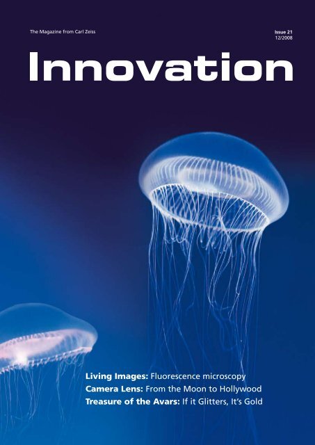
The Magazine from <strong>Carl</strong> <strong>Zeiss</strong> Issue 21<br />
12/ 2008<br />
<strong>Living</strong> <strong>Images</strong>: <strong>Fluorescence</strong> <strong>microscopy</strong><br />
<strong>Camera</strong> <strong>Lens</strong>: From the Moon to Hollywood<br />
Treasure of the Avars: If it Glitters, It’s Gold
The green fluorescent protein (GFP)<br />
comes from the Aequorea victoria<br />
jellyfish. It shines green when it is excited<br />
with ultraviolet or blue light.<br />
Asamu Shimomura described it for the<br />
first time in 1962. In 1994, Martin Chalfie<br />
succeeded in bringing GFP to expression<br />
outside the Aequorea victoria jellyfish,<br />
enabling its breakthrough as a genetic<br />
marker. Roger Tsien, who had long<br />
focused his research on the field of<br />
imaging cell biology, created fluorescent<br />
dyes to make cellular calcium visible.<br />
With his work, he contributed to the<br />
general understanding of the protein.
Editorial<br />
Dear Readers,<br />
When the winners of the Nobel Prize for Chemistry were announced at<br />
the beginning of October, the news spread through the company like<br />
wildfire. This renowned award was bestowed upon three scientists this<br />
year who partly work with instruments from <strong>Carl</strong> <strong>Zeiss</strong>. Above all, this<br />
year’s Nobel Prize for Chemistry honors the enhancement of a procedure<br />
developed by <strong>Carl</strong> <strong>Zeiss</strong> exactly 100 years ago: fluorescence <strong>microscopy</strong>.<br />
Osamu Shimomura, Martin Chalfie and Roger Y. Tsien were honored for<br />
the discovery and development of the green fluorescent protein (GFP).<br />
This protein has become an important tool in life sciences. It allows<br />
scientists to observe processes in cells and, for example, track the spread<br />
of cancer cells.<br />
Understanding how diseases develop, even possibly preventing their<br />
outbreak and definitely accelerating their cure – these are the goals of<br />
the life sciences. Physician Harald zur Hausen, a <strong>Carl</strong> <strong>Zeiss</strong> customer too,<br />
also received the Nobel Prize this year. We at <strong>Carl</strong> <strong>Zeiss</strong> have a long tradition<br />
and passion for providing scientists such as zur Hausen with the<br />
right instruments. In doing so, we also aid the transition from science to<br />
the application. Think about minimally invasive or micro-incision surgery<br />
that increases the likelihood of faster patient healing through minimal<br />
patient trauma.<br />
Whether you enjoy the aesthetic of the images or reading exciting<br />
articles, we hope this issue of Innovation is to your liking: enjoy reading!<br />
Best wishes<br />
Dr. Michael Kaschke<br />
Member of the <strong>Carl</strong> <strong>Zeiss</strong> AG Executive Board<br />
Innovation 21, 12 / 2008
Table of Contents<br />
Editorial 3<br />
Panorama 6<br />
A Movie that Will Never Make it 10<br />
Wenders makes a fictitious movie<br />
Cover story: <strong>Living</strong> images<br />
100 Years of <strong>Living</strong> <strong>Images</strong> 16<br />
“Moving into New Dimensions” 24<br />
An interview with Dr. Ulrich Simon and<br />
Dr. Bernhard Ohnesorge<br />
16<br />
100 Years of <strong>Fluorescence</strong> Microscopy –<br />
More Current than Ever.<br />
On the Track of Viruses with<br />
with <strong>Fluorescence</strong> Signals 26<br />
The Future of <strong>Fluorescence</strong> Microscopy 30<br />
Guest article by Michael W. Davidson<br />
Essay 52<br />
The Right Light<br />
Innovation 21, 12 / 2008
Report<br />
The World by Candlelight 40<br />
Small Incision with a Big Impact 42<br />
Feature: Unearthed<br />
Bone Find Veiled in Mystery 32<br />
If it Glitters, It’s Gold 36<br />
32<br />
Kyffhäuser – Thuringian mountain range with many secrets<br />
Feature: From Above<br />
A Bird’s Eye View of the<br />
Jabel Akhdar 44<br />
Data Communication in the<br />
Fast Lane 48<br />
Sylt from the Air – Laser<br />
beam transport measured<br />
values and images<br />
across long distances<br />
Look at Laureates<br />
Preview 59<br />
Legal information 59<br />
48<br />
Innovation 21, 12 / 2008
Panorama<br />
THE attraction at photokina: the walk-through lens.<br />
Precisely in Focus<br />
ZEISS lenses now available for EOS cameras<br />
Canon enthusiasts have something to look forward to:<br />
they will soon be able to use ZEISS lenses with manual<br />
focus on their EOS cameras. The new ZE lenses transmit<br />
all information via the EF bayonet connection, i.e. via<br />
the electronic contacts, and support auto exposure,<br />
shutter priority and aperture priority. With digital SLRs,<br />
the lens data, all exposure data and even the exposure<br />
control of the flash can be accessed. Even if the focus is<br />
set manually, focusing is automatic. Even professionals<br />
see such a lens as a welcome addition to their equipment.<br />
Munich-based photographers Eva Maierhofer<br />
and Ulrich Wolf from MAIWOLF Photography had the<br />
opportunity to test the first ZE lenses. An enthused<br />
Wolf stated that “the lens delivered natural clarity and<br />
fantastic brilliance. The images really light up, regardless<br />
of the focal length.” Maierhofer added that she<br />
“was particularly fascinated at how the lenses provide<br />
a crisp image of even the smallest details such as hair<br />
at the edge of the image.” Currently, ZE lenses with<br />
two different focal lengths are available. Additional<br />
focal lengths will be added to the line within the next<br />
few months.<br />
Innovation 21, 12 / 2008
Cancer-causing Viruses<br />
Harald zur Hausen receives Nobel Prize for sensational discovery<br />
With dogged persistence, physician Harald zur Hausen<br />
worked on his theory that viruses can cause cancer –<br />
contrary to prevailing doctrines. He received the Nobel<br />
Prize for Medicine for proving his theory and thus<br />
destroying a medical dogma.<br />
Without a doubt, December 10, 2008, will go down as<br />
the highlight of zur Hausen’s research career. This is the<br />
day – the anniversary of Alfred Nobel's death – that the<br />
award he sponsored is presented.<br />
30 years ago, zur Hausen’s colleagues simply shook<br />
their heads when he stated that the human papillomavirus<br />
(HPV) triggers the onset of cervical cancer; he was<br />
ridiculed for his theory. Today, this is general scientific<br />
knowledge and many young women are now vaccinated<br />
against cervical cancer thanks to zur Hausen’s<br />
discovery.<br />
Some of the luster of the Nobel Prize also shines on <strong>Carl</strong><br />
<strong>Zeiss</strong>: Harald zur Hausen worked with a ZEISS transmission<br />
electron microscope. The pictures taken with it and<br />
their analysis finally lead to the pioneering conclusion.<br />
Although he has long since retired from active scientific<br />
work, the passionate researcher still uses this instrument<br />
today.<br />
In the Kingdom of the Camorra<br />
New award from <strong>Carl</strong> <strong>Zeiss</strong> and ARRI goes to Gomorrah by Matteo Garrone<br />
The writer scored a bestseller, the director received an<br />
international movie award from <strong>Carl</strong> <strong>Zeiss</strong> and ARRI.<br />
Both dealt with the subject of the Camorra, the Neapolitan<br />
version of the Mafia. Matteo Garrone made his<br />
movie called Gomorrah based on the novel by Roberto<br />
Saviano in 2008. While the book exposes and indicts,<br />
the movie is a socio-political study, an analysis of criminality<br />
as a way of life.<br />
This analysis earned Garrone international acclaim. In<br />
response, the jury of the Munich Film Festival presented<br />
him with the first Arri-<strong>Zeiss</strong> Award which is conferred<br />
for the best international film and has a cash value of<br />
50,000 euros. “Internationality is a key success factor<br />
for ARRI and <strong>Carl</strong> <strong>Zeiss</strong>,” said Dr. Dieter Kurz, President<br />
and CEO of <strong>Carl</strong> <strong>Zeiss</strong> AG when presenting the award to<br />
Garrone. “Therefore, it was only logical that we decided<br />
to contribute to the internationality of the movie<br />
award”.<br />
Don Ciro (Gianfelice Imparato) must realize that his work as an<br />
acountant for the mafia is becoming more dangerous.<br />
Innovation 21, 12 / 2008
SESAM Opens Up Totally New Worlds<br />
Electron vibrations become visible on semiconductors<br />
A very special electron microscope is allowing researchers<br />
to cross the current frontiers of knowledge: the<br />
SESAM from <strong>Carl</strong> <strong>Zeiss</strong>. For just under one year now,<br />
scientists at the Stuttgart Max Planck Institute for Metal<br />
Research have been conducting basic research in an<br />
Vaccum<br />
100 nm<br />
Ag<br />
SiN x<br />
Vaccum<br />
100 nm<br />
A surface plasmon can be depicted as a common vibration of<br />
electrons on the surface of a particle (photo: silver particle on<br />
silicium-nitride substrate). The minimal expansion of the silver<br />
particle leads to the development of characteristic shapes with<br />
antinodes on the corners (left) or center of the edges (right)<br />
of the triangular silver particle.<br />
Ag<br />
SiN x<br />
area where research was considered to be impossible<br />
until now: the focal points of their work are special<br />
light-emitting semiconductors and the still young field<br />
of plasmonics. Here, electronic and optical data are<br />
simultaneously processed, opening up totally new<br />
perspectives right up to optical computers.<br />
The technological basis of this research is the globally<br />
unique SESAM transmission electron microscope. The<br />
Head of the Stuttgart Center for Electron Microscopy,<br />
Dr. Peter A. van Aken, explains: “This electron microscope<br />
allows us to see effects which we only knew existed<br />
before. A decisive role is played here by a special<br />
filter in the microscope which permits the simultaneous<br />
examination of large areas of the sample.” This allows<br />
the visualization of changes in materials which are<br />
accompanied by the emission of light of a particular<br />
wavelength. The high resolution of the electron microscope<br />
makes it possible to exactly determine where in<br />
the sample the radiation is generated. “Such examinations<br />
help improve the fabrication of light-emitting<br />
semiconductor components,” says van Aken, “and we<br />
can make effects from the field of plasmonics visible.”<br />
Room for Half an Airplane<br />
Largest 3D measuring machine measures entire wings of fighter jets<br />
Airplane construction is precision work. Lockheed<br />
Martin is building the new F-35 Lightning II supersonic<br />
fighter for the US Air Force. Production is being monitored<br />
by the largest coordinate measuring machine<br />
ever built by <strong>Carl</strong> <strong>Zeiss</strong>: an MMZ B Plus gantry.<br />
The MMZ B Plus has a measuring range of 5 x 16 x 2.5<br />
meters. With a length of 16 meters, it can accommodate<br />
the wings of the jets and measure their skin. It<br />
also measures aerodynamic tools, wind tunnel models<br />
and 1:1 modules.<br />
The machine simplifies assembly and contributes to<br />
lean manufacturing for production with reduced outlay<br />
and consumption of resources. During machine handover,<br />
Larry Pike, Vice President of Quality at Lockheed<br />
Martin said: “This machine permits the transition from<br />
the inspection of single parts to process validation.”<br />
Bob Fiorentini, Vice President, F-35 Global Production,<br />
added: “This machine is the first installation of its kind<br />
and magnitude in our facility. It marks the beginning of<br />
a new era in optimized parts inspection.”<br />
The bridge-type measuring machine simplifies assembly and<br />
supports low-energy production.<br />
Innovation 21, 12 / 2008
Small Incision, Large Step<br />
New intraocular lenses make cataract operations<br />
more successful<br />
Robert Koch’s Microscope<br />
One of the instruments used by Koch<br />
can now be seen in the online museum<br />
A new lens makes it possible: cataract patients often no<br />
longer need glasses after surgery. Cataract surgery is<br />
one of the most frequently performed outpatient surgeries<br />
in the world and is practically a routine procedure<br />
in ophthalmic surgery. The patient receives an artificial<br />
intraocular lens that replaces the natural lens<br />
which has become cloudy. One challenge is the size of<br />
the incision through which the artificial lens is implanted.<br />
Large incisions during the operation can lead to<br />
astigmatism, a curvature of the cornea.<br />
<strong>Carl</strong> <strong>Zeiss</strong> Meditec has presented a worldwide innovation<br />
with the AT.LISA ® and AT.LISA.toric intraocular<br />
lenses. These lenses require only one small, 1.5-millimeter<br />
incision and are therefore suitable for the first time<br />
for real micro-incision surgery – which promises significantly<br />
better surgical results. In addition, the new<br />
intraocular lenses feature unique, multifocal high-performance<br />
optics which enable excellent vision both in<br />
the near and far zones, as well as in the area in<br />
between.<br />
It must have been a microscope from<br />
<strong>Carl</strong> <strong>Zeiss</strong>. Robert Koch, the great doctor<br />
and bacteriologist, proved in 1876 that<br />
infections are caused by micro-organisms.<br />
He demonstrated this using anthrax,<br />
whose pathogen he made visible<br />
under the microscope. Koch lived in<br />
what was is noe Wolsztyn, Poland, and<br />
most likely conducted his research there<br />
with pharmacist Josef Knechtel in his lab<br />
using ZEISS instruments. Based on the<br />
delivery books from <strong>Carl</strong> <strong>Zeiss</strong>, three<br />
microscopes were delivered, and all<br />
three went to Knechtel.<br />
Karlsruhe-based scientist Timo Mappes was able to<br />
acquire one of them for his microscope collection. It is<br />
No. 3479, VIIa stand, manufactured in 1877 and can be<br />
seen in Mappes' online museum of optical instruments.<br />
Even if other sources report that Koch had his own lab<br />
in Wollstein and ordered his own instruments from <strong>Carl</strong><br />
<strong>Zeiss</strong>, he nonetheless discovered the anthrax pathogen<br />
with an instrument from <strong>Carl</strong> <strong>Zeiss</strong>. As he wrote to<br />
Jena: “I admire and am very grateful for the <strong>Zeiss</strong> optical<br />
workshop; after all, I owe a great deal to your outstanding<br />
microscopes for a large portion of the success I<br />
have achieved in the name of science.”<br />
The Illusion of Night<br />
Further information is available<br />
under www.musoptin.com<br />
New projector for planetariums which does not brighten the background<br />
The darker the night, the brighter the stars. This is no<br />
different in planetariums than outdoors. A planetarium<br />
projector from <strong>Carl</strong> <strong>Zeiss</strong> can make stars shine with<br />
such radiance that they almost rival their natural counterparts<br />
in the night sky. However, this illusion soon<br />
vanishes in video projections which zoom in on cosmic<br />
nebulae and reveal the universe in 3D. The dome<br />
images become pale because they lack a black<br />
background.<br />
With powerdome ® VELVET, <strong>Carl</strong> <strong>Zeiss</strong> has developed a<br />
video projection system for domes which does not<br />
make the background appear gray. VELVET “projects”<br />
the darkest black imaginable and is unparalleled in this<br />
achievement. VELVET was an absolute sensation at the<br />
conference of the International Planetarium Society in<br />
Chicago at the beginning of July. Planetarium directors<br />
from all over the world were extremely impressed by<br />
the brilliance of the images. The blackness of the background<br />
highlights white texts as if they written in fluorescent<br />
letters, and objects seem to float freely in the<br />
air. The technical challenge consisted in markedly increasing<br />
the difference between the highest and lowest<br />
brightness levels compared to traditional projectors.<br />
While the latter feature a maximum contrast ratio of<br />
25,000:1, VELVET surpasses this one 100 times over:<br />
2,500,000:1.<br />
Digital all-dome video projection is increasingly supplementing<br />
the opto-mechanical display of the starlit sky<br />
in planetariums. For the very first time, powerdome<br />
VELVET is meeting a long-cherished wish: video projection<br />
is able to superimpose digital images on the optical<br />
night sky without impairing its brilliance. Gas nebulae<br />
and galaxies look as if they were immersed in the<br />
velvety-black depths of the universe – after all, the new<br />
projector has not been called VELVET for no reason.<br />
It will be launched on the market in mid-2009.<br />
Innovation 21, 12 / 2008
A Movie<br />
10<br />
Innovation 21, 12 / 2008
that Will Never Make it to the Silver Screen<br />
Emotional images, world-famous actors, moving<br />
scenes – a totally new approach has been taken<br />
in the 2009 <strong>Carl</strong> <strong>Zeiss</strong> calendar. In a unique project<br />
a movie storyline was created and implemented<br />
in fascinating images by Wim Wenders, one of<br />
the great masters of the art.<br />
Photos by Donata Wenders<br />
Innovation 21, 12 / 2008<br />
11
Wenders makes a fictitious movie<br />
12<br />
Innovation 21, 12 / 2008
A gigantic ruin is emerging in the<br />
heart of Berlin: eight towers made<br />
of bare concrete which form an almost<br />
haunting backdrop to the majestic<br />
dome of the German Cathedral.<br />
Wwhen the scene does not<br />
happen to be bathed in radiant sunlight,<br />
there is a definite whiff of<br />
doomsday in the air. The whole scene<br />
came alive in September. There was<br />
a constant flow of people checking<br />
the structural conditions of the site,<br />
examining the concrete towers and<br />
taking notes.<br />
One of them who wanted to gain his<br />
own impression was Wim Wenders.<br />
The director and photographer just<br />
finished with the Venice Film Festival<br />
where he was head of the jury and is<br />
now focusing on the project in Berlin.<br />
Wenders who is widely known<br />
for movies such as Wings of Desire,<br />
Paris, Texas and Buena Vista Social<br />
Club, as well as numberous photo<br />
projects began his next project – the<br />
2009 <strong>Carl</strong> <strong>Zeiss</strong> calendar.<br />
It was intended to be something<br />
special: a movie that will never make<br />
it to the silver screen. A story staged<br />
with all the details, but only captured<br />
using single, powerful images.<br />
“Filming” the fictional movie itself<br />
Innovation 21, 12 / 2008<br />
13
14 Innovation 21, 12 / 2008
is only part of the story that is told in<br />
24 photos. 24 is a magical number in<br />
the movie industry: 24 images per<br />
second represents the rhythm of single<br />
images in a theater that create<br />
the illusion of reality.<br />
Wim Wenders attracted actress Amber<br />
Valletta and actor Willem Dafoe<br />
for the project. They play two people<br />
in the imaginary film Tomorrow<br />
Morning who have survived a disaster<br />
but cannot find each other. In the<br />
end, they meet and look for a better<br />
future together.<br />
contains sketches and notes from<br />
Wim Wenders – excerpts from the<br />
storyboard, the set up and framework.<br />
Furthermore, 24 black-andwhite<br />
photos from Donata Wenders,<br />
the photographer and wife of the<br />
director, show the making-of scenes<br />
during production. The images on<br />
these pages provide an impression of<br />
what went on behind the scenes.<br />
In addition to the 24 large photos,<br />
the 2009 <strong>Carl</strong> <strong>Zeiss</strong> Calendar also<br />
Innovation 21, 12 / 2008<br />
15
Cover Story<br />
100 Years of <strong>Living</strong> <strong>Images</strong><br />
The development of fluorescence <strong>microscopy</strong><br />
began 100 years ago in Jena, and nowadays it is<br />
hard to imagine a world without the colorful<br />
images produced in biological research. Fluorescent<br />
dyes allow doctors to identify diseases or<br />
genetic mutations at a single glance. Meanwhile,<br />
scientists utilize the same methods to observe the<br />
processes that constitute life right down to a<br />
molecular level, and it is now possible to capture<br />
frames of even the most dynamic life processes.<br />
Text: Birgit Herden<br />
Scientific research: Michael Zölffel<br />
16<br />
Innovation 21, 12 / 2008
Report: Lorem ipsum<br />
Innovation 21, 12 / 2008<br />
17
Fluorescent Dyes Open Up Perspectives<br />
In the beginning, it<br />
seemed like nothing<br />
more than some annoying<br />
image interference.<br />
Taking his first<br />
look through a new type of microscope<br />
at the start of the last century,<br />
August Köhler noticed that some<br />
of his samples had an unexpectedly<br />
colorful tinge to them. The legendary<br />
researcher had irradiated his<br />
samples with ultraviolet light at <strong>Carl</strong><br />
<strong>Zeiss</strong> in the hope of achieving a particularly<br />
high resolution from its<br />
short wavelength, and the principle<br />
had proved to be successful. Yet<br />
Köhler had actually made a far more<br />
significant discovery as a mere sideline:<br />
in the invisible UV light, some<br />
of the irradiated structures were fluorescing<br />
in the most extraordinary<br />
range of colors. The cell membranes<br />
in Köhler's woody tissue samples<br />
suddenly appeared to be blue, while<br />
the wax-containing protective layer<br />
of the cells glowed yellow or white.<br />
“I initially saw this fluorescence as<br />
merely an irritating side-effect that I<br />
had to neutralize”, Köhler later reported<br />
in a lecture. “Only recently<br />
have I examined it more closely and<br />
come to the conclusion that the color<br />
of the fluorescent light can perhaps<br />
also be used to differentiate<br />
between different constituents of<br />
tissue.”<br />
Pages 16/17: Brain of<br />
a tree show captured using<br />
multifluorescence<br />
Hippocampus neurons of a transgenic mouse. Nerve cells in the brain are illuminated<br />
using different fluorescent proteins.<br />
It was over 100 years ago in April<br />
1908 that the researcher first presented<br />
this phenomenon to a wider<br />
public during a <strong>microscopy</strong> course<br />
at the Botanical Institute in Vienna.<br />
In the years following this presentation,<br />
he and Henry Siedentopf developed<br />
and perfected the fluorescence<br />
microscope, which at that time was<br />
still referred to as the luminescence<br />
microscope.<br />
Köhler would not be around to experience<br />
just how far-sighted he had<br />
been with his pioneering work. He<br />
laid the foundations for a technology<br />
has come to form an essential and<br />
fundamental component of biological<br />
research.<br />
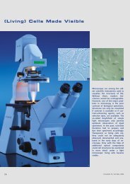
A needle in a haystack. Using today's<br />
fluorescence microscopes, cellular biologists<br />
can take a direct look at life<br />
in all its teeming glory. Every cell features<br />
the busy activities of thousands<br />
of proteins that are, as a rule, colorless,<br />
all engaged in bringing forth<br />
the wonders of life through their<br />
complex interactions. Trying to follow<br />
the fate of an individual component<br />
of this mass is like trying to find<br />
the proverbial needle in a haystack.<br />
Yet the search becomes much easier<br />
if the haystack can be faded out,<br />
leaving just the needle colorfully illuminated.<br />
18 Innovation 21, 12 / 2008
Cover story: <strong>Living</strong> images<br />
“I initially saw this<br />
fluorescence as merely an<br />
irritating side-effect that I<br />
had to neutralize.”<br />
Prof. August Köhler<br />
Using high-resolution images and<br />
films, researchers are now able to<br />
follow events that occur even within<br />
an individual cell, making different<br />
cellular constituents visible depending<br />
on their focus of interest at the<br />
time. “Using a fluorescence microscope<br />
gets me so close that I can almost<br />
shake hands with individual<br />
molecules”, enthuses Professor Volker<br />
Haucke, who conducts research at<br />
the Free University of Berlin into<br />
how signals from nerve cells function.<br />
Two things he cites as helping<br />
to drive his work forward are the human<br />
genome project and fluorescence<br />
<strong>microscopy</strong>. The much-lauded<br />
genome project enables biologists to<br />
look up every gene and thus every<br />
protein of a cell in a database. Yet it<br />
is only thanks to the fluorescence microscope<br />
that he is able to investigate<br />
the way in which molecules actually<br />
interact. Knowledge of the<br />
constituent parts is rendered in three<br />
dimensions, which often provides<br />
the key to understanding.<br />
<strong>Living</strong> images. We have come a long<br />
way from August Köhler's first fluorescence<br />
microscope, which he lovingly<br />
referred to as his “quartz microscope”<br />
due to the optical<br />
components it contained, all the way<br />
up to today's virtuoso levels of imaging.<br />
The first scientists to become interested<br />
in the new technology were<br />
primarily botanists, largely because<br />
autofluorescence is so pronounced in<br />
the plant world. Important progress<br />
in this field was made by staining<br />
preparations with fluorescent chemicals.<br />
While carrying out experiments<br />
in the 1930s with the dye acridine<br />
orange, the botanist Siegfried Strugger<br />
suddenly found he could make<br />
out living bacteria glowing bright<br />
green in a soil sample that was fluorescing<br />
reddish-brown. What was<br />
particularly interesting was the fact<br />
that the dye did not<br />
The person<br />
August Köhler (1866 – 1948)<br />
August Köhler was born in<br />
Darmstadt, Germany. He studied<br />
zoology, botany, minerology,<br />
physics and chemistry in<br />
his hometown, in Heidelberg<br />
and and in Giessen. He joined<br />
<strong>Carl</strong> <strong>Zeiss</strong> in Jena at the age<br />
of 34.<br />
He played a key role in the<br />
development of the UV microscope.<br />
The resolution of the<br />
light microscope could be doubled<br />
using optics manufactured<br />
from a UV light permeable rock<br />
crystal. While conducting his<br />
research work, August Köhler<br />
noticed that natural objects<br />
such as cell membranes began<br />
to illuminate when they were<br />
irradiated with UV light. Recognizing<br />
this inherent fluorescence<br />
is one of his greatest<br />
achievements. In 1908, he present<br />
a luminescence microscope<br />
to the public for the first time.<br />
New fluorescent dyes with extreme spectral properties.<br />
Innovation 21, 12 / 2008<br />
19
Time-lapse exposure of dividing kidney cells in rats, stained with the GFP and HcRed proteins.<br />
necessarily kill the cells. Strugger was<br />
able to stain living plants and ultimately<br />
succeeded in tracking the<br />
flow of water within them. This continues<br />
to be one of the greatest<br />
strengths of fluorescence <strong>microscopy</strong>.<br />
Although you can achieve impressive<br />
levels of visibility of the most detailed<br />
structures of a cell using an<br />
electron microscope, the frames obtained<br />
in a vacuum will always be<br />
snapshots.<br />
“With a fluorescence<br />
microscope, I can practically<br />
greet single molecules<br />
with a handshake.”<br />
Prof. Volker Haucke, FU Berlin<br />
Using fluorescent dyes, doctors built<br />
on this success in subsequent years<br />
by identifying the agents involved in<br />
diseases such as tuberculosis, leprosy,<br />
malaria and smallpox. Depending on<br />
the dye, it was no longer just UV<br />
light that was being employed, but<br />
also blue light. <strong>Fluorescence</strong> can occur<br />
at a wide variety of wavelengths,<br />
though the principle is always the<br />
same. Incident light causes an electron<br />
in a dye molecule to be raised<br />
to a higher energy level. Within a<br />
few nanoseconds, it returns to its<br />
natural state, releasing light at the<br />
same time, though a portion of the<br />
energy received is lost in the form of<br />
heat. The light emitted is lower in<br />
energy and therefore has a longer<br />
wavelength. In comparison to the<br />
light projected onto the molecule,<br />
the fluorescence is shifted towards<br />
the red region of the spectrum.<br />
Meanwhile, the new dyeing methods<br />
and objects of analysis were also<br />
driving forward the development of<br />
microscope optics. In the beginning,<br />
the most important thing was to<br />
capture as much as possible of the<br />
fluorescence, which was often quite<br />
weak. A milestone was achieved in<br />
1953 with the fast NEOFLUAR objective<br />
with a calcium fluoride lens<br />
which was launched by ZEISS-WIN-<br />
KEL. Following numerous improvements,<br />
this gave rise to the<br />
EC Plan-NEOFLUAR range of objective<br />
lenses, which, even today, continue<br />
to represent the “workhorses”<br />
in fluorescence <strong>microscopy</strong> for scientists<br />
all over the world. Optics development<br />
reached its apex in<br />
the form of the high-performance<br />
C-APOCHROMAT objective lenses<br />
corrected for entire regions of the<br />
spectrum from UV to IR, which are<br />
also produced by <strong>Carl</strong> <strong>Zeiss</strong>.<br />
Light from above. An epifluorescence<br />
microscope developed by<br />
ZEISS-WINKEL in 1955 represented a<br />
further breakthrough: the sample<br />
was no longer illuminated from below<br />
using the technique of dark-field<br />
<strong>microscopy</strong>, something which had<br />
required high light intensity and laborious<br />
alignment. This had caused<br />
fundamental problems in trying to<br />
separate the much weaker fluorescent<br />
light from the incident excitation<br />
light, which otherwise would<br />
have completely obscured it. In epifluorescence<br />
<strong>microscopy</strong>, however,<br />
the very bright lamplight generated<br />
by special light sources initially falls<br />
on an obliquely positioned mirror,<br />
which reflects it downwards through<br />
the objective lens in the direction of<br />
the mounted specimen. The trick<br />
here lies in the special “dichroic”<br />
beam splitter, which reflects the light<br />
of one color while being transparent<br />
for a different color. For example, if<br />
a specimen illuminated in blue fluoresces<br />
green, the green light emitted<br />
upwards by the specimen can pass<br />
20 Innovation 21, 12 / 2008
Cover story: <strong>Living</strong> images<br />
Gullet of the C. elegans<br />
threadwom with GFPstained<br />
ganglion cells.<br />
The person<br />
Nobel Prize for Chemistry<br />
through the mirror unhindered while<br />
the blue light is reflected. This process<br />
also requires two color filters<br />
based on a design with some highly<br />
complex features: one filter for the<br />
excitation light (so that only blue<br />
light is radiated, for example) and a<br />
second filter to filter the fluorescent<br />
light emanating from the specimen<br />
after it has passed the beam splitter.<br />
This second filter can then also be<br />
used to influence the perceived color<br />
of the image. Thus, it is this second<br />
filter that can further influence the<br />
color appearance of the fluorescent<br />
signal.<br />
In the 1960s, the development of the<br />
dichroic beam splitter was significantly<br />
boosted by the work of the<br />
Dutch researcher Johan Ploem. It was<br />
thanks to him that a module was finally<br />
created that brought together<br />
filters and mirrors optimized to work<br />
in perfect combination with each<br />
other. It was only due to this groundbreaking<br />
invention that it finally became<br />
possible to perform rapid<br />
changes of the fluorescent dyes.<br />
Colored probes.This achievement became<br />
particularly significant due to a<br />
completely different development.<br />
Up to that point in time, researchers<br />
working in the field of fluorescence<br />
<strong>microscopy</strong> had used chemical dyes<br />
that stained different materials to<br />
differing degrees. However, this only<br />
allowed for a rather coarse distinction<br />
to be made between cells and<br />
their constituent parts. As early as<br />
1939, the American pathologist Albert<br />
Coons had come up with an<br />
idea that was to have a profound<br />
impact, namely that if you were to<br />
mark antibodies with fluorescent<br />
dyes, that should then enable you to<br />
specifically stain any desired pathogen<br />
and indeed many other structures.<br />
1939 All mammals produce<br />
antibodies as a defense against a<br />
pathogen. In animal experiments, it<br />
is possible to provoke this kind of immune<br />
response to almost any structure<br />
introduced into the bloodstream.<br />
Just two years later, Coons<br />
found the solution he was looking<br />
for. He began by linking a fluorescent<br />
dye to antibodies that had been<br />
induced by pneumonia. These kinds<br />
of antibodies attach themselves specifically<br />
to the bacteria that cause<br />
the disease, which meant that Coons<br />
could use his color-labeled antibodies<br />
to make these bacteria visible<br />
too.<br />
Osamu Shimomura studied<br />
pharmacy in Nagasaki, later<br />
organic chemistry. In 1961,<br />
he discovered the green fluorescent<br />
protein (GFP) in the<br />
Aequorea jellyfish. He worked<br />
at the universities of Princeton<br />
and Boston, and at the Woods<br />
Hole marine biology lab. Today<br />
he operates a private photo<br />
protein lab.<br />
Martin Chalfie studied biology.<br />
He has been a professor for<br />
biology at Columbia University<br />
since 1982. Martin Chalfie<br />
used GFP to examine the<br />
processes in the cells of the<br />
threadwork C. elegans. He<br />
succeeded in bringing the<br />
GFP gene to expression<br />
outside the Aequorea victoria<br />
jellyfish for the first time.<br />
Roger Y. Tsien studied<br />
chemistry and physics at<br />
Harvard University. He has<br />
been a professor for pharmacology,<br />
chemistry and biochemistry<br />
at the University of<br />
California since 1989 and<br />
conducts research at the<br />
Howard Hughes Medical<br />
Institute. Thanks to him,<br />
several variations of GFP are<br />
now available, which exhibit<br />
different fluorescent spectrums<br />
and thus illuminate in<br />
different colors.<br />
Innovation 21, 12 / 2008<br />
21
Head of a three day-old zebra fish (from above) with GFP-stained motoneurons of the<br />
after brain on the left.<br />
Gland of a mouse near the lower jaw.<br />
He further honed this method over<br />
the subsequent decades and, in the<br />
1960s, cell researchers began to carry<br />
out targeted investigations of tissue<br />
using antibodies dyed in different<br />
colors. Working with the new fluorescence<br />
microscopes, they were able<br />
to have one cell type illuminated in<br />
red and a different type illuminated<br />
in green. This enabled them, for example,<br />
to carry out experiments to<br />
determine which types of cells make<br />
up the immune system, the nervous<br />
system and many other tissues.<br />
Fluorescent “probes” continue to be<br />
used in research and medicine right<br />
up to the present day, and scientists<br />
now have an enormous arsenal of<br />
techniques at their disposal. For example,<br />
geneticists use fluorescent DNA<br />
probes that bind specifically to select<br />
regions of chromosomes. In amniocentesis,<br />
these probes can be used to dye<br />
the chromosomes of an unborn child,<br />
which makes it possible to detect serious<br />
genetic defects at a glance.<br />
Noble Prize for jellyfish protein. Another<br />
technique of genetic engineering<br />
goes even further. It is based<br />
upon the fluorescent protein molecule<br />
of a jellyfish, which glows green<br />
in ultraviolet and blue light. This<br />
year's Nobel Prize for chemistry was<br />
awarded to Osamu Shimomura, Martin<br />
Chalfie and Roger Tsien [See The<br />
Person] for the discovery and development<br />
of this green protein.<br />
If a scientist wishes to analyze cells in<br />
a culture dish, it is now possible to<br />
alter the genetic make-up of these<br />
cells in such a way that they produce<br />
the jellyfish protein themselves and<br />
glow green in blue light. Genetic engineers<br />
can attach the glowing green<br />
protein to any protein molecule they<br />
choose. This does not affect the cells,<br />
which continue to live normally except<br />
for the fact that they and their<br />
progeny now fluoresce at a specific<br />
point or at a desired time. Since the<br />
colored jellyfish protein was discovered,<br />
similar molecules have been introduced<br />
in other colors. Genetic engineers<br />
can now turn cell cultures<br />
into a veritable mix of colors and<br />
then light them up.<br />
The depths of laser technology. Yet<br />
none of this would be possible if it<br />
were not for the extraordinary progress<br />
made in microscope technology.<br />
Nowadays, most researchers do not<br />
look at their fluorescent samples<br />
with the naked eye. A digital camera<br />
captures an image that is then processed<br />
and analyzed using special<br />
software. In 1982, <strong>Carl</strong> <strong>Zeiss</strong> introduced<br />
the first commercial laserscanning<br />
microscope. In this device, a<br />
laser beam scans a sample in multiple<br />
layers point-by-point, while any<br />
stray light that does not stem from<br />
the respective point is screened off<br />
by a pinhole aperture. The pixels are<br />
then reconstructed into a three-dimensional<br />
image by a computer. This<br />
offers the advantage of allowing<br />
even deeper layers to be clearly delineated.<br />
22 Innovation 21, 12 / 2008
Cover story: <strong>Living</strong> images<br />
In some cases, however, this kind of<br />
laser beam is simply too intense.<br />
When neurologists want to make<br />
highly-branched nerve cells visible or<br />
ophthalmologists are investigating a<br />
damaged retina, they need the gentlest,<br />
least-intrusive method they can<br />
find. One viable alternative is “twophoton<br />
<strong>microscopy</strong>,” which represents<br />
a variation on the confocal microscope.<br />
When the focused beam of<br />
a laser excites a fluorophore by causing<br />
it to simultaneously absorb two<br />
photons, the fluorophore emits a<br />
single photon at a higher energy. By<br />
irradiating a point with low-energy<br />
red laser light, it is possible to make<br />
the specimen fluoresce blue contrary<br />
to the rule that otherwise applies.<br />
Low-energy infrared light is gentle<br />
on tissue. Moreover, all radiated<br />
light can be used in this case without<br />
the need for a pinhole aperture,<br />
since the blue fluorescence anyway<br />
only occurs in the focus of the highest<br />
intensity of the red light.<br />
power of even the best light microscope<br />
is thus approximately 200<br />
nanometers. Yet a single influenza<br />
virus has a diameter of just 100<br />
nanometers, and there are many<br />
constituents of cells that are even<br />
smaller. In order to make these visible,<br />
“Stimulated Emission Depletion”<br />
(STED) exploits a further characteristic<br />
of fluorescent molecules, namely<br />
the fact that two pulses of a laser<br />
beam, one immediately following<br />
the other, can be used to excite and<br />
then immediately quench fluorescence.<br />
In STED, the excitation laser<br />
beam is surrounded by a ringshaped,<br />
quenching beam. The outer<br />
beam narrows the spot focused on<br />
by the excitation pulse, thereby causing<br />
the spot to have a diameter significantly<br />
below the Abbe limit.<br />
In Photoactivated Localization Microscopy<br />
(PALM), which is currently<br />
under development at <strong>Carl</strong> <strong>Zeiss</strong>, the<br />
individual fluorescing molecules are<br />
so far apart from each other that<br />
they can be individually distinguished,<br />
which means that the individual<br />
flashes of light from thousands<br />
of frames can be composed<br />
into a high-resolution image.<br />
This is one example of how <strong>Carl</strong> <strong>Zeiss</strong><br />
is pushing the boundaries of research<br />
and driving forward new developments<br />
in fluorescence <strong>microscopy</strong>. A<br />
constant supply of new inventions<br />
over a 100-year period has allowed<br />
us to see these colorful images in<br />
ever greater detail, and it is truly unusual<br />
for one company to have had<br />
such a profound influence on a technical<br />
development. Yet, in reality, the<br />
biotechnological era has only just begun,<br />
and with it the prospect of an<br />
even greater role for fluorescence<br />
<strong>microscopy</strong>.<br />
Revealing the nano world. Increasingly<br />
refined techniques and inventions<br />
have transformed fluorescence<br />
<strong>microscopy</strong> into an extraordinarily<br />
powerful tool for life sciences over<br />
the last 20 years. Specimens can be<br />
made to glow on demand when the<br />
chemical environment alters or when<br />
one molecule approaches another.<br />
Scientists have even managed to<br />
break the Abbe resolution limit.<br />
When Ernst Abbe developed the scientific<br />
basis of <strong>microscopy</strong>, he recognized<br />
that there is a fundamental<br />
limit to the resolving power of any<br />
microscope due to the wave nature<br />
of light. The maximum resolving<br />
Innovation 21, 12 / 2008<br />
23
Interview<br />
“Moving into New Dimensions”<br />
Dr. Ulrich Simon and Dr. Bernhard Ohnesorge discuss the future of fluorescence<br />
<strong>microscopy</strong> and what comes after it<br />
Dr. Bernhard Ohnesorge: “More than a simple microscope.”<br />
Dr. Ulrich Simon: “Recognize details and substructures.“<br />
“Impossible, is not part<br />
of my vocabulary” could<br />
be the motto of Dr. Ulrich<br />
Simon, Head of <strong>Carl</strong><br />
<strong>Zeiss</strong> Microscopy. In an<br />
interview, he and Dr. Bernhard Ohnesorge,<br />
who is responsible for Biosciences,<br />
explain the insights that<br />
will be made possible through <strong>microscopy</strong><br />
in the coming years.<br />
How is a modern fluorescence microscope<br />
different than the light microscopes<br />
we used in school?<br />
Dr. Bernhard Ohnesorge: The first<br />
noticeable difference is the price. A<br />
microscope for a school only costs a<br />
few thousand euros. The high-end<br />
microscopes used in research go for<br />
50-80 thousand euros, and up to<br />
400,000 euros for the laser scanning<br />
systems. You can already see that this<br />
is much more than a simple microscope.<br />
It includes laser technology, a<br />
sophisticated detection system and<br />
advanced software to evaluate the<br />
data. Furthermore, the cells that are<br />
examined can be kept alive and deliberately<br />
influenced. In fact, a modern<br />
fluorescence microscope is a<br />
downright research platform.<br />
What can fluorescence microscope<br />
deliver that justifies such a cost?<br />
Dr. Ulrich Simon: If you observe living<br />
cells under a traditional light microscope,<br />
you don't see anything at<br />
first as the cell is transparent. Today,<br />
researchers want to see details and<br />
sub-structures. They examine the interaction<br />
of bio-molecules and try to<br />
understand how cells work – and<br />
why they sometimes do not work.<br />
This is what fluorescence <strong>microscopy</strong><br />
can enable.<br />
The main objective here is to understand<br />
diseases. Even the latest medical<br />
technology often only treats<br />
the symptoms of diseases, the cause<br />
of which we often cannot identify.<br />
Medicines are developed based on<br />
the “trial and error” principle. The<br />
long package insert informs us that<br />
we are actually creating more side<br />
effects than the desired effect. In<br />
the treatment of cancer, for example,<br />
remedying the adverse effects<br />
is more involved that the actual<br />
therapy.<br />
<strong>Fluorescence</strong> <strong>microscopy</strong> can change<br />
this by providing the information<br />
needed to explain the actual interconnections.<br />
The specific function of<br />
a cell is triggered by the interaction<br />
of molecules. However, there is<br />
sometimes a molecule that does not<br />
belong. Once I understand this, I can<br />
24 Innovation 21, 12 / 2008
Cover story: <strong>Living</strong> images<br />
develop an agent to eliminate precisely<br />
this molecule. And I need fluorescence<br />
to understand these details.<br />
What was the key advance that<br />
made this possible?<br />
Dr. Ulrich Simon: The development<br />
of fluorescence dyes that do not disrupt<br />
the processes in the cell was a<br />
key event. In the past, cells were<br />
stained using synthetic dyes. However,<br />
this quickly killed the cells. Today,<br />
fluorescing proteins, living colors,<br />
found in the ocean are used. They<br />
are not toxic to the cells. A good <strong>Carl</strong><br />
<strong>Zeiss</strong> customer, Roger Tsien, won his<br />
Nobel Prize for this discovery.<br />
An advance into new dimensions<br />
was also a key factor. The world is<br />
still two dimensional under a school<br />
microscope. An onion epidermis is<br />
typical of this. I can always only<br />
view one plane; the rest is blurry and<br />
must be cut away. Modern research<br />
<strong>microscopy</strong> is at least three dimensional.<br />
Today, we are even talking<br />
about multi-dimensional examinations.<br />
They observe different colors,<br />
time lapses and substance concentrations<br />
in the three spatial dimensions.<br />
A key advance, however, is that we<br />
are attempting to resolve the image<br />
to the molecular level. For more than<br />
a century, it was thought that Abbe’s<br />
law of diffraction limited the resolution.<br />
Only recently were techniques<br />
developed, with which it was possible<br />
to clearly exceed the existing diffraction<br />
limit.<br />
How is this possible?<br />
Dr. Bernhard Ohnesorge: There are<br />
different developments. PAL <strong>microscopy</strong>,<br />
or photo-activated localization<br />
<strong>microscopy</strong>, is particularly interesting.<br />
It is based on the fact that you<br />
actually very precisely localize a single<br />
fluorescing molecule. This molecule<br />
is imaged as a “diffractive disc”<br />
by the optical system, meaning it appears<br />
blurry and molecules lying next<br />
to each other are blurred. However,<br />
with PAL <strong>microscopy</strong>, the single fluorescing<br />
molecules are so far apart,<br />
that their images do not touch each<br />
other. This is made possible by photo-active<br />
dyes that can be activated<br />
and deactivated with exciting light.<br />
Over several thousand cycles, new<br />
molecules are repeatedly excited so<br />
that the observer receives a high-resolution<br />
image, the same as a puzzle.<br />
How far along are you with development?<br />
Dr. Ulrich Simon: We have already<br />
achieved fantastic resolution of less<br />
than 20 nanometers. We are currently<br />
installing five prototypes at the<br />
sites of well-known customers. We<br />
have not yet reached a point that we<br />
can observe living specimens with<br />
this technology, but I am sure we will<br />
also succeed here.<br />
Is there even a frontier that you<br />
could ever reach?<br />
Dr. Ulrich Simon: In general, you<br />
can't evaluate more light than a molecule<br />
emits. This means that the<br />
detail of real-time exposures is limited<br />
will. However, I never want to<br />
here developers say “impossible.”<br />
Throughout history, people have too<br />
often thought that we couldn't go<br />
any further. The limits that we know<br />
today apply to fluorescing specimens,<br />
i.e. those that illuminate naturally.<br />
This does not always have to be<br />
the case. As much as we love fluorescence,<br />
we are still thinking about future<br />
procedures to generate contrast<br />
entirely without manipulating the<br />
specimen.<br />
Thank you very much for this interview.<br />
This interview was conducted<br />
by Dr. Birgit Herden<br />
Innovation 21, 12 / 2008<br />
25
Cover Story<br />
On the Track<br />
26 Innovation 21, 12 / 2008
Report: Lorem ipsum<br />
of Viruses with <strong>Fluorescence</strong> Signals<br />
Viruses play a significantly bigger role in the<br />
outbreak of illnesses than previously thought.<br />
They trigger AIDS, hepatitis and various cancers.<br />
The human papilloma virus, for example, is responsible<br />
for cervical cancer in over 90% of cases.<br />
Innovation 21, 12 / 2008<br />
27
Penetration of viruses into living uterus<br />
cancer cells. The path can be monitored<br />
using fluorescent markers. Various stages<br />
of the infection path of viruses can be<br />
seen in the image. (1) Virus outside the<br />
cells, (2) Virus with multiple contacts to<br />
the cell membrane without being able to<br />
attach or penetrate, (3) Virus that efficiently<br />
penetrates the cell membrane and<br />
(4) moves further through the nucleus<br />
membrane in the interior of the nucleus.<br />
Viruses invade cells and,<br />
because they themselves<br />
have no metabolism,<br />
use that of their<br />
host to proliferate. But<br />
this is where the problem starts.<br />
These interactions between the virus<br />
and host organism are subject to<br />
continuous changes.<br />
The dynamics of infection processes.<br />
In order to get a better understanding<br />
of these processes, scientists are<br />
researching the persistence of viruses,<br />
among other things – in other<br />
words, the mechanism that allows<br />
viruses to survive in certain parts of<br />
the body. Another important research<br />
area deals with the proliferation<br />
of viruses and the outbreak of<br />
illnesses due to virus infections.<br />
One of the main topics of interest in<br />
this area is the ability to precisely<br />
characterize the individual steps of<br />
this infection process, i.e., to find<br />
out how a virus penetrates the cell<br />
membrane and which paths it takes<br />
inside the cell. Knowledge of such<br />
correlations is necessary to be able<br />
to use viruses or parts of viruses as a<br />
transportation vehicle in immune<br />
and gene therapy, for example.<br />
The “virus” object has now been<br />
described in detail and extensively<br />
researched with the aid of biochemical,<br />
electron-microscopical and structural<br />
analyses. Nonetheless, these<br />
methods always only provide snapshots.<br />
The dynamics of the infection<br />
process are not reflected. However,<br />
thanks to sophisticated light-<strong>microscopy</strong><br />
technologies, transportation<br />
processes and protein interactions in<br />
single living cells can now also be examined.<br />
<strong>Fluorescence</strong> markers allow<br />
these processes to become visible.<br />
Scenario of a virus migration. At the<br />
Chemistry and Biochemistry Faculty<br />
of the Ludwig-Maximilians University<br />
in Munich, researchers from the<br />
work group headed by Prof. Dr.<br />
Christoph Bräuchle documented the<br />
infection path of a virus in single living<br />
cells in 2001. In doing so, the virus<br />
is coupled with a fluorescent dye<br />
molecule. Marked in this way it then<br />
appears under the microscope as a<br />
fluorescence point. A highly sensitive<br />
single-molecule microscope traces<br />
the fluorescence signal with spatial<br />
resolution of 40 nanometers and<br />
temporal resolution of 10 milliseconds.<br />
The path that the virus takes is<br />
made visible as if in a film, so that<br />
the individual infection steps can be<br />
precisely tracked. In recent years<br />
these tracking experiments have<br />
been consistently refined and enhanced.<br />
Tracking of fluorescence-marked viruses.<br />
In the Virology Department at<br />
the Institute of Hygiene at the University<br />
Hospital of Heidelberg, the<br />
work group headed by Dr. Barbara<br />
Müller is also using the single-molecule<br />
microscope to analyze the entire<br />
infection process. The green fluorescent<br />
protein GFP is being used for<br />
this (see also: “Nobel Prize for Chemistry”<br />
on page 21).<br />
The Heidelberg researchers are marking<br />
virions – i.e., virus particles that<br />
are outside of a cell – with GFP. The<br />
marker fuses with the virus' own<br />
structure protein Gag, so that the<br />
28 Innovation 21, 12 / 2008
Cover story: <strong>Living</strong> images<br />
fluorescence signals can exactly track<br />
the traces left by the virus when it<br />
makes contact with the host cell and<br />
the subsequent transport within the<br />
cell.<br />
Such single-virus tracking experiments<br />
require a well-equipped inverted<br />
microscope for multicolor imaging,<br />
a temperature-controlled research<br />
environment and a very precise laser<br />
illumination unit. In addition, it demands<br />
a high-aperture lens and extremely<br />
sensitive detectors.<br />
This device setup can be supplemented<br />
with microscope-based systems.<br />
An optical measuring process is fluorescence<br />
correlation spectroscopy,<br />
which obtains information from con-<br />
stantly varying fluorescence intensity.<br />
High-resolution optical images are<br />
also created by confocal laser scanning<br />
<strong>microscopy</strong>, or CLSM for short.<br />
The TIRF technique – which stands<br />
for “total internal reflection fluorescence<br />
<strong>microscopy</strong>” – can be used to<br />
examine structures located very near<br />
the surfaces.<br />
Dieter Brocksch, Monika Etspüler<br />
Virus particle bound<br />
to the surface of liver<br />
cancer cells.<br />
The details<br />
Virus history<br />
Viruses were detected in fixated<br />
cells for the first time in<br />
1980 using simple fluorescencemarked<br />
virus particles. One<br />
year later the first single-particle<br />
tracking experiments were<br />
performed in live cells. In 1985<br />
it was possible to observe viruses<br />
in real time in the lightmicroscopic<br />
differential interference<br />
contrast technique.<br />
Tracking viruses in living cells<br />
became a reality five years later<br />
with the aid of fluorescence<br />
video <strong>microscopy</strong>.<br />
The introduction of TIRF microscope<br />
technology in 1995 made<br />
it possible to detect single fluorescent<br />
molecules. The number<br />
of single-virus tracking experiments<br />
has increased continuously<br />
since 2001. Today, scientists<br />
are able to track the<br />
fluorescence-marked virus directly<br />
in a single living cell and<br />
observe it over a long period of<br />
time.<br />
Innovation 21, 12 / 2008<br />
29
Guest Article<br />
The Future of <strong>Fluorescence</strong> Microscopy<br />
However, determining what will be<br />
needed in the future is a daunting<br />
task. For example, who could have<br />
predicted the exquisite idea for<br />
photoactivation localization superresolution<br />
<strong>microscopy</strong> (PAL-M), or<br />
the hallmark development of the<br />
“Brainbow” recombinant Cre-Lox<br />
system for imaging neuronal pathby<br />
Michael W. Davidson<br />
Florida State University<br />
The latest fluorescence imaging techniques using a combination of synthetic dyes and immunofluorescence.<br />
The advances that we<br />
have witnessed in fluorescence<br />
<strong>microscopy</strong> over<br />
the past two decades<br />
have been nothing short<br />
of breathtaking. Microscopes and<br />
camera systems have, for the most<br />
part, kept pace with the development<br />
of fluorophores to enable researchers<br />
to observe subcellular processes<br />
with ever increasing temporal<br />
speed and spatial resolution. Today,<br />
fluorescence <strong>microscopy</strong> has evolved<br />
into a staple methodology ranging<br />
from live-cell imaging to drug discovery<br />
and medical diagnosis.<br />
ways in the nervous systems of living<br />
animals? This results in the following<br />
questions: What new fluorophores<br />
and labeling technologies will be<br />
needed in the future? How quickly<br />
will instrumentation evolve ?<br />
Fluorescent proteins are geneticallyencoded<br />
markers that can be fused<br />
to virtually any target protein of interest<br />
using traditional molecular biology<br />
techniques. The green fluorescent<br />
protein (GFP) obtained from a<br />
30 Innovation 21, 12 / 2008
Cover story: <strong>Living</strong> images<br />
jellyfish has been modified and is<br />
now seeing the highest level of application.<br />
Perhaps the potentially<br />
most useful fluorescent proteins belong<br />
to a new class termed “optical<br />
highlighters.” These are proving to<br />
be very efficacious in tracking dynamic<br />
events in living cells. In general,<br />
fluorescent proteins are non-toxic<br />
and feature excellent photostability,<br />
although their brightness falls far<br />
short of that exhibited by synthetic<br />
dyes and quantum dots.<br />
In the future, we can expect to see<br />
significant advances in the realm of<br />
fluorophore technologies. Improvements<br />
in synthetics will continue as<br />
manufacturers strive to compete<br />
with fluorescent proteins. Furthermore,<br />
quantum dots will become<br />
very useful. Hybrid systems are also<br />
receiving a considerable amount of<br />
attention and many new strategies<br />
are emerging. Finally, there appears<br />
to be no end to the new and promising<br />
fluorophore candidates emerging<br />
from fluorescent proteins. Dynamic<br />
biosensors that monitor a<br />
variety of cellular processes, including<br />
pH, voltage and sugar metabolism<br />
are being introduced in laboratories<br />
around the world.<br />
Littered with acronyms such as PAL-<br />
M, STED, 4Pi, STORM, SIM, and<br />
RESOLFT, the field of superresolution<br />
<strong>microscopy</strong> is currently the most rapidly<br />
evolving frontier in fluorescence<br />
imaging. Almost every month heralds<br />
the introduction of a new technique<br />
that promises to stretch or<br />
break the diffraction barrier. Although<br />
seemingly unrelated in many<br />
respects, the diverse technologies<br />
that comprise the foundation of superresolution<br />
<strong>microscopy</strong> all have<br />
the common goal of imaging biological<br />
specimens on a molecular level.<br />
Among the important questions for<br />
the future are exactly where are the<br />
resolution limits and how quickly<br />
and effectively can we bridge the<br />
gap (if at all) between optical and<br />
electron <strong>microscopy</strong>? More importantly,<br />
can all of this be done with<br />
living cells and, at some point, even<br />
live animals?<br />
Several manufacturers are now starting<br />
to offer solutions or are are<br />
working on developments that<br />
promise to spread this technology to<br />
the mainstream. Still, the complex<br />
fluorophore technology for superresolution<br />
<strong>microscopy</strong> is highly demanding<br />
and most methods have<br />
not yet been extrapolated to live-cell<br />
imaging. Hopefully this technology<br />
will be developed to the point of<br />
having auxiliary devices that attach<br />
to widefield or confocal microscopes<br />
with plug-in software modules for<br />
gathering images.<br />
The complete article can be found<br />
at www.zeiss.com/innovation<br />
The details<br />
Virtual Campus<br />
Together with biophysicist Michael<br />
W. Davidson from Florida<br />
State University, <strong>Carl</strong> <strong>Zeiss</strong> has<br />
created an education and science<br />
platform on the Internet<br />
regarding the topic of <strong>microscopy</strong><br />
and digital images. The<br />
virtual “ZEISS Campus” is a<br />
knowledge source that presents<br />
theory and technology<br />
based on applications. On this<br />
website, methods and techniques<br />
of fluorescence <strong>microscopy</strong><br />
are described with the aid<br />
of detailed descriptions, interactive<br />
animations, application<br />
examples and picture galleries.<br />
All materials were developed<br />
on microscope systems from<br />
<strong>Carl</strong> <strong>Zeiss</strong>.<br />
The websites not only provide<br />
information about current scientific<br />
topics regarding fluorescence<br />
<strong>microscopy</strong> – users also<br />
have the opportunity to publish<br />
their applications. With this<br />
website, <strong>Carl</strong> <strong>Zeiss</strong> is addressing<br />
all “microscopers,” particularly<br />
young scientists who can<br />
expand their knowledge at the<br />
“ZEISS Campus.”<br />
Further information<br />
is available at<br />
www.zeiss.com/campus<br />
Innovation 21, 12 / 2008<br />
31
Feature<br />
Bone Find Veiled in Mystery<br />
There are many secrets associated with the<br />
Kyffhäuser mountains. Located south of the Harz<br />
mountain range, it became famous through the<br />
Legend of Barbarossa. However, there is another<br />
mystery surrounding the cavernous mountain.<br />
Human remains and skeletons from the Bronze<br />
Age with strong signs of injury were discovered<br />
during excavations near Bad Frankenhausen.<br />
There has long been speculation that these injuries<br />
were the result of a form of cannibalism.<br />
32<br />
Innovation 21, 12 / 2008
Report: Lorem ipsum<br />
Innovation 21, 12 / 2008<br />
33
Researchers looking for clues into cause of trau<br />
A lot has been told<br />
about the Kyffhäuser<br />
in German folklore.<br />
According to legend,<br />
Emperor Frederick Barbarossa has<br />
been sleeping in a chamber under<br />
the mountain since the 12th century,<br />
waiting for the day when he will<br />
once again walk the earth. A discovery<br />
made around 50 years ago points<br />
to a much earlier time. During excavations,<br />
archaeologist Professor Günther<br />
Behm-Blancke found human remains.<br />
Tools and ceramic containers<br />
also found there suggested that the<br />
bone material dates back to the<br />
Bronze Age, or about 3,000 years<br />
ago.<br />
Some of these remains showed traces<br />
of severe injuries. Researchers<br />
found indications of cutting, chopping<br />
and beating, primarily on the<br />
ends of the long bones and in the<br />
joints. However, there were also indi-<br />
Günther Behm-Blancke (right)<br />
during excavations<br />
at the Kyffhäuser in 1955.<br />
cations of violence on the spine and<br />
skull. Many bones were completely<br />
destroyed. Until now, experts were<br />
hard tasked to determine what<br />
caused these extraordinary changes.<br />
The observations repeatedly led to<br />
speculation that cannibalism might<br />
have been involved.<br />
Analysis using the latest microscope<br />
technology. These prehistoric relicts<br />
recently caught the eye of researchers.<br />
Scientists from the Thuringian<br />
Office for the Preservation of Historical<br />
Monuments and Archaeology in<br />
Weimar examined the bones using<br />
the latest microscope technology at<br />
the <strong>Carl</strong> <strong>Zeiss</strong> Microscopy Application<br />
Center in Jena. Furthermore, the<br />
3,000-year-old findings were documented<br />
to make them available for<br />
research activities in the future.<br />
A SteREO Discovery.V20 from <strong>Carl</strong><br />
<strong>Zeiss</strong> was used. This stereo microscope<br />
is particularly well-suited for<br />
applications in materials research<br />
and quality inspections, and for biological<br />
and medical examinations. It<br />
permits 3D observation and provides<br />
high-contrast images with high<br />
depth of field. “The extraordinarily<br />
large viewing area was exactly what<br />
the scientists needed to examine the<br />
bones,” states Jan Birkenbeil, one of<br />
the specialists for digital image analysis<br />
at <strong>Carl</strong> <strong>Zeiss</strong>. The AxioCam HRc<br />
microscope camera was used for documentation.<br />
Its high image quality<br />
allows reliable statements on the<br />
condition of the bone material.<br />
Impact injuries and lacerations noticeable.<br />
“The microscopic examinations<br />
verified the earlier suspicions,”<br />
explains archaeologist Dr. Diethard<br />
Walter from the Thuringian Museum<br />
for Pre- and Protohistory in Weimar.<br />
The fibrous-frayed edges typical of<br />
impact injuries were easily recognizable,<br />
while the lacerations had a<br />
smooth contour. Magnification also<br />
showed that no new bone tissue had<br />
formed in these locations. “This<br />
clearly indicates that the injuries<br />
were inflicted around the time of<br />
death,“ explains anthropologist Sabine<br />
Birkenbeil from the Thuringian<br />
State Office.<br />
Elimination delivers answers. The<br />
scientists used very unconventional<br />
methods to more accurately specify<br />
the time of death. They inflicted severe<br />
contusions and lacerations on<br />
the haunch of a recently slaughtered<br />
pig using bronze knives and axes.<br />
They then separated the muscles and<br />
tendons and examined under the<br />
stereo microscope the marks left by<br />
these injuries. In fact, they appeared<br />
very similar to the human remains<br />
from the Bronze Age. Finally, old<br />
bones were subjected to the same<br />
procedure for comparison. They splintered<br />
as a result of the high level of<br />
decalcification.<br />
The calc-sinter deposits that partially<br />
covered the injuries refuted the argument<br />
that the damage to the<br />
bones resulted from improper handling<br />
during the excavations. “Today,<br />
we can assume that the bone material<br />
was actually subjected to these<br />
attacks at the time of death or shortly<br />
thereafter,“ says Diethard Walter,<br />
interpreting the results. “Based on<br />
what we know right now, these people<br />
were intentionally dismembered.”<br />
34 Innovation 21, 12 / 2008
Feature: Unearthed<br />
ma on remains<br />
Other questions surround the find.<br />
For example, there were only injuries<br />
to the bones of adults and juveniles,<br />
not on those of children. As the indentations<br />
show, the strokes were<br />
made with a great deal of accuracy.<br />
Some of the skeletons exhibited<br />
scorch marks. Both Sabine Birkenbeil<br />
and Diethard Walter come to the<br />
conclusion that “A type of cannibalism<br />
cannot be deduced from the<br />
bone finds.“ Stomach contents at<br />
best would provide this information;<br />
and after 3,000 years, there is nothing<br />
left over.<br />
Many questions, few answers.It is<br />
still unclear what the purpose of this<br />
brutal procedure was. It is also unknown<br />
what drove the people into<br />
the caves of the Kyffhäuser. Was it a<br />
clan that retreated for unknown reasons?<br />
The large number of bones of<br />
children found there speaks for this<br />
theory. Or were burial rituals held in<br />
the interior of the mountain? The<br />
numerous offerings could be an indication<br />
of such events.<br />
Some of the human bones exhibit scorch<br />
marks.<br />
“Measuring prehistoric societies using<br />
current criteria would not be of<br />
much help,“ warns Diethard Walter.<br />
From his point of view the question<br />
is not “cannibalism, yes or no.“ “The<br />
total find is what gives us an insight<br />
into the intellectual and cultural life<br />
of this period,” says the archaeologist.<br />
As before, little is known about<br />
the Bronze Age in central Germany.<br />
Monika Etspüler<br />
Cuts on the bones (here a collarbone)<br />
show that the bodies from the<br />
Kyffhäuser were sometimes systematically<br />
dismembered.<br />
Innovation 21, 12 / 2008<br />
35
Feature<br />
If it Glitters, It’s Gold<br />
On the trail of the secret behind the Treasure of the Avars<br />
36 Innovation 21, 12 / 2008
Feature: Unearthed<br />
More than 200 years<br />
ago, farmers in the<br />
eastern Hungarian<br />
village of Nagyszentmiklós<br />
discovered a ten kilogram<br />
gold treasure. Vienna-based archaeologists<br />
have now examined the<br />
gold containers using the latest microscopic<br />
methods to learn more<br />
about how they were created.<br />
Ganymede was simply spectacular.<br />
His long blond hair made the young<br />
man irresistible – not only for women,<br />
but also for Zeus. The father of<br />
the gods therefore appeared as an<br />
eagle and whisked the striking<br />
young man off to the top of Mount<br />
Olympus. From then on, Ganymede<br />
was to serve as a butler to the gods<br />
and pour their wine.<br />
This scene is illustrated on one of the<br />
containers belonging to one of the<br />
most important gold treasures from<br />
the early Middle Ages in Europe. The<br />
treasure from Nagyszentmiklós, the<br />
Treasure of the Avars is a 23-part<br />
drink set presumably manufactured<br />
in the 7th or 8th century. Today, it is<br />
on display at the Art History Museum<br />
in Vienna and is only rarely removed<br />
from its bullet-proof glass display<br />
case. One such occasion was a research<br />
project of the Vienna Institute<br />
for Archaeological Science (VIAS) together<br />
with the Art History Museum.<br />
Scientists wanted to know how the<br />
valuable gold containers were made.<br />
Non-destructively examined. Absolute<br />
care must be taken to ensure<br />
that archaeological objects are not<br />
modified or damaged. Therefore,<br />
non-destructive examination methods<br />
such as scanning electron <strong>microscopy</strong><br />
were the only possibilities.<br />
However, the objects must fit inside<br />
the specimen chamber of a scanning<br />
electron microscope (SEM). The VIAS<br />
purchased its own EVO 60 XVP from<br />
<strong>Carl</strong> <strong>Zeiss</strong> with the largest standard<br />
specimen chamber available. Because<br />
several of the gold containers were<br />
too tall, the Sales and Service departments<br />
at <strong>Carl</strong> <strong>Zeiss</strong> developed a technical<br />
solution together with VIAS archaeologist<br />
Mathis Mehofer to<br />
expand the specimen chamber laterally.<br />
Soft bedding. Gold is a very soft material.<br />
Gold containers are easily<br />
scratched. To prevent this, Mehofer<br />
also had to place them on soft bedding<br />
in the SEM. He first looked for a<br />
foam material that does not shrink<br />
or crumble in the vacuum of the<br />
specimen chamber. Using life-sized<br />
copies of the treasure, he then optimized<br />
the work routine step-by-step.<br />
Only then did the examination of<br />
the priceless original begin. Project<br />
coordinator Dr. Birgit Bühler, a spe-<br />
Innovation 21, 12 / 2008<br />
37
The goldsmith positioned the half-moon<br />
shaped tool several times to create an<br />
S-shaped structure.<br />
Stylized plant subject.<br />
Ring decoration seen on several mugs.<br />
Presumably, the same stamping tool was<br />
always used.<br />
niques, scanned all the gold containers<br />
using a reflected light microscope<br />
and identified the most interesting<br />
surface regions which were then examined<br />
in the SEM.<br />
The treasure comprises richly decorated<br />
jugs, cups, goblets and mugs<br />
that depict hunting and blood sport<br />
scenes. Project leader Dr. Falko Daim,<br />
General Director of the Roman-Germanic<br />
Central Museum in Mainz is<br />
amazed at the quality of the goldsmith<br />
work. “I was surprised that the<br />
customer and goldsmith had such<br />
unbelievable knowledge of the subject<br />
and the underlying myths,” says<br />
Daim, who initiated the research<br />
project. Greek, Persian, Byzantine<br />
and Christian themes are depicted<br />
on the containers.<br />
Finely engraved. The goldsmith made<br />
the mugs from one piece and<br />
adorned the surface with various engravings.<br />
These are ornaments that<br />
are pressed into the metal using a<br />
stamping tool and which appear as a<br />
negative form of the original shape.<br />
To achieve this, he utilized different<br />
carving tools in a personal manner.<br />
Many tools were applied several<br />
times in slightly different positions<br />
and hit with a hammer to create the<br />
“signature of the goldsmith.“ Because<br />
the SEM delivers images with<br />
“The art-historical evaluation will provide information<br />
on the intellectual horizon of a ‘barbaric’<br />
court. It was probably much different than what<br />
we learned in school. It must have been an<br />
unbelievably networked world.”<br />
Dr. Falko Daim, General Director of the Roman-Germanic Central<br />
Museum in Mainz about the current examinations of the<br />
Avarian gold treasure.<br />
high depth of field, it is ideal for examining<br />
and documenting 3D surfaces.<br />
Small irregularities in stamping<br />
tools that become visible in the SEM<br />
image, for example, are particularly<br />
interesting. This enables archaeologists<br />
to determine if the same stamping<br />
tool was used on different containers.<br />
The scientists suspect that the gold<br />
containers were manufactured by a<br />
Byzantine culture in the late 7th or<br />
8th century; however, they did not<br />
retain their original form. The canisters<br />
were originally bottles to which<br />
someone later added handles. “You<br />
can see that someone soldered over<br />
the carving pattern,“ says Mehofer.<br />
“The goldsmith who produced the<br />
bottle would never have done this.”<br />
Using the EDX detector connected to<br />
the SEM, precise analyses of the material<br />
were made to ascertain the<br />
composition of the solder and to determine<br />
if the same solder was used<br />
on all jugs.<br />
Hard work. Over a period of several<br />
months, more than 2000 SEM pictures<br />
were taken to document the<br />
38 Innovation 21, 12 / 2008
Feature: Unearthed<br />
surface of the goldsmith’s work.<br />
There were also more than 1000 single<br />
measurements for material analysis.<br />
The job of the art historians in the<br />
project team is now to interpret the<br />
results, arrange art history comparisons<br />
with other finds and draw conclusions<br />
about the history of the<br />
more than one thousand year old<br />
gold containers. The only thing that<br />
is known about their past with certainty<br />
is that farmers in Nagyszentmiklós<br />
stumbled across the golden<br />
mugs in 1799 while they were preparing<br />
the foundation for a barn.<br />
The treasure had probably been buried<br />
there for many years. It is unknown<br />
who buried these treasures<br />
and why. Perhaps the prestigious<br />
drink set was once in the possession<br />
of the Avars, a horse-riding people<br />
that had settled in the Carpathian<br />
basin in the early Middle Ages.<br />
The treasure was found in what is<br />
now Sânnicolau Mare, Romania. At<br />
the end of the 18th century, this region<br />
belonged to the Austro-Hungarian<br />
monarchy. Although the<br />
farmers initially intended to keep<br />
their discovery a secret, their treasure<br />
trove was finally reported to the<br />
Vienna Royal Chancellery (Hofkanzlei)<br />
as required by law at the time.<br />
Ownership of the Treasure of the Avars,<br />
including the Ganymede canister,<br />
was thus transferred to the doubleheaded<br />
eagle, the symbol of the<br />
Austro-Hungarian Empire.<br />
According to legend, Zeus compensated<br />
Ganymede’s father with a<br />
grapevine for the kidnapping of his<br />
son. For his service as a divine sommelier,<br />
Ganymede was blessed with<br />
eternal youth so that his radiant<br />
beauty would never fade.<br />
Ingrid G. Fritz<br />
The details<br />
Avars<br />
Horse-riding nation that originally<br />
lived as nomads in the<br />
Carpathian basin from the 6th<br />
to 8th century. The Avars had<br />
their origins in Central Asia,<br />
from where they advanced<br />
westward during the barbarian<br />
invasions and settled in parts of<br />
modern-day Hungary and Austria.<br />
The Empire of the Avars<br />
perished following its defeat at<br />
the hands of Charlemagne in<br />
791. It is said that the majority<br />
of the royal treasure of the Avars<br />
was transported to Aachen<br />
as spoils of war and most likely<br />
melted down.<br />
Archaeologist Matthias Mehofer carefully<br />
closes the door on the specimen chamber<br />
to examine the gold jug in the scanning<br />
electron microscope.<br />
Dr. Falko Daim, Roman-Germanic Central<br />
Museum Mainz, Dr. Birgit Bühler, VIAS,<br />
Vienna and Viktor Freiberger, Art History<br />
Museum, Vienna (from left).<br />
Location of the Avars.<br />
Dnjestr<br />
Donau<br />
Theiß<br />
Sânnicolau Mare<br />
Innovation 21, 12 / 2008<br />
39
Report<br />
The World by Candlelight<br />
Space exploration has given humanity<br />
more than Teflon pans. It has also<br />
given us the most picturesque film<br />
Stanley Kubrick has ever shot: Barry<br />
Lyndon. Kubrick filmed William M.<br />
Thackeray’s novel in 1975 using the<br />
Planar F0.7/50 mm lens which <strong>Carl</strong><br />
<strong>Zeiss</strong> developed on behalf of NASA<br />
for its images of the moon. The very<br />
high-speed lens allowed images to<br />
be taken by candlelight, thus making<br />
it possible to render an authentic<br />
impression of 18th century Europe.<br />
Barry Lyndon depicts what Irish soldier<br />
Redmond Barry looked like in<br />
the middle of the 18th century in order<br />
to eke out a better place for<br />
himself in society, how he got tangled<br />
up in the commotion of the<br />
Seven Year War, how he first joined<br />
the English army and was then<br />
forced to join the Prussian army and<br />
how he married the rich Lady Lyndon<br />
in Great Britain. Nevertheless, his endeavors<br />
to climb the social ladder<br />
failed; the title of nobility he so desired<br />
remained but a dream.<br />
Baroque images. In William Makepeace<br />
Thackeray’s novel The Memoirs<br />
of Barry Lyndon Esquire, the protagonist<br />
recounts his life story and<br />
exaggerates quite a bit. The reader<br />
must determine what is truth and<br />
what is fiction. The film changes the<br />
perspective and slows down the action.<br />
A narrator comments on the<br />
happenings in voiceover and anticipates<br />
future events so that the viewer<br />
sees less of a film than he does a<br />
baroque image, a picture of society<br />
as it was in the 18th century.<br />
Kubrick took a chance on this image.<br />
In his first historic film after three science<br />
fiction films (including 2001: A<br />
Space Odyssey and A Clockwork Orange)<br />
he didn’t just want to tell an<br />
exciting tale and shed light on social<br />
relationships in a critical manner. He<br />
wanted to capture the mood that<br />
prevailed before the French Revolution;<br />
he wanted to authentically depict<br />
the atmosphere. One film critic<br />
states that he tried to “make the<br />
beauty of Baroque painting something<br />
to experience through film.”<br />
To do this he needed equipment<br />
which enabled him to shoot even in<br />
semidarkness. After all, houses at<br />
that time were illuminated by a candelabrum<br />
at best, although enough<br />
light was often provided by only a<br />
single candle.<br />
<strong>Lens</strong> progress. Kubrick thus needed<br />
a particularly high-speed lens. He<br />
saw images of the moon taken with<br />
the Planar F0.7/50 mm. This lens had<br />
been commissioned from <strong>Carl</strong> <strong>Zeiss</strong><br />
by NASA in 1967, which wanted to<br />
take photos of the moon’s surface.<br />
Kubrick had the ZEISS lens, which<br />
was two stops “faster” than all other<br />
lenses of the time, attached to his<br />
Mitchell BNC 35 mm camera and<br />
filmed all inside scenes solely by candlelight<br />
– a sensation in 1975.<br />
Director, screenwriter and producer<br />
Kubrick, who is famous – and perhaps<br />
notorious – for his perfectionism,<br />
conducted meticulous research<br />
for Barry Lyndon and filmed on location<br />
in Germany and Great Britain.<br />
“The composition is aimed at contemporary<br />
portrait, landscape and<br />
genre depiction,” writes the German<br />
Film Museum in Frankfurt in its “Kubrick’s<br />
Film” exhibition where the<br />
Planar F0.7/50 mm is also on display.<br />
The images look like paintings. The<br />
actors do not act but are a part of<br />
the composition. Each cloud and<br />
each belt buckle has its fixed place<br />
within the composition. This creates<br />
a clear distance to what is being depicted<br />
for the viewer – exactly as Ku-<br />
40 Innovation 21, 12 / 2008
Report: Lorem ipsum<br />
brick wanted, because the time of<br />
Barry Lyndon is gone forever. Perhaps<br />
this is part of the reason why,<br />
despite its Oscars, Barry Lyndon was<br />
a flop in the eyes of viewers as well<br />
as critics and the film never experienced<br />
financial success. It is a feast<br />
for the eyes and shows what an exceptionally<br />
gifted film-maker can<br />
achieve with the right technology.<br />
After being restored at <strong>Carl</strong> <strong>Zeiss</strong>,<br />
the lens will take its place in the<br />
“Stanley Kubrick” exhibition opening<br />
in Valencia in the fall of 2009.<br />
Ursula Walther<br />
Further information is available at<br />
www.stanleykubrick.de<br />
Innovation 21, 12 / 2008<br />
41
Report<br />
Small Incision with a Big Impact<br />
Together with <strong>Carl</strong> <strong>Zeiss</strong> Meditec, Prof. Dr. H. Michael Mayer (right) holds a workshop twice a year at the Orthopedic Hospital<br />
in Munich-Harlaching.<br />
Back pain is a widely spread ailment.<br />
Over 30 million people in Germany<br />
alone suffer from occasional or even<br />
chronic pain and muscle tension. Going<br />
under the knife is sometimes the<br />
only remedy. A surgical microscope<br />
is a valuable assistant in such cases.<br />
Stenosis of the vertebral canal, curvature<br />
of the spine, herniated disks –<br />
the list of possible back diseases<br />
seems to never end. In certain cases,<br />
surgery is the only way to help anguished<br />
patients regain their quality<br />
of life. Therefore, it is that much<br />
more important for these surgeries<br />
to be as gentle as possible and affect<br />
patients as little as possible. This is<br />
the case with microsurgical techniques.<br />
Workshop for microsurgery. Together<br />
with <strong>Carl</strong> <strong>Zeiss</strong> Meditec, the Orthopedic<br />
Hospital in Munich-Harlaching<br />
holds a workshop twice a year to<br />
ensure that microsurgery gains ac-<br />
42 Innovation 21, 12 / 2008
ceptance worldwide, even for procedures<br />
on the spine. A total of 350<br />
doctors from 60 countries have attended<br />
these events since their<br />
launch in 2004. Over several days,<br />
the participants have the opportunity<br />
to exchange information on the<br />
various diseases of the spine, and discuss<br />
and apply the latest minimally<br />
invasive techniques.<br />
Surgical microscopes are indispensable.<br />
Without these visualization systems,<br />
microsurgery would not be<br />
possible. An incision only a few centimeters<br />
long provides the surgeon<br />
with access to the surgical field. Light<br />
and the appropriate magnification<br />
are required to gain an insight into<br />
deep-lying structures via these relatively<br />
small openings. “This is even<br />
more important the closer we are to<br />
blood vessels, the spine and nerve<br />
canal,” says Prof. Dr. H. Michael Mayer,<br />
Medical Director of the Orthopedic<br />
Hospital in Munich-Harlaching.<br />
The surgical microscopes used during<br />
the workshops were developed by<br />
<strong>Carl</strong> <strong>Zeiss</strong> for microsurgical procedures.<br />
The OPMI ® Vario/S 88 is considered<br />
an all-rounder by specialists.<br />
It is small and compact, and is readily<br />
used in cosmetic and reconstructive<br />
surgery. Mayer prefers working with<br />
the OPMI Vario/NC 33 which has<br />
been configured for the special demands<br />
of spinal surgery. The surgical<br />
microscope with the easily moveable<br />
arm enables doctors to work in an<br />
ergonomically correct, relaxed position.<br />
The advantage is that surgeons<br />
no longer stand to the side, but can<br />
operate from the head end of the<br />
patient. OPMI Pentero ® is one of the<br />
stars of surgical microscopes. It can<br />
be used for neuro, ENT and spinal<br />
surgery. The system offers numerous<br />
extras, from an integrated camera to<br />
functions for the capture and editing<br />
of videos, up to integration into the<br />
hospital network for the exchange<br />
of patient data.<br />
Improve surgical techniques. In the<br />
latest workshop, the 40 participants<br />
dealt with the possibilities of microsurgical<br />
techniques for the cervical<br />
spine. At the Institute for Anatomy<br />
at the Ludwig-Maximilians University,<br />
the doctors practiced the procedures<br />
on specimens under realistic<br />
conditions. The task, for example,<br />
was to surgically join two vertebrae<br />
and to widen the vertebral canal in<br />
one location. The benefits of microsurgery<br />
for the patient are the key<br />
to the utilization of optical aids. This<br />
primarily involves reducing pain, the<br />
small scars and the better prognosis<br />
associated with it. “This means that<br />
the gentler the procedure, the shorter<br />
the hospital stay,” says Mayer,<br />
summarizing the benefits of this surgical<br />
technique.<br />
Monika Etspüler<br />
Innovation 21, 12 / 2008<br />
43
Feature<br />
A Bird’s Eye View<br />
A different way of flying. When Dr. Wolfgang<br />
Schäper’s machines take off, he is behind the<br />
wheel of the remote control. There are various<br />
high-resolution digital cameras on board, which<br />
the hobby pilot utilizes to take photos from a<br />
bird’s eye view. His latest order led him and his<br />
two model planes to the sultanate of Oman.<br />
44<br />
Innovation 21, 12 / 2008
Report: Lorem ipsum<br />
of the Jabel Akhdar<br />
Innovation 21, 12 / 2008<br />
45
<strong>Camera</strong> Flyers over Oman<br />
The stepped terrace on the Jabel Akhdar are more than 500 meters high.<br />
Take-off from the edge of a deep valley.<br />
Working for science and<br />
research, the engineer<br />
from Immenstaad am<br />
Bodensee is often on the<br />
road. His model airplanes<br />
have taken off in the Himalayas, in<br />
Bolivia and in Island to record information<br />
on wind, humidity and temperature.<br />
He was led to Oman by a<br />
call from architect Knut Lohrer in<br />
Muscat, who has been tasked by the<br />
Tourism Ministry to create a master<br />
plan to develop the Jabel Akhdar.<br />
To complete his job, Lohrer needs<br />
aerial photos that will provide information<br />
on the terrain, and flora and<br />
fauna, and that will later be used for<br />
mapping. “There are practically no<br />
high-resolution photos of the Jabel<br />
Akhdar and corresponding map material<br />
is hardly available,” states<br />
Wolfgang Schäper. The hobby pilot<br />
and his specially equipped model<br />
planes were thus a logical choice.<br />
This was his third trip to Oman. The<br />
sultanate is located on the southeastern<br />
coast of the Arabian peninsula<br />
and is almost as large as Germany,<br />
but sparsely populated. At 2500<br />
meters, the Jabel Akhdar is one of<br />
the highest peaks in the Hajar Mountains<br />
in the northern area of the<br />
country. Within the past ten years,<br />
the test-tube city of Sayh Qatanah<br />
was built on a giant plateau at a<br />
somewhat lower elevation – approx.<br />
2000 meters. Around 20 percent of<br />
the area is already populated. However,<br />
anyone that settled there or in<br />
the surrounding area of the city was<br />
able to implement their own ideas,<br />
opening the door to uncontrolled<br />
growth. Knut Lohrer is one of those<br />
tasked with bringing order to chaos.<br />
The intention is to expand Sayh<br />
Qatanah into a metropolitan center<br />
while opening up the surrounding<br />
area to tourism. Ecologically sensitive<br />
regions such as the dry valleys – the<br />
Wadis – or the 1000-year-old stock of<br />
juniper trees can be better protected<br />
by pointing development in a specific<br />
direction. The climate here is good<br />
for such a plan: while the temperature<br />
at sea level during the summer<br />
easily tops 50°C, a Mediterranean climate<br />
is prevalent at 2000 meters. For<br />
Oman, this project is also an investment<br />
in the future as there are few<br />
alternatives to oil and gas. One of<br />
them is to develop the country for<br />
tourism.<br />
Telescopic glasses as a visual aid.<br />
Wolfgang Schäper’s first job was to<br />
photographically capture the terrain<br />
on the outskirts of Sayh Qatanah<br />
which is primarily intended for use<br />
46 Innovation 21, 12 / 2008
Report: From Above<br />
as a largely natural park. “Horus,“<br />
the name of one of the gods in ancient<br />
Egyptian mythology meaning<br />
“distance,“ is certainly living up to its<br />
name. The model airplane can reach<br />
altitudes up to 2,400 meters and cover<br />
a radius of around 1,000 meters.<br />
At this distance, the broad wings appear<br />
as a thin line in the sky. Wolfgang<br />
Schäper uses telescopic glasses<br />
to still be able to properly navigate<br />
the model airplane. <strong>Carl</strong> <strong>Zeiss</strong> adjusted<br />
them to the special needs of the<br />
hobby pilot. The Kepler-type telescope<br />
consisting of two converging<br />
lenses was integrated into the eyeglass<br />
lenses. This enables the pilot to<br />
use both hands to control the model<br />
plane and simultaneously observe<br />
flight movements. <strong>Carl</strong> <strong>Zeiss</strong> set the<br />
focus on infinite.<br />
Maximum Concentration. The telescopic<br />
glasses provide 3x magnification,<br />
meaning that a larger image of<br />
the model plane is reproduced on<br />
the retina and is thus perceived as<br />
closer by the visual system. The same<br />
as when we look through binoculars,<br />
head movements are also enhanced<br />
by a factor of three without our<br />
brains being able to compensate for<br />
the optical impression gained. Maximum<br />
concentration and steady nerves<br />
are therefore required to reliably<br />
control the model at such great distances.<br />
Take-off and landing have to be<br />
learned. The model plane takes off<br />
from sloped terrain or from the edge<br />
of a cliff. Thrown into the air, the 3.5<br />
kilograms, 1.35 meters long, 2.15<br />
meters wide plane takes to the air. A<br />
quiet electric motor ensures a climbing<br />
speed of five meters per second.<br />
The working altitude of 300 meters<br />
was quickly reached. However,<br />
weather and lighting conditions<br />
must also play along. The best time<br />
for take off on the Jabel Akhdar is<br />
early afternoon.<br />
Wolfgang Schäper took a few overview<br />
photos following a short climb.<br />
At the end of the flight, he captured<br />
several details from a lower altitude.<br />
However, the plane spent the majority<br />
of its time in the air gliding over<br />
the region to scan the entire surface.<br />
The only companions were a pair of<br />
vultures that occasionally flew along<br />
before disappearing once again to<br />
lofty heights.<br />
A hard landing. The model plane almost<br />
always landed on narrow, untraveled<br />
mountain roads. This had a<br />
fatal result on one of the machines<br />
when it was met by a strong downwash<br />
suddenly it was no longer possible<br />
to touch and go. The plane hit<br />
Catherine and Wolfgang Schäper<br />
with Knut Lohrer in Oman.<br />
hard, tearing the fuselage to pieces.<br />
After that, there was only one Horus<br />
representative available.<br />
However, the results can be easily<br />
seen: “A total of 1900 photos were<br />
taken during the 14 flights,“ says a<br />
proud Wolfgang Schäper. Knut<br />
Lohrer’s job is now to put them together<br />
like a puzzle to create an<br />
overall view of the region surrounding<br />
Sayh Qatanah.<br />
Monika Etspüler<br />
A lonely mountain road serves as a<br />
runway.<br />
Innovation 21, 12 / 2008<br />
47
Feature<br />
Data Communication in the Fast Lane<br />
Laser beam transport measured values and images across long distances<br />
Radio links are everywhere.<br />
Ships communicate<br />
with each other via radio.<br />
Satellites use it to transmit<br />
their data packets to<br />
earth. However, the big drawback to<br />
this technology that works in the radio<br />
frequency range is the low transmission<br />
rate. By comparison, communication<br />
via laser beam enables<br />
the movement of large amounts of<br />
data over long distances in a matter<br />
of seconds.<br />
Sylt from an altitude of 500 kilometers.<br />
The island of Sylt is easily recognizable<br />
from an airplane. It lies lengthwise<br />
in the North Sea, usually surrounded<br />
by mist and clouds. However,<br />
there are photos that show a<br />
completely different Sylt. Residential<br />
areas and rows of houses can be seen<br />
without a problem. But these pictures<br />
were not taken from an altitude<br />
of 12 kilometers; they were<br />
taken 500 kilometers from the earth<br />
using radar technology.<br />
Look at the earth. The German radar<br />
satellite, TerraSAR-X, delivers razorsharp<br />
images. It was launched in<br />
June last year from the Baikonur<br />
spaceport with the intention of observing<br />
the earth. It has orbited our<br />
planet ever since. TerraSAR-X is considered<br />
the most powerful non-military<br />
radar system currently in space.<br />
Large quantities of data must reach<br />
the earth station in order to produce<br />
images like those of Sylt. <strong>Carl</strong> <strong>Zeiss</strong><br />
developed the optical components<br />
for the laser communication terminal.<br />
The 120 mm main telescope, as<br />
well as the transmitting and receiving<br />
units must meet the highest optical<br />
demands. Furthermore, the company’s<br />
specialists manufactured<br />
mirrors and components to detect<br />
and track the counter terminal. All<br />
parts had to be designed to work<br />
with absolute reliability even in extreme<br />
temperatures and vibrations,<br />
while the optical quality must remain<br />
constant for the entire duration<br />
of the several-year mission.<br />
At sea and on land. Laser communication<br />
is not limited to the exchange<br />
of data between orbit and the earth.<br />
Its high degree of interception<br />
proof-ness, good availability outside<br />
of licensed wavebands and the compact<br />
terminal design make this technology<br />
an ideal tool for use on water<br />
and on land.<br />
In military applications, laser technology<br />
enables transmission of data<br />
between the ships of a naval unit. It<br />
can be used to monitor borders, secure<br />
harbor facilities and to control<br />
unmanned oil platforms and pipelines.<br />
The main benefit is the high data<br />
transmission rate. For example, TerraSAR-X<br />
achieves up to five gigabits<br />
per second in space, the equivalent<br />
of transmitting the contents of a<br />
DVD in around ten seconds. “This enables<br />
us to deploy sensors with increasingly<br />
higher resolution and deliver<br />
their results practically in real<br />
time. We are then able, for example,<br />
to quickly determine the extent of<br />
natural disasters around the globe<br />
and initiate the corresponding rescue<br />
measures,” explains Dr. Martin<br />
Gerken who is in charge of the development<br />
of such special optics.<br />
Drawbacks of an open Europe. Border<br />
monitoring in Europe following<br />
implementation of the Schengen<br />
Agreement is becoming more and<br />
more important with increasing globalization.<br />
For some this meant more<br />
freedom and space, others saw this<br />
as an opportunity for illegal activities.<br />
The number of refugees coming<br />
to Europe via the “blue border” of<br />
48 Innovation 21, 12 / 2008
Report: Lorem ipsum<br />
Innovation 21, 12 / 2008<br />
49
A feasible next step in laser communication: data transmission at sea for civilian use.<br />
countries along the Mediterranean is<br />
increasing. Border police worry that<br />
terrorists, among others, could sneak<br />
in via the “green border“ in the east.<br />
The fight against smuggling and organized<br />
crime presents the European<br />
Union with new challenges.<br />
Large areas of critical border regions<br />
are being increasingly monitored<br />
from towers, upon which thermal<br />
imagers and cameras are installed.<br />
Currently, data is transmitted to local<br />
centers via fiber optics or radio relays.<br />
Furthermore, specially equipped<br />
50 Innovation 21, 12 / 2008
Report: From Above<br />
border patrol vehicles are deployed<br />
as mobile surveillance units. “The<br />
goal is to supplement the technical<br />
equipment with laser technology.<br />
This will enable mission forces to<br />
better communicate with each other<br />
and react more quickly and specifically<br />
in critical situations,” explains<br />
Dr. Karl Pietzsch, Manager of Border<br />
Surveillance/Facility Protection at<br />
<strong>Carl</strong> <strong>Zeiss</strong>.<br />
Stable connection. According to Pietzsch,<br />
this still sounds like some vision<br />
for the future. However, during<br />
a joint exercise with the German<br />
Aerospace Center in 2006, <strong>Carl</strong> <strong>Zeiss</strong><br />
verified that laser technology is suitable,<br />
in principle, for border surveillance:<br />
data was successfully transmitted<br />
via laser beam at a rate of one<br />
gigabit between a fixed installation<br />
and a moving vehicle 1.5 kilometers<br />
away. This range can be expanded to<br />
about 20 kilometers.<br />
A demonstration for representatives<br />
of the Germany Army this summer in<br />
Eckernförde showed that laser communication<br />
from a ship is also feasible.<br />
A stable connection to a land<br />
station was established at a distance<br />
of 24 kilometers. The data transmission<br />
rate was 125 megabit per second.<br />
“We must ask today how data<br />
communication will look tomorrow.<br />
Traditional radio links on ships can<br />
never be completely replaced, but<br />
we are on the verge of completing<br />
the move to laser communication,“<br />
says Andreas Plaznik from Strategy<br />
and Marketing at <strong>Carl</strong> <strong>Zeiss</strong> about<br />
the development.<br />
Future advances will also enable the<br />
use of this equipment on submarines.<br />
Only very small amounts of<br />
data can be transmitted under water<br />
via traditional antennas. However,<br />
because water exhibits high transmittance<br />
for laser beams in the bluegreen<br />
range, distances up to 200 meters<br />
can be achieved under water. A<br />
buoy or a ship serves as a transmitting<br />
and receiving station for the<br />
further exchange of data with ships,<br />
satellites and ground stations.<br />
High technical requirements. “Communication<br />
between ships demands<br />
different systems than those required<br />
for communications in border<br />
regions. However, in general, the<br />
same technology is used,” says Martin<br />
Gerken. It also requires the transmission<br />
and receiver terminal to be<br />
perfectly aligned to each other. Stabilizing<br />
the transmitter laser beam<br />
on mobile units such as the border<br />
patrol vehicles presented an additional<br />
technical challenge. <strong>Carl</strong> <strong>Zeiss</strong><br />
developed a special platform for this<br />
that can be used in the air, on land,<br />
at sea and under water. This opens<br />
the door for a global communications<br />
strategy. An event in March this<br />
year could be a step in this direction.<br />
TerraSAR-X succeeded in using a laser<br />
beam to establish contact to another<br />
satellite at a range of 5000 kilometers.<br />
Monika Etspüler<br />
Innovation 21, 12 / 2008<br />
51
Essay<br />
The Right Light<br />
Even on stage light and color belong together<br />
Light<br />
52 Innovation 21, 12 / 2008
Above all else, light gives us the capacity to see, to<br />
identify objects and to experience colors as true discoveries<br />
rather than just random sensations.<br />
Light is color, color is life, light is life:<br />
light generates color like flames<br />
generate light. Colors are the children<br />
of light, light is the mother of colors.<br />
Johannes Itten<br />
Color theorist from the Bauhaus period (1888 - 1967)<br />
Delving into the realms of daylight and night-light (the<br />
night has its own form of brightness too!) is a fascinating<br />
and endlessly engrossing activity. Human beings<br />
have long been preoccupied with the task of imitating<br />
the phenomenon of light in myriad variations using artificial<br />
light sources. Personally, I have spent more than<br />
forty years of my life carrying out research, development<br />
and design using artificial light and I could never<br />
have imagined that I would find this infinitely multifaceted<br />
topic so interesting, challenging and creative.<br />
There are always new discoveries to be made, whether<br />
you approach the subject out of curiosity, as a challenge<br />
or simply through an affinity for everything it entails.<br />
Both contemporary and future aspects are of interest to<br />
us in relation to this topic. The wealth of available<br />
lighting technologies is greater nowadays than ever before.<br />
In fact, it is so extensive that it is sometimes hard<br />
to fully grasp.<br />
The foundations of modern lighting design were laid<br />
by Adolphe Appia (1862 – 1928) in his stage designs at<br />
the beginning of the 20th century. He was one of the<br />
first progressive artists to separate artificial light from<br />
realism in the visual arts and to integrate “light, space<br />
and the human body“ into a unified, three-dimensional<br />
visual experience. Around the same time, the painter<br />
Wassily Kandinsky (1866 – 1944) was incorporating this<br />
important shift in artistic style into the painter's palette.<br />
It really made no difference whether the light was<br />
painted or emitted by spotlights in terms of the actual<br />
design concepts involved. What Appia discovered was<br />
echoed in the decisions he took in regard to lighting,<br />
which differentiated between formative light and diffused<br />
light.<br />
The difference between these two forms is very simple.<br />
Concentrated light that can be shifted in any and all<br />
directions is formative light. Spotlighting is included<br />
under formative light, as are the shadows this produces.<br />
In contrast, Appia classifies reflections and indirect,<br />
soft light as diffused light.<br />
All forms and trends of artificial light sources are incorporated<br />
within these generic terms of “formative light“<br />
and “diffused light“, including equipment in the form<br />
of light projecting devices (spotlights), ambient light in<br />
lighting architecture and general room illumination.<br />
Appia's definition of light developed the blueprint for<br />
using light to create emotional and atmospheric moods,<br />
especially by determining the direction and ambience<br />
of lighting.<br />
This was a revolutionary step for artistic, visual composition<br />
in the theatre, which makes it all the more remarkable<br />
that this simple and logical interpretation of<br />
light had not paved the way even earlier for the overdue<br />
arrival of symbolism. Appia's acutely clear perceptions<br />
were eminently simple. The sun had always been<br />
the “formative light,“ while the “light of the sky“<br />
fulfilled the task of diffused light.<br />
Another key issue in addition to Appia's assertions on<br />
light is that of the characteristics of color temperature<br />
(the “color of light“) and of color in general. Light is<br />
not merely light: any implementation of artificial light<br />
must also make the distinction between warm and cool<br />
light, though the dividing line between these different<br />
forms of light is far from being clear-cut. Viewed from a<br />
basic physics standpoint, warm light is defined as a<br />
glowing tungsten filament and cool light as a discharge<br />
gap between two electrodes. This entire palette can be<br />
altered, just as light in general can be changed into all<br />
color variants using tinted glass, color filters, and dichroic<br />
substrate media. This is obviously something that<br />
has to be conducted in the spectrum of visible light.<br />
Innovation 21, 12 / 2008<br />
53
Essay<br />
Visible light only forms a small portion of the spectrum<br />
of electromagnetic waves. The short wavelengths in the<br />
invisible spectrum are referred to as ultraviolet radiation,<br />
while the long wavelengths are classed as infrared<br />
radiation. Within the scope of visual applications, the<br />
wavelengths of the invisible spectrum are obviously not<br />
particularly important in terms of what we see. However,<br />
invisible ultraviolet radiation can actually be made<br />
visible in the form of luminescence. It can be put to<br />
good use in proximity to the visible portion of the spectrum,<br />
though as the wavelengths become shorter they<br />
become potentially harmful to human health, yet are<br />
still of great importance for physical applications.<br />
Radiation in the infrared region is something we are<br />
primarily familiar with in regard to the pleasant feeling<br />
of warmth it generates, especially when conveyed to<br />
our body.<br />
The key design components when it comes to creating<br />
moods are thus the direction of light in all its variants<br />
plus light temperatures and color filters. Used in varying<br />
combinations, they render light creations that are either<br />
viewed by the observer (theatre, light installations) or<br />
turned into spatial experiences for the participant (room<br />
lighting, light architecture, cities and landscape scenery).<br />
In any representation that employs artificial light,<br />
one principle always applies, namely the act of defining<br />
a personal idea by means of the selected mood.<br />
When it comes to decisions on light direction, it is important<br />
to consider whether you are dealing with a<br />
spatial arrangement that is to be viewed or utilized (i.e.<br />
accessed). It is also important to remember that an object<br />
that is to be utilized does not necessarily have to<br />
constitute an experience. This kind of lighting analysis<br />
is essential for any artificially constructed space. It has<br />
long been understood just how important it is, even in<br />
residential and work spaces, to work with well thoughtout<br />
light directions, and that the required mood can<br />
only be transmitted by color temperatures and object<br />
colors (the inherent, perceived surface colors of objects).<br />
In this case, it is also very important to define the<br />
Lighting design in the theater: using light to alter spatial effects.<br />
54 Innovation 21, 12 / 2008
White light on an empty stage used as a design component.<br />
correct brightness of the artificial light, especially if<br />
non-luminous object colors comprise a significant portion<br />
of the interior. One of the basic issues to address is<br />
the function that the space is being designed to fulfill.<br />
Is it intended to be an office, a living room or a space<br />
designed for artistic purposes? If the lighting is to be<br />
solely “practical“, then it is advisable to equip the space<br />
with the lightest possible object colors and to select a<br />
cool, shadow-free light. Warm surface colors, warm<br />
light temperatures and a range of light directions<br />
mixed together are the correct way to structure personal<br />
living spaces. Selecting the right light sources is just<br />
as important as selecting the color of the paint on the<br />
walls of the room and choosing how it is furnished.<br />
I was pleased to read that there has recently been a<br />
move to choose colors other than white for lighting<br />
and object colors in hospital rooms. It has long been<br />
known that light also possesses a healing energy and<br />
that object colors in pastel hues can promote people's<br />
sense of well-being. Though in relation to hospital<br />
rooms, it is generally considered that a brilliant, bright<br />
white evokes the required sense of cleanliness.<br />
We have also all become aware of how the right lighting<br />
can play a decisive role in purchasing decisions,<br />
whether consciously or subconsciously. A badly-lit piece<br />
of meat is bound to seem unappetizing, while clothing<br />
can take on completely different color tones in the<br />
wrong light. The color of light and the color of an<br />
object are mutually dependent.<br />
Theories on the application of colors and their classification<br />
have existed since antiquity in the form of so<br />
called “theories of colors“. The first theories we are<br />
aware of were those of Plato, Aristotle and Pythagoras,<br />
which are technically referred to as the “pre-systems.“<br />
Since the Renaissance, numerous other theories have<br />
come into play, which are designed to help us better<br />
understand connections between colors and thus to<br />
make more targeted use of the effects of color in our<br />
work. The more recent color systems of the 20th century<br />
are based on scientific findings and allow for more<br />
precise analyses of differences between colors while<br />
offering artists and designers varying versions of color<br />
harmonies. Some of these theories are disputed in cer-<br />
Innovation 21, 12 / 2008<br />
55
Essay<br />
at Chinese or Islamic tradition, Ars Magna, Chakra,<br />
liturgy or anthroposophy. Of course, these empirical<br />
agreements also apply whenever we work with colors<br />
within our own cultural background and employ them<br />
in a conscious manner.<br />
Although I cannot go into detail about every color, let<br />
us take a brief look at the color red.<br />
The color red on stage.<br />
tain quarters, and it is up to the user to decide which to<br />
apply in each case. The topic is so complex that physicists<br />
continue to stridently argue over points of reference<br />
to this very day, just as Goethe and Newton were<br />
unable to come up with common denominators in the<br />
18th century.<br />
When discussing color in terms of cultural backgrounds<br />
and psychology, we tend to talk about different “metasystems,“<br />
though the essential contents are actually<br />
similar for all theories of color. The theories always feature<br />
light and dark and black and white, and the colors<br />
always lie in the visible spectrum of wavelengths between<br />
380 and 780 nanometers and are employed in<br />
either additive, subtractive or complimentary forms.<br />
Deviations in opinions primarily occur in relation to the<br />
usage of additive or subtractive color mixing. These<br />
“metasystems“ are particular to specific cultural backgrounds,<br />
spiritual theories, religions or rites. Colors are<br />
interpreted differently depending on the basis of the<br />
visual experience in each case, whether we are looking<br />
This is a difficult color, a color that is stressful and that<br />
evokes aggressiveness. Above all, it is the color of danger,<br />
it signals a warning, and it stands for disaster, battle<br />
and bloodshed. Yet the color red is also the color of<br />
love, of the most powerful emotions. It is hard to imagine<br />
how a color can give off such opposing signals. But<br />
it is not only the color in itself that functions as a parameter.<br />
The saturation of the color is also key, in other<br />
words whether the red is a very dark red or a light red.<br />
Equally, the effect of a square of color measuring 30 x<br />
30 centimeters is not the same as a room painted<br />
entirely in red, and red used as the basis for an entire<br />
stage set is very different from red used as a suggestive<br />
device as part of a costume. Extending our interpretation<br />
of the color to the realms of taste and smell, we<br />
can say that red is a sweet color, while in terms of<br />
acoustic sensations it is very loud. It can be fun to see<br />
how the color red is assigned to a certain instrument or<br />
a specific key in a musical composition. In a similar vein<br />
to how Sergei Prokofiev assigned an animal to his musical<br />
instruments, thereby creating a link between music<br />
and the animal kingdom, there have been numerous<br />
turn-of-the-century attempts by composers and painters<br />
to utilize colors to provide a visual accompaniment<br />
to entire orchestral works. And the associations with<br />
red do not end there. Enthusiasts of color analysis, primarily<br />
those from the Bauhaus period such as Johannes<br />
Itten und Wassily Kandinsky, also assigned shapes to<br />
colors. In their minds, the color red corresponded to the<br />
shape of a cube.<br />
Physicists, scientists and artists have all devoted themselves<br />
to wrestling with concepts of color. I perceive this<br />
complexity in a similar vein. Even if thoughts and considerations<br />
remain incomplete, we still have to come<br />
to a conclusion in the course of a production and ana-<br />
56 Innovation 21, 12 / 2008
Complementary colors as a warrant of the best visual color effects.<br />
lyze which are the key contents of our understanding<br />
of color. Perhaps I have now strayed into the depths of<br />
psychological imaginings, but the color red can serve as<br />
an example here too. For us to sense and see the color<br />
red as being red at all, we have to have light of the corresponding<br />
color. Leaving natural light to one side for a<br />
moment, red can really only be perceived as red if there<br />
is enough of a red component in the artificial light.<br />
Thus, the artificial light source is responsible for the<br />
quality of what we see. If we see a red-colored object in<br />
daylight, it will be a cold “un-red”, whereas if we see it<br />
under an incandescent light, it will be a warm, rich red.<br />
Under a monochrome sodium light, we would not see<br />
any red at all, but would search for it in vain and see<br />
nothing but a dirty grey.<br />
What do all these connections tell us? Light and color<br />
belong together. They are dependent upon each other.<br />
Nurturing a partnership has never been particularly<br />
easy, but at least here the physical unity is clear and<br />
unequivocal, which is not always the case in other types<br />
of partnership!<br />
Making intelligent and creative use of artificial light is a<br />
wonderful thing since it implies such indispensable solidarity<br />
between art and technique. For us there is no art<br />
without technology, while in contrast to us nature has<br />
no technical hurdles to overcome.<br />
Max Keller (born 1945) has run the lighting department of the<br />
Münchner Kammerspiele theater for the last 30 years. As well<br />
as lecturing at various universities, he also contributes to international<br />
journals as a commentator and journalist.<br />
Innovation 21, 12 / 2008<br />
57
Look at Laureates<br />
Short-pulse Laser Light<br />
Prof. Dr. Ursula Keller<br />
How can individual stages of photosynthesis<br />
be observed in plants,<br />
and how can chemical reactions be<br />
monitored? The Swiss physicist<br />
Prof. Dr. Ursula Keller has the<br />
answer: by using ultra-short flashes<br />
of light.<br />
With the development of the SESAM<br />
(Semiconductor Saturable Absorber<br />
Mirror), Ursula Keller has succeeded<br />
in generating such ultrashort flashes<br />
of light in the form of laser pulses.<br />
The secret of this pulsed light in the<br />
pico or femto ranges (10 -12 to 10 -17<br />
seconds) lies in the use of various<br />
semiconductor materials such as<br />
gallium arsenide. These absorb weak<br />
laser light and reflect light with<br />
higher intensity, generating ultrashort<br />
laser flashes that can be exactly<br />
controlled by the nano-technical<br />
structure of the chip and by the<br />
configuration of the laser.<br />
Ursula Keller studied at the Swiss<br />
Federal Institute of Technology (ETH)<br />
in Zurich and at Stanford University<br />
in the USA. She subsequently conducted<br />
research at the AT&T Bell<br />
Laboratories in New Jersey. When<br />
she returned to the ETH as the institute’s<br />
first female professor of physics,<br />
she was just 33 years old. Since<br />
then, she has devoted her attention<br />
to generating ultra-short laser pulses<br />
in an energy range between millijoules<br />
and picojoules (10 -3 to 10 -14<br />
joules) at the Institute of Quantum<br />
Electronics.<br />
Examination methods based on the<br />
use of ultrashort laser pulses are utilized,<br />
for example, to conduct detailed<br />
analyses of chemical reactions<br />
in cells. Short pulses also enable precise<br />
incisions to be made in corneal<br />
surgery. Furthermore, a large number<br />
of applications exist in computer<br />
technology and telecommunications.<br />
In addition to receiving the <strong>Carl</strong> <strong>Zeiss</strong><br />
Research Award in 1998, Ursula<br />
Keller’s work in the field of ultrashort<br />
laser pulses has also been<br />
honored with the Berthold Leibinger<br />
Innovation Prize (2004) and the<br />
Philip Morris Research Award (2005).<br />
Scientists receive the <strong>Carl</strong> <strong>Zeiss</strong> Research<br />
Award for outstanding international<br />
research in optics. Presented<br />
for the first time in 1990, it is one<br />
of the most renowned awards in<br />
this field and has a cash value of<br />
25,000 euros.<br />
58 Innovation 21, 12 / 2008
Service<br />
Preview of Innovation 22<br />
Innovation – The Magazine from <strong>Carl</strong> <strong>Zeiss</strong><br />
Issue 21, December 2008<br />
Issued by:<br />
<strong>Carl</strong> <strong>Zeiss</strong> AG, Oberkochen, Germany<br />
Corporate Communications<br />
Jörg Nitschke<br />
Editors:<br />
Silke Schmid (Editor-in-Chief)<br />
Dr. Dieter Brocksch, Gudrun Vogel<br />
<strong>Carl</strong> <strong>Zeiss</strong> AG<br />
Corporate Communications<br />
<strong>Carl</strong>-<strong>Zeiss</strong>-Str. 22<br />
73446 Oberkochen, Germany<br />
innovation@zeiss.de<br />
Phone +49 (0) 7364/20-8208<br />
Fax +49 (0) 7364/20-3122<br />
Contributors to this issue:<br />
Monika Etspüler, Ursula Walther, MSW;<br />
Dr. Ingrid Fritz, Dr. Dieter Brocksch,<br />
<strong>Carl</strong> <strong>Zeiss</strong>; Dr. Birgit Herden, Max Keller<br />
English Translation: Clive Poole, Paul Soos<br />
Conception and design:<br />
Overall coordination: Nicola Schindler MSW,<br />
Manfred Schindler Werbeagentur OHG,<br />
Aalen; www.msw.de<br />
Pictures courtesy of: <strong>Carl</strong> <strong>Zeiss</strong>; Cover photo:<br />
Corbis, Rhett Butler; Page 7 top: dpa; Page<br />
7 bottom: © 2008 PROKINO Filmverleih<br />
GmbH; Page 9 Top: Dr. Timo Mappes;<br />
S. 16-17: Prof. E. Fuchs, S. Bauch, Primate<br />
Center Göttingen; Page 18: Dr. J. Livet,<br />
MCB, Harvard University; Page 20: E. Dultz,<br />
EMBL, Heidelberg; Page 21: Prof. R. Schnabel,<br />
Institute for Genetics, Technical University<br />
of Brunswick; Page 22 left: Prof. M.<br />
Bastmeyer, University of Karlsruhe, Dr.<br />
Monika Marx, <strong>Carl</strong> <strong>Zeiss</strong>; Page 22 right: S.<br />
Sheu, MCB, Harvard University; Pages 28-29:<br />
Prof. Dr. Christoph Bräuchle, Ludwig-Maximilians<br />
University Munich; Page 30: Michael<br />
W. Davidson, Florida State University; Pages<br />
32-33: dpa; Pages 34-35: Thuringian Office<br />
for the Preservation of Historical Monuments<br />
and Archaeology, Weimar; Pages 36-<br />
37, Page 39: Art History Museum, Vienna;<br />
Pages 40-41: German Film Museum, Frankfurt;<br />
Pages 44-47: C.+W. Schäper; Page 49:<br />
DLR; Pages 50/51: Corbis; Page 52: Getty<br />
<strong>Images</strong>; Pages 54-57: Max Keller; Page 58:<br />
ETH Zurich; Page 59 top: Corbis<br />
Printing: C. Maurer Druck und Verlag,<br />
Geislingen an der Steige<br />
Innovation – The Magazine from <strong>Carl</strong> <strong>Zeiss</strong><br />
appears twice a year in German and English.<br />
ISSN 1431-8040<br />
The contents of the articles do not always<br />
reflect the opinion of the publisher. Reprint<br />
only with the exclusive written permission<br />
of <strong>Carl</strong> <strong>Zeiss</strong>.<br />
2009 – The Year of Astronomy: In<br />
1609, Galileo aimed a telescope at<br />
the sky and discovered things that<br />
have changed how the world sees<br />
itself. This inspired the UNESCO to<br />
declare the 400th anniversary of this<br />
event the Year of Astronomy. Fittingly,<br />
<strong>Carl</strong> <strong>Zeiss</strong> presents VELVET, a<br />
video projection system for planetarium<br />
domes that renders the blackest<br />
black as never before.<br />
Focal length: 1700 millimeters, 1:4<br />
speed, 15 lens elements in 13 assemblies,<br />
1.9 meters long and 267 kilograms<br />
heavy – the second largest<br />
civilian tele lens in the world – developed<br />
and built by <strong>Carl</strong> <strong>Zeiss</strong>. It will<br />
be delivered in the spring.<br />
Nobel Prize: The green fluorescing<br />
protein GFP discovered by Osamu<br />
Shimomura comes from the<br />
Aequorea victoria jellyfish. Martin<br />
Chalfie recognized its usefulness<br />
as a genetic marker. Roger Tsien<br />
developed GFP variations with different<br />
fluorescing spectrums that<br />
enable the separate observation of<br />
single cell organelles.<br />
To order an issue or change your mailing<br />
address, please contact Renate König<br />
+49 (0) 7364/20-2878 or innovation@zeiss.de<br />
EN_90_010_021I Printed in Germany CM-MSW_II/2009 NToo<br />
Innovation 21, 12 / 2008<br />
59
Innovation –<br />
The Magazine from <strong>Carl</strong> <strong>Zeiss</strong> can<br />
be found on the Internet at<br />
www.zeiss.de/innovation