Intro to Nuclear Medicine - University of Miami
Intro to Nuclear Medicine - University of Miami
Intro to Nuclear Medicine - University of Miami

You also want an ePaper? Increase the reach of your titles
YUMPU automatically turns print PDFs into web optimized ePapers that Google loves.
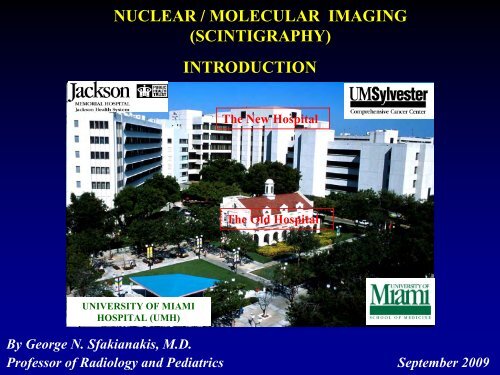
NUCLEAR / MOLECULAR IMAGING<br />
(SCINTIGRAPHY)<br />
INTRODUCTION<br />
The New Hospital<br />
The Old Hospital<br />
UNIVERSITY OF MIAMI<br />
HOSPITAL (UMH)<br />
By George N. Sfakianakis, M.D.<br />
Pr<strong>of</strong>essor <strong>of</strong> Radiology and Pediatrics September 2009
X-rays (CT), UltraSound, MRI<br />
<strong>Nuclear</strong>: Planar/SPECT/PET<br />
External<br />
Source <strong>of</strong> Radiation<br />
The Patient becomes<br />
the Source <strong>of</strong> Radiation
NUCLEAR / MOLECULAR IMAGING<br />
(SCINTIGRAPHY)<br />
It is based on Molecular Action<br />
It follows a predefined Task for numerous applications:<br />
• Identify The Target<br />
• Find its Biological Properties<br />
• Select the Carrier ( Molecule <strong>to</strong> reach the Target )<br />
• Radiolabel the Carrier Radiopharmaceutical<br />
• Inject the patient – Incubate<br />
• Image and Interpret
NUCLEAR IMAGING (SCINTIGRAPHY)<br />
Of particular Importance are:<br />
• The Biological Properties <strong>of</strong> the Radiopharmaceutical<br />
• And the Chemical and Physical Properties <strong>of</strong> the Radioiso<strong>to</strong>pe
NUCLEAR IMAGING (SCINTIGRAPHY)<br />
Single Pho<strong>to</strong>n<br />
PET<br />
Camera<br />
Coincidence, ToF<br />
Collimation<br />
The <strong>Nuclear</strong> Cameras generate Images<br />
<strong>of</strong> the Distribution inside the body<br />
<strong>of</strong> the given Radiopharmaceutical
In <strong>Nuclear</strong> <strong>Medicine</strong> we use<br />
Radiopharmaceuticals<br />
(Organic or Inorganic Molecules labeled with)<br />
Radioactive A<strong>to</strong>ms
HISTORY OF NUCLEAR MEDICINE<br />
It is related <strong>to</strong> the<br />
Rational Science and Rational <strong>Medicine</strong><br />
When and how the whole thing started?
HIPPOCRATES<br />
Father <strong>of</strong> Rational<br />
<strong>Medicine</strong><br />
HISTORY<br />
DEMOCRITOS<br />
Father <strong>of</strong> the A<strong>to</strong>mic<br />
Theory <strong>of</strong> the Matter<br />
EPICUROS<br />
Father <strong>of</strong> <strong>Nuclear</strong> Science<br />
and <strong>Nuclear</strong> <strong>Medicine</strong><br />
The Miracle <strong>of</strong> the 5th Century BC
HISTORY OF NUCLEAR MEDICINE
HUMAN INTELLIGENCE PROGRESS<br />
1) SURVIVAL: Labor, use <strong>of</strong> Animals (Slaves)<br />
Observation/Machines : Primitive Science<br />
2) WONDER about the world around/inside us<br />
a) Mysticism: Gods and Super-Powers<br />
Religions (Prophets, Saints, Preachers)<br />
b) Rational Thinking: Reason, Dialectics<br />
Philosophy-Theories (Philosophers)<br />
3) EXPERIMENT /Theories =Advanced Science<br />
4) SEPARATION <strong>of</strong> Science from Philosophy<br />
5) <strong>Intro</strong>duction <strong>of</strong> COMPUTER and GENETICS
RATIONAL THINKING:<br />
Epilepsy:<br />
“...It is not in my opinion any more<br />
divine or more sacred than other<br />
diseases, but<br />
it has a natural cause……<br />
It is also curable, no less than<br />
other diseases…….”<br />
HIPPOCRATES<br />
FDG-ΡΕΤ<br />
ICTAL INTERICTAL<br />
RELIGIOUS THINKING:<br />
“ Epilepsy is the result <strong>of</strong> effect <strong>of</strong><br />
the gods or the devils ”
SCIENCE and PHILOSOPHY<br />
Science deals with Known Facts<br />
Philosophy with Speculation
PHILOSOPHY<br />
A philosopher Loves (philos) the Knowledge (sophia)<br />
Originally, in ancient times,<br />
a philosopher was also a scientist<br />
(Pythagoras: Philosopher and Mathematician )<br />
Later, after the renaissance<br />
Science was separated from philosophy
PHILOSOPHY<br />
Change is a rearrangement <strong>of</strong> a<strong>to</strong>ms<br />
that remain themselves unchanged<br />
The First Philosophers (+Scientists)<br />
How does Change occur?<br />
rain<br />
snow
The First Philosophers (+Scientists) lived here<br />
ANCIENT GREECE
DEMOCRITOS<br />
Father <strong>of</strong> the A<strong>to</strong>mic Theory<br />
<strong>of</strong> the Matter<br />
“The World Outside and the<br />
World Inside Us is made <strong>of</strong><br />
small invisible, indivisible<br />
and indestructible Particles<br />
the A<strong>to</strong>ms”
The Miracle <strong>of</strong> the 5th Century BC<br />
Art Reflected Nature And Beauty<br />
Aphrodite (Venus) <strong>of</strong> Milos Vic<strong>to</strong>ry <strong>of</strong> Samothrace
THE CLASSICAL CIVILIZATION WAS<br />
TRANSFERRED EAST<br />
THE EMPIRE OF ALEXANDER THE GREAT
CLASSICAL CIVILIZATION WAS ALSO<br />
TRANSFERRED WEST<br />
THE ROMAN EMPIRE
PHILOSOPHY AND<br />
SCIENCE THRIVED IN THE<br />
ROMAN EMPIRE<br />
(GRECO-ROMAN<br />
CIVILIZATION)<br />
DE RERUM NATURA<br />
by LUCRETIUS CARUS<br />
In this poem he reviews the<br />
ancient philosophers<br />
<strong>of</strong> the classical and the roman<br />
periods
EPICUROS<br />
A PHILOSOPHER OF<br />
THE ROMAN PERIOD<br />
Father <strong>of</strong> <strong>Nuclear</strong> Science<br />
and <strong>Nuclear</strong> <strong>Medicine</strong><br />
“The A<strong>to</strong>ms may pass through<br />
an Unstable Phase<br />
before they are stabilized<br />
in their perpetual form”
• The ancient Greek Philosophers introduced<br />
the Theory <strong>of</strong> the A<strong>to</strong>m and the<br />
Theory <strong>of</strong> the Unstable (=Radioactive) A<strong>to</strong>m<br />
• In <strong>Nuclear</strong> <strong>Medicine</strong><br />
We use radioactive A<strong>to</strong>ms<br />
Injection<br />
<strong>of</strong> Patients<br />
Radiopharmaceuticals<br />
Decay <strong>of</strong> Radioactive A<strong>to</strong>ms
THERE FOLLOWS RELIGIOUS POWERS<br />
Byzantium imposed Christianity and persecuted Philosophy
The Arabs imposed Muslim Religion<br />
but some <strong>of</strong> their philosophers preserved the ancient philosophy
AND THE MEDIEVAL TIMES<br />
CHRISTIANS<br />
MUSLIMS
THE RENAISSANCE<br />
IN THE 14 th CENTURY<br />
REVITALIZED<br />
SCIENCE AND<br />
PHILOSOPHY<br />
LEONARDO DA VINCI:<br />
MONA LISA
HISTORY<br />
It <strong>to</strong>ok more than 2000 years for Science <strong>to</strong> Recover<br />
Modern A<strong>to</strong>mic Theory<br />
John Dal<strong>to</strong>n 1880<br />
Discovery <strong>of</strong> Natural and Artificial Radioactivity<br />
Henry Becquerel 1896 Marie Curie 1897
RADIATION FROM RADIUM<br />
The A<strong>to</strong>m is not indestructible
HISTORY<br />
Dmitry Ivanovic Mendeleyev 1896<br />
The Periodic Table <strong>of</strong> the Elements
SCIENCE PHILOSOPHY AND ART<br />
RETURNED TO CLASSICS<br />
MODERN THEORY ABOUT THE<br />
STRUCTURE OF THE ATOM<br />
Les Bourgeois de Calais by Rodin
HISTORY<br />
Theory <strong>of</strong> Relativity 1905-1916<br />
Albert Einstein<br />
Albert Einstein:<br />
1905 Mass and energy are interchangeable<br />
Special Theory <strong>of</strong> Relativity (Space-Time)<br />
1907 E=mc 2 - Pho<strong>to</strong>electric Effect<br />
1916 General theory <strong>of</strong> relativity
MEDICINE LAGGED BEHIND BASIC SCIENCES
KING CHARLE’S II<br />
in his<br />
TUBERCULOSIS<br />
CLINIC
THE PRACTICE OF SURGERY<br />
<strong>Medicine</strong> was one <strong>of</strong> the last sciences <strong>to</strong> recover and still suffers
FINALLY RENAISSANCE CAME TO MEDICINE<br />
Discovery <strong>of</strong> X-Rays 1895: Wilhelm Conrad Roentgen
The first use <strong>of</strong> radioiso<strong>to</strong>pes in humans<br />
The Tracer Principle<br />
Georg de Hevesy 1913<br />
Nobel Price winner 1943
PHYSICS AND INSTRUMENTATION
SPECTRUM OF<br />
ELECTROMAGNETIC<br />
RADIATION
RADIOACTIVE ATOM DECAY<br />
a) Electromagnetic radiation: Single Pho<strong>to</strong>n Imaging / SPECT<br />
b) Particular radiation: Therapy and PET<br />
e-<br />
annihilation<br />
2 x 511keV pho<strong>to</strong>ns PET
NUCLEAR MEDICINE IMAGING/THERAPY<br />
SINGLE PHOTON (γ,x)<br />
PLANAR / TOMOGRAPHY<br />
(SPECT)<br />
Single Pho<strong>to</strong>n Emitters:<br />
99m<br />
Tc, 201 Tl, 67 Ga, 131,123 I, 111 In<br />
POSITRON (e + )<br />
EMISSION TOMOGRAPHY<br />
(PET)<br />
Positron Emitters:<br />
18<br />
F, 15 O, 13 N, 11 C, 82 Rb, 68 Ga<br />
Ana<strong>to</strong>mical/Functional Imaging<br />
Metabolic Molecular Imaging<br />
THERAPY<br />
Particle Emitters: e - electron (beta) and alpha (a)<br />
32<br />
P, 131 I, 89 Sr, 90 Y, 153 Sm, 166 Ho<br />
Therapy <strong>of</strong> benign and malignant diseases<br />
IN VITRO STUDIES<br />
low energy gamma or electrons ( 125 I, 3 H, 14 C)
RADIOISOTOPES IN NUCLEAR MEDICINE<br />
PATIENT IMAGING<br />
PLANAR AND SPECT: SINGLE PHOTON γ, x RAYS<br />
energy > 80keV ( 99m Tc- 203 TI etc)<br />
PET STUDIES<br />
POSITRONS e + ( 18 FDG etc)<br />
PATIENT THERAPY<br />
PARTICULAR RADIATION<br />
electrons ( 131 I) or alpha particles<br />
IN VITRO STUDIES<br />
GAMMA OR ELECTRONS<br />
low energy ( 125 I, 3 H, 14 C)
NUCLEAR MEDICINE<br />
RADIOPHARMACEUTICALS<br />
Organic or Inorganic Molecules<br />
labeled with Radioactive Iso<strong>to</strong>pes<br />
Radioactive Iso<strong>to</strong>pes can be<br />
Single Pho<strong>to</strong>n Emitters: γ, x rays (Planar and SPECT)<br />
Positron Emitters: e + or positron (PET studies)<br />
Particle Emitters: e - electron or beta, and alpha (Therapy)<br />
Low Energy γ or e - Emitters:( in vitro studies)
A. SINGLE PHOTON IMAGING<br />
PLANAR and TOMOGRAPHIC (SPECT)
HISTORY<br />
Development <strong>of</strong> Rectilinear Scanner 1950<br />
Benedict Cassen<br />
1950 Benedict Cassen invents rectilinear scanner
HISTORY<br />
Rectilinear Scanner<br />
1954 William Myers operates a rectilinear scanner
HISTORY<br />
Development <strong>of</strong> Gamma Camera 1957<br />
Hal Anger<br />
1957: Hal Anger invents gamma camera
ANGER GAMMA CAMERA
1) SINGLE PHOTON SCINTIGRAPHY<br />
(Anger Gamma Camera Principle )<br />
Collimation<br />
(gamma/x-rays)
SPECT JASZCZACK PHANTOM STUDIES
PORTABLE GAMMA CAMERA
TOMOGRAPHY<br />
SINGLE PHOTON (γ,x)<br />
TOMOGRAPHY (SPECT)<br />
POSITRON EMISSION<br />
TOMOGRAPHY (PET)
THE THREE TOMOGRAPHIC PLANES
SPECT HOFFMAN PHANTOM STUDIES
THE FIRST<br />
SPECT<br />
CAMERA
SPECT CAMERA THREE DETECTOR
SPECT CAMERA THREE DETECTOR
Functional <strong>Nuclear</strong> Studies<br />
Sculpture by Colder
Ana<strong>to</strong>mical Studies<br />
Aphrodite <strong>of</strong> Milos
SPECT/CT<br />
Combines a Dual-headed Gamma Camera for Functional Imaging<br />
and a Multislice CT Scanner for ana<strong>to</strong>mical definition<br />
PHILIPS Precedence<br />
SIEMENS Symbia<br />
Approx. Price Tag: $1M depending on the configuration.
67<br />
Ga SPECT / CT<br />
SPECT<br />
SPECT + CT<br />
CT<br />
SPECT / CT for accurate localization <strong>of</strong> the abscess
111<br />
In Prostascint SPECT / CT<br />
Symbia<br />
Prostate cancer –Accurate localization <strong>of</strong> LN metastasis
111<br />
In Prostascint SPECT / CT<br />
Recurrence
111<br />
In Octreotide SPECT/CT<br />
Symbia<br />
Primary and metastatic Carcinoid tumor
111<br />
In Prostascint SPECT / CT<br />
Is there a recurrence?
Primary (Pancreas)<br />
and Metastatic (Pelvis)<br />
Carcinoid<br />
111<br />
In Octreotide SPECT/CT
111<br />
In Octreotide SPECT/CT<br />
Paraganglioma
99m<br />
Tc-Sestamibi SPCT/CT<br />
Parathyroid Adenoma<br />
Early Study
Late Study<br />
99m<br />
Tc-Sestamibi SPCT/CT<br />
Parathyroid Adenoma<br />
PLANAR MIBI<br />
EARLY LATE
B. POSITRON EMISSION TOMOGRAPHY<br />
(PET)
HISTORY<br />
Development <strong>of</strong> Cyclotron<br />
Ernest O. Lawrence<br />
Ernest O. Lawrence (1901-1958)
HISTORY<br />
Development <strong>of</strong> PET Scanner<br />
Ter-Pogossian<br />
Wagner in the Ter-Pogossian scanner at Washing<strong>to</strong>n<br />
<strong>University</strong>, 1973
PROPERTIES OF POSITRON EMITTERS<br />
• CHEMICAL<br />
Can Label Metabolic Molecules (Glucose, AA, etc)<br />
• PHYSICAL<br />
Small: Do not alter the Biochemical Properties<br />
<strong>of</strong> the Metabolic Molecules (Biochemical Recognition Maintained)<br />
Therefore allow Molecular Imaging<br />
Emit Positrons: Need special Cameras (PET cameras)<br />
Allow Tomography and Quantitation<br />
Have short half lives: Low Radiation Exposure<br />
Need Cyclotrons <strong>to</strong> produce them (or special genera<strong>to</strong>rs)
CYCLOTRON: STRUCTURE AND FUNCTION<br />
Electromagnetic core <strong>of</strong> the<br />
cyclotron for acceleration <strong>of</strong><br />
charged suba<strong>to</strong>mic particles
CYCLOTRON: PRODUCTION OF RADIOISOTOPES<br />
Charged particles (p,d) are highly accelerated and hit the target,<br />
a stable nucleus; they are incorporated and produce radioiso<strong>to</strong>pes
CYCLOTRON:SYNTHESIS OF RADIOPHARMACEUTICALS<br />
Radiopharmaceuticals are synthesized in heavily shielded cells,<br />
using the newly produced radioactive iso<strong>to</strong>pes
CYCLOTRON:THE BABY CYCLOTRONS<br />
Small Hospital Cyclotrons are available for clinical production
POSITRON DECAY: ANNIHILATION OF e + + e -<br />
Once a Positron e + is out <strong>of</strong><br />
the decaying nucleus, soon<br />
encounters an Electron e -<br />
and they Annihilate<br />
producing Two Pho<strong>to</strong>ns<br />
very energetic ( 511 keV ),<br />
which run on the same<br />
Straight Line but Opposite<br />
Direction<br />
e + e -
2. POSITRON EMISSION TOMOGRAPHY<br />
a) COINCIDENCE b) TIME OF FLIGHT<br />
HOW ΡΕΤ WORKS<br />
DECAY ANNIHILATION COINCIDENCE<br />
t 2<br />
Positronium<br />
t 1<br />
DECAY ANNIHILATION TIME <strong>of</strong> FLIGHT t 2 -t 1
POSITRON EMISSION TOMOGRAPHY (PET)<br />
Commonly Used Positron-Emitting Radionuclides
RADIOPHARMACEUTICALS FOR PET<br />
•<br />
11<br />
C(20min)-Carbon Monoxide Blood Pool-Volume<br />
•<br />
11<br />
C-Carbon Dioxide Tissue pH<br />
•<br />
11<br />
C- Dopamine Neurorecep<strong>to</strong>rs<br />
•<br />
11<br />
C- N-methyl-spiperone Neurorecep<strong>to</strong>rs<br />
•<br />
13<br />
N(10min)- Ammonia Perfusion<br />
•<br />
13<br />
N- Amino Acids Tumors<br />
•<br />
15<br />
0 2 (2min) - Oxygen Metabolism/Tumors<br />
•<br />
15<br />
0H 2 - Water Blood Flow<br />
•<br />
18<br />
F(110min)-2 Deoxy-Glucose Metabolism/Tumors<br />
•<br />
18<br />
F-DOPA Parkinson/Tumors<br />
•<br />
18<br />
F-Haloperidol Neurorecep<strong>to</strong>rs<br />
•<br />
18<br />
F- Amino Acids Tumors
PET CLINICAL APPLICATIONS<br />
• Brain Function and Tumors<br />
• Myocardial Perfusion and Viability<br />
• Oncologic Applications<br />
• Infection / Inflammation etc
ΡΕΤ APPLICATIONS<br />
CLINICAL<br />
18<br />
F-FDG ( 18 F-Fluoro-Deoxy-Glucose)<br />
18<br />
F-DOPA ( 18 F-Di-Oxy-Phenyl-Alanine)<br />
82<br />
Rb and 68 Ga Genera<strong>to</strong>rs<br />
UNDER DEVELOPMENT<br />
18<br />
F-Aminoacids, 11 C-Aminoacids / Nucleotides, 18 F-Recep<strong>to</strong>rs<br />
RESEARCH<br />
Blood Flow / Blood Pool / Utilization <strong>of</strong> Ο 2<br />
Imaging Recep<strong>to</strong>rs / Organelles<br />
DNA / RNA / Gene Studies / Treatments<br />
Special Studies <strong>of</strong> Ischemia, Alzheimer's etc.
WHAT IS FDG-PET AND HOW DOES IT WORK?<br />
• Most Tumors metabolize glucose in excess <strong>of</strong> normal tissues<br />
• Myocardium and Brain Cortex metabolize excessively glucose<br />
• FDG-PET quantitates in vivo the glucose metabolic activity<br />
<strong>of</strong> tumors and normal organs (brain and heart)
PRINCIPLES <strong>of</strong> FDG IMAGING<br />
A. Modify the molecule <strong>of</strong> Glucose<br />
<strong>to</strong> 2-Deoxy-Glucose (DG).<br />
B. Label 2-Deoxy-Glucose with a<br />
radioiso<strong>to</strong>pe, 18 F, which<br />
does not alter its properties<br />
for biochemical recognition:<br />
18<br />
F-2-Deoxy-Glucose = FDG
PRINCIPLES OF FDG IMAGING<br />
FDG enters the cells<br />
and it is phosphorylated like Glucose<br />
FDG-6-PO 4<br />
is not metabolized<br />
and accumulates, thus allowing imaging
CLINICAL EXPERIENCE with FDG/PET TUMOR IMAGING<br />
APPLICATIONS<br />
• CNS Function and Tumors<br />
• Myocardial Viability<br />
• Tumor Evaluation<br />
• Infection / Inflammation
CLINICAL EXPERIENCE with FDG/PET TUMOR IMAGING<br />
ADVANTAGES<br />
• It provides Accurate information about most Tumors<br />
• The information is Functional and Quantitative<br />
• It is Easy <strong>to</strong> recognize the Tumors on PET studies<br />
• Covers the Entire Body
CLINICAL EXPERIENCE with FDG/PET TUMOR IMAGING<br />
DISADVANTAGES<br />
• Brain, Heart and Liver accumulate FDG and this limits the sensitivity<br />
for low intensity lesions in or around these organs<br />
• The Kidneys recognize the difference between Glucose and FDG<br />
and do not not reabsorb FDG and create interpretation issues<br />
• Muscles and Brown Fat accumulate FDG as Glucose and this results in<br />
bowel, vocal cords, body muscles and fat visualization issues<br />
• WBCs (Infections) Muscles etc (Inflammations) accumulate FDG<br />
and this generates problems in differentiating them from tumors,<br />
but this may diagnose infection or inflammation if needed
CLINICAL EXPERIENCE with FDG/PET TUMOR IMAGING<br />
SOLUTIONS<br />
• Four hours NPO eliminates visualization <strong>of</strong> the heart<br />
• Rest during incubation (+day before) eliminates the muscles<br />
• Warm Environment eliminates visualization <strong>of</strong> brown fat<br />
(Propranolole 20-40mg before FDG may also work)<br />
• Accurate localization could be achieved using the CT<br />
a) Correlation with previous CT “side-by-side” visually<br />
b) Fusion PET+CT with “computer programs”<br />
c) PET/CT “combined acquisition” Imaging
The EVOLUTION <strong>of</strong> CLINICAL FDG/PET IMAGING<br />
Cameras and Crystals<br />
Hybrid ΡΕΤ/SPECT NaI<br />
Dedicated PET NaI<br />
New Crystal Technology<br />
ALLEGRO <br />
GSO<br />
Combined ΡΕΤ/CT<br />
ADAC-PHILIPS<br />
GSO<br />
GE<br />
BGO<br />
SIEMENS/CTI<br />
LSO
IMAGE FUSION<br />
? ?<br />
CT<br />
PET<br />
CT FUSED WITH PET
Design features & benefits<br />
image fusion<br />
side by side display <strong>of</strong><br />
2 different studies<br />
Simultaneous processing<br />
<strong>of</strong> both studies<br />
au<strong>to</strong>matic overlay and<br />
manual alignment<br />
applicable <strong>to</strong> brain- and<br />
WB studies<br />
import <strong>of</strong> other modalities via<br />
optional DICOM s<strong>of</strong>tware<br />
Meningioma after RT ?<br />
malignant lesion ?<br />
in visual cortex ?<br />
MRI<br />
MRI<br />
Image courtesy <strong>of</strong> Pr<strong>of</strong> .Dr. Carreras,<br />
Madirid, Spain<br />
FDG-PET<br />
Fusion FusionPET/MRI
EQUIPMENT FOR PET and PET/CT SCINTIGRAPHY<br />
DEDICATED PET<br />
CAMERAS<br />
Ring-Detec<strong>to</strong>rs<br />
NaI/BGO LSO-GSO<br />
using coincidence<br />
principle<br />
Provide independent<br />
PET images<br />
PET/CT<br />
CAMERAS<br />
Combinations <strong>of</strong> ring<br />
PET detec<strong>to</strong>rs and<br />
CT X-ray machines<br />
Provide independent<br />
and fused PET/CT<br />
images<br />
PET CT
FUSION PET AND MRI<br />
Patient with a recent left CVA had an FDG-PET
QUANTIFICATION OF PET STUDIES<br />
Accurate Quantification
PRACTICAL QUANTIFICATION <strong>of</strong> PET<br />
Standardized Uptake Value (SUV)<br />
SUV units indicate uptake = % <strong>of</strong> injected dose (weight corrected) /gram tissue<br />
bladder<br />
Recurrence 9 SUV<br />
Bone Mets 3 SUV<br />
bladder
The EVOLUTION <strong>of</strong> CLINICAL FDG/PET IMAGING<br />
Clinical Utilization<br />
Neuro<br />
Cardiac<br />
Tumors<br />
Tumors<br />
Tumors<br />
Tumors
IN VIVO (NON-IMAGING) STUDIES<br />
1) THYROID UPTAKE<br />
2) VITAMIN B-12 ABSORPTION (SHILLING’S TEST)<br />
3) BLOOD/PLASMA/RBC VOLUMES<br />
4) PLATELET SURVIVAL AND SEQUESTRATION<br />
5) RBC SURVIVAL AND SEQUESTRATION<br />
6) GI TRACT BLOOD OR PROTEIN LOSS<br />
7) HELICOBACTERIUM PYLORI STUDIES<br />
8) FERROKINETICS<br />
9) RENAL CLEARANCES (GFR-ERPF-FF)
NUCLEAR MEDICINE STUDIES<br />
Safe<br />
Easy<br />
Available<br />
Cost effective
NUCLEAR MEDICINE PROCEDURES ARE<br />
CLINICALLY USEFUL BECAUSE<br />
• They provide comprehensive information about<br />
structure and function simultaneously<br />
• They have high sensitivity or specificity or both<br />
• Many examine the whole body or organ systems<br />
• They are non-invasive and well <strong>to</strong>lerated tests<br />
• Often provide information no other tests can<br />
• They are less expensive than most test or sequences <strong>of</strong><br />
tests for similar information (except PET/CT)
RADIATION AND LIFE<br />
<strong>Nuclear</strong> Imaging involves a very low dose <strong>of</strong> radiation<br />
harmless <strong>to</strong> infants, children and adults
QUANTITATIVE SCINTIGRAPHY 1<br />
1. GRAPH GENERATION FROM DYNAMIC STUDIES<br />
FOR SEMI-QUANTITATION<br />
Renography, GI Motility, GE emptying, EF, EF, Flow<br />
2. SEMI-QUANTITATION OF ORGAN UPTAKE FROM<br />
STATIC IMAGES<br />
67<br />
Ga Pulmonary, Renal, Thyroid Uptake, Lung V/Q,<br />
201<br />
TI tumor, etc
QUANTITATIVE SCINTIGRAPHY 2<br />
3 ACCURATE QUANTIFICATION OF ORGAN<br />
PERFUSION AND FUNCTION USING BLOOD<br />
(URINE) SAMPLING AND IMAGING (PET) WITH<br />
SOPHISTICATED ANALYSIS<br />
4 PROBE GENERATED STRIPCHART OR DIRECT<br />
COUNTING (old, non-used in most centers)<br />
(Thyroid uptake, Renography, Xe washout, Flow)
TIME ACTIVITY GRAPHS:<br />
FLOW GRAPHS<br />
RENOGRAMS
QUALITY CONTROL IN NM<br />
OFF PEAK PEAKED
FACTORS AFFECTING THE USE OF<br />
RADIONUCILES IN CHILDREN<br />
• Short half-life iso<strong>to</strong>pes ( 99m Tc - 6 hrs)<br />
• No Particular Radiation<br />
• High efficiency cameras<br />
The Radiation Exposure from <strong>Nuclear</strong><br />
Imaging is equivalent and even lower<br />
than x-ray studies<br />
(cys<strong>to</strong>graphy)
NUCLEAR MEDICINE STUDIES AT JMH/UM<br />
EQUIPMENT<br />
We have the best SPECT camera (3 detec<strong>to</strong>r at JMH)<br />
6 Dual-Head and 4 Single-Head SPECT<br />
3 Planar (1 portable), 1 SPECT/CT (DFB)<br />
1 hybrid PET, 1 dedicated PET (JMH) and 3 PET/CT (UMHC, DFB, BBL)<br />
RADIOPHARMACEUTICALS<br />
We use a Tc genera<strong>to</strong>r and Kits<br />
commercially available Planar and SPECT RPs<br />
and 18 FDG, the only commercially available RP for PET<br />
We expect soon a second SPET/CT camera (UMHC)<br />
and a CYCLOTRON for short lived positron emitters (UMHC)<br />
( 15 O, 2 min; 13 N, 10 min; 11 C, 20 min)<br />
We pursue<br />
A PET/CT camera for JMH<br />
September 2009
IMAGING AND THERAPEUTIC APPLICATIONS<br />
IN NUCLEAR MEDICINE<br />
Planar Static and Dynamic Imaging applications<br />
Organ Imaging, Hypertension, Infections, Tumors<br />
Tomography<br />
Single pho<strong>to</strong>n (SPECT), Positron (PET)<br />
Non Imaging Studies<br />
Renal Clearance, Blood Volume, Vitamin B12 absorption<br />
Therapy<br />
Benign{Thyro<strong>to</strong>xicosis, Arthropathies (Rheuma<strong>to</strong>id, Hemophiliac)}<br />
Malignant (Thyroid cancer, Lymphomas, Bone Pain from Mets)
THERAPY WITH SYSTEMIC<br />
USE OF UNSEALED<br />
RADIOPHARMACEUTICALS
Adapted from Woot<strong>to</strong>n.<br />
Radiation Protection <strong>of</strong><br />
Patients. 1993.<br />
Penetration <strong>of</strong> Particulate and<br />
Electromagnetic Radiation<br />
HUMAN TISSUES<br />
Subcutaneous<br />
tissue<br />
Dermis<br />
Epidermis<br />
Alpha particles<br />
Beta particles<br />
Gamma rays
Radiation Safety Issues (environment)<br />
Alpha and Beta rays are not a problem (attenuated)<br />
Gamma rays are a problem because they emerge and irradiate environment<br />
Therefore pure Beta emitters are preferable ( 90 Y)<br />
Body Tissues
THERAPY WITH UNSEALED<br />
RADIOPHARMACEUTICALS<br />
A) BENIGN DISEASES<br />
Thyro<strong>to</strong>xicosis<br />
-Graves Disease (10-30 mCi 131 INa po)<br />
-Toxic Adenoma (30-150 mCi 131 INa po)<br />
-Toxic Multinodular Goiter (20-150 mCi 131 INa po)<br />
-Non Toxic Multinodular Goiter (100-300 mCi 131 INa po)<br />
Synovec<strong>to</strong>my (Hemophiliac or Rheuma<strong>to</strong>id Arthritis)<br />
(5-10 mCi 32 P Chromic Phosphate (Colloid) intraarticularly)
GRAVES DISEASE<br />
At treatment (15 mCi 131 INa po) 4 months later
TOXIC ADENOMA<br />
PRE AND POST THERAPY WITH 131 I<br />
At treatment (29.2 mCi 131 INa po) 8 months later
<strong>Nuclear</strong> Synovec<strong>to</strong>my<br />
INDICATIONS<br />
Hemophiliac joints<br />
Rheuma<strong>to</strong>id arthritis<br />
METHOD<br />
Arthrocentesis<br />
Test with 99m Tc--SC<br />
Instillation <strong>of</strong> 05-15 mCi <strong>of</strong> 32 P-colloid<br />
RESULTS<br />
Improvement in motion<br />
Relief <strong>of</strong> pain<br />
Prevent hemorrhage
THERAPY WITH UNSEALED<br />
RADIOPHARMACEUTICALS<br />
B) MALIGNANT DISEASES<br />
Treatment <strong>of</strong> Thyroid Cancer (150 - 300 mCi 131 INa orally)<br />
[Ablation <strong>of</strong> the Thyroid gland or Remnants (150 mCi 131 INa orally)]<br />
Treatment <strong>of</strong> Bone Pain from Metastasis<br />
(4 mCi 89 Sr iv or 1.2 mCi/kg 153 Sm EDTMP iv)<br />
Bone Marrow Ablation Holmium-166-DOTMP iv<br />
(or 90 YMoAb or 131 IMoAb)<br />
Treatment Non-Hodgkin’s Lymphoma ( 131 IMoAb or 90 YMoAb)<br />
Treatment <strong>of</strong> Pheochromocy<strong>to</strong>mas Paragangliomas and Neuroblas<strong>to</strong>mas<br />
( 131 I-MIBG)
THERAPY OF TUMORS with UNSEALED<br />
or SYSTEMIC RADIOPHARMACEUTICALS<br />
A) INDICATION<br />
Biopsy proven diagnosis<br />
B) PREREQUISITES<br />
Pro<strong>of</strong> that the tumor accumulates sufficient percentage<br />
<strong>of</strong> the dose <strong>of</strong> the radiopharmaceutical (dosimetry)<br />
Dose not <strong>to</strong> exceed safety limits for normal tissues (dosimetry)<br />
Patient’s clinical condition and labora<strong>to</strong>ry values permit
15 yo boy with thyroid carcinoma after thyroidec<strong>to</strong>my + ablation, rising TGB<br />
TREATMENT <strong>of</strong> METASTATIC THYROID CANCER<br />
Dosimetry<br />
Treatment<br />
lungs<br />
standard<br />
4 th day during dosimetry<br />
With 2 mCi <strong>of</strong> 131 INa po<br />
7 days post treatment with<br />
302 mCi <strong>of</strong> 131 INa po
THERAPY OF INTRACTABLE BONE PAIN<br />
DUE TO BLASTIC METASTATIC DISEASE<br />
• Phosphorous-32 (Bone marrow suppression)<br />
(T-1/2 = 14days) 60-90% efficacy<br />
• Strontium-89 (Mild transient BM suppression)<br />
(T-1/2 = 8 days) (Metastron)<br />
Effective in 80% prostate, 81% breast metastasis<br />
• Complexes (Mild transient BM suppression)<br />
Samarium-153-EDTMP (T-1/2 = 47 hr)<br />
(Quadramet)<br />
Yttrium-90-DTPA-Ab (T-1/2 = 64 hr)
TREATMENT OF INTRACTABLE BONE PAIN<br />
FROM METASTATIC CANCERS<br />
February January next year<br />
December next year<br />
Deteriorating metastatic disease with intractable bone pain<br />
pro<strong>of</strong> that the tumor accumulates the radiopharmaceutical
RADIOIMMUNOTHERAPY IN<br />
NON-HODGKIN’S LYMPHOMA
External Beam Radiation vs<br />
Radioimmunotherapy for Indolent NHL<br />
External<br />
beam radiation<br />
Radioimmunotherapy
Rituximab: First Monoclonal Antibody<br />
Approved for the Treatment <strong>of</strong> NHL<br />
FDA-approved<br />
indication:<br />
relapsed or<br />
refrac<strong>to</strong>ry lowgrade<br />
or<br />
follicular, CD20+,<br />
B-cell non-<br />
Hodgkin’s<br />
lymphoma<br />
Rituxan (rituximab) prescribing information. South San Francisco, Calif: Genentech; 2002.
90<br />
Y Zevalin/ 131 I Bexxar Produce a Crossfire Effect<br />
Naked antibody<br />
Radiolabeled antibody<br />
Rituxin 90 Y Zevalin/ 131 I Bexxar
Radioimmunotherapy<br />
Yttrium 90 ibritumomab tiuxetan (Zevalin ® )<br />
– Anti-CD20<br />
– Approved for commercial use in the United States,<br />
February 2002<br />
Yttrium 90 epratuzumab<br />
– Anti-CD22<br />
– Investigational<br />
Iodine 131 <strong>to</strong>situmomab (Bexar ® )<br />
– Anti-CD20<br />
– Approved for commercial use in the United States,<br />
June 2003
Radionuclides Commonly Used in RIT<br />
Physical<br />
half-life<br />
life<br />
Decay<br />
type<br />
Particle<br />
energy<br />
(MeV)<br />
Gamma<br />
energy<br />
(MeV)<br />
Particle<br />
path<br />
length<br />
(mm)<br />
90<br />
Y 2.7 days β 2.3 None 5.3<br />
131<br />
I 8 days β, γ 0.6 0.364 0.8
90<br />
Y Ibritumomab Tiuxetan<br />
Monoclonal<br />
antibody<br />
Chela<strong>to</strong>r<br />
90 Y radionuclide<br />
Ibritumomab<br />
– Murine monoclonal<br />
antibody that binds<br />
<strong>to</strong> the CD20 antigen<br />
Tiuxetan (chela<strong>to</strong>r)<br />
– Conjugated <strong>to</strong><br />
antibody, forms<br />
strong urea-type<br />
bond with<br />
radioiso<strong>to</strong>pe<br />
Iso<strong>to</strong>pe: 90 Y (pure β emitter)<br />
β radiation
131<br />
I Tositumomab<br />
Monoclonal<br />
antibody<br />
131 I radionuclide<br />
Tositumomab<br />
– Murine IgG2 alpha<br />
monoclonal antibody<br />
that binds <strong>to</strong> the<br />
CD20 antigen<br />
Tyrosine residue bond<br />
conjugates antibody<br />
with radionuclide<br />
Radionuclide— 131 I<br />
(β and γ emitter)<br />
β + γ radiation
Zevalin Integrated Safety (N = 349):<br />
Median Blood Counts After Treatment<br />
Hemoglobin<br />
(g/dL<br />
dL)<br />
ANC<br />
(10 3 /µL)<br />
16<br />
12<br />
Hb = 10<br />
8<br />
4<br />
ANC = 1000<br />
0<br />
Hemoglobin<br />
ANC<br />
0 2 4 6 8 10 12 14<br />
Study week<br />
Platelets<br />
450<br />
400<br />
350<br />
300<br />
250<br />
200<br />
150<br />
100<br />
50<br />
0<br />
Platelets<br />
(10 3 /µL)<br />
Plts = 50,000
REQUIREMENTS FOR LICENSING<br />
TO ADMINISTER UNSEALED<br />
RADIOPHARMACEUTICALS<br />
TO PATIENTS (DIAGNOSIS-THERAPY)<br />
200 hours <strong>of</strong> Basic Training<br />
Physics-Radiopharmacy-Instrumentation<br />
Radiobiology-Radiation Protection<br />
Mathematics-Statistics-Computers<br />
4-12 months Clinical Experience
BOARDS IN NUCLEAR MEDICINE<br />
1) AMERICAN BOARD OF NUCLEAR MEDICINE (3-years)<br />
1 year “prepara<strong>to</strong>ry” training<br />
(Rad/Path/Med/Surgery/Pedi,etc)<br />
3 years training in all aspects <strong>of</strong> Nucl Med (incl. >8 mos CT)<br />
2) RADIOLOGY WITH BOARDS IN NM (new 5-year program)<br />
1 year “prepara<strong>to</strong>ry” training<br />
4 Years Radiology (including 8 months <strong>Nuclear</strong> <strong>Medicine</strong>)<br />
1 Year straight <strong>Nuclear</strong> <strong>Medicine</strong> training