UltraVIEW VoX 3D Live Cell Imaging System
UltraVIEW VoX 3D Live Cell Imaging System
UltraVIEW VoX 3D Live Cell Imaging System

Create successful ePaper yourself
Turn your PDF publications into a flip-book with our unique Google optimized e-Paper software.
S P E C I F I C A T I O N S<br />
<strong>UltraVIEW</strong> <strong>VoX</strong> <strong>3D</strong> <strong>Live</strong><br />
<strong>Cell</strong> <strong>Imaging</strong> <strong>System</strong><br />
<strong>Cell</strong>ular <strong>Imaging</strong> and Analysis<br />
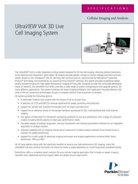
The <strong>UltraVIEW</strong> ® <strong>VoX</strong> is a fully integrated confocal system designed for <strong>3D</strong> live cell imaging. Allowing cellular interactions<br />
to be observed and measured in great detail, <strong>3D</strong> analysis provides greater context to cellular changes and more accurate<br />
results. Based on the Yokogawa ® CSU-X1 spinning disk confocal scanner, synchronized by PerkinElmer ® patented<br />
ProSync ® technology and powered by our award winning Volocity ® software, this system provides exceptional speed,<br />
quality and performance for high speed fluorescence imaging of living cells. Designed to meet the continually developing<br />
needs of research, the <strong>UltraVIEW</strong> <strong>VoX</strong> offers scientists a wide range of system configurations and upgrade options, for<br />
many different applications. The system combines the latest imaging hardware, with application focused software and<br />
state of the art computing technologies, to give a complete solution from acquisition to analysis.<br />
All systems provide the following options:<br />
• A solid-state modular laser engine with the choice of up to six laser lines<br />
• A selection of CCD and EMCCD cameras optimized for speed, sensitivity and resolution<br />
• Support for upright and inverted microscopes from all major manufacturers<br />
• Support for an extensive choice of microscope hardware accessories for <strong>3D</strong>, multi-positional and multi-channel<br />
imaging<br />
• The option of Macintosh ® or Windows ® computing platforms to suit your preference, with a range of computer<br />
models including 64-bit systems to meet your performance needs<br />
• The latest release of Volocity Acquisition, Volocity Visualization and Volocity Quantitation software for an integrated<br />
acquisition to analysis solution<br />
• Software supplied with an <strong>Imaging</strong> License Server application to allow analysis software to be shared across a<br />
network for added productivity<br />
• Support for a wide range of advanced imaging techniques and analysis applications including FRAP, Ratio,<br />
Restoration, TIRF and FRET<br />
All of these options along with the significant benefits of easy-to-use high-performance <strong>3D</strong> imaging, make the<br />
<strong>UltraVIEW</strong> <strong>VoX</strong> the confocal instrument of choice for either a single laboratory or a multi-functional centralized facility.<br />
PerkinElmer offers a complete system solution for your cellular imaging application that includes an expert imaging<br />
specialist team, dedicated technical support desk and global service organization.
SPECIFICATIONS<br />
<strong>UltraVIEW</strong> <strong>VoX</strong> <strong>System</strong><br />
Confocal Scanning Unit<br />
Model<br />
Type<br />
Scanning speed<br />
Synchronization<br />
Laser coupling<br />
Excitation range<br />
Emission range<br />
Camera mounts<br />
Camera switching<br />
Yokogawa ® CSU-X1<br />
Microlens-enhanced Nipkow disk<br />
Standard: 5000 rpm<br />
High (optional): 10000 rpm<br />
From ProSync 2 controller<br />
Direct injection<br />
400-650 nm<br />
420-750 nm<br />
Single or dual (optional)<br />
Automated (optional)<br />
<strong>System</strong> Synchronization<br />
Source<br />
ProSync 2 synchronization controller<br />
Inputs USB 2.0<br />
External laser safety interlock<br />
Laser interlock key-switch<br />
Outputs<br />
6-channel synchronized laser control (channel and power)<br />
Camera synchronization control<br />
CSU synchronization control<br />
Analog piezo Z axis control<br />
3-channel electro-mechanical shutter control<br />
3-port optical switch control<br />
PhotoKinesis ® Unit control<br />
Laser blanking<br />
Active blanking to minimize photo-damage and photo-bleaching<br />
Laser Excitation<br />
Laser combiner Modular Laser <strong>System</strong> 2.0<br />
Laser lines<br />
Select up to 6 lines from a choice of 7 including:<br />
Solid state 405 nm (50 mW)<br />
Solid state 440 nm (40 mW)<br />
Solid state 488 nm (50 mW)<br />
Solid state 515 nm (25 mW)<br />
Solid state 561 nm or 594 nm (50 mW)<br />
Solid state 640 nm (50 mW)<br />
(Powers stated are raw laser power)<br />
Laser control<br />
Full control over laser switching and power levels<br />
from Volocity via ProSync 2 controller<br />
Blanking and channel crosstalk
Dimensions<br />
MLS2.0 supplied in up to 3 stackable cases<br />
Control box; Lasers500 nm<br />
498 mm x 567 mm x 104 mm (w, d, h)<br />
Dichroic Filters<br />
Dichroic 1<br />
Dichroic 2<br />
Dichroic 3<br />
Dichroic switching<br />
Compatible with 405 / 488 / 568 / 640 nm laser lines<br />
Transmission in pass band >80%<br />
Reflectance in emission band >90%<br />
Dye compatibility includes DAPI, GFP2, GFP, mRFP, Cy5<br />
Compatible with 405 / 440 / 514 / 640 nm laser lines<br />
Transmission in pass band >80%<br />
Reflectance in emission band >90%<br />
Dye compatibility includes CFP, YFP, Cy5<br />
Compatible with 405 - 440 / 515 / 561 nm laser lines<br />
Transmission in pass band >80%<br />
Reflectance in emission band >90%<br />
Dye compatibility includes DAPI, CFP/YFP, mRFP<br />
Automated, controlled from Volocity<br />
Emission Filters<br />
Emission filter position 1<br />
Emission filter position 2<br />
Emission filter position 3<br />
Emission filter position 4<br />
Emission filter position 5<br />
Emission filter position 6<br />
Emission filter position 7<br />
Emission filter position 8<br />
Empty position for high speed fast sequential<br />
multicolor imaging<br />
Green (single band emission discrimination)<br />
525 nm center, 50 nm half-power bandwidth<br />
Compatible with 488 nm laser line<br />
Red (dual band emission discrimination)<br />
445 nm center, 60 nm half-power bandwidth<br />
615 nm center, 70 nm half-power bandwidth<br />
Compatible with 405/561 nm laser lines<br />
Cyan/Far red (dual band emission discrimination)<br />
485 nm center, 60 nm half-power bandwidth<br />
705 nm center, 90 nm half-power bandwidth<br />
Compatible with 440/640 nm laser lines<br />
Yellow (single band emission discrimination)<br />
587 nm center, 125 nm half-power bandwidth<br />
Compatible with 515 nm laser line<br />
DIC analyzer<br />
446 nm centre, 33 nm half-power bandwidth<br />
524 nm centre, 42 nm half-power bandwidth<br />
600 nm centre, 36 nm half-power bandwidth<br />
677 nm centre, 28 nm half-power bandwidth<br />
Compatible with 405 / 488 / 561 / 640 nm laser lines<br />
Cyan/Yellow (triple band fast sequential)<br />
477 nm center, 45 nm half-power bandwidth<br />
575 nm center, 100 nm half-power bandwidth<br />
705 nm center, 90 nm half-power bandwidth<br />
Compatible with 405 / 440 / 515 / 640 nm laser lines<br />
3
SPECIFICATIONS<br />
Emission filter position 9<br />
Emission filter switching<br />
Emission filter<br />
switching speed<br />
Cyan / Yellow / Red (triple band fast sequential)<br />
480 nm centre, 49 nm half-power bandwidth<br />
536 nm centre, 24 nm half-power bandwidth<br />
685 nm centre, 229 nm half-power bandwidth<br />
Compatible with 405 / 440 / 515 / 561 nm laser lines<br />
Automated, controlled from Volocity<br />
162 ms between sequential positions fully loaded<br />
55 ms between sequential positions unloaded<br />
Emission filters supplied with 594 nm laser available on request<br />
Camera Options<br />
Hamamatsu ® ORCA-R2 Camera head Hermetic vacuum-sealed head<br />
Dual cooling<br />
<strong>Imaging</strong> device<br />
Effective pixels<br />
<strong>Cell</strong> size<br />
Dual scan mode<br />
Pixel clock<br />
Readout noise<br />
(r.m.s typical)<br />
Full well capacity<br />
(typical)<br />
Dynamic range (typical)<br />
Cooling temperature<br />
Dark current<br />
Exposure time<br />
Air or water cooling<br />
ER-150 progressive scan interline CCD<br />
1344 (H) x 1024 (V)<br />
6.45 µm (H) x 6.45 µm (V)<br />
Normal scan / fast scan<br />
14.00 MHz (normal scan)<br />
28.00 MHz (fast scan)<br />
6 electrons (normal scan)<br />
10 electrons (fast scan)<br />
18 000 electrons (HDR mode off)<br />
36 000 electrons (HDR mode on)<br />
3000:1 (at normal scan, 1 x 1 binning)<br />
-35 ºC (air cooled)<br />
-40 ºC (water cooled at 20 ºC)<br />
0.0005 electrons/pixel/s (at -40 ºC)<br />
1 ms to 6 s<br />
Binning 2 x 2, 4 x 4, 8 x 8<br />
Sub-array readout<br />
Dual light mode<br />
Analog gain<br />
Analog offset<br />
Yes<br />
Low light / high light<br />
Yes (10x maximum)<br />
Yes<br />
Interface IEEE1394b-2002 (FireWire 800)<br />
Frame rate<br />
QE<br />
A/D converter<br />
16.2 fps (maximum at 1 x 1 binning)<br />
>15 fps (1 x 1 binning, fully synchronized)<br />
>70% (peak)<br />
12 bits<br />
4
Hamamatsu ® C9100-50 Camera head Hermetic vacuum-sealed head<br />
Proprietary to PerkinElmer<br />
Cooling<br />
Forced air<br />
<strong>Imaging</strong> device<br />
Effective pixels<br />
<strong>Cell</strong> size<br />
Pixel clock<br />
Readout noise<br />
(r.m.s typical)<br />
Full well capacity (typical)<br />
Electron multiplier gain max.<br />
Cooling temperature<br />
Dark current<br />
Exposure time<br />
Frame transfer EMCCD<br />
1000 (H) x 1000 (V)<br />
8 µm (H) x 8 µm (V)<br />
35.00 MHz<br />
10 electrons (at minimum EM gain)<br />
25 fps (1 x 1 binning, fully synchronized)<br />
>60% (peak)<br />
Hamamatsu ® C9100-13 Camera head Hermetic vacuum-sealed head<br />
Dual cooling<br />
<strong>Imaging</strong> device<br />
Effective pixels<br />
<strong>Cell</strong> size<br />
Pixel clock<br />
Readout noise (r.m.s typical)<br />
Full well capacity (typical)<br />
Electron multiplier gain max. 1200x<br />
Cooling temperature<br />
Dark current<br />
Exposure time<br />
Air or water cooling<br />
Back-thinned frame transfer EMCCD<br />
512 (H) x 512 (V)<br />
16 µm (H) x 16 µm (V)<br />
Up to 11 MHz (EMCCD)<br />
Up to 2.75 MHz (normal CCD)<br />
25 electrons (at minimum EM gain,<br />
11 MHz EMCCD)<br />
SPECIFICATIONS<br />
Hamamatsu ® C9100-13 Sub-array readout Yes<br />
continued Interface Camera Link<br />
A/D converter<br />
Frame rate<br />
Real time image processing<br />
Ultra low light detection<br />
QE<br />
16 bits<br />
31.9 fps (maximum at 1 x 1 binning)<br />
>25 fps (1 x 1 binning, fully synchronized)<br />
Background subtraction, shading correction<br />
Photon imaging mode<br />
>90% (peak)<br />
Hamamatsu ® C9100-14 Camera head Hermetic vacuum-sealed head<br />
Dual cooling<br />
<strong>Imaging</strong> device<br />
Effective pixels<br />
<strong>Cell</strong> size<br />
Pixel clock<br />
Readout noise (r.m.s typical)<br />
Full well capacity (typical)<br />
Electron multiplier gain max.<br />
Cooling temperature<br />
Dark current<br />
Exposure time<br />
Air or water cooling<br />
Back-thinned frame transfer EMCCD<br />
1024 (H) x 1024 (V)<br />
13 µm (H) x 13 µm (V)<br />
Up to 11 MHz (EMCCD)<br />
Up to 2.75 MHz (normal CCD)<br />
10 electrons (at minimum EM gain,<br />
11 MHz EMCCD)<br />
8.5 fps (1 x 1 binning, fully synchronized)<br />
Background subtraction, shading correction<br />
Photon imaging mode<br />
>90% (peak)<br />
Note: The <strong>UltraVIEW</strong> <strong>VoX</strong> can can support single or multiple camera configurations including simultaneous imaging.<br />
Computer Workstation Options<br />
Windows ® PC (32-bit, 4 GB)<br />
Lenovo ® S20 workstation<br />
32-bit Windows ® XP Professional<br />
Intel ® Core2 Quad W3520 – 2.66GHz x 4<br />
4 GB RAM<br />
1 TB disk storage (RAID 0)<br />
ATI Radeon HD 5870 graphics for high-performance <strong>3D</strong> visualization<br />
6
Windows ® PC (64-bit, 4 GB)<br />
Windows ® PC (64-bit, 8 GB)<br />
Windows ® PC (64-bit, 16 GB)<br />
Apple ® Mac ® Pro<br />
Display options<br />
Lenovo ® S20 workstation<br />
64-bit Windows ® XP x64 Professional<br />
Intel ® Core2 Quad W3520 – 2.66GHz x 4<br />
4 GB RAM<br />
1 TB disk storage (RAID 0)<br />
ATI Radeon HD 5870 graphics for high-performance <strong>3D</strong> visualization<br />
Lenovo ® S20 workstation<br />
64-bit Windows ® XP x64 Professional<br />
Intel ® Core2 Quad W3520 – 2.66GHz x 4<br />
8 GB RAM<br />
1 TB disk storage (RAID 0)<br />
ATI Radeon HD 5870 graphics for high-performance <strong>3D</strong> visualization<br />
Lenovo ® D20 workstation<br />
64-bit Windows ® XP x64 Professional<br />
Intel ® Xeon X5550 – 2.66GHz x 8<br />
16 GB RAM<br />
1 TB disk storage (RAID 0)<br />
ATI Radeon HD 5870 graphics for high-performance <strong>3D</strong> visualization<br />
Apple ® Mac ® Pro workstation<br />
Mac ® OS X<br />
Intel ® Xeon Quad-Core “Nehalem” – 2.8 GHz x 4<br />
6 GB RAM<br />
2 TB disk storage (RAID 0)<br />
ATI Radeon HD 5770 graphics for high-performance <strong>3D</strong> visualization<br />
24” or 30” PC HD flat panel LCD display<br />
23” or 30” Apple ® cinema HD flat panel display<br />
Note: We reserve the right to change the computer specifications without notice as part of our program of continuous instrument improvement.<br />
Software<br />
Volocity<br />
Multi-platform <strong>3D</strong> cellular imaging and analysis software. Compatible with<br />
Windows ® XP, Windows ® XP x64, Windows Vista ® , Windows Vista ® x64<br />
and Mac ® OS X. Award-winning user interface. Multi-processor aware for<br />
high performance on multi-core hardware.<br />
Import formats include:<br />
• Apple ® PICT • OME TIFF<br />
• BioRad ® PIC • Openlab LIFF<br />
• DeltaVision ® DV • Openlab Raw<br />
• GE IN <strong>Cell</strong> • PerkinElmer AIC<br />
• ICS/IDS<br />
• PerkinElmer TIFF<br />
• JPEG<br />
• TIFF<br />
• Leica ® LEI • TIFF with PerkinElmer extensions<br />
• Leica ® LIF • Till Vision<br />
• MetaMorph ® STK • Windows ® BMP<br />
• Olympus ® OIF • Zeiss ® LSM<br />
• Olympus ® TIFF<br />
Export formats include:<br />
• JPEG<br />
• Openlab Raw<br />
• OME TIFF • QuickTime ® movie<br />
• TIFF<br />
• QuickTime ® VR<br />
• ICS/IDS<br />
• AVI movie (Windows ® only)<br />
• Openlab LIFF • WMV movie (Windows ® only)<br />
7
SPECIFICATIONS<br />
Volocity Acquisition<br />
Volocity Visualization<br />
Volocity Quantitation<br />
<strong>Imaging</strong> License Server<br />
Volocity Restoration (optional)<br />
Parallel processing and video streaming architecture<br />
Acquisition direct to disk (not limited by RAM)<br />
Extensive range of supported PerkinElmer and third-party<br />
hardware including microscopes, stages, filter wheels, Z drives<br />
Single or multiple channel experiments<br />
2D or <strong>3D</strong> image stacks<br />
Single or multiple timepoint experiments (fixed or live cell timelapse)<br />
XY multi-point acquisition (with a supported XY stage)<br />
Allows data review during acquisition<br />
Fully integrated with <strong>UltraVIEW</strong> <strong>VoX</strong> and ProSync 2<br />
synchronization controller<br />
Interactive rendering of <strong>3D</strong> and 4D multi-channel datasets<br />
Range of renders including high resolution, intensity, surface and<br />
brightfield rendering<br />
High quality ray tracer rendering for publication images<br />
Independent control of channel appearance<br />
Playback in <strong>3D</strong> over time<br />
Bookmarks to store and replicate viewing state<br />
Interactive movie creation interface<br />
Snapshot and QuickTime ® VR creation from <strong>3D</strong> images<br />
Stack re-slicing<br />
Detect objects using a range of criteria such as intensity and size<br />
Time-resolved 2D and <strong>3D</strong> morphological and intensity measurements<br />
Automatic <strong>3D</strong> object tracking<br />
Colocalization analysis of 2D and <strong>3D</strong> datasets<br />
Calculation of Pearson's correlation coefficient and Manders' coefficients<br />
Manual or automatic thresholding (using the method of Costes et al.)<br />
Identification of regions of colocalization and exclusion in 2D or <strong>3D</strong><br />
with PDM channels<br />
Analysis of full images or regions/objects of interest<br />
Offline 3-image 2D and <strong>3D</strong> FRET and ratio image analysis<br />
Batch processing<br />
Data analysis including summaries, normalization and trend analysis<br />
Chart generation in 2D and <strong>3D</strong><br />
Storage and recall of measurement and analysis routines<br />
Export of data to Microsoft ® Excel ® and other software packages<br />
Share Volocity licenses across multiple computers<br />
Add additional software licenses as required<br />
License per concurrent user rather than by machine<br />
Maximize the return on software investment<br />
Group and user-level security<br />
Integrates with Windows ® Active Directory ®<br />
Report on usage by user and by group<br />
Reserve licenses for individual users or groups<br />
Supports Mac ® OS X and Windows ® clients<br />
Restore and improve confocality for both widefield and confocal images<br />
Iterative maximum-entropy deconvolution algorithm<br />
Fast <strong>3D</strong> deconvolution algorithm<br />
Real-time (video-rate) fast <strong>3D</strong> deconvolution algorithm<br />
Point-spread function generator for widefield, LSM, 2-photon<br />
and spinning disk imaging systems<br />
Point-spread measurement from acquired images<br />
8
Volocity FRAP (optional)<br />
Volocity Ratio Acquisition<br />
(optional)<br />
<strong>Imaging</strong> Computing Server<br />
(optional)<br />
Fully automated FRAP acquisition and analysis<br />
(with a supported bleaching device)<br />
Support for <strong>3D</strong> bleaching and recovery<br />
Fully integrated with other acquisition techniques, no need to quit<br />
and restart<br />
Supports multiple bleach-recovery cycles<br />
Supports acquisition of additional non-recovery channels<br />
Single exponential, double exponential and Soumpasis curve fitting models<br />
Automatic calculation of T1/2, mobile fraction, immobile fraction<br />
and diffusion coefficient<br />
Automatic generation of recovery chart with raw data and fitted curve<br />
Included free of charge with the PhotoKinesis Unit<br />
Fully automated ratiometric acquisition and analysis<br />
2D or <strong>3D</strong> ratiometric acquisition<br />
<strong>Live</strong> simultaneous visualization of A, B, Ratio and IMD channels<br />
<strong>Live</strong> plotting of raw, ratiometric and IMD intensities<br />
Online background correction and thresholding<br />
Automatic online measurement and charting of regions of interest<br />
High-performance cluster computing solution<br />
Expand the cluster at any time<br />
Compatible with Volocity restoration, ray tracing and quantitation tools<br />
Note: Volocity software is regularly updated and the version supplied with a system order will be the current release version on the date of<br />
dispatch. The software is cross-platform with licensing controlled using a hardware key. All systems are provided with a twelve-month software<br />
maintenance agreement that entitles the user to free downloadable software upgrades during this period. Free upgrades apply only to the<br />
products included with the purchase. For system upgrades, all software expiry dates must be aligned on each hardware key.<br />
PhotoKinesis Accessory (optional)<br />
General<br />
PhotoKinesis Targeted Illumination Device<br />
Scan mechanism<br />
Dual-galvo<br />
Scan spot size<br />
3 sizes, software-selectable<br />
Scan modes<br />
Spot or arbitrary regions of interest<br />
Calibration<br />
Automatic<br />
<strong>Imaging</strong> path<br />
Straight, avoiding mirror loss and distortions<br />
<strong>Imaging</strong> to bleaching switch time
SPECIFICATIONS<br />
Z Positioning (optional)<br />
XY stage piezo (100 µm)<br />
XY stage piezo (200 µm)<br />
XY stage piezo (250 µm)<br />
XY stage piezo (500 µm)<br />
Objective collar piezo<br />
Microscope focus<br />
XY Positioning (optional)<br />
Automated XY stage<br />
(inverted microscopes)<br />
Automated XY stage<br />
(upright microscopes)<br />
Stage-top piezo Z<br />
100 µm travel<br />
1 nm repeatability<br />
10 ms move time (typical)<br />
Compatible with 26 x 76 mm slides and 35 mm Petri dishes<br />
Stage-top piezo Z<br />
200 µm travel<br />
1 nm repeatability<br />
10 ms move time (typical)<br />
Compatible with 26 x 76 mm slides and 35 mm Petri dishes<br />
Stage-top piezo Z<br />
250 µm travel<br />
2.5 nm repeatability<br />
20 ms move time (typical)<br />
Compatible with 26 x 76 mm slides, up to 70 mm Petri dishes<br />
and 85 x 128 mm MTP<br />
Stage-top piezo Z<br />
500 µm travel<br />
5 nm repeatability<br />
20 ms move time (typical)<br />
Compatible with 26 x 76 mm slides, up to 70 mm Petri dishes<br />
and 85 x 128 mm MTP<br />
Objective-mounted piezo Z<br />
100 µm travel<br />
30 nm repeatability (typical)<br />
Volocity can control most automated microscope focus drives<br />
Performance will depend on microscope model<br />
114 x 75 mm travel<br />
Secondary Dichroic Filters for Simultaneous <strong>Imaging</strong> (optional)<br />
Secondary dichroic ( 484 nm )<br />
Secondary dichroic ( 509 nm )<br />
Secondary dichroic ( 580 nm )<br />
Secondary dichroic ( 662 nm )<br />
484 nm edge filter wavelengths < 484 nm to horizontal camera<br />
wavelengths > 484 nm to vertical camera<br />
Compatible with 405 nm laser line and ≥ 488 nm<br />
509 nm edge filter wavelengths < 509 nm to horizontal camera<br />
wavelengths > 509 nm to vertical camera<br />
Compatible with 440 / 515 CFP/YFP dyes<br />
580 nm edge filter wavelengths < 580 nm to vertical camera<br />
compaitble with 405 or 440 or 488 nm lines imaged with<br />
561 or 594 or 640 nm lines<br />
662 nm edge filter wavelengths < 662 nm to horizontal camera<br />
wavelengths > 662 nm to vertical camera<br />
Compatible with 640 nm laser line ≤ 594 nm<br />
Note: Simultaneous imaging configurations require the CSU - X1 - A3 confocal scanning unit and two identical cameras. Second camera kits include<br />
camera, C - mount, filterwheel , filters and CSU - X light path controller.<br />
Environmental Control (optional)<br />
Stage-top incubation Temperature control from ambient to 50 ºC<br />
Rapid temperature stabilization<br />
Excellent long-term stability<br />
Includes both stage and objective heater<br />
Premix gas control (optional)<br />
Requires automated XY stage<br />
Full incubation<br />
Fully encloses microscope stage and objectives<br />
Temperature control from ambient +10 ºC (32 ºC typical) to 42 ºC<br />
Stability +/- 0.2 ºC over 24 hours<br />
CO 2<br />
enrichment (optional)<br />
Anti-vibration Table (optional)<br />
Large isolation<br />
Passive 750 mm x 1200 mm ScienceDesk<br />
workstation<br />
Custom 750 mm x 1200 mm x 60 mm breadboard<br />
Adjustable wrist rest<br />
Single shelf<br />
Overhead shelf (750 mm)<br />
Schrader valve air connection<br />
Recommended for inverted microscope systems<br />
Medium isolation Passive 750 mm x 900 mm ScienceDesk<br />
workstation<br />
Custom 750 mm x 900 mm x 60 mm breadboard<br />
Adjustable wrist rest<br />
Single shelf<br />
CPU/movable shelf<br />
Schrader valve air connection<br />
Recommended for upright microscope systems<br />
11
SPECIFICATIONS<br />
Microscope Compatibility<br />
Leica ®<br />
Nikon ®<br />
Olympus ®<br />
Zeiss ®<br />
Upright microscope<br />
support<br />
DMI series (inverted)<br />
DMIR series (inverted)<br />
DM5000/5500/6000B (upright)<br />
TE200/300/2000 (inverted)<br />
TiE (inverted)<br />
Eclipse 80i/90i (upright)<br />
IX70/80/81 (inverted)<br />
BX51/61 (upright)<br />
AxioVert 200M (inverted)<br />
AxioObserver (inverted)<br />
AxioImager A1 (upright)<br />
Supporting stand for using an <strong>UltraVIEW</strong> <strong>VoX</strong> on an upright microscope<br />
Allows fine XYZ positioning of the <strong>UltraVIEW</strong> <strong>VoX</strong> onto the microscope camera<br />
port for exact alignment<br />
Prevents damage to microscopes from heavy accessories<br />
Tilt action for easy servicing<br />
Note: For laser safety reasons, the <strong>UltraVIEW</strong> <strong>VoX</strong> must only be installed on a microscope that has a 100:0% switching slider between the camera<br />
Environmental / Electrical<br />
Supply voltage<br />
100-240 V AC (50-60 Hz)<br />
Operating temperature 15-40 ºC ambient<br />
15-30 ºC with air-cooled C9100-13 or C9100-14 camera<br />
Humidity<br />
30-70% (non-condensing)<br />
As part of our continuous improvement program we reserve the right to alter the specifications listed in this document.<br />
LASER RADIATION<br />
AVOID EXPOSURE TO BEAM<br />
CLASS 3B LASER PRODUCT<br />
MAXIMUM OUTPUT: 100 mW<br />
PULSE DURATION: CONTINUOUS WAVE<br />
WAVELENGTH RANGE: 405 – 660 nm<br />
THIS LASER PRODUCT CONFORMS TO IEC 60825-1,<br />
21 CFR 1040.10 & 1040.11<br />
For further information on our technical specifications, applications and support services please<br />
visit www.cellularimaging.com<br />
PerkinElmer, Inc.<br />
940 Winter Street<br />
Waltham, MA 02451 USA<br />
P: (800) 762-4000 or<br />
(+1) 203-925-4602<br />
www.perkinelmer.com<br />
For a complete listing of our global offices, visit www.perkinelmer.com/ContactUs<br />
Copyright ©2009-2010, PerkinElmer, Inc. All rights reserved. PerkinElmer ® is a registered trademark of PerkinElmer, Inc. All other trademarks are the property of their respective owners.<br />
008842A_02 Sep. 2010