Eosinophilic esophagitis: Updated consensus ... - AInotes
Eosinophilic esophagitis: Updated consensus ... - AInotes
Eosinophilic esophagitis: Updated consensus ... - AInotes

Create successful ePaper yourself
Turn your PDF publications into a flip-book with our unique Google optimized e-Paper software.
Clinical reviews in allergy and immunology<br />
Series editors: Donald Y. M. Leung, MD, PhD, and Dennis K. Ledford, MD<br />
<strong>Eosinophilic</strong> <strong>esophagitis</strong>: <strong>Updated</strong> <strong>consensus</strong><br />
recommendations for children and adults<br />
Chris A. Liacouras, MD, Glenn T. Furuta, MD, Ikuo Hirano, MD,DanAtkins,MD,StephenE.Attwood,MD,FRCS,FRCSI,MCh,<br />
Peter A. Bonis, MD, A. Wesley Burks, MD, Mirna Chehade, MD, Margaret H. Collins, MD, Evan S. Dellon, MD, MPH,<br />
Ranjan Dohil, MD, Gary W. Falk, MD, MS, Nirmala Gonsalves, MD, Sandeep K. Gupta, MD, David A. Katzka, MD,<br />
Alfredo J. Lucendo, MD, PhD, Jonathan E. Markowitz, MD, MSCE, Richard J. Noel, MD, Robert D. Odze, MD, FRCP,<br />
Philip E. Putnam, MD, FAAP, Joel E. Richter, MD, FACP, MACG, Yvonne Romero, MD, Eduardo Ruchelli, MD, Hugh A. Sampson,<br />
MD, Alain Schoepfer, MD, Nicholas J. Shaheen, MD, MPH, Scott H. Sicherer, MD, Stuart Spechler, MD, Jonathan M. Spergel,<br />
MD, PhD, Alex Straumann, MD, Barry K. Wershil, MD, Marc E. Rothenberg, MD, PhD,* and Seema S. Aceves, MD, PhD* Aurora<br />
and Denver, Colo, Milwaukee, Wis, Cincinnati, Ohio, Rochester, Minn, Philadelphia, Pa, Basel and Lausanne, Switzerland, Chapel Hill and<br />
Durham, NC, Boston, Mass, Chicago, Ill, San Diego, Calif, New York, NY, Indianapolis, Ind, Tomelloso, Spain, Greenville, SC, and North Shields,<br />
United Kingdom<br />
<strong>Eosinophilic</strong> <strong>esophagitis</strong> (EoE) is a clinicopathologic condition of<br />
increasing recognition and prevalence. In 2007, a <strong>consensus</strong><br />
recommendation provided clinical and histopathologic<br />
guidance for the diagnosis and treatment of EoE; however, only<br />
a minority of physicians use the 2007 guidelines, which require<br />
fulfillment of both histologic and clinical features. Since 2007,<br />
the number of EoE publications has doubled, providing new<br />
disease insight. Accordingly, a panel of 33 physicians with<br />
expertise in pediatric and adult allergy/immunology,<br />
gastroenterology, and pathology conducted a systematic review<br />
of the EoE literature (since September 2006) using electronic<br />
databases. Based on the literature review and expertise of the<br />
panel, information and recommendations were provided in each<br />
of the following areas of EoE: diagnostics, genetics, allergy<br />
testing, therapeutics, and disease complications. Because<br />
accumulating animal and human data have provided evidence<br />
that EoE appears to be an antigen-driven immunologic process<br />
that involves multiple pathogenic pathways, a new conceptual<br />
definition is proposed highlighting that EoE represents a<br />
chronic, immune/antigen-mediated disease characterized<br />
clinically by symptoms related to esophageal dysfunction and<br />
histologically by eosinophil-predominant inflammation. The<br />
diagnostic guidelines continue to define EoE as an isolated<br />
chronic disorder of the esophagus diagnosed by the need of both<br />
clinical and pathologic features. Patients commonly have high<br />
rates of concurrent allergic diatheses, especially food<br />
sensitization, compared with the general population. Proved<br />
therapeutic options include chronic dietary elimination, topical<br />
corticosteroids, and esophageal dilation. Important additions<br />
since 2007 include genetic underpinnings that implicate EoE<br />
*These authors contributed equally to this work.<br />
Received for publication February 12, 2011; accepted for publication February 17, 2011.<br />
Reprint requests: Chris A. Liacouras, MD, Exton Specialty Center, The Children’s Hospital<br />
of Philadelphia, 481 John Young Way, Exton, PA 19341. E-mail: liacouras@<br />
email.chop.edu.<br />
0091-6749/$36.00<br />
Ó 2011 American Academy of Allergy, Asthma & Immunology<br />
doi:10.1016/j.jaci.2011.02.040<br />
susceptibility caused by polymorphisms in the thymic stromal<br />
lymphopoietin protein gene and the description of a new<br />
potential disease phenotype, proton pump inhibitor-responsive<br />
esophageal eosinophila. Further advances and controversies<br />
regarding diagnostic methods, surrogate disease markers,<br />
allergy testing, and treatment approaches are discussed.<br />
(J Allergy Clin Immunol 2011;nnn:nnn-nnn.)<br />
Key words: Eosinophils, eosinophilic, <strong>esophagitis</strong>, esophageal,<br />
food allergy<br />
Since the publication of the eosinophilic <strong>esophagitis</strong> (EoE)<br />
<strong>consensus</strong> recommendation (CR) in 2007, 1 scientific publications<br />
focusing on EoE have nearly doubled, and the recognition of<br />
patients who have eosinophil-predominant <strong>esophagitis</strong> has increased<br />
dramatically. Early studies described aspects of the condition<br />
in children, but it has become clear that adults have a<br />
similar disorder. Practice patterns in multiple subspecialties<br />
(adult and pediatric gastroenterology, allergy/immunology, pulmonary<br />
medicine, and otolaryngology) have begun to include<br />
EoE in the differential diagnosis of various clinical presentations.<br />
Increased recognition, along with the chronic nature of EoE, has<br />
led to a steady increase in prevalence.<br />
A salient aspect of the 2007 CR was that EoE was defined as a<br />
clinicopathologic entity in which esophageal eosinophilia was a<br />
necessary but not sufficient criterion for diagnosis. Most recent<br />
publications pertaining to EoE, however, have included cohorts of<br />
patients in whom the diagnosis was based solely on the histologic<br />
finding of esophageal eosinophilia. In fact, a recent study and a 2010<br />
survey of 1836 physician members of the American College of<br />
Gastroenterology, the American Academy of Allergy, Asthma &<br />
Immunology, and the North American Society of Pediatric Gastroenterology,<br />
Hepatology, and Nutrition identified that only one third<br />
of respondents used the CR definition. 2,3 Furthermore, the 2007<br />
CR statements regarding the clinical presentation, pathogenesis,<br />
treatment, and complications have elucidated ambiguities and controversy<br />
in the context of expanding clinical experience and increased<br />
scientific investigation.<br />
1
2 LIACOURAS ET AL<br />
J ALLERGY CLIN IMMUNOL<br />
nnn 2011<br />
Abbreviations used<br />
APT: Atopy patch test<br />
CR: Consensus recommendation<br />
EoE: <strong>Eosinophilic</strong> <strong>esophagitis</strong><br />
GERD: Gastroesophageal reflux disease<br />
hpf: High-power field<br />
PPI: Proton pump inhibitor<br />
SPT: Skin prick test<br />
TSLP: Thymic stromal lymphopoietin<br />
To address these concerns, an interdisciplinary expert panel was<br />
convened with the following goals: (1) to provide clarity to the<br />
definition, nomenclature, clinical presentation, histology, and diagnostic<br />
testing; (2) to report on various disease phenotypes that might<br />
exist; (3) to evaluate allergic manifestations and allergy tests related<br />
to antigenic causes of the disease; (4) to review, reassess, and provide<br />
recommendations on dietary and medical treatments; and (5) to<br />
review the use of esophageal dilation and complications associated<br />
with EoE. The interdisciplinary panel subsequently generated this<br />
Complete information for the authors, including affiliations and study responsibilities, is<br />
shown in Appendix E1 in this article’s Online Repository at www.jacionline.org.<br />
Disclosure of potential conflict of interest: C. A. Liacouras is a consultant for Cephalon, a<br />
speaker for Nutricia and Abbott, and a physicianmemberoftheAmerican Partnership for<br />
<strong>Eosinophilic</strong> Disorders. G. T. Furuta is a consultant for Meritage and Sunovion and has<br />
received research support from the National Institutes of Health and the American<br />
Partnership for <strong>Eosinophilic</strong> Disorders. I. Hirano is a consultant for Meritage Pharma. D.<br />
Atkins is a consultant for Sunovion and has received research supportfromAstraZeneca.<br />
P. A. Bonis has received research support from TIGER. A. W. Burks is a consultant for ActoGeniX<br />
NV, Intelliject, McNeil Nutritionals, Novartis, Pfizer, and Schering-Plough; is a<br />
minority stockholder for Allertein and MastCell, Inc; is on the Dannon Co Probiotics Advisory<br />
Board; is on the Nutricia Expert Panel; has received research support from the National<br />
Institutes of Health, the Food Allergy andAnaphylaxisNetwork(FAAN),andthe<br />
Wallace Research Foundation; has provided legal consultation/expert witness testimony<br />
on the topic of food allergy; is on the FAAN medical Board of Directors; is an American<br />
College of Allergy, Asthma & Immunology (ACAAI) DermatologicalAllergyCommittee<br />
Member; is a National Institutes of Health (NIH) HAI Study Section Member; is on the US<br />
Food and Drug Administration (FDA) Reviewer Board; and is on the Journal of Allergy and<br />
Clinical Immunology (JACI) Editorial Board. M. H. Collins is a consultant for Sunovion,<br />
Meritage Pharma, and Cephalon and is a memberoftheAmericanPartnershipfor<strong>Eosinophilic</strong><br />
Disorders (APFED) medical advisory panel. E. S. Dellon has received research support<br />
form AstraZeneca, the American College of Gastroenterology, and the NIH/UNC. R.<br />
Dohil is a stockholder in Meritage Pharmaceuticals. G. W. Falk is a consultant for Eurand.<br />
N. Gonsalves has received research support from the Campaign Urging Research for <strong>Eosinophilic</strong><br />
Disease (CURED) and David & Denise Bunning. S. K. Gupta is a speaker for<br />
Abbott and a consultant for Baxter and Meritage. H. A. Sampson is a consultant for Allertein<br />
Therapeutics, LLC; has received research support from the Food Allergy Initiative and<br />
the National Institute for Allergy and Infectious Diseases (NIAID)/NIH; is a consultant and<br />
scientific advisor for the Food Allergy Initiative; is a medical advisor for the FAAN; is a<br />
scientific advisor for the University of Nebraska–Food Allergy Research and Resource Program<br />
(FARRP); and is 45% owner of Herbal Springs, LLC. A. Schoepfer has received research<br />
support from AstraZeneca, GlaxoSmithKline, and Dr Falk Ah. N. J. Shaheen is a<br />
consultant for AstraZeneca. S. H.SichererisaconsultantfortheFoodAllergyInitiative<br />
and has received research support from the NIAID/NIH. S. Spechler is a consultant for<br />
Torax Medical, Xenoport, and Ironwood Pharmaceuticals and has received research support<br />
from Takeda Pharmaceuticals. J. M. Spergel is on the DBV Scientific board of directors;<br />
has received research support from Cephalon, APFED, and the NIH/DOD; and is on<br />
the APFED Medial Advisory Board and the American Academy of Allergy, Asthma & Immunology<br />
(AAAAI)’s Annual MeetingProgramCommittee.B.K.Wershilisaspeakerfor<br />
Prometheus Pharmaceutical, Inc; has received research support fromMeritage;andisSecretary/Treasurer<br />
for Children’s Digestive Health Foundation. M. E. Rothenberg has equity<br />
interest to reslizumab in Cephalon; is a consultant for Array Biopharma, Biocrystal Pharmaceuticals,<br />
and Endo Pharmaceuticals; hasreceivedresearchsupportfromtheNational<br />
Institutes of Health, the FAAN, and the Hana Foundation; and is on the American Partnership<br />
for <strong>Eosinophilic</strong> Disorders Medical Advisory Board and the International <strong>Eosinophilic</strong><br />
Society Executive Council. S.S.Aceves is coinventor of the Mertiage Pharma Oral Viscous<br />
Budesonide and is chair of the medical advisory panel of APFED. The rest of the authors<br />
have declared that they have no conflict of interest.<br />
report with the hope that the following information will provide a<br />
framework to improve care and develop future studies on EoE.<br />
METHODOLOGY<br />
A task force of 33 physicians with recognized expertise in the<br />
clinical evaluation, endoscopy, histopathology, genetics, allergy,<br />
and treatment of EoE was gathered to address specific clinically<br />
relevant topics. The expert panel consisted of pediatric and adult<br />
gastroenterologists, allergists, and pathologists. A systematic<br />
review of the English-language medical literature between September<br />
2006 and August 2010 was performed by using electronic<br />
databases (MEDLINE, PubMed, and Ovid). Relevant data were<br />
discussed among committee members in a series of conference<br />
calls. Critical evaluations included study design, numbers of<br />
patients, definitions used, outcomes reported, and potential<br />
biases. The chair of each committee synthesized the data, and<br />
inconsistencies were resolved by means of discussion until a<br />
<strong>consensus</strong> was achieved. The recommendations of each committee<br />
included a review and update of the 2007 Consensus Report,<br />
clinical recommendations, and proposals for future research. The<br />
manuscript was reviewed and approved by all 33 participants.<br />
REVIEW OF 2007 CR AND GUIDELINES<br />
The original 2007 <strong>consensus</strong> definition of EoE, based on both a<br />
threshold number of eosinophils and clinical parameters of upper<br />
intestinal symptoms for which gastroesophageal reflux disease<br />
(GERD) was not the underlying cause, might have been problematic.<br />
First, the histologic finding of 15 or more eosinophils per<br />
high-power field (hpf) in an esophageal biopsy specimen carries<br />
no proved biological significance or power to discriminate among<br />
various esophageal diseases. Second, the exhortation to eliminate<br />
GERD as a potential cause of esophageal eosinophilia (as<br />
determined by best clinical practice, which can include either<br />
failure of proton pump inhibitors [PPIs] to resolve symptoms and<br />
ongoing eosinophilia or a normal pH impedance monitoring<br />
study) has not been rigorously applied or validated.<br />
The reasons for this are multiple and relate, in part, to the<br />
identification of at least 2 groups of patients with eosinophilpredominant<br />
esophageal inflammation. One group, best described<br />
as having GERD with more eosinophils than usual, has abnormal<br />
pH monitoring results and a clinicopathologic response to PPIs. 4,5<br />
Another group, best described as having PPI-responsive esophageal<br />
eosinophilia, has normal pH study results but nevertheless<br />
shows a clinicopathologic response to proton pump inhibition. 6<br />
Whether this latter group represents GERD that was not identified<br />
by means of pH impedance–monitoring studies* or a clinical response<br />
to the potential anti-inflammatory properties of PPIs is not<br />
yet certain. In neither of these groups has an association with an<br />
antigenic or immunologic cause of esophageal eosinophilia<br />
been thoroughly studied.<br />
*pH testing can be done with a transnasal tube with a pH sensor at its tip, where the tip is<br />
placed 5 cm above the manometrically determined proximal border of the lower<br />
esophageal sphincter, or can be done with a wireless pH capsule that is pinned 6 cm<br />
above the squamocolumnar junction at endoscopy. Both systems capture pH recordings<br />
to measure the percentage of total time that an acidic pH (
J ALLERGY CLIN IMMUNOL<br />
VOLUME nnn, NUMBER nn<br />
LIACOURAS ET AL 3<br />
Another inherent problem was the use of the abbreviation<br />
‘‘EE.’’ Although easily understood by allergists and pathologists,<br />
among gastroenterologists EE classically defines ‘‘erosive <strong>esophagitis</strong>.’’<br />
The use of the abbreviation EoE rather than EE for EoE<br />
should eliminate the potential for this confusion.<br />
No studies have been published since the CR that would clearly<br />
permit diagnosis or phenotype discrimination based on pathognomonic<br />
clinical/histologic features or biomarkers. Thus although<br />
many studies performed since 2007 have used the 2007 CR as<br />
proposed, the majority do not, leaving diagnostic uncertainty both<br />
for patients and within the published literature. 7 Because of the increasing<br />
recognition of patients with esophageal eosinophilia and<br />
the clinical demand for a more relevant diagnostic guideline, an urgent<br />
need has developed to revise the previously published CR. 8-10<br />
TABLE I. Diseases associated with esophageal eosinophilia<br />
GERD<br />
EoE<br />
<strong>Eosinophilic</strong> gastrointestinal diseases<br />
Celiac disease<br />
Crohn disease<br />
Infection<br />
Hypereosinophilic syndrome<br />
Achalasia<br />
Drug hypersensitivity<br />
Vasculitis<br />
Pemphigoid vegetans<br />
Connective tissue disease<br />
Graft-versus-host disease<br />
CONCEPTUAL DEFINITION AND DIAGNOSTIC<br />
GUIDELINES FOR EoE<br />
Proposed conceptual definition of EoE<br />
Recognition of the differences between esophageal eosinophilia<br />
as a histologic descriptor and EoE as a disease, in itself, is<br />
critical (Table I). As clinical experience has developed and as<br />
more patients are being identified, varying phenotypes based on<br />
symptoms or anatomic abnormalities (eg, stricturing) might define<br />
a ‘‘spectrum’’ of EoE. As supported by a number of past<br />
and recent basic/translational studies and clinical experience<br />
demonstrating that the underlying cause of EoE is likely an aberrant<br />
‘‘immune’’ or ‘‘antigenic’’ response associated with consistent<br />
endoscopic, histologic, and genetic abnormalities, a<br />
conceptual definition for EoE is proposed. 11-45 Use of this conceptual<br />
definition not only will provide a framework to refine<br />
our perceptions and hypotheses but also will guide future diagnostic<br />
tests, therapeutic modalities, and pathogenetic studies on EoE.<br />
Conceptual definition<br />
<strong>Eosinophilic</strong> <strong>esophagitis</strong> represents a chronic, immune/<br />
antigen-mediated esophageal disease characterized clinically by<br />
symptoms related to esophageal dysfunction and histologically<br />
by eosinophil-predominant inflammation.<br />
Proposed diagnostic guideline for EoE<br />
In conjunction with this conceptual definition of EoE, recent<br />
clinical experience and research supports revisions in the original<br />
diagnostic guidelines for EoE. Rationales for statements in the<br />
current guidelines compared with the 2007 CR are listed in Table II.<br />
Diagnostic guideline<br />
EoE is a clinicopathologic disease. Clinically, EoE is<br />
characterized by symptoms related to esophageal dysfunction.<br />
Pathologically, 1 or more biopsy specimens must show<br />
eosinophil-predominant inflammation. With few exceptions, 15<br />
eosinophils/hpf (peak value) is considered a minimum threshold<br />
for a diagnosis of EoE. The disease is isolated to the esophagus,<br />
and other causes of esophageal eosinophilia should be excluded,<br />
specifically PPI-responsive esophageal eosinophilia. The disease<br />
should remit with treatments of dietary exclusion, topical corticosteroids,<br />
or both. EoE should be diagnosed by clinicians, taking<br />
into consideration all clinical and pathologic information; neither<br />
of these parameters should be interpreted in isolation.<br />
For optimal pathologic evaluation, multiple biopsy specimens<br />
from the proximal and distal esophagus should be obtained and<br />
evaluated for a variety of pathologic features. Pathologists should<br />
report all abnormalities associated with EoE, such as the peak<br />
eosinophil value (obtained from the area with the highest density<br />
of eosinophils), eosinophilic microabscesses, surface layering of<br />
eosinophils, extracellular eosinophil granules, basal cell hyperplasia,<br />
dilated intercellular spaces, and lamina propria fibrosis. In<br />
a few circumstances patients might have strong clinical evidence<br />
for EoE and have less than 15 eosinophils/hpf, with other<br />
histologic features indicative of eosinophilic inflammation.<br />
An emerging body of literature and clinical experience describes<br />
a subset of patients whose symptoms and histopathologic<br />
findings are responsive to PPI treatment and who might or might<br />
not have well-documented GERD. Until more is known regarding<br />
this subgroup of patients, these patients should be given diagnoses<br />
of PPI-responsive esophageal eosinophilia. Future studies should<br />
be performed to determine whether PPIs help to diminish an<br />
immune/antigen-driven response, as is known to occur in patients<br />
with EoE.<br />
SUMMARY OF LITERATURE SINCE THE 2007 CR<br />
History and physical examination<br />
Update of 2007 recommendations. Several studies have<br />
confirmed previously described clinical features of EoE, but no<br />
pathognomonic features have been identified. Clinical manifestations<br />
of EoE in children are nonspecific and vary by age such<br />
that diagnosis based on symptoms alone is not feasible. Infants<br />
and toddlers often present with feeding difficulties, whereas<br />
school-aged children are more likely to present with vomiting or<br />
pain. 46,47 Dysphagia is a predominant symptom in adolescents.<br />
EoE in children is most often present in association with other<br />
manifestations of atopic diathesis (food allergy, asthma, eczema,<br />
chronic rhinitis, and environmental allergies) and is responsive to<br />
elimination of specific dietary antigens in that population.<br />
The typical patient with EoE is an atopic male (male/female<br />
ratio, 3:1) who presents in childhood or during the third or fourth<br />
decades of life; however, EoE can occur at any age. 48,49 EoE occurs<br />
in most racial and ethnic groups, although many studies have<br />
reported predominance in non-Hispanic whites; the reason for<br />
this requires further investigation. 50-54 Physical examinations<br />
are useful in children to identify normal growth patterns and in<br />
both children and adults to identify comorbid allergic diseases;<br />
however, no features on physical examination are specific in
4 LIACOURAS ET AL<br />
J ALLERGY CLIN IMMUNOL<br />
nnn 2011<br />
TABLE II. Rationale for definition of and diagnostic guidelines for EoE<br />
1. Change in EE abbreviation. EE often has been used as an abbreviation for erosive <strong>esophagitis</strong>. Use of the abbreviation EoE rather than EE for eosinophilic<br />
<strong>esophagitis</strong> should eliminate the potential for confusion.<br />
2. Inclusion of the word chronic. Clinical experience supports that EoE is a chronic disease that will require long-term follow-up and treatment.<br />
3. Inclusion of the term immune/antigen driven. An increasing body of clinical, translational, and basic evidence supports a role of an aberrant immune<br />
response (potentially reversible with treatment) as an underlying pathogenetic feature of EoE.<br />
4. Continued use of the word clinicopathologic. No biomarker or pathognomonic element has been identified that would eliminate the need for both<br />
symptoms and an abnormal histology to make the diagnosis.<br />
5. No change in threshold number of 15 eosinophils/hpf. Since the 2007 CR, no studies have identified a clear ‘‘lower limit of esophageal eosinophilia’’ or<br />
threshold number that would define EoE or have identified other histologic features or pattern of disease distribution that are pathognomonic of EoE.<br />
6. No change in the use of hpf as the unit of measurement for eosinophilia. No studies have yet determined a standardized size of an hpf, and this might be<br />
practically unachievable. This issue is problematic because the size of an hpf can alter the reported number of eosinophils per hpf.<br />
7. Inclusion of topical steroids/diet exclusions as a treatment. Current clinical evidence exists to include this paradigm to differentiate EoE from other<br />
diseases. Other potential therapies might exist but have not yet been supported in the literature.<br />
8. Exclusion of GERD reference. A number of other causes of esophageal eosinophilia have been identified, and a broader statement has been included that<br />
allows for clinical discretion to be used.<br />
9. Inclusion of patients with less than 15 eosinophils/hpf. A small number of patients with EoE (and who are treated with a PPI) might have less than the<br />
threshold number of eosinophils on their mucosal biopsy specimens associated with other features of eosinophilic inflammation, including microabscess<br />
formation, superficial layering, or extracellular eosinophil granules. Potential reasons for this finding include but are not limited to inadequate biopsy<br />
specimens, sampling error, chronic disease, or partial treatment response.<br />
10. Inclusion of the term PPI-responsive esophageal eosinophilia. Therapeutic/basic studies and clinical experience have identified a potential antiinflammatory<br />
or barrier-healing role for proton pump inhibition in patients with esophageal eosinophilia.<br />
making the diagnosis of EoE. In addition, no oral or pharyngeal<br />
manifestations of EoE have been identified, although some children<br />
who have EoE might present with laryngeal symptoms. 55<br />
Symptoms in adult patients with EoE are somewhat stereotypical<br />
and include dysphagia, chest pain, food impaction, and upper<br />
abdominal pain. Solid-food dysphagia continues to be the most<br />
common presenting symptom. 48,49,56,57 When examining all patients<br />
presenting with dysphagia in endoscopy units, EoE has a<br />
prevalence of up to 15%. 56,57 In some series chest pain is the second<br />
leading symptom in adults with EoE. 6,58 Whether chest pain from<br />
EoE can be differentiated from GERD or is due to esophageal hypersensitivity<br />
to acid remains to be determined. 6,58,59,60 Food impaction<br />
necessitating endoscopic bolus removal occurs in 33% to<br />
54% of adults with EoE. 61 Upper abdominal pain, symptoms of<br />
GERD, and nonspecific throat symptoms, including globus, have<br />
also been reported in some adults with EoE. Recent clinical observations<br />
suggest that chest discomfort associated with EoE might<br />
have different features than those reported in patients with GERD.<br />
A subgroup of patients has been increasingly recognized who<br />
have (1) a typical EoE symptom presentation, (2) have had GERD<br />
diagnostically excluded, and (3) demonstrated a clinicopathologic<br />
response to PPIs. 6,58,59,62,63 Terms used to describe these patients include<br />
PPI-responsive esophageal eosinophilia and PPI-responsive<br />
EoE. The latest term is controversial because limited evidence to<br />
support the effect of PPIs in an ‘‘immune/antigen-driven’’ inflammatory<br />
response exists. Potential explanations include healing of<br />
a disrupted epithelial barrier to prevent further immune activation,<br />
decreased eosinophil longevity, inherent anti-inflammatory properties<br />
of PPIs, or unreliable diagnostic testing. 64-67<br />
Avalidated symptom-assessment tool is not available, such that<br />
recent studies attempting to correlate symptoms with histology<br />
have too little objective basis and have yielded conflicting results.<br />
Some studies found such a correlation, 40,49,62,68,69 and others did<br />
not. 57,70-72 The correlation of symptom severity with the density<br />
of esophageal eosinophilia is therefore still controversial and is<br />
currently insufficient to permit either diagnosis at presentation<br />
or a critical assessment of the efficacy of therapy. The absence<br />
of symptoms in the face of active inflammation is particularly<br />
problematic because it is uncertain whether persistent inflammation<br />
will result in complications such as stricture formation.<br />
Committee clinical recommendations. Any patient with<br />
symptoms suggestive of EoE should undergo a careful history,<br />
with a particular focus on eating and swallowing habits. Both<br />
children and adults with EoE often rapidly adapt eating habits to<br />
manage their impaired esophageal function; a number of these<br />
compensatory behaviors will escape detection unless the clinician<br />
maintains a high index of suspicion (see Table E1 in this article’s<br />
Online Repository at www.jacionline.org).<br />
In children physical examination is essential to assess parameters<br />
of growth and nutrition that might be affected by the effect of<br />
the disease itself (eg, feeding difficulties that limit intake) or by<br />
attempts at therapy that involve severe dietary restrictions.<br />
Appropriate evaluations should be undertaken when signs indicative<br />
of other conditions that might involve the esophagus (eg,<br />
Crohn disease and eosinophilic gastroenteritis) or that might<br />
mimic the condition (eg, GERD and achalasia) are present.<br />
Because an emerging group of patients with PPI-responsive<br />
esophageal eosinophilia has been identified, clinical judgment, as<br />
well as information derived from therapeutic response to PPI, pH<br />
monitoring, or both, should be taken into careful consideration to<br />
distinguish <strong>esophagitis</strong> related to GERD from that caused by EoE.<br />
PPI responsiveness or diagnostic testing (pH monitoring) might<br />
not adequately distinguish GERD and EoE.<br />
Committee future recommendations. Validated<br />
symptom-assessment tools that can be used to discriminate EoE<br />
from other causes of esophageal eosinophilia and to monitor the<br />
effect of treatments in therapeutic trials are urgently needed.<br />
Studies to identify a reliable biomarker of inflammation will be<br />
required to limit the number of endoscopies needed to confirm<br />
control over the inflammatory process. Additional mechanistic<br />
studies clarifying PPI-responsive esophageal eosinophilia will aid<br />
in our understanding of the pathogenesis of EoE. In the future<br />
these studies could also use translational methods (eg DNA<br />
microarrays or specific gene and protein levels through immunochemistry,<br />
ELISA, or both) to incorporate biologic measures to<br />
further refine the clinical definition of EoE.
J ALLERGY CLIN IMMUNOL<br />
VOLUME nnn, NUMBER nn<br />
LIACOURAS ET AL 5<br />
Endoscopic and radiologic features<br />
Update of 2007 recommendations. A number of studies<br />
have confirmed the presence of esophageal abnormalities identifiable<br />
by means of endoscopy in patients with EoE, including<br />
fixed esophageal rings (sometimes called corrugated rings or<br />
trachealization), transient esophageal rings (sometimes called<br />
feline folds or felinization), whitish exudates, longitudinal furrows,<br />
edema, diffuse esophageal narrowing, narrow-caliber<br />
esophagus, and esophageal lacerations induced by passage of<br />
the endoscope (a manifestation of mucosal fragility that, when<br />
severe, gives the esophagus the appearance of crepe paper).<br />
However, because all of these endoscopic features have been<br />
described in other esophageal disorders, none can be considered<br />
pathognomonic for EoE.<br />
Two studies have provided information on the diagnostic utility<br />
of these endoscopic findings. In one study of 222 patients with<br />
dysphagia who had endoscopy with esophageal biopsy, 33 (15%)<br />
had histologic evidence of EoE. 57 Among 21 patients who had endoscopic<br />
features suggestive of EoE, the diagnosis was confirmed<br />
by means of biopsy in only 8 (38%). Ten (9.8%) of 102 patients<br />
with a normal endoscopic examination had histologic evidence<br />
of EoE. Esophageal eosinophilia was frequently found in patients<br />
who had other causes of dysphagia (eg, reflux <strong>esophagitis</strong> and<br />
peptic stricture). Another study described similar findings. 56<br />
Among 261 patients with dysphagia who had endoscopy with<br />
esophageal biopsy, 31 (12%) had histologic evidence of EoE.<br />
However, only 12 (34%) of 35 patients with esophageal rings<br />
seen on endoscopy were confirmed to have esophageal eosinophils<br />
on biopsy. The optimal number of mucosal biopsy specimens<br />
that should be obtained to maximize the diagnostic yield<br />
of EoE has begun to be addressed. 73,74 By using 15 eosinophils/<br />
hpf as a threshold for diagnosis, one study identified a diagnostic<br />
sensitivity of 84%, 97%, and 100% for obtaining 2, 3, and 6 biopsy<br />
specimens, respectively. 73<br />
Barium contrast radiography can identify a number of the<br />
anatomic and mucosal abnormalities of EoE, but the sensitivity of<br />
radiography as a diagnostic test for this condition appears to be<br />
low. One study found that barium swallow results were normal in<br />
12 of 17 children with EoE, including 4 who had endoscopy for<br />
food impaction. 75<br />
Committee clinical recommendations. Endoscopy with<br />
esophageal biopsy remains the only reliable diagnostic test for<br />
EoE. However, the finding of isolated esophageal eosinophilia<br />
without determining corroborating symptoms and ruling out other<br />
causes of esophageal eosinophilia is inadequate to make the<br />
diagnosis of EoE. In the appropriate clinical setting the finding of<br />
any of the endoscopic features described above supports but does<br />
not establish the diagnosis of EoE (see Table E2 and Fig E1 in this<br />
article’s Online Repository at www.jacionline.org). Esophageal<br />
biopsy specimens should be taken to seek histologic evidence<br />
of EoE in patients with unexplained dysphagia, even if results<br />
of endoscopy appear normal or identify a potential cause of dysphagia<br />
other than EoE.<br />
Two to 4 mucosal biopsy specimens of the proximal and distal<br />
esophagus should be obtained. In children and, when indicated, in<br />
adults biopsy specimens of the gastric antrum and duodenum<br />
should be obtained once to exclude other potential causes of<br />
esophageal eosinophilia. There are limited data to support routine<br />
gastric or duodenal biopsies in adults in the absence of symptoms<br />
or endoscopic abnormalities suggesting other gastrointestinal<br />
disorders, although it is reasonable for these biopsies to be<br />
performed.<br />
Radiography is not a recommended routine diagnostic test for<br />
EoE but can be helpful in selected cases not only to characterize<br />
anatomic abnormalities that can be difficult to define endoscopically<br />
but also to provide information on the length and diameter<br />
of complicated esophageal strictures. Findings of a narrowcaliber<br />
esophagus (see definition in the ‘‘Disease complications’’<br />
section) or proximal cervical esophageal stricture might be<br />
overlooked. Communication with the radiologist regarding indications<br />
for the esophagram is important so that the entire<br />
esophagus, including the caliber and distensibility of the esophageal<br />
lumen, will be fully assessed.<br />
Committee future recommendations. Further studies<br />
are needed to determine agreement among endoscopists in<br />
identifying the endoscopic features of EoE and to define the<br />
diagnostic utility of the individual endoscopic features of<br />
EoE. 73,74<br />
Histologic findings<br />
Update of 2007 recommendations. Eosinophils and<br />
extracellular eosinophil granules. No prospective studies<br />
have determined a threshold number of esophageal eosinophils<br />
that can establish a diagnosis of EoE with high specificity and<br />
sensitivity and consistently allow differentiation of EoE from<br />
other causes of esophageal eosinophilia. One study that related<br />
peak eosinophil counts in esophageal biopsy specimens from<br />
patients with EoE to symptom frequency or severity reported a<br />
lack of correlation between eosinophil density and symptoms in<br />
untreated patients with new diagnoses. 70 However, another study<br />
that correlated a composite score found some correlation between<br />
symptom subcomponents (dysphagia and anorexia/early satiety)<br />
and inflammation. 46 Pediatric patients who had esophageal<br />
biopsy specimens obtained between 1982 and 1999 with 15 or<br />
more eosinophils/hpf and as few as 5 or more eosinophils/hpf<br />
were significantly more likely to have increased eosinophil<br />
numbers in subsequent esophageal biopsy specimens. Surface<br />
layering and microabscesses were found only in biopsy specimens<br />
that had 15 or more eosinophils/hpf. 76 These data are supported<br />
by a case-control study that found that the odds ratios<br />
for the findings of basal zone hyperplasia and extracellular eosinophil<br />
granules were 44 and greater than 100 for patients with EoE<br />
(defined as >20 eosinophils/hpf) versus those without EoE, respectively.<br />
In that study epithelial desquamation and microabscesses<br />
were present only in patients with greater than 20<br />
eosinophils/hpf. 77<br />
Some studies have shown that significant eosinophilic inflammation<br />
occurs in the proximal esophagi of adults with EoE but not<br />
GERD 78 ; others have not confirmed this finding. 6 Previous studies<br />
reported patients with EoE with increased eosinophil numbers<br />
in the distal esophagus. 79 Some have found that a significant proportion<br />
of adult patients with greater than 15 eosinophils/hpf had<br />
GERD/PPI-responsive esophageal eosinophilia. 6 Currently, neither<br />
histopathology nor distribution of inflammatory changes in<br />
esophageal biopsy specimens predicts response to PPI therapy.<br />
However, eosinophilic microabscesses and surface layering of<br />
eosinophils are more typical of findings associated with EoE<br />
than GERD. In a limited number of patients, the presence of<br />
extracellular eosinophil granules (depicted by extracellular
6 LIACOURAS ET AL<br />
J ALLERGY CLIN IMMUNOL<br />
nnn 2011<br />
FIG 1. Histology of the esophagus (mucosal biopsy specimens). A, Normal esophagus. B, EoE.<br />
C, EoE, superficial layering of surface eosinophils (arrow). D, EoE, microabscess (arrow).<br />
deposition of granule proteins, including eosinophil peroxidase,<br />
major basic protein, and eosinophil-derived neurotoxin) was<br />
found to be a useful feature for histologic distinction of EoE<br />
from GERD. 80-82 One pediatric study showed that basal cell hyperplasia<br />
and extracellular eosinophil granules correlated with<br />
symptoms. 77<br />
From a technical standpoint, one study identified a benefit of<br />
evaluating the peak number of eosinophils per hpf, as opposed to<br />
the average number, by using the number of eosinophils, extracellular<br />
eosinophil granules, epithelial changes, and eosinophils<br />
per hpf in patients with EoE. 51 Unfortunately, the actual size of<br />
the hpf described in many studies is quite variable and frequently<br />
not reported. These limitations continue to create significant<br />
problems in comparing data across institutions and between different<br />
studies. 83<br />
Associated histologic features and other cell types<br />
observed in patients with EoE. Lamina propria fibrosis is<br />
found in most biopsy specimens from children and adults with<br />
EoE and has been shown to be less prevalent in biopsy specimens<br />
from patients with GERD and healthy subjects. 78,84,85 In some<br />
studies subepithelial fibrosis was one of the histologic features<br />
that improved after treatment with topical steroids or anti–IL-5<br />
(mepolizumab). 40,69 Other histologic findings, such as basal<br />
zone hyperplasia, elongation of rete pegs, and dilated intercellular<br />
spaces, are also consistently associated with EoE, but their diagnostic<br />
specificity is less certain. 86,87 Some studies have also identified<br />
that mast cells are increased in biopsy specimens from<br />
patients with EoE compared with those from patients with<br />
GERD. 24,41,81,88 IgE-bearing cells are more common in biopsy<br />
specimens from patients with EoE compared with those from<br />
patients with GERD and are also not detected in control specimens.<br />
24,41 The number of intraepithelial regulatory T cells are increased<br />
in esophageal biopsy specimens from patients with EoE<br />
and those with GERD compared with normal mucosa but are<br />
not significantly different when comparing EoE with GERD. 29<br />
B-cell numbers are increased in biopsy specimens from<br />
patients with EoE compared with those seen in control subjects<br />
as well. 41<br />
In both murine and translational studies, the cytokine IL-5<br />
remains a focal point. IL-5 has been identified in human biopsy<br />
specimens and has been shown to drive eosinophil-mediated<br />
esophageal remodeling in murine models. 11,26,89 Periostin, an extracellular<br />
matrix protein associated with heart and lung repair<br />
and remodeling, has been shown to be increased in the esophagi<br />
of patients with EoE. The presence of periostin correlates with increased<br />
eosinophil levels in patients with EoE but not in patients<br />
whose eosinophil levels are less than the threshold criteria that<br />
were used to define EoE. 90 Confirming and expanding prior<br />
genetic studies, expression of eotaxin-1/CCL11 and eotaxin-3/<br />
CCL26 genes have been reported to be increased in biopsy specimens<br />
from patients with EoE compared with those seen in control<br />
specimens. 91,92 Fibroblast growth factor 9, IL-13, IL-15,<br />
and TGF-b1 levels are also increased in biopsy specimens from<br />
patients with EoE and patients with GERD compared with those<br />
seen in normal biopsy specimens. 93<br />
Committee clinical recommendations. Histopathologic<br />
features of esophageal mucosal biopsy specimens must be<br />
interpreted in conjunction with the patient’s clinical information<br />
(see Table E3 in this article’s Online Repository at www.<br />
jacionline.org and Fig 1) Until more specific studies are performed,<br />
it is important that all histologic features, including<br />
peak eosinophil counts obtained from the most densely populated<br />
hpf, eosinophil microabscess formation, superficial layering of<br />
eosinophils, extracellular eosinophil granules, basal cell hyperplasia,<br />
dilated intercellular spaces, rete peg elongation, subepithelial<br />
lamina propria fibrosis, and increases in numbers of<br />
other cell types, such as lymphocytes, be evaluated and noted<br />
in pathology reports. Inflammatory changes in patients with<br />
EoE might be focal and might not be present in all biopsy specimens<br />
from a single patient. Because of the nonspecific nature of<br />
symptoms in children, assessment of gastric and duodenal mucosa<br />
is recommended.
J ALLERGY CLIN IMMUNOL<br />
VOLUME nnn, NUMBER nn<br />
LIACOURAS ET AL 7<br />
Committee future recommendations. Wide variations<br />
in practice patterns and clinical experience have created controversy<br />
and differences of opinion regarding the most optimal<br />
method of defining eosinophil-predominant inflammation. Although<br />
measurement of the absolute number of eosinophils per<br />
hpf has allowed identification of patients with EoE, the method<br />
remains problematic. Limitations of this method include lack of<br />
standardization of the size of an hpf, variability in the definition of<br />
an intraepithelial eosinophil in hematoxylin-stained tissue sections,<br />
and lack of information regarding the absolute threshold<br />
number of eosinophils that distinguishes EoE from other causes<br />
of esophageal eosinophilia. The diagnostic significance and<br />
specificity of other features of eosinophil-predominant inflammation,<br />
such as eosinophilic microabscess formation, superficial<br />
layering of eosinophils, and the presence of extracellular eosinophil<br />
granules, are also still unknown.<br />
Prospective studies are needed to help identify histologic<br />
features that can differentiate EoE from other causes of esophageal<br />
eosinophilia, in particular GERD. Standardization of the<br />
unit for eosinophil enumeration (hpf vs square millimeters) might<br />
facilitate comparison of patients between different studies. Studies<br />
that validate associated features of inflammation (see Table<br />
E3) might allow discrimination between EoE and GERD and<br />
might also potentially allow differentiation of EoE phenotypes.<br />
Studies should address practical issues, such as the optimal number<br />
of tissue samples to survey, the anatomic location with the<br />
highest yield of diagnostic mucosal biopsy specimens (proximal/middle/distal),<br />
and the best method of quantitating eosinophils.<br />
7,94,95 Transcriptome analysis of mucosal samples might<br />
potentially determine novel pathogenetic mechanisms and allow<br />
for molecular diagnostics. Expression of eotaxin-3, periostin,<br />
COX-2, IL-5, IgE, and a large panel of EoE-specific transcripts<br />
(eg, EoE transcriptome) should be investigated in larger studies<br />
to determine whether they are helpful in distinguishing EoE<br />
from GERD.<br />
Other diagnostic modalities<br />
Update of 2007 recommendations. Studies before 2007<br />
showed that in patients with reflux <strong>esophagitis</strong>, standard transnasal<br />
and wireless capsule pH-recording systems demonstrate<br />
variability in acid pH monitoring. 66,67 One recent study detected<br />
significantly greater symptom association with the addition of impedance<br />
to pH testing compared with pH testing alone; however,<br />
the clinical implication of this association has not been determined.<br />
96 The performance characteristics of transnasal and wireless<br />
pH and combination pH impedance testing in patients with<br />
EoE are uncertain.<br />
In a cohort study adults with esophageal eosinophilia in their<br />
mid and proximal esophagi without endoscopic evidence of<br />
erosive reflux disease underwent pH monitoring while off PPI<br />
therapy. 6 At baseline, pH testing revealed that 15 (71%) of 21 patients<br />
demonstrated pathologic acid reflux disease, whereas 6 did<br />
not. Both groups were then treated with twice-daily PPI therapy<br />
for 2 months and underwent follow-up endoscopy. Among subjects<br />
with pathologic acid reflux, 12 (80%) had resolution of<br />
esophageal eosinophilia, suggesting the eosinophils might have<br />
been present in response to acid reflux. Three subjects had persistent<br />
eosinophilia despite PPI therapy because they might have had<br />
2 diseases (GERD and EoE) or because they were nonadherent in<br />
taking their PPIs. Among the 6 subjects with baseline esophageal<br />
eosinophilia with normal pH test results, 2 (33%) demonstrated<br />
resolution of their eosinophilia with PPI therapy, despite the<br />
lack of objective evidence for acid reflux disease, suggesting either<br />
a lack of diagnostic accuracy of the pH-monitoring study<br />
to identify pathologic acid reflux at baseline or that PPIs have<br />
an independent mechanism for improvement of esophageal eosinophilia.<br />
Four (66%) subjects demonstrated persistent esophageal<br />
eosinophilia, reflecting either classic EoE or lack of subject adherence<br />
to PPI therapy.<br />
When studied systematically in pediatric patients, neither acid<br />
nor nonacid reflux occurs in patients with EoE in a manner that<br />
differs from that seen in age-matched control subjects. 97,98 Other<br />
tests have assisted in understanding more about the pathophysiology<br />
of EoE but have not added effect in making the diagnosis of<br />
EoE. One study demonstrated that patients with EoE sensed 0.1 N<br />
hydrochloric acid earlier than comparison control groups. 59 The<br />
association of motility disturbances with EoE remain controversial,<br />
with some studies demonstrating abnormalities, such as<br />
high amplitude contractions, increased esophageal pressurization,<br />
and disordered wave patterns, whereas others reveal normal<br />
motility. 48,97,99-101 Prolonged esophageal manometry and pH<br />
testing in children with EoE found that ineffective peristalsis correlated<br />
with dysphagia in children with EoE compared with control<br />
subjects. 97,100<br />
Endoscopic ultrasonography has been shown to detect a greater<br />
mucosal and muscular thickness in patients with EoE compared<br />
with that seen in control subjects. 97,99 Impedance planimetry (EndoFLIP;<br />
Crospon, Inc, Carlsbad, Calif), a technology that uses a<br />
bag filled with a conductive solution and multiple impedance<br />
electrodes to simultaneously measure pressure and volume,<br />
detected significant pathologic changes in esophageal wall compliance<br />
and distensibility, which could be an important measure<br />
of esophageal dysfunction in patients with EoE. 102 Highresolution<br />
esophageal manometry with pressure topography demonstrated<br />
abnormal esophageal pressurization patterns in adults<br />
with EoE compared with that seen in patients with GERD or<br />
healthy control subjects and might reflect reduced esophageal<br />
compliance. 101 Endoscopic confocal laser microscopy might<br />
detect structural changes not apparent on visible light (routine)<br />
endoscopy specific to EoE. 94<br />
Committee clinical recommendations. Esophageal pH<br />
monitoring (and pH impedance, where available) is a useful<br />
diagnostic test to evaluate for GERD in patients with esophageal<br />
eosinophilia. Other testing modalities do not yet offer clear<br />
clinical benefit in diagnostic testing.<br />
Committee future recommendations. Well-designed<br />
studies to investigate the role of pH and pH impedance monitoring<br />
in patients with GERD and EoE are needed, particularly in adults<br />
with EoE, in whom differentiation between EoE and GERD<br />
appears to be more problematic than in children and adolescents.<br />
Analysis of esophageal dysfunction might provide vital information<br />
for monitoring disease progression and therapeutic response.<br />
Prolonged ambulatory motility monitoring, as opposed to stationary<br />
monitoring, can provide optimal insights because<br />
abnormalities might only be intermittently present in patients<br />
with EoE. Other testing modalities require further investigation.<br />
GENOTYPIC FEATURES OF ESOPHAGEAL<br />
EOSINOPHILIA<br />
A growing body of literature supports the immunologic basis<br />
and genotypic features of EoE. During the past 4 years, a number
8 LIACOURAS ET AL<br />
J ALLERGY CLIN IMMUNOL<br />
nnn 2011<br />
of studies have helped shape the new conceptual definition of EoE<br />
as an immune/antigen-mediated disease.<br />
Update of 2007 recommendations<br />
In the 2007 CR the basic genetic features of EoE were limited<br />
to one study identifying the esophageal transcriptome of patients<br />
with EoE and its distinction from the transcriptome of healthy<br />
patients, as well patients with nonspecific chronic <strong>esophagitis</strong><br />
(likely reflux associated with esophageal peak eosinophils counts<br />
J ALLERGY CLIN IMMUNOL<br />
VOLUME nnn, NUMBER nn<br />
LIACOURAS ET AL 9<br />
IgE, and FceRI-positive cells. 22,41,42,84,89,105 Esophageal remodeling<br />
appears to play a role in esophageal dysfunction in a process<br />
pathogenically similar to asthma. 27,84,85<br />
Committee clinical recommendations<br />
A thorough evaluation by an allergist or immunologist is<br />
recommended because of the high rates of concurrent asthma,<br />
allergic rhinitis, eczema, and food allergy/anaphylaxis; the<br />
potential seasonality of EoE diagnoses; and the complex interplay<br />
among multiple allergic diatheses. Additional testing for asthma<br />
and allergies is recommended to improve the diagnosis and<br />
control of concurrent atopic diseases.<br />
Committee future recommendations<br />
Future publications should clearly document and use standard<br />
definitions of allergic rhinitis, asthma (including asthma severity<br />
and level of control), and food allergy (rather than sensitization)<br />
when assessing and documenting concurrent allergic diseases in<br />
patients with EoE. Studies that assess the clinical effect of a<br />
‘‘nonallergic’’ EoE phenotype on disease progression, therapeutic<br />
response, or both would be of interest.<br />
LABORATORY EVALUATION<br />
Peripheral eosinophil counts and eosinophil<br />
granule proteins<br />
Update of 2007 recommendations. Four studies have<br />
documented peripheral eosinophilia in adult and pediatric<br />
patients with EoE, with 40% to 50% having increased numbers<br />
of circulating eosinophils (>300-350 per mm 3 ). 3,77,119 Peripheral<br />
eosinophilia decreases after successful esophageal topical<br />
corticosteroid therapy and can correlate with tissue eosinophil<br />
numbers (r 5 0.68). 120 One study suggests that esophageal eosinophils<br />
in patients with EoE express HLA-DR, invoking the capacity<br />
of eosinophils to act as antigen-presenting cells. 121<br />
Peripheral eosinophil cationic protein levels did not correlate<br />
with tissue eosinophil numbers, although there were statistically<br />
nonsignificant eosinophilic cationic protein decreases after<br />
therapy. 120<br />
Cytokines and PBMCs<br />
Update of 2007 recommendations. Several studies have<br />
sought to address potential peripheral EoE biomarkers and<br />
response to treatment. Multiplex plasma assays showed that<br />
patients with food allergy and EoE had increased levels and<br />
spontaneous dendritic cell release of IL-5 and IL-13. 45 Plasma<br />
basic fibroblast growth factor and serum IL-15 levels have recently<br />
been reported to be increased in patients with EoE compared<br />
with those seen in control subjects, and IL-15 levels might<br />
decrease after medical treatment. 104,122 Adults with EoE had<br />
significantly decreased levels of the serum chemokine thymus<br />
and activation-regulated chemokine after treatment with oral budesonide,<br />
but thymus and activation-regulated chemokine levels<br />
did not correlate with tissue eosinophil numbers. An intriguing<br />
study in adults with allergic eosinophilic gastroenteritis noted<br />
a phenotypic distinction between T H 2 cells, with increased numbers<br />
of food allergen–specific IL-5 1 T H 2 cells in eosinophilic<br />
gastroenteritis but IL-5 2 T H 2 cells in IgE-mediated peanut allergy;<br />
this interesting observation warrants evaluation in patients<br />
with EoE. 123<br />
Emerging studies have identified candidate surrogate disease<br />
markers in patients with EoE. Areas of uncertainty include the<br />
degree of correlation with EoE disease activity and the ability<br />
of these markers to distinguish atopic persons with or without<br />
EoE.<br />
Committee clinical recommendations. There is currently<br />
insufficient information to support the clinical utility of any<br />
single peripheral marker to function as a surrogate disease<br />
indicator of histologic inflammation in patients with EoE.<br />
Although peripheral eosinophil counts can correlate with tissue<br />
eosinophilia in some patients with EoE, if obtained, changes in<br />
peripheral eosinophilia should be interpreted with consideration<br />
for the patient’s age, adherence to aeroallergen avoidance, pollen<br />
season, and control of comorbid allergic disease.<br />
Total IgE<br />
Update of 2007 recommendations. One additional<br />
pediatric and 2 adult studies support previous findings suggesting<br />
that total IgE levels are increased (>114 kU/L) in 50% to<br />
60% of patients with EoE. 109,111 Higher total IgE levels are reported<br />
in allergen-sensitized versus nonsensitized patients with<br />
EoE. Total IgE levels were not predictive of the therapeutic response<br />
in one study (budesonide) and did not decrease by 15<br />
days of treatment.<br />
Committee clinical recommendations. There are currently<br />
inadequate data to support the utility of measuring the total<br />
IgE level as a surrogate disease indicator of histologic inflammation<br />
in patients with EoE.<br />
Committee future recommendations. Future candidate<br />
surrogate disease marker studies in patients with EoE should<br />
include large, multicenter, longitudinal studies of adult and<br />
pediatric patients with EoE compared with healthy, GERD,<br />
and atopic control populations. Reasonable peripheral marker<br />
candidates include IL-5, IL-13, IL-15, eotaxin-3, basic fibroblast<br />
growth factor, eosinophils, and antigen-specific T-cell subsets.<br />
Aeroallergen-specific IgE<br />
Update of 2007 recommendations. The presence of<br />
allergic rhinitis, sensitization to aeroallergens, or both ranges<br />
from 24% to 78% in adult patients and 42% to 93% in children<br />
with EoE. 53,77,107,108,110,111,114,118,124<br />
Several studies (2 pediatric and 1 adult) clearly documented the<br />
presence of aeroallergen-specific serum IgE in patients with<br />
EoE. 77,109,111 Overall, 44% to 86% of patients have serum IgE to<br />
outdoor, indoor, or both inhalant aeroallergens. Thirty-two percent<br />
of pediatric patients had serum IgE to a cluster of aeroallergens<br />
that included pollens and grains, soy, and nuts/peanuts; 86%<br />
of adults have polysensitization to aeroallergens; and 61% (11/18)<br />
of patients had birch-associated oral allergy syndrome. 109,111<br />
Current studies document rates of 71% to 93% for SPT<br />
positivity to aeroallergens in pediatric and adult patients with<br />
EoE. 3,111,114,118,119,124,125 Publications since 2007 have documented<br />
sensitization rates through SPTs to outdoor aeroallergens,<br />
including grass, weeds, trees, and molds (64% to 93%), and indoor<br />
aeroallergens, including dog, cat, cockroach, and dust mites<br />
(16% to 69%). 77,107,108,111,116,118 Even in the pediatric population,<br />
inhalant sensitization was as high as food sensitization,<br />
and it is common for adult and pediatric patients to have polysensitization<br />
to aeroallergens.
10 LIACOURAS ET AL<br />
J ALLERGY CLIN IMMUNOL<br />
nnn 2011<br />
Retrospective analyses show decreased EoE diagnosis in<br />
the winter and increased diagnosis in the spring, summer, and<br />
fall in a total of 583 pediatric and adult patients with<br />
EoE. 3,53,107,108,110,116 Tree and grass pollen levels can directly<br />
correlate with the numbers of patients given a diagnosis of EoE.<br />
Sensitization to pollens that cross-react with plant-derived food<br />
allergens might provide a link between pollen sensitization and<br />
subsequent food ingestion in triggering EoE, although studies to<br />
address this possibility are currently lacking.<br />
Committee clinical recommendations. Because numerous<br />
studies document aeroallergen sensitization and seasonal<br />
variability in patients with newly diagnosed EoE, a complete<br />
evaluation of patients with EoE for aeroallergen sensitization is<br />
frequently warranted in both adult and pediatric patients because<br />
this might alter clinical management.<br />
Aeroallergens might have a complementary role in EoE<br />
pathogenesis, and appropriate avoidance measures should be<br />
recommended. Treating physicians might want to consider a<br />
patient’s aeroallergen sensitization profile and seasonality<br />
when assessing esophageal biopsy results in patients with<br />
EoE.<br />
Committee future recommendations. Seasonality,<br />
whether driven by aeroallergens or the consumption of seasonal<br />
foods, requires further investigation. Studies that document clear<br />
EoE instigation, propagation, and/or exacerbation by aeroallergens<br />
in human subjects are warranted but likely difficult to<br />
perform because of the need for biopsy evaluation. Surrogate<br />
markers for EoE might help in this regard. Studies that document<br />
the effects of aeroallergen-specific immunotherapy in patients<br />
with EoE would be of interest. Prospective studies documenting<br />
aeroallergen sensitization in larger populations of patients with<br />
EoE and examining the potential correlation between seasonal<br />
variability in symptoms and relevant pollen sensitization are<br />
needed.<br />
Food-specific IgE<br />
Update of 20007 recommendations. Four articles documented<br />
serum specific IgE in patients with EoE. 77,109,111,126 None<br />
of these studies documented the clinical significance of serum IgE<br />
sensitization to EoE diagnosis or management. Sensitization to<br />
foods is common in children with EoE, and one study suggested<br />
that food-specific IgE testing might be more sensitive than SPTs,<br />
but the significance of these positive test results remain unclear.<br />
Current studies of adults with EoE indicate a higher rate of<br />
positive test results to foods than was appreciated before the 2007<br />
CR. Among adult patients, 50% had positive results to at least<br />
1 food, the most common being peanut (38%), egg (27%), and soy<br />
(23%). 125,127 Additional publications concerning children continue<br />
to show higher rates of positive test results to foods than generally<br />
reported from the adult studies. 25,110,111,124 Although<br />
studies support a high rate of sensitization to foods in patients<br />
with EoE and a subset of patients with EoE might have acute allergic<br />
reactions to foods, warranting evaluation for IgE-mediated<br />
food allergies, there are limited data addressing the diagnostic<br />
value of SPTs for identifying foods that might directly contribute<br />
to EoE. In one study more than 20 subjects were evaluated for the<br />
relationship of SPT results to milk, egg, soy, wheat, corn, and beef<br />
to outcomes of dietary elimination in patients with EoE. 25 Positive<br />
predictive value ranged from 57% to 96%, negative predictive<br />
value ranged from 58% to 75%, specificity ranged from 14% to<br />
65%, and sensitivity ranged from 90% to 98%. Skin tests were<br />
slightly more sensitive than patch tests and generally less specific.<br />
Only one study documented predictive values for SPTs, and no<br />
studies documented predictive values for serum IgE-based dietary<br />
elimination. 25 Local production of IgE in patients with EoE might<br />
explain a potential disconnect between positive test results and<br />
actual EoE food triggers. 41<br />
Several additional studies with regard to food atopy patch tests<br />
(APTs) have been reported. 16,25,110,111,114,126,127 Positive patch<br />
test rates range from 30% to 95% in children and adults with negative<br />
predictive values of greater than 90% (except milk, with a<br />
negative predictive value of 50%) and variable positive predictive<br />
values in children. 127 Using SPT- and APT-based elimination<br />
diets, milk and egg were the most common EoE triggers in children.<br />
Food patch testing remains to be standardized and validated<br />
in children and adults. Evidence that APTs induce a local immune<br />
response reflecting the immunopathology seen in patients with<br />
EoE also remains to be demonstrated.<br />
Committee clinical recommendations. Because of the<br />
high rates of food allergies and anaphylaxis in patients with EoE,<br />
serum IgE and skin prick testing for immediate-type food allergy<br />
is warranted to identify comorbid food-induced allergic disease in<br />
patients with EoE.<br />
The clinical utility of IgE testing for dietary intervention in<br />
patients with EoE remains largely unknown. Medically supervised<br />
food reintroduction might be necessary for patients with<br />
previous allergic reactions to a food or IgE-mediated sensitivity<br />
documented by SPT responses, serum food-specific IgE levels, or<br />
both because loss of tolerance during food avoidance might result<br />
in significant reactions on reintroduction of the food. 128-130 Patients<br />
with EoE who are found to have positive skin test results<br />
to foods should be appropriately evaluated for immediate hypersensitivity<br />
reactions and prescribed epinephrine, if indicated.<br />
Food triggers in patients with EoE can currently only be identified<br />
by documenting disease remission after specific food antigen<br />
elimination followed by EoE recrudescence on specific food reintroduction.<br />
As such, although SPTs, serum IgE tests, and food<br />
patch tests can be used to help identify foods that are associated<br />
with EoE, these tests alone are not sufficient to make the diagnosis<br />
of food allergy–driven EoE. 131<br />
Committee future recommendations. The clinical utility<br />
of serum food-specific IgE levels and SPT responses for<br />
generating successful food antigen elimination diets in patients<br />
with EoE and the use of commercial food extracts versus fresh<br />
foods for SPTs and APTs in patients with EoE require further<br />
investigation. Multicenter studies that standardize and validate<br />
food APTs in patients with EoE are needed. Future studies should<br />
clearly document clinical and histologic benefit from food APTs,<br />
SPTs, and/or serum IgE–directed dietary interventions. Studies<br />
that evaluate food antigen–specific PBMCs as an in vitro diagnostic<br />
test for food allergy in patients with EoE would be of interest.<br />
Further studies that evaluate the potential increased efficiency of<br />
combined testing methods (serum and/or SPT and/or APT) in directing<br />
successful dietary elimination in patients with EoE are<br />
needed.<br />
MEDICAL AND DIETARY THERAPY<br />
PPI therapy<br />
Update of 2007 recommendations. As previously reported<br />
in the 2007 CR, acid suppression continues to be an<br />
effective tool in fulfilling the diagnostic guidelines for EoE. PPI
J ALLERGY CLIN IMMUNOL<br />
VOLUME nnn, NUMBER nn<br />
LIACOURAS ET AL 11<br />
therapy is useful in treating patients with esophageal eosinophilia<br />
secondary to GERD. 6,132,133 Patients with isolated esophageal<br />
eosinophilia who are treated with PPIs and have a significant<br />
improvement of their symptoms and esophageal eosinophilia<br />
either have GERD or a yet undefined PPI-responsive esophageal<br />
eosinophilia 3,134 ; the lack of a clinicopathologic response to PPI<br />
treatment in patients adherent to the treatment regimen with<br />
compatible symptoms of EoE and isolated esophageal eosinophilia<br />
is consistent with the diagnosis of EoE (see the ‘‘Diagnostic<br />
guidelines’’ section). 110 However, apart from PPI-responsive<br />
esophageal eosinophilia, PPIs might be useful as a cotherapy in<br />
patients with diagnosed EoE because they might alleviate symptoms<br />
related to secondary GERD, which might be present with<br />
EoE. 59 PPI therapy alone is not effective as a primary treatment<br />
for patients with EoE.<br />
Committee clinical recommendations. In many patients<br />
PPIs are useful to help eliminate GERD as a cause of esophageal<br />
eosinophilia. The recommended PPI dose that should be used to<br />
eliminate PPI-responsive esophageal eosinophilia is 20-40 mg,<br />
once or twice daily for 8 to 12 weeks in adults (depends on patient<br />
and chosen PPI), and 1 mg/kg per dose, twice daily for 8 to 12<br />
weeks in children (for maximal dosing use adult recommendations).<br />
PPIs are useful in treating patients with EoE in whom<br />
GERD is a comorbid disease. Finally, although the mechanism of<br />
PPIs is thought to primarily involve acid blockade, PPIs might<br />
also affect esophageal eosinophilia by means of other mechanisms<br />
and thus be helpful in a subset of patients described as<br />
having PPI-responsive esophageal eosinophilia.<br />
Committee future recommendations. Patients with EoE<br />
might have an enhanced sensitivity to acid, even in the absence of<br />
pathologic reflux defined by conventional pH criteria. As a result,<br />
the presence of both GERD and EoE in a patient might not<br />
represent simple coexistence but instead a synergistic mechanism.<br />
Therefore the committee recommends additional studies<br />
clarifying the relationship among esophageal acid exposure and<br />
its capacity to increase eotaxin-3 production, esophageal eosinophilia,<br />
and clinical symptoms. In addition, future studies are<br />
needed not only to clarify PPI-responsive esophageal eosinophilia<br />
but also the endoscopic and histologic features that might<br />
distinguish GERD from EoE. Emerging data suggest a potential<br />
role for endoscopic ultrasound, deep tissue biopsies to examine<br />
for lamina propria fibrosis, identification of activated mast cells,<br />
and evaluation of the esophageal transcriptome.<br />
Dietary therapy<br />
Update of 2007 recommendations. Dietary therapy<br />
continues to be effective in children given a diagnosis of EoE.<br />
The literature continues to demonstrate that the use of dietary<br />
therapy leads to near-complete resolution of both clinical and<br />
histologic abnormalities. 110,135 One study suggested that dietary<br />
restriction might reverse esophageal fibrosis. 136 Three dietary<br />
regimens have been shown to be effective: (1) the strict use of<br />
an amino acid–based formula, (2) dietary restriction based on<br />
multimodality allergy testing, and (3) dietary restriction based<br />
on eliminating the most likely food antigens. Similar results<br />
(clinical and histologic response) have been documented when<br />
using either method of dietary restriction; however, when compared<br />
with the administration of a strict elemental formula in allergic<br />
patients, elemental formula continues to be the most<br />
effective dietary therapy. 137 Available data suggest that tolerance<br />
to foods associated with EoE is unlikely to develop<br />
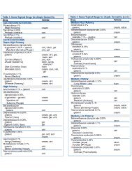
TABLE III. Recommended doses of corticosteroids for EoE<br />
Topical swallowed corticosteroids<br />
Initial doses (see references for preparation and administration<br />
information)<br />
Fluticasone (puffed and swallowed through a metered-dose inhaler)<br />
Adults: 440-880 mg twice daily<br />
Children: 88-440 mg twice to 4 times daily (to a maximal adult dose)<br />
Budesonide (as a viscous suspension)<br />
Children (
12 LIACOURAS ET AL<br />
J ALLERGY CLIN IMMUNOL<br />
nnn 2011<br />
Committee clinical recommendations. Topical corticosteroid<br />
therapy should be considered in all children and adults<br />
given a diagnosis of EoE for both initial and maintenance therapy<br />
(Table III). The type and duration of steroid therapy depends on<br />
the disease severity, the patient’s lifestyle, the ability of the<br />
patient to continue the medication, and family resources. Doses<br />
of various steroid preparations are listed in Table III. Clinical<br />
experience and concern for ongoing symptoms, esophageal inflammation,<br />
and complications of untreated disease has led to<br />
the recommendation that after induction of clinicopathologic<br />
remission, topical corticosteroid therapy might need to be maintained;<br />
however, long-term therapy must be individualized for<br />
each patient. When topical steroids are used chronically, in addition<br />
to observing for side effects, growth should be carefully monitored<br />
in children.<br />
Committee future recommendations. Further studies<br />
are needed to clarify the specifics of topical steroid therapy. Most<br />
importantly, investigation needs to be performed regarding the<br />
most effective topical steroid dose required for initial disease<br />
treatment and maintenance therapy for both children and adults.<br />
In addition, further research needs to be performed with regard to<br />
steroid resistance, the effect of corticosteroids on esophageal<br />
fibrosis, the need to treat to histologic normalcy, quality-of-life<br />
issues, growth, consequences of prolonged use (eg, adrenal<br />
suppression), and steroid effects on bone density.<br />
Cromolyn sodium, leukotriene receptor<br />
antagonists, biologics, and other therapies<br />
Update of 2007 recommendations. No additional information<br />
has been reported with regard to the use of cromolyn<br />
sodium or leukotriene receptor antagonists in patients with EoE.<br />
Cromolyn sodium has no apparent therapeutic benefit for patients<br />
with EoE. Leukotriene receptor antagonists might induce symptomatic<br />
relief when given at high dosages; its use has no<br />
demonstrable effect on esophageal eosinophilia. 141 In a single<br />
study anti–TNF-a had no benefit in patients with EoE. 142 A few<br />
studies in a small number of patients have been published using<br />
anti–IL-5. The studies demonstrated a significant decrease in<br />
esophageal eosinophil numbers and improvement in a few parameters<br />
of esophageal remodeling; however, the clinical response<br />
was variable. 40,143<br />
Committee clinical recommendations. Treatment of<br />
EoE with cromolyn sodium, leukotriene receptor antagonists, and<br />
immunosuppressive agents (azathioprine or 6-mercaptopurine) for<br />
the treatment of EoE is not recommended. The lack of combined<br />
clinical and histologic benefit and potential side effects currently<br />
outweigh any potential benefit. Biologicagentsawait further clinical<br />
studies and are not recommended for routine use at the present<br />
time. 144 Several additional studies on anti–IL-5 therapy are pending.<br />
Committee future recommendations. Other potential<br />
agents for the treatment of EoE should be investigated. Potential<br />
future treatments include immunosuppressive agents and the use<br />
of mAbs, such as anti–IL-5, anti–IL-13, and anti-eotaxin. In<br />
addition, the study of serum biochemical markers might aid in the<br />
development of potential future therapies.<br />
DISEASE COMPLICATIONS<br />
Update of 2007 recommendations<br />
In the 2007 CR, little information was available regarding the<br />
definitions, prevalence, and management of EoE complications,<br />
including food impaction, esophageal stricture, narrow-caliber<br />
esophagus, and esophageal perforation. The understanding of<br />
EoE complications is hindered by the lack of uniform definitions<br />
for these well-recognized problems. The wide ranges of reported<br />
prevalence figures reflect not only variability of definitions of<br />
these complications but also differences in patient selection,<br />
period of follow-up, and treatment patterns.<br />
Food impaction was defined as an event occurring after food<br />
ingestion, where food matter is retained in the esophagus,<br />
requiring either an emergency physician visit or endoscopic<br />
intervention. Self-limited food retention or transient dysphagia<br />
after eating not requiring intervention would not qualify as food<br />
impaction in this definition. Although such a definition overlooks<br />
clinically significant but self-resolving impaction, the committee<br />
considered it preferable to a definition that arbitrarily designates<br />
duration or severity of symptoms or other less quantifiable<br />
outcome. By using the definition of food retention requiring<br />
endoscopic extraction, the prevalence of food impaction among<br />
larger adult series of patients with EoE (>50 subjects) ranged<br />
from 30% to 55%. 3,53,145,146 In addition to these cohorts, investigators<br />
have assessed patients presenting with esophageal food impaction<br />
to define the proportion of impactions occurring<br />
secondary to EoE. Adult series have reported that 11% to 55%<br />
of food impactions demonstrated EoE. 61,147,148<br />
Stricture definition is problematic, given that esophageal rings<br />
are a common manifestation of the disease state in adults and<br />
because rings imply some degree of esophageal stricture. Similar<br />
to Schatzki rings, the rings in patients with EoE have an axial<br />
length of less than a few millimeters. Strictures might be arbitrarily<br />
differentiated by a length of greater than 1 cm. The committee<br />
considered rings and strictures as distinct entities, as reported in<br />
the literature. With respect to narrow-caliber esophagus, this is<br />
differentiated from a stricture by the generalized involvement of<br />
the majority of the esophagus. Although a radiographic definition<br />
might be preferable to endoscopic assessment, most reports of this<br />
outcome used a subjective endoscopic impression to classify<br />
patients. Stricture prevalence in larger adult series ranged from<br />
11% to 31%. 3,53,71,145,149-151 Narrow-caliber esophagus has a reported<br />
prevalence of approximately 10% in larger series. 3,115,145<br />
The definition of esophageal injury patterns should be examined<br />
on the basis of the following suggested classification scheme.<br />
A full perforation or Boerhaave syndrome is characterized by a<br />
full-thickness tear with esophageal or gastric contents in the chest<br />
cavity that requires surgical treatment, usually with thoracotomy,<br />
drainage, repair, and possible resection. A partial rupture is<br />
defined by limited air or contrast extravasation into the mediastinum,<br />
often manageable by using a conservative approach.<br />
Esophageal intramural tears are identified endoscopically as<br />
deep lacerations extending into the esophageal submucosa or<br />
radiographically by contrast extending outside the esophageal<br />
lumen but contained within the esophageal wall. Circumferential<br />
intramural dissection is defined by circular detachment of the<br />
esophageal mucosa and not associated with leakage of esophageal<br />
contents into the mediastinum or thoracic cavities.<br />
There have been 19 patients with EoE reported with spontaneous<br />
or non–dilation-related perforation, and of the 14 spontaneous<br />
perforations, there were 2 full perforations and 12 partial<br />
tears. 146,152-159 Five postendoscopy perforations without preceding<br />
esophageal dilation were partial tears. 160-162 Of the 19 perforations<br />
reported, 7 patients required surgical intervention (ie, 4<br />
primary repairs and 3 esophageal resections). None of the
J ALLERGY CLIN IMMUNOL<br />
VOLUME nnn, NUMBER nn<br />
LIACOURAS ET AL 13<br />
reported perforations have been fatal. Three cases of circumferential<br />
intramural dissection and numerous cases of spontaneous and<br />
endoscopy-associated esophageal intramural tears have been<br />
reported. 146,158,159<br />
A number of medical and dietary approaches have been studied<br />
for EoE. Limited data are available on the effect of these therapies<br />
on complications of EoE. The majority of data exist regarding the<br />
effect of topical steroids on the incidence of food impaction in<br />
adults with EoE. In a retrospective study of 21 adults treated with<br />
fluticasone for 6 weeks, the prevalence of food impaction<br />
decreased from 71% to 0 %. 163 A prospective study reported<br />
food impaction in 83% before therapy and 0% after 12 weeks<br />
of fluticasone. 151 The 15-day treatment period in a randomized<br />
controlled trial of budesonide allowed for detection of a significant<br />
improvement in dysphagia but was too short a treatment period<br />
to determine an effect on food impaction. 133 A randomized<br />
controlled trial of fluticasone for 6 weeks reported food impaction<br />
in 81% of patients receiving placebo and 76% of those receiving<br />
fluticasone. After therapy for 6 weeks, no food impactions were<br />
observed in either group. A retrospective study of 32 adults<br />
with EoE who were contacted a mean of 3 years after therapy<br />
with 6 weeks of fluticasone observed recurrent dysphagia in<br />
91% and food impaction in 28%. 139 Available data on systemic<br />
steroids and elimination and elemental diets have been largely<br />
confined to pediatric studies in which the baseline prevalence of<br />
food impaction was either low (
14 LIACOURAS ET AL<br />
J ALLERGY CLIN IMMUNOL<br />
nnn 2011<br />
TABLE IV. Major unresolved issues affecting the diagnosis and treatment of EoE<br />
1. Optimization of methodologies for histologic analysis of mucosal samples<br />
A. Number of biopsy specimens<br />
B. Proximal versus distal biopsy specimens<br />
C. Number of hpfs that need to be examined<br />
D. Effect of associated histologic features on diagnostic sensitivity<br />
E. Mean versus peak number of eosinophils<br />
F. Size of hpf used to quantitate eosinophils<br />
G. Use of completely filled versus partially filled hpfs<br />
H. Reporting eosinophil density as number per hpf versus number per unit area (mm 2 )<br />
I. Eosinophil degranulation and optimal markers (MBP, EPO, and EDN)<br />
2. Exclusion of esophageal acid/nonacid disease (PPI therapy and diagnostic testing)<br />
3. Lack of <strong>consensus</strong> on criteria to diagnose eosinophilic disease in the remainder of the gastrointestinal tract<br />
4. Allergy testing in adults and children in guiding dietary exclusions<br />
5. Optimal end points of treatment (eg, symptom relief and histologic normalcy)<br />
6. Frequency of endoscopy in follow-up (Is it needed in asymptomatic patients)<br />
7. Maintenance treatment (dose and duration)<br />
8. Validated measurements of symptoms, endoscopic findings, histology, and quality of life<br />
EDN, Eosinophil-derived neurotoxin; EPO, eosinophil peroxidase; MBP, major basic protein.<br />
for patients with EoE compared with those with other benign<br />
entities, such as Schatzki ring or peptic stricture. Techniques<br />
described for esophageal dilation in patients with EoE include the<br />
use of both through-the-scope and bougie dilators. Bougies might<br />
be preferred because the esophageal lumen can be narrowed in<br />
multiple sites and sometimes diffusely. Advocates of through-thescope<br />
balloons point to 2 theoretical advantages: they apply<br />
exclusively radial force during dilation and afford the ability to<br />
immediately assess esophageal injury. The practice of gradual<br />
esophageal dilation with a target goal of 15 to 18 mm and limiting<br />
the progression of dilation diameter per session to 3 mm or less<br />
after resistance is encountered is reasonable but has not been<br />
specifically addressed in patients with EoE. Furthermore, this<br />
dilation approach has not been validated and applies to bougie and<br />
not through-the-scope balloon dilation. Multiple dilation sessions<br />
are often required for high-grade esophageal stenosis in patients<br />
with EoE. Monitoring progressive dilations during a single<br />
session with repeat endoscopy to examine for mucosal tears is<br />
controversial. This is more readily achieved with through-thescope<br />
balloons and more cumbersome with bougie dilation.<br />
Complications of dilation can be associated with younger age<br />
and more dilations, narrowing in the upper third of the esophagus,<br />
and inability to traverse the narrowing with the scope before<br />
dilation. 170,171 These predictors of risk were largely based on inclusion<br />
of esophageal intramural tears as a complication. Although<br />
such tears have been described in patients with EoE,<br />
there is no information that suggests that these tears represent<br />
anything more than a successful dilation.<br />
Committee future recommendations<br />
Prospective studies are needed to define the role of esophageal<br />
dilation in the management of EoE. Such studies will help define<br />
technical factors to optimize response and minimize risk of<br />
dilation. Studies are also needed to assess the degree of stricture<br />
response to medical and dietary therapy of EoE that would<br />
obviate the need for esophageal dilation.<br />
CONCLUSIONS AND FUTURE GOALS<br />
Since the initial set of recommendations and guidelines<br />
published in 2007, there has been a significant increase in<br />
clinical experience and the number of publications on the<br />
subject of EoE. However, despite the growing interest and everincreasing<br />
body of literature, a great deal remains unknown<br />
(Table IV). Because of the confusion between esophageal eosinophilia<br />
and EoE, we provided both a conceptual definition<br />
of and diagnostic guidelines for EoE to assist clinicians in identifying<br />
the differences between histologic findings and the disease<br />
process. In addition, we anticipate that this definition and<br />
guideline will help to shape future research focusing on critical<br />
clinical and pathogenetic questions. As clinical experience<br />
broadens and research discovers more intriguing features of<br />
this disease, modifications of the presented definition and<br />
guideline are inevitable. Future challenges include developing<br />
appropriate translational studies that will allow definition of<br />
EoE phenotypic subsets. Of importance is the need to differentiate<br />
EoE from GERD, which has caused much controversy.<br />
Other important questions that mandate future research surround<br />
allergic testing, treatment approaches, the genetic basis<br />
of the disease, and diagnostic criteria, including molecular<br />
markers, the use of dilation, and the management of the complications<br />
of EoE.<br />
The joint effort of pediatric and adult clinical and basic<br />
scientists in a variety of subspecialties has been paramount in<br />
the rapid understanding of this disease process. It is critical that<br />
leaders from all of the above specialties continue to work<br />
together and undertake studies on natural history, pathophysiology,<br />
biomarkers, diagnosis, and therapeutic approaches not<br />
only to increase the scientific and clinical knowledge of EoE but<br />
also to improve the lives of children and adults affected by the<br />
disease.<br />
We thank Wendy Moore, MS, for her outstanding technical and administrative<br />
support and Mark Holbreich, MD, for his thoughtful and timely review of the<br />
manuscript. We also thank the following groups and persons for their ongoing<br />
dedication and support for the manuscript, patient advocacy, education, and/or<br />
research for eosinophilic <strong>esophagitis</strong>: the American Academy of Allergy,<br />
Asthma & Immunology (www.aaaai.org); the American Gastroenterological<br />
Association (www.gastro.org); the American Partnership for <strong>Eosinophilic</strong> Disorders<br />
(www.apfed.org); the Bunning Family; the Children’s Digestive Health<br />
and Nutrition Foundation (www.cdhnf.org); the Campaign for Urging Research<br />
for <strong>Eosinophilic</strong> Disease (www.curedfoundation.org); the Food Allergy Initiative;<br />
the North American Society of Pediatric Gastroenterology, Hepatology
J ALLERGY CLIN IMMUNOL<br />
VOLUME nnn, NUMBER nn<br />
LIACOURAS ET AL 15<br />
and Nutrition (www.naspghan.org); the National Institutes of Health (www.nih.<br />
gov); and the International Gastrointestinal Eosinophil Researchers (www.tigeregid.cdhnf.org).<br />
Finally, we are extremely grateful to all of the patients and<br />
families for their participation and support of EoE research.<br />
What do we know<br />
d EoE is a clinicopathologic disease isolated to the esophagus.<br />
d EoE represents a chronic, immune/antigen-mediated,<br />
esophageal disease characterized clinically by symptoms<br />
related to esophageal dysfunction and histologically by<br />
eosinophil-predominant inflammation.<br />
d With few exceptions, 15 eosinophils/hpf (peak value) is<br />
considered a minimum threshold for a diagnosis of EoE.<br />
d Endoscopy with biopsy is currently the only reliable diagnostic<br />
test for EoE.<br />
d An allergy evaluation is warranted in patients given a diagnosis<br />
of EoE.<br />
d The disease should remit with treatments of dietary exclusion,<br />
topical corticosteroids, or both.<br />
What is still unknown<br />
d Pathophysiology of PPI-responsive esophageal eosinophilia<br />
d Pathognomonic features that define EoE<br />
d Optimal histologic descriptive elements that characterize<br />
EoE<br />
d Biomarkers and molecular signatures that aid in the diagnosis<br />
of EoE<br />
d Therapeutic/predictive accuracy of skin prick and patch<br />
testing in patients with EoE<br />
d Therapeutic value of dietary therapy in adults with EoE<br />
d Natural history of EoE and rates and predictive indexes<br />
of complications (food impaction, esophageal stricture,<br />
and esophageal narrowing)<br />
d Importance of treating asymptomatic patients with isolated<br />
esophageal eosinophilia<br />
REFERENCES<br />
1. Furuta GT, Liacouras CA, Collins MH, Gupta SK, Justinich C, Putnam PE, et al.<br />
<strong>Eosinophilic</strong> <strong>esophagitis</strong> in children and adults: a systematic review and <strong>consensus</strong><br />
recommendations for diagnosis and treatment. Gastroenterology 2007;133:<br />
1342-63.<br />
2. Spergel JM, Book WM, Mays E, Song L, Shah SS, Talley NJ, et al. Variation in<br />
prevalence, diagnostic criteria, and initial management options for eosinophilic<br />
gastrointestinal diseases in the United States. J Pediatr Gastroenterol Nutr<br />
2011;52:300-6.<br />
3. Dellon ES, Gibbs WB, Fritchie KJ, Rubinas TC, Wilson LA, Woosley JT, et al.<br />
Clinical, endoscopic, and histologic findings distinguish eosinophilic <strong>esophagitis</strong><br />
from gastroesophageal reflux disease. Clin Gastroenterol Hepatol 2009;7:<br />
1305-13; quiz 1261.<br />
4. Ngo P, Furuta GT, Antonioli DA, Fox VL. Eosinophils in the esophagus—peptic<br />
or allergic eosinophilic <strong>esophagitis</strong> Case series of three patients with esophageal<br />
eosinophilia. Am J Gastroenterol 2006;101:1666-70.<br />
5. Rodrigo S, Abboud G, Oh D, DeMeester SR, Hagen J, Lipham J, et al. High intraepithelial<br />
eosinophil counts in esophageal squamous epithelium are not specific<br />
for eosinophilic <strong>esophagitis</strong> in adults. Am J Gastroenterol 2008;103:435-42.<br />
6. Molina-Infante J, Ferrando-Lamana L, Ripoll C, Hernandez-Alonso M, Mateos<br />
JM, Fernandez-Bermejo M, et al. Esophageal eosinophilic infiltration responds to<br />
proton pump inhibition in most adults. Clin Gastroenterol Hepatol 2011;9:110-7.<br />
7. Dellon ES, Fritchie KJ, Rubinas TC, Woosley JT, Shaheen NJ. Inter- and intraobserver<br />
reliability and validation of a new method for determination of eosinophil<br />
counts in patients with esophageal eosinophilia. Dig Dis Sci 2010;55:1940-9.<br />
8. Odze RD. Pathology of eosinophilic <strong>esophagitis</strong>: what the clinician needs to<br />
know. Am J Gastroenterol 2009;104:485-90.<br />
9. Spechler SJ, Genta RM, Souza RF. Thoughts on the complex relationship between<br />
gastroesophageal reflux disease and eosinophilic <strong>esophagitis</strong>. Am J Gastroenterol<br />
2007;102:1301-6.<br />
10. Molina-Infante J, Ferrando-Lamana L, Mateos-Rodriguez JM, Perez-Gallardo B,<br />
Prieto-Bermejo AB. Overlap of reflux and eosinophilic <strong>esophagitis</strong> in two patients<br />
requiring different therapies: a review of the literature. World J Gastroenterol<br />
2008;14:1463-6.<br />
11. Mishra A, Hogan SP, Brandt EB, Rothenberg ME. IL-5 promotes eosinophil trafficking<br />
to the esophagus. J Immunol 2002;168:2464-9.<br />
12. Mishra A, Rothenberg ME. Intratracheal IL-13 induces eosinophilic <strong>esophagitis</strong><br />
by an IL-5, eotaxin-1, and STAT6-dependent mechanism. Gastroenterology<br />
2003;125:1419-27.<br />
13. Akei HS, Mishra A, Blanchard C, Rothenberg ME. Epicutaneous antigen exposure<br />
primes for experimental eosinophilic <strong>esophagitis</strong> in mice. Gastroenterology<br />
2005;129:985-94.<br />
14. Blanchard C, Mishra A, Saito-Akei H, Monk P, Anderson I, Rothenberg ME. Inhibition<br />
of human interleukin-13-induced respiratory and oesophageal inflammation<br />
by anti-human-interleukin-13 antibody (CAT-354). Clin Exp Allergy 2005;<br />
35:1096-103.<br />
15. Spergel JM. <strong>Eosinophilic</strong> o<strong>esophagitis</strong> and pollen. Clin Exp Allergy 2005;35:<br />
1421-2.<br />
16. Spergel JM, Andrews T, Brown-Whitehorn TF, Beausoleil JL, Liacouras CA.<br />
Treatment of eosinophilic <strong>esophagitis</strong> with specific food elimination diet directed<br />
by a combination of skin prick and patch tests. Ann Allergy Asthma Immunol<br />
2005;95:336-43.<br />
17. Blanchard C, Wang N, Stringer KF, Mishra A, Fulkerson PC, Abonia JP, et al.<br />
Eotaxin-3 and a uniquely conserved gene-expression profile in eosinophilic<br />
<strong>esophagitis</strong>. J Clin Invest 2006;116:536-47.<br />
18. Gupta SK, Fitzgerald JF, Kondratyuk T, HogenEsch H. Cytokine expression in<br />
normal and inflamed esophageal mucosa: a study into the pathogenesis of allergic<br />
eosinophilic <strong>esophagitis</strong>. J Pediatr Gastroenterol Nutr 2006;42:22-6.<br />
19. Kagalwalla AF, Sentongo TA, Ritz S, Hess T, Nelson SP, Emerick KM, et al. Effect<br />
of six-food elimination diet on clinical and histologic outcomes in eosinophilic<br />
<strong>esophagitis</strong>. Clin Gastroenterol Hepatol 2006;4:1097-102.<br />
20. Yamazaki K, Murray JA, Arora AS, Alexander JA, Smyrk TC, Butterfield JH,<br />
et al. Allergen-specific in vitro cytokine production in adult patients with eosinophilic<br />
<strong>esophagitis</strong>. Dig Dis Sci 2006;51:1934-41.<br />
21. Bhattacharya B, Carlsten J, Sabo E, Kethu S, Meitner P, Tavares R, et al. Increased<br />
expression of eotaxin-3 distinguishes between eosinophilic <strong>esophagitis</strong><br />
and gastroesophageal reflux disease. Hum Pathol 2007;38:1744-53.<br />
22. Blanchard C, Mingler MK, Vicario M, Abonia JP, Wu YY, Lu TX, et al. IL-13<br />
involvement in eosinophilic <strong>esophagitis</strong>: transcriptome analysis and reversibility<br />
with glucocorticoids. J Allergy Clin Immunol 2007;120:1292-300.<br />
23. Bullock JZ, Villanueva JM, Blanchard C, Filipovich AH, Putnam PE, Collins<br />
MH, et al. Interplay of adaptive th2 immunity with eotaxin-3/c-C chemokine<br />
receptor 3 in eosinophilic <strong>esophagitis</strong>. J Pediatr Gastroenterol Nutr 2007;45:<br />
22-31.<br />
24. Kirsch R, Bokhary R, Marcon MA, Cutz E. Activated mucosal mast cells differentiate<br />
eosinophilic (allergic) <strong>esophagitis</strong> from gastroesophageal reflux disease.<br />
J Pediatr Gastroenterol Nutr 2007;44:20-6.<br />
25. Spergel JM, Brown-Whitehorn T, Beausoleil JL, Shuker M, Liacouras CA. Predictive<br />
values for skin prick test and atopy patch test for eosinophilic <strong>esophagitis</strong>.<br />
J Allergy Clin Immunol 2007;119:509-11.<br />
26. Lucendo AJ, De Rezende L, Comas C, Caballero T, Bellon T. Treatment with topical<br />
steroids downregulates IL-5, eotaxin-1/CCL11, and eotaxin-3/CCL26 gene<br />
expression in eosinophilic <strong>esophagitis</strong>. Am J Gastroenterol 2008;103:2184-93.<br />
27. Mishra A, Wang M, Pemmaraju VR, Collins MH, Fulkerson PC, Abonia JP, et al.<br />
Esophageal remodeling develops as a consequence of tissue specific IL-5-induced<br />
eosinophilia. Gastroenterology 2008;134:204-14.<br />
28. Rothenberg ME. Biology and treatment of eosinophilic <strong>esophagitis</strong>. Gastroenterology<br />
2009;137:1238-49.<br />
29. Tantibhaedhyangkul U, Tatevian N, Gilger MA, Major AM, Davis CM. Increased<br />
esophageal regulatory T cells and eosinophil characteristics in children with eosinophilic<br />
<strong>esophagitis</strong> and gastroesophageal reflux disease. Ann Clin Lab Sci<br />
2009;39:99-107.<br />
30. Zhu X, Wang M, Crump CH, Mishra A. An imbalance of esophageal effector and<br />
regulatory T cell subsets in experimental eosinophilic <strong>esophagitis</strong> in mice. Am J<br />
Physiol Gastrointest Liver Physiol 2009;297:G550-8.<br />
31. Aceves SS, Chen D, Newbury RO, Dohil R, Bastian JF, Broide DH. Mast cells<br />
infiltrate the esophageal smooth muscle in patients with eosinophilic <strong>esophagitis</strong>,<br />
express TGF-beta1, and increase esophageal smooth muscle contraction.<br />
J Allergy Clin Immunol 2010;126:1198-204, e4.
16 LIACOURAS ET AL<br />
J ALLERGY CLIN IMMUNOL<br />
nnn 2011<br />
32. Blanchard C, Stucke EM, Burwinkel K, Caldwell JM, Collins MH, Ahrens A,<br />
et al. Coordinate interaction between IL-13 and epithelial differentiation cluster<br />
genes in eosinophilic <strong>esophagitis</strong>. J Immunol 2010;184:4033-41.<br />
33. Caldwell JM, Blanchard C, Collins MH, Putnam PE, Kaul A, Aceves SS, et al.<br />
Glucocorticoid-regulated genes in eosinophilic <strong>esophagitis</strong>: a role for FKBP51.<br />
J Allergy Clin Immunol 2010;125:879-88, e8.<br />
34. Dellon ES, Chen X, Miller CR, Fritchie KJ, Rubinas TC, Woosley JT, et al. Tryptase<br />
staining of mast cells may differentiate eosinophilic <strong>esophagitis</strong> from gastroesophageal<br />
reflux disease. Am J Gastroenterol 2011;106:264-71.<br />
35. Fuentebella J, Patel A, Nguyen T, Sanjanwala B, Berquist W, Kerner JA, et al.<br />
Increased number of regulatory T cells in children with eosinophilic <strong>esophagitis</strong>.<br />
J Pediatr Gastroenterol Nutr 2010;51:283-9.<br />
36. Neilsen CV, Bryce PJ. Interleukin-13 directly promotes oesophagus production of<br />
CCL11 and CCL24 and the migration of eosinophils. Clin Exp Allergy 2010;40:<br />
427-34.<br />
37. Patel AJ, Fuentebella J, Gernez Y, Nguyen T, Bass D, Berquist W, et al. Increased<br />
HLA-DR expression on tissue eosinophils in eosinophilic <strong>esophagitis</strong>. J Pediatr<br />
Gastroenterol Nutr 2010;51:290-4.<br />
38. Rothenberg ME, Spergel JM, Sherrill JD, Annaiah K, Martin LJ, Cianferoni A,<br />
et al. Common variants at 5q22 associate with pediatric eosinophilic <strong>esophagitis</strong>.<br />
Nat Genet 2010;42:289-91.<br />
39. Sherrill JD, Gao PS, Stucke EM, Blanchard C, Collins MH, Putnam PE, et al.<br />
Variants of thymic stromal lymphopoietin and its receptor associate with eosinophilic<br />
<strong>esophagitis</strong>. J Allergy Clin Immunol 2010;126:160-5, e3.<br />
40. Straumann A, Conus S, Grzonka P, Kita H, Kephart G, Bussmann C, et al. Antiinterleukin-5<br />
antibody treatment (mepolizumab) in active eosinophilic o<strong>esophagitis</strong>:<br />
a randomised, placebo-controlled, double-blind trial. Gut 2010;59:21-30.<br />
41. Vicario M, Blanchard C, Stringer KF, Collins MH, Mingler MK, Ahrens A, et al.<br />
Local B cells and IgE production in the oesophageal mucosa in eosinophilic o<strong>esophagitis</strong>.<br />
Gut 2010;59:12-20.<br />
42. Yen EH, Hornick JL, Dehlink E, Dokter M, Baker A, Fiebiger E, et al. Comparative<br />
analysis of FcepsilonRI expression patterns in patients with eosinophilic and<br />
reflux <strong>esophagitis</strong>. J Pediatr Gastroenterol Nutr 2010;51:584-92.<br />
43. Zhu X, Wang M, Mavi P, Rayapudi M, Pandey AK, Kaul A, et al. Interleukin-15<br />
expression is increased in human eosinophilic <strong>esophagitis</strong> and mediates pathogenesis<br />
in mice. Gastroenterology 2010;139:182-93, e7.<br />
44. Zuo L, Fulkerson PC, Finkelman FD, Mingler M, Fischetti CA, Blanchard C, et al. IL-<br />
13 induces esophageal remodeling and gene expression by an eosinophilindependent,<br />
IL-13R alpha2-inhibited pathway. J Immunol 2010;185:660-9.<br />
45. Frischmeyer-Guerrerio PA, Guerrerio AL, Chichester KL, Bieneman AP, Hamilton<br />
RA, Wood RA, et al. Dendritic cell and T cell responses in children with food<br />
allergy. Clin Exp Allergy 2011;41:61-71.<br />
46. Aceves SS, Newbury RO, Dohil MA, Bastian JF, Dohil R. A symptom scoring tool<br />
for identifying pediatric patients with eosinophilic <strong>esophagitis</strong> and correlating<br />
symptoms with inflammation. Ann Allergy Asthma Immunol 2009;103:401-6.<br />
47. Mukkada VA, Haas A, Maune NC, Capocelli KE, Henry M, Gilman N, et al.<br />
Feeding dysfunction in children with eosinophilic gastrointestinal diseases. Pediatrics<br />
2010;126:e672-7.<br />
48. Garcia-Compean D, Gonzalez Gonzalez JA, Marrufo Garcia CA, Flores Gutierrez<br />
JP, Barboza Quintana O, Galindo Rodriguez G, et al. Prevalence of eosinophilic<br />
<strong>esophagitis</strong> in patients with refractory gastroesophageal reflux disease symptoms:<br />
a prospective study. Dig Liver Dis 2011;43:204-8.<br />
49. Kapel RC, Miller JK, Torres C, Aksoy S, Lash R, Katzka DA. <strong>Eosinophilic</strong><br />
<strong>esophagitis</strong>: a prevalent disease in the United States that affects all age groups.<br />
Gastroenterology 2008;134:1316-21.<br />
50. Foroutan M, Norouzi A, Molaei M, Mirbagheri SA, Irvani S, Sadeghi A, et al.<br />
<strong>Eosinophilic</strong> <strong>esophagitis</strong> in patients with refractory gastroesophageal reflux disease.<br />
Dig Dis Sci 2010;55:28-31.<br />
51. Lai AL, Girgis S, Liang Y, Carr S, Huynh HQ. Diagnostic criteria for eosinophilic<br />
<strong>esophagitis</strong>: a 5-year retrospective review in a pediatric population. J Pediatr Gastroenterol<br />
Nutr 2009;49:63-70.<br />
52. Ronkainen J, Talley NJ, Aro P, Storskrubb T, Johansson SE, Lind T, et al. Prevalence<br />
of oesophageal eosinophils and eosinophilic o<strong>esophagitis</strong> in adults: the<br />
population-based Kalixanda study. Gut 2007;56:615-20.<br />
53. Prasad GA, Alexander JA, Schleck CD, Zinsmeister AR, Smyrk TC, Elias RM,<br />
et al. Epidemiology of eosinophilic <strong>esophagitis</strong> over three decades in Olmsted<br />
County, Minnesota. Clin Gastroenterol Hepatol 2009;7:1055-61.<br />
54. Dalby K, Nielsen RG, Kruse-Andersen S, Fenger C, Bindslev-Jensen C, Ljungberg<br />
S, et al. <strong>Eosinophilic</strong> o<strong>esophagitis</strong> in infants and children in the region of<br />
southern Denmark: a prospective study of prevalence and clinical presentation.<br />
J Pediatr Gastroenterol Nutr 2010;51:280-2.<br />
55. Bove M, Tegtmeyer B, Persson S, Bergquist H. The pharyngeal mucosa is not<br />
involved in eosinophilic o<strong>esophagitis</strong>. Aliment Pharmacol Ther 2009;30:<br />
495-500.<br />
56. Mackenzie SH, Go M, Chadwick B, Thomas K, Fang J, Kuwada S, et al. <strong>Eosinophilic</strong><br />
o<strong>esophagitis</strong> in patients presenting with dysphagia—a prospective analysis.<br />
Aliment Pharmacol Ther 2008;28:1140-6.<br />
57. Prasad GA, Talley NJ, Romero Y, Arora AS, Kryzer LA, Smyrk TC, et al. Prevalence<br />
and predictive factors of eosinophilic <strong>esophagitis</strong> in patients presenting<br />
with dysphagia: a prospective study. Am J Gastroenterol 2007;102:2627-32.<br />
58. Poh CH, Gasiorowska A, Navarro-Rodriguez T, Willis MR, Hargadon D, Noelck<br />
N, et al. Upper GI tract findings in patients with heartburn in whom proton pump<br />
inhibitor treatment failed versus those not receiving antireflux treatment. Gastrointest<br />
Endosc 2010;71:28-34.<br />
59. Krarup AL, Villadsen GE, Mejlgaard E, Olesen SS, Drewes AM, Funch-Jensen P.<br />
Acid hypersensitivity in patients with eosinophilic o<strong>esophagitis</strong>. Scand J Gastroenterol<br />
2010;45:273-81.<br />
60. Straumann A. The natural history and complications of eosinophilic <strong>esophagitis</strong>.<br />
Gastrointest Endosc Clin N Am 2008;18:99-118, ix.<br />
61. Desai TK, Stecevic V, Chang CH, Goldstein NS, Badizadegan K, Furuta GT. Association<br />
of eosinophilic inflammation with esophageal food impaction in adults.<br />
Gastrointest Endosc 2005;61:795-801.<br />
62. Peterson KA, Thomas KL, Hilden K, Emerson LL, Wills JC, Fang JC. Comparison<br />
of esomeprazole to aerosolized, swallowed fluticasone for eosinophilic<br />
<strong>esophagitis</strong>. Dig Dis Sci 2010;55:1313-9.<br />
63. Sayej WN, Patel R, Baker RD, Tron E, Baker SS. Treatment with high-dose proton<br />
pump inhibitors helps distinguish eosinophilic <strong>esophagitis</strong> from noneosinophilic<br />
<strong>esophagitis</strong>. J Pediatr Gastroenterol Nutr 2009;49:393-9.<br />
64. Kedika RR, Souza RF, Spechler SJ. Potential anti-inflammatory effects of proton<br />
pump inhibitors: a review and discussion of the clinical implications. Dig Dis Sci<br />
2009;54:2312-7.<br />
65. Merwat SN, Spechler SJ. Might the use of acid-suppressive medications predispose<br />
to the development of eosinophilic <strong>esophagitis</strong> Am J Gastroenterol 2009;<br />
104:1897-902.<br />
66. Schlesinger PK, Donahue PE, Schmid B, Layden TJ. Limitations of 24-hour intraesophageal<br />
pH monitoring in the hospital setting. Gastroenterology 1985;89:<br />
797-804.<br />
67. Murphy DW, Yuan Y, Castell DO. Does the intraesophageal pH probe accurately<br />
detect acid reflux Simultaneous recording with two pH probes in humans. Dig<br />
Dis Sci 1989;34:649-56.<br />
68. Aceves SS, Ackerman SJ. Relationships between eosinophilic inflammation, tissue<br />
remodeling, and fibrosis in eosinophilic <strong>esophagitis</strong>. Immunol Allergy Clin<br />
North Am 2009;29:197-211, xiii-xiv.<br />
69. Aceves SS, Newbury RO, Chen D, Mueller J, Dohil R, Hoffman H, et al. Resolution<br />
of remodeling in eosinophilic <strong>esophagitis</strong> correlates with epithelial response<br />
to topical corticosteroids. Allergy 2010;65:109-16.<br />
70. Pentiuk S, Putnam PE, Collins MH, Rothenberg ME. Dissociation between symptoms<br />
and histological severity in pediatric eosinophilic <strong>esophagitis</strong>. J Pediatr Gastroenterol<br />
Nutr 2009;48:152-60.<br />
71. Schoepfer AM, Gonsalves N, Bussmann C, Conus S, Simon HU, Straumann A,<br />
et al. Esophageal dilation in eosinophilic <strong>esophagitis</strong>: effectiveness, safety,<br />
and impact on the underlying inflammation. Am J Gastroenterol 2010;105:<br />
1062-70.<br />
72. Baxi S, Gupta SK, Swigonski N, Fitzgerald JF. Clinical presentation of patients<br />
with eosinophilic inflammation of the esophagus. Gastrointest Endosc 2006;64:<br />
473-8.<br />
73. Shah A, Kagalwalla AF, Gonsalves N, Melin-Aldana H, Li BU, Hirano I. Histopathologic<br />
Variability in Children With <strong>Eosinophilic</strong> Esophagitis. Am J Gastroenterol<br />
2009;104:716-21.<br />
74. Yantiss RK, Odze RD. Optimal approach to obtaining mucosal biopsies for assessment<br />
of inflammatory disorders of the gastrointestinal tract. Am J Gastroenterol<br />
2009;104:774-83.<br />
75. Binkovitz LA, Lorenz EA, Di Lorenzo C, Kahwash S. Pediatric eosinophilic<br />
<strong>esophagitis</strong>: radiologic findings with pathologic correlation. Pediatr Radiol<br />
2010;40:714-9.<br />
76. DeBrosse CW, Collins MH, Buckmeier Butz BK, Allen CL, King EC, Assa’ad<br />
AH, et al. Identification, epidemiology, and chronicity of pediatric esophageal eosinophilia,<br />
1982-1999. J Allergy Clin Immunol 2010;126:112-9.<br />
77. Aceves SS, Newbury RO, Dohil R, Schwimmer J, Bastian JF. Distinguishing eosinophilic<br />
<strong>esophagitis</strong> in pediatric patients: clinical, endoscopic, and histologic<br />
features of an emerging disorder. J Clin Gastroenterol 2007;41:252-6.<br />
78. Lee S, de Boer WB, Naran A, Leslie C, Raftopoulous S, Ee H, et al. More than<br />
just counting eosinophils: proximal oesophageal involvement and subepithelial<br />
sclerosis are major diagnostic criteria for eosinophilic o<strong>esophagitis</strong>. J Clin Pathol<br />
2010;63:644-7.<br />
79. Liacouras CA, Spergel JM, Ruchelli E, Verma R, Mascarenhas M, Semeao E,<br />
et al. <strong>Eosinophilic</strong> <strong>esophagitis</strong>: a 10-year experience in 381 children. Clin Gastroenterol<br />
Hepatol 2005;3:1198-206.
J ALLERGY CLIN IMMUNOL<br />
VOLUME nnn, NUMBER nn<br />
LIACOURAS ET AL 17<br />
80. Kephart GM, Alexander JA, Arora AS, Romero Y, Smyrk TC, Talley NJ, et al.<br />
Marked deposition of eosinophil-derived neurotoxin in adult patients with eosinophilic<br />
<strong>esophagitis</strong>. Am J Gastroenterol 2010;105:298-307.<br />
81. Mueller S, Neureiter D, Aigner T, Stolte M. Comparison of histological parameters<br />
for the diagnosis of eosinophilic o<strong>esophagitis</strong> versus gastrooesophageal<br />
reflux disease on oesophageal biopsy material. Histopathology<br />
2008;53:676-84.<br />
82. Protheroe C, Woodruff SA, de Petris G, Mukkada V, Ochkur SI, Janarthanan S,<br />
et al. A novel histologic scoring system to evaluate mucosal biopsies from patients<br />
with eosinophilic <strong>esophagitis</strong>. Clin Gastroenterol Hepatol 2009;7:749-55, e11.<br />
83. Dellon ES, Aderoju A, Woosley JT, Sandler RS, Shaheen NJ. Variability in diagnostic<br />
criteria for eosinophilic <strong>esophagitis</strong>: a systematic review. Am J Gastroenterol<br />
2007;102:2300-13.<br />
84. Aceves SS, Newbury RO, Dohil R, Bastian JF, Broide DH. Esophageal remodeling<br />
in pediatric eosinophilic <strong>esophagitis</strong>. J Allergy Clin Immunol 2007;119:<br />
206-12.<br />
85. Chehade M, Sampson HA, Morotti RA, Magid MS. Esophageal subepithelial fibrosis<br />
in children with eosinophilic <strong>esophagitis</strong>. J Pediatr Gastroenterol Nutr<br />
2007;45:319-28.<br />
86. Genevay M, Rubbia-Brandt L, Rougemont AL. Do eosinophil numbers differentiate<br />
eosinophilic <strong>esophagitis</strong> from gastroesophageal reflux disease Arch Pathol<br />
Lab Med 2010;134:815-25.<br />
87. Steiner SJ, Kernek KM, Fitzgerald JF. Severity of basal cell hyperplasia differs in<br />
reflux versus eosinophilic <strong>esophagitis</strong>. J Pediatr Gastroenterol Nutr 2006;42:<br />
506-9.<br />
88. Lucendo AJ, Bellon T, Lucendo B. The role of mast cells in eosinophilic <strong>esophagitis</strong>.<br />
Pediatr Allergy Immunol 2009;20:512-8.<br />
89. Mishra A. Mechanism of eosinophilic <strong>esophagitis</strong>. Immunol Allergy Clin North<br />
Am 2009;29:29-40, viii.<br />
90. Blanchard C, Mingler MK, McBride M, Putnam PE, Collins MH, Chang G, et al.<br />
Periostin facilitates eosinophil tissue infiltration in allergic lung and esophageal<br />
responses. Mucosal Immunol 2008;1:289-96.<br />
91. Bhattacharya B, Carlsten J, Sabo E, Kethu S, Meitner P, Tavares R, et al. Increased<br />
expression of eotaxin-3 distinguishes between eosinophilic <strong>esophagitis</strong><br />
and gastroesophageal reflux disease. Hum Pathol 2007;38:1744-53.<br />
92. Lucendo AJ, Navarro M, Comas C, Pascual JM, Burgos E, Santamaria L, et al.<br />
Immunophenotypic characterization and quantification of the epithelial inflammatory<br />
infiltrate in eosinophilic <strong>esophagitis</strong> through stereology: an analysis of<br />
the cellular mechanisms of the disease and the immunologic capacity of the<br />
esophagus. Am J Surg Pathol 2007;31:598-606.<br />
93. Mulder DJ, Pacheco I, Hurlbut DJ, Mak N, Furuta GT, MacLeod RJ, et al. FGF9-<br />
induced proliferative response to eosinophilic inflammation in o<strong>esophagitis</strong>. Gut<br />
2009;58:166-73.<br />
94. Rogart JN, Nagata J, Loeser CS, Roorda RD, Aslanian H, Robert ME, et al. Multiphoton<br />
imaging can be used for microscopic examination of intact human gastrointestinal<br />
mucosa ex vivo. Clin Gastroenterol Hepatol 2008;6:95-101.<br />
95. Rubio CA, Glaessgen A. An improved method to visualize eosinophils in eosinophilic<br />
<strong>esophagitis</strong>. In Vivo 2006;20:681-5.<br />
96. Savarino E, Tutuian R, Zentilin P, Dulbecco P, Pohl D, Marabotto E, et al. Characteristics<br />
of reflux episodes and symptom association in patients with erosive<br />
<strong>esophagitis</strong> and nonerosive reflux disease: study using combined impedance-pH<br />
off therapy. Am J Gastroenterol 2010;105:1053-61.<br />
97. Dalby K, Nielsen RG, Kruse-Andersen S, Fenger C, Durup J, Husby S. Gastroesophageal<br />
reflux disease and eosinophilic <strong>esophagitis</strong> in infants and children.<br />
A study of esophageal pH, multiple intraluminal impedance and endoscopic ultrasound.<br />
Scand J Gastroenterol 2010;45:1029-35.<br />
98. Rosen R, Furuta G, Fritz J, Donovan K, Nurko S. Role of acid and nonacid reflux<br />
in children with eosinophilic <strong>esophagitis</strong> compared with patients with gastroesophageal<br />
reflux and control patients. J Pediatr Gastroenterol Nutr 2008;46:520-3.<br />
99. Korsapati H, Babaei A, Bhargava V, Dohil R, Quin A, Mittal RK. Dysfunction of<br />
the longitudinal muscles of the oesophagus in eosinophilic o<strong>esophagitis</strong>. Gut<br />
2009;58:1056-62.<br />
100. Nurko S, Rosen R, Furuta GT. Esophageal dysmotility in children with eosinophilic<br />
<strong>esophagitis</strong>: a study using prolonged esophageal manometry. Am J Gastroenterol<br />
2009;104:3050-7.<br />
101. Roman S, Hirano I, Kwiatek MA, Gonsalves N, Chen J, Kahrilas PJ, et al. Manometric<br />
features of eosinophilic <strong>esophagitis</strong> in esophageal pressure topography.<br />
Neurogastroenterol Motil 2011;23:208-e111.<br />
102. Kwiatek MA, Hirano I, Kahrilas PJ, Rothe J, Luger D, Pandolfino JE. Mechanical<br />
properties of the esophagus in eosinophilic <strong>esophagitis</strong>. Gastroenterology 2011;<br />
140:82-90.<br />
103. Blanchard C, Wang N, Stringer KF, Mishra A, Fulkerson PC, Abonia JP, et al.<br />
Eotaxin-3 and a uniquely conserved gene-expression profile in eosinophilic<br />
<strong>esophagitis</strong>. J Clin Invest 2006;116:536-47.<br />
104. Zuo L, Fulkerson PC, Finkelman FD, Mingler M, Fischetti CA, Blanchard C,<br />
et al. IL-13 induces esophageal remodeling and gene expression by an<br />
eosinophil-independent, IL-13R alpha 2-inhibited pathway. J Immunol 2010;<br />
185:660-9.<br />
105. Abonia JP, Blanchard C, Butz BB, Rainey HF, Collins MH, Stringer K, et al. Involvement<br />
of mast cells in eosinophilic <strong>esophagitis</strong>. J Allergy Clin Immunol 2010;<br />
126:140-9.<br />
106. Blanchard C, Wang N, Rothenberg ME. <strong>Eosinophilic</strong> <strong>esophagitis</strong>: pathogenesis,<br />
genetics, and therapy. J Allergy Clin Immunol 2006;118:1054-9.<br />
107. Almansa C, Krishna M, Buchner AM, Ghabril MS, Talley N, DeVault KR, et al.<br />
Seasonal distribution in newly diagnosed cases of eosinophilic <strong>esophagitis</strong> in<br />
adults. Am J Gastroenterol 2009;104:828-33.<br />
108. Moawad FJ, Veerappan GR, Lake JM, Maydonovitch CL, Haymore BR, Kosisky<br />
SE, et al. Correlation between eosinophilic o<strong>esophagitis</strong> and aeroallergens. Aliment<br />
Pharmacol Ther 2010;31:509-15.<br />
109. Roy-Ghanta S, Larosa DF, Katzka DA. Atopic characteristics of adult patients<br />
with eosinophilic <strong>esophagitis</strong>. Clin Gastroenterol Hepatol 2008;6:531-5.<br />
110. Spergel JM, Brown-Whitehorn TF, Beausoleil JL, Franciosi J, Shuker M, Verma<br />
R, et al. 14 years of eosinophilic <strong>esophagitis</strong>: clinical features and prognosis.<br />
J Pediatr Gastroenterol Nutr 2009;48:30-6.<br />
111. Erwin EA, James HR, Gutekunst HM, Russo JM, Kelleher KJ, Platts-Mills TA.<br />
Serum IgE measurement and detection of food allergy in pediatric patients with<br />
eosinophilic <strong>esophagitis</strong>. Ann Allergy Asthma Immunol 2010;104:496-502.<br />
112. Dohil R, Newbury R, Fox L, Bastian J, Aceves S. Oral viscous budesonide is effective<br />
in children with eosinophilic <strong>esophagitis</strong> in a randomized, placebocontrolled<br />
trial. Gastroenterology 2010;139:418-29.<br />
113. Chehade M. IgE and non-IgE-mediated food allergy: treatment in 2007. Curr<br />
Opin Allergy Clin Immunol 2007;7:264-8.<br />
114. Sugnanam KK, Collins JT, Smith PK, Connor F, Lewindon P, Cleghorn G, et al.<br />
Dichotomy of food and inhalant allergen sensitization in eosinophilic <strong>esophagitis</strong>.<br />
Allergy 2007;62:1257-60.<br />
115. Veerappan GR, Perry JL, Duncan TJ, Baker TP, Maydonovitch C, Lake JM,<br />
et al. Prevalence of eosinophilic <strong>esophagitis</strong> in an adult population undergoing<br />
upper endoscopy: a prospective study. Clin Gastroenterol Hepatol 2009;7:420-6,<br />
e1-2.<br />
116. Wang FY, Gupta SK, Fitzgerald JF. Is there a seasonal variation in the incidence<br />
or intensity of allergic eosinophilic <strong>esophagitis</strong> in newly diagnosed children<br />
J Clin Gastroenterol 2007;41:451-3.<br />
117. Mishra A, Hogan SP, Brandt EB, Rothenberg ME. An etiological role for aeroallergens<br />
and eosinophils in experimental <strong>esophagitis</strong>. J Clin Invest 2001;107:<br />
83-90.<br />
118. Rayapudi M, Mavi P, Zhu X, Pandey AK, Abonia JP, Rothenberg ME, et al. Indoor<br />
insect allergens are potent inducers of experimental eosinophilic <strong>esophagitis</strong><br />
in mice. J Leukoc Biol 2010;88:337-46.<br />
119. Chehade M, Sampson HA. Epidemiology and etiology of eosinophilic <strong>esophagitis</strong>.<br />
Gastrointest Endosc Clin N Am 2008;18:33-44, viii.<br />
120. Straumann A, Conus S, Degen L, Felder S, Kummer M, Engel H, et al. Budesonide<br />
is effective in adolescent and adult patients with active eosinophilic <strong>esophagitis</strong>.<br />
Gastroenterology 2010;139:1526-37, e1.<br />
121. Patel AJ, Fuentebella J, Gernez Y, Nguyen T, Bass D, Berquist W, et al. Increased<br />
HLA-DR expression on tissue eosinophils in eosinophilic <strong>esophagitis</strong>. J Pediatr<br />
Gastroenterol Nutr 2010;51:290-4.<br />
122. Huang JJ, Joh JW, Fuentebella J, Patel A, Nguyen T, Seki S, et al. Eotaxin and<br />
FGF enhance signaling through an extracellular signal-related kinase (ERK)-<br />
dependent pathway in the pathogenesis of eosinophilic <strong>esophagitis</strong>. Allergy<br />
Asthma Clin Immunol 2010;6:25.<br />
123. Prussin C, Lee J, Foster B. <strong>Eosinophilic</strong> gastrointestinal disease and peanut allergy<br />
are alternatively associated with IL-51 and IL-5(-) T(H)2 responses.<br />
J Allergy Clin Immunol 2009;124:1326-32, e6.<br />
124. Collins MH, Blanchard C, Abonia JP, Kirby C, Akers R, Wang N, et al. Clinical,<br />
pathologic, and molecular characterization of familial eosinophilic <strong>esophagitis</strong><br />
compared with sporadic cases. Clin Gastroenterol Hepatol 2008;6:621-9.<br />
125. Penfield JD, Lang DM, Goldblum JR, Lopez R, Falk GW. The role of allergy<br />
evaluation in adults with eosinophilic <strong>esophagitis</strong>. J Clin Gastroenterol 2010;<br />
44:22-7.<br />
126. Dalby K, Nielsen RG, Kruse-Andersen S, Fenger C, Bindslev-Jensen C, Ljungberg<br />
S, et al. <strong>Eosinophilic</strong> o<strong>esophagitis</strong> in infants and children in the region of<br />
southern Denmark: a prospective study of prevalence and clinical presentation.<br />
J Pediatr Gastroenterol Nutr 2010;51:280-2.<br />
127. Castellano Mdel R, Cimbollek S, Quiralte J. Defining the role of food allergy in a<br />
population of adult patients with eosinophilic <strong>esophagitis</strong>. Inflamm Allergy Drug<br />
Targets 2010;9:257-62.<br />
128. Barbi E, Gerarduzzi T, Longo G, Ventura A. Fatal allergy as a possible consequence<br />
of long-term elimination diet. Allergy 2004;59:668-9.
18 LIACOURAS ET AL<br />
J ALLERGY CLIN IMMUNOL<br />
nnn 2011<br />
129. Flinterman AE, Knulst AC, Meijer Y, Bruijnzeel-Koomen CA, Pasmans SG.<br />
Acute allergic reactions in children with AEDS after prolonged cow’s milk elimination<br />
diets. Allergy 2006;61:370-4.<br />
130. Boyce JA, Assa’ad A, Burks AW, Jones SM, Sampson HA, Wood RA, et al.<br />
Guidelines for the diagnosis and management of food allergy in the United States:<br />
report of the NIAID-sponsored expert panel. J Allergy Clin Immunol 2010;126<br />
(suppl):S1-58.<br />
131. Boyce JA, Assa’ad A, Burks AW, Jones SM, Sampson HA, Wood RA, et al.<br />
Guidelines for the diagnosis and management of food allergy in the United States:<br />
summary of the NIAID-sponsored expert panel report. J Allergy Clin Immunol<br />
2010;126:1105-18.<br />
132. Dellon ES, Farrell TM, Bozymski EM, Shaheen NJ. Diagnosis of eosinophilic<br />
<strong>esophagitis</strong> after fundoplication for ‘‘refractory reflux’’: implications for preoperative<br />
evaluation. Dis Esophagus 2010;23:191-5.<br />
133. Sayej WN, Patel R, Baker RD, Tron E, Baker SS. Treatment with high-dose proton<br />
pump inhibitors helps distinguish eosinophilic <strong>esophagitis</strong> from noneosinophilic<br />
<strong>esophagitis</strong>. J Pediatr Gastroenterol Nutr 2009;49:393-9.<br />
134. Dranove JE, Horn DS, Davis MA, Kernek KM, Gupta SK. Predictors of response<br />
to proton pump inhibitor therapy among children with significant esophageal eosinophilia.<br />
J Pediatr 2009;154:96-100.<br />
135. Spergel JM. <strong>Eosinophilic</strong> <strong>esophagitis</strong> in adults and children: evidence for a food allergy<br />
component in many patients. Curr Opin Allergy Clin Immunol 2007;7:274-8.<br />
136. Abu-Sultaneh SM, Durst P, Maynard V, Elitsur Y. Fluticasone and food allergen<br />
elimination reverse sub-epithelial fibrosis in children with eosinophilic <strong>esophagitis</strong>.<br />
Dig Dis Sci 2011;56:97-102.<br />
137. Chehade M, Aceves SS. Food allergy and eosinophilic <strong>esophagitis</strong>. Curr Opin Allergy<br />
Clin Immunol 2010;10:231-7.<br />
138. Konikoff MR, Noel RJ, Blanchard C, Kirby C, Jameson SC, Buckmeier BK, et al.<br />
A randomized, double-blind, placebo-controlled trial of fluticasone propionate for<br />
pediatric eosinophilic <strong>esophagitis</strong>. Gastroenterology 2006;131:1381-91.<br />
139. Helou EF, Simonson J, Arora AS. 3-yr-follow-up of topical corticosteroid treatment<br />
for eosinophilic <strong>esophagitis</strong> in adults. Am J Gastroenterol 2008;103:2194-9.<br />
140. Schaefer ET, Fitzgerald JF, Molleston JP, Croffie JM, Pfefferkorn MD, Corkins<br />
MR, et al. Comparison of oral prednisone and topical fluticasone in the treatment<br />
of eosinophilic <strong>esophagitis</strong>: a randomized trial in children. Clin Gastroenterol<br />
Hepatol 2008;6:165-73.<br />
141. Gupta SK, Peters-Golden M, Fitzgerald JF, Croffie JM, Pfefferkorn MD, Molleston<br />
JP, et al. Cysteinyl leukotriene levels in esophageal mucosal biopsies of children<br />
with eosinophilic inflammation: are they all the same Am J Gastroenterol<br />
2006;101:1125-8.<br />
142. Straumann A, Bussmann C, Conus S, Beglinger C, Simon HU. Anti-TNF-alpha<br />
(infliximab) therapy for severe adult eosinophilic <strong>esophagitis</strong>. J Allergy Clin<br />
Immunol 2008;122:425-7.<br />
143. Stein ML, Collins MH, Villanueva JM, Kushner JP, Putnam PE, Buckmeier BK,<br />
et al. Anti-IL-5 (mepolizumab) therapy for eosinophilic <strong>esophagitis</strong>. J Allergy<br />
Clin Immunol 2006;118:1312-9.<br />
144. Netzer P, Gschossmann JM, Straumann A, Sendensky A, Weimann R, Schoepfer<br />
AM. Corticosteroid-dependent eosinophilic o<strong>esophagitis</strong>: azathioprine and 6-mercaptopurine<br />
can induce and maintain long-term remission. Eur J Gastroenterol<br />
Hepatol 2007;19:865-9.<br />
145. Gonsalves N, Policarpio-Nicolas M, Zhang Q, Rao MS, Hirano I. Histopathologic<br />
variability and endoscopic correlates in adults with eosinophilic <strong>esophagitis</strong>. Gastrointest<br />
Endosc 2006;64:313-9.<br />
146. Straumann A, Bussmann C, Zuber M, Vannini S, Simon HU, Schoepfer A. <strong>Eosinophilic</strong><br />
<strong>esophagitis</strong>: analysis of food impaction and perforation in 251 adolescent<br />
and adult patients. Clin Gastroenterol Hepatol 2008;6:598-600.<br />
147. Byrne KR, Panagiotakis PH, Hilden K, Thomas KL, Peterson KA, Fang JC. Retrospective<br />
analysis of esophageal food impaction: differences in etiology by age<br />
and gender. Dig Dis Sci 2007;52:717-21.<br />
148. Kerlin P, Jones D, Remedios M, Campbell C. Prevalence of eosinophilic <strong>esophagitis</strong><br />
in adults with food bolus obstruction of the esophagus. J Clin Gastroenterol<br />
2007;41:356-61.<br />
149. Bohm M, Richter JE, Kelsen S, Thomas R. Esophageal dilation: simple and effective<br />
treatment for adults with eosinophilic <strong>esophagitis</strong> and esophageal rings and<br />
narrowing. Dis Esophagus 2010;23:377-85.<br />
150. Kanakala V, Lamb CA, Haigh C, Stirling RW, Attwood SE. The diagnosis of primary<br />
eosinophilic o<strong>esophagitis</strong> in adults: missed or misinterpreted Eur J Gastroenterol<br />
Hepatol 2010;22:848-55.<br />
151. Lucendo AJ, De Rezende L. Endoscopic dilation in eosinophilic <strong>esophagitis</strong>: a<br />
treatment strategy associated with a high risk of perforation. Endoscopy 2007;<br />
39:376-7.<br />
152. Cohen MS, Kaufman AB, Palazzo JP, Nevin D, Dimarino AJ Jr, Cohen S. An audit<br />
of endoscopic complications in adult eosinophilic <strong>esophagitis</strong>. Clin Gastroenterol<br />
Hepatol 2007;5:1149-53.<br />
153. Riou PJ, Nicholson AG, Pastorino U. Esophageal rupture in a patient with idiopathic<br />
eosinophilic <strong>esophagitis</strong>. Ann Thorac Surg 1996;62:1854-6.<br />
154. Spahn TW, Vieth M, Mueller MK. Paracetamol-induced perforation of the esophagus<br />
in a patient with eosinophilic <strong>esophagitis</strong>. Endoscopy 2010;42(suppl 2):<br />
E31-2.<br />
155. Robles-Medranda C, Villard F, Bouvier R, Dumortier J, Lachaux A. Spontaneous<br />
esophageal perforation in eosinophilic <strong>esophagitis</strong> in children. Endoscopy 2008;<br />
40(suppl 2):E171.<br />
156. Giles H, Smith L, Tolosa D, Miranda MJ, Laman D. <strong>Eosinophilic</strong> <strong>esophagitis</strong>: a<br />
rare cause of esophageal rupture in children. Am Surg 2008;74:750-2.<br />
157. Gomez Senent S, Adan Merino L, Froilan Torres C, Plaza Santos R, Suarez de<br />
Parga JM. [Spontaneous esophageal rupture as onset of eosinophilic <strong>esophagitis</strong>].<br />
Gastroenterol Hepatol 2008;31:50-1.<br />
158. Liguori G, Cortale M, Cimino F, Sozzi M. Circumferential mucosal dissection<br />
and esophageal perforation in a patient with eosinophilic <strong>esophagitis</strong>. World J<br />
Gastroenterol 2008;14:803-4.<br />
159. Quiroga J, Prim JM, Moldes M, Ledo R. Spontaneous circumferential esophageal<br />
dissection in a young man with eosinophilic <strong>esophagitis</strong>. Interact Cardiovasc<br />
Thorac Surg 2009;9:1040-2.<br />
160. Shim LS, Grehan M. Education and imaging. Gastrointestinal: oesophageal perforation<br />
during endoscopy for food impaction in eosinophilic o<strong>esophagitis</strong>.<br />
J Gastroenterol Hepatol 2010;25:428.<br />
161. Nantes O, Jimenez FJ, Zozaya JM, Vila JJ. Increased risk of esophageal perforation<br />
in eosinophilic <strong>esophagitis</strong>. Endoscopy 2009;41(suppl 2):E177-8.<br />
162. Kaplan M, Mutlu EA, Jakate S, Bruninga K, Losurdo J, Keshavarzian A. Endoscopy<br />
in eosinophilic <strong>esophagitis</strong>: ‘‘feline’’ esophagus and perforation risk. Clin<br />
Gastroenterol Hepatol 2003;1:433-7.<br />
163. Arora AS, Perrault J, Smyrk TC. Topical corticosteroid treatment of dysphagia<br />
due to eosinophilic <strong>esophagitis</strong> in adults. Mayo Clin Proc 2003;78:830-5.<br />
164. Wolfsen HC, Hemminger LL, Achem SR. <strong>Eosinophilic</strong> <strong>esophagitis</strong> and Barrett’s<br />
esophagus with dysplasia. Clin Gastroenterol Hepatol 2007;5:A18.<br />
165. Francalanci P, De Angelis P, Minnei F, Diomedi Camassei F, Torroni F, Dall’Oglio<br />
L, et al. <strong>Eosinophilic</strong> <strong>esophagitis</strong> and Barrett’s esophagus: an occasional association<br />
or an overlap disease Esophageal ‘‘double trouble’’ in two children.<br />
Digestion 2008;77:16-9.<br />
166. Morrow JB, Vargo JJ, Goldblum JR, Richter JE. The ringed esophagus: histological<br />
features of GERD. Am J Gastroenterol 2001;96:984-9.<br />
167. Mukkada V, Atkins D, Furuta GT. Uncertain association of Barrett’s esophagus<br />
with eosinophilic <strong>esophagitis</strong>. Clin Gastroenterol Hepatol 2008;6:832-3.<br />
168. Hirano I. Dilation in eosinophilic <strong>esophagitis</strong>: to do or not to do Gastrointest Endosc<br />
2010;71:713-4.<br />
169. Jung KW, Gundersen N, Kopacova J, Arora AS, Romero Y, Katzka D, et al. Occurrence<br />
of and risk factors for complications after endoscopic dilation in eosinophilic<br />
<strong>esophagitis</strong>. Gastrointest Endosc 2011;73:15-21.<br />
170. Dellon ES, Gibbs WB, Rubinas TC, Fritchie KJ, Madanick RD, Woosley JT, et al.<br />
Esophageal dilation in eosinophilic <strong>esophagitis</strong>: safety and predictors of clinical<br />
response and complications. Gastrointest Endosc 2010;71:706-12.<br />
171. Jung YM, Lee HS, Lee DH, Jeong YJ, Choi TH, Lee SH, et al. [Clinical significance<br />
of incidentally detected eosinophilic <strong>esophagitis</strong> with pathologic review].<br />
Korean J Gastroenterol 2010;55:162-8.
J ALLERGY CLIN IMMUNOL<br />
VOLUME nnn, NUMBER nn<br />
LIACOURAS ET AL 18.e1<br />
APPENDIX E1. SUBCOMMITTEE PARTICIPANTS<br />
Definition and diagnostic guidelines<br />
Chair: Glenn T. Furuta, MD; Professor of Pediatrics; Director,<br />
Gastrointestinal <strong>Eosinophilic</strong> Diseases Program; Section of Pediatric<br />
Gastroenterology, Hepatology and Nutrition; The Children’s<br />
Hospital of Denver, National Jewish Health; University of Colorado<br />
Denver School of Medicine, Aurora and Denver, Colorado<br />
Richard J. Noel, MD; Assistant Professor of Pediatrics; Division<br />
of Pediatric Gastroenterology; Medical College of Wisconsin,<br />
Milwaukee, Wisconsin<br />
Philip E. Putnam, MD, FAAP; Professor of Pediatrics; Division<br />
of Pediatric Gastroenterology, Hepatology, and Nutrition;<br />
Cincinnati Children’s Hospital Medical Center; University of<br />
Cincinnati College of Medicine, Cincinnati, Ohio<br />
Yvonne Romero, MD; Assistant Professor of Medicine; Division<br />
of Gastroenterology & Hepatology and the Department of<br />
Otolaryngology; Mayo Clinic, Rochester, Minnesota<br />
Eduardo Ruchelli, MD; Associate Professor of Pathology and<br />
Laboratory Medicine; Department of Pathology; The Children’s<br />
Hospital of Philadelphia; University of Pennsylvania School of<br />
Medicine, Philadelphia, Pennsylvania<br />
Stuart Spechler, MD; Professor of Medicine, Berta M. and Cecil<br />
O. Patterson Chair in Gastroenterology; Division of Gastroenterology;<br />
VA North Texas Healthcare System; University of Texas<br />
Southwestern Medical Center, Dallas, Texas<br />
Alex Straumann, MD; Department of Gastroenterology; University<br />
Hospital of Basel; University of Basel School of Medicine,<br />
Basel, Switzerland<br />
Genotype and phenotype<br />
Chair: Marc E. Rothenberg, MD, PhD; Division of Allergy and<br />
Immunology; Director, Cincinnati Center for <strong>Eosinophilic</strong> Disorders;<br />
Professor of Pediatrics; Cincinnati Children’s Hospital<br />
Medical Center; University of Cincinnati College of Medicine,<br />
Cincinnati Ohio<br />
Margaret H. Collins, MD; Professor of Pathology; Division of<br />
Pathology; Cincinnati Children’s Hospital Medical Center; University<br />
of Cincinnati College of Medicine, Cincinnati, Ohio<br />
Evan S. Dellon, MD, MPH; Assistant Professor of Medicine;<br />
Center for Esophageal Disease and Swallowing; Division of<br />
Gastroenterology and Hepatology; UNC Hospitals; University<br />
of North Carolina School of Medicine, Chapel Hill, North<br />
Carolina<br />
Robert D. Odze MD, FRCP; Professor of Pathology; Chief, GI<br />
Pathology Service; Brigham and Women’s Hospital; Harvard<br />
Medical School, Boston, Massachusetts<br />
Barry K. Wershil, MD; Chief, Division of Pediatric Gastroenterology,<br />
Hepatology, and Nutrition; Children’s Memorial Hospital;<br />
University of Chicago, Chicago, Illinois<br />
Allergic evaluation<br />
Chair: Seema S. Aceves, MD, PhD; Assistant Professor of Pediatrics<br />
and Medicine; Director, <strong>Eosinophilic</strong> Gastrointestinal<br />
Disorders Clinic; Division of Allergy and Immunology; Rady<br />
Children’s Hospital; University of California, San Diego, San<br />
Diego, California<br />
Dan Atkins, MD; Professor of Pediatrics; Head, Division of<br />
Ambulatory Pediatrics; Co-Director Gastrointestinal <strong>Eosinophilic</strong><br />
Diseases Program; National Jewish Health; University of<br />
Colorado Medical School, Denver, Colorado<br />
A. Wesley Burks, MD; Professor and Chief, Pediatric Allergy<br />
and Immunology; Duke University Hospital; Duke; University<br />
Medical Center, Durham, North Carolina<br />
Hugh A. Sampson, MD; Professor of Pediatrics; Dean for<br />
Translational Biomedical Sciences ; Division of Pediatric Allergy<br />
and Immunology; Mount Sinai Hospital; Mount Sinai School of<br />
Medicine, New York, New York<br />
Scott H. Sicherer, MD; Professor of Pediatrics; Chief, Division<br />
of Pediatric Allergy and Immunology; Mount Sinai Hospital;<br />
Mount Sinai School of Medicine, New York, New York<br />
Jonathan M. Spergel, MD, PhD; Associate Professor of Pediatrics;<br />
Chief, Allergy Section; Co-Director, Center for Pediatric<br />
<strong>Eosinophilic</strong> Disorders; Division of Allergy and Immunology;<br />
The Children’s Hospital of Philadelphia; University of Pennsylvania<br />
School of Medicine, Philadelphia, Pennsylvania<br />
Dietary and medical treatment<br />
Chair: Chris A. Liacouras, MD; Professor of Pediatrics; Co-<br />
Director, Center for Pediatric <strong>Eosinophilic</strong> Disorders; Division<br />
of Gastroenterology, Hepatology, and Nutrition; The Children’s<br />
Hospital of Philadelphia; University of Pennsylvania School of<br />
Medicine, Philadelphia, Pennsylvania<br />
Peter A. Bonis, MD; Associate Professor of Medicine; Division<br />
of Gastroenterology; Tufts Medical Center; Tufts University School<br />
of Medicine, Boston, Massachusetts<br />
Mirna Chehade, MD; Assistant Professor of Pediatrics; Mount<br />
Sinai Center for <strong>Eosinophilic</strong> Disorders; Division of Gastroenterology;<br />
Mount Sinai Hospital–Jaffe Food Allergy Institute;<br />
Mount Sinai School of Medicine, New York, New York<br />
Ranjan Dohil, MD; Professor of Pediatrics; Division of<br />
Pediatric Gastroenterology and Hepatology; Rady Children’s<br />
Hospital; San Diego University of California, San Diego,<br />
California<br />
Nirmala Gonsalves, MD; Assistant Professor of Medicine;<br />
Division of Gastroenterology; Northwestern University Hospital;<br />
Northwestern University–Feinberg School of Medicine, Chicago,<br />
Illinois<br />
Sandeep K. Gupta, MD; Professor of Clinical Pediatrics and<br />
Clinical Medicine,Co-Director, Center for <strong>Eosinophilic</strong> Diseases;<br />
Division of Pediatric Gastroenterology, Hepatology, and Nutrition;<br />
Riley Hospital for Children; Indiana University School of<br />
Medicine, Indianapolis, Indiana<br />
David A. Katzka, MD; Professor of Medicine; Division of<br />
Gastroenterology and Hepatology; Mayo Clinic, Rochester,<br />
Minnesota<br />
Alfredo J. Lucendo, MD, PhD; Department of Gastroenterology;<br />
Hospital General de Tomelloso, Tomelloso, Spain<br />
Jonathan E. Markowitz, MD, MSCE; Pediatric Gastroenterology<br />
and Nutrition; Greenville Hospital System University<br />
Medical Center, Greenville, South Carolina<br />
Dilation and complications<br />
Chair: Ikuo Hirano, MD; Professor of Medicine; Division of<br />
Gastroenterology; Northwestern University Feinberg School of<br />
Medicine, Chicago, Illinois<br />
Stephen E. Attwood, MD, FRCS, FRCSI, MCh; Senior<br />
Lecturer in Surgery and Consultant Surgeon, Northumbria<br />
Healthcare Department of Surgery; North Tyneside Hospital;<br />
Newcastle University, North Shields, Tyne 1 Wear, United<br />
Kingdom
18.e2 LIACOURAS ET AL<br />
J ALLERGY CLIN IMMUNOL<br />
NNN 2011<br />
Gary W. Falk, MD, MS; Professor of Medicine; Division of<br />
Gastroenterology; Hospital of the University of Pennsylvania;<br />
University of Pennsylvania School of Medicine, Philadelphia,<br />
Pennsylvania<br />
Joel E. Richter, MD, FACP, MACG; Professor and Chair;<br />
Department of Medicine; Temple University Hospital; Temple<br />
University Department of Medicine, Philadelphia,<br />
Pennsylvania<br />
Alain Schoepfer, MD; Department of Gastroenterology and<br />
Hepatology; University Hospital Lausanne, Lausanne,<br />
Switzerland<br />
Nicholas J. Shaheen, MD, MPH; Professor of Medicine and<br />
Epidemiology; Director, Center for Esophageal Diseases and<br />
Swallowing; Division of Gastroenterology; UNC Hospitals;<br />
University of North Carolina School of Medicine, Chapel Hill,<br />
North Carolina
J ALLERGY CLIN IMMUNOL<br />
VOLUME nnn, NUMBER nn<br />
LIACOURAS ET AL 18.e3<br />
FIG E1. Endoscopic features of EoE. A, Normal esophagus. B, Esophageal furrowing. C, White mucosal<br />
plaques. D, Esophageal ring trachealization. E, Small-caliber esophagus with mucosal tearing after<br />
endoscopy.
18.e4 LIACOURAS ET AL<br />
J ALLERGY CLIN IMMUNOL<br />
NNN 2011<br />
TABLE E1. Symptoms associated with EoE<br />
Dysphagia and feeding dysfunction<br />
Coping mechanisms: avoiding highly textured foods, such as meats, and bulky foods, such as bagels; cutting food in small pieces; lubricating foods before<br />
eating with liquids or butter; extensive chewing of foods; washing food down with liquids; prolongation of mealtimes<br />
Food impaction<br />
Coping mechanisms: drinking liquid to wash food down, raising hands above head, jumping up and down, waiting for food to dissolve or to pass into<br />
stomach<br />
Chest pain<br />
Coping mechanisms: avoiding foods or liquids that exacerbate pain, such as highly textured or bulky foods, alcohol, or acidic drinks<br />
GERD-like symptoms recalcitrant to medical and surgical GERD management<br />
Abdominal pain<br />
Vomiting<br />
Anorexia and early satiety
J ALLERGY CLIN IMMUNOL<br />
VOLUME nnn, NUMBER nn<br />
LIACOURAS ET AL 18.e5<br />
TABLE E2. Endoscopic and radiologic features of EoE<br />
Isolated stricture (proximal or distal)<br />
Longitudinal narrowing (small or narrow-caliber esophagus)<br />
Longitudinal shearing (crepe paper esophagus)<br />
White exudates<br />
Linear furrows or vertical lines<br />
Fixed esophageal rings (corrugated rings or trachealization)<br />
Transient esophageal rings (feline folds or felinization)
18.e6 LIACOURAS ET AL<br />
J ALLERGY CLIN IMMUNOL<br />
NNN 2011<br />
TABLE E3. Histologic features of EoE<br />
Mucosal eosinophilia<br />
Eosinophil microabscess formation<br />
Superficial layering of eosinophils<br />
Extracellular eosinophil granules<br />
Epithelial desquamation<br />
Basal zone hyperplasia<br />
Rete peg elongation<br />
Dilated intercellular spaces<br />
Subepithelial fibrosis/sclerosis–lamina propria fibrosis<br />
Mastocytosis and mast cell degranulation<br />
CD8 1 lymphocytes and B cells