

surgical techniques of hallux amputation - The Podiatry Institute
surgical techniques of hallux amputation - The Podiatry Institute
surgical techniques of hallux amputation - The Podiatry Institute

Create successful ePaper yourself
Turn your PDF publications into a flip-book with our unique Google optimized e-Paper software.
CHAPTER 37<br />
SURGICAL TECHNIQUES OF HALLUX AMPUTATION<br />
James L. Boucbard, DPM<br />
Many surgeons have documented the success <strong>of</strong><br />
more distal <strong>amputation</strong>s emphasizing the <strong>techniques</strong><br />
<strong>of</strong> limb preseruation. <strong>The</strong> primary function <strong>of</strong> the<br />
lower extremity is locomotion ancl the podiatrist's<br />
goal is to presefl/e this function. Because the amount<br />
<strong>of</strong> energy expended in walking increases as the level<br />
<strong>of</strong> the <strong>amputation</strong> progresses proximally, the<br />
primary objective <strong>of</strong> <strong>amputation</strong> surgery when<br />
necessary is the maintenance <strong>of</strong> a functional stump<br />
at a level capable <strong>of</strong> healing. \[hen a neurotrophic<br />
diabetic patient presents with a chronic fool ulceration,<br />
a more proximal <strong>amputation</strong> is not always<br />
necessary. Partial forefoot <strong>amputation</strong>s may allow'<br />
for preseruation <strong>of</strong> a functional foot with a major<br />
advantage being the abiliry to bear weight.<br />
<strong>The</strong> purpose <strong>of</strong> this presentation is to present<br />
the principles and <strong>techniques</strong> <strong>of</strong> <strong>hallux</strong> <strong>amputation</strong><br />
surgery emphasizing the intraoperative procedules<br />
and outlining the stirgical <strong>techniques</strong> <strong>of</strong> both <strong>hallux</strong><br />
disarticulation and osteotomy <strong>hallux</strong> <strong>amputation</strong>s.<br />
<strong>The</strong> advantages and disadvantages <strong>of</strong> both<br />
<strong>techniques</strong> are presented. One <strong>of</strong> the major<br />
objectives <strong>of</strong> <strong>hallux</strong> amputaiion surgery is to<br />
maintain a functional limb, which is capable <strong>of</strong><br />
adequate wound healing, thus preventing the need<br />
for additional <strong>amputation</strong> at a more proximal 1eve1.<br />
GENERAL PRINCIPLES<br />
Most surgeons aplree on certain basic principles in<br />
<strong>amputation</strong> sllrgery in order to obtain a successful<br />
result. <strong>The</strong>se may be categorized in the following<br />
anatomic groups: skin, mtiscle function, nelve<br />
endings, blood vessels, bony prominences, and<br />
diseased tissues. <strong>The</strong>se principles are applicable to<br />
<strong>hallux</strong> <strong>amputation</strong>.<br />
Great care must be taken by the surgeon to<br />
minimize trauma to the skin, by avoiding unnecessary<br />
instrumentation or excessive handling <strong>of</strong> the<br />
tissues. In order to preserue the deep circulation and<br />
viability <strong>of</strong> the tissues, anatomic dissection with<br />
separation <strong>of</strong> tissue layers is avoided. During<br />
surgery, consideration must be given to each tissue<br />
encountered and the role it would play in providing<br />
function following <strong>hallux</strong> <strong>amputation</strong>. It is essential<br />
that all diseased tissue is exciseci and that no dead<br />
space remains prior to final skin closure. It is<br />
imp<strong>of</strong>iant to cletermine whether the wound should<br />
be packecl open, or ciosed by secondary healing,<br />
delayed primary closure at a later date, or closed<br />
primarily at the time <strong>of</strong> surgery. \fhen in doubt. the<br />
surgeon should leave the wound open.<br />
If the wound is closed it should be with a nonreactive<br />
material, usually a mon<strong>of</strong>ilament suture with<br />
no tension on the wound edges. <strong>The</strong> flap should<br />
have adequate length fbr appropriate closure without<br />
tension, which requires <strong>surgical</strong> planning. In the<br />
presence <strong>of</strong> peripheral vascular disease, the surgery<br />
must be meticulous with delicate handling <strong>of</strong> ail<br />
tissues. Strict aseptic technique should be utiiized to<br />
avoid wound infection, which may lead to a<br />
disastrous result. In the presence <strong>of</strong> serious infection<br />
and ischemia, packing the wound open usually<br />
provides a lou.er rate <strong>of</strong> w-ound complications or<br />
sepsis. Staging the time and level <strong>of</strong> arnputation is<br />
imperative. in the presence <strong>of</strong> infection, appropriate<br />
antibiotics gir.en preoperatively and postoperatively<br />
help assure appropriate healing <strong>of</strong> the wound edges.<br />
Preseruation <strong>of</strong> muscle function is extremely<br />
imp<strong>of</strong>tant when a <strong>hallux</strong> <strong>amputation</strong> is performed.<br />
<strong>The</strong> surgeon shor,rld use great care to preserue the<br />
filnction <strong>of</strong> the intrinsic muscle attachments to the<br />
base <strong>of</strong> the proxirnal phalanx. <strong>The</strong> insertion <strong>of</strong> the<br />
intrinsic muscle attachments are violated in total<br />
<strong>hallux</strong> disarticulation <strong>amputation</strong>, ancl there is a high<br />
incidence <strong>of</strong> muscle imbalance, which commonly<br />
presents as instability <strong>of</strong> the first ray that may cause<br />
inversion or valars deformity <strong>of</strong> the foot. In seYere<br />
foot infection, there may be extensive necrosis <strong>of</strong><br />
majol muscle function necessitatinEl a more proximal<br />
<strong>amputation</strong> such as paftial or complete first ray<br />
<strong>amputation</strong>. In more severe cases, a below-knee or<br />
above-knee <strong>amputation</strong> may be necessary.<br />
Painftrl and disabling slump neuromas are common<br />
following <strong>amputation</strong>. Al1 sensory nelves<br />
encountered should be sharply incised at a proximal<br />
level, protecting them from any potential exlernal<br />
force such as the patient's shoe. <strong>The</strong> incised nerve<br />
ending should be alloqred to retract proximally to<br />
avoid reinnelation <strong>of</strong> the skin or distal anatomic<br />
stftictllres.
CHAPTER 37 203<br />
ffi&<br />
ffiffi<br />
ffifr<br />
Itffi<br />
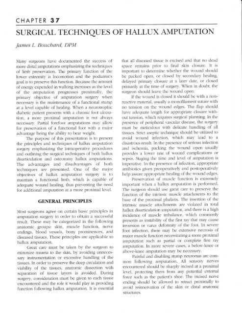
Figure J. A comparison <strong>of</strong> disarticr'rlation amplltation<br />
(right) .1ncl osteotomlr <strong>amputation</strong> (lcti)<br />
<strong>techniques</strong>.<br />
Figure.1. Adeqnate bleeding uras notecl at the tilne <strong>of</strong> the skin incision.<br />
t.i,i.h rr'rt , .tr,r,-tg indication thrt the patient had aclequete circulation<br />
firr appropriate healing <strong>of</strong> the rvolincl tirllon'ing alnplltation<br />
Figure 5. <strong>The</strong> flrst metatarsal head n'as iclentifieci :rnd clisarticulated at<br />
the first metatarsal phalangeal loint<br />
Figure 6. All necrotic ancl diseasecl tissrte n':Ls<br />
rernovecl at the time <strong>of</strong> <strong>amputation</strong> to p1'event<br />
further complications from infection, or in n-rore<br />
severe c:1ses necrotizing f:LSCiiiis
204 CHAPTER 37<br />
Figure 7. Frequent lavagc [.ith cool sterile $'ater prior to srollnd<br />
closure helpecl to provide priman, nound healing. Note the he:rlthy<br />
cartilxge on the remaining flrst metatars.Ll heacl prior to $'ound closure.<br />
Figure 8. Intraoperative cultures inclucling gram<br />
stain. cuhuic and sensitir-iq' for aerobes ancl<br />
an:Lerobes. acid fast stain, tnd fi-rngal<br />
cultures n'ere taken. Amplltation specimen s,as<br />
sent tbr bone ctiltures and identification <strong>of</strong> a<br />
clean proximal n'rargin. <strong>The</strong> importance <strong>of</strong><br />
appropdate bone biopsv to iclentil_v a clean mal:-<br />
gin ancl bone cultures cannot be o\rer<br />
emphasizecl.<br />
Figure 9. <strong>The</strong> <strong>surgical</strong> area was closecl primarilv s,-ith a non-absorbable<br />
synthetic sutlrre and dr,v sterile compressive dressings \\'ere applied,<br />
firllos,'ed b1, reclressing <strong>of</strong> the I'ouncl in three days. Because adequate<br />
hemost:rsis u,,as provided, no tubes or drains l.ere necessatw. <strong>The</strong><br />
patient nras dischargecl from the hospital rl,.ith instructions for strict<br />
non-r'eight bearing on crutches for 10-14 days follorved b.v partial<br />
w-eight beering in a <strong>surgical</strong> shoe.<br />
Figure 10. Ts,-o weeks postoperati\.e result follon-ing right <strong>hallux</strong><br />
<strong>amputation</strong>. Note prior <strong>hallux</strong> <strong>amputation</strong> <strong>of</strong> the left <strong>hallux</strong> one year<br />
prer.ious11,.
CHAPTER 37 205<br />
POSTOPERAITVE MANAGEMENT<br />
Most postoperative regimes following halh:x<br />
<strong>amputation</strong> initially require strict non-weight bearing<br />
<strong>of</strong> the involved extremily. It is important for the<br />
podiatric surgeon to not neglect the contralateral<br />
1imb, which is at increased risk. This requires<br />
frequent monitoring and protective measures. <strong>The</strong><br />
use <strong>of</strong> the Cam walker is an excellent regime to<br />
progress from a non-weight bearing to a pafiial<br />
weight bearing status. <strong>The</strong> Cam walker can be used<br />
effectively until it is appropriate to advance to the<br />
use <strong>of</strong> a shoe. An extra depth shoe with a prescribed<br />
plastizote medial forefoot filler and inlay, combined<br />
with a rigid shank is recommended for ambulation<br />
following successful healing. <strong>The</strong> use <strong>of</strong> a neutral<br />
position <strong>of</strong>ihotic device with intrinsic forefoot<br />
posting helps to control the potential postoperative<br />
forefoot varus position following <strong>hallux</strong> <strong>amputation</strong><br />
surgery.<br />
Historically, diabetic patients presenting with a<br />
foot r:lceration would have had an <strong>amputation</strong> at a<br />
proximal level because <strong>of</strong> the assumed inabiliry to<br />
heal at the level <strong>of</strong> distal <strong>amputation</strong>s. \7hen an<br />
infection has involved the plantar spaces <strong>of</strong> the foot,<br />
it has been believed that the only hope <strong>of</strong> saving the<br />
patient's limb was to perform a guillotine<br />
<strong>amputation</strong> <strong>of</strong> the affected limb. \(hen a<br />
neurotrophic diabetic patient presents with foot<br />
ulcerations, it is not always necessary to perform a<br />
proximal <strong>amputation</strong>. Partial forefoot <strong>amputation</strong>s<br />
such as a <strong>hallux</strong> <strong>amputation</strong> a1low the minimum<br />
possible <strong>amputation</strong> at the lowest level with a major<br />
advantage <strong>of</strong> the ability to bear weight.<br />
BIBLIOGRAPHY<br />
Bilk D. Selby JV, et al: Lower extremity <strong>amputation</strong>s in people u'ith<br />
cliahetes: epidemiology and prevention. Diabetu Care 1,990:73:573.<br />
Chang B, Bock D, Jacobs R, Darling R, Leather R. Shah D. Increased<br />
limb sah'age by the use <strong>of</strong> unconventional fbot <strong>amputation</strong>s. Jr<br />
V as c S urg 7994;79 :347 -9.<br />
Downs DM, Jacobs RL. Treatment <strong>of</strong> resistant ulcers on the plantar<br />
surfhce <strong>of</strong> the gleat toe in diabettcs. J Bone Jaint Swg Am<br />
7L)82:64:930-3.<br />
Duggar. GE, et a1, Time-Line for the diabetic frrot. In: McGlamry ED.<br />
editor. <strong>Podiatry</strong> Instltute Update 1987. Tucker (GA): Podiatrl,=<br />
<strong>Institute</strong> Publishing; 1987.<br />
Fronek A, Coel M, Bernstein EF. <strong>The</strong> imp<strong>of</strong>iance <strong>of</strong> combined muitisegmental<br />
pressllre and Doppler flon' r'elociqv stuclies in the<br />
diagnosis <strong>of</strong> peripheral arterial occlusive disease. Sarxerl'<br />
1978;8'1:840-7.<br />
Gavin, LA. Perioperative management <strong>of</strong> the diabetic patient:<br />
enclocrinology and metabolism Clin N Am 7L)92;21:157-75.<br />
Goldstein B, Citron D, Neshit C. Diabetic foot infections. Diabetes Care<br />
7996;79:638-41.<br />
Griffiths, GD, \i7ieman, TJ. N,{eticulous attention to foot crre impror-es<br />
the prognosis in diabetic ulceration <strong>of</strong> the foot. Surgerl Gyn Obst<br />
7992:174:19 51.<br />
Lance B, Kirschenbaum S. Distal ischemia s'ith digital gansirene sec<br />
ondary to buerger's clisease../ Faot Sarg 1991;30,531 17.<br />
Levin M. Foot lesions in patients w-ith cliabetes mellitus. Cbron Comp Diab<br />
7996;25'147-55.<br />
Levin ME, O'Neil L\Xr. <strong>The</strong> Diabetic Foot. St. Louis: CV Nlosb-v; 1!88. p.<br />
77.<br />
Pinzur NIS, Gold J, SchnartT- D, et al. Energv demands for u'alking in<br />
d\rsvascular xmplltees as related to the level ot.L]nlputetion. OrtbLl)<br />
199275:1033-1 .<br />
Pinzur MS, Sage R, Abral'ram NI, et al. Limb salvage in inf'ectecl lori-er<br />
extremity gangrene. Foot Ankle 1988:8:212-5<br />
Sizer JS,<br />
\Mreelock FC. Digital <strong>amputation</strong>s in diabetic patients. SzzrgerT<br />
7972:i2:L)80-9.<br />
litti M. Robinson D, Hauer-Jensen M, Thompson B. Ranval T, Barone<br />
G, et a1. Vound healing in fbrefoot <strong>amputation</strong>s: the predrctive<br />
r:lrre <strong>of</strong> 1,,( fre


