International Carl Zeiss Workshop on International Carl Zeiss ...
International Carl Zeiss Workshop on International Carl Zeiss ...
International Carl Zeiss Workshop on International Carl Zeiss ...
Create successful ePaper yourself
Turn your PDF publications into a flip-book with our unique Google optimized e-Paper software.
<str<strong>on</strong>g>Internati<strong>on</strong>al</str<strong>on</strong>g> <str<strong>on</strong>g>Carl</str<strong>on</strong>g> <str<strong>on</strong>g>Zeiss</str<strong>on</strong>g> <str<strong>on</strong>g>Workshop</str<strong>on</strong>g> <strong>on</strong><br />
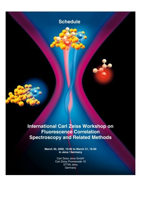
Schedule<br />
<str<strong>on</strong>g>Internati<strong>on</strong>al</str<strong>on</strong>g> <str<strong>on</strong>g>Carl</str<strong>on</strong>g> <str<strong>on</strong>g>Zeiss</str<strong>on</strong>g> <str<strong>on</strong>g>Workshop</str<strong>on</strong>g> <strong>on</strong><br />
Fluorescence Correlati<strong>on</strong><br />
Spectroscopy and Related Methods<br />
March 30, 2000, 10:00 to March 31, 16:00<br />
in Jena / Germany<br />
<str<strong>on</strong>g>Carl</str<strong>on</strong>g> <str<strong>on</strong>g>Zeiss</str<strong>on</strong>g> Jena GmbH<br />
<str<strong>on</strong>g>Carl</str<strong>on</strong>g> <str<strong>on</strong>g>Zeiss</str<strong>on</strong>g> Promenade 10<br />
07745 Jena<br />
Germany
<str<strong>on</strong>g>Internati<strong>on</strong>al</str<strong>on</strong>g> <str<strong>on</strong>g>Carl</str<strong>on</strong>g> <str<strong>on</strong>g>Zeiss</str<strong>on</strong>g> <str<strong>on</strong>g>Workshop</str<strong>on</strong>g> <strong>on</strong> Fluorescence Correlati<strong>on</strong> Spectroscopy and Related Methods<br />
March, 30 to March 31, Jena / Germany<br />
1 WELCOME TO JENA 2<br />
2 LOCATION 2<br />
3 TRANSPORTATION 2<br />
4 PARKING LOTS 3<br />
5 REGISTRATION DESK 3<br />
6 CONFERENCE OFFICE 4<br />
7 TALKS 4<br />
8 POSTER 4<br />
9 FACTORY TOUR 4<br />
10 DEMONSTRATION OF CONFOCOR 2 5<br />
11 AN EVENING IN THE OPTICAL MUSEUM 5<br />
12 PUBLICATION OF RESEARCH ARTICLES 5<br />
13 INTERNATIONAL SCIENTIFIC COMMITTEE 10<br />
14 SCHEDULE 12<br />
15 POSTER TITLES 16<br />
16 ABSTRACTS 21<br />
- 1 -
<str<strong>on</strong>g>Internati<strong>on</strong>al</str<strong>on</strong>g> <str<strong>on</strong>g>Carl</str<strong>on</strong>g> <str<strong>on</strong>g>Zeiss</str<strong>on</strong>g> <str<strong>on</strong>g>Workshop</str<strong>on</strong>g> <strong>on</strong> Fluorescence Correlati<strong>on</strong> Spectroscopy and Related Methods<br />
March, 30 to March 31, Jena / Germany<br />
1 WELCOME TO JENA<br />
Welcome to Jena, welcome at <str<strong>on</strong>g>Carl</str<strong>on</strong>g> <str<strong>on</strong>g>Zeiss</str<strong>on</strong>g>. On behalf of the organizati<strong>on</strong> committee of the<br />
“<str<strong>on</strong>g>Internati<strong>on</strong>al</str<strong>on</strong>g> <str<strong>on</strong>g>Carl</str<strong>on</strong>g> <str<strong>on</strong>g>Zeiss</str<strong>on</strong>g> <str<strong>on</strong>g>Workshop</str<strong>on</strong>g> <strong>on</strong> Fluorescence Correlati<strong>on</strong> Spectroscopy (FCS) and<br />
Related Methods”, we wish you a pleasant stay and a lot of fruitful discussi<strong>on</strong>s.<br />
Reviewing the program, we were impressed about the large amount of promising<br />
c<strong>on</strong>tributi<strong>on</strong>s to the workshop. The variety of subjects shows, that Fluorescence<br />
Correlati<strong>on</strong> Spectroscopy is a well established method now and finds its way into lots of<br />
diverse research areas.<br />
The <str<strong>on</strong>g>Internati<strong>on</strong>al</str<strong>on</strong>g> <str<strong>on</strong>g>Carl</str<strong>on</strong>g> <str<strong>on</strong>g>Zeiss</str<strong>on</strong>g> <str<strong>on</strong>g>Workshop</str<strong>on</strong>g> <strong>on</strong> Fluorescence Correlati<strong>on</strong> Spectroscopy (FCS)<br />
and Related Methods c<strong>on</strong>tinues the series of C<strong>on</strong>foCor User Clubs organized by<br />
<str<strong>on</strong>g>Carl</str<strong>on</strong>g> <str<strong>on</strong>g>Zeiss</str<strong>on</strong>g> (1997 Leuven/Belgium, 1998 Jena/Germany, 50 participants each).<br />
The change in name reflects the intenti<strong>on</strong> of the organizers to invite all scientists<br />
interested in FCS instrumentati<strong>on</strong>, theory, applicati<strong>on</strong>s and related techniques to<br />
participate the meeting, regardless whether they use a <str<strong>on</strong>g>Carl</str<strong>on</strong>g> <str<strong>on</strong>g>Zeiss</str<strong>on</strong>g> C<strong>on</strong>foCor or not.<br />
The workshop will start Thursday, March 30th at 10:00 am at the C<strong>on</strong>ference Room of<br />
<str<strong>on</strong>g>Carl</str<strong>on</strong>g> <str<strong>on</strong>g>Zeiss</str<strong>on</strong>g> Jena, <str<strong>on</strong>g>Carl</str<strong>on</strong>g>-<str<strong>on</strong>g>Zeiss</str<strong>on</strong>g>-Promenade 10 (formerly known as Tatzendpromenade 1a). The<br />
registrati<strong>on</strong> desk will open at 8:00.<br />
Dr. Winfried Wiegräbe<br />
<str<strong>on</strong>g>Carl</str<strong>on</strong>g> <str<strong>on</strong>g>Zeiss</str<strong>on</strong>g> Jena GmbH<br />
Microscopy<br />
Advanced Imaging Systems<br />
2 LOCATION<br />
<str<strong>on</strong>g>Carl</str<strong>on</strong>g> <str<strong>on</strong>g>Zeiss</str<strong>on</strong>g> Jena GmbH<br />
<str<strong>on</strong>g>Carl</str<strong>on</strong>g>-<str<strong>on</strong>g>Zeiss</str<strong>on</strong>g>-Promenade 10<br />
07745 Jena<br />
Germany<br />
The c<strong>on</strong>ference room is located at the large, flat building in fr<strong>on</strong>t of the main<br />
entrance to <str<strong>on</strong>g>Carl</str<strong>on</strong>g> <str<strong>on</strong>g>Zeiss</str<strong>on</strong>g> Jena GmbH. This building houses the canteen of the<br />
“Fachhochschule”, too.<br />
3 TRANSPORTATION<br />
If you stay at the ibis or Esplanade hotel, we recommend to take the Bus starting at the<br />
city center named "Holzmarkt" with destinati<strong>on</strong> "Burgau" (lines 10,13), or<br />
"Goeschwitz" (line 10), bus stop "Fachhochschule" or “<str<strong>on</strong>g>Zeiss</str<strong>on</strong>g> Werk”. <str<strong>on</strong>g>Zeiss</str<strong>on</strong>g> is located<br />
between both stops <strong>on</strong> the right side, coming from the center.<br />
From the "Best Western Hotel", we have arranged a shuttle. It will start <strong>on</strong> Thursday,<br />
30 th at 9:00 am and <strong>on</strong> Friday, 31 st at 8:30 am. If you plan to use this service, please<br />
inform the recepti<strong>on</strong> desk of the hotel when arriving.<br />
- 2 -
<str<strong>on</strong>g>Internati<strong>on</strong>al</str<strong>on</strong>g> <str<strong>on</strong>g>Carl</str<strong>on</strong>g> <str<strong>on</strong>g>Zeiss</str<strong>on</strong>g> <str<strong>on</strong>g>Workshop</str<strong>on</strong>g> <strong>on</strong> Fluorescence Correlati<strong>on</strong> Spectroscopy and Related Methods<br />
March, 30 to March 31, Jena / Germany<br />
Fachhochschule<br />
<str<strong>on</strong>g>Zeiss</str<strong>on</strong>g> Werk<br />
4 PARKING LOTS<br />
Parking lots are available in fr<strong>on</strong>t of the c<strong>on</strong>ference building exclusively for participants of the<br />
workshop.<br />
- 3 -
<str<strong>on</strong>g>Internati<strong>on</strong>al</str<strong>on</strong>g> <str<strong>on</strong>g>Carl</str<strong>on</strong>g> <str<strong>on</strong>g>Zeiss</str<strong>on</strong>g> <str<strong>on</strong>g>Workshop</str<strong>on</strong>g> <strong>on</strong> Fluorescence Correlati<strong>on</strong> Spectroscopy and Related Methods<br />
March, 30 to March 31, Jena / Germany<br />
5 REGISTRATION DESK<br />
The registrati<strong>on</strong> desk will be open <strong>on</strong> Thursday, 30 th March from 8:00 to 12:00 am. Later<br />
registrati<strong>on</strong> is possible at the c<strong>on</strong>ference office.<br />
Please prepare the c<strong>on</strong>ference fee (100,- DM, 50,- DM for students) in cash. Unfortunately, credit<br />
cards are not accepted.<br />
6 CONFERENCE OFFICE<br />
During the workshop a c<strong>on</strong>ference office is located at the “Innovati<strong>on</strong>sraum”. Leave the<br />
c<strong>on</strong>ference room through the corridor at the west side. The c<strong>on</strong>ference office is located<br />
<strong>on</strong>e floor below the c<strong>on</strong>ference room.<br />
At the c<strong>on</strong>ference office, you may receive messages by ph<strong>on</strong>e and FAX:<br />
Ph<strong>on</strong>e: ++49 – (0)3641 – 64 – 3254<br />
FAX: ++49 – (0)3641 – 64 – 3347<br />
At the office you can check your slides and settings of your computer when using a<br />
beamer.<br />
If you need any additi<strong>on</strong>al assistance, please feel free to c<strong>on</strong>tact the c<strong>on</strong>ference office or<br />
any member of the organizati<strong>on</strong> committee.<br />
7 TALKS<br />
Talks are scheduled to take 20 min plus 5 min for discussi<strong>on</strong>. For your talk, there will be a<br />
slide projector (35mm), an overhead projector, a beamer to be used together with your<br />
laptop and a laser pointer available.<br />
If you need additi<strong>on</strong>al equipment (e.g. video), please c<strong>on</strong>tact the c<strong>on</strong>ference office.<br />
8 POSTER<br />
For your posters, we have prepared boards approximately 180 cm high and 90 cm wide.<br />
Posters should be mounted <strong>on</strong> Thursday, 30th March between 8:00 and 10:00 am.<br />
Posters will be <strong>on</strong> display during the whole workshop.<br />
All posters are listed stating at page 16. Posters are numbered. Please attach your poster<br />
at the board showing the appropriate number.<br />
9 FACTORY TOUR<br />
There will be factory tours to the optics producti<strong>on</strong> and the system integrati<strong>on</strong> of the C<strong>on</strong>foCor<br />
2. The tours will be during the breaks. Each tour will last approximately half a hour.<br />
Because each tour is restricted to 25 pers<strong>on</strong>s, please put your name <strong>on</strong> the lists available at the<br />
registrati<strong>on</strong> desk.<br />
The tours will start in the c<strong>on</strong>ference office.<br />
- 4 -
<str<strong>on</strong>g>Internati<strong>on</strong>al</str<strong>on</strong>g> <str<strong>on</strong>g>Carl</str<strong>on</strong>g> <str<strong>on</strong>g>Zeiss</str<strong>on</strong>g> <str<strong>on</strong>g>Workshop</str<strong>on</strong>g> <strong>on</strong> Fluorescence Correlati<strong>on</strong> Spectroscopy and Related Methods<br />
March, 30 to March 31, Jena / Germany<br />
10 DEMONSTRATION OF CONFOCOR 2<br />
You might be interested in having a dem<strong>on</strong>strati<strong>on</strong> of the C<strong>on</strong>foCor 2, the new fluorescence<br />
correlati<strong>on</strong> microscope of <str<strong>on</strong>g>Carl</str<strong>on</strong>g> <str<strong>on</strong>g>Zeiss</str<strong>on</strong>g>. There are two instruments <strong>on</strong> display, <strong>on</strong>e is located at the<br />
c<strong>on</strong>ference room, the other <strong>on</strong>e is shown at the casino. The casino is approached through the<br />
corridor at the west end of the c<strong>on</strong>ference room.<br />
The C<strong>on</strong>foCor 2 is dem<strong>on</strong>strated during the breaks and the poster sessi<strong>on</strong>. Please feel free to<br />
schedule a private dem<strong>on</strong>strati<strong>on</strong> with <strong>on</strong>e of the <str<strong>on</strong>g>Carl</str<strong>on</strong>g> <str<strong>on</strong>g>Zeiss</str<strong>on</strong>g> FCS experts.<br />
11 AN EVENING IN THE OPTICAL MUSEUM<br />
On Thursday evening there will be a get together at the optical museum of Jena. The optical<br />
museum is located near the city center, not far away from the Esplanade and ibis hotels.<br />
Between 18:00 and 19:00, there will be buses from the c<strong>on</strong>ference locati<strong>on</strong> to the optical<br />
museum. The buses start in fr<strong>on</strong>t of the c<strong>on</strong>ference building.<br />
12 PUBLICATION OF RESEARCH ARTICLES<br />
Abstracts are available at www.zeiss.de/fcsevents. They will be <strong>on</strong> display some time after<br />
the workshop to. Additi<strong>on</strong>al abstracts are welcome. Please mail them to<br />
wiegraebe@zeiss.de, preferable as an <strong>on</strong>e page Word 97 for Windows document or as<br />
plain text. You are free to include links to your own web page. Please use as subject<br />
"Abstract FCS workshop" and the name of the first author.<br />
The November 2000 issue of the journal "Biological Chemistry" will be published as<br />
highlight issue covering the topics of the workshop. "Research Articles" or "Short<br />
Communicati<strong>on</strong>s" should be submitted directly to:<br />
Biological Chemistry<br />
Editorial Office<br />
attn. Dr. Ulrich Hartl<br />
Walter de Gruyter GmbH & Co.<br />
Genthiner Str. 13<br />
D-10785 Berlin<br />
Tel.: +49-30-26005-279<br />
Fax: +49-30-26005-298<br />
E-mail: biol.chem.editorial@deGruyter.de<br />
Internet: www.degruyter.de/journals/bc<br />
Deadline for submissi<strong>on</strong>: May, 31st, 2000.<br />
The articles will undergo the standard review process of the journal. You will find the<br />
instructi<strong>on</strong>s for authors at http://www.degruyter.de/journals/bc/bcins.html and at the<br />
c<strong>on</strong>ference site.<br />
- 5 -
<str<strong>on</strong>g>Internati<strong>on</strong>al</str<strong>on</strong>g> <str<strong>on</strong>g>Carl</str<strong>on</strong>g> <str<strong>on</strong>g>Zeiss</str<strong>on</strong>g> <str<strong>on</strong>g>Workshop</str<strong>on</strong>g> <strong>on</strong> Fluorescence Correlati<strong>on</strong> Spectroscopy and Related Methods<br />
March, 30 to March 31, Jena / Germany<br />
Scope and policy of the Journal<br />
Biological Chemistry provides rapid publicati<strong>on</strong> for reports <strong>on</strong> molecular studies of<br />
excepti<strong>on</strong>al biological interest. The Journal publishes full length papers, short<br />
communicati<strong>on</strong>s, reviews and mini-reviews. Reviews are published by invitati<strong>on</strong> <strong>on</strong>ly, but<br />
suggesti<strong>on</strong>s to the Editor-in-Chief are welcome.<br />
Submissi<strong>on</strong> of a manuscript to Biological Chemistry implies that the work described has<br />
not been published before and is not under c<strong>on</strong>siderati<strong>on</strong> for publicati<strong>on</strong> elsewhere. It is<br />
the corresp<strong>on</strong>ding author's resp<strong>on</strong>sibility to ensure that all authors see the manuscript<br />
and approve of its submissi<strong>on</strong> for publicati<strong>on</strong>. Once the manuscript is accepted, it must<br />
not be published elsewhere without the c<strong>on</strong>sent of the copyright holders.<br />
Submissi<strong>on</strong> of manuscripts<br />
One original and three high-quality copies of the manuscript, including all figures and<br />
tables, should be submitted to:<br />
Biological Chemistry<br />
Editorial Office<br />
attn. Dr. Ulrich Hartl<br />
Walter de Gruyter GmbH & Co.<br />
Genthiner Str. 13<br />
D-10785 Berlin<br />
Tel.: +49-30-26005-279<br />
Fax: +49-30-26005-298<br />
E-mail: biol.chem.editorial@deGruyter.de<br />
Each manuscript should be accompanied by a cover letter c<strong>on</strong>taining a brief statement by<br />
the authors as to the element of novelty up<strong>on</strong> which they base their request for<br />
publicati<strong>on</strong> in Biological Chemistry. The reviewing process may be expedited by indicating<br />
the names and full postal addresses (including ph<strong>on</strong>e and fax numbers) of at least four<br />
potential referees who are not editors or members of the Editorial Board of the Journal.<br />
Review of manuscripts and speed of publicati<strong>on</strong><br />
Papers will be independently reviewed by at least two referees selected by the Editors of<br />
Biological Chemistry. On the average, decisi<strong>on</strong>s will be reached within three to four<br />
weeks of submissi<strong>on</strong>. When papers are accepted subject to revisi<strong>on</strong>, revisi<strong>on</strong> is allowed<br />
<strong>on</strong>ly <strong>on</strong>ce. The revised manuscript (<strong>on</strong>e original and two copies) must be submitted<br />
within two m<strong>on</strong>ths of the authors' notificati<strong>on</strong> of c<strong>on</strong>diti<strong>on</strong>al acceptance. It is the aim of<br />
the Journal to publish papers within four m<strong>on</strong>ths after their receipt by the Editor-in-Chief.<br />
Preparati<strong>on</strong> of manuscripts<br />
Manuscripts must be written in clear and c<strong>on</strong>cise English and should be regarded as final<br />
texts. Illustrati<strong>on</strong>s must be submitted in original quality with all four copies. No changes<br />
may be made at the proof state other than correcti<strong>on</strong> of printer's errors.<br />
- 6 -
<str<strong>on</strong>g>Internati<strong>on</strong>al</str<strong>on</strong>g> <str<strong>on</strong>g>Carl</str<strong>on</strong>g> <str<strong>on</strong>g>Zeiss</str<strong>on</strong>g> <str<strong>on</strong>g>Workshop</str<strong>on</strong>g> <strong>on</strong> Fluorescence Correlati<strong>on</strong> Spectroscopy and Related Methods<br />
March, 30 to March 31, Jena / Germany<br />
General format and length<br />
Manuscripts (including table legends, figure legends and references) should be typed<br />
double-spaced with f<strong>on</strong>t size 12 letters <strong>on</strong> <strong>on</strong>ly <strong>on</strong>e side of A4 or 8 1/2 x 11" paper.<br />
Pages should be numbered (with the title page as 1) and have margins of 2.5 cm (1 inch)<br />
<strong>on</strong> all sides. Footnotes in the text should be avoided; parentheses should be used instead.<br />
Full length papers and reviews should occupy no more than eight printed pages, short<br />
communicati<strong>on</strong>s and mini-reviews should not exceed four printed pages. Each full page<br />
of printed text corresp<strong>on</strong>ds to approximately 1400 words. Please allow sufficient space<br />
for tables and illustrati<strong>on</strong>s within the page limit.<br />
Secti<strong>on</strong>s<br />
- Full length papers should be organized into Title page, Summary, Key words,<br />
Introducti<strong>on</strong>, Results, Discussi<strong>on</strong>, Materials and Methods, Acknowledgments,<br />
References, Tables and Figure legends.<br />
- Short communicati<strong>on</strong>s should be subdivided into an Abstract, Key words, and a single<br />
secti<strong>on</strong> of main text without headings. Experimental procedures should be described<br />
in legends to figures or footnotes to tables. Acknowledgments and References should<br />
be presented as in full length papers.<br />
Title page<br />
The title page should include (a) a short and informative full article title (series titles are<br />
not accepted); (b) names of all authors (with <strong>on</strong>e forename in full for each author),<br />
followed by their affiliati<strong>on</strong>s (department, instituti<strong>on</strong>, city with postcode, country); (c) the<br />
mailing address, fax and ph<strong>on</strong>e number of the corresp<strong>on</strong>ding author; (d) a running title<br />
of 50 characters or less. If there is more than <strong>on</strong>e instituti<strong>on</strong> involved in the work,<br />
authors' names should be linked by superscript c<strong>on</strong>secutive numbers to the appropriate<br />
instituti<strong>on</strong>s. If required, small letters should be used to indicate present addresses.<br />
Summary and key words<br />
The sec<strong>on</strong>d page of the manuscript should c<strong>on</strong>tain the summary and the key words. The<br />
summary should be a single paragraph of no more than 200 words (full length paper or<br />
review) or 100 words (short communicati<strong>on</strong> or mini-review) which must be<br />
comprehensible to readers before they have read the paper. Abbreviati<strong>on</strong>s and reference<br />
citati<strong>on</strong>s should be avoided. Below the summary up to six key words, which are not part<br />
of the title, should be given in alphabetical order and separated by slashes (/). Together<br />
with the title the key words provide the basis for the annual Subject Index.<br />
Acknowledgments<br />
Acknowledgments should be placed at the end of the text.<br />
Names of funding organizati<strong>on</strong>s should be written in their entirety.<br />
- 7 -
<str<strong>on</strong>g>Internati<strong>on</strong>al</str<strong>on</strong>g> <str<strong>on</strong>g>Carl</str<strong>on</strong>g> <str<strong>on</strong>g>Zeiss</str<strong>on</strong>g> <str<strong>on</strong>g>Workshop</str<strong>on</strong>g> <strong>on</strong> Fluorescence Correlati<strong>on</strong> Spectroscopy and Related Methods<br />
March, 30 to March 31, Jena / Germany<br />
References<br />
For citati<strong>on</strong>s authors are encouraged to rely as far as possible up<strong>on</strong> articles published in<br />
primary research journals.<br />
Unpublished results and pers<strong>on</strong>al communicati<strong>on</strong>s should be cited as such within the<br />
text; pers<strong>on</strong>al communicati<strong>on</strong>s must be substantiated by a letter of permissi<strong>on</strong> Meeting<br />
abstracts may not be cited.<br />
Within the text references should be cited by author and date; et al. should be used if<br />
there are more than two authors. At the end of the text citati<strong>on</strong>s should be in<br />
alphabetical order and should c<strong>on</strong>tain complete titles.<br />
The number of references should not exceed 150 for reviews or 30 for mini-reviews.<br />
The names of Journals should be abbreviated according to the World List of Scientific<br />
Periodicals.<br />
The volume number of a journal should be given in italics. Citati<strong>on</strong>s should be in<br />
accordance with the following examples:<br />
Kyte, J., and Doolittle, R.F. (1982). A simple method for displaying the hydrophobic<br />
character of a protein. J. Mol. Biol. 157, 105-132.<br />
Khan, M.A. (1987). HLA and ankylosing sp<strong>on</strong>dylitis. In: Ankylosing<br />
Sp<strong>on</strong>dylitis. New Clinical Applicati<strong>on</strong>s in Rheumatology, Vol. I,<br />
J.J. Calabro and C. Dick, eds. (Lancaster, England: MTP Press,<br />
Ltd.), pp. 23-45.<br />
Hogan, B., Costantini, F, and Lacy, E. (1986). Manipulating the<br />
Mouse Embryo: A Laboratory Manual (Cold Spring Harbor, New<br />
York: Cold Spring Harbor Laboratory Press).<br />
Tables<br />
Tables should be typed <strong>on</strong> separate pages and be numbered c<strong>on</strong>secutively using Arabic<br />
numerals. If a table is excepti<strong>on</strong>ally large or c<strong>on</strong>tains special symbols it should be<br />
submitted camera-ready. A short descriptive title, column headings and (if necessary)<br />
footnotes should make each table self-explanatory. Please indicate in the manuscript the<br />
approximate positi<strong>on</strong> of each table.<br />
Illustrati<strong>on</strong>s<br />
Illustrati<strong>on</strong>s will be reduced in size to fit, whenever possible the width of a single column,<br />
i.e. 80 mm, or a double column, i.e. 168 mm, of text. Ideally, single column figures<br />
should be submitted with a width of 100 mm, double column figures with a width of<br />
210 mm. Lettering of all figures within the article should be uniform in style (preferably a<br />
sans serif typeface) and of sufficient size (so that the final height will be approximately 2<br />
mm). Uppercase letters A, B, C etc. should be used to identify individual parts of multipart<br />
figures. On the back of each figure the name of the first author the figure number<br />
and the top margin should be indicated, preferably with a soft pencil. All figures must be<br />
cited in the text in numerical order and the approximate positi<strong>on</strong> of each should be<br />
- 8 -
<str<strong>on</strong>g>Internati<strong>on</strong>al</str<strong>on</strong>g> <str<strong>on</strong>g>Carl</str<strong>on</strong>g> <str<strong>on</strong>g>Zeiss</str<strong>on</strong>g> <str<strong>on</strong>g>Workshop</str<strong>on</strong>g> <strong>on</strong> Fluorescence Correlati<strong>on</strong> Spectroscopy and Related Methods<br />
March, 30 to March 31, Jena / Germany<br />
indicated in the margin. Reference to figures is to be made in the text as Figure 1 etc.,<br />
Fig. 1 etc. should be used in the figure legends.<br />
Photographs<br />
Photographs should be provided as high-quality glossy prints in order to withstand the<br />
inevitable loss of c<strong>on</strong>trast and detail inherent in the printing process.<br />
Color plates<br />
To partially offset the cost of producti<strong>on</strong>, color figures will be printed with the following<br />
charges to the author:<br />
DM 1200.- for the first illustrati<strong>on</strong> and DM 600.- for each subsequent illustrati<strong>on</strong> in <strong>on</strong>e<br />
article. The total cost for color figures will be reduced if several color illustrati<strong>on</strong>s appear<br />
<strong>on</strong> <strong>on</strong>e printed page.<br />
Line drawings<br />
These should be provided as high-quality prints. No additi<strong>on</strong>al artwork, redrawing or<br />
typesetting will be d<strong>on</strong>e by the publisher. Note that faint shading or stippling may be lost<br />
up<strong>on</strong> reproducti<strong>on</strong>, heavy staining or stippling may appear black.<br />
Figure Legends<br />
Theses should be provided <strong>on</strong> separate, numbered manuscript pages. All symbols and<br />
abbreviati<strong>on</strong>s used in the figures must be explained, except for standard abbreviati<strong>on</strong>s or<br />
others defined in the preceding text.<br />
Nomenclature<br />
With respect to biochemical terminology the Journal will follow the rules laid down by<br />
the IUPAC-IUB Commissi<strong>on</strong> <strong>on</strong> Biochemical Nomenclature as given in Biochemical<br />
Nomenclature and Related Documents, published by the Biochemical Society, U.K.<br />
Enzyme names should be in accordance with the recommendati<strong>on</strong>s of the IUPAC-IUB<br />
Commissi<strong>on</strong> <strong>on</strong> Biochemical Nomenclature, 1978, as given in Enzyme Nomenclature<br />
published by Academic Press, New York, 1992. Genotypes should be given in italics,<br />
phenotypes should not be italicized. Nomenclature of bacterial genetics should follow<br />
Demerec et al. (1966). Genetics, 54, 61-76.<br />
Abbreviati<strong>on</strong>s<br />
The Journal accepts standard Journal of Biological Chemistry abbreviati<strong>on</strong>s. Uncomm<strong>on</strong><br />
abbreviati<strong>on</strong>s should be defined parenthetically within the text up<strong>on</strong> first appearance.<br />
- 9 -
<str<strong>on</strong>g>Internati<strong>on</strong>al</str<strong>on</strong>g> <str<strong>on</strong>g>Carl</str<strong>on</strong>g> <str<strong>on</strong>g>Zeiss</str<strong>on</strong>g> <str<strong>on</strong>g>Workshop</str<strong>on</strong>g> <strong>on</strong> Fluorescence Correlati<strong>on</strong> Spectroscopy and Related Methods<br />
March, 30 to March 31, Jena / Germany<br />
Cover illustrati<strong>on</strong>s<br />
Each cover of Biological Chemistry will have an illustrati<strong>on</strong> selected from <strong>on</strong>e of the<br />
articles published in the Issue. Authors who wish to have a color photograph c<strong>on</strong>sidered<br />
for the cover should submit a glossy print, 18cm by 24cm or 8 inches by 10 inches,<br />
and/or a slide. A short descriptive summary of what the picture shows should be<br />
included.<br />
Offprints<br />
For each article 20 offprints are supplied free of charge. An offprint Order Form will<br />
accompany the page proofs and should be completed and returned immediately.<br />
13 INTERNATIONAL SCIENTIFIC COMMITTEE<br />
- Chair: Prof. Rudolf Rigler<br />
Department of Medical Biochemistry and Biophysics<br />
The Karolinska Institute<br />
The Laboratory of Medical Biophysics, MBB<br />
S-171 77 Stockholm,<br />
SWEDEN<br />
- Univ. Doz. Dr. Manfred Auer<br />
Novartis Leading Scientist<br />
Research Programme Head: "Fluorescence based HTS Technology"<br />
Associate Professor, University of Salzburg,<br />
Novartis Forschungsinstitut, NFI<br />
Allergic diseases<br />
Brunner Strasse 59<br />
A-1235 Vienna<br />
AUSTRIA<br />
- 10 -
<str<strong>on</strong>g>Internati<strong>on</strong>al</str<strong>on</strong>g> <str<strong>on</strong>g>Carl</str<strong>on</strong>g> <str<strong>on</strong>g>Zeiss</str<strong>on</strong>g> <str<strong>on</strong>g>Workshop</str<strong>on</strong>g> <strong>on</strong> Fluorescence Correlati<strong>on</strong> Spectroscopy and Related Methods<br />
March, 30 to March 31, Jena / Germany<br />
- Prof. Manfred Eigen<br />
Arbeitsgruppe 081 (Biochemische Kinetik)<br />
MPI für biophysikalische Chemie<br />
Am Faßberg<br />
37070 Göttingen<br />
Germany<br />
- Prof. Enrico Gratt<strong>on</strong><br />
Department of Physics<br />
University of Illinois<br />
at Urbana-Champaign<br />
1110 West Green Street<br />
Urbana, IL 61801-3080<br />
USA<br />
- Prof. Ant<strong>on</strong>ie Visser<br />
Microspectroscopy Centre<br />
Wageningen University<br />
Dreijenlaan 3<br />
6703 Ha Wageningen<br />
The Netherlands<br />
- Prof. Horst Vogel<br />
Institut of Physical Chemistry<br />
Swiss Federal Institute of Technology<br />
1015 Lausanne<br />
Switzerland<br />
- 11 -
<str<strong>on</strong>g>Internati<strong>on</strong>al</str<strong>on</strong>g> <str<strong>on</strong>g>Carl</str<strong>on</strong>g> <str<strong>on</strong>g>Zeiss</str<strong>on</strong>g> <str<strong>on</strong>g>Workshop</str<strong>on</strong>g> <strong>on</strong> Fluorescence Correlati<strong>on</strong> Spectroscopy and Related Methods<br />
March, 30 to March 31, Jena / Germany<br />
14 SCHEDULE<br />
Thursday, 30 th March<br />
Time Authors Title Abstract<br />
(page)<br />
08:00-<br />
10:00<br />
Registrati<strong>on</strong><br />
Setting up Posters<br />
4, 2<br />
10:00-<br />
10:15<br />
F.v.Falkenhausen<br />
President<br />
<str<strong>on</strong>g>Carl</str<strong>on</strong>g> <str<strong>on</strong>g>Zeiss</str<strong>on</strong>g> Jena GmbH, 07745<br />
Jena, Germany<br />
Welcome Address<br />
10:15-<br />
10:30<br />
W. Wiegräbe<br />
<str<strong>on</strong>g>Carl</str<strong>on</strong>g> <str<strong>on</strong>g>Zeiss</str<strong>on</strong>g> Jena GmbH, 07745<br />
Jena, Germany<br />
Organizati<strong>on</strong>al Remarks<br />
10:30-<br />
11:00<br />
R. Rigler<br />
Karolinska Institute, Dept. of<br />
Medical Biophysics, 17177<br />
Stockholm, Sweden<br />
Introducti<strong>on</strong> into FCS<br />
11:00-<br />
12:00<br />
Posters<br />
Factory Tour<br />
4, 5<br />
12:00-<br />
12:25<br />
A. Casoli and M. Schönhoff<br />
MPI for Colloids and Interfaces,<br />
14476 Potsdam/Golm, Germany<br />
Fluorescence Correlati<strong>on</strong> Spectroscopy as a Tool to<br />
investigate Single Molecule Probe Dynamics in<br />
Ultrathin Polymer Films<br />
27<br />
12:25-<br />
12:50<br />
Z. Földes-Papp and R. Rigler<br />
Karolinska Institute, Dept. of<br />
Medical Biophysics, 17177<br />
Stockholm, Sweden<br />
Fluorescent Nucleotide Tagging, Bead Loading and<br />
Ex<strong>on</strong>ucleolytic Degradati<strong>on</strong> of DNA for Sequencing<br />
of Single DNA Molecules<br />
12:50-<br />
14:30<br />
Lunch<br />
- 12 -
<str<strong>on</strong>g>Internati<strong>on</strong>al</str<strong>on</strong>g> <str<strong>on</strong>g>Carl</str<strong>on</strong>g> <str<strong>on</strong>g>Zeiss</str<strong>on</strong>g> <str<strong>on</strong>g>Workshop</str<strong>on</strong>g> <strong>on</strong> Fluorescence Correlati<strong>on</strong> Spectroscopy and Related Methods<br />
March, 30 to March 31, Jena / Germany<br />
Thursday, 30 th March (c<strong>on</strong>tinued)<br />
Time Authors Title Abstract<br />
(page)<br />
14:30-<br />
15:00<br />
15:00-<br />
15:25<br />
15:25-<br />
16:10<br />
16:10-<br />
16:35<br />
16:35-<br />
17:00<br />
17:00-<br />
18:00<br />
18:00-<br />
21:00<br />
E. Gratt<strong>on</strong><br />
Department of Physics, University<br />
of Illinois at Urbana-Champaign, IL<br />
61801-3080 Urbana, USA<br />
K. Gall, K. Palo, and P. Kask<br />
Evotec BioSystems AG, 22525<br />
Hamburg, Germany<br />
G. Jung, A. Zumbusch, and C.<br />
Bräuchle<br />
Inst. f. Phys. Chemie, LMU<br />
München, 81377 München,<br />
Germany<br />
A. Koltermann, U. Kettling, T.<br />
Winkler, J. Stephan and M. Eigen<br />
Max Planck Institute for<br />
biophysical chemistry, 37077<br />
Goettingen, Germany<br />
to be announced<br />
FIDA, more than a glimpse bey<strong>on</strong>d the FCS horiz<strong>on</strong><br />
Posters<br />
Factory Tour<br />
Two-Colour Fluorescence Correlati<strong>on</strong> Spectroscopy<br />
of the Green Fluorescent Protein<br />
Dual-color fluorescence cross-correlati<strong>on</strong><br />
spectroscopy and its applicati<strong>on</strong> in biotechnology<br />
4, 5<br />
Poster sessi<strong>on</strong> 16<br />
An Evening at the Optical Museum 5<br />
33<br />
35<br />
- 13 -
<str<strong>on</strong>g>Internati<strong>on</strong>al</str<strong>on</strong>g> <str<strong>on</strong>g>Carl</str<strong>on</strong>g> <str<strong>on</strong>g>Zeiss</str<strong>on</strong>g> <str<strong>on</strong>g>Workshop</str<strong>on</strong>g> <strong>on</strong> Fluorescence Correlati<strong>on</strong> Spectroscopy and Related Methods<br />
March, 30 to March 31, Jena / Germany<br />
Friday, 31 st March<br />
Time Authors Title Abstract<br />
(page)<br />
09:00-<br />
9:30<br />
09:30-<br />
9:55<br />
09:55-<br />
10:20<br />
10:20-<br />
11:00<br />
11:00-<br />
11:25<br />
11:25-<br />
11:50<br />
11:50-<br />
12:15<br />
12:15-<br />
12:40<br />
T. Visser<br />
University of Wageningen,<br />
Netherlands<br />
R. Brock<br />
Institute of Organic Chemistry,<br />
72076 Tübingen, Germany<br />
A. Pramanik and R. Rigler<br />
Department of Medical<br />
Biochemistry and Biophysics, The<br />
Karolinska Institute, 17177<br />
Stockholm, Sweden<br />
P. Schwille<br />
Max-Planck-Institut für<br />
biophysikalische Chemie, AG<br />
Experimentelle Biophysik; Turm IV,<br />
081, 37077 Göttingen, Germany<br />
T. Wohland, R. Hovius and H.<br />
Vogel<br />
EPFL, 1015 Lausanne, Switzerland<br />
R. Jordan* and J. Klingauf<br />
Max-Planck-Institute for<br />
Biophysical Chemistry /<br />
Department of Membrane<br />
Biophysics, 37077 Göttingen,<br />
Germany<br />
N. Petersen<br />
Univ. Western Ontario,<br />
Department of Chemistry, N6A<br />
5B7 L<strong>on</strong>d<strong>on</strong>, Canada<br />
FCS of flavins and flavoproteins 50<br />
Fluorescence Correlati<strong>on</strong> Microscopy - Perspectives<br />
for Intracellular High-Throughput Sceening<br />
FCS-analysis of ligand-receptor interacti<strong>on</strong>s in the<br />
membranes of cultured cells.<br />
Posters<br />
Factory Tour<br />
Molecular dynamics in cells and <strong>on</strong> membranes: new<br />
challenges for FCS<br />
The Characterizati<strong>on</strong> of a Transmembrane Receptor<br />
Protein by Fluorescence Correlati<strong>on</strong> Spectrosocpy<br />
Synaptic Vesicle Dynamics Studied by Fluorescence<br />
Correlati<strong>on</strong> Spectroscopy<br />
24<br />
43<br />
4, 5<br />
Spatial Correlati<strong>on</strong> Spectroscopy 42<br />
48<br />
51<br />
32<br />
- 14 -
<str<strong>on</strong>g>Internati<strong>on</strong>al</str<strong>on</strong>g> <str<strong>on</strong>g>Carl</str<strong>on</strong>g> <str<strong>on</strong>g>Zeiss</str<strong>on</strong>g> <str<strong>on</strong>g>Workshop</str<strong>on</strong>g> <strong>on</strong> Fluorescence Correlati<strong>on</strong> Spectroscopy and Related Methods<br />
March, 30 to March 31, Jena / Germany<br />
Friday, 31 st March (c<strong>on</strong>tinued)<br />
Time Authors Title Abstract<br />
(page)<br />
12:40-<br />
13:30<br />
13:30-<br />
14:00<br />
14:00-<br />
14:25<br />
14:25-<br />
15:10<br />
15:10-<br />
15:35<br />
15:35-<br />
16:00<br />
M. Auer<br />
(1) Novartis Pharma Research,<br />
4002 Basel, Switzerland, (2)<br />
Massachusetts Institute of<br />
Technology, Cambridge, MA<br />
02139, (3) Novartis<br />
Forschungsinstitut, 1235 Vienna,<br />
Austria<br />
S. Ashman, U. Haupts, M.<br />
Ruediger, S. Turc<strong>on</strong>i, and A. Pope<br />
SmithKline Beecham, CM19 5AW<br />
Harlow, United Kingdom<br />
Ch. Buehler(1), J. Dreessen(1), K.<br />
Mueller(1), P.T.C. So(2), C.Y.<br />
D<strong>on</strong>g(2), A. Schilb(1), U.<br />
Hassiepen(1), K. Stoeckli(1), and<br />
M. Auer(3)<br />
(1) Novartis Pharma Research,<br />
4002 Basel, Switzerland, (2)<br />
Massachusetts Institute of<br />
Technology, Cambridge, MA<br />
02139, (3) Novartis<br />
Forschungsinstitut, 1235 Vienna,<br />
Austria<br />
J. Bieschke<br />
MPI f. biophys. Chemistry, 37077<br />
Göttingen, Germany<br />
Lunch<br />
C<strong>on</strong>focal fluorescence spectroscopy and<br />
nanoscanning in drug discovery<br />
Single Molecule Detecti<strong>on</strong> in uHTS 21<br />
Posters<br />
Factory Tour<br />
Multi-phot<strong>on</strong> Excitati<strong>on</strong> of intrinsic Protein<br />
Fluorescence has the potential to dramatically<br />
improve Pharmaceutical Drug-Screening<br />
Detecti<strong>on</strong> of amyloid aggregates by SIFT<br />
4, 5<br />
26<br />
- 15 -
<str<strong>on</strong>g>Internati<strong>on</strong>al</str<strong>on</strong>g> <str<strong>on</strong>g>Carl</str<strong>on</strong>g> <str<strong>on</strong>g>Zeiss</str<strong>on</strong>g> <str<strong>on</strong>g>Workshop</str<strong>on</strong>g> <strong>on</strong> Fluorescence Correlati<strong>on</strong> Spectroscopy and Related Methods<br />
March, 30 to March 31, Jena / Germany<br />
15 POSTER TITLES<br />
No. Authors Title Abstract<br />
(page)<br />
4 G. Bohnert, S.O. Kirschstein and<br />
L. Kittler<br />
Institut für Molekulare<br />
Biotechnologie Jena e.V., 7745<br />
Jena,<br />
DNA-Polymerase proof-reading activities m<strong>on</strong>itored<br />
by fluorescence-correlati<strong>on</strong>-spectroscopy<br />
15 L. Cognet, P.H.M. Lommerse, Individual eGFP and eYFP fusi<strong>on</strong> proteins studied in<br />
G.S. Harms, G.A. Blab, H.P. cell membranes<br />
Spaink and Th. Schmidt<br />
Leiden University, 2333 AL<br />
Leiden, Netherlands<br />
1 P. Czerney<br />
Dyomics GmbH, 07743 Jena,<br />
Germany<br />
14 F. Delie(1), R. Gurny(1) and A.<br />
Zimmer(2)<br />
(1) Department of Pharmceutical<br />
Technology and<br />
Biopharmaceutics, 1211 Geneva,<br />
Switzerland, (2)Biocenter, Johann<br />
Wolfgang Goethe-Universität,<br />
Institut for Pharmaceutical<br />
Technology, 60439 Frankfurt<br />
(Main), Germany<br />
6 H. Häberlein<br />
Dept. of Pharmaceutical Biology,<br />
35032 Marburg, Germany<br />
2 A. Hascher, K. Korn, H.-H.<br />
Foerster, U. Hahn and F. Bordusa<br />
Universtität Leipzig; Institut fuer<br />
Biochemie; Fakultaet fuer<br />
Biowissenschaften, Pharmazie &<br />
Physiologie, 4103 Leipzig,<br />
Germany<br />
Tailor-made dyes for Fluorescence Correlati<strong>on</strong><br />
Spectroscopy (FCS)<br />
Fluorescence Correlati<strong>on</strong> Spectroscopy for the<br />
Characterisati<strong>on</strong> of Drug Delivery Systems<br />
Aspects in the development and applicati<strong>on</strong> of small<br />
ligands in FCS.<br />
Chymotrypsin-catalyzed fluorescence labeling of<br />
rib<strong>on</strong>uclease T1<br />
23<br />
28<br />
29<br />
30<br />
- 16 -
<str<strong>on</strong>g>Internati<strong>on</strong>al</str<strong>on</strong>g> <str<strong>on</strong>g>Carl</str<strong>on</strong>g> <str<strong>on</strong>g>Zeiss</str<strong>on</strong>g> <str<strong>on</strong>g>Workshop</str<strong>on</strong>g> <strong>on</strong> Fluorescence Correlati<strong>on</strong> Spectroscopy and Related Methods<br />
March, 30 to March 31, Jena / Germany<br />
Poster titles (c<strong>on</strong>tinued)<br />
No. Authors Title Abstract<br />
(page)<br />
16 M.A. Hink, J.W. Borst, G.N.M. Intracellular Fluorescence Correlati<strong>on</strong> Spectroscopy:<br />
van der Krogt J. Goedhart, T.W.J. towards m<strong>on</strong>itoring Signal Transducti<strong>on</strong> Processes in<br />
Gadella Jr.,and A.J.W.G. Visser vivo.<br />
University of Wageningen, 6703<br />
HA Wageningen, Netherlands<br />
22 A.Digris, V.Skakun, E.Novikov,<br />
M.A.Hink and A.J.W.G.Visser<br />
K.U.Leuven, 3001 Heverlee,<br />
Belgium<br />
23 T. Jankowski, V. Jüngel, and R.<br />
Janka<br />
<str<strong>on</strong>g>Carl</str<strong>on</strong>g> <str<strong>on</strong>g>Zeiss</str<strong>on</strong>g> Jena GmbH, 07745<br />
Jena, Germany<br />
Software tools for global analysis in Fluorescence<br />
Correlati<strong>on</strong> Spectroscopy<br />
Optimizing Opticts for Cross Correlati<strong>on</strong><br />
7 R. Kiehl<br />
Glutathi<strong>on</strong>e: The essential factor for mitoch<strong>on</strong>drial<br />
energy-linked funkti<strong>on</strong>s<br />
Reinhold Kiehl Institute for<br />
Molecular Medicine/Biology,<br />
93437 Furth, Germany<br />
8 T. Kobayashi and J. Gruenberg<br />
Riken Fr<strong>on</strong>tier Research System,<br />
351-0198 Wako-shi, Saitama,<br />
Japan<br />
17 K. Korn, H.-H. Foerster, R. Rigler<br />
and U. Hahn<br />
Universtität Leipzig; Institut fuer<br />
Biochemie; Fakultaet fuer<br />
Biowissenschaften, Pharmazie &<br />
Physiologie, 4103 Leipzig,<br />
Germany<br />
FCS Analysis of an Organelle Specific Antibody 34<br />
FCS measurements of phages displaying RNase T1 or<br />
RNase A<br />
21 N. Kunst<br />
MicroSpectroscopy<br />
Development of a piezo driven xyz-translati<strong>on</strong> stage<br />
for intracellular FCS<br />
Center,<br />
Dreijenlaan 3, 6703 HA<br />
Wageningen, The Netherlands<br />
31<br />
37<br />
38<br />
- 17 -
<str<strong>on</strong>g>Internati<strong>on</strong>al</str<strong>on</strong>g> <str<strong>on</strong>g>Carl</str<strong>on</strong>g> <str<strong>on</strong>g>Zeiss</str<strong>on</strong>g> <str<strong>on</strong>g>Workshop</str<strong>on</strong>g> <strong>on</strong> Fluorescence Correlati<strong>on</strong> Spectroscopy and Related Methods<br />
March, 30 to March 31, Jena / Germany<br />
Poster titles (c<strong>on</strong>tinued)<br />
No. Authors Title Abstract<br />
(page)<br />
3 E. Lopez-Calle, J. R. Fries*, D. A Strategy for Highly Parallel Synthesis of Tyrosineand<br />
Histidine-Reactive Labeling Reagents<br />
Riester, D. Winkler, A. Wiesner, S.<br />
Dröge, H. Knorr, S. Viebrock, C.<br />
Winkler, G. Mielck, M. Wrobel<br />
39<br />
EVOTEC BioSystems AG, Life<br />
Science Technologies -Chemistry-,<br />
22525 Hamburg, Germany<br />
18 T. Neumann, S.O. Kirschstein, G. Measurement of the dynamic instability of<br />
Bohnert, J.A. Camacho Gomez, L. microtubules by FCS experiments<br />
Kittler and E. Unger<br />
40<br />
Institut für Molekulare<br />
Biotechnologie Jena e.V., 7745<br />
Jena, Germany<br />
9 O. Pänke, K. Häsler and W. Junge On the Stator of Rotary ATP Synthase: The Binding<br />
Strength of Subunit Delta to (Alpha Beta) 3 As<br />
Universität Osnabrück; FB<br />
Determinated by Fluorescence Correlati<strong>on</strong><br />
Biologie/Chemie; Abt. Biophysik,<br />
Spectroscopy<br />
49069 Osnabrück, Germany<br />
10 J. Rao, A. Nicol, P. Jordan, D.<br />
Zicha<br />
Imperial Cancer Research Fund,<br />
WC2A 3PX L<strong>on</strong>d<strong>on</strong>, United<br />
Kingdom<br />
11 J. Schell, O. Schäfer, J. Tatzelt*<br />
and D. Riesner<br />
Heinrich-Heine-Universität, Inst.<br />
für Physikalische Biologie, 40225<br />
Düsseldorf, Germany<br />
Interacti<strong>on</strong>s of mutant EGF receptors analysed by<br />
fluorescence correlati<strong>on</strong> spectroscopy (FCS)<br />
Influence of solvent c<strong>on</strong>diti<strong>on</strong>s and chaper<strong>on</strong>es <strong>on</strong><br />
Pri<strong>on</strong> Protein aggregati<strong>on</strong><br />
41<br />
45<br />
46<br />
- 18 -
<str<strong>on</strong>g>Internati<strong>on</strong>al</str<strong>on</strong>g> <str<strong>on</strong>g>Carl</str<strong>on</strong>g> <str<strong>on</strong>g>Zeiss</str<strong>on</strong>g> <str<strong>on</strong>g>Workshop</str<strong>on</strong>g> <strong>on</strong> Fluorescence Correlati<strong>on</strong> Spectroscopy and Related Methods<br />
March, 30 to March 31, Jena / Germany<br />
Poster titles (c<strong>on</strong>tinued)<br />
No. Authors Title Abstract<br />
(page)<br />
12 H. Schürer(1), A. Buchynskyy(2),<br />
K. Korn(1), P. Welzel(2) and U.<br />
Hahn(1)<br />
Investigati<strong>on</strong> of aptamer/moenomycin interacti<strong>on</strong> by<br />
FCS<br />
46<br />
(1)Institut für Biochemie, Fakultät<br />
für Biowissenschaften, (2)Institut<br />
für Organische Chemie, Fakultät<br />
für Chemie und Mineralogie,<br />
Universität Leipzig, Germany<br />
13 G. Stanciu<br />
Department of Physics, University<br />
"Politehnica" of Bucharest, 7206<br />
Bucharest, Romania<br />
24 K. Starchev, J. Ricka and J. Buffle<br />
Universite de Geneve, CH 1211<br />
Geneva, Switzerland<br />
25 J. Toivola<br />
University of Jyväskylä, 40351<br />
Jyväskylä, Finland<br />
Investigati<strong>on</strong>s <strong>on</strong> new semic<strong>on</strong>ductor materials (HgBr<br />
and HgBrI)<br />
Noise <strong>on</strong> Fluorescence Correlati<strong>on</strong> Spectroscopy<br />
Fluorescence Correlati<strong>on</strong> Spectrosocpy<br />
5 E. Van Rompaey<br />
FCS, a technique to study characteristics of gene<br />
complexe<br />
Ghent University-General<br />
Biochemistry and Physical<br />
Pharmacy, 9000 Ghent, Belgium<br />
26 E. Van Craenenbroeck, G. A statistical analysis of fluorescence fluctuati<strong>on</strong> data<br />
Matthys, J. Beirlant and Y. with rare events<br />
Engelborghs<br />
Laboratory of Biomolecular<br />
Dynamics, 3001 Leuven, Belgium<br />
49<br />
- 19 -
<str<strong>on</strong>g>Internati<strong>on</strong>al</str<strong>on</strong>g> <str<strong>on</strong>g>Carl</str<strong>on</strong>g> <str<strong>on</strong>g>Zeiss</str<strong>on</strong>g> <str<strong>on</strong>g>Workshop</str<strong>on</strong>g> <strong>on</strong> Fluorescence Correlati<strong>on</strong> Spectroscopy and Related Methods<br />
March, 30 to March 31, Jena / Germany<br />
Poster titles (c<strong>on</strong>tinued)<br />
No. Authors Title Abstract<br />
(page)<br />
27 S. Wennmalm, H. Blom and R.<br />
Rigler<br />
Medical Biophysics, Karolinska<br />
Institutet, 112 60 Stockholm,<br />
Sweden<br />
19 O. Zschörnig, J.Pittler, and G.<br />
Bohnert<br />
University of Leipzig, Institute for<br />
Medical Physics and Biophysics,<br />
4103 Leipzig, Germany<br />
Fluorescence Correlati<strong>on</strong> Spectroscopy in the<br />
Ultraviolet<br />
Different modes of Ca 2+<br />
mediated interacti<strong>on</strong> of<br />
annexin V with phospholipid vesicles<br />
- 20 -
<str<strong>on</strong>g>Internati<strong>on</strong>al</str<strong>on</strong>g> <str<strong>on</strong>g>Carl</str<strong>on</strong>g> <str<strong>on</strong>g>Zeiss</str<strong>on</strong>g> <str<strong>on</strong>g>Workshop</str<strong>on</strong>g> <strong>on</strong> Fluorescence Correlati<strong>on</strong> Spectroscopy and Related Methods<br />
March, 30 to March 31, Jena / Germany<br />
16 ABSTRACTS<br />
Single Molecule Detecti<strong>on</strong> in ultra-High Throughput Screening<br />
Steve Ashman<br />
Molecular Interacti<strong>on</strong>s & New Assay Technologies, SmithKline Beecham, New Fr<strong>on</strong>tiers Science Park (North),<br />
Third Avenue, Harlow, Essex CM19 5AW, UK<br />
Homogeneous fluorescence techniques are now established as the most important<br />
readout strategy for miniaturised HTS in the future 1 . However, this encompasses a wide<br />
range of different approaches, each with particular advantages, limitati<strong>on</strong>s and<br />
applicati<strong>on</strong>s. In this talk, we discuss a mixture of theoretical and practical aspects aimed<br />
at identifying the techniques of choice for robust miniaturised (e.g. 1536-well of greater<br />
densities) screening of different classes of drug targets. Where possible the single<br />
molecule detecti<strong>on</strong> techniques explored are compared to a macroscopic (ensemble)<br />
fluorescent equivalent. The latter m<strong>on</strong>itor the time and assay volume weighted-average<br />
ensemble signal from the well and include most of those techniques currently used in<br />
HTS 1 (fluorescence intensity, prompt/time-resolved energy transfer, anisotropy).<br />
3,4<br />
Single molecule detecti<strong>on</strong> techniques (e.g FCS ) m<strong>on</strong>itor the optical/biophysical<br />
properties of individual molecules within the assay well in a stochastic fashi<strong>on</strong> by using a<br />
tightly focussed (typically 10 -15<br />
L) volume element. Traditi<strong>on</strong>ally, FCS has been used to<br />
analyse binding reacti<strong>on</strong>s via differences in the diffusi<strong>on</strong> rates of fluorescently-labelled<br />
analytes undergoing binding reacti<strong>on</strong>s 4 . However, a number of recent developments<br />
have opened the way to c<strong>on</strong>siderably broader applicati<strong>on</strong>s including the discriminati<strong>on</strong> of<br />
molecular brightnesses ("FIDA", ref. 5, 6), distance-independent formati<strong>on</strong> of labelled<br />
complexes 7,8<br />
(analogous to energy-transfer techniques) and a range of 2-dimensi<strong>on</strong>al<br />
techniques in which a combinati<strong>on</strong> of biophysical properties are analysed simultaneously<br />
(e.g. brightness and diffusi<strong>on</strong> rates ("FIMDA") or anisotropy).<br />
C<strong>on</strong>t.<br />
- 21 -
<str<strong>on</strong>g>Internati<strong>on</strong>al</str<strong>on</strong>g> <str<strong>on</strong>g>Carl</str<strong>on</strong>g> <str<strong>on</strong>g>Zeiss</str<strong>on</strong>g> <str<strong>on</strong>g>Workshop</str<strong>on</strong>g> <strong>on</strong> Fluorescence Correlati<strong>on</strong> Spectroscopy and Related Methods<br />
March, 30 to March 31, Jena / Germany<br />
Microscopic techniques, by definiti<strong>on</strong>, do not suffer from losses in signal quality as assay<br />
volumes are reduced. It remains to be seen whether this will turn out to be a decisive<br />
factor at the miniaturisati<strong>on</strong> limit of discrete well MTP screening (probably around 1 µl,<br />
based up<strong>on</strong> liquid handling, evaporati<strong>on</strong> etc.). Although Microtiter plate readers to<br />
perform HTS assays in the 5-10µl range are now readily available, single molecule<br />
detecti<strong>on</strong> has the potential advantages of very low volume assays and the rich<br />
informati<strong>on</strong> c<strong>on</strong>tent of the signal output.<br />
1. Pope, A.J and Moore, K.J.M (1999) Drug Discovery Today 4:350-362.<br />
2. Owicki, J (1998). Presentati<strong>on</strong> at SBS Annual Meeting. Baltimore<br />
3. Maiti S, Haupts U and Webb WW (1997) Proc. Natl. Acad Sci. 94, 11753<br />
4. Auer, M., Moore, K.J.M., Meyer-Almes, F-J., Guenther, R., Pope, A.J., and Stoekli, K. (1998) Drug<br />
Discovery Today 3 : 457.<br />
5. <str<strong>on</strong>g>Internati<strong>on</strong>al</str<strong>on</strong>g> Patent WO 98/16814, published 23rd April, 1998<br />
6. Kaask, P., Palo, K., Ullmann, D., Gall, K. (1999). Fluorescence-intensity distributi<strong>on</strong> analysis and its<br />
applicati<strong>on</strong>s in biomolecular detecti<strong>on</strong> technology. Proc Natl Acad Sci (USA) 96:13756-13751<br />
7. Kettling U, Kolterman A, Schwille P, and Eigen M (1998) Proc Natl Acad Sci 95, 1416<br />
8. Winkle, T., Kettling, U., Koltermann & Eigen, M (1999) Proc Natl Acad Sci 96:1375<br />
- 22 -
<str<strong>on</strong>g>Internati<strong>on</strong>al</str<strong>on</strong>g> <str<strong>on</strong>g>Carl</str<strong>on</strong>g> <str<strong>on</strong>g>Zeiss</str<strong>on</strong>g> <str<strong>on</strong>g>Workshop</str<strong>on</strong>g> <strong>on</strong> Fluorescence Correlati<strong>on</strong> Spectroscopy and Related Methods<br />
March, 30 to March 31, Jena / Germany<br />
DNA-POLYMERASE PROOF-READING ACTIVITIES MONITORED BY FLUORESCENCE-<br />
CORRELATION-SPECTROSCOPY<br />
G. BOHNERT, S.O. KIRSCHSTEIN AND L. KITTLER<br />
Institute of Molecular Biotechnology e.V., Department of Single Cell and Single Molecule Techniques,<br />
Beutenbergstr. 11, D-07747 Jena, Germany<br />
C<strong>on</strong>focal fluorescence correlati<strong>on</strong> spectroscopy (FCS) is ideally suited for m<strong>on</strong>itoring<br />
molecular interacti<strong>on</strong>s in soluti<strong>on</strong>. FCS allows the discriminati<strong>on</strong> between bound and<br />
free states of the molecules based <strong>on</strong> the autocorrelati<strong>on</strong> time of the temporal<br />
fluctuati<strong>on</strong>s in fluorescence. The fluctuati<strong>on</strong>s are induced by molecular diffusi<strong>on</strong><br />
through the small detecti<strong>on</strong> volume (in the femtoliter range).<br />
In our c<strong>on</strong>tributi<strong>on</strong> the proof-reading activities of the thermophilic DNA-polymerases<br />
Pfu (proof-reading active) and Taq (proof-reading inactive) have been studied,<br />
employing the FCS-technique. To this end the 3‘-end of the primer exposed to proofreading<br />
is fluorescently labeld. Two different target DNAs are used. The first produces<br />
a perfect match of the primer with the target, whereas the sec<strong>on</strong>d creates a<br />
mismatch at the 3‘-end of the primer. In the case of a perfect match incubati<strong>on</strong> with<br />
Pfu results in an essentially unaltered correlati<strong>on</strong> time. In the case of a mismatch,<br />
however, the average autocorrelati<strong>on</strong> time is significantly decreased. A careful<br />
analysis shows the presence of two comp<strong>on</strong>ents. The first <strong>on</strong>e is identical with the<br />
original diffusi<strong>on</strong> time of the primer-target-hybrid. The sec<strong>on</strong>d <strong>on</strong>e corresp<strong>on</strong>ds to<br />
the faster diffusi<strong>on</strong> of a single nucleotide generated by proof-reading activity. To<br />
c<strong>on</strong>firm the experimental procedure, incubati<strong>on</strong>s with Taq instead of Pfu are<br />
performed. The autocorrelati<strong>on</strong> time remained essentially unchanged for both<br />
targets, indicating that no significant proof-reading had taken place. The enzymatic<br />
proof-reading efficacy is significantly influenced by the spacer length between<br />
fluorescence label and 3‘-end of the primer.<br />
- 23 -
<str<strong>on</strong>g>Internati<strong>on</strong>al</str<strong>on</strong>g> <str<strong>on</strong>g>Carl</str<strong>on</strong>g> <str<strong>on</strong>g>Zeiss</str<strong>on</strong>g> <str<strong>on</strong>g>Workshop</str<strong>on</strong>g> <strong>on</strong> Fluorescence Correlati<strong>on</strong> Spectroscopy and Related Methods<br />
March, 30 to March 31, Jena / Germany<br />
Fluorescence Correlati<strong>on</strong> Microscopy – Perspectives for Intracellular High-Throughput<br />
Screening<br />
Roland Brock<br />
Institute of Organic Chemistry, University of Tübingen, Auf der Morgenstelle 15, 72076 Tübingen,<br />
Ph.: +49-7071-2977629, Fax: +49-7071-295560, roland.brock@uni-tuebingen.de<br />
Fluorescence Correlati<strong>on</strong> Microscopy (FCM) is highly attractive for cellular highthroughput<br />
screening providing complementary informati<strong>on</strong> <strong>on</strong> molecular interacti<strong>on</strong>s<br />
and molecular distributi<strong>on</strong> with high sensitivity within the c<strong>on</strong>text of an intact cell.<br />
Absolute numbers (N) of mobile molecules inside the cell as well as diffusi<strong>on</strong> c<strong>on</strong>stants<br />
(D) are obtained directly from analysis of the autocorrelati<strong>on</strong> functi<strong>on</strong>. The fluorescence<br />
per molecule (fpm), calculated by dividing the fluorescence signal through the number of<br />
molecules, yields informati<strong>on</strong> <strong>on</strong> homo-aggregati<strong>on</strong> of fluorescently labeled molecules.<br />
However, the correct determinati<strong>on</strong> of N, D, and fpm as read-outs in screening, requires<br />
freely diffusing molecules, the absence of photobleaching of molecules inside the<br />
detecti<strong>on</strong> volume, and a fluorescence yield independent of the local envir<strong>on</strong>ment,<br />
respectively. Transiently expressed free GFP as well as a fusi<strong>on</strong> protein of the epidermal<br />
growth factor receptor (EGFR) with GFP (EGFP-variant in both cases) were employed as<br />
paradigmatic test-cases to investigate to which degree these prerequisites could be<br />
fulfilled and thereby identify potential applicati<strong>on</strong>s of cellular FCM-based screening<br />
applicati<strong>on</strong>s.<br />
For free GFP, measurement times of a few sec<strong>on</strong>ds were sufficient for obtaining<br />
autocorrelati<strong>on</strong> functi<strong>on</strong>s. In c<strong>on</strong>trast to this, for the EGFR-GFP fusi<strong>on</strong> protein, diffusi<strong>on</strong><br />
c<strong>on</strong>stants l<strong>on</strong>ger by a factor of <strong>on</strong>e hundred compared to free GFP, dictated<br />
measurement times for acquisiti<strong>on</strong> of autocorrelati<strong>on</strong> functi<strong>on</strong>s incompatible with highthroughput<br />
applicati<strong>on</strong>s. Furthermore, initial photobleaching occurred at laser powers as<br />
low as<br />
0.4 kW/cm 2 – the minimum laser power required for recording of autocorrelati<strong>on</strong><br />
functi<strong>on</strong>s. Correct determinati<strong>on</strong> of the fracti<strong>on</strong> of mobile molecules would require<br />
protocols relating the integrated fluorescence signal originating from bleached molecules<br />
to the <strong>on</strong>e measured for the mobile fracti<strong>on</strong> during the autocorrelati<strong>on</strong> measurement. At<br />
laser powers required for rapid autocorrelati<strong>on</strong> measurements of free GFP,<br />
photodestructi<strong>on</strong> occurred for the receptor-fusi<strong>on</strong> protein. This observati<strong>on</strong> dem<strong>on</strong>strates<br />
that the dynamic range over which diffusi<strong>on</strong> autocorrelati<strong>on</strong> times for different proteins<br />
or protein complexes versus free protein can be determined simultaneously, is<br />
significantly smaller than the total temporal dynamic range accessible to FCS. The fpm<br />
was determined for free nuclear and cytoplasmic GFP. While slightly l<strong>on</strong>ger diffusi<strong>on</strong><br />
autocorrelati<strong>on</strong> times were found for cytoplasmic GFP, the fpm was identical for both<br />
subcellular locati<strong>on</strong>s, compatible with applicati<strong>on</strong>s addressing oligomerizati<strong>on</strong>s of GFPfusi<strong>on</strong><br />
within both compartments.<br />
C<strong>on</strong>t.<br />
- 24 -
<str<strong>on</strong>g>Internati<strong>on</strong>al</str<strong>on</strong>g> <str<strong>on</strong>g>Carl</str<strong>on</strong>g> <str<strong>on</strong>g>Zeiss</str<strong>on</strong>g> <str<strong>on</strong>g>Workshop</str<strong>on</strong>g> <strong>on</strong> Fluorescence Correlati<strong>on</strong> Spectroscopy and Related Methods<br />
March, 30 to March 31, Jena / Germany<br />
It can be c<strong>on</strong>cluded that because of the restraints imposed <strong>on</strong> throughput and the<br />
additi<strong>on</strong>al complicati<strong>on</strong>s in determining molecule numbers, applicati<strong>on</strong>s of FCM in<br />
cellular high-throughput screening should focus <strong>on</strong> small molecules and molecular<br />
complexes. Furthermore, automati<strong>on</strong> of image recogniti<strong>on</strong> and placement of the<br />
measurement volume for FCS will be a more feasible task for these systems. For slowly<br />
diffusi<strong>on</strong> molecules or molecular aggregates more heterogeneous distributi<strong>on</strong>s are<br />
expected that complicate the identificati<strong>on</strong> of a representative locati<strong>on</strong> for subsequent<br />
FCS measurements.<br />
- 25 -
<str<strong>on</strong>g>Internati<strong>on</strong>al</str<strong>on</strong>g> <str<strong>on</strong>g>Carl</str<strong>on</strong>g> <str<strong>on</strong>g>Zeiss</str<strong>on</strong>g> <str<strong>on</strong>g>Workshop</str<strong>on</strong>g> <strong>on</strong> Fluorescence Correlati<strong>on</strong> Spectroscopy and Related Methods<br />
March, 30 to March 31, Jena / Germany<br />
MULTI-PHOTON EXCITATION OF INTRINSIC PROTEIN FLUORESCENCE HAS THE<br />
POTENTIAL TO DRAMATICALLY IMROVE PHARMACEUTICAL DRUG-SCREENING<br />
Ch. Buehler 1 , J. Dreessen 1 , K. Mueller 1 , P.T.C. So 2 , C.Y. D<strong>on</strong>g 2 , A. Schilb 1 , U. Hassiepen 1 ,<br />
K. Stoeckli 1 , and M. Auer 3<br />
1<br />
Novartis Pharma Research, CTA/LFU/NAT, 4002 Basel, Switzerland<br />
2<br />
Department of Mechanical Engineering, Massachusetts Institute of Technology, 77 Massachusetts Avenue,<br />
Cambridge, MA 02139<br />
3<br />
Novartis Forschungsinstitut, Dermatology, A-1235 Vienna, Austria<br />
Multiple-phot<strong>on</strong> excitati<strong>on</strong> (MPE) of intrinsic protein fluorescence is likely to become a<br />
very valuable tool for assay development and drug screening. Since numerous proteins<br />
c<strong>on</strong>tain intrinsic fluorophores (tryptophan or tyrosine) which naturally sense molecular<br />
structures, dynamics, and interacti<strong>on</strong>s, intrinsic fluorescence assays allow for a label-free<br />
and therefore rapid and unbiased study of protein ligand interacti<strong>on</strong>s. C<strong>on</strong>venti<strong>on</strong>al UVspectroscopy<br />
and UV-based drug screening are often hampered by the indiscriminate<br />
background, photo-bleaching, scattering, large signal variability across the sample carrier,<br />
and interfering assay compounds ( e.g. inner filter effect). These problems can be<br />
significantly minimized by means of molecular three-phot<strong>on</strong> excitati<strong>on</strong> (3PE) with infrared<br />
femtosec<strong>on</strong>d light pulses. Since the n<strong>on</strong>-linear nature of MPE limits the regi<strong>on</strong> of<br />
photo-interacti<strong>on</strong> to a sub-femtoliter volume at the focal spot, out-of-focus photobleaching,<br />
background generati<strong>on</strong>, and the inner filter effect are dramatically minimized.<br />
We dem<strong>on</strong>strate the general feasibility of 3PE for numerous proteins and illustrate the<br />
technique’s excellent potential for high-throughput screening (improved well-to-well<br />
accuracy, reduced inner filter). By using the (intrinsic) fluorescence intensity of a threephot<strong>on</strong><br />
excited protein-ligand, we were able to discriminate between ligands of different<br />
affinity in binding assays.<br />
- 26 -
<str<strong>on</strong>g>Internati<strong>on</strong>al</str<strong>on</strong>g> <str<strong>on</strong>g>Carl</str<strong>on</strong>g> <str<strong>on</strong>g>Zeiss</str<strong>on</strong>g> <str<strong>on</strong>g>Workshop</str<strong>on</strong>g> <strong>on</strong> Fluorescence Correlati<strong>on</strong> Spectroscopy and Related Methods<br />
March, 30 to March 31, Jena / Germany<br />
Fluorescence Correlati<strong>on</strong> Spectroscopy as a Tool to investigate Single Molecule Probe<br />
Dynamics in Ultrathin Polymer Films<br />
Alain Casoli and M<strong>on</strong>ika Schönhoff<br />
Dye molecules as tracers in polymer matrices can be used to investigate the local mobility<br />
in bulk polymer materials. Our interest is the investigati<strong>on</strong> of ultrathin films, e.g.<br />
polyelectrolyte self-assembled multilayers (SAM), where a str<strong>on</strong>g complexati<strong>on</strong> of<br />
opposite charges leads to extremely slow polymer dynamics. The idea of our work is to<br />
apply FCS to dyes in polymer films, where especially in heterogeneous systems, such as<br />
SAMs, the local informati<strong>on</strong> is of great interest.<br />
To establish the procedure, first a reference system of Rhodamin 6G embedded in PDMS<br />
films of different molecular weight, spin cast <strong>on</strong>to glass, is investigated in a <str<strong>on</strong>g>Zeiss</str<strong>on</strong>g><br />
C<strong>on</strong>focor spectrometer. In additi<strong>on</strong> to ensemble bleaching, intensity fluctuati<strong>on</strong>s of the<br />
fluorescence signal are found. Single molecule fluctuati<strong>on</strong>s are identified, they occur <strong>on</strong><br />
the time scale of sec<strong>on</strong>ds, no fast processes are detected. A large number of single<br />
fluctuati<strong>on</strong> measurements is accumulated to calculate correlati<strong>on</strong> functi<strong>on</strong>s. The<br />
correlati<strong>on</strong> time shows a dependence <strong>on</strong> molecular weight.<br />
Several mechanisms such as lateral diffusi<strong>on</strong>, rotati<strong>on</strong>al diffusi<strong>on</strong> or bleaching are<br />
discussed as potential origins of intensity fluctuati<strong>on</strong>s. C<strong>on</strong>trol experiments and the<br />
functi<strong>on</strong>al dependence of the correlati<strong>on</strong> functi<strong>on</strong> lead to the interpretati<strong>on</strong> that the<br />
fluctuati<strong>on</strong>s are arising from rotati<strong>on</strong>al diffusi<strong>on</strong> of surface bound dye molecules. The dye<br />
in the PDMS films is probably electrostatically bound to the glass surface, leading to<br />
much slower rotati<strong>on</strong>al processes than expected for probes in the bulk polymer matrix.<br />
The correlati<strong>on</strong> time is dependent <strong>on</strong> the polymer molecular weight, but the variati<strong>on</strong> of<br />
the local mobility at the surface is much lower than that of the macroscopic polymer<br />
viscosity.<br />
We have thus shown the applicability of FCS to slow probe molecule dynamics in<br />
ultrathin films. Further experiments are under way <strong>on</strong> dye bound to SAMs, where also<br />
very slow (sub-sec<strong>on</strong>d), but no fast processes are found.<br />
- 27 -
<str<strong>on</strong>g>Internati<strong>on</strong>al</str<strong>on</strong>g> <str<strong>on</strong>g>Carl</str<strong>on</strong>g> <str<strong>on</strong>g>Zeiss</str<strong>on</strong>g> <str<strong>on</strong>g>Workshop</str<strong>on</strong>g> <strong>on</strong> Fluorescence Correlati<strong>on</strong> Spectroscopy and Related Methods<br />
March, 30 to March 31, Jena / Germany<br />
Tailor-made dyes for Fluorescence Correlati<strong>on</strong> Spectroscopy (FCS)<br />
P. Czerney<br />
Dyomics GmbH, Botzstraße 5, D-07743 Jena<br />
Tel.: (+49) 03641 948386; Fax.: (+49) 03641 948387; e-mail: p.czerney@dyomics.com<br />
Different innovative applicati<strong>on</strong>s in „life science“ have led to a new dye chemistry. At<br />
present time the design of functi<strong>on</strong>al dyes is the most important and interesting field in<br />
dye chemistry. Needs by using these materials are focused <strong>on</strong> high thermal and<br />
photochemical stability in aqueous solvents, pH-undependent absorpti<strong>on</strong> and<br />
fluorescence, high quantum yield and different possibilities for covalent coupling to<br />
proteins or olig<strong>on</strong>ucleotides.<br />
Additi<strong>on</strong>ally, development in laser technology has extended the applicati<strong>on</strong>s of organic<br />
dyes in the near infrared. Specially developed NIR-dyes are currently used as effective<br />
photoreceivers for diode lasers, and become the key materials in different analytical and<br />
diagnostic applicati<strong>on</strong>s.<br />
Our research group in Jena (www.dyomics.com) has experience in design, synthesis and<br />
covalent coupling of different fluorophores and efficient light emitting marker dyes for<br />
more than twenty years.<br />
This versatility permits the design of until now unknown red and NIR-absorbing indicator<br />
systems as well as new NIR-emitting markers for FCS and multi color assays useable in<br />
standard methods of molecular biology, for example PCR, sequencing and primer<br />
el<strong>on</strong>gati<strong>on</strong>. Most of them bel<strong>on</strong>g to the class of water soluble polymethines, showing<br />
good l<strong>on</strong>g term stability.<br />
- 28 -
<str<strong>on</strong>g>Internati<strong>on</strong>al</str<strong>on</strong>g> <str<strong>on</strong>g>Carl</str<strong>on</strong>g> <str<strong>on</strong>g>Zeiss</str<strong>on</strong>g> <str<strong>on</strong>g>Workshop</str<strong>on</strong>g> <strong>on</strong> Fluorescence Correlati<strong>on</strong> Spectroscopy and Related Methods<br />
March, 30 to March 31, Jena / Germany<br />
Fluorescence Correlati<strong>on</strong> Spectroscopy for THE characterisati<strong>on</strong> OF drug delivery<br />
systems<br />
F. Delie 1 , R. Gurny 1 and A. Zimmer 2<br />
(1) Department of Pharmceutical Technology and Biopharmaceutics, 1211 Geneva, Switzerland,<br />
(2)Biocenter, Johann Wolfgang Goethe-Universität, Institut for Pharmaceutical Technology, 60439 Frankfurt<br />
(Main), Germany<br />
Fluorescence Correlati<strong>on</strong> Spectroscopy (FCS) is a unique and very sensitive technique to<br />
dynamically characterise ligand-receptor interacti<strong>on</strong>. The FCS principle is based <strong>on</strong> a time<br />
resolved analysis of the fluorescence intensity fluctuati<strong>on</strong>s. The sensitivity of such a<br />
system allows a single molecule detecti<strong>on</strong>.<br />
This technique has been used for drug characterisati<strong>on</strong>, high-throughput screening in<br />
pharmaceutical drug discovery and, more recently, to measure the diffusi<strong>on</strong> coefficient of<br />
drugs within living cells (1).<br />
Part of the effort in pharmaceutical development is based <strong>on</strong> modulating drug availability<br />
by the use of carriers that will either c<strong>on</strong>trolled the release of the compound or target the<br />
drug to the active site. The most comm<strong>on</strong>ly used systems are entrapment in liposomes or<br />
polymeric particles, and complexati<strong>on</strong> with macromolecules. The nature of the<br />
associati<strong>on</strong> of the active compounds to the carriers is rather critical to improve the<br />
systems and to predict in vivo release kinetics. By providing a real time in situ<br />
characterisati<strong>on</strong>, FCS may assist the design of new drug delivery systems.<br />
The utility of this method in the field of drug delivery systems was investigated with a<br />
special attenti<strong>on</strong> to olig<strong>on</strong>ucleotide delivery systems. In the present study, we measured<br />
in a time resolved manner the interacti<strong>on</strong>s between a fluorescently labelled<br />
olig<strong>on</strong>ucleotide and cati<strong>on</strong>ically charged peptides and polymeric nanoparticles<br />
instantaneously in soluti<strong>on</strong>.<br />
- 29 -
<str<strong>on</strong>g>Internati<strong>on</strong>al</str<strong>on</strong>g> <str<strong>on</strong>g>Carl</str<strong>on</strong>g> <str<strong>on</strong>g>Zeiss</str<strong>on</strong>g> <str<strong>on</strong>g>Workshop</str<strong>on</strong>g> <strong>on</strong> Fluorescence Correlati<strong>on</strong> Spectroscopy and Related Methods<br />
March, 30 to March 31, Jena / Germany<br />
Chymotrypsin-catalyzed fluorescence-labeling of rib<strong>on</strong>uclease T1<br />
Antje Hascher, Kerstin Korn, Hans-Heinrich Foerster, Ulrich Hahn and Frank Bordusa<br />
Institut fuer Biochemie, Fakultaet fuer Biowissenschaften, Pharmazie und Psychologie Universitaet Leipzig,<br />
Germany<br />
Since proteins play an essential role in numerous biological processes, many attempts<br />
have been made to study their interacti<strong>on</strong> with their envir<strong>on</strong>ment. In this c<strong>on</strong>text, FCS<br />
becomes more and more important. Usually proteins are detectable by fluorescence<br />
spectroscopy, via the amino acids Trp and Tyr or after labeling with a fluorescent dye.<br />
One main problem appears using c<strong>on</strong>venti<strong>on</strong>al labeling procedures due to the unselective<br />
coupling (e. g. lysine-moities) of the dye to the protein molecules. In the approach<br />
described here specific enzymes were used to link <strong>on</strong>e dye per protein selectively to the<br />
N-terminus of the target. Since we used the back reacti<strong>on</strong> of chymotrypsin for selective<br />
labeling, the target protein had to show an N-terminal Arg for specific substrate<br />
recogniti<strong>on</strong> [1]. The target protein - in our case rib<strong>on</strong>uclease T1 – was modified first by<br />
recombinant techniques by adding an Arg at the N-terminus yielding the variant RG-<br />
RNase T1. For labeling, the fluorescent dye 6-carboxy-tetramethylrhodamine (TAMRA)<br />
was chemically linked to the tripeptide ester Gly-Pro-Tyr-OMe resulting in TAMRA-GPY-<br />
OMe. The fluorescent dye labeled tripeptide ester was then enzymatically ligated to the<br />
N-terminal Arg of the RG-RNase T1 variant. Purified TAMRA-GPY-RG-RNase T1 could be<br />
proven by FCS measurements. Furthermore the selective N-terminal labeling of RNase T1<br />
could be verified by the protease mediated cleavage of the fluorescent dye from the<br />
labeled protein.<br />
[1] Schellenberger, V., Turck, C. W., Hedstrom, L., and Rutter, W. J. (1993) Mapping the S´-Subsites of<br />
Serine Protease Using Acyl Transfer to Mixtures of Peptide Nucleophiles. Biochemistry 32, 4349-4353.<br />
- 30 -
<str<strong>on</strong>g>Internati<strong>on</strong>al</str<strong>on</strong>g> <str<strong>on</strong>g>Carl</str<strong>on</strong>g> <str<strong>on</strong>g>Zeiss</str<strong>on</strong>g> <str<strong>on</strong>g>Workshop</str<strong>on</strong>g> <strong>on</strong> Fluorescence Correlati<strong>on</strong> Spectroscopy and Related Methods<br />
March, 30 to March 31, Jena / Germany<br />
FLUORESCENCE CORRELATION SPECTROSCOPY: TOWARDS MONITORING SIGNAL<br />
TRANSDUCTION PROCESSES IN VIVO<br />
M.A. Hink, J.W. Borst, G.N.M. van der Krogt J. Goedhart, T.W.J. Gadella Jr.and A.J.W.G. Visser<br />
MicroSpectroscopy Centre, Laboratories for Biochemistry and Molecular Biology, Wageningen University,<br />
Dreyenlaan 3, 6703 HA Wageningen, The Netherlands<br />
(e-mail: mark.hink@laser.bc.wau.nl)<br />
We report <strong>on</strong> the applicati<strong>on</strong> of fluorescence correlati<strong>on</strong> spectroscopy to analyze the<br />
dynamical behaviour of fluorescent molecules in various living cells. Since autofluorescent<br />
comp<strong>on</strong>ents can severely disturb the experiments, special attenti<strong>on</strong> must be paid to the<br />
choice of cell type, fluorescent probe and excitati<strong>on</strong> and detecti<strong>on</strong> c<strong>on</strong>diti<strong>on</strong>s.<br />
Spodoptera frugiperda (insect) cells were used to observe the diffusi<strong>on</strong> of recombinant<br />
proteins, which were fused to mutants of green fluorescent protein. In additi<strong>on</strong>, we have<br />
measured the diffusi<strong>on</strong> of several fluorescent phospholipids and receptor proteins in the<br />
plasma membrane of these cells. One example of a signal transducti<strong>on</strong> process in plants<br />
is the formati<strong>on</strong> of nodules in legume roots. Nod factors, which are secreted by<br />
Rhizobium bacteria, are the key signalling molecules in this process. Using fluorescence<br />
correlati<strong>on</strong> spectroscopy we have determined the distributi<strong>on</strong> and dynamics of a<br />
fluorescent Nod factor in root hairs of Vicia sativa plants. Other examples of diffusi<strong>on</strong> of<br />
green fluorescent protein derivatives in special tobacco protoplasts and suspensi<strong>on</strong> cells<br />
having a low level of autofluorescence, are presented.<br />
- 31 -
<str<strong>on</strong>g>Internati<strong>on</strong>al</str<strong>on</strong>g> <str<strong>on</strong>g>Carl</str<strong>on</strong>g> <str<strong>on</strong>g>Zeiss</str<strong>on</strong>g> <str<strong>on</strong>g>Workshop</str<strong>on</strong>g> <strong>on</strong> Fluorescence Correlati<strong>on</strong> Spectroscopy and Related Methods<br />
March, 30 to March 31, Jena / Germany<br />
Synaptic Vesicle Dynamics Studied by Fluorescence Correlati<strong>on</strong> Spectroscopy<br />
Randolf Jordan*, Jürgen Klingauf<br />
Max-Planck Institute for Biophysical Chemistry, Department of Membrane Biophysics,<br />
37077 Göttingen, Germany<br />
Little is known about the dynamics of synaptic vesicles within small mammalian<br />
synaptic bout<strong>on</strong>s. In order to visualize the movements of single vesicles in<br />
hippocampal synapses in culture we adopted fluorescence correlati<strong>on</strong> spectroscopy<br />
(FCS). After staining approx. 30 % of the vesicle pool with FM 1-43 by electric field<br />
stimulati<strong>on</strong> a small volume fracti<strong>on</strong> (• 0.05 fl c<strong>on</strong>focal detecti<strong>on</strong> volume) of single<br />
identified bout<strong>on</strong>s was imaged. In resting bout<strong>on</strong>s we observed large fluctuati<strong>on</strong>s in<br />
fluorescence due to single vesicle movements with apparent diffusi<strong>on</strong> c<strong>on</strong>stants<br />
between 0.5·10 -3 and 5·10 -3 µm 2 /s, when analysed by autocorrelati<strong>on</strong> or Fourier<br />
transformati<strong>on</strong>.<br />
To probe the involvement of active transport we applied the actin filament inhibitors<br />
Cytochalasin D (10 µM) or Latrunculin B (1 µM), the microtubule blocker Colchicine<br />
(10 µM), or ML-7 (15 µM), a potent inhibitor of myosin light chain kinase. In the<br />
presence of ML-7 we found frequencies between 0.07 to 0.7 Hz to be suppressed<br />
significantly in power. Cytochalasin D, Latrunculin B as well as Colchicine did not<br />
have clearly detectable effects.<br />
The influence of kinases and phosphatases in vesicle mobilisati<strong>on</strong> was tested by<br />
applicati<strong>on</strong> of Forskolin (10 µM), an adenylyl cyclase activator, or ocadaic acid (5 µM),<br />
a phosphatase blocker. Both lead to an increased vesicle mobility.<br />
Thus, our results suggest that vesicle movement/mobilisati<strong>on</strong> in small synaptic<br />
terminals is caused both by diffusi<strong>on</strong> and myosin-based transport and is under<br />
c<strong>on</strong>trol of a variety of kinases and phosphatases.<br />
- 32 -
<str<strong>on</strong>g>Internati<strong>on</strong>al</str<strong>on</strong>g> <str<strong>on</strong>g>Carl</str<strong>on</strong>g> <str<strong>on</strong>g>Zeiss</str<strong>on</strong>g> <str<strong>on</strong>g>Workshop</str<strong>on</strong>g> <strong>on</strong> Fluorescence Correlati<strong>on</strong> Spectroscopy and Related Methods<br />
March, 30 to March 31, Jena / Germany<br />
Two-Colour Fluorescence Correlati<strong>on</strong> Spectroscopy of the Green Fluorescent Protein<br />
G. Jung, A. Zumbusch, C. Bräuchle<br />
Inst. f. Physikalische Chemie, LMU München<br />
Fluorescence correlati<strong>on</strong> spectroscopy (FCS) is widely used for the analysis of the<br />
photophysical and chemical behaviour of dye molecules in the microsec<strong>on</strong>d time range<br />
[1]. In the present study, we apply FCS with two-colour excitati<strong>on</strong> for the investigati<strong>on</strong> of<br />
the photochemistry of the Green Fluorescent Protein. The mutants that we investigate<br />
exhibit a single absorpti<strong>on</strong> peak corresp<strong>on</strong>ding to the ani<strong>on</strong>ic chromophore(class II,<br />
following the nomenclature of Ref.[2]). From bulk saturati<strong>on</strong> experiment it is known that<br />
excitati<strong>on</strong> (with λ 1<br />
= 476 nm or 496 nm) of these mutants leads to a photoproduct that<br />
corresp<strong>on</strong>d to the neutral chromophore. This state can efficientely be depopulated with a<br />
sec<strong>on</strong>d excitati<strong>on</strong> at λ 2<br />
= 407 nm. This two-colour effect is also seen in FCS of mutant<br />
E222Q where the additi<strong>on</strong>al illluminati<strong>on</strong> reduces the fracti<strong>on</strong> of dark molecules.<br />
However, even in the case of saturati<strong>on</strong> of λ 2<br />
, some molecules remain in the dark state.<br />
The photodynamics of the molecule are therefore described within the framework of a 4-<br />
level system with two dark states, where the saturati<strong>on</strong> with λ 2<br />
reduces the rate equati<strong>on</strong><br />
system to a 3-level system with <strong>on</strong>e dark state [3]. The complete analysis of the twocolour<br />
FCS data leads to rate c<strong>on</strong>stants that allow the explanati<strong>on</strong> of the observed<br />
behaviour in the two-coluor bulk saturati<strong>on</strong> experiments. First results <strong>on</strong> other mutants<br />
will be presented. An outlook <strong>on</strong> how the results of FCS can be directly exploited to<br />
fluorescence imaging in biology will be givven.<br />
[1] J. Widengren et al., J. Phys. Chem. 99 (1995), 13368.<br />
[2] R. Y. Tsien, Annu. Rev. Biochem. 67 (1998), 509.<br />
[3] G. Jung et al., J. Phys. Chem. A 104 (2000), 873.<br />
- 33 -
<str<strong>on</strong>g>Internati<strong>on</strong>al</str<strong>on</strong>g> <str<strong>on</strong>g>Carl</str<strong>on</strong>g> <str<strong>on</strong>g>Zeiss</str<strong>on</strong>g> <str<strong>on</strong>g>Workshop</str<strong>on</strong>g> <strong>on</strong> Fluorescence Correlati<strong>on</strong> Spectroscopy and Related Methods<br />
March, 30 to March 31, Jena / Germany<br />
FCS Analysis of an Organelle Specific Antibody<br />
Toshihide Kobayashi and Jean Gruenberg<br />
RIKEN Fr<strong>on</strong>tier Research System, 2-1, Hirosawa, Wako-shi, Saitama 351-0198, Japan, Department of<br />
Biochemistry, Sciences II, 30 quai E. Ansermet, 1211-Geneva-4, Switzerland<br />
Little is known about the structure and functi<strong>on</strong> of membrane domains in the vacuolar<br />
apparatus of animal cells. Endosomes in particular c<strong>on</strong>tain in their lumen a complex<br />
system of poorly characterized internal membranes. We find that internal membranes of<br />
late endosomes c<strong>on</strong>tain high amounts of a unique phospholipid, lysobisphosphatidic acid<br />
(LBPA), thus forming specialized domains within endosomes. These domains are involved<br />
in sorting and transport of both proteins and lipids from late endosomes. In the present<br />
study, we have further characterized the distributi<strong>on</strong> of LBPA using m<strong>on</strong>ocl<strong>on</strong>al antibody<br />
(6C4) which specifically recognizes LBPA. When the 6C4 antibody was pre-treated with<br />
intact late endosomes, immunoreactivity was unaffected. In c<strong>on</strong>trast, immunoreactivity<br />
was abolished when using late endosomes pre-disrupted by a freeze-thaw and s<strong>on</strong>icati<strong>on</strong><br />
protocol, which yields a homogenous populati<strong>on</strong> of small vesicles derived from both<br />
internal and external membranes. 6C4 antibody microinjected into the cytoplasm did not<br />
stain late endosomes, whereas the antibody added to the medium was accumulatedinto<br />
late endosomes. Direct evidence that the antibody could bind an internal epitope,<br />
accessible after late endosome disrupti<strong>on</strong>, was obtained by correlati<strong>on</strong> fluorescence<br />
spectrometry. Approximately 45 % fluorescently labeled 6C4 antibody was able to react<br />
with s<strong>on</strong>icated vesicles obtained from late endosomes, but not from other membranes.<br />
Size predicti<strong>on</strong>s derived from the diffusi<strong>on</strong> c<strong>on</strong>stants c<strong>on</strong>firmed that the antibody was<br />
then associated with vesicles with Mr. bigger than <strong>on</strong>e milli<strong>on</strong> kD, in c<strong>on</strong>trastto the free<br />
antibody (predicted Mr. 200 kD). Altogether, these experiments suggest that the 6C4<br />
epitope is present <strong>on</strong> late endosome internal membranes.<br />
Kobayashi et al. (1998) Nature, 392, 193-197<br />
Kobayashi et al. (1999) Nature Cell Biol. 1, 113-118<br />
Kobayashi et al. (2000) Mol. Biol. Cell in press<br />
- 34 -
<str<strong>on</strong>g>Internati<strong>on</strong>al</str<strong>on</strong>g> <str<strong>on</strong>g>Carl</str<strong>on</strong>g> <str<strong>on</strong>g>Zeiss</str<strong>on</strong>g> <str<strong>on</strong>g>Workshop</str<strong>on</strong>g> <strong>on</strong> Fluorescence Correlati<strong>on</strong> Spectroscopy and Related Methods<br />
March, 30 to March 31, Jena / Germany<br />
Dual-color fluorescence cross-correlati<strong>on</strong> spectroscopy and its applicati<strong>on</strong> in<br />
biotechnology<br />
Andre Koltermann * , Ulrich Kettling, Thorsten Winkler, Jens Stephan and Manfred Eigen<br />
Dept. Biochemical Kinetics and * Work Group Experimental Biophysics, Max Planck Institute for biophysical<br />
chemistry, Göttingen, Germany<br />
Fluorescence-based assay technologies play an increasing role for research and diagnostic<br />
applicati<strong>on</strong>s in life sciences. Precise and sensitive detecti<strong>on</strong> and even real-time m<strong>on</strong>itoring<br />
of biological reacti<strong>on</strong>s as well as large number screening with low analysis time at high<br />
sensitivity - so-called high-throughput screening (HTS) - are demands to modern<br />
fluorescence-based assay technologies. These can be classified into: Fluorescence<br />
polarizati<strong>on</strong> (FP), time-resolved fluorescence (TRF), fluorescence res<strong>on</strong>ance energy<br />
transfer (FRET), and fluorescence correlati<strong>on</strong> spectroscopy (FCS). Classical applicati<strong>on</strong>s of<br />
fluorescence spectroscopy detect emissi<strong>on</strong> which is co llected from comparatively large<br />
ensembles of fluorescent particles, i.e. the signal is averaged over space and time. In<br />
c<strong>on</strong>trast to this, c<strong>on</strong>focal fluorescence methods such as FCS restrict the probe volume to<br />
a tiny spot of less than <strong>on</strong>e femtoliter, which is the size of a typical bacterial cell. The<br />
high spatial resoluti<strong>on</strong> can be accomplished by epi-illuminati<strong>on</strong> of a microscope objective<br />
with appropriate laser beams and co nfocal imaging of the collected emissi<strong>on</strong> phot<strong>on</strong>s<br />
<strong>on</strong>to a detector. A sufficiently high temporal resoluti<strong>on</strong> (down to the range of<br />
nanosec<strong>on</strong>ds) is enabled through the use of avalanche photo diodes as sensitive singlephot<strong>on</strong><br />
detectors in comb inati<strong>on</strong> with suitable data collecti<strong>on</strong> systems. Employing<br />
fluorophore-labeled biomolecules at c<strong>on</strong>centrati<strong>on</strong>s of nanomole per liter and below, <strong>on</strong><br />
average less than <strong>on</strong>e emitter resides in the femtoliter focal volume. The temporal<br />
behavior of these single molecules can be followed and underlying biomolecular<br />
reacti<strong>on</strong>s can be analyzed at the single molecular level [1].<br />
Recently, the single-color FCS has been extended to a dual-color cross-correlati<strong>on</strong> scheme<br />
which shows several advantageous characteristics[1,2]. Dual-color FCS allows the tracing<br />
of two spectrally distinguishable fluorophores at the same time, and the cross-correlati<strong>on</strong><br />
analysis of the two signals gives access to number and time c<strong>on</strong>stants of correlated<br />
fluorescence fluctuati<strong>on</strong>s in the different emissi<strong>on</strong> ranges. Co mpared to single-color<br />
autocorrelati<strong>on</strong> measurements, signal specificity and accuracy is str<strong>on</strong>gly improved by this<br />
new method, i.e. even at large excess of background fluorescence in each channel, dualcolor<br />
FCS enables specific detecti<strong>on</strong> of double fluorescent molecules.<br />
The typical assay format investigated by the dual-color cross-correlati<strong>on</strong> method is based<br />
<strong>on</strong> the distincti<strong>on</strong> between double-labeled and single-labeled molecules. The technique<br />
enables to examine preferably reacti<strong>on</strong>s that can be attributed to either formati<strong>on</strong> or<br />
destructi<strong>on</strong> of linkages between two fluorophore-tagged (molecular) fragments or<br />
associati<strong>on</strong> or dissociati<strong>on</strong> between two fluorophore-tagged biomolecules.<br />
C<strong>on</strong>t.<br />
- 35 -
<str<strong>on</strong>g>Internati<strong>on</strong>al</str<strong>on</strong>g> <str<strong>on</strong>g>Carl</str<strong>on</strong>g> <str<strong>on</strong>g>Zeiss</str<strong>on</strong>g> <str<strong>on</strong>g>Workshop</str<strong>on</strong>g> <strong>on</strong> Fluorescence Correlati<strong>on</strong> Spectroscopy and Related Methods<br />
March, 30 to March 31, Jena / Germany<br />
Its universality was dem<strong>on</strong>strated by a variety of biochemical reacti<strong>on</strong>s, for example<br />
binding studies by annealing of oligomeric DNA [2], or enzyme kinetic studies of<br />
catalyzed cleavage of DNA and polypeptides [3]. Dual-color FCS was furthermore<br />
adapted to screening applicati<strong>on</strong>s and was termed Rapid Assay Processing by Integrati<strong>on</strong><br />
of Dual-color Fluorescence Cross-correlati<strong>on</strong> Spectroscopy (RAPID FCS) [4]. While<br />
c<strong>on</strong>venti<strong>on</strong>al autocorrelati<strong>on</strong> methods identify molecules by their diffusi<strong>on</strong> properties,<br />
therefore requiring a c<strong>on</strong>siderable amount of analysis time, RAPID FCS simply counts<br />
double-labeled molecules. Data collecti<strong>on</strong> times for precise determinati<strong>on</strong> of molecular<br />
c<strong>on</strong>centrati<strong>on</strong>s lay in the range of <strong>on</strong>e sec<strong>on</strong>d and below, corresp<strong>on</strong>ding to a throughput<br />
of up to 10 5 samples per day.<br />
Besides cross-correlati<strong>on</strong> FCS, alternative algorithms for data processing of multi-color<br />
fluorescence signals emanating from single molecules are currently examined. Special<br />
attenti<strong>on</strong> is given to the extracti<strong>on</strong> of accurate signals at shor test analysis times possible,<br />
thereby enhancing the throughput rate. C<strong>on</strong>focal Flu orescence Coincidence Analysis<br />
(CFCA) uses a coincidence algorithm. This approach was combined with technical<br />
improvements, like modificati<strong>on</strong>s c<strong>on</strong>cerning the laser source and a c<strong>on</strong>trolled external<br />
enhancement of c<strong>on</strong>centrati<strong>on</strong> fluctuati<strong>on</strong>. As a result, sampling times in the range of<br />
100 ms were achieved [5]. The presented achievements break the ground for throughput<br />
rates as high as 10 6<br />
samples per day using small amounts of sample substance and<br />
therefore c<strong>on</strong>stitute a solid base for screening applicati<strong>on</strong>s in drug discovery and<br />
evoluti<strong>on</strong>ary biotechnology.<br />
[1] Eigen, M. and Rigler, R. Sorting single molecules: applicati<strong>on</strong> to diagnostics and evoluti<strong>on</strong>ary<br />
biotechnology. Proc.Natl.Acad.Sci.USA 91(13):5740-5747, 1994.<br />
[2] Schwille, P., Meyeralmes, F.J., and Rigler, R. Dual-Color Fluorescence Cross-Correlati<strong>on</strong> Spectroscopy for<br />
Multicomp<strong>on</strong>ent Diffusi<strong>on</strong>al Analysis in Soluti<strong>on</strong>. Biophys.J. 72(4):1878-1886, 1997.<br />
[3] Kettling, U., Koltermann, A., Schwille, P., and Eigen, M. Real-time enzyme kinetics m<strong>on</strong>itored by dualcolor<br />
fluorescence cross-correlati<strong>on</strong> spectroscopy. Proc.Natl.Acad.Sci.USA 95:1416-1420, 1998.<br />
[4} Koltermann, A., Kettling, U., Bieschke, J., Winkler, T., and Eigen, M. Rapid assay processing by<br />
integrati<strong>on</strong> of dual-color fluorescence cross-correlati<strong>on</strong> spectroscopy: High throughput screening for enzyme<br />
activity. Proc.Natl.Acad.Sci.USA 95:1421-1426, 1998.<br />
[5] Winkler, T., Kettling, U., Koltermann, A., and Eigen, M. C<strong>on</strong>focal fluorescence coincidence analysis: an<br />
approach to ultra high-throughput screening. Proc.Natl.Acad.Sci.USA 96(4):1375-1378, 1999.<br />
- 36 -
<str<strong>on</strong>g>Internati<strong>on</strong>al</str<strong>on</strong>g> <str<strong>on</strong>g>Carl</str<strong>on</strong>g> <str<strong>on</strong>g>Zeiss</str<strong>on</strong>g> <str<strong>on</strong>g>Workshop</str<strong>on</strong>g> <strong>on</strong> Fluorescence Correlati<strong>on</strong> Spectroscopy and Related Methods<br />
March, 30 to March 31, Jena / Germany<br />
FCS measurements of phages displaying RNase T1 or RNase A<br />
Kerstin Korn 1 , Hans-Heinrich Foerster 1 , Rudolf Rigler 2 and Ulrich Hahn 1<br />
1<br />
Institut für Biochemie, Fakultät für Biowissenschaften, Universität Leipzig, Germany<br />
2<br />
Department of Medical Biophysics, Karolinska Institutet, Stockholm, Sweden<br />
With the aim to develop a new screening procedure for finding rib<strong>on</strong>uclease (RNase) T1<br />
variants with altered specificities, we fused the RNase T1 or the RNase A gene,<br />
respectively, to the 5’-site of gene III of the filamentous bacteriophage M13. This resulted<br />
in the presentati<strong>on</strong> of these enzymes <strong>on</strong> the exterior surface of the phage particle. The<br />
phages were prepared with high purity [1,2].<br />
RNase T1 catalyses the hydrolysis of single stranded RNA specifically <strong>on</strong> the 3’-site of<br />
guanosin residues (G). Since RNase A has a specificity for pyrimidine residues (C, U), it<br />
can be used to simulate RNase T1 variants with altered specificities compared to RNase<br />
T1 wildtype. A substrate molecule (a gapped heteroduplex c<strong>on</strong>taining a single stranded<br />
G) has already been designed and proven to be useful for the determinati<strong>on</strong> of RNase T1<br />
activity in FCS measurements [3]. The substrate used in the experiments described here<br />
was changed like that C, U and A, but no G were placed in the single stranded part.<br />
Hydrolysis of the rhodamine B-labelled substrate was determined by autocorrelati<strong>on</strong><br />
analysis. Due to the enzymatic cleavage, the autocorrelati<strong>on</strong> signal changed after adding<br />
phages displaying RNase A, whereas phages displaying RNase T1 did not change the<br />
signal. These experiments showed the possibility for distinguishing between these two<br />
different phage species just by yielding pure yes-or-no decisi<strong>on</strong>s.<br />
[1] Hubner, B., Korn, K., Foerster, H.-H. & Hahn, U. (1997) Display of rib<strong>on</strong>uclease T1 <strong>on</strong> the surface of<br />
bacteriophage M13. Nucleosides Nucleotides 16, 727-732.<br />
[2] Korn, K., Foerster, H.-H. & Hahn, (2000) U. Phage display of RNase A and improved method for<br />
purificati<strong>on</strong> of phages displaying RNases. Biol. Chem. 381, 197-181.<br />
[3] Korn, K., Wennmalm, S., Foerster, H.-H., Hahn, U. & Rigler, R. Analyzing the RNase T1 mediated<br />
cleavage of an immobilized gapped heteroduplex via fluorescence correlati<strong>on</strong> spectroscopy. Biol. Chem., in<br />
press.<br />
- 37 -
<str<strong>on</strong>g>Internati<strong>on</strong>al</str<strong>on</strong>g> <str<strong>on</strong>g>Carl</str<strong>on</strong>g> <str<strong>on</strong>g>Zeiss</str<strong>on</strong>g> <str<strong>on</strong>g>Workshop</str<strong>on</strong>g> <strong>on</strong> Fluorescence Correlati<strong>on</strong> Spectroscopy and Related Methods<br />
March, 30 to March 31, Jena / Germany<br />
Development of a piezo driven xyz-translati<strong>on</strong> stage for intracellular FCS<br />
B.H. Kunst<br />
MicroSpectroscopy Center, Dreijenlaan 3, 6703 HA Wageningen, The Netherlands.<br />
A user-friendly scanning device has been developed to scan cells in <strong>on</strong>e-, two- and in<br />
three dimensi<strong>on</strong>s. At each scanning point, a fluorescence correlati<strong>on</strong> spectrograph is<br />
recorded using the C<strong>on</strong>focor I.<br />
The scanning device (Tritor 3D 101SG, Piezosystem, Jena) has a maximum<br />
displacement of 80 µm with a reproducibility better than 60 nm in the feedback<br />
mode (using the integrated strain gauges). The in-house developed software (based<br />
<strong>on</strong> Labview, Nati<strong>on</strong>al Instruments Inc., Austin, Texas, USA) sends a start signal to<br />
the fluorescence correlati<strong>on</strong> spectrograph (C<strong>on</strong>focor I, <str<strong>on</strong>g>Zeiss</str<strong>on</strong>g> Inc., Oberkochen).<br />
When the measurement ends, the stage is moved to a new positi<strong>on</strong> and a new<br />
measurement starts.<br />
The modular sample holder allows for rapid interchange of samples. To prevent cell<br />
movement, samples are embedded in 0.3% agarose. Presently <strong>on</strong>e- and twodimensi<strong>on</strong>al<br />
scans are made.<br />
Our homepage: http://gcg.tran.wau.nl/local/MSC/<br />
The author acknowledges STW for financial support (WBI. 4797) and Prof. Dr. A.J.W.G. Visser for<br />
leading the project.<br />
- 38 -
<str<strong>on</strong>g>Internati<strong>on</strong>al</str<strong>on</strong>g> <str<strong>on</strong>g>Carl</str<strong>on</strong>g> <str<strong>on</strong>g>Zeiss</str<strong>on</strong>g> <str<strong>on</strong>g>Workshop</str<strong>on</strong>g> <strong>on</strong> Fluorescence Correlati<strong>on</strong> Spectroscopy and Related Methods<br />
March, 30 to March 31, Jena / Germany<br />
A strategy for highly parallel synthesis of tyrosine- and histidine-reactive labeling<br />
reagents<br />
E. Lopez-Calle, J. R. Fries*, D. Riester, D. Winkler, A. Wiesner, S. Dröge, H. Knorr, S. Viebrock, C.<br />
Winkler, G. Mielck, M. Wrobel<br />
Evotec Biosystems AG, Schnackenburgallee 114, 22525 Hamburg, Germany,<br />
Tel. ++49 40 560810, Fax. ++49 40 56081222, Email: joachim.fries@evotec.de, www.evotec.com<br />
Due to the extreme sensitivity of fluorescence, the applicati<strong>on</strong> of highly developed<br />
fluorescence-based techniques like EVOTEC’s FCS+plus have become a powerful tool in<br />
assay development and HTS applicati<strong>on</strong>s. Biological probes generally exhibit no or a low<br />
auto-fluorescence and thus it is necessary to incorporate fluorescent labels into such<br />
systems. The most comm<strong>on</strong> method for labeling is the modificati<strong>on</strong> of lysines or<br />
cysteines. However, in cases where lysines and cysteines are essential for biological<br />
activity or sterically not available, alternatives are required. The labeling of<br />
tyrosines/histidines by diaz<strong>on</strong>ium salts has proven to be a versatile method for the<br />
derivatizati<strong>on</strong> of proteins. The availability of these reagents, however, is str<strong>on</strong>gly limited.<br />
Here we describe a method for the parallel solid phase synthesis of tyrosine-/ histidinereactive<br />
labels. After diazotizati<strong>on</strong> the resulting diaz<strong>on</strong>ium salt reacts with the aromatic<br />
system. This approach is also suitable for the generati<strong>on</strong> of reagents for detecti<strong>on</strong>,<br />
purificati<strong>on</strong> and characterizati<strong>on</strong> of proteins.<br />
- 39 -
<str<strong>on</strong>g>Internati<strong>on</strong>al</str<strong>on</strong>g> <str<strong>on</strong>g>Carl</str<strong>on</strong>g> <str<strong>on</strong>g>Zeiss</str<strong>on</strong>g> <str<strong>on</strong>g>Workshop</str<strong>on</strong>g> <strong>on</strong> Fluorescence Correlati<strong>on</strong> Spectroscopy and Related Methods<br />
March, 30 to March 31, Jena / Germany<br />
Measurement of the dynamic instability of microtubules by FCS experiments<br />
Tobias Neumann, Steffen Omar Kirchstein, Georg Bohnert, Juan Alberto Camacho Gomez<br />
Le<strong>on</strong>hard Kittler, and Eberhard Unger<br />
IMB Jena<br />
In cells the microtubule cytoskelet<strong>on</strong> plays an essential role in cell divisi<strong>on</strong> and cell<br />
morphogenesis. Microtubules are formed by polymerizati<strong>on</strong> of •/• tubulin dimers.<br />
Inhibiti<strong>on</strong> or promoti<strong>on</strong> of microtubule formati<strong>on</strong> can be determined by turbidity<br />
measurements up to drug c<strong>on</strong>centrati<strong>on</strong>s of 10 -8 M. Pharmacologically relevant drug<br />
c<strong>on</strong>centrati<strong>on</strong>s are some orders of magnitudes lower.<br />
Microtubules are dynamic polymers that change stochastically between periods of<br />
growth and shrinkage, a property known as dynamic instability. Drugs like Taxol or<br />
vinblastine interfere with dynamic instability, prevent cells from reaching the<br />
checkpoints in mitosis and lead cells to switch to apoptosis. Until now the<br />
measurement of microtubule dynamic instability was a time-c<strong>on</strong>suming process<br />
difficult to manage in a cost-effective screening assay.<br />
Using FCS-measurements as a new approach we dem<strong>on</strong>strate the influence of Taxol<br />
and other compounds <strong>on</strong>to the equilibrium between microtubules and tubulin<br />
dimers. By additi<strong>on</strong> of tetramethylrhodamin-labeled tubulin dimers <strong>on</strong>to microtubule<br />
suspensi<strong>on</strong>s beeing in an equilibrium between microtubules and tubulin dimers we<br />
can measure the exchange rate between polymer and dimer. This rate can be<br />
regarded as a measure of dynamic instability of microtubules. The effect of the<br />
microtubule drugs could be dem<strong>on</strong>strated down to c<strong>on</strong>centrati<strong>on</strong>s of 10 -10 M, thus<br />
providing the basis for a fast in vitro screening assay for tubulin binders at<br />
pharmacologically relevant drug c<strong>on</strong>centrati<strong>on</strong>s.<br />
- 40 -
<str<strong>on</strong>g>Internati<strong>on</strong>al</str<strong>on</strong>g> <str<strong>on</strong>g>Carl</str<strong>on</strong>g> <str<strong>on</strong>g>Zeiss</str<strong>on</strong>g> <str<strong>on</strong>g>Workshop</str<strong>on</strong>g> <strong>on</strong> Fluorescence Correlati<strong>on</strong> Spectroscopy and Related Methods<br />
March, 30 to March 31, Jena / Germany<br />
On the stator of rotary ATP synthase: The binding strength of subunit δ to (αβ) 3<br />
as<br />
determined by fluorescence correlati<strong>on</strong> spectroscopy<br />
Oliver Pänke, Katrin Häsler and Wolfgang Junge<br />
Div. of Biophysics, Dept. Biology/Chemistry, Universität Osnabrück, D-49069 Osnabrück, Germany<br />
ATP synthase is presently c<strong>on</strong>ceived as a rotary enzyme. Prot<strong>on</strong> flow drives the rotor,<br />
namely subunits c 12<br />
εγ, relative to the stator, namely subunits ab 2<br />
δ(αβ) 3<br />
, and extrudes<br />
sp<strong>on</strong>taneously formed ATP from three symmetrically arranged binding sites <strong>on</strong> ( αβ) 3<br />
into<br />
the soluti<strong>on</strong>. We asked whether the binding of subunit δ to (αβ) 3<br />
is of sufficient strength<br />
to hold against the elastic strain which is generated during the operati<strong>on</strong> of this enzyme.<br />
According to current estimates the elastically stored energy is about 50 kJ/mol.<br />
Subunit δ was specifically labeled without impairing its functi<strong>on</strong>. Its associati<strong>on</strong> with<br />
solubilized ( αβ) 3<br />
γ in detergent-free buffer was studied by fluorescence correlati<strong>on</strong><br />
spectroscopy (FCS). A very str<strong>on</strong>g tendency of δ to dimerize in detergent-free buffer was<br />
apparent ( K d<br />
≤ 0.2 nM). Taking the upper limit of this figure into account, the<br />
dissociati<strong>on</strong> c<strong>on</strong>stant between m<strong>on</strong>omeric δ and (αβ) 3<br />
γ was 0.8 nM if not smaller. It is<br />
equivalent to a free energy of binding of at least 52 kJ/mol and therewith sufficient for<br />
the assumed hold-functi<strong>on</strong> of δ in the stator. Our data were compatible with a single<br />
binding site for δ <strong>on</strong> the hexag<strong>on</strong> of (αβ) 3<br />
.<br />
- 41 -
<str<strong>on</strong>g>Internati<strong>on</strong>al</str<strong>on</strong>g> <str<strong>on</strong>g>Carl</str<strong>on</strong>g> <str<strong>on</strong>g>Zeiss</str<strong>on</strong>g> <str<strong>on</strong>g>Workshop</str<strong>on</strong>g> <strong>on</strong> Fluorescence Correlati<strong>on</strong> Spectroscopy and Related Methods<br />
March, 30 to March 31, Jena / Germany<br />
Spatial Correlati<strong>on</strong> Spectroscopy<br />
Nils O. Petersen<br />
Department of Chemistry, University of Western Ontario, L<strong>on</strong>d<strong>on</strong>, Ontario N6A 5B7, Canada<br />
Spatial correlati<strong>on</strong> spectroscopy provides an opportunity to measure the spatial frequency<br />
as well as the occupati<strong>on</strong> number <strong>on</strong> a surface or through an object in which the<br />
dynamics is either slow or n<strong>on</strong>-exiting. In this presentati<strong>on</strong>, the focus will be <strong>on</strong> the<br />
interpretati<strong>on</strong> of the amplitude of the correlati<strong>on</strong> functi<strong>on</strong> in the c<strong>on</strong>text of aggregati<strong>on</strong><br />
phenomena. Examples will be drawn from fluorescence measurements of molecules <strong>on</strong><br />
cell surfaces as well as analysis of sec<strong>on</strong>dary i<strong>on</strong> mass spectrometry (SIMS) images. The<br />
c<strong>on</strong>cept of cross-correlati<strong>on</strong> spectroscopy can also be used in the spatial domain. In this<br />
case, the cross-correlati<strong>on</strong> functi<strong>on</strong> becomes a sensitive and quantitative measure of the<br />
co-localizati<strong>on</strong> of different molecules. In this presentati<strong>on</strong>, examples will be provided<br />
from co-localizati<strong>on</strong> of cell membrane comp<strong>on</strong>ents as well as from co-localizati<strong>on</strong> of<br />
molecular or atomic species measured by SIMS. While the spatial correlati<strong>on</strong><br />
spectroscopy does not have any inherent dynamic informati<strong>on</strong>, it is possible to study the<br />
kinetics of processes that occur between distinct spatial measurements. The timecorrelati<strong>on</strong><br />
of pairs of images can therefore provide measured of slow diffusi<strong>on</strong> or flow<br />
processes and, in principle, slow intermolecular interacti<strong>on</strong>s. A few examples from work<br />
<strong>on</strong> cell surfaces will be presented.<br />
- 42 -
<str<strong>on</strong>g>Internati<strong>on</strong>al</str<strong>on</strong>g> <str<strong>on</strong>g>Carl</str<strong>on</strong>g> <str<strong>on</strong>g>Zeiss</str<strong>on</strong>g> <str<strong>on</strong>g>Workshop</str<strong>on</strong>g> <strong>on</strong> Fluorescence Correlati<strong>on</strong> Spectroscopy and Related Methods<br />
March, 30 to March 31, Jena / Germany<br />
FCS-analysis of ligand-receptor interacti<strong>on</strong>s in the membrane of cultured cells<br />
Aladdin Pramanik and Rudolf Rigler<br />
Dept. of Medical Biochemistry and Biophysics, Karolinska Institute, S-171 77 Stockholm, Sweden<br />
Receptor binding studies <strong>on</strong> peptide/horm<strong>on</strong>e ligands most often require the use of<br />
radioactively labeled ligands and include several washing steps to remove unbound<br />
ligands. In certain cases, the numbers of receptors are few per cell and no specific<br />
binding is detected because of high background. In additi<strong>on</strong>, the half-life of the receptorligand<br />
complex is often shorter or similar to the time required for separati<strong>on</strong>s of free and<br />
bound ligands, respectively. Specific interacti<strong>on</strong>s between certain ligands (e.g. peptides,<br />
horm<strong>on</strong>es, natural products) and their different receptor subtypes are, therefore, often<br />
overlooked by the c<strong>on</strong>venti<strong>on</strong>al equilibrium binding technique. In FCS the small laser<br />
volume element (0.2 fl) allows the detecti<strong>on</strong> of single molecules as well as the<br />
measurement of molecular properties at specific coordinates in the cell membrane (Rigler<br />
et al. (1999) PNAS 96:13318-13323). FCS allows detecti<strong>on</strong> of the interacti<strong>on</strong> of ligands<br />
with binding sites of receptors <strong>on</strong> the molecular level in their native envir<strong>on</strong>ment <strong>on</strong> cell<br />
surfaces with single molecule detecti<strong>on</strong> sensitivity (Rigler et al. (1999) PNAS 96:13318-<br />
13323); Pramanik et al. (1999) Biomed. Chromatogr. 13:119-120; Bo<strong>on</strong>en et al. (2000)<br />
Planta Med. 66:7-10). This technique permits the identificati<strong>on</strong> of receptors or target<br />
molecules which were not possible before to detect by isotope labeling. With FCS <strong>on</strong>e<br />
can analyze a mixture of multiple ligand-receptor complexes possessing different<br />
molecular weights (M 1<br />
, M 2<br />
, M 3<br />
) and different diffusi<strong>on</strong> times ( τ D1<br />
, τ D2<br />
, τ D3<br />
), respectively.<br />
The beauty of FCS technique is that there is no need for separating unbound ligand from<br />
bound <strong>on</strong>e to calculate the receptor bound and free ligand fracti<strong>on</strong>s y<br />
1,<br />
y 2<br />
and y 3<br />
corresp<strong>on</strong>ding to M 1<br />
/τ D1<br />
, M 2<br />
/τ D2<br />
and M 3<br />
/τ D3<br />
, respectively or to calculate other parameters<br />
describing binding of the fluorescent labeled ligand. Thus, it is possible to perform largescale<br />
drug and/or active compound screening in cell cultures using FCS technique.<br />
To dem<strong>on</strong>strate receptor binding in the membranes of living cells we will report our<br />
studies c<strong>on</strong>ducted <strong>on</strong> interacti<strong>on</strong>s between the ligands galanin (GAL), a neuropeptide<br />
that displays a variety of important biological acti<strong>on</strong>s and is thought to be implicated in<br />
several human disorders such as Alzheimer’s disease, Depressi<strong>on</strong> and Feeding Disorders<br />
(Bartfai et al. (1993) Crit. Rev. Neurobiol. 7:229-274) and the epidermal growth factor<br />
(EGF) that plays a crucial role in the molecular network communicati<strong>on</strong> of physiological<br />
processes (Bo<strong>on</strong>stra et al. (1995) Cell Biol. Int. 19:413-430; Zwick et al. (1999) Trends<br />
Pharmacol. Sci. 20:408-412). Both GAL and EGF have been labeled by a fluorophore<br />
tetramethyl rhodamine isothiocyanate (Rh) which provides the fluorescence signal in the<br />
interacti<strong>on</strong> of the ligands with their respective receptors. The results dem<strong>on</strong>strate the<br />
presence of specific binding of GAL and EGF to their respective membrane-bound<br />
receptors in cultured cells. The specificity of the binding is attested by the c<strong>on</strong>sistent<br />
displacement of bound Rh-GAL and Rh-EGF following additi<strong>on</strong> of 1,000 -fold molar<br />
excess of unlabeled GAL and EGF, respectively.<br />
C<strong>on</strong>t.<br />
- 43 -
<str<strong>on</strong>g>Internati<strong>on</strong>al</str<strong>on</strong>g> <str<strong>on</strong>g>Carl</str<strong>on</strong>g> <str<strong>on</strong>g>Zeiss</str<strong>on</strong>g> <str<strong>on</strong>g>Workshop</str<strong>on</strong>g> <strong>on</strong> Fluorescence Correlati<strong>on</strong> Spectroscopy and Related Methods<br />
March, 30 to March 31, Jena / Germany<br />
The binding specificity of GAL is further c<strong>on</strong>firmed when its binding was displaced by the<br />
GAL antag<strong>on</strong>ist M40 whereas vascular EGF was unable to displace Rh-EGF binding,<br />
dem<strong>on</strong>strating no cross-reacti<strong>on</strong>.Evidence for these specific interacti<strong>on</strong>s was verified by<br />
an equilibrium saturati<strong>on</strong> binding experiment. Rh-GAL and EGF binding to the cell<br />
membranes are saturated at nanomolar c<strong>on</strong>centrati<strong>on</strong>. The Scatchard plots show a<br />
binding process with K ass<br />
of 0.8·10 9 M -1<br />
for GAL and K ass<br />
of 1.5·10 9 M -1 for EGF. The<br />
dissociati<strong>on</strong> kinetics follow a single exp<strong>on</strong>ential functi<strong>on</strong> characteristic for a relatively slow<br />
dissociati<strong>on</strong> process with k diss<br />
= 3.7·10 -4 s -1 for GAL and k diss<br />
= 2.9·10 -4 s -1 for EGF. The<br />
appearance of two binding complexes through distributi<strong>on</strong> of diffusi<strong>on</strong> times may<br />
suggest that these are representati<strong>on</strong>s of two different forms or subtypes of GAL or EGF<br />
receptors. The fact that exposure of the cells to pertussis toxin interferes with the binding<br />
of GAL and EGF to their respective receptors c<strong>on</strong>siders an allosteric system (M<strong>on</strong>od et al.<br />
(1965) J. Mol. Biol. 12:88-118), involving at least two or possibly several c<strong>on</strong>formati<strong>on</strong>al<br />
states of both the receptor and the G-protein. Finally, we have dem<strong>on</strong>strated the<br />
possibility to determine directly associati<strong>on</strong> rate c<strong>on</strong>stants for ligand-receptor interacti<strong>on</strong>s<br />
<strong>on</strong> the cell surface i.e. in the natural situati<strong>on</strong>. This study is of pharmaceutical significance<br />
since it will provide insights that FCS can be used as a rapid technique for studying<br />
ligand-receptor interacti<strong>on</strong>s in cell cultures, which is <strong>on</strong>e step forward for large-scale<br />
drug screening in cell cultures.<br />
1.Tjernberg, L., Pramanik, A., Björling, S., Thyberg, P, Thyberg, J., Nordstedt, C., Terenius, L., and<br />
Rigler,R., (1999) “Amyloid β-peptide polymerizati<strong>on</strong> studied by fluorescence correlati<strong>on</strong> spectroscopy”.<br />
Chem. Biol. 6, 53-62.<br />
2. Pramanik, A., Juréus, A., Langel, Ü., Bartfai, T. and Rigler, R. (1999) “Galanin receptor binding in<br />
the membranes of cultured cells measured by Fluorescence Correlati<strong>on</strong> Spectroscopy”. Biomed.<br />
Chromatogr. 13, 119-120.<br />
3. Rigler, R., Pramanik, A., J<strong>on</strong>ass<strong>on</strong>, P., Kratz, G., Janss<strong>on</strong>, O. T., Nygren, P.-Å., Ståhl, S., Ekberg, K.,<br />
Johanss<strong>on</strong>, B.-L., Uhlén, S., Uhlén, M., Jörnvall, H. and Wahren, J. (1999) “Specific Binding of Proinsulin<br />
C-Peptide to Human Cell Membranes”. Proc. Natl. Acad. Sci. U.S.A., 96, 13318-13323.<br />
4.Pramanik, A., Thyberg, P. and Rigler, R. (2000) “Molecular interacti<strong>on</strong>s of peptides with phospholipid<br />
vesicle membranes as studied by Fluorescence Correlati<strong>on</strong> Spectroscopy”. Chem. Phys. Lipids 104, 35-47.<br />
5.Bo<strong>on</strong>en, G., Pramanik, A., Rigler, R. and Häberlein, H. (2000) “Evidence for specific interacti<strong>on</strong>s between<br />
a kavain derivative and human cortical neur<strong>on</strong>s measured by Fluorescence Correlati<strong>on</strong> Spectroscopy”.<br />
Planta Med. 66, 7-10.<br />
6. Reznikov, K., Kolesnikova, L., Pramanik, A.,T., Tan-no, K., Gileva, I., Yakovleva., T., Rigler, R., Terenius,<br />
L., and Bakalkin, G. (2000). Clustering of apoptotic cells via bystander killing by peroxides. Faseb J., 000,<br />
000-000.<br />
7. Wahren, J., Ekberg, K., Johanss<strong>on</strong>, J., Henrikss<strong>on</strong>, M. Pramanik, A., Johanss<strong>on</strong>, B.-L., Rigler, R., and<br />
Jörnvall, H. (2000) “Role of C-peptide in human Physiology”. A. J. Physiol., 278, 000-000.<br />
8. Pramanik, A. and Rigler, R. (2000) “FCS-assay of ligand-receptor interacti<strong>on</strong>s in living cells”. In:<br />
Fluorescence Correlati<strong>on</strong> Spectroscopy. Theory and Applicati<strong>on</strong>s (Els<strong>on</strong>, E. L. and Rigler, R., eds.). Springer-<br />
Verlag, in press.<br />
- 44 -
<str<strong>on</strong>g>Internati<strong>on</strong>al</str<strong>on</strong>g> <str<strong>on</strong>g>Carl</str<strong>on</strong>g> <str<strong>on</strong>g>Zeiss</str<strong>on</strong>g> <str<strong>on</strong>g>Workshop</str<strong>on</strong>g> <strong>on</strong> Fluorescence Correlati<strong>on</strong> Spectroscopy and Related Methods<br />
March, 30 to March 31, Jena / Germany<br />
Interacti<strong>on</strong>s of mutant EGF receptors analysed by Fluorescence Correlati<strong>on</strong> Spectroscopy<br />
(FCS)<br />
Jagdish Rao, Alastair Nicol, Peter Jordan, and Daniel Zicha<br />
NR6 cells (kindly provided by Alan Wells) and fluorescently labelled EGF were used in an<br />
FCS analysis of the mobility of mutant receptors. FCS can distinguish interacti<strong>on</strong>s<br />
between molecules of different sizes by measuring their diffusi<strong>on</strong> times. N<strong>on</strong>-specific<br />
interacti<strong>on</strong>s required absence of free dye. Albumin also needed to be eliminated since it<br />
was introducing an unacceptable background signal. Measurements in live cells<br />
c<strong>on</strong>firmed increased mobility of receptors possessing truncated cytosolic domains and<br />
indicated additi<strong>on</strong>al interacti<strong>on</strong>s after internalisati<strong>on</strong>.<br />
- 45 -
<str<strong>on</strong>g>Internati<strong>on</strong>al</str<strong>on</strong>g> <str<strong>on</strong>g>Carl</str<strong>on</strong>g> <str<strong>on</strong>g>Zeiss</str<strong>on</strong>g> <str<strong>on</strong>g>Workshop</str<strong>on</strong>g> <strong>on</strong> Fluorescence Correlati<strong>on</strong> Spectroscopy and Related Methods<br />
March, 30 to March 31, Jena / Germany<br />
Influence of solvent c<strong>on</strong>diti<strong>on</strong>s and chaper<strong>on</strong>es <strong>on</strong> Pri<strong>on</strong> Protein aggregati<strong>on</strong><br />
Jens Schell, Oliver Schäfer, Jörg Tatzelt and Detlev Riesner<br />
Institut für Physikalische Biologie, Heinrich Heine Universität Düsseldorf; Department of Cellular<br />
Biochemistry, Max-Planck Institut für Biochemie, Martinsried; both Germany<br />
Pri<strong>on</strong>s are the causative agents of Creutzfeldt-Jakob disease in human, scrapie in sheep<br />
and bovine sp<strong>on</strong>giform encephalopathy. They are largely composed of the pathogenic<br />
isoform, designated PrP Sc , of the cellular pri<strong>on</strong> protein (PrP C ). In c<strong>on</strong>trast to PrP C , PrP Sc and<br />
the N-terminal truncated form PrP 27-30, reveal a high c<strong>on</strong>tent of β-sheet structure, are<br />
not soluble in mild detergents, partially resistant to proteolytic digesti<strong>on</strong> and infectious.<br />
Recombinant (r)PrP and solubilized (s)PrP27-30 adjusted to 0.2 % SDS aquire properties<br />
reminiscent to that of PrP C , and after removing the SDS by diluti<strong>on</strong> at neutral pH they<br />
assume structural features of PrP Sc . After a fast structural transiti<strong>on</strong> oligomeric multimers<br />
are formed within minutes to hours. In a slow process up to days large amorphous<br />
aggregates composed of these multimers are formed.<br />
These aggregati<strong>on</strong> processes were analyzed by fluorescence correlati<strong>on</strong> spectroscopy and<br />
differential ultracentrifugati<strong>on</strong> experiments. The influence of solvent c<strong>on</strong>diti<strong>on</strong>s and of<br />
the molecular chaper<strong>on</strong>e GroEL were analyzed.<br />
The experiments revealed that molecular chaper<strong>on</strong>es can inhibit the aggregati<strong>on</strong> of PrP<br />
but were unable to productively fold PrP into a soluble form at neutral pH after release.<br />
If PrP is released from GroEL or GroEL/ES by additi<strong>on</strong> of ATP, aggregati<strong>on</strong> of PrP can be<br />
observed. The details of this aggregati<strong>on</strong> process were different as compared to that in<br />
the absence of GroEL. Significant differences in respect to the proteolytic stability of<br />
these aggregates were not found so far. Ultrastructural alterati<strong>on</strong>s are under<br />
investigati<strong>on</strong>.<br />
Chemical chaper<strong>on</strong>es as glycerol, DMSO or TMAO did not inhibit the PrP aggregati<strong>on</strong>,<br />
but they affected the intermediate states of the aggregati<strong>on</strong> process.<br />
- 46 -
<str<strong>on</strong>g>Internati<strong>on</strong>al</str<strong>on</strong>g> <str<strong>on</strong>g>Carl</str<strong>on</strong>g> <str<strong>on</strong>g>Zeiss</str<strong>on</strong>g> <str<strong>on</strong>g>Workshop</str<strong>on</strong>g> <strong>on</strong> Fluorescence Correlati<strong>on</strong> Spectroscopy and Related Methods<br />
March, 30 to March 31, Jena / Germany<br />
Investigati<strong>on</strong> of aptamer/moenomycin interacti<strong>on</strong> by FCS<br />
Heike Schürer 1 , Andrej Buchynskyy 2 , Kerstin Korn 1 , Peter Welzel 2 and Ulrich Hahn 1<br />
1<br />
Institut für Biochemie, Fakultät für Biowissenschaften,<br />
2<br />
Institut für Organische Chemie, Fakultät für Chemie und Mineralogie, Universität Leipzig, Germany<br />
The antibiotic moenomycin is the <strong>on</strong>ly known inhibitor of the enzyme which catalyzes<br />
transglycosylati<strong>on</strong>, <strong>on</strong>e of the last steps in peptidoglyc<strong>on</strong> biosynthesis of the bacterial cell<br />
wall [1]. The transglycosylati<strong>on</strong> reacti<strong>on</strong> is a highly promising target for the design of new<br />
antibiotics. The moenomycins inhibit this reacti<strong>on</strong> due to binding to the enzyme. They<br />
represent interesting lead compounds for novel therapeutics. In vitro selecti<strong>on</strong> technology<br />
(SELEX: systematic evoluti<strong>on</strong> of ligands by exp<strong>on</strong>ential enrichment) is a promising tool<br />
for identifying olig<strong>on</strong>ucleotide sequences from large random sequence libraries that bind<br />
to a variety of molecular targets with high affinity and specificity [2]. Using the SELEXtechnology<br />
we selected 2’-NH 2<br />
-modified RNA molecules which bind to moenomycin with<br />
high affinity.<br />
Binding studies of aptamers with moenomycin using fluorecsence correlati<strong>on</strong><br />
spectroscopy (FCS) have shown, that FCS represents a new and highly sensitive method<br />
for the investigati<strong>on</strong> of aptamer/target interacti<strong>on</strong>s. For the FCS measurements a<br />
tetramethylrhodamine-labelled analogue of moenomycin was used [3]. Binding of<br />
aptamers to moenomycin* by formati<strong>on</strong> of RNA-moenomycin* complexes was identified<br />
by a change of the autocorrelati<strong>on</strong> signal. Due to the differences of diffusi<strong>on</strong> times of<br />
free moenomycin* (75µs) versus moenomycin*/aptamer complex (400µs) we were able<br />
to calculate the percentage of the formed complex. Detailed analysis of these<br />
aptamer/moenomycin interacti<strong>on</strong>s are still in progress.<br />
In summary, FCS technology will be a powerful tool for the fast screening of<br />
aptamer/target interacti<strong>on</strong>s and the use of FCS will eliminate the handling of<br />
radioactively labelled aptamers.<br />
[1] For leading references, see El-Abadla, N., Lampilas, M., Hennig, L., Findeisen, M., Welzel, P.,<br />
Müller, D., Markus, A. and Heijenoort, J. (1999) Tetrahedr<strong>on</strong> 55, 699-722.<br />
[2] For reviews <strong>on</strong> in vitro selecti<strong>on</strong>, see Tuerk, C. and Gold, L. (1990) Science 249, 505-510; Ellingt<strong>on</strong>,<br />
A. D. and Szostak, J. W. (1990) Nature 346, 818-822; Famulok, M. (1999) Curr. Opin. Struct. Biol.<br />
9, 324-329.<br />
[3] Unpublished results.<br />
- 47 -
<str<strong>on</strong>g>Internati<strong>on</strong>al</str<strong>on</strong>g> <str<strong>on</strong>g>Carl</str<strong>on</strong>g> <str<strong>on</strong>g>Zeiss</str<strong>on</strong>g> <str<strong>on</strong>g>Workshop</str<strong>on</strong>g> <strong>on</strong> Fluorescence Correlati<strong>on</strong> Spectroscopy and Related Methods<br />
March, 30 to March 31, Jena / Germany<br />
Molecular dynamics in cells and <strong>on</strong> membranes: new challenges for fluorescence<br />
correlati<strong>on</strong> spectroscopy<br />
Petra Schwille<br />
AG Experimentelle Biophysik, MPI für biophysikalische Chemie, Göttingen<br />
Intracellular applicati<strong>on</strong>s of fluorescence correlati<strong>on</strong> spectroscopy (FCS) promise direct<br />
access to a large variety of molecular parameters like c<strong>on</strong>centrati<strong>on</strong>s, particle mobility<br />
and internal dynamics, rates of associati<strong>on</strong>, dissociati<strong>on</strong>, recogniti<strong>on</strong> or enzyme activity in<br />
the cytosol, membranes, and organelles <strong>on</strong> the single molecule level. However, most<br />
applicati<strong>on</strong>s to date have been performed in aqueous buffer soluti<strong>on</strong>, while accessing the<br />
cellular envir<strong>on</strong>ment bears several difficulties in experimental performance, like<br />
photobleaching and l<strong>on</strong>g-term depleti<strong>on</strong> of dye resources, as well as enhanced<br />
background, e.g. by scattering and cellular autofluorescence. Bey<strong>on</strong>d that, exposure to<br />
intense light as required for high signal-to noise ratios in FCS may induce severe<br />
photodamage resulting in malfuncti<strong>on</strong> of the cellular machinery. We show that these<br />
difficulties can be overcome by proper instrumentati<strong>on</strong> and design of the experiments,<br />
e.g. employing multiphot<strong>on</strong> excitati<strong>on</strong>. Applying both <strong>on</strong>e- and two-phot<strong>on</strong> excitati<strong>on</strong><br />
(1PE and 2PE), several examples are given for intracellular diffusi<strong>on</strong> and active transport<br />
in tubular structures, as well as internal dynamics of fluorescent particles such as GFP<br />
(green fluorescent protein) in mammalian and plant cells through diffracti<strong>on</strong> limited focal<br />
spots with a spatial resoluti<strong>on</strong> of ca. 0.4 µm 3 . Mobility parameters can be measured over<br />
a wide range of characteristic molecular residence times from about 10 -3 to 10 3 ms with<br />
data collecti<strong>on</strong> times of 10-100 s. Deviati<strong>on</strong>s from normal diffusi<strong>on</strong> of fluorescent probes<br />
that may be explained by interacti<strong>on</strong> with cellular structures are found in various cell<br />
compartments. Translati<strong>on</strong>al membrane diffusi<strong>on</strong> in live RBL (rat basophilic leukemia) cells<br />
was studied <strong>on</strong> single lipid analogs and IgE receptor proteins and also showed significant<br />
deviati<strong>on</strong>s from Brownian moti<strong>on</strong> possibly due to spatial restricti<strong>on</strong>s. Giant unilamellar<br />
vesicles were employed as model membrane systems in order to characterize diffusi<strong>on</strong> of<br />
fluorescent lipid analogs in different lipid phases, their coexistence clearly distinguished<br />
by multiphasic curve shapes of the autocorrelati<strong>on</strong> functi<strong>on</strong>, with diffusi<strong>on</strong> coefficients<br />
ranging from 10 -7 to 10 -11 cm 2 /s. These results <strong>on</strong> model membrane systems may<br />
implicate phase heterogeneity as an explanati<strong>on</strong> for the anomalous subdiffusi<strong>on</strong><br />
phenomen<strong>on</strong> <strong>on</strong> cell surfaces. From our experience with different applicati<strong>on</strong>s of<br />
intracellular FCS we c<strong>on</strong>clude that although both excitati<strong>on</strong> alternatives work well in thin<br />
preparati<strong>on</strong>s like single cells, 2PE can substantially improve signal quality in turbid<br />
preparati<strong>on</strong>s like plant cells and deep cell layers in tissue. At comparable signal levels, 2PE<br />
furthermore minimizes l<strong>on</strong>g-term photobleaching of spatially restricted dye resources<br />
which is an essential advantage if single molecule experiments or measurements at ultralow<br />
c<strong>on</strong>centrati<strong>on</strong>s are c<strong>on</strong>sidered. In the cell lines observed, background<br />
autofluorescence did not seriously inhibit intracellular FCS with both 1PE and 2PE.<br />
Schwille P., Haupts U., Maiti S., and Webb W.W. (1999)<br />
Molecular dynamics in living cells observed by fluorescence correlati<strong>on</strong> spectroscopy with <strong>on</strong>e- and twophot<strong>on</strong><br />
excitati<strong>on</strong>, Biophys. J. 77(4):2251-2265<br />
- 48 -
<str<strong>on</strong>g>Internati<strong>on</strong>al</str<strong>on</strong>g> <str<strong>on</strong>g>Carl</str<strong>on</strong>g> <str<strong>on</strong>g>Zeiss</str<strong>on</strong>g> <str<strong>on</strong>g>Workshop</str<strong>on</strong>g> <strong>on</strong> Fluorescence Correlati<strong>on</strong> Spectroscopy and Related Methods<br />
March, 30 to March 31, Jena / Germany<br />
A statistical analysis of fluorescence fluctuati<strong>on</strong> data with rare events<br />
E. Van Craenenbroeck, G. Matthys, J. Beirlant and Y. Engelborghs.<br />
Laboratory of Biomolecular Dynamics, University of Leuven, Celestijnenlaan 200 D, B3001 Leuven, Belgium.<br />
A new statistical method is proposed to analyse fluorescence correlati<strong>on</strong> spectroscopy<br />
(FCS) measurements that cannot be evaluated with a classical autocorrelati<strong>on</strong><br />
functi<strong>on</strong> [1]. It applies to binding studies where <strong>on</strong>e of the interacting particles has a<br />
much brighter fluorescence intensity with respect to the other, which causes high<br />
fluorescence bursts whenever these molecules are detected. This biases the<br />
autocorrelati<strong>on</strong> functi<strong>on</strong>, making it in most cases impossible to use this functi<strong>on</strong> as a<br />
fitting equati<strong>on</strong>.<br />
Here, a statistical approach is used to quantify the amount of fluorescence found in<br />
bursts, thereby enabling to perform binding studies in cases where the fluorescence<br />
per molecule of both interacting species differs greatly. The method is dem<strong>on</strong>strated<br />
<strong>on</strong> a system of known compositi<strong>on</strong>, making it a promising tool for future FCS<br />
measurements.<br />
The analysis presented here can be c<strong>on</strong>sidered complementary to the phot<strong>on</strong><br />
counting histogram (PCH) method introduced by Chen and coworkers [2] or FIDA as<br />
presented by Kask et al. [3]. Unlike PCH, the method can be applied in c<strong>on</strong>diti<strong>on</strong>s<br />
where the fluorescence bursts are rare events. PCH <strong>on</strong> the other hand is more<br />
c<strong>on</strong>venient to analyze systems in which the interacting particles show a small<br />
difference in fluorescence intensity.<br />
E. Van Craenenbroeck, G. Matthys, J. Beirlant and Y. Engelborghs, ´A statistical analysis of fluorescence<br />
correlati<strong>on</strong> data´ J Fluoresc 9 (1999) 325.<br />
Y. Chen, J.D. Muller, P.T.C. So and E. Gratt<strong>on</strong>, ´The phot<strong>on</strong> counting histogram in fluorescence fluctuati<strong>on</strong><br />
spectroscopy´ Biophys J 77 (1999) 553.<br />
P. Kask, K. Palo, D. Ullmann, and K. Gall . Fluorescence-intensity distributi<strong>on</strong> analysis and its applicati<strong>on</strong> in<br />
biomolecular detecti<strong>on</strong> technology . Proc. Natl. Acad. Sci. (USA) (1999) 96, 13756-13761.<br />
- 49 -
<str<strong>on</strong>g>Internati<strong>on</strong>al</str<strong>on</strong>g> <str<strong>on</strong>g>Carl</str<strong>on</strong>g> <str<strong>on</strong>g>Zeiss</str<strong>on</strong>g> <str<strong>on</strong>g>Workshop</str<strong>on</strong>g> <strong>on</strong> Fluorescence Correlati<strong>on</strong> Spectroscopy and Related Methods<br />
March, 30 to March 31, Jena / Germany<br />
Fluorescence Correlati<strong>on</strong> Spectroscopy of Flavins and Flavoproteins<br />
Ant<strong>on</strong>ie Visser, Petra van den Berg, Mark Hink, Valentin Petushkov<br />
MicroSpectroscopy Centre, Department of Agrotechnology and Food Sciences, Laboratory of<br />
Biochemistry, Wageningen University Dreijenlaan 3, 6703 HA Wageningen, The Netherlands<br />
Fluorescence correlati<strong>on</strong> spectroscopy has been applied to the free flavin cofactors<br />
FMN and FAD and riboflavin (vitamin B2) and to some highly fluorescent<br />
flavoproteins functi<strong>on</strong>ing as antenna proteins in bacterial bioluminescence. The<br />
experimental c<strong>on</strong>diti<strong>on</strong>s have been optimized to obtain autocorrelati<strong>on</strong> traces, which<br />
can be interpreted <strong>on</strong> the basis of photophysical and structural characteristics. Both<br />
FMN and riboflavin undergo instanteneous photobleaching, which is manifested by a<br />
distinctly reduced particle number. From the ratio of FAD- and FMNautocorrellograms<br />
a sub-microsec<strong>on</strong>d dynamic effect can be observed, which is<br />
originating most likely from the stacking and unstacking of the two aromatic moieties<br />
in FAD. The autocorrelati<strong>on</strong> traces of antenna proteins reflect translati<strong>on</strong> diffusi<strong>on</strong><br />
<strong>on</strong>ly at relatively low laser intensity. Higher excitati<strong>on</strong> intensity induced irreversible<br />
photodissociati<strong>on</strong> of the flavin ligand.<br />
- 50 -
<str<strong>on</strong>g>Internati<strong>on</strong>al</str<strong>on</strong>g> <str<strong>on</strong>g>Carl</str<strong>on</strong>g> <str<strong>on</strong>g>Zeiss</str<strong>on</strong>g> <str<strong>on</strong>g>Workshop</str<strong>on</strong>g> <strong>on</strong> Fluorescence Correlati<strong>on</strong> Spectroscopy and Related Methods<br />
March, 30 to March 31, Jena / Germany<br />
The Characterizati<strong>on</strong> of a Transmembrane Receptor Protein by Fluorescence Correlati<strong>on</strong><br />
Spectrosocpy<br />
Thorsten Wohland, Ruud Hovius, and Horst Vogel<br />
Many receptor proteins play an essential role in cell functi<strong>on</strong>ing and are important targets<br />
for therapeutic agents. Fluorescence Correlati<strong>on</strong> Spectroscopy (FCS) is a suitable<br />
technique to investigate molecular interacti<strong>on</strong>s with and between these receptors and<br />
thus to elucidate receptor functi<strong>on</strong>. The advantage of FCS lies in its wide dynamic range<br />
in the time and c<strong>on</strong>centrati<strong>on</strong> domain, its sensitivity, and thus in the small amounts of<br />
material needed. The characteristic time of the processes that can be measured spans<br />
more than eleven orders of magnitude, between several nanosec<strong>on</strong>ds up to hours; the<br />
measurable c<strong>on</strong>centrati<strong>on</strong> range extends from 0.1 nM to several µM.<br />
Here we report <strong>on</strong> experiments to determine c<strong>on</strong>centrati<strong>on</strong>s and the translati<strong>on</strong>al<br />
diffusi<strong>on</strong> coefficients by FCS to investigate the properties of the 5-hydroxytryptamine<br />
(=serot<strong>on</strong>in) receptor of type 3 (5HT 3<br />
-R) in vitro and in vivo. The 5HT 3<br />
-R is a<br />
homopentamer, c<strong>on</strong>sisting of subunits of a relative molecular mass of 54 kDa. This<br />
neuroreceptor functi<strong>on</strong>s as a ligand-gated i<strong>on</strong> channel, which influences am<strong>on</strong>g other<br />
things anxiety and depressi<strong>on</strong> in human beings. It is therefore a good example for a<br />
pharmacologically important membrane receptor.<br />
First, ligand binding to detergent-solubilized 5HT 3<br />
-R was measured using fluorescent<br />
antag<strong>on</strong>ists. These experiments yielded directly values for the equilibrium c<strong>on</strong>stants and<br />
the stoichiometry of the binding reacti<strong>on</strong>s as well as <strong>on</strong> the molecular mass of the<br />
purified receptor protein. Interestingly, the homopentameric receptor binds <strong>on</strong>ly <strong>on</strong>e<br />
antag<strong>on</strong>ist; this is an important result which cannot be obtained easily and directly by<br />
other methods.<br />
We then proceeded to characterize the diffusi<strong>on</strong> of the 5HT 3<br />
-R in transiently transfected<br />
HEK293 cells. FCS measurements over the whole cell body could distinguish specifically<br />
between labeled receptors in different cellular compartments. The receptors show a wide<br />
range of diffusi<strong>on</strong> c<strong>on</strong>stants probably reflecting the inhomogeneous compositi<strong>on</strong> of the<br />
plasma membrane, but their diffusi<strong>on</strong> is in all cases much slower than diffusi<strong>on</strong> of<br />
hydrophobic membrane probes. At membrane areas of high receptor c<strong>on</strong>centrati<strong>on</strong>s<br />
str<strong>on</strong>g photobleaching can be seen indicating that the receptor is immobile or very slowly<br />
diffusing, possibly arranged in clusters with reduced mobility.<br />
This work shows that FCS delivers important informati<strong>on</strong> of molecular interacti<strong>on</strong>s <strong>on</strong> a<br />
cellular level. The measurable c<strong>on</strong>centrati<strong>on</strong>s as well as the time and spatial resoluti<strong>on</strong> lie<br />
well inside the range of biological interest.<br />
- 51 -
Analyse<br />
Molecular Interacti<strong>on</strong>s<br />
Efficiently:<br />
A Nanoliter<br />
is Enough<br />
Ideal for soluti<strong>on</strong>s<br />
Miniaturizati<strong>on</strong>, as everywhere, is <strong>on</strong> the<br />
advance in biochemistry. C<strong>on</strong>foCor 2, the<br />
fluorescence correlati<strong>on</strong> microscope, now<br />
provides a direct measurement method for<br />
characterizing molecular interacti<strong>on</strong>s in nanoliter<br />
assays. It yields the ratio of free and<br />
bound molecules in a homogeneous, freesoluti<strong>on</strong><br />
assay comprising a single step.<br />
Results are obtained within sec<strong>on</strong>ds.<br />
Ideal inside cells<br />
The LSM 510, the c<strong>on</strong>focal laser microscope,<br />
will show you the locati<strong>on</strong> of labeled<br />
molecules. The C<strong>on</strong>foCor 2 will help you to<br />
understand how these molecules interact.<br />
The measurement volume, not bigger than<br />
an E.coil bacterium, easily fits into a cell. So<br />
biochemical processes can be investigated<br />
in their natural envir<strong>on</strong>ment. Interested<br />
C<strong>on</strong>tact us for details!<br />
<str<strong>on</strong>g>Carl</str<strong>on</strong>g> <str<strong>on</strong>g>Zeiss</str<strong>on</strong>g> · Microscopy · D-07740 Jena<br />
Ph<strong>on</strong>e ++49-36 41-64-16 16 · Fax -64-3144<br />
USA (800)2 33-23 43 · Japan (03)33 55-03 41<br />
micro@zeiss.de · www.zeiss.de/micro


