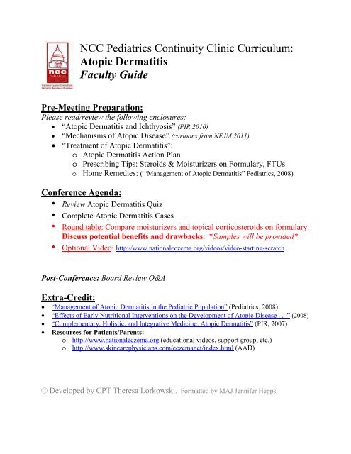
NCC Pediatrics Continuity Clinic Curriculum: Atopic Dermatitis ...
NCC Pediatrics Continuity Clinic Curriculum: Atopic Dermatitis ...
NCC Pediatrics Continuity Clinic Curriculum: Atopic Dermatitis ...

You also want an ePaper? Increase the reach of your titles
YUMPU automatically turns print PDFs into web optimized ePapers that Google loves.
<strong>NCC</strong> <strong>Pediatrics</strong> <strong>Continuity</strong> <strong>Clinic</strong> <strong>Curriculum</strong>:<br />
<strong>Atopic</strong> <strong>Dermatitis</strong><br />
Faculty Guide<br />
Pre-Meeting Preparation:<br />
Please read/review the following enclosures:<br />
• “<strong>Atopic</strong> <strong>Dermatitis</strong> and Ichthyosis” (PIR 2010)<br />
• “Mechanisms of <strong>Atopic</strong> Disease” (cartoons from NEJM 2011)<br />
• “Treatment of <strong>Atopic</strong> <strong>Dermatitis</strong>”:<br />
o <strong>Atopic</strong> <strong>Dermatitis</strong> Action Plan<br />
o Prescribing Tips: Steroids & Moisturizers on Formulary, FTUs<br />
o Home Remedies: ( “Management of <strong>Atopic</strong> <strong>Dermatitis</strong>” <strong>Pediatrics</strong>, 2008)<br />
Conference Agenda:<br />
• Review <strong>Atopic</strong> <strong>Dermatitis</strong> Quiz<br />
• Complete <strong>Atopic</strong> <strong>Dermatitis</strong> Cases<br />
• Round table: Compare moisturizers and topical corticosteroids on formulary.<br />
Discuss potential benefits and drawbacks. *Samples will be provided*<br />
• Optional Video: http://www.nationaleczema.org/videos/video-starting-scratch<br />
Post-Conference: Board Review Q&A<br />
Extra-Credit:<br />
• “Management of <strong>Atopic</strong> <strong>Dermatitis</strong> in the Pediatric Population” (<strong>Pediatrics</strong>, 2008)<br />
• “Effects of Early Nutritional Interventions on the Development of <strong>Atopic</strong> Disease . . .” (2008)<br />
• “Complementary, Holistic, and Integrative Medicine: <strong>Atopic</strong> <strong>Dermatitis</strong>” (PIR, 2007)<br />
• Resources for Patients/Parents:<br />
o http://www.nationaleczema.org (educational videos, support group, etc.)<br />
o http://www.skincarephysicians.com/eczemanet/index.html (AAD)<br />
© Developed by CPT Theresa Lorkowski. Formatted by MAJ Jennifer Hepps.
Article skin<br />
<strong>Atopic</strong> <strong>Dermatitis</strong> and Ichthyosis<br />
Roselyn E. Epps, MD*<br />
Author Disclosure<br />
Dr Epps has disclosed<br />
no financial<br />
relationships relevant<br />
to this article. This<br />
commentary does not<br />
contain a discussion<br />
of an unapproved/<br />
investigative use of a<br />
commercial<br />
product/device.<br />
Objectives After completing this article, readers should be able to:<br />
1. Identify the characteristic features of atopic dermatitis and the factors that worsen it.<br />
2. Understand that children who have atopic dermatitis are prone to recurrent infections,<br />
particularly with Staphylococcus aureus and herpes simplex virus.<br />
3. Know the signs of Wiskott-Aldrich syndrome.<br />
4. Plan the appropriate treatment of atopic dermatitis (emollients, corticosteroids,<br />
antibiotics, and allergen elimination when appropriate).<br />
5. Recognize ichthyosis vulgaris and know that ichthyosis commonly occurs in children<br />
who have atopic dermatitis.<br />
6. List the effective therapies in the management of ichthyosis vulgaris.<br />
7. Distinguish between tinea pedis and atopic dermatitis.<br />
8. Discuss the relationship of atopic dermatitis and food allergies and how to evaluate a<br />
patient who has both.<br />
9. Explain why children who have one component of atopy syndrome (allergic rhinitis,<br />
asthma, atopic dermatitis) have a threefold greater risk of developing a second<br />
component.<br />
<strong>Atopic</strong> <strong>Dermatitis</strong><br />
<strong>Atopic</strong> dermatitis (AD) is a chronic, relapsing dermatosis that features dry skin (xerosis),<br />
pruritus, and a personal or family history of eczema, allergic rhinitis or allergies, or asthma.<br />
Children who have one component of the atopic triad (AD, asthma, allergic rhinitis) are<br />
three times as likely to develop a second component. There is no sex predilection, and the<br />
onset frequently is in infancy. Although many affected children outgrow the condition by<br />
age 5 years, AD may persist into adolescence and adulthood. A smaller percentage of<br />
patients experience the onset of AD as older children or in adulthood.<br />
The incidence and prevalence of AD have increased in the United States and worldwide,<br />
particularly in developed nations. Fewer than 10% of children were affected in the 1970s,<br />
but recent epidemiologic studies estimate that 15% to 20% of children are diagnosed with<br />
AD. The reason for the increased rate is unknown. The “hygiene hypothesis” proposes that<br />
decreased exposure to infectious and biologic antigens may result in an increased response<br />
to environmental antigens or perhaps to decreased immune suppression. Additional<br />
research must be conducted to determine the reasons for the increased prevalence and to<br />
address the trend.<br />
Pathophysiology<br />
Manifestations of AD are believed to be due to the interaction of certain genes, the<br />
environment, and immunologic response to the environment and specific trigger factors.<br />
Patients who have AD may be considered to have systemic changes, not just manifestations<br />
in the skin. Susceptible individuals can react to internal and environmental triggers in<br />
certain target organs, not only resulting in skin eruptions, but also in asthma and allergic<br />
rhinitis. Patients may exhibit extrinsic immunoglobulin E (IgE)-mediated sensitization<br />
due to external antigens, with allergenic signs and elevated allergen-specific IgE, or<br />
intrinsic sensitization, without IgE-mediated sensitization.<br />
*Chief, Division of Dermatology, Children’s National Medical Center, George Washington University School of Medicine and<br />
Health Sciences, Washington, DC.<br />
278 <strong>Pediatrics</strong> in Review Vol.31 No.7 July 2010<br />
Downloaded from http://pedsinreview.aappublications.org/ by Theresa Lorkowski on January 5, 2012
skin<br />
atopic dermatitis and ichthyosis<br />
In acute AD lesions, T-helper 2 (TH2) cells are<br />
present in larger numbers than normal and have increased<br />
expression of specific cytokines that, in turn,<br />
stimulate B cells to produce IgE, resulting in peripheral<br />
eosinophilia. Cytokines and chemokines are released<br />
from cells in the skin, attracting other inflammatory cells<br />
and producing inflammatory mediators and reactions.<br />
Keratinocytes, Langerhans cells, endothelial cells,<br />
monocyte-macrophages, and eosinophils all play roles in<br />
the acute and chronic inflammation of AD.<br />
<strong>Clinic</strong>al Manifestations<br />
Generally, the primary lesion is a red, rough, poorly<br />
defined dry papule or plaque. Scaling may be seen. There<br />
is no central clearing. In children of color or deep pigmentation,<br />
plaques can be papular or follicular, especially<br />
on the trunk or over the extensor areas of joints.<br />
AD is diagnosed clinically and manifests particular<br />
patterns at different ages. Frequently, infants present<br />
with rough patches or plaques on the cheeks, the dorsa of<br />
the wrists, the ankles, and the lateral extremities. The<br />
perioral and diaper areas customarily are spared. After<br />
infancy, children develop flexural involvement, and the<br />
cheek areas improve. The neck, antecubital and popliteal<br />
fossae, and gluteal folds frequently are involved (Fig. 1).<br />
Teenagers are more likely to experience eyelid eruptions.<br />
With age, the hands and feet also become more<br />
problematic, and AD may present as dyshidrotic eruptions.<br />
Any part of the body, from the scalp to the soles<br />
and including the lips and genitalia, may be affected at<br />
any age. Eruptions occur whether or not an offending<br />
factor or trigger is identified. Exacerbations and remissions<br />
are common and to be expected.<br />
Secondary skin changes occur frequently. Oozing,<br />
weeping, and crust formation can develop, which may<br />
represent secondary infection. Hyperpigmentation as<br />
well as hypopigmentation and depigmentation (loss of<br />
pigmentation) can occur. Normal color of the skin usually<br />
returns when the signs and symptoms of AD resolve.<br />
Weeks to months may be required for the hyperpigmentation<br />
and hypopigmentation to resolve. If excoriations<br />
are deep or the inflammation is severe, scarring or depigmentation<br />
can be permanent.<br />
Lichenification, a hallmark of AD, is thickening and<br />
accentuation of skin markings due to chronic scratching.<br />
Lichenified skin on the hands and feet is more likely to<br />
fissure. Excoriations are common, and some patients<br />
create erosions and deeper wounds by unrelenting, intense,<br />
repeated trauma. Repeated friction and trauma<br />
promote inflammation and trigger inflammatory reactions<br />
and pathways in affected skin. Lichenification also<br />
Figure 1. <strong>Atopic</strong> dermatitis often involves the antecubital<br />
fossa. Lichenification and excoriations are evident in this<br />
example.<br />
may occur in other dermatologic conditions that feature<br />
chronic scratching and pruritus.<br />
Friction on the skin and scratching the skin are known<br />
to exacerbate pruritus and can initiate the “itch-scratch<br />
cycle,” in which the child scratches, itching in the involved<br />
area increases, the child continues to scratch, and<br />
the cycle continues. Plaques, papules, and nodules can<br />
result because of the escalating “itch-scratch cycle.”<br />
Secondary Infection<br />
Patients who have AD are more likely to develop skin and<br />
possibly systemic infections. One reason superinfection<br />
occurs more easily is due to altered skin barrier function,<br />
including apparent and imperceptible excoriations, fissures,<br />
and skin defects (Fig. 2). For patients who have<br />
AD, seemingly uninvolved skin is not normal. In addition<br />
to greater irritancy and dryness, there are immunologic<br />
differences in the type of TH2 cells and an increase<br />
in the number of TH2 cells within the skin. Staphylococcus<br />
aureus is an important cause of superinfection. S<br />
aureus colonization by age 6 months, with frequent<br />
<strong>Pediatrics</strong> in Review Vol.31 No.7 July 2010 279<br />
Downloaded from http://pedsinreview.aappublications.org/ by Theresa Lorkowski on January 5, 2012
skin<br />
atopic dermatitis and ichthyosis<br />
Figure 2. <strong>Atopic</strong> dermatitis of this leg shows erythema,<br />
excoriations, and scaling consistent with superinfection.<br />
colonization during the first year after birth, is associated<br />
with an increased prevalence and severity of AD. Impaired<br />
skin barrier function, a defective host immune<br />
response, and increased synthesis of extracellular matrix<br />
adhesion substances promote S aureus colonization.<br />
Exotoxins secreted by S aureus penetrate the skin<br />
barrier and stimulate T cells and antigen-presenting<br />
cells, thereby exacerbating and contributing to persistent<br />
skin inflammation. S aureus overgrowth and superinfection<br />
can result in flares, impetigo, folliculitis, cellulitis,<br />
abscesses, bacteremia, and sepsis. Methicillin-resistant<br />
S aureus, now more common in the community, can be<br />
particularly problematic for patients who have AD and<br />
their families. The patient’s infection must be treated,<br />
and treating the family may be necessary to minimize the<br />
risk of AD exacerbation in the patient. The clinician<br />
should consider culturing the atopic patient who is febrile,<br />
is unresponsive to therapy, or shows an inadequate<br />
response to maximized treatment that includes antibiotic<br />
therapy. Other bacteria also may be cultured from the<br />
AD patient’s wounds and should be treated accordingly.<br />
Although patients who have AD develop bacterial<br />
infections, they also may acquire viral and fungal infections.<br />
Eczema herpeticum occurs when AD is superinfected<br />
with a herpesvirus, either herpes simplex virus or<br />
varicella-zoster virus. Vesicles develop on affected and<br />
apparently unaffected skin and can be very painful. When<br />
disseminated, there may be associated viremia, fever, and<br />
lymphadenopathy, and patients can become very ill. Ocular<br />
involvement may occur innocuously when the patient<br />
rubs his or her eyes. Acyclovir should be administered<br />
intravenously in critical disseminated infections or<br />
orally for localized, recurrent infections in patients who<br />
have AD. If herpesvirus infection involves the eye or<br />
periocular area, ophthalmology consultation is essential<br />
to manage herpes keratitis and to prevent permanent loss<br />
of vision.<br />
Dermatophytes and yeast also can superinfect the<br />
skin. Patients who have AD can develop tinea capitis and<br />
tinea pedis, and it can be difficult at times to distinguish<br />
AD from tinea infection because both may involve pruritus,<br />
scaling, and inflammation. On physical examination,<br />
unlike AD, tinea pedis frequently develops in the<br />
toe web spaces (particularly the third and fourth). Tinea<br />
lesions frequently feature expanding plaques with central<br />
clearing and peripheral papules and scale. Potassium<br />
hydroxide slide examination of a sample taken from the<br />
skin from any affected body area, including the skin, the<br />
scalp, and hair, can help make the diagnosis.<br />
Allergy and Environment<br />
Allergic contact dermatitis can exacerbate AD. Common<br />
contact triggers include fragrances and preservatives in<br />
personal care products such as soaps, cleansers, shampoos,<br />
detergents, and certain emollients. Among other<br />
materials and substances that commonly elicit symptoms<br />
of allergic contact dermatitis are wool, nickel, synthetics,<br />
dyes, and rubber.<br />
Physical and environmental factors also can play a<br />
role. Temperature changes between cold and hot environments<br />
(as when moving from an air conditioned<br />
enclosure to hot outdoor weather) or change of season<br />
can be problematic. Some children prefer warm or cool<br />
temperatures. Therefore, their dermatitis is milder in the<br />
summer or winter, respectively. Other environmental<br />
variables such as dust and mites, pollen, and ambient<br />
humidity can have an impact. Because sweating can<br />
produce pruritus and skin eruptions for some patients,<br />
treatment of AD may require modifying exercise regimens.<br />
Clothing tags, coarse fabrics, snug clothing, and<br />
footwear can worsen symptoms in a localized distribution.<br />
Emotional factors such as stress, anger, sleepiness,<br />
and boredom often increase pruritus.<br />
The role of foods in causing AD can be significant for<br />
some children; food allergies can be present in up to 40%<br />
of patients who have AD. Symptoms include pruritus,<br />
urticaria, contact dermatitis, and exacerbation of AD as<br />
well as wheezing, asthma, and anaphylaxis. The symptoms<br />
can be immediate or delayed. Among the leading<br />
allergenic foods are milk and dairy products, eggs, wheat,<br />
soy, and peanuts. Some children outgrow allergies to<br />
particular foods, but peanuts and eggs are often the<br />
exception. Although some foods are difficult to avoid,<br />
the improved availability of nutritional information, di-<br />
280 <strong>Pediatrics</strong> in Review Vol.31 No.7 July 2010<br />
Downloaded from http://pedsinreview.aappublications.org/ by Theresa Lorkowski on January 5, 2012
skin<br />
atopic dermatitis and ichthyosis<br />
etary counseling, and food labeling helps families make<br />
proper dietary choices for children who have food allergies.<br />
Allergy skin prick testing usually is more reliable after<br />
age 2 years; specific radioallergosorbent testing can be<br />
performed in infancy. The patient must not take oral<br />
antihistamines or steroid medications for several days<br />
before skin prick allergy testing; AD should be controlled<br />
as much as possible to allow proper evaluation while<br />
minimizing patient discomfort. Avoidance of allergenic<br />
substances identified by allergy testing occasionally benefits<br />
the patient who has AD, but AD can be exacerbated<br />
by unrelated factors while allergies are present. Allergy<br />
testing may be repeated and expanded if the patient does<br />
not improve after avoidance therapy.<br />
Management<br />
Treatment of AD requires a coordinated plan aimed at<br />
moisturizing dry skin, decreasing inflammation, treating<br />
any infections, and avoiding irritants and other factors<br />
associated with dermatitis flares. The regimen must be<br />
discussed with the family, patient, and clinicians to ensure<br />
compliance.<br />
Bathing is an important aspect of general skin care for<br />
patients who have AD. Baths and showers should be brief<br />
and the water comfortably warm, never hot. After exposure<br />
to water, the skin should be patted or excess water<br />
brushed off of the skin before applying medication and<br />
moisturizer. Some patients improve and are maintained<br />
with daily or twice-daily bathing. Other patients experience<br />
drying and increased pruritus or discomfort with<br />
water contact, making infrequent bathing the required<br />
approach. In addition, during flares, some patients are<br />
unable to bathe or shower due to discomfort and pain.<br />
Bathing may be resumed when symptoms decrease.<br />
Although not necessary, a variety of commercial products,<br />
including cleansers, soaps, oils, and oatmeal powders,<br />
can be combined with bath water. Fragrance-free<br />
soaps and cleansers are preferred, but which product<br />
benefits or is tolerated by each patient differs. The use of<br />
bubble bath, shampoo, and dishwater detergent to<br />
cleanse the body should be avoided. For some, dilute<br />
chlorine bleach baths are beneficial, particularly for children<br />
whose AD improves after swimming in chlorinated<br />
pools. One-quarter to one-half cup of bleach in the<br />
bathtub (24 gallons or a standard tub filled 4 to 6 inches)<br />
should create a sufficient concentration without bleaching<br />
or damaging linens. Dilute white vinegar, extra light<br />
olive oil, and other products also have been used for<br />
bathing.<br />
Because the skin of patients who have AD is dry, the<br />
use of emollients is a cornerstone of therapy. Even without<br />
visible lesions, dry skin often is pruritic. Many products<br />
are available; no single emollient provides relief,<br />
moisturizes the skin, and improves skin barrier function<br />
for all patients. The medication vehicle (eg, cream versus<br />
ointment) and the presence of fragrance, preservatives,<br />
or other additives can affect the patient’s response. Lotions,<br />
creams, ointments, and oils are composed of varying<br />
amounts of oil and water. Ointments are composed<br />
of more petroleum jelly, creams contain more water than<br />
oil, and lotions contain more water than cream. If the<br />
skin is excoriated or fissured, stinging or pain can occur<br />
from products containing more water. An optimal time<br />
for moisturizer application is immediately after the bath<br />
or shower. Many patients benefit from several emollient<br />
applications per day.<br />
Topical corticosteroids have been a mainstay of AD<br />
therapy for approximately 50 years. Hydrocortisone (up<br />
to 1%) is available over the counter, and numerous<br />
prescription preparations are available (Table). Ointments,<br />
creams, lotions, gels, foams, and oil preparations<br />
are available. Different preparations deliver corticosteroid<br />
through the skin in varying potencies. If preparations<br />
are used sparingly and appropriately, adverse effects<br />
should be minimized. Adverse effects include skin atrophy,<br />
telangiectasias, striae, and systemic absorption. The<br />
use of potent and fluorinated corticosteroids on the face<br />
and intertriginous and diaper areas should be avoided<br />
due to increased absorption through thinner or occluded<br />
skin. Middle-strength to more potent corticosteroid<br />
medications may be required for treating lichenified areas<br />
or on the hands and feet due to the increased skin<br />
thickness and keratin of the skin layers.<br />
Several clinical trials of topical corticosteroid use in<br />
the pediatric age group have been performed or are in<br />
progress; some topical corticosteroids are approved specifically<br />
for use in children and some infants. Many<br />
practitioners find topical corticosteroids useful to break<br />
the itch-scratch cycle, treat acute flares, and minimize<br />
symptoms of inflammation. When signs and symptoms<br />
improve, the frequency of topical corticosteroid application<br />
should be reduced while moisturizer use is continued.<br />
Continuous, prolonged application of topical corticosteroids<br />
also can produce tachyphylaxis or decreased<br />
effectiveness of the medication.<br />
Oral corticosteroid therapy has limited use in treating<br />
AD. Although helpful for some severe flares, once therapy<br />
is discontinued, the rebound or subsequent flare that<br />
may occur might be more severe than the initial exacerbation<br />
and more difficult to control. Some patients become<br />
oral corticosteroid-dependent in their attempt to<br />
<strong>Pediatrics</strong> in Review Vol.31 No.7 July 2010 281<br />
Downloaded from http://pedsinreview.aappublications.org/ by Theresa Lorkowski on January 5, 2012
skin<br />
atopic dermatitis and ichthyosis<br />
Table. Topical Corticosteroids Ranked Strongest<br />
(Class I) to Weakest (Class VII)<br />
Medication<br />
Available Formulation<br />
Class I Clobetasol propionate 0.05% Cream, ointment, gel, foam<br />
Betamethasone dipropionate Ointment<br />
augmented 0.05%<br />
Diflorasone diacetate 0.05% Ointment<br />
Fluocinonide 0.01%<br />
Ointment<br />
Halobetasol propionate 0.05% Cream, ointment<br />
Class II Amcinonide 0.01% Ointment<br />
Betamethasone dipropionate Ointment<br />
0.05%<br />
Betamethasone dipropionate Cream<br />
augmented 0.05%<br />
Desoximetasone 0.25%<br />
Cream, ointment<br />
Desoximetasone 0.05%<br />
Gel<br />
Fluocinonide 0.05%<br />
Cream, ointment, gel, solution<br />
Halcinonide 0.1%<br />
Cream, ointment, solution<br />
Mometasone furoate 0.1% Ointment<br />
Class III Amcinonide 0.1% Cream, lotion<br />
Betamethasone valerate 0.1% Ointment<br />
Desoximetasone 0.05%<br />
Cream<br />
Fluocinonide emollient 0.05% Cream<br />
Fluticasone propionate 0.005% Ointment<br />
Halcinonide 0.1%<br />
Solution<br />
Triamcinolone acetonide 0.1% Ointment<br />
Class IV Betamethasone valerate 0.12% Foam<br />
Fluocinonide acetonide 0.025% Ointment<br />
Flurandrenolide 0.05%<br />
Ointment<br />
Fluticasone propionate 0.05% Cream<br />
Hydrocortisone valerate 0.2% Ointment<br />
Mometasone furoate 0.1% Cream, lotion<br />
Triamcinolone acetonide 0.1% Cream, ointment<br />
Class V Betamethasone dipropionate Lotion<br />
0.05%<br />
Betamethasone valerate 0.1% Cream<br />
Clocortolone pivalate 0.1% Cream<br />
Desonide 0.05%<br />
Ointment<br />
Fluocinolone acetonide 0.025% Cream<br />
Flurandrenolide 0.05%<br />
Cream<br />
Fluticasone propionate 0.01% Oil<br />
Fluticasone propionate 0.05% Cream<br />
Hydrocortisone butyrate 0.1% Cream, ointment<br />
Hydrocortisone valerate 0.2% Cream<br />
Prednicarbate 0.1%<br />
Cream<br />
Triamcinolone acetonide 0.1% Lotion<br />
Class VI Alclometasone 0.05% Cream, ointment<br />
Betamethasone valerate 0.1% Lotion<br />
Desonide 0.05%<br />
Cream<br />
Fluocinolone acetonide 0.01% Cream, lotion<br />
Hydrocortisone butyrate 0.1% Solution<br />
Triamcinolone acetonide 0.1% Cream<br />
Triamcinolone acetonide Cream, lotion<br />
0.025%<br />
Class VII Hydrocortisone acetonide,<br />
dexamethasone<br />
Cream, ointment, lotion<br />
Note: Vehicle affects medication potency for several products.<br />
prevent flares and are more likely to<br />
develop adverse systemic effects such<br />
as hypothalamic-pituitary axis suppression,<br />
growth retardation, and cushingoid<br />
features.<br />
Chronic, high-dose, or highpotency<br />
oral corticosteroid use has<br />
been shown to cause osteopenia or<br />
osteoporosis in children and adults. It<br />
is not known whether chronic intermittent<br />
topical corticosteroid use affects<br />
the bones of children. Some physicians<br />
give vitamin D and calcium<br />
supplementation to patients who have<br />
AD. Of note, the American Academy<br />
of <strong>Pediatrics</strong> has released new recommendations<br />
regarding vitamin D supplementation<br />
in children; the recommended<br />
minimum dose was doubled<br />
to 400 IU daily for infants and children.<br />
Clearly, corticosteroid use in<br />
children who have AD, the impact of<br />
therapy on bone health, and the role of<br />
vitamin D and calcium supplementation<br />
merit additional scientific study.<br />
Topical calcineurin inhibitors are<br />
newer elements of the therapeutic<br />
armamentarium. Pimecrolimus 1%<br />
cream is approved for mild-tomoderate<br />
AD. Tacrolimus ointment is<br />
available in 0.03% and 0.1% strengths<br />
and is targeted for moderate-to-severe<br />
AD. Tacrolimus 0.03% and pimecrolimus<br />
are approved by the United States<br />
Food and Drug Administration for<br />
those ages 2 years and older; tacrolimus<br />
0.1% is intended for those ages 15<br />
years and older. Both medications can<br />
be used on any part of the body and are<br />
particularly beneficial for the eyelids,<br />
face, and intertriginous areas.<br />
The most common adverse effects<br />
reported are burning at the site of application,<br />
headache, upper respiratory<br />
tract symptoms, cough, and pyrexia.<br />
In addition, exacerbation of viral infections,<br />
including herpesvirus infection,<br />
verrucae, and molluscum contagiosum,<br />
may be more likely in patients<br />
who use these products. A black box<br />
warning was placed on both medica-<br />
282 <strong>Pediatrics</strong> in Review Vol.31 No.7 July 2010<br />
Downloaded from http://pedsinreview.aappublications.org/ by Theresa Lorkowski on January 5, 2012
skin<br />
atopic dermatitis and ichthyosis<br />
tions, stating that long-term safety of topical calcineurin<br />
inhibitors has not been established and that these medications<br />
are not recommended for use in children younger<br />
than age 2 years. Additional therapeutic trials in children<br />
who have AD are planned and needed.<br />
Several prescription topical nonsteroidal moisturizing<br />
creams have been approved for use in AD. Their purpose<br />
is to improve the hydrolipid layer and barrier function,<br />
relieve AD symptoms, and promote wound healing.<br />
They may be used alone or in combination with topical<br />
corticosteroids and calcineurin inhibitors. The nonsteroid<br />
creams <strong>Atopic</strong>lair® Nonsteroidal Cream (Graceway<br />
Pharmaceuticals, Bristol, Tenn.), Eletone® Cream<br />
(Ferndale Laboratories, Ferndale, Mich.), Epiceram<br />
Skin Barrier Emulsion (Promius Pharmaceuticals,<br />
Bridgewater, NJ), and MimyX Cream (Steifel Pharmaceuticals,<br />
Bristol, Tenn.) are approved for all ages, for use<br />
on any area of the body, and may be used two to three<br />
times a day. Zetania cream (Tiber Laboratories, Suwanee,<br />
Ga.) is approved for children 2 years of age and<br />
older. Patients allergic to any components of the creams<br />
should avoid their use.<br />
Oral antihistamine drugs have been prescribed for<br />
patients who have AD. Although not statistically proven<br />
to be useful for treating pruritus generally, oral antihistamines<br />
can be helpful for children who have an<br />
urticarial component or decreased or altered sleep patterns<br />
due to pruritus.<br />
Wiscott-Aldrich Syndrome<br />
Wiskott-Aldrich syndrome is one important condition<br />
to consider in patients who have AD. This X-linked<br />
recessive disorder features eczematous eruptions in association<br />
with thrombocytopenia and recurrent infections.<br />
Thrombocytopenic purpura and hemorrhagic<br />
events may occur. The identified Wiskott-Aldrich syndrome<br />
protein (WASP) gene codes for a cytoplasmic<br />
protein that has multiple functions. The impaired humoral<br />
immune response to polysaccharide antigens<br />
seen in patients who have Wiskott-Aldrich syndrome<br />
makes patients susceptible to bacteria such as Streptococcus<br />
pneumoniae and Pneumocystis jiroveci and, later, to<br />
viruses. After the second decade of life, these patients are<br />
at risk for developing leukemia and lymphoma.<br />
Job Syndrome<br />
Another important condition to consider is Job syndrome,<br />
or hyperimmunoglobulin E syndrome (HIES),<br />
which is defined by eczematous eruptions associated with<br />
IgE concentrations greater than 2,000 IU/mL and repeated<br />
skin and sinopulmonary infections. The classic<br />
autosomal dominant form is due to a mutation in the<br />
signal transducer and activator of the transcription 3<br />
(STAT3) gene. Skin eruptions appear during the newborn<br />
period, with onset of infections during the first 3<br />
postnatal months. Although the type of skin infection<br />
can vary, “cold” abscesses are typical and feature slight<br />
redness, no or low-grade fever, little systemic involvement,<br />
and minimal signs and symptoms, unlike abscesses<br />
seen in patients unaffected by HIES. Paronychiae and<br />
candidal infections are common. Although the eczematous<br />
symptoms usually resolve, the recurrent pulmonary<br />
infections due to S aureus and Haemophilus influenzae<br />
progress to chronic lung infections and subsequent lung<br />
changes. Of note, children who have AD can have very<br />
high concentrations of IgE; conversely, patients who<br />
have HIES can have normal IgE concentrations.<br />
Ichthyosis<br />
Ichthyosis represents a group of disorders that involves<br />
abnormal epidermal skin barrier function, keratinization,<br />
and desquamation. Multiple types of ichthyosis have<br />
been described. Initially defined descriptively, the disorders<br />
now can be distinguished by genetic, histologic,<br />
biochemical, and molecular methods.<br />
Ichthyosis vulgaris (IV) is the most common type,<br />
with an incidence of 1 in 250. The onset is during infancy<br />
or childhood, not at birth. Inheritance can be autosomal<br />
dominant or sporadic, so patients have a varied presentation.<br />
IV usually presents as fine white scales on the skin,<br />
sparing the antecubital and popliteal fossae. Scaling is<br />
most obvious on the lateral lower legs (Fig. 3). Hyperlinearity<br />
is noted on the palms and soles. IV can be<br />
innocuous and appear as an isolated finding. The histopathology<br />
may show a thinned-to-absent granular layer<br />
and a compact superficial stratum corneum. However, a<br />
skin biopsy may not be diagnostic; microscopically, IV<br />
can look like normal skin. IV often improves with age,<br />
and manifestations in adulthood may be minimal.<br />
There are many forms of ichthyosis, most of which are<br />
rare. Ichthyosis can be inherited in autosomal or X-linked<br />
patterns or by spontaneous mutation. Although IV is<br />
rather common, X-linked ichthyosis, lamellar ichthyosis,<br />
and harlequin fetus are rare, well-described forms<br />
(Fig. 4). Several syndromes and related conditions of<br />
note include ichthyosis as part of the clinical picture. KID<br />
syndrome is defined as keratitis, ichthyosis, and deafness.<br />
Netherton syndrome, also called ichthyosis linearis circumflexa,<br />
features congenital erythroderma as well as<br />
atopic dermatitis, hair shaft abnormalities, and high IgE<br />
concentrations.<br />
<strong>Pediatrics</strong> in Review Vol.31 No.7 July 2010 283<br />
Downloaded from http://pedsinreview.aappublications.org/ by Theresa Lorkowski on January 5, 2012
skin<br />
atopic dermatitis and ichthyosis<br />
Figure 3. Ichthyosis vulgaris demonstrates fine, white scales.<br />
Management<br />
Treatment of IV usually involves the use of topical salicylic<br />
acid; lactic acid; or urea in lotion, cream, or ointment<br />
form. These products moisturize, soften the skin,<br />
and aid in desquamation. For patients who have both IV<br />
and AD, these products are more likely to cause irritation.<br />
The products should be used cautiously in children<br />
because total body application can result in systemic<br />
absorption and serious adverse effects. Salicylic acid, in<br />
particular, should be used in children after 1 year of age<br />
and then with caution due to risks of salicylate toxicity.<br />
Research<br />
Significant research has been performed in IV, AD, and<br />
related disorders. IV often coexists with AD, and research<br />
has shown a genetic basis for this association in<br />
certain populations. Gene mutations in keratin proteins<br />
alter skin barrier function. Most important, the FLG gene<br />
produces profilaggrin, and filaggrin is critical for AD<br />
expression. Multiple international and familial studies<br />
have shown that FLG mutations in patients who have AD<br />
alter normal skin formation, function, and hydration and<br />
result in severe AD, as well as asthma associated with AD.<br />
Figure 4. Lamellar ichthyosis features larger, platelike scales.<br />
The mutation for IV also has been identified. Studies<br />
have shown that Northern European patients who have<br />
IV have a statistically significant increased risk for developing<br />
AD as well. Also, patients who have both IV and<br />
AD have a statistically significant increased risk for developing<br />
asthma. Overall, there is strong evidence for a<br />
genetic and molecular basis for the association of IV and<br />
AD. More studies are in progress and are necessary for<br />
Summary<br />
• Based on strong research evidence and consensus, a<br />
multifaceted, individualized approach to treatment<br />
benefits patients who have atopic dermatitis and<br />
includes bathing, emollients, topical antiinflammatory<br />
medications, allergen avoidance, and<br />
the use of antistaphylococcal antibiotics and<br />
antihistamines when clinically indicated. (1)(2)<br />
• Based on strong research evidence, mutations in the<br />
FLG gene cause ichthyosis vulgaris, resulting in<br />
alterations in the skin protein filaggrin. (3)(4)<br />
• Based on strong research evidence, atopic dermatitis<br />
is associated with certain populations who have<br />
ichthyosis vulgaris. (5)(6)(7)<br />
284 <strong>Pediatrics</strong> in Review Vol.31 No.7 July 2010<br />
Downloaded from http://pedsinreview.aappublications.org/ by Theresa Lorkowski on January 5, 2012
Mechanisms for <strong>Atopic</strong> Disease<br />
Statistics Review: Odds ratio: Odds of <strong>Atopic</strong> Disease with Filaggrin MUTATION (Disease/No Disease)<br />
Odds of <strong>Atopic</strong> Disease with NORMAL Filaggrin (Disease/No Disease)
Filaggrin Mutations Associated with Skin and Allergic Diseases<br />
Alan D. Irvine, M.D., W.H. Irwin McLean, Ph.D., D.Sc., and Donald Y.M. Leung, M.D., Ph.D.<br />
N Engl J Med 2011; 365:1315-1327 October 6, 2011
Potency<br />
Group<br />
Steroids on Formulary at WR-B (Jan 2014)<br />
Percentage<br />
Strength<br />
Brand Name<br />
(Alt Brand Name)<br />
Generic Name<br />
Size<br />
Augmented Betamethasone<br />
Dipropionate 0.05% lotion Diprolene 30 ml<br />
Class I<br />
Clobetasol Proprionate 0.05% ointment Temovate (Cormax) 15 g, 30 g<br />
15 ml, 30 ml, 45<br />
Clobetasol Proprionate 0.05% cream Temovate-E (Cormax) ml<br />
Temovate Scalp<br />
Clobetasol Proprionate<br />
0.05% solution (Cormax)<br />
25 ml, 30 ml<br />
Flurandrenolide 4mcg/cm tape Cordran 200 cm<br />
Augmented Betamethasone<br />
Class II Dipropionate 0.05% cream Diprolene AF 15 g<br />
Betamethasone Dipropionate 0.05% ointment Diprosone 15 g<br />
Fluocinonide 0.05% cream Lidex 15 g, 60 g<br />
Fluocinonide 0.05% gel Lidex 15 g, 60 g<br />
Fluocinonide 0.05% ointment Lidex 15 g, 60 g<br />
Fluocinonide 0.05% solution Lidex 60 ml<br />
Class III Amcinonide 0.1% cream Cyclocort 15 g, 60 g<br />
Betamethasone Dipropionate 0.05% lotion Diprosone 60 ml<br />
Fluocinonide Emollient 0.05% cream Lidex-E 15 g, 60 g<br />
Class IV Hydrocortisone Valerate 0.2% ointment Westcort 15 g, 60 g<br />
Triamcinolone Acetonide 0.1% ointment Kenalog 15 g, 80 g, 454 g<br />
Triamcinolone Acetonide 0.2% aerosol Kenalog 63 g<br />
Class V Betamethasone Valerate 0.1% cream Valisone 15 g<br />
Betamethasone Valerate 0.1% lotion Valisone 60 ml<br />
Desonide 0.05% ointment Tridesilon (DesOwen) 15 g, 60 g<br />
Flurandrenolide 0.05% lotion Cordran 60 ml<br />
Hydrocortisone Valerate 0.2% cream Westcort 15 g, 60 g<br />
Kenalog (Aristocort<br />
Triamcinolone Acetonide<br />
0.1% cream EQ)<br />
15 g, 80 g, 454 g<br />
Triamcinolone Acetonide 0.025% ointment Kenalog 15 g<br />
Class VI Desonide☺ 0.05% cream Tridesilon (DesOwen) 15 g, 60 g<br />
Fluocinolone Acetonide 0.01% oil Dermasmooth (Capex) 120 ml<br />
Triamcinolone Acetonide 0.025% cream Kenalog 15 g, 80 g<br />
Class VII Hydrocortisone 2.5% cream (Hytone) 28 g<br />
Hydrocortisone (OTC)☺ 1% cream 30 g<br />
0.5% cream 30 g<br />
1% lotion 118 ml<br />
1% ointment 30 g<br />
☺Safe for facial use
Moisturizers on Formulary at WR-B (Jan 2014)<br />
Aquaphor (mineral oil/petroleum)<br />
Cetaphil cream<br />
Hydrocerin lotion<br />
Vanicream<br />
454 g jar<br />
454 g jar<br />
480 ml<br />
454 g jar<br />
Finger Tip Units (FTUs)<br />
• Amount of ointment/cream product that extends from the tip of an adult finger to the first<br />
flexural crease of the DIP, when dispensed from a tube with the standard 5mm nozzle.<br />
• Used to gauge the amount of topical steroid to use on a given area of affected skin.<br />
• Provides guide to the appropriate size of tube that should be prescribed for a patient.<br />
• 1 FTU = 0.5 g = treats an area 2x the size of an adult’s hand with fingers together,<br />
• 2 FTUs = 1g = treats an area equivalent to 4 adult handprints (10 x 10cm), etc.<br />
• 30 g of product is necessary to fully cover an adult patient.
“Home Remedies”<br />
Additional Types of Baths (www.nationaleczema.org)<br />
Vinegar Baths: Referred to as the “pickle the patient” treatment. Add one cup to one pint of vinegar to the bath. Can be<br />
used as a wet dressing too as it kills bacteria.<br />
Bath Oil Baths: Oils in the bath are a favorite of some providers and patients. Bath oils can leave the tub slippery – be<br />
careful. They can also leave a hard-to-clean film. See if they work for you.<br />
Salt Baths: When there is a significant flare the bath water may sting or be uncomfortable. Add one cup of table salt to<br />
the bath water to decrease this side effect.<br />
Baking Soda Baths: Added to a bath or made into a paste it can be used to relieve the itching.<br />
Tar Baths: Tar baths can sooth inflammation and itch. Tar bath oil or tar shampoo can be used. Warning: if the skin is<br />
open or excoriated the tar baths can sting.<br />
Oatmeal baths: Added to a bath or made into a paste it can be used to relieve the itching.
<strong>Atopic</strong> <strong>Dermatitis</strong> Quiz<br />
1. The atopic triad includes: <strong>Atopic</strong> dermatitis, asthma, and allergic rhinitis. Children with<br />
one atopic disease are three times more likely to develop a second atopic disease.<br />
2. Describe the underlying etiology of atopic dermatitis.<br />
AD results from a complex interaction between genetics, immune response, and environment.<br />
The epidermis has structural changes and/or lower concentration of different proteins, including<br />
filaggrin and ceramides, which increases surface pH and interferes with the epidermal barrier<br />
function. Colonization and overgrowth of bacteria, such as S. aureus, may further interfere<br />
with the skin barrier due to exotoxin and protease production. The impaired barrier function<br />
enhances water loss allowing for increased xerosis, susceptibility to trauma, and absorption of<br />
irritants and allergens that induce local inflammatory response. The inflammatory responses in<br />
patients with AD may be overactive due to underlying genetics, but the patients may also have<br />
an upregulated Th2 immune response due to the limited Th1 activation (hygiene hypothesis).<br />
3. List the sites typically affected by atopic dermatitis depending on age:<br />
Infants Cheeks, dorsa of wrists and ankles, and the lateral extremities<br />
Children Neck, flexural surfaces- antecubital and popliteal fossae, and gluteal folds<br />
Teenagers Eyelids, hands and feet<br />
4. True or False: <strong>Atopic</strong> dermatitis can develop on any part of the body regardless of age.<br />
Specific distribution patterns are associated with age (see #3), but not exclusive. All areas of the<br />
body may be affected, including genitalia and lips. “From the scalp to the soles”<br />
5. Place the following topical corticosteroids in or order of decreasing potency:<br />
Class I (strongest)<br />
Class V<br />
Flurandrenoloide Tape<br />
Triamcinolone Acetonide 0.025% ointment<br />
Triamcinolone Acetonide 0.1% cream<br />
Class II<br />
Desonide 0.05% ointment<br />
Fluocinonide 0.05% cream<br />
Fluocinonide 0.05% ointment<br />
Class VI<br />
Desonide 0.05% cream<br />
Class III<br />
Fluoocinolone Acetonide 0.01% oil<br />
Fluocinonide 0.05% emollient cream<br />
Class VII (weakest)<br />
Class IV<br />
Hydrocortisone 2.5% cream<br />
Hydrocortisone Valerate 0.2% ointment<br />
Triamcinolone Acetonide 0.1% ointment<br />
6. List adverse effects of topical corticosteroids. Consider local and systemic.<br />
• Local: Atrophy, striae, telangiectasia, pigmentary changes, easy bruising, hypertrichosis,<br />
acne like eruptions, allergic/contact/irritant reaction.<br />
• Systemic: hypothalamic-pituitary-adrenal axis suppression, Cushing syndrome, growth<br />
retardation, glaucoma, cataracts, osteopenia/osteoporosis.
<strong>Atopic</strong> <strong>Dermatitis</strong> Cases<br />
Case 1:<br />
Colton is a 15 month old male infant who presents for a routine well visit. He is the only child of<br />
well-educated parents who live in the suburbs. His height and weight have been stable at the 50 th<br />
percentile. He was breastfed for the first year of his life and continues to breastfeed at night. He<br />
was introduced to solid foods at 6 months of age and has fully transitioned to table foods. His<br />
mother boasts that he has “never had a runny nose in his life” and that she uses hand sanitizer<br />
“all the time, of course”. Her only concern is for a red, dry, itchy rash that keeps recurring on his<br />
cheeks, arms, and legs. She has not noticed any triggers.<br />
Based on history, what are protective and predisposing factors for Colton developing AD<br />
• Protective: exclusive breastfeeding for at least the first 4<br />
months of life; introduction of solid foods after 4 months of life.<br />
• Predisposing: limited exposure to pathogens and dirt (first born,<br />
no illnesses, over-cleanliness) which could lead to up-regulation of<br />
the Th2 immune response (Hygiene Hypothesis—see diagram);<br />
frequent use of hand sanitizer which can be drying and contain<br />
fragrances/additives that further irritate the skin.<br />
On exam, Colton’s rash appears consistent with a mild-to-moderate eczema flare, with large dry<br />
erythematous plaques covering his cheeks and chin. His arms and legs have multiple papules<br />
and vesicles in the flexural surfaces with scratch marks.<br />
What treatment would you recommend<br />
Discuss with mother that treatment of eczema is a four-prong approach:<br />
1. Moisturization: “Soak and seal”. Bathe 1-2 times a day for 10-15min during flares, using<br />
gentle cleanser without scrubbing, and pat dry. Cover with moisturizer/emollient within 3<br />
min of bath to lock in water. Type of moisturizer is patient/parent preference but should be<br />
hypoallergenic with limited fragrances/additives to prevent further skin irritation. (Refer to<br />
the National Eczema Association Seal of Acceptance).<br />
2. Inflammation Reduction- Topical steroids are the first-line and only treatment approved for<br />
children
4. Avoidance of Triggers/Irritants: Recommend fragrance/preservative-free soaps, cleansers,<br />
shampoos, and detergents. Wear loose, nonabrasive cotton clothing. Wash all clothing<br />
before wearing with extra-rinse cycle. Avoid overheating during exercise or the summer.<br />
Use mattress and pillow covers, vacuum frequently if dust mites are a concern. Bathe/rinse<br />
off allergens and/or use antihistamines if pollens or animal dander exacerbate symptoms. Do<br />
not restrict diet, only avoid known food allergens. Limit stress. Keep fingernails short.<br />
His mother is wary of starting topical steroids and asks about alternatives or homeopathic<br />
treatments that are available.<br />
What would you recommend or advise against<br />
• Validate that her concerns are not unreasonable and that topical steroids can have a number<br />
of side effects, particularly if not used in appropriate areas or if used for prolonged courses.<br />
• An initial approach could be to focus on good skin care with regular moisturization and<br />
bathing. Moisturization is proven to decrease the need for steroids.<br />
• See Extra-Credit article on CAM therapies. Examples with no proven benefit are<br />
supplementation with EFAs and probiotics. Examples with proven benefits include:<br />
o Traditional Chinese Medicine herbs: Proven benefits, but caution should be taken<br />
due to risk of contamination. Agranulocytosis and liver toxicity have been observed.<br />
o Honey: Inherent anti-inflammatory properties; promotes healing. It can be mixed<br />
with wax and oil and applied to the skin with limited side effects.<br />
o Massage therapy: Relieves stress, improves blood flow, and enhances emollient<br />
delivery. Oils should be avoided as they can further irritate the skin.<br />
Prior to leaving, you obtain additional family history to better delineate his risk for additional<br />
atopic disease. His mother notes that she and all her sisters struggled with asthma as kids. His<br />
father is adopted so family history is unknown, but he does have a h/o anaphylaxis to peanuts.<br />
How would you counsel Colton’s mother regarding his risk, particularly for food allergies<br />
• Discuss the atopic triad and how it presents as the “atopic march”. Counsel that Colton<br />
does have a higher risk (3x greater than non-atopic child) of developing asthma or AR.<br />
• The development of additional atopic disease could be related to a common phenotype (e.g.<br />
defective filaggrin) or be a result of increased sensitization due to impaired skin barrier.<br />
• Do not restrict his diet unless a temporal relationship between an eczema flare and<br />
introduction of a food was noticed. If suspicion, consider RAST and referral to allergy.
Case 2<br />
Sarah is a 9 year old female brought in by her mother with the complaint of worsening eczema.<br />
The family just moved to the DC area from Arizona after her father was injured in Afghanistan.<br />
She is currently living in an apartment in Building 62 with her parents and two younger sisters.<br />
She has had eczema since infancy that has been controlled with periodic use of moisturizers and<br />
topical steroids. Her eczema today is the worse it has ever been, completely involving her arms,<br />
legs, and neck with patches on her trunk. She has been unable to sleep because of the pruritis<br />
and has little interest in going outside to play because of discomfort with walking.<br />
What further history would you like to know<br />
• Skin care regimen- moisturizers, bathing<br />
• Medications – including current and past topical therapies that have been effective<br />
• Allergies/Triggers- exposure to dust mites, carpeting, animals, new soaps/cleansers<br />
• Additional atopic disease<br />
• PMHx- including immunizations and significant childhood diseases<br />
• Additional social history- school attendance, contacts with skin eruptions<br />
Her mother, accustomed to these visits, brings in multiple half used 15 g tubes of steroids that<br />
were prescribed or purchased from the drugstore. She says she has tried all of them for 1-2 days<br />
with no relief. The only one that has helped was a new script for Desonide 0.05% cream, that is<br />
now half-full. She has been using it once a day for the past week on all of Sarah’s affected areas.<br />
Has the Desonide been used appropriately What clues can you use to answer this<br />
question, other than Sarah’s clinical symptoms<br />
Minimal improvement in clinical symptoms suggests inconsistent or inappropriate use of the<br />
topical steroid; however, complicating infections must also be considered.<br />
Another clue that the medication is not being used appropriately is the discrepancy between the<br />
amount of steroid left in the tube and the history of usage (see FTU Chart):<br />
• Coverage of a full leg = 4.5 FTUs<br />
• Coverage of a full arm = 2.5 FTUs<br />
• Coverage of the face and neck 2 FTUs.<br />
Coverage of all extremities & face<br />
= 16 FTUs = 8g of steroid/ day =<br />
56g of steroid/ 1 week<br />
If she has used ½ of a 15g Desonide tube, she has in fact used only 8g steroid/ 1 week.<br />
On exam, you notice that some of Sarah’s lesions appear impetiginous. You also learn she has a<br />
history of abscesses and that her father is receiving home IV antibiotics for MRSA osteomyelitis.<br />
How does this history and exam affect the treatment for Sarah’s eczema flare<br />
The history and likely impetigo suggest bacterial infection with possible overgrowth, which<br />
would explain why she had minimal improvement with topical steroids. Her skin should be<br />
cultured, especially to look for MRSA. Treatment should include not only a short course of<br />
oral antibiotics but also skin decolonization with bleach baths. Her mother and sisters should<br />
also be tested for MRSA and decolonized to prevent re-colonization.
What are additional types of superinfections that can occur in patients with AD<br />
• Viral: eczema herpeticum, eczema vaccinatum, molloscum contagiousum, papilloma (warts)<br />
• Fungal: Trichophytan sp (tinea), Malassezia (seborrhea)<br />
You ultimately decide to treat Sarah with an oral antibiotic for secondary bacterial infection,<br />
decolonize her with daily bleach baths for a week and nasal mupirocin, decrease inflammation<br />
with a low potency corticosteroid for her face and a medium potency corticosteroid for her<br />
extremities, and start her on a regular bathing and moisturizing regimen.<br />
She returns in one week with no further signs of infection. History and medication tubes support<br />
compliance. She continues, however, to complain of pruritis and pain and there has been only<br />
moderate improvement. There is no concern for secondary viral or fungal infection.<br />
What would else could you recommend<br />
(1) Wet wrap therapy to help better seal in the moisture, concentrate the topical steroids, and<br />
protect the skin from further irritation.<br />
• Recommend taking a soaking bath with gentle cleanser, no scrubbing and patting dry.<br />
• Follow immediately with application of topical steroids to affected areas and moisturizer to<br />
non-affected areas. Use a plastic spoon or tongue depressor to prevent contamination.<br />
• Soak dressings— long underwear, tube socks, Kerlix, ace bandages— in warm water and<br />
wring out excess water. Apply dressings and cover with dry material.<br />
• Avoid chilling by placing on more layers, using blankets, or turning up the heat. Leave on for<br />
1-2 hours until dry or overnight if comfortable. Reapply moisturizers after removal.<br />
(2) Additional options would be to increase the potency of steroids or switch to a topical<br />
calcineurin inhibitor (TCI). TCIs should not be used in conjunction with wet wraps. Further<br />
counseling on triggers and use of antihistamines to decrease pruritis should also be provided.
<strong>Atopic</strong> <strong>Dermatitis</strong> Board Review<br />
1. A 7-year-old girl presents in September with an intensely itchy rash of several weeks'<br />
duration. During the summer she had many mosquito bites and one area of ringworm, but<br />
otherwise she has had no prior skin conditions. She has had no fever, joint pain, myalgias,<br />
fatigue, or change in appetite or activity. Antihistamines helped when she had the insect bites,<br />
but now they have little effect. No other family members have a rash. On physical examination,<br />
the rash is apparent on exposed areas and consists of papules, vesicles, and wheals, some in a<br />
linear array or in triangular clusters. There are also numerous hyperpigmented macules. The<br />
scalp is involved, but the palms and soles are spared.<br />
Of the following, the MOST likely cause of this rash is<br />
A. <strong>Atopic</strong> dermatitis<br />
B. Id reaction to recent fungal infection<br />
C. Hypersensitivity reaction to insect bites<br />
D. Recurrent impetigo with postinflammatory changes<br />
E. Scabies<br />
The girl described in the vignette has papular urticaria, an annoying, pruritic eruption that is the<br />
result of an insect bite-induced hypersensitivity reaction. It typically affects children between 2<br />
and 10 years of age, who experience chronic or recurrent eruptions of papules, vesicles, and<br />
wheals, often with excoriations because of their persistent itchiness. The age distribution is<br />
presumed to be because children younger than 1 year of age are unable to mount the<br />
hypersensitivity reaction, and most children have developed tolerance to insects by age 10 years.<br />
The lesions typically are symmetrically distributed and grouped in linear or triangular clusters<br />
that are concentrated on exposed surfaces, including the scalp, waist, and sock lines. As<br />
described for the girl in the vignette, the palms and soles are spared. Often, hyperpigmented or<br />
violaceous macules persist as the original eruption heals. Although most cases are related to flea<br />
and mosquito bites, any biting insect may be involved, including bed bugs and mites. The rash<br />
can be frustrating for families and clinicians alike because of its chronicity, the limited treatment<br />
options (primarily symptomatic care), and the paradox that only one family member may be<br />
affected, even though it is due to insect bites.<br />
Papular urticaria differs from atopic dermitis by its lack of family history, distribution,<br />
configuration, and absence of scale. Although id reactions are typically symmetric and itchy,<br />
they often involve hands and feet and usually are preceded by a worsening of the underlying<br />
condition, which is often a fungal infection such as tinea capitis . Recurrent impetigo is<br />
characterized by crusting and responds well to antibiotic treatment. Like papular urticaria,<br />
scabies is insect-related and involves an immune reaction. However, scabies produces smaller<br />
papules or vesicles, frequently involves the hands and intertriginous areas, and is characterized<br />
by burrows that are not present with popular urticara. Also, scabies often affect multiple family<br />
members.
2. A 6-year-old boy presents with a 2-year history of frequent pruritic, erythematous eruptions on<br />
his arms and legs. The rash usually worsens during winter but occurs intermittently throughout<br />
the year. His mother has tried various moisturizers, but they have not been effective in<br />
controlling the rash. On physical examination, you note erythematous patches on his antecubital<br />
and popliteal regions bilaterally.<br />
Of the following the MOST appropriate initial step in management for this patient is<br />
A. food allergy skin testing<br />
B. oral antibiotic therapy<br />
C. oral antihistamine therapy<br />
D. topical calcineurin inhibitor therapy<br />
E. topical corticosteroid therapy<br />
The appearance of pruritic, erythematous patches in the antecubital and popliteal areas described<br />
for the boy in the vignette is most consistent with atopic dermatitis. <strong>Atopic</strong> dermatitis, also called<br />
eczema, is a common skin condition that affects up to 10% of children and frequently is the first<br />
presentation of atopy (ie, atopic dermatitis, allergic rhinitis, and asthma). Because the underlying<br />
mechanism for atopic dermatitis involves defects in the epidermal barrier function & subsequent<br />
evaporative loss, the mainstay of therapy is topical emollients. These include moisturizers (eg,<br />
creams, lotions, ointments) and occlusive products such as solid vegetable shortening or<br />
petroleum jelly. Because skin hydration also is important, patients should apply a moisturizer or<br />
an occlusive product after bathing for 10 to 15 minutes in warm (not lukewarm or tepid) water.<br />
Almost all children who have eczema have an overabundance of Staphylococcus aureus and<br />
inflammatory cytokine production. Topical corticosteroids are used to reduce inflammation when<br />
emollients and occlusive creams do not control symptoms adequately. Topical corticosteroids are<br />
available in different preparations (eg, ointment, cream, lotion) and potencies. Regular use of<br />
topical corticosteroids is associated with hypopigmentation, bruising, acne, and thinning of the<br />
skin. The patient should be monitored for secondary infections.<br />
Other therapies targeted at reducing bacteria include topical antibiotics, systemic antibiotics, and<br />
bleach baths. One-eighth to one-half cup of bleach per full bath two to three times a week can<br />
reduce S aureus colonization, but such baths also can be irritating to the skin. Further, although a<br />
few studies have demonstrated improvement in eczema using bleach baths, a recent Cochrane<br />
review did not support its use. Oral antibiotics also failed to demonstrate improvement when<br />
analyzed in a meta-analysis.<br />
Because atopic dermatitis typically involves a cyclical process of itching and scratching, patients<br />
frequently receive oral antihistamines to reduce pruritus. Sometimes these are helpful, but oral<br />
antihistamines often are inadequate and usually are added to therapy when topical emollients or<br />
topical corticosteroids fail to control symptoms.<br />
Topical calcineurin inhibitors were introduced in 2000 and approved for the treatment of atopic<br />
dermatitis in children older than 2 years. However, their use has been curtailed significantly after<br />
a black box warning was added in 2006. These medications are not as effective for eczema as<br />
moderate- to high-potency topical corticosteroids, but they are an alternative to mild-potency<br />
corticosteroids and result in less hypopigmentation, thinning of the skin, or bruising.
Both food and inhalant allergens may play roles in atopic dermatitis. Approximately 30% of<br />
children who have moderate-to-severe atopic dermatitis are ingesting a food that is exacerbating<br />
their eczema via an immunoglobulin E- or T cell-mediated process. Consultation with an<br />
allergist and consideration for skin or patch testing to commonly implicated foods (eg, milk, egg,<br />
wheat, soy, peanut) may be warranted. Some investigators have advocated reducing dust mite<br />
concentrations by encasing the mattress, pillow, and box spring in conjunction with regular<br />
washing of the bed linen. Because the child in the vignette only has mild atopic dermatitis,<br />
pursuing food or inhalant allergen testing is not an appropriate initial step.<br />
3. A 12-month-old girl presents with a 3-month history of a pruritic rash that involves her<br />
cheeks, neck, anterior trunk, and antecubital and popliteal areas. The rash improves after use of<br />
an over-the- counter topical steroid cream but still is present most days, and the infant often<br />
wakes up at night scratching. On physical examination, you observe a raised erythematous rash<br />
that has areas of lichenification.<br />
Of the following, the MOST helpful intervention is to<br />
A. eliminate fruit and acidic juices from the diet<br />
B. eliminate milk, eggs, soy, and wheat from the diet<br />
C. perform aeroallergen allergy testing<br />
D. perform food allergy testing<br />
E. recommend a skin biopsy<br />
Some 30% to 40% of infants who have moderate-to-severe atopic dermatitis (AD), such as<br />
described for the infant in the vignette, may have an underlying immunoglobulin (Ig) E-mediated<br />
food allergy exacerbating the AD. For some infants, food ingestion may result in immediate<br />
worsening of AD severity, although most infants do not demonstrate this immediate reaction.<br />
Many foods have been implicated in AD, but five (milk, eggs, soy, wheat, and peanut) account<br />
for 90% of the causative allergens. Both allergy skin testing and measurement of serum IgE to<br />
these foods can help to identify and eliminate likely triggers. Either a negative IgE blood test<br />
(
A skin biopsy can provide insight into the pathophysiology of chronic rashes or lesions.<br />
Generally, skin biopsies neither are advised nor provide insight into the causes of typical AD<br />
manifestations in infants, but atypical presentations or lack of expected improvement with<br />
appropriate therapy should prompt consideration of a dermatology referral.<br />
4. A 2-year-old boy presents for evaluation of a chronic pruritic eruption. His medical history is<br />
remarkable for recurrent epistaxis, otitis media, and pneumonia. Physical examination reveals<br />
erythematous, slightly scaling patches on the trunk and in the antecubital and popliteal fossae.<br />
Petechiae are present profusely.<br />
Of the following, these findings are MOST suggestive of:<br />
A. Acrodermatitis enteropathica<br />
B. Ataxia telangiectasia<br />
C. <strong>Atopic</strong> dermatitis<br />
D. Langerhans cell histiocytosis<br />
E. Wiskott-Aldrich syndrome<br />
Wiskott-Aldrich syndrome is an X-linked disease characterized by partial but significant<br />
deficiency in both the antibody and cell-mediated immune compartments. It produces a<br />
predictable triad of : (1) eczema; (2) severe thrombocytopenic purpura; (3) recurrent infections<br />
of all types. Classic presentation is bleeding, eczema, and recurrent otitis media. There is an<br />
increased risk of atopic disorders, lymphoma/leukemia, and infection from S. pneumonia, S.<br />
aureus, and H. influnezae type B. IgE/IgA ratio is very elevated, and IgM is depressed; there is<br />
an absence of blood group antibodies (isohemaglutinins) on laboratory evaluation. Treatment is<br />
supportive (IVIG and antibiotics). Splenectomy is often necessary to manage the<br />
thrombocytopenia.<br />
Ataxia-telangiectasia is an autosomal-recessive disorder of chromosomal repair. Mutations in<br />
immunoglobulin and T-cell receptor genes are not repaired, and progressive loss of immune<br />
function results. The clinical manifestations include: (1) progressive cerebellar ataxia; (2)<br />
oculocutaneous telangiectasia; (3) recurrent infections. The latter usually begin as recurrent<br />
sinopulmonary infections; a progressive development of infections of all types follows. The<br />
disorder is associated with a very high incidence of lymphoreticular malignancy in the 2 nd and 3 rd<br />
decades of life (e.g. non-Hodgkin’s lymphoma and gastric carcinoma). Deficient IgA and IgE<br />
levels are often seen initially, and progressive loss of other immunoglobulin types and cellmediated<br />
immune defects are common.<br />
Acrodermatitis enteropathica is an autosomal recessive metabolic disorder affecting the uptake<br />
of zinc, characterized by periorificial and acral dermatitis, alopecia, and diarrhea. Similar<br />
features may be present in acquired zinc deficiency. Skin lesions may be secondarily infected<br />
by bacteria such as Staphylococcus aureus or fungi like Candida albicans. Langerhans cell<br />
histiocytosis is a rare disease involving clonal proliferation of Langerhans cells—abnormal<br />
dendritic cells deriving from bone marrow and capable of migrating from skin to lymph nodes.<br />
<strong>Clinic</strong>ally, its manifestations range from isolated bone lesions to multisystem disease