RESUSCITATION
RESUSCITATION
RESUSCITATION

You also want an ePaper? Increase the reach of your titles
YUMPU automatically turns print PDFs into web optimized ePapers that Google loves.
ABC<br />
OF<br />
<strong>RESUSCITATION</strong><br />
Fifth edition<br />
Edited by M C Colquhoun, A J Handley and T R Evans
ABC OF<br />
<strong>RESUSCITATION</strong><br />
Fifth Edition
ABC OF<br />
<strong>RESUSCITATION</strong><br />
Fifth Edition<br />
Edited by<br />
M C Colquhoun<br />
Chairman of the Resuscitation Council (UK)<br />
A J Handley<br />
Past chairman of the Resuscitation Council (UK) and Chairman of<br />
ILCOR working party on basic life support<br />
and<br />
T R Evans<br />
Past chairman of the Resuscitation Council (UK)
© BMJ Publishing Group 2004<br />
All rights reserved. No part of this publication may be reproduced, stored in a retrieval system,<br />
or transmitted, in any form or by any means, electronic, mechanical, photocopying, recording<br />
and/or otherwise, without the prior written permission of the publishers.<br />
First edition 1986<br />
Second edition 1990<br />
Third edition 1995<br />
Fourth edition 1999<br />
This edition published in 2004<br />
by BMJ Publishing Group, BMA House, Tavistock Square,<br />
London WC1H 9JR<br />
www.bmjbooks.com<br />
British Library Cataloguing in Publication Data<br />
A catalogue record for this book is available from the British Library<br />
ISBN 0 72791669 6<br />
Typeset by Newgen Imaging Systems (P) Ltd., Chennai, India<br />
Printed and bound in Malaysia by Times Offset<br />
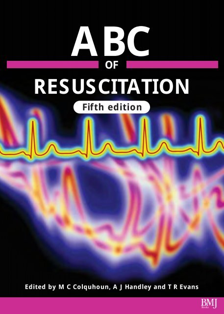
Cover image shows a computer-enhanced image of an electrocardiogram<br />
trace showing showing an abnormal heart beat (red). A healthy heartbeat is<br />
seen at the top (yellow) for comparison with permission from<br />
Mehan Kulyk/Science Photo Library
Contents<br />
Contributors<br />
Introduction<br />
Introduction to the Fifth Edition<br />
Notes on the algorithm approach to resuscitation<br />
Acknowledgements<br />
1 Basic life support 1<br />
Anthony J Handley<br />
2 Ventricular fibrillation 5<br />
Michael Colquhoun, Charles D Deakin, Douglas Chamberlain<br />
3 The automated external defibrillator 12<br />
Roy Liddle, C Sian Davies, Michael Colquhoun, Anthony J Handley<br />
4 Asystole and pulseless electrical activity 16<br />
Michael Colquhoun, A John Camm<br />
5 Management of peri-arrest arrhythmias 20<br />
Michael Colquhoun, Richard Vincent<br />
6 Airway control, ventilation, and oxygenation 25<br />
Robert Simons<br />
7 Post-resuscitation care 32<br />
Peter A Oakley, Anthony D Redmond<br />
8 Resuscitation in pregnancy 36<br />
Stephen Morris, Mark Stacey<br />
9 Resuscitation at birth 39<br />
Anthony D Milner<br />
10 Resuscitation of infants and children 43<br />
David A Zideman, Kenneth Spearpoint<br />
11 Resuscitation in the ambulance service 50<br />
Andrew K Marsden<br />
12 Resuscitation in hospital 54<br />
T R Evans<br />
13 Cardiopulmonary resuscitation in primary care 58<br />
Michael Colquhoun, Brian Steggles<br />
14 Resuscitation of the patient with major trauma 63<br />
Charles D Deakin<br />
15 Near drowning 72<br />
Mark Harries<br />
16 Drugs and their delivery 75<br />
Michael Colquhoun, David Pitcher, Jerry Nolan<br />
17 Cardiac pacing and implantable cardioverter defibrillators 81<br />
Michael Colquhoun, A John Camm<br />
18 Infection risks and resuscitation 87<br />
A J Harry Walmsley, David A Zideman<br />
19 Teaching resuscitation 90<br />
Ian Bullock, Geralyn Wynn, Carl Gwinnutt, Jerry Nolan, Sam Richmond, Jonathan Wylie,<br />
Bob Bingham, Michael Colquhoun, Anthony J Handley<br />
20 Training manikins 97<br />
Gavin D Perkins, Michael Colquhoun, Robert Simons<br />
21 The ethics of resuscitation 102<br />
Peter J F Baskett<br />
Index 107<br />
vi<br />
vii<br />
ix<br />
xi<br />
xiii<br />
v
Contributors<br />
Peter J F Baskett<br />
Consultant Anaesthetist Emeritus, Frenchay Hospital and the<br />
Royal Infirmary, Bristol<br />
Bob Bingham<br />
Consultant Anaesthetist, Great Ormond Street Hospital for<br />
Children NHS Trust, London<br />
Ian Bullock<br />
Head of Education and Training, Royal Brompton and<br />
Harefield NHS Trust; and Honorary Clinical Teaching Fellow,<br />
Imperial College, London<br />
A John Camm<br />
Professor of Clinical Cardiology, St George’s Hospital Medical<br />
School, London<br />
Douglas Chamberlain<br />
Professor of Resuscitation Medicine, University of Wales<br />
College of Medicine, Cardiff<br />
Michael Colquhoun<br />
Senior Lecturer in Prehospital Care, Wales Heart Research<br />
Institute, Cardiff<br />
C Sian Davies<br />
Programme Manager, National Defibrillator Programme, the<br />
Department of Health, London<br />
Charles D Deakin<br />
Consultant Anaesthetist, Southampton University Hospital,<br />
Southampton<br />
T R Evans<br />
Consultant Cardiologist, Royal Free Hospital, London<br />
Carl Gwinnutt<br />
Consultant Anaesthetist, Hope Hospital, Salford<br />
Anthony J Handley<br />
Consultant Physician and Cardiologist, Colchester<br />
Mark Harries<br />
Consultant Physician, Northwick Park and St Mark’s NHS<br />
Trust, Harrow<br />
Roy Liddle<br />
Resuscitation Training Officer, Wythenshaw Hospital,<br />
Manchester<br />
Andrew K Marsden<br />
Consultant Medical Director, Scottish Ambulance Service,<br />
Edinburgh<br />
Anthony D Milner<br />
Emetrius Professor of Neonatology, Guy’s and St Thomas’s<br />
Hospital Trust, London<br />
Stephen Morris<br />
Consultant Obstetric Anaesthetist, Llandough Hospital and<br />
Community NHS Trust, South Glamorgan<br />
Jerry Nolan<br />
Consultant Anaesthetist, Royal United Hospital, Bath<br />
Peter A Oakley<br />
Consultant in anaesthesia and trauma, Department of<br />
Trauma Research, University Hospital of North Staffordshire,<br />
Stoke on Trent<br />
Gavin D Perkins<br />
Research Fellow in Intensive Care Medicine, Birmingham<br />
Heartlands Hospital, Birmingham<br />
David Pitcher<br />
Consultant Cardiologist, Worcestershire Royal Hospital,<br />
Worcester<br />
Anthony D Redmond<br />
Professor of Emergency Medicine, Keele University, and<br />
Consultant in Emergency Medicine, the North Staffordshire<br />
Hospital NHS Trust, Stoke on Trent<br />
Sam Richmond<br />
Consultant Neonatologist, Sunderland Royal Hospital,<br />
Sunderland<br />
Robert Simons<br />
Consultant Anaesthetist, Royal Free Hospital, London<br />
Kenneth Spearpoint<br />
Senior Resuscitation Officer, Hammersmith Hospitals NHS<br />
Trust, London<br />
Mark Stacey<br />
Consultant Obstetric Anaesthetist, Llandough Hospital and<br />
Community NHS Trust, South Glamorgan<br />
Brian Steggles<br />
Chairman, Faculty of Prehospital Care, Royal College of<br />
Surgeons, Edinburgh<br />
Richard Vincent<br />
Professor of Medicine, Brighton and Sussex Medical School,<br />
Brighton<br />
A J Harry Walmsley<br />
Clinical Director and Consultant in Anaesthetics, East Sussex<br />
Hospitals NHS Trust, Eastbourne<br />
Jonathan Wylie<br />
Consultant Neonatologist, The James Cook University Hospital,<br />
Middlesbrough<br />
Geralyn Wynn<br />
Resuscitation Training Officer, Royal Free Hospital, London<br />
David A Zideman<br />
Consultant Anaesthetist, Hammersmith Hospital NHS Trust,<br />
London<br />
vi
Introduction<br />
The modern era of resuscitation began in 1960 with the publication of the classic paper by Jude, Kouwenhoven, and<br />
Knickerbocker on closed chest cardiac compression, which showed that the circulation could be maintained during cardiac arrest<br />
without the need for thoracotomy. A few years earlier Elam, Safar, and Gordon had established expired air ventilation as the most<br />
effective method for providing artificial ventilation for a patient who had stopped breathing. The effectiveness of closed chest<br />
defibrillation had been demonstrated by Zoll a few years earlier. By combining the techniques of chest compression with expired<br />
air ventilation, it became possible to maintain the viability of a patient in cardiopulmonary arrest until a defibrillator could be<br />
brought to the scene. Special units were established that were able to resuscitate patients at high risk of developing cardiac arrest,<br />
and special hospital cardiac arrest teams were created.<br />
After coronary care units were established for patients with acute myocardial infarction, it became apparent that most deaths<br />
from the condition occurred in the early stages, not because the myocardium was severely damaged, but because of potentially<br />
treatable disturbances in the cardiac rhythm. Once the effectiveness of resuscitation in hospital was established, the realisation that<br />
two thirds of deaths from coronary heart disease occurred before hospital admission led to attempts to provide coronary care, and<br />
particularly defibrillation, in the community. The credit for this development goes to Pantridge in Belfast, who pioneered the first<br />
mobile coronary care unit staffed by a doctor and nurse. This early experience confirmed the high incidence of lethal arrhythmias<br />
at the onset of myocardial infarction and many patients attended by the mobile units were successfully resuscitated from cardiac<br />
arrest. Pantridge and his coworkers also drew attention to the value of cardiopulmonary resuscitation (CPR) performed by<br />
bystanders before the arrival of the mobile unit.<br />
In the early 1970s, Leonard Cobb, a cardiologist in Seattle, inspired by these results, equipped paramedics with defibrillators<br />
and trained firefighters to act as first responders and perform basic life support. The fire service in Seattle is highly coordinated<br />
and a standard fire appliance can reach any part of the city within four minutes. CPR was, therefore, already in progress when<br />
more highly trained ambulance paramedics arrived some minutes later.<br />
Two factors were found to be crucial determinants of survival from cardiac arrest. The first was the presence of bystanders able<br />
to perform basic life support. The second was the speed with which defibrillation was performed. To reduce this time interval<br />
further, the firefighters in Seattle were equipped with defibrillators, a process facilitated by the development of the semi-automatic<br />
advisory models that require less training to use.<br />
Vickery, the chief of the fire service in Seattle, made the important suggestion that CPR by members of the public should be<br />
the first stage in the provision of coronary care outside hospital. Together with Cobb, he inaugurated training in resuscitation<br />
techniques for the public to further increase the practice of CPR. The widespread provision of bystander CPR in the community,<br />
coupled with the provision of prompt defibrillation, has resulted in survival rates of up to 40% being reported from that area of<br />
the United States.<br />
In the United Kingdom, progress in community resuscitation was slower to gain momentum, but progress has been rapid in<br />
recent years. Scotland became the first country in the world to equip every emergency ambulance with a defibrillator. These are<br />
now standard equipment throughout the United Kingdom, with survival rates of up to 50% reported when cardiac arrest is<br />
witnessed by an ambulance crew. Initiatives to train the public in CPR techniques have proved popular and have made an<br />
important contribution to improved survival rates.<br />
More recently, resuscitation in the community has made a crucial advance with the introduction of “public access<br />
defibrillation”—a concept intended to further reduce the delay in defibrillation by placing defibrillators in busy public places for<br />
use by trained lay people before the arrival of the ambulance service. The rhythm recognition algorithms in modern automated<br />
defibrillators have proved sufficiently accurate and the machines are simple to operate by suitably trained lay people. Some public<br />
access defibrillation programmes have reported impressive results and England now has the first national public access<br />
defibrillation programme in the world. The British Heart Foundation has been instrumental in supplying defibrillators for use by<br />
the public, and although public access defibrillation is in its early stages in the United Kingdom, several people who have<br />
collapsed at railway stations or airports have been resuscitated by lay people before the arrival of the emergency medical services.<br />
Major efforts have been made to improve hospital resuscitation in the United Kingdom. Increasingly, proficiency in<br />
resuscitation skills is expected at postgraduate examinations and has been become a pre-requisite for appointment to many<br />
specialist posts. The automated defibrillator has enabled a wider range of staff to administer the first crucial shocks with the<br />
minimum of delay. In the ideal situation, a patient is promptly defibrillated by those present at the time of the arrest well before<br />
the arrival of the hospital cardiac arrest team. These may be junior medical or nursing staff with relatively limited experience.<br />
The recognition that many hospital patients who suffer cardiopulmonary arrest display warning signs indicating an underlying<br />
deterioration in their clinical condition has led to a redefinition of the roles of hospital cardiac arrest team. Increasingly, medical<br />
emergency teams are called at the first appearance of such premonitory signs to prevent cardiac arrest by the intensive<br />
management of the factors complicating the patient’s underlying condition. Should cardiac arrest occur the chances of<br />
resuscitation are increased by concentrating the experienced staff and equipment at the patient’s bedside.<br />
vii
Training in resuscitation techniques for hospital staff has improved greatly with the appointment of specialist resuscitation<br />
training officers and the provision of standardised, validated, advanced life support courses available nationally. Separate courses<br />
administered by the Resuscitation Council (UK) teach adult, paediatric, or neonatal resuscitation.<br />
The Resuscitation Council (UK) comprises doctors from many disciplines and others who share the desire to improve<br />
standards of resuscitation both in hospital and in the community. Members of the Resuscitation Council (UK), with invited<br />
experts, produced the first edition of the ABC of Resuscitation in 1986 with the intention that it should serve as a practical guide<br />
to resuscitation for the 1980s. The second, third, and fourth editions moved into the 1990s and it is our intention that the<br />
fifth edition will perform the same function in the new millennium.<br />
Michael Colquhoun,<br />
Chairman<br />
Anthony J Handley<br />
Chairman BLS and AED Subcommittee<br />
Past Chairman<br />
T R Evans<br />
Past Chairman<br />
Resuscitation Council (UK)<br />
5th Floor<br />
Tavistock House North<br />
Tavistock Square<br />
London WC1H 9HR<br />
Telephone: 020 7388 4678,<br />
Email: enquiries@resus.org.uk<br />
Website: www.resus.org.uk<br />
viii
Introduction to the Fifth Edition<br />
The formation of the International Liaison Committee on Resuscitation (ILCOR) in 1992 was a landmark in international<br />
cooperation to improve the management of patients who suffer cardiopulmonary arrest. By the second half of the 1990s, common<br />
resuscitation guidelines were in use throughout most of Europe and in many other countries worldwide. At the same time, it<br />
became widely recognised that there was inadequate scientific evidence on which to base recommendations for best practice in<br />
many areas of resuscitation.<br />
During the late 1990s an extensive review was undertaken of the scientific evidence on which current resuscitation practice was<br />
based. Two international conferences, and extensive work by subcommittees that examined individual topics in detail, led to the<br />
publication of the International Guidelines 2000. This represents a consensus based on a critical evaluation of the scientific evidence<br />
on which current practice is based. New procedures had to pass a rigorous evidence-based evaluation before being recommended.<br />
Revision or deletion of some practices or procedures from the existing guidelines resulted when a lack of evidence confirmed the<br />
effectiveness of a procedure or when new evidence suggested harm or ineffectiveness, or indicated that superior therapies were<br />
now available. These guidelines are seen as the most effective and easily teachable resuscitation guidelines that current knowledge,<br />
research, and experience can provide.<br />
In the fifth edition of the ABC of Resuscitation, the guidelines and treatment algorithms recommended are based on guidelines<br />
published by the European Resuscitation Council and the Resuscitation Council (UK), which are, in turn, derived from the<br />
International Guidelines 2000 Consensus on Science.<br />
Reference<br />
International Guidelines 2000 for cardiopulmonary resuscitation and emergency cardiovascular care—an international consensus<br />
on science. Resuscitation 2000;46:1-448<br />
Resuscitation Guidelines 2000. London: Resuscitation Council (UK), 2000.<br />
Michael Colquhoun<br />
Chairman of the Resuscitation Council (UK) and Chairman,<br />
Research Subcommittee<br />
Anthony J Handley<br />
Past Chairman, Resuscitation Council (UK) and Chairman of ILCOR<br />
Working Party on Basic Life Support<br />
T R Evans<br />
Past Chairman, Resuscitation Council (UK)<br />
ix
Notes on the algorithm approach to resuscitation<br />
Resuscitation algorithms first appeared during the 1980s and have become a major method used to depict critical points in the<br />
assessment and treatment of victims of cardiac arrest. They serve as educational tools and are designed to act as aides mémoires to<br />
assist the performance of rescuers, providing a convenient and illustrative summary of large amounts of information. They are not<br />
designed, however, to be comprehensive or proscriptive; the clinician in charge should always determine whether a step in an<br />
algorithm is appropriate for an individual patient, and should be prepared to deviate from the algorithm if the patient’s condition<br />
requires this. It is not expected that all the algorithms will be memorised in all their detail. They provide a ready source of<br />
reference to lead the clinician through the process of assessment and treatment necessary during a resuscitation procedure.<br />
The following important recommendations apply to the interpretation of all resuscitation algorithms:<br />
●<br />
●<br />
●<br />
●<br />
●<br />
●<br />
●<br />
●<br />
Treat the patient not the monitor<br />
When proceeding through an algorithm it is assumed that the previous stage has been unsuccessful, and that the patient<br />
remains in cardiac arrest<br />
The algorithms assume that basic life support is always performed<br />
Interventions should only be undertaken when an appropriate indication exists<br />
Most of the stages in the algorithms are based on procedures for which there is good scientific evidence of effectiveness.<br />
Procedures that are less likely to be effective but which are worthy of consideration are contained in footnotes<br />
The provision of an adequate airway, ventilation, and oxygenation with chest compression and defibrillation are considered the<br />
more important interventions and take precedence over establishing intravenous access or the administration of drugs<br />
Several drugs, such as adrenaline (epinephrine), lignocaine (lidocaine) and atropine can be administered via the tracheal tube<br />
when intravenous access is not available. The endotracheal dose is 2-2.5 the intravenous dose and should be diluted in an<br />
adequate quantity (10 ml) of carrier fluid<br />
Where a peripheral intravenous line is employed, intravenous drugs should usually be administered rapidly as a bolus and<br />
followed with a 20-30 ml bolus of intravenous fluid to enhance delivery into the central circulation<br />
xi
Acknowledgements<br />
The editors are grateful to the following companies for their help with illustrations of equipment.<br />
Ambu Ltd, St Ives, Cambridgeshire; Medtronic Physio Control, Watford; Cook Critical Care (UK), Letchford, Hertfordshire;<br />
Laerdal Medical Ltd, Orpington, Kent; Medtronic, Watford, Hertfordshire; St Jude, Coventry, Warickshire; Vitalograph Ltd, Maids<br />
Moreton, Buckingham; Zoll Medical (UK) Ltd, Manchester. The figure of implantable cardioverter defibrillators from 1992 and<br />
2002 is supplied by C D Finlay, CRT coordinator, Guidant Canada Corporation, Toronto.<br />
The diagram of a laryngeal mask airway in situ on page 30 is adapted from Kirk RM, ed. General surgical operations. London:<br />
Churchill Livingstone, 1987.<br />
We would like to thank the following people for their help in providing photographs: Michael Colquhoun; Cliff Randall,<br />
Welsh Ambulance Service NHS Trust; Dr Rupert Evans and staff of the accident and emergency department, University Hospital<br />
of Wales, Cardiff; the resuscitation training department, Worcester Royal Hospitals, Worcester; Gavin D Perkins, Simon Giles, and<br />
John Dodds at Birmingham Heartlands Hospital.<br />
Thanks also to Judy Wood and Linda Sullivan for their secretarial help.<br />
xiii
1 Basic life support<br />
Anthony J Handley<br />
Basic life support is the maintenance of an airway and the<br />
support of breathing and the circulation without using<br />
equipment other than a simple airway device or protective<br />
shield. A combination of expired air ventilation (rescue<br />
breathing) and chest compression is known as<br />
cardiopulmonary resuscitation (CPR), which forms the basis of<br />
modern basic life support. The term “cardiac arrest” implies a<br />
sudden interruption of cardiac output, which may be reversible<br />
with appropriate treatment. It does not include the cessation of<br />
heart activity as a terminal event in serious illness; in these<br />
circumstances the techniques of basic life support are usually<br />
inappropriate.<br />
Survival after cardiac arrest is most likely to be the outcome<br />
in the following circumstances: when the event is witnessed;<br />
when a bystander summons help from the emergency services<br />
and starts resuscitation; when the heart arrests in ventricular<br />
fibrillation; and when defibrillation and advanced life support<br />
are instituted at an early stage. Basic life support is one link in<br />
this chain of survival. It entails assessment followed by action—<br />
the ABC: A is for assessment and airway, B is for breathing, and<br />
C is for circulation.<br />
If breathing: recovery position<br />
Circulation present<br />
Continue rescue breathing<br />
Check circulation every minute<br />
Check responsiveness<br />
Open airway<br />
Check breathing<br />
Breathe<br />
Assess 10 seconds only<br />
Shake and shout<br />
Head tilt/chin lift<br />
Look, listen, and feel<br />
Two effective breaths<br />
Signs of circulation<br />
No circulation<br />
Compress chest<br />
100/min 15:2 ratio<br />
Adult basic life support. Send or go for help as soon as possible according to<br />
guidelines. Adapted from Resuscitation Guidelines 2000, London:<br />
Resuscitation Council (UK), 2000<br />
Assessment<br />
Rapidly assess any danger to the patient and yourself from<br />
hazards such as falling masonry, gas, electricity, fire, or traffic<br />
because there is no sense in having two patients. Establish<br />
whether the patient is responsive by gently shaking his or her<br />
shoulders and asking loudly “Are you all right” Be careful not<br />
to aggravate any existing injury, particularly of the cervical spine.<br />
Airway<br />
Establishing and maintaining an airway is the single most useful<br />
manoeuvre that the rescuer can perform.<br />
Loosen tight clothing around the patient’s neck. Extend,<br />
but do not hyperextend, the neck, thus lifting the tongue off<br />
the posterior wall of the pharynx. This is best achieved by<br />
placing your hand on the patient’s upper forehead and<br />
exerting pressure to tilt the head. Remove any obvious<br />
obstruction from the mouth; leave well fitting dentures in<br />
place. Place two fingertips under the point of the chin to lift it<br />
forwards. This will often allow breathing to restart.<br />
Look, listen, and feel for breathing: look for chest<br />
movement, listen close to the mouth for breath sounds, and<br />
feel for air with your cheek. Look, listen, and feel for<br />
10 seconds before deciding that breathing is absent.<br />
Recovery position<br />
If the patient is unconscious but is breathing, place him or her<br />
in the recovery position. If necessary, support the chin to<br />
maintain an airway. In this position the tongue will fall away<br />
from the pharyngeal wall and any vomit or secretion will<br />
dribble out of the corner of the mouth rather than obstruct the<br />
airway or, later on, cause aspiration.<br />
If no response is<br />
given, shout for help<br />
Are you<br />
all right<br />
Establishing responsiveness<br />
1
ABC of Resuscitation<br />
Breathing<br />
If breathing is absent, send a bystander to telephone for an<br />
ambulance. If you are on your own, go yourself. The exception<br />
to this rule is when the patient is a child or the cause of the<br />
patient’s collapse is near drowning, drug or alcohol<br />
intoxication, trauma, or choking. Under these circumstances it<br />
is likely that you are dealing with a primary respiratory arrest<br />
and appropriate resuscitation should be given for about<br />
one minute before seeking help.<br />
Return to the patient and maintain an airway by tilting the<br />
head and lifting the chin. Pinch the nose closed with the<br />
fingers of your hand on the forehead. Take a breath, seal your<br />
lips firmly around those of the patient, and breathe out until<br />
you see the patient’s chest clearly rising. It is important for<br />
each full breath to last about two seconds. Lift your head away,<br />
watching the patient’s chest fall, and take another breath of air.<br />
The chest should rise as you blow in and fall when you take<br />
your mouth away. Each breath should expand the patient’s<br />
chest visibly but not cause overinflation as this will allow air to<br />
enter the oesophagus and stomach. Subsequent gastric<br />
distension causes not only vomiting but also passive<br />
regurgitation into the lungs, which often goes undetected.<br />
If the patient is still not breathing after two rescue breaths<br />
(or after five attempts at ventilation, even if unsuccessful),<br />
check for signs of a circulation. Look and listen for any<br />
movement, breathing (other than an occasional gasp), or<br />
coughing. Take no more than 10 seconds to make your check.<br />
Head tilt and jaw lift<br />
The best pulse to feel in an emergency is the<br />
carotid pulse, but if the neck is injured the<br />
femoral pulse may be felt at the groin<br />
Turning casualty into the recovery position<br />
Expired air resuscitation<br />
2
Basic life support<br />
If you are a healthcare provider, and have been trained to<br />
do so, feel for a pulse as part of your check for signs of<br />
a circulation.<br />
If no signs of a circulation are present continue with rescue<br />
breaths but recheck the circulation after every 10 breaths or<br />
about every minute.<br />
Circulation<br />
If there are no signs of a circulation (cardiac arrest) it is<br />
unlikely that the patient will recover as a result of CPR alone, so<br />
defibrillation and other advanced life support are urgently<br />
required. Ensure that the patient is on his or her back and<br />
lying on a firm, flat surface, then start chest compressions.<br />
The correct place to compress is in the centre of the lower<br />
half of the sternum. To find this, and to ensure that the risk of<br />
damaging intra-abdominal organs is minimised, feel along the<br />
rib margin until you come to the xiphisternum. Place your<br />
middle finger on the xiphisternum and your index finger on<br />
the bony sternum above, then slide the heel of your other hand<br />
down to these fingers and leave it there. Remove your first<br />
hand and place it on top of the second. Press down firmly,<br />
keeping your arms straight and elbows locked. In an adult<br />
compress about 4-5 cm, keeping the pressure firm, controlled,<br />
and applied vertically. Try to spend about the same amount of<br />
time in the compressed phase as in the released phase and aim<br />
for a rate of 100 compressions/min (a little less than two<br />
compressions per second). After every 15 compressions tilt the<br />
head, lift the chin, and give two rescue breaths. Return your<br />
hands immediately to the sternum and give 15 further<br />
compressions, continuing compressions and rescue breaths in a<br />
ratio of 15 : 2. It may help to get the right rate and ratio by<br />
counting: “One, two, three, four .…”<br />
If two trained rescuers are present one should assume<br />
responsibility for rescue breaths and the other for chest<br />
compression. The compression rate should remain at<br />
100/min, but there should be a pause after every<br />
15 compressions that is just long enough to allow two rescue<br />
breaths to be given, lasting two seconds each. Provided the<br />
patient’s airway is maintained it is not necessary to wait for<br />
exhalation before resuming chest compressions.<br />
Precordial thump<br />
Studies have shown that an initial precordial (chest) thump<br />
may restart the recently arrested heart. This is particularly the<br />
case if the onset of cardiac arrest is witnessed.<br />
Choking<br />
A patient who is choking may have been seen eating or a child<br />
may have put an object into his or her mouth. Often the<br />
patient grips his or her throat with their hand.<br />
If the patient is still breathing, he or she should be<br />
encouraged to continue coughing. If the flow of air is<br />
completely obstructed, or the patient shows signs of becoming<br />
weak, try to remove the foreign body from the mouth. If this is<br />
not successful give five firm back blows between the scapulae;<br />
this may dislodge the obstruction by compressing the air that<br />
remains in the lungs, thereby producing an upward force<br />
behind it. If this fails to clear the airway then try five abdominal<br />
thrusts. Make a fist of one of your hands and place it just below<br />
the patient’s xiphisternum. Grasp this fist with your other hand<br />
and push firmly and suddenly upwards and posteriorly. Then<br />
alternate abdominal thrusts with back slaps.<br />
Hand position for chest compression<br />
The precordial thump is taught as a<br />
standard part of advanced life support<br />
Basic life<br />
support<br />
Yes<br />
Check circulation<br />
Unconscious<br />
Open airway<br />
Check mouth<br />
Check breathing<br />
Attempt ventilation<br />
No<br />
Chest compressions<br />
Management of choking in adults. Adapted from Resuscitation Guidelines<br />
2000, London: Resuscitation Council (UK), 2000<br />
3
ABC of Resuscitation<br />
If a choking patient becomes unconscious, this may result<br />
in the muscles around the larynx relaxing enough to allow air<br />
past the obstruction. If breathing does not resume, open the<br />
patient’s airway by lifting the chin and tilting the head, and<br />
then attempt to give two effective rescue breaths. If this fails,<br />
start chest compressions, alternating 15 compressions with a<br />
further attempt to give rescue breaths. In this situation, the<br />
chest compressions are given to relieve airway obstruction<br />
rather than to circulate the blood as in cardiac arrest.<br />
Dangers of resuscitation<br />
Until fairly recently the main concern in resuscitation was for<br />
the patient, but attention has now been directed towards the<br />
rescuer, particularly in the light of fears about the transmission<br />
of AIDS. However, no case of AIDS due to transfer from patient<br />
to rescuer (or vice versa) by mouth to mouth resuscitation has<br />
been reported. Despite the presence of the virus in saliva, it<br />
does not seem that transmission occurs via this route in the<br />
absence of blood to blood contact. Nevertheless, there is still<br />
concern about the possible risk of infection, and those who<br />
may be called on to administer resuscitation should be allowed<br />
to use some form of barrier device. This may take the form of a<br />
ventilation mask (for mouth to mask ventilation) or a filter<br />
device placed over the mouth and nose. The main requirement<br />
of these devices is that they should not hinder an adequate flow<br />
of air and not provide too large a dead space. Resuscitation<br />
must not be delayed while such a device is being sought.<br />
Choking and back blows<br />
Abdominal thrusts in a conscious patient<br />
Further reading<br />
● Handley AJ, Monsieurs KG, Bossaert LL. European Resuscitation<br />
Council guidelines 2000 for adult basic life support. Resuscitation<br />
2001;48:199-205.<br />
● Ornato JP. Efficacy vs. effectiveness: The case of active<br />
compression-decompression (ACD) CPR. Resuscitation 1997;34:3-4.<br />
●<br />
International guidelines 2000 for cardiopulmonary resuscitation<br />
and emergency cardiovascular care—an international consensus<br />
on science. Part 3: adult basic life support. Resuscitation 2000;<br />
46:29-71.<br />
4
2 Ventricular fibrillation<br />
Michael Colquhoun, Charles D Deakin, Douglas Chamberlain<br />
The normal cardiac cycle is controlled by an orderly sequence<br />
of depolarisation spreading into the ventricular myocardium<br />
through specialised conducting tissue. In ventricular fibrillation<br />
(VF) this coordinated sequence is lost and individual muscle cells<br />
depolarise in an apparently random fashion with the loss of all<br />
coordinated muscular activity. The heart stops functioning as<br />
an effective pump and, in the absence of cardiac output, the<br />
myocardium becomes more ischaemic and irreversible cerebral<br />
anoxic damage occurs within a few minutes.<br />
The definite treatment for VF is to<br />
apply an electrical countershock<br />
from a defibrillator<br />
The sooner an electrical countershock from a defibrillator<br />
can be given after the onset of VF, then the greater the chance of<br />
successful defibrillation. Several clinical studies have shown that<br />
the probability of successful defibrillation and subsequent<br />
survival to hospital discharge is inversely related to the time<br />
interval between the onset of VF and delivery of the first<br />
countershock. The chance of success declines by about<br />
7-10% for each minute delay in administering the shock.<br />
During VF the myocardial cells continue to contract rapidly<br />
and exhaust the limited oxygen and high energy phosphate<br />
stores contained in the cells, which are not replenished.<br />
Anaerobic metabolism results in intracellular acidosis as cellular<br />
homeostasis breaks down. In the absence of defibrillation, the<br />
amplitude of the fibrillatory waveform decreases progressively as<br />
myocardial oxygen and energy reserves are exhausted and<br />
terminal asystole eventually supervenes. This process may be<br />
slowed by effective basic life support techniques that provide<br />
a limited supply of blood to the myocardium.<br />
Five minutes<br />
Onset<br />
Electrocardiographic appearances<br />
In VF the electrocardiograph shows a bizarre, irregular<br />
waveform that is apparently random in both frequency and<br />
amplitude. VF is sometimes classified as either coarse or fine,<br />
depending on the amplitude of the complexes. The treatment<br />
of each form is the same and the only practical implication of<br />
a distinction is to give some indication of the potential for<br />
successful defibrillation and to serve as a reminder that VF may<br />
be mistaken for asystole.<br />
10 minutes<br />
Epidemiology<br />
VF is the commonest initial rhythm leading to cardiac arrest,<br />
particularly in patients with coronary heart disease. VF may be<br />
Pulseless ventricular tachycardia is treated in the same way as VF<br />
5
ABC of Resuscitation<br />
preceded by ventricular tachycardia and is seen in up to<br />
80-90% of those patients dying suddenly outside hospital in<br />
whom the cardiac rhythm can be monitored without delay. It is<br />
particularly common in the early stages of myocardial<br />
infarction. It is therefore important that those general<br />
practitioners and ambulance staff who are often the first to<br />
attend to such patients should carry defibrillators. Considerable<br />
effort is being devoted to training members of the public to<br />
carry out basic life support to extend the window of<br />
opportunity for successful defibrillation. This has been effective<br />
in reducing the delay in defibrillation, and impressive rates of<br />
successful resuscitation have been reported.<br />
Electrical defibrillation<br />
Electrical defibrillation is the only reliable method of<br />
defibrillation; no drug has a consistent defibrillatory effect.<br />
Defibrillation aims to depolarise most of the myocardium<br />
simultaneously, thereby allowing the natural pacemaker tissue<br />
to resume control of the heart. Depolarisation of a critical mass<br />
of myocardium is necessary and this depends on the<br />
transmyocardial current flow (measured in Amperes) rather<br />
than the energy of the delivered shock (measured in Joules).<br />
A precordial thump may occasionally abolish ventricular<br />
tachycardia or VF by generating a small intrinsic electrical<br />
current within the heart. This technique is most likely to be<br />
successful if applied very soon after onset of the arrythmia, so a<br />
thump should be considered in cases of witnessed, particularly<br />
monitored, cardiac arrest.<br />
History of defibrillation<br />
Prevost and Batelli are usually credited with the discovery in<br />
1900 that VF could be reversed by defibrillation. They were able<br />
to initiate and abolish fibrillation in experimental animals by the<br />
application of AC and DC shocks. Their work remained<br />
dormant for many years, probably because the importance of<br />
VF in humans was not recognised until the 1940s. Wiggers<br />
repeated their work in the 1930s, which then prompted Claude<br />
Beck, a surgeon in Cleveland, to attempt defibrillation in<br />
humans who developed VF while undergoing thoracotomy.<br />
Between 1937 and 1947 Beck made several unsuccessful<br />
attempts using a homemade AC defibrillator, developed by<br />
Kouwenhoven, with electrodes placed directly on the heart.<br />
His first success came in 1947 when VF developed in a 14 year<br />
old boy whose chest was being closed after surgery for funnel<br />
chest. Kouwenhoven was also instrumental in the development<br />
of the external defibrillator, which was first successfully<br />
employed by Paul Zoll in a patient with recurrent VF and<br />
pulseless ventricular tachycardia complicating sinoatrial disease.<br />
The first successful defibrillation outside hospital was reported<br />
by Pantridge in 1967<br />
A continuous<br />
electrocardiogram<br />
recording showing the<br />
successful treatment of<br />
VF by a countershock<br />
(delivered at the arrow)<br />
6
Ventricular fibrillation<br />
Factors influencing defibrillation<br />
Transmyocardial current flow<br />
A shock that is too low in energy may result in a current flow<br />
that is inadequate to achieve successful defibrillation. Higher<br />
energy shocks may result in greater current flow but carry the<br />
risk of causing damage if the current is too high. The optimal<br />
shock energy is one that will achieve defibrillation successfully<br />
while causing minimal electrical injury to the myocardium.<br />
Achieving an appropriate current flow will reduce the number<br />
of shocks required and may limit further myocardial damage.<br />
The magnitude of the current passing through the heart<br />
will depend on the voltage delivered by the defibrillator and<br />
the transthoracic impedance—that is, the resistance to current<br />
flow through the chest wall, lungs, and myocardium. The<br />
relationship between these factors can be expressed by a simple<br />
mathematical equation.<br />
Transthoracic impedence<br />
In adults transthoracic impedence averages about 60 Ohms,<br />
with 95% of the population lying in the range of 30-90 Ohms.<br />
Current flow will be highest when transthoracic impedence<br />
is at its lowest. To achieve this the operator should press firmly<br />
when using handheld electrode paddles. A conductive<br />
electrode gel or defibrillator pads should be used to reduce the<br />
impedance at the electrode and skin interface. Self-adhesive<br />
monitor or defibrillator electrodes do not require additional<br />
pressure. In patients with considerable chest hair, poor<br />
electrode contact and air trapping will increase the impedance.<br />
This can be avoided by rapidly shaving the chest in the areas<br />
where the electrodes are placed. Transthoracic impedance is<br />
about 9% lower when the lungs are empty, so defibrillation is<br />
best carried out during the expiratory phase of ventilation. It is<br />
also important to avoid positioning the electrodes over the<br />
breast tissue of female patients because this causes high<br />
impedance to current flow.<br />
Defibrillator shock waveform<br />
The effectiveness of a shock in terminating VF depends on the<br />
type of shock waveform discharged by the defibrillator.<br />
Traditionally, defibrillators delivered a monophasic sinusoidal<br />
or damped sinusoidal waveform. Recently it has been shown<br />
that biphasic waveforms (in which the polarity of the shock<br />
changes) are more effective than monophasic shocks of<br />
equivalent energy. Defibrillators that deliver biphasic shocks are<br />
now in clinical use, and considerable savings in size and weight<br />
result from the reduced energy levels needed. Biphasic shocks<br />
have been widely employed in implantable cardioverter<br />
defibrillators (ICDs) because their increased effectiveness<br />
allows more shocks to be given for any particular battery size.<br />
Defibrillators that use biphasic waveforms offer the<br />
potential of both greater efficiency and less myocardial damage<br />
than conventional monophasic defibrillators. Much of this<br />
evidence has been gained from studies conducted during the<br />
implantation of cardioverter defibrillators but some evidence<br />
shows that the increased efficiency of biphasic waveforms leads<br />
to higher survival rates during resuscitation attempts.<br />
Energy levels<br />
The likelihood of successful defibrillation depends, to some<br />
extent, on chance. For example, a success rate of 70% means<br />
failure in 30 out of 100 patients. If a further shock, with the<br />
same 70% chance of success, is given to those 30 patients an<br />
additional 21 successes will be achieved (70% of 30).<br />
When using a defibrillator with a monomorphic waveform it<br />
is recommended that the first shock should be at an energy<br />
Determinants of current flow<br />
● Energy of delivered shock<br />
● Transthoracic impedence<br />
● Electrode position<br />
● Shock waveform<br />
● Body size<br />
● Electrode size<br />
Determinants of current flow<br />
E<br />
I <br />
TT1<br />
I peak discharge current<br />
E energy selected<br />
TTI transthoracic impedance<br />
Determinants of transthoracic<br />
impedence<br />
● Shock energy<br />
● Electrode size<br />
● Electrical contact<br />
● Number of and time since previous shocks<br />
● Phase of ventilation<br />
● Distance between electrodes<br />
● Paddle or electrode pressure<br />
Voltage (volts)<br />
2000<br />
1000<br />
0<br />
–1000<br />
0 5 10<br />
Edmark<br />
Gurvich<br />
50<br />
40<br />
30<br />
20<br />
10<br />
0<br />
–10<br />
–20<br />
15<br />
Time (mins)<br />
Edmark monophasic and Gurvich biphasic defibrillator waveforms<br />
Current (amps)<br />
7
ABC of Resuscitation<br />
level of 200 J. Should this be unsuccessful, a second shock at<br />
the same energy level may prove effective because the<br />
transthoracic impedance is reduced by repeated shocks. If two<br />
shocks at 200 J are unsuccessful, the energy setting should be<br />
increased to 360 J for the third and subsequent attempts.<br />
Current guidelines recommend that biphasic shocks of<br />
equivalent effectiveness to monophasic shocks may be used<br />
during resuscitation attempts. Although this equivalence is not<br />
clearly defined, and may vary between different types of<br />
biphasic waveform, a biphasic shock of 150 J is commonly<br />
considered to be at least as effective as a 200 J monophasic<br />
shock. Many automated biphasic defibrillators do not employ<br />
escalating shock energies and have produced similar clinical<br />
outcomes to the use of conventional monophasic defibrillators<br />
in which the third and subsequent shocks are delivered at 360 J.<br />
Technological advances<br />
The most important technological advance in recent times has<br />
been the introduction of defibrillators that incorporate<br />
biphasic waveform technology. Another technique to increase<br />
efficiency is the use of sequentially overlapping shocks that<br />
produce a shifting electrical vector during a multiple pulse<br />
shock. This technique may also reduce the energy<br />
requirements for successful defibrillation.<br />
Defibrillators have also been developed that measure the<br />
transthoracic impedance and then deliver a current<br />
determined by this. The optimal current for terminating VF lies<br />
between 30 and 40 Amperes with a monophasic damped<br />
sinusoidal waveform. Studies are in progress to determine the<br />
equivalent current dosages for biphasic shocks.<br />
Electrode position<br />
● The ideal electrode position allows maximum current to flow<br />
through the myocardium. This will occur when the heart lies<br />
in the direct path of the current<br />
● The standard position consists of one electrode placed to the<br />
right of the upper sternum below the right clavicle and the<br />
other placed in the midaxillary line at the level of the fifth<br />
left intercostal space<br />
● An alternative is to place one electrode to the left of the<br />
lower sternal border and the other on the posterior chest wall<br />
below the angle of the left scapula<br />
● Avoid placing electrodes directly over breast tissue in women<br />
Electrode size or surface area<br />
●<br />
●<br />
●<br />
Low transthoracic impedence is achieved with larger<br />
electrodes<br />
Above an optimum size the transmyocardial current will be<br />
reduced<br />
The usual electrode sizes employed are 10-13 cm in diameter<br />
for adults and 4.5-8 cm for infants and children<br />
Body size<br />
●<br />
●<br />
Infants and children require shocks of lower energy than<br />
adults to achieve defibrillation<br />
Over the usual range of weight encountered in adults, body<br />
size does not greatly influence the energy requirements<br />
Manual defibrillation<br />
Manual defibrillators use electrical energy from batteries or<br />
from the mains to charge a capacitor, and the energy stored is<br />
then subsequently discharged through electrodes placed on the<br />
casualty’s chest. These may either be handheld paddles or<br />
electrodes similar to the adhesive electrodes used with<br />
automated defibrillators. The energy stored in the capacitor<br />
may be varied by a manual control on which the calibration<br />
points indicate the energy in Joules delivered by the machine.<br />
Modern defibrillators allow monitoring of the<br />
electrocardiogram (ECG) through the defibrillator electrodes<br />
and display the rhythm on a screen. With a manual<br />
defibrillator, the operator interprets the rhythm and decides if<br />
a shock is required. The strength of the shock, the charging of<br />
the capacitor, and the delivery of the shock are all under the<br />
control of the operator. Most modern machines allow these<br />
procedures to be performed through controls contained in the<br />
handles of the paddles so that the procedure may be<br />
accomplished without removing the electrodes from the chest<br />
wall. Considerable skill and training are required, mainly<br />
because of the need to interpret the ECG.<br />
Procedure for defibrillation<br />
The universal algorithm for the management of cardiac arrest<br />
is designed to be used with both manual and automated<br />
defibrillators. In this chapter we cover the procedures<br />
recommended for manual defibrillation. The use of automated<br />
defibrillators is covered in Chapter 3.<br />
Recognising the importance of reducing to a minimum the<br />
delay between onset of VF and the application of a<br />
defibrillatory shock, the patient’s rhythm should be determined<br />
Manual<br />
defibrillation<br />
With a manual defibrillator, the cardiac rhythm may be<br />
monitored through the paddles or adhesive electrodes placed<br />
on the chest in the position where a shock will then be given<br />
8
Ventricular fibrillation<br />
at the earliest possible opportunity. Basic life support should be<br />
started if the defibrillator is not available immediately, but it<br />
should not delay delivery of the shock. If the arrest was<br />
witnessed, particularly in patients who are already monitored,<br />
and a defibrillator is not immediately available, a precordial<br />
thump should be given.<br />
In the presence of VF (or pulseless ventricular tachycardia)<br />
the left-hand side of the universal algorithm should be<br />
followed. Up to three shocks are given initially. In machines<br />
that deliver a monophasic waveform, energy levels of 200 J, 200 J,<br />
and 360 J should be used. Shocks of equivalent energy should<br />
be used with defibrillators that administer biphasic shocks. If<br />
more than one shock is required, the paddles or adhesive<br />
electrodes should be left in position on the patient’s chest<br />
while the defibrillator is recharged, and the monitor observed<br />
for any change in rhythm. When all three shocks are required,<br />
the objective should be to deliver these within one minute. This<br />
sequence should not normally need to be interrupted by basic<br />
life support, but if a delay occurs, because the equipment<br />
available does not permit rapid recharging between shocks, it is<br />
appropriate to consider providing basic life support between<br />
shocks.<br />
The carotid pulse should be checked only if the<br />
ECG changes to a rhythm compatible with a cardiac output.<br />
However, it is important to remember that after a shock is given<br />
a delay of a few seconds often occurs before the ECG display is<br />
again of diagnostic quality. In addition, successful defibrillation<br />
is often followed by a period of apparent asystole before a<br />
coordinated rhythm is established. Even if a rhythm that is<br />
normally compatible with a cardiac output is obtained, a period<br />
of impaired myocardial contractility often occurs, resulting in<br />
a weak or impalpable carotid pulse. It is important not to make<br />
a spurious diagnosis of pulseless electrical activity under these<br />
circumstances; for this reason the algorithm recommends only<br />
one minute of cardiopulmonary resuscitation (CPR) before<br />
reassessment of the rhythm and a further pulse check.<br />
After tracheal intubation chest compressions should<br />
continue uninterrupted at a rate of 100 per minute (except for<br />
defibrillation, pulse checks, or other procedures), while<br />
ventilation is continued at a rate of about 12 ventilations per<br />
minute. Continuous chest compressions may be possible with a<br />
laryngeal mask airway (LMA), but the seal around the larynx<br />
must prevent gas leaking and permit adequate ventilation of<br />
the lungs. If this is not possible, chest compressions should be<br />
interrupted to allow the usual 15 : 2 compression : ventilation ratio.<br />
Intravenous access should be established at an early stage in<br />
the management of cardiac arrest. Although cannulation of the<br />
central veins allows drugs to be delivered rapidly into the<br />
circulation, more complications can occur, some of which are<br />
serious. In most circumstances peripheral venous cannulation is<br />
quicker, easier, and safer. The choice will be determined by the<br />
skills of those present and the equipment available.<br />
In recent recommendations on the treatment of patients<br />
with VF refractory to initial attempts at defibrillation,<br />
anti-arrhythmic drugs have achieved less prominence.<br />
Amiodarone is currently recommended in the United Kingdom<br />
as the agent most likely to be successful in this situation.<br />
Lidocaine (lignocaine) may be considered as an alternative if<br />
amiodarone is not available but should not be given if the<br />
patient has previously received amiodarone. Procainamide is<br />
another alternative, although it is not widely employed in the<br />
United Kingdom. Further information about vasoconstrictor<br />
drugs and anti-arrhythmic agents is given in Chapter 16.<br />
If the patient remains in VF after one minute of CPR, then<br />
up to three further shocks should be given, each at 360 J<br />
(or the equivalent with a biphasic defibrillator), and the<br />
VF/VT<br />
Defibrillate<br />
x 3, as<br />
necessary<br />
CPR<br />
1 minute<br />
Cardiac arrest<br />
Precordial thump, if appropriate<br />
Basic life support algorithm, if appropriate<br />
Attach defibrillator/monitor<br />
Assess rhythm<br />
± Check pulse<br />
During CPR, correct reversible causes<br />
If not done already:<br />
• Check electrode/paddle positions and<br />
contact<br />
• Attempt/verify: Airway and O 2 ,<br />
intravenous access<br />
• Give adrenaline (epinephrine) every<br />
3 minutes<br />
• Consider: Amiodarone, atropine/<br />
pacing, buffers<br />
Potentially reversible causes<br />
• Hypoxia<br />
• Hypovolaemia<br />
• Hyper- or hypokalaemia and metabolic disorders<br />
• Hypothermia<br />
• Tension pneumothrax<br />
• Tamponade<br />
• Toxic/therapeutic disturbances<br />
• Thromboembolic or mechanical obstruction<br />
Non-VF/VT<br />
CPR 3 minutes<br />
(1 minute if<br />
immediately<br />
after<br />
defibrillation)<br />
The advanced life support algorithm for the management of cardiac arrest<br />
in adults. Adapted from Resuscitation Guidelines 2000, London: Resuscitation<br />
Council (UK), 2000<br />
The patient’s airway should be secured. Tracheal intubation is<br />
the preferred method, but this depends on the experience of<br />
the rescuer; the LMA or Combi-tube are acceptable alternatives.<br />
Once the airway is secure, ventilation is performed with as high<br />
a concentration of oxygen as possible<br />
With each loop of the algorithm 1 mg of adrenaline<br />
(epinephrine) should be administered. Vasopressin in a single<br />
intravenous dose of 40 units has recently been proposed as an<br />
alternative pending the outcome of further assessment of its<br />
role<br />
9
ABC of Resuscitation<br />
13:10:46 Combo Pads<br />
Shock 3200 J<br />
13:10:53 Combo Pads<br />
Postshock Segment 1<br />
Analysis 7<br />
Motion<br />
13:11:01 Combo Pads Medtronic Physio-Control<br />
Nonshockable<br />
Segment 1<br />
Nonshockable<br />
No shock advised<br />
monitor must be checked between each shock. The interval<br />
between batches of shocks should not exceed one minute, even<br />
if the airway has not been secured or intravenous access<br />
obtained, because the best chance of successful resuscitation<br />
still rests with defibrillation.<br />
The loop on the left-hand side of the algorithm is<br />
continued with each sequence of three shocks (assuming<br />
successful defibrillation does not occur), which is followed by<br />
one minute of CPR. Further attempts to secure the airway or<br />
gain intravenous access may be attempted if necessary.<br />
Adrenaline (epinephrine) should be given with each loop or<br />
about every three minutes.<br />
The use of alkalising or buffering agents has achieved less<br />
prominence in resuscitation guidelines in recent years. The use<br />
of bicarbonate may be considered if the arterial pH is less than<br />
7.1 (or [H] 0.80 mol/l), if cardiac arrest is associated with<br />
overdose of tricyclic drugs or in the presence of hyperkalaemia.<br />
An initial dose of 50 mmol is used, with further doses<br />
determined by the results of blood gas analysis.<br />
If VF persists, the position of the paddles may be changed<br />
or a different defibrillator, or paddles, or both, may be tried.<br />
Drugs given intravenously may take several minutes to exert<br />
their full effect, and drugs given by the endobronchial route<br />
may take even longer. Nothing is gained, however, by delaying<br />
further shocks because defibrillation remains the only<br />
intervention capable of restoring a spontaneous circulation.<br />
The algorithms are not intended to preclude the use of agents<br />
such as calcium, magnesium, or potassium salts whether for the<br />
treatment of known deficiencies in a particular patient, on<br />
clinical suspicion (for example, magnesium deficiency in<br />
patients on long-term diuretics), or on an empirical basis.<br />
Safety<br />
Care is needed to ensure that use of the defibrillator does not<br />
pose a risk to any of the staff participating in the resuscitation<br />
attempt. When defibrillation is carried out, it is essential that<br />
no part of any member of the team is in direct contact with the<br />
patient. The operator must shout “stand clear” and check that<br />
all those present have done so before giving the shock. There<br />
are traps for the unwary: wet surroundings or clothing are<br />
dangerous; intravenous infusion equipment must not be held<br />
Continuous ECG recording showing VF successfully treated by a<br />
countershock<br />
Defibrillation—points to note<br />
● The number of “loops” completed during any particular<br />
cardiac arrest is a matter of judgment based on the clinical<br />
state of the patient and the prospects for a successful<br />
outcome<br />
● Resuscitation that was started appropriately should not be<br />
abandoned while the rhythm is still recognisable VF; the<br />
development of persistent asystole is an indication that<br />
the prospects of success are poor<br />
● Few situations call for resuscitation efforts continuing for<br />
more than one hour, exceptions being cardiac arrest in<br />
children, after drowning, or in the presence of hypothermia<br />
or drug overdose<br />
Epidemiology of ventricular fibrillation<br />
● 70 000 deaths per annum in the United Kingdom are sudden<br />
cardiac deaths<br />
● Most sudden deaths are due to coronary disease<br />
● Most coronary deaths occur outside hospital<br />
● 50% of those who die of acute myocardial infarction do so<br />
within an hour of the onset<br />
● VF rhythm at onset in 85-90% of patients<br />
10
Ventricular fibrillation<br />
by assistants; the operator must be certain not to touch any part<br />
of the electrode surface; care is needed to ensure that excess<br />
electrode gel does not allow an electrical arc to form across the<br />
surface of the chest wall; and care is needed to ensure that the<br />
electrode gel does not spread from the chest wall to the<br />
operator’s hands.<br />
The use of gel defibrillator pads reduces the last two risks<br />
considerably. If the patient has a glyceryl trinitrate patch fitted<br />
then this should be removed before attempting defibrillation<br />
because an apparent explosion may occur if current is<br />
conducted through the foil backing used in some<br />
preparations.<br />
Further reading<br />
● Cummins RO, Hazinski MF, Kerber RE, Kudenchuk P, Becker L,<br />
Nichol G, et al. Low-energy biphasic waveform defibrillation:<br />
evidence based review. Circulation 1998;97:1654-67.<br />
●<br />
Cummins RO, Ornato JP, Thies WH, Pepe PE. Improving<br />
survival from sudden cardiac arrest: the “chain of survival”<br />
concept: a statement for health professionals from the Advanced<br />
Life Support Subcommittee and the Emergency Cardiac Care<br />
Committee of the American Heart Association. Circulation<br />
1991;83:1832-47.<br />
● De Latorre F, Nolan J, Robertson C, Chamberlain D, Baskett P.<br />
European Resuscitation Council Guidelines 2000 for adult<br />
advanced life support. Resuscitation 2001;48:211-21.<br />
● Eisenberg MS, Copass MK, Hallstrom AP, Blake B, Bergner L,<br />
Short FA, et al. Treatment of out-of-hospital cardiac arrest<br />
by rapid defibrillation by emergency medical technicians.<br />
N Engl J Med 1980;302:1379-83.<br />
●<br />
International guidelines 2000 for cardiopulmonary resuscitation<br />
and emergency cardiac care—an international consensus on<br />
science. Resuscitation 2000;46:109-13 (Defibrillation), 167-8 (The<br />
algorithm approach to ACLS emergencies), 169-84<br />
(A guide to the international ACLS algorithms).<br />
Pantridge JF, Geddes JS. A mobile intensive care unit in<br />
the management of myocardial infarction. Lancet<br />
1967;II:271.<br />
Robertson C, Pre-cordial thump and cough techniques in<br />
advanced life support. Resuscitation 1992;24:133-5.<br />
Safar P. History of cardiopulmonary—cerebral resuscitation.<br />
In Cardiopulmonary resuscitation. Kaye W, Bircher NG, eds.<br />
London: Churchill Livingstone, 1989.<br />
Weaver WD, Cobb LA, Hallstrom AP, Farhrenbruch C, Copass MK,<br />
Factors influencing survival after out-of-hospital cardiac arrest.<br />
J Am Coll Cardiol 1986;7:752-7.<br />
Zoll P, Linenthal AJ, Gibson W, Paul MH, Normal LR.<br />
Termination of ventricular fibrillation in man by externally<br />
applied countershock. N Engl J Med 1956;254:727-32.<br />
●<br />
●<br />
●<br />
●<br />
●<br />
11
3 The automated external defibrillator<br />
Roy Liddle, C Sian Davies, Michael Colquhoun, Anthony J Handley<br />
The principles of electrical defibrillation of the heart and the<br />
use of manual defibrillators have been covered in Chapter 2.<br />
In this chapter we describe the automated external defibrillator<br />
(AED), which is generally considered to be the most important<br />
development in defibrillator technology in recent years.<br />
Development of the AED<br />
AED development came about through the recognition that, in<br />
adults, the commonest primary arrhythmia at the onset of<br />
cardiac arrest is ventricular fibrillation (VF) or pulseless<br />
ventricular tachycardia (VT). Survival is crucially dependent on<br />
minimising the delay before providing definitive therapy with<br />
a countershock. Use of a manual defibrillator requires<br />
considerable training, particularly in the skills of<br />
electrocardiogram (ECG) interpretation, and this greatly<br />
restricts the availability of prompt electrical treatment for these<br />
life-threatening arrhythmias.<br />
In many cases conventional emergency medical systems<br />
cannot respond rapidly enough to provide defibrillation within<br />
the accepted time frame of eight minutes or less. This has led to<br />
an investigation into ways of automating the process of<br />
defibrillation so that defibrillators might be used by more people<br />
and, therefore, be more widely deployed in the community.<br />
Principles of automated<br />
defibrillation<br />
When using an AED many of the stages in performing<br />
defibrillation are automated. All that is required of the<br />
operator is to recognise that cardiac arrest may have occurred<br />
and to attach two adhesive electrodes to the patient’s chest.<br />
These electrodes serve a dual function, allowing the ECG to be<br />
recorded and a shock to be given should it be indicated. The<br />
process of ECG interpretation is undertaken automatically and<br />
if the sophisticated electronic algorithm in the device detects<br />
VF (or certain types of VT) the machine charges itself<br />
automatically to a predetermined level. Some models also<br />
display the ECG rhythm on a monitor screen.<br />
When fully charged, the device indicates to the operator<br />
that a shock should be given. Full instructions are provided by<br />
Modern AED<br />
The International 2000 guidelines for<br />
cardiopulmonary resuscitation (CPR) and<br />
emergency cardiac care recommend that<br />
healthcare workers with a duty to perform<br />
CPR should be trained, equipped, and<br />
authorised to perform defibrillation<br />
Public access defibrillation should be<br />
established:<br />
● When the frequency of cardiac arrest is<br />
such that there is<br />
a reasonable probability of the use of an<br />
AED within five years<br />
● When a paramedic response time of less<br />
than five minutes cannot be achieved<br />
● When the AED can be delivered to the<br />
patient within five minutes<br />
Ventricular fibrillation<br />
12
The automated external defibrillator<br />
voice prompts and written instructions on a screen. Some<br />
models feature a simple 1-2-3 numerical scheme to indicate the<br />
next procedure required, and most illuminate the control<br />
that administers the shock. After the shock has been delivered,<br />
the AED will analyse the ECG again and if VF persists the<br />
process is repeated up to a maximum of three times in any one<br />
cycle. AEDs are programmed to deliver shocks in groups of<br />
three in accordance with current guidelines. If the third shock<br />
is unsuccessful the machine will then indicate that CPR should<br />
be performed for a period (usually one minute) after which<br />
the device will instruct rescuers to stand clear while it<br />
reanalyses the rhythm. If the arrhythmia persists, the machine<br />
will charge itself and indicate that a further shock is required.<br />
Advantages of AEDs<br />
The simplicity of operation of the AED has greatly reduced<br />
training requirements and extended the range of people that<br />
are able to provide defibrillation. The advent of the AED has<br />
allowed defibrillation by all grades of ambulance staff (not just<br />
specially trained paramedics) and in the United Kingdom the<br />
goal of equipping every emergency ambulance with a<br />
defibrillator has been achieved. Many other categories of<br />
healthcare professionals are able to defibrillate using an AED,<br />
and in most acute hospital wards and many other departments<br />
defibrillation can be undertaken by the staff present (usually<br />
nurses), well before the arrival of the cardiac arrest team.<br />
It is almost impossible to deliver an inappropriate shock<br />
with an AED because the machine will only allow the operator<br />
to activate the appropriate control if an appropriate arrhythmia<br />
is detected. The operator, however, still has the responsibility<br />
for delivering the shock and for ensuring that everyone else is<br />
clear of the patient and safe before the charge is delivered.<br />
Electrode<br />
position for<br />
AED<br />
Public access defibrillation<br />
Conditions for defibrillation are often only optimal for as little<br />
as 90 seconds after the onset of defibrillation, and the need to<br />
reduce to a minimum the delay before delivery of a<br />
countershock has led to the development of novel ways of<br />
providing defibrillation. This is particularly so outside hospital<br />
where members of the public, rather than medical personnel,<br />
usually witness the event. The term “public access<br />
defibrillation” is used to describe the process by which<br />
defibrillation is performed by lay people trained in the use of<br />
an AED. These individuals (who are often staff working at<br />
places where the public congregate) operate within a system<br />
that is under medical control, but respond independently,<br />
usually on their own initiative, when someone collapses.<br />
Early schemes to provide defibrillators in public places<br />
reported dramatic results. In the first year after their<br />
introduction at O’Hare airport, Chicago, several airline<br />
passengers who sustained a cardiac arrest were successfully<br />
resuscitated after defibrillation by staff at the airport. In Las<br />
Vegas, security staff at casinos have been trained to use AEDs<br />
with dramatic result; 56 out of 105 patients (53%) with VF<br />
survived to be discharged from hospital. The closed circuit<br />
TV surveillance in use at the casinos enabled rapid<br />
identification of potential patients, and 74% of those<br />
defibrillated within three minutes of collapsing survived.<br />
Other locations where trained lay people undertake<br />
defibrillation are in aircraft and ships when a conventional<br />
response from the emergency services is impossible. In one<br />
report the cabin crew of American Airlines successfully<br />
Defibrillation by first aiders<br />
AED on a railway station<br />
13
ABC of Resuscitation<br />
defibrillated all patients with VF, and 40% survived to leave<br />
hospital.<br />
In the United Kingdom the remoteness of rural<br />
communities often prevents the ambulance service from<br />
responding quickly enough to a cardiac arrest or to the early<br />
stages of acute myocardial infarction. Increasingly, trained lay<br />
people (termed “first responders”) living locally and equipped<br />
with an AED are dispatched by ambulance control at the same<br />
time as the ambulance itself. They are able to reach the patient<br />
and provide initial treatment, including defibrillation if<br />
necessary, before the ambulance arrives. Other strategies used<br />
to decrease response times include equipping the police and<br />
fire services with AEDs.<br />
The provision of AEDs in large shopping complexes,<br />
airports, railway stations, and leisure facilities was introduced<br />
as government policy in England in 1999 as the “Defibrillators<br />
in Public Places” initiative. The British Heart Foundation has<br />
supported the concept of public access defibrillation<br />
enthusiastically and provided many defibrillators for use by<br />
trained lay responders working in organised schemes under<br />
the supervision of the ambulance service. As well as being used<br />
to treat patients who have collapsed, it is equally valid to apply<br />
an AED as a precautionary measure in people thought to be at<br />
risk of cardiac arrest—for example, in patients with chest pain.<br />
If cardiac arrest should subsequently occur, the rhythm will be<br />
analysed at the earliest opportunity, enabling defibrillation<br />
with the minimum delay.<br />
Assess victim according to basic life support guidelines<br />
Basic life support, if AED not immediately available<br />
Shock indicated<br />
After every 3 shocks<br />
CPR 1 minute<br />
Algorithm for the use of AEDs<br />
Switch defibrillator on<br />
Attach electrodes<br />
Follow spoken or visual directions<br />
Analyse<br />
No shock indicated<br />
If no circulation<br />
CPR 1 minute<br />
Sequence of actions with an AED<br />
Once cardiac arrest has been confirmed it may be necessary<br />
for an assistant to perform basic life support while the<br />
equipment is prepared and the adhesive electrodes are<br />
attached to the patient’s chest. The area of contact may need<br />
to be shaved if it is particularly hairy, and a small safety razor<br />
should be carried with the machine for this purpose.<br />
The pulse or signs of a circulation should not be checked<br />
during delivery of each sequence of three shocks because this<br />
will interfere with the machine’s analysis of the patient’s<br />
ECG trace. Most machines have motion sensors that can<br />
detect any interference by a rescuer and will advise no contact<br />
between shocks.<br />
Once the AED is ready to use, the following sequence<br />
should be used:<br />
●<br />
●<br />
●<br />
●<br />
Ensure safety of approach. If two rescuers are present one<br />
should go for help and to collect the AED while the other<br />
assesses the patient.<br />
Start CPR if the AED is not immediately available. Otherwise<br />
switch on the machine and apply the electrodes. One<br />
electrode should be placed at the upper right sternal border<br />
directly below the right clavicle. The other should be placed<br />
lateral to the left nipple with the top margin of the pad<br />
approximately 7 cm below the axilla. The correct position is<br />
usually indicated on the electrode packet or shown in a<br />
diagram on the AED itself. It may be necessary to dry the<br />
chest if the patient has been sweating noticeably or shave hair<br />
from the chest in the area where the pads are applied.<br />
Follow the voice prompts and visual directions. ECG analysis<br />
is usually performed automatically, but some machines<br />
require activation by pressing an “analyse” button.<br />
If a shock is indicated ensure that no one is in contact with<br />
the patient and shout “stand clear.” Press the shock button<br />
once it is illuminated and the machine indicates it is ready to<br />
deliver the shock.<br />
Safety factors<br />
● All removable metal objects, such as chains and medallions,<br />
should be removed from the shock pathway—that is, from the<br />
front of the chest. Body jewellery that cannot be removed will<br />
need to be left in place. Although this may cause some minor<br />
skin burns in the immediate area, this risk has to be balanced<br />
against the delay involved in its removal<br />
● Clothing should be open or cut to allow access to the<br />
patient’s bare frontal chest area<br />
● The patient’s chest should be checked for the presence of<br />
self-medication patches on the front of the chest (these may<br />
deflect energy away from the heart)<br />
● Oxygen that is being used—for example, with a pocket<br />
mask—should be directed away from the patient or turned<br />
off during defibrillation<br />
● The environment should be checked for pools of water or<br />
metal surfaces that connect the patient to the operator. It is<br />
important to recognise that volatile atmospheres, such as<br />
petrol or aviation fumes, can ignite with a spark<br />
Other factors<br />
● Use screens to provide some dignity for the<br />
patient if members of the public are<br />
present<br />
● Support may be required for people<br />
accompanying the casualty<br />
14
The automated external defibrillator<br />
● Repeat as directed for up to three shocks in any one<br />
sequence. Do not check for a pulse or other signs of a<br />
circulation between the three shocks.<br />
● If no pulse or other sign of a circulation is found, perform<br />
CPR for one minute. This will be timed by the machine, after<br />
which it will prompt the operator to reanalyse the rhythm.<br />
Alternatively, this procedure may start automatically,<br />
depending on the machine’s individual features or settings.<br />
Shocks should be repeated as indicated by the AED.<br />
● If a circulation returns after a shock, check for breathing and<br />
continue to support the patient by rescue breathing if<br />
required. Check the patient every minute to ensure that signs<br />
of a circulation are still present.<br />
● If the patient shows signs of recovery, place in the recovery<br />
position.<br />
● Liaise with the emergency services when they arrive and<br />
provide full details of the actions undertaken.<br />
● Report the incident to the medical supervisor in charge of<br />
the AED scheme so that data may be extracted from the<br />
machine. Ensure all supplies are replenished ready for the<br />
next use.<br />
The diagram of the algorithm for the use of AEDs is adapted from<br />
Resuscitation Guidelines 2000, London: Resuscitation Council (UK),<br />
2000.<br />
Further reading<br />
● Bossaert L, Koster R. Defibrillation methods and strategies.<br />
Resuscitation 1992;24:211-25.<br />
● Cummins RO. From concept to standard of care Review of the<br />
clinical experience with automated external defibrillators.<br />
Ann Emerg Med 1989;18:1269-76.<br />
● Davies CS, Colquhoun MC, Graham S, Evans, T, Chamberlain D.<br />
Defibrillators in public places: the introduction of a national<br />
scheme for public access defibrillation in England. Resuscitation<br />
2002;52:13-21.<br />
● European Resuscitation Council Guidelines 2000 for automated<br />
●<br />
●<br />
●<br />
●<br />
●<br />
●<br />
defibrillation. Resuscitation 2001;48:207-9.<br />
International guidelines 2000 for cardiopulmonary<br />
resuscitation and cardiovascular emergency cardiac care—an<br />
international consensus on science. The automated defibrillator:<br />
key link in the chain of survival. Resuscitation 2000;46:73-91.<br />
International Advisory Group on Resuscitation ALS Working<br />
Group. The universal algorithm. Resuscitation 1997;34:109-11.<br />
Page RL, Joglar JA, Kowal RC, Zagrodsky JD, Nelson LL,<br />
Ramaswamy K, et al. Use of automated external defibrillators by<br />
a US airline. N Eng J Med 2000;343:1210-15.<br />
Resuscitation Council (UK). Immediate life support manual.<br />
London: Resuscitation Council (UK), 2002.<br />
Robertson CE, Steen P, Adjey J. European Resuscitation Council.<br />
Guidelines for adult advanced support. Resuscitation 1998;37:81-90.<br />
Valenzuela TD, Roe DJ, Nichol G, Clark LL, Spaite DW, Hardman<br />
RG. Outcomes of rapid defibrillation by security officers after<br />
cardiac arrest in casinos. N Eng J Med 2000;343:1206-9.<br />
15
4 Asystole and pulseless electrical activity<br />
Michael Colquhoun, A John Camm<br />
Definition and epidemiology<br />
Cardiac arrest can occur via three main mechanisms:<br />
ventricular fibrillation (VF), ventricular asystole, or pulseless<br />
electrical activity (PEA). PEA was formerly known as<br />
electromechanical dissociation but, by international agreement,<br />
PEA is now the preferred term.<br />
In the community, VF is the commonest mode of cardiac<br />
arrest, particularly in patients with coronary disease, as<br />
described in Chapter 2. Asystole is the initial rhythm in about<br />
10% of patients and PEA accounts for an even smaller<br />
proportion, probably less than 5%. The situation is different in<br />
hospital, where the primary mechanism of cardiac arrest is<br />
more often asystole or PEA. These rhythms are much more<br />
difficult to treat than VF and carry a much worse prognosis.<br />
Asystolic cardiac arrest<br />
Suppression of all natural or artificial cardiac pacemakers in<br />
asystolic cardiac arrest leads to ventricular standstill. Under<br />
normal circumstances an idioventricular rhythm will maintain<br />
cardiac output when either the supraventricular pacemakers<br />
fail or atrioventricular conduction is interrupted. Myocardial<br />
disease, electrolyte disturbance, anoxia, or drugs may suppress<br />
this idioventricular rhythm and cause asystole.<br />
Excessive vagal activity may suddenly depress sinus or<br />
atrioventicular node function and cause asystole, especially<br />
when sympathetic tone is reduced—for example, by blockers.<br />
Asystole will also occur as a terminal rhythm when VF is not<br />
successfully treated; the amplitude of the fibrillatory waveform<br />
declines progressively as myocardial energy and oxygen<br />
supplies are exhausted and asystole supervenes. When asystole<br />
occurs under these circumstances virtually no one survives.<br />
The chances of successful resuscitation are greater when<br />
asystole occurs at the onset of the arrest as the primary rhythm<br />
rather than as a secondary phenomenon.<br />
Diagnosis and electrocardiographic appearances<br />
Asystole is diagnosed when no activity can be seen on the<br />
electrocardiogram (ECG). Atrial and ventricular asystole usually<br />
coexist so that the ECG is a straight line with no recognisable<br />
deflections representing myocardial electrical activity. This<br />
straight line may, however, be distorted by baseline drift,<br />
electrical interference, respiratory movements, and artefacts<br />
arising from cardiopulmonary resuscitation (CPR).<br />
A completely straight line on the monitor screen often means<br />
that a monitoring lead has become disconnected.<br />
VF may be mistaken for asystole if only one ECG lead is<br />
monitored or if the fibrillatory activity is of low amplitude.<br />
As VF is so readily treatable and resuscitation is more likely to<br />
be successful, it is vital that great care is taken before<br />
diagnosing asystole to the exclusion of VF. The<br />
electrocardiographic leads and their connections must all be<br />
checked, as must the gain and brilliance of the monitor. All<br />
contact with the patient should cease briefly to reduce the<br />
possibility of interference. An alternative ECG lead should be<br />
Asystole: baseline drift is present. The ECG is rarely a completely straight<br />
line in asystole<br />
The onset of ventricular asystole complicating complete heart block<br />
Onset of asystole due to sinoatrial block<br />
If the ECG appears as a straight line the leads, gain, and electrical<br />
connections must be checked<br />
Ventricular asystole. Persistent P waves due to atrial depolarisation are seen<br />
16
Asystole and pulseless electrical activity<br />
recorded when the monitor has the facility to do this, or the<br />
defibrillator monitor electrodes should be moved to different<br />
positions.<br />
On occasions, atrial activity may continue for a short time<br />
after the onset of ventricular asystole. In this case, the ECG will<br />
show a straight line interrupted by P waves but with no<br />
evidence of ventricular depolarisation.<br />
BP<br />
ECG<br />
0<br />
PEA<br />
Diagnosis<br />
PEA is the term used to describe the features of cardiac arrest<br />
despite normal (or near normal) electrical excitation. The<br />
diagnosis is made from a combination of the clinical features of<br />
cardiac arrest in the presence of an ECG rhythm that would<br />
normally be accompanied by cardiac output.<br />
The importance of recognising PEA is that it is often<br />
associated with specific clinical conditions that can be treated<br />
when PEA is promptly identified.<br />
Causes<br />
The causes of PEA can be divided into two broad categories. In<br />
“primary” PEA, excitation-contraction coupling fails, which<br />
results in a profound loss of cardiac output. Causes include<br />
massive myocardial infarction (particularly of the inferior wall),<br />
poisoning with drugs (for example, blockers, calcium<br />
antagonists), or toxins, and electrolyte disturbance<br />
(hypocalcaemia, hyperkalaemia).<br />
In “secondary” PEA, a mechanical barrier to ventricular<br />
filling or cardiac output exists. Causes include tension<br />
pneumothorax, pericardial tamponade, cardiac rupture,<br />
pulmonary embolism, occlusion of a prosthetic heart valve, and<br />
hypovolaemia. These are summarised in the 4Hs/4Ts<br />
mnemonic (see base of algorithm). Treatment in all cases is<br />
directed towards the underlying cause.<br />
Management of asystole and PEA<br />
Guidelines for the treatment of cardiopulmonary arrest caused<br />
by asystole or PEA are contained in the universal advanced life<br />
support algorithm.<br />
Treatment for all cases of cardiac arrest is determined by<br />
the presence or absence of a rhythm likely to respond<br />
to a countershock. In the absence of a shockable rhythm<br />
“non-VF/VT” is diagnosed. This category includes all patients<br />
with asystole or PEA. Both are treated in the same way, by<br />
following the right-hand side of the algorithm.<br />
When using a manual defibrillator and ECG monitor,<br />
non-VF/VT will be recognised by the clinical appearance of the<br />
patient and the rhythm on the monitor screen. When using an<br />
automated defibrillator, non-VF/VT rhythms are diagnosed<br />
when the machine dictates that no shock is indicated and the<br />
patient has no signs of a circulation. When the rhythm is<br />
checked on a monitor screen, the ECG trace should be<br />
examined carefully for the presence of P waves or other<br />
electrical activity that may respond to cardiac pacing. Pacing is<br />
often effective when applied to patients with asystole due to<br />
atrioventricular block or failure of sinus node discharge.<br />
It is unlikely to be successful when asystole follows extensive<br />
myocardial impairment or systemic metabolic upset. The role<br />
of cardiac pacing in the management of patients with<br />
cardiopulmonary arrest is considered further in Chapter 17.<br />
As soon as a non-VF/VT rhythm is diagnosed, basic life<br />
support should be performed for three minutes, after which<br />
the rhythm should be reassessed. During this first loop of the<br />
Pulseless electrical activity in a patient with acute myocardial infarction.<br />
Despite an apparently near normal cardiac rhythm there was no blood<br />
pressure (BP)<br />
PEA can be a primary cardiac<br />
event or secondary to a potentially<br />
reversible disorder<br />
Cardiac arrest<br />
Precordial thump, if appropriate<br />
Basic life support algorithm, if appropriate<br />
Attach defibrillator/monitor<br />
Assess rhythm<br />
± Check pulse<br />
Non-VF/VT<br />
CPR 3 minutes (1 minute if immediately after defibrillation)<br />
During CPR, correct reversible causes<br />
If not done already:<br />
• Check electrode/paddle positions and contact<br />
• Attempt/verify: Airway and O 2<br />
, intravenous access<br />
• Give adrenaline (epinephrine) every 3 minutes<br />
• Consider: Amiodarone, atropine/pacing, buffers<br />
Potentially reversible causes<br />
• Hypoxia<br />
• Hypovolaemia<br />
• Hyper- or hypokalaemia and metabolic disorders<br />
• Hypothermia<br />
• Tension pneumothorax<br />
• Tamponade<br />
• Toxic/therapeutic disturbances<br />
• Thromboembolic or mechanical obstruction<br />
The advanced life support algorithm for the management of non-VF cardiac<br />
arrest in adults. Adapted from Resuscitation Guidelines 2000, London:<br />
Resuscitation Council (UK), 2000<br />
17
ABC of Resuscitation<br />
algorithm, the airway may be secured, intravenous access<br />
obtained, and the first dose of adrenaline (epinephrine) given.<br />
If asystole is present atropine, in a single dose of 3 mg<br />
intravenously (6 mg by tracheal tube), should be given to block<br />
the vagus nerve completely.<br />
The best chance of resuscitation from asystole or PEA<br />
occurs when a secondary, treatable cause is responsible for the<br />
arrest. For this reason the search for such a cause assumes<br />
major importance. The most common treatable causes are<br />
listed as the 4Hs and 4Ts at the foot of the universal algorithm.<br />
Loops of the right-hand side of the algorithm are repeated,<br />
with further doses of adrenaline (epinephrine) given every<br />
three minutes while the search for an underlying cause is made<br />
and treatment instigated.<br />
If, during the treatment of asystole or PEA, the rhythm<br />
changes to VF (which will be evident on a monitor screen or by<br />
an automated external defibrillator advising that a shock is<br />
indicated) then the left-hand side of the universal algorithm<br />
should be followed with attempts at defibrillation.<br />
Asystole after defibrillation<br />
If asystole or PEA occurs immediately after the delivery of a<br />
shock, CPR should be administered but the rhythm and<br />
circulation should be checked after only one minute before any<br />
further drugs are given. This procedure is recommended<br />
because a temporarily poor cardiac output due to myocardial<br />
stunning after defibrillation may result in an impalpable pulse<br />
and a spurious diagnosis. After one minute of CPR the cardiac<br />
output might improve and the presence of a circulation<br />
becomes apparent. In this situation further adrenaline<br />
(epinephrine) could be detrimental, and this recommended<br />
procedure is designed to avoid this.<br />
If asystole or PEA is confirmed, the appropriate drugs<br />
should be administered and a further two minutes of CPR are<br />
given to complete the loop.<br />
Spurious asystole may also occur after the delivery of a<br />
shock when monitoring is conducted through the defibrillator<br />
electrodes using gel defibrillator pads. This becomes<br />
increasingly likely when a number of shocks have been<br />
delivered through the same gel pads. Monitoring with the<br />
defibrillator electrodes is unreliable in this situation and a<br />
diagnosis of asystole should be confirmed independently by<br />
conventional electrocardiograph monitoring leads.<br />
4Hs<br />
● Hypoxia<br />
● Hypovolaemia<br />
● Hyper- or hypokalaemia and metabolic<br />
causes<br />
● Hypothermia<br />
4Ts<br />
● Tension pneumothorax<br />
● Tamponade<br />
● Toxic or therapeutic disturbance<br />
● Thromboembolic or mechanical<br />
After the delivery of a shock, it takes a few<br />
moments before the monitor display recovers;<br />
during this time the rhythm may be<br />
interpreted erroneously as asystole. With<br />
modern defibrillators this period is relatively<br />
short but it is important to be aware of the<br />
potential problem, particularly with older<br />
equipment<br />
Gel defibrillator pads may cause spurious<br />
asystole to be seen because they are able to<br />
act like a capacitor and store small quantities<br />
of electrical charge sufficient to mask the<br />
electrical activity from the heart<br />
Asystole after<br />
defibrillation<br />
18
Asystole and pulseless electrical activity<br />
Drug treatments<br />
Atropine is recommended in the treatment of cardiac arrest<br />
due to asystole or PEA to block fully the effects of possible vagal<br />
overactivity; its use in this role is considered further in<br />
Chapter 16. In the past, calcium, alkalising agents, high dose<br />
adrenaline (epinephrine), and other pressor drugs have been<br />
employed, but little evidence is available to justify their use and<br />
none are included in current treatment guidelines. These are<br />
also considered in Chapter 16.<br />
Interest has recently been focused on a possible role of<br />
adenosine antagonists in the treatment of asystolic cardiac<br />
arrest. Myocardial ischaemia is a potent stimulus for the release<br />
of adenosine, which then accumulates in the myocardium and<br />
slows the heart rate by suppressing cardiac automaticity; it may<br />
also produce atrioventricular block. Adenosine attenuates<br />
adrenergic mediated increases in myocardial contractility and<br />
may increase coronary blood flow. Although these effects may<br />
be cardioprotective, it has been suggested that under some<br />
circumstances they may produce or maintain cardiac asystole.<br />
Aminophylline and other methylxanthines act as adenosine<br />
receptor blocking agents, and anecdotal accounts of successful<br />
resuscitation from asystole after their use have led to more<br />
detailed investigation. A pilot study reported encouraging<br />
results but subsequent small studies have not shown such<br />
dramatic results nor any clear benefit from the use of<br />
aminophylline. There may be a subgroup of patients who would<br />
benefit greatly from adenosine receptor blockade but at<br />
present they cannot be identified. The use of aminophylline is<br />
not included in current resuscitation guidelines and its use in<br />
the treatment of asystole remains empirical pending further<br />
evidence.<br />
Further reading<br />
● European Resuscitation Council. European Resuscitation<br />
Council guidelines 2000 for adult advanced life support.<br />
Resuscitation 2001;48:211-21.<br />
● International guidelines 2000 for cardiopulmonary resuscitation<br />
and emergency cardiovascular care—an international consensus<br />
on science. Part 6: advanced cardiovascular life support.<br />
Resuscitation 2000;46:169-84.<br />
● Mader TJ, Smithline HA, Gibson P. Aminophylline in<br />
undifferentiated out-of-hospital cardiac arrest. Resuscitation 1999;<br />
41:39-45.<br />
● Viskin S, Belhassen B, Berne R. Aminophylline for bradysystolic<br />
cardiac arrest refractory to atropine and epinephrine. Ann Intern<br />
Med 1993;118:279-81.<br />
19
5 Management of peri-arrest arrhythmias<br />
Michael Colquhoun, Richard Vincent<br />
A coordinated strategy to reduce death from cardiac arrest<br />
should include not only cardiopulmonary resuscitation but also<br />
measures to treat potentially malignant arrhythmias that may<br />
lead to cardiac arrest or complicate the period after<br />
resuscitation. The term “peri-arrest arrhythmia” is used to<br />
describe such a cardiac rhythm disturbance in this situation.<br />
Cardiac arrest should be prevented wherever possible by the<br />
effective treatment of warning arrhythmias. Ventricular<br />
fibrillation is often triggered by ventricular tachycardia and<br />
asystole may complicate progressive bradycardia or complete<br />
heart block. Malignant rhythm disturbances may also<br />
complicate the post-resuscitation period and effective treatment<br />
will greatly improve the patient’s chance of survival.<br />
Staff who provide the initial management of patients with<br />
cardiopulmonary arrest are not usually trained in the<br />
management of complex arrhythmias, and the peri-arrest<br />
arrhythmia guidelines are designed to tackle this situation. The<br />
European Resuscitation Council (ERC) first published<br />
guidelines for the management of peri-arrest arrhythmias in<br />
1994. These were revised in 2001, based on the evidence review<br />
undertaken in preparation for the International Guidelines 2000.<br />
The recommendations are intended to be straightforward in<br />
their application and, as far as possible, applicable in all<br />
European countries, not withstanding their different traditions<br />
of anti-arrhythmic treatment.<br />
The guidelines offer advice on the appropriate treatment<br />
that might be expected from any individual trained in the<br />
immediate management of cardiac arrest. They also indicate<br />
when expert help should be sought and offer suggestions for<br />
more advanced strategies when such help is not immediately<br />
available.<br />
Four categories of rhythm disturbance are considered and<br />
the recommended treatments for each are summarised in the<br />
form of an algorithm. The first algorithm covers the treatment<br />
of bradycardia, defined as a ventricular rate of less than<br />
60 beats/min. Two further algorithms summarise the treatment<br />
of patients with tachycardia, defined as a ventricular rate of<br />
greater than 100 beats/min. The two tachycardia algorithms<br />
are distinguished by the width of the QRS complex. A “narrow<br />
complex tachycardia” is defined as a QRS duration of 100 msec<br />
or less, whereas a “broad complex tachycardia” has a QRS<br />
complex of greater than 100 milliseconds. Finally, an<br />
algorithm has been developed for the treatment of atrial<br />
fibrillation.<br />
The principles of treatment for peri-arrest arrhythmias are<br />
similar to those used in other clinical contexts but the following<br />
points deserve emphasis:<br />
● The algorithms are designed specifically for the<br />
peri-arrest situation and are not intended to encompass all<br />
clinical situations in which such arrhythmias may be<br />
encountered.<br />
● In all cases, treatment is determined by clinical assessment of<br />
the patient and not by the electrocardiographic appearances<br />
alone.<br />
● The algorithms are intended for clinicians who do not regard<br />
themselves as experts in the management of arrhythmias.<br />
Complete heart block complicating inferior infarction: narrow<br />
QRS complex<br />
Atrial fibrillation with complete heart block. Bradycardia may arise for many<br />
reasons. Assessment of the cardiac output is essential<br />
Asystole lasting 2.5 seconds due to sinoatrial block<br />
Antidromic atrioventricular re-entrant tachycardia<br />
20
Management of peri-arrest arrhythmias<br />
● The arrows in the algorithms that indicate progression from<br />
one treatment stage to the next are only followed if the<br />
arrhythmia persists.<br />
● All anti-arrhythmic strategies (whether physical manoeuvres,<br />
drugs, or electrical treatment) may have pro-arrhythmic<br />
effects. Therefore, remember that deterioration of the<br />
patient’s condition may be the result of treatment rather<br />
than its lack of efficacy.<br />
● The pro-arrhythmic effects of anti-arrhythmic drugs become<br />
potentially greater hazards when more than one drug is used.<br />
The use of more than one anti-arrhythmic drug in any<br />
situation is a matter for very skilled judgement.<br />
● The use of multiple anti-arrhythmic drugs or high doses of<br />
a single agent may cause myocardial depression and<br />
hypotension. The extent to which sequential treatment<br />
should be carried out in the face of these risks will be<br />
a matter for skilled clinical judgement.<br />
● The algorithms include doses based on average body weight<br />
and may need adjustment in specific cases.<br />
Bradycardias<br />
Bradycardia is defined as a ventricular rate below 60 beats/min.<br />
However, it is important to recognise the patient whose heart<br />
rate, although greater than 60 beats/min, may be<br />
inappropriately slow for their haemodynamic state.<br />
If any of these signs are present, atropine (500 mcg<br />
intravenously) should be given. If this is successful, subsequent<br />
treatment should be governed by the presence or absence of<br />
risk factors for asystole (see below). If atropine is unsuccessful,<br />
however, further doses of 500 mcg may be given up to a<br />
maximum of 3 mg. Transcutaneous/external pacing should be<br />
used if equipment is available. If atropine is unsuccessful and if<br />
external pacing is not available, a low dose adrenaline<br />
(epinephrine) infusion is recommended; isoprenaline is no<br />
longer advocated. All these measures must be regarded as<br />
holding procedures until temporary transvenous pacing can be<br />
established.<br />
In the absence of adverse signs, or after successful<br />
treatment with 500 mcg atropine, further intervention is only<br />
justified if the patient is thought to be at high risk of asystole.<br />
Factors that suggest this are a previous history of asystole,<br />
Möbitz type 2 atrioventricular (AV) block, complete heart block<br />
with a wide QRS complex, or ventricular pauses greater than<br />
three seconds. If any of these factors are present, further<br />
atropine should be given, or external pacing instituted while<br />
temporary transvenous pacing is being arranged. If no adverse<br />
signs are present, and the patient is not at risk of asystole, the<br />
patient should simply be observed closely.<br />
Rapid broad complex tachycardia: if the patient is unconscious, without<br />
a pulse, treatment is the same as for ventricular fibrillation<br />
If appropriate, give oxygen and establish intravenous (i.v.) access<br />
Atropine 500 µg i.v.<br />
Satisfactory response<br />
No<br />
Yes<br />
Interim measures:<br />
- Atropine 500 µg i.v.<br />
repeat to maximum 3 mg<br />
- Transcutaneous (external) pacing<br />
OR<br />
- Adrenaline (epinephrine) i.v.<br />
2-10 µg/min<br />
Seek expert help<br />
Arrange transvenous pacing<br />
Adverse signs<br />
- Systolic BP
ABC of Resuscitation<br />
AV junction may have a complex width within the normal<br />
range. Supraventricular tachycardia characteristically has a<br />
narrow QRS, but it may be widened when conduction is<br />
abnormal—for example, in the presence of bundle branch<br />
block. The guidelines make no assumption that the mechanism<br />
of tachycardia has been accurately defined and the<br />
recommendations for treatment are based on a simple<br />
electrocardiogram classification into narrow or broad complex<br />
tachycardia. In the context of peri-arrest arrhythmia, it is always<br />
safest to assume that a broad complex tachycardia is ventricular<br />
in origin.<br />
Broad complex tachycardia<br />
Little harm results if supraventricular tachycardia is treated as<br />
a ventricular arrhythmia; however, the converse error may have<br />
serious consequences.<br />
The first question that determines management is whether<br />
a palpable pulse is present. Pulseless ventricular tachycardia<br />
should be treated as cardiac arrest using the ventricular<br />
fibrillation or pulseless ventricular tachycardia protocols<br />
described in Chapter 2.<br />
If a pulse is present oxygen should be administered and<br />
intravenous access established if this has not already been done.<br />
Treatment will then be determined by the presence or absence<br />
of adverse signs. The algorithm describes four such signs:<br />
●<br />
●<br />
●<br />
●<br />
A systolic blood pressure less than 90 mmHg.<br />
The presence of chest pain.<br />
The presence of heart failure.<br />
A ventricular rate of more than 150 beats/min.<br />
If any of these signs are present the situation should be<br />
regarded as an emergency and cardioversion, under<br />
appropriate sedation, should be attempted. If the plasma<br />
potassium concentration is known to be less than 3.6 mmol/l,<br />
especially in the presence of recent myocardial infarction, an<br />
infusion of potassium and magnesium is recommended<br />
(according to the algorithm) before cardioversion is<br />
undertaken. If cardioversion is unsuccessful it is appropriate to<br />
administer an anti-arrhythmic agent before further attempts are<br />
made; amiodarone is considered the agent of first choice.<br />
If these measures are unsuccessful additional doses of<br />
amiodarone or alternative anti-arrhythmic drugs may be<br />
considered, preferably given under expert guidance. Overdrive<br />
pacing may be a useful strategy in this situation if the necessary<br />
expertise is available.<br />
In the absence of adverse signs the situation is less urgent.<br />
If the serum potassium concentration is known to be low an<br />
infusion of potassium and magnesium should be given. If the<br />
potassium concentration is unknown it must be measured<br />
immediately. Amiodarone is again recommended as the drug of<br />
first choice to stop the tachycardia; lignocaine (lidocaine)<br />
remains an alternative. With most patients there should be<br />
time to consult expert help to advise about management.<br />
Synchronised cardioversion should preferably be attempted after<br />
allowing one hour for the amiodarone infusion to take effect.<br />
If cardioversion is initially unsuccessful further doses of<br />
amiodarone should be given, allowing time for the drug’s<br />
powerful anti-arrhythmic action before cardioversion is repeated.<br />
Should the patient’s condition deteriorate and adverse<br />
signs develop, immediate electrical cardioversion should be<br />
undertaken.<br />
Broad complex tachycardia: treatment will depend on the presence of<br />
adverse signs<br />
Treat broad complex tachycardia as sustained ventricular tachycardia**<br />
If not already done, give oxygen and establish intravenous (i.v.) access<br />
No<br />
Pulse<br />
Use VF protocol<br />
If potassium known to be low<br />
see panel<br />
- Amiodarone 150 mg i.v.<br />
over 10 minutes<br />
OR<br />
- Lidocaine i.v. 50 mg over 2 minutes<br />
repeated every 5 minutes to a<br />
maximum dose of 200 mg<br />
Seek<br />
expert<br />
help<br />
Synchronised DC shock*<br />
100J : 200J : 360J<br />
or appropriate biphasic energy<br />
No<br />
If necessary,<br />
further amiodarone 150 mg i.v.<br />
over 10 minutes, then 300 mg over 1 hour<br />
and repeat shock<br />
Yes<br />
Adverse signs<br />
- Systolic BP 150 beats/minute<br />
- Give potassium chloride i.v.<br />
up to 60 mmol, maximum<br />
rate 30 mmol/hour<br />
- Give magnesium sulphate<br />
i.v. 5 ml 50% in 30 minutes<br />
Seek<br />
expert<br />
help<br />
Doses throughout are based on an adult of average body weight<br />
* Note 1: DC shock always given under sedation/general anaesthesia.<br />
** Note 2: For paroxysms of torsades de pointes, use magnesium as above or overdrive pacing<br />
(expert help strongly recommended).<br />
Yes<br />
Synchronised DC shock*<br />
100J : 200J : 360J<br />
or appropriate biphasic energy<br />
If potassium known to be low<br />
see panel<br />
Amiodarone 150 mg i.v.<br />
over 10 minutes<br />
Further cardioversion<br />
as necessary<br />
For refractory cases consider<br />
additional pharmacological agents:<br />
amiodarone, lidocaine,<br />
procainamide or sotalol;<br />
or overdrive pacing<br />
Caution: drug induced<br />
myocardial depression<br />
Algorithm for broad complex tachycardia. Adapted from ALS Course Provider<br />
Manual. 4th ed. London: Resuscitation Council (UK), 2000<br />
Overdrive pacing is a technique whereby the<br />
heart is paced for a limited period at a rate<br />
higher than the tachycardia. The tachycardia<br />
may be abolished with a return of normal<br />
rhythm when the pacemaker is switched off<br />
Narrow complex tachycardia<br />
A narrow complex tachycardia is virtually always<br />
supraventricular in origin—that is, the activating impulse of<br />
the tachycardia passes through the AV node. Supraventricular<br />
22
Management of peri-arrest arrhythmias<br />
tachycardias are, in general, less dangerous than those of<br />
ventricular origin and only rarely occur after the successful<br />
treatment of ventricular tachyarrhythmias. Nevertheless, they<br />
are a recognised trigger for the development of ventricular<br />
fibrillation in vulnerable patients.<br />
If the patient is pulseless in association with a narrow<br />
complex tachycardia, then electrical cardioversion should be<br />
attempted immediately. As in the treatment of any serious<br />
rhythm disturbance, oxygen should be administered and<br />
intravenous access established.<br />
At this stage it is important to exclude the presence of atrial<br />
fibrillation. This is a common arrhythmia occurring before<br />
cardiac arrest and often in the post-resuscitation period. In<br />
atrial fibrillation the ventricular response is irregular, unlike<br />
the regular ventricular pattern seen with other rhythms that<br />
arise above the AV junction. At faster ventricular rates it may be<br />
difficult to determine whether the rhythm is regular because<br />
the variation in the R-R interval, which is a feature of atrial<br />
fibrillation, becomes less pronounced. Atrial fibrillation may<br />
then seem to be regular and the distinction can only be made<br />
if an adequate rhythm strip is examined carefully for variability<br />
in the underlying rate. Guidance to treat atrial fibrillation is<br />
provided in the next section and accompanying algorithm.<br />
Regular narrow complex tachycardia<br />
Vagotonic manoeuvres, such as the Valsava manoeuvre or<br />
carotid sinus massage, should always be considered as first line<br />
treatment. Caution is required, however, as profound vagal<br />
tone may cause a sudden bradycardia and trigger ventricular<br />
fibrillation, particularly in the presence of acute ischaemia or<br />
digitalis toxicity. Carotid sinus massage may result in rupture of<br />
an atheromatous plaque and the possibility of a stroke.<br />
The drug of choice for the initial treatment of regular<br />
supraventricular tachycardia is adenosine 6 mg by rapid bolus<br />
injection. If this is unsuccessful up to three further doses of 12<br />
mg may be given, allowing one to two minutes between<br />
injections. If adenosine fails to convert the rhythm, then expert<br />
help should be sought and the patient checked carefully for the<br />
presence of adverse signs.<br />
In the presence of one or more of these adverse signs<br />
treatment should consist of synchronised DC cardioversion<br />
after appropriate sedation. If this is unsuccessful a further<br />
attempt at cardioversion should be made after a slow<br />
intravenous injection and subsequent infusion of amiodarone.<br />
If circumstances permit, up to one hour should be allowed for<br />
the drug to exert its anti-arrhythmic effect before further<br />
attempts at cardioversion are made.<br />
In the absence of adverse signs there is no single<br />
recommendation in the ERC Guidelines for the treatment of<br />
persistent narrow complex tachycardia because of the different<br />
traditions between European countries. The suggestions<br />
offered include a short acting blocker (esmolol), a calcium<br />
channel blocking agent (verapamil), digoxin, or amiodarone.<br />
Verapamil is widely used in this situation, but it is important to<br />
remember that there are several contra-indications. These<br />
include arrhythmias associated with the Wolff-Parkinson-White<br />
syndrome, tachycardias that are, in fact, ventricular in origin,<br />
and some of the childhood supraventricular arrhythmias.<br />
A potentially serious interaction may occur between verapamil<br />
and adrenergic blocking agents; this is particularly likely to<br />
happen if both drugs have been administered intravenously.<br />
Narrow complex tachycardia<br />
Pulseless (heart rate<br />
usually >250 beats/<br />
minute)<br />
Synchronised DC shock*<br />
100J : 200J : 360J<br />
or appropriate biphasic energy<br />
Choose from:<br />
- Esmolol: 40 mg i.v. over 1 minute+<br />
infusion 4 mg/minute<br />
(i.v. injection can be repeated<br />
and infusion increased<br />
incrementally to 12 mg/minute)<br />
OR<br />
- Verapamil 5-10 mg i.v.**<br />
OR<br />
- Amiodarone: 300 mg i.v. over 1 hour,<br />
may be repeated once if necessary<br />
OR<br />
- Digoxin: maximum dose 500 µg<br />
i.v. over 30 minutes x 2<br />
Narrow complex<br />
tachycardia<br />
If not already done, give oxygen<br />
and establish intravenous (i.v.) access<br />
Vagal manoeuvres<br />
(caution if possible digitalis<br />
toxicity, acute ischaemia, or<br />
presence of carotoid bruit for carotoid<br />
sinus massage)<br />
Adenosine 6 mg by rapid<br />
bolus injection; if unsuccessful,<br />
follow, if necessary, with up to 3<br />
doses each of 12 mg every 1-2 minutes***<br />
Caution with adenosine in known<br />
Wolff-Parkinson-White syndrome<br />
Seek<br />
expert<br />
help<br />
No<br />
Yes<br />
Adverse signs<br />
- Systolic BP 200 beats/minute<br />
Atrial<br />
fibrillation<br />
Follow AF algorithm<br />
Synchronised DC shock*<br />
100J : 200J : 360J<br />
or appropriate biphasic energy<br />
If necessary, amiodarone 150 mg i.v.<br />
over 10 minutes, then 300 mg over 1 hour<br />
and repeat shock<br />
Doses throughout are based on an adult of average body weight<br />
A starting dose of 6 mg adenosine is currently outside the UK licence for this agent.<br />
* Note 1: DC shock always given under sedation/general anaesthesia.<br />
** Note 2: Not to be used in patients receiving blockers.<br />
*** Note 3: Theophylline and related compounds block the effect of adenosine. Patients on dipyridamile,<br />
carbamazepine, or with denervated hearts have a markedly exaggerated effect, which may<br />
be hazardous.<br />
Algorithm for narrow complex tachycardia (presumed supraventricular<br />
tachycardia). Adapted from ALS Course Provider Manual. 4th ed. London:<br />
Resuscitation council (UK), 2000<br />
Regular narrow complex tachycardia:<br />
adverse signs<br />
● A systolic blood pressure less than<br />
90 mmHg<br />
● The presence of chest pain<br />
● Heart failure<br />
● A heart rate greater than 200 beats/min<br />
23
ABC of Resuscitation<br />
Atrial fibrillation<br />
In atrial fibrillation all coordination of atrial systole is lost and<br />
ventricular filling during diastole becomes a passive process.<br />
The orderly control of ventricular rate and rhythm that exists<br />
during normal sinus rhythm is lost and the ventricular rate is<br />
determined by the refractory period of the AV node. When this<br />
is short a rapid ventricular rate may result, which further<br />
reduces cardiac output.<br />
The treatment of atrial fibrillation centres on three key<br />
objectives: to control ventricular rate, to restore sinus rhythm,<br />
and to prevent systemic embolism.<br />
Thrombus forms in the left atrium, particularly in the atrial<br />
appendage, as a result of the disturbed blood flow. Such<br />
thrombus may form within hours of the onset of atrial<br />
fibrillation and the risk of embolisation is particularly great at<br />
the point that sinus rhythm is restored. The need for<br />
anticoagulation to reduce this risk fundamentally influences<br />
the approach to treatment of this arrhythmia.<br />
Patients may be placed into one of three risk groups<br />
depending on the ventricular rate and the presence of clinical<br />
symptoms and signs. The treatment of each is summarised in<br />
the algorithm.<br />
Patients with a ventricular rate greater than 150 beats/min,<br />
those with ongoing ischaemic cardiac pain, and those who have<br />
critically reduced peripheral perfusion are considered at<br />
particularly high risk. Immediate anticoagulation with heparin<br />
and an attempt at cardioversion is recommended. This should<br />
be followed by an infusion of amiodarone to maintain sinus<br />
rhythm if it has been restored, or control ventricular rate in<br />
situations in which atrial fibrillation persists or recurs.<br />
Patients with a ventricular rate of less than 100 beats/min,<br />
with no symptoms, and good peripheral perfusion constitute a<br />
low risk group. When the onset of atrial fibrillation is known to<br />
have been within the previous 24 hours anticoagulation with<br />
heparin should be undertaken before an attempt is made to<br />
restore sinus rhythm, either by pharmacological or electrical<br />
means. Two drugs are suggested, amiodarone or flecainide,<br />
which are both given by intravenous infusion. Only one drug<br />
should be used in an individual patient to minimise the risk of<br />
pro-arrhythmic effects and myocardial depression. DC<br />
cardioversion may be attempted, either as a first line treatment<br />
to restore sinus rhythm, or when pharmacological efforts have<br />
been unsuccessful. If atrial fibrillation is of longer standing<br />
(more than 24 hours) the decision to attempt to restore sinus<br />
rhythm should be made after careful clinical assessment, taking<br />
into account the chances of achieving and maintaining a normal<br />
rhythm. The majority of patients in this group will require initial<br />
anticoagulation with heparin while treatment with warfarin is<br />
stabilised. Elective cardioversion should not be attempted before<br />
the patient has been anticoagulated for three to four weeks.<br />
The two groups of patients discussed so far represent the<br />
two extremes of risk posed by atrial fibrillation. An additional<br />
group at intermediate risk is classified on the basis of a heart<br />
rate of 100-150 beats/min. This group poses a difficult<br />
challenge for treatment and in all cases expert help should be<br />
consulted if available. Further management depends on the<br />
presence or absence of poor peripheral perfusion or structural<br />
heart disease. Treatment depends on the length of time that<br />
fibrillation has been present. If this is less than 24 hours, the<br />
patient should receive immediate anticoagulation with heparin<br />
followed by an attempt at cardioversion, either using drugs<br />
(amiodarone or flecainide) or electrically. If fibrillation has<br />
been present for more than 24 hours, heparin and warfarin<br />
should be started and elective cardioversion considered once<br />
the oral anticoagulation has been stabilised (international<br />
normalised ratio 2-3) for three to four weeks.<br />
24<br />
If appropriate, give oxygen and establish intravenous (i.v.) access<br />
High risk<br />
- Heart rate >150 beats/minute<br />
- Ongoing chest pain<br />
- Critical perfusion<br />
Seek<br />
expert<br />
help<br />
Immediate heparin<br />
and synchronised DC shock*<br />
100J : 200J : 360J<br />
or appropriate biphasic energy<br />
Amiodarone: 300 mg i.v.<br />
over 1 hour. May be repeated<br />
once if necessary<br />
No<br />
Onset known to be<br />
within 24 hours<br />
Initial rate control<br />
- β blockers, oral or i.v.<br />
OR<br />
- Verapamil i.v. (or oral)**<br />
OR<br />
- Diltiazem, oral<br />
(or i.v. if available)**<br />
OR<br />
- Digoxin, i.v. or oral<br />
OR<br />
Consider anticoagulation:<br />
- Heparin<br />
- Warfarin for later<br />
synchronised DC shock*,<br />
if indicated<br />
No<br />
Intermediate risk<br />
- Rate 100-150 beats/min<br />
- Breathlessness<br />
Yes<br />
Attempt cardioversion:<br />
- Heparin<br />
Seek<br />
expert<br />
help<br />
Poor perfusion<br />
and/or known structural<br />
heart disease<br />
- Flecainide 100-150 mg i.v.<br />
over 30 minutes<br />
OR<br />
- Amiodarone: 300 mg i.v.<br />
over 1 hour. May be<br />
repeated once if necessary<br />
Synchronised DC shock*,<br />
if indicated<br />
Yes<br />
Doses throughout are based on an adult of average body weight<br />
* Note 1: DC shock always given under sedation/general anaesthesia.<br />
** Note 2: Not to be used in patients receiving blockers.<br />
No<br />
Low risk<br />
- - Heart rate rate
6 Airway control, ventilation, and oxygenation<br />
Robert Simons<br />
Introduction<br />
Oxidative cellular metabolism requires a constant consumption<br />
of oxygen by all organs, amounting to 250 ml/min in a typical<br />
adult. This necessitates both an unobstructed bellows action by<br />
the lungs to replace oxygen and eliminate carbon dioxide in<br />
the gas phase, and a continuous pulsatile action by the heart<br />
for effective delivery of the blood to the tissues.<br />
The “oxygen cascade” described by J B West 35 years ago<br />
explains how oxygen is conducted down a series of tension<br />
gradients from the atmosphere to cellular mitochondria. These<br />
“supply and demand” gradients increase when disease states or<br />
trauma interfere with the normal oxygen flux. Partial or total<br />
reduction of ventilation or blood flow are obvious examples<br />
and form the fundamental basis for the ABC of resuscitation.<br />
Other subtle causes coexist. Within the lung parenchyma at<br />
alveolar level, ventilation (V) and pulmonary artery perfusion<br />
(Q) are optimally matched to maintain an efficient V/Q ratio<br />
such that neither wasted ventilation (dead-space effect) nor<br />
wasted perfusion (shunt effect) occurs.<br />
Medical conditions and trauma—for example, aspiration,<br />
pneumonia, sepsis, haemorrhage, pneumothorax, pulmonary<br />
haematoma, and myocardial damage caused by infarction or<br />
injury—can severely impair pulmonary gas exchange and result<br />
in arterial desaturation. This causes hypoxaemia (low blood<br />
oxygen tension and reduced oxyhaemoglobin saturation).<br />
The resulting clinical cyanosis may pass unrecognised in poor<br />
ambient light conditions and in black patients. The use of<br />
pulse oximetry (SpO 2 ) monitoring during resuscitation is<br />
recommended but requires pulsatile blood flow to function.<br />
A combination of arterial hypoxaemia and impaired arterial<br />
oxygen delivery (causing myocardial damage, acute blood loss,<br />
or severe anaemia) may render vital organs reversibly or<br />
irreversibly hypoxic. The brain will respond with loss of<br />
consciousness, risking (further) obstructed ventilation or<br />
unprotected pulmonary aspiration (or both). Impaired oxygen<br />
supply to the heart may affect contractility and induce rhythm<br />
disturbances if not already present. Renal and gut hypoxaemia<br />
do not usually present immediate problems but may contribute<br />
to “multiple organ dysfunction” at a later stage.<br />
Airway patency<br />
Failure to maintain a patent airway is a recognised cause of<br />
avoidable death in unconscious patients. The principles of<br />
airway management during cardiac arrest or after major<br />
trauma are the same as those during anaesthesia.<br />
Airway patency may be impaired by the loss of normal<br />
muscle tone or by obstruction. In the unconscious patient<br />
relaxation of the tongue, neck, and pharyngeal muscles causes<br />
soft tissue obstruction of the supraglottic airway. This may be<br />
corrected by the techniques of head tilt with jaw lift or jaw<br />
thrust. The use of head tilt will relieve obstruction in 80% of<br />
patients but should not be used if a cervical spine injury is<br />
suspected. Chin lift or jaw thrust will further improve airway<br />
patency but will tend to oppose the lips. With practice, chin lift<br />
Normal ventilation of a 70 kg adult comprises:<br />
● A respiratory minute volume of 6 l/min air containing 21%<br />
oxygen, with a tidal volume of 500 ml at 12 breaths/min<br />
● An expired oxygen level of 16-17%, hence its use in expired<br />
air resuscitation<br />
● Cardiac output is typically 5 l/min at 60-80 beats/min. In the<br />
presence of a normal haemoglobin level and arterial oxygen<br />
saturation above 94%, this amounts to an oxygen availability<br />
of 1000 ml/min<br />
● Average tissue oxygen extraction is only 25%, thereby<br />
providing reserves for increased oxygen extraction during<br />
exercise, disease, or trauma where oxygen delivery is<br />
impaired<br />
Pulse oximeter<br />
“A secure airway and ventilation with oxygen<br />
remains the gold standard for ventilation in<br />
patients requiring assisted ventilation”<br />
Wenzel et al (2001)<br />
The ABC philosophy in both cardiac and<br />
trauma life support relies on a combination<br />
of actions to achieve airway patency, optimal<br />
ventilation, and cardiac output, and to restore<br />
and maintain circulatory blood volume<br />
25
ABC of Resuscitation<br />
and jaw thrust can be performed without causing cervical spine<br />
movement. In some patients, airway obstruction may be<br />
particularly noticeable during expiration, due to the flap-valve<br />
effect of the soft palate against the nasopharyngeal tissues,<br />
which occurs in snoring. Obstruction may also occur by<br />
contamination from material in the mouth, nasopharynx,<br />
oesophagus, or stomach—for example, food, vomit, blood,<br />
chewing gum, foreign bodies, broken teeth or dentures, blood,<br />
or weed during near-drowning.<br />
Laryngospasm (adductor spasm of the vocal cords) is one of<br />
the most primitive and potent animal reflexes. It results from<br />
stimuli to, or the presence of foreign material in, the oro- and<br />
laryngopharynx and may ironically occur after cardiac<br />
resuscitation as the brain stem reflexes are re-established.<br />
Recovery posture<br />
Patients with adequate spontaneous ventilation and circulation<br />
who cannot safeguard their own airway will be at risk of<br />
developing airway obstruction in the supine position. Turning<br />
the patient into the recovery position allows the tongue to fall<br />
forward, with less risk of pharyngeal obstruction, and fluid in<br />
the mouth can then drain outwards instead of soiling the<br />
trachea and lungs. This is described in Chapter 1.<br />
Spinal injury<br />
The casualty with suspected spinal injuries requires careful<br />
handling and should be managed supine, with the head and<br />
cervical spine maintained in the neutral anatomical position;<br />
constant attention is needed to ensure that the airway remains<br />
patent. The head and neck should be maintained in a neutral<br />
position using a combination of manual inline immobilisation,<br />
a semi-rigid collar, sandbags, spinal board, and securing straps.<br />
The usual semi-prone recovery position should not be used<br />
because considerable rotation of the neck is required to<br />
prevent the casualty lying on his or her face. If a casualty must<br />
be turned, he or she should be “log rolled” into a true lateral<br />
position by several rescuers in unison, taking care to avoid<br />
rotation or flexion of the spine, especially the cervical spine.<br />
If the head or upper chest is injured, bony neck injury should<br />
be assumed to be present until excluded by lateral cervical<br />
spine radiography and examination by a specialist. Further<br />
management of the airway in patients in whom trauma to the<br />
cervical spine is suspected is provided in Chapter 14.<br />
Casualties with spinal injury often develop significant gastric<br />
atony and dilation, and may require nasogastric aspiration or<br />
cricoid pressure to prevent gastric aspiration and<br />
tracheobronchial soiling.<br />
Vomiting and regurgitation<br />
Rescuers should always be alert to the risk of contamination of<br />
the unprotected airway by regurgitation or vomiting of fluid or<br />
solid debris. Impaired consciousness from anaesthesia, head<br />
injury, hypoxia, centrally depressant drugs (opioids and<br />
recreational drugs), and circulatory depression or arrest will<br />
rapidly impair the cough and gag reflexes that normally<br />
prevent tracheal soiling.<br />
Vomiting is an active process of stomach contraction with<br />
retrograde propulsion up the oesophagus. It occurs more<br />
commonly during lighter levels of unconsciousness or when<br />
cerebral perfusion improves after resuscitation from cardiac<br />
arrest. Prodromal retching may allow time to place the patient<br />
in the lateral recovery position or head down (Trendelenburg)<br />
tilt, and prepare for suction or manual removal of debris from<br />
the mouth and pharynx.<br />
Regurgitation is a passive, often silent, flow of stomach<br />
contents (typically fluid) up the oesophagus, with the risk of<br />
Airway patency maintained by the head tilt/chin lift<br />
Airway patency maintained by jaw thrust<br />
Medical conditions affecting the cough<br />
and gag reflexes include:<br />
● Cerebrovascular accidents<br />
● Bulbar and cranial nerve palsies<br />
● Guillain-Barré syndrome<br />
● Demyelinating disorders<br />
● Motor neurone disease<br />
● Myasthenia gravis<br />
26
Airway control, ventilation, and oxygenation<br />
inhalation and soiling of the lungs. Acid gastric fluid may cause<br />
severe chemical pneumonitis. Failure to maintain a clear airway<br />
during spontaneous ventilation may encourage regurgitation.<br />
This is because negative intrathoracic pressure developed<br />
during obstructed inspiration may encourage aspiration of<br />
gastric contents across a weak mucosal flap valve between the<br />
stomach and oesophagus. Recent food or fluid ingestion,<br />
intestinal obstruction, recent trauma (especially spinal cord<br />
injury or in children), obesity, hiatus hernia, and late<br />
pregnancy all make regurgitation more likely to occur. During<br />
resuscitation, chest compression over the lower sternum<br />
and/or abdominal thrusts (no longer recommended) increase<br />
the likelihood of regurgitation as well as risking damage to the<br />
abdominal organs.<br />
Gaseous distension of the stomach increases the likelihood<br />
of regurgitation and restricts chest expansion. Inadvertent<br />
gastric distension may occur during assisted ventilation,<br />
especially if large tidal volumes and high inflation pressures are<br />
used. This is particularly likely to happen if laryngospasm is<br />
present or when gas-powered resuscitators are used in<br />
conjunction with facemasks.<br />
The cricoid pressure, or Sellick manoeuvre, is performed by<br />
an assistant and entails compression of the oesophagus between<br />
the cricoid ring and the sixth cervical vertebra to prevent<br />
passive regurgitation. It must not be applied during active<br />
vomiting, which could provoke an oesophageal tear.<br />
Choking<br />
Asphyxia due to impaction of food or other foreign body in the<br />
upper airway is a dramatic and frightening event. In the<br />
conscious patient back blows and thoracic thrusts (the<br />
modified Heimlich manoeuvre) have been widely<br />
recommended. If respiratory obstruction persists, the patient<br />
will become unconscious and collapse. The supine patient may<br />
be given further thoracic thrusts, and manual attempts at<br />
pharyngeal disimpaction should be undertaken. Visual<br />
inspection of the throat with a laryngoscope and the use of<br />
Magill forceps or suction is desirable.<br />
Suction<br />
Equipment for suction clearance of the oropharynx is essential<br />
for the provision of comprehensive life support. When<br />
choosing one of the many devices available, considerations of<br />
cost, portability, and power supply are paramount. Devices<br />
powered by electricity or compressed gas risk exhaustion of the<br />
power supply at a critical time; battery operated devices require<br />
regular recharging or battery replacement. Hand or foot<br />
operated pumps are particularly suitable for field use and suit<br />
the occasional user. Ease of cleaning and reassembly are<br />
important factors when choosing such a device. A rigid, wide<br />
bore metal or plastic suction cannula can be supplemented by<br />
the use of soft plastic suction catheters when necessary. A<br />
suction booster that traps fluid debris in a reservoir close to the<br />
patient may improve the suction capability.<br />
Sellick manoeuvre of cricoid pressure<br />
Abdominal thrust<br />
If attempts at relieving choking are<br />
unsuccessful, the final hypoxic event may be<br />
indistinguishable from other types of cardiac<br />
arrest. Treatment should follow the ABC<br />
(airway, breathing, and circulation) routine,<br />
although ventilation may be difficult or<br />
impossible to perform. The act of chest<br />
compression may clear the offending object<br />
from the laryngopharynx<br />
Surgical intervention: needle and surgical cricothyrotomy<br />
In situations in which the vocal cords remain obstructed—for<br />
example, by a foreign body, maxillofacial trauma, extrinsic<br />
pressure, or inflammation—and the patient can neither<br />
self-ventilate nor be ventilated using the airway adjuncts<br />
discussed below, urgent recourse to needle jet ventilation or<br />
surgical cricothyrotomy, or both, should be considered.<br />
Narrow-bore oxygen tubing connected to a wall or cylinder<br />
flowmeter supplying oxygen up to 4 bar/60 p.s.i. can be pushed<br />
into a syringe barrel and attached to a 12-14 gauge needle or<br />
cannula inserted through the cricothyroid membrane. A hole<br />
27
ABC of Resuscitation<br />
cut in the oxygen tubing enables finger tip control of<br />
ventilation. Minimise barotrauma or pneumothorax by<br />
maintaining a one second:four second inflation to exhalation<br />
cycle to allow adequate time for expiration. A second open<br />
transcricoid needle or cannula may facilitate expiration but<br />
spontaneous ventilation by this route will be inadequate and<br />
strenuous inspiratory efforts will rapidly induce pulmonary<br />
oedema. Beware of jet needle displacement resulting in<br />
obstruction, gastric distension, pharyngeal or mediastinal<br />
perforation, and surgical emphysema.<br />
Jet ventilation can maintain reasonable oxygenation for up<br />
to 45 minutes despite rising CO 2 levels until a cricothrotomy or<br />
definitive tracheostomy can be performed. If needle jet<br />
ventilation is unavailable or is ineffective, cricothyrotomy may<br />
be life saving and should not be unduly delayed. In the absence<br />
of surgical instruments any strong knife, scissors point, large<br />
bore cannula, or similar instrument can be used to create an<br />
opening through the cricothyroid membrane. An opening of<br />
5-7 mm diameter is made and needs to be maintained with an<br />
appropriate hollow tube or airway. Assisted ventilation may be<br />
applied directly to the orifice or tube.<br />
Tracheostomy is time consuming and difficult to perform<br />
well in emergency situations. It is best undertaken as a formal<br />
surgical procedure under optimum conditions. Jet ventilation is<br />
preferred to cricothyrotomy when the patient is less than<br />
12 years of age.<br />
Hand operated<br />
pump<br />
Airway support and ventilation<br />
devices<br />
Hygiene considerations<br />
Because of concerns about transmissible viral or bacterial<br />
infections, demand has increased for airway adjuncts that<br />
prevent direct patient and rescuer contact. This subject is<br />
considered further in Chapter 18.<br />
Barrier or shield devices<br />
These consist of a plastic sheet with a central airway that<br />
incorporate a one-way patient valve or filter. Although these<br />
devices are compact and inexpensive, they generally do not seal<br />
effectively nor maintain airway patency, and may present a high<br />
inspiratory resistance, especially when wet. Using an anaesthetic<br />
style disposable filter heat and moisture exchanger device on<br />
the airway devices described below affords additional<br />
protection to patient and rescuer and prevents contamination<br />
of self-inflating bags and other equipment.<br />
Resuscitation airways may be used to<br />
ensure airway patency or isolation, to<br />
provide a port for positive pressure<br />
ventilation, and to facilitate oxygen<br />
enrichment<br />
Foot pump<br />
Tongue support<br />
The oral Guedel airway improves airway patency but requires<br />
supplementary jaw support. A short airway will fail to support<br />
the tongue; a long airway may stimulate the epiglottis or larynx<br />
and induce vomiting or laryngospasm in lightly unconscious<br />
patients. Soft nasopharyngeal tubes are better tolerated but<br />
may cause nasopharyngeal bleeding, and they require some<br />
skill to insert. These simple airways do not protrude from the<br />
face and are therefore suitable for use in combination with<br />
mask ventilation.<br />
Ventilation masks<br />
The use of a ventilation mask during expired air resuscitation,<br />
especially when it has a non-rebreathing valve or filter, offers<br />
the rescuer protection against direct patient contact.<br />
The rescuer seals the mask on the patient’s face using a firm<br />
Life key and face<br />
shield<br />
28
Airway control, ventilation, and oxygenation<br />
two-handed grip and blows through the mask while lifting the<br />
patient’s jaw. Transparent masks with well-fitting, air-filled cuffs<br />
provide an effective seal on the patient’s face and may<br />
incorporate valves through which the rescuer can conduct<br />
mouth-to-mask ventilation. Detachable valves are preferred,<br />
which leave a mask orifice of a standard size into which a<br />
self-inflating bag mount (outside diameter 22 mm, inside<br />
diameter 15 mm) may be fitted. These enable rapid conversion<br />
to bag-valve-mask ventilation.<br />
Tidal volumes of 700-1000 ml are currently recommended<br />
for expired air ventilation by mouth or mask in the absence<br />
of supplementary oxygen. Given the difficulty experienced<br />
by most rescuers in achieving adequate tidal volumes by<br />
mouth or mask ventilation, such guidelines may be difficult to<br />
achieve in practice. If the casualty’s lips are opposed, only<br />
limited air flow may be possible through the nose, and<br />
obstructed expiration may be unrecognised in some<br />
patients. The insertion of oral or nasal airways is, therefore,<br />
advisable when using mask ventilation. Rescuers risk injury<br />
when performing mouth-to-mask ventilation in moving<br />
vehicles.<br />
Some rescue masks incorporate an inlet port for<br />
supplementary oxygen, although in an emergency an oxygen<br />
delivery tube can be introduced under the mask cuff or<br />
clenched in the rescuer’s mouth.<br />
Bag-valve devices<br />
Self-refilling manual resuscitation bags are available that attach<br />
to a mask and facilitate bag-valve-mask (BVM) ventilation with<br />
air and supplementary oxygen. They are capable of delivering<br />
tidal volumes in excess of 800 ml; these volumes are now<br />
considered to be excessive, difficult to deliver, and liable to<br />
distend the stomach with air. Tidal volumes of 500 ml will<br />
suffice if supplementary oxygen in excess of 40% is provided,<br />
and smaller devices have been marketed accordingly. Oxygen<br />
supplementation through a simple side port on the bag or<br />
mask will provide only 35-50% inspired concentration.<br />
The addition of oxygen via an oxygen reservoir bag at<br />
a flow rate of 8-12 l/min will ensure inspired oxygen levels<br />
of 80-95%.<br />
Airway isolation<br />
Tracheal intubation with a cuffed tube, “the definitive airway,”<br />
is the gold standard for airway protection, allowing positive<br />
pressure ventilation of the lungs without gaseous inflation of<br />
the stomach, gastric regurgitation, and pulmonary soiling.<br />
However, the technique is not easy to perform, requires<br />
additional equipment and considerable experience, and is only<br />
tolerated at deep levels of unconsciousness. In emergency<br />
situations the risk of laryngospasm, regurgitation, vomiting,<br />
and misplaced intubation is ever present.<br />
Oropharyngeal, pharyngotracheal, and oesophageal<br />
“supraglottic” airways<br />
These devices maintain oral and pharyngolaryngeal patency<br />
without jaw support and provide a port for expired air and<br />
bag-valve ventilation. The devices have one or two inflatable<br />
cuffs that can be inflated in the pharynx and upper<br />
oesophagus, permitting positive pressure ventilation and<br />
oesophageal isolation, respectively, thereby facilitating<br />
their use in anaesthesia and resuscitation for both<br />
spontaneous and controlled ventilation. Examples<br />
include the pharyngotracheal lumen airway and the<br />
Combi-tube.<br />
Mouth-to-mask<br />
ventilation<br />
Bag-valve-mask<br />
ventilation<br />
For inexperienced rescuers BVM ventilation is difficult<br />
because of the need to apply the mask securely while lifting<br />
the jaw and squeezing the bag. A firm two-handed grip on<br />
the mask may be preferred, with an additional rescuer<br />
squeezing the bag. Effective volumes may be more easily<br />
achieved by mouth-to-mask than by mouth-to-mouth or<br />
bag-valve-mask ventilation<br />
Oesophageal<br />
obturators. Top:<br />
Esophageal<br />
Obturator<br />
Airway; lower:<br />
Esophageal<br />
Gastric Tube<br />
Airway<br />
29
ABC of Resuscitation<br />
Laryngeal mask airway<br />
This innovative airway adjunct has revolutionised anaesthetic<br />
and resuscitation practice. The curved tube, terminating in a<br />
spoon-shaped rubber mask with an inflatable rim, is passed<br />
blindly into the hypopharynx to isolate and seal the laryngeal<br />
inlet. The trachea is thus protected against aspiration from<br />
sources both above and below the larynx. Several mask sizes are<br />
available to fit patients ranging from babies to large adults.<br />
Patients seem to tolerate a laryngeal mask airway (LMA) at a<br />
level of consciousness somewhere between that required for an<br />
oral airway and a tracheal intubation.<br />
Developments of this device included the flexible<br />
reinforced LMA (for head and neck surgery) and the<br />
intubating laryngeal mask, which allows the blind passage of a<br />
separate soft-tipped, flexible reinforced tracheal tube through<br />
the LMA lumen.<br />
Whether the LMA is safe for use with a “full stomach” has<br />
been of concern, but its increasing popularity in emergencies<br />
by personnel unskilled in tracheal intubation is encouraging.<br />
One multicentre trial recorded an incidence of aspiration of<br />
only 1.5%. Although competence in LMA insertion can be<br />
acquired with minimal training, the high cost of single use<br />
versions may preclude its wider acceptance by paramedic and<br />
hospital resuscitation services.<br />
Manikin for<br />
practising<br />
tracheal<br />
intubation<br />
Tracheal intubation<br />
This technique entails flexing the patient’s neck and extending<br />
the head at the atlanto-occipital junction. A laryngoscope is<br />
used to expose the epiglottis by lifting the jaw and base of the<br />
tongue forward, and the larynx is seen. A curved tube is<br />
inserted into the trachea through the vocal cords. Inflation of<br />
the tracheal cuff isolates the airway and enables ventilation to<br />
be performed safely. The potential risks of the technique<br />
include stimulating laryngospasm and vomiting in a<br />
semiconscious patient, trauma to the mouth and larynx,<br />
unilateral bronchial intubation, unrecognised intubation of the<br />
oesophagus, and injury to an unstable cervical spine. If initial<br />
attempts at tracheal intubation are not successful within<br />
30 seconds the patient should be reventilated with oxygen and<br />
repeat laryngoscopy should be undertaken with careful<br />
attention to orolaryngeal alignment. For difficult intubations<br />
the careful use of a flexible stylet, its tip kept strictly within the<br />
tracheal tube, may help curve and stiffen the tube before<br />
intubation. Alternatively, the pre-passage of a long, thin flexible<br />
gum-elastic bougie between the cords during laryngoscopy acts<br />
as a guide down which to “railroad” the tube into the larynx.<br />
Techniques for tracheal intubation that avoid formal<br />
laryngoscopy have been advocated, such as blind nasal<br />
intubation, digital manipulation of the tube in the<br />
laryngopharynx, and transillumination with lighted tube stylets.<br />
These have limited success even in experienced hands. If one<br />
or two further attempts at intubation are unsuccessful the<br />
procedure should be abandoned without delay and alternative<br />
methods of airway control chosen.<br />
Accidental oesophageal intubation or tracheal tube<br />
dislodgement after initial successful intubation may pass<br />
undetected in clothed, restless patients intubated in dark or<br />
restricted conditions, or during long transits. The incidence of<br />
incorrect intubation varies with experience but some<br />
publications report rates of oesophageal intubation by<br />
paramedic and emergency medical technicians as high as 17-<br />
50%. Simple clinical observation of a rising chest or precordial,<br />
lung, and stomach auscultation may be misleading.<br />
Confirmation of correct tracheal placement by other<br />
techniques is advised. These include the use of an “oesophageal<br />
LMA in situ<br />
Tracheal tube<br />
Laryngoscope<br />
30
Airway control, ventilation, and oxygenation<br />
detector device,” in which unrestricted fast aspiration with a 50<br />
ml syringe or bulb confirms correct tracheal placement, and<br />
the use of end-tidal CO 2 monitoring. In the presence of low<br />
cardiac output or cardiac arrest when the expired CO 2 may be<br />
negligible or non-existent, CO 2 monitoring devices may falsely<br />
suggest oesophageal intubation, leading to unnecessary<br />
removal of a properly placed tracheal airway.<br />
Supplementary oxygen<br />
Room air contains 21% oxygen, expired air only 16%. In shock,<br />
a low cardiac output together with ventilation-perfusion<br />
mismatch results in severe hypoxaemia (low arterial oxygen<br />
tension). The importance of providing a high oxygen gradient<br />
from mouth to vital cells cannot be overemphasised, so oxygen<br />
should be added during cardiopulmonary emergencies as soon<br />
as it is available. An initial inspired oxygen concentration of<br />
80-100% is desirable. For a self-ventilating patient this is best<br />
achieved by a close-fitting oxygen reservoir face mask with a<br />
flow rate of 10-12 l/min. For ventilated patients, oxygen at a<br />
similar flow rate should be added to the reservoir behind the<br />
ventilation bag as explained above. An improvement in the<br />
patient’s colour is a sign of improved tissue oxygenation.<br />
Portable oximeters with finger or ear probes are<br />
increasingly used to measure arterial oxygen saturation,<br />
provided an adequate pulsatile blood flow is present; they are<br />
useless and misleading in the presence of cardiac arrest.<br />
Normal arterial saturation is in excess of 93% compared with a<br />
venous saturation of about 75%. Arterial oxygen saturation<br />
should be maintained above 90% by combining adequate<br />
ventilation with oxygen supplementation. Premature newborns<br />
at risk of retrolental fibroplasia and type II chronic respiratory<br />
failure patients (“blue bloaters”) dependent on a hypoxic drive<br />
to breathe are the only rarely encountered patient groups likely<br />
to be harmed by prolonged high oxygen therapy.<br />
Physical principles of oxygen therapy devices<br />
Typically these devices are driven from a pressurised oxygen<br />
source to which varying amounts of air are added by<br />
entrainment. “Entrainment” embraces actions ranging<br />
from simple patient activated inspiration to customised<br />
Venturi-operated devices.<br />
“Non-reservoir” masks that profess to deliver oxygen at<br />
greater than 40% will require high oxygen flows in excess of<br />
10 l/min. By way of example, a 60% “Venturi style” mask requires<br />
15 l/min oxygen flow to generate the required 50:50 oxygen:air<br />
mixture to satisfy a peak inspiratory flow of 30 l/min. For<br />
oxygen fractions above 60%, masks or resuscitation bags<br />
incorporating reservoir bags or large-bore tubes are the only<br />
practical answer because these can accumulate oxygen between<br />
breaths. Even so, oxygen flows of 12-15 l/min are required to<br />
achieve inspired concentrations above 80% in such devices.<br />
To maintain the heart and the brain<br />
Give oxygen now and again.<br />
Not now and again,<br />
But NOW, AND AGAIN,<br />
AND AGAIN, AND AGAIN, AND AGAIN.<br />
(Adapted from a well-known limerick)<br />
Airway management trainer (Laerdal) allows ventilation of the manikin<br />
with a range of airway adjuncts including tracheal intubation<br />
Further reading<br />
● Baskett P, Nolan J, Parr M. Tidal volumes which are perceived to<br />
be adequate for resuscitation. Resuscitation 1996;31:231-4.<br />
● Brain A. The laryngeal mask—a new concept in airway<br />
management. Br J Anaesthesia 1983;53:801-5.<br />
● Brain AIJ, Verghese C, Strube PJ. The LMA “ProSeal”—a<br />
laryngeal mask with an oesophageal vent. Br J Anaesthesia<br />
2000;84:65-74.<br />
● Davies PRF, Tighe SQM, Greenslade GL, Evans GH. Laryngeal<br />
mask airway and tracheal tube insertion by unskilled personnel.<br />
Lancet 1990;336:977-9.<br />
● Gabbott DA, Baskett PJF. Management of the airway and<br />
ventilation during resuscitation. Br J Anaesthesia 1997;79:159-71.<br />
● International guidelines 2000 for cardiopulmonary resuscitation<br />
and emergency cardiovascular care—an international consensus<br />
on science. Part 3: adult basic life support. Resuscitation<br />
2000;46:29-71.<br />
● International guidelines 2000 for cardiopulmonary resuscitation<br />
and emergency cardiovascular care—an international consensus<br />
on science. Part 6: Section 1: introduction to ACLS 2000:<br />
overview of recommended changes in ACLS from the guidelines<br />
2000 conference. Resuscitation 2000; 46:103-7.<br />
● International guidelines 2000 for cardiopulmonary resuscitation<br />
and emergency cardiovascular care—an international consensus<br />
on science. Part 6: Section 3: adjuncts for oxygenation,<br />
ventilation and airway control. Resuscitation 2000;46:115-25.<br />
● Oczenski W, Krenn H, Dahaba AA, Binder M, El-Schahawi-<br />
Kienzl, Kohout S, et al. Complications following the use of the<br />
Combitube, tracheal tube and laryngeal mask airway. Anaesthesia<br />
1999;54:1161-5.<br />
● Sellick BA. Cricoid pressure to control regurgitation of stomach<br />
contents during induction of anaesthesia. Lancet 1961;ii:404-6.<br />
● Stone BJ, Leach AB, Alexander CA. The use of the laryngeal<br />
mask airway by nurses during cardiopulmonary resuscitation.<br />
Results of a multicentre trial. Anaesthesia 1994;49:3-7.<br />
● Tanigawa K, Shigematsu A. Choice of airway devices for<br />
12,020 cases of nontraumatic cardiac arrest in Japan. Prehosp<br />
Emerg Care 1998;2:96-100.<br />
● Wenzel W, Idris AH, Dorges V, Nolan JP, Parr MJ, Gabrielli A,<br />
et al. The respiratory system during resuscitation: a review of the<br />
history, risk of infection during assisted ventilation, respiratory<br />
mechanics and ventilation strategies for patients with an<br />
unprotected airway. Resuscitation 2001;49:123-34.<br />
31
7 Post-resuscitation care<br />
Peter A Oakley, Anthony D Redmond<br />
Full recovery from cardiac arrest is rarely immediate.<br />
The restoration of electrocardiographic complexes and a<br />
palpable pulse mark the start and not the end of a successful<br />
resuscitation attempt. The true endpoint is a fully conscious,<br />
neurologically intact patient with a spontaneous stable cardiac<br />
rhythm and an adequate urine output. The chances of<br />
achieving this are greatly enhanced if the conditions for<br />
successful resuscitation are met.<br />
Once spontaneous cardiac output has been restored, a<br />
senior clinician must consider transferring the patient to an<br />
intensive care area to provide a suitable environment and level<br />
of care to optimise physiological recovery and respond to any<br />
further episodes of cardiac arrest. A decision to keep the<br />
patient on a general ward is rarely appropriate and should only<br />
be made by someone of experience and authority. Implicit in<br />
such a decision is a judgement that the patient’s prognosis is so<br />
poor that intensive care will be futile or that, on re-evaluation<br />
of the patient’s condition and pre-existing health status, further<br />
resuscitation attempts would be inappropriate. An early<br />
decision to institute palliative care instead of intensive care is<br />
confounded by the difficulty in interpreting the patient’s<br />
prognosis on the basis of the immediate post-arrest findings.<br />
If in doubt, it is essential to implement full intensive care and<br />
reconsider the decision later, when the prognosis is more clear.<br />
Before withdrawing active treatment, it is important to seek the<br />
views of the patient’s relatives and, if available, the declared<br />
wishes of the patient. However, it is unfair to leave a palliative<br />
care decision entirely to the relatives. Legally, this remains a<br />
medical responsibility, although it is crucial to have the support<br />
of the relatives in making such a decision.<br />
Physiological system support<br />
Post-resuscitation care is based on meticulous physiological<br />
control to optimise recovery. The focus is no longer confined<br />
to airway, breathing, and circulation; other physiological<br />
systems assume particular importance, especially the nervous<br />
system.<br />
Airway and ventilation<br />
In a coronary care unit or similar setting, where immediate<br />
recognition and intervention is at hand, the patient may show<br />
little respiratory compromise after a brief episode of ventricular<br />
fibrillation. Rapid return of an effective cerebral circulation<br />
may restore the gag reflex, protecting the airway from<br />
aspiration. On a general hospital ward, although cardiac arrest<br />
is often witnessed, it may be many minutes before definitive<br />
treatment can be started.<br />
If the time from the onset of cardiac arrest to return of full<br />
consciousness is more than about three minutes, the airway will<br />
be at risk and spontaneous ventilation may be inadequate.<br />
If not already inserted during cardiopulmonary resuscitation<br />
(CPR), a cuffed endotracheal (ET) tube should then be used<br />
to protect the airway, to deliver high-concentration oxygen, to<br />
facilitate control of the ventilation, and to help correct the<br />
Intensive post-resuscitation care<br />
Successful resuscitation is more likely if:<br />
● Arrest was witnessed<br />
● Underlying arrhythmia was ventricular<br />
fibrillation<br />
● CPR was started immediately and<br />
maintained<br />
● Successful cardioversion achieved in<br />
2-3 minutes and not longer than 8 minutes<br />
Re-establishing perfusion<br />
● Restored cardiac output<br />
● Adequate organ perfusion pressure<br />
● Good oxygenation<br />
● Normal blood glucose<br />
Minimising reperfusion injury<br />
● Hypothermia<br />
32
Post-resuscitation care<br />
acidosis. It also provides a route for endobronchial suction of<br />
sputum and aspirated material. Depending on the patient’s<br />
level of consciousness, anaesthesia or sedation will be required<br />
to insert the ET tube and to allow it to be tolerated by the<br />
patient. This should be administered by an experienced<br />
clinician to avoid further cardiovascular compromise and<br />
hypoxia. Once an ET tube is in place, it should only be<br />
removed after stopping any sedative drugs and checking that<br />
the airway reflexes and ventilation have returned to normal.<br />
While manual ventilation with a self-inflating bag is<br />
acceptable in the first instance, better control of the pCO 2 is<br />
achieved with a mechanical ventilator. The ventilator settings<br />
should be adjusted according to frequent blood gas analyses to<br />
ensure that the pCO 2 is low enough to help compensate for any<br />
severe metabolic acidosis and to avoid cerebral vasodilatation if<br />
brain swelling is present; it should not be so low as to cause<br />
cerebral vasoconstriction and further brain ischaemia. “Low<br />
normal” values of 4.5-5.0 kPa are generally appropriate. It is<br />
important not to rely on end-tidal CO 2 values as an estimate of<br />
pCO 2 . They are inaccurate in the face of a compromised<br />
circulation or ventilation-perfusion abnormalities within the<br />
lung.<br />
Early attempts at mouth-to-mouth or bag-valve-mask<br />
ventilation may have introduced air into the stomach.<br />
An initially misplaced tracheal tube will do the same. Gastric<br />
distension provokes vomiting, is uncomfortable, and impairs<br />
ventilation. It is important to decompress the stomach with<br />
a nasogastric tube.<br />
A chest radiograph is an essential early adjunct to postresuscitation<br />
care. It may show evidence of pulmonary oedema<br />
or aspiration and allows the position of the ET tube and central<br />
venous line to be checked. It may also show mechanical<br />
complications of CPR, such as a pneumothorax or rib fractures.<br />
Remember too that vigorous CPR can cause an anterior flail<br />
segment leading to severe pain and impaired ventilatory<br />
capacity.<br />
Immediately after restoration of a cardiac rhythm<br />
complete the following checklist<br />
● Ensure that the ET tube is correctly placed in the trachea,<br />
using direct laryngoscopy or end-tidal CO 2 monitoring<br />
● Ensure that the patient is being adequately ventilated with<br />
100% oxygen. Listen with a stethoscope and confirm<br />
adequate and equal air entry. If pneumothorax is suspected<br />
insert a chest drain<br />
● Measure arterial pH and gases, repeating frequently<br />
● Measure urea, creatinine and electrolytes, including calcium<br />
and magnesium<br />
● Measure plasma glucose<br />
● Obtain a chest radiograph.<br />
● Insert a urinary catheter and measure the urinary output<br />
● Insert a nasogastic tube and aspirate the contents of the<br />
stomach<br />
● Obtain a 12 lead electrocardiogram<br />
● Measure cardiac enzymes<br />
Circulation<br />
The haemodynamics of the period after cardiac arrest are<br />
complex and further arrhythmias are likely. Continuous<br />
electrocardiographic monitoring is mandatory and guides<br />
therapy for arrhythmias. Thrombolysis may be contraindicated<br />
after CPR as the associated physical trauma makes the patient<br />
vulnerable to haemorrhage, especially if the arrest has been<br />
prolonged. However, if the period of CPR is short, the benefits<br />
of thrombolysis may outweigh the risks.<br />
Survivors of cardiac arrest may have acute coronary artery<br />
occlusion that is difficult to predict clinically or on<br />
electrocardiographic findings. Coronary angiography and<br />
angioplasty should be considered in suitable candidates.<br />
Invasive monitoring should be considered in any patient<br />
who is intubated or who requires the administration of<br />
haemodynamically active drugs after cardiac arrest.<br />
An indwelling arterial catheter is invaluable for monitoring<br />
the blood pressure on a beat-to-beat basis, at the same time<br />
allowing repeated blood gas estimations to monitor the effects<br />
of ventilation and identify disturbances in the electrolytes and<br />
acid-base balance.<br />
A pulmonary artery catheter, transoesophageal Döppler<br />
monitor, or pulse contour cardiac output (PiCCO) monitor<br />
allows haemodynamic variables (directly measured or derived<br />
by computer algorithms) to be tracked and adjusted by the<br />
careful use of fluids, inotropes, vasodilators, or diuretics. The<br />
benefits of a pulmonary artery catheter must be weighed<br />
against the risks of its placement through the heart,<br />
precipitating further arrhythmias. A transoesophageal Döppler<br />
A check chest x ray is essential<br />
Transfer to the<br />
intensive care<br />
unit<br />
33
ABC of Resuscitation<br />
monitor, while less accurate, is less invasive and has fewer risks,<br />
but can only be used in intubated patients. The PiCCO device<br />
requires a central venous catheter and a large-bore arterial<br />
cannula to be inserted. It allows estimates of cardiac index,<br />
systemic vascular resistance, intrathoracic blood volume, and<br />
extravascular lung water to be made.<br />
Transthoracic or transoesophageal echocardiography<br />
provides a more detailed snapshot of cardiac function, but is<br />
more operator dependent. It allows ventricular wall and valve<br />
movements to be visualised, an estimate of ejection fraction to<br />
be made, and overall cardiac performance to be judged.<br />
Neurological management<br />
After cardiac arrest, special attention must be paid to ongoing<br />
cerebral resuscitation. Although cardiovascular compromise is<br />
likely, it is neurological dysfunction that tends to cause most<br />
concern.<br />
The brain suffers further harm when its blood flow is<br />
restored. Reperfusion injury results from a cascade of events<br />
occurring in brain tissue. Intracellular levels of glutamate<br />
increase and an excitatory neurotransmitter is released from<br />
presynaptic terminals. This activates ion channels, causing<br />
calcium to be transported from the extracellular to the<br />
intracellular fluid. This, in turn, leads to the accumulation of<br />
oxygen free radicals and the activation of degradative enzymes.<br />
Pharmacological interventions, including thiopentone,<br />
steroids, and nimodopine, have not be shown to be of benefit<br />
in preventing or kerbing reperfusion injury. Recent studies<br />
have shown that lowering the body temperature to 32-34C<br />
(mild hypothermia) for 12-24 hours in comatose survivors can<br />
improve both survival and neurological outcome. Hypothermia<br />
lowers the glutamate level, reducing the production of oxygen<br />
free radicals. It may also decrease intracranial pressure (ICP)<br />
but is associated with a lower cardiac index, higher systemic<br />
vascular resistance, and hyperglycaemia. Temperatures below<br />
32C should be avoided because they are associated with a high<br />
risk of arrhythmias. Cooling can be conveniently achieved by<br />
the use of an external device consisting of a mattress with a<br />
cover, delivering cold air over the entire body surface<br />
(TheraKool, Kinetic Concepts, Wareham, United Kingdom),<br />
supplemented by the use of ice packs if it is proving difficult to<br />
achieve the target temperature.<br />
Other secondary insults after resuscitation include seizures<br />
and intracranial hypertension. If mechanical ventilation and<br />
muscle relaxants are being used, “clinical” fits may not be<br />
recognised. Remember that subclinical seizures may also occur<br />
and will only be evident using EEG techniques.<br />
Cerebral perfusion depends on an adequate mean arterial<br />
pressure (MAP). Although monitoring ICP can inform the<br />
clinician about the cerebral perfusion pressure (CPP MAP <br />
ICP), no evidence has been found to show that such<br />
monitoring is beneficial in this situation. If the ICP is raised to<br />
20 mm Hg, elevating the MAP to 90 mm Hg would provide a<br />
“neurosurgical” target CPP of 70 mm Hg, but driving the heart<br />
too hard may threaten an already compromised myocardium.<br />
Nevertheless, it is wise to choose an MAP target of at least<br />
80 mm Hg (and ideally 90 mm Hg) if raised ICP is strongly<br />
suspected. Careful cardiovascular monitoring may help to<br />
achieve and maintain what can be a very delicate balance.<br />
The potential benefits of vasodilator drugs that reduce<br />
cardiac afterload must be balanced against their potential<br />
threat to CPP.<br />
Judging the prognosis in patients who remain comatose<br />
after cardiac arrest is fraught with problems. Although fixed<br />
dilated pupils are worrying, they are not reliable as an indicator<br />
of outcome. Hypercarbia, atropine, and adrenaline<br />
When the heart stops the brain may be damaged by the initial<br />
ischaemia and by the reduced cerebral perfusion that is<br />
inevitable, even with high quality CPR. When cerebral blood<br />
flow ceases the electroencephalogram (EEG) becomes flat<br />
within 10 seconds and cerebral glucose is used up within<br />
one minute. Microthrombi and sludging occur in the myriad of<br />
tiny cerebral vessels. While neuronal activity may continue for<br />
up to one hour, a good neurological outcome is unlikely after<br />
more than three minutes of arrested circulation at normal<br />
ambient temperatures<br />
Grand mal fits are common and do not by themselves imply a<br />
poor prognosis. Treatment with phenytoin should be instituted,<br />
with careful haemodynamic monitoring. Focal fits and<br />
myoclonic jerks are also common and may not respond to<br />
phenytoin. Drugs, such as sodium valproate and clonazepam,<br />
should be considered for focal fits and myoclonic jerks,<br />
respectively. Myoclonic jerks were previously considered to<br />
indicate a dismal prognosis but many patients make a good<br />
recovery, especially young patients in whom the cause of cardiac<br />
arrest was respiratory rather than cardiovascular (for example,<br />
acute severe asthma)<br />
In patients who remain unconscious without sedation 72 hours<br />
after a cardiac arrest, the absense of cortical somato-sensory<br />
evoked potentials (SSEPs) has been claimed to be a reliable<br />
indicator of poor outcome. Others consider it necessary to wait<br />
for a week before SSEPs can predict poor outcome with<br />
certainty. Such delays are frustrating when a good outcome<br />
seems unlikely. In the absence of other reasons to institute<br />
palliative care, full care should be continued for up to a week<br />
before making a final evaluation, especially in otherwise fit<br />
patients whose cardiac arrest had been caused by hypoxia rather<br />
than by a primary cardiac arrhythmia<br />
34
Post-resuscitation care<br />
(epinephrine) may all cause this sign in the immediate<br />
post-arrest phase.<br />
Early indicators of poor outcome have proved elusive,<br />
hampering palliative care decisions. The absence of motor<br />
function at 72 hours has been used as a predictor, but may be<br />
affected by residual sedative drugs in the circulation. Adjunct<br />
investigations, such as computerised tomography scan,<br />
magnetic resonance imaging, and EEG, may be helpful.<br />
However, it may be several days before a CT scan will show<br />
cerebral infarction and the EEG may be affected by residual<br />
sedation. MRI is useful in the diagnosis of global ischaemic<br />
encephalopathy. Biochemical markers such as neutron-specific<br />
enolase in blood and cerebrospinal fluid may offer supportive<br />
evidence of severe brain injury.<br />
Metabolic problems<br />
Meticulous control of pH and electrolyte balance is an essential<br />
part of post-arrest management. Bicarbonate, with its wellrecognised<br />
complications (shift of the oxygen dissociation<br />
curve to the left, sodium and osmolar load, paradoxical<br />
intracellular acidosis, and hypokalaemia), should be avoided if<br />
possible. If used, it should be carefully titrated in small doses,<br />
using repeated arterial sampling to monitor its effects.<br />
Hypokalaemia may have precipitated the original cardiac<br />
arrest, particularly in elderly patients taking digoxin and<br />
diuretics. Potassium may be administered by a central line in<br />
doses of up to 40 mmol in an hour.<br />
A low serum magnesium concentration can also cause<br />
arrhythmias. As it has few side effects, magnesium can be safely<br />
administered to patients with frequent ectopics or atrial<br />
fibrillation without waiting for laboratory confirmation of<br />
hypomagnesaemia. Even when the level is normal, the<br />
administration of magnesium may suppress arrhythmias.<br />
A urinary catheter and graduated collection bottle are<br />
necessary to monitor urine output. An adequate cardiac output<br />
and blood pressure should produce 40-50 ml of urine<br />
every hour.<br />
Conclusion<br />
A commitment to treat cardiac arrest is a commitment to<br />
critical care after resuscitation. The patient who survives should<br />
generally be managed in an intensive care unit and is likely to<br />
need at least a short period of mechanical ventilation. If the<br />
conscious level does not return rapidly to normal, induced<br />
hypothermia should be considered.<br />
Predicting longer term neurological outcome in the<br />
immediate post-arrest period is fraught with difficulties.<br />
The initial clinical signs are not reliable indicators. The<br />
duration of the arrest and the duration and degree of<br />
post-arrest coma have some predictive value but can be<br />
misleading. Although not valid immediately after the arrest,<br />
electrophysiological tests, especially SSEPs, are valuable<br />
adjuncts to support a clinical judgement of very poor<br />
neurological recovery.<br />
Unless an informed, senior opinion has been sought,<br />
received, and agreed, the decision to resuscitate must always be<br />
followed by full post-resuscitation care.<br />
Blood glucose may rise as a stress response, particularly if there<br />
has been a serious cerebral insult, and this may be exacerbated<br />
by adrenaline (epinephrine) or underlying diabetes. Blood<br />
glucose levels should be kept within the normal range to avoid<br />
the harmful effects of both hyperglycaemia (increase in cerebral<br />
metabolism) and hypoglycaemia (loss of the brain’s major<br />
energy source)<br />
A prolonged period of cardiac arrest or a persistently low<br />
cardiac output after restoration of a spontaneous circulation<br />
may precipitate acute renal failure, especially in the face of<br />
pre-existing renal impairment. It may be necessary to consider<br />
haemofiltration for urgent correction of intractable acidosis,<br />
fluid overload, or severe hyperkalaemia, and to manage<br />
established renal failure in the medium term. In renal failure<br />
after cardiac arrest, remember to adjust the doses of<br />
renally excreted drugs such as digoxin to avoid toxicity<br />
Further reading<br />
● Bernard SA, Gray TW, Buist MD, Jones BM, Silvester W,<br />
Gutteridge G, et al. Treatment of comatose survivors of<br />
out-of-hospital cardiac arrest with induced hypothermia.<br />
N Eng J Med 2002;346:557-63.<br />
● Inamasu J, Ichikizaki K. Mild hypothermia in neurological<br />
emergency: an update. Ann Emerg Med 2002;40:220-30.<br />
● Jorgensen EO, Holm S. The course of circulatory and cerebral<br />
recovery after circulatory arrest: influence of pre-arrest, arrest<br />
and post-arrest factors. Resuscitation 1999;42:173-82.<br />
● Morris HR, Howard RS, Brown P. Early myoclonic status and<br />
outcome after cardiorespiratory arrest. J Neurol Neurosurg<br />
Psychiatry 1998;64:267-8.<br />
● Premachandran S, Redmond AD, Liddle R, Jones JM.<br />
Cardiopulmonary arrest in general wards: a retrospective study<br />
of referral patterns to an intensive care facility and their<br />
influence on outcome. J Accid Emerg Med 1997;14:26-9.<br />
● Robertson CE. Cardiac arrest and cardiopulmonary resuscitation in<br />
adults. Cambridge textbook of accident and emergency medicine.<br />
Cambridge: Cambridge University Press, 1997, pp. 62-80.<br />
● The Hypothermia After Cardiac Arrest Study Group. Mild<br />
therapeutic hypothermia to improve the neurologic outcome<br />
after cardiac arrest. N Eng J Med 2002;346:549-56.<br />
● Zandbergen EGJ, de Haan RJ, Stoutenbeek CP, Koelman JHTM,<br />
Hijdra A. Systematic review of early prediction of poor outcome<br />
in anoxic-ischaemic coma. Lancet 1998;352:1808-12.<br />
35
8 Resuscitation in pregnancy<br />
Stephen Morris, Mark Stacey<br />
Cardiac arrest occurs only about once in every 30 000 late<br />
pregnancies, but survival from such an event is exceptional.<br />
Most deaths are due to acute causes, with many mothers<br />
receiving some form of resuscitation. However, the number of<br />
indirect deaths—that is, deaths from medical conditions<br />
exacerbated by pregnancy—is greater than from conditions<br />
that arise from pregnancy itself. The use of national guidelines<br />
can decrease mortality, an example being the reduction in the<br />
number of deaths due to pulmonary embolus and sepsis after<br />
caesarean section. In order to try and reduce mortality from<br />
amniotic fluid embolism, a national database for suspected<br />
cases has been established.<br />
Factors peculiar to pregnancy that weigh the balance<br />
against survival include anatomical changes that make it<br />
difficult to maintain a clear airway and perform intubation,<br />
pathological changes such as laryngeal oedema, physiological<br />
factors such as increased oxygen consumption, and an<br />
increased likelihood of pulmonary aspiration. In the third<br />
trimester the most important factor is compression of the<br />
inferior vena cava and impaired venous return by the gravid<br />
uterus when the woman lies supine. These difficulties may be<br />
exaggerated by obesity. All staff directly or indirectly concerned<br />
with obstetric care need to be trained in resuscitation skills.<br />
A speedy response is essential. Once respiratory or cardiac<br />
arrest has been diagnosed the patient must be positioned<br />
appropriately and basic life support started immediately. This<br />
must be continued while venous access is secured, any obvious<br />
causal factors are corrected (for example, hypovolaemia), and<br />
the necessary equipment, drugs, and staff are assembled.<br />
Basic life support<br />
Airway<br />
A clear airway must be quickly established with head tilt-jaw<br />
thrust or head tilt-chin lift manoeuvres, and then maintained.<br />
Suction should be used to aspirate vomit. Badly fitting dentures<br />
and other foreign bodies should be removed from the mouth<br />
and an airway should be inserted. These procedures should be<br />
performed with the patient inclined laterally or supine, with<br />
the uterus displaced as described on the next page.<br />
Breathing<br />
In the absence of adequate respiration, intermittent positive<br />
pressure ventilation should be started once the airway has been<br />
cleared; mouth-to-mouth, mouth-to-nose, or mouth-to-airway<br />
ventilation should be carried out until a self-inflating bag and<br />
mask are available. Ventilation should then be continued with<br />
100% oxygen using a reservoir bag. Because of the increased<br />
risk of regurgitation and pulmonary aspiration of gastric<br />
contents in late pregnancy, cricoid pressure (see Chapter 6)<br />
should be applied until the airway has been protected by<br />
a cuffed tracheal tube.<br />
Ventilation is made more difficult by the increased oxygen<br />
requirements and reduced chest compliance that occur in<br />
pregnancy, the latter due to rib flaring and splinting of the<br />
Physiological changes in late pregnancy relevant to<br />
cardiopulmonary resuscitation<br />
Respiratory<br />
● Increased ventilation<br />
● Increased oxygen demand<br />
● Reduced chest compliance<br />
● Reduced functional residual capacity<br />
Cardiovascular<br />
● Incompetent gastroesophageal (cardiac) sphincter<br />
● Increased intragastric pressure<br />
● Increased risk of regurgitation<br />
Specific difficulties in pregnant patients<br />
Airway<br />
Patient inclined laterally for:<br />
● Suction or aspiration<br />
● Removing dentures or foreign bodies<br />
● Inserting airways<br />
Breathing<br />
● Greater oxygen requirement<br />
● Reduced chest compliance<br />
● More difficult to see rise and fall of chest<br />
● More risk of regurgitation and aspiration<br />
Circulation<br />
External chest compression difficult because:<br />
● Ribs flared<br />
● Diaphragm raised<br />
● Patient obese<br />
● Breasts hypertrophied<br />
● Supine position causes inferior vena cava<br />
● Compression by the gravid uterus<br />
Inclined lateral position using Cardiff wedge<br />
Anatomical features relevant to difficult<br />
intubation or ventilation<br />
● Full dentition<br />
● Large breasts<br />
● Oedema or obesity of neck<br />
● Supraglottic oedema<br />
● Flared ribcage<br />
● Raised diaphragm<br />
36
Resuscitation in pregnancy<br />
diaphragm by the abdominal contents. Observing the rise and<br />
fall of the chest in such patients is also more difficult.<br />
Circulation<br />
Circulatory arrest is diagnosed by the absence of a palpable<br />
pulse in a large artery (carotid or femoral). Chest compressions<br />
at the standard rate (see Chapter 1) and ratio of 15 : 2 are<br />
given. Chest compression on a pregnant woman is made<br />
difficult by flared ribs, raised diaphragm, obesity, and breast<br />
hypertrophy. Because the diaphragm is pushed cephalad by the<br />
abdominal contents the hand position for chest compressions<br />
should similarly be moved up the sternum, although currently<br />
no guidelines suggest exactly how far. In the supine position an<br />
additional factor is compression of the inferior vena cava by the<br />
gravid uterus, which impairs venous return and so reduces<br />
cardiac output; all attempts at resuscitation will be futile unless<br />
the compression is relieved. This is achieved either by placing<br />
the patient in an inclined lateral position by using a wedge or<br />
by displacing the uterus manually. Raising the patient’s legs will<br />
improve venous return.<br />
Manual displacement of uterus<br />
Lateral displacement of the uterus<br />
Effective forces for chest compression can be generated with<br />
patients inclined at angles of up to 30, but pregnant women<br />
tend to roll into a full lateral position when inclined at angles<br />
greater than this, making chest compression difficult. The<br />
Cardiff resuscitation wedge is not commercially available, so<br />
other techniques need to be used. One technique is the “human<br />
wedge,” in which the patient is tilted onto a rescuer’s knees to<br />
provide a stable position for basic life support. Alternatively, the<br />
patient can be tilted onto the back of an upturned chair.<br />
Purpose-made wedges are available in maternity units, but any<br />
available cushion or pillow can be used to wedge the patient<br />
into the left inclined position. An assistant should, however,<br />
move the uterus further off the inferior vena cava by bimanually<br />
lifting it to the left and towards the patient’s head.<br />
Advanced life support<br />
Intubation<br />
Tracheal intubation should be carried out as soon as facilities<br />
and skill are available. Difficulty in tracheal intubation is more<br />
common in pregnant women, and specialised equipment for<br />
advanced airway management may be required. A short obese<br />
neck and full breasts due to pregnancy may make it difficult to<br />
insert the laryngoscope into the mouth. The use of a short<br />
handled laryngoscope or one with its blade mounted at more<br />
than 90 (polio or adjustable blade) or demounting the blade<br />
from the handle during its insertion into the mouth may help.<br />
Mouth-to-mouth or bag and mask ventilation is best<br />
undertaken without pillows under the head and with the head<br />
and neck fully extended. The position for intubation, however,<br />
requires at least one pillow to flex the neck and extend the<br />
head. The pillow removed to facilitate initial ventilation must,<br />
therefore, be kept at hand for intubation.<br />
In the event of failure to intubate the trachea or ventilate<br />
the patient’s lungs with a bag and mask, insertion of a laryngeal<br />
mask airway (LMA) should be attempted. Cricoid pressure<br />
must be temporarily removed in order to place the LMA<br />
successfully. Once the LMA is in place, cricoid pressure should<br />
be reapplied.<br />
Defibrillation and drugs<br />
Defibrillation and drug administration is in accordance with<br />
advanced life support recommendations. On a practical note,<br />
Alternative method for lateral position<br />
Cardiff wedge<br />
37
ABC of Resuscitation<br />
it is difficult to apply an apical defibrillator paddle with the<br />
patient inclined laterally, and great care must be taken to<br />
ensure that the dependant breast does not come into contact<br />
with the hand holding the paddle. This problem is avoided if<br />
adhesive electrodes are used.<br />
Increasingly, magnesium sulphate is used for the treatment<br />
and prevention of eclampsia. If a high serum magnesium<br />
concentration has contributed to the cardiac arrest, consider<br />
giving calcium chloride. Tachyarrhythmias due to toxicity by<br />
the anaesthetic agent bupivacaine are probably best treated<br />
by electrical cardioversion or with bretylium rather than<br />
lidocaine (lignocaine).<br />
Caesarean section<br />
This is not merely a last ditch attempt to save the life of the<br />
fetus, but it plays an important part in the resuscitation of the<br />
mother. Many successful resuscitations have occurred after<br />
prompt surgical intervention. The probable mechanism for the<br />
favourable outcome is that occlusion of the inferior vena cava is<br />
relieved completely by emptying the uterus, whereas it is only<br />
partially relieved by manual uterine displacement or an<br />
inclined position. Delivery also improves thoracic compliance,<br />
which will improve the efficacy of chest compressions and the<br />
ability to ventilate the lungs.<br />
After cardiac arrest, non-pregnant adults suffer irreversible<br />
brain damage from anoxia within three to four minutes, but<br />
pregnant women become hypoxic more quickly. Although<br />
evidence shows that the fetus can tolerate prolonged periods of<br />
hypoxia, the outlook for the neonate is optimised by immediate<br />
caesarean section.<br />
If maternal cardiac arrest occurs in the labour ward,<br />
operating theatre, or accident and emergency department, and<br />
basic and advanced life support are not successful within<br />
five minutes, the uterus should be emptied by surgical<br />
intervention. Given the time taken to prepare theatre packs,<br />
this procedure is probably best carried out with just a scalpel.<br />
Time will pass very quickly in such a high-pressure situation,<br />
and it is advisable to practise this scenario, particularly in the<br />
accident and emergency department. Cardiopulmonary<br />
resuscitation must be continued throughout the operation and<br />
afterwards because this improves the prognosis for mother and<br />
child. If necessary, transabdominal open cardiac massage can<br />
be performed. After successful delivery both mother and infant<br />
should be transferred to their appropriate intensive care units<br />
as soon as clinical conditions permit. The key factor for<br />
successful resuscitation in late pregnancy is that all midwifery,<br />
nursing, and medical staff concerned with obstetric care should<br />
be trained in cardiopulmonary resuscitation.<br />
Retention of cardiopulmonary resuscitation skills is poor,<br />
particularly in midwives and obstetricians who have little<br />
opportunity to practise them. Regular short periods of practice<br />
on a manikin are therefore essential.<br />
Members of the public and the ambulance service should<br />
be aware of the additional problems associated with<br />
resuscitation in late pregnancy. The training of ambulance staff<br />
is of particular importance as paramedics are likely to be the<br />
primary responders to community obstetric emergency calls.<br />
Paramedics are often the primary responders to obstetric emergency calls,<br />
and so awareness of problems associated with resuscitation in late pregnancy<br />
is important<br />
The timing of caesarean section and the<br />
speed with which surgical delivery is<br />
carried out is critical in determining the<br />
outcome for mother and fetus. Most of the<br />
children and mothers who survive<br />
emergency caesarean deliveries are<br />
delivered within five minutes of maternal<br />
cardiac arrest<br />
Further reading<br />
● Department of Health. Report on Confidential enquiry into<br />
maternal deaths in the United Kingdom 1997–1999. London:<br />
HMSO, 2001.<br />
● European Resuscitation Council. Part 8: Advanced challenges in<br />
resuscitation. Section 3: Special challenges in ECC. 3F: Cardiac<br />
arrest associated with pregnancy. Resuscitation 2000;46:293-5.<br />
● Goodwin AP, Pearce AJ. The human wedge: a manouevre to<br />
relieve aortocaval compression in resuscitation during late<br />
pregnancy. Anaesthesia 1992;47:433-4.<br />
● Page-Rodriguez A, Gonzalez-Sanchez JA. Perimortem cesarean<br />
section of twin pregnancy: case report and review of the<br />
literature. Acad Emerg Med 1999;6:1072-4.<br />
● Whitten M, Irvine LM. Postmortem and perimortem cesarean<br />
section: what are the indications J R Soc Med 2000;93:6-9.<br />
38
9 Resuscitation at birth<br />
Anthony D Milner<br />
The first priority for all those responsible for the care of babies<br />
at birth must be to ensure that adequate resuscitation facilities<br />
are available. Sadly, some babies have irreversible brain damage<br />
by the time of delivery, but it is unacceptable that any damage<br />
should occur after delivery due to inadequate equipment or<br />
insufficiently trained staff. For this reason, there should always<br />
be at least two healthcare professionals at all deliveries—one<br />
who is primarily responsible for the care of the mother, and the<br />
other, who must be trained in basic neonatal resuscitation, to<br />
look after the baby.<br />
All babies known to be at increased risk should be delivered<br />
in a unit with full respiratory support facilities and must always be<br />
attended by a doctor who is skilled in resuscitation and solely<br />
responsible for the care of that baby. Whenever possible, there<br />
should also be a trained assistant who can provide additional help<br />
if necessary. Babies at increased risk make up about a quarter of<br />
all deliveries and about two thirds of those requiring resuscitation;<br />
the remaining one third are babies born after a normal<br />
uneventful labour who have no apparent risk factors. Staff on<br />
labour wards must, therefore, always be prepared to provide<br />
adequate resuscitation until further help can be obtained.<br />
Equipment<br />
The padded platform on which the baby is resuscitated can<br />
either be flat or have a head-down tilt. It can be wall mounted<br />
or kept on a trolley, provided that one is available for each<br />
delivery area. It is essential that there should be an overhead<br />
heater with an output of 300-500 Watts mounted about 1 m<br />
above the platform. This must have a manual control because<br />
servo systems are slow to set up and likely to malfunction when<br />
the baby’s skin is wet. These heaters are essential, as even in<br />
environments of 20-24 C the core temperature of an<br />
asphyxiated wet baby can drop by 5 C in as many minutes.<br />
Facilities must be available for facemask and tracheal<br />
tube resuscitation. The laryngeal mask airway is also<br />
potentially useful. The use of oxygen versus air during<br />
resuscitation at birth is controversial because high<br />
concentrations of oxygen may be toxic in some circumstances.<br />
The current international recommendation is that 100%<br />
oxygen should be used initially if it is available. As the latest<br />
generation of resuscitation systems have air and oxygen mixing<br />
facilities it will usually be possible to reduce the inspired<br />
oxygen fraction to a lower level once the initial phase of<br />
resuscitation is over. Additional equipment needed includes an<br />
overhead light, a clock with a second hand, suction equipment,<br />
stethoscope, an electrocardiogram (ECG) monitor, and an<br />
oxygen saturation monitor.<br />
Procedure at delivery<br />
It is common practice during labour to aspirate the pharynx<br />
with a catheter as soon as the face appears. But this is almost<br />
always unnecessary unless the amniotic fluid is stained with<br />
meconium or blood. Aggressive pharyngeal suction can delay<br />
the onset of spontaneous respiration for a considerable time.<br />
Once the baby is delivered the attendant should wipe any<br />
High-risk deliveries<br />
Delivery<br />
● Fetal distress<br />
● Reduced fetal movement<br />
● Abnormal presentation<br />
● Prolapsed cord<br />
● Antepartum haemorrhage<br />
● Meconium staining of liquor<br />
● High forceps<br />
● Ventouse<br />
● Caesarean section under general<br />
anaesthetic<br />
Maternal<br />
● Severe pregnancy-induced hypertension<br />
● Heavy sedation<br />
● Drug addiction<br />
● Diabetes mellitus<br />
● Chronic illness<br />
Fetal<br />
● Multiple pregnancy<br />
● Pre-term ( 34/52)<br />
● Post-term ( 42/52)<br />
● Small for dates<br />
● Rhesus isoimmunisation<br />
● Hydramnios and oligohydramnios<br />
● Abnormal baby<br />
Resuscitation equipment<br />
● Padded shelf or resuscitation trolley<br />
● Overhead heater<br />
● Overhead light<br />
● Oxygen and air supply<br />
● Clock<br />
● Stethoscope<br />
● Airway pressure manometer and pressure<br />
relief valve<br />
● Facemask<br />
● Oropharyngeal airways 00, 0<br />
● Resuscitation system (facemask, T-piece,<br />
bag and mask)<br />
● Suction catheters (sized 5, 8, 10 gauge)<br />
● Mechanical and/or manual suction with<br />
double trap<br />
● Two laryngoscopes with spare blades<br />
● Tracheal tubes 2, 2.5, 3, 3.5, and 4 mm,<br />
introducer<br />
● Laryngeal masks<br />
● Umbilical vein catheterisation set<br />
● 2, 10, and 20 ml syringes with needles<br />
● Intraosseous needle<br />
● ECG and transcutaneous oxygen saturation<br />
monitor<br />
● Note: capnometers are a strongly<br />
recommended optional extra<br />
39
ABC of Resuscitation<br />
excess fluid off the baby with a warm towel to reduce<br />
evaporative heat loss, while examining the child for major<br />
external congenital abnormalities such as spina bifida and<br />
severe microcephaly. Most babies will start breathing during<br />
this period as the median time until the onset of spontaneous<br />
respiration is only 10 seconds. They can then be handed to<br />
their parents. If necessary, the baby can be encouraged to<br />
breathe by skin stimulation—for example, flicking the baby’s<br />
feet; those not responding must be transferred immediately to<br />
the resuscitation area.<br />
Resuscitation procedure<br />
Once it is recognised that the newborn baby is failing to<br />
breathe spontaneously and adequately, the procedures<br />
standardised in the International Resuscitation Guidelines<br />
published in 2000 should be followed. These guidelines<br />
acknowledge that few resuscitation interventions have been<br />
subjected to randomised controlled trials. However, there have<br />
been a number of small physiological studies on the effects of<br />
these interventions.<br />
Check first for respiratory efforts and listen and feel for air<br />
movement. If respiratory movements are present, even if they<br />
are vigorous, but there is no tidal exchange, then the airway is<br />
obstructed. This can usually be overcome by placing the head<br />
in a neutral position (which may require a small roll of cloth<br />
under the shoulders) and gently lifting the chin. An<br />
oropharyngeal airway may occasionally be required, particularly<br />
if the baby has congenital upper airway obstruction, such as<br />
choanal atresia.<br />
If respiratory efforts are feeble or totally absent, count the<br />
heart rate for 10-15 seconds with a stethoscope over the<br />
praecordium. If the heart rate is higher than 80 beats/min it is<br />
sufficient to repeat skin stimulation, but if this fails to improve<br />
respiration then proceed to facemask resuscitation.<br />
Facemask resuscitation<br />
Only facemasks with a soft continuous ring provide an<br />
adequate seal. Most standard devices for manual resuscitation<br />
of the neonate fail to produce adequate tidal exchange when<br />
the pressure-limiting device is unimpeded. Thus, a satisfactory<br />
outcome almost always depends on the inflation pressure<br />
stimulating the baby to make spontaneous inspiratory efforts<br />
(Head’s paradoxical reflex). Tidal exchange can be increased<br />
by using a 500 ml rather than a 250 ml reservoir, which allows<br />
inflation pressure to be maintained for up to one second.<br />
More satisfactory tidal exchange can be achieved with a<br />
T-piece system. In this system, a continuous flow of air and<br />
oxygen is led directly into the facemask at 4-6 l/min; the lungs<br />
are inflated by intermittently occluding the outlet from the<br />
mask. It is essential to incorporate a pressure valve into the<br />
fresh gas tubing so that the pressure cannot exceed 30 cmH 2 O.<br />
The baby’s lungs are inflated at a rate of about 30/min, allowing<br />
one second for each part of the cycle. Listen to the baby’s chest<br />
after 5-10 inflations to check for bilateral air entry and a<br />
satisfactory heart rate. If the heart rate falls below 80 beats/min<br />
proceed immediately to tracheal intubation.<br />
Tracheal intubation<br />
Most operators find a straight-bladed laryngoscope preferable<br />
for performing neonatal intubation. This is held in the left<br />
hand with the baby’s neck gently extended, if necessary by the<br />
assistant. The laryngoscope is passed to the right of the tongue,<br />
ensuring that it is swept to the left of the blade, which is<br />
advanced until the epiglottis comes into view. The tip of the<br />
Neonatal resuscitation trolley<br />
Dry the baby. Remove any wet towels and cover. Start the clock or<br />
note the time Assess colour, tone, breathing, and heart rate<br />
If not breathing. Open the airway<br />
If still not breathing. Give five inflation breaths. Look for a response.<br />
If no increase in heart rate look for chest movement<br />
If no response. Recheck head position. Apply jaw thrust.<br />
Repeat inflation breaths Look for a response.<br />
If no increase in heart rate look for chest movement<br />
If still no response. Try alternative airway opening manoeuvres.<br />
Repeat inflation breaths Look for a response.<br />
If no increase in heart rate look for chest movement<br />
When chest is moving. Give ventilation breaths. Check the heart rate<br />
If heart rate is not detectable or slow (
Resuscitation at birth<br />
blade can then be positioned either proximal to or just under<br />
the epiglottis so that the cords are brought into view. Gentle<br />
backward pressure over the larynx may be needed at this stage.<br />
As the upper airway tends to be filled with fluid it may have to<br />
be cleared with the suction catheter held in the right hand.<br />
Once the cords are visible, pass the tracheal tube with the<br />
right hand and remove the laryngoscope blade, taking care that<br />
this does not displace the tube out of the larynx. Most people<br />
find it necessary to use an introducer to stiffen straight tracheal<br />
tubes. It is then essential to ensure that the tip of the<br />
introducer does not protrude, to avoid tracheal and<br />
mediastinal perforation. If intubation proves difficult, because<br />
the anatomy of the upper airway is abnormal or because of a<br />
lack of adequately trained personnel, then a laryngeal mask<br />
may be inserted.<br />
Attach the tracheal tube either to a T-piece system<br />
incorporating a 30-40 cmH 2 O blow-off valve (see above) or to a<br />
neonatal manual resuscitation device. If a T-piece is used,<br />
maintain the initial inflation pressure for two to three seconds.<br />
This will help lung expansion. The baby can subsequently be<br />
ventilated at a rate of 30/min, allowing about one second for<br />
each inflation.<br />
Inspect the chest during the first few inflations, looking for<br />
evidence of chest wall movement, and confirm by auscultation<br />
that gas is entering both lungs. If no air is entering the lungs<br />
then the most likely cause is that the tip of the tracheal tube is<br />
lying in the oesophagus. If this is suspected, remove the tube<br />
immediately and oxygenate with a mask system. If auscultation<br />
shows that gas is entering one lung only, usually the right,<br />
withdraw the tube by 1 cm while listening over the lungs. If this<br />
leads to improvement, the tip of the tracheal tube was lying in<br />
the main bronchus. If no improvement is seen then the<br />
possible causes include pneumothorax, diaphragmatic hernia,<br />
or pleural effusion.<br />
Severe bradycardia<br />
If the heart rate falls below 60 beats/min, chest compression<br />
must be started by pressing with the tips of two fingers over<br />
sternum at a point that is one finger’s breadth below an<br />
imaginary line joining the nipples. If there are two rescuers it is<br />
preferable for one to encircle the chest with the hands and<br />
compress the same point with the thumbs, while the other<br />
carries out ventilation. The chest should be compressed by about<br />
one third of its diameter. Give one inflation for every three chest<br />
compressions at a rate of about 120 “events” per minute. This<br />
will achieve about 90 compressions each minute. Those babies<br />
who fail to respond require 10 mcg/kg (0.1 ml/kg of 1/10 000<br />
solution) of adrenaline (epinephrine) given down the tracheal<br />
tube. If no improvement is seen within 10-15 seconds the<br />
umbilical vein should be catheterised with a 5 French gauge<br />
catheter. This is best achieved by transecting the cord 2-3 cm<br />
away from the abdominal skin and inserting a catheter until<br />
blood flows freely up the catheter. The same dose of adrenaline<br />
(epinephrine) can then be given directly into the circulation.<br />
Although evidence shows that sodium bicarbonate can<br />
make intracellular acidosis worse, its use can often lead to<br />
improvement, and the current recommendation is that the<br />
baby should then be given 1-2 mmol/kg of body weight over<br />
two to three minutes. This should be given as 2-4 ml/kg of 4.2%<br />
solution. Those who fail to respond, or who are in<br />
asystole, require further doses of adrenaline (epinephrine)<br />
(10-30 mcg/kg). This can be given either intravenously or<br />
injected down the tracheal tube.<br />
It is reasonable to continue with alternate doses of<br />
adrenaline (epinephrine) and sodium bicarbonate for<br />
20 minutes, even in those who are born in apparent asystole,<br />
Neonatal tracheal intubation equipment<br />
Bag mask for neonatal resuscitation<br />
Paediatric face masks.<br />
41
ABC of Resuscitation<br />
provided that a fetal heart beat was noted at some time within<br />
15 minutes of delivery. Resuscitation efforts should not be<br />
continued beyond 20 minutes unless the baby is making at least<br />
intermittent respiratory efforts.<br />
Naloxone therapy<br />
Intravenous or intramuscular naloxone (100 mcg/kg) should<br />
be given to all babies who become pink and have an obviously<br />
satisfactory circulation after positive pressure ventilation but fail<br />
to start spontaneous respiratory efforts. Often the mothers have<br />
a history of recent opiate sedation. Alternatively, naloxone can<br />
be given down the tracheal tube. If naloxone is effective then<br />
an additional 200 micrograms/kg may be given intramuscularly<br />
to prevent relapse. Naloxone must not be given to infants of<br />
mothers addicted to opiates because this will provoke severe<br />
withdrawal symptoms.<br />
Pharyngeal suction<br />
● Rarely necessary unless amniotic fluid<br />
stained with meconium or blood and the<br />
baby asphyxiated<br />
● Can delay onset of spontaneous respiration<br />
for a long time if suction is aggressive<br />
● Not recommended by direct mouth suction<br />
or oral mucus extractors because of<br />
congenital infection<br />
Meconium aspiration<br />
A recent large, multicentre, randomised trial has shown that<br />
vigorous babies born through meconium should be treated<br />
conservatively. The advice for babies with central nervous<br />
system depression and thick meconium staining of the liquor<br />
remains—that direct laryngoscopy should be carried out<br />
immediately after birth. If this shows meconium in the pharynx<br />
and trachea, the baby should be intubated immediately and<br />
suction applied directly to the tracheal tube, which should then<br />
be withdrawn. Provided the baby’s heart rate remains above<br />
60 beats/min this procedure can be repeated until meconium<br />
is no longer recovered.<br />
Hypovolaemia<br />
Acute blood loss from the baby during delivery may complicate<br />
resuscitation. It is not always clear that the baby has bled, so it is<br />
important to consider this possibility in any baby who remains<br />
pale with rapid small-volume pulses after adequate gas<br />
exchange has been achieved. Most babies respond well to a<br />
bolus (20-25 ml/kg) of an isotonic saline solution. It is rarely<br />
necessary to provide the baby with blood in the labour suite.<br />
Pre-term babies<br />
Babies with a gestation of more than 32 weeks do not differ<br />
from full-term babies in their requirement for resuscitation.<br />
At less than this gestation they may have a lower morbidity and<br />
mortality if a more active intervention policy is adopted.<br />
However, no evidence has been found to show that a rigid<br />
policy of routine intubation for all babies with a gestation of<br />
less than 28 or 30 weeks leads to an improved outcome.<br />
Indeed, unless the operator is extremely skilful, this<br />
intervention may produce hypoxia in a previously lively pink<br />
baby and predispose to intraventricular haemorrhage. A<br />
reasonable compromise is to start facemask resuscitation after<br />
15-30 seconds, unless the baby has entirely adequate respiratory<br />
efforts, and proceed to intubation if the baby has not achieved<br />
satisfactory respiratory efforts by 30-60 seconds. This policy may<br />
need to be modified for the delivery of prophylactic surfactant<br />
therapy, or if the neonatal unit is a considerable distance from<br />
the labour suite.<br />
Evidence is increasing to show that the pre-term baby is at<br />
greatest risk from overinflation of the lungs immediately after<br />
birth, and inflation volumes as little as 8 ml/kg may be capable<br />
of producing lung damage. The lowest inflation pressure<br />
compatible with adequate chest wall expansion should<br />
therefore be used. Sometimes, however, pressures in excess of<br />
30 cmH 2 0 will be necessary to inflate the surfactant-deficient<br />
lungs.<br />
The goal of all deliveries—a healthy new born baby. With permission from<br />
Steve Percival/Science Photo Library<br />
Further reading<br />
● International guidelines 2000 for cardiopulmonary resuscitation<br />
and emergency cardiac care—a consensus on science. Part 11<br />
neonatal resuscitation. Resuscitation 2000;46:401-6.<br />
● Niermeyer S, Kattwinkel J, Van Reempts P, Nadkarni V, Philips B,<br />
Zideman D, et al. International guidelines for neonatal<br />
resuscitation: an excerpt from the guidelines 2000 for<br />
cardiopulmonary resuscitation and emergency cardiac care:<br />
Contributors and reviewers for the neonatal resuscitation<br />
guidelines. Pediatrics 2000;106:E29.<br />
● Ellemunter H, Simma B, Trawoger R, Maurer H. Intraosseous<br />
lines in preterm and full term neonates. Arch Dis Child<br />
1999;80:F74-F75.<br />
● Field DJ, Milner AD, Hopkin IE. Efficacy of manual resuscitation<br />
at birth. Arch Dis Child 1986;61:300-2.<br />
● Saugstad OD, Roorwelt T, Aalen O. Resuscitation of asphyxiated<br />
newborn infants with room air or oxygen: an international<br />
controlled trial: the Resair 2 Study. Pediatrics 1998:102:e1.<br />
● Saugstad OD. Mechanisms of tissue injury by oxygen radicals:<br />
implications for neonatal disease. Acta Pediatr 1996;85:1-4.<br />
● Vyas H, Field DJ, Milner AD, Hopkin IE. Physiological responses<br />
to prolonged and slow rise inflation. J Pediatr 1981;99:635-9.<br />
42
10 Resuscitation of infants and children<br />
David A Zideman, Kenneth Spearpoint<br />
The aetiology of cardiac arrest in infants and children is<br />
different from that in adults. Infants and children rarely have<br />
primary cardiac events. In infants the commonest cause of<br />
death is sudden infant death syndrome, and in children aged<br />
between 1 and 14 years trauma is the major cause of death. In<br />
these age groups a primary problem is found with the airway.<br />
The resulting difficulties in breathing and the associated<br />
hypoxia rapidly cause severe bradycardia or asystole. The poor<br />
long-term outcome from many cardiac arrests in childhood is<br />
related to the severity of cellular anoxia that has to occur<br />
before the child’s previously healthy heart succumbs. Organs<br />
sensitive to anoxia, such as the brain and kidney, may be<br />
severely damaged before the heart stops. In such cases<br />
cardiopulmonary resuscitation (CPR) may restore cardiac<br />
output but the child will still die from multisystem failure in the<br />
ensuing days, or the child may survive with serious neurological<br />
or systemic organ damage. Therefore, the early recognition of<br />
the potential for cardiac arrest, the prevention and limitation<br />
of serious injury, and earlier recognition of severe illness is<br />
clearly a more effective approach in children.<br />
Definitions<br />
● An infant is a child under one year of age<br />
● A child is aged between one and eight years<br />
● Children over the age of eight years should<br />
be treated as adults<br />
Stimulate and check responsiveness<br />
Open airway. Head tilt, chin lift (jaw thrust)<br />
Check breathing. Look, listen, feel<br />
Yes<br />
If breathing, place<br />
in recovery position<br />
Paediatric basic life support<br />
Early diagnosis and aggressive treatment of respiratory or<br />
cardiac insufficiency, aimed at avoiding cardiac arrest, are the<br />
keys to improving survival without neurological deficit in<br />
seriously ill children. Establishment of a clear airway and<br />
oxygenation are the most important actions in paediatric<br />
resuscitation. These actions are prerequisites for other forms of<br />
treatment.<br />
Resuscitation should begin immediately without waiting for<br />
the arrival of equipment. This is essential in infants and<br />
children because clearing the airway may be all that is required.<br />
Assessment and treatment should proceed simultaneously to<br />
avoid losing vital time. As in any resuscitation event, the<br />
Airway-Breathing-Circulation sequence is the most appropriate.<br />
If aspiration of a foreign body is strongly suspected, because<br />
of sudden onset of severe obstruction of the upper airway, the<br />
steps outlined in the section on choking should be taken<br />
immediately.<br />
Assess responsiveness<br />
Determine responsiveness by carefully stimulating the child.<br />
If the child is unresponsive, shout for help. Move the child only<br />
if he or she is in a dangerous location.<br />
Yes<br />
No<br />
Breathe. Two effective breathes<br />
Assess for signs of a circulation<br />
Check pulse (10 seconds maximum)<br />
No<br />
Compress chest. Five compressions:<br />
One ventilation, 100 compressions/minute<br />
Continue resuscitation<br />
Algorithm for paediatric basic life support<br />
If no chest rise<br />
- reposition airway<br />
- re-attempt up to five times<br />
If no success<br />
- treat as for<br />
airway obstruction<br />
Airway<br />
Open the airway by tilting the head and lifting the lower jaw.<br />
Care must be taken not to overextend the neck (as this may<br />
cause the soft trachea to kink and obstruct) and not to press on<br />
the soft tissues in the floor of the mouth. Pressure in this area<br />
will force the tongue into the airway and cause obstruction.<br />
The small infant is an obligatory nose breather so the patency<br />
of the nasal passages must be checked and maintained.<br />
Alternatively, the jaw thrust manoeuvre can be used when a<br />
Opening infant airway<br />
43
ABC of Resuscitation<br />
history of trauma or damage to the cervical spine is suspected.<br />
Maintaining the paediatric airway is a matter of trying various<br />
positions until the most satisfactory one is found. Rescuers<br />
must be flexible and willing to adapt their techniques.<br />
Breathing<br />
Assess breathing for 10 seconds while keeping the airway open by:<br />
●<br />
●<br />
●<br />
Looking for chest and abdominal movement<br />
Listening at the mouth and nose for breath sounds<br />
Feeling for expired air movement with your cheek.<br />
If the child’s chest and abdomen are moving but no air can<br />
be heard or felt, the airway is obstructed. Readjust the airway<br />
and consider obstruction by a foreign body. If the child is not<br />
breathing, expired air resuscitation must be started<br />
immediately. With the airway held open, the rescuer covers the<br />
child’s mouth (or mouth and nose for an infant) with their<br />
mouth and breathes out gently into the child until the chest is<br />
seen to rise. Minimise gastric distension by optimising the<br />
alignment of the airway and giving slow and steady inflations.<br />
Give two effective breaths, each lasting about 1-1.5 seconds, and<br />
note any signs of a response (the child may cough or “gag”).<br />
Up to five attempts may be made to achieve two effective<br />
breaths when the chest is seen to rise and fall.<br />
Circulation<br />
Recent evidence has questioned the reliability of using a pulse<br />
check to determine whether effective circulation is present.<br />
Therefore, the rescuer should observe the child for 10 seconds<br />
for “signs of a circulation.” This includes any movement,<br />
coughing, or breathing (more than an odd occasional gasp).<br />
In addition, healthcare providers are expected to check for the<br />
presence, rate, and volume of the pulse. The brachial pulse is<br />
easiest to feel in infants, whereas for children use the carotid<br />
pulse. The femoral pulse is an alternative for either. If none of<br />
the signs of a circulation have been detected, then start chest<br />
compressions without further delay and combine with<br />
ventilation. Immediate chest compressions, combined with<br />
ventilation, will also be indicated when a healthcare provider<br />
detects a pulse rate lower than 60 beats/min.<br />
In infants and children the heart lies under the lower third<br />
of the sternum. In infants, compress the lower third of the<br />
sternum with two fingers of one hand; the upper finger should<br />
be one finger’s breadth below an imaginary line joining the<br />
nipples. When more than one healthcare provider is present,<br />
the two-thumbed (chest encirclement) method of chest<br />
compression can be used for infants. The thumbs are aligned<br />
one finger’s breadth below an imaginary line joining the<br />
nipples, the fingers encircle the chest, and the hands and<br />
fingers support the infant’s rib cage and back. In children,<br />
the heel of one hand is positioned over a compression point<br />
two fingers’ breadth above the xiphoid process. In both infants<br />
and children the sternum is compressed to about one third of<br />
the resting chest diameter; the rate is 100 compressions/min.<br />
The ratio of compressions to ventilations should be 5 : 1,<br />
irrespective of the number of rescuers. The compression phase<br />
should occupy half of the cycle and should be smooth, not jerky.<br />
In larger, older children (over the age of eight years) the<br />
adult two-handed method of chest compression is normally<br />
used (see Chapter 1). The compression rate is 100/min and<br />
the compression to ventilation ratio is 15 : 2, but the<br />
compression depth changes to 4-5 cm.<br />
Mouth-to-mouth and nose ventilation<br />
Chest compression in infants and children<br />
Activation of the emergency medical services<br />
When basic life support is being provided by a lone rescuer the<br />
emergency medical services must be activated after one minute<br />
44
Resuscitation of infants and children<br />
because the provision of advanced life support procedures is<br />
vital to the child’s survival. The single rescuer may be able to<br />
carry an infant or small child to the telephone, but older<br />
children will have to be left. Basic life support must be restarted<br />
as soon as possible after telephoning and continued without<br />
further interruption until advanced life support arrives. In<br />
circumstances in which additional help is available or the child<br />
has known heart disease, then the emergency medical services<br />
should be activated without delay.<br />
Activate emergency services after one minute.<br />
Choking<br />
If airway obstruction caused by aspiration of a foreign body is<br />
witnessed or strongly suspected, special measures to clear the<br />
airway must be undertaken. Encourage the child, who is<br />
conscious and is breathing spontaneously, to cough and clear<br />
the obstruction themselves. Intervention is only necessary if<br />
these attempts are clearly ineffective and respiration is<br />
inadequate. Never perform blind finger sweeps of the pharynx<br />
because these can impact a foreign body in the larynx. Use<br />
measures intended to create a sharp increase in pressure within<br />
the chest cavity, such as an artificial cough.<br />
Back blows<br />
Hold the infant or child in a prone position and deliver up to<br />
five blows to the middle of the back between the shoulder<br />
blades. The head must be lower than the chest during this<br />
manoeuvre. This can be achieved by holding a small infant<br />
along the forearm or, for older children, across the thighs.<br />
Chest thrusts<br />
Place the child in a supine position. Give up to five thrusts to<br />
the sternum. The technique of chest thrusts is similar to that<br />
for chest compressions. The chest thrusts should be sharper<br />
and more vigorous than compressions and carried out at a<br />
slower rate of 20/min.<br />
Check mouth<br />
Remove any visible foreign bodies.<br />
Back blows for choking infants and children are delivered between the<br />
shoulder blades with the subject prone<br />
Open airway<br />
Reposition the head by the head tilt and chin lift or jaw thrust<br />
manoeuvre and reassess air entry.<br />
Breathe<br />
Attempt rescue breathing if there are no signs of effective<br />
spontaneous respiration or if the airway remains obstructed.<br />
It may be possible to ventilate the child by positive pressure<br />
expired air ventilation when the airway is partially obstructed,<br />
but care must be taken to ensure that the child exhales most of<br />
this artificial ventilation after each breath.<br />
Repeat<br />
If the above procedure is unsuccessful in infants it should be<br />
repeated until the airway is cleared and effective respiration<br />
established. In children, abdominal thrusts are substituted for<br />
chest thrusts after the second round of back blows.<br />
Subsequently, back blows are combined with chest thrusts or<br />
abdominal thrusts in alternate cycles until the airway is cleared.<br />
Paediatric advanced life support<br />
The use of equipment in paediatric resuscitation is fraught with<br />
difficulties. Not only must a wide range be available to<br />
correspond with different sized infants and children but the<br />
rescuer must also choose and use each piece accurately.<br />
Abdominal thrusts<br />
● In children over one year deliver up to five<br />
abdominal thrusts after the second five<br />
back blows. Use the upright position<br />
(Heimlich manoeuvre) if the child is<br />
conscious<br />
● Unconscious children must be laid supine<br />
and the heel of one hand placed in the<br />
middle of the upper abdomen. Up to five<br />
sharp thrusts should be directed upwards<br />
toward the diaphragm<br />
● Abdominal thrusts are not recommended<br />
in infants because they may cause damage<br />
to the abdominal viscera<br />
45
ABC of Resuscitation<br />
Effective basic life support is a prerequisite for successful<br />
advanced life support.<br />
Airway and ventilation management<br />
Airway and ventilation management is particularly important in<br />
infants and children during resuscitation because airway and<br />
respiratory problems are often the cause of the collapse. The<br />
airway must be established and the infant or child should be<br />
ventilated with high concentrations of inspired oxygen.<br />
Airway adjuncts<br />
Use an oropharyngeal (Guedel) airway if the child’s airway<br />
cannot be maintained adequately by positioning alone during<br />
bag-valve-mask ventilation. A correctly sized airway should<br />
extend from the centre of the mouth to the angle of the jaw<br />
when laid against the child’s face. A laryngeal mask can be used<br />
for those experienced in the technique.<br />
Tracheal intubation is the definitive method of securing the<br />
airway. The technique facilitates ventilation and oxygenation<br />
and prevents pulmonary aspiration of gastric contents, but it<br />
does require training and practice. A child’s larynx is narrower<br />
and shorter than that of any adult and the epiglottis is relatively<br />
longer and more U-shaped. The larynx is also in a higher, more<br />
anterior, and more acutely angled position than in the adult.<br />
A straight-bladed laryngoscope and plain plastic uncuffed<br />
tracheal tubes are therefore used in infants and young<br />
children. In children aged over one year the appropriate size of<br />
tracheal tube can be assessed by the following formula:<br />
Internal diameter (mm) (age in years/4) 4<br />
Infants in the first few weeks of life usually require a tube of<br />
size 3-3.5 mm, increasing to a size 4 when aged six to<br />
nine months.<br />
Basic life support must not be interrupted for more than<br />
30 seconds during intubation attempts. After this interval the<br />
child must be reoxygenated before a further attempt is made.<br />
If intubation cannot be achieved rapidly and effectively at this<br />
stage it should be delayed until later in the advanced life<br />
support protocol. Basic life support must continue.<br />
Oxygenation and ventilation adjuncts<br />
A flowmeter capable of delivering 15 l/min should be attached<br />
to the oxygen supply from either a central wall pipeline or an<br />
independent oxygen cylinder. Facemasks for mouth-to-mask or<br />
bag-valve-mask ventilation should be made of soft clear plastic,<br />
have a low dead space, and conform to the child’s face to form<br />
a good seal. The circular design of facemask is recommended,<br />
especially when used by the inexperienced resuscitator. The<br />
facemask should be attached to a self-inflating bag-valve-mask of<br />
either 500 ml or 1600 ml capacity. The smaller bag size has a<br />
pressure-limiting valve attached to limit the maximum airway<br />
pressure to 30-35 cm H 2 O and thus prevent pulmonary damage.<br />
Occasionally, this pressure-limiting valve may need to be<br />
overridden if the child has poorly compliant lungs. An oxygen<br />
reservoir system must be attached to the bag-valve-mask system,<br />
thereby enabling high inspired oxygen concentrations of over<br />
80% to be delivered. The Ayre’s T-piece with the open-ended<br />
bag (Jackson Reece modification) is not recommended because<br />
it requires specialist training to be able to operate it safely and<br />
effectively.<br />
Management protocols for advanced life support<br />
Having established an airway and effective ventilation with high<br />
inspired oxygen, the next stage of the management depends on<br />
the cardiac rhythm. The infant or child must therefore be<br />
attached to a cardiac monitor or its electrocardiogram (ECG)<br />
monitored through the paddles of a defibrillator.<br />
VF/VT<br />
Defibrillate<br />
as necessary<br />
CPR<br />
1 minute<br />
Basic life support algorithm<br />
Ventilate/oxygenate<br />
Attach defibrillator/monitor<br />
Assess rhythm<br />
± Check pulse<br />
During CPR<br />
• Attempt/verify:<br />
Tracheal intubation<br />
Intraosseous/vascular access<br />
• Check<br />
Electrode/paddle positions and contact<br />
• Give<br />
Adrenaline (epinephrine) every 3 minutes<br />
• Consider anti-arrhythmics<br />
• Consider acidosis<br />
Consider giving bicarbonate<br />
• Correct reversible causes<br />
Hypoxia<br />
Hypovolaemia<br />
Hyper- or hypokalaemia<br />
Hypothermia<br />
Tension pneumothorax<br />
Tamponade<br />
Toxic/therapeutic disturbances<br />
Thromboemboli<br />
Algorithm for paediatric advanced life support<br />
Guedel oropharyngeal airways<br />
Laerdal face masks<br />
Non VF/VT<br />
Asystole;<br />
Pulseless<br />
electrical<br />
activity<br />
Adrenaline<br />
(epinephrine)<br />
CPR 3 minutes<br />
46
Resuscitation of infants and children<br />
Non-ventricular fibrillation/non-ventricular tachycardia<br />
Asystole is the commonest cardiac arrest rhythm in infancy and<br />
childhood. It is the final common pathway of respiratory or<br />
circulatory failure and is usually preceded by an agonal<br />
bradycardia.<br />
The diagnosis of asystole is made on electrocardiographic<br />
evidence in a pulseless patient. Care must be taken to ensure<br />
that the electrocardiograph leads are correctly positioned and<br />
attached and that the monitor gain is turned up. Effective basic<br />
life support and ventilation with high-flow oxygen through a<br />
patent airway are essential. Having established a secure airway<br />
and intravenous or intraosseous access, 10 mcg/kg (0.1 ml/kg of<br />
1 : 10 000) of adrenaline (epinephrine) is administered followed<br />
by three minutes of basic life support. If asystole persists then a<br />
further dose of 0.1 ml/kg of 1:10 000 adrenaline (epinephrine)<br />
should be administered during the subsequent three minute<br />
period of CPR. If asystole persists, further three-minute<br />
sequences of CPR with adrenaline (epinephrine) at doses of<br />
10-100 mcg/kg (0.1 ml/kg of 1:1000) may be given while<br />
considering other drugs and interventions.<br />
Alkalising agents are of unproven benefit and should be<br />
used only after clinical diagnosis of profound acidosis in<br />
patients with respiratory or circulatory arrest if the first dose of<br />
adrenaline (epinephrine) has been ineffective. The dose of<br />
bicarbonate is 1 mmol/kg and is given as a single bolus by slow<br />
intravenous injection, ideally before the second dose of<br />
adrenaline (epinephrine). If an alkalising agent is used then<br />
the cannula must be thoroughly flushed with normal saline<br />
before any subsequent dosing with adrenaline (epinephrine)<br />
because this drug will be chemically inactivated by the<br />
alkalising agent. Subsequent treatment with alkalising agents<br />
should be guided by the blood pH.<br />
A bolus of normal saline should follow the intravenous or<br />
intraosseous injection of any drug used in resuscitation,<br />
especially if the injection site is peripheral. The amount should<br />
be 5-20 ml, depending on the size of the child. When cardiac<br />
arrest has resulted from circulatory failure a larger bolus of<br />
fluid should be given if no response or only a poor response to<br />
the initial dose of adrenaline (epinephrine) is seen. Examples<br />
of such cases are children with hypovolaemia from blood loss,<br />
gastroenteritis, or sepsis when a profound distributive<br />
hypovolaemic shock may occur. These children require<br />
20 ml/kg of a crystalloid (normal saline or Ringer’s lactate)<br />
or a colloid (5% human albumin or an artificial colloid).<br />
Pulseless electrical activity<br />
Formerly known as electromechanical dissociation, pulseless<br />
electrical activity (PEA) is described as a normal (or near<br />
normal) ECG in the absence of a detectable pulse. If not<br />
treated, this rhythm will soon degenerate through agonal<br />
bradycardia to asystole. It is managed in the same way as<br />
asystole, with oxygenation and ventilation accompanying basic<br />
life support and adrenaline (epinephrine) to support coronary<br />
and cerebral perfusion.<br />
Ventricular fibrillation and pulseless ventricular tachycardia<br />
Ventricular fibrillation is relatively rare in children, but it is<br />
occasionally seen in cardiothoracic intensive care units or in<br />
patients being investigated for congenital heart disease. In<br />
contrast to the treatment of asystole, defibrillation takes<br />
precedence. Defibrillation is administered in a series of<br />
three energy shocks followed by one minute of basic life<br />
support. The defibrillation energy is 2 J/kg for the first shock,<br />
2 J/kg for the second rising to 4 J/kg for the third and all<br />
subsequent defibrillation attempts. For defibrillators with<br />
Two arrest rhythms<br />
● Non-VF/VT: asystole or pulseless electrical<br />
activity<br />
● Ventricular fibrillation or pulseless<br />
ventricular tachycardia<br />
Asystole<br />
● Common arrest rhythm in children<br />
● ECG evidence in a pulseless patient<br />
Asystole in an infant or child<br />
PEA<br />
● Absence of cardiac output with normal or<br />
near normal ECG<br />
● ECG evidence in pulseless patient<br />
Broad and slow rhythm is associated with pulseless electrical activity<br />
Ventricular fibrillation and pulseless<br />
ventricular tachycardia<br />
● Characteristic ECG in pulseless patient<br />
● Relatively rare in children<br />
● Treatment is immediate defibrillation<br />
47
ABC of Resuscitation<br />
stepped current levels the nearest higher step to the calculated<br />
energy level required should be selected.<br />
Ventilation and chest compressions should be continued at all<br />
times except when shocks are being delivered or the ECG is<br />
being studied for evidence of change. Paediatric paddles<br />
should be used in children below 10 kg, but in bigger children<br />
the larger adult electrode will minimise transthoracic<br />
impedance and should be used when the child’s thorax is<br />
broad enough to permit electrode-to-chest contact over the<br />
entire paddle surface. One paddle should be placed over the<br />
apex of the heart and one beneath the right clavicle.<br />
Alternatively, a front-to-back position can be used.<br />
Consider giving adrenaline (epinephrine) every<br />
three minutes during resuscitation. In ventricular fibrillation<br />
adrenaline (epinephrine) should be administered as<br />
10 mcg/kg initially followed by 10-100 mcg/kg for all<br />
subsequent administrations.<br />
Endotracheal tube<br />
Oral<br />
length<br />
(cm)<br />
18-21<br />
18<br />
17<br />
16<br />
15<br />
14<br />
13<br />
12<br />
10<br />
Internal<br />
diameter<br />
(mm)<br />
7.5-8.0 (cuffed)<br />
7.0 (uncuffed)<br />
6.5<br />
6.0<br />
5.5<br />
5.0<br />
4.5<br />
4.0<br />
3.5<br />
3.0-3.5<br />
Length<br />
14<br />
50 60 80 100 120 140 150 cm<br />
12<br />
10<br />
8<br />
6<br />
4<br />
2<br />
1<br />
9 months<br />
6 months<br />
3 months<br />
Weight 5 10 20 30 40 50kg<br />
Age (years)<br />
Other considerations<br />
As mentioned previously, it is rare for infants and children to<br />
have a primary cardiac arrest. Therefore, it is important to seek<br />
out and treat the initial cause of the cardiorespiratory collapse.<br />
This cause should be sought while basic and advanced life<br />
support continues. The most common causes can be<br />
summarised as the 4Hs and 4Ts.<br />
When detected, the underlying cause must be treated<br />
rapidly and appropriately.<br />
4Hs and 4Ts<br />
●<br />
●<br />
●<br />
●<br />
●<br />
●<br />
●<br />
●<br />
Hypoxia<br />
Hypovolaemia<br />
Hyper- or hypokalaemia<br />
Hypothermia<br />
Tension pneumothorax<br />
Tamponade<br />
Toxic or therapeutic disturbances<br />
Thromboembolism<br />
Adrenaline/epinephrine (ml of 1 in 10 000)<br />
intravenous or intraosseous<br />
Adrenaline/epinephrine (ml of 1 in 1000)<br />
endotracheal<br />
*Atropine (ml of 100µg/ml)<br />
intravenous or intraosseous<br />
Atropine (ml of 600µg/ml)<br />
*Amiodarone (ml of 30µg/ml prefilled)<br />
(bolus in cardiac arrest, slowly over 3 minutes if not)<br />
intravenous or intraosseous<br />
*Amiodarone (ml of 50µg/ml concentrated<br />
solution)<br />
*Bicarbonate (mmol)<br />
intravenous or intraosseous<br />
*Calcium chloride (ml of 10%)<br />
intravenous or intraosseous<br />
*Lidocaine/lignocaine (ml of 1%)<br />
intravenous or intraosseous<br />
Initial DC defibrillation (J) for ventricular<br />
fibrillation or pulseless ventricular tachycardia<br />
Initial DC cardioversion (J) for supraventricular<br />
tachycardia with shock (synchronous) or ventricular<br />
tachycardia with shock (non-synchronous)<br />
0.5 1 2 3 4 5<br />
0.5 1 2 3 4 5<br />
1 2 4 6 8 10<br />
- 0.3 0.7 1 1.3 1.7<br />
0.8 1.5 3.5 5 6.5 8.5ml<br />
dilute appropriately in 5% glucose<br />
0.5 1 2 3 4 5ml<br />
dilute appropriately in 5% glucose<br />
5 10 20 30 40 50<br />
mmol<br />
0.5<br />
0.5<br />
1 2 3 4 5<br />
1 2 3 4 5<br />
10 20 40 60 80 100J<br />
5 5 10 15 20 25J<br />
Drug doses and equipment sizes<br />
An important consideration when managing cardiac arrest in<br />
children is the correct estimation of drug doses, fluid volumes,<br />
and equipment sizes. There are two systems in current use. The<br />
first entails a calculation based on the length of the child and a<br />
specifically designed tape measure (the Broselow tape. The<br />
other uses a length-weight-age nomogram chart (the Oakley<br />
chart). It is important to become familiar with and to use one<br />
of these systems.<br />
Audit of results<br />
The future development of paediatric guidelines will be<br />
determined by an examination of published scientific evidence.<br />
The Utstein Template has aided the uniform collection of data<br />
from paediatric resuscitation attempts.<br />
Drugs and fluid administration<br />
If venous access has not been established before the<br />
cardiorespiratory collapse, peripheral venous access should be<br />
attempted. This is notoriously difficult in small ill children.<br />
Central venous access is also difficult except in the hands of<br />
experts, is hazardous in children, and is unlikely to provide a<br />
more rapid route for drugs. If venous access is not gained<br />
within 90 seconds, the intraosseous route should be attempted.<br />
**Initial fluid bolus in shock (ml)<br />
intravenous or intraosseous (crystalloid or colloid)<br />
Glucose (ml of 10%)<br />
intravenous or intraosseous<br />
Lorazepam (ml of 5mg diluted to 5ml in<br />
0.9% saline) intravenous or intraosseous<br />
Lorazepam (ml of 5mg/ml neat)<br />
Diazepam (mg rectal tube solution)<br />
(if lorazepam or intravenous access not available)<br />
rectal<br />
Naloxone neonatal (ml of 20µg/ml)<br />
intravenous or intraosseous<br />
Naloxone adult (ml of 400µg/ml)<br />
*Salbutamol (mg nebuliser solution) by<br />
nebuliser (dilute to 2.5-5 ml in physiological saline)<br />
100 200 400 600 800 1000<br />
0.5 1 2 3 4 5<br />
- - 0.4 0.6 0.8 1<br />
2.5 5 - - - -<br />
- 0.25 0.5 0.75 1 1.25<br />
- 2.5 5 5 5 5mg<br />
* Caution! Non-standard drug concentrations may be available:<br />
Use atropine 100 µg/ml or prepare by diluting 1 mg to 10 ml or 600 µg to 6 ml in 0.9% saline<br />
Bicarbonate is available in various concentrations (8.4% has 1 mmol/ml; 4.2% has<br />
0.5 mmol/ml; 1.26% has 0.15 mmol/ml). In infants, avoid 8.4% or dilute to at least 4.2%.<br />
Note that 1 ml of calcium chloride 10% is equivalent to 3 ml of calcium gluconate 10%<br />
Use lidocaine/lignocaine (without adrenaline/epinephrine) 1% or give half the volume of 2%<br />
(or dilute appropriately)<br />
In the initial nebulised dose of salbutamol, ipratropium may be added to the nebuliser in<br />
doses of 250 µg for a 10 kg child and 500 µg for an older child. Salbutamol may also<br />
be given by slow intravenous injection (5 µg/kg over 5 minutes), but beware of the different<br />
concentrations available (eg 50 and 500 µg/ml)<br />
** In uncontrolled haemorrhage, give fluid in careful, repeated increments (eg 5 ml/kg<br />
rather than 20 ml/kg at once) to maintain a palpable pulse and minimum acceptable<br />
blood pressure until bleeding is controlled<br />
The Oakley chart<br />
25<br />
50 100 150 200 250<br />
2.5 5 10 10 10 10mg<br />
48
Resuscitation of infants and children<br />
Intraosseous infusion needle placed in the upper tibia<br />
Intraosseous access is a safe, simple, and rapid means of<br />
circulatory access for infants and children. Resuscitation drugs,<br />
fluid, and blood can be safely given via this route and rapidly<br />
reach the heart. Complications are uncommon and usually<br />
result from prolonged use of the site or poor technique.<br />
Marrow aspirate can be drawn and used to estimate<br />
concentrations of haemoglobin, sodium, potassium, chloride,<br />
glucose, venous pH, and blood groups.<br />
If circulatory access proves impossible to achieve within<br />
two to three minutes, some drugs, including adrenaline<br />
(epinephrine) and atropine, can be given down the tracheal<br />
tube. Data from studies on animals and humans suggest that<br />
the endotracheal dose of adrenaline (epinephrine) should be<br />
10 times the standard dose, but doubts have been cast on the<br />
reliability of this route and intravenous or intraosseous drug<br />
administration is preferable.<br />
Further reading<br />
● APLS Working Group. Advanced paediatric life support. The practical<br />
approach. 3rd ed. London: BMJ Publishing Group, 2001.<br />
● European Resuscitation Council. Guidelines 2000 for<br />
cardiopulmonary resuscitation and cardiovascular care—an<br />
international consensus on science. Resuscitation 2000;46:301-400.<br />
● Nadkarni V, Hazinski MF, Zideman DA, Kattwinkel K, Quan L,<br />
Bingham R, et al. Paediatric life support: an advisory statement<br />
by the Paediatric Life Support Working Group of the<br />
International Liaison Committee on Resuscitation. Resuscitation<br />
1997;34:115-27.<br />
● Luten R, Wears R, Broselow J, Zaritsky A, Barnett T, Lee T.<br />
Length based endotracheal tube and emergency equipment<br />
selection in paediatrics. Ann Emerg Med 1992;2:900-4.<br />
● Oakley P. Inaccuracy and delay in decision making in paediatric<br />
resuscitation and a proposed reference chart to reduce error.<br />
BMJ 1988;297:817-9.<br />
● Oakley P, Phillips B, Molyneux E, Mackway-Jones K. Paediatric<br />
resuscitation. BMJ 1994;306:1613.<br />
● Zaritsky A, Nadkarni V, Hanzinski MF, Foltin G, Quan L, Wright<br />
J, et al. Recommended guidelines for uniform reporting of<br />
paediatric advanced life support: the paediatric utstein style.<br />
Resuscitation 1995;30:95-116.<br />
The algorithms for paediatric basic life support and paediatric<br />
advanced life support are adapted from Resuscitation Guidelines<br />
2000, London: Resuscitation Council (UK), 2000. The diagrams<br />
of Guedel oropharyngeal airways and Laerdal masks are adapted<br />
from Newborn Life Support Manual, London: Resuscitation Council<br />
(UK). The diagram of and intraosseous infusion needle is courtesy<br />
of Cook Critical Care (UK).<br />
49
11 Resuscitation in the ambulance service<br />
Andrew K Marsden<br />
Sudden death outside hospital is common. In England alone,<br />
more than 50 000 medically unattended deaths occur each<br />
year. The survival of countless patients with acute myocardial<br />
infarction, primary cardiac arrhythmia, trauma, or vascular<br />
catastrophe is threatened by the lack of immediate care outside<br />
hospital. The case for providing prompt and effective<br />
resuscitation at the scene of an emergency is overwhelming,<br />
but only comparatively recently has this subject begun to<br />
receive the attention it deserves.<br />
Development<br />
The origin of the modern ambulance can be traced to Baron<br />
von Larrey, a young French army surgeon who, in 1792, devised<br />
a light vehicle to take military surgeons and their equipment to<br />
the front battle lines of the Napoleonic wars. Larrey’s walking<br />
carts or horse-drawn ambulances volantes (“flying ambulances”)<br />
were the forerunners of the sophisticated mobile intensive care<br />
units of today.<br />
The delivery of emergency care to patients before<br />
admission to hospital started in Europe in the 1960s.<br />
Professor Frank Pantridge pioneered a mobile coronary care<br />
unit in Belfast in 1966, and he is generally credited with<br />
introducing the concept of “bringing hospital treatment to the<br />
community.” He showed that resuscitation vehicles crewed by<br />
medical or nursing staff could effectively treat patients with<br />
sudden illness or trauma.<br />
The use of emergency vehicles carrying only paramedic<br />
staff, who were either in telephone contact with a hospital or<br />
acting entirely without supervision, was explored in the early<br />
1970s, most extensively in the United States. The Medic 1<br />
scheme started in Seattle in 1970 by Dr Leonard Cobb used the<br />
fire tenders of a highly coordinated fire service that could<br />
reach an emergency in any part of the city within four minutes.<br />
All firefighters were trained in basic life support and<br />
defibrillation and were supported by well-equipped Medic 1<br />
ambulances crewed by paramedics with at least 12 months<br />
full-time training in emergency care.<br />
In the United Kingdom the development of civilian<br />
paramedic schemes was slow. The Brighton experiment in<br />
ambulance training began in 1971 and schemes in other<br />
centres followed independently over the next few years. It was<br />
only due to individual enthusiasm (by pioneers like Baskett,<br />
Chamberlain, and Ward) and private donations for equipment<br />
that any progress was made. A pilot course of extended training<br />
in ambulance was launched after the Miller Report (1966-1967)<br />
and recognition by the Department of Health of the value of<br />
pre-hospital care. Three years later, after industrial action by<br />
the ambulance service, the then Minister of Health, Kenneth<br />
Clarke, pronounced that paramedics with extended training<br />
should be included in every emergency ambulance call, and he<br />
made funding available to provide each front-line ambulance<br />
with a defibrillator.<br />
In Scotland an extensive fundraising campaign enabled<br />
advisory defibrillators to be placed in each of the<br />
500 emergency vehicles by the middle of 1990 and a<br />
Seattle fire truck<br />
Seattle ambulance<br />
A helicopter is used to speed the response<br />
50
Resuscitation in the ambulance service<br />
sophisticated programme (“Heartstart Scotland”) was initiated<br />
to review the outcome of every ambulance resuscitation attempt.<br />
Chain of survival<br />
The ambulance service is able to make useful contributions to<br />
each of the links in the chain of survival that is described in<br />
Chapter 1.<br />
Early awareness and early access<br />
The United Kingdom has had a dedicated emergency call<br />
number (999) to access the emergency services since 1937. In<br />
Europe, a standard emergency call number (112) is available and<br />
a number of countries, including the United Kingdom, respond<br />
to this as well as to their usual national emergency number.<br />
All ambulance services in the United Kingdom now employ<br />
a system of prioritised despatch, either Advanced Medical<br />
Priority Despatch or Criteria Based Despatch, in which the<br />
call-taker follows a rigorously applied algorithm to ensure that<br />
the urgency of the problem is identified according to defined<br />
criteria and that the appropriate level of response is assigned.<br />
Three categories of call are usually recognised:<br />
NHS Training Manual<br />
●<br />
●<br />
●<br />
Category A—Life threatening (including cardiopulmonary<br />
arrest). The aim is to get to most of these calls within<br />
eight minutes<br />
Category B—Emergency but not immediately life<br />
threatening<br />
Category C—Non-urgent. An appropriate response is<br />
provided; in some cases the transfer of the call is transferred<br />
to other agencies, such as NHS Direct.<br />
Early ACCESS<br />
Early ACLS<br />
Having assigned a category to the call (often with the help<br />
of a computer algorithm), the call-taker will pass it to a<br />
dispatcher who, using appropriate technology such as<br />
automated vehicle location systems, will ask the nearest<br />
ambulance or most appropriate resource to respond. In the<br />
case of cardiorespiratory arrest this may also include a<br />
community first responder who can be rapidly mobilised with<br />
an automated defibrillator.<br />
The ambulance control room staff will also provide<br />
emergency advice to the telephone caller, including instructions<br />
on how to perform cardiopulmonary resuscitation if appropriate.<br />
The speed of response is critical because survival after<br />
cardiorespiratory arrest falls exponentially with time. The<br />
Heartstart Scotland scheme has shown that those patients who<br />
develop ventricular fibrillation after the arrival of the ambulance<br />
crew have a greater than 50% chance of long-term survival.<br />
The ambulance controller should ensure that patients with<br />
suspected myocardial infarction are also attended promptly by<br />
their general practitioner. Such a “dual response” provides the<br />
patient with effective analgesia, electrocardiographic<br />
monitoring, defibrillation, and advanced life support as soon as<br />
possible. It also allows pre-hospital thrombolysis.<br />
to<br />
get<br />
help<br />
Chain of survival<br />
Early CPR<br />
to<br />
buy<br />
time<br />
Early DEFIBRILLATION<br />
to<br />
restart<br />
heart<br />
to<br />
stabilize<br />
Early cardiopulmonary resuscitation<br />
The benefits of early cardiopulmonary resuscitation have been<br />
well established, with survival from all forms of cardiac arrest at<br />
least doubled when bystander cardiopulmonary resuscitation is<br />
undertaken. All emergency service staff should be trained in<br />
effective basic life support and their skills should be regularly<br />
refreshed and updated. In most parts of the United Kingdom<br />
ambulance staff also train the general public in emergency life<br />
support techniques.<br />
Ambulance dispatch desk<br />
51
ABC of Resuscitation<br />
Early defibrillation<br />
Every front-line ambulance in the United Kingdom now carries<br />
a defibrillator, most often an advisory or automated external<br />
defibrillator (AED) that can be used by all grades of ambulance<br />
staff.<br />
The results of early defibrillation with AEDs operated by<br />
ambulance staff are encouraging. In Scotland alone, where<br />
currently over 35 000 resuscitation attempts are logged on the<br />
database, 16 500 patients have been defibrillated since 1988,<br />
with almost 1800 long-term survivors—that is, 150 survivors<br />
per year—an overall one year survival rate from out-of-hospital<br />
ventricular fibrillation of about 10%.<br />
The introduction of AEDs has revolutionised defibrillation<br />
outside hospital. The sensitivity and specificity of these<br />
defibrillators is comparable to manual defibrillators and the<br />
time taken to defibrillate is less. AEDs have high-quality data<br />
recording, retrieval, and analysis systems and, most importantly,<br />
potential users become competent in their use after<br />
considerably less training. The development of AEDs has<br />
extended the availability of defibrillation to any first responder,<br />
not only ambulance staff (see Chapter 3). It is nevertheless<br />
important that such first responder schemes, which often<br />
include the other emergency services or the first aid societies,<br />
are integrated into a system with overall medical control usually<br />
coordinated by the ambulance service.<br />
Early advanced life support<br />
The standardised course used to train paramedics builds on the<br />
substantial basic training and experience given to ambulance<br />
technicians. It emphasises the extended skills of venous<br />
cannulation, recording and interpreting electrocardiograms<br />
(ECGs), intubation, infusion, defibrillation, and the use of<br />
selected drugs. In 1992 the Medicines Act was amended to<br />
permit ambulance paramedics to administer approved drugs<br />
from a range of prescription only medicines.<br />
The paramedic training course covers, in a modular form,<br />
the theoretical and practical knowledge needed for the<br />
extended care of emergency conditions in a minimum<br />
instruction time of 400 hours. Four weeks of the course is<br />
provided in hospital under the supervision of clinical tutors in<br />
cardiology, accident and emergency medicine, anaesthesia, and<br />
intensive care. Training in emergency paediatrics and obstetric<br />
care (including neonatal resuscitation) is also provided. All<br />
grades of ambulance staff are subject to review and audit as<br />
part of the clinical governance arrangements operated by<br />
Ambulance Trusts. Paramedics must refresh their skills annually<br />
and attend a residential intensive revision course at an<br />
approved centre every three years. Opportunities are also<br />
provided for further hospital placement if necessary.<br />
The ability to provide early advanced life support<br />
techniques other than defibrillation—for example, advanced<br />
airway care and ventilation—probably contributes to the overall<br />
success of ambulance based resuscitation. The precise role of<br />
the ambulance service in delivering advanced life support<br />
remains controversial, but the overwhelming impression is that<br />
paramedics considerably enhance the professional image of the<br />
service and the quality of patient care provided.<br />
Coordination and audit<br />
Local enthusiasm remains a cornerstone for developing<br />
resuscitation within the ambulance service, but growing interest<br />
from the Department of Health and senior ambulance<br />
Equipment for front-line ambulance<br />
● Immediate response satchel—bag, valve, mask (adult and<br />
child), hand-held suction, airways, laryngoscopy roll,<br />
endotracheal tubes, dressing pads, scissors<br />
● Portable oxygen therapy set<br />
● Portable ventilator<br />
● Defibrillator and monitor and accessories, pulse oximeter<br />
● Sphygmomanometer and stethoscope<br />
● Entonox<br />
● Trolley cots, stretchers, poles, pillows, blankets<br />
● Rigid collars<br />
● Vacuum splints<br />
● Spine immobiliser, long spine board<br />
● Fracture splints<br />
● Drug packs, intravenous fluids, and cannulas<br />
● Waste bins, sharps box<br />
● Maternity pack<br />
● Infectious diseases pack<br />
● Hand lamp<br />
● Rescue tools<br />
Drugs sanctioned for use by trained ambulance staff<br />
● Oxygen ● Nalbuphine<br />
● Entonox ● Syntometrine<br />
● Aspirin ● Sodium bicarbonate<br />
● Nitroglycerine ● Glucose infusion<br />
● Adrenaline (epinephrine) ● Saline infusion<br />
1:10 000 ● Ringer’s lactate infusion<br />
● Lignocaine ● Polygeline infusion<br />
● Atropine ● Metoclopramide<br />
● Diazepam ● Frusemide<br />
● Salbutamol ● Morphine sulphate<br />
● Glucagon ● Benzyl penicillin<br />
● Naxloxone<br />
Outline syllabus for paramedic training<br />
Theoretical knowledge<br />
Basic anatomy and physiology<br />
● Respiratory system (especially mouth and larynx)<br />
● Heart and circulation<br />
● Central and autonomic nervous system<br />
Presentation of common disorders<br />
● Respiratory obstruction, distress, or failure<br />
● Presentations of ischaemic heart disease<br />
● Differential diagnosis of chest pain<br />
● Complications and management of acute myocardial<br />
infarction<br />
● Acute abdominal emergencies<br />
● Open and closed injury of chest and abdomen<br />
● Limb fractures<br />
● Head injury<br />
● Fitting<br />
● Burns<br />
● Maxillofacial injuries<br />
● Obstetric care<br />
● Paediatric emergencies<br />
Practical skills<br />
Observing and assessing patient<br />
● Assessing the scene of the emergency<br />
● Taking a brief medical history<br />
● Observing general appearance, pulse, blood pressure (with<br />
sphygmomanometer), level of consciousness (with Glasgow<br />
scale)<br />
● Undertaking systemic external examination for injury<br />
● Recording and interpreting the ECG and rhythm monitor<br />
Interventions<br />
● Basic life support<br />
● Defibrillation<br />
● Intubation<br />
● Vascular access<br />
● Drug administration<br />
52
Resuscitation in the ambulance service<br />
authorities is now leading to greater central encouragement<br />
and coordination.<br />
The Joint Royal Colleges’ Ambulance Liaison Committee<br />
includes representatives from the Royal Colleges of Physicians,<br />
Surgeons, Anaesthetists, General Practitioners, Paediatricians,<br />
Nurses, and Midwives who meet regularly with representatives<br />
from the ambulance service and other professional groups.<br />
This body, and its equivalent in Scotland, the Professional<br />
Advisory Group, provide a strong voice for pre-hospital care<br />
based on a sound medical and professional footing.<br />
Audit of resuscitation practice and outcomes using the<br />
Utstein template is an important component of ambulance<br />
resuscitation practice. To allow interservice comparisons, most<br />
services audit their performance against outcome criteria, such<br />
as the return of spontaneous circulation and survival to leave<br />
hospital alive.<br />
The ambulance services now have their own professional<br />
association, the Ambulance Services Association, which sets and<br />
regulates ambulance standards, including evidence based<br />
guidelines for ambulance care. Lobbying from this group,<br />
together with representations from other groups, has now<br />
resulted in the formal “State Registration” of ambulance<br />
paramedics as professionals supplementary to medicine.<br />
Benefits<br />
The number of successful resuscitations each year is a relatively<br />
easy benefit to quantify. Rates at well established centres vary<br />
between 20 and 100 successful resuscitations each year for<br />
populations of about 350 000. Success in this context means<br />
discharge from hospital of an active, mentally alert patient who<br />
would otherwise have stood no chance of survival without<br />
pre-hospital care. Techniques that provide comfort and prevent<br />
complications are less readily assessed but may also be<br />
important.<br />
The observed benefits of an ambulance<br />
service able to provide resuscitation<br />
skills<br />
● Successful cardiopulmonary resuscitation<br />
● Increasing awareness of the need for a<br />
rapid response to emergencies<br />
● Improved monitoring and support of the<br />
critically ill<br />
● Improved standard of care for non-urgent<br />
patients<br />
Further reading<br />
● National Health Service Training Directorate. Ambulance service<br />
paramedic training manual. Bristol: National Health Service<br />
Training Directorate, 1991.<br />
● Cobbe SM, Redmond MJ, Watson JM, Hollingworth J,<br />
Carrington DJ. “Heartstart Scotland”—initial experience<br />
of a national scheme for out of hospital defibrillation.<br />
●<br />
BMJ 1991;302:1517-20.<br />
Cummins RO, Ornato JP, Thies WH, Pepe PE. Improving<br />
survival from sudden cardiac arrest: the “chain of survival”<br />
concept. Circulation 1991;83:1832-47.<br />
● Lewis SJ, Holmberg S, Quinn E, Baker K, Grainger R, Vincent R,<br />
et al. Out of hospital resuscitation in East Sussex, 1981-1989. Br<br />
Heart J 1993;70:568-73.<br />
●<br />
●<br />
●<br />
Mackintosh A, Crabb ME, Granger R, Williams JH,<br />
Chamberlain DA. The Brighton resuscitation ambulances: review<br />
of 40 consecutive survivors of out of hospital cardiac arrest.<br />
BMJ 1978;i:1115-8.<br />
Partridge JF, Adgey AA, Geddes JS, Webb SW. The acute coronary<br />
attack. Tunbridge Wells: Pitman Medical, 1975.<br />
Sedgwick ML, Watson J, Dalziel K, Carrington DJ, Cobbe SM.<br />
Efficacy of out of hospital defibrillation by ambulance<br />
technicians using automatic external defibrillators.<br />
The Heartstart Scotland project. Resuscitation 1991;24:73-87.<br />
53
12 Resuscitation in hospital<br />
T R Evans<br />
Patients suffering a cardiac arrest in a British hospital have a<br />
one in three chance of initial successful resuscitation, a one in<br />
five chance of leaving hospital alive, and a one in seven chance<br />
of still being alive one year later. Younger patients and those<br />
nursed in a specialist area (such as a Cardiac Care Unit or<br />
accident and emergency department) at the time of cardiac<br />
arrest have a considerably better outlook, with about twice the<br />
chance of surviving one year. Any patient who suffers a<br />
cardiopulmonary arrest in hospital has the right to expect the<br />
maximum chance of survival because the staff should be<br />
appropriately trained and equipped in all aspects of<br />
resuscitation.<br />
In specialist areas a fully equipped resuscitation trolley<br />
should always be on site with staff trained in advanced life<br />
support, preferably holding the Advanced Life Support<br />
Provider Certificate of the Resuscitation Council (UK). Every<br />
general ward should have its own defibrillator, usually an<br />
automated external defibrillator (AED), with the maximum<br />
number of staff, particularly nursing staff, trained to use it.<br />
AEDs should also be available in other areas such as<br />
outpatients, physiotherapy, and radiology. The minimum<br />
requirement for any hospital must be to have one defibrillator<br />
and one resuscitation trolley on each clinical floor.<br />
As a cardiac arrest can occur anywhere in the hospital, it is<br />
essential that as many as possible of the clerical, administrative,<br />
and other support staff should be trained in basic life support<br />
to render immediate assistance while awaiting the arrival of the<br />
cardiac arrest team.<br />
Training of staff in cardiopulmonary<br />
resuscitation<br />
All medical and nursing students should be required to show<br />
competence in basic life support, the use of basic airway<br />
adjuncts, and the use of an AED. Medical schools should run<br />
advanced life support courses for final year medical students,<br />
either over a three day period or on a modular basis. Students<br />
should have an advanced life support provider certificate<br />
approved by the Resuscitation Council (UK) before qualifying.<br />
If this cannot be achieved at the present time the intermediate<br />
life support course of the Resuscitation Council (UK), a<br />
one day course, should be considered.<br />
All qualified medical and nursing personnel should possess<br />
the skills they are likely to have to practise in the event of a<br />
cardiorespiratory arrest, depending on their specialty and the<br />
role that they would have to take. The minimum requirement is<br />
basic life support plus training in the use of an AED. Staff<br />
should requalify at regular intervals, specified by the<br />
resuscitation committee of the hospital within the clinical<br />
governance protocols followed by their employing authority.<br />
Medical staff and nursing staff working in critical care areas or<br />
who form part of the resuscitation team should hold a current<br />
advanced life support provider certificate approved by the<br />
Resuscitation Council (UK). Staff dealing with children should<br />
possess a paediatric advanced life support certificate, and if<br />
Adult resuscitation room in accident and emergency department<br />
Hospital area types<br />
Specialist<br />
● Cardiac care<br />
● Intensive care<br />
● Emergency<br />
● Operating theatres<br />
● Specialist intervention areas—for example, catheterisation<br />
laboratories, endoscopy units<br />
General<br />
● Wards<br />
● Departments—for example, physiotherapy, outpatients,<br />
radiology<br />
Common parts<br />
● The overall concourse areas<br />
A defibrillation station should be prominent in areas<br />
of high risk<br />
54
Resuscitation in hospital<br />
they deal with neonates they should hold a current provider<br />
certificate in neonatal resuscitation.<br />
To maintain the standard of resuscitation in the hospital it<br />
is valuable to have a core of instructors to help run “in-house”<br />
courses and advise the resuscitation team. It is hoped that in<br />
the future the Royal Colleges will require evidence of advanced<br />
life support skills before permitting entry to higher medical<br />
diploma examinations. Some specialist training committees<br />
already require specialist registrars to possess an advanced life<br />
support certificate before specialist registration can be granted.<br />
It is unacceptable to have to wait for the arrival of the<br />
cardiac arrest trolley on a general medical ward or in an area,<br />
such as outpatients, in which cardiac arrests may occur. Most<br />
survivors from cardiac arrest have developed a shockable<br />
rhythm, such as ventricular fibrillation or pulseless ventricular<br />
tachycardia, and may be successfully shocked before the arrival<br />
of the cardiac arrest team. The function of this team is then to<br />
provide advanced life support techniques, such as advanced<br />
airway management and drug therapy.<br />
The resuscitation committee<br />
Every hospital should have a resuscitation committee as<br />
recommended in the Royal College of Physicians’ report.<br />
Its composition will vary. The committee should ensure that<br />
hospital staff are appropriately and adequately trained, that<br />
there is sufficient resuscitation equipment in good working<br />
order throughout the hospital, and that adequate training<br />
facilities are available. The minutes of the committee’s<br />
meetings should be sent to the medical director or appropriate<br />
medical executive or advisory committee of the hospital and<br />
should highlight any dangerous or deficient areas of practice,<br />
such as lack of equipment or properly trained staff.<br />
Postgraduate deans or tutors (or both) should be ex-officio<br />
members of the committee to facilitate liaison on training<br />
matters and to ensure that adequate time and money is set<br />
aside to allow junior doctors to receive training in resuscitation.<br />
The resuscitation officer<br />
The resuscitation officer should be an approved instructor in<br />
advanced life support, often also in paediatric advanced life<br />
support and sometimes in advanced trauma life support. The<br />
background of resuscitation officers is usually that of a nurse<br />
with several years’ experience in a critical care unit, an<br />
operating department assistant, or a very experienced<br />
ambulance paramedic. The resuscitation officer is directly<br />
responsible to the chair of the resuscitation committee and<br />
receives full backing in carrying out the role as defined by that<br />
committee. It is essential that a dedicated resuscitation training<br />
room is available and that adequate secretarial help, a<br />
computer, telephone, fax machine, and office space are<br />
provided to enable the resuscitation officer to work efficiently.<br />
As well as conducting the in-hospital audit of resuscitation, he<br />
or she should be encouraged to undertake research studies to<br />
further their career development.<br />
Doctors, nurses, and managers do not always recognise the<br />
crucial importance of having a resuscitation officer, especially<br />
when funding has been a major issue. Training should be<br />
mandatory for all staff undertaking general medical care. It is<br />
likely that many specialties will require formal training in<br />
cardiopulmonary resuscitation before a certificate of<br />
accreditation is granted in that specialty.<br />
It is advisable that the recommendations of the Royal<br />
College of Physicians’ report and the recommendations of the<br />
A cardiac arrest team training<br />
The resuscitation committee<br />
● Specialists in:<br />
Cardiology or general medicine<br />
Anaesthesia and critical care<br />
Emergency medicine<br />
Paediatrics<br />
● Resuscitation officer<br />
● Nursing staff representative<br />
● Pharmacist<br />
● Administrative and support staff<br />
representative—for example, porters<br />
● Telephonists’ representative<br />
The resuscitation committee should receive a<br />
regular audit of resuscitation attempts, hold<br />
audit meetings, and take remedial action if it<br />
seems necessary. Resuscitation provision and<br />
performance should be regularly reviewed as<br />
part of the clinical governance process<br />
Training<br />
Training room and equipment<br />
Resuscitation team structure<br />
Chair of the resuscitation committee<br />
Committee<br />
Resuscitation officer<br />
Administration<br />
Secretarial support<br />
55
ABC of Resuscitation<br />
Resuscitation Council (UK) should be implemented in full in<br />
all hospitals. All hospitals should have a unique telephone<br />
number to be used in case of suspected cardiac arrest. It would<br />
be helpful if hospitals standardised this number (222 or 2222)<br />
so that staff moving from hospital to hospital do not have to<br />
learn a new number each time they move. This emergency<br />
number should be displayed prominently on every telephone.<br />
When the number is dialled an audible alarm should be<br />
sounded in the telephone room of the hospital, giving the call<br />
equal priority with a fire alarm call. Because the person<br />
instigating the call may not know exactly what location they are<br />
calling from, the telephone should indicate this—for example,<br />
“cardiac arrest, Jenner Hoskin ward, third floor.” By pressing a<br />
single button in the telephone room all the cardiac arrest<br />
bleeps should be activated, indicating a cardiac arrest and its<br />
location.<br />
The hospital resuscitation committee should determine the<br />
composition of the cardiac arrest team. In multistorey hospitals<br />
those carrying the cardiac bleep must have an override facility<br />
to commandeer the lifts.<br />
The resuscitation officer must ensure that after any<br />
resuscitation attempt, the necessary documentation is<br />
accurately completed in “Utstein format.” Nursing staff should<br />
check and restock the resuscitation trolley after every<br />
resuscitation attempt.<br />
It is essential that the senior doctor and nurse at the cardiac<br />
arrest should debrief the team, whether resuscitation has been<br />
successful or not. Problems should be discussed frankly. If any<br />
member of staff is especially distressed then a confidential<br />
counselling facility should be made available through the<br />
occupational health or psychological medicine department.<br />
Presence of relatives<br />
It is now accepted by many resuscitation providers and<br />
institutions that the relatives of those who have suffered a<br />
cardiac arrest may wish to witness the resuscitation attempt.<br />
This applies particularly to the parents of children. Clear<br />
guidelines are available from the Resuscitation Council (UK)<br />
detailing how relatives should be supported during<br />
cardiopulmonary resuscitation procedures. Allowing relatives to<br />
witness resuscitation attempts seems, in many cases, to allow<br />
them to feel that everything possible has been done for their<br />
relative even if the attempt at resuscitation is unsuccessful, and<br />
may be a help in the grieving process.<br />
Do not attempt resuscitation orders<br />
For some patients, attempts at cardiopulmonary resuscitation<br />
are not appropriate because of the terminal nature of their<br />
illness or the futility of the attempt. Every hospital resuscitation<br />
committee should agree a “do not attempt resuscitation”<br />
(DNAR) policy with its ethics committee and medical advisory<br />
committee (see Chapter 21). In many cases it may be<br />
appropriate to discuss the suitability of attempting<br />
cardiopulmonary resuscitation with the patient or with his or<br />
her relatives in the light of the patient’s diagnosis, the<br />
probability of success, and the likely quality of subsequent life.<br />
When a competent person has expressed his or her views<br />
on resuscitation in a correctly executed and applicable advance<br />
directive or “living will,” these wishes should be respected.<br />
DNAR orders and the reasons for them must be clearly<br />
documented in the medical notes and should be signed by the<br />
consultant in charge or, in his or her absence, by a doctor of at<br />
least specialist registrar grade. All such entries should be dated<br />
The cardiac arrest team<br />
● Specialist registrar or senior house officer<br />
in medicine<br />
● Specialist registrar or senior house officer<br />
in anaesthesia<br />
● Junior doctor<br />
● Nursing staff<br />
● Operating department assistant (optional)<br />
Practising in the resuscitation training room<br />
The resuscitation training room<br />
This room should be totally dedicated to<br />
resuscitation training and fully equipped with<br />
resuscitation manikins, arrhythmia simulators,<br />
intubation trainers, and other required<br />
training aids<br />
DNAR orders<br />
● Hospital’s policy must be agreed with ethics<br />
and medical advisory committees<br />
● Discuss with patients or relatives (or both)<br />
when appropriate<br />
● Advance directive or “living will” views must<br />
be respected<br />
● DNAR orders must be documented and<br />
signed by the doctor responsible<br />
● All DNAR decisions must be discussed by<br />
staff involved<br />
● All DNAR orders must be documented in<br />
nursing notes<br />
● In the absence of a DNAR order<br />
cardiopulmonary resuscitation must be<br />
commenced<br />
● Policy must be regularly reviewed<br />
56
Resuscitation in hospital<br />
and the hospital should have a policy of reviewing such orders<br />
on a regular basis. Any DNAR order only applies to that<br />
particular admission for the patient and needs to be renewed<br />
on subsequent admissions if still appropriate. It is essential that<br />
the medical and nursing staff discuss any decision not to<br />
attempt to resuscitate a patient. Any such order should be<br />
clearly documented in the nursing notes. In the absence of a<br />
DNAR order cardiopulmonary resuscitation must be<br />
commenced on every patient irrespective of disease or age.<br />
Guidelines on the application of such policies have been<br />
published jointly by the British Medical Association, the Royal<br />
College of Nursing, and the Resuscitation Council (UK).<br />
Heartstart UK and community training schemes<br />
All hospitals should encourage community training in basic life<br />
support in their catchment area. The hospital management<br />
should be encouraged to provide facilities for the community to<br />
undertake training within the hospital, using hospital staff and<br />
equipment. Schemes such as “Heartstart UK” should be<br />
supported and the relatives of patients with cardiac disease and<br />
those at high risk of sudden cardiac arrest should be targeted<br />
for training<br />
Medical emergency teams<br />
It has been recognised for some time that many patients in<br />
hospital show clinical signs and symptoms that herald an<br />
imminent cardiac and respiratory arrest. These patients have<br />
obviously been deteriorating for several hours before they<br />
suffer a cardiac arrest. Hospitals are now introducing medical<br />
emergency teams to attend to such cases consisting of doctors<br />
and nurses experienced in critical care medicine. Specific<br />
criteria have been developed to guide ward staff when to call<br />
such teams and their introduction has been shown to reduce<br />
the incidence of cardiac arrest. Whether survival to hospital<br />
discharge is improved is still debatable. The introduction of<br />
such teams into hospitals is to be encouraged. Because of the<br />
national shortage of “high dependency” beds, some hospitals<br />
have critical care nurses to monitor the progress of patients<br />
recently discharged from the intensive care unit to a general<br />
ward. They watch for any deterioration subsequent to the very<br />
significant “step down” in the level of care and expertise that<br />
can be provided.<br />
Further reading<br />
● Resuscitation Council (UK).Cardiopulmonary Resuscitation<br />
Guidance for Clinical Practice and training in Hospitals. London:<br />
Resuscitation Council (UK), 2000.<br />
● Chamberlain DA, Cummins RO, Abramson N, Allen M.<br />
Recommended guidelines for uniform reporting of data from<br />
out-of-hospital cardiac arrest: the “Utstein style”. Resuscitation<br />
1991;22:1-26.<br />
● Royal College of Nursing, British Medical Association.<br />
Cardiopulmonary resuscitation. London: RCN, 1993.<br />
● Royal College of Physicians. Resuscitation from cardiopulmonary<br />
arrest: training and organization.<br />
J R Coll Physicians Lond 1987;21:1-8.<br />
● Soar J, McKay U. A revised role for the cardiac arrest team<br />
Resuscitation 1998;38:145-9.<br />
● Tunstall-Pedoe H, Bailey L, Chamberlain DA, Marsden AK,<br />
Ward ME, Zideman DA. Survey of 3765 cardiopulmonary<br />
resuscitations in British Hospitals (the BRESUS study):<br />
methods and overall results. BMJ 1992;304:1347-51.<br />
● Williams R. The “do not resuscitate” decision: guidelines for<br />
policy in the adult. J R Coll Physicians Lond 1993;27:139-40.<br />
57
13 Cardiopulmonary resuscitation in primary care<br />
Michael Colquhoun, Brian Steggles<br />
More attempts are now being made in the community to<br />
resuscitate patients who suffer cardiopulmonary arrest. In many<br />
cases general practitioners and other members of the primary<br />
healthcare team will play a vital part, either by initiating<br />
treatment themselves or by working with the ambulance<br />
service. Few medical emergencies challenge the skills of a<br />
medical professional to the same extent as cardiac arrest, and<br />
the ability or otherwise of personnel to deal adequately with<br />
this situation may literally mean the difference between life and<br />
death for the patient.<br />
The public expects doctors, nurses, and members of related<br />
professions to be able to manage such emergencies. Studies of<br />
resuscitation skills in healthcare professionals have consistently<br />
shown major deficiencies in all groups tested. Surveys of those<br />
who work in the community have shown that many are<br />
inadequately trained to resuscitate patients.<br />
Cardiopulmonary arrest may be a rare event in everyday<br />
general practice but it is essential that all members of the<br />
primary care team are competent in basic life support and be<br />
able to provide immediate treatment (particularly basic life<br />
support) for those who collapse with a life-threatening<br />
condition.<br />
It is equally important to be able to recognise patients with<br />
acute medical conditions that may lead to cardiac arrest<br />
because appropriate treatment may prevent its occurrence or<br />
increase the chance of full recovery.<br />
Training is not onerous and the equipment required is not<br />
excessive compared with the value of a life saved.<br />
Recommended equipment for general<br />
practice<br />
Basic<br />
● Automated external defibrillator (AED)<br />
● Defibrillator electrodes<br />
● Manual defibrillator<br />
● Pocket mask<br />
● Oxygen cylinders<br />
● Hand-held suction device<br />
For use by trained staff<br />
● Oropharyngeal or Guedel airway<br />
● Laerdal mask airway<br />
Drugs<br />
● Adrenaline (epinephrine)<br />
● Atropine<br />
● Amiodarone<br />
● Naloxone<br />
Causes of cardiopulmonary arrest<br />
The British Heart Foundation statistics indicate that acute<br />
myocardial infarction is the cause of cardiac arrest in 70% of<br />
patients in whom resuscitation is attempted by general<br />
practitioners, and in the majority of the remaining patients<br />
severe coronary disease without actual infarction is responsible<br />
for the cardiac arrest. In only 12% of patients is cardiac arrest<br />
caused by non-cardiac disease. Other disorders, including valve<br />
disease, cardiomyopathy, aortic aneurysm, cerebrovascular<br />
disease, and subarachnoid haemorrhage, are among some of<br />
the vascular causes of cardiac arrest treated by general<br />
practitioners. Non-vascular causes include trauma,<br />
electrocution, respiratory disease, near drowning, intoxication,<br />
hypovolaemia, and drug overdose. In many of these conditions,<br />
appropriate management (particularly of the airway) by<br />
someone trained in resuscitation skills may prevent cardiac<br />
arrest.<br />
Acute myocardial infarction<br />
The statistics given above show how important it is that general<br />
practitioners be trained in resuscitation skills; it is not sound<br />
practice to attend a case of acute myocardial infarction without<br />
being equipped to defibrillate. All front-line ambulances in the<br />
United Kingdom now carry a defibrillator, so if the general<br />
A hand operated pump is one of the pieces of equipment<br />
recommended for general practice<br />
Coronary heart disease is the commonest<br />
cause of sudden cardiac death, and cardiac<br />
arrest is particularly likely to occur in the<br />
early stages of myocardial infarction. About<br />
two thirds of all patients who die of coronary<br />
disease do so outside hospital, around half in<br />
the first hour after the onset of symptoms<br />
because of the development of ventricular<br />
fibrillation. This lethal, yet readily treatable,<br />
arrhythmia (sometimes preceded by<br />
ventricular tachycardia) is responsible for 85-<br />
90% of cases of sudden death<br />
58
Cardiopulmonary resuscitation in primary care<br />
practitioner does not have access to one, he or she should<br />
attend with the ambulance service. Such a dual response is<br />
recommended for the management of myocardial infarction<br />
and has several advantages. The general practitioner will be<br />
aware of the patient’s history and can provide diagnostic<br />
skills, administer opioid analgesics, and treat left ventricular<br />
failure while the ambulance service can provide the<br />
defibrillator and skilled help should cardiac arrest occur. Some<br />
practitioners will also administer thrombolytic drugs to patients<br />
with acute myocardial infarction and achieve a worthwhile<br />
saving in “pain to needle” time. When a call is received that a<br />
patient has collapsed, the same dual response should be<br />
instigated.<br />
Practice organisation<br />
Staff who receive emergency calls must be aware of the<br />
importance of symptoms like collapse or chest pain and pass<br />
the call on to the doctor without delay.<br />
Cardiac arrest may occur on the surgery premises when no<br />
doctor is immediately available. All reception and secretarial<br />
staff should, therefore, be competent in the techniques of basic<br />
life support with the use of a pocket mask or similar device;<br />
these techniques should be practised regularly on a training<br />
manikin. Practice Nurses and District Nurses should be expert<br />
in performing basic life support and, when a practice owns a<br />
defibrillator, they should be trained and competent in its use.<br />
Such trained nurses may also provide valuable assistance on an<br />
emergency call. It is possible that the advent of the first<br />
responder automated external defibrillator (AED)<br />
(see Chapter 3) will bring defibrillation within the<br />
scope of reception and other ancillary staff interested<br />
in first aid.<br />
All personnel who provide care for patients with acute<br />
myocardial infarction should be equipped and trained to deal<br />
with the most common lethal complication of acute coronary<br />
syndromes; 5% of all patients with acute infarction attended by<br />
a general practitioner experience a cardiac arrest in his or her<br />
presence. In one published series the presenting rhythm was<br />
one likely to respond to a DC shock in 90% of patients; 75% of<br />
patients were initially resuscitated and admitted to hospital alive<br />
and 63% were discharged alive.<br />
If a general practitioner does not have access to a<br />
defibrillator they should attend a case of acute<br />
myocardial infarction with the ambulance service<br />
Emergency calls are usually received by<br />
receptionists, although other procedures may<br />
apply outside office hours. Increasingly,<br />
emergency cover is provided by cooperatives<br />
or primary care centres based at community<br />
hospitals or specially designated premises.<br />
Resuscitation equipment<br />
Resuscitation equipment will be used relatively infrequently<br />
and it is preferable to select items that are easy both to<br />
use and maintain. Staff must know where to find the<br />
equipment when it is needed and need to be trained in its use<br />
to a level that is appropriate to the individuals’ expected roles.<br />
Each practice should have a named person responsible for<br />
checking the state of readiness of all resuscitation drugs and<br />
equipment, including the AED, on a regular basis. Disposable<br />
items, such as adhesive defibrillator electrodes, have a finite<br />
shelf life and will require replacement from time to time if<br />
unused.<br />
Defibrillators<br />
The principles of defibrillation and the types of defibrillator<br />
available are discussed in Chapters 2 and 3. AEDs offer several<br />
potential advantages over other methods of defibrillation: the<br />
machines are cheaper, smaller, and lighter to carry than<br />
conventional defibrillators and they are designed for infrequent<br />
use or occasional use with minimal maintenance. Skill in the<br />
Automated external defibrillator<br />
59
ABC of Resuscitation<br />
recognition of electrocardiogram rhythms is not required and<br />
the automation of several stages in the process of defibrillation<br />
is a distinct advantage to the doctor, who may well be working<br />
with very limited help. AEDs have been successfully employed<br />
both by general practitioners and lay first aiders in the<br />
treatment of patients with ventricular fibrillation in the<br />
community.<br />
Manual defibrillators may be appropriate for<br />
use in general practice, but the greater<br />
training required and the fact that they are<br />
less portable limits the number of staff who<br />
can use them<br />
Airway management<br />
The ability to give expired air ventilation, using a pocket mask<br />
with a one way valve, is the minimum skill expected. Other<br />
simple airway barrier devices are not as effective as a pocket<br />
mask and may provide substantial resistance to lung inflation.<br />
Devices such as the oropharyngeal or Guedel airway are<br />
suitable for use by those who are appropriately trained; a range<br />
of sizes may need to be kept available. For those with<br />
appropriate experience, the laryngeal mask airway has an<br />
increasing role in the management of the airway in<br />
unconscious patients outside hospital. Tracheal intubation and<br />
the use of other advanced airway techniques are only<br />
appropriate for use by those who have undergone extensive<br />
training and who practise the skills regularly.<br />
Training in resuscitation techniques<br />
Training and practice are necessary to acquire skill in<br />
resuscitation techniques, and the principles behind such<br />
training are covered in Chapter 19. Repeated tuition and<br />
practice are the most successful methods of learning and<br />
retaining resuscitation skills. The levels of skill required by<br />
different members of the primary healthcare team will vary<br />
according to the individual’s role and, in some cases, their<br />
enthusiasm. The aim of an individual healthcare practice<br />
should be to provide as competent a response as possible within<br />
the resources available.<br />
All those in direct contact with patients should be trained in<br />
basic life support and related resuscitation skills, such as the<br />
recovery position. As a minimum requirement they should be<br />
able to provide effective basic life support with an airway<br />
adjunct such as a pocket mask. Doctors, nurses, and healthcare<br />
workers, such as physiotherapists, should also be able to use an<br />
AED effectively. Other personnel—for example, receptionists—<br />
may also be trained to use an AED; they are nearly always<br />
present when a practice is open and may have to respond<br />
before more highly trained help is available.<br />
Training should be provided for each trainee up to the<br />
appropriate level required by their role within the practice. In<br />
many cases, particularly for higher levels of skill, the services of<br />
a resuscitation officer (RO) will be required. The organisations<br />
that manage the provision of primary care (Primary Care<br />
Groups or Trusts, Local Healthcare Cooperatives, or Local<br />
Health Groups) should consider engaging the services of an<br />
RO. Ambulance Service Training Schools can also provide<br />
training to a similar level of competency. The Voluntary Aid<br />
Societies and comparable organisations train their members in<br />
resuscitation skills, including the use of an AED, and may be<br />
engaged to provide training for some members of the primary<br />
healthcare team. Knowledgeable members of the practice team<br />
can undertake training for the other members of their own<br />
practice. No evidence base exists on which to make definite<br />
recommendations about the frequency of refresher training<br />
specifically for those working in primary healthcare teams.<br />
The consensus view, based on studies of comparable providers,<br />
suggests that doctors and nurses should have refresher training<br />
in basic life support every six to 12 months. Retraining in the<br />
The report by the Resuscitation Council (UK)<br />
entitled Cardiopulmonary Resuscitation Guidance for<br />
Clinical Practice and Training in Primary Care<br />
recommends that all practices should acquire an AED<br />
and that they should be available to those providing<br />
cover out-of-hours, whether it be in a primary care<br />
centre or as part of a deputising service or<br />
cooperative.<br />
Oxygen<br />
Current resuscitation guidelines emphasise the use of oxygen,<br />
and this should be available whenever possible. Oxygen<br />
cylinders should be appropriately maintained and the national<br />
safety standards followed. Every practice should have guidelines<br />
that allow non-medical staff to administer high-flow oxygen<br />
Suction<br />
The requirement for batteries is a disadvantage for suction<br />
equipment that is likely to be used infrequently. Similarly, the<br />
need for mains electricity adds greatly to the cost and restricts<br />
the location where a suction device can be used. For these<br />
reasons, simple mechanical portable hand-held suction devices<br />
are recommended<br />
Drugs<br />
The role of drugs in the management of cardiopulmonary<br />
arrest is discussed in detail in Chapter 16. No drug has been<br />
shown convincingly to influence the outcome of<br />
cardiopulmonary arrest, and few are therefore recommended<br />
for routine use<br />
Universal precautions<br />
Standard procedures should be followed to minimise the risk of<br />
cross infection. Gloves should be available together with a<br />
suitable means of disposing of contaminated sharps<br />
60
Cardiopulmonary resuscitation in primary care<br />
use of the AED for this group of workers should be carried out<br />
at least as often.<br />
The importance of acquiring and maintaining competency<br />
in resuscitation skills may be an appropriate subject to include<br />
in an employee’s job description. It is also a suitable subject<br />
for inclusion in individual personal development plans and<br />
may in due course form part of re-validation procedures.<br />
Ethical issues<br />
It is essential to identify individuals in whom cardiopulmonary<br />
arrest is a terminal event and when resuscitation is<br />
inappropriate. Community hospitals, hospices, nursing homes,<br />
and similar establishments where the primary healthcare team<br />
is responsible for the care of patients should be encouraged to<br />
implement “do not attempt resuscitation” (DNAR) policies so<br />
that inappropriate or unwanted resuscitation attempts are<br />
avoided. National guidelines published by the British Medical<br />
Association, the Resuscitation Council (UK), and the Royal<br />
College of Nursing provide detailed guidance on which local<br />
practice can be based.<br />
The overall responsibility for a DNAR decision in the<br />
community rests with the doctor in charge of the patient’s care,<br />
which will usually be the general practitioner. The opinions of<br />
other members of the medical and nursing team, the patient,<br />
and their relatives should be taken into account in reaching the<br />
decision. The most senior member of the medical team should<br />
enter the DNAR decision and the reason for it in the medical<br />
records. Exactly what relatives have been told should be<br />
documented, together with any additional comments made at<br />
the time. This decision should be reviewed regularly in the<br />
light of the patient’s condition. Any such DNAR decision<br />
should also be recorded in the nursing notes when applicable<br />
and be effectively communicated to all members of the multidisciplinary<br />
team involved in the patient’s care. This should<br />
include all those who may become involved, such as the<br />
emergency medical services, so that inappropriate<br />
999 telephone calls at the time of death are avoided.<br />
Performance management<br />
The procedures carried out and the outcome of all<br />
resuscitation attempts should be the subject of audit. This may<br />
be carried out either by an individual practice or at a local level<br />
in which a number of practices provide the primary care<br />
service.<br />
A local review of resuscitation attempts should highlight<br />
serious deficiencies in training, equipment, or procedures.<br />
The Risk Manager of a primary care organisation should be<br />
made aware of any problems, difficulties, or considerations of<br />
relevance in the locality in which they serve. When an audit has<br />
identified deficiencies, it is imperative that steps are taken to<br />
improve performance. The training received by members of<br />
the primary healthcare team is also a suitable subject for audit<br />
and might be undertaken at both practice level or within the<br />
primary care organisation.<br />
Accurate records of all resuscitation attempts should be<br />
kept for audit, training, and medical legal reasons. The<br />
responsibility for this will rest with the most senior member of<br />
the practice team. Such records may need to be sent to the<br />
Risk Manager or Record Management Department of the local<br />
primary care organisation. The electronic data stored by most<br />
AEDs during a resuscitation attempt is an additional resource<br />
that should be used for audit purposes. The effectiveness of<br />
local DNAR policies is also a suitable subject for audit.<br />
Refresher training Courses are important for those in primary health care<br />
teams<br />
Recommended training and practice for<br />
primary care staff in contact with<br />
patients<br />
Basic<br />
● Basic life support<br />
● Use of airway adjunct such as pocket mask<br />
● Use of AED<br />
Advanced<br />
● Intravenous access and infusion<br />
● Analgesia for patients with myocardial<br />
infarction<br />
● Rhythm recognition and treatment of periarrest<br />
arrhythmias<br />
● Advanced airway management techniques<br />
● Use of drugs<br />
● Principles of management of trauma<br />
Training<br />
● Training to appropriate level<br />
● Resuscitation officer training for higher<br />
skills<br />
● Regular training update, preferably every<br />
six months<br />
The audit should include the availability and<br />
performance of individuals involved in the<br />
resuscitation attempts and the standard,<br />
availability, and reliability of the equipment<br />
used. The methods by which urgent calls are<br />
received and processed should be the subject<br />
of regular review and is also a suitable subject<br />
for audit at practice level. This could take the<br />
form of critical incident debriefing<br />
Useful addresses<br />
● The Faculty of Pre-hospital Care. The Royal<br />
College of Surgeons of Edinburgh,<br />
Nicolson Street, Edinburgh EH8 9DW.<br />
Tel: 0131 527 1600<br />
● British Association for Immediate Medical<br />
Care (BASICS), Turret House, Turret Lane,<br />
Ipswich IP4 1DL.<br />
Tel: 0870 16549999<br />
● British Heart Foundation, 14 Fitzhardinge<br />
Street, London W1H 6DH.<br />
Tel: 020 7935 0185<br />
61
ABC of Resuscitation<br />
Pre-hospital care<br />
For many years suitably trained and equipped doctors in the<br />
United Kingdom, principally general practitioners, have<br />
worked with the emergency services to provide medical<br />
assistance and to resuscitate patients who have had accidents or<br />
serious medical emergencies. Many such local immediate care<br />
schemes belong to the British Association for Immediate Care.<br />
The Faculty of Pre-hospital Care was established by the Royal<br />
College of Surgeons of Edinburgh in 1996, with the principal<br />
aim of embracing all activity in the field of pre-hospital care<br />
and the professions involved in that work. The faculty is actively<br />
involved in training for those who provide pre-hospital care and<br />
holds both Diploma and Fellowship examinations in Immediate<br />
Medical Care.<br />
Further reading<br />
● Resuscitation Council (UK).Cardiopulmonary resuscitation:<br />
guidance for clinical practice and training in primary care. London:<br />
Resuscitation Council (UK), 2001.<br />
● Colquhoun MC. Defibrillation by general practitioners.<br />
Resuscitation 2002;52:143-8.<br />
● Colquhoun MC, Jevon P. Resuscitation in primary care. Oxford:<br />
Butterworth Heinemann, 2001.<br />
● Pai GR, Naites NE, Rawles JM. One thousand heart attacks in the<br />
Grampion. The place of cardiopulmonary resuscitation in<br />
general practice. BMJ 1987;294:352-4.<br />
● British Medical Association Resuscitation Council (UK), Royal<br />
College of Nursing. Decisions relating to cardiopulmonary<br />
resuscitation. A joint statement from the British Medical<br />
Association, The Resuscitation Council (UK) and the Royal<br />
College of Nursing. London: British Medical Association, 2001.<br />
● Weston CFM, Penny WJ, Julian DG. On behalf of the British<br />
Heart Foundation Working Group. Guidelines for the early<br />
management of patients with myocardial infarction. BMJ<br />
1994;308:767-71.<br />
62
14 Resuscitation of the patient with major trauma<br />
Charles D Deakin<br />
In the United Kingdom, trauma is the most common cause of<br />
death in patients aged below 40 years, accounting for over<br />
3000 deaths and 30 000 serious injuries each year. The<br />
landmark report of the Royal College of Surgeons (1988) on<br />
the management of patients with major injuries highlighted<br />
serious deficiencies in trauma management in the United<br />
Kingdom. In the same year, the introduction of the American<br />
College of Surgeon’s Advanced Trauma Life Support course<br />
aimed to improve standards of trauma care, emphasising the<br />
importance of a structured approach to treatment.<br />
Resuscitation of the trauma patient entails a primary survey<br />
followed by a secondary survey. The primary survey aims to<br />
identify and treat life-threatening conditions immediately and<br />
follows the well established sequence of A (airway and cervical<br />
spine stabilisation), B (breathing), C (circulation), D [disability<br />
(neurological assessment)], and E (exposure). The secondary<br />
survey is based on an anatomical examination of the head,<br />
chest, abdomen, genito-urinary system, limbs, and back and<br />
aims to provide a thorough check of the entire body. Any<br />
sudden deterioration or adverse change in the patient’s<br />
condition during this approach necessitates repeating the<br />
primary survey to identify new life-threatening conditions.<br />
Management and treatment of cardiac arrest in trauma<br />
patients follows the principles detailed in earlier chapters. The<br />
primary arrhythmia in adult traumatic cardiac arrest is pulseless<br />
electrical activity (PEA), and specific causes should be sought<br />
and treated. Paediatric traumatic arrests are usually due to<br />
hypoxia or neurological injury, but, in either case, adequate<br />
ventilation is particularly important in the management of<br />
these patients.<br />
In the United Kingdom, trauma is the most common cause of death in<br />
patients aged less than 40 years<br />
Receiving the patient<br />
Management of the trauma patient in hospital should begin<br />
with a clear and concise handover from the ambulance crew,<br />
who should give a summary of the incident, the mechanism of<br />
injury, the clinical condition of the patient on scene, suspected<br />
injuries, and any treatment given in the pre-hospital setting.<br />
During this handover, it is imperative that the receiving team<br />
remain silent and listen to these important details.<br />
It is important that a well organised trauma team receives the patient<br />
Trauma team<br />
It is important that a well organised trauma team should<br />
receive the patient. Ideally this will comprise a team leader, an<br />
“airway” doctor, and two “circulation” doctors, each doctor<br />
being paired with a member of the nursing team. An additional<br />
nurse may be designated to care for relatives; a radiographer<br />
forms the final team member.<br />
Primary survey<br />
Airway and cervical spine stabilisation<br />
Airway<br />
Some degree of airway obstruction is the rule rather than the<br />
exception in patients with major trauma and is present in as<br />
The airway is at risk from blood, tissue debris, swelling, vomit,<br />
and mechanical disruption<br />
63
ABC of Resuscitation<br />
many as 85% of patients who have “survivable” injuries but<br />
nevertheless die after major trauma. The aim of airway<br />
management is to allow both adequate oxygenation to prevent<br />
tissue hypoxia and adequate ventilation to prevent hypercapnia.<br />
The airway is at risk from:<br />
●<br />
●<br />
●<br />
●<br />
●<br />
Blood<br />
Tissue debris<br />
Swelling<br />
Vomit<br />
Mechanical disruption.<br />
Loss of consciousness diminishes the protective upper<br />
airway reflexes (cough and gag), endangering the airway<br />
further through aspiration and its sequelae.<br />
Examine the patient for airway obstruction. If the patient is<br />
able to talk it means that the airway is patent and breathing<br />
and the circulation is adequate to perfuse the brain with<br />
oxygenated blood. Signs of airway obstruction include:<br />
●<br />
●<br />
●<br />
●<br />
●<br />
Stridor (may be absent in complete obstruction)<br />
Cyanosis<br />
Tracheal tug<br />
“See-saw” respiration<br />
Inadequate chest wall movement.<br />
Jaw thrust opens the airway while maintaining cervical spine alignment<br />
Oxygen<br />
Aim to give 100% oxygen to all patients by delivering 15 l/min<br />
through an integrated mask and reservoir bag. Lower<br />
concentrations of oxygen should not be given to trauma<br />
patients with chronic obstructive pulmonary disease even<br />
though they may rely on hypoxic drive. However, respiratory<br />
deterioration in these patients will necessitate intubation.<br />
Basic airway manoeuvres<br />
Manoeuvres to open the airway differ from those used in the<br />
management of primary cardiac arrest. The standard head tilt<br />
and chin lift results in significant extension of the cervical<br />
spine and is inappropriate when cervical spine injury is<br />
suspected. Airway manoeuvres must keep the cervical spine<br />
in a neutral alignment. These are:<br />
●<br />
●<br />
Jaw thrust—the rescuer’s fingers are placed along the angle<br />
of the jaw with the thumbs placed on the maxilla. The jaw is<br />
then lifted, drawing it anteriorly, thus opening the airway<br />
Chin lift—this achieves the same as a jaw thrust by lifting the<br />
tip of the jaw anteriorly.<br />
Airway adjuncts<br />
If basic airway manoeuvres fail to clear the airway, consider the<br />
use of adjuncts, such as an oropharyngeal (Guedel) or<br />
nasopharyngeal airway. The oropharyngeal airway is inserted into<br />
the mouth inverted and then rotated 180 before being inserted<br />
fully over the tongue. The nasopharyngeal airway is inserted<br />
backwards into the nostril as far as the proximal flange, using a<br />
safety pin to prevent it slipping into the nostril. It should be used<br />
with caution in patients with suspected basal skull fracture.<br />
Suction is an important adjunct to airway management.<br />
Blood, saliva, and vomit frequently contribute to airway<br />
obstruction and must be removed promptly. Be careful not to<br />
trigger vomiting in patients who are semi-conscious. Be prepared<br />
to roll the patient and tip them head down if they vomit, taking<br />
particular care of those who cannot protect their airway—for<br />
example, those who are unconscious or those on a spinal board.<br />
Definitive airways<br />
It is important to secure the airway early to allow effective<br />
ventilation. The gold standard is endotracheal intubation because<br />
a cuffed tracheal tube isolates the airway from ingress of debris.<br />
The oropharyngeal (Guedel) airway is inserted into the mouth inverted and<br />
then rotated 180 before being inserted fully over the tongue<br />
64
Resuscitation of the patient with major trauma<br />
Endotracheal intubation is a skill requiring considerable<br />
experience and is more difficult in trauma patients. Unless<br />
patients are completely obtunded with a Glasgow Coma Score<br />
(GCS) of 3, intubation can only be performed safely with the<br />
use of anaesthetic drugs and neuromuscular blocking drugs,<br />
together with cricoid pressure to prevent aspiration of gastric<br />
contents.<br />
Distorted anatomy, blood, and secretions, and the presence<br />
of a hard cervical collar all impair visualisation of the vocal<br />
cords. Removal of the collar and use of manual inline<br />
stabilisation will improve the view at laryngoscopy. Better<br />
visualisation of the vocal cords may be obtained by using the<br />
flexible tip of a McCoy laryngoscope, and cricoid pressure,<br />
directed backwards, upwards, and to the right (BURP<br />
manoeuvre), may also improve visualisation.<br />
A gum elastic bougie, with a tracheal tube “railroaded” over<br />
it, can be used to intubate the cords when they are not directly<br />
visible. Once the tracheal tube is inserted it is vital to confirm<br />
that it is in the correct position, particularly to exclude<br />
oesophageal intubation. Look and listen (with a stethoscope)<br />
for equal chest movement, and listen over the epigastrium to<br />
exclude air entry in the stomach, which occurs after<br />
oesophageal intubation. Capnography (measurement of<br />
expired carbon dioxide) is the best method of confirming<br />
tracheal placement, either using direct measurement of<br />
exhaled gases or watching for the change of colour of<br />
carbon dioxide sensitive paper.<br />
The laryngeal mask airway (LMA) and Combi-tube have<br />
both been advocated as alternative airways when endotracheal<br />
intubation fails or is not possible. The LMA is relatively easy to<br />
insert and does not require visualisation of the vocal cords<br />
for insertion. The cuff forms a loose seal over the laryngeal<br />
inlet but only provides limited protection of the trachea<br />
from aspiration. The Combi-tube is also inserted blind. It is a<br />
double lumen tube, the tip of which may either enter the<br />
trachea or, more usually, the oesophagus. Once inserted, the<br />
operator has to identify the position of the tube and ventilate<br />
the patient using the appropriate lumen. Neither of these<br />
devices should be used by operators unfamiliar with their<br />
insertion.<br />
Surgical airway<br />
A surgical approach is necessary if other means of securing<br />
a clear airway fail. Access is gained to the trachea through the<br />
cricothyroid membrane and overlying skin. Several techniques<br />
are used as described below.<br />
Needle cricothyroidotomy—a large (14G ) needle is inserted<br />
through the cricothyroid membrane in the midline.<br />
Spontaneous respiration is not possible through such a small<br />
lumen and high-pressure oxygen must be delivered down the<br />
cannula. A three-way tap or the side-port of a “Y” connector<br />
allows intermittent insufflation (one second on, four seconds<br />
off). This technique delivers adequate oxygen but fails to clear<br />
carbon dioxide and can only be used for periods not exceeding<br />
30 minutes. Care must be taken to ensure that airway<br />
obstruction does not prevent insufflated air from escaping<br />
through the laryngeal inlet.<br />
Insertion of “minitrach” device—the “minitrach” has become<br />
popular as a device for obtaining a surgical airway. It is a<br />
short, 4.0 mm, uncuffed tube that is inserted through the<br />
cricothyroid membrane using a Seldinger technique. A<br />
guidewire is inserted through a hollow needle, the needle<br />
removed and the minitrach introduced over the guidewire.<br />
It is too small to allow spontaneous ventilation, but oxygen<br />
can be delivered as with a needle cricothyroidotomy or using<br />
a self-inflating ventilation bag.<br />
Removal of the hard collar and use of manual inline stabilisation will<br />
improve the view at laryngoscopy<br />
Indications for endotracheal intubation are:<br />
●<br />
●<br />
●<br />
●<br />
●<br />
●<br />
Apnoea<br />
Failure of basic airway manoeuvres to<br />
maintain an airway<br />
Failure to maintain adequate<br />
oxygenation via a face mask<br />
Protection of the airway from blood or<br />
vomit<br />
Head injury requiring ventilation<br />
Progressive airway swelling likely to cause<br />
obstruction—for example, upper airway<br />
burns.<br />
A gum elastic bougie can be used to intubate the cords when they are not<br />
directly visible<br />
65
ABC of Resuscitation<br />
Surgical cricothyroidotomy—surgical cricothyroidotomy is<br />
the most difficult of the three procedures to perform but<br />
provides the best airway. A large, preferably transverse,<br />
incision is made in the cricothyroid membrane through both<br />
overlying the skin and the membrane itself. Tracheal dilators<br />
are then used to expand the incision and a cuffed<br />
tracheostomy tube (6.0-8.0 mm) is inserted into the trachea.<br />
An alternative technique entails insertion of a gum elastic<br />
bougie through the incision with a 6.0 mm cuffed endotracheal<br />
tube “railroaded” over it. Care must be taken not to advance<br />
the tube into the right main bronchus.<br />
Cervical spine<br />
An injury to the cervical spine occurs in about 5% of patients<br />
who suffer blunt trauma, whereas the incidence with<br />
penetrating trauma is less than 1%, provided that the neck is<br />
not directly involved. It is important to assume that all patients<br />
with major trauma have an unstable cervical spine injury until<br />
proven otherwise.<br />
Cervical spine stabilisation should be carried out at the<br />
same time as airway management. Most patients with suspected<br />
cervical spine injuries will be delivered by the ambulance crew<br />
on a spinal board with a hard collar, head blocks, and straps<br />
already in place. If not, manual inline stabilisation must be<br />
applied immediately, and a hard collar fitted, together with<br />
lateral support and tape. Some compromise may be necessary<br />
if the patient is uncooperative because attempts to fit a hard<br />
collar may cause excessive cervical spine movement.<br />
Hard collars must be fitted correctly; too short a collar will<br />
provide inadequate support, whereas too tall a collar may<br />
hyperextend the neck. The collar must be reasonably tight,<br />
otherwise the chin tends to slip below the chin support. Several<br />
different types of hard collar are available. One commonly used<br />
is the Stifneck extrication collar, which is sized by measuring<br />
the vertical distance from the top of the patient’s shoulders to<br />
the bottom of the chin with the head in a neutral position.<br />
Sizing posts on the collar are then adjusted to the same<br />
distance before the collar is fitted to the patient.<br />
Once the head is secured firmly in head blocks, consider<br />
loosening or removing the cervical collar because evidence<br />
shows that tight collars can cause an increase in intracranial<br />
pressure. Pressure sores are also a risk if the hard collar is left<br />
in place for several days. Patients should also be removed from<br />
the spinal board as soon as possible.<br />
Thyroid<br />
notch<br />
Cricothyroid<br />
membrane<br />
Anatomical location of the cricothyroid membrane<br />
Thyroid<br />
cartlidge<br />
Cricoid<br />
cartlidge<br />
Trachea<br />
Breathing<br />
Once the airway has been secured, attention must be turned to<br />
assessment of breathing and identification of any<br />
life-threatening conditions. The chest must be exposed and<br />
examined carefully. Assess the respiratory rate and effort and<br />
examine for symmetry of chest excursion. Look for any signs of<br />
injury, such as entry wounds of penetrating trauma or bruising<br />
from blunt trauma. Feel for surgical emphysema, which is often<br />
associated with rib fractures, a pneumothorax, flail segment, or<br />
upper airway disruption.<br />
Five main life-threatening thoracic conditions that must be<br />
identified and treated immediately are:<br />
●<br />
●<br />
●<br />
●<br />
●<br />
Tension pneumothorax<br />
Haemothorax<br />
Flail chest<br />
Cardiac tamponade<br />
Open chest wound.<br />
Tension pneumothorax causes respiratory and circulatory<br />
collapse within minutes and is often exacerbated by positive<br />
pressure ventilation. Asymmetric chest wall excursion,<br />
Key<br />
dimensions<br />
of patient<br />
Sizing of the “Stifneck” collar<br />
Key<br />
dimensions<br />
of Stifneck<br />
If all the following criteria are met, cervical spine<br />
stabilisation is unnecessary:<br />
● No neck pain ● No distracting injury<br />
● No localised tenderness ● Patient alert and oriented<br />
● No neurological signs or<br />
symptoms<br />
● No loss of consciousness<br />
66
Resuscitation of the patient with major trauma<br />
contralateral tracheal deviation, absent breath sounds, and<br />
hyperresonance to percussion all indicate a significant tension<br />
pneumothorax. Initial treatment by needle decompression aims to<br />
relieve pressure quickly before insertion of a definitive chest<br />
drain. Needle decompression is performed by inserting a<br />
l4G cannula through the second intercostal space (immediately<br />
above the top of the third rib) in the midclavicular line. In the 5%<br />
of patients who have a chest wall thickness greater than 4.5 cm, a<br />
longer needle or rapid insertion of a chest drain is required.<br />
Haemothorax is suggested by absent breath sounds and<br />
stony dullness to percussion. The presence of air<br />
(haemopneumothorax) may mask dullness to percussion,<br />
particularly in a supine patient. It requires prompt insertion of<br />
a chest drain. Bleeding at more than 200 ml/hour may require<br />
surgical intervention.<br />
Flail chest occurs when multiple rib fractures result in a free<br />
segment of chest wall that moves paradoxically with respiration.<br />
Patients are at risk of both haemothorax and pneumothorax<br />
and will rapidly progress to respiratory failure. Early<br />
endotracheal intubation is required.<br />
Not all these features may be present in clinical practice.<br />
Heart sounds are often quiet in hypovolaemic patients and<br />
central venous pressure may not be raised if the patient is<br />
hypovolaemic. Pericardiocentesis is performed by insertion of a<br />
needle 1-2 cm inferior to the left xiphochondral junction with a<br />
wide bore cannula aimed laterally and posteriorly at 45 towards<br />
the tip of the left scapula. Connecting an electrocardiogram<br />
(ECG) to the needle and observing for injury potential as the<br />
needle penetrates the myocardium has traditionally been<br />
advocated as a means of confirming anatomical location.<br />
Nowadays, many accident and emergency departments have<br />
access to portable ultrasound, which provides better visualisation.<br />
Open chest wounds require covering with a three-sided<br />
dressing (to prevent formation of a tension pneumothorax) or<br />
an Asherman seal together with early insertion of a chest drain.<br />
Blunt trauma is associated with pulmonary contusion, which<br />
may not be apparent on early chest x ray examination but can<br />
result in significantly impaired gas exchange.<br />
Circulation<br />
Hypovolaemic shock is a state in which oxygen delivery to<br />
the tissues fails to match oxygen demand. It rapidly leads to<br />
tissue hypoxia, anaerobic metabolism, cellular injury, and<br />
irreversible damage to vital organs. Although external<br />
haemorrhage is obvious, occult bleeding into body cavities is<br />
common and the chest, abdomen, and pelvis must be<br />
examined carefully in hypovolaemic patients. Isolated head<br />
injuries rarely cause hypotension (although blood loss from<br />
scalp lacerations can be significant).<br />
Estimation of blood loss, particularly on scene, is inaccurate<br />
but nevertheless provides some indication of the severity of<br />
external haemorrhage. Assessment of the circulatory system<br />
begins with a clinical examination of the pulse, blood pressure,<br />
capillary refill time, pallor, peripheral circulation, and level of<br />
consciousness. Most physiological variables in adults change<br />
little until more than 30% blood volume has been lost; children<br />
compensate even more effectively. Any patient who is<br />
hypotensive through blood loss has, therefore, lost a significant<br />
volume and further loss may result in haemodynamic collapse.<br />
Hypovolaemic shock has been classified into four broad<br />
classes (I-IV).<br />
● Class I is blood loss less than 15% total blood volume (750 ml)<br />
during which physiological variables change little<br />
● Class II is blood loss of 15-30% (800-1500 ml), which results<br />
in a moderate tachycardia and delayed capillary refill but no<br />
change in systolic blood pressure<br />
Bilateral needle decompression (note that the left-sided needle has become<br />
dislodged)<br />
Cardiac tamponade is diagnosed by the<br />
classic Beck’s triad:<br />
● Muffled heart sounds<br />
● Raised central venous pressure<br />
● Systemic hypotension<br />
Asherman seal<br />
Classification of hypovolaemic shock and changes in<br />
physiological variables<br />
Class I Class II Class III Class IV<br />
Blood loss<br />
% 15 15-30 30-40 40<br />
ml 750 800-1500 1500-2000 2000<br />
Blood<br />
pressure<br />
Very low<br />
Systolic Normal Normal Decreased Barely<br />
Diastolic Normal Decreased Decreased recordable<br />
Pulse Normal 100-120 120 (thready) 120 (very<br />
(beats/min)<br />
thready)<br />
Capillary Normal Slow Slow Undetectable<br />
refill<br />
(2 seconds)(2 seconds)<br />
Respiratory Normal Tachypnoea Tachypnoea Tachypnoea<br />
rate (20/min) (20/min)<br />
Extremities Normal Pale Pale Clammy, cold<br />
Mental state Alert Restless or Anxious, Drowsy,<br />
aggressive drowsy, confused or<br />
aggressive unconscious<br />
67
ABC of Resuscitation<br />
●<br />
●<br />
●<br />
Class III is blood loss of 30-40% (1500-2000 ml), which is<br />
associated with a thready tachycardic pulse, systolic<br />
hypotension, pallor, and delayed capillary refill<br />
Class IV blood loss is in excess of 45% (more than 2000 ml)<br />
and is associated with barely detectable pulses, extreme<br />
hypotension, and a reduced level of consciousness<br />
Some texts claim that the radial, femoral, and carotid pulses<br />
disappear sequentially as blood pressure falls below specific<br />
levels. This technique tends to overestimate blood pressure;<br />
the radial pulse may still be palpable at pressures<br />
considerably lower than a systolic of 80 mmHg.<br />
Blood tests are of little use in the initial assessment of<br />
haemorrhage because the haematocrit is unchanged<br />
immediately after an acute bleed.<br />
In patients with impalpable pulses, the<br />
causes of PEA must be actively sought and<br />
excluded:<br />
●<br />
●<br />
●<br />
●<br />
●<br />
●<br />
Hypovolaemia<br />
Hypothermia<br />
Hypoxaemia<br />
Tamponade (cardiac)<br />
Tension pneumothorax<br />
Acidosis<br />
Management of haemorrhage<br />
External bleeding can often be controlled by firm compression<br />
and elevation. Compression of a major vessel<br />
(for example, femoral artery) may be more effective than<br />
compression over the wound itself. Internal bleeding requires<br />
immediate surgical haemostasis.<br />
Intravenous access<br />
Two large-bore intravenous cannulae (14G) should be<br />
inserted. These can be used to draw blood samples for<br />
cross-match, full blood count, urea, and electrolytes. Central<br />
venous access allows measurement of central venous pressure<br />
as a means of judging the adequacy of volume expansion. It<br />
should only be undertaken by an experienced physician<br />
because the procedure may be difficult in a hypovolaemic<br />
patient. Recent guidelines from the National Institute for<br />
Clinical Excellence recommend using ultrasound to locate the<br />
vein. After insertion, a chest x ray examination is necessary to<br />
exclude an iatrogenic pneumothorax.<br />
Over the past decade, management of hypovolaemic shock<br />
has moved away from restoration of blood volume to a<br />
normovolaemic state to one of permissive hypotension. Blood<br />
volume is restored only to levels that allow vital organ perfusion<br />
(heart, brain) without accelerating blood loss, which is<br />
generally considered to be a systolic blood pressure of about<br />
80 mmHg. Permissive hypotension has been shown to improve<br />
morbidity and mortality in animal models and clinical studies<br />
of acute hypovolaemia secondary to penetrating trauma.<br />
The benefits of permissive hypotension may also apply to<br />
haemorrhage secondary to blunt trauma.<br />
Patients with raised intracranial pressure may need higher<br />
blood pressures to maintain adequate cerebral perfusion.<br />
The same may be true for trauma patients with chronic<br />
hypertension.<br />
Debate still continues as to the optimal fluid for<br />
resuscitation in acute hypovolaemia. It is the volume of fluid<br />
that is probably the most important factor in initial<br />
resuscitation. As a general rule, isotonic saline (0.9%) is a<br />
suitable fluid with which to commence volume resuscitation.<br />
After the initial 2000 ml of 0.9% saline, colloid may be<br />
considered if further volume expansion is required. Once<br />
30-40% blood volume has been replaced, it is necessary to<br />
consider the additional use of blood. Intravenous fluid<br />
resuscitation in children should begin with boluses of 20 ml/kg,<br />
titrated according to effect.<br />
Crystalloids<br />
Crystalloids freely cross capillary membranes and equilibrate<br />
within the whole intracellular and extracellular fluid spaces. As<br />
a result, intravascular retention of crystalloids is poor<br />
Excess intravenous fluid given before<br />
surgical haemostasis is achieved may<br />
have a detrimental effect for several<br />
reasons:<br />
● Increased blood pressure dislodges blood<br />
clots<br />
● Increased blood pressure accelerates<br />
bleeding<br />
● Bleeding requires further fluids, resulting<br />
in a dilutional coagulopathy<br />
● Intravenous fluids generally cause<br />
hypothermia<br />
● Hypothermia may result in arrhythmias<br />
Intravenous<br />
access<br />
68
Resuscitation of the patient with major trauma<br />
(about 20%) and at least three times the actual intravascular<br />
volume deficit must be infused to achieve normovolaemia.<br />
Colloids<br />
Colloids are large molecules that remain in the intravascular<br />
compartment until they are metabolised. Therefore, they<br />
provide more efficient volume restoration than crystalloids.<br />
After one to two hours, the plasma volume supporting effect is<br />
similar to that seen with crystalloids. The main colloids<br />
available are derived from gelatins:<br />
●<br />
●<br />
Gelofusine<br />
Haemaccel (unsuitable for transfusion with whole blood<br />
because of its high calcium content).<br />
Hypertonic saline<br />
Hypertonic saline (7.5%) is an effective volume expander, the<br />
effects of which are prolonged if combined with the<br />
hydrophilic effects of dextran 70. In an adult, about 250 ml<br />
(4 ml/kg) hypertonic saline dextran (HSD) provides a similar<br />
haemodynamic response to that seen with 3000 ml of<br />
0.9% normal saline. Hypertonic saline acts through several<br />
pathways to improve hypovolaemic shock:<br />
●<br />
●<br />
●<br />
Effective intravascular volume expansion and improved<br />
organ blood flow<br />
Reduced endothelial swelling, improving microcirculatory<br />
blood flow<br />
Lowering of intracranial pressure through an osmotic effect.<br />
Clinical studies are limited but some evidence shows that<br />
HSD may be of benefit in patients with head injury in<br />
particular.<br />
Crystalloids<br />
Advantages<br />
● Balanced electrolyte composition<br />
● Buffering capacity (lactate)<br />
● No risk of anaphylaxis<br />
● Little disturbance to haemostasis<br />
● Promotes diuresis<br />
● Cheap<br />
Disadvantages<br />
● Poor plasma volume expansion<br />
● Large quantities needed<br />
● Risk of hypothermia<br />
● Reduced plasma colloid osmotic pressure<br />
● Tissue oedema<br />
● Contributes to multiple organ dysfunction<br />
syndrome<br />
Colloids<br />
Advantages<br />
● Effective plasma volume expansion<br />
● Moderately prolonged increase in plasma<br />
volume<br />
● Moderate volumes required<br />
● Maintain plasma colloid osmotic pressure<br />
● Minimal risk of tissue oedema<br />
Disadvantages<br />
● Risk of anaphylactoid reactions<br />
● Some disturbance of haemostasis<br />
● Moderately expensive<br />
Blood<br />
Once a patient has lost more than 30-40% of their blood<br />
volume, a transfusion will be required to maintain adequate<br />
oxygen-carrying capacity. Appropriately cross-matched blood is<br />
ideal, but the urgency of the situation may only allow time to<br />
complete a type-specific cross-match or necessitate the<br />
immediate use of “O” rhesus negative blood. Aim to maintain<br />
haemoglobin above 8.0 g/dl. Deranged coagulation may be a<br />
significant problem with massive transfusion, requiring<br />
administration of clotting products and platelets.<br />
Intravenous fluids should ideally be warmed before<br />
administration to minimise hypothermia; 500 ml blood at 4C<br />
will reduce core temperature by about 0.5C. Large volumes of<br />
cold fluids can, therefore, cause significant hypothermia, which<br />
is itself associated with significant morbidity and mortality.<br />
If the patient is pregnant the gravid uterus should be<br />
displaced laterally to avoid hypotension associated with<br />
aortocaval compression; blankets under the right hip will<br />
suffice if a wedge is not available. If the patient requires<br />
immobilisation on a spinal board, place the wedge underneath<br />
the board.<br />
Disability (neurological)<br />
A rapid assessment of neurological status is performed as part<br />
of the primary survey. Although an altered level of<br />
consciousness may be caused by head injury, hypoxia and<br />
hypotension are also common causes of central nervous system<br />
depression. Be careful not to attribute a depressed level of<br />
consciousness to alcohol in a patient who has been drinking.<br />
Regular re-evaluation of disability is essential to monitor trends.<br />
A more detailed assessment using the Glasgow Coma Score can<br />
be performed with the primary or secondary survey.<br />
Blood—one unit of packed cells will raise the haemoglobin by about 1 g/l<br />
69
ABC of Resuscitation<br />
It is important to document pupillary size and reaction to<br />
light. If spinal injury is suspected, cord function (gross motor<br />
and sensory evaluation of each limb) should be documented<br />
early, preferably before endotracheal intubation. High-dose<br />
corticosteroids have been shown to reduce the degree of<br />
neurological deficit if given within the first 24 hours after<br />
injury. Methylprednisolone is generally recommended, as early<br />
as possible: 30 mg/kg intravenously over 15 minutes followed by<br />
an infusion of 5.4 mg/kg/hour for 23 hours.<br />
Neurological status can be<br />
assessed using the simple<br />
AVPU mnemonic:<br />
● Alert<br />
● Responds to voice<br />
● Responds to pain<br />
● Unconscious<br />
Glasgow Coma Scale<br />
Eye opening Verbal response Motor response<br />
Spontaneously 4 Orientated 5 Obeys commands 6<br />
To speech 3 Confused 4 Localises to pain 5<br />
To pain 2 Inappropriate words 3 Flexion (withdrawal) 4<br />
Never 1 Incomprehensible 2 Flexion (decerebrate) 3<br />
sounds<br />
Silent 1 Extension 2<br />
No response 1<br />
Exposure<br />
Remove any remaining clothing to allow a complete<br />
examination; log roll the patient to examine the back.<br />
Hypothermia should be actively prevented by maintaining a<br />
warm environment, keeping the patient covered when possible,<br />
warming intravenous fluids, and using forced air warming<br />
devices.<br />
Secondary survey<br />
The secondary survey commences once the primary survey is<br />
complete, and it entails a meticulous head-to-toe evaluation.<br />
Head<br />
Examine the scalp, head, and neck for lacerations, contusions,<br />
and evidence of fractures. Examine the eyes before eyelid<br />
oedema makes this difficult. Look in the ears for cerebrospinal<br />
fluid leaks, tympanic membrane integrity, and to exclude a<br />
haemotympanum.<br />
Thorax<br />
Re-examine the chest for signs of bruising, lacerations,<br />
deformity, and asymmetry. Arrhythmias or acute ischaemic<br />
changes on the ECG may indicate cardiac contusion. A plain<br />
chest x ray is important to exclude pneumothorax,<br />
haemothorax, and diaphragmatic hernia; a widened<br />
mediastinum may indicate aortic injury and requires a chest<br />
computerised tomography, which is also useful in the detection<br />
of rib fractures that may be missed on a plain chest x ray.<br />
Fluid levels in the chest will only be apparent on x ray if the<br />
patient is erect.<br />
Abdomen<br />
Examine the abdomen for bruising and swelling. Carefully<br />
palpate each of the four quadrants; large volumes of blood can<br />
be lost into the abdomen, usually from hepatic or splenic<br />
injuries, without gross clinical signs. Diagnostic peritoneal<br />
lavage or ultrasonography can be performed quickly in the<br />
accident and emergency department. Exploratory laparotomy<br />
must be performed urgently when intra-abdominal bleeding is<br />
suspected. Women of childbearing age should have a<br />
pregnancy test.<br />
Further reading<br />
●<br />
●<br />
●<br />
●<br />
A comatose patient (GCS 8) will require endotracheal<br />
intubation. Secondary brain injury is minimised by ensuring<br />
adequate oxygenation (patent airway), adequate ventilation<br />
(to prevent cerebral vasodilatation caused by hypercapnia), and<br />
the treatment of circulatory shock to ensure adequate cerebral<br />
perfusion. Prompt neurosurgical review is vital, particularly in<br />
patients who have clinical or radiographic evidence of an<br />
expanding space-occupying lesion<br />
Summary<br />
● Management of the patient with acute trauma begins with a<br />
primary survey aimed at identifying and treating<br />
life-threatening injuries. It entails exposing the patient to<br />
allow examination of the airway, breathing, circulation, and<br />
disability (neurological examination)<br />
● The secondary survey is a thorough head-to-toe examination<br />
to assess all injuries and enable a treatment plan to be<br />
formulated<br />
American College of Surgeons. Advanced Trauma Life Support<br />
Course ® Manual. American College of Surgeons. 6th ed. 1997.<br />
Alderson P, Schierhout G, Roberts I, Bunn F. Colloids vs<br />
crystalloids for fluid resuscitation in critically ill patients.<br />
(Cochrane Review). In: The Cochrane Library, Issue 3. Oxford:<br />
Update Software, 2002.<br />
Driscoll P, Skinner D, Earlam R. ABC of Major Trauma. 3rd ed.<br />
London: BMJ Books, 2000.<br />
National Institute for Clinical Excellence. Guidance on the use of<br />
ultrasound locating devices for placing central venous catheters.<br />
Technology Appraisal Guidance No.49. September 2002.<br />
London: NICE, 2002.<br />
70
Resuscitation of the patient with major trauma<br />
Limbs<br />
These should be examined for tenderness, bruising, and<br />
deformity. A careful neurological and vascular assessment must<br />
be made and any fractures reduced and splinted.<br />
Spinal column<br />
The patient should be log rolled to examine the spine for<br />
tenderness and deformity. Sensory and motor deficits,<br />
priapism, and reduced anal tone will indicate the level of any<br />
cord lesion. Neurogenic shock is manifest by bradycardia and<br />
hypotension, the severity of which depends on the cord level of<br />
the lesion.<br />
The line drawings in this chapter are adapted from the ALS<br />
Course Provider Manual. 4th ed. London: Resuscitation Council<br />
(UK), 2000. The photograph of the airway at risk is reproduced<br />
for the from the chapter on Maxillofacial injuries by Iain<br />
Hutchison and Perter Hardee in the ABC of Major Trauma.<br />
3rd ed. London: BMJ Publishing Group, 2000.<br />
71
15 Near drowning<br />
Mark Harries<br />
Introduction<br />
At times, cold can protect life as well as endanger it. There have<br />
been extraordinary examples of survival after very long periods<br />
of submersion in ice-cold water. Such cases highlight the<br />
differences in the approach to resuscitation that sets the<br />
management of individuals who nearly drown apart from all<br />
other circumstances in which cardiopulmonary arrest has<br />
occurred.<br />
Management at the scene<br />
Rewarming<br />
Attempts to rewarm patients with deep hypothermia outside<br />
hospital are inappropriate but measures to prevent further heat<br />
loss are important. Good evidence suggests that when cardiac<br />
arrest has occurred, chest compression alone is as effective as<br />
chest compression with expired air resuscitation.<br />
Extracorporeal rewarming plays such an important role that<br />
unconscious patients with deep hypothermia should not be<br />
transported to a hospital that lacks these facilities.<br />
To prevent further heat loss in conscious patients with<br />
hypothermia, wet clothing should be removed before the<br />
patient is wrapped in thick blankets. Hot drinks do not help and<br />
should be avoided. Shivering is a good prognostic sign. Attempts<br />
to measure core temperature at the scene are pointless.<br />
Post-immersion collapse<br />
It requires at least two adults to lift a person from the water<br />
into a boat. Head-out upright immersion in water at body<br />
temperature results in a 32-66% increase in cardiac output<br />
because of the pressure of the surrounding water. On leaving<br />
the water this resistance to circulation is suddenly removed and,<br />
when added to venous pooling, the post-immersion circulatory<br />
collapse that occurs is believed to be the cause of death in<br />
many individuals found conscious in cold water but who perish<br />
within minutes of rescue. To counter this, it is recommended<br />
that patients be lifted out of the water in the prone position.<br />
A fit young woman was cross-country skiing with friends, when<br />
she fell down a water-filled gully and became trapped beneath<br />
an ice sheet. Frantic efforts were made to extract her, but after<br />
40 minutes, all movements ceased. Her body was eventually<br />
recovered, one hour and 19 minutes later, through a hole cut in<br />
the ice downstream. She was pronounced dead at the scene, but<br />
cardiopulmonary resuscitation was administered throughout the<br />
air-ambulance flight back to hospital, where her rectal<br />
temperature was recorded to be 13.7C. Her body was<br />
rewarmed by means of an extracorporeal membrane<br />
oxygenator. Then, after 35 days on a ventilator and a further<br />
five months of rehabilitation, she was able to resume her<br />
regular duties—as a hospital doctor<br />
Essential factors concerning the immersion incident<br />
Length of time submerged<br />
Quality of immediate<br />
resuscitation<br />
Temperature of the water<br />
Shallow water<br />
A buoyancy aid being used by<br />
the casualty<br />
Nature of the water<br />
(fresh or salt)<br />
Favourable outcome associated<br />
with submersion for less than five<br />
minutes<br />
Favourable outcome if heart beat<br />
can be restored at once<br />
Favourable outcome associated<br />
with immersion in ice-cold water<br />
(below 5C), especially infants<br />
Consider fracture or dislocation of<br />
cervical spine<br />
Likely to be profoundly<br />
hypothermic. The patient may<br />
not have aspirated water. See<br />
post-immersion collapse<br />
Ventilation/perfusion mismatch<br />
from fresh water inhalation more<br />
difficult to correct. Risk of<br />
infection from river water high.<br />
Consider leptospirosis<br />
Associated injuries<br />
Patients recovered from shallow water, particularly those with<br />
head injuries, often have an associated fracture or dislocation<br />
of the cervical spine. Those that have entered the water from a<br />
height may also suffer intra-abdominal and thoracic or spinal<br />
injuries (or both).<br />
Resuscitation<br />
Circulatory arrest should be managed in a unit in which<br />
facilities are available for bypass and extracorporeal rewarming.<br />
Therefore, a decision to intubate and selection of the target<br />
hospital is therefore taken on scene but practical difficulties<br />
mean that venous or arterial canulation is better left until<br />
arrival in hospital. Continuous chest compression should be<br />
applied without rewarming throughout transportation.<br />
The role of procedures that are intended to drain water<br />
from the lungs and airways is controversial. Placing the patient’s<br />
Rescue helicopter<br />
72
Near drowning<br />
head down in the lateral position probably only recovers water<br />
from the stomach. Aspiration of gastric contents is a constant<br />
hazard and is one of the reasons for attempting to intubate an<br />
unconscious patient at an early stage.<br />
Hypothermia may render the carotid pulse impalpable, but<br />
it is important not to commence chest compression without<br />
evidence of cardiac arrest for fear of inducing ventricular<br />
fibrillation in a patient whose circulation, although sluggish, is<br />
otherwise intact. Electrocardiogram monitoring should be<br />
available. Defibrillation is ineffective if the myocardium is cold<br />
and there are obvious concerns for personal safety when<br />
discharging an electric charge in or around water. The bucking<br />
action of the craft makes expired air ventilation hazardous in<br />
an inshore rescue boat.<br />
Management in hospital<br />
Decision to admit<br />
The decision to admit depends on whether water has been<br />
aspirated, because it is this that places the patient at risk from<br />
pulmonary oedema. Haemoptysis, lung crackles, fluffy shadows<br />
on the chest x ray, and hypoxia when breathing air are all signs<br />
of aspiration and are indications for hospital admission. If<br />
pulmonary oedema develops, it usually does so within<br />
four hours. Therefore, if after four hours the patient remains<br />
free of symptoms, he or she may be discharged home safely.<br />
Pulmonary oedema and positive end expiratory pressure<br />
A low reading thermometer with a rectal probe inserted at least<br />
10 cm is often used to measure the patient’s core temperature.<br />
Devices that measure the temperature of the tympanic<br />
membrane are a satisfactory alternative, provided that the<br />
patient’s temperature is within the range of the device used.<br />
In the presence of a low core temperature a correction factor is<br />
required to calculate the true arterial blood oxygen saturation.<br />
A falling arterial PaO 2 level is a sign of impending respiratory<br />
distress syndrome (“normal atrial pressure pulmonary<br />
oedema”) and an indication for assisted ventilation with<br />
positive end expiratory pressure (PEEP). The ideal pressure<br />
setting for PEEP is that which maintains the PaO 2 above 10 kPa,<br />
with an inspired oxygen fraction (FiO 2 ) below 0.6. Evidence<br />
suggests that aspirated fresh water is more likely than seawater<br />
to produce pulmonary oedema. Systemic steroids have no<br />
effect on outcome and offer no advantage.<br />
Rewarming<br />
Extracorporeal membrane oxygenation with extracorporeal<br />
warming is the gold standard treatment for patients with<br />
profound hypothermia. The Swiss Mountain Rescue Service has<br />
recovered the bodies of 46 individuals over the years, all with<br />
deep hypothermia from burial in snow. Fifteen out of 32<br />
treated with extracorporeal rewarming survived. Conscious<br />
patients can be placed in a bath maintained at a temperature<br />
of 42C.<br />
Fluid and electrolyte balance<br />
Plasma electrolyte differences between patients who aspire<br />
fresh water and seawater are seldom clinically important. In<br />
either situation, the patient is often hypovolaemic and in need<br />
of intravenous fluid replacement, preferably using a crystalloid.<br />
Metabolic acidosis should be corrected by adequate<br />
oxygenation and plasma expansion; administration of sodium<br />
bicarbonate should be unnecessary. Water intoxication<br />
resulting in fits has been reported in infants after near<br />
drowning in backyard pools.<br />
Hypothermia<br />
● Rewarm in bath water at 40C<br />
● Remove wet clothing if casualty can be sheltered<br />
● Actively rewarm with extracorporeal bypass if necessary<br />
Essential early measures<br />
Tracheal intubation for Secures the airway in the event of<br />
unconscious patients regurgitation<br />
Electrocardiogram<br />
Pulseless patient may have<br />
bradyarrhythmias or ventricular<br />
fibrillation<br />
Nasogastric tube<br />
Decompresses the stomach thereby<br />
assisting ventilation. Reduces the<br />
risk of regurgitation<br />
Rectal temperature<br />
Use low reading thermometer.<br />
Insert the probe at least 10 cm<br />
Arterial blood gases<br />
Low PaO 2 breathing air is a marker<br />
for pulmonary oedema or<br />
atelectasis with shunting. A pH less<br />
than 7 is associated with poor<br />
prognosis<br />
Chest x ray examination Shows aspirated fluid. Early<br />
indication of pulmonary oedema<br />
Central venous line<br />
Essential for monitoring level of<br />
positive end-expiratory pressure<br />
respiration<br />
Culture blood for both aerobic Septicaemia common. Consider<br />
and anaerobic organisms “exotic” organisms. Brain abscess is<br />
a late complication<br />
Further measures<br />
● Measure arterial gases: ensure low temperature correction<br />
● In case of hypothermia raise core temperature above 28C<br />
before defibrillation<br />
● Consider plasma expanders and prophylactic antibiotics<br />
Shadowing<br />
in the left<br />
lower zone<br />
and right<br />
mid-zone<br />
represents<br />
aspirated<br />
water. The<br />
patient is at<br />
the risk of<br />
developing<br />
adult<br />
respiratory<br />
distress<br />
syndrome<br />
73
ABC of Resuscitation<br />
Infection<br />
Lung infection is common after near drowning, especially if<br />
brackish water has been aspirated. Embolism of infected<br />
material from the lungs to the arterial tree may result in brain<br />
abscesses or death from systemic aspergillosis. A blood culture<br />
should be undertaken in all instances in which aspiration has<br />
occurred. Leptospirosis has been reported after immersion in<br />
lakes or reservoirs, possibly due to ingestion of water<br />
contaminated with rats’ urine. Outpatient follow-up with a<br />
chest x ray taken two weeks later is advisable for all patients<br />
who have been immersed in water, irrespective of their clinical<br />
state on admission.<br />
Prognostic signs<br />
A pH of 7 or less indicates severe acidosis and is a poor<br />
prognostic sign. A low PaO 2 provides an early indication that<br />
water has been inhaled with the attendant risk of pulmonary<br />
oedema. The presence of ventricular fibrillation is an adverse<br />
sign and responds poorly to defibrillation when the core<br />
temperature is below 28C. The circulation must be supported<br />
by chest compression until further attempts can be made when<br />
the core temperature has been raised above this level.<br />
Resuscitation on scene<br />
●<br />
●<br />
●<br />
●<br />
●<br />
Chest compression alone for circulatory arrest<br />
No re-warming for deep hypothermia<br />
Intubate unconscious patients<br />
Defibrillation is unlikely to succeed<br />
Associated trauma may include fracture of the cervical spine<br />
Resuscitation in hospital<br />
●<br />
●<br />
●<br />
●<br />
●<br />
Aspiration is an indication for admission<br />
Facilities for extracorporeal blood re-warming should be<br />
available<br />
Correct arterial blood gas measurements for low core<br />
temperature<br />
Pulmonary oedema seldom develops later than four hours<br />
after immersion<br />
Blood-born sepsis is a late complication<br />
Further reading<br />
● Gilbert M, Busund R, Skagseth A, Nilsen PA, Solb. JP.<br />
Resuscitation from accidental hypothermia of 13.7 C with<br />
cardiac arrest. Lancet 2000;355:375-6.<br />
● Golden FStC, Hervey GR, Tipton MJ. Circum-rescue collapse,<br />
sometimes fatal, associated with rescue of immersion victims.<br />
J Roy Nav Med Serv 1991;77:139.<br />
● Golden FStC. Immersion in cold water: effects on performance<br />
and safety. In: Harries MJ, Williams C, Stanish WD Michaeli-Lyle<br />
J. eds.Oxford textbook of sports medicine, 2nd ed. Oxford: Oxford<br />
University Press, 1998:241-54.<br />
●<br />
●<br />
Orlowski JP, Abulleli MM, Phillips JM. Effects of tonicities of<br />
saline solutions on pulmonary injury in drowning. Crit Care Med<br />
1987;1:126.<br />
Walpoth BH, Walpoth-Aslan BN, Mattle HP, Radanov BP, Schroth<br />
G, Schaeffler L, et al. Outcome of survivors of accidental deep<br />
hypothermia and circulatory arrest treated with extracorporeal<br />
blood warming. N Engl J Med 1997;337:500-5.<br />
74
16 Drugs and their delivery<br />
Michael Colquhoun, David Pitcher, Jerry Nolan<br />
Drugs are given for several purposes during resuscitation<br />
attempts, but their use is largely empirical because it is thought<br />
that the known pharmacological actions of a drug might be<br />
beneficial. Much of the experimental evidence on the role of<br />
drugs has been derived from animal work, but the results have<br />
often been contradictory and the applicability of animal data to<br />
human cardiopulmonary resuscitation (CPR) is unclear.<br />
Controlled, prospective studies of the use of drugs in the<br />
clinical practice of resuscitation are difficult to perform and<br />
few have been carried out; in most cases such trials have raised<br />
doubts about the value of drugs rather than provided evidence<br />
of any benefit.<br />
Current resuscitation guidelines recommend that drugs<br />
should be used when scientific evidence shows that drugs are of<br />
value, rather than for historical or theoretical reasons, or on<br />
the basis of anecdotal evidence alone. In many cases the<br />
strength of the evidence of benefit is inadequate to make a<br />
definite recommendation. In most cases, guidance represents<br />
a compromise between the available scientific evidence and a<br />
consensus view of experts who have reviewed that evidence.<br />
This chapter is concerned with the principal drugs used<br />
during resuscitation attempts and in the peri-arrest situation<br />
when drug treatment, especially of cardiac rhythm disturbance,<br />
may prevent cardiac arrest. The routes by which drugs may be<br />
administered in these circumstances are also described.<br />
Routes of drug delivery<br />
During resuscitation attempts most drugs are<br />
given by the intravenous or endobronchial<br />
route, but the intraosseous route is used<br />
occasionally, particularly in children.<br />
Intracardiac injection can damage the lungs,<br />
myocardium, and coronary arteries and<br />
should not be attempted<br />
Complications of central venous cannulation<br />
They include:<br />
● Haemorrhage<br />
● Arterial puncture<br />
● Extravascular drug administration regardless of the vein used<br />
● Pneumothorax if cannulation of the subclavian vein is<br />
attempted<br />
Central venous cannulation may be hazardous after the<br />
administration of thrombolytic drugs, and, if required, in this<br />
circumstance the femoral approach is recommended. A widebore<br />
cannula placed in the antecubital fossa through which<br />
drugs may be flushed with normal saline seems to be the best<br />
route in clinical practice, at least during the early stages of most<br />
resuscitation attempts<br />
Routes of Drug administration<br />
Intravenous routes<br />
Peripheral venous cannulation is safe, easily learned, and does<br />
not require interruption of CPR. It may, however, be difficult in<br />
hypovolaemic or obese patients. A large vein, usually in the<br />
antecubital fossa, is the site of choice, and drugs injected here<br />
during CPR reach peak concentration in the major systemic<br />
arteries 1.5-3 minutes later. This circulation time is reduced if<br />
the bolus of drug is followed by a normal saline flush, so once<br />
a cannula is in place it should be connected to an intravenous<br />
infusion that can be run in rapidly to aid drug administration.<br />
Raising the limb and massaging the veins will also speed<br />
delivery to the central circulation.<br />
Central venous cannulation allows drugs to reach their site<br />
of action more rapidly and in a higher concentration, but the<br />
technique requires greater skill. It is particularly useful when<br />
cannulation of peripheral veins is technically difficult. The<br />
internal jugular and subclavian veins are most often used. The<br />
femoral vein is also available and this option is often forgotten.<br />
Subclavian cannulation requires interruption of chest<br />
compressions.<br />
Endobronchial route<br />
Tracheal intubation is performed at an early stage during some<br />
resuscitation attempts, and the endobronchial route may be the<br />
first available for the administration of drugs.<br />
It is not the route of first choice and evidence of the<br />
efficiency of this method is conflicting. Endobronchial drugs<br />
Central venous cannulation can be practised on a manikin<br />
Adminstration of drugs via the endobronchial route<br />
● Adrenaline (epinephrine), atropine, naloxone, and lidocaine<br />
(lignocaine) may be given by this route; the recommended<br />
doses are double those given intravenously<br />
● The drugs should be diluted to a total volume of 10 ml with<br />
sterile water or isotonic saline and injected through a<br />
catheter passed beyond the tip of the tracheal tube; their rate<br />
of absorption will depend on the efficiency of CPR and will<br />
be reduced in the presence of pulmonary oedema<br />
75
ABC of Resuscitation<br />
should only be given through a correctly sited tracheal tube<br />
and should not be given through other airway management<br />
devices, such as the laryngeal mask or Combi-tube.<br />
Intraosseous route<br />
Venous sinusoids in the intramedullary canal drain directly into<br />
the central circulation. Drugs may be given through a special<br />
intraosseous cannula inserted into the proximal tibia (2 cm<br />
below the tibial tuberosity on the anteromedial side) or distal<br />
tibia (2 cm proximal to the medial malleolus). This technique is<br />
used particularly in children, but it is also effective in adults.<br />
Any drug that can be given intravenously can<br />
also be given by the intraosseous route; the<br />
doses are the same as for the intravenous<br />
route<br />
Anti-arrhythmic drugs<br />
Two serious concerns about the use of anti-arrhythmic drugs<br />
are especially applicable to their use during resuscitation<br />
attempts and the period immediately after resuscitation. The<br />
first is their potential to provoke potentially dangerous cardiac<br />
arrhythmia as well as suppressing some abnormal rhythms—the<br />
“pro-arrhythmic” effect, which varies from drug to drug.<br />
The second concern is the negative inotropic effect<br />
possessed by nearly all anti-arrhythmic drugs. This is of<br />
particular importance in the context of resuscitation attempts<br />
because myocardial function is often already compromised.<br />
Lidocaine (lignocaine)<br />
Lidocaine is the anti-arrhythmic drug that has been studied<br />
most extensively. It has been used to treat ventricular<br />
tachycardia (VT) and ventricular fibrillation (VF) and to<br />
prevent recurrences of these arrhythmias after successful<br />
resuscitation. Several trials have shown that lidocaine is<br />
effective in preventing VF after acute myocardial infarction but<br />
no reduction in mortality has been shown, probably because<br />
the trials were conducted in a setting in which defibrillation was<br />
readily available to reverse VF if it occurred. It is no longer<br />
recommended for use in these circumstances.<br />
Its role in the prevention of ventricular arrhythmia has<br />
been extended to the treatment of VF, particularly when used<br />
as an adjunct to electrical defibrillation—for example, when VF<br />
persists after initial DC shocks. Animal studies have shown that<br />
lidocaine increases the threshold for VF. However, the results<br />
may have been influenced by the experimental techniques<br />
used, and may not apply in humans. In one randomised,<br />
placebo-controlled trial a beneficial effect was seen on the<br />
defibrillation threshold, albeit in the special circumstance of<br />
patients undergoing coronary artery surgery. One clinical trial<br />
in humans showed a threefold greater occurrence of asystole<br />
after defibrillation when lidocaine had been given beforehand.<br />
A recent systematic review concluded that the evidence<br />
supporting the efficacy of lidocaine was poor. The evidence<br />
supporting amiodarone was stronger and sufficient to<br />
recommend the use of amiodarone in preference to lidocaine<br />
in the treatment of shock-refractory VF and pulseless VT. On<br />
the basis of established use, lidocaine remains an acceptable,<br />
alternative treatment for VT and shock refractory VF/VT when<br />
adverse signs are absent. Current evidence, however, suggests<br />
that lidocaine is very much a drug of second choice behind<br />
amiodarone in these circumstances.<br />
Amiodarone<br />
Amiodarone is effective in the treatment of both<br />
supraventricular and ventricular arrhythmias. The main<br />
anti-arrhythmic action of amiodarone arises from its ability to<br />
prolong the duration of the myocardial action potential and<br />
Anti-arrhythmic drugs may be used in<br />
resuscitation attempts to terminate lifethreatening<br />
cardiac arrhythmia, to facilitate<br />
electrical defibrillation, and to prevent<br />
recurrence of arrhythmia after successful<br />
defibrillation<br />
Administration of lidocaine<br />
● It is given as a bolus (1.0-1.5 mg/kg) intravenously to achieve<br />
therapeutic levels<br />
● A second dose of 0.5-0.75 mg/kg may be given over three to<br />
five minutes if the arrhythmia proves refractory, but the total<br />
dose should not exceed 3 mg/kg (or more than 200-300 mg)<br />
during the first hour of treatment<br />
● If the arrhythmia responds to lidocaine it is common practice<br />
to try to maintain therapeutic levels using an infusion at<br />
1-4 mg/min<br />
● The difference between therapeutic and toxic plasma<br />
concentrations is small, so patients must be observed carefully<br />
for toxicity including slurred speech, depressed<br />
consciousness, muscular twitching, and fits<br />
Administration of amiodarone<br />
● In cardiac arrest amiodarone is given intravenously as<br />
a 300 mg bolus diluted in 20 ml of 5% dextrose or from<br />
a pre-filled syringe<br />
● A further bolus of 150 mg may be given for recurrent or<br />
refractory VF and VT, followed by an infusion of 1 mg/min<br />
for six hours, followed by 0.5 mg/minute up to a maximum<br />
dose of 2 g in the first 24 hours<br />
76
Drugs and their delivery<br />
thereby increase cardiac refractoriness (Class 3). It is a complex<br />
drug with several other pharmacological effects, including<br />
minor and adrenoceptor blocking actions.<br />
No strong evidence recommends the use of one particular<br />
anti-arrhythmic drug during cardiopulmonary arrest. However,<br />
on the basis of a single prospective, randomised, controlled<br />
trial (ARREST study), amiodarone was recommended as<br />
first choice for shock refractory VF and VT in the<br />
2000 Resuscitation Guidelines. Since then, a prospective<br />
randomised trial (ALIVE trial) showed that, compared with<br />
lidocaine, treatment with amiodarone led to substantially<br />
higher rates of survival to hospital admission in patients with<br />
shock-resistant VF. The trial was not designed to have adequate<br />
statistical power to show an improvement in survival to hospital<br />
discharge. Amiodarone has the additional advantage of being<br />
the only currently available anti-arrhythmic drug to possess no<br />
substantial negative inotropic effect.<br />
Flecainide<br />
A potent sodium channel blocking drug (Class 1c) that results<br />
in substantial slowing of conduction of the action potential. It<br />
has proved effective in the termination of atrial flutter, atrial<br />
fibrillation (including pre-excited atrial fibrillation), VT,<br />
atrioventricular nodal re-entrant tachycardia (AVNRT), and<br />
junctional tachycardia associated with accessory pathway<br />
conduction (AVRT). Flecainide is currently included in the<br />
peri-arrest arrhythmia algorithm for atrial fibrillation. It is<br />
effective in the treatment of ventricular tachyarrhythmia but its<br />
place in resuscitation in this role is undetermined at present.<br />
Bretylium<br />
Bretylium has been used in the treatment of refractory VF and<br />
VT but no evidence shows its superiority over other drugs. Its<br />
anti-arrhythmic action is slow in onset and its other<br />
pharmacological effects, including adrenergic neurone<br />
blockade, result in hypotension that may be severe. Because of<br />
the high incidence of adverse effects, the availability of safer<br />
drugs that are at least as effective, and the limited availability of<br />
the drug, it has been removed from current resuscitation<br />
algorithms and guidelines.<br />
adrenoceptor blocking agents<br />
These drugs (Class 2) are widely used in the treatment of<br />
patients with acute coronary syndromes and are given to the<br />
majority of such patients in the absence of contra-indications.<br />
blocking drugs may reduce the incidence of VF in this<br />
situation and reduce mortality when given intravenously in the<br />
early stages of acute infarction. The main benefit is due to<br />
the prevention of ventricular rupture rather than the<br />
prevention of ventricular arrhythmias.<br />
Esmolol<br />
A short acting 1 receptor blocking drug currently included in<br />
the treatment algorithm for narrow complex tachycardia, which<br />
may be used to control the rate of ventricular response to atrial<br />
fibrillation or atrial flutter. It has a complicated dosing regimen<br />
and requires slow intravenous infusion.<br />
Sotalol<br />
A non-selective blocker with additional Class 3 activity that<br />
prolongs the duration of the action potential and increases<br />
cardiac refractoriness. It may be given by slow intravenous<br />
infusion, but it is not readily available as an injectable<br />
preparation. Large doses are required to produce useful<br />
Class 3 effects and are poorly tolerated because of fatigue or<br />
bradycardia due to its non-selective blocking actions.<br />
Pro-arrhythmic actions may also occur, which may cause the<br />
torsades de pointes type of polymorphic VT.<br />
Calcium channel blocking drugs<br />
Verapamil and diltiazem are calcium channel blocking drugs<br />
that slow atrio-ventricular conduction by increasing<br />
refractoriness in the AV node. These actions may terminate or<br />
modify the behaviour of re-entry tachycardia involving the<br />
AV node, and may help to control the rate of ventricular<br />
response in patients with atrial fibrillation or flutter. Both drugs<br />
have strong negative inotropic actions that may precipitate or<br />
worsen cardiac failure, and both have largely been replaced in<br />
the treatment of regular narrow complex tachyarrhythmia by<br />
adenosine. Intravenous verapamil is contraindicated in patients<br />
taking blockers because severe hypotension, bradycardia, or<br />
even asystole may result.<br />
Adenosine<br />
Adenosine is the drug of choice in the treatment of<br />
supraventricular tachycardia due to a re-entry pathway that<br />
includes the AV node. Adenosine produces transient AV block<br />
and usually terminates such arrhythmias. The half-life of the<br />
drug is very short (about 15 seconds) and its side effects of<br />
flushing, shortness of breath, and chest discomfort, although<br />
common, are short lived. If an arrhythmia is not due to a<br />
re-entry circuit involving the AV node—for example, atrial<br />
flutter or atrial fibrillation—it will not be terminated by<br />
adenosine but the drug may produce transient AV block that<br />
slows the rate of ventricular response and helps clarify the atrial<br />
rhythm. Adenosine should be given in an initial dose of 6 mg as<br />
a rapid intravenous bolus given as quickly as possible followed<br />
by a rapid saline flush. If no response is observed within one to<br />
two minutes a 12 mg dose is given in the same manner. Because<br />
of the short half-life of the drug the arrhythmia may recur and<br />
repeat episodes may be treated with additional doses,<br />
intravenous esmolol, or with verapamil. An intravenous<br />
infusion of amiodarone is an alternative strategy.<br />
Atropine<br />
Atropine antagonises the parasympathetic neurotransmitter<br />
acetylcholine at muscarinic receptors; its most clinically<br />
important effects are on the vagus nerve. By decreasing vagal<br />
tone on the heart, sinus node automaticity is increased and<br />
AV conduction is facilitated. Increased parasympathetic tone—<br />
for example, after acute inferior myocardial infarction—may<br />
lead to bradyarrhythmias such as sinus bradycardia, AV block,<br />
or asystole; atropine is often an effective treatment in this setting.<br />
Atropine may sometimes be beneficial in the treatment of<br />
AV block. This is particularly so in the presence of a narrow<br />
complex escape rhythm arising high in the conducting system.<br />
Complete heart block with a slow broad complex<br />
idioventricular escape rhythm is much less likely to respond to<br />
atropine. The recommended treatment is an initial dose of<br />
500 mcg intravenously, repeated after 3-5 minutes as necessary<br />
up to a maximum dose of 3.0 mg.<br />
Atropine is most effective in the treatment of asystolic<br />
cardiac arrest when this is due to profound vagal discharge.<br />
It has been widely used to treat asystole when the cause is<br />
uncertain, but it has never been proved to be of value in this<br />
77
ABC of Resuscitation<br />
situation; such evidence that exists is limited to small series and<br />
case reports. Asystole carries a grave prognosis, however, and<br />
anecdotal accounts of successful resuscitation after atropine,<br />
and its lack of adverse effects, lead to its continued use. In<br />
asystole it should be given only once as a dose of 3 mg<br />
intravenously, which will produce full vagal blockade.<br />
Magnesium<br />
Magnesium deficiency, like hypokalaemia with which it often<br />
coexists, may be caused by long-term diuretic treatment,<br />
pre-dispose a patient to ventricular arrhythmias and sudden<br />
cardiac death, and cause refractory VF.<br />
Catecholamines and Vasopressin<br />
Catecholamines<br />
Coronary blood flow during closed chest CPR is determined by<br />
the pressure gradient across the myocardial circulation, which<br />
is the difference between aortic and right atrial pressure.<br />
By producing vasoconstriction in the peripheral circulation<br />
catecholamines and other vasopressor drugs raise the aortic<br />
pressure, thereby increasing coronary and cerebral perfusion.<br />
Much evidence from experimental work in animals shows that<br />
these actions increase the likelihood of successful resuscitation.<br />
In spite of this, adrenaline (epinephrine) does not improve<br />
survival or neurological recovery in humans. Adrenaline<br />
(epinephrine) is the drug currently recommended in the<br />
management of all forms of cardiac arrest.<br />
Pending definitive placebo-controlled trials, the indications,<br />
dose, and time interval between doses of adrenaline<br />
(epinephrine) have not changed. In practical terms, for non-<br />
VF/VT rhythms each “loop” of the algorithm (see Chapter 3)<br />
lasts three minutes and, therefore, adrenaline (epinephrine) is<br />
given with every loop. For shockable rhythms the process of<br />
rhythm assessment and the administration of three shocks<br />
followed by one minute of CPR will take between two and<br />
three minutes. Therefore, adrenaline (epinephrine) should be<br />
given with each loop.<br />
Experimental work in animals has suggested potential<br />
advantages from larger doses of adrenaline (epinephrine) than<br />
those currently used. Small case series and retrospective studies<br />
of higher doses after human cardiac arrest have reported<br />
favourable outcomes. Clinical trials conducted in the early<br />
1990s showed that the use of higher doses (usually 5 mg) of<br />
adrenaline (epinephrine) (compared with the standard dose of<br />
1 mg) was associated with a higher rate of return of spontaneous<br />
circulation. However, no substantial improvement in the rate of<br />
survival to hospital discharge was seen, and high-dose<br />
adrenaline (epinephrine) is not recommended.<br />
Adrenaline (epinephrine) may also be used in patients with<br />
symptomatic bradycardia if both atropine and transcutaneous<br />
pacing (if available) fail to produce an adequate increase in<br />
heart rate.<br />
Vasopressin<br />
Preliminary clinical studies suggest that vasopressin may<br />
increase the chance of restoring spontaneous circulation in<br />
humans with out-of-hospital VF. Animal studies, and the clinical<br />
evidence that exists, suggest that it may be particularly useful<br />
when the duration of cardiac arrest is prolonged. In these<br />
circumstances the vasoconstrictor response to adrenaline<br />
(epinephrine) is attenuated in the presence of substantial<br />
acidosis, whereas the response to vasopressin is unchanged.<br />
Magnesium treatment<br />
● Magnesium deficiency should be corrected if known to be<br />
present<br />
● 2 g of magnesium sulphate is best given as an infusion over<br />
10-20 minutes, but in an emergency it may be given as an<br />
undiluted bolus<br />
● Magnesium is an effective treatment for drug-induced<br />
torsades de pointes, even in the absence of demonstrable<br />
magnesium deficiency<br />
● One suitable regimen is an initial dose of 1-2 g (8-16 mEq)<br />
diluted in 50-100 ml of 5% dextrose administered over<br />
5-60 minutes<br />
● Thereafter, an infusion of 0.5-1.0 g/hour is given; the rate and<br />
duration of the infusion is determined by the clinical<br />
situation<br />
Potassium<br />
Hypokalaemia, like magnesium deficiency, pre-disposes cardiac<br />
arrhythmia. Diuretic therapy is the commonest cause of<br />
potassium depletion. This may be exacerbated by the action of<br />
endogenous or administered catecholamines, which stimulate<br />
potassium uptake into cells at the expense of extracellular<br />
potassium. Hypokalaemia is more common in patients taking<br />
regular diuretic therapy and is associated with a higher<br />
incidence of VF after myocardial infarction; correction of<br />
hypokalaemia reduces the risk of cardiac arrest. When VT or VF<br />
is resistant to defibrillation, despite the use of amiodarone, the<br />
possibility of severe hypokalaemia is worth investigating and<br />
treating<br />
Actions of catecholamines<br />
● Within the vascular smooth muscle of the peripheral<br />
resistance vessels, both 1 and 2 receptors produce<br />
vasoconstriction<br />
● During hypoxic states it is thought that the 1 receptors<br />
become less potent and that 2 adrenergic receptors<br />
contribute more towards maintaining vasomotor tone. This<br />
may explain the ineffectiveness of pure 1 agonists, whereas<br />
adrenaline (epinephrine) and noradrenaline<br />
(norepinephrine), which both possess 1 and 2 agonist<br />
action, have been shown to enhance coronary perfusion<br />
pressure considerably during cardiac arrest<br />
● The 2 agonist activity seems to become increasingly<br />
important as the duration of circulatory arrest progresses<br />
● The agonist activity (which both drugs possess) seems to<br />
have a beneficial effect, at least partly by counteracting<br />
2-mediated coronary vasoconstriction<br />
● Several clinical trials have compared different catecholaminelike<br />
drugs in the treatment of cardiac arrest but none has<br />
been shown to be more effective than adrenaline<br />
(epinephrine), which, therefore, remains the drug of choice<br />
Actions of adrenaline (epinephrine)<br />
● Stimulates 1, 2, 1, and 2 receptors<br />
● The vasoconstrictor effect on receptors is thought to be<br />
beneficial<br />
● The stimulation may be detrimental<br />
● Increased heart rate and force of contraction results, thereby<br />
raising myocardial oxygen requirements<br />
● Increased glycogenolysis increases oxygen requirements and<br />
produces hypokalaemia, with an increased chance of<br />
arrhythmia<br />
● To avoid the potentially detrimental effects, selective<br />
1 agonists have been investigated but have been found<br />
to be ineffective in clinical use<br />
78
Drugs and their delivery<br />
In one small study of 40 patients, more patients treated with<br />
vasopressin were successfully resuscitated and survived for 24<br />
hours compared with those who received adrenaline<br />
(epinephrine); no difference in survival to hospital discharge<br />
was noted. In another study, 200 patients with in-hospital<br />
cardiac arrest (all rhythms) were given either vasopressin 40 U<br />
or adrenaline (epinephrine) 1 mg as the initial vasopressor.<br />
Forty members (39%) of the vasopressin group survived for<br />
one hour compared with 34 (35%) members of the adrenaline<br />
(epinephrine) group (P 0.66). A European multicentre<br />
out-of-hospital study to determine the effect of vasopressin<br />
versus adrenaline (epinephrine) on short-term survival has<br />
almost finished recruiting the planned 1500 patients.<br />
The International Resuscitation Guidelines 2000<br />
recommend using vasopressin as an alternative to<br />
adrenaline (epinephrine) for the treatment of<br />
shock-refractory VF in adults. Not all experts agree with<br />
this decision and the Advanced Life Support Working Group<br />
of the European Resuscitation Council (ERC) has not included<br />
vasopressin in the ERC Guidelines 2000 for adult advanced life<br />
support.<br />
Inadequate data support the use of vasopressin in patients<br />
with asystole or pulseless electrical activity (PEA) or in infants<br />
and children.<br />
Calcium<br />
Calcium has a vital role in cardiac excitation–contraction<br />
coupling mechanisms. However, a considerable amount of<br />
evidence suggests that its use during cardiac arrest is ineffective<br />
and possibly harmful.<br />
Neither serum nor tissue calcium concentrations fall after<br />
cardiac arrest; bolus injections of a calcium salts increase<br />
intracellular calcium concentrations and may produce<br />
myocardial necrosis or uncontrolled myocardial contraction.<br />
Smooth muscle in peripheral arteries may also contract in the<br />
presence of high calcium concentrations and further reduce<br />
blood flow. The brain is particularly susceptible to this action.<br />
Alkalising drugs<br />
The return of spontaneous circulation and adequate ventilation<br />
is the best way to ensure correction of the acid-base<br />
disturbances that accompany cardiopulmonary arrest.<br />
During cardiac arrest gas exchange in the lungs ceases,<br />
whereas cellular metabolism continues in an anaerobic<br />
environment; this produces a combination of respiratory and<br />
metabolic acidosis. The most effective treatment for this<br />
condition (until spontaneous circulation can be restored) is<br />
chest compression to maintain the circulation and ventilation<br />
to provide oxygen and remove carbon dioxide.<br />
Sodium bicarbonate<br />
Much of the evidence about the use of sodium bicarbonate has<br />
come from animal work, and both positive and negative results<br />
have been reported; the applicability of these results to humans<br />
is questionable. No adequate prospective studies have been<br />
performed to investigate the effect of sodium bicarbonate on<br />
the outcome of cardiac arrest in humans, and retrospective<br />
studies have focused on patients with prolonged arrests in<br />
whom resuscitation was unlikely to be successful. Advantages<br />
have been reported in relation to a reduction in defibrillation<br />
thresholds, higher rates of return of spontaneous circulation,<br />
a reduced incidence of recurrent VF, and an increased rate of<br />
hospital discharge. Benefit seems most probable when the dose<br />
Action of vasopressin (the natural anti-diuretic hormone)<br />
● In pharmacological doses, it acts as a potent peripheral<br />
vasoconstrictor, producing effects by direct stimulation of<br />
V1 receptors on smooth muscle<br />
● The half-life of vasopressin is about 20 minutes, which is<br />
considerably longer than that of adrenaline (epinephrine).<br />
In experimental animals in VF or with PEA vasopressin<br />
increased coronary perfusion pressure, blood flow to vital<br />
organs, and cerebral oxygen delivery<br />
● Unlike adrenaline (epinephrine), vasopressin does not<br />
increase myocardial oxygen consumption during CPR<br />
because it is devoid of agonist activity<br />
● After administration of vasopressin the receptors on vascular<br />
smooth muscle produce intense vasoconstriction in the skin,<br />
skeletal muscle, and intestine<br />
● Release of endothelial nitric oxide prevents vasopressininduced<br />
constriction of coronary, cerebral, and renal vessels<br />
On the basis of the evidence from animal<br />
work and clinical studies the use of calcium is<br />
not recommended in the treatment of asystole<br />
or PEA, except in known cases of<br />
hypocalcaemia or hyperkalaemia or when<br />
calcium channel blockers have been<br />
administered in excessive doses<br />
Sodium bicarbonate in cardiac arrest<br />
● Bicarbonate exacerbates intracellular acidosis because the<br />
carbon dioxide that it generates diffuses rapidly into cells; the<br />
effects may be particularly marked in the brain, which lacks<br />
the phosphate and protein buffers found in other tissues<br />
● The accumulation of carbon dioxide in the myocardium<br />
causes further depression of myocardial contractility<br />
● An increase in pH will shift the oxygen dissociation curve to<br />
the left, further inhibiting release of oxygen from<br />
haemoglobin<br />
● Sodium bicarbonate solution is hyperosmolar in the<br />
concentrations usually used and the sodium load may<br />
exacerbate cerebral oedema<br />
● In the experimental setting hyperosmolarity is correlated with<br />
reduced aortic pressure and a consequential reduction in<br />
coronary perfusion<br />
Alternatives to sodium bicarbonate<br />
● These include tris hydroxymethyl aminomethane (THAM),<br />
Carbicarb (equimolar combination of sodium bicarbonate<br />
and sodium carbonate), and tribonate (a combination of<br />
THAM, sodium acetate, sodium bicarbonate, and sodium<br />
phosphate)<br />
● Each has the advantage of producing little or no carbon<br />
dioxide, but studies have not shown consistent benefits over<br />
sodium bicarbonate<br />
79
ABC of Resuscitation<br />
of bicarbonate is titrated to replenish the bicarbonate ion and<br />
given concurrently with adrenaline (epinephrine), the effect of<br />
which is enhanced by correction of the pH.<br />
In the past, infusion of sodium bicarbonate has been<br />
advocated early in cardiac arrest in an attempt to prevent or<br />
reverse acidosis. Its action as a buffer depends on the excretion<br />
of the carbon dioxide generated from the lungs, but this is<br />
limited during cardiopulmonary arrest. Only judicious use of<br />
sodium bicarbonate can be recommended, and correction of<br />
acidosis should be based on determinations of pH and base<br />
excess. Arterial blood is not suitable for these measurements;<br />
central venous blood samples better reflect tissue acidosis.<br />
It has been recommended that sodium bicarbonate should<br />
be considered at a pH of less than 7.0-7.1 ([H]-1 80 mmol/l)<br />
with a base excess of less than 10; however, the general level<br />
of acidosis is not generally agreed upon. Doses of 50 mmol of<br />
bicarbonate should be titrated against the pH. On the basis of<br />
the potentially detrimental effects described above, many<br />
clinicians rarely give bicarbonate. However, it is indicated for<br />
cardiac arrest associated with hyperkalaemia or with tricyclic<br />
antidepressant overdose.<br />
Pharmacological approaches to<br />
cerebral protection after<br />
cardiac arrest<br />
The cerebral ischaemia that follows cardiac arrest results in the<br />
rapid exhaustion of cerebral oxygen, glucose, and high-energy<br />
phosphates. Cell membranes start to leak almost immediately<br />
and cerebral oedema results. Calcium channels in the cell<br />
membranes open, calcium flows into the cells, and this triggers<br />
a cascade of events that result in neuronal damage.<br />
If resuscitation is successful, reperfusion of the cerebral<br />
circulation can damage nerve cells further. Several mechanisms<br />
for this have been proposed, including vasospasm, red cell<br />
sludging, hypermetabolic states, and acidosis.<br />
Treatment of cerebral oedema<br />
Immediately after the return of spontaneous circulation<br />
cerebral hyperaemia occurs. After 15-30 minutes of reperfusion<br />
global cerebral blood flow decreases, which is due, in part to<br />
cerebral oedema, with resulting cerebral hypoperfusion.<br />
Pharmacological measures to reduce cerebral oedema,<br />
including the use of diuretics, may exacerbate the period of<br />
hypoperfusion and should be avoided. Corticosteroids increase<br />
the risk of infection and gastric haemorrhage, and raise blood<br />
glucose concentration but no evidence has been found to<br />
support their use.<br />
Calcium channel blockers<br />
Because of the role that calcium may play in causing neuronal<br />
injury, calcium channel blocking drugs have been investigated<br />
for their possible protective effect both in animal experiments<br />
and in several clinical trials. No drug, including lidoflazine,<br />
nimodipine, flunarizine, or nicardipine, has been found to be<br />
beneficial. Several different calcium entry channels exist and<br />
only the voltage-dependent L type is blocked by the drugs<br />
studied, so excess calcium entry may not have been prevented<br />
under the trial conditions.<br />
Excitatory amino acid receptor antagonists<br />
Recently, the excitatory amino acid neurotransmitters<br />
(especially glutamate and aspartate) have been implicated in<br />
causing neuronal necrosis after ischaemia. The N-methyl-<br />
D-aspartate (NMDA) receptor, which has a role in controlling<br />
calcium influx into the cell, has been studied, but<br />
unfortunately no benefit from specific NMDA receptor<br />
antagonists has been seen.<br />
Free radicals<br />
Oxygen-derived free radicals have been implicated in the<br />
production of ischaemic neuronal damage. During both<br />
ischaemia and reperfusion the natural free radical scavengers<br />
are depleted. In certain experimental settings exogenous free<br />
radical scavengers (desferrioxamine, superoxide dismutase, and<br />
catalase) have been shown to influence an ischaemic insult to<br />
the brain, suggesting a potential use for these drugs, although<br />
no clear role in clinical practice has currently been defined.<br />
●<br />
●<br />
●<br />
●<br />
●<br />
Early attempts at cerebral protection aimed at<br />
reproducing the depression in brain<br />
metabolism seen in hypothermia, and<br />
barbiturate anaesthesia was investigated for<br />
this purpose. Two recent studies have shown<br />
improved neurological outcome with the<br />
induction of mild hypothermia (33 C) for 24<br />
hours after cardiac arrest (see Chapter 7)<br />
Summary<br />
The use of drugs in resuscitation attempts has only rarely<br />
been based on sound scientific or clinical trial evidence<br />
In most cases the rationale for their use has been based on<br />
animal work or anecdotes, or has developed empirically<br />
All drugs have a risk of adverse effects but the magnitude of<br />
these is often difficult to quantify<br />
Formal clinical evaluation in large prospective studies is<br />
required for all drugs, even those already in current use. The<br />
obstacles to such research are formidable but must be tackled<br />
so that future resuscitation practice can be based on sound<br />
scientific evidence<br />
Finally, remember that most patients who survive cardiac<br />
arrest are those who are defibrillated promptly; at best,<br />
pharmacological treatment retards the effects of hypoxia and<br />
acidosis until the cardiac rhythm can be restored.<br />
Further reading<br />
● Dorian P, Cass D, Schwartz B, Cooper R, Gelaznikas R, Barr A.,<br />
et al. Amiodarone as compared with lidocaine for shock resistant<br />
ventricular fibrillation (ALIVE). N Engl J Med 2002;346:884-90.<br />
● International guidelines 2000 for cardiopulmonary resuscitation<br />
and emergency cardiovascular care—an international consensus<br />
on science. Part 6 advanced cardiovascular life support. Section 5<br />
pharmacology 1: agents for arrhythmias. Resuscitation<br />
2000;46:135-53. Section 6 Pharmacology 2: Agents to optimize<br />
cardiac output and blood pressure. Resuscitation 2000;46:155-62.<br />
● Kudenchuk PJ, Cobb LA, Copass MK, Cummins RO, Doherty<br />
AM, Farenbruch CE, et al. Amiodarone for resuscitation after<br />
out-of-hospital cardiac arrest due to ventricular fibrillation<br />
(ARREST). N Engl J Med 1999;341:871-8.<br />
80
17 Cardiac pacing and implantable cardioverter<br />
defibrillators<br />
Michael Colquhoun, A John Camm<br />
Cardiac pacing<br />
An artificial cardiac pacemaker is an electronic device that is<br />
designed to deliver a small electrical charge to the myocardium<br />
and thereby produce depolarisation and contraction of cardiac<br />
muscle. The charge is usually applied directly to the<br />
endocardium through transvenous electrodes; sometimes<br />
epicardial or oesophageal electrodes are used. They are all<br />
specialised invasive techniques and require considerable<br />
expertise and specialised equipment.<br />
Non-invasive external pacing utilises cutaneous electrodes<br />
attached to the skin surface and provides a quick method of<br />
achieving pacing in an emergency situation. It is relatively easy<br />
to perform and can, therefore, be instigated by a wide range of<br />
personnel and used in environments in which invasive methods<br />
cannot be employed. Increasingly, the defibrillators used in the<br />
ambulance service and the coronary care unit incorporate the<br />
facility to use this type of pacing.<br />
Pacemakers may be inserted as an interim measure to treat<br />
a temporary or self-limiting cardiac rhythm disturbance or<br />
implanted permanently when long-term treatment is required.<br />
A temporary pacing system is often inserted as a holding<br />
measure until definitive treatment is possible.<br />
Electrocardiogram appearances<br />
The discharge from the pulse generator is usually a square wave<br />
that rises almost instantaneously to a preset output voltage,<br />
decays over the course of about 0.5 msec, then falls abruptly to<br />
zero. The conventional electrocardiogram (ECG) monitor or<br />
recorder cannot follow these rapid fluctuations and when the<br />
pacing stimulus is recorded it is usually represented as a single<br />
spike on the display or printout; some digital monitors may fail<br />
to record the spike at all. Although this spike may lack detail,<br />
recognition of a stimulus artefact is usually adequate for<br />
analysis of the cardiac rhythm.<br />
Pacing modes<br />
Two basic pacing modes are used. With fixed rate, or<br />
asynchronous, pacing the generator produces stimuli at regular<br />
intervals, regardless of the underlying cardiac rhythm.<br />
Unfortunately, competition between paced beats and the<br />
intrinsic cardiac rhythm may lead to irregular palpitation, and<br />
stimulation during ventricular repolarisation can lead to serious<br />
ventricular arrhythmias, including ventricular fibrillation (VF).<br />
This is not the pacing mode of choice.<br />
With demand, or synchronous, pacing the generator senses<br />
spontaneous QRS complexes that inhibit its output. If the<br />
intrinsic cardiac rate is higher than the selected pacing rate<br />
then the generator will be inhibited completely. If a<br />
spontaneous QRS complex is not followed by another within a<br />
predetermined escape interval an impulse is generated. This<br />
mode of pacing minimises competition between natural and<br />
paced beats and reduces the risk of inducing arrhythmias.<br />
Some pacemakers have an escape interval after a sensed<br />
event (the hysteresis interval) that is substantially longer than<br />
Ventricular pacing spikes seen before the QRS complex<br />
Dual chamber<br />
pacemaker in situ<br />
Atrial and ventricular pacing artefacts seen with dual chamber pacing<br />
81
ABC of Resuscitation<br />
the automatic interval (the interval between two consecutive<br />
stimuli during continuous pacing). This may permit more<br />
spontaneous cardiac activity before the pacemaker fires. With<br />
temporary pacing systems a control on the pulse generator<br />
allows selection of the pacing mode; with permanent systems<br />
the unit may be converted from demand to fixed rate mode by<br />
placing a magnet over the generator.<br />
Indications for pacing<br />
The principal indication for pacing is bradycardia. This may<br />
arise because of failure of the sinoatrial node to generate an<br />
impulse or because failure of impulse conduction occurs in<br />
the atrioventricular (AV) node or His–Purkinje system.<br />
A permanent pacing system is most often used to treat sinus<br />
bradycardia, sinus arrest, and AV block.<br />
Pacing is also used for tachycardia; a paced beat or sequence<br />
of beats is used to interrupt the tachycardia and provides an<br />
opportunity for sinus rhythm to become re-established. Atrial<br />
flutter and certain forms of junctional tachycardia may be<br />
terminated by atrial pacing. Ventricular burst pacing is<br />
sometimes used to treat ventricular tachycardia (VT), but this<br />
requires an implanted defibrillator to be used as a backup.<br />
Certain types of malignant ventricular arrhythmia may be<br />
prevented by accelerating the underlying heart rate by pacing;<br />
this is particularly valuable for preventing polymorphic VT.<br />
Pacing during resuscitation attempts<br />
In the context of resuscitation, pacing is most commonly used<br />
to treat bradycardia preceeding cardiac arrest or complications<br />
in the post-resuscitation period; complete (third-degree)<br />
AV block is the most important bradycardia in this situation.<br />
Pacing may also be used as a preventive strategy when the<br />
occurrence of serious bradycardia or asystole can be<br />
anticipated. This is considered further in the section on the<br />
management of bradycardia (Chapter 5). One particularly<br />
important use is in patients with acute myocardial infarction<br />
(MI) in whom lesser degrees of conduction disturbance may<br />
precede the development of complete AV block; prophylactic<br />
temporary pacing should be considered in these circumstances.<br />
Pacing is indicated in the treatment of asystolic cardiac<br />
arrest provided that some electrical activity, which may<br />
represent sporadic atrial or QRS complexes, is present. It is<br />
ineffective after VF has degenerated into terminal asystole.<br />
Emergency cardiac pacing<br />
Pacing must be instituted very quickly in the treatment or<br />
prevention of cardiac arrest. Although transvenous pacing is<br />
the ideal, it is seldom possible in the cardiac arrest setting,<br />
particularly outside hospital; even in hospital it takes time to<br />
arrange. Non-invasive pacing is quick and easy to perform and<br />
requires minimal training. Therefore, it is suitable to be used<br />
by a wide range of personnel including nurses and paramedics.<br />
Unfortunately, non-invasive pacing is not entirely reliable and is<br />
best considered to be a holding measure to allow time for the<br />
institution of temporary transvenous pacing.<br />
External cardiac percussion is performed by administering<br />
firm blows at a rate of 100 per minute over the heart to the left<br />
of the lower sternum, although the exact spot in an individual<br />
patient usually has to be found by trial and error. The hand<br />
should fall a few inches only; the force used is less than<br />
a precordial thump and is usually tolerated by a conscious<br />
patient; it should be reduced to the minimum force required to<br />
produce a QRS complex.<br />
Non-invasive methods<br />
Fist or thump pacing<br />
When pacing is indicated but cannot be instituted without a<br />
delay, external cardiac percussion (known as fist or thump<br />
Principal indications for pacing<br />
1. Third-degree (complete) AV block:<br />
● When pauses of three seconds or more or any escape rate<br />
of more than 40 beats/min or symptoms due to the block<br />
occur<br />
● Arrhythmias or other medical conditions requiring drugs<br />
that result in symptomatic bradycardia<br />
● After catheter ablation of the AV junction<br />
● Post-operative or post-MI AV block not expected to resolve<br />
2. Sinus node dysfunction with:<br />
● Symptomatic bradycardia or pauses that produce symptoms<br />
● Chronotropic incompetence<br />
3. Chronic bifascicular and trifascicular block associated with:<br />
● Intermittent third-degree AV block<br />
● Mobitz type II second-degree AV block<br />
4. Hypersensitive carotid sinus syndrome and neurally mediated<br />
syncope<br />
5. Tachycardias:<br />
● Symptomatic recurrent supraventricular tachycardia<br />
reproducibly terminated by pacing, after drugs and<br />
catheter ablation fail to control the arrhythmia or produce<br />
intolerable side effects<br />
● Sustained pause-dependent VT when pacing has been<br />
shown to be effective in prevention<br />
Pacing may be used in the following<br />
conditions:<br />
● Bradycardia preceding cardiac arrest<br />
● Preventative strategy for serious bradycardia<br />
or asystole<br />
● Acute MI<br />
● Asystolic cardiac arrest<br />
External cardiac pacemaker<br />
82
Cardiac pacing and implantable cardioverter defibrillators<br />
pacing) may generate QRS complexes with an effective cardiac<br />
output, particularly when myocardial contractility is not<br />
critically compromised. Conventional cardiopulmonary<br />
resuscitation (CPR) should be substituted immediately if<br />
QRS complexes with a discernible output are not being achieved.<br />
Transcutaneous external pacing<br />
Many defibrillators incorporate external pacing units and use<br />
the same electrode pads for ECG monitoring and defibrillation.<br />
Alternatively, pacing may be the sole function of a dedicated<br />
external pacing unit. The pacing electrodes are attached to the<br />
patient’s chest wall after suitable preparation of the skin, if time<br />
allows. The cathode should be in a position corresponding to<br />
V3 of the ECG and the anode on the left posterior chest wall<br />
beneath the scapula at the same level as the anterior electrode.<br />
This configuration is also appropriate for defibrillation and will<br />
not interfere with the subsequent placement of defibrillator<br />
electrodes in the conventional anterolateral position, should<br />
this be necessary.<br />
Both defibrillation and pacing may be performed with<br />
electrodes placed in an anterolateral position, but the electrode<br />
position should be changed if a high pacing threshold or loss<br />
of capture occurs. It is important to ensure that the correct<br />
electrode polarity is employed, otherwise an unacceptably high<br />
pacing threshold may result. Modern units with integral cables<br />
that connect the electrodes to the pulse generator ensure<br />
the correct polarity, provided the electrodes are positioned<br />
correctly.<br />
With the unit switched on, the pacing rate is selected<br />
(usually 60-90 per minute) and the demand mode is normally<br />
chosen if the machine has that capability. If electrical<br />
interference is substantial (as may arise from motion artefact),<br />
problems with sensing may occur and the unit may be<br />
inappropriately inhibited; in this case it is better to select the<br />
fixed rate mode. The fixed rate mode may also be required if<br />
the patient has a failing permanent pacemaker because the<br />
temporary system may be inhibited by the output from the<br />
permanent generator.<br />
The pacing current is gradually increased from the<br />
minimum setting while carefully observing the patient and the<br />
ECG. A pacing artefact will be seen on the ECG monitor and,<br />
when capture occurs, it will be followed by a QRS complex,<br />
which is, in turn, followed by a T wave. Contraction of skeletal<br />
muscle on the chest wall may also be seen. The minimum<br />
current that achieves electrical capture is known as the pacing<br />
threshold, and a value above this is selected when the patient is<br />
paced. The presence of a palpable pulse confirms capture and<br />
mechanical contraction. Failure to achieve an output despite<br />
good electrical capture on the ECG is analogous to<br />
electromechanical dissociation, and an urgent search for<br />
correctable causes should be made before concluding that the<br />
myocardium is not viable.<br />
When the external pacing unit is not part of a defibrillator,<br />
defibrillation may be performed in the conventional manner,<br />
but the defibrillator paddles should be placed as far as possible<br />
from the pacing electrodes to prevent electrical arcing.<br />
Invasive methods<br />
Temporary transvenous pacing<br />
A bipolar catheter that incorporates two pacing electrodes at<br />
the distal end is introduced into the venous circulation and<br />
passed into the right ventricle. Pacing is performed once a<br />
stable position with an acceptable threshold has been found,<br />
usually at a site near the right ventricular apex. X ray screening<br />
is usually used to guide the placement of the pacing wire, but<br />
when this is not easily available flotation electrode systems, such<br />
External pacemaker with electrodes<br />
Pacing procedure<br />
● Switch on unit<br />
● Select pacing rate<br />
● Choose demand mode if available<br />
● Select fixed rate mode if significant<br />
interference, or if a failing permanent<br />
pacemaker<br />
● Increase pacing current gradually<br />
observing patient and ECG<br />
● Pacing artefact appears on ECG when<br />
capture occurs<br />
● Minimum current to achieve capture is the<br />
pacing threshold<br />
External pacing can be extremely<br />
uncomfortable for a conscious patient and<br />
sedation and analgesia may be required. Once<br />
successful pacing has been achieved, plans<br />
for the insertion of a transvenous system<br />
should be made without delay because<br />
external pacing is only a temporary measure<br />
Chest compression can be performed with<br />
transcutaneous pacing electrodes in place.<br />
The person performing the compression is<br />
not at risk because the current energies are<br />
very small and the electrodes are well<br />
insulated. It is usual practice, however, to turn<br />
the unit off should CPR be required<br />
83
ABC of Resuscitation<br />
as the Swan-Ganz catheter, that feature an inflatable balloon<br />
near the tip offer an alternative method of entering the right<br />
ventricle. A central vein, either the subclavian or jugular, is<br />
cannulated to provide access to the venous circulation.<br />
Manipulation of the catheter is easier than when peripheral<br />
venous access is used, and the risks of subsequent displacement<br />
are less. Full aseptic precautions must be used because the<br />
pacemaker may be required for several days and infection of<br />
the system may be disastrous.<br />
Once a potentially suitable position has been found the<br />
pacing catheter/electrode is connected to a pulse generator<br />
and the pacing threshold (the minimum voltage that will<br />
capture the ventricle) is measured. This should be less than<br />
1 volt, and the patient is paced at three times the threshold or<br />
3 volts, whichever is the higher. If the threshold is high, the<br />
wire should be repositioned and the threshold measured again.<br />
Regular checks should be undertaken—a rise in threshold will<br />
indicate the development of exit block (failure of the pacing<br />
stimulus to penetrate the myocardium) or displacement of the<br />
pacing wire.<br />
Defibrillation may be performed in patients fitted with a<br />
temporary transvenous pacing system but it is important that<br />
the defibrillator paddles do not come into contact with the<br />
temporary pacing wire and associated leads, and that electrical<br />
arcing to the pacing wire through conductive gel does not<br />
occur.<br />
Temporary<br />
pacing wire in<br />
right ventricle<br />
Permanent pacemakers<br />
Modern permanent pulse generators are extremely<br />
sophisticated devices. Most use two leads to enable both sensing<br />
and pacing of the right atrium as well as the right ventricle.<br />
This allows both atrial and ventricular single-chamber pacing<br />
and dual-chamber pacing, in which both pacing and sensing<br />
can take place in the atrium and ventricle to allow more<br />
physiological cardiac stimulation.<br />
Some devices also increase the rate of pacing automatically<br />
to match physiological demand. Modern generators are<br />
programmable, whereby an electromagnetic signal from an<br />
external programming device is used to modify one or more of<br />
the pacing functions. The optimal mode for the individual<br />
patient may be selected or the feature may be used to diagnose<br />
and treat certain pacing complications. External programming<br />
allows modifications of pacing characteristics or the<br />
incorporation of features that had not been anticipated at the<br />
time of implantation.<br />
Pulse generator and pacing wire<br />
Defibrillation and permanent<br />
pacemakers<br />
The sophisticated electronics contained in modern pulse<br />
generators may be damaged by the output from a defibrillator,<br />
although a protection circuit contained in the generator helps<br />
to reduce this risk. Defibrillator electrodes should be placed as<br />
far as possible from a pacemaker generator, but at least 12.5 cm.<br />
To achieve this, it is often best to use the anteroposterior<br />
position.<br />
If the generator has been put in the usual position below<br />
the left clavicle, the conventional anterolateral position may be<br />
suitable. After successful resuscitation the device should be<br />
checked to ensure that the programming has not been<br />
affected.<br />
A further complication is that current from the defibrillator<br />
may travel down the pacing electrode and produce burns at the<br />
point at which the electrode tip lies against the myocardium.<br />
Chest radiograph<br />
showing<br />
biventricular<br />
pacemaker with<br />
leads in the right<br />
ventricle, right<br />
atrium, and<br />
coronary sinus<br />
(arrows)<br />
84
Cardiac pacing and implantable cardioverter defibrillators<br />
This may result in a rise in the electrical threshold and loss<br />
of pacing. This complication may not become apparent until<br />
some time after the shock has been given. For this reason the<br />
pacing threshold should be checked regularly for several weeks<br />
after successful resuscitation.<br />
The implantable cardioverter<br />
defibrillator<br />
The implantable cardioverter defibrillator (ICD) was developed<br />
for the prevention of sudden cardiac death in patients with lifethreatening<br />
ventricular arrhythmias, particularly sustained VT<br />
or VF. Observational studies and recent prospective studies<br />
have shown their effectiveness.<br />
Technological advances have been rapid and modern<br />
cardioverter-defibrillators are much smaller than their<br />
predecessors. One or more electrodes are usually inserted<br />
transvenously, although a subcutaneous electrode is sometimes<br />
used. Some new designs use subcutaneous electrodes<br />
exclusively and are implanted over the heart; no transvenous or<br />
intracardiac electrodes are required.<br />
Currently available models feature several tachycardia zones<br />
with rate detection criteria and tiered therapy (low-energy<br />
cardioversion and high-energy defibrillation shocks)<br />
independently programmable for each zone. All feature<br />
programmable ventricular demand pacing. Extensive diagnostic<br />
features are available, including stored ECGs of the rhythm<br />
before and after tachycardia detection and treatment.<br />
Programmable anti-tachycardia pacing is an option with many<br />
models.<br />
Defibrillation is achieved by an electric charge applied<br />
between the anodal and cathodal electrodes. The site and<br />
number of anodes and cathodes, the shape of the shock<br />
waveform, and the timing and sequence of shocks can all be<br />
pre-programmed. Biphasic shocks (in which the polarity of the<br />
shock waveform reverses during the discharge) are widely used.<br />
The capacitors are charged from an integral battery, which<br />
takes 5-30 seconds after the recognition of the arrhythmia.<br />
Implantable defibrillators incorporating an atrial lead are<br />
also available. These provide dual-chamber pacing and can also<br />
distinguish atrial from ventricular tachyarrhythmias. They are<br />
used in patients who require an ICD and concomitant dualchamber<br />
pacing, and in patients with supraventricular<br />
tachycardias that may lead to inappropriate ICD discharge.<br />
Atrial defibrillators have also become available in recent years<br />
to treat paroxysmal atrial fibrillation. Detailed supervision and<br />
follow up are required with all devices.<br />
Resuscitation in patients with an ICD<br />
Should resuscitation be required in a patient with an ICD, basic<br />
life support should be carried out in the usual way. If<br />
defibrillation is attempted no substantial shock will be felt by<br />
the rescuer. If it is deemed necessary to turn the device off this<br />
may be accomplished by placing a magnet over the ICD. If<br />
external defibrillation is attempted the same precautions<br />
should be observed as for patients with pacemakers, placing the<br />
defibrillator electrodes as far from the unit as possible. If<br />
resuscitation is successful the ICD should be completely reassessed<br />
to ensure that it has not been adversely affected by the<br />
shock from the external defibrillator.<br />
Indications for implantation of an ICD<br />
It is important to recognise those patients who are successfully<br />
resuscitated from cardiac arrest yet remain at risk of developing<br />
a further lethal arrhythmia. ICDs have been shown to be<br />
Defibrillation by an ICD<br />
Changes in ICDs over 10 years (1992–2002). Apart from reduction in size,<br />
the implant technique and required hardware have also improved—from<br />
the sternotomy approach with four leads and abdominal implantation to the<br />
present two-lead transvenous endocardial approach that is no more invasive<br />
than a pacemaker requires<br />
Cardioversion of ventricular tachycardia by an ICD<br />
Abdominal insertion or thoracotomy (needed with earlier<br />
models) is rarely required because most devices are now placed<br />
in an infraclavicular position similar to that used for a<br />
pacemaker<br />
ICDs for secondary prevention<br />
● Cardiac arrest due to VT or VF<br />
● Spontaneous VT causing syncope or significant<br />
haemodynamic compromise<br />
● Sustained VT without syncope or cardiac arrest with an<br />
ejection rate of 35% but no worse than Classs 3 of the<br />
New York Heart Association classification of heart failure<br />
● For patients who have not suffered life threatening<br />
arrhythmia but are at high risk of sudden cardiac death<br />
85
ABC of Resuscitation<br />
effective in the prevention of sudden cardiac death in these<br />
patients and are, therefore, indicated as a “secondary”<br />
preventative measure. In clinical trials ICDs have been shown to<br />
be more effective than anti-arrhythmic drugs in this role.<br />
All patients who are resuscitated from cardiac arrest due to<br />
VF or VT should routinely be considered for implantation of an<br />
ICD unless a treatable cause for the arrest is found. Similarly,<br />
ICDs should be routinely considered in patients with sustained<br />
VT leading to syncope or other substantial haemodynamic<br />
compromise, again unless a treatable cause is discovered.<br />
Patients with severe impairment of left ventricular function<br />
have a high incidence of sudden cardiac death. Implantation<br />
of an ICD may be indicated as a preventative measure if the left<br />
ventricular ejection fraction is less than 35% and they have<br />
experienced an episode of sustained VT, even without syncope<br />
or cardiac arrest.<br />
Patients resuscitated from VF occurring in the early stages<br />
of MI do not usually remain at risk of further episodes in the<br />
absence of other complications.<br />
It is also possible to identify patients who have not yet<br />
suffered a life-threatening arrhythmia yet remain at high risk of<br />
sudden cardiac death. The use of ICDs is justified as a<br />
“primary” preventative measure in these patients. One<br />
important group in this category comprises those patients with<br />
severe impairment of ventricular function after MI who have<br />
non-sustained VT on Holter monitoring and inducible VT on<br />
electrophysiological testing.<br />
Another group of patients who may justify insertion of an<br />
ICD as a primary preventative strategy are those with inherited<br />
conditions associated with a high risk of sudden cardiac death.<br />
These include structural disorders of cardiac muscle as well as<br />
physiological disorders involving abnormal ion transport<br />
mechanisms in the cell membrane.<br />
The photographs of the changes in ICDs over 10 years and the<br />
chest radiograph of the biventricular pacemaker are reproduced<br />
with permission from the chapter on 'Implantable devices for<br />
treating tachyarrythmias' by Timothy Houghton and Gerry C Kaye<br />
in the ABC of Interventional Cardiology. London: BMJ Publishing<br />
Group, 2004. The ICDs from 1992 and 2002 were supplied by C D<br />
Finlay, CRT coordinator, Guidant Canada Corporation, Toronto<br />
ICDs for primary prevention<br />
● Patients with severe impairment of ventricular function<br />
following MI<br />
● Patients with inherited conditions linked with high risk of<br />
sudden cardiac death<br />
● A history of MI plus<br />
Non sustained VT on 24 hour ECG monitoring<br />
Inductive VT on electrophysiological testing<br />
Left ventricular dysfunction with an ejection fraction of less<br />
than 35% but no worse than Class 3 of the New York Heart<br />
Association classification of heart failure<br />
● Familial cardiac conditions with high risk of sudden death<br />
incuding:<br />
– long QT syndrome<br />
– hypertrophic cardiomyopathy<br />
– bugada syndrome<br />
– arrhythmogenic right ventricular dysplasia (AVRD)<br />
– after repair of Tetralogy of Fallot<br />
The National Institute for Clinical Excellence (NICE) published<br />
guidance for the implantation of ICDs applicable to the United<br />
Kingdom in September 2000. These include recommendations<br />
for their use in patients who have been successfully resuscitated<br />
from cardiac arrest or who have sustained life-threatening<br />
arrhythmias<br />
The results of the MADIT II trial are likely to widen the<br />
indications for the prophylactic use of ICDs. This study<br />
investigated whether implantation of an ICD would reduce<br />
mortality in high-risk patients with coronary disease and<br />
impaired left ventricular function. The trial, which had no<br />
arrhythmia entry criteria, was stopped prematurely after a<br />
30% reduction in mortality was observed in post-MI patients with<br />
impaired left ventricular function randomised to receive an ICD<br />
Further reading<br />
● American College of Cardiology/American Heart Association.<br />
Guidelines for the implantation of cardiac pacemakers and<br />
antiarrhythmia devices. JACC 1998;31:1175-209.<br />
● Coats AJ. MADIT II, the Multicentre Automatic Defibrillator<br />
Implantation Trial II stopped early for mortality reduction. Has<br />
ICD therapy earned its evidence-based credentials Int J Cardiol<br />
2002;82:1-5.<br />
● Griffith MJ, Garratt CJ. Implantable devices for ventricular<br />
fibrillation and ventricular tachycardia. In: Julian DG, Camm AJ,<br />
Fox KM, Hall RJC, Poole-Wilson PA, eds. Diseases of the heart.<br />
2nd ed. London: WB Saunders, 1996.<br />
●<br />
●<br />
●<br />
National Institute for Clinical Excellence. Guidance on the use<br />
of implantable cardioverter defibrillators for arrhythmias.<br />
Technology appraisal guidance no 11. London: NICE,<br />
2000.<br />
Kishore AGR, Camm AJ, Bennett DH. Cardiac pacing. In: Julian<br />
DG, Camm AJ, Fox KM, Hall RJC, Poole-Wilson PA, eds. Diseases<br />
of the heart. 2nd ed. London: WB Saunders, 1996.<br />
Klein H, Auricchio A, Reek S, Geller C. New primary prevention<br />
trials of sudden cardiac death in patients with left ventricular<br />
dysfunction: SCD-HeFT and MADIT II. Am J Cardiol<br />
1999;83:91D-97D.<br />
86
18 Infection risks and resuscitation<br />
A J Harry Walmsley, David A Zideman<br />
In cardiopulmonary resuscitation basic life support (airway,<br />
breathing, and circulation) should not be delayed regardless of<br />
whether the possible infective state of the patient has been<br />
established. A great deal has been written about the risk of<br />
contact of healthcare workers, rescuers, first aiders, and the<br />
general public with blood or body fluids of patients being<br />
resuscitated who are considered to be possible carriers of blood<br />
borne viruses (BBVs).<br />
The potential risks of infection to the rescuer are from<br />
two sources: airway management (airway and breathing) and<br />
needlestick injuries (circulation).<br />
Although BBVs are the greatest potential risk to rescuers,<br />
other non-viral organisms can pose a threat (tuberculosis and<br />
meningococcus). If mouth-to-mouth ventilation is performed<br />
on a patient with open tuberculosis then the rescuer is at risk.<br />
Follow up in a chest clinic, including checking BCG status, will<br />
be necessary. Contact with droplet spray from a patient infected<br />
with meningococcal disease will require the rescuer to receive<br />
prophylactic antibiotics.<br />
Sharps box<br />
Guidelines<br />
A report from the Centers for Disease Control has emphasised<br />
that blood is the single most important source of human<br />
immunodeficiency virus (HIV), and hepatitis B (HBV) and C<br />
(HCV) viruses through the parenteral, mucous membrane, or<br />
non-intact skin exposure. However, other high-risk body fluids,<br />
such as semen, vaginal secretions, and cerebrospinal, synovial,<br />
pleural, peritoneal, pericardial, and amniotic fluids, should<br />
have the same universal precautions applied. Low-risk body<br />
fluids to which these universal precautions are less important<br />
include saliva, sputum, nasal secretions, faeces, sweat, tears,<br />
urine, and vomit, unless they contain visible blood. A series of<br />
epidemiological studies of the non-sexual contacts of patients<br />
with HIV suggests that the possibility of salivary transmission of<br />
HIV is remote, and a further study has shown that hepatitis B<br />
was not transmitted from resuscitation manikins.<br />
Airway management<br />
Wherever possible, healthcare workers and members of the<br />
general public should use some form of interpositional airway<br />
device when performing mouth-to-mouth resuscitation. This is<br />
particularly important when the risk is increased, such as when<br />
the saliva of trauma patients may be contaminated with blood.<br />
Face shields and pocket masks are two such airway devices.<br />
Before recommending such a device it is important to be<br />
satisfied that it will function effectively in its protective role and<br />
not interfere with the resuscitation techniques. Users must be<br />
properly trained and regularly tested. They should be properly<br />
informed about cleaning, sterilisation, and disposal and must<br />
be sure that the device is immediately available at all times<br />
when cardiopulmonary resuscitation may be necessary.<br />
Best practice demands that standard precautions against the<br />
transmission of infection should be used at all hospital<br />
Risk from needlestick injuries<br />
● Transmission of BBVs<br />
HBV<br />
HCV<br />
HIV<br />
● Seroconversion from known positive donor<br />
30% HBV<br />
3% HCV<br />
0.3% HIV<br />
0.03% if contamination to eyes or mouth<br />
Face shield<br />
87
ABC of Resuscitation<br />
resuscitation attempts. These should include face and eye<br />
protection and the use of gloves for both airway management<br />
and venous access.<br />
Needlestick injuries<br />
No evidence has been found to show that infection can be<br />
transmitted when the infected blood is in contact with intact skin.<br />
In the United Kingdom, definitive seroconversion has been<br />
identified in only five healthcare workers who experienced<br />
percutaneous injuries when in contact with HIV-positive<br />
patients. Factors associated with an increased risk of HIV<br />
infection include deep injury, injury caused by visibly<br />
bloodstained devices, injury with a hollow bore needle that has<br />
been used in an artery or vein, and terminal HIV-related illness<br />
in the source patient. The risk of seroconversion after a single<br />
needlestick injury from a positive donor follows the “rule of 3s”<br />
(see box on p 87).<br />
Action after exposure to HIV<br />
A formal protocol of optimum management should exist in each<br />
Trust. Such management will normally be the responsibility of<br />
the Occupational Health Department, and a 24 hour<br />
Occupational Health Service should be routinely in place.<br />
Post-exposure prophylaxis<br />
The need for post-exposure prophylaxis (PEP) is determined<br />
by the risk of transmission and must be given as soon as<br />
possible. Triple therapy with anti-retroviral drugs virtually<br />
eliminates the risk of transmission. Both the United Kingdom<br />
and the United States recommend a four week course of triple<br />
therapy when the risk of exposure to HIV is high. The current<br />
recommended regime is zidovudine 300 mg and lamivudine<br />
150 mg bd (Combivir) and nelfinavir 750 mg tds. An<br />
anti-emetic, such as domperidone, is often required.<br />
The most commonly reported side effects of these drugs are<br />
malaise, fatigue, insomnia, nausea, and vomiting.<br />
Action after exposure to HBV and<br />
HCV<br />
The greatest risk of acquiring a BBV infection from a<br />
needlestick injury comes from HBV E antigen positive, and<br />
surface antigen positive patients. If injury occurs, the recipient<br />
should have their antibody profile checked. High responders<br />
are at no risk. Low responders should receive a booster dose of<br />
vaccine and non-responders should receive HBV<br />
immunoglobulin; HBV transmission can be prevented if<br />
immunoglobulin is given within 48 hours.<br />
The risk of acquiring HCV infection is small. However, at<br />
present no prophylactic measures are advised. If blood tests<br />
carried out between six and eight weeks after the potential<br />
infection are positive then antiviral treatment may be indicated.<br />
Training manikins<br />
Practice in resuscitation techniques is an essential part of<br />
establishing an effective resuscitation service. Resuscitation<br />
training manikins have not been shown to be sources of virus<br />
infection. Nevertheless, sensible precautions must be taken to<br />
minimise potential cross infection and the manikins must be<br />
formally disinfected after each use according to the<br />
manufacturers’ recommendations.<br />
Indications for post exposure<br />
prophylaxis (PEP)<br />
● The donor is known or strongly suspected<br />
to be HIV positive<br />
● The injury was deep<br />
● The sharp was visibly bloodstained<br />
● The injury was caused by a hollow bore<br />
needle that had been in an artery or vein<br />
● Mucous membranes were exposed to body<br />
fluids or material at high risk of HIV<br />
infection<br />
Advice on treatment for those who may<br />
have become infected<br />
● Encourage skin puncture to bleed<br />
● Wash liberally with soap and water<br />
● Irrigate splashes into eyes or on mucous<br />
membrane with running water<br />
● Report immediately to occupational health<br />
during working hours or to accident and<br />
emergency out of hours<br />
● Complete accident or incident form<br />
● Take 5-10 ml sample of clotted blood for<br />
hepatic and renal function<br />
● Take blood sample for full blood count<br />
● Take blood from the patient if fully<br />
informed consent is obtained<br />
● Counselling, HIV testing, and drug<br />
prophylaxis may be required<br />
● All information to be kept confidential<br />
Biological or biochemical weapons decontamination practice<br />
Using barrier methods to prevent contamination should be practised as<br />
manikins<br />
88
Infection risks and resuscitation<br />
Conclusion<br />
Interpositional airway adjuncts should be used when<br />
performing mouth-to-mouth resuscitation. If a patient’s oral<br />
cavity or saliva is contaminated with visible blood then the use<br />
of an adjunct can reassure the rescuer. However, as the risks of<br />
catching BBVs from rescue breathing are virtually nil (provided<br />
that blood is not present) then there must be no delay waiting<br />
for such an airway adjunct to be provided. In hospitals,<br />
standard precautions should be used routinely to minimise risk.<br />
Common sense and simple precautions will make the<br />
rescuer safe.<br />
Life key<br />
Further reading<br />
● Cardo DM, Culver DH, Ciesielski CA, Srivastva PU, Marcus R,<br />
Abiteboul D, et al. Case control study of HIV seroconversion in<br />
health care workers after percutaneous exposure. N Engl J Med<br />
1997;337:1485-90.<br />
● Expert Advisory Group on AIDS, Advisory Group on Hepatitis.<br />
Guidance for clinical health care workers: protection against<br />
infection with blood borne viruses. Recommendations of the<br />
Expert Advisory Group on AIDS and the Advisory Group on<br />
Hepatitis [HSC 1998/063]. London: Department of Health, 1998.<br />
● Henderson DK. Post exposure chemoprophylaxis for<br />
occupational exposures to the human immunodeficiency virus.<br />
JAMA 1999;281:931-6.<br />
●<br />
●<br />
●<br />
Joint Committee on Vaccination and Immunisation.<br />
Immunisation against infectious disease. London: Department of<br />
Health, 1996.<br />
General Medical Council. Serious communicable diseases. London:<br />
General Medical Council, 1997.<br />
Taylor GP, Lyall BGH, Mercy D, Smith R, Chester T, Newall ML,<br />
et al. British HIV Association guidelines for prescribing antiretroviral<br />
therapy in pregnancy. Sex Transm Inf 1999;75:96-7.<br />
89
19 Teaching resuscitation<br />
Ian Bullock, Geralyn Wynn, Carl Gwinnutt, Jerry Nolan, Sam Richmond, Jonathan Wylie, Bob Bingham,<br />
Michael Colquhoun, Anthony J Handley<br />
Introduction<br />
Education in resuscitation techniques has been a priority for<br />
many years in the United Kingdom, and the need to teach the<br />
necessary knowledge and skills remains a constant challenge.<br />
Increased awareness among the public of the possibility of<br />
successful resuscitation from cardiopulmonary arrest has added<br />
to the need to determine the best ways of teaching life-saving<br />
skills, both to healthcare professionals and to the general<br />
public. In the United Kingdom the Resuscitation Council (UK)<br />
has more than 10 years experience of running nationally<br />
accredited courses and these have established the benchmarks<br />
for best practice.<br />
This chapter examines the principles of adult education<br />
and their application to the teaching of the knowledge and<br />
skills required to undertake resuscitation.<br />
Levels of training<br />
Resuscitation training may be categorised conveniently into<br />
four separate levels of attainment:<br />
●<br />
●<br />
●<br />
●<br />
Basic life support (BLS)<br />
BLS with airway adjuncts<br />
BLS with airway adjuncts plus defibrillation<br />
Advanced life support (ALS).<br />
Medical students practising resuscitation<br />
BLS<br />
This comprises assessment of the patient, maintenance of the<br />
airway, provision of expired air ventilation, and support of the<br />
circulation by chest compression. It is essential that all<br />
healthcare staff who are in contact with patients are trained in<br />
BLS and receive regular updates with manikin practice. The<br />
general public should also be trained in the techniques.<br />
BLS with airway adjuncts<br />
The use of simple mechanical airways and devices that do not<br />
pass the oropharnyx is often included within the term BLS.<br />
The use of facemasks and shields should be taught to all<br />
healthcare workers. Increasingly, first-aiders and the general<br />
public also request training in the use of these aids.<br />
BLS with airway adjuncts plus defibrillation<br />
The use of defibrillators (whether automated or manual) should<br />
be taught to all hospital medical staff, especially trained nursing<br />
staff working in units in which cardiac arrest occurs often—for<br />
example, coronary care units, accident and emergency<br />
departments, and intensive therapy units—and to all emergency<br />
ambulance crews. Training should also be available to general<br />
practitioners, who should be encouraged to own defibrillators.<br />
ALS<br />
ALS techniques should be taught to all medical and nursing<br />
staff who may be required to provide definitive treatment for<br />
cardiac arrest patients. They may be members of the hospital<br />
resuscitation team or work in areas like the accident and<br />
emergency department or cardiac care unit, where cardiac<br />
Medical students<br />
practising BLS<br />
and<br />
defibrillation<br />
90
Teaching resuscitation<br />
arrests occur most often. The techniques are taught to<br />
ambulance paramedics and to general practitioners who wish to<br />
acquire these skills.<br />
Adults as learners<br />
Most resuscitation training courses are designed for adults, and<br />
the educational process is very different to that used when<br />
teaching children. Adult candidates come to resuscitation<br />
courses from widely varying backgrounds and at different stages<br />
of their career development. Each individual has their own<br />
knowledge, strengths, anxieties, and hopes. Flexibility in the<br />
teaching of resuscitation will enable candidates to maximise<br />
their learning potential.<br />
The previous knowledge and skills of an adult learner<br />
greatly influence their potential to acquire new knowledge and<br />
skills. Adults attending resuscitation courses have high intrinsic<br />
motivation because they recognise the potential application of<br />
what they are learning and how they can apply it to the<br />
everyday context.<br />
The importance of being able to recognise the uniqueness<br />
of each candidate, and to create learning environments that<br />
help each individual, remains of the highest importance when<br />
teaching resuscitation techniques. This approach is largely<br />
accepted as an established principle in higher education and<br />
has had a substantial impact on how European resuscitation<br />
courses have developed.<br />
The question of how medical personnel and others are<br />
trained to respond to cardiopulmonary arrest patients is a key<br />
issue, but high quality research into the best approach to<br />
teaching is lacking.<br />
Although there seems to be a general acceptance that<br />
current training approaches are well developed and produce a<br />
high level of learner interaction, satisfaction, and professional<br />
development, little formal evaluation of courses has been<br />
reported to date.<br />
Previous studies adopting an observational approach have<br />
shown the benefit of ALS training in improving the outcome<br />
from cardiac arrest. These studies are useful in providing<br />
information about the syllabus and conduct of training but fail<br />
to indicate the strengths and weaknesses of training classes, and<br />
it proves difficult to compare one approach with another.<br />
Two important questions about the educational process are:<br />
●<br />
●<br />
How does it enable the acquisition of knowledge and skills<br />
and help their retention<br />
How does it facilitate the maintenance of expertise and<br />
clinical effectiveness<br />
The process of learning is largely dependent on the individual<br />
and the preferred personal approach of that individual towards<br />
learning. In order to teach adults in an optimal fashion it is<br />
important to ensure that this individuality and preferred learning<br />
style is considered and provided for, wherever possible.<br />
The importance of a balanced approach in delivering<br />
educational material means that no one of the four key areas of<br />
the curriculum (see box) is more important than the others.<br />
Yet many courses concentrate on only two of these areas, with<br />
the emphasis on knowledge and skills. Failing to acknowledge<br />
fully attitude and the building of relationships can have a<br />
detrimental effect on the outcome of this style of education.<br />
Retention of resuscitation skills<br />
This subject is one of the most studied areas of healthcare<br />
provision and several general principles have been established.<br />
Group learning<br />
Principles of adult education<br />
● Adult learners are likely to be highly<br />
motivated<br />
● They bring a wealth of experience to build<br />
upon<br />
● Knowledge presented as relevant to their<br />
needs is more likely to be retained<br />
● Timing a course to coincide with associated<br />
learning is likely to be most effective<br />
● Instructors should be aware of the needs<br />
and expectations of the adult learner<br />
Teaching adults<br />
● Treat them as adults<br />
● The “self” should not be under threat<br />
● Ensure active participation and self<br />
evaluation as part of the process<br />
● Previous experience should be recognised<br />
● Include occupational requirements to<br />
heighten motivation<br />
Key areas of the resuscitation curriculum<br />
● Knowledge<br />
● Skill<br />
● Attitude<br />
● Interpersonal relationships<br />
91
ABC of Resuscitation<br />
The retention of both cognitive knowledge and psychomotor<br />
skills of cardiopulmonary resuscitation by healthcare<br />
professionals and the lay public declines rapidly and is<br />
substantially weaker four to six months after instruction.<br />
Individuals formally tested one year after training often show a<br />
level of skill similar to that before training. The degree of skill<br />
retention does not correlate with the thoroughness of the<br />
initial training. Even when candidates are assessed as being<br />
fully competent at the end of a training session the skill decay is<br />
still rapid. Neither doctors, nurses, nor the lay public can<br />
accurately predict their level of knowledge or skill at basic<br />
resuscitation techniques when compared with the results of<br />
formal evaluation.<br />
Simplification of the training programme and the<br />
repetition of teaching and practice are the only techniques that<br />
have been shown to maximise recall. Research shows that<br />
experience acquired by attending actual cardiac arrests does<br />
not improve theoretical knowledge or skill in performing<br />
resuscitation. It has been shown that a health professional’s<br />
confidence in performing resuscitation correlates poorly with<br />
their competence.<br />
Adult BLS class<br />
Teaching resuscitation skills<br />
Resuscitation uses skills that are essentially practical, and<br />
practical training is necessary to acquire them; the<br />
development of sophisticated training manikins and other<br />
teaching aids has greatly assisted this process. Repetition of<br />
both theory and practice is an important component of any<br />
training programme.<br />
Role-play or simulation is used extensively to allow the<br />
candidate to incorporate new information into their own real<br />
world. The use of visual imagery to integrate skills acquired is<br />
one that healthcare professionals seem to be comfortable with<br />
and it adds a dynamic element. It also allows the candidate to<br />
apply the abstract components of new knowledge into the real<br />
world of everyday work. Asking candidates to think about<br />
clinical situations they have experienced will help them to<br />
appreciate their previous knowledge and allow the teacher to<br />
base new learning around this.<br />
The mastery of skills is concerned with how the candidate<br />
interacts with the teaching environment and is shaped by<br />
previous knowledge, skill, and attitude.<br />
The process of acquiring new skills, and therefore changing<br />
behaviour, seems to be dependent on the candidates being able<br />
to relate the new learning to their immediate situation. It is this<br />
“situation dependency” that enables candidates to organise,<br />
process, and apply new learning successfully into their work.<br />
Put simply, the educational approach is linked to their real<br />
world. Opportunities for candidates to integrate new<br />
knowledge, skills, and attitudes into their everyday practice<br />
need to be shaped as structured learning opportunities.<br />
These are constructed in a four-stage approach.<br />
The four-stage teaching approach<br />
This represents a staged approach to teaching a skill that is<br />
designed to apply the principles of adult learning to the<br />
classroom. The process is about knowledge and skill<br />
transference from an expert instructor to that of a novice<br />
(a candidate who aspires to be a member of the cardiac<br />
arrest team). In the staged approach the responsibility for<br />
performing the skill is gradually placed further away from the<br />
instructor and closer to the learner. The goal is a change in<br />
behaviour, with performance enhanced through regular<br />
practice.<br />
Retention of resuscitation skills<br />
● Poor retention in healthcare professionals and lay people<br />
evaluated from two weeks to three years after training<br />
● Individuals tested one year after training often show skills<br />
similar to those before training<br />
● Healthcare professionals and lay people cannot accurately<br />
predict their level of knowledge or skill at basic techniques<br />
● The degree of skill retention does not correlate with the<br />
thoroughness of the training<br />
● Simplification of the programme and repetition are the only<br />
techniques to have demonstrated recall<br />
● Repeated refresher courses have been shown to help<br />
retention of psychomotor skills<br />
● No evidence to show attendance at a cardiac arrest improves<br />
retention of knowledge or skills<br />
● Healthcare professionals’ confidence in their resuscitation<br />
skills correlates poorly with their ability<br />
Group learning<br />
92
Teaching resuscitation<br />
This approach places the emphasis on the candidate’s<br />
ability to frame learning around recognisable scenarios and<br />
removes the abstract thought necessary to acquire skills in<br />
isolation.<br />
Training healthcare workers<br />
Resuscitation Council (UK) training courses<br />
Practical training is an essential component of all the ALS<br />
courses developed by the Resuscitation Council (UK). These<br />
cover the resuscitation of both adult (ALS) and paediatric<br />
subjects (PALS) and have become widely available during the<br />
past 10 years. A neonatal life support course (NLS) was<br />
introduced in 2001. In order that the resuscitation courses<br />
administered by the Resuscitation Council (UK) are based on<br />
good educational practice, the Generic Instructor Course was<br />
developed. All potential instructors attend this course. The<br />
focus is to develop the ability to teach the related core skills of<br />
resuscitation within a universal approach to teaching.<br />
The ALS course<br />
In the United Kingdom, training in resuscitation before 1990<br />
was sporadic and uncoordinated. A study in 1981 found that in<br />
a group of junior hospital doctors tested none were able to<br />
perform BLS to American Heart Association standards. By the<br />
mid 1980s little had changed; although over half the junior<br />
doctors tested could attempt BLS, the standard to which it was<br />
being performed was just as poor. Similar results were reported<br />
among nursing staff. In response to these findings, the Royal<br />
College of Physicians recommended that all doctors, medical<br />
students, nurses, dental practitioners, and paramedical staff<br />
should undergo regular training in the management of<br />
cardiopulmonary arrest.<br />
As a direct response, the first British course was held the same<br />
year at St Bartholomew’s Hospital, London, using Resuscitation<br />
Council (UK) guidelines. Over the following five years, ALS-type<br />
courses were set up in a variety of centres throughout the United<br />
Kingdom and by 1994 a standardised ALS course was established<br />
under the direction of the Resuscitation Council (UK). The aim<br />
of the course was “to teach the theory and practical skills required<br />
to manage cardiopulmonary arrest in an adult from the time<br />
when arrest seems imminent, until either the successful<br />
resuscitation of the patient who enters the Intensive or Cardiac<br />
Care Unit, or the resuscitation attempt is abandoned and the<br />
patient declared dead.”<br />
The ALS course was originally designed to be appropriate<br />
for all healthcare professionals working in a clinical<br />
environment. All participants, whatever their background or<br />
grade, are taught using standardised material and the latest<br />
European Resuscitation Council (ERC) guidelines and<br />
algorithms. For each course, the programme and participating<br />
instructors must be registered and approved by the<br />
Resuscitation Council (UK). Quality control is reinforced by<br />
evaluation forms completed by the candidates and by the use of<br />
regional representatives who are empowered to visit and<br />
inspect courses and provide independent feedback.<br />
The course is very intensive and lasts a minimum of<br />
two days, with a maximum candidate-to-faculty ratio of 3 : 1.<br />
The multidisciplinary faculty must be ALS instructors or<br />
instructor candidates (those who have completed the instructor<br />
course but have yet to complete two teaching assignments).<br />
All candidates receive the ALS course manual at least<br />
four weeks before attending the course, together with a<br />
multiple choice test for self-assessment, and are expected to be<br />
competent in BLS. During the course, a series of practical skill<br />
The four-stage teaching approach<br />
Stage 1: silent demonstration of the skill<br />
In this first stage, the instructor demonstrates the skill as<br />
normally undertaken, without any commentary or explanation.<br />
The procedure is performed at the normal speed to achieve<br />
realism and thereby help the student to absorb the instructor’s<br />
expertise. It allows the learner a unique “fly on the wall” insight<br />
into the performance of the skill. Through the instructor’s<br />
demonstration the candidate has a benchmark of excellence, an<br />
animated performance that will facilitate the acquirement of<br />
the skill, and help move him or her from novice to expert<br />
Stage 2: repeat demonstration with dialogue informing learners of the<br />
rationale for actions<br />
This stage allows the transference of factual information from<br />
teacher to learner. Here, the instructor is able to slow down the<br />
whole performance of the skill, explain the basis for his actions,<br />
and, where appropriate, indicate the evidence base for the skill.<br />
During this stage the instructor leads candidates from what they<br />
already know to what they need to know. The opportunity to<br />
reinforce important principles helps to facilitate the integration<br />
of information and psychomotor skills. Importantly, the learner<br />
is engaged and involved in the practice of the skill, without<br />
being threatened by the need to perform it<br />
Stage 3: repeat demonstration guided by one of the learners<br />
The responsibility for performing the skill now firmly moves<br />
towards the learner, with emphasis on using cognitive<br />
understanding to guide the psychomotor activity. The learner<br />
talks the instructor through the skill in a staged and logical<br />
sequence based on recollection of the previously observed<br />
practice. It is also the responsibility of the instructor to ensure<br />
that, in simulated practice, the skill is not seen in relative<br />
isolation but is placed within the proper context of a real<br />
cardiac arrest. Time to reflect on the skill learnt and the<br />
opportunity to ask questions all add to the importance of this<br />
stage, and positive reinforcement of good practice by the<br />
instructor helps to shape the future practice of the individual<br />
learner<br />
Stage 4: repeat demonstration by the learner and practice of the skill by<br />
all learners<br />
This stage completes the teaching and learning process, and<br />
helps establish the ability of the student to perform a particular<br />
skill. It is this stage that the skills are transferred from the<br />
expert (instructor) to the novice (candidate), with the<br />
candidate being an active investigator of the environment<br />
rather than a passive recipient of stimuli and rewards<br />
ALS manual<br />
93
ABC of Resuscitation<br />
stations and workshops, supplemented by lectures, are used to<br />
teach airway management, defibrillation, arrhythmia<br />
recognition, the use of drugs, and post-resuscitation care.<br />
Causes and prevention of cardiac arrest, cardiac arrest in<br />
special circumstances, ethical issues, and the management of<br />
bereavement are also covered.<br />
The overall emphasis of the course is towards the team<br />
management of cardiac arrest. This is taught in cardiac arrest<br />
simulation (CASteach) scenarios that are designed to be as<br />
realistic as possible, using modern manikins and up-to-date<br />
resuscitation equipment. Each scenario is designed to allow the<br />
candidates to integrate the knowledge and skills learnt while, at<br />
the same time, developing the interpersonal skills required for<br />
team leadership. During the course, summative assessments are<br />
made of the candidates’ abilities to perform BLS, airway<br />
control, and defibrillation. A further multiple choice paper,<br />
which includes questions on rhythm recognition, is undertaken.<br />
Finally, overall skills are assessed using a cardiac arrest<br />
simulation test (CAStest). Standardised test scenarios and<br />
uniform assessment criteria are used to ensure that every<br />
candidate (independent of course centre) reaches the same<br />
national standard.<br />
Successful candidates receive a Resuscitation Council (UK)<br />
ALS Provider Certificate, valid for three years, after which they<br />
are encouraged to undertake a recertification course to ensure<br />
that they remain up-to-date. The award of this certificate only<br />
implies successful completion of the course and does not<br />
constitute a licence to practise the skills taught. Participants<br />
who show the appropriate qualities to be an instructor are<br />
invited to attend a two day Generic Instructor Course,<br />
supervised by an educationalist, which focuses on lecturing<br />
techniques and the teaching of practical skills.<br />
PALS course<br />
PALS courses follow similar principles to those for adults. They<br />
last two days, are multidisciplinary, and encourage the<br />
development of teamwork. The majority of the training is<br />
carried out in small groups, and scenarios are used throughout.<br />
At the end an assessment is carried out, which is based on basic<br />
and ALS scenarios and a multiple choice questionnaire.<br />
PALS is an international course that was initially developed<br />
by the American Heart Association and the American Academy<br />
of Pediatrics in the late 1980s. It was introduced into Europe<br />
and the United Kingdom in 1992 and is run in the<br />
United Kingdom under the auspices of the Resuscitation Council<br />
(UK) using ERC guidelines. This has allowed the regulations for<br />
PALS courses to mirror those for ALS (see above) and for the<br />
Council to ensure that standards remain high.<br />
Since 1992 there has been rapid expansion; in the first<br />
five years over 5000 providers were trained and 540 instructors<br />
now teach at 48 course centres. Instructors are selected for<br />
their experience with acutely ill children, their ability to<br />
communicate, and their performance during the provider<br />
course. After selection they undertake the Generic Instructor<br />
Course followed by a period of supervised teaching until they<br />
are considered to be fully trained.<br />
The ERC is currently developing its own PALS course that<br />
will be similar in content and format to the American Heart<br />
Association version. It is planned that this will eventually<br />
replace PALS in the United Kingdom. It is also planned that<br />
instructor and provider qualifications will be fully transferable<br />
from PALS (UK) to the European course.<br />
Newborn life support course<br />
Resuscitation at birth is needed in around 10% of all deliveries<br />
in the United Kingdom. Thus, it is the most common form of<br />
By the end of 2001, over 65 000 healthcare professionals had<br />
successfully completed a Resuscitation Council (UK) ALS<br />
Course. The ALS course is now well established throughout the<br />
United Kingdom, with about 550 courses being run annually in<br />
over 200 centres. After the 1998 guidelines update, the course<br />
manual was adopted by the ERC as the core material for a<br />
European ALS course. The fourth edition of the ALS manual<br />
was published in 2000 and incorporated recommendations<br />
made in the International Guidelines 2000 for<br />
Cardiopulmonary Resuscitation. The ALS manual has been<br />
translated into Portuguese, Italian, and German and the ALS<br />
course has now been adopted by 11 countries across Europe<br />
The great advantage of a multidisciplinary ALS course is that<br />
the doctors, nurses, and other healthcare professionals who will<br />
be working together as a resuscitation team, train and practise<br />
together. This contributes to the realism of simulation and<br />
encourages constructive interaction between team members.<br />
However, not all healthcare staff require a comprehensive ALS<br />
course; they may be overwhelmed with information and skills<br />
that are not relevant to their practice and this will distract them<br />
from acquiring the core skills. In an attempt to meet the needs<br />
of these healthcare providers and standardise much of the<br />
training already undertaken by Resuscitation Officers, the<br />
Resuscitation Council (UK) has introduced a one-day Immediate<br />
Life Support (ILS) course at the beginning of 2002. This course<br />
provides certificated training in prevention of cardiac arrest,<br />
BLS, safe defibrillation, airway management with basic adjuncts,<br />
and cardiac arrest team membership<br />
The PALS course is multidisciplinary: 50% of the participants<br />
are medical and 50% come from nursing, paramedical, or allied<br />
professions. Although suitable for anyone who may encounter<br />
sick children, the course is aimed particularly at doctors training<br />
in specialties involving the care of children, and nurses and<br />
allied healthcare workers specialising in acute or emergency<br />
paediatrics<br />
NLS manual<br />
94
Teaching resuscitation<br />
resuscitation. The outcome is usually successful; 95% of<br />
resuscitated newborns survive and 95% of the survivors are<br />
normal. The need for resuscitation at birth is only partly<br />
predictable: 50% of all resuscitation takes place after an<br />
apparently normal pregnancy and labour. This means that all<br />
professionals who may be involved with deliveries—for<br />
example, midwives, paediatricians, neonatal nurses,<br />
obstetricians, anaesthetists, and ambulance personnel—need<br />
training in resuscitation of the newborn.<br />
The material taught is consistent with current European<br />
and International Guidelines and is published as the<br />
Resuscitation at Birth—The Newborn Life Support Provider Course<br />
Manual. This has been produced by a multidisciplinary<br />
committee working under the auspices of the Resuscitation<br />
Council (UK). The theoretical and practical skills taught<br />
include the following:<br />
●<br />
●<br />
●<br />
●<br />
The provision of the right environment and temperature<br />
control<br />
Airway management using mask techniques<br />
Chest compression<br />
Vascular access and the use of resuscitation drugs.<br />
The course then moves beyond the acquisition of basic skills<br />
to scenarios using manikins to simulate various types of<br />
resuscitation so that candidates can put the techniques learnt<br />
into practice. Candidates are assessed during the course and<br />
guidance is provided by a mentoring system so that problems<br />
can be rectified in good time. Candidates are tested at the end<br />
of the course by multiple-choice questions and a practical<br />
airway test in the form of a structured scenario or OSCI.<br />
The course was formally launched by the Resuscitation<br />
Council (UK) in April 2001 with support from the medical<br />
Royal Colleges and professional bodies like the British<br />
Association of Perinatal Medicine. Since the launch of this<br />
course, 30 course centres have been approved and nearly<br />
100 provider courses have been held, 130 instructors have been<br />
fully trained, and a further 97 are undertaking the GIC course.<br />
Nearly 2500 providers have been trained, of whom nearly<br />
40% are either midwives or nurses. The interest expressed by<br />
large numbers of professionals working with the newborn<br />
indicates that the NLS course will follow other Resuscitation<br />
Council (UK) courses in training large numbers of providers<br />
and thereby improving practice in the resuscitation of the<br />
newborn in the United Kingdom.<br />
Newborn resuscitation<br />
● Teaching neonatal resuscitation has traditionally been carried<br />
out informally in the delivery room. This approach is flawed<br />
because it cannot reach all the disciplines that need to<br />
acquire these skills, it does not allow time to practise skills like<br />
correct mask ventilation, and it leads to the haphazard<br />
passing on of both good and bad practice. Structured<br />
teaching, which has been so successful in improving<br />
resuscitation practice for older patients, is now being applied<br />
to the newborn<br />
● The Resuscitation Council (UK) has developed a<br />
multidisciplinary NLS course in line with its other ALS<br />
courses. This course is based on the same educational<br />
principles. The emphasis is on a firm understanding of the<br />
underlying physiology, followed by the learning of individual<br />
skills, and then the integration of the two into scenarios that<br />
promote working with other professionals in a team.<br />
Instructors are professionals with ongoing responsibility for<br />
providing resuscitation at birth who have shown exceptional<br />
ability while attending the provider course. They will then be<br />
required to undergo further training in how to teach by<br />
attending the Generic Instructor Course<br />
Useful addresses<br />
● The British Heart Foundation<br />
14 Fitzhardinge Street<br />
London W1H 4DH<br />
● The Resuscitation Council (UK)<br />
5th floor<br />
Tavistock House North<br />
London WC1H 9JR<br />
● The British Red Cross Society<br />
9 Grosvenor Crescent<br />
London SW1X 7EJ<br />
● The Royal Life Saving Society UK<br />
River House<br />
High Street<br />
Broom<br />
Warwickshire B50 4HN<br />
● St Andrew’s Ambulance Association<br />
St Andrew’s House<br />
48 Milton Street<br />
Glasgow G4 0HR<br />
● St John Ambulance<br />
27 St John’s Lane<br />
London EC1M 4BU<br />
Training the public<br />
Campaigns to teach BLS to members of the public in the<br />
United Kingdom have gained momentum in the 1990s as<br />
front-line ambulances became equipped with defibrillators.<br />
Training in BLS is provided by the voluntary first aid societies<br />
and the Royal Life Saving Society (UK). Pioneering schemes to<br />
teach the public have become increasingly common in recent<br />
years and many are coordinated through the Heartstart (UK)<br />
initiative of the British Heart Foundation. This scheme has a<br />
facilitatory role as well as providing practical help and financial<br />
support through professional coordinators and back-up staff.<br />
To date, more than 700 separate community-based schemes<br />
have become affiliated to Heartstart (UK). Each one aims to<br />
teach BLS to the lay public in a single session lasting about<br />
two hours. Instruction on the treatment of choking and the<br />
recovery position is also usually included. The basic syllabus is<br />
covered in the booklet Resuscitation for the Citizen, published by<br />
the Resuscitation Council (UK). The Foundation has also<br />
produced a range of teaching aids, such as booklets, wall charts,<br />
Several studies have clearly<br />
shown the value of BLS initiated<br />
by bystanders before the arrival<br />
of the emergency medical<br />
services<br />
Schools<br />
The teaching of first aid is not universal in British schools nor is<br />
knowledge of first aid required of every teacher. The subject is<br />
included within the National Curriculum in England and Wales<br />
but it is not compulsory. By contrast, BLS skills have been<br />
taught regularly in schools in other European countries, most<br />
notably Norway, for almost 40 years and successful application<br />
of the techniques has been reported. In recent years, the British<br />
Heart Foundation has promoted the teaching of BLS skills in<br />
schools through its Heartstart (UK) initiative. Individual schools<br />
are able to affiliate to the scheme and receive specially<br />
developed training materials and financial help towards the<br />
purchase of training manikins<br />
95
ABC of Resuscitation<br />
videos, and a variety of other support materials. Trainers are<br />
recruited from the statutory ambulance service and the<br />
voluntary first aid and life saving societies; many schemes have<br />
trained their own instructors. Practising the techniques on<br />
training manikins is an essential part of these classes and<br />
enforces the theoretical instruction provided.<br />
Conclusion<br />
The problem is to discover the best way to ensure that<br />
resuscitation skills are well taught, well learnt, and well<br />
retained. Much effort has been put into the development of<br />
training courses for lay people as well as healthcare<br />
professionals, and this does result in higher skill levels. Much<br />
work is still needed to address the problem of the rapid loss of<br />
knowledge and ability seen in all groups of learners. Good<br />
teaching, plenty of “hands-on” practice, and frequent retraining<br />
all seem to help. Ultimately, the real solution may lie in<br />
simplifying the techniques that are taught.<br />
Further reading<br />
● Resuscitation Council (UK). Cardiopulmonary Resuscitation:<br />
Guidance for practice and training for hospitals. London:<br />
Resuscitation Council (UK), 2000.<br />
● Resuscitation Council (UK). Cardiopulmonary Resuscitation:<br />
Guidance for practice and training for primary care. London:<br />
Resuscitation Council (UK), 2001.<br />
● Eisenberg M, Bergner L, Hallstron A. Cardiac resuscitation in<br />
the community. Importance of rapid provision and implications<br />
for programme planning. JAMA 1979;241:190.<br />
● Martean TM, Wynne G, Kaye W, Evans TR Resuscitation:<br />
Experience without feedback increases confidence but not skill.<br />
BMJ 1990;300:849-50.<br />
● Kaye W, Mancini ME, Rallis SF. Educational aspects: resuscitation<br />
training and evaluation. Clinics in critical care medicine. Edinburgh:<br />
Churchill Livingstone, 1989.<br />
● Knowles MS. The adult learner—a neglected species. London:<br />
Houston Publishing Company, 1984.<br />
● Lowenstein SR. CPR by medical and surgical house officers.<br />
Lancet 1981;ii:679.<br />
● Skinner D. CPR skills of preregistration house officers. BMJ<br />
1985;290:1549.<br />
● Wynne GA. Inability of trained nurses to perform basic life<br />
support. BMJ 1987;294:1198.<br />
● Royal College of Paediatrics and Child Health, Royal College of<br />
Obstetrics and Gynaecologists. Resuscitation of babies at birth.<br />
London: BMJ Books, 1997.<br />
● Royal College of Physicians. Resuscitation from cardiopulmonary<br />
arrest: training and organisation. J R Coll Physicians Lond<br />
1987;21:1.<br />
● Working Group of the European Resuscitation Council.<br />
Recommendations on resuscitation of babies at birth.<br />
Resuscitation 1998;37:103-10.<br />
96
20 Training manikins<br />
Gavin D Perkins, Michael Colquhoun, Robert Simons<br />
Both theoretical and practical skills are required to perform<br />
cardiopulmonary resuscitation. Theoretical skills can be learnt<br />
in the classroom, from written material or computer<br />
programmes. The acquisition of practical skills, however,<br />
requires the use of training manikins. It is impracticable as well<br />
as potentially dangerous to practise these procedures on<br />
human volunteers.<br />
Adult and paediatric manikins are available from several<br />
manufacturers worldwide; this chapter concentrates on those<br />
generally available in the United Kingdom.<br />
Manikin selection: general principles<br />
Training requirements<br />
The growing number of different manikins available today can<br />
make choosing which manikin to purchase a complex process.<br />
The most important question to ask initially is: which skills<br />
need to be acquired This will obviously depend on the class<br />
under instruction; the requirements of a lay class will be quite<br />
different from those of professional hospital staff learning<br />
advanced life support skills. The size of the class will also be<br />
important. For large classes it may be better to maximise the<br />
practical hands-on exposure by investing in several cheaper<br />
manikins rather than rely on one or two expensive, more<br />
complex models.<br />
Visual display and recording<br />
Manikins differ in the amount of feedback that they give to<br />
both student and instructor and in their ability to provide<br />
details about performance. Models vary greatly in<br />
sophistication, but most provide some qualitative indication<br />
that technique is adequate, such as audible clicks when the<br />
depth of chest compression is correct. Some manikins<br />
incorporate sensors that recognise the correct hand position<br />
and the rescuer’s attempts at shaking, opening the airway, and<br />
palpation of a pulse. The depths of ventilation and chest<br />
compression may also be recorded. An objective assessment of<br />
performance may be communicated to the student or<br />
instructor by means of flashing lights, meters, audible signals,<br />
or graphical display on a screen. A permanent record may be<br />
obtained for subsequent study or certification.<br />
Manikins that interface with computers will measure<br />
performance for a set period and compare adequacy of<br />
technique against established standards, such as those of the<br />
European Resuscitation Council or the American Heart<br />
Association. A score, indicating the number of correct<br />
manoeuvres, may form the basis of a test of competence.<br />
However, the software algorithms in some assessment<br />
programmes are very strict and only minimal deviations from<br />
these standards is tolerated. A minimum score of 70% correct<br />
cardiac compressions and ventilations may be taken to<br />
represent effective life support. This score on a Skillmeter<br />
Resusci Anne manikin is acceptable to the Royal College of<br />
General Practitioners of the United Kingdom as part of the<br />
MRCGP examination.<br />
Manikins are vital for learning practical cardiopulmonary resuscitation skills<br />
With all manikins, realistic appearance,<br />
accurate anatomical landmarks, and an<br />
appropriate response to any attempted<br />
resuscitation manoeuvre are essential<br />
Resuscitation skills that can be practised<br />
on manikins<br />
Basic life support<br />
● Manual airway control with or without<br />
simple airway adjuncts<br />
● Pulse detection<br />
● Expired air ventilation (mouth-to-mouth or<br />
mouth-to-mask)<br />
● Chest compression<br />
● Treatment of choking<br />
● Automated external defibrillation<br />
Advanced techniques<br />
● Precordial thump<br />
● Airway management skills<br />
● Interpretation of electrocardiographic<br />
arrhythmia<br />
●<br />
●<br />
Defibrillation and cardioversion<br />
Intravenous and intraosseous access<br />
(with or without administration of drugs)<br />
Related skills<br />
● Management of haemorrhage, fractures, etc.<br />
● Treatment of pneumothorax<br />
● Nursing care skills<br />
97
ABC of Resuscitation<br />
Maintenance and repair<br />
Manikins should be easy to clean. Some care is required,<br />
however, and the “skin” should not be permanently marked by<br />
lipstick or pens or allowed to become stained with extensive<br />
use. Many currently available manikins have replacements<br />
available for those components subject to extensive wear and<br />
tear. This is particularly true for the face, which bears the brunt<br />
of damage and where discoloration or wear will make the<br />
manikin aesthetically unattractive.<br />
Manikins are bulky and require adequate space for storage.<br />
A carrying case (preferably rigid and fitted with castors for<br />
heavier manikins) is essential for safe storage and transport.<br />
Cross infection and safety<br />
To minimise the risk of infection occurring during the conduct<br />
of simulated mouth-to-mouth ventilation the numbers of<br />
students using each manikin should be kept low and careful<br />
attention should be paid to hygiene. Students should be free of<br />
communicable infection, particularly of the face, mouth, or<br />
respiratory tract. Faceshields or other barrier devices (see<br />
Chapter 18) should be used when appropriate. Manikins<br />
should be disinfected during and after each training session<br />
according to the manufacturer’s instructions. Preparations<br />
incorporating 70% alcohol and chlorhexidine are often used.<br />
Hypochlorite solutions containing 500 ppm chlorine (prepared<br />
by adding 20 ml of domestic bleach to 1 l of water) are effective<br />
but unpleasant to use. They are best reserved for the thorough<br />
cleaning of manikins between classes. Moulded hair has now<br />
replaced stranded or artificial hair and is much easier to keep<br />
clean.<br />
Many modern manikins feature a disposable lower airway<br />
consisting of plastic lungs and connecting tubes. Expired air<br />
passes through a non-return valve in the side of the manikin<br />
during expiration. All disposable parts should be replaced in<br />
accordance with the manufacturer’s recommendations. Other<br />
manikins use a clean mouthpiece and disposable plastic bag<br />
insert for each student.<br />
Cost<br />
Cost will depend on the skills to be practised and the number<br />
of manikins required for a class. Sophisticated skills, such as<br />
monitoring, recording, and reporting facilities, increase cost<br />
further. Any budget should include an allowance for cleaning,<br />
provision of disposable items, and replacement parts. Another<br />
consideration is the ease with which the manikins can be<br />
updated when resuscitation guidelines and protocols change.<br />
Manikins can be<br />
used for a<br />
variety of<br />
training<br />
exercises<br />
Some manikins<br />
produce printed<br />
reports on<br />
performance<br />
Manikins for basic life support<br />
Airway<br />
The ability to open the airway by tilting the head or lifting the<br />
jaw, or both, is a feature of practically all manikins currently<br />
available. Modern manikins cannot be ventilated unless the<br />
appropriate steps to secure a patent airway have been taken.<br />
Regrettably, some manikins require excessive neck<br />
extension to secure airway patency; such action would be quite<br />
inappropriate in the presence of an unstable injury to the<br />
cervical spine.<br />
Back blows and abdominal thrusts used to treat the choking<br />
casualty can be practised convincingly only on a manikin made<br />
specifically for that purpose. A degree of simulation is, however,<br />
possible with most manikins.<br />
Choking Charlie can<br />
be used for the<br />
simulation of the<br />
management of<br />
choking<br />
98
Training manikins<br />
Breathing<br />
Most currently available manikins offer realistic simulation of<br />
chest wall compliance and resistance to expired air ventilation.<br />
In some manikins attempts to inflate the chest when the airway<br />
is inadequately opened or the use of excessive ventilation<br />
pressure will result in distension of the “stomach.” Some<br />
advanced manikins feature a stomach bag that may be emptied<br />
by the instructor under appropriate circumstances and used to<br />
simulate regurgitation into the patient’s mouth.<br />
Mouth-to-nose ventilation is difficult to perform on some<br />
manikins because the nose is small, too soft, too hard, or has<br />
inadequate nostrils. Access for nasal catheters and airways is<br />
also impracticable on most manikins for this reason.<br />
The design of most basic manikins does not readily permit<br />
the use of simple airway adjuncts—for example, the Guedel<br />
airway—because space in the oropharynx and hypopharynx is<br />
limited; special airway trainers are more suitable. The quality of<br />
ventilation while using a facemask depends on the seal between<br />
the mask and face of the manikin; a mask with an inflatable<br />
cuff will provide a better contact and seal. Similar<br />
considerations apply when a bag-valve-mask device is used. The<br />
rather rigid and inflexible faces of most manikins dictate that a<br />
firm, one-handed grip is required to prevent air leaks; in real<br />
life, a two-handed grip may be required on such occasions.<br />
Circulation<br />
The value of the pulse check to confirm cardiac arrest has<br />
recently been challenged. Although several manikins can<br />
generate, a palpable pulse (electronically or manually by<br />
squeezing an air bulb attached to the manikin by plastic tubing)<br />
this is becoming less important, especially for lay rescuers.<br />
Chest compression should be practised on manikins with<br />
appropriate chest wall compliance and recoil. Many manikins<br />
give some form of indication that the depth of compression is<br />
adequate, and some monitor the hand position. Few, if any,<br />
manikins allow carotid pulsation to be activated by rescuer<br />
chest compression.<br />
Defibrillation<br />
The use of automated external defibrillators (AEDs) is now<br />
considered to be part of the repertoire of basic life support<br />
skills. Some manufacturers produce training AEDs or models<br />
with separate training modules that generate a number of<br />
different scenarios for practice. These training AEDs cannot<br />
generate an electric countershock and so may be safely used<br />
with a standard basic life support manikin by attaching the<br />
training electrodes to the manikin’s chest. However, if a fully<br />
functional AED is used for training it is imperative that a<br />
manikin is used that has been specifically designed for<br />
defibrillation practice; it is dangerous to discharge the shock<br />
onto a standard manikin. Manikins for AED training either offer<br />
a number of pre-determined scenarios or allow the operator to<br />
determine his or her own scenarios. In addition to selecting the<br />
underlying rhythm, the operator may be able to prompt the<br />
defibrillator to give warnings such as “check pads position” or<br />
“call engineer.” Most advanced life support manikins that allow<br />
manual defibrillation will also allow defibrillation with an AED<br />
provided that the correct leads connecting the two machines are<br />
available. Before using a manikin for AED training it is<br />
important to refer to the manufacturer’s instructions to ensure<br />
that the AED and manikin are compatible.<br />
The recovery position<br />
Practising the recovery position is impracticable with manikins<br />
lacking flexible bodies and jointed limbs; in most cases a<br />
human volunteer is needed.<br />
Opening the airway<br />
Crash Kelly—some manikins can be used for trauma scenarios<br />
Laerdal AED training system<br />
99
ABC of Resuscitation<br />
Manikins for advanced life support<br />
Manikins for advanced life support training should ideally allow<br />
multiple tasks to be undertaken concurrently—for example,<br />
basic life support, electrocardiographic monitoring,<br />
defibrillation, tracheal intubation, and intravenous<br />
cannulation—and interaction or control of the scenario by the<br />
instructor. This enables team management of a cardiac arrest to<br />
be practised in an interactive fashion with the instructor<br />
altering conditions and presenting an evolving scenario in<br />
response to the treatment given.<br />
Some manikins feature optional extras that allow simulation<br />
of a variety of injuries—for example, burns, lacerations, and<br />
fractures. Other models permit procedures such as<br />
transtracheal jet ventilation, cricothyrotomy, pericardiocentesis,<br />
surgical venous access, and tube thoracostomy. Features such as<br />
these have proved invaluable for training in trauma care.<br />
Airway management<br />
Manikins dedicated to the teaching of airway management<br />
feature a head and neck containing an accurate simulation of<br />
the anatomy of the oropharynx and larynx. These models are<br />
usually mounted on a rigid baseboard that ensures stability<br />
while the head and neck are manoeuvred. Infant and neonatal<br />
equivalents are also available.<br />
A range of airway adjuncts may be used, although not all<br />
manikins allow practice of the full repertoire. In addition to the<br />
static airway manikins, a recent addition to the market allows<br />
the instructor to make dynamic changes to the condition of the<br />
airway. Through a complex set of inflatable bladders built into<br />
the manikin, it is possible to simulate trismus, laryngospasm,<br />
tongue swelling, pharyngeal obstruction, tension<br />
pneumothorax, and complete airway obstruction. In this way<br />
trainees can experience diverse and changing airway problems<br />
within the safe environment of a simulation exercise.<br />
Careful choice of a robust airway management trainer is<br />
recommended, and a lubricant spray or jelly should always be<br />
used. Damage to the mouth, tongue, epiglottis, and larynx is<br />
common so it is important to be sure that repair or<br />
replacement of these parts is easy and relatively inexpensive.<br />
Breathing<br />
Most manikins respond to artificial ventilation by symmetrical<br />
chest movement. Incorrect intubation, such as tube placement<br />
in the right main bronchus or oesophagus, will result in<br />
unilateral chest movement or distension of the stomach,<br />
respectively. More complex manikins allow the instructor to<br />
control chest movements and can generate a variety of<br />
different breath sounds. In addition, some allow the simulation<br />
and treatment of a tension pneumothorax by needle<br />
thoracocentesis and chest drain insertion.<br />
Electrocardiographic monitoring and rhythm recognition<br />
The ability to monitor and interpret the cardiac rhythm is<br />
crucial to the management of cardiac emergencies. An<br />
electronic rhythm generator may be connected to suitably<br />
designed manikins to enable arrhythmias to be simulated. The<br />
digitised electrocardiographic signal from the device may be<br />
monitored through chest electrodes or from the manikin chest<br />
studs that are used for defibrillation. Basic models provide the<br />
minimum requirements of sinus rhythm and the rhythms<br />
responsible for cardiac arrest (ventricular fibrillation,<br />
ventricular tachycardia, and asystole). More advanced models<br />
provide a wide range of arrhythmias and the heart rate,<br />
rhythm, or QRST morphology may be changed instantly by the<br />
instructor. These devices may be programmed to change<br />
100<br />
Manikin being used for advanced life support practice<br />
It is vital for all personnel involved in the care<br />
of the acutely ill patient to be able to manage<br />
an airway<br />
Ambu airway trainer shows cross-sectional anatomy of the airway<br />
Electrocardiogram simulator
Training manikins<br />
rhythm after the delivery of a direct current shock so that<br />
students are able to monitor the effects of defibrillation in a<br />
lifelike way. It should be remembered that energy levels of<br />
50-400 J are potentially lethal, and a specially designed manikin<br />
defibrillation skin that incorporates an attenuator box must<br />
always be used.<br />
Greater realism is provided by some manikins that produce<br />
a palpable pulse (and some blood pressure) when the<br />
electrocardiographic rhythm changes to one that is consistent<br />
with a cardiac output.<br />
Intravenous access<br />
Several models currently available enable practice in peripheral<br />
or central venous cannulation. A plastic skin overlies the<br />
“veins,” which are simulated by plastic tubes containing<br />
coloured liquid. The skin provides a realistic impression of<br />
cutaneous resistance while the veins provide further resistance<br />
to the needle; once the vein is entered the coloured fluid can<br />
be aspirated. Some models allow the placement of intravenous<br />
catheters by the Seldinger or catheter-through-cannula<br />
technique. Some are available that allow peripheral venous<br />
cannulation in several different sites. Manikins for central<br />
venous cannulation allow access to the subclavian, jugular, and<br />
femoral veins; these feature appropriate anatomical landmarks<br />
and may incorporate a compressible bulb that enables the<br />
instructor to simulate adjacent arterial pulsation. Other models<br />
allow venous cut-down procedures to be performed. Some<br />
paediatric manikins allow the practise of intraosseous needle<br />
insertion, peripheral cannulation, scalp vein cannulation, and<br />
umbilical cord catheterisation.<br />
Patient simulators<br />
Patient simulators are a natural progression from advanced life<br />
support training manikins. They were developed initially for<br />
training anaesthetists and they are now used for a wide variety<br />
of different scenarios. At present, four medical simulation<br />
centres in the United Kingdom provide training courses in the<br />
management of a variety of clinical scenarios. The simulators<br />
are set up in a mock operating theatre, resuscitation room, or<br />
other clinical area, and participants are able to manage a<br />
simulated patient scenario and see instantly the results of their<br />
decisions and actions. The use of actual medical equipment<br />
allows participants to learn the advantages and limitations of<br />
different instruments and devices. Full physiological<br />
monitoring—for example, blood pressure, central venous<br />
pressure, cardiac output, 12 lead electrocardiogram,<br />
electroencephalogram, pupil size—can be controlled by the<br />
instructor, allowing an almost real life experience without any<br />
risk to patients or participants. A recent exciting development<br />
is the production of a portable patient simulator. Although not<br />
possessing all of the features described above, it offers<br />
considerable advantages in terms of cost, portability, and ease<br />
of use.<br />
Conclusion<br />
Important advances have been made in the development of<br />
manikins for resuscitation training in the past few years. A wide<br />
choice of different manikins (and prices) now allows a variety<br />
of skills and patient scenarios to be practised. Before making a<br />
decision to purchase such equipment it is important to be clear<br />
who and how many are to be trained, and what skills are to be<br />
taught.<br />
Laerdal intravenous torso can be used for central venous cannulation<br />
Manufacturers and distributors<br />
● Adam Rouilly (London) Ltd<br />
Crown Road<br />
Eurolink Business Park<br />
Sittingbourne<br />
Kent ME10 3AG<br />
Telephone: 01795 471378<br />
Fax: 01795 479787<br />
● Drager Medical<br />
The Willows<br />
Mark Road,<br />
Hemel Hempstead<br />
Hertfordshire<br />
HP2 7BW<br />
Telephone: 01442 213542<br />
Fax: 01442 240327<br />
● Laerdal Medical Ltd<br />
Laerdal House<br />
Goodmead Road<br />
Orpington,<br />
Kent BR6 0HX<br />
Telephone: 01689 876634<br />
Fax: 01689 873800<br />
● Medicotest UK (Ambu)<br />
Burrel Road<br />
St Ives,<br />
Cambridgeshire PE27 3LE<br />
Telephone: 01480 498403<br />
Fax: 01480 498405<br />
Further reading<br />
● Committee for Evaluation of Sanitary Practices in CPR Training.<br />
Recommendations for decontaminating manikins used in CPR<br />
training. Respiratory Care 1984;29:1250-2.<br />
● Issengerg SB, McGaghie WC, Hart IR, Mayer JW, Felner JM,<br />
Petrusa ER, et al. Simulation technology for health care<br />
professional skills training and assessment. JAMA 1999;282:861-6.<br />
● Simons RS. Training aids and models. In Baskett PJF, ed.<br />
Cardiopulmonary resuscitation. Amsterdam: Elsevier, 1989.<br />
101
21 The ethics of resuscitation<br />
Peter J F Baskett<br />
Present-day knowledge, skill, pharmacy, and technology have<br />
proved effective in prolonging useful life for many patients.<br />
Countless thousands have good reason to be thankful for<br />
cardiopulmonary resuscitation (CPR) and the numbers rise<br />
daily. Yet, in the wake of this advance, a small but important<br />
shadow of bizarre and distressing problems is present. These<br />
problems must be freely and openly addressed if we are to<br />
avoid criticism from others and from our own consciences.<br />
Merely prolonging the<br />
process of dying<br />
These apparent errors of judgement are caused by several<br />
factors. In a high proportion of cases, particularly those<br />
occurring outside hospital, the patient and his or her<br />
circumstances are unknown to the rescuer who may well not be<br />
competent to assess whether resuscitation is appropriate. Sadly,<br />
through lack of communication, this state of affairs also occurs<br />
from time to time in hospital practice. A junior ward nurse,<br />
unless explicitly instructed not to do so, feels, not unreasonably,<br />
obliged to call the resuscitation team to any patient with<br />
cardiorespiratory arrest. The nurse is not qualified to certify<br />
death. The team is often unaware of the patient’s condition<br />
and prognosis and, because of the urgency of the situation, it<br />
begins treatment first and asks questions afterwards.<br />
Ideally, resuscitation should be attempted only in patients<br />
who have a high chance of successful revival for a comfortable<br />
and contented existence. A study of published reports<br />
containing the results of series of resuscitation attempts shows<br />
that this ideal is far from being attained.<br />
In retrospect, clearly in many cases the decision not to<br />
resuscitate could have been made before the event. As the<br />
number of deaths in hospital always exceeds the number of<br />
calls for resuscitation, a decision not to resuscitate is clearly<br />
being made. This situation does need improving.<br />
The matter has been addressed by national authorities in<br />
the United States, by the European Resuscitation Council, and<br />
by the Resuscitation Council (UK) in their Advanced Life<br />
Support Manual (1998) and the joint manual with the European<br />
Resuscitation Council in 2001. Clearly, national differences<br />
exist that are dictated by legal, economic, religious, and social<br />
variables, but it is apparent that non-coercive guidelines can be<br />
set out to reduce the number of futile resuscitation attempts<br />
and to offer advice as to when resuscitation should be<br />
discontinued in the patient who does not respond.<br />
The concept, from Australia, of the Medical Emergency<br />
Team (MET) that advocates a proactive role seems to offer a<br />
further way forward. Junior doctors and nurses are at liberty to<br />
call the team if a patient deteriorates in the general wards.<br />
Selection of patients “not for resuscitation”<br />
Two settings may be envisaged when the patients should not be<br />
resuscitated:<br />
●<br />
The unexpected cardiorespiratory arrest with no other<br />
obvious underlying disease. In this situation resuscitation<br />
should be attempted without question or delay<br />
Situations in which resuscitation is<br />
inappropriate<br />
● Resuscitation attempts in the mortally ill do<br />
not enhance the dignity and serenity that<br />
we hope for our relatives and ourselves<br />
when we die<br />
● All too often resuscitation is begun in<br />
patients already destined for life as cardiac<br />
or respiratory cripples or who are suffering<br />
the terminal misery of untreatable cancer<br />
● From time to time, but fortunately rarely,<br />
resuscitation efforts may help to create the<br />
ultimate tragedy, the persistent vegetative<br />
state, because the heart is more tolerant<br />
than the brain to the insult of hypoxia<br />
Survival rates after resuscitation<br />
● The survival rates to discharge from hospital are 14-21%<br />
● In each of these reports a substantial number, usually about<br />
50-60%, failed to respond to the initial resuscitation attempts<br />
● In many of these, particularly the younger patients, the effort<br />
was clearly justified initially<br />
● The cause of the arrest was apparently myocardial ischaemia<br />
and the outcome cannot be confidently predicted in any<br />
individual patient<br />
● Some of the papers, however, drew attention to the large<br />
proportion of patients in whom resuscitative efforts were<br />
inappropriate and unjustified; in one there was an incidence<br />
of 25% of patients in whom resuscitation merely prolonged<br />
the process of dying<br />
Role of the MET<br />
● Evaluate the patient’s condition<br />
● Advise on therapy<br />
● Transfer to a critical care unit, usually in<br />
consultation with the doctor in charge of<br />
the patient<br />
● In some situations recommend that to start<br />
resuscitation would be inappropriate<br />
102
The ethics of resuscitation<br />
●<br />
Cardiorespiratory arrest in a patient with serious underlying<br />
disease. Patients in this group should be assessed beforehand as<br />
to whether a resuscitation attempt is considered appropriate.<br />
The decision not to resuscitate revolves around many<br />
factors: the patient’s own wishes, which may include a “living<br />
will,” the patient’s prognosis both immediate and long term,<br />
the views of relatives and friends, who may be reporting the<br />
known wishes of a patient who cannot communicate, and the<br />
patient’s perceived ability to cope with disablement in the<br />
environment for which he or she is destined. Experience has<br />
shown that the “living will” often cannot be relied upon. The<br />
patient may have a change of mind when faced directly with<br />
death or may have envisaged death in different circumstances.<br />
The decision should not revolve around doctor pride.<br />
Decisions on whether to resuscitate are generally made<br />
about each patient in the environment of close clinical<br />
supervision, which is prevalent in critical care units, and the<br />
decision is then communicated to the resident medical and<br />
nursing staff. In the general wards, however, the potential for<br />
cardiac arrest in specific patients may not actually be<br />
considered and inappropriate resuscitation occurs by default.<br />
Staff are reluctant to label a mentally alert patient, who is<br />
nevertheless terminally ill, “not for resuscitation.” Sadly, doctors<br />
often refuse to acknowledge that the patient has reached endstage<br />
disease, perhaps because they have spent so much time<br />
and effort in treating them. Some doctors, having spent their<br />
career in hospital practice, cannot comprehend the difficulties<br />
for the severely disabled of an existence without adequate help<br />
in a poor and miserable social environment. In addition, other<br />
doctors fear medicolegal sanctions if they put their name to an<br />
instruction not to resuscitate.<br />
Fortunately, the climate of opinion is changing, and few<br />
members of the public or the profession now disagree with the<br />
concept of selecting patients deemed not suitable for<br />
resuscitation. The introduction of the MET may put the<br />
selection on a more experienced and scientific footing.<br />
The final decision maker should be the senior doctor in<br />
charge of the patient’s management. That senior doctor,<br />
however, will usually want to take cognisance of the opinions and<br />
wishes of the patient and the relatives and the views of the junior<br />
doctors, family practitioner, the MET if available, and nurses who<br />
have cared for the patient before arriving at a decision.<br />
Once the decision not to resuscitate has been made, it<br />
should be clearly communicated to the medical and nursing<br />
staff on duty and recorded in the patient’s notes. Because<br />
circumstances may change, the decision must be reviewed at<br />
intervals that may range from a few hours to weeks depending<br />
on the stability of the patient’s condition.<br />
A hospital ethical resuscitation policy<br />
“Do not resuscitate” policies have been introduced in Canada<br />
and the United States. They tend to be very formal affairs with<br />
a strict protocol to be followed.<br />
Nevertheless, to minimise tragedies and to improve success<br />
rates associated with resuscitation, it is helpful to establish an<br />
agreed non-coercive hospital ethical policy based on the<br />
principle of “resuscitation for all except when contraindicated.”<br />
The promulgation of such guidelines serves as a reminder that<br />
the decision must be faced and made.<br />
A hospital ethical resuscitation policy should contain the<br />
following guidelines:<br />
●<br />
The decision not to resuscitate should be made by a senior<br />
doctor who should consult others as appropriate<br />
A 32 year old woman was admitted in a quadriplegic state due to<br />
a spinal injury incurred when she had thrown herself from the<br />
Clifton Suspension Bridge. She had made 18 previous attempts<br />
at suicide over the previous five years, sometimes by taking an<br />
overdose of tablets of various kinds and sometimes by cutting<br />
her wrists. She had been injecting herself with heroin for the<br />
past seven years and had no close relationship with her family<br />
and no close friends. During her stay of two days in the<br />
intensive care unit she developed pneumonia and died.<br />
A conscious decision not to provide artificial ventilation and<br />
resuscitation had been made beforehand<br />
A 62 year old woman had a cardiac arrest in a thoracic ward two<br />
days after undergoing pneumonectomy for resectable lung<br />
cancer. Her remaining lung was clearly fibrotic and<br />
malfunctioning, and her cardiac arrest was probably hypoxic<br />
and hypercarbic in origin. Because no instructions had been<br />
given to the contrary, she was resuscitated by the hospital<br />
resuscitation team and spontaneous cardiac rhythm restored<br />
after 20 minutes. She required continuous artificial ventilation<br />
and was unconscious for a week. Over the following six weeks<br />
she gradually regained consciousness but could not be weaned<br />
from the ventilator. She was tetraplegic, presumably as a result<br />
of spinal cord damage from hypoxia, but regained some weak<br />
finger movements over two months. At three months her<br />
improvement had tailed off, and she was virtually paralysed in<br />
all four limbs and dependent on the ventilator. She died five<br />
months after the cardiac arrest. She was supported throughout<br />
the illness by her devoted and intelligent husband, who left his<br />
work to be with her and continued to hope for a spontaneous<br />
cure until very near the end<br />
Guidelines approved by the medical staff committee at<br />
Frenchay Hospital, Bristol<br />
There can be no rules; every patient must be considered<br />
individually and this decision should be reviewed as<br />
appropriate—this may be on a weekly, daily, or hourly basis.<br />
The decision should be made before it is needed and in many<br />
patients this will be on admission.<br />
The decision “Do not resuscitate” is absolutely compatible with<br />
continuing maximum therapeutic and nursing care.<br />
● Where the patient is competent (that is, mentally fit and<br />
conscious), the decision “DO NOT RESUSCITATE” should be<br />
discussed where possible with the patient. This will not always<br />
be appropriate but, particularly in those patients with a slow<br />
progressive deterioration, it is important to consider it<br />
● If the patient is not competent to make such decisions, the<br />
appropriate family members should be consulted<br />
● Factors that may influence the decision to be made should<br />
include:<br />
– quality of life before this illness (highly subjective and only<br />
truly known to the patient himself)<br />
– expected quality of life (medical and social) assuming<br />
recovery from this particular illness<br />
– likelihood of resuscitation being successful.<br />
The decision to “DO NOT RESUSCITATE” should be recorded<br />
clearly in medical and nursing notes, signed, and dated, and<br />
should be reviewed at appropriate intervals.<br />
The above guidelines have been in use for the past 16 years and<br />
during this period no medical or nursing staff have objected to<br />
their use. However, experience has shown that continual<br />
reminders to the medical and nursing staff to address the<br />
questions in relevant cases are required<br />
103
ABC of Resuscitation<br />
● The decision should be communicated to medical and<br />
nursing staff, recorded in the patient’s notes, and reviewed at<br />
appropriate intervals<br />
● The decision should be shared with the patient’s relatives<br />
except in a few cases in which this would be inappropriate.<br />
Other appropriate treatment and care should be continued.<br />
Termination of resuscitation<br />
attempts<br />
If resuscitation does not result in a relatively early return of<br />
spontaneous circulation then one of two options must be<br />
considered:<br />
●<br />
●<br />
Termination of further resuscitation efforts<br />
Support of the circulation by mechanical means, such as<br />
cardiac pacing, balloon pumping, or cardiopulmonary bypass.<br />
The decision to terminate resuscitative efforts will depend<br />
on a number of factors discussed below.<br />
The environment and access to emergency medical<br />
services<br />
Cardiac arrest occurring in remote sites when access to<br />
emergency medical services (EMS) is impossible or very<br />
delayed is not associated with a favourable outcome.<br />
Evidence of cardiac death<br />
Persistent ventricular fibrillation should be actively treated until<br />
established asystole or electromechanical dissociation (pulseless<br />
electrical activity) supervenes. Patients with asystole who are<br />
unresponsive to adrenaline (epinephrine) and fluid<br />
replacement are unlikely to survive except in extenuating<br />
circumstances. Resuscitation should be abandoned after<br />
15 minutes<br />
Evidence of cerebral damage<br />
Persistent fixed and dilated pupils, unrelated to previous drug<br />
therapy, are usually, but not invariably, an indication of serious<br />
cerebral damage, and consideration should be given to<br />
abandoning resuscitation in the absence of mitigating factors.<br />
If a measurement system is in place, intracranial pressure values<br />
greater than 30 mmHg are a poor prognostic sign<br />
Interval between onset of arrest and application of<br />
basic life support<br />
This is crucial in determining whether the outcome will<br />
include intact neurological function. Generally speaking, if the<br />
interval is greater than five minutes then the prognosis is poor<br />
unless mitigating factors, such as hypothermia or previous<br />
sedative drug intake, are present. Children also tend to be<br />
more tolerant of delay.<br />
Interval between basic life support and the application of<br />
advanced life support measures<br />
Survival is rare if defibrillation and/or drug therapy is<br />
unavailable within 30 minutes of cardiac arrest. Each patient<br />
must be judged on individual merit, taking into account evidence<br />
of cardiac death, cerebral damage, and the ultimate prognosis.<br />
Potential prognosis and underlying disease process<br />
Resuscitation should be abandoned early in patients with a<br />
poor ultimate prognosis and end-stage disease. Prolonged<br />
attempts in such patients are rarely successful and are<br />
associated with a high incidence of cerebral damage.<br />
Age<br />
Age in itself has less effect on outcome than the underlying<br />
disease process or the presenting cardiac rhythm. Nevertheless,<br />
patients in their 70s and 80s do not have good survival rates<br />
compared with their younger fellow citizens generally because<br />
of underlying disease, and earlier curtailment of resuscitative<br />
efforts is indicated. By contrast, young children, on occasion,<br />
seem to be tolerant of hypoxia and resuscitation should be<br />
continued for longer than in adults<br />
Drug intake before cardiac arrest<br />
Sedative, hypnotic, or narcotic drugs taken before cardiac<br />
arrest also provide a degree of cerebral protection against the<br />
effects of hypoxia and resuscitative efforts should be prolonged<br />
accordingly.<br />
Remediable precipitating factors<br />
Resuscitation should continue while the potentially remediable<br />
conditions giving rise to the arrest are treated. Such conditions<br />
include tension pneumothorax and cardiac tamponade.<br />
The outcome after cardiac arrest due to haemorrhagic<br />
hypovolaemia is notoriously poor. Factors to be taken into<br />
account include the immediate availability of very skilled<br />
surgery and very rapid transfusion facilities. Even under<br />
optimal conditions survival rates are poor and early<br />
termination of resuscitation is generally indicated if bleeding<br />
cannot be immediately controlled.<br />
Temperature<br />
Hypothermia confers protection against the effects of hypoxia.<br />
Resuscitation efforts should be continued for much longer in<br />
hypothermic than in normothermic patients; situations have<br />
been reported of survival with good neurological function after<br />
more than 45 minutes submersion in water. Resuscitation<br />
should be continued in hypothermic patients during active<br />
rewarming using cardiopulmonary bypass if available and<br />
appropriate (see Chapter 15)<br />
104
The ethics of resuscitation<br />
Other ethical problems arising in<br />
relation to resuscitation<br />
A number of other unsolved ethical problems do arise in<br />
relation to resuscitation, which need to be addressed.<br />
The diagnosis of death<br />
Traditionally, and in most countries, death is pronounced by<br />
medical practitioners. However, the question arises as to the<br />
wisdom and practicality of death being determined in some<br />
cases by non-medical healthcare professionals, such as nurses<br />
and ambulance personnel.<br />
The recognition (or validation) of death and formal<br />
certification are profoundly different. Formal certification<br />
must, by law, be undertaken by a registered medical<br />
practitioner, and this requirement will not change.<br />
Nevertheless, it is possible to identify patients in whom survival<br />
is very unlikely and when resuscitation would be both futile and<br />
distressing for relatives, friends, and healthcare personnel, and<br />
situations in which time and resources would be wasted in<br />
undertaking such measures. In such cases it has been proposed<br />
that the recognition of death may be undertaken by someone<br />
other than a registered medical practitioner, such as a trained<br />
ambulance paramedic or technician. In introducing such a<br />
proposal, it is essential to ensure that death is not erroneously<br />
diagnosed and a potential survivor is denied resuscitation.<br />
To avoid such an error, clear and simple guidelines have<br />
been drawn up in the United Kingdom by the Joint Royal<br />
Colleges Ambulance Liaison Committee identifying conditions<br />
unequivocally associated with death and those in which an<br />
electrocardiogram (ECG) will assist the diagnosis. In addition,<br />
a further group of patients with terminal illness should not be<br />
resuscitated when the wishes of the patient and doctor have<br />
been made clear.<br />
No instances have been recorded of patients surviving with<br />
the conditions listed in group A, nor of adults who have been<br />
submersed for over three hours. Authorities are agreed that it is<br />
totally inappropriate to commence resuscitation in these<br />
circumstances. The futility of CPR in patients with mortal<br />
trauma has been highlighted in several publications.<br />
The concept of a “Do Not Resuscitate” policy has received<br />
international support for patients with terminal illness whose<br />
condition has been recently reviewed by the family doctor, in<br />
consultation with the relatives and patient where appropriate.<br />
A study of 1461 patients found that when persistent ventricular<br />
fibrillation was excluded, all survivors had a return of<br />
spontaneous circulation within 20 minutes. No patient survived<br />
with asystole lasting longer than this time. In another group of<br />
1068 patients who experienced out-of-hospital cardiac arrest,<br />
only three survived among those who were transported to<br />
hospital with ongoing CPR. Those three survivors were<br />
discharged from hospital with moderate to severe cerebral<br />
disability. These findings support the proposal that death may<br />
be recognised in normothermic patients who have had a period<br />
of asystole lasting at least 15 minutes.<br />
It has been suggested that resuscitation attempts should be<br />
abandoned in patients with cardiac arrest in whom the time of<br />
collapse to the arrival of ambulance personnel exceeds<br />
15 minutes, provided that no attempt at CPR has been made in<br />
that time interval and the ECG has shown an unshockable<br />
rhythm. This recommendation is supported by a review of 414<br />
patients who had not received any CPR in the 15 or more<br />
minutes to ambulance arrival. No patient survived who had a<br />
non-shockable rhythm when the first ECG was recorded.<br />
This resulted in an algorithm for ambulance personnel<br />
Other resuscitation procedures<br />
Use of cardiac pacing<br />
● Cardiac pacing (internal or transthoracic) has little<br />
application in cardiac arrest. Pacing should be reserved for<br />
those patients with residual P wave activity or with very slow<br />
rhythms (see Chapter 17)<br />
Balloon pump and cardiopulmonary bypass<br />
● Clearly, use of this equipment depends on the immediate<br />
availability of the apparatus and skilled staff to operate it.<br />
Such intervention should be reserved for patients with a<br />
potentially good prognosis—for example cases of<br />
hypothermia, drug overdose, and those with conditions<br />
amenable to immediate cardiac, thoracic, or abdominal<br />
surgery<br />
Extract of Joint Royal Colleges Ambulance Liaison<br />
Committee Guidelines<br />
Group A—Conditions unequivocally associated with death<br />
● Decapitation<br />
● Massive cranial and cerebral destruction<br />
● Hemicorporectomy (or similar massive injury)<br />
● Decomposition<br />
● Incineration<br />
● Rigor mortis<br />
● Fetal maceration<br />
In these groups, death can be recognised by the clinical<br />
confirmation of cardiac arrest<br />
Group B—Conditions requiring ECG evidence of asystole<br />
● Submersion for more than three hours in adults over 18 years<br />
●<br />
of age, with or without hypothermia<br />
Continuous asystole, despite cardiopulmonary resuscitation<br />
(CPR), for more than 20 minutes in a normothermic patient<br />
● Patients who have received no resuscitation for at least 15<br />
minutes after collapse and who have no pulse or respiratory<br />
effort on arrival of the ambulance personnel<br />
Timings must be accurate<br />
In all these cases, the ECG record must be free from artefact<br />
and show asystole. There must be no positive history of sedative,<br />
hypnotic, anxiolytic, opiate, or anaesthetic drugs in the<br />
preceding 24 hours<br />
Group C—Terminal illness<br />
Cases of terminal illness when the doctor has given clear<br />
instructions that the patient is not for resuscitation<br />
Issues in training<br />
Use of the recently dead for practical skills training<br />
Opportunities for hands-on training in the practical skills<br />
required for resuscitation are limited. It is clear that tracheal<br />
intubation cannot be taught to everyone attending a cardiac<br />
arrest. Although the laryngeal mask may offer an alternative<br />
option for airway management in the short term, the<br />
introduction of that device on a widespread scale into<br />
anaesthetic practice has, in itself, reduced the opportunities for<br />
training in the anaesthetic room. Manikin training offers an<br />
alternative, but most would agree that training on patients is<br />
required to amplify manikin experience. Training in tracheal<br />
intubation on the recently dead has engendered a sharp debate<br />
and, although supported by some doctors, has met with strong<br />
opposition from members of the nursing profession. Informed<br />
consent is difficult to obtain at the sensitive and emotional time<br />
of bereavement, and approaches to relatives may be construed<br />
as coercion. Proceeding without consent may be considered as<br />
assault.<br />
The dilemma does not stop with tracheal intubation, and other<br />
techniques, such as fibre optic intubation, central venous access,<br />
surgical cut-down venous access, chest drain insertion, and<br />
surgical cricothyrotomy, should be considered.<br />
105
ABC of Resuscitation<br />
encountering death in these conditions, which has been<br />
accepted by the Professional Advisory Group of the Scottish<br />
Ambulance Service and the Central Legal Office to the<br />
Scottish Office Health Department.<br />
The validity of the proposed guidelines depends on the<br />
accurate diagnosis being cardiac arrest within the first 15 or so<br />
minutes of the “collapse.” As cardiac arrest might not, in fact,<br />
occur at the time of the initial collapse, the period of<br />
unsupported arrest could be less—perhaps much less—than<br />
15 minutes. In these circumstances, resuscitation could possibly<br />
still be successful. When the 15 minute asystole guideline has<br />
been used in the United States, however, this concern has<br />
proved to be unfounded.<br />
Whether or not these guidelines are followed, it is<br />
important that it is made clear what local arrangements should<br />
be followed by ambulance personnel once they have made a<br />
diagnosis of death. These must be disseminated throughout the<br />
service and to all other concerned groups.<br />
Legal aspects<br />
Doctors, nurses, and paramedical staff functioning in their<br />
official capacity have an obligation to perform CPR when<br />
medically indicated and in the absence of a “Do Not<br />
Resuscitate” decision.<br />
Many countries apply “Good Samaritan” laws in relation to<br />
CPR to protect lay rescuers acting in good faith, provided they are<br />
not guilty of gross negligence. In other countries the law may not<br />
be specifically written down but the “Good Samaritan” principle is<br />
applied by the judiciary. Such arrangements are essential for the<br />
creation and continuance of community and hospital CPR<br />
policies. At the time of writing, the author does not know of any<br />
case in which a lay person who has made a reasonable attempt at<br />
CPR has been successfully sued. Similar protection applies to<br />
teachers and trainers of citizen CPR programmes.<br />
Healthcare professionals performing CPR outside their<br />
place of work and acting as bystander citizens are expected to<br />
perform basic CPR within the limitations of the environment<br />
and facilities available to them.<br />
When acting in an official capacity, healthcare professionals<br />
are expected to be able to perform basic life support, and all<br />
doctors are expected additionally to provide the major elements<br />
of advanced life support, including airway management,<br />
ventilation with oxygen, defibrillation, intravenous cannulation,<br />
and appropriate drug therapy. Hospitals are expected to provide<br />
the appropriate resuscitation equipment and facilities. With<br />
increasing expectation of higher standards it is likely that these<br />
requirements will extend to family medical and dental practices;<br />
leisure, sports, and travel centres; trains; airplanes; ships; and<br />
major workplaces in the future.<br />
The status of a “Do Not Resuscitate” policy is rarely defined<br />
precisely in the legislature of most European countries. The<br />
majority of the judiciary, however, accept in practice that a<br />
decision not to resuscitate has been carefully arrived at and is<br />
based on the guidelines outlined above.<br />
Conclusion<br />
Modern medicine brings problems and ethical dilemmas.<br />
Public expectations have changed and will continue to change.<br />
Increasingly, doctors’ actions are questioned in the media and<br />
in the courts of law. We need to formulate answers and be more<br />
open with the public to explain how our actions are related<br />
entirely to their wellbeing. Only in this way will we keep in tune<br />
with society and practise the science of resuscitation with art<br />
and compassion.<br />
The involvement of relatives and close friends<br />
Bystanders should be encouraged to undertake immediate basic<br />
life support in the event of cardiorespiratory arrest. In many<br />
cases the bystander will be a close relative. Traditionally,<br />
relatives have been escorted away from the victim when the<br />
healthcare professionals arrive. However, it is clear that some<br />
relatives do not wish to be isolated from their loved one at this<br />
time and are deeply hurt if this is enforced. The Resuscitation<br />
Council (UK) has confirmed the need to identify and respect<br />
relatives’ wishes to remain with the victim. Clearly, care and<br />
consideration of the relative in these stressful situations become<br />
of increasing concern as the invasive nature of the resuscitation<br />
attempt escalates from basic life support, to defibrillation and<br />
venous access, and perhaps to chest drainage, cricothyrotomy,<br />
and even open chest cardiac massage<br />
Further reading<br />
● Adams S, Whitlock M, Higgs R, Bloomfield P, Baskett PJF. Should<br />
relatives be allowed to watch resuscitation BMJ 1994;308:1689.<br />
● American Heart Association, Emergency Cardiac Care<br />
Committee. Baskett PJF. Ethics in cardiopulmonary resuscitation.<br />
Resuscitation 1993;25:1-8.<br />
● Bonnin MJ, Pepe PE, Kimball KT, Clark PS. Distinct criteria for<br />
termination of resuscitation in the out of hospital setting. JAMA<br />
1993;270:1457-62.<br />
● Bossaert L. Ethical issues in resuscitation. In: Vincent JL, ed.<br />
Yearbook of intensive care and emergency medicine. New York: Springer<br />
Verlag, 1994.<br />
● Centers for Disease Control. Update: universal precautions for<br />
prevention of transmission of human immunodeficiency virus,<br />
hepatitis B virus and other blood borne pathogens in health<br />
care-settings. Morbid Mortal Wkly Rep 1988;37:377-88.<br />
● Royal College of Nursing, British Medical Association,<br />
Resuscitation Council (UK). Cardiopulmonary resuscitation—a<br />
statement from the Royal College of Nursing, the British Medical<br />
Association and the Resuscitation Council (UK), March 1993.<br />
● Guidelines for cardiopulmonary resuscitation and emergency<br />
cardiac care. Ethical considerations in resuscitation. JAMA<br />
1992;268:2282-8.<br />
● International guidelines 2000 for cardiopulmonary resuscitation<br />
and emergency cardiac care—an international consensus on<br />
science. Resuscitation 2000;46:17-28.<br />
● Gwinnutt CL, Columb M, Harris R. Outcome after cardiac arrest<br />
in adults in UK hospitals: effect of the 1997 guidelines.<br />
Resuscitation 2000;47:125-35.<br />
● Hillman K, Parr M, Flabouris A, Bishop G, Stewart A. Redefining<br />
in hospital resuscitation—the concept of the Medical Emergency<br />
Team. Resuscitation 2001;48:102-10.<br />
● Holmberg S, Ekstrom L. Ethics and practicalities of resuscitation.<br />
Resuscitation 1992;24:239-44.<br />
● Joint Royal Colleges Ambulance Liaison Committee. Newsletter<br />
1996 and 2001. Royal College of Physicians, London.<br />
● Kellerman AL, Hackman BB, Somes G. Predicting the outcome<br />
of unsuccessful prehospital advanced cardiac life support. JAMA<br />
1993;270:1433-6.<br />
● Marsden AK, Ng GA, Dalziel K, Cobbe SM. When is it futile for<br />
ambulance personnel to initiate cardiopulmonary resuscitation<br />
BMJ 1995;311:49-51.<br />
● Resuscitation Council UK. Advanced life support manual. London:<br />
Resuscitation Council UK, 1998 and 2001.<br />
● Parr MJA, Hadfield JH, Flabouris A, Bishop G, Hillman K. The<br />
Medical Emergency Team: 12 month analysis of reasons for<br />
activation, immediate outcome and not-for-resuscitation orders.<br />
Resuscitation 2001;50:39-44.<br />
106
Index<br />
Page numbers in bold type refer to figures; those in italic refer to tables or boxed material.<br />
ABC (airway, breathing, circulation) 1–3, 27<br />
abdominal examination 71<br />
abdominal thrusts 45<br />
accident and emergency (A & E) 54, 54, 63<br />
adenosine 77<br />
antagonists, systolic cardiac arrest 19<br />
narrow complex tachycardia 23<br />
adrenaline (epinephrine) 9, 10, 18, 78, 78<br />
adult education 91<br />
advance directive (“living will”) 56–7, 105<br />
advanced life support (ALS)<br />
algorithms 9, 46<br />
ambulance service 52<br />
paediatric 45–8, 46, 94, 94<br />
pregnancy 37–8<br />
training 90–1, 93–4, 94<br />
manikins 100, 100–1<br />
AED see automated external defibrillator<br />
age and resuscitation 104<br />
airway 1<br />
control see airway management<br />
defibrillation and 9<br />
isolation 29–31<br />
obstruction 26, 64<br />
oesophageal 29, 29<br />
oropharyngeal see Guedel airway<br />
pharyngotracheal 29<br />
surgical 27–8, 65<br />
see also breathing<br />
airway management 25–8, 26<br />
adjuncts 28–9, 46, 64<br />
choking 27, 27, 45<br />
cricoid pressure 27, 27<br />
cricothyrotomy 27–8, 65<br />
Guedel airway see Guedel airway<br />
head tilt 2, 25–6<br />
infection risks 28, 87–9<br />
intubation see tracheal intubation<br />
jaw lift 2, 25–6, 64<br />
jaw thrust 25–6, 64<br />
jet ventilation 28<br />
laryngeal mask 30, 30, 65<br />
oxygen see oxygen supplementation<br />
paediatric 43, 43–4, 46, 46<br />
post-resuscitation 32–3<br />
in pregnancy 36, 36<br />
primary care setting 60, 60<br />
recovery position 26<br />
spinal injury and 26, 64<br />
suction 27, 28, 60, 64<br />
tracheostomy 28<br />
training manikins 31, 98, 100<br />
trauma patients 63–6<br />
vomiting/regurgitation 26–7<br />
see also airway; ventilation<br />
alkalising agents 10, 79, 79–80<br />
ambulance service 50–3<br />
advanced life support 52<br />
benefits 53, 53<br />
chain of survival 51, 51<br />
coordination and audit 52–3<br />
CPR 51<br />
defibrillation 52<br />
development 50–1<br />
early access 51<br />
equipment and drugs 52<br />
paramedic training 52, 52<br />
prioritisation 51<br />
vehicles 50, 50<br />
aminophylline 19<br />
amiodarone 9, 76, 76–7<br />
anti-arrhythmic drugs 9, 10, 22, 76, 76–7<br />
see also specific drugs<br />
anticoagulation, atrial fibrillation 24<br />
Asherman seal 67<br />
aspiration 73<br />
asystole 16–19<br />
after defibrillation 18, 18, 18<br />
diagnosis 16–17<br />
drug treatment 18, 19<br />
ECG appearance 16, 16–17<br />
epidemiology 16<br />
management 17, 17–18<br />
paediatric 47, 47, 47<br />
peri-arrest 20<br />
atrial fibrillation 20, 23, 24, 24<br />
atropine 18, 19, 77–8<br />
automated external defibrillator (AED) 12, 12–15, 59<br />
advantages 13, 59–60<br />
ambulance staff 52<br />
development 12<br />
electrode position 13<br />
principles 12–13<br />
procedure 14, 14–15<br />
public access defibrillation 13–14<br />
safety 14<br />
training 99, 99<br />
-blockers 77<br />
babies, resuscitation at birth see neonatal resuscitation<br />
bag-valve-mask (BVM) 29, 29<br />
balloon pump 105<br />
basic life support (BLS) 1–4<br />
adult algorithm 1<br />
airway 1<br />
assessment 1, 1<br />
breathing 2–3<br />
choking 3–4<br />
circulation 3<br />
members of the public 95, 95<br />
paediatric 43–5<br />
precordial thump 3, 6<br />
recovery position 1<br />
training 90, 98–9<br />
blood gases 25, 73<br />
blood glucose, post-resuscitation 35<br />
blood transfusion 69, 69<br />
bradycardia 21, 21, 21<br />
neonatal 41–2<br />
breathing 2–3<br />
expired air resuscitation 2, 2–3<br />
normal 25<br />
107
Index<br />
breathing – Continued<br />
paediatric 44<br />
in pregnancy 36, 36–7<br />
training manikins 99, 100<br />
trauma patients 66–7<br />
see also airway; ventilation<br />
bretylium 77<br />
broad complex tachycardia 21, 22, 22<br />
buffering agents 10<br />
BURP manoeuvre 65<br />
caesarean section 38<br />
calcium 79, 79<br />
calcium channel blockers 23, 77, 80<br />
capnography 65<br />
cardiac arrest 1<br />
asystolic 16–17<br />
causes 58<br />
intravenous access 9<br />
management 8–10, 9, 17<br />
anti-arrhythmic drugs 9–10<br />
cerebral protection 80<br />
post-resuscitation 32–5<br />
see also defibrillation<br />
myocardial infarction 58–9<br />
non-VF/VT 17–18<br />
paediatric 47, 47<br />
pregnancy-related 36, 38<br />
teams 55–6, 56<br />
trauma and 63<br />
cardiac arrhythmias<br />
anti-arrhythmic drugs 9, 10, 22, 76, 76–7<br />
atrial fibrillation 20, 23, 24, 24<br />
bradycardia 21, 21, 21, 41–2<br />
paediatric 47, 47–8<br />
peri-arrest see peri-arrest arrhythmias<br />
tachycardia see tachycardia<br />
ventricular fibrillation see ventricular fibrillation (VF)<br />
see also cardiac pacing; defibrillation<br />
cardiac death 105<br />
cardiac output, after defibrillation 9<br />
cardiac pacing 17, 81–4<br />
defibrillation and 84–5<br />
dual chamber pacemaker 81<br />
ECG appearance 81, 81<br />
emergency 82<br />
external pacemaker 82<br />
ICDs and 85, 85, 85–6<br />
indications 82, 82<br />
invasive 83–4<br />
non-invasive 81, 82, 82–3, 83, 83<br />
pacing modes 81–2<br />
permanent 84<br />
during resuscitation 82<br />
temporary 82–4<br />
cardiac tamponade 67<br />
Cardiff wedge 37, 37<br />
cardiopulmonary bypass 106<br />
cardiopulmonary resuscitation (CPR) 1–3<br />
ambulance service 51<br />
ethics 102–6<br />
legal aspects 106<br />
paediatric 43–4, 44<br />
in pregnancy 36–7<br />
in primary care 58–62<br />
survival rates 102<br />
thrombolysis after 33<br />
training 54–5<br />
cardioversion 85<br />
CASteach 94<br />
CAStest 94<br />
catecholamines 78, 78<br />
central venous cannulation<br />
complications 75<br />
training manikins 101, 101<br />
cerebral oedema 80<br />
cerebral protection 80, 80<br />
cervical spine injury 66, 66<br />
chain of survival 51, 51<br />
chest compressions 3, 3, 83<br />
chest radiography<br />
dual chamber pacemaker 81<br />
near drowning 73, 73<br />
post-resuscitation 33, 33<br />
chest thrusts 45<br />
chest trauma 66–7, 70<br />
choking 3–4<br />
adult management 3<br />
airway management 27, 27, 45<br />
back blows 4, 45, 45<br />
infants and children 45, 45, 45<br />
loss of consciousness 4<br />
circulation 3<br />
assessment 67–8<br />
invasive monitoring 33–4<br />
paediatric 44<br />
post-resuscitation 33–4<br />
in pregnancy 37<br />
training manikins 99<br />
trauma patients 67–9<br />
colloids 69, 69<br />
Combi-tube 65<br />
complete heart block 20<br />
coronary heart disease 58, 58<br />
cough reflex 26<br />
CPR see cardiopulmonary resuscitation<br />
cricoid pressure 27, 27<br />
cricothyroid membrane 66<br />
cricothyrotomy 27–8, 65–6<br />
crystalloids 68–9, 69<br />
cyanosis 25<br />
death, diagnosis 105, 105–6<br />
defibrillation 6–11, 10<br />
AED see automated external defibrillator<br />
aims 6<br />
ambulance service 52<br />
asystole 18, 18, 18<br />
biphasic versus monophasic 7, 7–8<br />
cardiac output 9<br />
cardiac pacing and 84–5<br />
ECG appearance 6, 10<br />
electrodes 8<br />
energy levels 7–8<br />
glyceryl trinitrate patches and 11<br />
history 6<br />
implantable cardioverter defibrillator 85,<br />
85, 85–6, 86<br />
manual 8, 8<br />
myocardial infarction 58–9<br />
myocardial stunning 18<br />
in pregnancy 37–8<br />
primary care setting 59–60<br />
procedure 8–10<br />
public access 12, 13, 13–14<br />
safety 10–11<br />
shock waveform 7, 7<br />
time considerations 5<br />
training manikins 99, 99<br />
transmyocardial current flow 7, 7<br />
transthoracic impedence 7, 7<br />
Defibrillators in Public Places<br />
initiative 14<br />
diltiazem 77<br />
108
Index<br />
do not resuscitate (DNAR) orders 56, 56–7<br />
ethics 102–4, 103<br />
guidelines 103<br />
patient selection 102–3<br />
drugs 75–80<br />
administration 75–6<br />
paediatric 48–9<br />
routes 75<br />
ambulance service sanctioned 52<br />
anti-arrhythmic 9, 10, 22, 76, 76–7<br />
asystole management 18, 19<br />
neuroprotection 80, 80<br />
pregnancy and 37–8<br />
primary care 60<br />
see also individual drugs<br />
electrocardiography (ECG)<br />
asystole 16, 16–17, 18<br />
cardiac pacing 81, 81<br />
cardioversion 85<br />
death, diagnosis of 105, 105<br />
defibrillation 6, 10, 85<br />
pulseless electrical activity 5, 17<br />
training manikins 100, 100–1<br />
ventricular fibrillation 5, 5, 10, 12<br />
ventricular tachycardia 5<br />
electrodes, defibrillation 8<br />
electrolyte balance 22, 73<br />
electromechanical dissociation see pulseless electrical activity<br />
emergency medical services<br />
children and infants 44–5<br />
in-hospital 57<br />
see also ambulance service<br />
endobronchial drug administration 75, 75–6<br />
endotracheal intubation see tracheal intubation<br />
epinephrine see adrenaline (epinephrine)<br />
esmolol 77<br />
ethics 102–6<br />
excitatory amino acid receptor antagonists 80<br />
expired air resuscitation 2, 2–3<br />
exposure 70<br />
extracorporeal rewarming 73<br />
facemask resuscitation 40, 41, 46, 46<br />
“first responders” 13, 14<br />
flail chest 66–7<br />
flecainide 77<br />
fluid resuscitation 69, 69<br />
near drowning 73<br />
paediatric 48–9, 49<br />
trauma patients 68–9<br />
free radical scavengers 80<br />
gag reflex 26<br />
general practice see primary care<br />
Glasgow Coma Scale 69–70, 70<br />
glutamate antagonists, cerebral protection 80<br />
glyceryl trinitrate patches, defibrillation and 11<br />
“good Samaritan” laws 106<br />
grand mal fits, post-resuscitation 34<br />
Guedel airway 28, 29, 46, 64<br />
paediatric 46, 46<br />
gum elastic bougie 65, 65<br />
haemorrhage 68<br />
haemostasis 68–9, 69, 69<br />
haemothorax 66–7<br />
head injury 69–70<br />
head tilt 2, 25–6<br />
Heartstart UK 57<br />
Heimlich’s abdominal thrust 27<br />
hepatitis virus exposure 87, 88<br />
HIV exposure 4, 87, 88<br />
post-exposure prophylaxis 88, 88<br />
pre-hospital care 62<br />
hospitals, resuscitation in 54–7<br />
hypotension, permissive 68<br />
hypothermia 72, 73, 104<br />
hypovolaemic shock 67–70<br />
classification 68, 68–9<br />
management 67–70<br />
neonatal 42<br />
hypoxaemia 25<br />
immersion injury see near drowning<br />
implantable cardioverter defibrillator (ICD) 85, 86<br />
ECG 85<br />
indications 85–6<br />
resuscitation and 85<br />
infants, resuscitation see paediatric resuscitation<br />
infection<br />
near drowning 74<br />
resuscitation risks 4, 28, 87–9<br />
training manikins 88, 98<br />
intracranial pressure (ICP), post-resuscitation 34<br />
intraosseous access 48–9, 49, 76, 76<br />
intravenous access 75<br />
training manikins 101, 101<br />
trauma patients 68, 68<br />
intravenous fluid 48–9, 68–70<br />
intubation see tracheal intubation<br />
invasive monitoring, circulation 33–4<br />
jaw lift 2, 25–6, 64<br />
jaw thrust 25–6, 64<br />
jet ventilation 28<br />
labour, resuscitation procedure 39–40<br />
Laerdal masks 46<br />
laryngeal mask airway 30, 30, 65<br />
laryngospasm 26<br />
legal issues 106<br />
lidocaine 9, 76, 76<br />
life key 28, 89<br />
limb examination 71<br />
“living will” 56–7, 104<br />
magnesium 78<br />
manikins 97–101, 101<br />
advanced life support 100, 100–1<br />
airway management 31, 98, 100<br />
basic life support 98–9<br />
cost 98<br />
cross infection 88, 98<br />
defibrillation 99, 99<br />
display and recording 97–9<br />
ECG 100, 100–1<br />
intravenous access 101, 101<br />
maintenance 98<br />
patient simulators 101<br />
recovery position 99<br />
requirements 97<br />
selection 97–8<br />
skills practiced 97, 98<br />
meconium aspiration 42<br />
medical emergency teams (METs) 57, 102, 102<br />
medical staff, CPR training 54–5, 93–5<br />
metabolic problems, post-resuscitation 35<br />
myocardial infarction 58–9<br />
myocardial stunning 18<br />
naloxone, neonatal resuscitation 42<br />
narrow complex tachycardia 22–3, 23<br />
nasopharyngeal airway 64<br />
109
Index<br />
near drowning 72–4<br />
associated injuries 72<br />
electrolyte balance 73<br />
infection 74<br />
management<br />
essential factors 72<br />
in hospital 73–4, 74<br />
at the scene 72–3, 74<br />
prognostic signs 74<br />
resuscitation 72–3, 74<br />
rewarming 72<br />
needlestick injuries 87, 88<br />
neonatal resuscitation 39–42<br />
algorithm 40<br />
bradycardia 41–2<br />
equipment 39, 39, 40<br />
facemask resuscitation 40, 41<br />
hypovolaemia 42<br />
during labour 39–40<br />
meconium aspiration 42<br />
naloxone 42<br />
newborn life support course 94–5, 95<br />
pharyngeal suction 42<br />
pre-term babies 42, 42<br />
procedure 40–2<br />
tracheal intubation 40–1, 41<br />
neurogenic shock 71<br />
neurological assessment 69–70, 70, 104<br />
neurological management issues 34–5, 80<br />
neurological outcome 34, 34–5<br />
neurological reperfusion injury 34<br />
non-VF/VT cardiac arrest 17–18<br />
paediatric 47, 47<br />
nursing students, CPR training 54–5<br />
oesophageal airway 29, 29<br />
oropharyngeal airway see Guedel airway<br />
“oxygen cascade” 25<br />
oxygen supplementation 31<br />
air versus, at birth 39<br />
paediatric 46<br />
primary care 60<br />
trauma patients 64<br />
oxygen tension 25, 73<br />
pacemakers see cardiac pacing<br />
paediatric advanced life support (PALS) 94, 94<br />
paediatric resuscitation 43–9<br />
advanced life support 45–8, 46, 94, 94<br />
airway management 43, 43–4, 46, 46<br />
arrhythmias 47, 47–8<br />
asystole/PEA 47, 47, 47–8<br />
basic life support 43–5<br />
drug and fluid administration 48–9, 49<br />
see also neonatal resuscitation<br />
paramedic training 52, 52<br />
patient simulators 101<br />
peri-arrest arrhythmias 20–4<br />
asystole 20<br />
atrial fibrillation 20, 23<br />
bradycardias 21, 21, 21<br />
categories 20–1<br />
complete heart block 20<br />
guidelines 20<br />
tachycardias 21–3<br />
pericardiocentesis 67<br />
permissive hypotension 68<br />
pharyngotracheal airway 29<br />
pneumothorax 66–7, 67<br />
post-immersion collapse 72<br />
post-resuscitation care 32–5<br />
airway and ventilation 32–3<br />
circulation 33–4<br />
metabolic problems 35<br />
neurological management 34–5<br />
potassium 22, 78<br />
precordial thump 3, 6<br />
pregnancy<br />
advanced life support 37–8<br />
basic life support 36–7<br />
high-risk deliveries 39<br />
lateral displacement of the uterus 37,<br />
37, 70<br />
resuscitation problems 36<br />
pre-hospital care 62<br />
pre-term babies, neonatal resuscitation 42, 42<br />
primary care 58–62<br />
airway management 60, 60<br />
defibrillators 59, 59–60<br />
DNAR orders 61<br />
performance management 61–2<br />
practice organisation 61<br />
resuscitation equipment 58, 59–60<br />
resuscitation training 60–1, 61<br />
procainamide 9<br />
public access defibrillation 12, 13, 13–14<br />
public, resuscitation training 91, 95, 95–6<br />
pulmonary oedema, near drowning 72<br />
pulseless electrical activity (PEA) 16–19<br />
4Hs/4Ts mnemonic 18, 48<br />
diagnosis 17<br />
ECG appearance 5, 17<br />
paediatric 47, 47, 48<br />
primary/secondary forms 17<br />
pulse oximetry 25, 31<br />
recovery position 1, 2, 26<br />
manikins 99<br />
regurgitation, airway management 26–7<br />
relatives 56, 106<br />
renal failure, post-resuscitation 35<br />
resuscitation<br />
ethics 102–6<br />
hospital setting 54–7<br />
legal aspects 106<br />
neurological outcome 34, 34–5<br />
policy 103–4<br />
primary care 58–62<br />
relatives and 56, 106<br />
safety issues 4, 28, 87–9<br />
skill retention 91–2, 92<br />
success 32–5, 33<br />
survival rates 102<br />
termination 104<br />
training see training<br />
see also post-resuscitation care; individual methods;,<br />
specific situations<br />
resuscitation algorithms<br />
adult 1, 9<br />
advanced life support (ALS) 9, 46<br />
AEDs 14<br />
atrial fibrillation 24<br />
basic life support 1, 43<br />
bradycardia 21<br />
broad complex tachycardia 22<br />
cardiac arrest 9, 17<br />
narrow complex tachycardia 23<br />
neonatal 40<br />
paediatric 43, 46<br />
primary care response 59<br />
resuscitation committee 55, 55<br />
resuscitation officer 55–6<br />
resuscitation teams 55, 55–6<br />
rewarming 72, 73<br />
110
Index<br />
safety issues<br />
defibrillation 10–11, 14<br />
resuscitation 4, 28, 87–9<br />
saline 69<br />
schools, basic life support (BLS) 95<br />
seizures, post-resuscitation 34<br />
Sellick manoeuvre 27, 27<br />
sodium bicarbonate 79, 79<br />
sotalol 77<br />
spinal injury 66, 66<br />
airway management 26, 64<br />
collars 66, 66<br />
secondary survey 71<br />
“Stifneck” collar 66, 66<br />
students, CPR training 54–5, 90<br />
suction 27<br />
devices 28<br />
pharyngeal, neonatal resuscitation 42<br />
primary care setting 60<br />
trauma patients 64<br />
supraventricular tachycardia 22–3<br />
surgical airway 27–8, 65–6<br />
tachycardia 21–3<br />
broad complex 21, 22, 22<br />
narrow complex 22–3, 23<br />
supraventricular 22–3<br />
ventricular see ventricular tachycardia<br />
tension pneumothorax 66–7, 67<br />
thrombolysis after CPR 33<br />
tongue support 28<br />
tracheal intubation 30, 30–1<br />
neonatal resuscitation 40–1, 41<br />
paediatric 46<br />
post-resuscitation 32–3<br />
in pregnancy 36, 37<br />
trauma patients 64–5<br />
tracheostomy 28<br />
training 54–5, 90–6<br />
adults as learners 91, 91<br />
advanced life support (ALS) 93–4, 94<br />
basic life support (BLS) 90<br />
four-stage approach 92–3, 93<br />
levels 90–1<br />
manikins see manikins<br />
newborn life support course 94–5, 95<br />
paediatric advanced life support course 94, 94<br />
primary care 60–1<br />
public 91, 95–6<br />
Resuscitation Council (UK) courses 93–5<br />
skill retention 91–2, 92<br />
use of recently dead patients 106<br />
training room 56<br />
transcutaneous external pacing 82, 83, 83<br />
transmyocardial current flow 7, 7<br />
transthoracic impedence 7, 7<br />
trauma 63–71<br />
exposure 70<br />
intravenous access 68, 68<br />
neurological status 69–70, 70<br />
primary survey 62–70<br />
receiving the patient 63<br />
secondary survey 63, 70–1<br />
trauma team 63<br />
tris hydroxymethyl aminomethane (THAM) 79<br />
umbilical vein catheterisation 41<br />
uterus, lateral displacement 37, 37, 70<br />
vasopressin 9, 78–9, 79<br />
ventilation 28–9<br />
bag-valve-mask (BVM) 29, 29<br />
mouth-to-mask 29, 29<br />
normal 25<br />
paediatric adjuncts 46<br />
post-resuscitation 32–3<br />
in pregnancy 36, 36–7<br />
see also airway management; breathing; oxygen<br />
supplementation<br />
ventricular fibrillation (VF) 5–11<br />
classification 5<br />
defibrillation 6–11<br />
ECG appearance 5, 5, 10, 12<br />
epidemiology 5–6, 10<br />
paediatric 47–8, 48<br />
ventricular tachycardia 21–2<br />
ECG appearance 5<br />
paediatric 47–8<br />
preceding fibrillation 6<br />
verapamil 23, 77<br />
vomiting, airway management 26–7<br />
V/Q ratio 25<br />
Wolff-Parkinson-White syndrome 23<br />
111