Implementing Neuroscience Principles to Support Habilitation and ...
Implementing Neuroscience Principles to Support Habilitation and ...
Implementing Neuroscience Principles to Support Habilitation and ...

Create successful ePaper yourself
Turn your PDF publications into a flip-book with our unique Google optimized e-Paper software.
1984). Gravity provides a major source of information for the<br />
vestibular recep<strong>to</strong>rs.<br />
Three setnicircular c~mnuls are located in each inner ear.<br />
The three semicircular canals are oriented at right angles <strong>to</strong><br />
each other just like the three surfaces that meet in the corner<br />
of a room. If one would place one semicircular canal on each<br />
wall <strong>and</strong> the floor of the corner this would provide a good<br />
visual image of how the semicircular canals are related <strong>to</strong><br />
each other ana<strong>to</strong>mically. The structure of both sets of semicircular<br />
canaIs allows the brain <strong>to</strong> determine all head positions<br />
<strong>and</strong> movements. Corresponding canals on the right <strong>and</strong><br />
left side respond in a complementary way so that the brain<br />
can determine which direction the head is moving. When the<br />
head moves in one direction, the canals on one side will produce<br />
an excita<strong>to</strong>ry response while the corresponding canal on<br />
the other side will be inhibited. This causes a differential<br />
effect in central connections between the two sides <strong>and</strong><br />
allows the brain <strong>to</strong> interpret the direction of head movement.<br />
To fire the vestibular nerve, there must be a change in<br />
head position. rate (acceleration or deceleration), or direction<br />
in which the head is moving. When a person is engaged in a<br />
continuous angular movement (such as spinning at a constant<br />
speed <strong>and</strong> direction), the vestibular nerve does not tire. The<br />
two chambers are called the utriclr <strong>and</strong> the succule. The<br />
chambers respond <strong>to</strong> linear movement, especially along <strong>and</strong><br />
against the force of gravity. Jumping up <strong>and</strong> down, running.<br />
<strong>and</strong> riding a wheeled <strong>to</strong>y or in a car provide linear stimulation.<br />
Direct input from the vestibular organ travels <strong>to</strong> the<br />
vestibular nuclei (at the pon<strong>to</strong>medullary junction of the brain<br />
stem) <strong>and</strong> <strong>to</strong> a specialized portion of the cerebellum dedicated<br />
<strong>to</strong> vestibular processing (the flocculonodular lobe). The<br />
vestibulocerebellar connections are critical for postural control.<br />
Both the vestibular nuclei <strong>and</strong> cerebellum receive<br />
vestibular, proprioceptive, soma<strong>to</strong>sensory. <strong>and</strong> visual input.<br />
This multisensory information is organized <strong>to</strong> produce basic<br />
background movements necessary for postural control.<br />
Postural Control Network<br />
The postural control network is comprised of three primary<br />
descending mo<strong>to</strong>r pathways that work <strong>to</strong>gether <strong>to</strong> create<br />
background movements, enabling an individual <strong>to</strong> engage in<br />
other activities. The ltrrercrl ~~~stibulospinul truct is the largest<br />
of the tracts; it facilitates the extensor muscles, especially in<br />
the upper trunk <strong>and</strong> neck (Heimer, 1983; Noback &<br />
Demarest, 1981 ). The medid ~~e~stibulospinul truct is a small<br />
tract that facilities flexor <strong>to</strong>ne while inhibiting extensor <strong>to</strong>ne.<br />
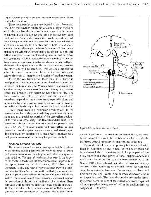
The third pathway establishes the balance of power within the<br />
system: the reticulospinal tract, provides additional support<br />
for excitation of flexion <strong>and</strong> inhibition of extension. These<br />
pathways work <strong>to</strong>gether <strong>to</strong> modulate body posture (Figure 8-<br />
4). The vestibulocerebellar connections are well-documented<br />
pathways which also have an important role in the mainte-<br />
Laaral ve\libulospinal rracl<br />
Ikld<br />
Figure 8-4. Postural control network<br />
Crdn~al nerve\<br />
I11 IV, VI<br />
wve eye rnu\cle\<br />
nance of posture <strong>and</strong> orientation. As stated above, the cerebellar<br />
connections with the vestibular nuclei provide the<br />
inhibi<strong>to</strong>ry control necessary for maintenance of posture.<br />
Postural control is a basic. primary functional behavior.<br />
Even in controlled studies where the vestibular organ has<br />
been removed, there is a serious initial change in postural stability<br />
but within a short period of time compensa<strong>to</strong>ry action<br />
reinstates some of the functions that have been lost (Darian-<br />
Smith, 1984). It is believed that other reflexive <strong>and</strong> sensory<br />
systems which contribute <strong>to</strong> postural control as well take<br />
over the orientation functions. Dependence on visual <strong>and</strong><br />
proprioceptive input seems <strong>to</strong> occur when vestibular input is<br />
no longer available. The interrelationships among the sensory<br />
systems form the core of the multidimensional maps that<br />
allow appropriate interaction of self in the environment. As<br />
Jongkees (1 974) states: