Thyroid and Parathyroid Ultrasound Examination - AIUM
Thyroid and Parathyroid Ultrasound Examination - AIUM
Thyroid and Parathyroid Ultrasound Examination - AIUM

Create successful ePaper yourself
Turn your PDF publications into a flip-book with our unique Google optimized e-Paper software.
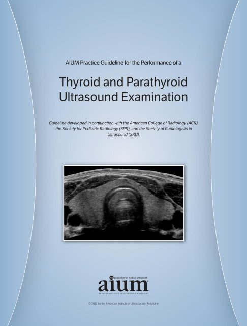
<strong>AIUM</strong> Practice Guideline for the Performance of a<br />
<strong>Thyroid</strong> <strong>and</strong> <strong>Parathyroid</strong><br />
<strong>Ultrasound</strong> <strong>Examination</strong><br />
Guideline developed in conjunction with the American College of Radiology (ACR),<br />
the Society for Pediatric Radiology (SPR), <strong>and</strong> the Society of Radiologists in<br />
<strong>Ultrasound</strong> (SRU).<br />
© 2013 by the American Institute of <strong>Ultrasound</strong> in Medicine
The American Institute of <strong>Ultrasound</strong> in Medicine (<strong>AIUM</strong>) is a multidisciplinary<br />
association dedicated to advancing the safe <strong>and</strong> effective<br />
use of ultrasound in medicine through professional <strong>and</strong> public<br />
education, research, development of guidelines, <strong>and</strong> accreditation.<br />
To promote this mission, the <strong>AIUM</strong> is pleased to publish, in conjunction<br />
with the American College of Radiology (ACR), the Society for<br />
Pediatric Radiology (SPR), <strong>and</strong> the Society of Radiologists in<br />
<strong>Ultrasound</strong> (SRU), this revised <strong>AIUM</strong> Practice Guideline for the<br />
Performance of a <strong>Thyroid</strong> <strong>and</strong> <strong>Parathyroid</strong> <strong>Ultrasound</strong> <strong>Examination</strong>.<br />
We are indebted to the many volunteers who contributed their time,<br />
knowledge, <strong>and</strong> energy to bringing this document to completion.<br />
The <strong>AIUM</strong> represents the entire range of clinical <strong>and</strong> basic science<br />
interests in medical diagnostic ultrasound, <strong>and</strong>, with hundreds of<br />
volunteers, the <strong>AIUM</strong> has promoted the safe <strong>and</strong> effective use of<br />
ultrasound in clinical medicine for more than 50 years. This document<br />
<strong>and</strong> others like it will continue to advance this mission.<br />
Practice guidelines of the <strong>AIUM</strong> are intended to provide the medical<br />
ultrasound community with guidelines for the performance <strong>and</strong> recording<br />
of high-quality ultrasound examinations. The guidelines reflect<br />
what the <strong>AIUM</strong> considers the minimum criteria for a complete examination<br />
in each area but are not intended to establish a legal st<strong>and</strong>ard of<br />
care. <strong>AIUM</strong>-accredited practices are expected to generally follow the<br />
guidelines with recognition that deviations from these guidelines will<br />
be needed in some cases, depending on patient needs <strong>and</strong> available<br />
equipment. Practices are encouraged to go beyond the guidelines to<br />
provide additional service <strong>and</strong> information as needed.<br />
14750 Sweitzer Ln, Suite 100<br />
Laurel, MD 20707-5906 USA<br />
800-638-5352 • 301-498-4100<br />
www.aium.org<br />
2013—<strong>AIUM</strong> PRACTICE GUIDELINE—<strong>Thyroid</strong> <strong>and</strong> <strong>Parathyroid</strong> <strong>Ultrasound</strong>
2013—<strong>AIUM</strong> PRACTICE GUIDELINE—<strong>Thyroid</strong> <strong>and</strong> <strong>Parathyroid</strong> <strong>Ultrasound</strong><br />
I. Introduction<br />
The clinical aspects contained in specific sections of this guideline (Introduction, Indications,<br />
Specifications of the <strong>Examination</strong>, <strong>and</strong> Equipment Specifications) were revised collaboratively<br />
by the American Institute of <strong>Ultrasound</strong> in Medicine (<strong>AIUM</strong>), the American College of<br />
Radiology (ACR), the Society for Pediatric Radiology (SPR), <strong>and</strong> the Society of Radiologists<br />
in <strong>Ultrasound</strong> (SRU). Recommendations for personnel requirements, written request for the<br />
examination, procedure documentation, <strong>and</strong> quality control vary among the organizations <strong>and</strong><br />
are addressed by each separately.<br />
This guideline is intended to assist practitioners performing sonographic evaluations of the<br />
thyroid gl<strong>and</strong>, parathyroid gl<strong>and</strong>s, <strong>and</strong> adjacent soft tissues. Occasionally, an additional <strong>and</strong>/or<br />
specialized examination with another modality may be necessary. While it is not possible to<br />
detect every abnormality, adherence to the following guidelines will maximize the probability<br />
of detecting most abnormalities that occur in the thyroid <strong>and</strong> parathyroid gl<strong>and</strong>s.<br />
II.<br />
Indications<br />
Indications for a thyroid <strong>and</strong> parathyroid ultrasound examination include but are not limited to 1 :<br />
1. Evaluation of the location <strong>and</strong> characteristics of palpable neck masses, including an<br />
enlarged thyroid;<br />
2. Evaluation of abnormalities detected by other imaging examinations, eg, a thyroid nodule<br />
detected on computed tomography, positron emission tomography–computed tomography,<br />
or magnetic resonance imaging, or seen on another ultrasound examination of the<br />
neck (eg, carotid ultrasound);<br />
3. Evaluation of laboratory abnormalities;<br />
4. Evaluation of the presence, size, <strong>and</strong> location of the thyroid gl<strong>and</strong>;<br />
5. Evaluation of patients at high risk for occult thyroid malignancy;<br />
6. Follow-up imaging of previously detected thyroid nodules, when indicated;<br />
7. Evaluation for regional nodal metastases in patients with proven or suspected thyroid<br />
carcinoma before thyroidectomy;<br />
8. Evaluation for recurrent disease or regional nodal metastases after total or partial<br />
thyroidectomy for thyroid carcinoma;<br />
9. Evaluation of the thyroid gl<strong>and</strong> for suspicious nodules before neck surgery for<br />
nonthyroid disease 2 ;<br />
10. Evaluation of the thyroid gl<strong>and</strong> for suspicious nodules before radioiodine ablation of<br />
the gl<strong>and</strong>;<br />
1<br />
www.aium.org
2013—<strong>AIUM</strong> PRACTICE GUIDELINE—<strong>Thyroid</strong> <strong>and</strong> <strong>Parathyroid</strong> <strong>Ultrasound</strong><br />
11. Identification <strong>and</strong> localization of parathyroid abnormalities in patients with known or<br />
suspected hyperparathyroidism 3,4 ;<br />
12. Assessment of the number <strong>and</strong> size of enlarged parathyroid gl<strong>and</strong>s in patients who have<br />
undergone previous parathyroid surgery or ablative therapy with recurrent symptoms of<br />
hyperparathyroidism;<br />
13. Localization of thyroid/parathyroid abnormalities or adjacent cervical lymph nodes for<br />
biopsy, ablation, or other interventional procedures; <strong>and</strong><br />
14. Localization of autologous parathyroid gl<strong>and</strong> implants.<br />
III.<br />
Qualifications <strong>and</strong> Responsibilities of Personnel<br />
See the <strong>AIUM</strong> Official Statement Training Guidelines for Physicians Who Evaluate <strong>and</strong> Interpret<br />
Diagnostic <strong>Thyroid</strong>/<strong>Parathyroid</strong> <strong>Ultrasound</strong> <strong>Examination</strong>s <strong>and</strong> the <strong>AIUM</strong> St<strong>and</strong>ards <strong>and</strong><br />
Guidelines for the Accreditation of <strong>Ultrasound</strong> Practices.<br />
2<br />
IV.<br />
Written Request for the <strong>Examination</strong><br />
The written or electronic request for an ultrasound examination should provide sufficient<br />
information to allow for the appropriate performance <strong>and</strong> interpretation of the examination.<br />
The request for the examination must be originated by a physician or other appropriately<br />
licensed health care provider or under the provider’s direction. The accompanying clinical<br />
information should be provided by a physician or other appropriate health care provider familiar<br />
with the patient’s clinical situation <strong>and</strong> should be consistent with relevant legal <strong>and</strong> local<br />
health care facility requirements.<br />
V. Specifications of the <strong>Examination</strong>s<br />
A. The <strong>Thyroid</strong> <strong>Examination</strong><br />
The examination should be performed with the neck in hyperextension. The right <strong>and</strong> left<br />
lobes of the thyroid gl<strong>and</strong> should be imaged in the longitudinal <strong>and</strong> transverse planes.<br />
Recorded images of the thyroid should include transverse images of the superior, mid, <strong>and</strong><br />
inferior portions of the right <strong>and</strong> left thyroid lobes; longitudinal images of the medial, mid, <strong>and</strong><br />
lateral portions of both lobes; <strong>and</strong> at least a transverse image of the isthmus. The size of each<br />
thyroid lobe should be recorded in 3 dimensions, anteroposterior, transverse, <strong>and</strong> longitudinal.<br />
The thickness (anteroposterior measurement) of the isthmus on the transverse view should<br />
be recorded. A color or power Doppler examination can be used to supplement the grayscale<br />
evaluation of either diffuse or focal abnormalities of the thyroid. It is often necessary to extend<br />
imaging to include the soft tissue above the isthmus (eg, to evaluate a possible pyramidal lobe<br />
www.aium.org
2013—<strong>AIUM</strong> PRACTICE GUIDELINE—<strong>Thyroid</strong> <strong>and</strong> <strong>Parathyroid</strong> <strong>Ultrasound</strong><br />
of the thyroid), congenital abnormalities such as a thyroglossal duct cyst, or if any superior<br />
palpable abnormality is noted. The examination should also include a brief evaluation of the<br />
lateral neck compartments.<br />
<strong>Thyroid</strong> abnormalities should be imaged in a way that allows for reporting <strong>and</strong> documentation<br />
of the following:<br />
1. The location, size, number, <strong>and</strong> character of significant abnormalities, including measurements<br />
of nodules <strong>and</strong> focal abnormalities in 3 dimensions;<br />
2. The localized or diffuse nature of any thyroid abnormality, including assessment of<br />
overall gl<strong>and</strong> vascularity 5,6 ;<br />
3. The sonographic features of any thyroid abnormality with respect to echogenicity,<br />
composition (degree of cystic change), margins (smooth or irregular), presence <strong>and</strong><br />
type of calcification (if present), <strong>and</strong> other relevant sonographic patterns 7–19 ; <strong>and</strong><br />
4. The presence <strong>and</strong> size of any abnormal lymph node in the lateral compartment of the<br />
neck (see section B below).<br />
In patients who have undergone complete or partial thyroidectomy, the thyroid bed should be<br />
imaged in transverse <strong>and</strong> longitudinal planes. Any masses or cysts in the region of the bed<br />
should be measured <strong>and</strong> reported. Additionally, the lateral neck should be evaluated as<br />
described in section B.<br />
3<br />
Whenever possible, comparison should be made with other appropriate imaging studies.<br />
Sonographic guidance may be used for aspiration or biopsy of thyroid abnormalities or other<br />
masses of the neck or for other interventional procedures. 20–22<br />
B. The Cervical Lymph Node Evaluation<br />
A high-resolution ultrasound examination of the neck is used for the staging of patients with<br />
thyroid cancer <strong>and</strong> other head <strong>and</strong> neck cancers <strong>and</strong> in the surveillance of patients after treatment<br />
of such cancers. 23–29 In these patients, the size <strong>and</strong> location of abnormal lymph nodes<br />
should be documented. Suspicious features such as calcification, cystic areas, absence of a central<br />
hilum, round shape, <strong>and</strong> abnormal blood flow should be documented. The location of an<br />
abnormal lymph node should be described according to the image-based nodal classification<br />
system developed by Som et al, 30 which corresponds to the clinical nodal classification system<br />
developed by the American Joint Committee on Cancer <strong>and</strong> the American Academy of<br />
Otolaryngology–Head <strong>and</strong> Neck Surgery, or in a fashion that allows the referring clinician to<br />
convert the location of abnormal nodes to that system.<br />
www.aium.org
2013—<strong>AIUM</strong> PRACTICE GUIDELINE—<strong>Thyroid</strong> <strong>and</strong> <strong>Parathyroid</strong> <strong>Ultrasound</strong><br />
C. The <strong>Parathyroid</strong> <strong>Examination</strong><br />
An examination for suspected parathyroid enlargement should include images in the region of<br />
the anticipated parathyroid gl<strong>and</strong> location. One of the important uses of parathyroid ultrasound<br />
is to try to localize parathyroid adenomas in patients with primary hyperparathyroidism<br />
to help with surgical planning. 3,4<br />
The examination should be performed with the neck hyperextended <strong>and</strong> should include<br />
longitudinal <strong>and</strong> transverse images from the carotid arteries to the midline bilaterally <strong>and</strong><br />
extending from the carotid artery bifurcation superiorly to the thoracic inlet inferiorly. As<br />
parathyroid gl<strong>and</strong>s may be hidden below the clavicles in the lower neck <strong>and</strong> upper<br />
mediastinum, it may also be helpful to have the patient swallow during the examination with<br />
constant real-time observation. Color <strong>and</strong>/or power or spectral Doppler ultrasound may be<br />
helpful. The upper mediastinum may be imaged with an appropriate probe by angling under<br />
the sternum from the sternal notch. Rarely, parathyroid adenomas may also be intrathyroidal.<br />
Although the normal parathyroid gl<strong>and</strong>s are usually not visualized with available sonographic<br />
technology, enlarged parathyroid gl<strong>and</strong>s may be visualized. When visualized, their location,<br />
size, <strong>and</strong> number should be documented, <strong>and</strong> measurements should be made in 3 dimensions.<br />
The relationship of any visualized parathyroid gl<strong>and</strong>(s) to the thyroid gl<strong>and</strong> should be<br />
documented, if applicable. 2,31,32<br />
4<br />
Whenever possible, comparison should be made with other appropriate imaging studies.<br />
Sonographic guidance may be used for aspiration or biopsy of parathyroid abnormalities or<br />
other masses of the neck or for other interventional procedures.<br />
VI.<br />
Documentation<br />
Adequate documentation is essential for high-quality patient care. There should be a permanent<br />
record of the ultrasound examination <strong>and</strong> its interpretation. Images of all appropriate<br />
areas, both normal <strong>and</strong> abnormal, should be recorded. Variations from normal size should be<br />
accompanied by measurements. Images should be labeled with the patient identification,<br />
facility identification, examination date, <strong>and</strong> side (right or left) of the anatomic site imaged.<br />
An official interpretation (final report) of the ultrasound findings should be included in the<br />
patient’s medical record. Retention of the ultrasound examination should be consistent both<br />
with clinical needs <strong>and</strong> with relevant legal <strong>and</strong> local health care facility requirements.<br />
Reporting should be in accordance with the <strong>AIUM</strong> Practice Guideline for Documentation of an<br />
<strong>Ultrasound</strong> <strong>Examination</strong>.<br />
www.aium.org
2013—<strong>AIUM</strong> PRACTICE GUIDELINE—<strong>Thyroid</strong> <strong>and</strong> <strong>Parathyroid</strong> <strong>Ultrasound</strong><br />
VII. Equipment Specifications<br />
<strong>Thyroid</strong> <strong>and</strong> parathyroid studies should be conducted with a linear transducer. The equipment<br />
should be adjusted to operate at the highest clinically appropriate frequency, realizing that<br />
there is a trade-off between resolution <strong>and</strong> beam penetration. For most patients, mean<br />
frequencies of 10 to 14 MHz or greater are preferred, though some patients may require a<br />
lower-frequency transducer for depth penetration. If the gl<strong>and</strong> is deep or extremely enlarged,<br />
a curved linear transducer may be necessary. Resolution should be of sufficient quality to evaluate<br />
the internal morphology of visible lesions. Doppler frequencies should be set to optimize<br />
flow detection. Diagnostic information should be optimized while keeping total sonographic<br />
exposure as low as reasonably achievable.<br />
VIII. Quality Control <strong>and</strong> Improvement, Safety, Infection Control,<br />
<strong>and</strong> Patient Education<br />
Policies <strong>and</strong> procedures related to quality control, patient education, infection control, <strong>and</strong><br />
safety should be developed <strong>and</strong> implemented in accordance with the <strong>AIUM</strong> St<strong>and</strong>ards <strong>and</strong><br />
Guidelines for the Accreditation of <strong>Ultrasound</strong> Practices.<br />
Equipment performance monitoring should be in accordance with the <strong>AIUM</strong> St<strong>and</strong>ards <strong>and</strong><br />
Guidelines for the Accreditation of <strong>Ultrasound</strong> Practices.<br />
5<br />
IX.<br />
ALARA Principle<br />
The potential benefits <strong>and</strong> risks of each examination should be considered. The ALARA (as<br />
low as reasonably achievable) principle should be observed when adjusting controls that affect<br />
the acoustic output <strong>and</strong> by considering transducer dwell times. Further details on ALARA may<br />
be found in the <strong>AIUM</strong> publication Medical <strong>Ultrasound</strong> Safety, Second Edition.<br />
www.aium.org
2013—<strong>AIUM</strong> PRACTICE GUIDELINE—<strong>Thyroid</strong> <strong>and</strong> <strong>Parathyroid</strong> <strong>Ultrasound</strong><br />
Acknowledgments<br />
This guideline was revised by the <strong>AIUM</strong> in collaboration with the American College of<br />
Radiology (ACR), the Society for Pediatric Radiology (SPR), <strong>and</strong> the Society of Radiologists<br />
in <strong>Ultrasound</strong> (SRU) according to the process described in the <strong>AIUM</strong> Clinical St<strong>and</strong>ards<br />
Committee Manual.<br />
Collaborative Committees<br />
Members represent their societies in the initial version <strong>and</strong> final revision of this guideline.<br />
<strong>AIUM</strong><br />
Robert D. Harris, MD, MPH<br />
Jill E. Langer, MD<br />
Robert A. Levine, MD<br />
6<br />
ACR<br />
Sheila Sheth, MD, Chair<br />
Sara J. Abramson, MD<br />
Helena Gabriel, MD<br />
Maitray D. Patel, MD<br />
SPR<br />
Judith A. Craychee, MD<br />
Cindy R. Miller, MD<br />
Henrietta Kotlus Rosenberg, MD<br />
Dayna M. Weinert, MD<br />
SRU<br />
William D. Middleton, MD<br />
Carl C. Reading, MD<br />
Mitchell E. Tublin, MD<br />
www.aium.org
2013—<strong>AIUM</strong> PRACTICE GUIDELINE—<strong>Thyroid</strong> <strong>and</strong> <strong>Parathyroid</strong> <strong>Ultrasound</strong><br />
<strong>AIUM</strong> Clinical St<strong>and</strong>ards Committee<br />
Leslie Scoutt, MD, Chair<br />
Joseph Wax, MD, Vice Chair<br />
Bryann Bromley, MD<br />
Lin Diacon, MD, RDMS, RPVI<br />
J. Christian Fox, MD, RDMS<br />
Pat Fulgham, MD<br />
Charlotte Henningsen, MS, RT, RDMS, RVT<br />
Adam Hiett, MD, RDMS<br />
Lars Jensen, MD<br />
Alex<strong>and</strong>er Levitov, MD<br />
Vicki Noble, MD, RDMS<br />
Anthony Odibo, MD, MSCE<br />
Deborah Rubens, MD<br />
Khaled Sakhel, MD<br />
Shia Salem, MD<br />
Jay Smith, MD<br />
Lami Yeo, MD<br />
7<br />
Original copyright 2007; revised 2013.<br />
www.aium.org
2013—<strong>AIUM</strong> PRACTICE GUIDELINE—<strong>Thyroid</strong> <strong>and</strong> <strong>Parathyroid</strong> <strong>Ultrasound</strong><br />
8<br />
References<br />
1. Solbiati L, Osti V, Cova L, Tonolini M. <strong>Ultrasound</strong> of thyroid, parathyroid gl<strong>and</strong>s <strong>and</strong> neck lymph nodes.<br />
Eur Radiol 2001; 11:2411–2424.<br />
2. Arciero CA, Shiue ZS, Gates JD, et al. Preoperative thyroid ultrasound is indicated in patients<br />
undergoing parathyroidectomy for primary hyperparathyroidism. J Cancer 2012; 3:1–6.<br />
3. Levy JM, K<strong>and</strong>il E, Yau LC, Cuda JD, Sheth SN, Tufano RP. Can ultrasound be used as the primary<br />
screening modality for the localization of parathyroid disease prior to surgery for primary<br />
hyperparathyroidism A review of 440 cases. ORL J Otorhinolaryngol Relat Spec 2011; 73:116–120.<br />
4. Patel CN, Salahudeen HM, Lansdown M, Scarsbrook AF. Clinical utility of ultrasound <strong>and</strong> 99mTc<br />
sestamibi SPECT/CT for preoperative localization of parathyroid adenoma in patients with primary<br />
hyperparathyroidism. Clin Radiol 2010; 65:278–287.<br />
5. Anderson L, Middleton WD, Teefey SA, et al. Hashimoto thyroiditis, part 1: sonographic analysis of<br />
the nodular form of Hashimoto thyroiditis. AJR Am J Roentgenol 2010; 195:208–215.<br />
6. Frates MC, Benson CB, Doubilet PM, et al. Prevalence <strong>and</strong> distribution of carcinoma in patients with<br />
solitary <strong>and</strong> multiple thyroid nodules on sonography. J Clin Endocrinol Metab 2006; 91:3411–3417.<br />
7. Bonavita JA, Mayo J, Babb J, et al. Pattern recognition of benign nodules at ultrasound of the thyroid:<br />
which nodules can be left alone AJR Am J Roentgenol 2009; 193:207–213.<br />
8. Chan BK, Desser TS, McDougall IR, Weigel RJ, Jeffrey RB Jr. Common <strong>and</strong> uncommon sonographic<br />
features of papillary thyroid carcinoma. J <strong>Ultrasound</strong> Med 2003; 22:1083–1090.<br />
9. Cooper DS, Doherty GM, Haugen BR, et al. Revised American <strong>Thyroid</strong> Association management<br />
guidelines for patients with thyroid nodules <strong>and</strong> differentiated thyroid cancer. <strong>Thyroid</strong> 2009; 19:<br />
1167–1214.<br />
10. Frates MC, Benson CB, Charboneau JW, et al. Management of thyroid nodules detected at US:<br />
Society of Radiologists in <strong>Ultrasound</strong> consensus conference statement. Radiology 2005; 237:<br />
794–800.<br />
11. Henrichsen TL, Reading CC. <strong>Thyroid</strong> ultrasonography, part 2: nodules. Radiol Clin North Am 2011;<br />
49:417–424, v.<br />
12. Hoang JK, Lee WK, Lee M, Johnson D, Farrell S. US Features of thyroid malignancy: pearls <strong>and</strong> pitfalls.<br />
Radiographics 2007; 27:847–865.<br />
13. Iannuccilli JD, Cronan JJ, Monchik JM. Risk for malignancy of thyroid nodules as assessed by<br />
sonographic criteria: the need for biopsy. J <strong>Ultrasound</strong> Med 2004; 23:1455–1464.<br />
14. Kim EK, Park CS, Chung WY, et al. New sonographic criteria for recommending fine-needle aspiration<br />
biopsy of nonpalpable solid nodules of the thyroid. AJR Am J Roentgenol 2002; 178:687–691.<br />
15. Layfield LJ, Cibas ES, Gharib H, M<strong>and</strong>el SJ. <strong>Thyroid</strong> aspiration cytology: current status. CA Cancer J<br />
Clin 2009; 59:99–110.<br />
16. Moon WJ, Jung SL, Lee JH, et al. Benign <strong>and</strong> malignant thyroid nodules: US differentiation—<br />
multicenter retrospective study. Radiology 2008; 247:762–770.<br />
17. Morris LF, Ragavendra N, Yeh MW. Evidence-based assessment of the role of ultrasonography in the<br />
management of benign thyroid nodules. World J Surg 2008; 32:1253–1263.<br />
18. Reading CC, Charboneau JW, Hay ID, Sebo TJ. Sonography of thyroid nodules: a “classic pattern”<br />
diagnostic approach. <strong>Ultrasound</strong> Q 2005; 21:157–165.<br />
www.aium.org
2013—<strong>AIUM</strong> PRACTICE GUIDELINE—<strong>Thyroid</strong> <strong>and</strong> <strong>Parathyroid</strong> <strong>Ultrasound</strong><br />
19. Tae HJ, Lim DJ, Baek KH, et al. Diagnostic value of ultrasonography to distinguish between benign <strong>and</strong><br />
malignant lesions in the management of thyroid nodules. <strong>Thyroid</strong> 2007; 17:461–466.<br />
20. Lewis BD, Hay ID, Charboneau JW, McIver B, Reading CC, Goellner JR. Percutaneous ethanol<br />
injection for treatment of cervical lymph node metastases in patients with papillary thyroid carcinoma.<br />
AJR Am J Roentgenol 2002; 178:699–704.<br />
21. Nam-Goong IS, Kim HY, Gong G, et al. Ultrasonography-guided fine-needle aspiration of thyroid<br />
incidentaloma: correlation with pathological findings. Clin Endocrinol (Oxf) 2004; 60:21–28.<br />
22. O’Malley ME, Weir MM, Hahn PF, Misdraji J, Wood BJ, Mueller PR. US-guided fine-needle aspiration<br />
biopsy of thyroid nodules: adequacy of cytologic material <strong>and</strong> procedure time with <strong>and</strong> without<br />
immediate cytologic analysis. Radiology 2002; 222:383–387.<br />
23. Ahuja AT, Ying M. Sonographic evaluation of cervical lymph nodes. AJR Am J Roentgenol 2005;<br />
184:1691–1699.<br />
24. Ahuja AT, Ying M, Yuen HY, Metreweli C. Power Doppler sonography of metastatic nodes from<br />
papillary carcinoma of the thyroid. Clin Radiol 2001; 56:284–288.<br />
25. Gor DM, Langer JE, Loevner LA. Imaging of cervical lymph nodes in head <strong>and</strong> neck cancer: the basics.<br />
Radiol Clin North Am 2006; 44:101–110, viii.<br />
26. Leboulleux S, Girard E, Rose M, et al. <strong>Ultrasound</strong> criteria of malignancy for cervical lymph nodes in<br />
patients followed up for differentiated thyroid cancer. J Clin Endocrinol Metab 2007; 92:3590–3594.<br />
27. Sheth S, Hamper UM. Role of sonography after total thyroidectomy for thyroid cancer. <strong>Ultrasound</strong> Q<br />
2008; 24:147–154.<br />
28. Shin JH, Han BK, Ko EY, Kang SS. Sonographic findings in the surgical bed after thyroidectomy:<br />
comparison of recurrent tumors <strong>and</strong> nonrecurrent lesions. J <strong>Ultrasound</strong> Med 2007; 26:1359–1366.<br />
29. Stulak JM, Grant CS, Farley DR, et al. Value of preoperative ultrasonography in the surgical<br />
management of initial <strong>and</strong> reoperative papillary thyroid cancer. Arch Surg 2006; 141:489–496.<br />
30. Som PM, Curtin HD, Mancuso AA. Imaging-based nodal classification for evaluation of neck<br />
metastatic adenopathy. AJR Am J Roentgenol 2000; 174:837–844.<br />
31. Reeder SB, Desser TS, Weigel RJ, Jeffrey RB. Sonography in primary hyperparathyroidism: review<br />
with emphasis on scanning technique. J <strong>Ultrasound</strong> Med 2002; 21:539–552.<br />
32. Yabuta T, Tsushima Y, Masuoka H, et al. Ultrasonographic features of intrathyroidal parathyroid<br />
adenoma causing primary hyperparathyroidism. Endocr J 2011; 58:989–994.<br />
9<br />
www.aium.org