to learn more about Thermal Imaging - Vetel Diagnostics
to learn more about Thermal Imaging - Vetel Diagnostics
to learn more about Thermal Imaging - Vetel Diagnostics

Create successful ePaper yourself
Turn your PDF publications into a flip-book with our unique Google optimized e-Paper software.
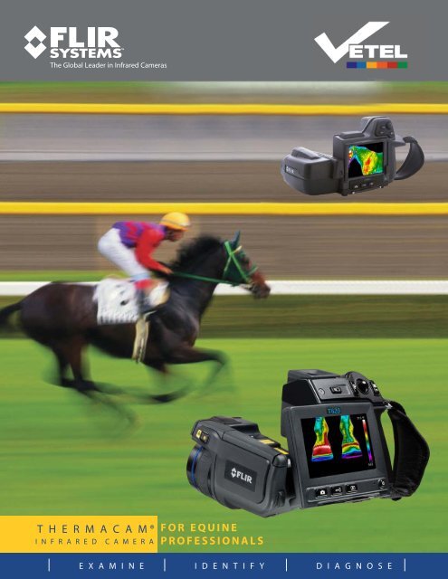
TM<br />
The Global Leader in Infrared Cameras<br />
T H E R M A C A M ®<br />
I N F R A R E D C A M E R A<br />
FOR EQUINE<br />
PROFESSIONALS<br />
| E X A M I N E | I D E N T I F Y | D I A G N O S E |
Every Equine Practitioner should have IR:<br />
an invaluable <strong>to</strong>ol for equine diagnosis<br />
Infrared imaging is a widely proven technology, known<br />
for both extreme sensitivity and high reliability.<br />
As a diagnostic <strong>to</strong>ol, thermal imaging is one of<br />
the most innovative, non-invasive modalities<br />
available <strong>to</strong> the veterinarian.<br />
Stressed tissue often presents as a thermographic<br />
abnormality prior <strong>to</strong> becoming structurally damaged.<br />
A thermal exam provides highly accurate<br />
diagnostic information that can enable the<br />
veterinarian <strong>to</strong> detect, confirm, and document<br />
many problems BEFORE the onset of serious injury.<br />
As prey animals, horses have a highly evolved<br />
instinct <strong>to</strong> mask pain and lameness in order <strong>to</strong><br />
appear less vulnerable in the wild. This can make<br />
diagnosis all the <strong>more</strong> challenging for the practitioner.<br />
Over the years improved technology, extensive<br />
clinical research and experience have brought<br />
thermography <strong>to</strong> a level which provides the veterinary<br />
professional a newfound ability <strong>to</strong> locate medical<br />
problems faster and proactively<br />
IN CLINICAL PRACTICE, EQUINE<br />
THERMOGRAPHY CAN:<br />
• Provide a highly sensitive, early indica<strong>to</strong>r of subtle<br />
circula<strong>to</strong>ry changes in soft tissue<br />
• Reveal inflamma<strong>to</strong>ry reactions in joints and tendons<br />
from two <strong>to</strong> six weeks prior <strong>to</strong> the clinical<br />
appearance of lameness<br />
• Allow the veterinarian <strong>to</strong> visualize neuro-vascular<br />
changes including the effect of vaso-active substances.<br />
LOCATING THE INJURY<br />
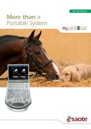
3 Steps <strong>to</strong> Equine Thermography<br />
1 Understanding<br />
Equine <strong>Thermal</strong><br />
<strong>Imaging</strong><br />
THERMAL SYMMETRY IS THE RULE<br />
• Changes of 1 degree centigrade or<br />
<strong>more</strong>, over 25% of the comparable<br />
ana<strong>to</strong>mic area are cause for further<br />
investigation.<br />
UNDERSTANDING ARTIFACTS<br />
• Clean body surface<br />
• Uniform hair coat<br />
• Feet picked out<br />
• His<strong>to</strong>ry of recent injections,<br />
therapy, medications, etc.<br />
• Has the horse recently been wrapped<br />
• Any evidence of dermatitis, poultice<br />
or blister on the skin<br />
• Evidence of clipping or hair loss<br />
• Scarring or areas of edema present<br />
2<br />
Implementing<br />
Equine <strong>Thermal</strong><br />
<strong>Imaging</strong> in Your<br />
Practice<br />
• Perform after physical exam<br />
• 10 – 15 minutes <strong>to</strong> complete<br />
• Learn normal vascular patterns<br />
• Use the horse as its’ own control<br />
• Be aware of potential artifacts<br />
• Focus on recognition of pathological<br />
patterns of heat - don’t<br />
worry <strong>about</strong> absolute<br />
temperatures.<br />
3<br />
Achieve a<br />
Successful Diagnosis<br />
As part of the complete examination<br />
thermal imaging can be a defining <strong>to</strong>ol<br />
in making an accurate diagnosis<br />
Equine thermal imaging is a professional<br />
diagnostic modality that gives the<br />
veterinary professional the ability <strong>to</strong><br />
accurately measure, access and document<br />
changes in circula<strong>to</strong>ry patterns.<br />
The result is an enhanced opportunity<br />
for early intervention and therapy and<br />
a chance <strong>to</strong> prevent crippling and<br />
debilitating injuries.
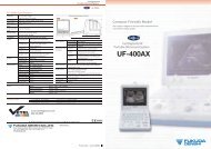
Identify problems BEFORE they become<br />
serious complications or injuries<br />
Clinical evaluations have shown proven results. Here are just a few examples of how equine thermography<br />
can identify clinical problems early on before serious complications occur.<br />
Dorsal view of the croup<br />
of a reining horse (fig. 1).<br />
Complaint was the horse<br />
resented circles <strong>to</strong> the left.<br />
Note the thermal asymmetry<br />
in the region of the gluteal<br />
muscles.<br />
Dorso-Plantar view of both<br />
hocks (fig 2.).<br />
Increased thermal emission<br />
is noted in the region of the<br />
Distal-Intertarsal and Tarsal<br />
Metatarsal joints.<br />
Both are abnormal,<br />
with the right significantly<br />
warmer than the left.<br />
fig.1<br />
fig. 2<br />
Bilateral Carpitis in a<br />
Rope horse (fig. 3).<br />
Increased thermal<br />
emission over the dorsal<br />
aspects of the joints, and<br />
lack of normal cooler<br />
region in the area<br />
of the extensor tendon.<br />
Flexor apparatus<br />
inflammation on<br />
Left Front leg (fig. 4).<br />
<strong>Thermal</strong> pattern is<br />
consistent with Flexor<br />
Tendonitis or Suspensory<br />
Desmitis.<br />
fig. 3 fig. 4<br />
Normal dorsal view of<br />
the stifle (fig 5).<br />
The sharp demarcation<br />
of temperature in the<br />
vertical band over the<br />
dorsal aspect of the joint<br />
represents the normal<br />
summation of reflected<br />
heat seen in the medial<br />
aspect of the stifle region<br />
Abnormal dorsal view<br />
of the stifle (fig. 6).<br />
Fig. 6. lacks the<br />
characteristic vertical<br />
temperature band in the<br />
center of the joint, and<br />
the circular heat pattern<br />
associated with the region<br />
of the femoral-patellar joint.<br />
fig. 5 fig. 6<br />
Dorsal PA-oblique view<br />
of the back (fig 7).<br />
Compare the asymmetrical<br />
contact pattern and<br />
inflammation in the<br />
region of the thoracic<br />
dorsal spinous processes<br />
with the contact pattern<br />
apparent in Fig. 8<br />
<strong>Thermal</strong> image of the<br />
saddle following exercise<br />
(fig. 8).<br />
The thermal emissions on<br />
the saddle correspond <strong>to</strong> the<br />
contact region of the dorsal<br />
view of the back in fig. 7.<br />
along with the corrolating<br />
asymmetry. Also note the<br />
uneven flocking in the seat of<br />
the saddle.<br />
fig. 7 fig. 8
About FLIR Systems<br />
With over 40 years experience and <strong>more</strong><br />
than 30,000 of its IR cameras in use, FLIR is<br />
the undisputed global leader in infrared<br />
systems. From industrial <strong>to</strong> military applications,<br />
thermography professionals have<br />
made FLIR their number one choice. No<br />
other company offers such a wide range of<br />
infrared cameras, software, service, training<br />
and support.<br />
FLIR’s ThermaCAM series of thermal imaging<br />
cameras have long set the standard for<br />
thermographic testing and analysis. Today<br />
they are the most widely used non-contact<br />
temperature measurement infrared cameras<br />
in the world.<br />
The Global Leaders in Infrared Cameras<br />
FLIR Systems, Bos<strong>to</strong>n<br />
Americas Thermography Center<br />
16 Esquire Rd.<br />
North Billerica, MA 01862<br />
1-800-464-6372<br />
www.flirthermography.com<br />
Natanya Nieman DVM<br />
Resident Veterinarian<br />
WinStar Farm LLC, Versailles, KY<br />
WinStar Farm uses Flir thermal imagingfor routine<br />
moni<strong>to</strong>ring of thier thoroughbred racing stable<br />
INFRARED SUCCESS STORIES<br />
Identifying the Causes of Diminished<br />
Performance in Equine Practice<br />
Jim Waldsmith , DVM, The Equine Center<br />
Many horses are presented <strong>to</strong> our practice with a request <strong>to</strong><br />
define subtle changes in the horse's gait which are believed<br />
<strong>to</strong> be associated with a decrease in the level of the horse's<br />
athletic ability.<br />
On clinical exam these horses are many times found <strong>to</strong> be<br />
"body sore" resenting flexion and palpation in several areas<br />
of their body. The lameness has not progressed <strong>to</strong> a head<br />
bob or hip hike that can be blocked, yet the owner or trainer<br />
is concerned that stress or injury exists that will effect the<br />
horse's future performance.<br />
In many of these cases the areas of pain and inflammation<br />
are difficult <strong>to</strong> define and document clinically, and the use<br />
of systemic anti-inflamma<strong>to</strong>ry medication is considered.<br />
The risk in this therapeutic scenario is that symp<strong>to</strong>ms may<br />
be masked, and the source(s) of the diminished performance<br />
remain unknown potentially leading <strong>to</strong> subsequent breakdown<br />
in the horse.<br />
After a complete clinical exam we find thermal imaging<br />
allows a quick and noninvasive assessment of the entire<br />
horse's body. Frequently areas of hoof imbalance, joint<br />
inflammation and abnormal tack wear can all be quickly<br />
identified. By simultaneously identifying and treating all<br />
areas of abnormality and inflammation the horse's athleticism<br />
is improved and risk of further injury diminished.<br />
We have found this pro<strong>to</strong>col <strong>to</strong> be very rewarding in<br />
our practice producing both client satisfaction and<br />
improvement in the athletic health of our equine patients.<br />
The Use of Thermography<br />
in Equine Medicine<br />
Tracy Turner , DVM, MS, Diplomate ACVS<br />
In equine practice, thermography can detect inflamma<strong>to</strong>ry<br />
changes well in advance of other modalities, many times up<br />
<strong>to</strong> two weeks prior <strong>to</strong> the onset of clinical lameness.<br />
With tendonitis, a detectable rise in temperature occurs<br />
prior <strong>to</strong> evidence of pain and swelling. As tendons heal,<br />
temperatures become <strong>more</strong> uniform but remain elevated,<br />
meaning thermal changes correlate well <strong>to</strong> structural<br />
reorganization. In cases of capsulitis/synovitis, as the joint<br />
becomes inflamed, the thermal pattern reveals an oval<br />
area of inflammation just over the joint.<br />
A particularly valuable use of Thermography is in detecting<br />
muscle injury. In addition <strong>to</strong> locating inflammation within a<br />
muscle or muscle group, atrophy can be detected before it<br />
is clinically apparent, presenting as areas of consistent<br />
decreased circulation and regional cooling when compared<br />
<strong>to</strong> the opposite side. Likewise, nerve injury due <strong>to</strong> direct<br />
trauma, secondary injury, or any disease that can affect blood<br />
flow can also be visualized with thermography.<br />
The uses in equine practice are endless:<br />
• Pre-purchase examinations,<br />
• Saddle fit,<br />
• A training aid <strong>to</strong> avoid injury<br />
(e.g., detecting hot shins before they buck),<br />
• Hoof balance and concussion injuries<br />
• Laminitis detection, and other foot diseases.<br />
• Capsulitis /synovitis in joints,<br />
• Tendonitis, (following tendon healing after injury),<br />
• Detecting muscle injury, atrophy and strain<br />
• Circula<strong>to</strong>ry changes before and after exercise.<br />
By utilizing <strong>Thermal</strong> <strong>Imaging</strong>, on a regular basis, in my<br />
clinical practice I find I am <strong>more</strong> able <strong>to</strong> efficiently and<br />
effectively locate both clinical and sub-clinical disease.<br />
Jim Waldsmith<br />
DVM, The Equine Center<br />
Tracy Turner<br />
DVM, MS, Diplomate ACVS<br />
"Thermography tells the<br />
practitioner what he didn't<br />
know he didn't know."<br />
For Veterinary <strong>Thermal</strong> <strong>Imaging</strong> call<br />
1 800 458-8890<br />
or visit veteldiagnostics.com