NTEGRA Tomo - NT-MDT
NTEGRA Tomo - NT-MDT
NTEGRA Tomo - NT-MDT

Create successful ePaper yourself
Turn your PDF publications into a flip-book with our unique Google optimized e-Paper software.
From flat slice to volume knowledge<br />
<strong><strong>NT</strong>EGRA</strong> <strong>Tomo</strong>
<strong><strong>NT</strong>EGRA</strong> <strong>Tomo</strong><br />
Flat 2D data from an intriguing 3D world? Not any more!<br />
Add the real 3rd dimension<br />
to your nanoworld!<br />
Have you ever dreamed about looking inside the matter,<br />
seeing the distribution of domains or particles within a polymer?<br />
Examining the 3D ultrastructure of a cell? Tracing the<br />
true context of branching structures such as polyurethane<br />
forms or nerves?<br />
<strong><strong>NT</strong>EGRA</strong> <strong>Tomo</strong> makes your dream come true. This integrated<br />
AFM/ultramicrotome slices your sample into<br />
nanometer thin layers then renders its 3D mage in a dynamic<br />
virtual model. See your sample's internal<br />
landscape in a whole new context.<br />
Image of Leica EM UC6 Ultramicrotome. Courtesy of Leica Microsystems<br />
36<br />
Copyright © <strong>NT</strong>-<strong>MDT</strong>, 2007
9<br />
<strong><strong>NT</strong>EGRA</strong> <strong>Tomo</strong><br />
Nanotomography: strong traditions and progress unite<br />
The microtomy has a history nearly two centuries long. Although the much-younger, SPM has<br />
been known for less than a quarter of a century, it is rapidly becoming the instrument of choice for<br />
nanotechnology. In <strong><strong>NT</strong>EGRA</strong> <strong>Tomo</strong>, <strong>NT</strong>-<strong>MDT</strong> has linked the two technologies, re-defining 3D<br />
imaging.<br />
Today's state-of-the-art ultramicrotome produces high quality sections only a few nanometers<br />
thick. SPM, using a variety of imaging modes, also elicits scientific data in nanometers. Uniting<br />
them two opens the door to true 3D information at the nanoscale. <strong><strong>NT</strong>EGRA</strong> <strong>Tomo</strong> images<br />
directly from the block-face, generating stable, well-oriented volumetric data and eliminating<br />
typical cutting artifacts such as tearing, stretching, and distortion. All you have to do is turn on the<br />
system, prepare your samples, insert and voila! Slice... image… slice… image… <strong><strong>NT</strong>EGRA</strong> <strong>Tomo</strong><br />
puts ultrastructure and internal structure at your fingertips.<br />
Contrast from unexpected sources<br />
Conventionally, ultramicrotomy is used for TEM imaging. To generate contrast in the fine<br />
ultrastructure for the TEM requires elaborate staining with heavy metals. SPMs minimize this type<br />
of sample preparation by using local physical properties in the surface ranging from elasticity and<br />
adhesion forces to dielectric capacitivity. Can't get contrast with one AFM technique? No need to<br />
prepare a new sample or restain. Whether you are investigating the 3D distribution of domains in a<br />
polymer or the ultrastructure in tissue, just switch to another AFM mode for the maximum<br />
in information.<br />
AFM , EM and LM: The perfect complement<br />
<strong><strong>NT</strong>EGRA</strong> <strong>Tomo</strong> is the perfect fit in your EM suite. Although it images directly from the block face,<br />
it still produces traditional sections that can be used for your TEM or light microscopy studies. Since<br />
each microscopy uses different mechanisms for imaging, the information is complementary.<br />
<strong><strong>NT</strong>EGRA</strong> <strong>Tomo</strong> bridges the gap.<br />
a.<br />
b.<br />
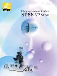
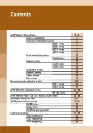
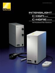
Nematode section revealing cell morphology.<br />
(a) AFM Phase imaging, 10x10 µm.<br />
(b) TEM image of a similar nematode part.<br />
Sample and TEM image courtesy Dr. M. Mueller and<br />
Dr. N. Matsko, ETH Center, Zurich, Switzerland.<br />
3<br />
2 1 7 6 5<br />
10<br />
a.<br />
4<br />
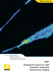
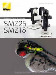
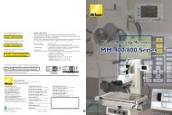
SPM tomography scheme (ultramicrotome combination with the SPM)<br />
1 - sample<br />
2 - sample holder<br />
3 - ultratome movable bar<br />
4 - ultratome cutter<br />
5 - SPM piezoscanner<br />
6 - probe holder<br />
7 - SPM measuring probe<br />
b.<br />
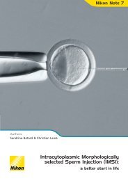
(a) PS/HIPS blend with silica. 15 sequential AFM<br />
images. Each section image is 40x20 µm.<br />
Space between sections is 200 nm.<br />
(b) 3D reconstruction.<br />
Sample courtesy of Dr. Aliza Tzur, Technion, Israel.<br />
Copyright © <strong>NT</strong>-<strong>MDT</strong>, 2007 37
<strong><strong>NT</strong>EGRA</strong> <strong>Tomo</strong><br />
Scanning probe microscopy<br />
in-situ: AFM (contact + semi-contact + non-contact) / Lateral Force Microscopy / Phase Imaging/Force Modulation/<br />
Adhesion Force Imaging/ Magnetic Force Microscopy/ Electrostatic Force Microscopy / Scanning Capacitance Microscopy/<br />
Kelvin Probe Microscopy/ Spreading Resistance Imaging/ Lithography: AFM (Force and Current)<br />
Sample size<br />
Sample weight<br />
10x5x5 mm<br />
Up to 10 g<br />
Scan range 100x100x7 µm<br />
Positioning resolution 5 µm<br />
Non-linearity, XY