Single stranded nucleic acid binding structures - Journal of Cell ...
Single stranded nucleic acid binding structures - Journal of Cell ...
Single stranded nucleic acid binding structures - Journal of Cell ...

Create successful ePaper yourself
Turn your PDF publications into a flip-book with our unique Google optimized e-Paper software.
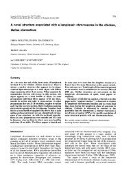
<strong>Journal</strong> <strong>of</strong> <strong>Cell</strong> Science 108, 1391-1396 (1995)<br />
Printed in Great Britain © The Company <strong>of</strong> Biologists Limited 1995<br />
1391<br />
<strong>Single</strong> <strong>stranded</strong> <strong>nucleic</strong> <strong>acid</strong> <strong>binding</strong> <strong>structures</strong> on chicken lampbrush<br />
chromosomes<br />
Irina Solovei 1 , Herbert Macgregor 1, * and Elena Gaginskaya 2<br />
1 Department <strong>of</strong> Zoology, University <strong>of</strong> Leicester, Leicester LE1 7RH, England<br />
2 Laboratory <strong>of</strong> Chromosome Structure and Function, Biological Institute, St Petersburg State University,St Petersburg 198904,<br />
Russia<br />
*Author for correspondence<br />
SUMMARY<br />
In chicken oocytes, proteins <strong>of</strong> the K/J family or their<br />
analogs, such as are known to be involved in mRNA processing<br />
in humans, are closely associated with nascent C-<br />
rich RNA transcripts on the loops <strong>of</strong> lampbrush chromosomes.<br />
Using labelled single <strong>stranded</strong> nucleotide probes and<br />
an antibody to protein K, these C-rich transcripts have been<br />
mapped to six different pairs <strong>of</strong> lampbrush loops situated<br />
on 3 macrochromosomes, the sex bivalent (ZW) and certain<br />
microchromosomes. Each <strong>of</strong> these loop pairs has a distinctive<br />
morphology. The observations represent cytological<br />
evidence <strong>of</strong> the connection between K-proteins and C-rich<br />
RNA transcripts. Another structure, the spaghetti marker<br />
<strong>of</strong> macrochromosome II, also preferentially binds C-rich<br />
homonucleotides. This spaghetti marker has a highly distinctive<br />
fine structural organization that is quite unlike that<br />
<strong>of</strong> lampbrush loops. Its proteins are not recognised by antibodies<br />
to protein K. Homonucleotide <strong>binding</strong> loops are recommended<br />
as potentially extremely valuable as markers on<br />
physical maps <strong>of</strong> chicken chromosomes.<br />
Key words: lampbrush chromosome, K-protein, homonucleotide<br />
<strong>binding</strong>, immun<strong>of</strong>luorescence, chromosome mapping<br />
INTRODUCTION<br />
It is now well known that both snRNP and hnRNP complexes<br />
play important roles in the complicated and highly regulated<br />
processing <strong>of</strong> nuclear pre-mRNAs. Some steps <strong>of</strong> the premRNA<br />
processing (e.g. splicing and polyadenylation) include<br />
a transcript sequence-specific recognition, and it seems likely<br />
that many other steps must also begin with a sequence-specific<br />
<strong>binding</strong> <strong>of</strong> RNA by proteins from snRNP and hnRNP<br />
complexes.<br />
Proteins from hnRNP complexes have been shown to<br />
recognize single <strong>stranded</strong> (ss) <strong>nucleic</strong> <strong>acid</strong>s, both ssDNA and<br />
ssRNA, and many <strong>of</strong> them specifically bind homonucleotide<br />
sequences (Patton et al., 1991; Datar et al., 1993; Dreyfuss et<br />
al., 1993). In particular, homonucleotide tracts are <strong>of</strong>ten<br />
present in upstream, non-coding, portions <strong>of</strong> genes. They have<br />
regulatory roles and are likely candidates as sites <strong>of</strong> recognition<br />
by regulatory RNPs and proteins. Indeed, several hnRNP<br />
proteins that play a role in transcription regulation are capable<br />
<strong>of</strong> recognizing single <strong>stranded</strong> <strong>nucleic</strong> <strong>acid</strong> homopolymers<br />
(Takimoto et al., 1993).<br />
Several proteins from hnRNP complexes belong to the<br />
family <strong>of</strong> RNP consensus RNA-<strong>binding</strong> proteins. They contain<br />
one or two RNA-<strong>binding</strong> domains responsible for the specific<br />
interaction <strong>of</strong> the proteins with RNA, in particular with oligo-<br />
RNA homonucleotides (Ghetti et al., 1992; Gorlach et al.,<br />
1992; Dreyfuss et al., 1993). Some <strong>of</strong> these proteins are evolutionarily<br />
conserved (Datar et al., 1993).<br />
Lampbrush chromosomes (LBC) are characterized by widespread<br />
RNA polymerase II transcription <strong>of</strong> various DNA<br />
sequences. Nascent RNA transcripts, complexed with proteins,<br />
make up the RNP matrix <strong>of</strong> lateral loops <strong>of</strong> LBCs and are<br />
abundant in nuclei <strong>of</strong> growing oocytes during the LBC stage<br />
(Callan, 1986). It is therefore not surprising that many snRNPs<br />
and proteins <strong>of</strong> hnRNP complexes involved in pre-mRNA processing<br />
are found in association with LBCs (DiMario et al.,<br />
1989).<br />
In preparations <strong>of</strong> amphibian LBCs, snRNPs are present<br />
both in loops and in specific <strong>structures</strong>, snurposomes, several<br />
kinds <strong>of</strong> which have been found in association with chromosomes<br />
or free in the germinal vesicle (Gall, 1991, 1992). Also,<br />
several proteins <strong>of</strong> hnRNP complexes are localized in lateral<br />
loops <strong>of</strong> amphibian LBCs (Pinol-Roma et al., 1989; Soulard et<br />
al., 1993). Localization <strong>of</strong> snRNP or protein components <strong>of</strong><br />
hnRNP complexes on bird LBCs have not yet been described.<br />
We have recently described a peculiar object associated with<br />
the short arm <strong>of</strong> chicken LBC II, so called the spaghetti marker<br />
(SM) (Fig. 1). The SM consists <strong>of</strong> a loose bundle <strong>of</strong> 15-16 nm<br />
thick fibres. Cytochemical tests indicate that its fibres contain<br />
little, if any, <strong>nucleic</strong> <strong>acid</strong> and are largely proteinaceous in<br />
nature (Solovei et al., 1992). The SM fibres bear no resemblance<br />
to lateral loop RNP matrix when examined by transmisson<br />
or high resolution scanning electron microscopy<br />
(Solovei et al., 1992). The SM lies next to a chromomere to<br />
which it seems to be attached. The exact nature <strong>of</strong> the attachment<br />
is impossible to resolve.
1392<br />
I. Solovei, H. Macgregor and E. Gaginskaya<br />
When fluorescence in situ <strong>nucleic</strong> <strong>acid</strong> hybridization (FISH)<br />
with the plasmid pHuR93 (Moyzis et al., 1988) containing the<br />
human-derived telomeric DNA repeat was carried out in our<br />
laboratory on chicken LBCs, the SM was always strongly<br />
labelled. This was consistent with an earlier personal communication<br />
from Dr Nancy Hutchison in which she informed us<br />
that the structure that we later characterized as the SM labelled<br />
with pHuR93. Both Dr Hutchison and ourselves recognised<br />
that the <strong>binding</strong> <strong>of</strong> pHuR93 to the SM was most likely to<br />
involve the poly-(dC) x poly-(dG) tracts flanking the telomere<br />
repeat in the recombinant plasmid.<br />
In this article we report the results <strong>of</strong> the incubation <strong>of</strong><br />
chicken LBCs with DNA and RNA probes and their competitors<br />
which were carried out under conditions that precluded the<br />
true hybridization <strong>of</strong> <strong>nucleic</strong> <strong>acid</strong>s. The <strong>binding</strong> <strong>of</strong> single<br />
<strong>stranded</strong> RNA and DNA homopolymers by the SM and by the<br />
RNP matrix <strong>of</strong> a range <strong>of</strong> distinctive loops on chicken LBCs<br />
is demonstrated and discussed.<br />
MATERIALS AND METHODS<br />
Chromosome isolation<br />
Lampbrush chromosomes (LBCs) were manually isolated from<br />
sexually-mature chickens (Gallus gallus domesticus, Rhode Island<br />
Red cross) by standard techniques (Macgregor and Varley, 1988)<br />
modified for bird oocytes (Solovei et al., 1992, 1993). After complete<br />
dispersion <strong>of</strong> nuclear sap, LBCs were centrifuged onto the surface <strong>of</strong><br />
a microscope slide, prefixed in 2% paraformaldehyde (5 minutes),<br />
fixed in 70% ethanol (1 hour) and then stored in 70% ethanol at 4°C.<br />
Probes and competitors<br />
Two types <strong>of</strong> labelled DNA probes were used for this study: (1)<br />
plasmid pHuR93 with an insertion (240 bp) <strong>of</strong> the human telomere<br />
repeat TTAGGG which was prepared by tailing the telomere repeat<br />
sequence with oligo-(dC) and ligating it to oligo-(dG)-tailed pBR322<br />
(Moyzis et al., 1988). This plasmid was labelled using a nick-translation<br />
kit and Biotin-16-dUTP (Boeringer Mannheim). The plasmid<br />
was used after denaturation by heating at 75°C (15 minutes) as a<br />
single <strong>stranded</strong> probe or directly as a double <strong>stranded</strong> probe; in the<br />
latter case, after nick-translation, DNA fragments were end-filled<br />
using Klenow fragments; (2) oligo DNAs (dC) 30 , (dG) 30 , (dA) 30 , all<br />
synthesized on a Applied Biosystems 394 DNA synthesizer and<br />
labelled with one molecule <strong>of</strong> Biotin on 5′-end.<br />
The following homonucleotides were used as unlabelled competitors<br />
for the biotinylated pHuR93 and oligo-(dC) 30 : (1) oligo-(dC) 25<br />
and oligo-(dG) 25 , both synthesized using an Applied Biosystems 394<br />
DNA synthesizer, and (2) RNA homopolymers poly-(C), poly-(G),<br />
poly-(U), and poly-(A) (Pharmacia).<br />
All probes were dissolved either in standard hybridization mixure<br />
(50% formamide, 2× SSC, 10% dextran sulfate) or in pure water.<br />
Plasmid pHuR93 was always used at a concentration <strong>of</strong> 50 ng/µl. Concentrations<br />
<strong>of</strong> synthesized oligonucleotides varied from 0.1 to 100<br />
ng/µl. The concentration ratio between labelled sequence and competitor<br />
varied from 1/10 to 1/10,000.<br />
Incubation conditions<br />
Incubations <strong>of</strong> LBC preparations with the biotinylated probes were<br />
carried out under the conditions that preclude true <strong>nucleic</strong> <strong>acid</strong> hybridization.<br />
DNA <strong>of</strong> LBCs was not denatured to prevent DNA/DNA<br />
hybridization. To prevent DNA/RNA-transcript hybridization some<br />
slides with chromosomes were treated with ribonuclease A (100<br />
µg/ml in 2× SSC, 1 hour at 37°C). 5 µl <strong>of</strong> probe was placed on a dry<br />
slide (with 4 sets <strong>of</strong> LBCs), covered by a coverslip, sealed with rubber<br />
cement and incubated on a slide warmer at 37°C or at room temperature<br />
for 2-12 hours. The competitors were either mixed directly with<br />
the labelled probe or the chromosomes were preincubated with a competitor<br />
prior to addition <strong>of</strong> the biotinylated probe. After incubation,<br />
slides were washed 3 times in 2× SSC at 37°C and 3 times in 2× SSC<br />
at room temperature. Labelled sequences bound to the chromosomes<br />
were detected with avidin DN conjugated with FITC (Vector Laboratories)<br />
without amplification steps. LBCs were counterstained with<br />
DAPI and propidium iodide (PI), air dried and mounted in antifade<br />
media (1% DABCO, 50% glycerol, 1× SSC).<br />
Immun<strong>of</strong>luorescent staining<br />
The monoclonal antibody 3C2 against human protein K (Matunis et<br />
al., 1992) was kindly donated by Dr G. Dreyfuss at the Howard<br />
Hughes Medical Institute Research Laboratories, USA. Lampbrush<br />
chromosomes were incubated in monoclonal antibody ascitic fluid at<br />
dilutions <strong>of</strong> 1:1,000 and 1:10,000. Detection was with FITC-conjugated<br />
goat anti-mouse F(ab′)2 (ICN ImmunoBiologicals) diluted<br />
1:200.<br />
Fluorescence microscopy<br />
Labelled <strong>structures</strong> were visualized with a fluorescence microscope<br />
and imaged using a Bio-Rad MRC600 Confocal Scanning Laser<br />
Attachment to a Zeiss Axiovert microscope. Grey-scale images were<br />
false coloured using the ‘autumn look-up table’ prior to being hard<br />
copied via a Sony UP-3000P colour video printer.<br />
RESULTS<br />
Two types <strong>of</strong> structure on chicken LBCs bind biotinylated<br />
single <strong>stranded</strong> <strong>nucleic</strong> <strong>acid</strong> probes under incubation conditions<br />
that preclude <strong>nucleic</strong> <strong>acid</strong> hybridization. They are the<br />
SM on the short arm <strong>of</strong> the lampbrush chromosome II and<br />
several pairs <strong>of</strong> lateral loops with distinctive morphologies at<br />
specific sites on the chicken LBC complement.<br />
The spaghetti marker (SM)<br />
The SM gave a strong fluorescent signal after treatment with<br />
denatured pHuR93 plasmid under conditions that precluded the<br />
formation <strong>of</strong> <strong>nucleic</strong> <strong>acid</strong> hybrid molecules (Fig. 2A). In the<br />
same experiments, the terminal chromomeres and certain interstitial<br />
sites <strong>of</strong> LBCs, all <strong>of</strong> which are known to contain telmomeric<br />
DNA sequences and bind pHuR93 under the true hybridization<br />
conditions (Solovei et al., 1994, A. Rodionov, personal<br />
communication), were never labelled. Likewise, the short transcription<br />
units situated at the very ends <strong>of</strong> LBC chromatids that<br />
contain transcribed telomeric DNA sequences and bind pHuR93<br />
under conditions <strong>of</strong> DNA to RNA-transcript hybridization were<br />
never labelled (Solovei et al., 1994). Taken together, these observations<br />
srongly support our claim that the incubation conditions<br />
used in our experiments did indeed preclude the true hybridisation<br />
<strong>of</strong> <strong>nucleic</strong> <strong>acid</strong>s. Labelling <strong>of</strong> the SM was therefore most<br />
probably due to <strong>binding</strong> <strong>of</strong> some part <strong>of</strong> pHuR93 to SM proteins.<br />
Plasmid pHuR93 was constructed from pBR322 and<br />
contains 240 bp <strong>of</strong> human telomere repeat (TTAGGG) flanked<br />
by at least 25 bp <strong>of</strong> (dC)x(dG) (Moyzis et al., 1988). Control<br />
experiments demonstrated that neither pure pBR322, nor PCRgenerated<br />
pure telomere repeat were recognized by the SM.<br />
Unlabelled oligo-(dC) 25, used as a competitor during incubation<br />
with pHuR93 (1:1000), completely abolished the fluorescent<br />
signal on the SM, whilst oligo-(dG) 25 did not affect it (Fig.<br />
2A). Binding <strong>of</strong> pHuR93 to the SM did not take place unless<br />
the plasmid DNA was denatured.
Homonucleotide <strong>binding</strong> to lampbrush chromosomes<br />
1393<br />
We conclude that the SM shows a preferential affinity for<br />
single-<strong>stranded</strong> poly-(C) nucleotide arrays and that the <strong>binding</strong><br />
<strong>of</strong> poly-(C) to the SM is not <strong>of</strong> the kind that characterises the<br />
formation <strong>of</strong> <strong>nucleic</strong> <strong>acid</strong> hybrid molecules but results from<br />
<strong>binding</strong> <strong>of</strong> single <strong>stranded</strong> poly-(C) to SM proteins.<br />
This conclusion was reinforced when we used biotinylated<br />
oligo-(dC) 30 as a probe in <strong>binding</strong> experiments with lampbrush<br />
chromosomes. The SM strongly bound biotinylated oligo-<br />
(dC) 30 under a variety <strong>of</strong> conditions, in standard <strong>nucleic</strong> <strong>acid</strong><br />
hybridization mixture or in pure water, at 37°C or at room temperature.<br />
The fluorescent signal was easily observable in all<br />
these experiments down to a probe concentration <strong>of</strong> 1 ng per<br />
slide (1 ng/5ul).<br />
When chromosomes were incubated with biotinylated oligo-<br />
(dC) 30 in the presence <strong>of</strong> a range <strong>of</strong> concentrations <strong>of</strong> poly C-<br />
RNA as a competitor, the SM was progressively less strongly<br />
labelled as the unlabelled competitor concentration was<br />
increased until, at a ratio <strong>of</strong> 1:1,000 labelled:unlabelled probe,<br />
the SM was no longer visibly labelled (Fig. 2B, Table 1). In<br />
the same way the <strong>binding</strong> <strong>of</strong> biotinylated oligo-(dC) 30 to the<br />
SM was suppressed by poly-(rG), although less strongly than<br />
it is by poly-(rC). Poly-(rU) had little or no effect on the<br />
<strong>binding</strong> <strong>of</strong> biotinylated oligo-(dC) (Table 1). Poly-(rA) had no<br />
effect at all. Therefore only poly-(rC) and poly-(rG) were able<br />
successfully to compete out the <strong>binding</strong> <strong>of</strong> oligo-(dC) 30 and<br />
could therefore be said to be specifically recognized by the SM.<br />
Neither biotinylated oligo-(dG) 30 nor biotinylated oligo-(dA) 30<br />
were bound by the SM.<br />
We realized that the success <strong>of</strong> poly-(rG) as a competitor<br />
in our experiments could have been a consequence <strong>of</strong><br />
annealing with the biotinylated probe oligo-(dC) 3 0 rather than<br />
<strong>binding</strong> to SM proteins. Accordingly, in subsequent experiments<br />
competitor oligonucleotides were applied before the<br />
incubation with labelled oligo-(dC) 3 0 . Chromosomes were<br />
preincubated for 2 hours at 37°C with unlabelled oligo-<br />
( d C ) 25, o l i g o - ( d G ) 2 5 , poly-(rC), or poly-(rG), washed in 2×<br />
SSC (3× 3 minutes), and then incubated for 2 hours at 37°C<br />
with biotinylated oligo-(dC) 3 0 . The ratios <strong>of</strong> labelled probe<br />
concentration to the concentration <strong>of</strong> competitors used for<br />
preincubation were 1:1,000. For poly-(rG), the ratio <strong>of</strong><br />
1:10,000 was also tried. Poly-(rC) completely abolished<br />
labelling <strong>of</strong> the SM, signifying that the SM proteins have a<br />
greater affinity for single <strong>stranded</strong> RN homopolymers than<br />
they have for single <strong>stranded</strong> DN homopolymers. Unlabelled<br />
poly-(rG) and oligo-(dC) 2 5 also suppressed labelling <strong>of</strong> the<br />
SM by oligo-(dC) 30 detectably but less effectively than poly-<br />
(rC) (Table 2).<br />
We conclude that the SM is a structure on chicken<br />
lampbrush chromosomes that contains proteins that preferentially<br />
bind single <strong>stranded</strong> poly-(C) RNA and oligo-(C) DNA.<br />
LBC lateral loops<br />
After incubation <strong>of</strong> chicken LBCs with biotinylated oligo-<br />
(dC) 30 under conditions excluding hybridization with <strong>nucleic</strong><br />
<strong>acid</strong>s, several <strong>binding</strong> sites were found in addition to the SM<br />
(Fig. 1). These sites include: (1) the pair <strong>of</strong> marker loops<br />
PBL11 on bivalent I (Fig. 3A); (2) a pair <strong>of</strong> very small loops<br />
near the terminal chromomere on the long arm <strong>of</strong> bivalent II;<br />
Fig. 1. Schematic drawing <strong>of</strong><br />
the first three chicken<br />
lampbrush macrobivalents<br />
(numbers I, II and III), the sex<br />
bivalent (ZW) and some<br />
microchromosomes (micro)<br />
showing the set <strong>of</strong> loops<br />
(coloured black and indicated<br />
by arrows) that bind poly(C)<br />
single <strong>stranded</strong> RNA and<br />
antibodies against human K<br />
protein. PBL 11, loops at the<br />
proximal border <strong>of</strong> the ‘bald’<br />
region 1 <strong>of</strong> chromosome 1;<br />
LL 31, lumpy loops 1 on<br />
chromosome 3; TGL,<br />
telomere giant loops <strong>of</strong> the<br />
ZW bivalent; TL, telomere<br />
loops <strong>of</strong> the<br />
microchromosomes; C,<br />
centromere region. SM<br />
indicates the spaghetti marker<br />
on the second bivalent which<br />
binds poly(C) single <strong>stranded</strong><br />
RNA and DNA but does not<br />
bind 3C2 antibodies. All<br />
loops and chromosome<br />
regions, with the exception <strong>of</strong><br />
the Spaghetti Marker, are<br />
named according to the<br />
nomenclature introduced by<br />
Chelysheva et al. (1990)
1394<br />
I. Solovei, H. Macgregor and E. Gaginskaya<br />
A<br />
B<br />
Fig. 2. Labelling <strong>of</strong> SM (arrowheads) on account <strong>of</strong> <strong>binding</strong><br />
<strong>of</strong> biotinylated pHuR93 or biotinylated oligo-(C)DNA alone<br />
or in competition with other unlabelled homonucleotide<br />
polymers. (A) Bivalent II after incubation with denatured<br />
biotinylated pHuR93 (left) and with the same denatured<br />
labelled plasmid probe plus unlabelled oligo-(dC) 25 (middle)<br />
or oligo-(dG) 25 (right), respectively, as competitors.<br />
(B) Labelling <strong>of</strong> SM with biotinylated oligo-(dC) 30 after preincubation<br />
with non-biotinylated poly-(rC) as a competitor.<br />
The concentration ratios labelled probe:competitor are<br />
indicated in each case. A bivalent II incubated with pure<br />
biotinylated oligo-(dC), with no unlabelled competitor<br />
present, is shown at the top left.<br />
(3) a pair <strong>of</strong> marker lumpy loops LL31 on the long arm <strong>of</strong><br />
bivalent III (Fig. 3C); (4) the ‘thin’ ends <strong>of</strong> the telomere giant<br />
loops on the chiasmate ends <strong>of</strong> the Z and W sex chromosomes;<br />
(5) the telomere loops on the short arms <strong>of</strong> some microchromosomes<br />
(Fig. 3E). In all these cases <strong>binding</strong> <strong>of</strong> labelled oligo-<br />
(dC) 30 was RNase-stable and was abolished by competing with<br />
Table 1. Labelling <strong>of</strong> SM by biotinylated oligo-(dC) in<br />
presence <strong>of</strong> competitor RNAs<br />
Probes and Probe/competitor Brightness <strong>of</strong><br />
competitors ratio SM<br />
Biotinylated +++<br />
oligo-(dC)30/no competiton<br />
Biotinylated 1/10 ++<br />
oligo-(dC)30/poly-(rC) 1/100 +<br />
1/1,000 −<br />
Biotinylated<br />
oligo-(dC)30/poly-(rG) 1/1,000 −<br />
Biotinylated<br />
oligo-(dC)30/poly-(rU) 1/1,000 +++<br />
unlabelled poly-(rC). As with the SM, labelling <strong>of</strong> these loops<br />
with oligo-(dC) 30 was suppressed by the unlabelled competitor<br />
poly-(rG), but not by oligo-(dG) 25 , oligo-(dA) 25 , poly-(rA)<br />
nor poly-(rU).<br />
Experiments with antibodies<br />
Monoclonal antibody 3C2, against human protein K, used in<br />
dilutions <strong>of</strong> 1:1,000 or 1:10,000, gave very strong positive<br />
reactions with all 5 kinds <strong>of</strong> oligo-(dC)-absorbing loops (Fig.<br />
3B,D,F) but no reaction whatsoever with the SM (Fig. 1). The<br />
Table 2. Labelling <strong>of</strong> SM by biotinylated oligo-(dC) after<br />
preincubation with competitor <strong>nucleic</strong> <strong>acid</strong>s<br />
Competitor <strong>nucleic</strong> <strong>acid</strong><br />
for preincubation<br />
Brightness <strong>of</strong><br />
SM<br />
Oligo-(dC)25 (1:1,000) +<br />
Oligo-(dG)25 (1:1,000) +++<br />
Poly-(rC) (1:1,000) −<br />
Poly-(rG) (1:1,000) ±<br />
(1:10,000) −
Homonucleotide <strong>binding</strong> to lampbrush chromosomes<br />
1395<br />
nuclei <strong>of</strong> erythrocytes (which were deliberately placed<br />
alongside chromosome preparations) also gave a positive<br />
immunoreaction which appeared as a few separate spots. This<br />
is consistent with the data <strong>of</strong> Matunis et al. (1992) who showed<br />
positive immunoreaction to hnRNP K in interphase nuclei <strong>of</strong><br />
HeLa cells using the same antibodies.<br />
A<br />
B<br />
C<br />
D<br />
E<br />
F<br />
Fig. 3. Labelling <strong>of</strong> loops on lampbrush bivalent I<br />
(A and B), bivalent III (C and D) and a microbivalent<br />
(E and F) on account <strong>of</strong> <strong>binding</strong> <strong>of</strong> biotinylated oligo-<br />
(dC) 30 (A,C and E) and immunostaining with<br />
antibodies against K protein (B,D and F). Bars:<br />
(A-C), 20 µm; (D-F), 10 µm.
1396<br />
I. Solovei, H. Macgregor and E. Gaginskaya<br />
DISCUSSION<br />
Our data indicate that two types <strong>of</strong> proteins that preferentially<br />
bind poly-(C) <strong>nucleic</strong> <strong>acid</strong>s are present on chicken LBCs. One<br />
<strong>of</strong> the protein types forms part <strong>of</strong> the RNP matrix <strong>of</strong> a specific<br />
set <strong>of</strong> loops. It is immunologically similar to protein K/J from<br />
human (Matunis et al., 1993) and Xenopus (Siomi et al., 1993),<br />
and is most likely nothing more than the chicken equivalent <strong>of</strong><br />
protein K/J. Proteins K/J bind poly-(C) <strong>nucleic</strong> <strong>acid</strong>s and they<br />
are known to be amongst the major pre-mRNA <strong>binding</strong><br />
proteins involved in mRNA processing (Matunis et al., 1992;<br />
Siomi et al., 1993; Burd and Dreyfuss 1994).<br />
The human protein K sequence-specifically binds to the C-<br />
rich region <strong>of</strong> proto-oncogene c-myc and therefore might be<br />
involved in regulation <strong>of</strong> its transcription (Takimoto et al.,<br />
1993). This protein also possesses a high affinity for poly-(C)<br />
RNA (Matunis et al., 1993), which makes it a likely candidate<br />
for involvement in the processing <strong>of</strong> RNA transcripts. These<br />
and other lines <strong>of</strong> evidence about the functional significance <strong>of</strong><br />
K protein have a purely biochemical basis.<br />
Our results again support the conclusion that proteins K/J<br />
bind specifically to C-rich RNA but they put the matter on a<br />
more cytological level by showing that in chicken oocytes,<br />
proteins K/J or their analogs are closely associated with the<br />
nascent RNA transcripts on the loops <strong>of</strong> lampbrush chromosomes.<br />
Futhermore, using labelled single <strong>stranded</strong> nucleotide<br />
probes and antibodies has enabled us to map directly a well<br />
defined set <strong>of</strong> chromosome regions where these C-rich transcripts<br />
are situated, so providing an approach to localization <strong>of</strong><br />
the sequences (genes) that specify such transcripts.<br />
The long, branching fibres <strong>of</strong> the SM, with their smooth<br />
outlines and uniform dimensions, are structurally very<br />
different from the RNP matrix <strong>of</strong> loops (Solovei et al., 1992).<br />
RNase treatment does not affect the appearance <strong>of</strong> the SM,<br />
from which we have concluded that RNA at least is not a sign<br />
i ficant component <strong>of</strong> its structure (Solovei et al., 1992). The<br />
SM is not recognized by the antibody to hnRNP K/J. For the<br />
moment, we can only conclude that its fibres consist largely<br />
<strong>of</strong> a different protein that preferentially binds C-rich RNA.<br />
The true nature <strong>of</strong> this extraordinary structure remains a<br />
m y s t e r y .<br />
Homonucleotide absorbing loops are potentially extremely<br />
valuable as convenient markers on the cytological maps <strong>of</strong><br />
chicken LBCs. They could be especially useful for chicken<br />
gene mapping by FISH.<br />
We thank Chris deLacey <strong>of</strong> the Leicester Advanced Light Microscope<br />
Facility for his expert technical guidance and help with the production<br />
<strong>of</strong> all the colour images in this paper. We gratefully acknowledge<br />
support for this research from The Wellcome Trust (Grant no.<br />
039293/Z/93 to H.C.M and I.S) and the International Science Foundation<br />
(Grant no. R2N000 to E.G. and I.S.).<br />
REFERENCES<br />
Burd, A. and Dreyfuss, G. (1994). Conserved <strong>structures</strong> and the diversity <strong>of</strong><br />
functions <strong>of</strong> RNA-<strong>binding</strong> proteins. Science 265, 615-621<br />
Callan, H. G. (1986). Lampbrush Chromosomes. Springer Verlag, Heidelberg<br />
and New York.<br />
Chelysheva, L. A., Solovei, I. V., Rodionov, A. V., Yakolev, A. F. and<br />
Gaginskaya, E. R. (1990). The lampbrush chromosomes <strong>of</strong> the chicken: the<br />
cytological maps <strong>of</strong> macrobivalents (in Russian). Cytology 32, 303-316.<br />
Datar, K. V., Dreyfuss, G. and Swanson, M. S. (1993). The human hnRNP M<br />
proteins: identification <strong>of</strong> a methionine/arginine-rich repeat motif in<br />
ribonucleoproteins. Nucl. Acids Res. 21, 439-446.<br />
DiMario, P. J., Bromley, S. E. and Gall, J. G. (1989). DNA-<strong>binding</strong> proteins<br />
on lampbrush chromosome loops. Chromosoma 97, 413-420.<br />
Dreyfuss, G., Matunis, M. J., Pinol-Roma, S. and Burd, C. G. (1993).<br />
hnRNP proteins and the biogenesis <strong>of</strong> mRNA. Annu. Rev. Biochem. 62, 289-<br />
321.<br />
Gall, J. G. (1991). Spliceosomes and Snurposomes. Science 252, 1499-1500.<br />
Gall, J. G. (1992). Organelle assembly in the amphibian germinal vesicle.<br />
Advan. Dev. Biochem. 1, 1-29.<br />
Ghetti, A., Pinol-Roma, S., Michael, W. M., Morandi, C. and Dreyfuss, G.<br />
(1992). hnRNP I, the polypyrimidine tract-<strong>binding</strong> protein: distinct nuclear<br />
localization and association with hnRNAs. Nucl. Acids Res. 20, 3671-3678.<br />
Gorlach, M., Wittekind, M., Beckman, R. A, Mueller, L. and Dreyfuss, G.<br />
(1992). Interaction <strong>of</strong> the RNA-<strong>binding</strong> domain <strong>of</strong> the hnRNP C proteins<br />
with RNA. EMBO J. 11, 3289-3295.<br />
Macgregor, H. C. and Varley, J. M. (1988). Working with Animal<br />
Chromosomes, 2nd edn. John Wiley & Sons, Chichester and New York.<br />
Matunis, M. J., Michael, W. M. and Dreyfuss, G. (1992) Characterization<br />
and primary structure <strong>of</strong> the poly-(C)-<strong>binding</strong> heterogeneous nuclear<br />
ribonucleoprotein complex K protein. Mol. <strong>Cell</strong> Biol. 12, 164-171.<br />
Moyzis, R. K., Buckingham, J. M., Cram, L. S. et al. (1988) A highly<br />
conserved repetitive DNA sequence (TTAGGG)npresent at the telomeres <strong>of</strong><br />
human chromosomes. Proc. Nat. Acad. Sci. USA 85, 6622-6626.<br />
Patton, J. G., Mayer, S. A., Tempst, P. and Nadal-Ginard, B. (1991).<br />
Characterization and molecular cloning <strong>of</strong> polypyrimidine tract-<strong>binding</strong><br />
protein: a component <strong>of</strong> a complex necessary for pre-mRNA splicing. Genes<br />
Dev. 5, 1237-1251.<br />
Pinol-Roma, S., Swanson M. S., Gall, J. G. and Dreyfuss G. (1989). A novel<br />
heterogenous nuclear RNP protein with a unique distribution on nascent<br />
transcripts. J. <strong>Cell</strong> Biol. 109, 2575-2587.<br />
Siomi, H., Matunis, M. J., Michael, W. M. and Dreyfuss, G. (1993). The premRNA<br />
<strong>binding</strong> K protein contains a novel evolutionarily conserved motif.<br />
Nucl. Acids Res. 21, 1193-1198.<br />
Solovei, I., Gaginskaya, E., Allen, T. and Macgregor, H. (1992). A novel<br />
structure associated with a lampbrush chromosome in the chicken Gallus<br />
domesticus. J. <strong>Cell</strong> Sci. 101, 759-772.<br />
Solovei, I., Gaginskaya, E. R. and Macgregor, H. C. (1993). Avian sex<br />
chromosomes in the lampbrush form, the ZW lampbrush bivalents from six<br />
species <strong>of</strong> bird. Chromosome Res. 1, 153-166.<br />
Solovei, I., Gaginskaya, E. and Macgregor, H. C. (1994). The arrangement<br />
and transcription <strong>of</strong> telomere DNA sequences at the ends <strong>of</strong> lampbrush<br />
chromosomes <strong>of</strong> birds. Chromosome Res. 2, 460-470.<br />
Soulard, M., Valle, V. D., Siomi, M. S., Pinol-Roma, S., Godogno, P., Bauvi,<br />
C., Bellini, M., Lacroix, J.-C., Monod, G., Dreyfuss, G. and Larsen, C.-J.<br />
(1993). hnRNP G: sequence and characterization <strong>of</strong> a glycosylated RNA<strong>binding</strong><br />
protein. Nucl. Acids Res. 21, 4210-4217.<br />
Takimoto, M., Tomonaga, T,. Matunis, M., Avigan, M., Krutzsch, H.,<br />
Dreyfuss, G. and Levens, D. (1993). Specific <strong>binding</strong> <strong>of</strong> heterogeneous<br />
ribonucleoproteins particle protein K to the human c-myc promoter, in vitro.<br />
J. Biol. Chem. 268, 18249-18258.<br />
(Received 9 November 1994 - Accepted 21 January 1995)