Ceramic Based Bio-Medical Implants - IFGL Bio Ceramics Limited
Ceramic Based Bio-Medical Implants - IFGL Bio Ceramics Limited
Ceramic Based Bio-Medical Implants - IFGL Bio Ceramics Limited

Create successful ePaper yourself
Turn your PDF publications into a flip-book with our unique Google optimized e-Paper software.
98 INTERCERAM 02/2010<br />
HIGH-PERFORMANCE CERAMICS<br />
R. Sarkar*, G. Banerjee**<br />
<strong>Ceramic</strong> <strong>Based</strong> <strong>Bio</strong>-<strong>Medical</strong> <strong>Implants</strong><br />
THE AUTHOR ABSTRACT KEYWORDS<br />
The corresponding author,<br />
Dr Ritwik Sarkar (38), Associate<br />
Professor, NIT, Rourkela,<br />
India, has about 15 years of<br />
experience in <strong>Ceramic</strong>s and<br />
Refractories. He worked with<br />
<strong>IFGL</strong> <strong>Bio</strong>ceramics Ltd, <strong>IFGL</strong> Refractories<br />
Ltd, CG & CRI, R&D<br />
of ACC Ltd, and H&R Johnson Ltd, all in India, and<br />
at the IKKM, RWTH Aachen, Germany (DAAD Fellowship).<br />
Dr Sarkar has received many awards in<br />
his career for academic excellence and his contributions<br />
to <strong>Ceramic</strong>s and Materials Science, including<br />
the Young Scientist Award. A life member<br />
of the Indian <strong>Ceramic</strong> Society & Indian Institute<br />
of <strong>Ceramic</strong>s, Dr Sarkar has more than 70 research<br />
publications and 9 patents to his credit.<br />
<strong>Bio</strong>ceramics are those engineered materials that are inorganic and nonmetallic<br />
in nature and find applications in the field of medicine. They are<br />
distinct from metals and their alloys and organic polymers but cover noncrystalline<br />
glasses and ceramic-based composites. <strong>Bio</strong>ceramic materials<br />
are used in the repair and reconstruction of diseased or damaged parts<br />
of the musculo-skeletal system. They are useful as joint or tissue replacements,<br />
as coatings to improve the biocompatibility of metal implants,<br />
as resorbable lattices which provide a temporary structure framework<br />
that is dissolved and replaced as the body rebuilds tissue and also<br />
even as a controlled drug delivery system.<br />
This paper deals mainly with three different types of biomedical implants<br />
made of ceramics, namely in the areas of hip joint femoral heads, orbital<br />
implants and bone regenerative dental applications.<br />
implants, femoral head,<br />
orbital implant, dental<br />
implant<br />
Interceram 59 (2010) [2]<br />
1 Introduction<br />
The history of bioceramics is especially interesting.<br />
In 1972, Amadeo Bobbio discovered<br />
Mayan skulls; some of them more than<br />
4000 years old, in which missing teeth had<br />
been replaced by nacre substitutes [1]. Natural<br />
nacre has a brick-mortar like microstructure<br />
where inorganic calcium carbonate<br />
layers (ceramic portion) are held together<br />
by organic protein “glue”. The inorganic<br />
portion represents 95 % of the volume of<br />
nacre; its highly specific properties (in particular<br />
its toughness) are due to the interaction<br />
of this inorganic component with the<br />
organic phase that is found between the calcium<br />
carbonate platelets. But the continuity<br />
of the evidence of use of ceramics in medical<br />
applications was not readily found till<br />
the modern era.<br />
* Department of <strong>Ceramic</strong> Engineering, National<br />
Institute of Technology, Rourkela, India;<br />
contact: ritwiksarkar@rediffmail.com<br />
** <strong>IFGL</strong> <strong>Bio</strong>ceramics Ltd, Kolkata, India;<br />
contact: gbanerjee@bajoria.in<br />
2 Uses<br />
<strong>Ceramic</strong>s are more similar to natural skeletal<br />
materials, but due to their limitations in<br />
mechanical properties, metallic implants are<br />
widely used. Improvements in ceramic<br />
processing technology in the 1960s led to<br />
great scientific interest in bioceramics [2]. A<br />
strong interest in the use of ceramics for biomedical<br />
engineering applications developed<br />
in the late 1960s, exemplified by the<br />
work of Hulbert and co-workers [3]. But<br />
this interest reached a plateau during the<br />
late 1970s and early 1980s, mainly due to<br />
conservative governmental regulatory systems<br />
which required substantially more<br />
clinical experience and successful trials on a<br />
long time scale. However, after the initial,<br />
highly successful endeavours the last two<br />
decades have seen a renaissance in the development<br />
and uses of ceramic based biomedical<br />
materials.<br />
The use of bioceramics has mainly concentrated<br />
on orthopaedics and dentistry. <strong>Bio</strong>ceramics<br />
in orthopaedic applications provide<br />
the advantage of chemical similarity to natural<br />
skeletal materials. Again in dental applications,<br />
the chemical similarity with natural<br />
dental materials and the excellent compressive<br />
load-bearing capacity have made<br />
ceramic-based biomaterials an ideal implant.<br />
<strong>Based</strong> on their chemical reactivity with the<br />
physiological environment, bioceramics can<br />
be broadly categorized in three types [4]:<br />
<strong>Bio</strong>inert bioceramics, such as alumina, result<br />
in little or no physiological reaction in<br />
the human body and tend to exhibit inherently<br />
low levels of reactivity which peak in<br />
the order of hundreds of years.<br />
Surface reactive or bioactive ceramics, such<br />
as bioglass, react in a positive way with local<br />
cells, i.e. they form bonds and have a substantially<br />
higher level of reactivity, peaking<br />
in the order of 100 days.<br />
Resorbable bioceramics are porous or nonporous<br />
structures which are slowly and<br />
gradually replaced by bone such as tricalcium<br />
phosphate, have even higher levels of reactivity,<br />
peaking in the order of 10 days.<br />
<strong>Bio</strong>ceramics are needed to alleviate pain and<br />
restore normal activity to diseased or damaged<br />
parts of the body. As people age, progressive<br />
deterioration of tissues requires replacements<br />
in many critical applications.<br />
Bone is especially vulnerable to fracture in<br />
older people because of a loss of bone den-
INTERCERAM 02/2010<br />
99<br />
HIGH-PERFORMANCE CERAMICS<br />
2<br />
1<br />
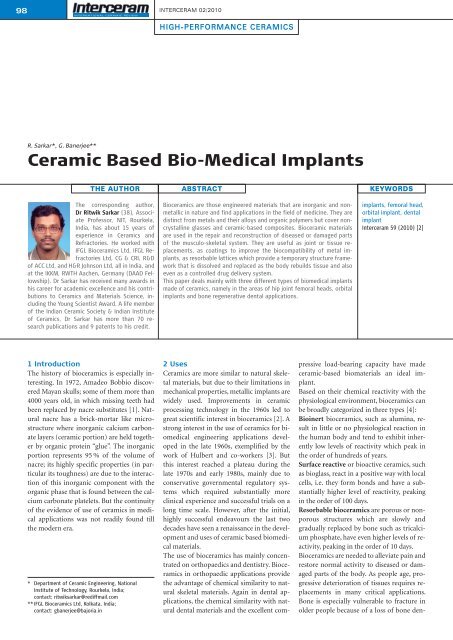
Fig. 1 • The hip joint and its details, with femoral head: a) metallic and b) ceramic<br />
(alumina)<br />
Fig. 2 • Microstructure of alumina hip ball<br />
sity and strength [5]. After successful research<br />
and many animal and human trials,<br />
various bioceramic products are now commercially<br />
available in the medical market as<br />
substitutes for the original damaged body<br />
parts and for many other critical applications.<br />
The major application areas are as follows.<br />
• Replacements for hips, knees, teeth, tendons<br />
and ligaments<br />
• Repair for periodontal diseases<br />
• Maxillofacial reconstruction<br />
• Augmentation and stabilization of the jaw<br />
bone<br />
• Spinal fusion, bone repair after tumour<br />
surgery<br />
• Pyrolytic carbon coatings for prosthetic<br />
heart valves<br />
• Treatment of cancer by localised delivery<br />
through radioactive glass microspheres<br />
Some of the important ceramic-based implants<br />
are detailed below.<br />
2.1 <strong>Ceramic</strong> femoral head (hip joint ball)<br />
The replacement of broken and worn out<br />
bones parts is not a new development.<br />
Themistocles Gluck (1853–1942), inventor<br />
of artificial biomaterials, changed the concept<br />
of contemporary surgery “from being<br />
the destructive art to become the reconstructive<br />
art”. He developed entire artificial<br />
joints, fabricated from ivory, and implanted<br />
them in many patients [6].<br />
A stainless steel-based artificial hip joint was<br />
first implanted by John Charnley in 1960<br />
[7]. Charnley’s design, although simple, remained<br />
virtually unchanged for three decades,<br />
even though the common problems<br />
associated with the metallic implants such<br />
as corrosion, wear, and loss of bone were<br />
known. These problems have restricted the<br />
lifespan of metallic implants to 10–12 years<br />
and they require revision surgery for many<br />
patients. To avoid this and to improve the<br />
quality of life, researchers started experimenting<br />
with ceramic implants, especially<br />
for hip joints.<br />
<strong>Ceramic</strong> implants have some inherent advantages<br />
such as inertness in a biological environment<br />
and biocompatibility; they produce<br />
nearly no wear debris and can be designed<br />
to match the material properties of<br />
natural bone. All these result in a much<br />
longer life of the prosthesis [8]. Again metal<br />
has such high stiffness that the bone surrounding<br />
a metallic implant no longer bears<br />
the load and the implant carries the entire<br />
load. As bone is a living tissue, it begins to<br />
resorb when it ceases to carry the load, causing<br />
the implant to loosen and eventually require<br />
replacement. <strong>Ceramic</strong>s can be tailored<br />
to allow bone to grow within their porous<br />
structures (scaffolds) and can completely<br />
incorporate the scaffold with natural bone<br />
within five to seven months.<br />
For the articulating surfaces on hip, knee,<br />
and shoulder, alumina- and zirconia-based<br />
ceramic implants are important. They are<br />
harder than metal, have higher scratch resistance<br />
and can be used on both the ball<br />
and socket components of an implant, such<br />
as the femoral head and the acetabular cup<br />
of a hip joint (Fig. 1). The ceramic hip prosthesis<br />
provides much longer life, reduces the<br />
side effects and is suitable for all patients, especially<br />
for younger people who require a<br />
longer healthy life without revision surgery.<br />
The clinical success associated with the use<br />
of ceramics has led to the implantation of<br />
more than 3.5 million alumina components<br />
and more than 600,000 zirconia femoral<br />
heads worldwide since 1990 [9]. Statistics<br />
show that more than 0.5 million hip replacement<br />
surgeries per year are performed<br />
globally. Patients who experience unbearable<br />
pain due to osteoarthritis or have broken<br />
hips in accidents undergo such operations.<br />
This, coupled with increased life expectancy,<br />
has triggered multi-directional research activity<br />
to identify ideal materials for such implants<br />
which will give the desired longevity<br />
and comfort. For femoral heads, fine grained<br />
alumina (Fig. 2) [10] with engineered properties<br />
and fitted with a Ti-Al-V stem is currently<br />
established as the best material combination<br />
for hip replacement surgeries. Table<br />
1 shows the property requirements of<br />
the alumina-based femoral heads as per international<br />
standards while Table 2 compares<br />
the wear characteristics of aluminabased<br />
(ceramic) femoral heads to SS 316 L<br />
grade (metallic) [10].<br />
2.2 Orbital implant<br />
An eye surgeon removes the eye-ball from<br />
the orbit of a patient when the person’s eye<br />
is damaged due to injury or disease, to avoid<br />
the risk of life or risk to the other eye. To fill<br />
up the orbital volume lost, an ocular implant<br />
is placed to achieve better cosmetic results<br />
and rehabilitation of the anophthalmic<br />
patient. The small ocular implant is not visible<br />
but it maintains the natural structure of<br />
the orbit and provides support to the artificial<br />
eye. It also restores the natural appearance<br />
of the eye and surrounding tissues.<br />
Though artificial eyes have been made for<br />
thousands of years, the first orbital implant<br />
was developed only about a century ago.<br />
Numerous materials like gold, cartilage, xenogeneic<br />
animal eyes, silver aluminium, silicone<br />
and glass beads were used to fill irregular<br />
cavities in the orbit. Most of these implants<br />
were found unsuitable due to various<br />
reasons and were discarded one after another.<br />
In 1941, an acrylic based, partially exposed<br />
orbital implant was introduced by<br />
Ruedemann [12], which imparted good<br />
motility to the artificial eye, but it was prone
100 INTERCERAM 02/2010<br />
HIGH-PERFORMANCE CERAMICS<br />
3<br />
4<br />
Fig. 3 • Similarity in<br />
structure:<br />
a) human bone and<br />
b) hydroxyapatite<br />
Fig. 4 • SEM photomicrograph<br />
of synthetic<br />
hydroxyapatite-based<br />
ocular implant<br />
prepared material. Synthetic material based<br />
implants allow a much improved control<br />
over size, weight, total porosity and porous<br />
structure and can easily be tailored to the requirements<br />
of better implantation [16–17].<br />
Figure 5 shows different shapes of synthetic<br />
hydroxyapatite-based orbital implants and<br />
pegs and properties of these implants are<br />
provided in Table 3 [18].<br />
The advantages of hydroxyapatite-based orbital<br />
implants are [18]:<br />
• inert, biocompatible and biochemically<br />
stable,<br />
• light weight to increase motility,<br />
• smooth surface,<br />
• porous structure for tissue in-growth, resulting<br />
in improved motility and minimum<br />
extrusion,<br />
• resilience.<br />
to infection, extrusion, and late position<br />
problems which had to be corrected. Lack of<br />
movement was also a major obstacle in restoring<br />
a natural appearance. Also these implants<br />
tended to drift in the orbit and were<br />
often rejected by the body, requiring further<br />
surgery. These problems inspired researchers<br />
to continuously search for a better orbital<br />
implant.<br />
Hydroxyapatite (HAp) [also known as<br />
Hydroxylapatite, Ca 5 (PO 4 ) 3 (OH), usually<br />
written as Ca 10 (PO 4 ) 6 (OH) 2 ] is the principal<br />
inorganic constituent of bone and<br />
teeth. The chemical similarity of HAp to<br />
bone and its excellent biocompatibility and<br />
bioactivity have attracted the attention of<br />
medical professionals [13–14]. Figure 3<br />
shows the microstructural similarity of human<br />
bone and hydroxyapatite [10]. For the<br />
past few decades, hydroxyapatite-based ceramics<br />
have been widely used in the field of<br />
orthopaedics and dentistry. Extensive research<br />
into the biological and physicochemical<br />
properties of this material has<br />
widened its scope of application and in recent<br />
years it has also found promising applications<br />
in many other areas of medical<br />
science.<br />
The first-ever hydroxyapatite-based orbital<br />
implant was inserted by Dr Arthur Perry in<br />
the 1990, after several years of preliminary<br />
research [15]. The porous implant was easily<br />
attached to the eye muscles and the tissues<br />
that covered the implant resulted in much<br />
improved motility of the implant, giving it<br />
the appearance of a natural eye. Figure 4<br />
shows the microstructure of such an implant<br />
and its porous nature [10]. For even<br />
better movement, a peg may be used to con-<br />
nect the artificial eye to the implant. In this<br />
way, small, darting movements of the natural<br />
eye can be imparted in the artificial eye.<br />
The result is a more natural-looking artificial<br />
eye which is difficult to distinguish from<br />
the natural eye.<br />
There are two types of hydroxyapatite-based<br />
orbital implants available: the first is natural<br />
coral based and the second is synthetically<br />
2.3 Dental implant – bone regenerative<br />
material<br />
The working environment of teeth is one of<br />
the most inhospitable in the human body. It<br />
faces wider temperature variations than<br />
most other parts, varying from sub-zero<br />
temperatures (for ice-creams) to hot tea/<br />
coffee/soup, encounters pH changes in the<br />
range 0.5 to 8, and it is a hostile environment<br />
filled with various micro-organisms,<br />
bacteria and viruses, etc. Added to this are<br />
the various forms of stresses associated with<br />
Table 1 • Property requirements of the alumina-based femoral head as per international<br />
standards<br />
Properties<br />
ISO Spec.<br />
(6474-198L IS: 5347)<br />
Density / g/cm³ >3.90<br />
Chemical composition Alumina >99.5 %<br />
Micro-hardness (Vickers) / GPa 20<br />
Compressive strength / MPa 4000<br />
Flexural strength / MPa 400<br />
Young’s modulus / GPa >350<br />
Wear resistance (alumina pin against polished alumina disc,<br />
contact pressure MPa, distilled water medium) / mm 3 /h<br />
INTERCERAM 02/2010<br />
101<br />
HIGH-PERFORMANCE CERAMICS<br />
5<br />
Table 3 • Properties of synthetic hydroxyapatite-based orbital implants<br />
Composition<br />
Hydroxyapatite<br />
Formula Ca 10 (P0 4 ) 6 (OH) 2<br />
Bulk density 0.6–0.7 g·cm –3<br />
Specific weight<br />
25 MPa<br />
Compressive strength for solid HAp<br />
>120 MPa<br />
Sizes<br />
Any size (14, 16, 18, 20 mm …)<br />
Shapes<br />
Spherical, conoidal, or any other<br />
Types<br />
Evisceration, enucleation<br />
Fig. 5 • Synthetic hydroxyapatite-based orbital<br />
implant: a) spherical shape, b) conoidal shape<br />
and c) pegs<br />
chewing where cyclic stresses may vary from<br />
20 to about 100 MPa. Teeth also need to satisfy<br />
another important criterion: aesthetics.<br />
As our society becomes more and more self<br />
conscious, any dental implant must have a<br />
colour and translucency as close to natural<br />
teeth as possible to maintain the natural<br />
look and beauty.<br />
Dental implants a re essential alternatives for<br />
bridging the gap where a tooth has been lost<br />
or removed. With dental implants, the artificial<br />
tooth root is placed into the jaw to hold<br />
a replacement tooth or bridge. Dental implants<br />
are ideal and the best option for people<br />
in good oral health who have lost a tooth<br />
or teeth due to periodontal disease, failure<br />
of endodontics, injury, or some other reason.<br />
Dental implants are actually more<br />
tooth saving than traditional bridgework, as<br />
implants do not rely on neighbouring teeth<br />
for structural integrity, shape and stability.<br />
Functions of a dental implant are:<br />
• to provide support for a denture, making<br />
it more secure and comfortable;<br />
• to support a bridge and eliminate the<br />
need for a removable partial denture;<br />
• to allow replacing one or more damaged<br />
teeth without affecting adjacent teeth.<br />
The advantages of dental implants are:<br />
• They prevent bone loss and gum recession,<br />
thus tooth saving.<br />
• They look and feel like original teeth and<br />
aesthetically they look natural.<br />
• No support from neighbouring teeth is<br />
required, giving a long term benefit for<br />
oral health.<br />
• They provide a permanent solution, no<br />
worries about displaced dentures and<br />
messy denture adhesives.<br />
• The success rate of dental implants is<br />
highly predictable, hence reliable.<br />
In the human body, bones, teeth and hard<br />
tissues have an inorganic component, primarily<br />
consisting of hydroxyapatite and an<br />
organic component, collagen. Hydroxyapatite<br />
is a surface-active material which reacts<br />
with its biological surroundings through the<br />
exchange of calcium and phosphate ions<br />
present in the material. These ions in turn<br />
trigger bio-mineralization to form the regenerative<br />
crystals through primary and<br />
secondary nucleation to form genetically<br />
determined hard tissues. There is evidence<br />
of bone formation on the implant and occurrences<br />
of bone remodelling around the<br />
implant which indicates that the characteristics<br />
of biological affinity, functional adaptability,<br />
and material stability of hydroxyapatite-based<br />
implants make it a suitable material<br />
for dental applications [19–20].<br />
Therefore, this material shows high potential<br />
for osteo-conduction in regenerative<br />
surgical modalities. The material offers a<br />
physical matrix to guide the in-growth of<br />
normal hard tissues into the required area<br />
and results in rapid bone formation with a<br />
predetermined structural pattern. Thanks to<br />
all these many advantages, hydroxyapatitebased<br />
dental implants have been widely<br />
used globally for more than a decade<br />
[21–22]. The interconnected porous structure<br />
with engineered optimal porosity provides<br />
for the accelerated physical process of<br />
dissolution and the biological process of cellular<br />
attachment and osteoid deposition.<br />
Hydroxyapatite (HAp) can also be used in<br />
combination with more bioactive resorbable<br />
materials such as beta tri-calcium phosphate<br />
(TCP) or bio-glass (BG) for increased<br />
resorbability, faster tissue in-growth and<br />
bone generation. Hydroxyapatite-based<br />
dental implants have the following advantages<br />
[23]:<br />
• provide a 3-dimensional porous structure,<br />
serving as osteoconductive matrix<br />
for bone forming cells;<br />
• no infectious/immunogenic effects, unlike<br />
allograft materials;<br />
• no risk of bacterial contamination, viral<br />
transmission or immunogenecity;<br />
• no risk of donor site complications and<br />
procurement morbidity akin to autograft<br />
procedures;<br />
• remains in vivo for a suitable period to<br />
maintain bioactive elements at the implantation<br />
site and optimize their release<br />
rate;<br />
• dough form can be made easily with<br />
blood/PRP;<br />
• granules can be placed conveniently and<br />
blocks can be reshaped as per application<br />
area design.<br />
3 Conclusions<br />
The full potential of bioceramic implants<br />
has started to be recognized over the past<br />
two to three decades. A special class of ceramic<br />
(material) has been developed to perform<br />
tailored functional biological activities<br />
in living beings. Revolutionary changes have<br />
also occurred in reconstructive surgery,<br />
which have improved the quality of life for<br />
the rehabilitated persons. It is possible, using<br />
specially designed and fabricated ceram-
102 INTERCERAM 02/2010<br />
HIGH-PERFORMANCE CERAMICS<br />
ics, to reconstruct diseased or damaged<br />
parts of the body. The ceramics meet the demands<br />
of the application requirements<br />
along with the biological criteria of compatibility<br />
and inertness/resorbability. The tailoring<br />
of composition, microstructure,<br />
physical properties and molecular surface<br />
chemistry of various bioceramic implants<br />
are the pillars of the continuing success of<br />
these materials and they also open up new<br />
avenues for many novel reconstructive surgery<br />
techniques that offer the chance of a<br />
damage free, pain free, maintenance free stable<br />
life for a longer time.<br />
References<br />
[1] Bobbio, A.: The first endosseous alloplastic implant<br />
in the history of man. Bulletin of the History of<br />
Dentistry 20 (1970) [1] 1–6<br />
[2] Rieger, W.: <strong>Ceramic</strong>s in orthopedics – 30 years of<br />
evolution and experience, in World Tribology Forum<br />
in Arthroplasty, Rieker, C., Oberholze, S., Wyss, U.<br />
(editors). Hans Huber Verlag, Bern (Suisse) 2001<br />
[3] Hulbert, S.F., Hench L.L., Forbers, D., Bowman,<br />
L.S.: History of <strong>Bio</strong>ceramics. <strong>Ceramic</strong>s International<br />
8 (1982) [4] 131–40<br />
[4] Shackelford, J. F.: <strong>Bio</strong>ceramics. Advanced <strong>Ceramic</strong>s<br />
Series, vol. 1. Gordon & Breach Science Publishers,<br />
New York 2005, 5<br />
[5] Hench, L. L.: <strong>Bio</strong>ceramics. J. Am. Ceram. Soc. 81<br />
(1998) [7] 1705–28<br />
[6] Murugappan, M.M.: <strong>Bio</strong> ceramics: The Past,<br />
Present and Future. 8t h Asian <strong>Bio</strong>ceramics Symposium<br />
2008, Chennai (India)<br />
[7] Charnley, J.: Total Hip Replacement by Low-Friction<br />
Arthroplasty, Clinical Orthopaedics and Related Research<br />
72 (1970) 7–21<br />
[8] Rahaman, M. N., Yao, A., Bal, B. S., Garino, J. P.,<br />
Ries, M. D.: <strong>Ceramic</strong>s for Prosthetic Hip and Knee<br />
Joint Replacement. J. Am. Ceram. Soc. 90 (2007) [7]<br />
1965–1988<br />
[9] Chevalier, J., Gremillard, L.: <strong>Ceramic</strong>s for medical<br />
applications: A picture for the next 20 years. Journal<br />
of the European <strong>Ceramic</strong> Society 29 (2009)<br />
1245–1255<br />
[10] Banerjee, G.: <strong>Bio</strong>ceramics <strong>Implants</strong>. 8 th Asian <strong>Bio</strong>ceramics<br />
Symposium 2008, Chennai (India)<br />
[11] Product catalogue: <strong>Ceramic</strong> Femoral Head. <strong>IFGL</strong><br />
<strong>Bio</strong>ceramics Ltd., Kolkata (India)<br />
[12] Ruedemann, A. D.: Plastic eye implant. Transactions<br />
of the American Ophthalmological Society 43<br />
(1945) 304–312<br />
[13] Ravaglioli, A., Krajewski, A.: <strong>Bio</strong>ceramics: Materials,<br />
Properties, Applications, Chapmann & Hall,<br />
London 1992<br />
[14] Li, J., Hermansson, L.: Mechanical Evaluation of<br />
Hot Isostatically Pressed Hydroxylapatite. Interceram<br />
39 (1990) [2] 13–15<br />
[15] Perry, A. C.: Integrated orbital implant. Journal of<br />
Ophthalmic Plastic & Reconstructive Surgery 6<br />
(1990) [4] 289<br />
[16] Kundu, B., Sinha, M. K., Mitra, M. K., Basu, D.:<br />
Fabrication and characterization of porous hydroxyapatite<br />
ocular implant followed by an in vivo<br />
study in dogs. Bulletin of Materials Science 27<br />
(2004) [2] 133–140<br />
[17] Kundu, B., Sinha, M. K., Mitra, S., Basu, D: Synthetic<br />
hydroxyapatite-based integrated orbital implants:<br />
A human pilot trial. Indian Journal of Opthalmology<br />
53 (2005) [4] 235–241<br />
[18] Product catalogue: CeraEye – synthetic hydroxyapatite<br />
orbital implant, <strong>IFGL</strong> <strong>Bio</strong>ceramics Ltd., Kolkata<br />
(India)<br />
[19] Ogiso, M., Tabata, T., Ichijo, T., Borgese, D.: Examination<br />
of human bone surrounded by a dense<br />
hydroxyapatite dental implant after long-term use.<br />
Journal of Long Term Effects of <strong>Medical</strong> <strong>Implants</strong> 2<br />
(1992) [4] 235–47<br />
[20] Ogiso, M.: Reassessment of long-term use of<br />
dense HA as dental implant: Case report. Journal of<br />
<strong>Bio</strong>medical Materials Research Part B: Applied <strong>Bio</strong>materials<br />
43 (1998) [3] 318–320<br />
[21] Ichikawa, T., Hirota, K., Kanitani, H.: Journal of<br />
Oral Implantology 22 (1996) 232<br />
[22] Passi, V., Marin, T. W., Miotti, A., Pareti, A.: in <strong>Ceramic</strong>s<br />
in substitutive and reconstructive surgery,<br />
Vincenzini, P. (ed.). Elsevier, New York 1991, 467<br />
[23] Product catalogue: <strong>Bio</strong>Graft – dental bone grafting<br />
material, <strong>IFGL</strong> <strong>Bio</strong>ceramics Ltd., Kolkata (India)<br />
Received: 24.01.2010<br />
INDEX OF ADVERTISERS<br />
<strong>Ceramic</strong> Gas Products Ltd. GB-Stoke-on-Trent 109<br />
Emde Industrie-Technik GmbH D-Nassau 87<br />
Felix Käppler GmbH D-Lauda-Königshofen 111<br />
ibea GmbH D-Hamburg 113<br />
Sigmund Lindner GmbH D-Warmensteinach 83<br />
Rimini Fiera I-Rimini IBC<br />
Specialty Aluminas F-Gardanne Cedex 127<br />
Unifair Exhibition Service Co., Ltd. CN-Guangzhou BC<br />
VVA Vereinigte Verlagsanstalten GmbH D-Düsseldorf 103