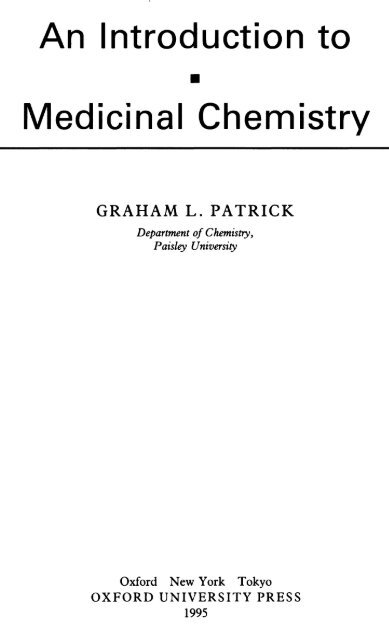
An Introduction to Medicinal Chemistry
An Introduction to Medicinal Chemistry
An Introduction to Medicinal Chemistry
Create successful ePaper yourself
Turn your PDF publications into a flip-book with our unique Google optimized e-Paper software.
<strong>An</strong> <strong>Introduction</strong> <strong>to</strong><br />
<strong>Medicinal</strong> <strong>Chemistry</strong><br />
GRAHAM
Oxford University Press, Wal<strong>to</strong>n Street, Oxford OX2 6DP<br />
Oxford
Preface<br />
This text is aimed at undergraduates who have a basic grounding in chemistry and are<br />
interested in a future career in the pharmaceutical industry. It attempts <strong>to</strong> convey<br />
something
Acknowledgements<br />
Figure
Contents<br />
Classification of drugs<br />
xiii<br />
1. Drugs
viii<br />
Contents
Contents
x<br />
Contents<br />
10.5 <strong>An</strong>tibacterial agents which inhibit cell wall synthesis 166<br />
10.5.1 Penicillins 166<br />
10.5.2 Cephalosporins
Contents<br />
xi<br />
11.16.2 Structure
xii<br />
Contents<br />
13.12.1 Conformational isomers 302<br />
13.12.2 Desolvation 303<br />
13.13 Variation
Classification of drugs
xiv<br />
Classification
1 • Drugs and the medicinal<br />
chemist<br />
In medicinal chemistry, the chemist attempts <strong>to</strong> design and synthesize a medicine or a<br />
pharmaceutical agent which will benefit humanity. Such
Drugs
Whilst penicillin<br />
Drugs and the medicinal chemist 3
4 Drugs and the medicinal chemist<br />
The drug is called diamorphine and it is the drug of choice when treating patients<br />
dying
Drugs
Drugs
10 The why and the wherefore<br />
Cy<strong>to</strong>plasm<br />
Nucleus<br />
Nuclear membrane<br />
Cell membrane<br />
Fig. 2.1 A typical cell. Taken from J. Mann, Murder, magic, and medicine, Oxford University Press<br />
(1992), with permission.<br />
Polar<br />
Head<br />
Group<br />
Polar<br />
Head<br />
Group<br />
Hydrophobia Tails<br />
Hydrophobia Tails<br />
Fig.
Where
Where do drugs work? 13<br />
Amphotericin is a fascinating molecule in that one half of the structure is made up<br />
of double bonds and is hydrophobic, while the other half contains a series of hydroxyl<br />
groups
3 • Protein structure<br />
In order <strong>to</strong> understand how drugs interact with proteins, it is necessary <strong>to</strong> understand<br />
their structure.<br />
Proteins have four levels of structure—primary, secondary, tertiary, and quaternary.<br />
3.1 The primary structure of proteins
16 Protein structure<br />
[ H O
18 Protein structure<br />
H R 0 H<br />
^ ^ ^ H-bonds<br />
O R H O<br />
<strong>An</strong>tiparallel Chains<br />
Residues<br />
/ above<br />
,X
The tertiary structure of proteins 19<br />
CD2<br />
H24<br />
Fig.
20 Protein structure<br />
I I I 10<br />
\\
The tertiary structure of proteins 21<br />
3.3.1 Covalent bonds<br />
Covalent bonds
22 Protein structure<br />
Fig. 3.10 Hydrogen bond.<br />
3.3.4 Van der Waals bonds<br />
Bond strength
24 Protein structure<br />
H-Bond<br />
Peptidef<br />
Chain<br />
r<br />
Peptide<br />
Chain<br />
Fig. 3.14 Bonding interactions with water.<br />
Therefore,
The quaternary structure of proteins 25<br />
number of ionic and hydrogen bonds contributing <strong>to</strong> the tertiary structure is reduced.
26 Protein structure<br />
again that tertiary structure
28 Drug action at enzymes<br />
Energy<br />
Transition State<br />
New<br />
Transition<br />
State<br />
Product<br />
WITHOUT CATALYST<br />
WITH CATALYST<br />
Product<br />
Fig. 4.2 Activation energy.<br />
Energy difference
Substrate binding
32 Drug action at enzymes<br />
Fig.
Substrate binding at an active site 33<br />
unable <strong>to</strong> accept any more substrate. Therefore, the bonding interactions between<br />
substrate and enzyme have <strong>to</strong> be properly balanced such that they are strong enough<br />
<strong>to</strong> keep
34 Drug action
Substrate binding
The catalytic role of enzymes 37<br />
Fig. 4.16 6-Mercap<strong>to</strong>purine.<br />
an example of an allosteric inhibi<strong>to</strong>r. It inhibits the first enzyme involved in the<br />
synthesis of purines and therefore blocks purine synthesis. This in turn blocks DNA<br />
synthesis.<br />
4.5 The catalytic role of enzymes<br />
We
R<br />
The catalytic role of enzymes<br />
Thymidylate<br />
Synthetase<br />
41<br />
o<br />
HN X<br />
R
42 Drug action
I<br />
NH 2<br />
CHO<br />
(OH<br />
Condensation R< The catalytic role
44 Drug action at enzymes<br />
met in the body and would have shown little or no selectivity. (The uses of alkylating<br />
agents
Nerve<br />
Neurotransmitters
48 Drug action at recep<strong>to</strong>rs<br />
CH 2<br />
+<br />
xNMe3<br />
NHR<br />
Acetylcholine<br />
R=H Noradrenaline<br />
R=Me Adrenaline<br />
NH<br />
Dopamine<br />
Sero<strong>to</strong>nin<br />
H 2NL<br />
CH 2 k CH 2<br />
5-Hydroxytryptamine<br />
Fig.<br />
Gamma-aminobutanoic acid
Recep<strong>to</strong>rs
50 Drug action at recep<strong>to</strong>rs<br />
Fig. 5.5 Binding of a messenger <strong>to</strong> a recep<strong>to</strong>r.<br />
reaction takes place, what
52 Drug action at recep<strong>to</strong>rs<br />
Induced Fit<br />
and<br />
Opening
How does the message get received? 53<br />
Recep<strong>to</strong>r<br />
Enzyme<br />
Active site<br />
(open)<br />
Recep<strong>to</strong>r<br />
Enzyme<br />
CD<br />
MESSENGER<br />
LJ<br />
Recep<strong>to</strong>r<br />
im<br />
Enzyme<br />
X<br />
MESSENGER<br />
Recep<strong>to</strong>r<br />
Enzyme<br />
Fig.
54 Drug action at recep<strong>to</strong>rs<br />
MESSENGER<br />
MESSENGER<br />
Recep<strong>to</strong>r<br />
——v^/<br />
Enzyme<br />
No<br />
Reaction<br />
Fig. 5.10 Membrane-bound enzyme deactivation.<br />
change in shape conceals the active site, shutting down that particular reaction<br />
(Fig. 5.10).<br />
Neurotransmitters switch
process whereby<br />
How does a recep<strong>to</strong>r change shape? 55
56 Drug action at recep<strong>to</strong>rs<br />
RECEPTOR<br />
PROTEIN<br />
CYTOPLASM<br />
Fig. 5.12 Recep<strong>to</strong>r protein positioned
58 Drug action at recep<strong>to</strong>rs<br />
neurotransmitter. The binding forces must be strong enough <strong>to</strong> bind the neurotransmitter<br />
effectively such that the recep<strong>to</strong>r changes shape. However, the binding forces<br />
cannot be <strong>to</strong>o strong, or else the neurotransmitter would not be able <strong>to</strong> leave and the<br />
recep<strong>to</strong>r would not be able <strong>to</strong> return <strong>to</strong> its original shape. Therefore, it is reasonable <strong>to</strong><br />
assume that
60 Drug action at recep<strong>to</strong>rs<br />
left-handed ammo acids) are also present as single enantiomers and therefore catalyse<br />
enantiospecific reactions—reactions which give only
A thorough understanding<br />
The design of antagonists 61
62 Drug action
Partial agonists 63<br />
RECEPTOR<br />
RECEPTOR<br />
Fig. 5.22 <strong>An</strong>tagonism
64 Drug action
Tolerance and dependence 65<br />
5.9 Desensitization<br />
Some drugs bind relatively strongly
66 Drug action at recep<strong>to</strong>rs<br />
D Neurotransmitter<br />
Recep<strong>to</strong>r<br />
Synthesis<br />
\<br />
u<br />
\ I//<br />
u .<br />
D<br />
Increase<br />
<strong>An</strong>tagonist<br />
D<br />
Fig. 5.26 Process of increasing cell sensitivity.<br />
for what little neurotransmitter
Tolerance and dependence 67<br />
level. During this period, the patient may be tempted <strong>to</strong> take the drug again in order<br />
<strong>to</strong> 'return
Structure of DNA 69<br />
NH 2<br />
NHo<br />
NHo<br />
Adenine Guanine . ^Cy<strong>to</strong>sine<br />
H<br />
Thymi<br />
Purines<br />
Fig.<br />
Pyrimidines
70 Nucleic acids<br />
6.1.2
Structure
72 Nucleic acids<br />
able <strong>to</strong> coil in<strong>to</strong> a 3D shape and this is known as super-coiling. During replication, the<br />
double strand of DNA must unravel, but due <strong>to</strong> the tertiary supercoiling this leads <strong>to</strong><br />
a high level
Drugs acting
74 Nucleic acids<br />
Fig. 6.9 Proflavine.<br />
best agents
Drugs acting on DNA 75<br />
CH3—N:<br />
MECHLORETHAMINE
Ribonucleic acid
78 Nucleic acids<br />
end<br />
AMINO<br />
«A/* Base Pairing<br />
ml Methylmosine<br />
I Ino.ic<br />
UH 2 Dihydraundine
Ribonucleic acid 79<br />
GROWING<br />
PROTEIN<br />
Growing<br />
Protein<br />
Fig. 6.19 Protein synthesis.<br />
Messenger
Summary 81
the never-ending quest<br />
Structure determination 83
84 Drug development
Structure-activity relationships
86 Drug development<br />
| | Potential Ionic Binding Sites<br />
O Potential
Structure-activity relationships
88 Drug development<br />
bonding or not, it could be replaced with an isosteric group such as methyl (see later).<br />
This would be more conclusive, but synthesis is more difficult.<br />
<strong>An</strong>other possibility
Synthetic analogues
90 Drug development<br />
7.5.1 Variation
Synthetic analogues
92 Drug development<br />
covered
Synthetic analogues
94 Drug development<br />
avoiding patent restrictions
Synthetic analogues
96 Drug development<br />
,OH<br />
OH<br />
GLIPINE<br />
A<br />
Fig. 7.20 Glipine analogues.<br />
B<br />
Me-<br />
COCAINE H Et 2NCH 2CH 2—O—C<br />
O<br />
PROCAINE<br />
Fig. 7.21 Cocaine
Recep<strong>to</strong>r theories 97<br />
NH 2 Me<br />
BOND<br />
ROTATION<br />
RECEPTOR 1<br />
RECEPTOR
Recep<strong>to</strong>r theories
102 Drug development<br />
Frequently,
Lead compounds
104 Drug development<br />
O<br />
IIS—NH<br />
— C—NH-CH2CH 2CH 2CH 3<br />
O<br />
O<br />
Fig. 7.29 Cimetidine.<br />
Fig. 7.30 Tolbutamide.<br />
described above. Clearly, this is a more demanding objective, but the cimetidine s<strong>to</strong>ry<br />
proves that
A case study—oxamniquine
Et 2NCH 2CH 2<br />
CH 2NEt 2
A case study—oxamniquine
108 Drug development<br />
Zero Activity<br />
Fig. 7.39<br />
Addition of a methyl group.<br />
© _ ©<br />
Net [— CH 2- CH 2-NH 2R
A case study—oxamniquine 109<br />
CH 3<br />
Cl<br />
Fig. 7.42 Structure V.<br />
CH 2 OH<br />
Fig. 7.43 Oxamniquine.<br />
Adding
110 Drug development<br />
found
112 Pharmacodynamics<br />
stream. Thirdly,
Drug distribution and 'survival' 113<br />
OXIDATIONS (catalysed by cy<strong>to</strong>chrome P-450)<br />
Oxidation
Drug dose levels 115<br />
can easily negotiate the fatty cell membranes get mopped up by fat tissue or are <strong>to</strong>o<br />
weak <strong>to</strong> bind <strong>to</strong> their recep<strong>to</strong>r sites.<br />
Consequently,
116 Pharmacodynamics<br />
Drugs
Drug design for pharmacokinetic problems 117<br />
NH—C—CH 2CH2NEt 2<br />
LIDOCAINE<br />
A further example of these tactics is provided in the penicillin field with methicillin<br />
(Chapter 10).<br />
8.3.3 Metabolic blockers<br />
Some drugs
118 Pharmacodynamics<br />
Other common metabolic reactions include aliphatic and aromatic C-hydroxylations<br />
(Fig. 8.7), N- and S-oxidations, O- and S-dealkylations, and deamination.<br />
Susceptible groups
Drug design
120 Pharmacodynamics
Drug design for pharmacokinetic problems<br />
121<br />
Blood<br />
Suppy<br />
Brain Cells<br />
H2N^ COOH<br />
COOH<br />
L-Dopa<br />
^N<br />
Enzyme<br />
Dopamine<br />
Fig. 8.14 Transport
122 Pharmacodynamics<br />
Me<br />
AZATHIOPRINE<br />
6-MERCAPTOPURINE<br />
Fig. 8.15 Azathioprine acts
Drug design for pharmacokinetic problems 123<br />
Fig. 8.18<br />
SALICYLIC ACID<br />
ASPIRIN<br />
R = H
124 Pharmacodynamics<br />
Many peptides
Drug design for pharmacokinetic problems 125<br />
——C—O—CH 2CH 2NEt 2<br />
O<br />
ADRENALINE<br />
PROCAINE<br />
Fig. 8.22<br />
used successfully in this respect and effectively inhibits dopa decarboxylase. Furthermore,<br />
since it is a highly polar compound containing two phenolic groups, a hydrazine<br />
moiety,
126 Pharmacodynamics<br />
8.3.9 Self-destruct drugs<br />
Occasionally, the problems faced are completely the opposite of those mentioned<br />
above.
grown<br />
Neurotransmitters as drugs? 127
Graphs and equations 129<br />
What are these physicochemical features which we have mentioned?<br />
Essentially, they refer
130 Quantitative structure-activity relationships (QSAR)<br />
Log(l/C)<br />
Fig.
Physicochemical properties
132 Quantitative structure-activity relationships (QSAR)
Physicochemical properties
134 Quantitative structure-activity relationships (QSAR)<br />
words, compounds having
Physicochemical properties
136 Quantitative structure-activity relationships (QSAR)<br />
fact true constants and are accurate only for the structures from which they were<br />
derived. They
Physicochemical properties
138 Quantitative structure-activity relationships (QSAR)<br />
Benzole acids containing electron donating substituents will have smaller K x values<br />
than benzoic acid itself and hence the value of a
Meta Hydroxyl Group<br />
Physicochemical properties 139
140 Quantitative structure-activity relationships (QSAR)<br />
O—P—OEt<br />
Fig. 9.11 Diethyl phenyl phosphate. —# Q Et<br />
membrane
Quantifying steric properties<br />
Hansch equation 141
142 Quantitative structure-activity relationships (QSAR)<br />
activity is related <strong>to</strong> only one such property, a simple equation can be drawn up.<br />
However, the biological activity of most drugs is related <strong>to</strong> a combination of physicochemical<br />
properties. In such cases, simple equations involving only one parameter are<br />
relevant only if the other parameters are kept constant. In reality, this is not easy <strong>to</strong><br />
achieve and equations which relate biological activity <strong>to</strong> more than one parameter are<br />
more common. These equations are known as Hansch equations and they usually<br />
relate biological activity
log [I]<br />
The Craig plot 143
144 Quantitative structure-activity relationships (QSAR)<br />
+ CT 1.0<br />
NO 2<br />
•<br />
CN<br />
SOjNHj CH3SO2<br />
3<br />
* - 50<br />
.<br />
CH 3CO<br />
CONH 2<br />
.25<br />
COjH<br />
-2.0 -1.6 -1.2 -- 8 '- 4<br />
CH 3CONH<br />
C? Br<br />
CF 3<br />
•OCF 3<br />
•<br />
.4 .8 1.2 1.6 2.0<br />
+ +<br />
T<br />
7C<br />
•<br />
OH<br />
OCH 3<br />
. .-.50<br />
t-Butyl<br />
NMej<br />
-.75<br />
- CT<br />
. .-1.0<br />
Fig. 9.16 Craig plot.<br />
9.16
The Topliss scheme 145<br />
and electron withdrawing properties (positive IT and positive a), whereas an OH<br />
substituent
146 Quantitative structure-activity relationships (QSAR)<br />
CH 3<br />
.—————————————————— Pr« ————————————<br />
E<br />
H, CH 2OCH 3
The Topliss scheme 147<br />
with a negative a fac<strong>to</strong>r (i.e. 4-OMe). If activity improves, further changes are suggested<br />
<strong>to</strong> test
148 Quantitative structure-activity relationships (QSAR)<br />
A AA N —N\k<br />
CH 2CH 2CO 2H<br />
Order of<br />
Synthesis<br />
R<br />
1<br />
2<br />
3<br />
4<br />
5678<br />
H<br />
4-C1<br />
4-MeO<br />
3-C1<br />
3-CF 3<br />
3-Br<br />
3-1<br />
3,5-Cl 2<br />
Biological<br />
Activity<br />
_<br />
L<br />
LMLM<br />
L<br />
M<br />
High<br />
Potency<br />
*<br />
*<br />
*<br />
M= More Activity<br />
L= Less Activity
Planning
150 Quantitative structure-activity relationships (QSAR)<br />
activity. For example, a hydrophobia substituent may be favoured in one part of the<br />
skele<strong>to</strong>n, while
Case study
152 Quantitative structure-activity relationships (QSAR)<br />
group
156 <strong>An</strong>tibacterial agents<br />
pron<strong>to</strong>sil
158 <strong>An</strong>tibacterial agents
<strong>An</strong>tibacterial agents which
others.<br />
<strong>An</strong>tibacterial agents which act against cell metabolism (antimetabolites) 161
162 <strong>An</strong>tibacterial agents<br />
MeO<br />
OMe<br />
Fig. 10.11 Sulfamethoxine.
<strong>An</strong>tibacterial agents which act against cell metabolism (antimetabolites) 163<br />
I<br />
S—NHR"<br />
O<br />
Reversible Inhibition
164 <strong>An</strong>tibacterial agents<br />
Fig. 10.15 Sulfonamide prevents PABA from binding
<strong>An</strong>tibacterial agents which
166 <strong>An</strong>tibacterial agents<br />
drug would have
<strong>An</strong>tibacterial agents which inhibit cell wall synthesis 167<br />
but was actually placed above the surface of the disinfectant. It says much for<br />
Fleming's observational powers that
168 <strong>An</strong>tibacterial agents<br />
-CH2-<br />
R——C-<br />
Acyl side chain<br />
-^-<br />
Benzyl Penicillin<br />
PEN G<br />
-O —CH 2 -<br />
Phenoxymethylpenicillin<br />
PEN V
<strong>An</strong>tibacterial agents which inhibit cell wall synthesis
170 <strong>An</strong>tibacterial agents<br />
• Ineffective when taken orally. Penicillin G can only be administered by injection. It<br />
is ineffective orally since it breaks down in the acid conditions of the s<strong>to</strong>mach.
is <strong>to</strong>lerated<br />
<strong>An</strong>tibacterial agents which inhibit cell wall synthesis 171
<strong>An</strong>tibacterial agents which inhibit cell wall synthesis 173<br />
H<br />
^S<br />
Fig. 10.27 Reduction
174 <strong>An</strong>tibacterial agents<br />
The problem of fi-lactamases became critical in 1960 when the widespread use of<br />
penicillin
<strong>An</strong>tibacterial agents which inhibit cell wall synthesis
176 <strong>An</strong>tibacterial agents<br />
Permeability barrier.<br />
It is difficult for penicillins <strong>to</strong> invade a Gram-negative bacterial cell due <strong>to</strong> the make<br />
up of the cell wall. Gram-negative bacteria have a coating on the outside of their eel<br />
wall which consists
<strong>An</strong>tibacterial agents which inhibit cell wall synthesis 177
178 <strong>An</strong>tibacterial agents<br />
H0 2C—C—CH 2CH 2CH 2—C—NH|<br />
Penicillin
groups<br />
<strong>An</strong>tibacterial agents which inhibit cell wall synthesis 179
180<br />
R
<strong>An</strong>tibacterial agents which inhibit cell wall synthesis 181<br />
probenicid slows down the rate at which penicillin is excreted by competing with it in<br />
the excretion mechanism. As a result, penicillin levels in the bloodstream are enhanced<br />
and the antibacterial activity increases—a useful tactic if faced with a particularly<br />
resistant bacterium.<br />
10.5.2 Cephalosporins<br />
Discovery and structure of cephalosporin C<br />
The second major group of (3-lactam antibiotics <strong>to</strong> be discovered were the cephalosporins.
<strong>An</strong>tibacterial agents which inhibit cell wall synthesis 183<br />
<strong>An</strong>alogues of cephalosporin C by variation of the 7-acylamino side-chain<br />
Access <strong>to</strong> analogues with varied side-chains at the 7-position initially posed a problem.<br />
Unlike penicillins,
184 <strong>An</strong>tibacterial agents<br />
Fig. 10.45 Cephalothin.<br />
CO 2 H<br />
reacted with an alcohol <strong>to</strong> give an imino ether. This product is now more susceptible
<strong>An</strong>tibacterial agents which inhibit cell wall synthesis 185<br />
r\ IU H H
186 <strong>An</strong>tibacterial agents<br />
= CH 3<br />
C0 2Me<br />
C0 2 Me<br />
- ttX I<br />
3<br />
C0 2 Me<br />
Fig. 10.48 Synthesis of 3-methylated cephalosporins.<br />
synthesis, which was first demonstrated by Eli Lilly, involves a ring expansion, where<br />
the five-membered thiazolidine ring in penicillin is converted <strong>to</strong> the six-membered<br />
dihydrothiazine ring
<strong>An</strong>tibacterial agents which inhibit cell wall synthesis
188 <strong>An</strong>tibacterial agents<br />
Second- and third-generation cephalosporins—oximinocephalosporins<br />
Research
<strong>An</strong>tibacterial agents which inhibit cell wall synthesis
190 <strong>An</strong>tibacterial agents<br />
Fig. 10.54 Clavulanic acid as an irreversible mechanism based inhibi<strong>to</strong>r.<br />
Plays a role<br />
in 6-lactamase •<<br />
resistance<br />
Acylamino side Opposite<br />
chain ^fcsent stereochemistry<br />
OH i <strong>to</strong> penicillins<br />
H N<br />
/ Carbon<br />
/ H /<br />
i^^--\<br />
H 3 C^'***', '<br />
//<br />
) ——— S "<br />
N-*^/^<br />
C0 2©<br />
Carbapenam nucleus<br />
Fig. 10.55 Thienamycin.<br />
Double bond leading
<strong>An</strong>tibacterial agents which inhibit cell wall synthesis 191<br />
Olivanic acids<br />
The olivanic acids (e.g. MM13902) (Fig. 10.56) were isolated from strains of Strep<strong>to</strong>myces<br />
olivaceus
192 <strong>An</strong>tibacterial agents
<strong>An</strong>tibacterial agents which inhibit cell wall synthesis
194 <strong>An</strong>tibacterial agents<br />
[Normal Mechanism)<br />
Peptide<br />
Chain<br />
^ — c* D-Ala—D-Ala —COzH<br />
OH<br />
/Transpeptidase Enzyme<br />
Peptide<br />
Chain<br />
C<br />
^— D-Ala<br />
I<br />
X X<br />
Peptide<br />
Peptide<br />
Peplide Chain<br />
Chain ^><br />
X-D-Ala——Gly<br />
[Mechanism Inhibited
<strong>An</strong>tibacterial agents which
196 <strong>An</strong>tibacterial agents<br />
L-Valine Me<br />
L-Lactate<br />
D-Valine<br />
Me<br />
NH /<br />
D ' Hyi<br />
D-Valine<br />
D-Hyi =<br />
D-Hydroxyisovaleric acid<br />
D-Hyi<br />
Fig. 10.64 Valinomycin.<br />
groups
<strong>An</strong>tibacterial agents which
198 <strong>An</strong>tibacterial agents<br />
L-LEU — L-DAB<br />
/ \<br />
D-PHE<br />
L-DAB<br />
L-DAB<br />
L-DAB<br />
L-DAB<br />
L-THR<br />
POLYMYXIN B<br />
L-DAB<br />
C=0<br />
I<br />
(CH 2) 4<br />
CH-CH 3<br />
Fig. 10.69 Polypeptide antibiotic. CH 2 CH 3<br />
such as nucleosides from the cell. The drug is injected intramuscularly and is useful<br />
against Pseudomonas strains which are resistant <strong>to</strong> other antibacterial agents.<br />
10.7 <strong>An</strong>tibacterial agents which impair protein synthesis<br />
Examples of such agents are the rifamycins which act against RNA, and the aminoglycosides,<br />
tetracyclines, and chloramphenicol which all act against the ribosomes.<br />
Selective <strong>to</strong>xicity
<strong>An</strong>tibacterial agents which impair protein synthesis
200 <strong>An</strong>tibacterial agents<br />
which was discovered in 1948. It is a broad-spectrum antibiotic, active against both<br />
Gram-positive and Gram-negative bacteria. Unfortunately, it does have side-effects
Agents which
202 <strong>An</strong>tibacterial agents<br />
o<br />
,CO2H<br />
HN > J Cl-LCKt, HNL<br />
NALIDIXIC ACID ENOXACILIN CIPROFLOXACIN<br />
Fig. 10.74 Quinolones and fluoroquinolones.<br />
compounds. It is active against Gram-negative bacteria and is useful in the short-term<br />
therapy
Drug resistance 203<br />
10.9 Drug resistance<br />
With such
204 <strong>An</strong>tibacterial agents<br />
transferred by means of bacterial viruses (bacteriophages) leaving the resistant cell<br />
and infecting a non-resistant cell. If the plasmid brought <strong>to</strong> the infected cell contains<br />
the gene required for drug resistance, then the recipient cell will be able <strong>to</strong> use that<br />
information and gain resistance. For example, the genetic information required <strong>to</strong><br />
synthesize (3-lactamases can be passed on in this way, rendering bacteria resistant <strong>to</strong><br />
penicillins.
11- The peripheral nervous<br />
system—cholinergics,<br />
anticholinergics, and<br />
anticholinesterases<br />
In Chapter 10, we discussed the medicinal chemistry of antibacterial agents and noted
206 The peripheral nervous system<br />
your home computer software, or perhaps trying <strong>to</strong> trace where a missing letter went,<br />
or finding the reason for the country's balance of payments deficit.<br />
However,
Mo<strong>to</strong>r nerves
208 The peripheral nervous system<br />
Skeletal<br />
" Muscle<br />
SOMATIC<br />
AUTONOMIC<br />
Smooth Muscle<br />
Cardiac Muscle<br />
Fig. 11.2 Mo<strong>to</strong>r nerves of the peripheral nervous system. N, Nicotinic recep<strong>to</strong>r;<br />
M, muscarinic recep<strong>to</strong>r.<br />
parasympathetic nerves. However, they synapse with different recep<strong>to</strong>rs on the<br />
target organs
Actions
210 The peripheral nervous system<br />
Note that the sympathetic and parasympathetic nervous systems oppose each other in<br />
their actions and could be looked upon as a brake and an accelera<strong>to</strong>r. The analogy is
The cholinergic system 211<br />
I<br />
j<br />
V<br />
Acetyl<br />
| Choline
212 The peripheral nervous system<br />
11.6 Agonists at the cholinergic recep<strong>to</strong>r<br />
One point might have occurred <strong>to</strong> the reader. If there is a lack of acetylcholine acting<br />
at
Agonists
Agonists at the cholinergic recep<strong>to</strong>r 215<br />
RECEPTOR SITE<br />
(MUSCARINIC)<br />
Fig. 11.11
Agonists at the cholinergic recep<strong>to</strong>r 217<br />
but since they are ring structures, the left-hand portion of the acetylcholine molecule<br />
is
Water<br />
Design of acetylcholine analogues 219
220 The peripheral nervous system
Design
<strong>An</strong>tagonists
224 The peripheral nervous system<br />
Hyoscine (1879-84)<br />
Hyoscine
Structural analogues based<br />
<strong>An</strong>tagonists of the muscarinic cholinergic recep<strong>to</strong>r 225
A large variety<br />
<strong>An</strong>tagonists of the muscarinic cholinergic recep<strong>to</strong>r 227
228 The peripheral nervous system<br />
In practice, the procedure is not always as simple as this, since the highly react<br />
electrophilic centre might react with another nucleophilic group before it reaches<br />
recep<strong>to</strong>r binding site.
<strong>An</strong>tagonists
230 The peripheral nervous system<br />
pleasing that theory
<strong>An</strong>tagonists
232 The peripheral nervous system<br />
; ^.---- Acetyl choline<br />
skele<strong>to</strong>n<br />
Atracurium<br />
Acetyl choline ----- '^X^-^/'<br />
skele<strong>to</strong>n<br />
Fig. 11.39 Pancuronium and vecuronium.<br />
R = H VECURONIUM<br />
R = Me PANCURONIUM<br />
The design of atracurium (Fig. 11.40) was based on the structures of tubocurarine and<br />
suxamethonium. It is superior <strong>to</strong> both since it lacks cardiac side-effects and is rapidly<br />
broken down in blood. This rapid breakdown allows the drug <strong>to</strong> be administered as an<br />
intravenous drip.<br />
MeO,<br />
OMe<br />
MeO'<br />
Me<br />
CH:<br />
,CH2-C—O—(CH2)5—O—C—CH2<br />
'OMe<br />
OMe<br />
'OMe<br />
Fig. 11.40<br />
MeO'<br />
Atracurium.<br />
OMe
drug design<br />
Other cholinergic antagonists 233
<strong>An</strong>ticholinesterases
<strong>An</strong>ticholinesterases
238 The peripheral nervous system<br />
ro<br />
^ii 0<br />
CH3 —C —O —CH2CH2NMe3 '0- fn<br />
x I " *> u 0<br />
»<br />
i •*' ^r NH StageB ^v^&v ^ CH3 - "" C^\ ^/^NH Stage<br />
O N I ———— O
<strong>An</strong>ticholinesterase drugs 239<br />
- PyrrolidineN<br />
Fig. 11.48 Physostigmine.<br />
Me<br />
discovered in 1864 as a product of the poisonous calabar beans from West Africa. The<br />
structure was established in 1925 (Fig. 11.48).<br />
Structure-activity relationships :
<strong>An</strong>ticholinesterase drugs
242 The peripheral nervous system<br />
Me<br />
O
<strong>An</strong>ticholinesterase drugs
244 The peripheral nervous system<br />
Fortunately, there
Pralidoxime—an organophosphate antidote
N—CH 3<br />
MeO.<br />
R'O*<br />
R
248 The opium analgesics<br />
Chinese decision—one
Morphine 249<br />
12.2 Morphine<br />
12.2.1 Structure and properties<br />
MeN<br />
N—CH3<br />
N——CH3<br />
Fig. 12.2 Structure
Morphine
Morphine 253<br />
To conclude, the 6-hydroxyl group is not required for analgesic activity and its<br />
removal can be beneficial <strong>to</strong> analgesic activity.<br />
The double bond at 7-8<br />
Several analogues including dihydromorphine (Fig. 12.6) have shown that
Development of morphine analogues 255<br />
morphine. Let us consider a diagrammatic representation of morphine as a T-shaped<br />
block with
Development
258 The opium analgesics<br />
R= ——CH 2 -CH=CH 2<br />
^ Allyl_______<br />
<strong>An</strong>tagonists<br />
NALORPHINE<br />
NALOXONE<br />
Fig. 12.16
Development of morphine analogues 259<br />
But how can this be? How can a compound be an antagonist of morphine but also<br />
act
260 The opium analgesics<br />
(A) BINDING
Development
262 The opium analgesics<br />
morphine. Notice that the two methyl groups in metazocine are cis with respect <strong>to</strong><br />
each other and represent the 'stumps' of the C ring.<br />
If
Development
264 The opium analgesics<br />
o<br />
N——C—CH=CH-Ph<br />
Et °2 C \__/ V.^V^ ^£--—N——C—CH=CH-Ph<br />
H<br />
\<br />
Fig. 12.23 Effect<br />
3Qx more potent than pethidine<br />
Zero Activity
Development of morphine analogues 265<br />
Chiral Centre<br />
R
266 The opium analgesics<br />
morphine
Development of morphine analogues 267<br />
RMgBr<br />
NMe<br />
Grignard<br />
NMe<br />
MeO<br />
MeO<br />
Fig. 12.28 Drug extension.<br />
MeN<br />
'<br />
[Complcxedl<br />
MeN<br />
Fig. 12.29 Grignard reaction leads
Recep<strong>to</strong>r theory
270 The opium analgesics<br />
analgesic has <strong>to</strong> cross the blood-brain barrier as the free base, but once across has <strong>to</strong><br />
be ionized in order <strong>to</strong> interact with the recep<strong>to</strong>r.
Recep<strong>to</strong>r theory
272 The opium analgesics<br />
acts as a partial agonist at the JJL recep<strong>to</strong>r <strong>to</strong> produce its analgesic effect. This might<br />
suggest that buprenorphine should suffer the same side-effects as morphine. The fact<br />
that
Agonists
Enkephalins
e<br />
Recep<strong>to</strong>r mechanisms 277
278 The opium analgesics<br />
narcotic analgesics. There is still a search <strong>to</strong> see if there are possibly two slightly<br />
different mu recep<strong>to</strong>rs, one which is solely due <strong>to</strong> analgesia and one responsible for<br />
the side-effects.<br />
12.7.2
membrane <strong>to</strong> a second membrane-bound protein.<br />
This protein then acts<br />
The future 279
280 The opium analgesics<br />
O<br />
II<br />
N —C-CH 2<br />
N-CH2-^<br />
Ethylke<strong>to</strong>cyclazocine<br />
(Kappa=mu > delta)<br />
U 50488<br />
(kappa
284 Cimetidine—a rational approach <strong>to</strong> drug design<br />
13.3 Histamine<br />
Histamine
Searching
Searching
Searching for a lead—AT-guanylhistamine 289<br />
as long as A/^-guanylhistamine is bound <strong>to</strong> the recep<strong>to</strong>r, it prevents histamine from<br />
binding
290 Cimetidine—a rational approach <strong>to</strong> drug design<br />
reach
Developing the lead—a chelation bonding theory 291<br />
HN<br />
cH 2 -cH 2 -x—c'<br />
N—I<br />
H<br />
H<br />
Strong<br />
Interaction<br />
Weak<br />
Interaction<br />
>-<br />
RECEPTOR<br />
X=NH, S<br />
RECEPTOR<br />
X=Me,
From partial agonist
Development of metiamide 295<br />
meaning that the ring is a weak base and mostly un-ionized. The pK a value for<br />
imidazole itself
Development of metiamide 297<br />
the same pK a as for imidazole itself, which shows that the electronic effects of the<br />
methyl group and the side-chain are cancelling each other out as far as pK a is<br />
concerned.) A pK a of 6.80 means that 20 per cent of metiamide is ionized in the<br />
imidazole ring. However, this is still significantly lower than the corresponding 40 per<br />
cent
Development
Cimetidine
Further studies—cimetidine analogues
304 Cimetidine — a rational approach <strong>to</strong> drug design<br />
they are likely <strong>to</strong> be highly solvated (i.e. surrounded by a 'water coat'). Before<br />
hydrogen bonding can take place <strong>to</strong> the recep<strong>to</strong>r , this 'water coat' has <strong>to</strong> be removed.<br />
The more solvated the group, the more difficult that will be.
Further studies—cimetidine analogues
306 Cimetidine—a rational approach <strong>to</strong> drug design<br />
Me<br />
N P*<br />
/ \<br />
Fig. 13.50 Cimetidine analogue with a<br />
nitroketeneaminal group.<br />
HN^\<br />
I V-CH 2-S—CH 2-CH 2-NH-C V<br />
l^_<br />
# H
Further studies—cimetidine analogues<br />
NC,<br />
OjjN<br />
"S,<br />
=-6<br />
=16.7<br />
I * ^H<br />
k<br />
4> =27<br />
H =14.2<br />
NMe<br />
H<br />
=33<br />
=15.1<br />
Fig. 13.53 Dipole moments
308 Cimetidine—a rational approach <strong>to</strong> drug design<br />
dipole moment
Famotidine and nizatidine 309<br />
• Replacing the sulfur a<strong>to</strong>m with a methylene unit leads <strong>to</strong> a drop in activity.<br />
• Placing the sulfur next <strong>to</strong> the ring lowers activity.<br />
• Replacing the furan ring with more hydrophobic rings such as phenyl or thiophene<br />
reduces activity.
312 Cimetidine—a rational approach <strong>to</strong> drug design<br />
effects.
Appendix
Appendix
(vesicles) containing<br />
The action of nerves 315
a<br />
The action of nerves 317
The action of nerves 319<br />
x<br />
Briv "-
Appendix
Secondary messengers
322 Secondary messengers<br />
number
Secondary messengers 323<br />
the process shown<br />
o=co=
Secondary messengers
Appendix 4 • Bacteria and<br />
bacterial nomenclature<br />
Bacterial nomenclature<br />
COCCI<br />
(Spherical)<br />
BACILLI<br />
(Cylindrical)<br />
STREPTOCOCCI<br />
(Chains)<br />
Fig. A4.1 Bacterial nomenclature.<br />
STAPHYLOCOCCI<br />
(Clusters)<br />
Some clinically important bacteria<br />
Organism<br />
Staphylococcus aureus<br />
Gram<br />
Positive<br />
Infections<br />
Skin<br />
Strep<strong>to</strong>coccus<br />
Escherichia coli<br />
Positive<br />
Negative<br />
Proteus species<br />
Salmonella species<br />
Shigella species<br />
Enterobacter species<br />
Pseudomonas aeruginosa<br />
Negative<br />
Negative<br />
Negative<br />
Negative<br />
Negative
Haemophilus influenzae<br />
Bacteroides fragilis<br />
Negative<br />
Negative<br />
Bacteria and bacterial nomenclature 327<br />
patients, i.e. cancer patients, commonly causes<br />
chest infections
Glossary<br />
ADDICTION<br />
Addiction can be defined as a habitual form of behaviour. It need not be harmful.
Glossary
Further reading<br />
Albert,
Index<br />
acetylcholine 47-8, 207, 209-26,<br />
229-40, 243, 282, 315,
drug<br />
addiction 1, 3, 5, 127, 248,<br />
262-3, 271-2,<br />
Index 333
334 Index<br />
intestinal infections
penillic acids 172<br />
pentagastrin<br />
Index 335
336 Index<br />
structure-activity analysis (SAR)<br />
82, 84-9, 248<br />
acetylcholine 214-15<br />
aryl tetrazolylalkanoic acids<br />
147-8<br />
benzenesulfonamides


