Western Blotting - KPL
Western Blotting - KPL
Western Blotting - KPL

You also want an ePaper? Increase the reach of your titles
YUMPU automatically turns print PDFs into web optimized ePapers that Google loves.
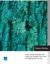
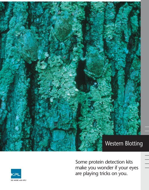
<strong>Western</strong> <strong>Blotting</strong><br />
Some protein detection kits<br />
make you wonder if your eyes<br />
are playing tricks on you.<br />
SEE MORE with <strong>KPL</strong>!
<strong>Western</strong> <strong>Blotting</strong><br />
No matter which <strong>KPL</strong> <strong>Western</strong> <strong>Blotting</strong> system you use,<br />
you’ll get a strong, clean signal every time.<br />
<strong>KPL</strong>’s blotting substrates and Protein<br />
Detector <strong>Western</strong> Blot Kits provide<br />
consistent, high quality results<br />
when analyzing proteins immobilized<br />
on membrane. Choose from a<br />
line of substrates and kits for either<br />
chemiluminescent or colorimetric<br />
methods of detecting specific antigens<br />
in a complex protein mixture.<br />
The cornerstone of <strong>KPL</strong>’s <strong>Western</strong><br />
blotting systems is our line of sensitive<br />
liquid substrates. You have a<br />
choice among multiple chemiluminescent<br />
and chromogenic substrates<br />
for peroxidase and phosphatase that<br />
provide varied levels of sensitivity.<br />
The chemiluminescent substrates<br />
provide a reliable alternative to conventional<br />
colorimetric ELISA with<br />
the advantage of increased sensitivity.<br />
Our chemiluminescent substrates<br />
for alkaline phosphatase detection,<br />
PhosphaGLO and PhosphaGLO<br />
Reserve AP Chemiluminescent<br />
Substrates, offer sensitivity and<br />
duration of signal in a one-component<br />
system.<br />
<strong>KPL</strong>’s substrates are offered as standalone<br />
reagents or combined with<br />
unique blocking and wash solutions<br />
and highly specific secondary antibodies<br />
to create comprehensive,<br />
fully optimized kits for <strong>Western</strong><br />
blotting. Each of these kits is<br />
designed to deliver the maximum<br />
signal-to-noise ratio available.<br />
Choose the detection system that<br />
best meets your needs and achieve<br />
clean, clear results blot after blot.<br />
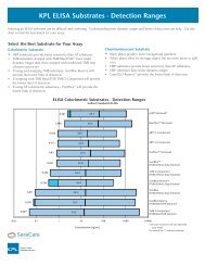
Substrates for <strong>Western</strong> <strong>Blotting</strong><br />
<strong>KPL</strong>’s <strong>Western</strong> blotting substrates for use in peroxidase and phosphatase reporter systems offer high quality, reproducible<br />
chemiluminescent and colorimetric detection. It’s your choice!<br />
LumiGLO LumiGLO ® PhosphaGLO PhosphaGLO TMB HRP 4 CN BCIP/NBT FirePhos<br />
Reserve HRP HRP Reserve AP AP Substrate Substrate Substrate AP Substrate Membrane<br />
Substrate Substrate Substrate AP Substrate<br />
Type chemi. chemi. chemi. chemi. color. color. color. color.<br />
Format 2-comp 2-comp 1-comp 1-comp 1- or 3- 2-comp 1- or 3- 1-comp<br />
comp<br />
comp<br />
Detection film or image film or image film or image film or image visual-dark blue visual - purple visual - purple visual - red<br />
Method analysis analysis analysis analysis precipitate precipitate precipitate<br />
Detection Sub-picogram; 0.6 picogram femtogram picogram 50 picogram 500 picogram low nanogram low nanogram<br />
Limit<br />
femtogram<br />
Stability of 8 hours 24 hours 2 years 2 years 1-comp- 1 hour 1-comp- NA<br />
Working Solution at 4 o C at 4 o C 1 year 1 year<br />
at 4 o C. at 4 o C.<br />
3-comp-<br />
3-comp-<br />
24 hours 1 hour<br />
Duration of 4 - 8 hours 1 - 2 hours 5 days 5 days NA NA NA NA<br />
Signal<br />
Enzyme HRP HRP AP AP HRP HRP AP AP<br />
Catalyst<br />
Kinetics 5 minutes 5 minutes 15 minutes 15 minutes 10 minutes 10 minutes 10 minutes 10 minutes<br />
Recommended nitrocellulose nitrocellulose nitrocellulose nitrocellulose nitrocellulose nitrocellulose nitrocellulose nitrocellulose<br />
Membrane or PVDF or PVDF or PVDF or PVDF or PVDF or PVDF or PVDF or PVDF<br />
comp = component • chemi. = chemiluminescent • color. = colorimetric • HRP = peroxidase • AP = phosphatase • NA = not applicable<br />
www.kpl.com
With <strong>KPL</strong>’s Protein Detector <br />
<strong>Western</strong> <strong>Blotting</strong> Systems, the<br />
results jump right out at you.
<strong>Western</strong> <strong>Blotting</strong><br />
Chemiluminescent Substrates and <strong>Western</strong> Blot Kits<br />
Rapid, Sensitive Detection with<br />
Luminol-based Substrates<br />
Chemiluminescent detection involves<br />
the creation of light through the catalysis<br />
of an enzyme substrate. Use of this<br />
method for protein detection allows an<br />
increase in sensitivity by orders of magnitude<br />
compared to traditional colorimetric<br />
<strong>Western</strong> or dot blotting. <strong>KPL</strong><br />
offers two unique luminol-based chemiluminescent<br />
substrates —LumiGLO ®<br />
and LumiGLO Reserve , for the rapid<br />
and sensitive detection of horseradish<br />
peroxidase (HRP)-labeled conjugates.<br />
Maximum Sensitivity with<br />
LumiGLO Reserve TM<br />
LumiGLO Reserve HRP Chemiluminescent<br />
Substrate offers an option for<br />
those assays where enhanced sensitivity<br />
is critical to success. This proprietary<br />
two-component substrate formulation<br />
provides greater than 20 times the sensitivity<br />
of standard LumiGLO with detection<br />
levels as low as the femtogram<br />
range (Figure 1).<br />
Long Signal Duration<br />
LumiGLO Reserve emits light over the<br />
course of 4-8 hours with the most<br />
intense emission occurring within the<br />
first two hours. Because of its extreme<br />
light intensity, most images may be captured<br />
well under 10 minutes; multiple<br />
exposures are easy to obtain. High signal<br />
intensity facilitates the detection of<br />
both high and low abundance proteins<br />
and makes it an ideal system for use<br />
with chemiluminescent imagers.<br />
Sample and Antibody<br />
Conservation<br />
LumiGLO Reserve provides the added<br />
benefit of strong signal with the use of<br />
reduced amounts of precious target and<br />
antibodies. Therefore, material of limited<br />
supply or higher expense can be conserved<br />
while maintaining your current<br />
level of sensitivity.<br />
Superior Signal to Noise<br />
LumiGLO Reserve delivers lower nonspecific<br />
signal than competitor substrates<br />
in its class (Figures 2 and 3).<br />
Convenience<br />
LumiGLO Reserve Chemiluminescent<br />
Substrate Kits are supplied in several kit<br />
formats to meet diverse user needs.<br />
This substrate can replace your current<br />
chemiluminescent substrate in existing<br />
assays where greater sensitivity is desired.<br />
LumiGLO Reserve<br />
<strong>Western</strong> Blot Kit<br />
LumiGLO Reserve is also offered as part<br />
of a fully optimized kit, the Protein<br />
Detector LumiGLO Reserve <strong>Western</strong> Blot<br />
Kit, providing femtogram detection of<br />
proteins immobilized on membranes.<br />
For assurance against background interference,<br />
this kit contains our unique<br />
Detector Block for optimal signal-tonoise<br />
ratio; very low background can be<br />
achieved without compromise to signal<br />
intensity (Figures 2 and 3). Detector<br />
Block is also offered separately.<br />
Reliable and Economical<br />
LumiGLO ®<br />
<strong>KPL</strong>’s original LumiGLO Chemiluminescent<br />
Substrate provides high quality<br />
results in a variety of immuoassays.<br />
LumiGLO Reserve Signal-to-Noise Comparison<br />
1 2 3 4 5 6<br />
LumiGLO<br />
LumiGLO Reserve<br />
ECL Plus<br />
A<br />
B<br />
C<br />
Figure 1: Relative expression of transcription factor,<br />
c-myc, using different chemiluminescent substrates. Five<br />
two-fold serial dilutions of purified c-myc (25 ng–1.56<br />
ng, lanes 1–5) were compared to a 64 µg total protein<br />
HeLa nuclear lysate (lane 6). Following separation on a<br />
4-20% PAGE gel and transfer to PVDF, protein was<br />
detected using a rabbit anti-c-myc antibody (1:200)<br />
and anti-rabbit HRP conjugate (1:10,000). Detection<br />
conditions were identical with the exception of substrate.<br />
While the c-myc lysate sample was not detectable with<br />
A) LumiGLO or C) ECL Plus after 10 minutes, the<br />
sample was easily detected with B) LumiGLO Reserve<br />
after just a 2-minute film exposure.<br />
LumiGLO Reserve<br />
A B C<br />
ECL Plus<br />
ECL Advance<br />
Figure 2: Comparison of low-end sensitivity using LumiGLO Reserve and ECL<br />
Detection Kits. Two-fold serial dilutions of Mouse IgG (1 ng – 31 pg) were<br />
separated by SDS-PAGE and transferred to PVDF. Under manufacturer’s recommended<br />
conditions, protein was detected using HRP-labeled anti-mouse antibody (varied<br />
dilutions according to recommended optimization) and each respective substrate:<br />
A) LumiGLO Reserve, B) ECL Plus, C) ECL Advance. Film was exposed for 10<br />
minutes and analyzed for sensitivity and signal to noise.<br />
www.kpl.com
LumiGLO vs. Leading Traditional Chemiluminescent Substrates<br />
Densitometry for LumiGLO vs. ECL<br />
Superior Signal to Noise with Detector Block<br />
50000<br />
45000<br />
40000<br />
LumiGLO<br />
ECL<br />
Relative Image Density<br />
35000<br />
30000<br />
25000<br />
20000<br />
15000<br />
LumiGLO Chemiluminescent<br />
Peroxidase Substrate<br />
Supersignal ® West Pico<br />
10000<br />
5000<br />
0<br />
0 50 100 150 200 250<br />
Concentration (ng)<br />
ECL Plus<br />
ECL<br />
Figure 4: Signal response comparison of LumiGLO vs. ECL<br />
chemiluminescent substrates in <strong>Western</strong> blotting. ß-galactosidase was<br />
electrophoresed, transferred to PVDF membrane and subsequently<br />
detected using rabbit anti-ß-gal followed by HRP conjugated goat<br />
anti-rabbit IgG (H+L). Each blot was treated with 5 mL of LumiGLO or<br />
ECL substrate and exposed to film for 10 minutes. The density of each<br />
band was analyzed on a Syngene GeneGenius image analyzer, using<br />
automatic background subtract.<br />
Figure 5: Detection of ß-galactosidase in <strong>Western</strong> blots using alternative<br />
chemiluminescent substrates. A two-fold dilution series of purified<br />
ß-galactosidase (from 25 was electrophoresed on a polyacrylamide gel<br />
and transferred to KODAK BIOMAX Multi-Blot Kit for Proteins. Each blot<br />
was detected under the same conditions using Protein Detector LumiGLO<br />
<strong>Western</strong> <strong>Blotting</strong> Kit, substituting 5 mL LumiGLO on 3 of the<br />
4 blots with 5 mL of leading competitive chemiluminescent substrates,<br />
respectively. Following a 10 minute film exposure, results were evaluated<br />
for sensitivity and signal:noise ratio.<br />
Researchers continue to select<br />
LumiGLO for reliable performance in<br />
<strong>Western</strong> blot detection.<br />
Ideal for Routine <strong>Western</strong> <strong>Blotting</strong><br />
LumiGLO detects low picogram quantities<br />
of target protein on blots. After<br />
reaction with membrane-bound HRP<br />
conjugates, light emission begins immediately<br />
and continues for approximately<br />
1–2 hours. Detection is possible within<br />
minutes of blot exposure.<br />
Greater Linearity<br />
LumiGLO produces a quantitatively linear<br />
signal on film that ensures a broader<br />
dynamic range of detection. While<br />
the light output from other chemiluminescent<br />
substrates tends to reduce<br />
sharply as the concentration of protein<br />
is titrated, a proportional reduction in<br />
sensitivity is achieved with LumiGLO<br />
(Figure 4).<br />
Low Background<br />
LumiGLO delivers superior signal-tonoise<br />
by design. When used with <strong>KPL</strong>’s<br />
Detector Block, nonspecific binding is<br />
further reduced and ensures low background<br />
without loss of signal intensity.<br />
Figure 5 compares the superior signalto-noise<br />
from blots blocked with<br />
Detector Block and subsequently<br />
detected with LumiGLO to leading<br />
competitor substrates.<br />
Cost Effective<br />
LumiGLO is more economical blot for<br />
blot compared to competitive products.<br />
Enjoy the benefits of a sensitive,<br />
reliable substrate while staying within<br />
budget.<br />
Figure 3: Signal linearity and signal-to-noise ratio comparison of advanced chemiluminescent substrates.<br />
Mouse IgG (250 pg to 3.91 pg) was transferred to nitrocellulose. Blots were detected under identical conditions<br />
with a 1:10,000 dilution of HRP-Goat-anti-Mouse IgG (except ECL Advance, 1:100,000) and exposed to<br />
film for 10 minutes. Densitometry was then performed using the Syngene GeneGenius. LumiGLO Reserve<br />
demonstrates superior signal linearity over a larger dynamic range and the greatest comparative signal-to-noise.<br />
LumiGLO <strong>Western</strong> Blot Kit<br />
The LumiGLO <strong>Western</strong> Blot Kit is<br />
designed for low picogram detection of<br />
proteins immobilized on membranes.<br />
Researchers choose this kit for its reliable,<br />
consistent performance. The combination<br />
of a stable, liquid conjugate<br />
and a sensitive chemiluminescent<br />
substrate allows rapid and accurate<br />
identification of samples. For assurance<br />
against background interference, this<br />
kit contains our unique Detector Block<br />
for optimal signal-to- noise ratio<br />
(Figure 5).<br />
SEE MORE with <strong>KPL</strong>!
<strong>Western</strong> <strong>Blotting</strong><br />
Sensitivity and Convenience with<br />
PhosphaGLO TM AP Substrates<br />
<strong>KPL</strong> offers two chemiluminescent substrates<br />
for use with alkaline phosphatase,<br />
PhosphaGLO Reserve TM and PhosphaGLO TM<br />
AP Substrates. PhosphaGLO Reserve<br />
Substrate overcomes the limitations<br />
posed by conventional chemiluminescent<br />
substrates for AP. With sensitivity in the<br />
femtogram range, PhosphaGLO Reserve<br />
enables detection of target protein in low<br />
concentrations. PhosphaGLO AP<br />
Substrate is recommended for routine<br />
detection of proteins in the picogram<br />
range (Figure 6).<br />
Low Background without<br />
Special Blockers<br />
PhosphaGLO Substrates produce superior<br />
signal and low background, providing<br />
a better signal to noise ratio and a cleaner<br />
image than other chemiluminescent substrates<br />
for AP (Figure 6). No special<br />
blockers are needed with either nitrocellulose<br />
or PVDF membrane.<br />
Ultimate Convenience<br />
Both PhosphaGLO AP Substrates offer<br />
exceptional convenience as one-component<br />
solutions ready to use. They are<br />
stable for up to two years when stored at<br />
4 o C. Their long glow times of 5 days<br />
facilitate assay development, enabling<br />
repeat exposures.<br />
Colorimetric Kits<br />
<strong>KPL</strong> also offers colorimetric <strong>Western</strong> blot<br />
systems when chemiluminescent detection<br />
is not preferred. Results can be interpreted<br />
by direct visualization of the blot.<br />
Everything you need to detect your antigen<br />
and primary antibody is supplied for<br />
convenience and optimal performance.<br />
Protein Detector TM TMB<br />
<strong>Western</strong> Blot Kit<br />
The Protein Detector TM TMB <strong>Western</strong> Blot<br />
Kit utilizes TMB Peroxidase Membrane<br />
Substrate (3,3’,5,5’-tetramethylbenzidine),<br />
the most sensitive chromogenic<br />
peroxidase substrate for <strong>Western</strong> and dot<br />
blotting applications. Detection limits are<br />
significantly increased as compared to<br />
other chromogenic membrane<br />
substrates. TMB produces a dark blue<br />
precipitate upon reaction with HRP.<br />
Protein Detector TM BCIP/NBT<br />
<strong>Western</strong> Blot Kit<br />
For detection of phosphatase-labeled<br />
conjugates, the Protein Detector<br />
BCIP/NBT <strong>Western</strong> Blot Kit is ideal.<br />
When reacted with alkaline phosphatase,<br />
BCIP/NBT produces clean, intense bands<br />
of purple precipitate. It also provides<br />
stable, more permanent results than<br />
other chromogenic substrates.<br />
<strong>Western</strong> Blot Reagents<br />
In addition to our line of substrates and kits,<br />
<strong>KPL</strong> offers a broad line of secondary antibodies,<br />
conjugates and support reagents in various<br />
packaging formats to suit your needs.<br />
Secondary Antibodies<br />
• Highly purified and specific.<br />
• Large offering of species-specific polyclonal<br />
antibodies<br />
• Enzyme, biotin and fluorochrome-labeled<br />
antibodies<br />
Support Reagents<br />
• One source for block solutions, wash<br />
buffers, diluents and stabilizers.<br />
• Consistently produced for reliable results.<br />
PhosphaGLO Reserve PhosphaGLO CDP Star ®<br />
AP Substrate AP Substrate Chemiluminescent<br />
Substrate<br />
Figure 6: Comparison of low-end sensitivity using PhosphaGLO Reserve AP Substrate, PhosphaGLO AP<br />
Substrate, and CDP-Star Chemiluminescent Substrate on PVDF membranes. Five-fold serial dilutions of<br />
mouse IgG (2 ng -3.2 pg) were separated by SDS-PAGE and transferred to membranes. Protein was<br />
detected using a 1:1000 dilution of biotin-labeled goat anti-mouse IgG, a 1:10,000 dilution of AP-labeled<br />
streptavidin and each respective substrate. Film was exposed for 10 minutes.<br />
LumiGLO is a registered trademark and LumiGLO<br />
Reserve, Protein Detector, PhosphaGLO, PhosphaGLO<br />
Reserve, FirePhos and Detector are trademarks<br />
of <strong>KPL</strong>, Inc.<br />
ECL, ECL Plus and ECL Advance are trademarks of GE<br />
Healthcare.<br />
SuperSignal West is a registered trademark of Pierce<br />
Biotechnology, Inc.<br />
BIOMAX is a trademark of KODAK.<br />
GeneGenius is a trademark of Syngene.<br />
Biodyne is a registered trademark of Pall/Gelman<br />
Corporation.<br />
CDP-Star is a registered trademark of Applied<br />
Biosystems.<br />
SEE MORE with <strong>KPL</strong>!
AP Membrane Color Substrates<br />
See RED and PURPLE in your <strong>Western</strong> blots!<br />
BCIP/NBT (5-Bromo-4-chloro-3-indolyl phosphate/<br />
nitroblue tetrazolium) is the most commonly used<br />
substrate for alkaline phosphatase (AP) detection in<br />
<strong>Western</strong> blotting due to its high sensitivity and low<br />
background staining. Traditional formulations of<br />
BCIP/NBT produce a deep purple color when reacted<br />
with AP conjugates.<br />
(Figure 1) , sharp band resolution and a clear image.<br />
The color produced is stable and linear over a wide<br />
dynamic range.<br />
Now <strong>KPL</strong>’s new BCIP/INT formulation, FirePhos TM<br />
AP Membrane Substrate, forms a red precipitate, providing<br />
a choice of red or purple precipitating AP substrates<br />
to better optimize your individual assays.<br />
FirePhos TM AP Membrane Substrate<br />
BCIP/NBT Phosphatase Substrate<br />
Sensitive and reproducible<br />
FirePhos AP BCIP/NBT Substrate BCIP/NBT Substrate<br />
Membrane Substrate (1-Component) (3-Component)<br />
Figure 1: <strong>KPL</strong>’s substrates for alkaline phosphatase<br />
detection in <strong>Western</strong> blotting demonstrate high sensitivity<br />
and low background. A two-fold dilution<br />
series from 250 ng to 3.9 ng of mouse IgG was loaded<br />
onto a Tris-HCl gradient gel, then blotted onto nitrocellulose.<br />
The antigen was detected with ReserveAP TM<br />
Goat Anti-mouse Conjugate (<strong>KPL</strong>) followed by<br />
FirePhos AP Membrane and BCIP/NBT Substrates (1-<br />
component and 3-component).<br />
No matter which substrate you choose, you’ll achieve<br />
sensitive and reproducible detection of proteins on<br />
<strong>Western</strong> blots. Both substrates provide sensitivity in<br />
the low nanogram range with minimal background<br />
SEE MORE with <strong>KPL</strong>!
AP Membrane Color Substrates<br />
ReserveAP TM<br />
Conjugates<br />
Non-fading<br />
FirePhos and BCIP/NBT produce a bright red or purple precipitate that can be<br />
deposited on nitrocellulose and PVDF membranes. They resist fading when<br />
exposed to light.<br />
Convenient<br />
These substrates are supplied as convenient one-component solutions that are<br />
ready to use. <strong>KPL</strong>’s BCIP/NBT is also available as a three-component solution.<br />
Both substrates are provided in several sizes to meet your needs.<br />
Improve your assay<br />
performance even further<br />
with ReserveAP TM -labeled<br />
Antibodies!<br />
<strong>KPL</strong>’s new ReserveAP TM Alkaline<br />
Phosphatase-labeled Conjugates<br />
exhibit high potency and consistent<br />
performance in immunoassays.<br />
These conjugates are the result of<br />
Ordering Information<br />
Catalog# Description Size<br />
FirePhos TM AP Membrane Substrate<br />
50-81-30 FirePhos Membrane Phosphatase Substrate (1-Component) 100 mL<br />
50-81-40 FirePhos Membrane Phosphatase Substrate (1-Component) 400 mL<br />
50-81-34 FirePhos Membrane Phosphatase Substratee (1-Component) 1 L<br />
BCIP/NBT Phosphatase Membrane Substrate<br />
50-81-18 BCIP/NBT Membrane Phosphatase Substrate (1-Component) 100 mL<br />
50-81-07 BCIP/NBT Membrane Phosphatase Substrate (1-Component) 600 mL<br />
50-81-10 BCIP/NBT Membrane Phosphatase Substrate (1-Component) 1 L<br />
advances in our conjugation chemistry<br />
and offer high signal and low<br />
background while meeting <strong>KPL</strong>’s<br />
standards for stability and reproducibility.<br />
They are ideal for<br />
demanding immunoassays that<br />
require high detection sensitivity,<br />
including ELISA, <strong>Western</strong> blotting<br />
and immunohistology. Visit<br />
www.kpl.com for more information<br />
on <strong>KPL</strong>’s ReserveAP conjugates.<br />
50-81-00 BCIP/NBT Membrane Phosphatase Substrate (3-Component) 300 mL<br />
Membrane Blocking<br />
71-83-00 Detector Block (5X) 240 mL<br />
<strong>KPL</strong>’s colorimetric membrane substrates are part of our comprehensive line of <strong>Western</strong> blottng<br />
kits and reagents. Ask about our AP chemiluminescent substrates that can significantly lower<br />
the detection limit of an assay.<br />
SEE MORE with <strong>KPL</strong>!<br />
To order or for more information, contact us at 800.638.3167 / 301.948.7755,<br />
fax 301.948.0169 or visit us at www.kpl.com.<br />
Gaithersburg, MD 20878<br />
Phone: 800.638.3167<br />
Fax: 301.948.0169<br />
www.kpl.com<br />
ISO 9001:2000 Registered<br />
FirePhos and ReserveAP are trademarks of <strong>KPL</strong>.<br />
ML348-02<br />
For research use only.<br />
© 2007 <strong>KPL</strong>, Inc. All rights reserved.<br />
SEE MORE with <strong>KPL</strong>!
Sensitivity, reliability and affordability!<br />
HRP Membrane Color Substrates<br />
<strong>KPL</strong> offers two substrates commonly used to detect<br />
horseradish peroxidase (HRP) conjugated antibodies<br />
that bring complementary benefits to membrane<br />
detection; TMB (3, 3´, 5, 5´-tetramethylbenzidene)<br />
Membrane Substrate and 4 CN (4-chloro-1-naphthol)<br />
Membrane Substrate. TMB substrate offers the most<br />
sensitive colorimetric detection for blotting applications<br />
and is recommended when high signal and low<br />
background are required. 4 CN substrate is highly<br />
reliable and is notable for its characteristically low<br />
background.<br />
• TMB Membrane Substrates (1- and<br />
(3-Component)<br />
In the presence of peroxidase conjugate, <strong>KPL</strong>’s TMB<br />
Membrane Substrates produce a stable, dark blue precipitate<br />
at the site of a positive reaction, enabling<br />
detection of as little as 50 pg of peroxidase. Our TMB<br />
substrates are available as 1- and 3- component solutions.<br />
1-component TMB Peroxidase Substrate is ready<br />
to use and contains hydrogen peroxide and buffer.<br />
Our 3-component TMB contains TMB in an organic<br />
base, H 2 O 2 in a citric acid buffer and a third component,<br />
TMB Membrane Enhancer. It provides sensitivity<br />
equivalent to 1-component TMB at an economic<br />
price. The substrate may be adapted as a soluble substrate<br />
for ELISA by omitting TMB Membrane<br />
Enhancer. Both substrates perform equally well on<br />
nitrocellulose and PVDF membranes.<br />
4 CN TMB<br />
4 CN produces a<br />
distinct, blue-purple<br />
precipitate and<br />
extremely low<br />
background.<br />
TMB produces a<br />
blue precipitate<br />
and provides<br />
greater sensitivity<br />
than 4 CN.<br />
• 4 CN Peroxidase Substrate (2-Component)<br />
4 CN (4-chloro-1-napthol) Peroxidase Membrane<br />
Substrate produces a purple precipitate at the reaction<br />
site in the presence of peroxidase conjugates. Due to<br />
its low background, 4 CN presents a desirable alternative<br />
to other, more sensitive substrates when background<br />
poses a problem.<br />
SEE MORE with <strong>KPL</strong>!
Support Reagents<br />
Benefits of <strong>KPL</strong>’s HRP Membrane Substrates<br />
•<br />
•<br />
TMB Peroxidase Membrane Substrate<br />
• Most sensitive colorimetric substrate for blotting<br />
• Delivers results at the picogram level<br />
• Recommended for both kinetic and endpoint ELISA<br />
• Contains no harmful organic solvents; safer and more sensitive<br />
than DAB or AEC.<br />
• 1-Component TMB-no preparation required.<br />
Ensures more consistent results<br />
• 3-Component TMB-same performance as 1-Component TMB<br />
at an economic price.<br />
• Blots remain stable with minimal fading<br />
Ordering Information<br />
4 CN Peroxidase Substrate<br />
• Easy mix-and-use, two-component solutions<br />
• Very low background, moderate sensitivity<br />
• High contrast image ideal for photography and publication<br />
Catalog# Description Size<br />
50-77-18 TMB Membrane Substrate (1-Component) 100 mL<br />
50-77-03 TMB Membrane Substrate (1-Component) 200 mL<br />
50-77-04 TMB Membrane Substrate (1-Component) 1 L<br />
50-77-00 TMB Membrane Substrate (3-Component) 440 mL<br />
<strong>KPL</strong> provides a broad range of<br />
assay support reagents for<br />
<strong>Western</strong> blot detection.<br />
Conjugate stabilizers, diluents,<br />
block and wash solutions are<br />
offered in convenient stable liquid<br />
form to ensure accurate<br />
reproducible results, blot after<br />
blot. Our Detector Block is<br />
ideal for use as a diluent/block<br />
solution for <strong>Western</strong> blots,<br />
Southern blots and Northern<br />
blots on a variety of membranes.<br />
• Detector TM Block<br />
Detector Block is recommended<br />
as a general blocking agent<br />
in membrane-based detection<br />
assays. It is especially useful<br />
when traditional blocking solutions<br />
(i.e., milk, BSA, casein)<br />
reduce sensitivity or do not adequately<br />
block background.<br />
Detector Block can be used as<br />
both a blocking solution and a<br />
conjugate diluent in membrane<br />
applications.<br />
50-77-01 TMB Membrane Enhancer 40 mL<br />
50-73-00 4 CN Substrate System (2-Component) 600 mL<br />
50-73-04 4 CN Substrate System (2-Component) 2700 mL<br />
Protein Detector TMB <strong>Western</strong> Blot Kit<br />
54-11-50 Protein Detector TMB <strong>Western</strong> Blot Kit 2500 cm 2<br />
Detector Block<br />
71-83-00 Detector Block 240 mL<br />
To order or for more information on <strong>KPL</strong>’s line of protein detection products, contact<br />
us at 800.638.3167 / 301.948.7755, fax 301.948.0169 or visit us at www.kpl.com.<br />
Gaithersburg, MD 20878<br />
Phone: 800.638.3167<br />
Fax: 301.948.0169<br />
www.kpl.com<br />
ISO 9001:2000 Registered<br />
Protein Detector is a trademark of <strong>KPL</strong>.<br />
©2008 <strong>KPL</strong>, Inc. All rights reserved.<br />
ML353-01<br />
SEE MORE with <strong>KPL</strong>!
Immunoassay Buffers<br />
Save preparation time with <strong>KPL</strong>’s new line of quality buffers!<br />
Lighten your work load with <strong>KPL</strong>’s new line of quality<br />
buffers. Offered as convenient liquid concentrates, they<br />
eliminate the need for time-consuming buffer preparation.<br />
<strong>KPL</strong> buffers are manufactured and carefully controlled for<br />
quality and consistent performance with our ISO 9001:2008-<br />
registered quality management system. All buffers are conductivity<br />
controlled. <strong>KPL</strong> offers a variety of buffers designed<br />
for use in general immunoassay applications such as <strong>Western</strong><br />
blotting, gel electrophoresis, ELISA and sample preparation.<br />
Description 1X Composition Applications<br />
10X Tris-Glycine 25 mM Tris, 192 mM glycine. For preparing a standard <strong>Western</strong> blot transfer buffer<br />
Transfer Buffer pH 8.3 (Towbin) and as a gel electrophoresis buffer for native<br />
Tris-glycine gels without SDS.<br />
.<br />
10X Tris-Glycine-SDS 25 mM Tris, 192 mM glycine, Running buffer for sodium dodecyl sulfate – polyacrylamide<br />
0.1% SDS. pH 8.3 gel electrophoresis (SDS-PAGE) of proteins.<br />
10X Phosphate-Buffered 10 mM Phosphate,150 mM NaCl. Wash buffer for general immunoassay applications<br />
Saline (PBS) pH 7.4 and sample preparation. Where applicable, Tween 20<br />
is a nonionic detergent that reduces nonspecific<br />
Phosphate-Buffered 10 mM Phosphate, 150 mM NaCl, binding and protein-protein interaction during wash steps.<br />
Tween 20, (PBST) 0.05% Tween 20. pH 7.4<br />
10X Tris-Buffered Saline 50 mM Tris, 150 mM NaCl.<br />
(TBS) pH 7.6<br />
10% Tween 20 10% Tween 20 Nonionic detergent additive for PBS and <strong>Western</strong> blotting<br />
wash solutions. Reduces nonspecific binding and proteinprotein<br />
interactions.<br />
See reverse side.<br />
SEE MORE with <strong>KPL</strong>!
<strong>KPL</strong> Quality Buffers (cont’d)<br />
Description 1X Composition Applications<br />
5% SDS 5% Sodium Detergent surfactant used in preparing proteins for<br />
Dodecyl Sulfate<br />
sodium dodecyl sulfate – polyacrylamide gel electrophoresis<br />
(SDS-PAGE) and as an additive to transfer<br />
buffers in <strong>Western</strong> blotting. SDS increases the elution<br />
rate of proteins from the gel.<br />
20X SSC 15 mM sodium citrate, Used in a variety of molecular biology applications.<br />
150 mM NaCl Facilitates transfer of nucleic acids to membranes.<br />
in DEPC water. pH 7.0<br />
10X Dulbecco's 8.5 mM sodium phosphate, For sample preparation and as a wash buffer in general<br />
PBS (D-PBS) 1.5 mM potassium phosphate, immunoassay, tissue, and cell culture applications.<br />
137 mM NaCl. pH 7.4 Not formulated with magnesium or calcium salts. Where<br />
applicable, Tween 20 is a non-ionic detergent that reduces<br />
nonspecific binding.<br />
10X Dulbecco's 8.5 mM sodium phosphate,<br />
with Tween 20.<br />
(D-PBST)<br />
Ordering Information<br />
1.5 mM potassium phosphate,<br />
137 mM NaCl, 2.7 mM KCl.<br />
0.05% Tween 20. pH 7.4<br />
10X Tris-Buffered 50 mM Tris, 150 mM NaCl, As a general wash buffer in immunoassay applications.<br />
Saline with 0.5% 0.05% Tween 20. pH 7.6 Tween 20 is a non-ionic detergent that reduces nonspecific<br />
Tween 20 (TBST)<br />
binding. The 10% Tween 20 formulation provides a more<br />
stringent wash than standard TBST formulations.<br />
10X Tris-Buffered 50 mM Tris, 150 mM NaCl,<br />
Saline with 10% 1.0% Tween 20. pH 7.6<br />
Tween 20 (TBST)<br />
Take the time and effort out of your buffer preparation. Choose from our line of popular buffers and<br />
enjoy the benefits - convenience, reliability and confidence - that <strong>KPL</strong>’s pretested, quality buffers<br />
bring to your research.<br />
Immunoassay<br />
Buffers<br />
Other Immunoassay Support<br />
Reagents Available from <strong>KPL</strong><br />
ELISA and <strong>Western</strong> <strong>Blotting</strong><br />
Applications:<br />
• AP and HRP Conjugate Stabilizers<br />
• 10% BSA Diluent/Blocking Solution<br />
Concentrate<br />
• Milk/Diluent Blocking Solution<br />
Concentrate<br />
• 20X Wash Solution Concentrate<br />
<strong>Western</strong> <strong>Blotting</strong> Applications:<br />
• Detector Block<br />
• Biotin Wash Kit<br />
ELISA Applications:<br />
• Coating Solution Concentrate<br />
• Stop Solutions<br />
Catalog# Description Size<br />
51-10-01 10X Tris-Glycine Transfer Buffer 1 L<br />
51-10-02 5 L<br />
51-11-01 10X Tris-Glycine SDS Buffer 1 L<br />
51-11-02 5 L<br />
51-13-01 10X Phosphate-Buffered Saline (PBS) 1 L<br />
51-13-02 5 L<br />
51-14-01 10X Phosphate-Buffered Saline with 200 mL<br />
51-14-02 Tween 20 (PBST) 1 L<br />
51-17-01 10X Tris-Buffered Saline (TBS) 1 L<br />
51-17-02 5 L<br />
51-12-01 10% Tween 20 200 mL<br />
51-12-02 1 L<br />
51-20-01 5% SDS 200 mL<br />
50-86-05 20X SSC 1 L<br />
51-15-01 10X Dulbecco's PBS (D-PBS) 1 L<br />
51-15-02 5 L<br />
51-16-01 10X Dulbecco's PBS with Tween 20 (D-PBST) 200 mL<br />
51-16-02 1 L<br />
51-18-01 10X Tris Buffered Saline with 0.5% Tween 20 200 mL<br />
51-18-02 (TBST) 1 L<br />
51-19-01 10X Tris Buffered Saline with 10% Tween 20 200 mL<br />
51-19-02 (TBST) 1 L<br />
To order or for more information on <strong>KPL</strong>’s full line of protein detection products, contact us at<br />
800.638.3167 / 301.948.7755, fax 301.948.0169 or visit us at www.kpl.com.<br />
Gaithersburg, MD 20878<br />
Phone: 800.638.3167<br />
Fax: 301.948.0169<br />
www.kpl.com<br />
ISO 9001:2008 Registered<br />
©2009 <strong>KPL</strong>, Inc. All rights reserved.<br />
ML363-03<br />
SEE MORE with <strong>KPL</strong>!
ReserveAP TM<br />
Conjugates<br />
Spice up your assay with our red hot high potency ReserveAP TM Conjugates!<br />
<strong>KPL</strong>’s new ReserveAP TM Alkaline Phosphatase<br />
(AP)-labeled antibody conjugates exhibit high<br />
potency and consistent performance in<br />
immunoassays. These conjugates are the result<br />
of advances in our conjugation technology and<br />
offer higher signal than our current line of AP<br />
conjugates while meeting the same standards<br />
for low background, stability and reproducibility.<br />
They are intended for demanding<br />
immunoassays that require high detection sensitivity,<br />
including ELISA, <strong>Western</strong> blotting and<br />
immunohistology.<br />
Higher Potency<br />
ReserveAP Conjugates are affinity purified and<br />
conjugated to the highest grade of alkaline<br />
phosphatase. In our studies, they generate twoto-three<br />
fold higher values than our current<br />
line of AP conjugates in ELISA and outperform<br />
AP conjugates offered by other manufacturers.<br />
Higher conjugate dilutability is also observed<br />
without loss of linearity, enabling precious<br />
antigen or primary antibody conservation.<br />
Consistent Performance<br />
Reproducible antibody conjugation and consistent<br />
performance are verified according to our<br />
ISO 9001:2000-registered quality assurance<br />
system. Lot consistency studies in which three<br />
lots were studied by ELISA indicated minimal<br />
variability.<br />
Excellent Value<br />
ReserveAP Conjugates provide high performance<br />
at an economical price.<br />
SEE MORE with <strong>KPL</strong>!
AP Substrates<br />
Chemiluminescent ELISA: A constant<br />
amount of antigen (mouse IgG)<br />
was coated onto a microtiter plate.<br />
The plate was probed with varying<br />
concentrations of goat anti-mouse<br />
IgG (H+L) AP conjugate, including<br />
<strong>KPL</strong>’s standard AP conjugate and<br />
ReserveAP conjugate, followed by<br />
<strong>KPL</strong>’s PhosphaGLO Chemiluminescent<br />
AP Substrate.<br />
<strong>KPL</strong> offers a range of sensitive substrates<br />
for the detection and quantification<br />
of phosphatase (AP) activity.<br />
They provide a choice of intense colors<br />
for ELISA and blotting applications.<br />
ELISA<br />
• FirePhos TM AP Microwell<br />
Substrate<br />
Conclusion: The results indicate that ReserveAP conjugate offers superior performance to our standard<br />
AP conjugate with a ~3-fold lower amount of conjugate required for detection.<br />
ReserveAP TM Conjugates Ordering Information<br />
Catalog# Description Size<br />
0751-1001 Goat Anti-Human IgA (α) 1.0 mg<br />
0751-1002 Goat Anti-Human IgG (γ) 1.0 mg<br />
0751-1003 Goat Anti-Human IgM (µ) 1.0 mg<br />
0751-1004 Goat Anti-Human IgE (ε) 1.0 mg<br />
0751-1006 Goat Anti-Human IgG (H+L) 1.0 mg<br />
0751-1007 Goat Anti-Human IgA+IgG+IgM (H+L) 1.0 mg<br />
2151-1002 F(ab’) 2 Anti-Human IgG (γ) 0.5 mg<br />
4751-1002 Goat Anti-Human IgG (γ) liquid 1.0 mg<br />
4751-1003 Goat Anti-Human IgM (µ) liquid 1.0 mg<br />
4751-1006 Goat Anti-Human IgG (H+L) liquid 1.0 mg<br />
0751-1802 Goat Anti-Mouse IgG (γ) HSA 1.0 mg<br />
0751-1803 Goat Anti-Mouse IgM (µ) HSA 1.0 mg<br />
0751-1806 Goat Anti-Mouse IgG (H+L) HSA 1.0 mg<br />
0751-1809 Goat Anti-Mouse IgG +IgM (H+L) HSA 1.0 mg<br />
4751-1802 Goat Anti-Mouse IgG (γ) HSA, liquid 1.0 mg<br />
4751-1806 Goat Anti-Mouse IgG (H+L) HSA, liquid 1.0 mg<br />
151-18-01 Goat Anti-Mouse IgA (α) HSA 0.5 mg<br />
0751-1807 Goat Anti-Mouse IgA+IgG+IgM (H+L) HSA 1.0 mg<br />
0751-1506 Goat Anti-Rabbit IgG (H+L) 1.0 mg<br />
0751-1516 Goat Anti-Rabbit IgG (H+L) HSA 1.0 mg<br />
4751-1506 Goat Anti-Rabbit IgG (H+L), liquid 1.0 mg<br />
4751-1516 Goat Anti-Rabbit IgG (H+L) HSA, liquid 1.0 mg<br />
HSA=Human Serum Adsorbed<br />
Visit our website at www.kpl.com for a complete listing of ReserveAP conjugates. To<br />
order or for more information on <strong>KPL</strong>’s protein research products, contact us at<br />
800.638.3167 / 301.948.7755, FAX 301.948.0169 or visit us at www.kpl.com.<br />
ML349-04<br />
For research use only.<br />
©2007 <strong>KPL</strong>, Inc. All rights reserved.<br />
• BluePhos ® AP Microwell<br />
Substrate<br />
• pNPP Phosphatase Substrate<br />
<strong>Blotting</strong><br />
• FirePhos AP Membrane<br />
Substrate<br />
• BCIP/NBT Phosphatase Substrate<br />
• PhosphaGLO AP Substrate<br />
• PhosphaGLO TM Reserve AP<br />
Substrate<br />
Whichever substrate you choose,<br />
enjoy the benefits of excellent signalto-noise<br />
with higher sensitivity and<br />
lower background than that of other<br />
AP substrates. Visit www.kpl.com<br />
for more information.<br />
Gaithersburg, MD 20878<br />
Phone: 800.638.3167<br />
Fax: 301.948.0169<br />
www.kpl.com<br />
ISO 9001:2000 Registered<br />
SEE MORE with <strong>KPL</strong>!
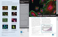
DyLight TM Fluorescent Conjugates<br />
<strong>KPL</strong>’s DyLight TM Conjugates - A Brilliant Choice!<br />
<strong>KPL</strong>’s DyLight TM conjugates offer a brilliant<br />
choice in a variety of multicolor detection applications,<br />
including fluorescence microscopy,<br />
flow cytometry, <strong>Western</strong> blotting, ELISA and<br />
array platforms. Our affinity purified antibodies<br />
combined with a series of outstanding<br />
DyLight dyes provide superior performance<br />
over conventional CyDye TM fluors, fluorescein<br />
and rhodamine, with performance comparable<br />
to that of Alexa Fluor ® dyes (Figure 1). Enjoy<br />
these advantages when you switch to <strong>KPL</strong>’s<br />
DyLight Conjugates:<br />
<strong>KPL</strong> offers eight DyLight dyes, including 405,<br />
488, 549, 594, 633, 649, 680 and 800 with<br />
well-differentiated excitation and emission<br />
spectra. Our extensive line of over 170 DyLight<br />
conjugates is available across a range of animal<br />
species immunoglobulin, including human,<br />
mouse, rabbit, rat, other species and streptavidin.<br />
See back cover to find out what sets <strong>KPL</strong>’s<br />
antibodies apart.<br />
Comparison of DyLight 649 and Alexa 647<br />
Conjugates in a Performance Assay<br />
• High sensitivity, bright signal enable conjugate<br />
conservation<br />
• More photostable than Cy, FITC; comparable<br />
to Alexa<br />
• High signal-to-noise ratios; low background<br />
• Wide selection of specific antibodies<br />
• Consistent, lot-to-lot performance<br />
• Narrow emission spectra enable specific,<br />
multicolor analysis<br />
• Compatible with a variety of buffers.<br />
Antigen Concentration (ng/mL)<br />
Figure 1. <strong>KPL</strong> DyLight 649 conjugates demonstrate comparable<br />
fluorescense intensity and photostability to Alexa 647 conjugates<br />
in a FLISA.<br />
Image above: HMVEC-L primary endothelial cells. F-actin detected with DY554-Phalloidin (rendered green). Microtubules detected with anti-tubulin<br />
antibody and DyLight 649 conjugated goat anti-mouse IgG (red). Nuclei detected with DAPI (blue)<br />
SEE MORE with <strong>KPL</strong>!
DyLight TM Fluorescent<br />
Conjugates<br />
DyLight Conjugated Affinity Purified<br />
Antibodies and Streptavidin<br />
<strong>KPL</strong> DyLight conjugates provide a choice of outstanding fluorescent<br />
conjugates with absorption spectra ranging from<br />
405 nm to 800 nm. Their emission profiles correspond to<br />
those of other commonly used fluorophores such as Alexa,<br />
FITC and the Cy dyes. Narrow, defined emission spectra enable<br />
the use of multiple fluorescence labeling and simultaneous<br />
identification of several target molecules in the same<br />
sample. Light output is comparable to IRDye and Alexa and<br />
more intense than Cy Dyes, FITC or TRITC. DyLight dyes<br />
demonstrate resistance to photobleaching, resulting in excellent<br />
photostability as well as high solubility in aqueous solutions<br />
across a range of pH values.<br />
Emission DyLight Dye Ex/Em(nm) Replaces<br />
Blue<br />
Green<br />
Yellow<br />
Orange<br />
Red<br />
Red<br />
Near IR<br />
Infrared<br />
nm = nanometers<br />
IR = infrared<br />
DyLight 405 400/420 Alexa 405 and<br />
Cascade Blue<br />
DyLight 488 493/518 Alexa 488, Fluorescein<br />
and FITC<br />
DyLight 549 550/568 Alexa 546, Alexa 555,<br />
Cy3, TRITC<br />
DyLight 594 593/618 Alexa 594, Texas Red<br />
DyLight 633 638/658 Alexa 633<br />
DyLight 649 646/674 Alexa 647, Cy5<br />
DyLight 680 682/715 Alexa 680, Cy5.5<br />
DyLight 800 770/794 IRDye 800<br />
Ordering Information<br />
Description DyLight 405* DyLight 488 DyLight 549 DyLight 594* DyLight 633* DyLight 649 DyLight 680 DyLight 800<br />
Anti-Mouse IgG (γ), HSA 072-03-18-02 072-04-18-02 072-05-18-02<br />
Anti-Mouse IgG (H+L), HSA 072-08-18-06 072-03-18-06 072-04-18-06 072-09-18-06 072-10-18-06 072-05-18-06 072-06-18-06 072-07-18-06<br />
Anti-Mouse IgG (H+L), HSA, 0.1 mg 042-08-18-06 042-03-18-06 042-04-18-06 042-09-18-06 042-10-18-06 042-05-18-06 042-06-18-06 042-07-18-06<br />
Anti-Mouse IgG (H+L), RbSA, HSA 072-03-18-18 072-04-18-18 072-05-18-18 072-06-18-18 072-07-18-18<br />
Anti-Mouse IgM (µ), HSA 072-03-18-03 072-04-18-03 072-05-18-03<br />
Anti-Mouse IgG+IgM (H+L), HSA 072-03-18-09 072-04-18-09 072-05-18-09<br />
F(ab’) 2 Anti-Mouse IgG (γ), HSA 202-03-18-02 202-04-18-02 202-05-18-02<br />
F(ab’) 2 Anti-Mouse IgG (H+L), HSA 202-08-18-06 202-03-18-06 202-04-18-06 202-09-18-06 202-10-18-06 202-05-18-06 202-06-18-06 202-07-18-06<br />
Anti-Human IgG (H+L) 072-08-10-06 072-03-10-06 072-04-10-06 072-09-10-06 072-10-10-06 072-05-10-06 072-06-10-06 072-07-10-06<br />
Anti-Human IgG (H+L), 0.1 mg 042-08-10-06 042-03-10-06 042-04-10-06 042-09-10-06 042-10-10-06 042-05-10-06 042-06-10-06 042-07-10-06<br />
Anti-Human IgG (γ) 072-03-10-02 072-04-10-02 072-05-10-02<br />
Anti-Human IgM (µ) 072-03-10-03 072-04-10-03 072-05-10-03<br />
F(ab’) 2 Anti-Human IgG (γ) 202-03-10-02 202-04-10-02 202-05-10-02<br />
F(ab’) 2 Anti-Human IgM (µ) 202-03-10-03 202-04-10-03 202-05-10-03<br />
F(ab’) 2 Anti-Human IgG (H+L) 202-08-10-06 202-03-10-06 202-04-10-06 202-09-10-06 202-10-10-06 202-05-10-06 202-06-10-06 202-07-10-06<br />
Anti-Rabbit IgG (H+L) 072-08-15-06 072-03-15-06 072-04-15-06 072-09-15-06 072-10-15-06 072-05-15-06 072-06-15-06 072-07-15-06<br />
Anti-Rabbit IgG (H+L), 0.1 mg 042-08-15-06 042-03-15-06 042-04-15-06 042-09-15-06 042-10-15-06 042-05-15-06 042-06-15-06 042-07-15-06<br />
Anti-Rabbit IgG (H+L), HSA 072-03-15-16 072-04-15-16 072-05-15-16 072-06-15-16 072-07-15-16<br />
F(ab’) 2 Anti-Rabbit IgG (H+L), HSA 202-08-15-16 202-03-15-16 202-04-15-16 202-09-15-16 202-10-15-16 202-05-15-16 202-06-15-16 202-07-15-16<br />
Anti-Rat IgG (H+L) 072-08-16-06 072-03-16-06 072-04-16-06 072-09-16-06 072-10-16-06 072-05-16-06 072-06-16-06 072-07-16-06<br />
Anti-Rat IgG (H+L), 0.1 mg 042-03-16-06<br />
Anti-Guinea Pig IgG (H+L) 072-03-17-06 072-04-17-06 072-05-17-06 072-06-17-06 072-07-17-06<br />
Anti-Chicken IgG (H+L) 072-08-24-06 072-03-24-06 072-04-24-06 072-09-24-06 072-10-24-06 072-05-24-06 072-06-24-06 072-07-24-06<br />
Anti-Dog IgG (H+L) 072-03-19-06 072-04-19-06 072-05-19-06 072-06-19-06 072-07-19-06<br />
Anti-Goat IgG (H+L) 072-08-13-06 072-03-13-06 072-04-13-06 072-09-13-06 072-10-13-06 072-05-13-06 072-06-13-06 072-07-13-06<br />
Anti-Horse IgG (H+L) 072-03-21-06 072-04-21-06 072-05-21-06 072-06-21-06 072-07-21-06<br />
Rabbit Anti-Sheep IgG (H+L) 072-03-23-06 072-04-23-06 072-05-23-06 072-06-23-06 072-07-23-06<br />
Anti-Swine IgG (H+L) 072-03-14-06 072-04-14-06 072-05-14-06 072-06-14-06 072-07-14-06<br />
Streptavidin 072-08-30-00 072-03-30-00 072-04-30-00 072-09-30-00 072-10-30-00 072-05-30-00 072-06-30-00 072-07-30-00<br />
Streptavidin, 0.1 mg 042-08-30-00 042-03-30-00 042-04-30-00 042-09-30-00 042-10-30-00 042-05-30-00 042-06-30-00 042-07-30-00<br />
*Coming soon!<br />
SEE MORE with <strong>KPL</strong>!
Near Infrared and Infrared Fluorophores DyLight<br />
680 and 800 Conjugates Offer Sensitive Multicolor<br />
Imaging in <strong>Western</strong> <strong>Blotting</strong><br />
DyLight Dyes Emission Spectra<br />
DyLight 680 and 800 dyes emit in the near infrared and<br />
infrared ranges of the light spectrum respectively and are<br />
ideal for multicolor protein detection in <strong>Western</strong> blotting.<br />
Unlike fluorescent conjugates that emit in the visible<br />
range, DyLight 680 and 800 conjugates provide a<br />
unique set of advantages:<br />
• Secondary antibodies with defined specificity and<br />
sensitivity<br />
• Brighter signal than visible fluorescence<br />
• Virtually no background autofluorescence from<br />
membranes or most biological specimens<br />
Effective Alternative to Chemiluminescence<br />
• Broader dynamic range than chemiluminescence<br />
• Quantitation accuracy superior to traditional<br />
methods<br />
• Easy to assay multiple proteins simultaneously on<br />
one <strong>Western</strong> blot<br />
• Cost-effective - eliminates need for chemiluminescence<br />
substrates, film and darkroom<br />
• Clean results - no bleeding from consecutive lanes<br />
Near Infrared Fluorescent Imaging with Secondary<br />
Antibodies labeled with DyLight 680 and 800<br />
Figure 2. DyLight 680-anti mouse IgG and DyLight<br />
800 anti-rabbit IgG secondary antibody conjugates<br />
provide low background and high signal in two-color<br />
<strong>Western</strong> blot detection of tubulin and TNFα. Membrane<br />
was imaged with the LI-COR Odyssey Infrared<br />
Imaging System.<br />
HSA = human serum adsorbed<br />
RbSA = rabbit serum adsorbed<br />
DyLight antibody conjugates are made in goat except<br />
anti-goat and anti-sheep antibodies are made in rabbit.<br />
Supplied in 1.0 mg lyophilized form except select 0.1<br />
mg sizes.<br />
1.800.638.3167 www.kpl.com
Immunofluorescence using DyLight Conjugates<br />
See bright fluorescence and low background with <strong>KPL</strong> DyLight conjugates in immunohistology<br />
applications. The winning combination of DyLight dyes and <strong>KPL</strong><br />
purified antibodies enables multicolor labeling of two or more targets with similar<br />
intensity and photostability to Alexa dyes without the limitations of fluorescein<br />
and CyDyes.<br />
Mouse primary cortical neurons<br />
MCx WCS stained with DyLight 549<br />
(red). Synaptophysin stained with<br />
DyLight 488 (green).<br />
Rat hippocampal neurons<br />
WCS stained with; DyLight 488<br />
(green). MAP2 stained with<br />
DyLight 549 (red).<br />
DyLight TM Fluorescent<br />
Conjugates<br />
<strong>KPL</strong> Antibodies-<br />
What Sets Them Apart<br />
In 1979, <strong>KPL</strong> pioneered the production<br />
of large-scale affinity purification and<br />
was the first company to commercialize<br />
affinity purified secondary antibodies.<br />
Rigorous standards throughout the antibody<br />
production process make our<br />
antibodies standout in the marketplace.<br />
Many manufacturers cut corners by beginning<br />
with inferior serum and extracting<br />
the useful antibody towards the end<br />
of their process. <strong>KPL</strong> spends considerable<br />
effort developing and purifying its own<br />
immunogen formulation to generate the<br />
antiserum, because pure immunogen<br />
results in a more potent and specific<br />
antibody prior to any purification steps.<br />
HMVEC-L primary endothelial cells<br />
F-actin detected with DY554-Phalloidin<br />
(rendered green). Microtubules<br />
stained with DyLight 649<br />
(red). Nuclei detected with DAPI<br />
(blue).<br />
A549 cells<br />
Cytokeratin stained with DyLight<br />
680 (pseudocolored white).<br />
Lamin A stained with DyLight<br />
549 (pseudocolored red).<br />
Further, ISO 9001:2008-certified quality<br />
procedures are carried out at more than<br />
six different stages of the antibody production<br />
cycle, and material that does not<br />
meet our high standards for potency and<br />
cross-reactivity is rejected. Our late<br />
stage purification process has been continually<br />
refined since 1979 and relies on<br />
a one of a kind custom column resin<br />
which is uniquely suited to our antibody<br />
manufacturing process.<br />
Finally, our process of pooling antiserum<br />
from multiple animals tempers natural<br />
serum variability, minimizing variances<br />
from animal-to-animal. The result is<br />
more standardized large-scale antibody<br />
lots with increased consistency. <strong>KPL</strong>’s experience,<br />
innovative processes, and attention<br />
to detail result in highperformance<br />
antibodies that are unique.<br />
NIH 3T3 cells<br />
F-actin detected with DY554-Phalloidin<br />
(rendered green). Microtubules<br />
stained with DyLight 649<br />
(red). Nuclei detected with DAPI<br />
(blue)<br />
Gaithersburg, MD<br />
Phone: 800.638.3167/301.948.7755<br />
Fax: 301.948.0169<br />
www.kpl.com<br />
To order or for more information on <strong>KPL</strong>’s line of unlabeled and conjugated<br />
affinity purified antibodies, contact us at 800.638.3167/301.948.7755,<br />
fax 301.948.0169 or visit www.kpl.com.<br />
ISO 9001:2008 Registered<br />
ML356-02<br />
For research use only.<br />
DyLight is a trademark of Thermo Fisher Scientific Inc. and its subsidiaries. Cy and Cy Dye are trademarks of GE Healthcare.<br />
Alexa Fluor is a registered trademark of Invitrogen.<br />
©2009 <strong>KPL</strong>, Inc. All rights reserved.<br />
SEE MORE with <strong>KPL</strong>!
Cy3 and Cy5 Conjugates<br />
Image above: Confocal fluorescence micrograph of HeLa cells stained with monoclonal antibody against mitochondria enzyme and Cy3-<br />
conjugated anti-mouse antibody (red); rabbit polyclonal antibody to histones in DNA and Cy5-conjugated anti-rabbit antibody (blue).<br />
<strong>KPL</strong> Cy TM Dye Conjugates- Improve your assay and brighten your day!<br />
Cy Dyes offer intense fluorescence when coupled<br />
with <strong>KPL</strong>’s affinity purified antibodies and streptavidin.<br />
The sensitivity and reproducibility of <strong>KPL</strong><br />
antibodies combined with the brightness of Cy3 and<br />
Cy5 dyes produce an exceptional set of conjugates<br />
ideal for multiple labeling experiments. Dye/protein<br />
ratios have been established to ensure optimal fluorescence<br />
with minimal background. They present<br />
maximum excitation/emission spectra at 550/570<br />
nm (Cy3) and 650/670 nm (Cy5).<br />
Fluorescence<br />
Cy3<br />
Wavelength (nm)<br />
Cy5<br />
Ex<br />
Em<br />
Cy dyes are excellent alternatives to most other fluorrescein<br />
dyes as they are brighter and offer greater<br />
photostability. Cy Dye conjugates are used in both<br />
visual and image analysis fluorescent microscopy<br />
and in situ hybridization. Cy3 conjugates are ideally<br />
used in visual color applications, whereas Cy5 conjugates<br />
emit in the far red spectrum and are not easily<br />
visualized.<br />
Benefits of <strong>KPL</strong> Cy Dye Conjugates<br />
High performance conjugates – optimized dyeprotein<br />
ratios ensure high signal-to-noise.<br />
Intense fluorescence – offers greater sensitivity<br />
than TRITC conjugates.<br />
Narrow emission spectra – enables sensitive,<br />
multi-color analysis.<br />
Excellent photostability – more photostable than<br />
TRITC conjugates.<br />
Consistent performance – minimal lot-to-lot variation<br />
reduces need for assay optimization.<br />
Buffer stability – after rehydration, conjugates are<br />
stable at pH 4-9.<br />
Instrument compatibility – excitation and emission<br />
spectra correspond with standard filter sets<br />
and laser settings.<br />
SEE MORE with <strong>KPL</strong>!
Excellent Performance<br />
As demonstrated below <strong>KPL</strong> Cy3- and Cy5-labeled conjugates produce brighter fluorescence than<br />
those of other suppliers.<br />
Cy3 and Cy5<br />
Conjugates<br />
Cytomegalovirus-infected cells<br />
detected with a biotinylated CMV<br />
probe and the DNADetector TM<br />
Fluorescent in situ Hybridization Kit<br />
using Cy3-Strept-avidin and DAPI.<br />
Microplates were coated with serially diluted mouse IgG at the indicated concentrations. Conjugates at a<br />
concentration of 0.01 mg/mL were applied and incubated for 30 minutes. Fluorescence was measured with<br />
a Perkin Elmer VICTOR 3 Multilabel Plate Reader.<br />
Ordering Information<br />
Description Cy3 Cy5<br />
Anti-Mouse IgG (γ), HSA 072-01-18-02 072-02-18-02<br />
F(ab’) 2 Anti-Mouse IgG (γ), HSA 202-01-18-02 202-02-18-02<br />
Anti-Mouse IgG (H+L), HSA 072-01-18-06 072-02-18-06<br />
F(ab’) 2 Anti-Mouse IgG (H+L), HSA 202-01-18-06 202-02-18-06<br />
Anti-Mouse IgG (H+L), RbSA, HSA 072-01-18-18 072-02-18-18<br />
Anti-Mouse IgM (µ), HSA 072-01-18-03 072-02-18-03<br />
Anti-Mouse IgG+IgM (H+L), HSA 072-01-18-09 072-02-18-09<br />
Anti-Rabbit IgG (H+L) 072-01-15-06 072-02-15-06<br />
F(ab’) 2 Anti-Rabbit IgG (H+L), HSA 202-01-15-16 202-02-15-16<br />
Anti-Rabbit IgG (H+L), HSA 072-01-15-16 072-02-15-16<br />
Anti-Rat IgG (H+L) 072-01-16-06 072-02-16-06<br />
F(ab’) 2 Anti-Human IgG (H+L) 202-01-10-06 202-02-10-06<br />
Anti-Human IgG (γ) 072-01-10-02 072-02-10-02<br />
F(ab’) 2 Anti-Human IgG (γ) 202-01-10-02 202-02-10-02<br />
Anti-Human IgM (µ) 072-01-10-03 072-02-10-03<br />
F(ab’) 2 Anti-Human IgM (µ) 202-01-10-03 202-02-10-03<br />
Anti-Guinea Pig IgG (H+L) 072-01-17-06 072-02-17-06<br />
Anti-Chicken IgG (H+L) 072-01-24-06 072-02-24-06<br />
Anti-Horse IgG (H+L) 072-01-21-06 072-02-21-06<br />
Anti-Swine IgG (H+L) 072-01-14-06 072-02-14-06<br />
Anti-Dog IgG (H+L) 072-01-19-06 072-02-19-06<br />
Anti-Sheep IgG (H+L) 072-01-23-06 072-02-23-06<br />
Anti-Goat IgG (H+L) 072-01-13-06 072-02-13-06<br />
Streptavidin 072-01-30-00 072-02-30-00<br />
HSA=human serum adsorbed RbSA=rabbit serum adsorbed<br />
Cy Dye antibody conjugates are made in goat except anti-goat and anti-sheep antibodies<br />
made in rabbit. Supplied in 1 mg lyophilized form.<br />
To order or for more information on <strong>KPL</strong>’s line of unlabeled and conjugated<br />
affinity purified antibodies, contact us at 800.638.3167 / 301.948.7755, fax<br />
301.948.0169 or visit us at www.kpl.com.<br />
Signal Detection<br />
Cy3 is excited maximally at 550 nm<br />
and fluoresces maximally at 570 nm.<br />
It is excited to about 50% of maximum<br />
with an argon laser (514 nm or<br />
528 nm lines), or to about 75% of<br />
maximum with a helium/neon laser<br />
(543 nm line) or mercury lamp (546<br />
nm line).<br />
Cy5 is excited maximally at 650 nm<br />
and fluoresces maximally at 670 nm.<br />
It is excited to about 98% of maximum<br />
with a krypton/argon laser (647<br />
nm line) or to about 63% of maximum<br />
with a helium/neon laser (633<br />
nm line). Cy5 produces minimal autofluorescence<br />
of biological specimens<br />
in this region of the spectrum.<br />
A confocal microscope equipped with<br />
the appropriate laser for excitation<br />
and a far-red detector enable double<br />
labeling with Cy3 and Cy5.<br />
Gaithersburg, MD 20878<br />
Phone: 800.638.3167<br />
Fax: 301.948.0169<br />
www.kpl.com<br />
ISO 9001:2000 Registered<br />
ML368-01<br />
For research use only.<br />
Cy Dye is a trademark of GE Healthcare.<br />
©2009 <strong>KPL</strong>, Inc. All rights reserved.<br />
SEE MORE with <strong>KPL</strong>!
Peroxidase Conjugates<br />
<strong>KPL</strong> Peroxidase Conjugates: Time-tested, Sensitive and Reliable<br />
Since 1979 <strong>KPL</strong> has provided quality affinity<br />
purified antibodies to researchers worldwide.<br />
Over the years we have refined our production<br />
process to provide antibodies with high potency<br />
and consistent performance in immunoassays.<br />
From the start <strong>KPL</strong> gives careful consideration<br />
to immunogen preparation, using a highly<br />
purifed formulation to generate antiserum. <strong>KPL</strong><br />
pools antiserum from multiple animals to<br />
reduce natural animal to animal serum variability.<br />
During the purification process our ISO<br />
9001:2008-certified quality procedures impose<br />
rigorous standards for potency and cross-reactivity.<br />
The result is standardized antibody lots<br />
with excellent reproducibility.<br />
Our extensive line of peroxidase (HRP) conjugates<br />
is available across a range of animal<br />
species, including human, mouse, rabbit and rat<br />
antibodies, as well as other animal species and<br />
streptavidin. They are affinity purified and in<br />
some cases further adsorbed to minimize crossreactivity<br />
between animal species or shared<br />
reactivity with other immunoglobulin classes.<br />
HRP-labeled F(ab’) 2 fragment antibodies are<br />
offered for assays requiring extremely low background<br />
and absence of F(c)-mediated binding.<br />
<strong>KPL</strong> reacts HRP of the highest quality with<br />
affinity purified antibodies and streptavidin<br />
using the periodate method of Nakane and<br />
Kawaoi. Special features of HRP include:<br />
• faster catalytic rate than alkaline phosphatase<br />
• generates more product in shorter incubation<br />
times<br />
• provides maximum sensitivity, low nonspecific<br />
binding<br />
• ideal for ELISA, <strong>Western</strong> blotting and<br />
immunohistology applications.<br />
SEE MORE with <strong>KPL</strong>!
Peroxidase Conjugates<br />
Peroxidase Conjugates Ordering Information<br />
(Partial listing)<br />
Catalog# Description Size<br />
04-10-06 HRP-labeled Goat Anti-Human IgG (H+L) 0.1 mg<br />
04-10-17 HRP-labeled Goat Anti-Human IgA+IgG+IgM (H+L), MSA 0.1 mg<br />
04-10-20 HRP-labeled Goat Anti-Human IgG (Fc) 0.1 mg<br />
074-1002 HRP-labeled Goat Anti-Human IgG (γ) 1.0 mg<br />
074-1003 HRP-labeled Goat Anti-Human IgM (μ) 1.0 mg<br />
074-1004 HRP-labeled Goat Anti-Human IgE (ε) 1.0 mg<br />
074-1006 HRP-labeled Goat Anti-Human IgG (H+L) 1.0 mg<br />
074-1007 HRP-labeled Goat Anti-Human IgA+IgG+IgM (H+L) 1.0 mg<br />
14-10-01 HRP-labeled Goat Anti-Human IgA (α) 0.5 mg<br />
214-1002 HRP-labeled F(ab’) 2 Goat Anti-Human IgG (γ) 0.5 mg<br />
214-1003 HRP-labeled F(ab’) 2 Goat Anti-Human IgM (μ) 0.5 mg<br />
214-1006 HRP-labeled F(ab’) 2 Goat Anti-Human IgG (H+L) 0.5 mg<br />
474-1002 HRP-labeled Goat Anti-Human IgG (γ), Liquid 1.0 mL<br />
474-1003 HRP-labeled Goat Anti-Human IgM (μ), Liquid 1.0 mL<br />
474-1006 HRP-labeled Goat Anti-Human IgG (H+L), Liquid 1.0 mL<br />
04-18-06 HRP-labeled Goat Anti-Mouse IgG (H+L), HSA 0.1 mg<br />
04-18-15 HRP-labeled Goat Anti-Mouse IgG (H+L), RtSA, HSA 0.1 mg<br />
04-18-18 HRP-labeled Goat Anti-Mouse IgG (H+L), RbSA, HSA 0.1 mg<br />
074-1802 HRP-labeled Goat Anti-Mouse IgG (γ), HSA 1.0 mg<br />
074-1803 HRP-labeled Goat Anti-Mouse IgM (μ), HSA 1.0 mg<br />
074-1806 HRP-labeled Goat Anti-Mouse IgG (H+L), HSA 1.0 mg<br />
074-18-061 HRP-labeled Goat Anti-Mouse IgG (H+L), XSA 1.0 mg<br />
074-1807 HRP-labeled Goat Anti-Mouse IgA+IgG+IgM (H+L), HSA 1.0 mg<br />
074-1809 HRP-labeled Goat Anti-Mouse IgG+IgM (H+L), HSA 1.0 mg<br />
14-18-01 HRP-labeled Goat Anti-Mouse IgA (α), HSA 0.5 mg<br />
214-1802 HRP-labeled F(ab’) 2 Goat Anti-Mouse IgG (γ), HSA 0.5 mg<br />
214-1806 HRP-labeled F(ab’) 2 Goat Anti-Mouse IgG (H+L), HSA 0.5 mg<br />
474-1802 HRP-labeled Goat Anti-Mouse IgG (γ), HSA, Liquid 1.0 mL<br />
474-1806 HRP-labeled Goat Anti-Mouse IgG (H+L), HSA, Liquid 1.0 mL<br />
074-1506 HRP-labeled Goat Anti-Rabbit IgG (H+L) 1.0 mg<br />
074-15-061 HRP-labeled Goat Anti-Rabbit IgG (H+L), XSA 1.0 mg<br />
074-1516 HRP-labeled Goat Anti-Rabbit IgG (H+L), HSA 1.0 mg<br />
214-1516 HRP-labeled F(ab’) 2 Goat Anti-Rabbit IgG (H+L), HSA 0.5 mg<br />
474-1506 HRP-labeled Goat Anti-Rabbit IgG (H+L), Liquid 1.0 mL<br />
474-1516 HRP-labeled Goat Anti-Rabbit IgG (H+L), HSA, Liquid 1.0 mL<br />
14-30-00 HRP-labeled Streptavidin 0.5 mg<br />
474-3000 HRP-labeled Streptavidin, Liquid, Molecular Grade 1.0 mL<br />
Visit our website at www.kpl.com for a complete listing of HRP-labeled antibodies.<br />
To order or for more information on <strong>KPL</strong>’s protein research products, contact us at<br />
800.638.3167 / 301.948.7755, FAX 301.948.0169 or visit us at www.kpl.com.<br />
<strong>KPL</strong> offers a range of sensitive<br />
substrates for use with HRP conjugates.<br />
They provide a choice of<br />
intense colors for ELISA, blotting<br />
and cell staining applications.<br />
ELISA<br />
• ABTS ® 1- and 2-Component<br />
Microwell Peroxidase Substrates<br />
• SureBlue TM TMB Peroxidase<br />
Substrate<br />
• SureBlue Reserve TM TMB<br />
Peroxidase Substrate<br />
• TMB Peroxidase Substrate<br />
<strong>Blotting</strong><br />
• 4 CN Peroxidase Substrate<br />
• TMB Membrane Peroxidase<br />
Substrate<br />
• LumiGLO ® Chemiluminescent<br />
Substrate<br />
• LumiGLO Reserve TM<br />
Chemiluminescent Substrate<br />
Whichever substrate you choose,<br />
enjoy the benefits of excellent signalto-noise<br />
and reproducibility.<br />
Gaithersburg, MD 20878<br />
Phone: 800.638.3167<br />
Fax: 301.948.0169<br />
www.kpl.com<br />
ISO 9001:2008 Registered<br />
For research use only.<br />
©2009 <strong>KPL</strong>, Inc. All rights reserved.<br />
ABTS is a registered trademark of Roche Biochemicals.<br />
ML371-01<br />
SEE MORE with <strong>KPL</strong>!
Ordering Information<br />
Catalog # Description Size<br />
Protein Detector <strong>Western</strong> <strong>Blotting</strong> Kits<br />
Each kit includes anti-mouse and anti-rabbit conjugates, Detector<br />
Block, Wash Solution Concentrate and Substrate.<br />
Phosphatase Chromogenic<br />
55-11-50 BCIP/NBT <strong>Western</strong> Blot Kit 2500 cm 2<br />
Peroxidase Chromogenic<br />
54-11-50 TMB <strong>Western</strong> Blot Kit 2500 cm 2<br />
Peroxidase Chemiluminescent<br />
54-12-50 LumiGLO ® <strong>Western</strong> Blot Kit 2500 cm 2<br />
54-13-50 LumiGLO Reserve TM <strong>Western</strong> 2400 cm 2<br />
Blot Kit<br />
Related Reagents and Kits<br />
Antibody Conjugates<br />
All antibodies listed below are produced in goat.<br />
For a complete antibody listing, refer to <strong>KPL</strong>’s Product Catalog.<br />
Phosphatase-labeled<br />
475-1006 Anti-Human IgG (H+L) 1.0 mL, liquid<br />
475-1806 Anti-Mouse IgG (H+L), HSA 1.0 mL, liquid<br />
475-1506 Anti-Rabbit IgG (H+L) 1.0 mL, liquid<br />
Peroxidase-labeled<br />
474-1006 Anti-Human IgG (H+L) 1.0 mL, liquid<br />
474-1806 Anti-Mouse IgG (H+L) HSA 1.0 mL, liquid<br />
474-1506 Anti-Rabbit IgG (H+L) 1.0 mL, liquid<br />
Biotin-labeled<br />
16-10-06 Anti-Human IgG (H+L) 0.5 mg<br />
176-1006 Anti-Human IgG (H+L) 2.0 mg<br />
Labeled Streptavidin<br />
474-3000 HRP-labeled 1.0 mL, liquid<br />
475-3000 AP-labeled 1.0 mL, liquid<br />
Catalog # Description Size<br />
Substrates for <strong>Western</strong> <strong>Blotting</strong><br />
Phosphatase Colorimetric Substrates<br />
50-81-18 BCIP/NBT Substrate 100 mL<br />
50-81-07 BCIP/NBT Substrate 600 mL<br />
50-81-30 FirePhos TM Membrane AP Substrate 100 mL<br />
50-81-40 FirePhos Membrane AP Substrate 400 mL<br />
50-81-34 FirePhos Membrane AP Substrate 1000 mL<br />
Phosphatase Chemiluminescent Substrates<br />
55-60-03 PhosphaGLO TM AP Substrate 30 mL<br />
55-60-04 PhosphaGLO AP Substrate 100 mL<br />
55-60-01 PhosphaGLO Reserve AP Substrate 30 mL<br />
55-60-02 PhosphaGLO Reserve AP Substrate 100 mL<br />
Peroxidase Chromogenic Substrates<br />
50-77-18 TMB Membrane Substrate 100 mL<br />
50-77-03 TMB Membrane Substrate 200 mL<br />
50-73-00 4 CN Substrate 600 mL<br />
50-73-04 4 CN Substrate 2700 mL<br />
Peroxidase Chemiluminescent Substrates<br />
54-61-02 LumiGLO Chemiluminescent Substrate 60 mL<br />
54-61-00 LumiGLO Chemiluminescent Substrate 240 mL<br />
54-61-01 LumiGLO Chemiluminescent Substrate 720 mL<br />
54-71-00 LumiGLO Reserve Substrate Kit 2400 cm 2<br />
54-71-01 LumiGLO Reserve Substrate Kit 600 cm 2<br />
Assay Support Reagents<br />
50-84-00 Coating Solution Concentrate 50 mL<br />
54-15-01 HRPStabilizer 200 mL<br />
55-15-00 APStabilizer 200 mL<br />
50-61-00 10% BSA Diluent/Blocking Solution 200 mL<br />
Concentrate<br />
50-82-01 Milk Diluent/Blocking Solution 200 mL<br />
Concentrate<br />
71-83-00 Detector TM Block (5X) 240 mL<br />
50-63-00 Wash Solution Concentrate (20X) 800 mL<br />
50-63-06 Biotin Wash Solution Concentrate (10X) 200 mL<br />
60-00-50 Biodyne ® B Nylon Membrane 1 roll<br />
HSA=human serum adsorbed.<br />
ML282-04<br />
To order or for more information,<br />
call <strong>KPL</strong> at 800.638.3167, 301.948.7755,<br />
Fax: 301.948.0169. www.kpl.com<br />
or contact your local sales partner.<br />
SEE MORE with <strong>KPL</strong>!