Spontaneous Breathing during Artificial Ventilation
We have known about ventilation procedures that allow spontaneous breathing at any time since the 1980’s . We introduced the first method of this kind in our book "PCV - two steps forward in intensive ventilation". Meanwhile, intensive ventilation has progressed by a few additional steps. The goal of this introduction is to describe the progress and thus highlight the relevance of spontaneous breathing during artificial ventilation.
We have known about ventilation procedures that allow spontaneous breathing at any time since the 1980’s . We introduced the first method of this kind in our book "PCV - two steps forward in intensive ventilation". Meanwhile, intensive ventilation has progressed by a few additional steps. The goal of this introduction is to describe the progress and thus highlight the relevance of spontaneous breathing during artificial ventilation.

Create successful ePaper yourself
Turn your PDF publications into a flip-book with our unique Google optimized e-Paper software.
D-7482-2010<br />
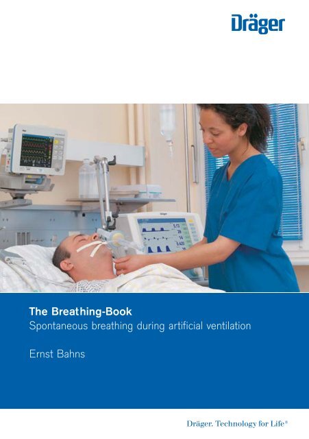
The <strong>Breathing</strong>-Book<br />
<strong>Spontaneous</strong> breathing <strong>during</strong> artificial ventilation<br />
Ernst Bahns
The <strong>Breathing</strong>-Book<br />
<strong>Spontaneous</strong> breathing <strong>during</strong> artificial ventilation<br />
Ernst Bahns
THE BREATHING-BOOK |<br />
CONTENTS<br />
Publisher<br />
Dräger Medical GmbH<br />
Moislinger Allee 53–55<br />
23558 Lübeck, Germany<br />
www.draeger.com<br />
Important note<br />
Medical expertise is continually undergoing change due to research and clinical experience.<br />
The author of this book intends to ensure that the views, opinions and assumptions in this<br />
book, especially those concerning applications and effects, correspond to the current state<br />
of knowledge. But this does not relieve the reader from the duty to personally carry the<br />
responsibilities for clinical measures.<br />
All rights to this book, especially the rights to reproduce and copy, are reserved by<br />
Dräger Medical GmbH. No part of this book may be reproduced or stored mechanically,<br />
electronically or photographically without prior written authorization by<br />
Dräger Medical GmbH.<br />
Design and graphics<br />
Gerrit Meyer Design, Lübeck
04|05<br />
CONTENTS<br />
Introduction 06<br />
The significance of spontaneous breathing 08<br />
<strong>Spontaneous</strong> breathing and artificial ventilation 08<br />
Misinterpretation of spontaneous breathing as an interference factor 10<br />
The development of spontaneous breathing as a treatment goal 12<br />
The technical principal of respiratory gas dosage 14<br />
<strong>Spontaneous</strong> breathing with conventional artificial ventilation 14<br />
Inspiratory valves 16<br />
Exhalation valves 18<br />
Radial compressor 20<br />
Side channel compressor with auxiliary circuitry 22<br />
Control of respiratory gas delivery <strong>during</strong> mechanical ventilation 24<br />
<strong>Spontaneous</strong> breathing in new ventilation modes 26<br />
<strong>Spontaneous</strong> breathing <strong>during</strong> mechanical ventilation 26<br />
<strong>Spontaneous</strong> breathing with PCV and APRV 28<br />
<strong>Spontaneous</strong> breathing with AutoFlow and MMV 30<br />
Passive features of the breathing apparatus 32<br />
Compliance and resistance 32<br />
Risk factors <strong>during</strong> artificial ventilation 34<br />
Lung protecting ventilation - manual adjustments 36<br />
AutoFlow and guaranteed volume - automatic pressure adjustment 38<br />
Systematics and control of the new methods 40<br />
The nomenclature problem 40<br />
Descriptions of methods and functions 42<br />
Control concept PCV and APRV 44<br />
Introduction to the modern methods 46<br />
Pressure limited, AutoFlow and PCV put into practice 48<br />
Clinical application of mechanical methods 50<br />
Conditions and malfunctions of gas exchange 50<br />
Respiratory adjustments <strong>during</strong> oxygenation malfunctions 52<br />
Ventilatilatory adjustments <strong>during</strong> ventilation malfunctions 54<br />
Effective gas exchange and spontaneous breathing recruitment 56<br />
Conventional weaning and universal weaning method PCV 58<br />
Weaning with PCV 60<br />
Success factors of independent spontaneous breathing in mechanical ventilation 62<br />
The future of spontaneous breathing in intensive ventilation 64<br />
Appendix 66<br />
Additional literature 66<br />
Index 74
THE BREATHING-BOOK |<br />
INTRODUCTION<br />
D-7169-2009<br />
Introduction<br />
Modern respiration systems offer various measures for the treatment of respiratory<br />
disorders. Today these devices do not only enable sufficient respiratory gas delivery<br />
for keeping up the gas exchange in the lung; thanks to advanced pneumatics,<br />
electronics and especially due to the implementation of computer technology,<br />
artificial ventilation may nowadays be specifically adapted to the respective gas<br />
exchange disorder.<br />
<strong>Artificial</strong> ventilation still remains to be a serious intervention for the patient.<br />
Specifically, the pressure conditions generated in the process differ considerably<br />
from those <strong>during</strong> natural breathing. These mechanical pressures affect the lungs<br />
and cardiovascular system which may result in undesired side effects. The goal to<br />
keep up the vital function of gas exchange with artificial breathing may thus be<br />
achieved only by reaching a balance of risks and benefits.<br />
Modern ventilation still faces several partially contradictory challenges. It must<br />
serve as a life preserving function with methods that causes stress on the patient<br />
however at same time it must keep side effects to a minimum. Modern ventilation<br />
faces the contradictory challenges with a new self-conception: The artificial<br />
measure is no longer the focus, but rather the natural function of normal breathing<br />
even though malfunctioning.
06|07<br />
The ventilation system must adapt to the patient and not vice versa. Disturbance of<br />
spontaneous breathing by artificial ventilation must be reduced to the absolute<br />
minimum. <strong>Spontaneous</strong> breathing must be promoted whenever possible. This<br />
applies not only to the period of weaning, but also to the complete process of<br />
artificial ventilation.<br />
We have known about ventilation procedures that allow spontaneous breathing at<br />
any time since the 1980’s . We introduced the first method of this kind in our book<br />
"PCV - two steps forward in intensive ventilation". Meanwhile, intensive ventilation<br />
has progressed by a few additional steps. The goal of this introduction is to describe<br />
the progress and thus highlight the relevance of spontaneous breathing <strong>during</strong><br />
artificial ventilation.<br />
Hence the title: "The <strong>Breathing</strong>-Book"
THE BREATHING-BOOK |<br />
THE SIGNIFICANCE OF SPONTANEOUS BREATHING<br />
The significance of spontaneous breathing<br />
SPONTANEOUS BREATHING AND ARTIFICIAL VENTILATION<br />
Natural spontaneous breathing increases the inner thorax volume by contracting<br />
the respiratory muscles. This creates a negative pressure in the lungs resulting in<br />
air being drawn in. However, artificial ventilation employs a reverse principle. The<br />
ventilator creates a negative pressure and thus pushes the respiratory gas into the<br />
lungs. The gas transport is called lung ventilation <strong>during</strong> both spontaneous and<br />
artificial ventilation.<br />
The breathing apparatus model in the illustration shows both principles of<br />
ventilation as it would take place in a homogenous lung area. Roughly simplified,<br />
it shows an extensive analogy of ventilation <strong>during</strong> spontaneous breathing and<br />
artificial ventilation. However, the model insufficiently demonstrates the actual<br />
physical conditions. In fact, the lungs do not consist of only a single area, but are<br />
made up of several million microscopically small vesicular structures, known as the<br />
alveoli.<br />
The realistic model demonstrates the difference between physiologic respiration<br />
and artificial ventilation. Every individual alveoli has different mechanical features<br />
and, depending on the position of each alveoli, the pressure of the ventilator or the<br />
respiratory muscle has a different effect: <strong>Breathing</strong> pressure primarily ventilates the<br />
upper lung areas while spontaneous breathing has a stronger effect on the lower<br />
areas close to the diaphragm.<br />
The relevant difference between ventilation and respiration does not solely concern<br />
lung ventilation, but rather the pressures effecting the lungs also display discernible<br />
differences: During ventilation, the lungs are constantly exposed to positive<br />
pressure, even <strong>during</strong> exhalation. During the exhalation phase, the ventilator<br />
provides a low pressure, known as PEEP*. During spontaneous breathing, pressure<br />
in the lungs is temporarily lower than the ambient pressure. In addition, pressures<br />
arising in the lungs <strong>during</strong> artificial ventilation may be many times higher than the<br />
pressures applied to the lungs <strong>during</strong> physiologic respiration.<br />
* PEEP = Positive End Expiratory Pressure
08|09<br />
Ventilator: pressure<br />
D-18280-2010<br />
Respiratory muscles: Inspiration<br />
The single compartment model of the breathing apparatus.<br />
Two forces take effect <strong>during</strong> ventilation of the lung. The ventilator causes a positive pressure and the<br />
respiratory muscles cause a negative pressure. Both forces add up and cause a volume shift.<br />
The breathing-related changes of the pulmonary pressures indicate that lung<br />
ventilation <strong>during</strong> artificial ventilation differs considerably from spontaneous<br />
breathing. The question of whether it is inferior has been an object of long-standing<br />
debate ever since the first implementation of mechanical ventilators. This<br />
discussion is not only limited to the effects on the lung as an organ and its<br />
ventilation. The effects on other organs are of concern as well. The cardiovascular<br />
system is of special importance, since it is more affected <strong>during</strong> ventilation than it<br />
would be <strong>during</strong> respiration.
THE BREATHING-BOOK |<br />
THE SIGNIFICANCE OF SPONTANEOUS BREATHING<br />
MISINTERPRETATION OF SPONTANEOUS BREATHING AS<br />
AN INTERFERENCE FACTOR<br />
An exceptional change in dealing with the patient's spontaneous breathing is<br />
currently taking place in the development of artificial ventilation. In the early years,<br />
keeping up the vital gas exchange function <strong>during</strong> abnormal breathing conditions<br />
stood at the center of attention.<br />
In those earlier days, mechanical ventilation was a measure in which the focus was<br />
on the lung. Potential negative effects to the lungs and other organs were accepted,<br />
if they were understood at all. The technical means for detecting and preventing<br />
side effects were not available at that time. The technical inadequacy of the<br />
ventilators at that time was even more serious: <strong>Spontaneous</strong> breathing often<br />
resulted in an impaired device functionality and, consequently, had to be<br />
suppressed by medication.<br />
Since this course of treatment with a smooth transition from artificial ventilation to<br />
natural respiration was not possible then. The devices of that time only enabled the<br />
abrupt transfer from artificial ventilation to spontaneous breathing . Mixed forms of<br />
machine ventilation and spontaneous breathing did not exist.<br />
If chemical paralysis of the patient using muscle relaxants is considered a<br />
questionable measure with today's knowledge, motivations of the past treatment<br />
concepts still remain current today. The calm and immobilized patient was<br />
misinterpreted then, unfortunately also frequently still today, as a patient without<br />
complications. These complications were recognized only once they had already<br />
become untreatable and a fatal ending of the patient had become inevitable.
10|11<br />
D-16659-2010<br />
<strong>Ventilation</strong> with the Spiromat<br />
Suppression of spontaneous breathing is necessary for disturbance-free mechanical ventilation.<br />
A smooth transition to natural spontaneous breathing is impossible.<br />
An additional serious problem occurred, especially if chemical paralysis was applied<br />
as a routine measure: This routine sedation impeded the patient's preparation for<br />
the time after artificial ventilation and thus delayed weaning from mechanical<br />
ventilation (10). The patient's personal contribution in this pre-weaning phase in<br />
the form of spontaneous breathing is a mandatory requirement.
THE BREATHING-BOOK |<br />
THE SIGNIFICANCE OF SPONTANEOUS BREATHING<br />
THE DEVELOPMENT OF SPONTANEOUS BREATHING AS TREATMENT GOAL<br />
One of the first attempts to prepare the patient for the time after artificial<br />
ventilation was undertaken in the middle of the last century. The effort necessary<br />
for the patient to trigger the mechanical breaths was increased. The intention was<br />
to train the patient by continuously increasing the respiratory effort for triggering a<br />
breath (no s). The trigger threshold was increased for this purpose. The goal was to<br />
prepare the patient for spontaneous breathing.<br />
As soon as the patient was able to exceed a certain trigger threshold, the method<br />
was abruptly switched over to pure spontaneous breathing. This kind of procedure<br />
has multiple disadvantages: The trigger effort burdens the breathing apparatus in<br />
an non-physiological manner and contributes nothing to lung ventilation.<br />
One method that allows for continuous transition from mechanical ventilation to<br />
spontaneous breathing to some extent was developed in the 1970’s and called SIMV*<br />
(18). This procedure enabled a spontaneous breathing for the first time <strong>during</strong> the<br />
mechanical exhalation phase. This was applied then and still is being used today,<br />
especially <strong>during</strong> weaning. It's acceptance seems to be decreasing, since SIMV in its<br />
original form has an essential disadvantage. The mechanical breaths may still be<br />
triggered by the patient, but spontaneous breathing is not possible <strong>during</strong> these<br />
strokes. This procedure thus only enables a ventilation pattern in which the<br />
mechanical breaths and spontaneous breathing take place successively. This<br />
resulted in phases where the patient is unable to breathe spontaneously.<br />
There are no medical indications for such interruptions of spontaneous breathing.<br />
There are no technical reasons - at least not in modern ventilators.<br />
The technical requirements for uninterrupted spontaneous breathing <strong>during</strong><br />
artificial ventilation, however, were not found in previous years. It was not until the<br />
1980's that this technology became available for the first time. This point in time is<br />
considered a development milestone by many users. Conventional ventilation that<br />
did not allow spontaneous breathing at any time due to technical limitations<br />
developed into modern ventilation. A new ventilation method with electromagnetic<br />
valves and a microprocessor controller met the requirements of allowing<br />
spontaneous breathing <strong>during</strong> artificial ventilation at any time.<br />
* SIMV = Synchronized Intermittent Mandatory <strong>Ventilation</strong>
12|13<br />
D-16655-2010<br />
<strong>Ventilation</strong> with the Evita 2<br />
The ventilation curves in the background demonstrate the development of the mechanic procedures.<br />
Bottom: controlled ventilation without spontaneous breathing, top: mechanical ventilation with<br />
uninterrupted spontaneous breathing.
THE BREATHING-BOOK |<br />
THE TECHNICAL PRINCIPAL OF RESPIRATORY GAS DELIVERY<br />
The technical principle of respiratory gas delivery<br />
SPONTANEOUS BREATHING WITH CONVENTIONAL ARTIFICIAL VENTILATION<br />
In the diagram, valve control <strong>during</strong> inspiratory pauses of conventional ventilation<br />
is simplified for a device without showing the additional function "base flow". This<br />
function will be explained later. The first step is to describe a ventilation with<br />
parameters set by the operator and without the capability of adapting the ventilation<br />
pattern by the patient. This is controlled ventilation.<br />
The inspiratory valve opens <strong>during</strong> the inspiratory phase until the respiratory gas of<br />
the respective breath is completely delivered. Longer mechanical inspiratory times<br />
are followed by a phase <strong>during</strong> which no respiratory gas flows until the end of the<br />
inspiratory phase. During the entire mechanical inspiratory time, the exhalation<br />
valve is completely closed <strong>during</strong> controlled ventilation. The exhalation valve opens<br />
at the beginning of the expiratory phase and releases the respiratory gas. During<br />
controlled ventilation, the inspiratory valve is closed <strong>during</strong> this phase. Only a timer<br />
is required to achieve inspiratory/expiratory phase control. These are the essential<br />
functional components of the ventilator for controlled ventilation.<br />
If the ventilator must adapt to a breathing patient, the technical challenge becomes<br />
more complex. In this case, both valves need to be able to react to the spontaneous<br />
patient respiration. These valves must be ready to open at any time.<br />
This principle was first realized in the exhalation phase. <strong>Spontaneous</strong> breathing in<br />
the mechanical exhalation phase has been part of ventilation technology standard<br />
since the 1970’s. To implement this into the mechanical inspiratory phase proved<br />
a little more difficult. Special ventilation methods enabled the patient to inhale<br />
<strong>during</strong> the mechanical inspiratory phase and thus to inhale more respiratory gas.<br />
But it was impossible to exhale in the mechanical inspiratory phase and thus the<br />
patient could not exhale before the mechanical exhalation phase. The ventilator<br />
maintained the exhalation valve closed <strong>during</strong> this phase and kept the patient from<br />
exhaling.<br />
The first ventilator to allow an uninterrupted spontaneous breathing at any time<br />
<strong>during</strong> mechanical ventilation was Evita in 1988 using the PCV* method. It took
14|15<br />
Inspiratory Valves<br />
Controls<br />
Patient<br />
P<br />
1 2 3<br />
Expiratory Valve<br />
V<br />
t<br />
t<br />
D-18281-2010<br />
Functional elements of a ventilator for respiratory gas dosage and inspiratory/expiratory phase<br />
control <strong>during</strong> controlled ventilation<br />
Left: Inspiratory valve and exhalation valve are closed and opened <strong>during</strong> the respiration phases by the<br />
control unit. Right: Real time respiration cycle curve with user defined respiration length phases (time<br />
control) - pressure (top) and flow (bottom). Valve control <strong>during</strong> the ventilation phases: 1) Inspiratory<br />
valves opened, exhalation valve closed; 2) Both valves closed 3) Inspiratory valve closed, exhalation valve<br />
opened. The combination of phase 1) and 2) forms the inspiratory phase, phase 3) forms the exhalation<br />
phase.<br />
more than a decade until another ventilator with this performance feature became<br />
commercially available. By now in modern ventilation, it is understood that a<br />
patient may breathe spontaneously at any time over the full cycle.<br />
There are quality differences in respiratory gas delivery for spontaneous breathing<br />
that can be explained when taking a closer look at the technical devices for<br />
respiratory gas dosing. The quality of spontaneous breathing depends not only on<br />
the features of these gas dosing devices, but also on the interaction of the individual<br />
systems.<br />
* PCV = Pressure Controlled <strong>Ventilation</strong>
THE BREATHING-BOOK |<br />
THE TECHNICAL PRINCIPAL OF RESPIRATORY GAS DOSING<br />
INSPIRATORY VALVES<br />
For reasons of simplicity, the device components for dosing the respiration gas were<br />
previously described as "the inspiratory valve" and "the exhalation valve". In fact,<br />
complex designs are necessary for gas dosage.<br />
Since the beginning of mechanical ventilation in the early years of the past century,<br />
valve systems in conjunction with pressurized gas sources were used for dosing<br />
respiratory gas. Designs using blowers to supply respiratory gas to valve systems<br />
have existed since the middle of the last century. If the blower and the patient are<br />
separated by a membrane, then it becomes bellows ventilation as commonly used in<br />
anesthesia. Respiratory gas dosage via controlled blowers was introduced only<br />
recently.<br />
Modern intensive ventilation uses two systems for respiratory gas dosage:<br />
Proportional valves (so called flow valves) for flow control and controlled blowers.<br />
Similar to a water taps, proportional valves can generate any given flow between<br />
0 and a maximum value. Different from a water tap, these changes do not require<br />
seconds, but occur within milliseconds. The precision and dynamics required by a<br />
sufficient respiratory gas dosage exceeds the performance of the pneumatic valve<br />
control used in the past. This is the reason why modern ventilators commonly use<br />
electromagnetic valve drives.<br />
For comparative purposes, an electromagnetic valve controls function like loud<br />
speakers, employing a live coil moving in a magnetic field. In a loud speaker, the<br />
coil deflection is performed depending on the current and it sets a membrane in<br />
motion. A tappet in an electromagnetically controlled valve is moved in a similar<br />
fashion. The movement must closely follow the current. If it does not, the music<br />
originating from the loud speaker will sound odd. In proportional valves, the gas<br />
flow will not be sufficient for controlling the gas dosage with the required accuracy.
16|17<br />
D-16656-2010<br />
Proportional valve (flow valve) in Evita XL<br />
Left: Cutaway model of the electromagnetic drive and the valve mechanism.<br />
Detail right: Shutter mechanism with sapphire ring and ruby ball.<br />
The ventilator proportional valves must resist high mechanical stresses. These<br />
valves must also be maintenance free and able to function with oxygen in high<br />
concentrations. That's why only high quality materials are employed: For example,<br />
the Evita's proportional valve closing mechanism consists of a sapphire ring and a<br />
ruby ball.<br />
The term "proportional" explains the technical control principle of the inspiratory<br />
valve. The technical principle is of lesser importance in comparison with the<br />
ventilation quality resulting from it. The term "demand valve" provides a good<br />
explanation: The valve must deliver exactly what the patient demands. If the patient<br />
demands a large amount, it will deliver a large amount. If the patient demands<br />
nothing, the valve will remain closed. Alternative designs exist with valves that never<br />
fully close. These deliver a small gas flow even if the patient does not require any<br />
respiratory gas. The gas will not enter the patient’s lungs but exits via an exhalation<br />
valve. Such designs are called "demand valve with base flow".
THE BREATHING-BOOK |<br />
THE TECHNICAL PRINCIPAL OF RESPIRATORY GAS DOSING<br />
EXHALATION VALVES<br />
Requirements for the exhalation valve are different, but not less than those for the<br />
inspiratory valves. It should allow the respiratory gas to flow freely at the beginning<br />
of exhalation phase and must not pose a large resistance. But it must also be able to<br />
close quickly, e.g. in order to sustain a constant residual pressure in the airways.<br />
This pressure must then be maintained as constant as possible - even if pressure<br />
fluctuations occur in the airways due to spontaneous breathing activity. The<br />
exhalation valve must be very sensitive and react very quickly.<br />
These high requirements are met by membrane valves. As with most inspiratory<br />
valves, the drive mechanism of diaphragm valves is usually electro-magnetic.<br />
Differing from inspiratory valves, the shutter mechanism and drive are spatially<br />
separated and not contained in a common housing. Dräger ventilators thus use two<br />
different drive systems: Older designs contain an air path between the valve<br />
membrane and drive. There the membrane is moved pneumatically. Newer designs<br />
like the Evita Infinity V500* use directly driven exhalation valves.<br />
Hygienic aspects and issues concerning the device treatment create special<br />
requirements. The exhalation valve comes into direct contact with the patient's<br />
exhaled air and thus should be autoclavable. The exhalation valve must meet two<br />
requirements in respect to the daily device preparations: One, it should be light and<br />
removable by hand, so that cleaning procedure may be performed quickly. Secondly,<br />
it should also be robust and housed in a compact housing without other functional<br />
components such as sensors, so that no damage may be caused by the daily device<br />
usage.
18|19<br />
D-19384-2009<br />
Exhalation valve of the Evita Infinity V500<br />
The valve mechanism is contained in a compact housing. The exhalation valve may be exchanged manually<br />
without using tools. The illustration shows a disposable valve.<br />
As described before, the exhalation valve is part of the patient system. Thus it should<br />
not only be evaluated as a functional component in the ventilator control, but also<br />
under logistical aspects <strong>during</strong> ventilator device operation. If the operator should<br />
decide not to disinfect the system, the circuit and the exhalation valve should be<br />
offered as disposable components.
THE BREATHING-BOOK |<br />
THE TECHNICAL PRINCIPAL OF RESPIRATORY GAS DOSING<br />
RADIAL COMPRESSOR<br />
The technical principal of respiratory gas dosage via controlled blowers<br />
(compressors) differs from the proportional valves described so far. As the<br />
description "proportional" does in the above context, the term "blower" only<br />
describes the technical function. "Compressor" describes the function <strong>during</strong><br />
ventilation more accurately: Respiratory gas is compressed and administered to the<br />
patient in a dosed manner.<br />
Two different types of compressors are used <strong>during</strong> ventilation:<br />
The radial compressor and the side channel compressor. Their common feature is<br />
the respiratory gas compression using centrifugal forces. This presents the decisive<br />
advantage that the respiratory gas may be taken from the ambient air or a low<br />
pressure source (like an oxygen concentrator). <strong>Ventilation</strong> may thus be performed<br />
without a central gas supply if required. The compressors differ in respiratory gas<br />
flow and respiration pressure control.<br />
Radial compressors work like a type of carousel for gas molecules. A special wheel<br />
accelerates the gas and propells it outwards radially using centrifugal force - hence<br />
the name. Thus pressure builds up behind the wheel. Once the path is free, the gas<br />
flows away from the compressor and into the patient's lungs. Pressure and flow are<br />
controlled via the wheel velocity.<br />
The maximum velocity of a radial compressor in a ventilator is 80,000 rotations per<br />
minute and is thus comparable to aircraft turbines. The radial compressor must be<br />
able to accelerate to high speeds within milliseconds. It must also operate at low<br />
noise levels and create low heat emissions. Knowing this, the development efforts<br />
for such a system may be easily understood.<br />
Dräger uses radial compressors in various areas of ventilation. They operate Carina<br />
for intensive ventilation - a ventilator that was primarily developed for non-invasive<br />
ventilation, e.g. via respiration masks.
20|21<br />
D-6237-2010<br />
Radial compressor in the Carina<br />
The compressor housing is opened and the compressor wheel is visible. The respiratory gas is propelled<br />
into the outer compressor area by the fast compressor wheel speed and supplied to the patient from<br />
there. Respiratory gas flow and pressure depend on the compressor wheel speed.<br />
Radial compressors use their high speed to create nearly all pressure required for<br />
ventilation purposes without additional equipment. But in order to achieve pressure<br />
changes, they must alter their speed. Acceleration and deceleration of the<br />
compressor wheel should be performed without delay. In the high speed range, this<br />
is technically difficult and only achievable to a certain extent.<br />
If almost completely delay-free pressure increases without quick speed changes are<br />
required, a side channel compressor in combination with an auxiliary valve offers<br />
an alternative in the gas dosing system of a ventilator.
THE BREATHING-BOOK |<br />
THE TECHNICAL PRINCIPAL OF RESPIRATORY GAS DOSING<br />
SIDE CHANNEL COMPRESSOR WITH AUXILIARY CIRCUITRY<br />
Side channel compressors use a less known and rarely used principle for gas<br />
compression. A special type of wheel rotates in a chamber and sustains a small<br />
amount of gas in a helical rotation. It is positioned to draw in ambient air into the<br />
chamber and propell it into the connected side chamber, the so called side channel<br />
- hence the name. The respiratory gas then has a slightly increased pressure. An<br />
auxiliary circuit is used to feed it to the patient if required. This auxiliary circuit<br />
consists of a valve and a compressor control and provides an adequate subsequent<br />
gas delivery into the side channel.<br />
This gas dosage principle resembles the valve system already described. However,<br />
the respiratory gas does not originate from a high pressure source - it is delivered<br />
from an upstream blower. The side channel compressor with auxiliary valve is used<br />
in the Savina ventilator.<br />
Side channel compressors with an auxiliary valve offer similar advantages for<br />
ventilation as the proportional valves: These compressors are able to generate very<br />
fast pressure changes, since they already store pressurized respiratory gas. The<br />
speed of the pressure change depends largely only on the auxiliary circuitry that is<br />
able to react extremely quickly if a gas dosage change is required.<br />
The comparison of both compressors with the proportional valves described above<br />
shows specific advantages for both the compressors and the proportional valves.<br />
Compressors operate with ambient air and require no pressurized gas supply, these<br />
compressors are independent of a central gas supply. Proportional valves are able to<br />
deliver every desired respiratory gas flow and, as opposed to compressors, they are<br />
able to generate a constant respiratory gas flow in any required intensity. Both<br />
compressors and proportional valves have an excellent reaction <strong>during</strong> spontaneous<br />
breathing. But proportional valves require higher technological effort to achieve<br />
this performance.
22|23<br />
D-6235-2010<br />
Savina side channel compressor<br />
The compressor housing is open: Right housing side with compressor wheel and drive. Left housing side<br />
with side channel, a circular depression into which the respiratory gas is propelled into the compressor<br />
wheel. Respiratory gas flow and pressure depend on an auxiliary circuitry (not visible in the illustration).<br />
The quality of spontaneous breathing depends not only on dosing systems and<br />
exhalation valves in particular. The more important question is how well the two<br />
systems have been matched to each other.<br />
The importance of why an optimal interaction of the individual components in the<br />
respiratory gas control will be described in the following chapter. The description<br />
on this interaction uses the term "inspiratory valves" for reasons of simplification<br />
although it also applies to compressors.
THE BREATHING-BOOK |<br />
THE TECHNICAL PRINCIPAL OF RESPIRATORY GAS DOSING<br />
CONTROL OF RESPIRATORY GAS DELIVERY DURING MECHANICAL VENTILATION<br />
In artificial ventilation, the patient and ventilator are connected via the patient<br />
system. Every respiratory gas delivery by the inspiratory valves thus immediately<br />
affects the patient’s lungs. This especially applies to closed systems. For these types<br />
of systems, <strong>during</strong> the mechanical inspiratory phase, there is no other way for the<br />
respiratory gas than the way into the patient's lungs. This at least applies for normal<br />
inspiratory pressure. Only dangerously high pressures trigger an additional safety<br />
valve that will open an external passage for the respiratory gas.<br />
The patient's total respiratory gas demand is unknown at the beginning of a breath.<br />
Neither the lung mechanics nor the intensity of a spontaneous effort is known<br />
before the triggering of a breath. Thus the ventilator must recognize the gas<br />
demand <strong>during</strong> the respiratory gas delivery and must continuously adjust the<br />
respiratory gas flow <strong>during</strong> the respiratory gas delivery. Adjusting the respiratory gas<br />
delivery is a balancing act, since the device must react without delay and yet<br />
without delivering too much gas. In a closed system, this would probably cause an<br />
excessive pressure in the lungs.<br />
There are various possibilities for protective mechanisms against potentially<br />
dangerous pressures due to excessive respiratory gas flows. Closed systems differs<br />
fundamentally from an open system. The protection principle in a closed system<br />
ensures a control of the inspiratory valve. The inspiration valve's performance is<br />
reduced for this purpose. It slowly opens and to an extent approaches the peak flow<br />
carefully. Thus the respiratory gas is delivered more slowly and the technically<br />
possible maximum valve performance is not completely utilized.<br />
Dräger designed the open system as an alternative to the closed system. This system<br />
continuously controls not only the inspiratory valve, but also the exhalation valve. If<br />
an excessive gas delivery occurs in an open system, the exhalation valve may already<br />
be opened <strong>during</strong> the mechanical inspiratory phase. Through this the exhalation<br />
valve contributes in the effort to avoid the generation of dangerous pressures. Thus<br />
the maximum performance of the inspiratory valve is completely utilized in this<br />
system.
24|25<br />
Exhalation valve closed<br />
Exhalation valve controlled<br />
D-18284-2010<br />
A model for exhalation valve control<br />
During conventional ventilation, the ventilator keeps the exhalation valve tightly shut, similar to a strong<br />
hand shutting off a hose. In an open system, the exhalation valve sensitively controls the exhalation valve,<br />
similar to a sensitive hand instinctively controlling the tube flow.<br />
The open system was first implemented by Dräger at the end of the 1970's. It has<br />
been continuously further developed and since then has been available in all<br />
Dräger intensive care ventilators. Other suppliers followed on this path considerably<br />
later, and the open system with a controlled exhalation valve slowly established itself<br />
in the ventilation technology.<br />
There is a simple explanation for the slower than expected development: Many older<br />
ventilators use exhalation valves that were simply overstrained with the<br />
requirements of the open system. Their reaction behavior was too slow. This forced<br />
some manufacturers to replace the exhalation valve by a new design before they<br />
could dare approach the new control technology.
THE BREATHING-BOOK |<br />
SPONTANEOUS BREATHING IN NEW VENTILATION MODES<br />
<strong>Spontaneous</strong> breathing in new ventilation modes<br />
SPONTANEOUS BREATHING DURING MECHANICAL VENTILATION<br />
The problem of conventional ventilation is that it offers only mechanical ventilation<br />
breaths that do not allow spontaneous breathing. If spontaneous breathing attempts<br />
occur in a closed system in spite of this, the disruptions described above will result.<br />
If the patient attempts to exhale, this will increase system pressure. A patient’s<br />
sudden cough will have even more drastic effects. This will quickly result in the<br />
occurance of pressures that will trigger an alarm. In addition, safety mechanisms<br />
may become active by aborting a mechanical breath or opening the system via a<br />
safety valve. Such disorders due to spontaneous breathing activities of older<br />
ventilators are due to technical factors.<br />
The consequences of hampered or delayed spontaneous breathing are serious.<br />
Patients may feel stressed if they cannot exhale <strong>during</strong> the mechanical breaths. A<br />
continuous hampering of spontaneous breathing will result in stress. If a forced<br />
exhalation <strong>during</strong> a mechanical breaths triggers an alarm due to the high airway<br />
pressure, this may cause additional stress. If the pressure increases further, the<br />
interrupted mechanical breaths will result in reduced ventilation and the opening<br />
of the safety valve may result in a temporary PEEP loss.
26|27<br />
P<br />
t<br />
P<br />
<strong>Spontaneous</strong> breathing (expiration)<br />
P max actual<br />
P max target<br />
D-18282-2010<br />
t<br />
Pressure increase due to spontaneous breathing <strong>during</strong> conventional ventilation<br />
The mechanical breath is superimposed with spontaneous breathing . However, exhalation <strong>during</strong> the<br />
mandatory stroke is not possible. The exhalation efforts cause an airway pressure increase exceeding the<br />
adjusted maximum airway pressure P max .<br />
In contrast, very good reasons exist for allowing spontaneous breathing at any time.<br />
The continuous availability of spontaneous breathing may possibly be perceived as<br />
less stress for many patients. This will increase the readiness to do additional respiratory<br />
work that may, for example, accelerate the weaning process. The patient will<br />
receive more “respiratory freedom”.<br />
Furthermore, unhampered spontaneous breathing may enable optimization of<br />
analgesic sedation. Implementation of such measures may become unavoidable in<br />
many cases, but at least there is an indication that may become obsolete if an<br />
adequate ventilator is employed: The use of sedatives solely for suppressing spontaneous<br />
breathing (56).
THE BREATHING-BOOK |<br />
SPONTANEOUS BREATHING IN NEW VENTILATION MODES<br />
SPONTANEOUS BREATHING WITH PCV AND APRV<br />
A solution to the clinical problems caused by conventional ventilation was achieved<br />
in the 1980’s via two different approaches in Austria and the USA. All the more<br />
remarkable is that neither of the two workgroups was intent on achieving<br />
spontaneous breathing <strong>during</strong> mechanical ventilation. Rather, both groups were<br />
looking for the solution to a specific clinical problem and in this way achieved a new<br />
viewpoint regarding spontaneous breathing <strong>during</strong> mechanical ventilation.<br />
The Austrian workgroup was searching to improve weaning, created the "universal<br />
weaning process" and called it PCV(6). The American workgroup was looking for<br />
ventilation improvements, discovered an effective carbon dioxide exhalation by<br />
short-term pressure relief and called it APRV* (19). Both procedures are largely<br />
identical in terms of the control behavior. In respect to their operation, they differ<br />
only in various parameters and in their clinical field of application.<br />
The historic contribution of having made the decisive advance in conventional ventilation<br />
must be awarded to both groups in equal shares. But it took years for the<br />
clinical benefit of both methods to be recognized. The continuous availability of<br />
spontaneous breathing <strong>during</strong> mechanical ventilation slowly formed terms used<br />
internationally, such as "Room to Breathe".<br />
* APRV = Airway Pressure Release <strong>Ventilation</strong>
28|29<br />
P<br />
<strong>Spontaneous</strong> breathing<br />
t<br />
P<br />
PCV<br />
P<br />
Pressure controlled<br />
ventilation<br />
t<br />
t<br />
P high<br />
APRV<br />
D-18283-2010<br />
P low<br />
PCV and APRV<br />
Top: The PCV method is represented as an addition of pressure controlled ventilation and spontaneous<br />
breathing .<br />
Bottom: APRV – Short term pressure relief from a higher airway pressure.
THE BREATHING-BOOK |<br />
SPONTANEOUS BREATHING IN NEW VENTILATION MODES<br />
SPONTANEOUS BREATHING WITH AUTOFLOW AND MMV<br />
Both PCV and APRV are methods whose ventilation is adjusted by choosing the<br />
desired ventilation pressure. Such methods are termed “pressure controlled”. In<br />
contrast to these, methods that require the selection of volumes are called “volume<br />
controlled”. "Room to Breathe" became available to volume controlled methods<br />
only around a decade after it was introduced to the pressure controlled ventilation<br />
(PCV).<br />
In contrast to pressure controlled ventilation, Room to Breathe was not integrated<br />
into volume controlled ventilation by using new methods, but by using an additional<br />
adjunct in already established methods, such as SIMV. Dräger introduced this<br />
feature under the name "AutoFlow". Since then it is available optionally for the total<br />
spectrum of volume controlled methods. With respect to the "Room to Breathe"<br />
concept, other suppliers restrict themselves to special methods or offer this feature<br />
exclusively for pressure controlled ventilation.<br />
The combination of "Room to breathe" with a volume controlled method featuring<br />
a variable mechanical ventilation proportion was especially promising. A method<br />
with a variable number of mechanical breaths that change depending on<br />
spontaneous breathing , as opposed to SIMV with a fixed number of breaths, has<br />
been known for some time under the name of MMV*. When the MMV method is<br />
combined with the AutoFlow option, spontaneous breathing receives a synergistic<br />
benefit.<br />
In addition to the previously described benefit of spontaneous breathing <strong>during</strong><br />
mechanical breaths, another advantage becomes obvious in MMV: The frequency of<br />
mechanically delivered (mandatory) breaths is dependant upon spontaneous<br />
breathing . The more a patient breathes spontaneously, the lower the frequency of<br />
mandatory breaths. The result was a rapid, wean from the ventilator especially in<br />
short term ventilation may be achieved. This applies, for example, to treatment after<br />
anesthesia.<br />
* MMV = Mandatory Minute <strong>Ventilation</strong>
30|31<br />
Measure tidal<br />
volume<br />
Control<br />
pressure<br />
Measure minute<br />
volume<br />
Control<br />
frequency<br />
D-18285-2010<br />
AutoFlow<br />
MMV<br />
Control loops in AutoFlow and MMV<br />
During AutoFlow, the tidal volume is measured and the breathing pressure is adjusted for the next breath<br />
as required. In MMV, the minute volume is measured continuously and the breathing frequency is adjusted<br />
as required. In both cases, the adjustment is performed upon a target value deviation.<br />
Weaning using the MMV method may be performed on uncomplicated cases, e.g. in<br />
post-operative ventilation, <strong>during</strong> the entire process without changing the<br />
ventilation method (15). Once the spontaneous breathing satisfies the entire<br />
demand <strong>during</strong> MMV-AutoFlow, the number of mechanical breaths is reduced to<br />
zero. Such a control principle will lead to the weaning goal in a harmonizing and<br />
continuous fashion.<br />
In addition to the adjustment of the ventilation to active spontaneous breathing<br />
described herein, another possibility exists for improving treatment quality: Through<br />
adjusting the ventilation to the passive features of the breathing apparatus, harmful<br />
side effects may also be reduced. Historically, such efforts are older than the<br />
development of free breathing capability at the end of the 1880’s.<br />
Adjustment of ventilation to the passive features began in the 1950’s.
THE BREATHING-BOOK |<br />
PASSIVE FEATURES OF THE BREATHING APPARATUS<br />
Passive features of the breathing apparatus<br />
COMPLIANCE AND RESISTANCE<br />
Orientation for adjusting ventilation parameters was formerly provided by standard<br />
adjustments. Adjustments to the device were individually adapted to the patient only<br />
after knowledge of the breathing apparatus features were accumulated. The passive<br />
breathing apparatus features may be described by static pressure-volume<br />
relationship and with the behavior of flowing gases.<br />
The static pressure-volume relationship serves to illustrate the elastic features of the<br />
breathing apparatus. Volume change due to a defined pressure change is described<br />
as elasticity. Compliance is a measure for the elasticity. In simplier terms, it may be<br />
demonstrated like a balloon that undergoes a gradual pressure increase while the<br />
balloon volume is monitored.<br />
At the beginning of the experiment while the balloon is barely filled, a type of<br />
threshold must be overcome. In the initial stage, the balloon will fill only slowly<br />
while the pressure is increased continuously. Once the balloon has been only<br />
partially filled, further pressure increases will result in considerable volume<br />
increases. This threshold is called opening pressure. The extension of the breathing<br />
apparatus proceeds comparitively: An opening pressure also exists there, combined<br />
with an overlying area in which pressure increases result in considerable volume<br />
increases. Continued expansion of the breathing apparatus will then result in<br />
smaller volume increases. However, it is not possible to explain this phenomenon<br />
The breathing apparatus becomes increasingly more rigid with higher expansion.<br />
Apparently the breathing apparatus has an optimum range in which little pressure<br />
change will effect large volume changes and there are threshold areas above and<br />
below it.<br />
In addition to the static pressure-volume relationships, the behavior of flowing gases<br />
may provide additional knowledge of the mechanics of the breathing apparatus.<br />
A driving pressure delta must exist in order for gases to flow. For example, if the<br />
respiratory gas should flow from the oral cavity into the alveoli, there must be a<br />
pressure difference between the pressure in the upper airways and alveolar<br />
pressure. Therefore, the strength of the respiratory gas flow is not only dependent
32|33<br />
Flow resistances in the airways<br />
Elastic<br />
restoration<br />
forces of<br />
lung and<br />
thorax<br />
D-18286-2010<br />
Respiratory mechanics<br />
The respiratory gas must overcome the airway flow resistances and the elastic restoring forces of the<br />
lungs and thorax. High resistance effects slow gas transport, strong restoring forces effect reduced<br />
volumes.<br />
on the pressure difference, but also on the airway resistance. This phenomenon can<br />
also be illustrated by a simple experiment: When brewing coffee, the coffee filter<br />
resists the flowing coffee to a certain extent. If two coffee filters are used, the coffee<br />
flows even slower due to the higher resistance.<br />
This simple experiment provides conclusions regarding ventilation: If the resistance<br />
in the airways increases, a larger pressure must be applied in order to achieve the<br />
same respiratory gas flow. The resistance of airway flow are quantitatively referred<br />
to as resistance.<br />
A summary view of compliance and resistance establishes the following with respect<br />
to respiratory mechanics: Compliance determines how much respiratory gas will<br />
eventually be transported into the lung and resistance determines how quickly this<br />
proceeds.
THE BREATHING-BOOK |<br />
PASSIVE FEATURES OF THE BREATHING APPARATUS<br />
RISK FACTORS DURING ARTIFICIAL VENTILATION<br />
<strong>Ventilation</strong> inevitability effects changes in the respiratory mechanics and thus<br />
requires continuous adaptation. Pressure limitation is a simple adjustment <strong>during</strong><br />
volume controlled ventilation and has been used since the middle of the last<br />
century <strong>during</strong> long term ventilation.<br />
During volume controlled ventilation, the delivered volumes remain constant even<br />
when lung mechanics change. In the original form, breath volumes, respiratory gas<br />
flow, the time pattern were predefined.<br />
<strong>Ventilation</strong> with constant tidal volumes and constant flow may effect local<br />
mechanical stresses if the respiratory gas spreads unevenly in the lungs. Such<br />
disruptions in gas distribution are especially characterized for diseased lungs. This<br />
can be illustrated with a simple lung model, the two compartment model. Both<br />
compartments differ in their resistance.<br />
The model illustrates how increased airway resistance in individual lung areas may<br />
impede gas distribution. If a tidal volume with constant flow is delivered under<br />
these circumstances, the compartment with the lower resistance is preferentially<br />
ventilated.<br />
The effects of different ventilatilatory modes result in different mechanical stresses<br />
on the lung tissue. The compartment with the smaller resistance may be<br />
temporarily overstretched. Pressure differences may arise between the<br />
compartments that may result in gas transport from one compartment into the<br />
other and thus effect so-called penduluft respiration. Mechanical stresses due to<br />
pressure differences may cause local tissue damage. If this penduluft phenomena<br />
continues over a period of time, a serious complication <strong>during</strong> ventilation affecting<br />
the entire organ may occur systemically.<br />
Lung parenchema regions areas with little resistance and high compliance are<br />
especially vulnerable. They are able to take in high volumes in a short time, which<br />
could possibly possibly lead to overdistention. Unfortunately, these are often the healthy<br />
areas of the lung. Their preferential properties may reduce quickly to mimic the<br />
diseased lung state. This means that ventilation with volumes that are too high<br />
endangers the healthy areas and may reduce their proportion of health vs diseased lung<br />
tissue. <strong>Ventilation</strong> with inadequate pressures, volumes and flows may cause the opposite<br />
of the desired therapeutic goal - thereby creating ventilator induced lung injury.
34|35<br />
P<br />
t<br />
R<br />
P!<br />
P<br />
D-494-2010<br />
Two-compartment model for illustrating the effects of a volume controlled constant flow ventilation<br />
The increased airway resistance in the right compartment results in a quicker filling and overdistention of<br />
the left compartment. Temporary pressure differences and different compartment filling volumes cause<br />
strong mechanical strains.<br />
Not only high pressures are problematic. During the exhalation phase, pressures<br />
that are also bear risks. A PEEP value which is too low can have the effect that lung<br />
areas will open and close <strong>during</strong> each respiration cycle. This will expose them to<br />
cyclical strains.<br />
The damages listed up to now were previously distinguished due to their primary<br />
cause. Thus, an overextension due to volume was called volutrauma and damage<br />
due to pressure was called barotrauma. The effects of a PEEP value which are too<br />
low were called atelectrauma. In recent times, this differentiation was abandoned<br />
and damages to the lungs caused by ventilation are now called VILI (Ventilator<br />
Induced Lung Injury).
THE BREATHING-BOOK |<br />
PASSIVE FEATURES OF THE BREATHING APPARATUS<br />
LUNG PROTECTIVE VENTILATION - MANUAL ADJUSTMENTS<br />
There are various ways to avoid unfavorable volumes and pressures <strong>during</strong><br />
ventilation. When ventilating a damaged lung, conventional knowledge states that<br />
a tidal volume of 6 milliliters per kg ideal bodyweight and ventilation pressures of<br />
thirty cmH 2 O should not be exceeded without reason (3). The PEEP is usually<br />
higher than five cmH 2 O. Special disease patterns may require ventilation pressures<br />
and volumes that deviate considerably from the values listed above. In rare and<br />
extreme cases, PEEP values of up to twenty cmH 2 O are necessary in order to enable<br />
gas exchange.<br />
The selection of individual suitable pressure and volume parameters is always a<br />
balancing act between optimum gas exchange and harmful side effects. There are<br />
efforts to minimize the side effects which may be caused by constant flow. An<br />
example illustrating this effort would be the ventilator delivers respiratory gas at a<br />
constant flow rate only at the beginning of a breath and then continues to gradually<br />
reduce the flow. This is known as decelerating flow.<br />
Such flow profiles may be adjusted directly on some ventilators. This may also occur<br />
as a consequence of the user performing additional adjustments on the ventilatory<br />
pressure: If the flow is limited to a maximum value, the ventilator is able to deliver<br />
a high flow only at the beginning of the breath. It must then reduce the respiratory<br />
gas delivery in order to avoid exceeding the adjusted pressure limit.<br />
The pressure limit is adjusted using the additional parameter P max . It is usually<br />
selected so that the tidal volume will still be completely delivered while the method<br />
remains to be volume controlled. Therefore, the time for respiratory gas delivery is<br />
dependant on the mechanical properties of the lungs. The higher the resistance,<br />
the longer the gas delivery will take. The temporal variation of decelerating flow<br />
and pressure limitation is shown in the illustration.
36|37<br />
P<br />
1 2 3<br />
V<br />
t<br />
t<br />
D-16657-2010<br />
Temporal variation of flow in comparison to airway pressure in pressure limited ventilation<br />
The adjusted constant flow is generated in the initial phase. As soon as the adjusted maximum airway<br />
pressure is achieved, the respiratory gas delivery is reduced (decelerating flow). As soon as the adjusted<br />
tidal volume is completely delivered, the respiratory gas delivery is halted.<br />
Pressure limited ventilation with decelerating flow causes effects that may in turn<br />
be explained using the two-compartment model. In contrast to constant flow<br />
ventilation, ideally both compartments are ventilated similarly. The pressure<br />
difference between the two compartments is then smaller. The more-compliant<br />
compartment fills somewhat readily, but no penduluft respiration will occur. If this<br />
model is transferred to the lungs, the advantage is as follows: Both high resistance<br />
compartments are appropriately ventilated without overdistending the<br />
compartments with low resistance.<br />
The essential advantage of pressure limited ventilation is the continuous flow<br />
adjustment <strong>during</strong> resistance changes. The higher the resistance, the slower the<br />
flow will decelerate - the respiratory gas delivery is distributed over a larger time<br />
period. The disadvantage of a manually adjusted pressure limitation is that the<br />
clinician must constantly check if the pressure limitation is adequate. Under<br />
certain circumstances, the tidal volume could be delivered with a reduced<br />
ventilation pressure and the pressure limit would then have to be adjusted<br />
manually.
THE BREATHING-BOOK |<br />
PASSIVE FEATURES OF THE BREATHING APPARATUS<br />
AUTOFLOW AND GUARANTEED VOLUME - AUTOMATIC PRESSURE ADJUSTMENT<br />
If the compliance changes in the process of volume controlled ventilation, it will<br />
affect the ventilation pressure. A stiff lung requires a higher pressure for the same<br />
volume and vice versa: If the compliance increases, respiratory gas may be delivered<br />
with reduced pressure. The smaller illustration on the next page, top left, shows the<br />
variation of pressure and increasing compliance <strong>during</strong> pressure limited ventilation.<br />
Remarkably, the pressure does not change at the beginning of the breath. Actually,<br />
an increasing compliance should result in the tidal volume being delivered with<br />
reduced pressure. Actually, a compliance increase in the pressure limited<br />
ventilation has no effect on the peak pressure. Only after the tidal volume has been<br />
completely delivered will the pressure decrease to a smaller value. If the peak<br />
pressure should also be reduced to this value at the beginning of the breath, the<br />
clinician must make a manual intervention. A reduction of pressure limit is<br />
required in order to distribute the gas delivery over a longer period of time <strong>during</strong><br />
the breath. After compliance changes, this adjustment must be repeated<br />
continuously. In pressure limited volume controlled ventilation, no automatic<br />
control has been implemented for this.<br />
Automatic pressure adjustment in volume controlled ventilation has been known<br />
since the 1990’s. Dräger introduced it with the AutoFlow option in Evita 4 - an<br />
option that has already been described relating to spontaneous breathing.<br />
In summary, the AutoFlow option offers two essential features: It allows<br />
spontaneous breathing <strong>during</strong> volume controlled ventilation at any time and<br />
automatically adjusts the pressure. Pressure control <strong>during</strong> AutoFlow is shown in<br />
the large illustration on the following page.
38|39<br />
P<br />
1<br />
2<br />
3<br />
IPPV<br />
V<br />
t<br />
1<br />
2<br />
3<br />
T insp t<br />
D-16652-2010<br />
Pressure controlled ventilation with AutoFlow®<br />
Three ventilation pressure curves recorded at different times in the course of a compliance increase.<br />
Small illustration, top left: Pressure limited ventilation with unchanged peak pressure in spite of<br />
compliance increase. Large graph: AutoFlow® – peak pressure decreases with increasing compliance.<br />
In AutoFlow, the actual delivered volume is compared with the adjusted tidal<br />
volume at every mechanical or spontaneous breath. If the delivered value was too<br />
high, pressure is automatically reduced by up to three cmH 2 O in the next<br />
ventilation cycle. This will then result in a lower volume. However, if too little<br />
volume was delivered, ventilation pressure in the next ventilation cycle will increase,<br />
resulting in a volume increase. As in conventional volume controlled methods, a<br />
limit protecting against pressure values that are too high may be entered in<br />
AutoFlow. The pressure cannot increase from this point onward.<br />
The Babylog 8000 plus for premature infant ventilation introduced a method<br />
essentially the same as AutoFlow. There it is called guaranteed volume pressure<br />
controlled ventilation with the designation VG*. Other manufacturers introduced<br />
automatic pressure control into volume controlled ventilation, without combining it<br />
with spontaneous breathing that is available at any time.<br />
* VG = Volume guarantee
THE BREATHING-BOOK |<br />
SYSTEMATICS AND CONTROL OF THE NEW METHODS<br />
Systematics and control of the new methods<br />
THE NOMENCLATURE PROBLEM<br />
In the previous chapters, the methods PCV, APRV, SIMV and MMV were presented as<br />
four examples of ventilation methods. AutoFlow and VG were described concerning<br />
additional ventilation method adjuncts. The goal of the following remarks is first, to<br />
give an overview of the most established methods and adjuncts, and to provide some<br />
orientation into the nomenclature jungle. A deeper insight into the nomenclature<br />
systematic is provided by a separate Dräger series booklet with the title "<strong>Ventilation</strong><br />
methods".<br />
There is a fairly confusing multitude of designations for methods and their<br />
supplemental adjuncts. At first the method names provide no logical connection.<br />
They describe the individual methods, but give little reference to their classification<br />
into a system. In addition, manufacturers have mainly taken their separate paths<br />
concerning nomenclature, and these have not always been constructive. Previously,<br />
some companies have intentionally searched for their own method designations in<br />
order to distinguish themselves from others. Also misleading was the fact that<br />
similar ventilation methods sometimes used different designations in different<br />
fields of application.<br />
This "alphabet soup" created by manufacturers is the impetus behind why ventilator<br />
operators have been demanding a unified nomenclature over all areas of ventilation<br />
for years. This is especially a challenge for those manufacturers who offer<br />
ventilators for the various fields of acute medicine - such as intensive medicine,<br />
emergency medicine, or anesthesia. The nomenclature should thus be as logical<br />
and self-explanatory as possible. A clear and cross-functional unified nomenclature<br />
avoids misunderstandings and thus perhaps human error.
40|41<br />
Respiration Effort<br />
purely mechanical<br />
VC-CMV<br />
spontaneous - mechanical<br />
VC-SIMV<br />
VC-MMV<br />
PC-AC<br />
purely spontaneous<br />
SPN-CPAP<br />
<strong>Ventilation</strong> parameter<br />
volume controlled<br />
VC-CMV<br />
PC-CMV<br />
VC-SIMV<br />
PC-AC<br />
VC-MMV<br />
PC-SIMV<br />
D-18296-2010<br />
pressure controlled<br />
Method classification<br />
Top: Methods classified by the proportion of mechanical respiration effort.<br />
Bottom: Methods classified by the parameter determining the lung ventilation.<br />
PC-SIMV+<br />
Classification of the methods into a system could be done by applying various<br />
criteria. If the criterion of respiratory effort is selected, then there are two<br />
categories: Mandatory methods and mechanically supported spontaneous breathing<br />
methods. With mandatory methods, the ventilator performs the ventilatory effort<br />
mainly independent of the patient's respiratory activity. During mechanically<br />
supported spontaneous breathing methods, the patient and machine share the<br />
effort. Mechanical support thereby adapts itself to the patient's respiratory activity<br />
leading to improved synchrony. The common feature of both categories is the<br />
trigger mechanism that is practically identical for both the mandatory methods and<br />
the mechanically supported current spontaneous breathing methods. Similarities<br />
exist in both categories concerning ventilation adjustment parameters.<br />
The essential distinguishing feature between the mandatory methods and the<br />
mechanically supported spontaneous breathing methods is the temporal breath<br />
variation. In the mechanical methods, breath duration is adjusted by the clinician<br />
using time parameters such as Tinsp, I:E and f or RR*. In the spontaneous breathing<br />
methods, temporal stroke variation is no longer adjusted by parameters, rather<br />
results from the lung mechanics and the patient's spontaneous breathing activity.<br />
* T insp = inspiration time, I:E = Inspratory/Expiratory ratio, f = respiratory frequency (ie: RR = respiratory rate)
THE BREATHING-BOOK |<br />
SYSTEMATICS AND CONTROL OF THE NEW METHODS<br />
DESCRIPTIONS OF METHODS AND FUNCTIONS<br />
Further classification of the ventilation modes is performed based on other<br />
differentiation criteria: Within the mandatory methods, classification is done<br />
depending on the adjustment parameter that significantly determines lung<br />
ventilation. The result is then the two categories of pressure controlled and volume<br />
controlled methods already described above. This rough classification of the<br />
mandatory methods can be found in a designation prefix of the new nomenclature<br />
introduced by Dräger: PC for pressure controlled or VC for volume controlled<br />
methods. Mechanically supported spontaneous breathing methods are designated by<br />
the prefix SPN (spontaneous). In spontaneous breathing methods, ventilation is<br />
normally adjusted via a pressure parameter. That's why this method is not<br />
differentiated by the prefixes PC and VC.<br />
The actual method designation essentially describes the interaction between the<br />
patient and ventilator. For example, an important interaction is the triggering of<br />
mechanical breaths by the patient. In mechanical ventilation, we distinguish<br />
between the different phases within a cycle, in which the ventilator is ready for the<br />
triggering of a breath. This trigger behavior is a differentiating feature that is<br />
reproduced in the method designation.<br />
The name AC (assist control) thus describes a method allowing the patient to<br />
trigger mandatory breaths at any time <strong>during</strong> the exhalation phase. The SIMV<br />
method, however, uses a respiration cycle that allows the triggering of a breath only<br />
at certain times at the end of the exhalation phase. The name CMV (controlled<br />
mechanical ventilation) describes a method that does not enable patient triggered<br />
breaths. In this case, the respiratory rate must be user defined by using fixed<br />
numbers - the ventilation frequency.
42|43<br />
Prefix Method designation Designation extension<br />
PC<br />
PCV<br />
APRV<br />
VC<br />
CMV<br />
AC<br />
SPN<br />
SIMV<br />
MMV<br />
CPAP<br />
VG<br />
AutoFlow<br />
PS<br />
D-18297-2010<br />
PPS<br />
VS<br />
Method designation structure<br />
The prefix describes the parameter effecting lung ventilation. The actual method designation describes the<br />
interaction between device and patient. Additional designation extensions describe additional control<br />
functions.<br />
The CPAP (Continuous Positive Airway Pressure) method in its pure form uses<br />
neither mandatory breaths nor mechanical pressure support. Here, the ventilator<br />
delivers the respiratory gas without performing mechanical effort.<br />
Designation extensions describe special control functions that are only briefly<br />
mentioned here and described in more detail in the booklet "<strong>Ventilation</strong> Modes".<br />
Examples include the previously described functions AutoFlow and VG, as well as<br />
support functions with the designations PS (pressure support) and VS (volume<br />
support).<br />
Exceptions within the spontaneous breathing methods are those using neither time<br />
parameters nor ventilation adjustment parameters. Here, both the temporal<br />
variation and the depth of the ventilation are not adjusted by the operator. They<br />
are controlled by the patient's respiratory drive. This method includes PPS<br />
(Proportional Pressure Support), PAV (Proporational Assist <strong>Ventilation</strong>), NAVA<br />
(Neural Adjusted Ventilatory Assist).
THE BREATHING-BOOK |<br />
SYSTEMATICS AND CONTROL OF THE NEW METHODS<br />
CONTROL CONCEPT PCV AND APRV<br />
Modern ventilation is characterized by spontaneous breathing being available at any<br />
time and automatic lung mechanics adaption. This was compared to the<br />
conventional methods represented by PCV and AutoFlow in the previous chapter.<br />
Here the patient can not always breathe spontaneously and an adaption to the lung<br />
mechanics requires manually performed measures. The following chapter now<br />
describes how ventilation parameters are adjusted on the patient and how modern<br />
mechanical ventilation is used in clinical application.<br />
Compared with conventional methods, the introduction of the PCV method opened<br />
up new paths of operation. Adjustment of the original Evita 1 in the 1988 was<br />
limited to only four parameters. In this case, the ventilation pressure was adjusted<br />
via the upper pressure level P high and the lower pressure level P low . The mechanical<br />
inspiratory time was defined with the duration of the upper pressure level T high ,<br />
while the mechanical exhalation time was defined by the duration of the lower<br />
pressure level T low .<br />
This represented a rather simple operating concept. The illustration reproduces the<br />
original PCV adjustment parameters in an Evita 1 in comparison with the<br />
conventional ventilation method VC-SIMV.<br />
The simple PCV operating concept with only four parameters only found limited<br />
acceptance. Some operators valued the simple principle with direct access to<br />
pressures and times and used the possibility to adjust extreme Inspiratory/<br />
Expiratory (I:E) time ratios. Others demanded the conventional adjustment they<br />
were used to from previous conventional methods.
44|45<br />
VC-SIMV<br />
D-18287-2010<br />
PC-AC<br />
P plat<br />
PEEP<br />
P low<br />
T I<br />
P high<br />
T high<br />
T E<br />
T low<br />
The original PCV adjustment parameters were derived from an adjustment of a conventional SIMV<br />
With pressure parameters, the PCV P low corresponds to the SIMV PEEP. The upper pressure value is<br />
directly adjusted as P high in the PCV, in SIMV it is a result of adjusted tidal volume and lung mechanics<br />
and designated plateau pressure P plat . In PCV, the time values for inspiratory time were originally entered<br />
as T high and T low . In SIMV they were a result of the adjustments of frequency (f) and inspiratory/expiratory<br />
ratio (I:E). In newer ventilators, the PCV and SIMV times are adjusted via the same parameters.<br />
The second generation of pressure controlled methods with free spontaneous<br />
breathing possibility fulfilled both requirements. The PCV operation was adapted<br />
to the conventional SIMV while the operating concept with four parameters was<br />
offered in the APRV method.<br />
PCV and APRV are nearly identical in respect to their behavior <strong>during</strong> spontaneous<br />
breathing and their reaction to changes in lung mechanics. They mainly differ in<br />
their operating concept and their previously described historical development. An<br />
additional difference is their clinical application.
THE BREATHING-BOOK |<br />
SYSTEMATICS AND CONTROL OF THE NEW METHODS<br />
INTRODUCTION TO MODERN METHODS<br />
<strong>Ventilation</strong> with PCV and APRV may be performed at different times. It can be<br />
performed after a previous conventional VC-SIMV ventilation by changing the<br />
method. Alternatively, the ventilation can begin from the start using PCV and APRV.<br />
Adjustment recommendations were published for both cases (32). The illustration<br />
reproduces the parameter adjustments in PCV ventilation.<br />
When changing over from a volume controlled ventilation with conventional VC-<br />
SIMV, the time adjustment is transferred to PCV ventilation and remains<br />
unchanged. Thus, no new time parameters need to be adjusted since they are<br />
identical for conventional SIMV and PCV. Before switching over to APRV, the values<br />
for T high and T low need to be converted from the SIMV adjustment values for<br />
respiration frequency and I:E ratio.<br />
Regarding the pressure parameters, the lower pressure value in PCV is identical to<br />
the PEEP of the SIMV adjustment. During APRV however, the lower pressure value is<br />
adjusted via the parameter P low . The upper pressure value needs to be readjusted for<br />
both PCV and APRV using parameters P insp <strong>during</strong> PCV and P high <strong>during</strong> APRV. The<br />
adjusted pressure value is dependant on the plateau pressure previously measured<br />
<strong>during</strong> VC-SIMV ventilation. After converting the mode, we recommend careful<br />
monitoring of the tidal volume for deviations from the target value, so that the<br />
ventilation pressure may be immediately readjusted.<br />
If PCV or APRV are already implemented at the start of ventilation, clinical literature<br />
recommends a ventilation pressure of 12-15 cmH 2 O above PEEP (32).<br />
Subsequently, the tidal volume should be observed and the ventilation pressure<br />
readjusted if required. Thereby, the breath volumes of lung protecting ventilation<br />
(3) should not be exceeded.
46|47<br />
Stand-by<br />
D-18288-2010<br />
1.) From previous volume constant<br />
ventilation with VC-AC<br />
– P plat → P insp<br />
– observe V T<br />
– readjust P insp if necessary<br />
2.) Immediately with ventilation start<br />
– P insp 12 – 15 cmH 2 0 above PEEP<br />
– observe V T<br />
– readjust P insp<br />
→ V T enlarged or reduced<br />
Initiating PCV ventilation<br />
1.) When switching over from a previous VC-SIMV ventilation, the plateau pressure serves as guideline for<br />
the adjustment of the ventilation pressure P insp . The tidal volume V t is observed and altered by readjusting<br />
the ventilation pressure as required.<br />
2.) At the start of PCV ventilation without previous conventional SIMV ventilation, we recommend an P insp<br />
adjustment of 12-15 cmH 2 O with subsequent monitoring of V t and readjustment of P insp corresponding with<br />
1.) as required.<br />
The velocity of respiratory gas delivered <strong>during</strong> PCV and APRV is adjusted via the<br />
parameter known as slope. In VC-SIMV, the respiratory gas delivery is controlled by<br />
the flow parameter. During the use of constant flow, there is no adjustment<br />
available for the selection of a pressure ramp.<br />
Only few parameters need to be adjusted for the transition from a conventional VC-<br />
SIMV ventilation to AutoFlow. In this case, the ventilator remains in volume<br />
controlled ventilation. Therefore, the clinician does not change the process, he only<br />
activates AutoFlow as an additional adjunct in the current mode. In AutoFlow, the<br />
parameter is not available as an adjustment for the respiratory gas velocity. It is<br />
adjusted via the pressure slope for both AutoFlow and PCV.
THE BREATHING-BOOK |<br />
SYSTEMATICS AND CONTROL OF THE NEW METHODS<br />
PRESSURE LIMITED, AUTOFLOW AND PCV PUT INTO PRACTICE<br />
A pressure limit <strong>during</strong> volume controlled ventilation already suffices to largely<br />
avoid potentially harmful pressures. During this mode, the application of an<br />
adequate pressure limitation may achieve an improved gas distribution in an<br />
inhomogeneous lung, due to the fact that instead of a constant flow, here a<br />
decelerating flow is generated. The advantages of pressure limitation in regard to<br />
lung protecting ventilation are opposed by disadvantages in clinical practice:<br />
Changes in the lung mechanics require the operator to manually readjust, and<br />
spontaneous breathing is not facilitated.<br />
AutoFlow provides a remedy for both problems. During VC-SIMV, pressure and flow<br />
are controlled by this additional function and do not require readjustment by the<br />
operator. The target value of this control is the target tidal volume. The result is a<br />
ventilation with constant tidal volume and minimal peak pressure. Protection from<br />
an exceedingly high ventilating pressure is thereby provided by an additional<br />
security alarm system. As in other operations, it is activated through the high airway<br />
pressure alarm. All Dräger ventilators provide a warning when the pressure from<br />
the respiratory gas dosage reaches 5 cmH 2 O below the set high airway pressure<br />
alarm setting.<br />
An essential advantage of AutoFlow in comparison with conventional ventilation<br />
appears <strong>during</strong> spontaneous breathing. The patient may breathe at any time. In<br />
practice, this means more comfort and less stress for the patient. During the<br />
mandatory breaths, the patient is no longer hindered from covering additional the<br />
demand for additional flow for spontaneous efforts. Essentially, breathing against<br />
becomes breathing with.<br />
An alternative to volume controlled ventilation with AutoFlow is provided by the<br />
pressure controlled PCV method. When compared with AutoFlow, PCV uses a<br />
different control behavior if there are changes in lung mechanics. In this case<br />
<strong>during</strong> PCV the ventilation pressure remains constant and the tidal volume varies.<br />
The preferred fields of application of AutoFlow and PCV with their different control<br />
behavior are as follows: AutoFlow is always recommended when ventilation should<br />
be performed with constant tidal volumes and the ventilation pressure is tolerable<br />
within certain limits. PCV is recommended when pressure-oriented ventilation is<br />
required. The delivered tidal volumes are not constant and may vary once the lung<br />
mechanics change.
48|49<br />
Volume constant<br />
AutoFlow<br />
VC-SIMV<br />
VC-SIMV pressure limited<br />
Basically:<br />
Room to Breathe<br />
PCV<br />
Pressure-controlled<br />
D-18289-2010<br />
PCV and AutoFlow derived from conventional VC-SIMV ventilation<br />
The airway pressure is limited to a maximum value by the additional adjustment parameter P max , the<br />
method remains volume controlled. AutoFlow: The pressure changes along with compliance changes and<br />
the volume remains constant. PCV: Compliance changes result in volume changes and the pressure<br />
remains constant. Both AutoFlow and PCV enable spontaneous breathing at any time.<br />
In spite of its different control behavior <strong>during</strong> changing lung mechanics, PCV<br />
provides the same advantages as AutoFlow. It delivers respiratory gas with<br />
decelerated flow for improved respiratory gas distribution. It also offers the<br />
extraordinarily important clinical possibility to spontaneously breathe at any time.<br />
This enables a smooth transition from mechanical ventilation to spontaneous<br />
breathing - an advantage that becomes fully apparent <strong>during</strong> weaning. <strong>Spontaneous</strong><br />
breathing <strong>during</strong> mechanical ventilation improves the gas distribution in the lungs<br />
and provides improvement in the patient’s ventilation especially in those lung areas<br />
that are only insufficiently ventilated by artificial ventilation (52). However, the<br />
advantages of spontaneous breathing are not limited to total lung ventilation; they<br />
also affect other gas exchange conditions.
THE BREATHING-BOOK |<br />
CLINICAL APPLICATION OF THE MECHANICAL METHODS<br />
Clinical application of the mechanical methods<br />
CONDITIONS AND DISORDERS OF GAS EXCHANGE<br />
<strong>Ventilation</strong> with its respiratory gas transport between atmosphere and alveoli is one<br />
requirement for pulmonary gas exchange. In addition, oxygen must constantly be<br />
transported via blood circulation from the alveoli and carbon dioxide removed.<br />
This sustains the respiratory gas diffusion in the alveoli. The lung circulation<br />
necessary for the respiratory gas transport is described as perfusion.<br />
The gas exchange in the lungs must be viewed holistically in terms of ventilation<br />
and perfusion. The ratio of alveoli ventilation and lung capillary perfusion is a<br />
measure for the gas exchange conditions. It is designated as the<br />
V<br />
ventilation / perfusion ratio and abbreviated .<br />
The illustration shows the ideal conditions and possible causes for a reduced<br />
pulmonary gas exchange with an example of various alveoli. The middle alveoli<br />
shows the most favorable conditions for the gas exchange: Here the ventilation and<br />
circulation proceed undisturbed and the ventilation perfusion ratio is optimal. The<br />
left and right alveoli show the two forms of disorders within the lungs with their<br />
effects on the ventilation perfusion ratio.<br />
Q<br />
Disorders of the pulmonary gas exchange are differentiated based on their causes of<br />
either poorly ventilated alveoli or insufficiently circulated alveoli. In the first case,<br />
alveoli may be disconnected from ventilation due to formation of atelectasis, so that<br />
V<br />
no oxygenation of the blood takes place there in spite of good ventilation ( ). This<br />
Q =0<br />
type of phenomenon is called a shunt. In the second case, intact alveoli may be cut<br />
off from circulation e.g. by an embolism, so that no gas exchange takes place in<br />
spite of good ventilation. This case is designated alveolar dead space ventilation<br />
V<br />
( ). An additional requirement for the pulmonary gas exchange is an intact<br />
Q =∞<br />
respiratory system. By acting as a type of "ventilation pump", it ensures that the<br />
respiratory gas can even get to the gas exchange location<br />
Gas exchange disorders may be treated with targeted ventilation measures once the<br />
cause of the disorders has been identified. But in terms of mechanical ventilation,<br />
there is an additional reason for dealing with pulmonary gas exchange and<br />
especially with lung perfusion: Lung perfusion is significantly influenced by
50|51<br />
<strong>Ventilation</strong><br />
pump<br />
Alveoli<br />
not<br />
ventilated<br />
optimal<br />
Alveoli<br />
not<br />
circulated<br />
V<br />
Q = 0 V<br />
= 0.8 V<br />
= ∞<br />
Q<br />
Q<br />
D-18290-2010<br />
The gas exchange model<br />
The model illustrates the function of the ventilation pump, the ideal condition and two extreme examples of<br />
pulmonary gas exchange. In the ideal condition, ventilation and circulation of the alveoli are optimal while<br />
the ventilation/perfusion ratio V./Q is 0.8. In extreme cases, the alveoli is not ventilated (V./Q. = 0) or<br />
circulated (V./Q. = ∞). The color code of the red blood cells symbolizes the gas exchange result. Red:<br />
good oxygenation, blue: poor oxygenation. The double arrow symbolizes the ventilation pump<br />
performance.<br />
ventilation itself. <strong>Spontaneous</strong> breathing influences lung perfusion as well. A<br />
comparison between ventilation and spontaneous breathing in terms of their effects<br />
on lung circulation will result in the same conclusions as a comparison in terms of<br />
lung ventilation. The natural way is the better way.<br />
Natural respiration usually improves lung perfusion and artificial ventilation<br />
impedes it. When employing ventilation methods in clinical practice, the question is<br />
not only which targeted adaptations may be performed on the method in order that<br />
a gas exchange disorder is reduced as much as possible. There is another challenge<br />
beyond this of not provoking such disorders by artificial ventilation.
THE BREATHING-BOOK |<br />
CLINICAL APPLICATION OF THE MECHANICAL METHODS<br />
RESPIRATION ADJUSTMENTS DURING OXYGENATION DISORDERS<br />
Gas exchange disorders require fast reactions for corrective treatment. The risk is<br />
that the patient cannot absorb sufficient oxygen and dispense sufficient carbon<br />
dioxide by his own means. A respiratory insufficiency will then occur. Treatment<br />
depends on the present gas exchange malfunction. Basically, differentiation is made<br />
between two categories: Oxygenation disorders and alveolar ventilation disorders.<br />
A differentiation of the gas exchange disorder is performed in the ICU via blood gas<br />
value measurements. If the arterial oxygen concentration is too low, this is<br />
considered as the leading symptom for an oxygenation disorder. This may be due to<br />
a change of the ventilation/perfusion ratio, e.g. due to a gas exchange area that is<br />
too small. Thereby, the volume at the end of the normal exhalation, the so-called<br />
functional residual capacity (FRC), is reduced.<br />
A reduction of the FRC is associated with an increased collapse of alveolar areas.<br />
The closure of the alveoli at the end of exhalation and the reopening at the<br />
beginning of inspiration causes shear forces that may in turn cause fluid<br />
accumulations (edema) and even to lung tissue damage. This creates the conditions<br />
for gas exchange deteriorate further.<br />
Thus an essential measure <strong>during</strong> oxygenation disorders is the restoration of an<br />
adequate gas exchange surface. In this case the goal is to keep the functional<br />
residual capacity stable and at an optimal value. This optimum provides not only<br />
the most favorable conditions for both the gas exchange and reduction of the<br />
respiratory effort. In addition, an increase of the fractional concentration of<br />
inspired oxygen (FiO 2 ).<br />
As a guideline for the treatment of oxygenation disorders, an example for measures<br />
<strong>during</strong> PCV ventilation is described in the following (32): They are extended for<br />
AutoFlow and also shown in the illustration. The FRC increases due to a higher<br />
average airway pressure. The measures are:
52|53<br />
a) increase ventilation<br />
pressure and PEEP<br />
P insp<br />
PEEP<br />
b) increase I : E<br />
D-18291-2010<br />
<strong>Ventilation</strong> adjustments due to oxygenation malfunctions<br />
In order to achieve an increase of the functional residual capacity (FRC), the average airway pressure may<br />
be increased using two different methods:<br />
a) Similar increase of ventilation pressure and PEEP.<br />
b) Increase of I:E ratio.<br />
a) The similar changes of the upper and lower pressure values P high and P low in<br />
APRV or of PEEP and P insp in PCV. During AutoFlow, only PEEP is increased.<br />
b) The increased time of the upper pressure T high and reduction of the bottom<br />
pressure time T low in APRV, so that a higher I:E ratio is achieved. In PCV and<br />
AutoFlow, the I:E ratio is increased by adjusting Tinsp and f.<br />
The selection of one of the two alternatives is dependent upon the individual case.<br />
In terms of the first possibility, clinical literature lists 30 cmH 2 O as the limit for a<br />
reasonable ventilation pressure and a tidal volume of no more than 6 ml/kg body<br />
weight (3). In terms of the second possibility, the following effect needs to be<br />
considered: The exhalation time may shorten so much that some lung areas can no<br />
longer be sufficiently ventilated. Thus, a remaining pressure above the selected<br />
PEEP will remain in the lung as intrinsic PEEP.
THE BREATHING-BOOK |<br />
CLINICAL APPLICATION OF THE MECHANICAL METHODS<br />
VENTILATION ADJUSTMENT DURING VENTILATION DISORDERS<br />
<strong>Ventilation</strong> disorders are characterized by an increased arterial CO 2 concentration.<br />
Differing from the oxygenation disorders whose causes usually lie in the lung,<br />
ventilation malfunctions are mostly caused by a weakness of the respiratory system.<br />
The reasons for reduced pump efficiency of the breathing apparatus are diverse and<br />
could be found in the respiratory control, respiratory muscles or in the airways. The<br />
central breath control could possibly be impaired e.g. by sedatives or a<br />
During treatment of ventilation disorders, the ventilator takes over a proportion of<br />
or the full pump efficiency of the respiratory system and thus guarantees alveolar<br />
ventilation. A possible adaption of the ventilation to the present disorder is shown in<br />
the illustration:<br />
The measures for increasing the alveolar ventilation are possible as follows:<br />
a) Increasing the ventilation pressure by increasing the upper pressure value P high<br />
in APRV or P insp in PCV. The tidal volume is increased in AutoFlow.<br />
b) The reduction of times T low and T high in APRV. In PCV and AutoFlow, the<br />
ventilation frequency is increased correspondingly.
54|55<br />
a) Increasing<br />
ventilation pressure<br />
P insp<br />
b) Increasing<br />
frequency<br />
D-18292-2010<br />
<strong>Ventilation</strong> adjustment <strong>during</strong> ventilation disorders<br />
A ventilation increase with a constant average airway pressure is achieved in two different ways:<br />
a) Increase of the upper pressure value and decrease of the lower pressure value by the same amount<br />
each.<br />
b) Increase of ventilation frequency<br />
Both measures intend to cause an increase of the respiratory minute volume and<br />
have specific advantages and disadvantages. If possible the increase of pressure and<br />
volume should not result in the overdistention of lung tissue.<br />
More important than reacting to a gas exchange disorders by adapting the<br />
ventilation is to avoid these complications from the beginning. This involves careful<br />
use of medications that are capable of reducing the respiratory system performance<br />
with their side effects (56). These medications include sedatives that are<br />
paradoxically applied in order to improve the conditions for artificial ventilation.
THE BREATHING-BOOK |<br />
CLINICAL APPLICATION OF THE MECHANICAL METHODS<br />
EFFECTIVE GAS EXCHANGE AND SPONTANEOUS BREATHING THROUGH<br />
RECRUITMENT<br />
If the lung condition deteriorates, the required measures for this are usually<br />
connected with higher stress levels for the patient. This further aggressive<br />
approach, in regards to ventilation is more encompassing than in regular usage.<br />
The issue at hand is not only the differentiation of measures in terms of how far<br />
excessive pressures affect the patient - e.g. concerning connection of the patient to<br />
the ventilator: This may be performed invasively via a tube or non-invasively via a<br />
mask. The matter of concern is stress in general. Increased ventilatory pressure in<br />
artificial ventilation is regarded as more invasive than the much smaller airway<br />
pressure <strong>during</strong> spontaneous breathing.<br />
While there is no alternative for most measures with more aggressive tactics, seen<br />
from a clinical viewpoint few occasions give reason to suppress spontaneous<br />
breathing by sedatives. <strong>Ventilation</strong> methods that do not enable spontaneous<br />
breathing due to technical limitations and thus require suppression of returning<br />
spontaneous breathing are therefore problematic. They lose one of the few<br />
possibilities to reduce the invasiveness of a therapeutic measure.<br />
<strong>Spontaneous</strong> breathing is possible in the methods PCV, APRV and AutoFlow at any<br />
time and therefore additional natural breathing is possible. Due to contraction the<br />
diaphragm dome flattens and causes a ventilation of the lower sections of the lungs<br />
close to the diaphragm. Due to gravity exactly these areas are well circulated.<br />
However, the less circulated upper areas are less ventilated <strong>during</strong> spontaneous<br />
breathing and thus a balanced ventilation/perfusion ratio results for the entire<br />
lung. This provides good conditions for gas exchange for the entire lung.<br />
The use of sedatives and especially relaxants deteriorates the conditions for gas<br />
exchange. Functional residual capacity is reduced and the gas exchange surface<br />
becomes smaller. The respiratory muscles weaken and artificial ventilation<br />
increasingly ventilates the upper and less well circulated lung sections. This results<br />
in an increasing formation of atelectasis in the lower lung regions.
56|57<br />
Reduced<br />
right-heart<br />
stress<br />
Reduced<br />
intrapulmonary<br />
shunt<br />
Increased<br />
cardiac output<br />
performance<br />
Perfusion<br />
Fewer<br />
sedatives<br />
Gas exchange<br />
Reduced<br />
average<br />
pressure<br />
Recruitment<br />
D-18294-2010<br />
pressure conditions<br />
in the lung<br />
Physiologic<br />
<strong>Ventilation</strong><br />
dorsal/basal<br />
lung section<br />
Sustain<br />
respiratory drive<br />
Effects of spontaneous breathing on the gas exchange<br />
The influencing factors classified regarding their effect on lung perfusion (top) and lung ventilation<br />
(bottom) and the general influencing factors (middle)<br />
<strong>Spontaneous</strong> breathing is partially able to remedy the negative effects of mechanical<br />
ventilation. Since spontaneous breathing strongly affects the basal-dorsal lung<br />
section, it thus reaches a type of "mechanical respiration problem zone". If the<br />
alveoli in these sections are partly filled with fluid and collapsed, the spontaneous<br />
breathing may possibly reopen and reventilate them. This type of "revival" of lung<br />
areas is also called "alveolar recruitment".
THE BREATHING-BOOK |<br />
CLINICAL APPLICATION OF THE MECHANICAL METHODS<br />
CONVENTIONAL WEANING AND UNIVERSAL WEANING METHOD PCV<br />
The weaning process from the ventilator is a systematic reduction of the<br />
invasiveness in artificial ventilation. The goal is to guide the patient back to selfrespiration,<br />
so that he/she will finally become independent of the ventilator. The<br />
weaning process is not a static process with a fixed schedule, it is a dynamic process.<br />
During conventional ventilation, multiple weaning phases with a respective<br />
individual ventilation method are differentiated. The left illustration shows a typical<br />
weaning process with the methods used in conventional ventilation. The following<br />
demonstrates how complicated the conventional weaning process can become.<br />
In the course of the weaning process, the treatment team specifically reduces the<br />
proportion of mechanical respiration support, which the patient will then replace<br />
by natural respiratory effort. The mixed ventilation forms used in this case are also<br />
called augmented ventilation. Of these, two principles are used in conventional<br />
ventilation: On the one hand, there is intermittent ventilation and on the other,<br />
pressure supported ventilation.<br />
During ventilation, e.g. SIMV, the mechanical breaths and spontaneous breathing<br />
are inter-mixed. The clinician thereby specifies the frequency and duration of the<br />
mechanical breaths. During pressure supported ventilation, however, frequency and<br />
duration of the mechanical support depend on the patient. The operator only<br />
defines the degree of the mechanical support.<br />
Both intermittent ventilation and pressure supported ventilation provide smooth<br />
transitions to pure spontaneous breathing. In conventional weaning, the established<br />
practice is to use intermittent and pressure supported ventilation at the same time.<br />
In this case, e.g. in the SIMV method, the spontaneous breathing between the<br />
mechanical strokes is supported with a mechanical pressure support.
58|59<br />
P<br />
Mechanical<br />
ventilation<br />
P<br />
1<br />
t<br />
1<br />
t<br />
2<br />
t<br />
2<br />
t<br />
3<br />
t<br />
3<br />
t<br />
D-18293-2010<br />
4<br />
t<br />
<strong>Spontaneous</strong><br />
breathing<br />
4<br />
t<br />
Conventional weaning and with PCV<br />
Left: Different methods are used in conventional weaning:<br />
1.) Pure mechanical ventilation without spontaneous breathing.<br />
2.) Augmented intermittent: <strong>Spontaneous</strong> breathing and mechanical ventilation alternate.<br />
3.) Augmented pressure supported: Mechanical support of spontaneous breathing.<br />
4.) Pure spontaneous breathing without mechanical support.<br />
Right: During weaning with PCV, only one method is used:<br />
1.) PCV without spontaneous breathing.<br />
2.) <strong>Spontaneous</strong> breathing only on the lower pressure level.<br />
3.) Continuous spontaneous breathing on both pressure levels.<br />
4.) Continuous spontaneous breathing - both pressure levels are adjusted to each other.<br />
The PCV weaning concept however uses only one method over the whole process<br />
from beginning to completion. This it is also known by some clinicians as the<br />
universal weaning method. The right illustration depicts a weaning course in PCV<br />
in the same phases as in conventional weaning.<br />
The advantage of such a weaning concept is apparent: The operator neither needs<br />
to decide on whether to switch over between different methods nor deal with the<br />
combination of two methods. The patient benefits from this concept as well. The<br />
patient benefits not only from the improved gas exchange due to the patient's<br />
spontaneous ventilation, but also from the continuous weaning process.
THE BREATHING-BOOK |<br />
CLINICAL APPLICATION OF THE MECHANICAL METHODS<br />
WEANING WITH PCV<br />
In the course of weaning, the invasiveness is gradually reduced and the inspired O 2<br />
concentration is reduced to less than 50%. The I:E ratio is reduced so far that the<br />
exhalation time takes longer than the inspiratory time.<br />
Following the above adjustments, the PEEP may be gradually reduced to 7-9 cmH 2 O<br />
in 2-3 cmH 2 O increments. In the further course of conventional weaning, the<br />
ventilation method would need to be changed to a method allowing spontaneous<br />
breathing on the PEEP level. Weaning with PCV deviates from this scheme at a very<br />
decisive point: In PCV, spontaneous breathing is possible from the start and is<br />
available at any time. The illustration shows an overview of the PCV weaning<br />
concept.<br />
Weaning with PCV consists, like conventional weaning, of a gradual reduction of<br />
invasiveness. Various phases are recommended for the gradual reduction of the<br />
mechanical ventilation (32, 55):<br />
1. Reduce inspiratory O 2 concentration to less than 50% (FiO 2 < 0.5).<br />
2. Reduce Inspiratory/Expiratory ratio (I:E < 1).<br />
3. Gradually reduce PEEP to 7-9 cmH 2 O and reduce the upper pressure value (P insp )<br />
adequately in order to lower the average airway pressure.<br />
4. Gradually reduce the ventilation pressure amplitude until the pressure difference<br />
between PEEP and P insp is 8-12 cmH 2 O.<br />
5. First reduce ventilation frequency to 8-9 strokes per minute. Then perform a<br />
further reduction of the I:E ratio and the frequency.<br />
6. Gradually adjust both pressure values in steps to match the average airway<br />
pressure. Lower the PEEP value only after complete adjustment of the pressure<br />
values.
60|61<br />
1<br />
45 26 2,5 12,0 10<br />
FiO2 Pinsp Ti f PEEP<br />
2<br />
45 26 2,2 12,0 10<br />
FiO2 Pinsp Ti f PEEP<br />
3<br />
45 24 2,2 12,0 8<br />
FiO2 Pinsp Ti f PEEP<br />
4<br />
45 18 2,2 12,0 8<br />
FiO2 Pinsp Ti f PEEP<br />
5<br />
45 18 2,2 8,0 8<br />
FiO2 Pinsp Ti f PEEP<br />
D-16660-2010<br />
6<br />
45 16 2,2 8,0 8<br />
FiO2 Pinsp Ti f PEEP<br />
Weaning with PCV<br />
Explanations see text
THE BREATHING-BOOK |<br />
CLINICAL APPLICATION OF THE MECHANICAL METHODS<br />
SUCCESS FACTORS OF THE INDEPENDENT SPONTANEOUS BREATHING IN<br />
MECHANICAL VENTILATION<br />
With increasing severity of a disease, mechanical ventilation often allows only little<br />
room for action. With increasing invasiveness of the ventilation, the danger<br />
increases of destroying what is actually supposed to be saved. In the end, the<br />
complications in ventilation often result in a "Circulus Vitiosus" (vicious cycle).<br />
They result in a chain of adverse effects that allow fewer and fewer ways out of the<br />
situation.<br />
In contrast to this, sometimes chains of events occur in ventilation that positively<br />
reinforce each other. An example for this is the free spontaneous breathing that is<br />
available at any time <strong>during</strong> mechanical ventilation.<br />
Free spontaneous breathing allows the use of fewer sedatives. This reduces<br />
damping of the respiratory reflex. Over time, the strengthened spontaneous<br />
breathing will re-open atelectatic portions of the lung consequently increasing the<br />
lung's surface area for improvements in gas exchange. Henceforth, the ventilating<br />
pressures may be reduced since a part of the ventilation can now be provided by<br />
spontaneous ventilation. The reduced ventilation pressure will have positive effects<br />
mainly on the upper pressure level <strong>during</strong> spontaneous breathing , and the circle is<br />
closed.<br />
The free spontaneous breathing in mechanical ventilation triggers a chain of events<br />
that positively reinforce each other and thus accelerates the therapeutic success.<br />
The continually increasing complexity of equipment used to be a sign of progress in<br />
the past: More ventilation methods, more adjustment parameters, more additional<br />
functions. The PCV concept deviates from this principle. As PCV covers the full<br />
treatment spectrum from the beginning to the completion of weaning, it offers<br />
more flexibility than a a concept that is using multiple methods. The treatment<br />
team does not need to concentrate on the point in time for converting from one<br />
mode to the other. The measures can be dosed more finely over the course of the<br />
treatment and invasiveness can be precisely controlled. The continuous weaning<br />
process with PCV provides not only more flexibility, it also reduces the workload for<br />
clinic personnel.
62|63<br />
Improved spontaneous breathing<br />
Fewer sedatives<br />
Stronger respiratory drive<br />
Lower ventilation pressure<br />
<strong>Spontaneous</strong> breathing<br />
Less atelectasis<br />
Fewer sedatives<br />
Weaning<br />
D-18295-2010<br />
Consequences of unimpaired spontaneous breathing with PCV<br />
The illustration displays the chain of positive effects of a freely available spontaneous breathing option on<br />
the treatment progress.<br />
Changing to a new ventilation method <strong>during</strong> the weaning process requires careful<br />
defining of indications and preparation for the new method. This is not applicable<br />
for PCV. An additional workload reduction is provided by weaning with PCV and the<br />
reduced use of sedatives.<br />
The PCV concept benefits hospital personnel with higher flexibility for therapeutic<br />
measures and work reduction within a continuous process. PCV is less a ventilation<br />
mode and more a ventilation therapy.
THE BREATHING-BOOK |<br />
CLINICAL APPLICATION OF THE MECHANICAL METHODS<br />
THE FUTURE OF SPONTANEOUS BREATHING IN INTENSIVE CARE VENTILATION<br />
The introduction of the first ventilation mode allowing spontaneous breathing at<br />
any time took place in the 1980’s. Due to the development of precision valves with<br />
microprocessor control it became possible. At the same time, additional innovation<br />
such as graphical monitoring, were introduced to intensive care ventilation so that<br />
the eighties are now regarded as the beginning of modern ventilation.<br />
For approximately 10 years, ventilation methods with spontaneous breathing being<br />
available at any time were only offered exclusively by one manufacturer. In the<br />
course of time, more and more clinicians have demanded this feature. It is now<br />
part of the standard equipment of all every intensive care ventilators, although they<br />
differ in quality.<br />
Features that previously were exclusive top features of a ventilator are now standard<br />
equipment. They include various diagnostic functions, e.g. for evaluation of<br />
spontaneous breathing capability.<br />
There is a tendency with intensive care ventilators parallel to universal devices to<br />
concentrate on certain essential performance features. This results in special<br />
devices being established with limited fields of application. The Carina ventilator is<br />
an example. This device Carina is highly suitable for mask ventilation. For other<br />
applications it only provides standard performance or does not include dispensable<br />
features. The Carina is a specialized and dedicated device for primarily non-invasive<br />
ventilation.
64|65<br />
D-16658-2010<br />
The path of modern ventilation<br />
Universal devices offer all performance features in the extremely high quality, while specialized devices<br />
concentrate on specific features. In every case, the goal is a mechanical ventilation that harmonically<br />
adapts to physiological breathing.<br />
In the future, it will be possible to concentrate on specific performance requirements<br />
with additional specialized devices. An example of this is Savina 300, a ventilator<br />
with excellent performance in regards to spontaneous features and thus equivalent<br />
to the Evita Infinity V500 in this performance feature. However, in regards to many<br />
other performance features, the universal device remains superior to the specialized<br />
device: Complex equipment for monitoring lung functions, automatic ventilation<br />
control by knowledge-based systems, data management for the integration of<br />
ventilation into the acute medicine system will be reserved for the universal device<br />
for the foreseeable future.<br />
In the future, additional specialized devices for intensive ventilation may possibly be<br />
developed, with one excelling performance feature and compromises in other areas.<br />
No compromises must be made in regards to a single feature in the future of<br />
intensive ventilation: <strong>Spontaneous</strong> breathing <strong>during</strong> mechanical ventilation is<br />
indispensable and must comply with the highest quality requirements.
THE BREATHING-BOOK |<br />
APPENDIX<br />
Appendix<br />
ADDITIONAL LITERATURE<br />
1 Amato MB, Barbas CS, Medeiros DM et al. (1998) Effect of a protectiveventilation<br />
strategy on mortality in the acute respiratory distress syndrome.<br />
N Engl J Med 338: 347-354<br />
2 American Association for Respiratory Care (1992) Consensus statement on the<br />
essentials of mechanical ventilators - 1992. Respir Care 37: 1000-1008<br />
3 ARDS Network (2000) <strong>Ventilation</strong> with lower tidal volumes as compared with<br />
traditional tidal volumes for acute lung injury and the acute respiratory distress<br />
syndrome. N Engl J Med 342:1301-1308<br />
4 Bahns E (1995) BIPAP – Two Stepps forward in <strong>Ventilation</strong>Beatmungsverfahren.<br />
Dräger, Lübeck<br />
5 Bahns E (2007) It began with the Pulmotor – Hundert Jahre maschinelle<br />
Beatmung. Dräger, Lübeck<br />
6 Baum M, Benzer H, Putensen C, Koller W (1989) Biphasic positive airway<br />
pressure (BIPAP): a new form of augmented ventilation. Anaesthesist 1989,<br />
38:452-458.<br />
7 Boles JM, Bion J, Connors A et al. (2007) Weanin XE „Weaning“ g from<br />
mechanical ventilation. Eur Respir J 29: 1033–1056<br />
8 Brochard L, Rauss A, Benito S, Conti G, Mancebo J, Rekik N, Gasparetto A,<br />
Lemaire F (1994) Comparison of three methods of gradual withdrawal from<br />
ventilatory support <strong>during</strong> weaning from mechanical ventilation. Am J Respir<br />
Crit Care Med 150: 896-903<br />
9 Brower RG, Lanken PN, MacIntyre N et al. (2004) Higher versus lower positive<br />
end-expiratory pressures in patients with the acute respiratory distress<br />
syndrome. N Engl J Med 351: 327-336
66|67<br />
10 Burchardi H, Rathgeber J, Sydow M (1995) The concept of analgosedation<br />
depends on the concept of mechanical ventilation. In Yearbook of Intensive Care<br />
and Emergency Medicine155-164. Berlin, Heidelberg, New York: Springer-Verlag.<br />
11 Caironi P, Langer T, Gattinoni L (2008) Acute lung injury/acute respiratory<br />
distress syndrome pathophysiology: what we have learned from computed<br />
tomography scanning. Curr Opin Crit Care;14: 64–69<br />
12 Calzia E, Radermacher P, Bein T (2006) Unveiling alveolar recruitment:<br />
the fascinating trail between theory and practice. Intensive care medicine 32:<br />
1686-1688<br />
13 Chatburn RL (1992) Classification of mechanical ventilators. Respir Care 37:<br />
1009-1025<br />
14 Crieé CP, Laier-Groeneveld G (1995) Die Atempumpe. Thieme, Stuttgart,<br />
New York<br />
15 Davis S, Potgieter PD, Linton DM. (1989) Mandatory minute volume weaning in<br />
patients with pulmonary pathology. Anaesth Intensive Care. 17(2):170-4.<br />
16 Deden K (2010) <strong>Ventilation</strong> modes in intensive care. Dräger, Lübeck<br />
17 DIN EN 794-1/A2 Lungenbeatmungsgeräte Teil 1: Besondere Anforderungen an<br />
Beatmungsgeräte für Intensivpflege. Beuth-Verlag, Berlin<br />
18 Downs JB, Klein EF, Desautels D, Modell JH, Kirby RR (1973) Intermittent<br />
mandatory ventilation: a new approach to weaning patients from mechanical<br />
ventilators. Chest.64: 331–335<br />
19 Downs JB, Stock MC (1987) Airway pressure release ventilation: a new concept<br />
in ventilatory support. Crit Care Med 15: 459-461<br />
20 Dreyfuss D, Saumon G (1998) Ventilator-induced lung injury: lessons from<br />
experimental studies. Am.J Respir.Crit Care Med 157: 294-323
THE BREATHING-BOOK |<br />
APPENDIX<br />
21 Esteban A, Alia I, Gordo F et al. (2000) Prospective randomized trial comparing<br />
pressure controlled ventilation and volume-controlled ventilation in ARDS.<br />
For the Spanish Lung Failure Collaborative Group. Chest 17: 690-696<br />
22 Esteban A, Ferguson ND, Meade MO et al. (2008) Evolution of Mechanical<br />
<strong>Ventilation</strong> in Response to Clinical Research. Am J Respir Crit Care Med 177:<br />
170–177<br />
23 Froese AB, Bryan AC (1974) Effects of anesthesia and paralysis on diaphragmatic<br />
mechanics in man. Anesthesiology, 41:242-255.<br />
24 Gattinoni L, Caironi P, Carlesso E (2005) How to ventilate patients with acute<br />
lung injury and acute respiratory distress syndrome. Curr Opin Crit Care 11:<br />
69–76<br />
25 Gattinoni L, Caironi P, Cressoni M, Chiumello D, Ranieri VM, Quintel M,<br />
et al. (2006) Lung recruitment in patients with the acute respiratory distress<br />
syndrome. N Engl J Med 354: 1775–86<br />
26 Gattinoni L, Presenti A, Torresin A, Baglioni S, Rivolta M, Rossi F, Scarani F,<br />
Marcolin R, Cappelletti G (1986) Adult respiratory distress syndrome profiles<br />
by.computed tomography. J Thorac Imaging, 1:25-30.<br />
27 Habashi NM (2005 )Other approaches to open-lung ventilation: Airway pressure<br />
release ventilation Crit Care Med Vol. 33, No. 3 (Suppl.)<br />
28 Haberthür C, Guttmann J, Osswald PM, Schweitzer M (2001) Beatmungskurven:<br />
Kursbuch und Atlas. Springer, Wien New York<br />
29 Hedenstierna G, Lattuada M (2002) Gas exchange in the ventilated patient.<br />
Curr Opin Crit Care 39: 39-44<br />
30 Henzler D, Dembinski R, Bensberg R, Hochhausen N, Rossaint R, Kuhlen R<br />
(2004) <strong>Ventilation</strong> with biphasic positive airway pressure in experimental<br />
lung injury: influence of transpulmonary pressure on gas exchange and<br />
haemodynamics. Intensive Care Med, 30:935-943.
68|69<br />
31 Hering R, Peters D, Zinserling J, Wrigge H, von Spiegel T, Putensen C (2002)<br />
Effects of spontaneous breathing <strong>during</strong> airway pressure release ventilation on<br />
renal perfusion and function in patients with acute lung injury. Intensive Care<br />
Med, 28:1426-1433.<br />
32 Hörmann Ch, Baum M, Putensen Ch et al. (1994) Biphasic Positive Airway<br />
Pressure (BIPAP XE „BIPAP“) - a new mode of augmented ventilation. European<br />
J Anaesthesiol 11: 37-42<br />
33 Kirby RR, Perry JC, Calderwood HW, Ruiz BC (1975) Cardiorespiratory effects of<br />
high positive.end-expiratory pressure. Anesthesiology, 43: 533-539.<br />
34 Klinke R, Pape HC, Silbernagl S (2005) Lehrbuch der Physiologie, 5. Aufl.<br />
Thieme, Stuttgart<br />
35 Kolobow T, Moretti MP, Fumagalli R et al. (1986) Severe impairment in lung<br />
function induced by high peak airway pressure <strong>during</strong> mechanical ventilation.<br />
Am Rev Respir Dis 135: 312-315<br />
36 Kuhlen R, Guttman J, Rossant R (2000) Neue Formen der Spontanatmung.<br />
Urban & Fischer, München Jena<br />
37 Lachmann B (1992) Open the lung and keep the lung open. Intensive Care<br />
Med 18: 319-321<br />
38 Larsen R, Ziegenfuß T (2009) Beatmung. 4. Auflage, Springer, Berlin Heidelberg<br />
New York<br />
39 Lewitzky MG (1986) Pulmonary physiology. 2nd edition, McGraw-Hill, New York<br />
40 Lumb AB (2005) Nunn’s applied respiratory physiology. 6th ed. Butterworth-<br />
Heinemann, Oxford London Boston<br />
41 MacIntyre NR (2004) Evidence-based ventilator weaning and discontinuation.<br />
Respir Care 49: 830–836<br />
42 Marini JJ, Gattinoni L (2004) Ventilatory management of acute respiratory<br />
distress syndrome: a consensus of two. Crit Care Med 32: 250–255
THE BREATHING-BOOK |<br />
APPENDIX<br />
43 Marini JJ, Ravenscraft SA (1992) Mean airway pressure: Physiologic<br />
determinants and clinical importance – part 1: Physiological determinants<br />
and measurements. Crit Care Med 20: 1461-1472<br />
44 Marini JJ, Ravenscraft SA (1992) Mean airway pressure: Physiologic<br />
determinants and clinical importance – part 2: Clinical implications. Crit Care<br />
Med 20: 1604-1616<br />
45 Mercat A, Richard JC, Vielle B et al. (2008) Positive end-expiratory pressure<br />
setting in adults with acute lung injury and acute respiratory distress syndrome:<br />
a randomized controlled trial. JAMA 299: 646–655<br />
46 Muscedere JG, Mullen JBM, Gan K, Slutsky AS (1994) Tidal ventilation at low<br />
airway pressures can augment lung injury. Am J Respir Crit Care Med 149:<br />
1327-1334<br />
47 Neumann P, Wrigge H, Zinserling J et al. (2005) <strong>Spontaneous</strong> breathing affects<br />
the spatial ventilation and perfusion distribution <strong>during</strong> mechanical ventilatory<br />
support. Crit Care Med 33: 1090-1095<br />
48 Oczenski W, Andel H, Werba A (2005) Atmen – Atemhilfen. 7. Auflage, Thieme,<br />
Stuttgart<br />
49 Putensen C, Muders T, Kreyer S, Wrigge H (2008) Lungenprotektive Beatmung –<br />
Assistierte Spontanatmung. Anästhesiol Intensivmed Notfallmed Schmerzther 6:<br />
456-462<br />
50 Putensen C, Muders T, Varelmann D, Wrigge H (2006) The impact of spontaneous<br />
breathing <strong>during</strong> mechanical ventilation. Curr Opin Crit Care 12: 13–18<br />
51 Putensen C, Wrigge H (2004) Clinical review: Biphasic positive airway pressure<br />
and airway pressure release ventilation Crit Care 8: 492–497<br />
52 Putensen C, Zech S, Wrigge H, Zinserling J, Stüber F, von Spiegel T, Mutz N<br />
(2001) Long-term effects of spontaneous breathing <strong>during</strong> ventilatory support in<br />
patients with acute lung injury. Am J Respir Crit Care Med, 164:43-49.
70|71<br />
53 Putensen, C., Mutz, N. J., Putensen-Himmer, G., and Zinserling, J (1999)<br />
<strong>Spontaneous</strong> breathing <strong>during</strong> ventilatory support improves ventilation-perfusion<br />
distributions in patients with acute respiratory distress syndrome. Am J Respir<br />
Crit Care Med 159: 1241-1248<br />
54 Räsänen J, Downs JB (1988) Cardiovascular effects of conventional<br />
positive.pressure ventilation and airway pressure release ventilation. Chest, 93:<br />
911-915.<br />
55 Rathgeber J (2010) Grundlagen der maschinellen Beatmung 2. Aufl., Thieme,<br />
Stuttgart<br />
56 Rathgeber J, Schorn B, Falk V, Kazmaier S, Burchardi H (1997) Biphasic<br />
intermittent positive airway pressure (BIPAP XE „BIPAP“) ventilation reduces<br />
duration of intubation and consumption of analgesics and sedatives following<br />
adult cardiac surgery. A prospective analysis in 596 patients. Eur J Anaesth 14:<br />
576-582<br />
57 Schmidt RF, Thews G, Lang, F (2000) Physiologie des Menschen. 28. Auflage,<br />
Springer, Berlin Heidelberg New York Tokio<br />
58 Schönhofer B (2008) Weaning XE „Weaning“ vom Respirator beginnt mit der<br />
Intubation. Aktuelle Konzepte der Beatmungsentwöhnung. Pneumologe 5:<br />
150-162<br />
59 Stock MC, Downs JB (1987) Airway pressure release ventilation. Crit Care Med,<br />
15: 462-466.<br />
60 Sydow M, Burchardi H, Ephraim E, Zielmann S (1994) Long-term effects of<br />
two different ventilatory modes on oxygenation in acute lung injury. Comparison<br />
of airway pressure release ventilation and volume-controlled inverse ratio<br />
ventilation. Am J Respir Crit Care Med 149: 1550-1556<br />
61 Tobin MJ (2006) Principles and Practice of mechanical ventilation. 2nd Ed.<br />
McGraw-Hill, New York St. Louis San Francisco
THE BREATHING-BOOK |<br />
APPENDIX<br />
62 Uhlig S, Frerichs I (2008) Lungenprotektive Beatmung – Pathophysiologie und<br />
Diagnostik. Anästhesiol Intensivmed Notfallmed Schmerzther 6: 438–445<br />
63 Ulmer WT, Nolte D, Lecheler J, Schäfer T (2003) Die Lungenfunktion.<br />
Physiologie und Pathophysiologie. 7. Auflage, Thieme, Stuttgart<br />
64 Villar J, Kacmarek RM, Hedenstierna G (2004) From ventilator-induced lung<br />
injury to physician-induced lung injury: why the reluctance to use small tidal<br />
volumes? Acta Anaesthesiol Scand 48: 267-271<br />
65 Villar J, Kacmarek RM, Perez-Mendez L, Guirre-Jaime A (2006) A high positive<br />
endexpiratory pressure, low tidal volume ventilatory strategy improves outcome<br />
in persistent acute respiratory distress syndrome: a randomized, controlled trial.<br />
Crit Care Med 34: 1311–1318<br />
66 Vincent JL, Slutsky AS, Brochard L (2005) Mechanical <strong>Ventilation</strong>. Update in<br />
Intensive Care Medicine, Springer, Berlin Heidelberg<br />
67 Wheeler AP (1993) Sedation, analgesia, and paralysis in the intensive care unit.<br />
Chest, 104: 566-577.<br />
68 Wrigge H, Zinserling J, Hering R, Schwalfenberg N, Stuber F, von Spiegel T,<br />
Schroeder S, Hedenstierna G, Putensen C (2001) Cardiorespiratory effects of<br />
automatic tube compensation <strong>during</strong> airway pressure release ventilation in<br />
patients with acute lung injury. Anesthesiology, 95: 382-389.
72|73
THE BREATHING-BOOK |<br />
INDEX<br />
INDEX<br />
additional functions 40, 62<br />
alveolar dead space ventilation 50<br />
alveoli 8, 32, 50 ff, 57<br />
Analgesic sedation 27<br />
APRV 28 ff, 40, 44 ff, 53 ff, 56<br />
atelectasis 50, 56, 62<br />
atelectrauma 35<br />
AutoFlow 30 ff, 38 ff, 43 ff, 47 ff, 52 ff, 56<br />
barotrauma 35<br />
base flow 14, 17<br />
bellows ventilation 16<br />
blower 16, 20, 22<br />
blower 20<br />
breathing apparatus 8 ff, 12, 31 ff, 50, 54 ff<br />
Cardiovascular system 9<br />
Carina 20 ff, 64<br />
Cerebral-cranial trauma 54<br />
compliance 32<br />
compliance 32 ff, 35, 38 ff, 49<br />
compressor 20 ff<br />
controlled 12, 14 ff<br />
conventional ventilation 12<br />
decelerating flow 37, 48<br />
demand valve 17<br />
device preparation 18<br />
diagnostic functions 64<br />
diaphragm 56<br />
diaphragm valve 18<br />
disposable valves 19<br />
electromagnetic valve control 16<br />
enclosed system 24<br />
Evita 13, 15, 17 ff, 37 ff, 44, 65<br />
exhalation valve 14 ff, 23 ff<br />
flow profile 36<br />
flow valve 16 ff<br />
functional residual capacity 52<br />
gas distribution malfunction 34<br />
gas exchange 6, 10, 36, 49 ff, 56 ff, 59<br />
inspiratory valves 15 ff, 18, 23<br />
intermittent ventilation 58<br />
intrinsic PEEP 53, 60<br />
invasiveness 56, 58, 60, 62<br />
lung capillary 50<br />
lung perfusion, see perfusion<br />
lung protective ventilation 36, 48<br />
mechanically supported spontaneous<br />
respiration methods 41 ff<br />
mixed forms 10, 58<br />
MMV 30 ff, 40<br />
muscle relaxants 10<br />
NAVA 43<br />
nomenclature 40, 42<br />
non-invasive ventilation 20<br />
open system 24 ff<br />
opening pressure 32<br />
oxygen concentrator 20<br />
oxygenation 50 ff<br />
patient system 19, 24<br />
PCV 7, 15, 28 ff, 40, 44 ff, 52 ff, 56, 58 ff<br />
peak pressure 38, 40, 48<br />
penduluft respiration 34, 37<br />
perfusion 50 ff<br />
PPS 43<br />
pressure controlled 29 ff, 39, 42, 45, 48<br />
pressure increase speed 47<br />
pressure limit 36<br />
pressure limit 36 ff, 48<br />
pressure ramp, see pressure increase<br />
speed:<br />
pressure relief 28 ff<br />
pressure support 43, 58<br />
proportional valves 16 ff, 20, 22
adial compressor 20 ff<br />
recruitment 56 ff<br />
residual capacity, see functional residual<br />
capacity resistance 33 ff, 37<br />
respiration minute volume 55<br />
respiratory effort 12, 27, 41, 52, 58<br />
respiratory gas dosage 14 ff, 20, 48<br />
Room to Breathe 28<br />
Room to Breathe 28, 30<br />
safety valve 24, 26<br />
Savina 22 ff<br />
sedatives 27, 54 ff, 62 ff<br />
shunt 50<br />
side channel compressor 20 ff<br />
SIMV 12, 30, 40, 42, 45 ff, 58<br />
single compartment model of the breathing<br />
apparatus 9<br />
Spiromat 11<br />
time parameter 41, 43, 46<br />
trigger 41 ff<br />
trigger threshold 12<br />
two-compartment model 34, 37<br />
universal weaning methods 28<br />
ventilation disorder 54 ff<br />
ventilation frequency 42<br />
ventilation masks 20<br />
ventilation pump 50 ff<br />
ventilation/perfusion ratio 51 ff<br />
volume controlled 30, 34 ff, 38 ff, 42, 46 ff<br />
volume guarantee 38 ff<br />
volutrauma 35<br />
weaning 11 ff, 58<br />
74|75
HEADQUARTERS<br />
Drägerwerk AG & Co. KGaA<br />
Moislinger Allee 53–55<br />
23558 Lübeck, Germany<br />
www.draeger.com<br />
REGION EUROPE CENTRAL<br />
AND EUROPE NORTH<br />
Dräger Medical GmbH<br />
Moislinger Allee 53–55<br />
23558 Lübeck, Germany<br />
Tel +49 451 882 0<br />
Fax +49 451 882 2080<br />
info@draeger.com<br />
REGION EUROPE SOUTH<br />
Dräger Médical S.A.S.<br />
Parc de Haute Technologie d’Antony 2<br />
25, rue Georges Besse<br />
92182 Antony Cedex, France<br />
Tel +33146115600<br />
Fax +33140969720<br />
dlmfr-contact@draeger.com<br />
REGION MIDDLE EAST, AFRICA,<br />
CENTRAL AND SOUTH AMERICA<br />
Dräger Medical GmbH<br />
Branch Office Dubai<br />
Dubai Healthcare City<br />
P.O. Box 505108<br />
Dubai, United Arab Emirates<br />
Tel + 971 436 24 762<br />
Fax + 971 436 24 761<br />
contactuae@draeger.com<br />
REGION ASIA / PACIFIC<br />
Draeger Medical South East Asia Pte Ltd<br />
25 International Business Park<br />
#04-27/29 German Centre<br />
Singapore 609916, Singapore<br />
Tel +65 6572 4388<br />
Fax +65 6572 4399<br />
asia.pacific@draeger.com<br />
REGION NORTH AMERICA<br />
Draeger Medical, Inc.<br />
3135 Quarry Road<br />
Telford, PA 18969-1042, USA<br />
Tel +1 215 721 5400<br />
Toll-free +1 800 437 2437<br />
Fax +1 215 723 5935<br />
info.usa@draeger.com<br />
90 66 665 | 01.11-2 | Marketing Communications | GM | LSL | LE | Printed in Germany | Subject to modifications | © 2011 Drägerwerk AG & Co. KGaA