XSL Formatter - H:\XML-FOP\fo\handbook.fo
XSL Formatter - H:\XML-FOP\fo\handbook.fo
XSL Formatter - H:\XML-FOP\fo\handbook.fo

Create successful ePaper yourself
Turn your PDF publications into a flip-book with our unique Google optimized e-Paper software.
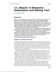
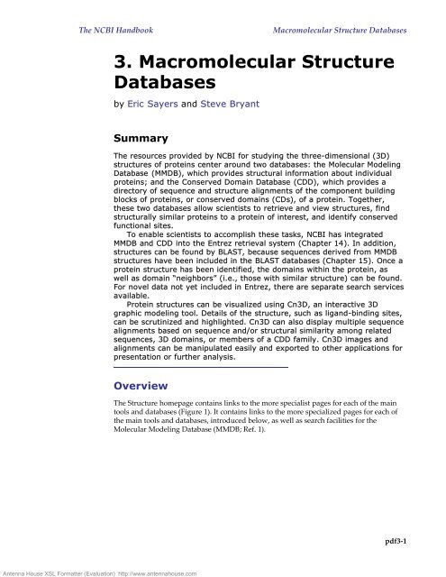
Antenna House <strong>XSL</strong> <strong>Formatter</strong> (Evaluation) http://www.antennahouse.comThe NCBI HandbookMacromolecular Structure Databases3. Macromolecular StructureDatabasesby Eric Sayers and Steve BryantSummaryThe resources provided by NCBI <strong>fo</strong>r studying the three-dimensional (3D)structures of proteins center around two databases: the Molecular ModelingDatabase (MMDB), which provides structural in<strong>fo</strong>rmation about individualproteins; and the Conserved Domain Database (CDD), which provides adirectory of sequence and structure alignments of the component buildingblocks of proteins, or conserved domains (CDs), of a protein. Together,these two databases allow scientists to retrieve and view structures, findstructurally similar proteins to a protein of interest, and identify conservedfunctional sites.To enable scientists to accomplish these tasks, NCBI has integratedMMDB and CDD into the Entrez retrieval system (Chapter 14). In addition,structures can be <strong>fo</strong>und by BLAST, because sequences derived from MMDBstructures have been included in the BLAST databases (Chapter 15). Once aprotein structure has been identified, the domains within the protein, aswell as domain “neighbors” (i.e., those with similar structure) can be <strong>fo</strong>und.For novel data not yet included in Entrez, there are separate search servicesavailable.Protein structures can be visualized using Cn3D, an interactive 3Dgraphic modeling tool. Details of the structure, such as ligand-binding sites,can be scrutinized and highlighted. Cn3D can also display multiple sequencealignments based on sequence and/or structural similarity among relatedsequences, 3D domains, or members of a CDD family. Cn3D images andalignments can be manipulated easily and exported to other applications <strong>fo</strong>rpresentation or further analysis.OverviewThe Structure homepage contains links to the more specialist pages <strong>fo</strong>r each of the maintools and databases (Figure 1). It contains links to the more specialized pages <strong>fo</strong>r each ofthe main tools and databases, introduced below, as well as search facilities <strong>fo</strong>r theMolecular Modeling Database (MMDB; Ref. 1).pdf3-1
Antenna House <strong>XSL</strong> <strong>Formatter</strong> (Evaluation) http://www.antennahouse.comThe NCBI HandbookMacromolecular Structure DatabasesFigure 1: The Structure homepage.This page can be <strong>fo</strong>und by selecting the Structure link on the tool bar atop many NCBI web pages. Two searchescan be per<strong>fo</strong>rmed from this page: (1) an EntrezStructure search, or (2) a Structure Summary search. Both querythe MMDB database. The difference is that the Entrez Structure can take any text as a query (such as a PDB code,protein name, text word, author, or journal) and will result initially in a list of one or more document summaries,displayed within the Entrez environment (Chapter 14), whereas only a PDB code or MMDB ID number can beused <strong>fo</strong>r the Structure Summary search, resulting in direct display of the Structure Summary page <strong>fo</strong>r that protein(Figure 2). Announcements about new features or updates can also be <strong>fo</strong>und on this page, as well as links tomore specialized pages on the various Structure databases and tools.MMDB is based on the structures within Protein Data Bank (PDB) and can be queriedusing the Entrez search engine, as well as via the more direct but less flexible StructureSummary search (see Figure 1). Once <strong>fo</strong>und, any structure of interest can be viewed usingCn3D (2), a piece of software that can be freely downloaded <strong>fo</strong>r Mac, PC, and UNIXplat<strong>fo</strong>rms.Often used in conjunction with Cn3D is the Vector Alignment Search Tool (VAST;Refs. 3, 4). VAST is used to precompute “structure neighbors” or structures similar toeach MMDB entry. For people that have a set of 3D coordinates <strong>fo</strong>r a protein not yet inMMDB, there is also a VAST search service. The output of the precomputed VASTsearches are displayed as one of the Non-Redundant PDB chain sets (nr-PDB), which canalso be downloaded. There are <strong>fo</strong>ur clustered subsets of MMDB that compose nr-PDB,each of which can be displayed as a list, showing one representative from each sequencesimilarcluster. The clusters can also be queried using a PDB code to retrieve the cluster inwhich it has been placed.pdf3-2
Antenna House <strong>XSL</strong> <strong>Formatter</strong> (Evaluation) http://www.antennahouse.comThe NCBI HandbookMacromolecular Structure DatabasesThe structures within MMDB are now being linked to the NCBI Taxonomy database(Chapter 4). Known as the PDBeast project, this ef<strong>fo</strong>rt makes it possible to find: (1) allMMDB structures from a particular organism; and (2) all structures within a node of thetaxonomy tree (such as lizards or Bacillus), which launches the Taxonomy Browsershowing the number of MMDB records in each node.The second database within the Structure resources is the Conserved DomainDatabase (CDD; Ref. 5), based largely on Pfam and SMART, collections of alignments thatrepresent functional domains conserved across evolution. CDD can be searched from theCDD page in several ways, including by a domain keyword search. Three tools have beendeveloped to assist in analysis of CDD: (1) the CD-Search, which uses a BLAST-basedalgorithm to search the position-specific scoring matrices (PSSM) of CDD alignments; (2)the CD-Browser, which provides a graphic display of domains of interest, along with thesequence alignment; and (3) the Conserved Domain Architecture Retrieval Tool(CDART), which searches <strong>fo</strong>r proteins with similar domain architectures.All the above databases and tools are discussed in more detail in other parts of thisChapter, including tips on how to make the best use of them.Content of the Molecular Modeling Database(MMDB)Sources of Primary DataTo build MMDB (1), 3D structure data are retrieved from the PDB database (6)administered by the Research Collaboratory <strong>fo</strong>r Structural Bioin<strong>fo</strong>rmatics (RCSB). In allcases, the structures in MMDB have been determined by experimental methods, primarilyX-ray crystallography and Nuclear Magnetic Resonance (NMR) spectroscopy. Theoreticalstructure models are omitted. The data in each record are then checked <strong>fo</strong>r agreementbetween the atomic coordinates and the primary sequence, and the sequence data arethen extracted from the coordinate set. The resulting agreement between sequence andstructure allows the record to be linked efficiently into searches and alignment displaysinvolving other NCBI databases.The data are converted into ASN.1 (7), which can be parsed easily and can also acceptnumerous annotations to the structure data. In contrast to a PDB record, a MMDB recordin ASN.1 contains all necessary bonding in<strong>fo</strong>rmation in addition to sequence in<strong>fo</strong>rmation,allowing consistent display of the 3D structure using Cn3D. The annotations provided inthe PDB record by the submitting authors are added, along with uni<strong>fo</strong>rmly definedsecondary structure and domain features. These features support structure-basedsimilarity searches using VAST. Finally, two coordinate subsets are added to the record:one containing only backbone atoms, and one representing a single-con<strong>fo</strong>rmer model incases where multiple con<strong>fo</strong>rmations or structures were present in the PDB record. Both ofthese additions further simplify viewing both an individual structure and its alignmentswith structure neighbors in Cn3D. When this process is complete, the record is assigned aunique Accession number, the MMDB-ID (Box 1), while also retaining the original <strong>fo</strong>urcharacterPDB code.Annotation of 3D DomainsAfter initial processing, 3D domains are automatically identified within each MMDBrecord. 3D domains are annotations on individual MMDB structures that define theboundaries of compact substructures contained within them. In this way, they are similarto secondary structure annotations that define the boundaries of helical or β-strandsubstructures. Because proteins are often similar at the level of domains, VAST compareseach 3D domain to every other one and to complete polypeptide chains. The results arestored in Entrez as a Related 3D Domain link.pdf3-3
Antenna House <strong>XSL</strong> <strong>Formatter</strong> (Evaluation) http://www.antennahouse.comThe NCBI HandbookMacromolecular Structure DatabasesTo identify 3D domains within a polypeptide chain, MMDB's domain parser searches<strong>fo</strong>r one or more breakpoints in the structure. These breakpoints fall between majorsecondary structure elements such that the ratio of intra- to interdomain contacts fallsabove a set threshold. The 3D domains identified in this way provide a means to bothincrease the sensitivity of structure neighbor calculations and also present 3Dsuperpositions based on compact domains as well as on complete polypeptide chains.They are not intended to represent domains identified by comparative sequence andstructure analysis, nor do they represent modules that recur in related proteins, althoughthere is often good agreement between domain boundaries identified by these methods.Links to Other NCBI ResourcesAfter initially processing the PDB record, structure staff add a number of links and otherin<strong>fo</strong>rmation that further integrate the MMDB record with other NCBI resources. To begin,the sequence in<strong>fo</strong>rmation extracted from the PDB record is entered into the Entrez Proteinand/or Nucleotide databases as appropriate, providing a means to retrieve the structurein<strong>fo</strong>rmation from sequence searches. As with all sequences in Entrez, precomputedBLAST searches are then per<strong>fo</strong>rmed on these sequences, linking them to other moleculesof similar sequence. For proteins, these BLAST neighbors may be different than thosedetermined by VAST; whereas VAST uses a conservative significance threshold, thestructural similarities it detects often represent remote relationships not detectable bysequence comparison. The literature citations in the PDB record are linked to PubMed sothat Entrez searches can allow access to the original descriptions of the structuredeterminations. Finally, semiautomatic processing of the “source” field of the PDB recordprovides links to the NCBI Taxonomy database. Although these links normally <strong>fo</strong>llow thegenus and species in<strong>fo</strong>rmation given, in some cases this in<strong>fo</strong>rmation is either absent in thePDB record or refers only to how a sample was obtained. In these cases, the staffmanually enters the appropriate taxonomy links.The MMDB RecordThe Structure Summary page <strong>fo</strong>r each MMDB record summarizes the database content<strong>fo</strong>r that record and serves as a starting point <strong>fo</strong>r analyzing the record using the NCBIstructure tools (Figure 2).pdf3-4
Antenna House <strong>XSL</strong> <strong>Formatter</strong> (Evaluation) http://www.antennahouse.comThe NCBI HandbookMacromolecular Structure DatabasesFigure 2: The Structure Summary page.The page consists of three parts: the header, the view bar, and the graphic display. The header contains basicidentifying in<strong>fo</strong>rmation about the record: a description of the protein (Description:), the author list (Deposition:,the species of origin (Taxonomy:, literature references (References:, the MMDB-ID (MMDB:), and the PDB code(PDB:). Several of these data serve as links to additional in<strong>fo</strong>rmation. For example, the species name links to theTaxonomy browser, the literature references link to PubMed, and the PDB code links to the PDB website. Theview bar allows the user to view the structure record either as a graphic with Cn3D or as a text record in eitherASN.1, PDB (RasMol), or Mage <strong>fo</strong>rmats. The latter can also be downloaded directly from this page. The graphicdisplay contains a variety of in<strong>fo</strong>rmation and links to related databases: (a) The Chain bar. Each chain of themolecule is displayed as a dark bar labeled with residue numbers. To the left of this bar is a Protein hyperlinkthat takes the user to a view of the protein record in Entrez Protein. The bar itself is also a hyperlink and displaysthe VAST neighbors of the chain. If a structure contains nucleotide sequences, they are displayed in the ordercontained in the PDB record. A Nucleotide hyperlink to their left takes the user to the appropriate record in EntrezNucleotide. (b) The VAST (3D) Domain bar. The colored bars immediately below the chain bar indicate thelocations of structural domains <strong>fo</strong>und by the original MMDB processing of the protein. In many cases, such adomain contains unconnected sections of the protein sequence, and in such cases, discontinuous pieces makingup the domain will have bars of the same color. To the left of the Domain bar is a 3D Domain hyperlink (3dDomains) that launches the 3D Domain browser in Entrez, where the user can find in<strong>fo</strong>rmation about eachconstituent domain. Selecting a colored segment displays the VAST Structure neighbors page <strong>fo</strong>r that domain.(c) The CD bar. Below the VAST Domain bar are rounded, rectangular bars representing conserved domains<strong>fo</strong>und by a CD-Search. The bars identify the best scoring hits; overlapping hits are shown only if the mutualoverlap with hits having better scores is less than 50%. The CDs hyperlink to the left of the bar displays the CD-Search page, showing the detailed alignment of the protein with the consensus sequence of the CD. Each of thecolored bars is also a hyperlink that displays the corresponding CD Summary page configured to show themultiple alignment of the protein sequence with members of the selected CD.pdf3-5
Antenna House <strong>XSL</strong> <strong>Formatter</strong> (Evaluation) http://www.antennahouse.comThe NCBI HandbookMacromolecular Structure DatabasesVAST Structure NeighborsAlthough VAST itself is not a database, the VAST results computed <strong>fo</strong>r each MMDBrecord are stored with this record and are summarized on a separate page <strong>fo</strong>r each 3Ddomain <strong>fo</strong>und in the protein (Figure 3). These pages can be accessed most easily byclicking on the 3D Domain bar in the graphic display of the Structure Summary page(Figure 2). Note that complete polypeptide chains are always included in the lists of 3DDomains as well.Figure 3: VAST Structure Neighbors page.The top portion of the page contains identifying in<strong>fo</strong>rmation about the 3D Domain, along with three functionalbars. (a) The View bar. This bar allows a user to view a selected alignment either as a graphic using Cn3D or asa sequence alignment in HTML, text, or mFASTA <strong>fo</strong>rmat. The user may select which chains to display in thealignment by checking the boxes that appear to the left of each neighbor in the lower portion of the page. (b)The nr-PDB bar. This bar allows a user to either display all matching records in MMDB or to limit the displayeddomains to only those within one of the <strong>fo</strong>ur nr-PDB sets. The user may also select how the matching domainsare sorted in the display and whether the results are shown as graphics or as tabulated data. (c) The Find bar.This bar allows the user to find specific structural neighbors by entering their PDB or MMDB identifiers as acomma-delimited list. (d) The lower portion of the page displays a graphical alignment of the various matchingdomains. The upper three bars show summary in<strong>fo</strong>rmation about the query sequence: the top bar shows themaximum extent of alignment <strong>fo</strong>und on all the sequences displayed on the current page (users should note thatthe appearance of this bar, there<strong>fo</strong>re, depends on which hits are displayed); the middle bar represents the querysequence itself that served as input <strong>fo</strong>r the VAST search; and the lower bar shows any matching CDs and isidentical to the CD Bar on the Structure Summary page. Listed below these three summary bars are the hitsfrom the VAST search, sorted according to the selection in the nr-PDB Bar. Aligned regions are shown in red,pdf3-6
Antenna House <strong>XSL</strong> <strong>Formatter</strong> (Evaluation) http://www.antennahouse.comThe NCBI HandbookMacromolecular Structure Databaseswith gaps indicating unaligned regions. To the left of each domain accession is a checkbox that can be used toselect any combination of domains to be displayed either on this page or using Cn3D. Moreover, each of the barsin the display is itself a link, and placing the mouse pointer over any bar reveals both the extent of thealignment by residue number and the data linked to the bar.nr-PDBThe non-redundant PDB database (nr-PDB) is a collection of <strong>fo</strong>ur sets of sequencedissimilarPDB polypeptide chains assembled by NCBI Structure staff. The <strong>fo</strong>ur sets differonly in their respective levels of non-redundancy, as explained below. The staff assembleseach set by comparing all the chains available from PDB with each other using the BLASTalgorithm. The chains are then clustered into groups of similar sequence using a singlelinkageclustering procedure. Chains within a sequence-similar group are automaticallyranked according to the quality of their structural data. Details of the measures used todetermine structure precision and completeness and the methodology of assembling thenr-PDB clusters can be <strong>fo</strong>und on the nr-PDB webpage.Content of the Conserved Domain Database (CDD)What Is a Conserved Domain (CD)?CDs are recurring units in polypeptide chains (sequence and structure motifs), the extentsof which can be determined by comparative analysis. Molecular evolution uses suchdomains as building blocks and may be recombined in different arrangements to makedifferent proteins with different functions. The CDD contains sequence alignments thatdefine the features that are conserved within each domain family. There<strong>fo</strong>re, the CDDserves as a classification resource that groups proteins based on the presence of thesepredefined domains. CDD entries often name the domain family and describe the role ofconserved residues in binding or catalysis. Conserved domains are displayed in MMDBStructure summaries and link to a sequence alignment showing other proteins in whichthe domain is conserved, which may provide clues on protein function.Sources of Primary DataThe collections of domain alignments in the CDD are imported either from two databasesoutside of the NCBI, named Pfam (8) and SMART (9); from a collection compiled withinthe NCBI, named LOAD; or from a database curated by the CDD staff. The first task is toidentify the underlying sequences in each collection and then link these sequences to thecorresponding ones in Entrez. If the CDD staff cannot find the Accession numbers <strong>fo</strong>r thesequences in the records from the source databases, they locate appropriate sequencesusing BLAST. Particular attention is paid to any resulting match that is linked to astructure record in MMDB, and the staff substitute alignment rows with such sequenceswhenever possible. After the staff imports a collection, they then choose a sequence thatbest represents the family. Whenever possible, the staff chooses a representative that has astructure record in MMDB.The Position-specific Score Matrix (PSSM)Once imported and constructed, each domain alignment in CDD is used to calculate amodel sequence, called a consensus sequence, <strong>fo</strong>r each CD. The consensus sequence liststhe most frequently <strong>fo</strong>und residue in each position in the alignment; however, <strong>fo</strong>r asequence position to be included in the consensus sequence, it must be present in at least50% of the aligned sequences. Aligned columns covered by the consensus sequence arethen used to calculate a PSSM, which memorizes the degree to which particular residuespdf3-7
Antenna House <strong>XSL</strong> <strong>Formatter</strong> (Evaluation) http://www.antennahouse.comThe NCBI HandbookMacromolecular Structure Databasesare conserved at each position in the sequence. Once calculated, the PSSM is stored withthe alignment and becomes part of the CDD. The RPS-BLAST tool locates CDs within aquery sequence by searching against this database of PSSMs.Reverse Position-specific BLAST (RPS-BLAST)RPS-BLAST (Chapter 15) is a variant of the popular Position-specific Iterated BLAST (PSI-BLAST) program. PSI-BLAST finds sequences similar to the query and uses the resultingalignments to build a PSSM <strong>fo</strong>r the query. With this PSSM the database is scanned againto draw in more hits and further refine the scoring model. RPS-BLAST uses a querysequence to search a database of precalculated PSSMs and report significant hits in asingle pass. The role of the PSSM has changed from “query” to “subject”; hence, the term“reverse” in RPS-BLAST. RPS-BLAST is the search tool used in the CD-Search service.The CD SummaryAnalogous to the Structure Summary page, the CD Summary page displays the availablein<strong>fo</strong>rmation about a given CD and offers various links <strong>fo</strong>r either viewing the CDalignment or initiating further searches (Figure 4). The CD Summary page can beretrieved by selecting the CD name on any page.Figure 4: CD Summary page.The top of the page serves as a header and reports a variety of identifying in<strong>fo</strong>rmation, including the name anddescription of the CD, other related CDs with links to their summary pages, as well as the source database,status, and creation date of the CD. A taxonomic node link (Taxa:)launches the Taxonomy Browser, whereas aProteins (Proteins:)link uses CDART to show other proteins that contain the CD. Below the header is theinterface <strong>fo</strong>r viewing the CD alignment, which can be done either graphically with Cn3D (if the CD contains asequence with structural data) or in HTML, text, or mFASTA <strong>fo</strong>rmat. It is also possible to view a selected numberpdf3-8
Antenna House <strong>XSL</strong> <strong>Formatter</strong> (Evaluation) http://www.antennahouse.comThe NCBI HandbookMacromolecular Structure Databasesof the top-listed sequences, sequences from the most diverse members, or sequences most similar to the query.The lower portion of the page contains the alignment itself. Members with a structural record in MMDB are listedfirst, and the identifier of each sequence links to the corresponding record.The Distinction between 3D Domains and CDsThe term “domain” refers in general to a distinct functional and/or structural unit of aprotein. Each polypeptide chain in MMDB is analyzed <strong>fo</strong>r the presence of two classes ofdomains, and it is important <strong>fo</strong>r users to understand the difference between them. Oneclass, called 3D Domains, is based solely on similar, compact substructures, whereas thesecond class, called Conserved Domains (CDs), is based solely on conserved sequencemotifs. These two classifications often agree, because the compact substructures within aprotein often correspond to domains joined by recombination in the evolutionary historyof a protein. Note that CD links can be identified even when no 3D structures within afamily are known. Moreover, 3D Domain links may also indicate relationships either tostructures not included in CDD entries or to structures so distantly related that nosignificant similarity can be <strong>fo</strong>und by sequence comparisons.Finding and Viewing StructuresFor an example query on finding and viewing structures, see Box 2.Why Would I Want to Do This?• To determine the overall shape and size of a protein• To locate a residue of interest in the overall structure• To locate residues in close proximity to a residue of interest• To develop or test chemical hypotheses regarding an enzyme mechanism• To locate or predict possible binding sites of a ligand• To interpret mutation studies• To find areas of positive or negative charge on the protein surface• To locate particularly hydrophobic or hydrophilic regions of a protein• To infer the 3D structure and related properties of a protein with unknownstructure from the structure of a homologous protein• To study evolutionary processes at the level of molecular structure• To study the function of a protein• To study the molecular basis of disease and design novel treatmentsHow to BeginThe first step to any structural analysis at NCBI is to find the structure records <strong>fo</strong>r theprotein of interest or <strong>fo</strong>r proteins similar to it. One may search MMDB directly byentering search terms such as PDB code, protein name, author, or journal in the EntrezStructure Search box on the Structure homepage. Alternative points of entry are shownbelow.pdf3-9
Antenna House <strong>XSL</strong> <strong>Formatter</strong> (Evaluation) http://www.antennahouse.comThe NCBI HandbookMacromolecular Structure DatabasesBy using the full array of Entrez search tools, the resulting list of MMDB records canbe honed, ideally, to a workable list from which a record can be selected. Users shouldnote that multiple records may exist <strong>fo</strong>r a given protein, reflecting different experimentaltechniques, conditions, and the presence or absence of various ligands or metal ions.Records may also contain different fragments of the full-length molecule. In addition,many structures of mutant proteins are also available. The PDB record <strong>fo</strong>r a givenstructure generally contains some description of the experimental conditions under whichthe structure was determined, and this file can be accessed by selecting the PDB code linkat the top of the Structure Summary page.Alternative Points of EntryStructure Summary pages can also be <strong>fo</strong>und from the <strong>fo</strong>llowing NCBI databases and tools:• Select the Structure links to the right of any Entrez record <strong>fo</strong>und; records withStructure links can also be located by choosing Structure links from the Displaypull-down menu.• Select the Related Sequences link to the right of an Entrez record to find proteinsrelated by sequence similarity and then select Structure links in the Display pulldownmenu.• Choose the PDB database from a blastp (protein-protein BLAST) search; onlysequences with structure records will be retrieved by BLAST. The RelatedStructures link provides 3D views in Cn3D.• Select the 3D Structures button on any BLink report to show those BLAST hits <strong>fo</strong>rwhich structural data are available.Viewing 3D Structures3D DomainsThe 3D domains of a protein are displayed on the Structure Summary page. It is useful toknow how many 3D domains a protein contains and whether they are continuous insequence when viewing the full 3D structure of the molecule.Secondary StructureKnowing the secondary structure of a protein can also be a useful prelude to viewing the3D structure of the molecule. The secondary structure can be viewed easily by firstselecting the Protein link to the left of the desired chain in the graphic display. Findingoneself in Entrez Protein, selecting Graphics in the Display pull-down menu presentssecondary structure diagrams <strong>fo</strong>r the molecule.Full Protein StructuresCn3D is a software package <strong>fo</strong>r displaying 3D structures of proteins. Once it has beeninstalled and the Internet browser has been configured correctly, simply selecting theView 3D Structure button on a Structure Summary page launches the application. Oncethe structure is loaded, a user can manipulate and annotate it using an array of options asdescribed in the Cn3D Tutorial. By default, Cn3D colors the structure according to thesecondary structure elements. However, another useful view is to color the protein bydomain (see Style menu options), using the same color scheme as is shown in the graphicdisplay on the Structure Summary page. These color changes also affect the residuesdisplayed in the Sequence/Alignment Viewer, allowing the identification of domain orsecondary structure elements in the primary sequence. In addition to Cn3D, users canalso display 3D structures with RasMol or Mage. Structures can also be saved locally asan ASN.1, PDB, or Mage file (depending on the choice of structure viewer) <strong>fo</strong>r laterdisplay.pdf3-10
Antenna House <strong>XSL</strong> <strong>Formatter</strong> (Evaluation) http://www.antennahouse.comThe NCBI HandbookMacromolecular Structure DatabasesFinding and Viewing Structure “Neighbors”For an example query on finding and viewing structure “neighbors”, see Box 2.Why Would I Want to Do This?• To determine structurally conserved regions in a protein family• To locate the structural equivalent of a residue of interest in another relatedprotein• To gain insights into the allowable structural variability in a particular proteinfamily• To develop or test chemical hypotheses regarding an enzyme mechanism• To predict possible binding sites of a ligand from the location of a binding site ina related protein• To identify sites where con<strong>fo</strong>rmational changes are concentrated• To interpret mutation studies• To find areas of conserved positive or negative charge on the protein surface• To locate conserved hydrophobic or hydrophilic regions of a protein• To identify evolutionary relationships across protein families• To identify functionally equivalent proteins with little or no sequenceconservationHow to BeginThe Vector Alignment Search Tool (VAST) is used to calculate similar structures on eachprotein contained in the MMDB. The graphic display on each Structure Summary page(Figure 2) links directly to the relevant VAST results <strong>fo</strong>r both whole proteins and 3Ddomains:• The 3D Domains link transfers the user to Entrez 3D Domains, showing a list ofthe VAST neighbors.• Selecting the chain bar displays the VAST Structure Neighbors page <strong>fo</strong>r the entirechain.• Selecting a 3D Domain bar displays the VAST Structure Neighbors page <strong>fo</strong>r theselected domain.Alternative Point of Entry• From any Entrez search, select Related 3D Domains to the right of any record<strong>fo</strong>und to view the Vast Structure Neighbors page.Viewing a 2D Alignment of Structure NeighborsA graphic 2D HTML alignment of VAST neighbors can be viewed as <strong>fo</strong>llows:pdf3-11
Antenna House <strong>XSL</strong> <strong>Formatter</strong> (Evaluation) http://www.antennahouse.comThe NCBI HandbookMacromolecular Structure Databases• On the lower portion of the VAST Structure Neighbors page (Figure 3), select thedesired neighbors to view by checking the boxes to their left.• On the View/Save bar, configure the pull-down menus to the right of the ViewAlignment button.• Select View Alignment.Viewing a 3D Alignment of Structure NeighborsAlignments of VAST structure neighbors can be viewed as a 3D image using Cn3D.• On the lower portion of the VAST Structure Neighbors page (Figure 3), select thedesired neighbors to view by checking the boxes to their left.• On the View/Save bar, configure the pull-down menus to the right of the View 3DStructure button.• Select View 3D Structure.Cn3D automatically launches and displays the aligned structures. Each displayedchain has a unique color; however, the portions of the structures involved in thealignment are shown in red. These same colors are also reflected in the Sequence/Alignment Viewer. Among the many viewing options provided by Cn3D, of particularuse is the Show/Hide menu that allows only the aligned residues to be viewed, only thealigned domains, or all residues of each chain.Finding and Viewing Conserved DomainsFor an example query on finding and viewing conserved domains, see Box 3.Why Would I Want to Do This?• To locate functional domains within a protein• To predict the function of a protein whose function is unknown• To establish evolutionary relationships across protein families• To interpret mutation studies• To predict the structure of a protein of unknown structureHow to BeginFollowing the Domains link <strong>fo</strong>r any protein in Entrez, one can find the conserveddomains within that protein. The CD-Search (or Protein BLAST, with CD-Search optionselected) can be used to find conserved domains (CDs) within a protein. Either theAccession number, gi number, or the FASTA sequence can be used as a query.Alternative Points of EntryIn<strong>fo</strong>rmation on the CDs contained within a protein can also be <strong>fo</strong>und from thesedatabases and tools:• From any Entrez search: select the Domains link to the right of a displayed record.pdf3-12
Antenna House <strong>XSL</strong> <strong>Formatter</strong> (Evaluation) http://www.antennahouse.comThe NCBI HandbookMacromolecular Structure Databases• From the Structure Summary page of a MMDB record: this page displays the CDswithin each protein chain immediately below the 3D Domain bar in the graphicdisplay. Selecting the CDs link shows the CD-Search results page.• From an Entrez Domains search: choose Domains from the Entrez Search pulldownmenu and enter a search term to retrieve a list of CDs. Clicking on anyresulting CD displays the CD Summary page. To find the location of this CD inan aligned protein, select the CD link <strong>fo</strong>llowing a protein name in the bottomportion of this page.• From the CDD page: locate CDs by entering text terms into the search box andproceed as <strong>fo</strong>r an Entrez CD search.• From a BLink report: select the CDD-Search button to display the CD-Searchresults page.• From the BLAST main page: <strong>fo</strong>llow the RPS-BLAST link to load the CD-Searchpage.Viewing Conserved DomainsResults from a CD search are displayed as colored bars underneath a sequence ruler.Moving the mouse over these bars reveals the identity of each domain; domains are alsolisted in a <strong>fo</strong>rmat similar to BLAST summary output (Chapter 15). Pairwise alignmentsbetween the matched region of the target protein and the representative sequence of eachdomain are shown below the bar. Red letters indicate residues identical to those in therepresentative sequence, whereas blue letters indicate residues with a positiveBLOSUM62 score in the BLAST alignment.Viewing Multiple Alignments of a Query Protein withMembers of a Conserved DomainThese can be displayed by clicking a CD bar within a MMDB Structure Summary page orfrom a hyperlinked CD name on a CD-Search results page.Viewing CD Alignments in the Context of 3D StructureIf members of a CD have MMDB records, one of these records can be viewed as a 3Dimage along with the sequence alignment using Cn3D (launched by selecting the pink doton a CD-Search results page). As in other alignment views, colored capital letters indicatealigned residues, allowing the sequence of the protein sequence of interest to be mappedonto the available 3D structure.Finding and Viewing Proteins with Similar DomainArchitecturesFor an example query on finding and viewing proteins with similar domain architectures,see Box 3.Why Would I Want to Do This?• To locate related functional domains in other protein families• To gain insights into how a given CD is situated within a protein relative toother CDspdf3-13
Antenna House <strong>XSL</strong> <strong>Formatter</strong> (Evaluation) http://www.antennahouse.comThe NCBI HandbookMacromolecular Structure Databases• To explore functional links between different CDs• To predict the function of a protein whose function is unknown• To establish evolutionary relationships across protein familiesHow to BeginFollowing the Domain Relatives link <strong>fo</strong>r any protein in Entrez, one can find other proteinswith similar domain architecture. The Conserved Domain Architecture Retrieval Tool(CDART) can take an Accession number or the FASTA sequence as a query to find out thedomain architecture of a protein sequence and list other proteins with related domainarchitectures.Alternative Point of Entryselecting the Show domain relatives button on a CD-Search results page also launches aCDART search, as does seelcting the Proteins link either on a CD Summary page or on arecord produced by an Entrez Domains search.Results of a CDART SearchThese are described in Figure 5. The protein “hits”, which have similar domainarchitectures to the query sequence, can be further refined by taxonomic group, in whichthe results can be limited to selected nodes of the taxonomic tree. Furthermore, searchresults may be limited to those that contain only particular conserved domains.Figure 5: A CDART results page.At the top of the CDART results page in a yellow box, the query sequence CDs are represented as “beads on astring”. Each CD had a unique color and shape and is labeled both in the display itself and in a legend located atthe bottom of the page. The shapes representing CDs are hyperlinked to the corresponding CD Summary page.The matching proteins to the query are listed below the yellow box, ranked according to the number of nonpdf3-14
Antenna House <strong>XSL</strong> <strong>Formatter</strong> (Evaluation) http://www.antennahouse.comThe NCBI HandbookMacromolecular Structure Databasesredundant hits to the domains in the query sequence. Each match is either a single protein, in which case itsAccession number is shown, or is a cluster of very similar proteins, in which case the number of members in thecluster is shown. Cluster members can be displayed by selecting the logo to the left of its diagram. Selecting anyprotein Accession number displays the flatfile <strong>fo</strong>r that protein. To the right of any drawing <strong>fo</strong>r a single protein(either in the main results page or after expanding a protein cluster) is a more> link, which displays the CD-Search results page <strong>fo</strong>r the selected protein so that the sequence alignment, e.g., of a CDART hit with a CDcontained in the original protein of interest, can be examined.Links Between Structure and Other ResourcesIntegration with Other NCBI ResourcesAs illustrated in the sections above, there are numerous connections between theStructure resources and other databases and tools available at the NCBI. What <strong>fo</strong>llows is alisting of major tools that support connections.EntrezBecause Entrez is an integrated database system (Chapter 14), the links attached to eachstructure give immediate access to PubMed, Protein, Nucleotide, 3D Domain, orTaxonomy records.BLASTAlthough the BLAST service is designed to find matches based solely on sequence, thesequences of Structure records are included in the BLAST databases, and by selecting thePDB search database, BLAST searches only the protein sequences provided by MMDBrecords. A new Related Structure link provides 3D views <strong>fo</strong>r sequences with structuredata identified in a BLAST search.BLinkThe BLink report represents a precomputed list of similar proteins <strong>fo</strong>r many proteins (see,<strong>fo</strong>r example, links from LocusLink records; Chapter 18). The 3D Structures option on anyBLink report shows the BLAST hits that have 3D structure data in MMDB, whereas theCDD-Search button displays the CD-Search results page <strong>fo</strong>r the query protein.Microbial GenomesA particularly useful interface with the structural databases is provided on the MicrobialGenomes page (10). To the left of the list of genomes are several hyperlinks, two of whichoffer users direct access to structural in<strong>fo</strong>rmation. The red [D] link displays a listing ofevery protein in the genome, each with a link to a BLink page showing the results of aBLAST pdb search <strong>fo</strong>r that protein. The [S] link displays a similar protein list <strong>fo</strong>r theselected genome, but now with a listing of the conserved domains <strong>fo</strong>und in each proteinby a CD-Search.Links to Non-NCBI ResourcesThe Protein Data Bank (PDB)As stated elsewhere, all records in the MMDB are obtained originally from the ProteinData Bank (PDB) (6). Links to the original PDB records are located on the StructureSummary page of each MMDB record. Updates of the MMDB with new PDB recordsoccur once a month.Pfam and SMARTThe CDD staff imports CD collections from both the Pfam and SMART databases. Linksto the original records in these databases are located on the appropriate CD Summarypage. Both Pfam and SMART are updated several times per year in roughly bimonthlyintervals, and the CDD staff update CDD accordingly.pdf3-15
Antenna House <strong>XSL</strong> <strong>Formatter</strong> (Evaluation) http://www.antennahouse.comThe NCBI HandbookMacromolecular Structure DatabasesSaving Output from Database SearchesExporting Graphics Files from Cn3DStructures displayed in Cn3D can be exported as a Portable Network Graphics (PNG) filefrom within Cn3D (the Export PNG command in the File menu). The structure file itself,in the orientation currently being viewed, can also be saved <strong>fo</strong>r later launching in Cn3D.Saving Individual MMDB RecordsIndividual MMDB records can be saved/downloaded to a local computer directly fromthe Structure Summary page <strong>fo</strong>r that record. Save File in the View bar downloads the filein a choice of three <strong>fo</strong>rmats: ASN.1 (select Cn3D); PDB (select RasMol); or Mage (selectMage).Saving VAST AlignmentsAlignments of VAST neighbors can be saved/downloaded from the VAST StructureNeighbors page of any MMDB record. By selecting options in the View Alignment pulldownmenu, the alignment data can be saved, <strong>fo</strong>rmatted as HTML, text, ASN.1,ormFASTA.FTPUsers can download the NCBI Structure databases from the NCBI FTP site: ftp://ftp.ncbi.nih.gov/mmdb. A Readme file contains descriptions of the contents and in<strong>fo</strong>rmationabout recent updates. Within the mmdb directory are <strong>fo</strong>ur subdirectories that contain the<strong>fo</strong>llowing data:• mmdbdata: the current MMDB database• vastdata: the current set of VAST neighbor annotations to MMDB records• nrtable: the current non-redundant PDB database• pdbeast: table listing the taxonomic classification of MMDB recordsFrequently Asked Questions• Cn3D [http://www.ncbi.nih.gov/Structure/CN3D/cn3dfaq.shtml]• VAST searches [http://www.ncbi.nih.gov/Structure/VAST/vastsearch_faq.html]• CDD [http://www.ncbi.nlm.nih.gov/Structure/cdd/cdd_help.shtml]References1. Wang Y, Anderson JB, Chen J, Geer LY, He S, Hurwitz DI, Liebert CA, Madej T,Marchler GH, Marchler-Bauer A, et al. MMDB: Entrez's 3D-structure database. NucleicAcids Res 30:249–252; 2002.2. Wang Y, Geer LY, Chappey C, Kans JA, Bryant SH. Cn3D: sequence and structureviews <strong>fo</strong>r Entrez. Trends Biochem Sci 25:300–302; 2000.pdf3-16
Antenna House <strong>XSL</strong> <strong>Formatter</strong> (Evaluation) http://www.antennahouse.comThe NCBI HandbookMacromolecular Structure Databases3. Madej T, Gibrat J-F, Bryant SH. Threading a database of protein cores. Proteins 23:356–369; 1995.4. Gibrat J-F, Madej T, Bryant SH. Surprising similarities in structure comparison. CurrOpin Struct Biol 6:377–385; 1996.5. Marchler-Bauer A, Panchenko AR, Shoemaker BA, Thiessen PA, Geer LY, Bryant SH.CDD: a database of conserved domain alignments with links to domain threedimensionalstructure. Nucleic Acids Res 30:281–283; 2002.6. Westbrook J, Feng Z, Jain S, Bhat TN, Thanki N, Ravichandran V, Gilliland GL, BluhmW, Weissig H, Greer DS, et al. The Protein Data Bank: unifying the archive. NucleicAcids Res 30:245–248; 2002.7. Ohkawa H, Ostell J, Bryant S. MMDB: an ASN.1 specification <strong>fo</strong>r macromolecularstructure. Proc Int Conf Intell Syst Mol Biol 3:259–267; 1995.8. Bateman A, Birney E, Cerruti L, Durbin R, Etwiller L, Eddy SR, Griffiths-Jones S, HoweKL, Marshall M, Sonnhammer ELL. The Pfam proteins family database. Nucleic AcidsRes 30:276–280; 2002.9. Letunic I, Goodstadt L, Dickens NJ, Doerks T, Schultz J, Mott R, Ciccarelli F, Copley RR,Ponting CP, Bork P. SMART: a web-based tool <strong>fo</strong>r the study of genetically mobiledomains. Recent improvements to the SMART domain-based sequence annotationresource. Nucleic Acids Res 30:242–244; 2002.10. Wang Y, Bryant S, Tatusov R, Tatusova T. Links from genome proteins to known 3Dstructures. Genome Res 10:1643–1647; 2000.pdf3-17
Antenna House <strong>XSL</strong> <strong>Formatter</strong> (Evaluation) http://www.antennahouse.comThe NCBI HandbookMacromolecular Structure DatabasesBox 1: Accession numbers.MMDB records have several types of Accession numbers associated with them, representing the <strong>fo</strong>llowing datatypes:• Each MMDB record has at least three Accession numbers: the PDB code of the corresponding PDB record(e.g., 1CYO, 1B8G); a unique MMDB-ID (e.g., 645, 12342); and a gi number <strong>fo</strong>r each protein chain. A newMMDB-ID is assigned whenever PDB updates either the sequence or coordinates of a structure record,even if the PDB code is retained.• If an MMDB record contains more than one polypeptide or nucleotide chain, each chain in the MMDBrecord is assigned an Accession number in Entrez Protein or Nucleotide consisting of the PDB code<strong>fo</strong>llowed by the letter designating that chain (e.g., 1B8GA, 3TATB, 1MUHB).• Each 3D Domain identified in an MMDB record is assigned a unique integer identifier that is appended tothe Accession number of the chain to which it belongs (e.g., 1B8G A 2). This new Accession numberbecomes its identifier in Entrez 3D Domains. New 3D Domain identifiers are assigned whenever a newMMDB-ID is assigned.• For conserved domains, the Accession number is based on the source database:Pfam:SMART:LOAD:CD: cd00101pfam00049smart00078LOAD Toprim(curated by CDD staff)pdf3-18
Antenna House <strong>XSL</strong> <strong>Formatter</strong> (Evaluation) http://www.antennahouse.comThe NCBI HandbookMacromolecular Structure DatabasesBox 2: Example query: finding and viewingstructural data of a protein.Finding the Structure of a ProteinSuppose that we are interested in the biosynthesis of aminocyclopropanes and would like to find structuralin<strong>fo</strong>rmation on important active site residues in any available aminocyclopropane synthases. To begin, wewould go to the Structure main page and enter “aminocyclopropane synthase” in the Search box. Pressing Enterdisplays a short list of structures, one of which is 1B8G, 1-aminocyclopropane-1-carboxylate synthase. Perhapswe would like to know the species from which this protein was derived. Selecting the Taxonomy link to the rightshows that this protein was derived from Malux x domestica, or the common apple tree. Going back to the Entrezresults page and selecting the PDB code (1B8G) opens the Structure Summary page <strong>fo</strong>r this record. The species isagain displayed on this page, along with a link to the Journal of Molecular Biology article describing how thestructure was determined. We immediately see from this page that this protein appears as a dimer in thestructure, with each chain having three 3D domains, as identified by VAST. In addition, CD-Search hasidentified an “aminotran_1_2” CD in each chain. Now we are ready to view the 3D structure.Viewing the 3D StructureOnce we have <strong>fo</strong>und the Structure Summary page, viewing the 3D structure is straight<strong>fo</strong>rward. To view thestructure in Cn3D, we simply select the View 3D Structure button. The default view is to show helices in green,strands in brown, and loops in blue. This color scheme is also reflected in the Sequence/Alignment Viewer.Locating an Active SiteUpon inspecting the structure, we immediately notice that a small molecule is bound to the protein, likely at theactive site of the enzyme. How do we find out what that molecule is? One easy way is to return to the StructureSummary page and select the link to the PDB code, which takes us to the PDB Structure Explorer page <strong>fo</strong>r 1B8G.Quickly, we see that pyridoxal-5′-phosphate (PLP) is a HET group, or heterogen, in the structure. Our interestpiqued, we would now like to know more about the structural domain containing the active site. Returning toCn3D, we manipulate the structure so that PLP is easily visible and then use the mouse to double-click on anyPLP atom. The molecule becomes selected and turns yellow. Now from the Show/Hide menu, we choose Selectby distance and Residues only and enter 5 Angstroms <strong>fo</strong>r a search radius. Scanning the Sequence/AlignmentViewer, we see that seven residues are now highlighted: 117-119, 230, 268, 270, and 279. Glancing at the 3DDomain display in the Structure Summary page, we note that all of these residues lie in domain 3. We now <strong>fo</strong>cusour attention on this domain.Viewing Structure Neighbors of a 3D DomainGiven that this enzyme is a dimer, we arbitrarily choose domain 3 in chain A, the accession of which is thus1B8GA3. By clicking on the 3D Domain bar at a point within domain 3, we are taken to the VAST StructureNeighbors page <strong>fo</strong>r this domain, where we find nearly 200 structure neighbors.Restricting the Search by TaxonomyPerhaps we would now like to identify some of the most evolutionarily distant structure neighbors of domain1B8GA3 as a means of finding conserved residues that may be associated with its binding and/or catalyticfunction. One powerful way of doing this is to choose structure neighbors from phylogenetically distantorganisms. We there<strong>fo</strong>re need to combine our present search with a Taxonomy search. Given that 1B8G isderived from the superkingdom Eukaryota, we would like to find structure neighbors in other superkingdomtaxa, such as Eubacteria and Archaea. Returning to the Structure Summary page, select the 3D Domains link inthe graphic display to open the list of 3D Domains in Entrez. Finding 1B8GA3 in the list, selecting the Related3D Domains link shows a list of all the structure neighbors of this domain. From this page, we select Preview/Index, which shows our recent queries. Suppose our set of related 3D Domains is #5. We then per<strong>fo</strong>rm twosearches:1. #5 AND “Archaea”[Organism]2. #5 AND “Eubacteria”[Organism]pdf3-19
Antenna House <strong>XSL</strong> <strong>Formatter</strong> (Evaluation) http://www.antennahouse.comThe NCBI HandbookMacromolecular Structure DatabasesLooking at the Archaea results, we find among them 1DJUA3, a domain from an aromatic aminotransferasefrom Pyrococcus horikoshii. Concerning the Eubacteria results, we find among the several hundred matchingdomains 3TATA2, a tyrosine aminotransferase from Escherichia coli.Viewing a 3D Superposition of Active SitesReturning to the VAST Structure Neighbors page <strong>fo</strong>r 1B8GA3, we want to select 1DJUA3 and 3TATA2 to displayin a structural alignment. One way to do this is to enter these two Accession numbers in the Find box and pressFind. We now see only these two neighbors, and we can select View 3D Structure to launch Cn3D.Cn3D again displays the aligned residues in red, and we can highlight these further by selecting Show alignedresidues from the Show/Hide menu. The excellent agreement between both the active site structures and thecon<strong>fo</strong>rmations of the bound ligands is readily apparent. Furthermore, by selecting Style/Coloring Shortcuts/Sequence Conservation/Variety, we can easily see that the most highly conserved residues are concentratednear the binding site (Figure 6).Figure 6: Structural alignment between 1DJUA3 and 3TATA2.pdf3-20
Antenna House <strong>XSL</strong> <strong>Formatter</strong> (Evaluation) http://www.antennahouse.comThe NCBI HandbookMacromolecular Structure DatabasesBox 3: Example query: finding and viewing CDs in aprotein.Finding CDs in a ProteinSuppose that we are interested in topoisomerase enzymes and would like to find human topoisomerases thatmost closely resemble those <strong>fo</strong>und in eubacteria and thus may share a common ancestor. Further suppose thatthrough a colleague, we are aware of a recent and particularly interesting crystal structure of a topoisomerasefrom Escherichia coli with PDB code 1I7D. How can we identify the conserved functional domains in this proteinand then find human proteins with the same domains? From the Structure main page, we enter the PDB code1I7D in the Structure Summary search box and quickly find the Structure Summary page <strong>fo</strong>r this record. We seethat in this crystal structure, the protein is complexed with a single-stranded oligonucleotide. We also see thatthe protein has five 3D Domains. Three CDs align to the sequence as well, one of which, TOP1Bc, overlaps withthe other two: TOPRIM and Topoisom_bac.Analyzing CDs Found in a ProteinThe Struture summary page displays only the CDs that give the best match to the protein sequence. To see all ofthe matching CDs, we can easily per<strong>fo</strong>rm a full CD-Search. Select the Protein link to the left of the graphic toreveal the flatfile <strong>fo</strong>r the record. Then <strong>fo</strong>llow the Domains link in the Link menu on the right to view the resultsof the CD-Search. Select Show Details to see all CDs matching the query sequence. We find that five CDs matchthis sequence and can view the statistics of each match. It is clear that the Topoisom_bac domain is an excellentmatch, far better than the overlapping TOP1Bc and TOPO1Ac shown below it in the graphic. The two TOPRIMdomains, on the other hand, do not match nearly so well, although both matches are still quite significant. Wecan learn more about these CDs by studying the pairwise alignments at the bottom of the page and by studyingtheir CD Summary pages, reached by selecting the links to their left.Finding Other Proteins with Similar Domain ArchitectureWe now would like to find human proteins that have these same CDs. To per<strong>fo</strong>rm a CDART search, simplyselect the Show Domain Relatives button. To limit these results to human proteins, we select the Subset byTaxonomy button. A taxonomic tree is then displayed, and we next check the box <strong>fo</strong>r Mammal, the lowest taxaincluding Homo sapiens. SelectingChoose then displays a Common Tree, and by clicking on the appropriate“scissor” icons, we can cut away all branches except the one leading to H. sapiens. We can execute this taxonomicrestriction by selecting Go back, and we now find a much shorter list of CDART results. In the most similargroup, we find two members, one of which is NP_004609. Selecting the more> link <strong>fo</strong>r this record shows the CD-Search results <strong>fo</strong>r this human protein. Interestingly, we find that the Topoisom_bac domain is very wellconserved, whereas only a portion of the TOPRIM domain has been retained.Viewing a CD Alignment with a 3D StructureWe now would like to view the alignment of the Topoisom_bac domain in the human protein to other membersof this CD. On the CD-Search page, select the colored bar of this CD to see a CD-Browser window displaying thealignment. Note that two members of this CD have MMDB records, and selecting their Accession numbersreveals that both are E. coli proteins. To view how the human sequence maps onto the structure of one of thebacterial proteins, select View 3D Structure, and Cn3D will launch. As in other cases, aligned residues are shownin red. This exercise suggests that the Topoisom_bac domain <strong>fo</strong>rms a “donut” structure—a ring with a large,vacant cavity in the middle (Figure 7).pdf3-21
Antenna House <strong>XSL</strong> <strong>Formatter</strong> (Evaluation) http://www.antennahouse.comThe NCBI HandbookMacromolecular Structure DatabasesFigure 7: Donut structure in Topoisom_bac.pdf3-22