ucla pathology and laboratory medicine - the UCLA Department of ...
ucla pathology and laboratory medicine - the UCLA Department of ...
ucla pathology and laboratory medicine - the UCLA Department of ...
You also want an ePaper? Increase the reach of your titles
YUMPU automatically turns print PDFs into web optimized ePapers that Google loves.
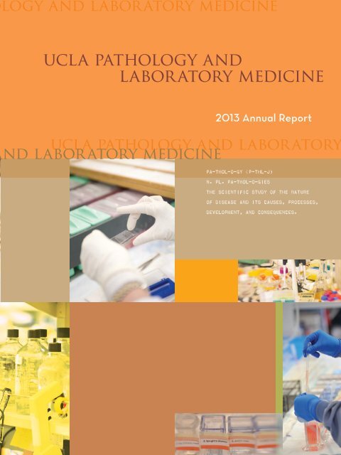
logy <strong>and</strong> <strong>laboratory</strong> <strong>medicine</strong><strong>ucla</strong> <strong>pathology</strong> <strong>and</strong><strong>laboratory</strong> <strong>medicine</strong>2013 Annual Report<strong>ucla</strong> <strong>pathology</strong> <strong>and</strong> <strong>laboratory</strong>nd <strong>laboratory</strong> <strong>medicine</strong>pa-thol-o-gy (p-thl-j)n. pl. pa-thol-o-giesThe scientific study <strong>of</strong> <strong>the</strong> nature<strong>of</strong> disease <strong>and</strong> its causes, processes,development, <strong>and</strong> consequences.
contentsintroduction1Jonathan Braun, MD, PhDclinical services3567Growth In Outreach Enhances Teaching <strong>and</strong> ResearchWhile Streng<strong>the</strong>ning <strong>Department</strong>’s Bottom LineBringing <strong>UCLA</strong> Pathology/Laboratory Services to The Community<strong>Department</strong> Will Play Key Role In <strong>UCLA</strong> Health’s Partnership With China<strong>UCLA</strong>’s Exp<strong>and</strong>ing Global Presenceresearch enterprise9111213Deciphering The Genetic Basis <strong>of</strong> Disease Across Populations: Path <strong>and</strong> Lab’s Pioneering RoleClinical Genomics Center Uses Powerful Sequencing Test to Improve Diagnosis <strong>and</strong>Treatment <strong>of</strong> Genetic DisordersNext-Generation Sequencing <strong>of</strong> Human Genome Paving <strong>the</strong> Way Toward Personalized MedicineFund Managers: Invaluable Members <strong>of</strong> <strong>the</strong> Research Teameducation15171819“Flipping” <strong>the</strong> Traditional Classroom: Students Do <strong>the</strong> Teaching in DGSOM Histo<strong>pathology</strong> LabInteractive Labs Introduce Medical Students to Dermato<strong>pathology</strong>Med School puts Molecular Genetics FirstUnderst<strong>and</strong>ing <strong>the</strong> Complexities <strong>of</strong> Inflammationresearch services21232425Translational Pathology Core Laboratory Seeks to Fulfill <strong>the</strong> Promise <strong>of</strong> BiobankingPathology Portal Improves Access to <strong>Department</strong>’s ServicesEngageUC will Inform Federal <strong>and</strong> State Debate on Biobanking PracticesFreeze-Dried Storage <strong>of</strong> Tissues May Offer “Best Of Both Worlds”department in depth26293032Who’s Who in Pathology & MetricsAwards & RecognitionAlumniOpportunities for Givingeditors: karyn greenstone & sharon webb • writer: dan gordon • design: susan l<strong>and</strong>esmann designphotography: margaret sison • additional photography: todd cheney, reed hutchinson, jian yu rao & kosal taing© 2013 Regents <strong>of</strong> <strong>the</strong> University <strong>of</strong> California
jonathan braun, md, phd<strong>the</strong> quiet heroA friend <strong>of</strong> mine, whose work is philosophy, is always reading. Do you know his favoriteperiodical? It is People Magazine. He explained that it explored each week how fame <strong>and</strong> wealthdo not guarantee a happy or meaningful life! In this annual report, we reflect on my quiet heroes –those who not only bring about real <strong>and</strong> important change, but also provide <strong>the</strong> inspiration to bringout what is best in o<strong>the</strong>rs <strong>and</strong> make <strong>the</strong> world that much more <strong>of</strong> a better place.Thanks to Dr. Binder <strong>and</strong> <strong>the</strong> amazing people who fashion Pathology into a new mode <strong>of</strong> caredelivery, <strong>and</strong> educate our physician trainees not only in <strong>pathology</strong> expertise but also in <strong>the</strong> role <strong>of</strong>service. With <strong>the</strong> opening <strong>of</strong> BURL Laboratories, we are now poised to extend this special amalgam<strong>of</strong> quality <strong>and</strong> service to communities throughout <strong>the</strong> north, west, <strong>and</strong> south <strong>of</strong> Los Angeles County.Extending our boundaries even fur<strong>the</strong>r, our <strong>Department</strong>, along with <strong>UCLA</strong> Health <strong>and</strong> <strong>the</strong> Jonsson ComprehensiveCancer Center, has entered into l<strong>and</strong>mark arrangements with <strong>the</strong> Cancer Institute <strong>and</strong> Hospital, ChineseAcademy <strong>of</strong> Medical Sciences (CAMS). This builds on our successful partnerships over <strong>the</strong> past several years incare <strong>and</strong> teaching with academic health centers <strong>and</strong> municipal Hangzhou. The new relationship with CAMS willbroaden <strong>the</strong> scope <strong>of</strong> <strong>the</strong>se partnerships for clinical trials <strong>and</strong> translational research with leading investigatorsthroughout China.While we extend our global presence in China <strong>and</strong> elsewhere, we are also mindful <strong>of</strong> <strong>the</strong> needs closer to home,as we begin consultative tele<strong>pathology</strong> services to underserved <strong>pathology</strong> practices located throughout Sou<strong>the</strong>rnCalifornia. Partnerships with six communities will initiate this program, <strong>and</strong> will build from <strong>the</strong>re to a network tosupport community-based practices throughout California.Even closer to home, our Clinical Genomics Center has implemented <strong>the</strong> remarkable breakthroughs in highthroughputsequencing <strong>and</strong> disease-diagnostic genetic bioinformatics as a practical health care service. Thisis finally bringing closure to families <strong>and</strong> individuals who have been waiting years for what <strong>the</strong>y once thought werequestions without answers. The rapid diagnosis <strong>of</strong> complex genetic diseases allows patients, families, <strong>and</strong> <strong>the</strong>irhealth care team to move onto planning for medical care, <strong>and</strong> strategies to identify <strong>and</strong> eventually avert diseasein unaffected family members.Our department is intensely devoted to research, nearly $50M presently in force. What drives this creativeenterprise is David Islas <strong>and</strong> our Fund Managers. They pilot projects through <strong>the</strong> complex path from inceptionto funding, <strong>and</strong> steward <strong>the</strong>se precious funds to assure <strong>the</strong> greatest value to our investigators <strong>and</strong> traineescientists in <strong>the</strong>ir challenging experimental work.It is in <strong>the</strong> classroom where researchers <strong>of</strong> tomorrow are inspired by some <strong>of</strong> <strong>the</strong> most creative <strong>and</strong> innovativefaculty in <strong>the</strong> nation. Through <strong>the</strong> innovation <strong>of</strong> <strong>the</strong> “flipped” classroom, Elena Stark inspires future medicalleaders to think independently, <strong>and</strong> Wayne Grody, Steve Bensinger <strong>and</strong> Ch<strong>and</strong>ra Smart encourage fundamentalunderst<strong>and</strong>ing <strong>of</strong> human processes, <strong>and</strong> not simple rote learning.As <strong>the</strong> path from research to clinical innovation shortens, <strong>the</strong> distinctions between health care <strong>and</strong> researchdiminish as patient biospecimens serve care <strong>and</strong> research today, <strong>and</strong> data to guide care <strong>of</strong> <strong>the</strong> same patientswith improved care tomorrow. This creates conceptual, ethical, <strong>and</strong> operational challenges for dynamic healthsystems like <strong>UCLA</strong> centers. Sarah Dry, William Yong , Justin Perry <strong>and</strong> <strong>the</strong>ir teams in <strong>the</strong> TranslationalPathology Core Laboratory, EngageUC, <strong>and</strong> <strong>the</strong> Pathology Portal, have created a new framework to solve<strong>the</strong>se challenges for <strong>the</strong> <strong>UCLA</strong> clinical <strong>and</strong> research communities.In this annual report, read <strong>the</strong> stories <strong>of</strong> <strong>the</strong>se quiet heroes: exceptional people, <strong>and</strong> <strong>the</strong> exceptional ways that<strong>the</strong>y enable <strong>UCLA</strong> to serve <strong>the</strong> needs <strong>and</strong> dreams <strong>of</strong> our community.Dr. Jonathan Braun, Pr<strong>of</strong>essor <strong>and</strong> Chair, <strong>Department</strong> <strong>of</strong> Pathology <strong>and</strong> Laboratory Medicine<strong>pathology</strong> & <strong>laboratory</strong> <strong>medicine</strong> at <strong>ucla</strong> 1
BURL opening receptionattendees, including DavidFeinberg, MD, MBA, CEO,<strong>UCLA</strong> Hospital System;Lee Flores, BURL manager;Arnold Scheer, CAO;Jonathan Braun, MD, PhD,Chair; Shannon O’Kelley,COO, <strong>UCLA</strong> Hospital System;Scott Binder, MD, Sr. ViceChair, Clinical Services.<strong>the</strong> slides for <strong>the</strong> specimens obtained in <strong>the</strong> <strong>Department</strong><strong>of</strong> Medicine’s Division <strong>of</strong> Dermatology for <strong>the</strong>diagnosis <strong>of</strong> skin diseases. Those slides are analyzedby trained dermatopathologists under <strong>the</strong> leadership<strong>of</strong> Dr. Ch<strong>and</strong>ra Smart, <strong>the</strong> program’s director. Theinterdisciplinary program, unique in Sou<strong>the</strong>rn California,represents a direction that Dr. Binder expects tobecome a significant trend in <strong>the</strong> outreach program.Lung <strong>and</strong> Molecular Pathology. Lung <strong>pathology</strong><strong>and</strong> molecular <strong>pathology</strong> services now include alarge <strong>and</strong> comprehensive panel <strong>of</strong> sophisticatedmolecular tests to better evaluate lung cancers <strong>and</strong> <strong>of</strong>ferimproved guidance for treatment <strong>and</strong> clinical trialoptions to patients. The new genetic tests for lungcancer have been developed by <strong>UCLA</strong>’s MolecularDiagnostics Laboratories to complement <strong>the</strong>traditional anatomic <strong>pathology</strong> evaluation <strong>of</strong> lungcancer. Ultimately, this will enable <strong>the</strong> <strong>pathology</strong>team to exp<strong>and</strong> its role from classifying <strong>and</strong> staging acancer to reporting <strong>the</strong> genetic changes that mayprovide clues to treatment strategies <strong>and</strong> informclinicians <strong>and</strong> patients on <strong>the</strong> prognosis <strong>of</strong> <strong>the</strong> disease.Gastrointestinal (GI) Pathology. The GI <strong>pathology</strong>service is a large-volume, sub-specialty-trained groupthat incorporates modern diagnostic techniques,maintains an intra-divisional diagnostic conference,participates in clinical research <strong>and</strong> attends internationalGI <strong>pathology</strong> meetings. Devoted to anatomic<strong>pathology</strong> diagnostics, <strong>the</strong> service prepared 18,845diagnostic reports in 2012. It provides additional subspecialtyconsultations intra-operatively <strong>and</strong> on weekends.Advances in <strong>the</strong> clinical application <strong>of</strong> <strong>the</strong> molecularunderst<strong>and</strong>ing <strong>of</strong> disease have resulted in <strong>the</strong>service augmenting reports using molecular genetic<strong>and</strong> immunohistochemical (IHC) techniques.explore Outreachpathnet.medsch.<strong>ucla</strong>.edu/outreachGenitourinary (GU) Pathology. The genitourinary<strong>pathology</strong> section has developed or adoptedadvanced <strong>and</strong> sophisticated technologies to fur<strong>the</strong>rimprove <strong>the</strong> quality <strong>of</strong> patient care. All cases <strong>of</strong> prostatectomyare now processed as whole-mount sections,enabling accurate enumeration <strong>of</strong> tumors as well as<strong>the</strong> reporting <strong>of</strong> <strong>the</strong> pathologic features <strong>of</strong> each tumorindividually. A team consisting <strong>of</strong> pathologists, radiologists<strong>and</strong> urologists has developed a targeted biopsytechnology that allows improved detection <strong>of</strong> clinicallysignificant prostate cancers. Many patients withindolent tumors are now enrolled in <strong>the</strong> active surveillanceprogram instead <strong>of</strong> receiving radical treatmentsthat carry potentially significant side effects.Neuro<strong>pathology</strong>. The neuro<strong>pathology</strong> section isnow conducting isocitrated dehydrogenase 1 <strong>and</strong> 2(IDH1 <strong>and</strong> IDH2) mutation detection, sequencing teststhat can detect common <strong>and</strong> rare mutations <strong>of</strong> <strong>the</strong>IDH1 gene as well as IDH2 mutations – <strong>the</strong> presence <strong>of</strong>which in gliomas is associated with a better prognosis<strong>and</strong> can affect patient management. Tests are <strong>of</strong>feredfor FUS <strong>and</strong> TDP-43, immunohistochemical markersthat can assist in <strong>the</strong> classification <strong>of</strong> frontotemporallobar degeneration. In addition to existing musculardystrophy markers, investigations <strong>of</strong> rare musculardystrophies are <strong>of</strong>fered, as are evaluations for epilepsydisorders <strong>and</strong> rare genetic disorders, <strong>the</strong> latter usingwhole genome sequencing.Hemato<strong>pathology</strong>. The outreach workflow continuesto increase for <strong>the</strong> hemato<strong>pathology</strong> section, withan 8 percent increase in bone marrow cases <strong>and</strong> a 14percent increase in flow cases for 2012 vs. 2011. Since2009, bone marrow volume has increased 34 percent<strong>and</strong> flow panels to leukemia have increased 31 percent.The hemato<strong>pathology</strong> service’s consultations includeevaluation <strong>of</strong> blood, bone marrow, lymph nodes <strong>and</strong>hematolymphoid lesions in o<strong>the</strong>r body sites that aresubmitted by pathologists <strong>and</strong> oncologists throughout<strong>the</strong> nation as well as from o<strong>the</strong>r countries.Hepatobiliary/Renal Pathology. The hepatobiliary<strong>pathology</strong> service provides rapid, high-qualitydiagnostic histo<strong>pathology</strong> for liver disease on bothbiopsy <strong>and</strong> surgical material, with extensive experiencein hepatitis <strong>and</strong> biliary disease, neoplastic hepatobiliarydisease <strong>and</strong> transplant <strong>pathology</strong>. The renal<strong>pathology</strong> section h<strong>and</strong>les a large volume <strong>of</strong> diagnosticrenal biopsies, <strong>and</strong> also evaluates <strong>and</strong> interprets transplantbiopsies, nephrectomy specimens <strong>and</strong> posttransplantationcomplications. n4 <strong>pathology</strong> & <strong>laboratory</strong> <strong>medicine</strong> at <strong>ucla</strong>
inging <strong>ucla</strong> <strong>pathology</strong>/<strong>laboratory</strong>services to <strong>the</strong> communityThe <strong>Department</strong> <strong>of</strong> Pathology <strong>and</strong> LaboratoryMedicine has joined <strong>UCLA</strong> Health<strong>and</strong> <strong>the</strong> <strong>Department</strong> <strong>of</strong> Medicine in a partnershipto deliver additional health services to <strong>the</strong> SanFern<strong>and</strong>o Valley, Conejo Valley, <strong>and</strong> SouthBay regions <strong>of</strong> Los Angeles County.In March, <strong>the</strong> <strong>Department</strong> <strong>of</strong> Pathology <strong>and</strong>Laboratory Medicine <strong>and</strong> <strong>UCLA</strong> Health openeda state-<strong>of</strong>-<strong>the</strong>-art outreach reference <strong>laboratory</strong>,Bruin University Reference Laboratory (BURL), inPanorama City. Laboratory support <strong>of</strong> new <strong>UCLA</strong>Health facilities followed, including “STAT” labs(designed to provide quick-turnaround services)serving <strong>UCLA</strong> Health’s new primary <strong>and</strong> secondarycare facility in Westlake Village. The BURL WestlakeVillage Patient Service Center has patient blood drawstations <strong>and</strong> <strong>of</strong>fers immediate results for many blood<strong>and</strong> urine chemistries, along with o<strong>the</strong>r STAT testing.“BURL is designed as a price-competitive,quality-driven outpatient <strong>laboratory</strong>with locations throughout <strong>the</strong> SanFern<strong>and</strong>o Valley, Conejo Valley, Westside<strong>and</strong> South Bay regions <strong>of</strong> Los Angeles.”- Scott Binder, MD, Medical Director, BURLThe Westlake Village <strong>of</strong>fice is <strong>the</strong> first <strong>of</strong> six new<strong>UCLA</strong> facilities scheduled to open over <strong>the</strong> next twoyears in service areas beyond <strong>UCLA</strong>’s Westwood <strong>and</strong>Santa Monica campuses; o<strong>the</strong>r sites include Thous<strong>and</strong>Oaks, Porter Ranch, Calabasas, Santa Clarita <strong>and</strong>Redondo Beach. The state-<strong>of</strong>-<strong>the</strong>-art <strong>of</strong>fices will<strong>of</strong>fer a full range <strong>of</strong> primary-care sub-specialties(internal <strong>medicine</strong>, family <strong>medicine</strong>, pediatrics,women’s health), as well as ancillary <strong>and</strong> secondarycareservices. The new practices will also engagein community education <strong>and</strong> outreach throughnewsletters, physician lectures <strong>and</strong> participationin outreach programs accessible to <strong>the</strong> community(e.g., in shopping centers, health fairs <strong>and</strong>farmer’s markets).By establishing new locations, supporting <strong>the</strong>mwith local <strong>laboratory</strong> facilities, <strong>and</strong> participatingin local programs, <strong>UCLA</strong> is exp<strong>and</strong>ing its ability toprovide convenient, high-quality care to people wholive or work outside traditional primary care serviceareas, while providing a gateway to higher levels <strong>of</strong>care within <strong>the</strong> <strong>UCLA</strong> system for patients who needsuch services. nCLINICAL SERVICES<strong>pathology</strong> & <strong>laboratory</strong> <strong>medicine</strong> at <strong>ucla</strong> 5
Digital imaging will facilitateimproved communication <strong>and</strong>education among pathologists.CLINICAL SERVICESFern<strong>and</strong>o Palma-Diaz, MD, Assistant Director, Tele<strong>pathology</strong><strong>ucla</strong>’s exp<strong>and</strong>ing global presenceThe <strong>UCLA</strong> <strong>Department</strong> <strong>of</strong> Pathology <strong>and</strong> LaboratoryMedicine has been at <strong>the</strong> forefront <strong>of</strong>international tele<strong>pathology</strong> development through itspartnership with China, <strong>and</strong> has now committeda significant effort toward integrating tele<strong>pathology</strong>into daily practice closer to home. Recently, <strong>the</strong>department received a substantial grant from <strong>the</strong>State <strong>of</strong> California to provide consultative tele<strong>pathology</strong>services to underserved <strong>pathology</strong> practicesthroughout Sou<strong>the</strong>rn California. The funding willbe used to acquire digital slide scanners that will beinstalled in remote <strong>pathology</strong> groups, <strong>and</strong> will enable<strong>the</strong> department to begin space remodeling to accommodatetele<strong>pathology</strong> functions.Once installed <strong>and</strong> operational, <strong>the</strong>se scannerswill allow pathologists working in underserved communitiesto obtain second-opinion consultationsfrom <strong>UCLA</strong> specialists on challenging <strong>pathology</strong>cases. This technology enables rapid transmission<strong>of</strong> scanned slide data to <strong>the</strong> consultant pathologist,avoiding delays that occur when glass slides mustbe transported from one site to ano<strong>the</strong>r. In additionto improving patient care – particularly forcases with urgent diagnoses – digital imaging willfacilitate improved communication <strong>and</strong> educationamong pathologists.agreement between Ronald Reagan <strong>UCLA</strong> MedicalCenter <strong>and</strong> Second Affiliate Hospital <strong>of</strong> ZhejiangUniversity (SAHZJU) in China. As part <strong>of</strong> <strong>the</strong> agreement,<strong>UCLA</strong> pathologists provide second-opiniondiagnoses to pathologists <strong>and</strong> o<strong>the</strong>r physicians inSAHZJU. Both cytological <strong>and</strong> histological casesare included, <strong>and</strong> cases span a broad range <strong>of</strong> organsystems, including head <strong>and</strong> neck, lung, gynecological,s<strong>of</strong>t tissue <strong>and</strong> bone, neurological, <strong>and</strong> hematological.In addition, periodic multidisciplinary videoconferencesare held to discuss interesting cases.The program has exp<strong>and</strong>ed to reach much <strong>of</strong> Chinathrough additional collaborative agreements withtwo major reference labs (Dian Inc. <strong>and</strong> Adicon Inc.)<strong>and</strong> prominent institutions such as Cancer Hospital,Chinese Academy <strong>of</strong> Medical Science. The program’ssuccess has also stimulated similar collaborationsin o<strong>the</strong>r areas, including radiology, oncology, <strong>and</strong>nuclear <strong>medicine</strong>. nexplore Tele<strong>pathology</strong><strong>pathology</strong>.<strong>ucla</strong>.edu/tele<strong>pathology</strong>The department’s international tele<strong>pathology</strong>program began in 2010 with a collaborative<strong>pathology</strong> & <strong>laboratory</strong> <strong>medicine</strong> at <strong>ucla</strong> 7
8 <strong>pathology</strong> & <strong>laboratory</strong> <strong>medicine</strong> at <strong>ucla</strong>
esearch enterprisedeciphering <strong>the</strong> genetic basis <strong>of</strong> disease acrosspopulations: path <strong>and</strong> lab’s pioneering rolerRecent breakthroughs in high-throughputgenotyping <strong>and</strong> sequencing technologies haveushered in an era <strong>of</strong> large-scale genome-widedisease studies that have identified hundreds <strong>of</strong> geneticvariants associated with risk for many diseases. Studiesaimed at unraveling <strong>the</strong> genetic basis <strong>of</strong> Mendeliantraits (those caused by <strong>the</strong> disruption <strong>of</strong> a single gene)have been particularly successful. Technological <strong>and</strong>computational advances have made it possible to scanDNA in patients that have <strong>the</strong> disease – revealing, inmost cases, <strong>the</strong> responsible genes. The efficiency <strong>of</strong>such studies is reinforced by <strong>the</strong> successful translation<strong>of</strong> <strong>the</strong>se findings in clinical settings, with <strong>UCLA</strong> actingas a pioneer in analyzing <strong>the</strong> entire protein-encodinggenome <strong>of</strong> a patient for diagnostic purposes.But <strong>the</strong> most common diseases – including mostcancers, type 2 diabetes, <strong>and</strong> heart disease – do nothave Mendelian traits; ra<strong>the</strong>r, <strong>the</strong>y are a complexproduct <strong>of</strong> many genes that toge<strong>the</strong>r influencedisease risk. Although large-scale genome-widestudies have been highly successful in identifying<strong>the</strong> loci harboring genetic risk variants for commondiseases, <strong>the</strong> complex correlation structure <strong>of</strong> <strong>the</strong>human genome means that <strong>the</strong> risk variants identifiedin <strong>the</strong>se studies typically are not <strong>the</strong>mselves causal,but are associated with unobserved causal geneticvariants. Thus, although identifying <strong>the</strong> causal variantsis crucial in underst<strong>and</strong>ing <strong>the</strong> functional basis <strong>of</strong> adisease, for most common diseases <strong>the</strong> true causalvariants have yet to be discovered.In an attempt to bridge this gap, Dr. Bogdan Pasaniuc<strong>and</strong> colleagues in <strong>the</strong> <strong>UCLA</strong> <strong>Department</strong> <strong>of</strong> Pathology<strong>and</strong> Laboratory Medicine are conducting studies usingsophisticated genotyping or sequencing approachesfor tens to hundreds <strong>of</strong> thous<strong>and</strong>s <strong>of</strong> individuals <strong>of</strong>different ethnicities. Dr. Pasaniuc’s lab has found agreat benefit to studying multi-ethnic populations.“Not only is it important to study <strong>the</strong> genetic basis<strong>of</strong> disease across a wide range <strong>of</strong> ethnicities for <strong>the</strong>benefit <strong>of</strong> a larger group <strong>of</strong> people, but computational<strong>and</strong> statistical approaches can leverage geneticdiversity across populations to streng<strong>the</strong>n our abilityto find causal variants,” Dr. Pasaniuc explains.Of particular interest in studying disease acrossethnicities are admixed populations (populations withrecent ancestry from different continents) such asAfrican Americans <strong>and</strong> Hispanic Americans, who are<strong>of</strong>ten medically underserved while carrying a disproportionatelyhigh burden <strong>of</strong> disease. Because <strong>of</strong> <strong>the</strong>diversity <strong>of</strong> <strong>the</strong>ir genomes, admixed populations <strong>of</strong>fer<strong>the</strong> promise <strong>of</strong> capturing additional genetic diversitycompared with studies <strong>of</strong> homogeneous populationssuch as Europeans. This poses particular technicalBogdan Pasaniuc, PhDfocuses on admixedpopulations forgreater underst<strong>and</strong>ing<strong>of</strong> genetic risk factorsfor common diseases.<strong>pathology</strong> & <strong>laboratory</strong> <strong>medicine</strong> at <strong>ucla</strong> 9
Of particular interest in studying diseaseacross ethnicities are admixed populations...such as African Americans <strong>and</strong> HispanicAmericans, who are <strong>of</strong>ten medically underservedwhile carrying a disproportionatelyhigh burden <strong>of</strong> disease.challenges, since in admixed populations, correlationamong genetic variants exists both at a fine scale in<strong>the</strong> ancestral populations <strong>and</strong> at a coarse scale, dueto chromosomal segments <strong>of</strong> recently shared ancestry.By jointly modeling both types <strong>of</strong> genetic structurein African American genomes, Dr. Pasaniuc <strong>and</strong>colleagues showed that a combined approach achievesgreater statistical power to identify risk loci than whatcan be achieved in studies <strong>of</strong> homogeneous populationssuch as Africans or Europeans.The vast amount <strong>of</strong> data currently being generatedby sequencing technologies increases <strong>the</strong> need foraccurate yet computationally efficient methods <strong>of</strong>performing disease scans in multi-ethnic cohorts todecipher <strong>the</strong> genetic basis <strong>of</strong> disease in various populations.Such efforts will go a long way toward translatinggenetic findings for common diseases to <strong>the</strong> clinic,improving diagnostics <strong>and</strong> treatment decisions. nexplore Dr. Pasaniuc’s labbogdanlab.<strong>pathology</strong>.<strong>ucla</strong>.edu10 <strong>pathology</strong> & <strong>laboratory</strong> <strong>medicine</strong> at <strong>ucla</strong>
clinical genomics center uses powerful sequencing testto improve diagnosis <strong>and</strong> treatment <strong>of</strong> genetic disordersIt is estimated that as much as 10% <strong>of</strong> <strong>the</strong> U.S. populationsuffers from a Mendelian genetic disease.More than 4,000 Mendelian genetic diseases havebeen described, with <strong>the</strong> precise genetic mutationidentified. These discoveries have contributed significantlyto our underst<strong>and</strong>ing <strong>of</strong> biology, as wellas <strong>the</strong> mutations that lead to human diseases.However, an even larger set <strong>of</strong> genes remains to bediscovered. Dr. Stan Nelson’s research has led to <strong>the</strong>successful implementation <strong>of</strong> genomic sequencingapproaches that enable researchers to efficiently <strong>and</strong>economically search <strong>the</strong> entire human genome inindividuals for disease-causing mutations. Usingthis approach, Dr. Nelson’s group has identified <strong>the</strong>genetic cause for more than 17 Mendelian geneticdiseases in <strong>the</strong> last three years.test called clinical exome sequencing. Under Dr. Nelson’sguidance as <strong>the</strong> center’s director, this genomesequencing test is not only proving to be much moresensitive at finding <strong>the</strong> root cause <strong>of</strong> many serioushuman diseases, but it is also resulting in novel genediscovery <strong>and</strong> leading to new approaches to <strong>the</strong> development<strong>of</strong> <strong>the</strong>rapies. In <strong>the</strong> past year,Dr. Nelson’s group has been able to specificallydiagnose children with developmental delay, autism,seizure disorders, muscular dystrophy, <strong>and</strong> o<strong>the</strong>rdisorders. Many <strong>of</strong> <strong>the</strong>se children have mutations inpreviously unrecognized genes; providing <strong>the</strong> specificdiagnosis ends a patient’s medical odyssey <strong>and</strong> <strong>of</strong>tenpoints health care providers toward more tailoredmedical interventions. nRESEARCH ENTERPRISEBased on <strong>the</strong>se successful efforts, Dr. Nelson <strong>and</strong>colleagues have launched <strong>the</strong> <strong>UCLA</strong> Clinical GenomicsCenter, featuring a new <strong>and</strong> powerful diagnosticexplore Clinical Genomics<strong>pathology</strong>.<strong>ucla</strong>.edu/genomicsFar left <strong>and</strong> below:Hane Lee, PhDBottom left:Stan Nelson, MD.
next-generation sequencing <strong>of</strong> human genomepaving <strong>the</strong> way toward personalized <strong>medicine</strong>Next-generation sequencing(NGS) <strong>of</strong>fers <strong>the</strong> promise<strong>of</strong> significantly enhancing ourunderst<strong>and</strong>ing <strong>of</strong> how geneticdifferences affect health <strong>and</strong>disease. Compared withconventional Sanger sequencing(considered “first-generation”technology), <strong>the</strong>se new methodscan inexpensively yield largevolumes <strong>of</strong> sequence data. Thecontrast is stark: For example,NGS technology can produce 600,000 megabytes<strong>of</strong> data at a cost <strong>of</strong> four cents per megabyte, whereasfirst-generation technology produces 0.06 megabytesat a cost <strong>of</strong> $1,500 per megabyte <strong>of</strong> data.The second-generation sequencers, such as <strong>the</strong> HiSeq2500 <strong>and</strong> Ion Proton, have already realized whatwas once only a dream: <strong>the</strong> ability to sequence onehuman genome in one day for $1,000. Now, thirdgenerationsequencers, such as those produced byOxford Nanopore Technologies, have <strong>the</strong> potential toachieve what was once unthinkable: sequencinga human genome in 15 minutes.Now, third-generation sequencers,have <strong>the</strong> potential to achieve whatwas once unthinkable: sequencinga human genome in 15 minutes.The ability to sequence an entire human genomequickly <strong>and</strong> cheaply will pr<strong>of</strong>oundly affect <strong>the</strong> waywe think about scientific approaches in basic, applied<strong>and</strong> clinical research. With fur<strong>the</strong>r improvement <strong>of</strong><strong>the</strong> bioinformatics infrastructure <strong>and</strong> more detailedclinical annotation <strong>of</strong> <strong>the</strong> human genome, NGS-basedpersonalized <strong>medicine</strong> is on <strong>the</strong> way to revolutionizedisease diagnosis, prognosis <strong>and</strong> treatment. nexplore Next-Generation Sequencing<strong>pathology</strong>.<strong>ucla</strong>.edu/cmsXinmin Li, PhD, Director, Clinical Microarray Core
14 <strong>pathology</strong> & <strong>laboratory</strong> <strong>medicine</strong> at <strong>ucla</strong>T
education“flipping” <strong>the</strong> traditional classroom: studentsdo <strong>the</strong> teaching in dgsom histo<strong>pathology</strong> labThe traditional lecture setting is going by <strong>the</strong>wayside in some academic arenas, replaced by a newmethod <strong>of</strong> teaching that better engages students,increasing <strong>the</strong>ir enthusiasm <strong>and</strong> sparking <strong>the</strong>ir curiosity.The interactive learning environment <strong>of</strong> <strong>the</strong> “flippedclassroom” is sometimes called “inverted instruction”because <strong>the</strong> majority <strong>of</strong> teaching is by students tostudents. It’s a platform that elevates students’ intake,processing, <strong>and</strong> recall <strong>of</strong> data.The flipped-classroom concept has been gainingmomentum in a variety <strong>of</strong> educational institutions,including <strong>the</strong> classrooms at <strong>UCLA</strong>’s David Geffen School<strong>of</strong> Medicine, where <strong>the</strong> <strong>Department</strong> <strong>of</strong> Pathology<strong>and</strong> Laboratory Medicine strives for this enriched level<strong>of</strong> teaching by flipping <strong>the</strong> first-year histo<strong>pathology</strong><strong>laboratory</strong> sessions. The approach is not to beconfused with problem-based learning (PBL), in which,for example, groups <strong>of</strong> students analyze a case studythat reflects lectures by one or more pr<strong>of</strong>essors. WhilePBL is also a form <strong>of</strong> active learning, it requires a baseknowledge specific to <strong>the</strong> topic – i.e., <strong>the</strong> case studydemonstrates <strong>the</strong> information that was previouslytaught traditionally.In a flip-teaching/inverted-classroom scenario, <strong>the</strong>authority to introduce <strong>and</strong> explain primary informationbecomes <strong>the</strong> responsibility <strong>of</strong> <strong>the</strong> students. Studentsresearch, ga<strong>the</strong>r <strong>and</strong> evaluate <strong>the</strong> subject from varioussources – instructor-created supplements (e.g.,PowerPoint modules), <strong>the</strong> internet, <strong>and</strong> textbooks –prior to class. After such preparation, <strong>the</strong>y teach it toeach o<strong>the</strong>r in <strong>the</strong> classroom/lab setting. This materialintegration, also known as hybrid learning, promotesmany perspectives on a subject, raising questions ormaking connections that might not have been revealedfrom simply listening to a lecture.It’s a win-win structure for both <strong>the</strong> pr<strong>of</strong>essor <strong>and</strong><strong>the</strong> students: The pr<strong>of</strong>essor gains time to observe <strong>the</strong>students <strong>and</strong> communicate with <strong>the</strong>m, while <strong>the</strong>students gain a wider field <strong>of</strong> cognition as <strong>the</strong> flippedclassroom adapts to different forms <strong>of</strong> comprehension.The students master <strong>the</strong> information because <strong>the</strong>y areforced to explain <strong>the</strong> material to <strong>the</strong>ir peers.In <strong>the</strong> histo<strong>pathology</strong> <strong>laboratory</strong> for first-yearstudents at <strong>UCLA</strong>’s David Geffen School <strong>of</strong> Medicine,Dr. Elena Stark conducts a flipped classroom.Dr. Stark’s lab classroom in <strong>the</strong> 1P corridor has eighttables hosting seven students each, <strong>the</strong> groups havingbeen assigned at <strong>the</strong> beginning <strong>of</strong> <strong>the</strong> year. At <strong>the</strong> head<strong>of</strong> each table is a computer monitor with access to ashared folder containing 3-6 Aperio virtual slides.First up: <strong>the</strong> Assessment Quiz. Starting <strong>of</strong>f with a testmay sound unorthodox, but because <strong>the</strong> studentshave prepared <strong>the</strong> material on <strong>the</strong>ir own, it is <strong>the</strong>irresponsibility to arrive at <strong>the</strong> <strong>laboratory</strong> already in anactive state <strong>of</strong> mind. This way, from minute one <strong>of</strong> <strong>the</strong>lab session, <strong>the</strong>ir knowledge can be molded, sharpened,challenged <strong>and</strong> streng<strong>the</strong>ned.Since <strong>the</strong> quiz isn’t for grading purposes, <strong>the</strong> evaluationgives <strong>the</strong> pr<strong>of</strong>essor a good sense <strong>of</strong> <strong>the</strong> degree<strong>of</strong> studying, explaining <strong>and</strong> underst<strong>and</strong>ing taking placeeach week. By eliminating any anxiety over a grade,students benefit by focusing on <strong>the</strong>ir own knowledge,M. Elena Stark, MD, PhD.<strong>pathology</strong> & <strong>laboratory</strong> <strong>medicine</strong> at <strong>ucla</strong> 15
<strong>the</strong> approach supports critical thinking,since students need to comprehend <strong>the</strong>subject both for <strong>the</strong>mselves <strong>and</strong> to beable to teach it to o<strong>the</strong>rs.information, <strong>and</strong> each explains <strong>the</strong> material in his orher own way. “This is how you study,” asserts Dr. Stark.“This is how you learn.” The students listen to oneano<strong>the</strong>r, interrupting with astute questions, seekingmore knowledge from each o<strong>the</strong>r. It is an active<strong>and</strong> rich conversation – <strong>the</strong> opposite <strong>of</strong> <strong>the</strong> passivenote-taking (or checking one’s text messages) thatoccurs in <strong>the</strong> lecture hall.comparing it to <strong>the</strong> knowledge held by o<strong>the</strong>rs, <strong>and</strong>learning what is expected <strong>of</strong> <strong>the</strong>m up to that point.What’s noteworthy is that although <strong>the</strong> students knowthat <strong>the</strong> quiz is not for a grade, <strong>the</strong> average score isabout 80%, with a mode <strong>of</strong> 90%-100%: pro<strong>of</strong> that <strong>the</strong>majority <strong>of</strong> students come to <strong>the</strong> lab session prepared<strong>and</strong> ready to teach.The second phase begins once <strong>the</strong> quiz Scantron formsare collected. Now it is time for <strong>the</strong> students to discuss<strong>the</strong>ir answers with <strong>the</strong>ir peers.A brief overview presented by Dr. Stark introduces<strong>the</strong> Aperio slide to which <strong>the</strong> h<strong>and</strong>out/teaching guiderefers, orienting <strong>the</strong> students for <strong>the</strong>ir investigation.With <strong>the</strong> ability to navigate <strong>and</strong> magnify <strong>the</strong> image onmonitors, each group consults with its members on<strong>the</strong> issues prompted by <strong>the</strong> h<strong>and</strong>out/teaching guide.A give-<strong>and</strong>-take discussion ensues.Again, Dr. Stark circulates <strong>and</strong> confirms. She remindseveryone that when learning from each o<strong>the</strong>r,“emphasize thinking your way through <strong>the</strong> material vs.memorizing tidbits <strong>of</strong> information.”“Why did you pick ‘A’ over ‘B’?”“Because ‘B’ identifies both characteristics…”Meanwhile, Dr. Stark circulates <strong>and</strong> confirms <strong>the</strong>person(s) with <strong>the</strong> correct answer(s), asking <strong>the</strong>m toexp<strong>and</strong> on <strong>the</strong>ir thought processes surrounding <strong>the</strong>question. After <strong>the</strong> entire quiz has been scrutinizedwithin each group, <strong>the</strong> class connects as a whole forano<strong>the</strong>r review. Dr. Stark asks <strong>the</strong> questions to all:“The answer to #5 is?”“And why is this important to recognize?”(Assessment quizzes are later posted to Intranet/ANGEL as a supplement for reference. The studentscan email <strong>the</strong> pr<strong>of</strong>essor with more queries.)The third stage employs a h<strong>and</strong>out, which serves asa teaching guide. The students open <strong>the</strong> first Aperioslide. Today, <strong>the</strong> lesson is on <strong>the</strong> histo<strong>pathology</strong> <strong>of</strong> <strong>the</strong>mucosa <strong>of</strong> <strong>the</strong> stomach, <strong>and</strong> <strong>the</strong> first slide is “StomachFundus.” The teaching guide consists <strong>of</strong> multiple pagescontaining directions (“find,” “locate,” “identify”) alongwith probing questions (“What are <strong>the</strong> components<strong>of</strong> <strong>the</strong> lamina propria?” “Does <strong>the</strong> structure <strong>and</strong> location<strong>of</strong> GALT make sense in terms <strong>of</strong> its function?”);a comparison chart; <strong>and</strong> fill-in-<strong>the</strong>-blank sentences(Describe, to your peers, <strong>the</strong> cell populations that arefound in <strong>the</strong> gastric gl<strong>and</strong>s). Toge<strong>the</strong>r, <strong>the</strong>se promptscover <strong>the</strong> vast data that <strong>the</strong> students had to exploreprior to class.A summarization session follows, conducted byDr. Stark, who again redirects any applied notions orinquiries from one student to <strong>the</strong>m all.“Good question! Who has a possible answer?”As time <strong>and</strong> subject matter progress, so does <strong>the</strong>flip teaching process, with three more Aperio slides.The lab period concludes with <strong>the</strong> students applying<strong>the</strong>ir knowledge on two case studies, presented by aguest lecturer.Many pr<strong>of</strong>essors <strong>and</strong> instructors are praising <strong>the</strong>flipped classroom approach, but does this alternativelearning process make for more enriching education?According to some first-year medical students, itcertainly does. One student notes that working ingroups presents a less intimidating environmentfor inquiring about misunderst<strong>and</strong>ings <strong>and</strong> delvinginto challenges. Ano<strong>the</strong>r declares that <strong>the</strong> approachsupports critical thinking, since students need tocomprehend <strong>the</strong> subject both for <strong>the</strong>mselves <strong>and</strong> tobe able to teach it to o<strong>the</strong>rs. It was none o<strong>the</strong>r thanAlbert Einstein who agreed: “If you can’t explain itsimply, you don’t underst<strong>and</strong> it well enough.” nDr. Stark notes that “<strong>the</strong> same information is packagedin different ways” in <strong>the</strong> flipped classroom. Differentstudents in a group emphasize different pieces <strong>of</strong>16 <strong>pathology</strong> & <strong>laboratory</strong> <strong>medicine</strong> at <strong>ucla</strong>
Principles <strong>of</strong> inheritance<strong>and</strong> molecular geneticmechanisms underlie allfields <strong>of</strong> <strong>medicine</strong> — fromsurgery to psychiatry <strong>and</strong>everything in between.Wayne Grody, MD, PhDmed school puts molecular genetics firstWith <strong>the</strong> advent <strong>of</strong> <strong>the</strong> “block” curriculum severalyears ago, instruction <strong>of</strong> <strong>UCLA</strong> medicalstudents in <strong>the</strong> field <strong>of</strong> genetics moved from <strong>the</strong> lastquarter <strong>of</strong> <strong>the</strong> second year, at <strong>the</strong> close <strong>of</strong> <strong>the</strong>ir preclinicalstudies, to Block 1 <strong>of</strong> <strong>the</strong> first year – indeed,starting in <strong>the</strong> very first week <strong>of</strong> that block. This reflects<strong>the</strong> underst<strong>and</strong>ing that we have entered <strong>the</strong> era<strong>of</strong> genomic <strong>medicine</strong>. Principles <strong>of</strong> inheritance <strong>and</strong>molecular genetic mechanisms underlie allfields <strong>of</strong> <strong>medicine</strong> – from surgery to psychiatry <strong>and</strong>everything in between. Patient care in all fields willincreasingly rely on a thorough underst<strong>and</strong>ing <strong>of</strong><strong>the</strong>se concepts in order to provide optimal state<strong>of</strong>-<strong>the</strong>-arttests <strong>and</strong> treatments, while avoidinginappropriate use <strong>of</strong> molecular diagnostic testswith <strong>the</strong> potential to harm <strong>the</strong> patient, both medically<strong>and</strong> psychosocially.The <strong>Department</strong> <strong>of</strong> Pathology <strong>and</strong> LaboratoryMedicine is responsible for <strong>the</strong> content in Block 1,making it <strong>the</strong> first discipline <strong>and</strong> first cohort <strong>of</strong>medical school faculty that <strong>the</strong> new studentsencounter. While <strong>the</strong> students can’t be expectedto have any clinical context for <strong>the</strong>se concepts as<strong>the</strong>y start <strong>the</strong> curriculum, many are surprised <strong>and</strong>excited to learn that <strong>the</strong>re are clinical applicationsto <strong>the</strong> molecular biology content <strong>the</strong>y studied asundergraduates, according to Dr. Wayne Grody,pr<strong>of</strong>essor <strong>and</strong> director <strong>of</strong> <strong>UCLA</strong>’s Molecular DiagnosticsLaboratories, who presents most <strong>of</strong> <strong>the</strong> lectures<strong>and</strong> <strong>laboratory</strong> exercises during that first week.An important component <strong>of</strong> Dr. Grody’s approach to<strong>the</strong> course, <strong>and</strong> to <strong>the</strong> entire “Genetics Thread” thatstretches through all four years <strong>of</strong> medical school, isto convey to <strong>the</strong> students that molecular genetic testsdo not exist in a vacuum. These tests carry tremendousconsequences for <strong>the</strong> person being tested, aswell as for that individual’s blood relatives. Dr. Grody’slectures <strong>and</strong> lab exercises stress <strong>the</strong>se ethical issuesjust as strongly as <strong>the</strong> technical aspects, delving intosuch philosophical dilemmas as genetic privacy,informed consent, insurance <strong>and</strong> employment discrimination,inequity <strong>of</strong> access to expensive genetictests, <strong>and</strong> <strong>the</strong> burdens <strong>of</strong> exclusive gene patents.Student evaluations over <strong>the</strong> years demonstratethat <strong>the</strong>y greatly appreciate this side <strong>of</strong> <strong>the</strong> story<strong>and</strong> find it interesting <strong>and</strong> challenging, providingmuch food for thought <strong>and</strong> discussion both in <strong>and</strong>outside <strong>the</strong> classroom. nexplore Education<strong>pathology</strong>.<strong>ucla</strong>.edu/education18 <strong>pathology</strong> & <strong>laboratory</strong> <strong>medicine</strong> at <strong>ucla</strong>
underst<strong>and</strong>ing <strong>the</strong> complexities <strong>of</strong> inflammationInflammation is a complex, multi-componentresponse that protects <strong>the</strong> host from a harmfulinvader or tissue destruction. It represents an earlydefense warning system <strong>of</strong> <strong>the</strong> body, designed tominimize danger, mitigate damage <strong>and</strong> start <strong>the</strong>repair process. To achieve this essential task, <strong>the</strong>inflammatory system relies on an intricate series <strong>of</strong>molecular sensors stationed on high alert throughout<strong>the</strong> body. Once <strong>the</strong>se sensors sound <strong>the</strong> alarm, <strong>the</strong>body activates an ever-growing array <strong>of</strong> molecular<strong>and</strong> cellular effectors tasked with protecting <strong>the</strong>host from dangerous foreign invaders or minimizingtissue damage from traumatic injury. Without <strong>the</strong> fullarmamentarium <strong>of</strong> this system, people can experiencea myriad <strong>of</strong> problems, ranging from life-threateninginfections to an autoimmune disease such as lupus.If left unchecked, excessive inflammatory responsescan cause significant disease <strong>and</strong> death, likelycontributing to a<strong>the</strong>rosclerosis, diabetes <strong>and</strong> obesity,to name a few.Dr. Steven Bensinger, who has been studying <strong>the</strong>inflammatory response <strong>and</strong> <strong>the</strong> immune system fornearly 15 years, teaches first-year medical studentsabout <strong>the</strong> principles <strong>of</strong> inflammation. “The goal is notto memorize details, many <strong>of</strong> which will ultimatelyfade from <strong>the</strong>ir memory,” he explains. “Ra<strong>the</strong>r, I want<strong>the</strong> students to fundamentally underst<strong>and</strong> how <strong>the</strong>inflammatory system works.”Excessive inflammatoryresponses can causesignificant disease <strong>and</strong>death, likely contributingto a<strong>the</strong>rosclerosis,diabetes <strong>and</strong> obesity,to name a few.Dr. Bensinger starts with anatomy so that studentscan see <strong>the</strong> tissues that are involved, <strong>the</strong>n introduces<strong>the</strong> cellular players <strong>of</strong> <strong>the</strong> inflammatory system:neutrophil <strong>and</strong> macrophage. Next, he covers <strong>the</strong>myriad molecular effectors dedicated to rapidlyresponding to danger signals. Finally, students learnabout <strong>the</strong> resolution <strong>of</strong> <strong>the</strong> inflammatory response –including <strong>the</strong> brakes <strong>of</strong> <strong>the</strong> system, keeping <strong>the</strong> hostfrom <strong>the</strong> ever-escalating response. “The principles <strong>of</strong>inflammation, including how it functions in health<strong>and</strong> contributes to disease, will serve as a foundationthroughout students’ medical education,” Dr.Bensinger notes. nEDUCATIONSteve Bensinger,VMD, PhD, <strong>and</strong>anatomy students.<strong>pathology</strong> & <strong>laboratory</strong> <strong>medicine</strong> at <strong>ucla</strong> 19
20 <strong>pathology</strong> & <strong>laboratory</strong> <strong>medicine</strong> at <strong>ucla</strong>T
esearch servicestranslational <strong>pathology</strong> core <strong>laboratory</strong>seeks to fulfill <strong>the</strong> promise <strong>of</strong> biobankingThe growing emphasis on translational research(converting <strong>laboratory</strong> findings to clinical gains) <strong>and</strong>personalized <strong>medicine</strong> has increased <strong>the</strong> importance<strong>of</strong> high-quality human tissue <strong>and</strong> fluid samples toidentify potential <strong>the</strong>rapeutic targets, find prognosticbiological markers, <strong>and</strong> recognize markers that willfacilitate tailoring <strong>of</strong> individual <strong>the</strong>rapies for cancerpatients. The biobanking practices associated withstoring such samples have changed significantly over<strong>the</strong> past decade. A leader in this changing arena isDr. Sarah Dry, director <strong>of</strong> <strong>the</strong> <strong>UCLA</strong> <strong>Department</strong> <strong>of</strong>Pathology <strong>and</strong> Laboratory Medicine’s TranslationalPathology Core Laboratory (TPCL), which includes<strong>the</strong> <strong>UCLA</strong> Institutional Biobank.In August 2012, TPCL became <strong>the</strong> fourth biorepositoryin <strong>the</strong> United States to be accredited by <strong>the</strong> College<strong>of</strong> American Pathologists (CAP). Similar to <strong>laboratory</strong>accreditation, CAP biorepository accreditation is arigorous process designed to ensure that biorepositorieshave st<strong>and</strong>ard operating procedures in place thatconform to current best-practice st<strong>and</strong>ards; processesfor continual quality control monitoring <strong>and</strong> improvement;<strong>and</strong> appropriate initial <strong>and</strong> ongoing staff trainingprocesses. TPCL is now in <strong>the</strong> process <strong>of</strong> obtainingClinical Laboratory Improvement Amendments (CLIA)certification, as more patients want <strong>the</strong>ir samplesbanked for future personalized-<strong>medicine</strong> clinical use.Given <strong>the</strong> storage costs (space, freezers, electricity)involved with traditional biosample storage, biobankingresearchers are exploring alternative storage methods.One technique being studied by Dr. William H. Yong,<strong>UCLA</strong> neuropathologist, involves room temperaturestorage <strong>of</strong> lyophilized samples (see page 25).Two innovative additions to <strong>UCLA</strong>/TPCL biobankingare being introduced this year. One involves linkingwhole-slide digital images to samples in <strong>the</strong> biobankdatabase. The quality control slide for each biosampleis scanned on TPCL’s Aperio AT scanner <strong>and</strong> stored. Athumbnail image <strong>of</strong> <strong>the</strong> scanned slide appears in <strong>the</strong>database, <strong>and</strong> a link to <strong>the</strong> full image enables staff orresearchers to immediately determine if <strong>the</strong> biosamplesatisfies research needs. The second innovationinvolves <strong>the</strong> installation <strong>of</strong> Daedalus Crimson s<strong>of</strong>twareat <strong>UCLA</strong>. Crimson permits identification <strong>of</strong> soonto-be-discardedclinical samples for use in research.This technology will make it easier for researchers toobtain (with appropriate informed consent) both patientspecific<strong>and</strong> disease-specific samples.An important effort underway at <strong>UCLA</strong> involves institutingglobal informed consent for <strong>the</strong> use <strong>of</strong> remnantbiosamples in research. Recent federal <strong>and</strong> stateconcerns about patient privacy given current genomictesting capabilities suggest that continued use <strong>of</strong>Sarah Dry, MD, PhD<strong>pathology</strong> & <strong>laboratory</strong> <strong>medicine</strong> at <strong>ucla</strong> 21
Clara Magyar, PhD,Joseph Bautista,Voicu Ciobanu, PhDSarah Dry, MD,Delia Adefuin<strong>and</strong> Ping Fu <strong>of</strong><strong>the</strong> TranslationalPathology CoreLaboratory.CAP biorepository accreditation is a rigorousprocess designed to ensure that biorepositorieshave st<strong>and</strong>ard operating procedures in place thatconform to current best-practice st<strong>and</strong>ards.“anonymous” or coded samples may not be consideredsufficient protection for human research subjects, <strong>and</strong>that informed consent may be required. Dr. Dry hasbeen leading a <strong>UCLA</strong> group that is evaluating thisissue <strong>and</strong> determining how to implement appropriatechanges at <strong>UCLA</strong>. This effort parallels <strong>and</strong> complementsDr. Dry’s efforts on her recent National Institutes<strong>of</strong> Health grant for biobanking <strong>and</strong> informed consent.Pioneering biobanking practices ensures that <strong>UCLA</strong>will continue to lead <strong>the</strong> way in translational research<strong>and</strong> personalized <strong>medicine</strong>. nexplore Translational Pathology Core Laboratory<strong>pathology</strong>.<strong>ucla</strong>.edu/tpcl22 <strong>pathology</strong> & <strong>laboratory</strong> <strong>medicine</strong> at <strong>ucla</strong>
A centralized <strong>and</strong> comprehensiveresource for investigators lookingto access services <strong>of</strong>fered by <strong>the</strong>department’s core facilities.RESEARCH SERVICES<strong>pathology</strong> portal improves accessto department’s servicesThe new Center for Pathology ResearchServices (CPRS), under <strong>the</strong> leadership <strong>of</strong> JustinPerry (manager) <strong>and</strong> Dr. Sarah Dry (director), willserve as a centralized <strong>and</strong> comprehensive resource forinvestigators looking to access services <strong>of</strong>fered by <strong>the</strong>department’s core facilities. These facilities include:Clinical Microarray Core LaboratoryClinical <strong>and</strong> Translational Research Laboratoryto a single location. The CPRS is staffed by a group<strong>of</strong> research coordinators <strong>and</strong> located on <strong>the</strong> A-level<strong>of</strong> <strong>the</strong> Center for Health Sciences building, enablingresearchers to contact a single <strong>of</strong>fice or website forall <strong>of</strong> <strong>the</strong>ir study needs. This includes basic inquiresabout services <strong>of</strong>fered, protocol reviews, pricing,budget development, Institutional Review Boardsupport, invoicing, result reporting <strong>and</strong> specimenh<strong>and</strong>ling, among o<strong>the</strong>r operational <strong>and</strong> logisticalservices.Justin PerryHigh-Throughput ClinicalProteomics Core LaboratoryTranslational Pathology Core Laboratory<strong>UCLA</strong> Immunogenetics CenterRa<strong>the</strong>r than investigators spending valuable time<strong>and</strong> resources determining whom to contact about aparticular question, all inquiries can now be directedWith <strong>the</strong> CPRS, researchers can expect increasedaccess to <strong>the</strong> available services, improved coordination<strong>and</strong> processing speed for service requests, <strong>and</strong>significant cost savings through greater use <strong>of</strong> sharedresources. The CPRS aims to facilitate <strong>and</strong> exp<strong>and</strong>utilization <strong>of</strong> <strong>the</strong> department’s core facilities whilesupporting <strong>UCLA</strong> researchers in <strong>the</strong>ir efforts. nexplore Research Services<strong>pathology</strong>.<strong>ucla</strong>.edu/rsl<strong>pathology</strong> & <strong>laboratory</strong> <strong>medicine</strong> at <strong>ucla</strong> 23
EngageUC will inform federal <strong>and</strong> statedebate on biobanking practicesthree-year, $2 million National Institute <strong>of</strong>A Health grant has been awarded to <strong>UCLA</strong><strong>Department</strong> <strong>of</strong> Pathology <strong>and</strong> Laboratory Medicine’sSarah Dry, MD, PhD, <strong>and</strong> two colleagues at UCSan Francisco. This effort to inform debate on <strong>the</strong>issue <strong>of</strong> informed consent practices in <strong>the</strong> use <strong>of</strong>biosamples for genomic research, is in responseto concerns expressed by both federal <strong>and</strong> stategovernment funding agencies.Researchers will solicit communityparticipation <strong>and</strong> adviceon biobanking <strong>and</strong> informedconsent issues.“EngageUC,” has three objectives:Develop harmonized biobanking operations <strong>and</strong>governance practices among <strong>the</strong> University <strong>of</strong>California’s (UC’s) five biomedical campuses (Davis,Irvine, Los Angeles, San Diego <strong>and</strong> San Francisco);Create uniform administrative practices forinter-UC research activities by working with<strong>the</strong> institutional <strong>of</strong>ficials at each campus; <strong>and</strong>Conduct a r<strong>and</strong>omized clinical trial <strong>of</strong> differenttypes <strong>of</strong> informed consent forms for use <strong>of</strong>biosamples in research.The researchers will solicit community participation<strong>and</strong> advice on biobanking <strong>and</strong> informed consent issuesusing a process known as Deliberate CommunityEngagement during weekend-long meetings in bothNor<strong>the</strong>rn <strong>and</strong> Sou<strong>the</strong>rn California. Ultimately, <strong>the</strong>irgoal is to have a st<strong>and</strong>ing community advisory boardto UC on issues <strong>of</strong> biobanking <strong>and</strong> <strong>the</strong> use <strong>of</strong> biosamplesin research. nexplore EngageUC<strong>pathology</strong>.<strong>ucla</strong>.edu/engageuc24 <strong>pathology</strong> & <strong>laboratory</strong> <strong>medicine</strong> at <strong>ucla</strong>
Now a group headed byWilliam H. Yong, MD, is seekingto achieve <strong>the</strong> best <strong>of</strong> bothworlds — developing betterways to store tissues at roomtemperature while preservingnucleic acids <strong>and</strong> proteins.RESEARCH SERVICESWilliam H. Yong, MDfreeze-dried storage <strong>of</strong> tissues may<strong>of</strong>fer “best <strong>of</strong> both worlds”Using a freezer to store tissue <strong>and</strong> blood isarguably <strong>the</strong> most effective approach to modernpathological analysis. But <strong>the</strong>re are drawbacks,including <strong>the</strong> cost <strong>of</strong> freezing <strong>the</strong> biospecimens<strong>and</strong> <strong>the</strong>ir vulnerability to thawing as a result <strong>of</strong>freezer failure or o<strong>the</strong>r events. Securing funding <strong>and</strong>space for banks <strong>of</strong> freezers can be challenging forbiomedical institutions. On <strong>the</strong> o<strong>the</strong>r h<strong>and</strong>, formalinfixed paraffin embedded tissues – <strong>the</strong> most commonbiospecimens stored – can remain relatively stableat room temperature over <strong>the</strong> long term, but <strong>the</strong>setissues contain fragmented <strong>and</strong> cross-linked nucleicacids, limiting <strong>the</strong>ir value. Now a group headed byWilliam H. Yong, MD, director <strong>of</strong> <strong>the</strong> Brain TissueTranslational Resource, is seeking to achieve <strong>the</strong>best <strong>of</strong> both worlds – developing better ways to storetissues at room temperature while preserving nucleicacids <strong>and</strong> proteins.Dr. Yong’s group has conducted studies showing thatwhen brain tumor tissue is freeze-dried (lyophilized)<strong>and</strong> stored in vacuum-sealed vials for a year at roomtemperature, nucleic acids <strong>and</strong> proteins are suitablefor histology, mutation detection, <strong>and</strong> proteinanalyses, including enzymatic assays. Ribonucleicacids (RNA), though amenable for genetic analyses,show some evidence <strong>of</strong> degradation in such conditions,so Dr. Yong <strong>and</strong> colleagues are exploring ways tobetter stabilize RNA in <strong>the</strong> freeze-dried tissue as <strong>the</strong>ymove closer to <strong>the</strong>ir ultimate goal. A variety <strong>of</strong> factors,including light, oxygen, humidity <strong>and</strong> peroxidation,can degrade nucleic acids, so Dr. Yong’s group isevaluating additives that might mitigate <strong>the</strong>ir effects.The researchers have also lyophilized samples thatare 2-4 years old to determine <strong>the</strong>ir characteristics.While <strong>the</strong> investigation into <strong>the</strong> freeze-dried storageapproach continues, Dr. Yong <strong>and</strong> colleagues arealso looking at ways to protect precious frozensamples from being lost as a result <strong>of</strong> extended loss<strong>of</strong> power from earthquakes or o<strong>the</strong>r events. Theybelieve diversified storage, in both location <strong>and</strong>modality, may be <strong>the</strong> answer. Samples can be storedin freezers in different rooms or buildings so that if<strong>the</strong>re is damage in one location, all will not be lost.In <strong>the</strong> future, <strong>the</strong> group hopes that stabilized tissuesstorable at room temperature will lower <strong>the</strong> monetary,labor, space <strong>and</strong> environmental costs necessary tosupport biobanks, which are critical for propellingpersonalized molecular <strong>the</strong>rapy research. nexplore Brain TissueTranslational Resource<strong>pathology</strong>.<strong>ucla</strong>.edu/bttr<strong>pathology</strong> & <strong>laboratory</strong> <strong>medicine</strong> at <strong>ucla</strong> 25
who’s who in <strong>pathology</strong>EXECUTIVE LEADERSHIPJonathan Braun, MD, PhDPr<strong>of</strong>essor <strong>and</strong> ChairScott W. Binder, MDSenior Vice Chair,Clinical ServicesLinda G. Baum, MD, PhDVice Chair, Academic AffairsKenneth A. Dorshkind, PhDVice Chair, ResearchThomas A. Drake, MDVice Chair, Information SystemsCharles R. Lassman, MD, PhDVice Chair, Clinical EducationElaine F. Reed, PhDVice Chair, Research ServicesElena Stark, MD, PhDVice Chair, Medical <strong>and</strong>Dental EducationArnold Scheer, MPHChief Administrative OfficerCLINICAL LEADERSHIPTamar Baruch-Oren, MDMedical Director,Olympia Medical CenterLinda G. Baum, MD, PhDMedical Director, <strong>UCLA</strong>Clinicial LaboratoriesScott W. Binder, MDMedical Director,Clinical Outreach ServicesSteven D. Hart, MDMedical Director, SM<strong>UCLA</strong>Kimberly A. Mislick, MD, PhDMedical Director,Northridge HospitalRobert J. Morin, MDMedical Director, Harbor-<strong>UCLA</strong> Medical CenterScott D Nelson, MD.Chief, Pathology, SM<strong>UCLA</strong>Nora Ostrzega, MDChief <strong>and</strong> Medical Director,<strong>UCLA</strong> Olive View CountyMedical CenterDIVISION CHIEFSLinda G. Baum, MD, PhDLaboratory MedicineJonathan W. Said, MDAnatomic PathologySECTION CHIEFSSophia K. Apple, MDBreast PathologySunita M. Bhuta, MDHead <strong>and</strong> Neck PathologyScott W. Binder, MDDermato<strong>pathology</strong>Anthony W. Butch, PhDClinical ChemistryAnthony W. Butch, PhDToxicologyGalen R. Cortina, MD, PhDGastrointestinal PathologyMichael C. Fishbein, MDAutopsy/Decedent AffairsMichael C. Fishbein, MDCardiac PathologyBen J. Glasgow, MDOphthalmic PathologyWayne W. Grody, MD, PhDMolecular Pathology <strong>and</strong>Clinical Genomics,Orphan Disease TestingJiaoti Huang, MD, PhDGenitourinary PathologyRomney Humphries, PhDClinical MicrobiologyCharles R. Lassman, MD, PhDLiver Pathology;Renal PathologyStephen Lee, MDHematologyNeda A. Moatamed, MDGynecologic PathologyScott D. Nelson, MDOrthopedic Pathology,Skeletal & S<strong>of</strong>t Tissue PathologyJian Yu Rao, MDCyto<strong>pathology</strong>Nagesh P. Rao, PhD, FACMGCytogeneticsElaine F. Reed, PhDImmunogeneticsEmma Taylor, MDDermato<strong>pathology</strong>, VA West LosAngeles Healthcare CenterMichael A. Teitell, MD, PhDPediatric <strong>and</strong> Neonatal PathologyHarry V. Vinters, MDNeuro<strong>pathology</strong>W. Dean Wallace, MDPulmonary PathologyAlyssa Ziman, MDTransfusion MedicineFELLOWSHIP PROGRAMSophia K. Apple, MDDirector, Cyto<strong>pathology</strong>Galen R. Cortina, MD, PhDDirector, GI <strong>and</strong> LiverMichael C. Fishbein, MDDirector, CardiopulmonaryWayne W. Grody, MD, PhDDirector, Molecular GeneticsDirector, Molecular PathologyCharles R. Lassman, MD, PhDDirector, Surgical PathologyNora Ostrzega,MDDirector, Surgical Pathology(Olive View)Sheeja T. Pullarkat, MDDirector, Hemato<strong>pathology</strong>Nagesh P. Rao, PhD, FACMGDirector, CytogeneticsG. Peter Sarantopoulos,MDDirector, Dermato<strong>pathology</strong>William H. Yong, MDDirector, Neuro<strong>pathology</strong>Alyssa Ziman, MDDirector,Transfusion Medicine ProgramRESIDENCY PROGRAMCharles R. Lassman, MD, PhDDirectorDinesh S. Rao, MD, PhDAssociate DirectorPeggy S. Sullivan, MDAssociate DirectorAlyssa Ziman, MDAssociate DirectorMEDICAL & DENTAL EDUCATIONElena Stark, MD, PhDDirector, Integrative Anatomy;Thread Chair, Anatomy <strong>and</strong>Histo<strong>pathology</strong>, School <strong>of</strong>MedicineThomas A. Drake, MDBlock 1 Chair, School <strong>of</strong> MedicineKathleen A. Kelly, PhDBlock 4 Chair, School <strong>of</strong> MedicineLee A. Goodglick, PhDCo-Director, IMED SeminarSeries, School <strong>of</strong> DentistryCENTERS & LABORATORIESScott W. Binder, MDMedical Director, BruinUniversity ReferenceLaboratory (BURL)Sunita M. Bhuta, MDDirector, Transmission ElectronMicroscopy LaboratoryAnthony W. Butch, PhDDirector, Olympic AnalyticalLaboratory Director, Clinical <strong>and</strong>Translational ResearchLaboratoryKingshuk Das, MDDirector <strong>of</strong> Operations, GeneticMedicine; Associate Director,Diagnostic MolecularPathology LaboratorySarah M. Dry, MDDirector, TranslationalPathology Core Laboratory;Director, Center forPathology Research ServicesWayne W. Grody, MD, PhDDirector, Molecular DiagnosticsLaboratories; Director,Orphan Disease TestingLaboratory; Director,Genetic MedicineOliver Hankinson, PhDChair Molecular ToxicologyInterdepartmental ProgramJames LeBlanc, PhDDirector, High ThroughputClinical ProteomicsXinmin Li, PhDTechnical Director, ClinicalMicroarray Core LaboratoryStanley Nelson, MDDirector, Clinical GenomicsCenterElaine F. Reed, PhDDirector, ImmunogeneticsCenterDavid B. Seligson, MDCo-Director, BiomarkerInnovations Laboratory;Director, Tissue Array CoreFacilitySophie X. Song, MD, PhDDirector, Clinical FlowCytometryLaboratory; Director, BoneMarrow LaboratoryMichael A. Teitell, MD, PhDDirector, Cancer NanotechnologyProgram Area (JCCC)W. Dean Wallace, MDDirector, Laboratory SafetyADMINISTRATIONJosephine AlviarManager Academic Services& PayrollMarkus AveryDirector, Personnel <strong>and</strong> PayrollPam BumertsManager, Transfusion MedicineDebra CobbDirector <strong>of</strong> Operations,Clinical LaboratoriesPaul ColonnaManager, Microbiology,Cytogenetics <strong>and</strong> MolecularPathologyDiana CraryManager, Core Laboratories<strong>and</strong> Point <strong>of</strong> Care TestingElisa DeRoblesManager, Billing <strong>and</strong> OutreachServices; Assistant to SeniorVice Chair, Clinical ServicesGeri GoodeliunasManager, Clinical Laboratory,SM<strong>UCLA</strong>Karyn GreenstoneDirector <strong>of</strong> Communications<strong>and</strong> Strategic ProjectsSharon HigginsDirector <strong>of</strong> Operations:Space Facilities, Safety<strong>and</strong> ComplianceChristopher Hern<strong>and</strong>ezDirector <strong>of</strong> FinanceDavid IslasManager,Research AdministrationChristina KimManager, Student Affairs<strong>and</strong> Training ActivitiesDebra LacavaManager,<strong>UCLA</strong> Immunogenetics CenterMary LevinProgram Director, School <strong>of</strong>Cytotechnology; Clinical LabManager, Cytology ServicesMary Alice MitaAdministrative Director,Clinical ServicesJustin PerryManager, Sales & Services <strong>and</strong>Procurement Activities;Manager, Clinical ResearchServicesJustine PomakianManager, Special ProjectsMerian RazManager, Clinical SupportServicesAnn ShadlerDirector <strong>of</strong> Operations,Anatomic PathologyDennis SunseriManager,Pathology Information SystemsMarivic VisicoManager, Regulatory Affairs,Compliance, Education <strong>and</strong>SafetyNora WarschawManager,Molecular DiagnosticsLaboratoriesSharon WebbDirector <strong>of</strong> BusinessDevelopmentFULL PROFESSORSSophia K. Apple, MDRaymond L. Barnhill, MDLinda G. Baum, MD, PhDJudith A. Berliner, PhDSunita M. Bhuta, MDScott W. Binder, MDJonathan Braun, MD, PhDAnthony W. Butch, PhDDavid A. Bruckner, A, ScDMichael J. Cecka, PhDDavid S. Chia, PhDAlistair J. Cochran, MDGalen R. Cortina, MD, PhDGay M. Crooks, MBBSKenneth A. Dorshkind, PhDThomas A. Drake, MDRita B. Effros, PhDMichael C. Fishbein, MDRichard A. Gatti, MDDavid W. Gjertson, PhDBen J. Glasgow, MDWayne W. Grody, MD, PhDOliver Hankinson, PhDSharon L. Hirschowitz, MDJiaoti Huang, MD, PhDKathleen A. Kelly, PhDCharles R. Lassman, MD, PhDStephen Lee, MD26 <strong>pathology</strong> & <strong>laboratory</strong> <strong>medicine</strong> at <strong>ucla</strong>
Xinmin Li, PhDScott D. Nelson, MDJian Yu Rao, MDNagesh P. Rao, PhD, FACMGElaine F. Reed, PhDNora Rozengurt, DVM, PhDJonathan W. Said, MDRobert H. Schiestl, PhDElena Stark, MD, PhDMichael A. Teitell, MD, PhDPeter J. Tontonoz, MD, PhDRobert B. Trelease, PhDHarry V. Vinters, MDHanlin L. Wang, MD, PhDWilliam H. Yong, MDASSOCIATE PROFESSORTamar Baruch-Oren, MDNicole A. Dawson, MDSarah M. Dry, MDLee A. Goodglick, MBA, PhDXin Liu, MD, PhDQun Lu, MDJoseph M.A. Miller, PhDNeda A. Moatamed, MDSheeja T. Pullarkat, MDFabiola Quintero-Rivera, MD, FACMGRajalingam Raja, PhDDavid B. Seligson, MDSophie X. Song, MD, PhDW. Dean Wallace, MDAlyssa Ziman, MDASSISTANT PROFESSORSteven J. Bensinger, VMD, PhDKingshuk Das, MDDavid W. Dawson, MD, PhDJosh L. Deignan, PhD, FACMGSamuel W. French, MD, PhDOmai B. Garner, PhDSteven D. Hart, MDHailiang Hu, PhDRomney Humphries, PhDSibel Kantarci, PhD, FACMGNegar Khanlou, MDJames P. Lister, PhDDavid Lu, MDKimberly A. Mislick, MD, PhDBita V. Naini, MDM. Fern<strong>and</strong>o Palma-Diaz, MDBogdan Pasaniuc, PhDDinesh S. Rao, MD, PhDG. Peter Sarantopoulos, MDStephen P. Schettler, PhDCh<strong>and</strong>ra N. Smart, MDLu Song, PhDPeggy S. Sullivan, MDYin Sun, PhDCarlos Tirado, PhD, FACMGMadhuri Wadehra, PhDAparche B. Yang, MDQiuheng (Jennifer) Zhang, PhDwho’s research who in services <strong>pathology</strong>JOINT APPOINTMENTSteven M. Dubinett, MDTomas Ganz, MD, PhDRichard P. Kaplan, MDMichael Kuo, MDJerzy W. Kupiec-Weglinski, MD, PhDSiavash K. Kurdistani, MDBenhur Lee, MDStanley Nelson, MDMichael Phelps, PhDCharalabos E. Pothoulakis, MDGary Schiller, MDS. Andrew Schwartz, MDRam R. Singh, MDEmma Taylor, MDJames G. Tidball, PhDHong Wu, MD, PhDAnna Wu Work, PhDMichael Zucker, MDENDOWED CHAIRSScott W. Binder, MDPritzker Family Endowed TermChair in PathologyMichael C. Fishbein, MDFrances <strong>and</strong> Albert PianskyChair in AnatomyRichard A. Gatti, MDRebecca Smith Chair in A-TResearchBenjamin J. Glasgow, MDWasserman Pr<strong>of</strong>essor <strong>of</strong>OpthalmologyJerzy W. Kupiec-Weglinski, MD, PhDJoan S. <strong>and</strong> Ralph N. GoldwynChair in Immunobiology <strong>and</strong>TransplantationMichael E. Phelps, PhDNorton Simon Chair in BiophysicsCharalabos Pothoulakis, MDEli <strong>and</strong> Edy<strong>the</strong> L. BroadFoundation Chair in InflammatoryBowel Disease ResearchMichael A. Teitell, MD, PhDLya <strong>and</strong> Harrison LattaEndowed Chair in PathologyEMERITUSAnthony M. Adinolfi, PhDMarcel A. Baluda, PhDJohn H. Campbell, PhDPasquale A. Cancilla, MDCarmine D. Clemente, PhDWalter F. Coulson, PhDHideo H. Itabashi, MDPaul I. Liu, MDJoseph M. Mirra, MDFaramarz Naeim, MDRoberta K. Nieberg, MDDonald E. Paglia, MDShi-Kaung Peng, MDLawrence D. Petz, MDDavid D. Porter, MDDenis O. Rodgerson, PhDGeorge S. Smith, MDNora C. Sun, MDMitsuo T. Takasugi, PhDJulien L. VanLancker, MDMaurice A. Verity, MDElizabeth Wagar, MDHOUSESTAFFJosephine Aguilar, PSFSerge Alexanian, MDLayla Alizadeh, MDFern<strong>and</strong>o Antelo, MDKulvara Anuruckparadorn, MDRamir Arcega, MDMeenakshi Bhasin, MDKritsanapol (Pong) Boon-Unge, MDMarian Butcher, MDShelley Chang, MD, PhDSue Chang, MDSalma Dabiri, MDAna Laura De La Cruz, MDMat<strong>the</strong>w M. DeNicola, MDDonny Dumani, MDV<strong>and</strong>a Farahm<strong>and</strong>, MDGregory A. Fishbein, MDDorina Gui, MD, PhDJulie Jackson, MDAaron James, MDMichael E. Kallen, MDSusan Kerkoutian, MDJustin Kerstetter, MDMazdak A. Khalighi, MDAdnan R. Khan, MDChristopher J.Y. Kim, MDDong-Hoon Kim, MD. PhDJutatip Kintarak, MDNam K. Ku, MDCorina Kwan, DOThomas D. Lee, MDErick Lin, MD, PhDLawrence K. Low, MD, PhDShino D. Magaki, MD, PhDBrian Nagao, MDIrma V. Oliva, MDBeth Palla, MDJeffrey M. Petersen, MDMonica Phillips, MDKantang Satayasoontorn, MDAtsuko Seki, MDLisa Smith, DOMarie Sohsman, MDAlbert Su, MDKeng-Chih (Kenny) Su, MDEric Swanson, MDMaria E. Vergara-Lluri, MDAnnie Wu, MDWinnie Wu, MDSteven Yea, MD, PhDASSISTANT & ASSOCIATERESEARCHERSCheryl Irene Champion, PhDRong Rong Huang, MDJanina Jiang, PhDYiping Jin, MDYael D. Korin, PhDJames F. Leblanc, PhDHane Lee, PhDSangderk Lee, PhDNu Lu, MDClara E. Magyar, PhDVei Hsien Mah, MDEncarnacion Montecino-Rodriguez,PhDKotoka Nakamura, PhDPing Rao, PhDHong Yu, MD, PhDPOST-DOCTORAL SCHOLARSM<strong>and</strong>ana Amiri, PhDBeata Berent Maoz, PhDJelena Brezo, PhDApril M. Bobenchik, PhDJinkuk Choi, PhDEszter Deak, PhDThilini Ranga Fern<strong>and</strong>o, PhDJessica Aaron Fowler, PhDEkambaram Ganapathy, PhDNegar Montakhab Ghahramani, PhDCarmen Giltner, PhDAyaka Ito, PhDMarius Ciprian Jones, PhDRonik Khachatoorian, PhDYoko Kidani, PhDStephen David Lee, PhDZhen Li, PhDMahta Nili, PhDIan H. McHardy, PhDJeffrey Thomas McNamara, PhDShelley Anne Miller, PhDKiyoko Miyata, PhDAmelie Claire Montel-Hagen, PhDJayanth Kumar Palanichamy,PhDpublicationsare onlineChristina M. Priest, PhDSalemiz S<strong>and</strong>oval, PhDShakir Sayani, PhDKatrin Schaefer, PhDSamuel Storm, PhDS<strong>and</strong>ra Thiemann, PhDNicole Marie Valenzuela, PhDHaolei Wan, PhDBo Wang, PhDKevin Jason Williams, PhDTing-Hsiang Sherry Wu, PhDYuxin Yin, PhDThomas Andrew Zangle, PhDLi Zhang, PhDGRADUATE STUDENTRESEARCHERSJoseph P. ArgusMichael Dawson ArensmanRobert BrownDana CaseChee Jia ChinJennifer Ping ChouJeffrey Nels DockAmy Helene HenkinJason Seung Pyo HongWilliam Sang KimLisa KohnLin LinNathan Thomas MartinNorma Iri Rodriguez MalaveXin RongSahar SalehiEriko Christine ShimadaTara Ann TeslaaMaomeng TongChristine Marie Van HornNicole C. WalshJiexin WangWen-Yun YangAutumn Gabrielle York<strong>pathology</strong>.<strong>ucla</strong>.edu/publicationsfor updated faculty publications.DEPARTMENT IN DEPTH<strong>pathology</strong> & <strong>laboratory</strong> <strong>medicine</strong> at <strong>ucla</strong> 27
metricsfacilitiesTotal number <strong>of</strong> square feet <strong>of</strong>Clinical, Research, <strong>and</strong> Teaching spacetotal space in square feet = 254,980department <strong>of</strong> <strong>pathology</strong>total = 1,226 peopleclinical:146,485research:41,543staff: 984core lab: 12,568pr<strong>of</strong>essional researchseries: 18faculty: 117residents/fellows: 45postdoc researchers: 37grad student researchers: 25awaiting tenantimprovements:10,387educational, academic,& administration43,997clinical metrics 2013(projected)research fundingtotal = $46.7 milliono<strong>the</strong>r granting agencies: $16mnih funding: $30.7mnearly6 MILLIONclinical lab tests15department inventions in 201259,500outreach cases41,000cytology cases31,000surgical <strong>pathology</strong> cases7,100molecular <strong>pathology</strong> cases21,000cytogenetics cases28 <strong>pathology</strong> & <strong>laboratory</strong> <strong>medicine</strong> at <strong>ucla</strong>
Raymond Barnhill, MD: Founders’ Award <strong>of</strong> <strong>the</strong>American Society <strong>of</strong> Dermato<strong>pathology</strong>awards & recognitionsElena Stark, MD, PhD: <strong>UCLA</strong> DGSOM Class <strong>of</strong>2015 Golden Apple Award for Excellence in TeachingDEPARTMENT IN DEPTHKingshuk Das, MD: 2012 Daljit s. & ElaineSarkaria FellowshipGregory Fishbein, MD: 2013 Daljit S. & ElaineSarkaria FellowshipSamuel French, Sr., MD:2014 American Society for Investigative PathologyGold-Headed Cane AwardDennis Goldfinger, MD: The Suzanne LedinLectureship And Award, Suzanne LedinMemorial Foundation, Presented By The CaliforniaBlood Bank Society, April 3, 2012Wayne Grody, MD, PhD:President, American College <strong>of</strong> Medical GeneticsArno Roscher Endowed Lectureship, InternationalCollege <strong>of</strong> SurgeonsBowes Award Lectureship in Medical Genetics,Harvard Medical SchoolGolden Apple Award, <strong>UCLA</strong> Intercampus MedicalGenetics Training ProgramAMP Leadership Award, Association forMolecular PathologyMike Teitell, MD, PhD: 2013 Harrison LattaChair in PathologyRobert Trelease, PhD: appointed AssociateEditor, Anatomical Sciences Education (<strong>of</strong>ficialjournal <strong>of</strong> American Association <strong>of</strong> Anatomists).Jonathan Wisco, PhD: American Association<strong>of</strong> Anatomists 2013 Basmajian AwardVoted BEST DOCTORS® 2013:Scott Binder, MDAlistair Cochran, MDGalen Cortina, MD, PhDCharles Lassman, MD, PhDSheeja Pullarkat, MDJonathan Said, MDHanlin Wang, MD, PhDElaine Reed, PhD: ASHI Rose PayneDistinguished Scientist Award & 2012 RachelMcKenna Memorial Lecturerexplore faculty awards & recognitions<strong>pathology</strong>.<strong>ucla</strong>.edu/news<strong>pathology</strong> & <strong>laboratory</strong> <strong>medicine</strong> at <strong>ucla</strong> 29
alumni updateWHERE ARE THEY NOW?CANADAWASHINGTONMONTANANORTH DAKOTAMINNESOTAOREGONWISCONSINNEW YORKMASSACHUSETTSRHODE ISLANDNEBRASKAIOWAPENNSYLVANIACONNECTICUTCALIFORNIANEVADAUTAHCOLORADOMISSOURIILLINOISOHIOINDIANAKENTUCKYVIRGINIANEW JERSEYMARYLANDDISTRICT OF COLUMBIAARIZONANEW MEXICOOKLAHOMAARKANSASTENNESSEENORTH CAROLINASOUTH CAROLINAALABAMAGEORGIATEXASLOUISIANAALASKAFLORIDAHAWAIIBRAZIL1-3 ALUMNI7-9 ALUMNI13-15 ALUMNI4-6 ALUMNI10-12 ALUMNI100+ ALUMNI30 <strong>pathology</strong> & <strong>laboratory</strong> <strong>medicine</strong> at <strong>ucla</strong>
The Clinical <strong>and</strong> Research Alumni Committees area vital part <strong>of</strong> <strong>the</strong> <strong>Department</strong> <strong>of</strong> Pathology <strong>and</strong> LaboratoryMedicine at <strong>UCLA</strong>. Coordinating <strong>the</strong> needs <strong>of</strong><strong>the</strong> alumni with <strong>the</strong> resources <strong>of</strong> <strong>the</strong> department, <strong>the</strong>committees provide educational <strong>and</strong> mentoring opportunities,encourage activity <strong>and</strong> philanthropy,<strong>and</strong> provide an opportunity for alumni to keep <strong>and</strong>make new <strong>and</strong> valued connections. Valerie McWhorter,MD, is Chair <strong>of</strong> <strong>the</strong> Clinical Research Committee;alumni updateJosh Deignan, PhD, FACMG chairs <strong>the</strong> ResearchAlumni Committee.The Pathology Clinical <strong>and</strong> Research Alumni Committeesare delighted to represent our former trainees<strong>and</strong> ensure <strong>the</strong> link between <strong>the</strong>m <strong>and</strong> <strong>the</strong> <strong>UCLA</strong><strong>Department</strong> <strong>of</strong> Pathology <strong>and</strong> Laboratory Medicineremains strong. To learn more, or to subscribe to ournewsletter, please visit our website.DEPARTMENT IN DEPTHRecent Highlights:n The first Clinical Alumni Newsletter is introducedn Clinical <strong>and</strong> Research Alumni Web pageslaunched: <strong>pathology</strong>.<strong>ucla</strong>.edu/alumnin New Clinical Alumni Facebook Page(search “<strong>UCLA</strong> Pathology Alumni”)n Active Research Alumni LinkedIn Page(search “<strong>UCLA</strong> Pathology Research Alumni)n Alumni Mentoring <strong>and</strong> Teaching Programs:. Mohammad Kamal, MD <strong>and</strong> Valerie McWor<strong>the</strong>r,MD address career issues. Kimberly Mislick, MD, PhD addresses medicaldirectorship <strong>of</strong> <strong>the</strong> community <strong>laboratory</strong>explore...Clinical & Research Alumni<strong>pathology</strong>.<strong>ucla</strong>.edu/alumniActive Research Alumni LinkedIn Page(search “<strong>UCLA</strong> PathologyResearch Alumni”)New Clinical Alumni Facebook Page(search “<strong>UCLA</strong> Pathology Alumni”)
opportunities for givingENDOWED CHAIRSExecutive Endowed Chair: $3,000,000Permanent Endowed Chair: $2,000,000Pr<strong>of</strong>essional Development 5-Year(renewable) Term Chair: $1,000,000Recruitment/DistinguishedService/Teaching (1-5 year) Term Chair: $500,000EDUCATIONGraduate Student Researcher: $500,000Postdoctoral Researcher/Fellow: $1,000,000Clinical Resident Trainee: $500,000Summer Youth Trainee: $10,000Teaching Awards: $250,000Lectureships: $250,000CLINICAL INNOVATIONCore Laboratories: $2,000,000• Clinical Genomics Laboratory• Translational Pathology Core Lab• Clinical Microarray Core Lab• High-throughput ClinicalProteomics Core Lab• Clinical Immunology Research LabDEDICATED RESEARCHResearch Funding: $250,000• Stem Cells in Prostate Cancer• Finding New Treatments for Brain Cancer• Personalizing Treatment for Sarcomas• Molecular Therapy <strong>of</strong> Obesity And Diabetes• Women’s Health Studies• Restoring <strong>the</strong> Aging Immune System• Advancing Transfusion Medicine• Inflammatory Bowel Disease(Crohn’s <strong>and</strong> Ulcerative Colitis)• Controlling Inflammation-Mediated A<strong>the</strong>rosclerosis• Rapid Diagnosis <strong>of</strong> Congenital Mendelian DiseaseNAMING OPPORTUNITIESFor support <strong>of</strong> a dedicated research instituteor center, including capital improvements<strong>and</strong>/or programmatic support: $10,000,000• University Immunogenetics Centerexplore opportunities for giving<strong>and</strong> make donations online<strong>pathology</strong>.<strong>ucla</strong>.edu/giving2012 Volkswagen City <strong>of</strong> Angels Fun Ride,benefitting <strong>the</strong> <strong>UCLA</strong> Blood & Platelet Center.L to R: Dr. Linda Baum, Dr. Scott Binder,Dr. Alyssa Ziman, Peter Heumann, City <strong>of</strong> AngelsFun Ride Founder; Maria Marsh, owner <strong>of</strong> PaceSportswear; Howie Neftin, owner <strong>of</strong> NeftinVolkswagen; <strong>and</strong> Pamela Bumerts.32 <strong>pathology</strong> & <strong>laboratory</strong> <strong>medicine</strong> at <strong>ucla</strong>
We Thank Our Generous Supporters, Including:American Cancer Society, Inc.American Society<strong>of</strong> HematologyDr. Richard Braun& Mrs. Barbara BraunCrohn’s & ColitisFoundation <strong>of</strong> AmericaJoseph Drown FoundationThe Leona M. <strong>and</strong> Harry B.Helmsley Charitable TrustLya Cordova LattaMizutani Foundation for GlycoscienceDrs. Elaine <strong>and</strong> Daljit SarkariaDEPARTMENT IN DEPTHA Closer LookThe Crohn’s & Colitis Foundation<strong>of</strong> America, <strong>and</strong> The Leona M. <strong>and</strong>Harry B. Helmsley Charitable TrustThrough a competitive grant to JonathanBraun, MD, PhD, The Crohn’s & ColitisFoundation <strong>of</strong> America, <strong>and</strong> The LeonaM. <strong>and</strong> Harry B. Helmsley CharitableTrust, generously support <strong>the</strong> <strong>Department</strong><strong>of</strong> Pathology <strong>and</strong> Laboratory Medicinein research on <strong>the</strong> role <strong>of</strong> intestinalmicrobes in Crohn’s disease. This multicenternational study aims to uncover <strong>the</strong>disease-significant microbes <strong>and</strong> <strong>the</strong>irproducts, <strong>and</strong> to determine how to target<strong>the</strong>m for treatment <strong>and</strong> prevention <strong>of</strong> thiscommon intestinal disease. CCFA has beena longst<strong>and</strong>ing partner <strong>of</strong> <strong>the</strong> <strong>Department</strong>,advancing its research on Crohn’s disease<strong>and</strong> colorectal cancer, <strong>and</strong> <strong>the</strong> careerdevelopment <strong>of</strong> young scientists <strong>and</strong>doctors devoted to <strong>the</strong>se health concerns <strong>of</strong>our community.Ms. Agi Hirshberg <strong>and</strong> <strong>the</strong>Hirshberg Foundation forPancreatic Cancer ResearchThe <strong>Department</strong> <strong>of</strong> Pathology <strong>and</strong> LaboratoryMedicine is grateful for contributionsfrom Ms. Agi Hirshberg <strong>and</strong> <strong>the</strong> HirshbergFoundation for Pancreatic CancerResearch in support <strong>of</strong> innovative researchunder <strong>the</strong> direction <strong>of</strong> David Dawson, M.D.His work focuses on genetic modificationsthat have shown potential to slow or stop<strong>the</strong> cancer’s progression.The Foundation also has long-sustainedkey partnerships with <strong>the</strong> <strong>UCLA</strong> Centerfor Pancreatic Diseases, <strong>UCLA</strong> Division<strong>of</strong> Digestive Diseases, Simms/Mann <strong>UCLA</strong>Center for Integrative Oncology, <strong>UCLA</strong><strong>Department</strong> <strong>of</strong> Chemistry <strong>and</strong> Biochemistry,<strong>and</strong> o<strong>the</strong>rs <strong>and</strong> has been instrumentalin <strong>the</strong> University’s efforts to find a curefor pancreatic cancer, an aggressive <strong>and</strong>deadly disease.Joseph Drown FoundationIn <strong>the</strong> past year, <strong>the</strong> Joseph DrownFoundation renewed its support <strong>of</strong> importantresearch in ataxia-telangiectsia (A-T)under <strong>the</strong> direction <strong>of</strong> Richard Gatti, M.D.Private contributions have helped <strong>UCLA</strong>become a leader in this area, <strong>and</strong> such vitalfunding helps to ensure continued progresstoward improved treatments for DNArepair-deficiency disorders. The <strong>Department</strong>is appreciative <strong>of</strong> <strong>the</strong> Foundation’scommitment to preserving excellence inA-T <strong>and</strong> related investigations.“It is a privilege to support a department thattrains people <strong>of</strong> such high caliber <strong>and</strong> integrity,who go out <strong>and</strong> do so much for <strong>the</strong> community.”– Lya Cordova Latta
<strong>UCLA</strong> <strong>Department</strong> <strong>of</strong> Pathology<strong>and</strong> Laboratory Medicine<strong>ucla</strong> pathoDavid Geffen School <strong>of</strong> Medicine at <strong>UCLA</strong>10833 Le Conte AvenueLos Angeles, CA 90095-1732Ronald Reagan <strong>UCLA</strong> Medical Center757 Westwood PlazaLos Angeles, CA 90095Santa Monical <strong>UCLA</strong> Medical Center& Orthopaedic Hospital1260 15th Street, Ste. 808Los Angeles, CA 90404<strong>ucla</strong> <strong>pathology</strong> a


