Product Catalog for Sectra PACS Clinical Applications
Product Catalog for Sectra PACS Clinical Applications
Product Catalog for Sectra PACS Clinical Applications

You also want an ePaper? Increase the reach of your titles
YUMPU automatically turns print PDFs into web optimized ePapers that Google loves.
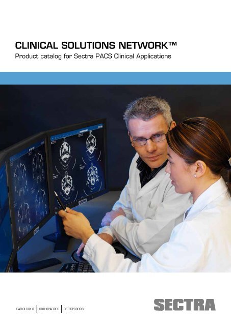
CLINICAL SOLUTIONS NETWORK<strong>Product</strong> catalog <strong>for</strong> <strong>Sectra</strong> <strong>PACS</strong> <strong>Clinical</strong> <strong>Applications</strong>RADIOLOGY IT ORTHOPAEDICS OSTEOPOROSIS
<strong>Sectra</strong> <strong>Clinical</strong> Solutions Network, page 2
INTRODUCTIONAdd new functionality to your <strong>PACS</strong>This product catalog describes the clinical applicationsavailable <strong>for</strong> <strong>Sectra</strong> <strong>PACS</strong> workstations through <strong>Clinical</strong>Solutions Network (CSN) members. These applicationsare available on your computer desktop, perfectly integratedwith the <strong>Sectra</strong> <strong>PACS</strong> workstation.Freedom of choiceAs a <strong>Sectra</strong> <strong>PACS</strong> user, you have the freedom to choose theapplication you prefer running on the <strong>PACS</strong> workstations.Companies that currently are not members of the <strong>Sectra</strong>CSN program can also be included on a case-by-case basis.For more in<strong>for</strong>mation, please contact csn@sectra.se.More in<strong>for</strong>mationFor more in<strong>for</strong>mation about CSN and <strong>Sectra</strong>, please visithttp://www.sectra.se/medical/csn or the <strong>Sectra</strong> User Web,https://userweb.sectra.se. You can also contact your <strong>Sectra</strong>sales representative or our CSN manager at csn@sectra.se.Solutions <strong>for</strong> all clinical needsIn addition to the products listed in this catalog, <strong>Sectra</strong> offersa broad range of advanced product options <strong>for</strong> variousclinical disciplines, such as the <strong>Sectra</strong> 3D Package, the<strong>Sectra</strong> OrthoStation Package and the <strong>Sectra</strong> OsteoporosisPackage, to mention just a few. For more in<strong>for</strong>mation,please contact your <strong>Sectra</strong> sales representative.FDA approval and CE markingThe CSN members are responsible <strong>for</strong> FDA approval andCE marking of their clinical applications. <strong>Sectra</strong> has providedall CSN members with the opportunity to test theirapplications on <strong>Sectra</strong> <strong>PACS</strong> workstations and they arekept in the process of <strong>Sectra</strong> development cycles.Solutions available from <strong>Sectra</strong>Some of the CSN solutions are directly available from<strong>Sectra</strong>, which means you purchase high quality productswith the assurance that <strong>Sectra</strong> will take care of installation,support and all other issues. These solutions are describedon pages 10-21.After the installation of the clinical application, a workstationtest is conducted to verify the existing <strong>Sectra</strong> <strong>PACS</strong>.Instructions <strong>for</strong> such a test are provided through the CSNmembershipNote: To add a new clinical application to your workstation, please contact the CSN member directly.<strong>Sectra</strong> <strong>Clinical</strong> Solutions Network, page 3
<strong>Sectra</strong> <strong>Clinical</strong> Solutions Network, page 4
TABLE OF CONTENTSSolutions sold directly by <strong>Sectra</strong>Amirsys 10• Diagnostic decision supportMirada Medical 12• Image fusion and molecular imaging• Nuclear medicine and PET/CT visualizationNuance 14• Speech recognition and structured reportingSyntheticMR 16• Advanced visualization• Cardiology and angiography• Functional MR and perfusion analysisVital Images 18• Advanced visualization• Cardiology and angiography• Functional MR and perfusion analysis• Image fusion and molecular imaging• Virtual colonoscopy<strong>Sectra</strong> <strong>Clinical</strong> Solutions Network, page 5
<strong>Clinical</strong> applications available through CSN partnersEXINI Diagnostics 20• Cardiology and angiography• Nuclear medicine and PET/CT visualizationG2Speech 22• Speech recognition and structured reportingGruppo Soluzioni Tecnologiche 24• Speech recognition and structured reportingiCAD 26• Computer Aided DetectionIlexis 28• OdontologyMammography Reporting System 30• Speech recognition and structured reportingMedImage 32• Image fusion and molecular imaging• Nuclear Medicine and PET/CT visualizationMedis Medical Imaging Systems 34• Cardiology and angiographyMedviso 36• Advanced visualization• Cardiology and angiographyMerge Healthcare 38• Computer Aided DetectionMIM Software 40• Nuclear Medicine and PET/CT visualizationNordicNeuroLab 42• Functional MR and perfusion analysispeerVue 44• Peer reviewPie Medical Imaging 46• Cardiology and angiography<strong>Sectra</strong> <strong>Clinical</strong> Solutions Network, page 6
OVERVIEW PER FUNCTIONALITYAdvanced visualization<strong>Sectra</strong> workstation products have built-in packages <strong>for</strong> 3D visualization and basic cardiology, tailored <strong>for</strong> effective general-purpose usage. The CSNpartners below add the possibility to integrate more specialized clinical applications.• Medviso 36• SyntheticMR 16• TeraRecon 50• Vital Images 18• QiImaging/Ziosoft 48Cardiology and angiography• EXINI Diagnostics 20• Medis Medical Imaging Systems 34• Medviso 36• Pie Medical Imaging 46• SyntheticMR 16• Vital Images 18Computer Aided Detection (CAD)• iCAD 26• Merge Healthcare 38• VuCOMP 52Diagnostic decision support• Amirsys 10Functional MR and perfusion analysis• NordicNeuroLab 42• SyntheticMR 16• Vital Images 18Image fusion and molecular imaging• MedImage 32• MIM Software 40• Mirada Medical 12• Vital Images 18<strong>Sectra</strong> <strong>Clinical</strong> Solutions Network, page 8
Diagnostic decision supportAmirsys<strong>Product</strong>s• STATdx Premier®Functionality highlights• Point-of-care clinical decision supportContact infoContact your local <strong>Sectra</strong> representative <strong>for</strong> more in<strong>for</strong>mation and a pricing example.www.amirsys.comDiagnostic Decision SupportSTATdx Premier is a digital, web-based point-of-care decision support system designed to help radiologists make fast,accurate diagnoses <strong>for</strong> difficult cases. By streamlining the time required to research and complete a difficult imaging diagnosis,STATdx Premier ensures radiologists have the on demand reference tools they need wherever they read.Integration with <strong>Sectra</strong> <strong>PACS</strong>With just a right-click to open the Image Toolbox popup menu, the <strong>Sectra</strong> <strong>PACS</strong> user can instantly access STATdxPremier. Searchable in 47 different languages, STATdx Premier includes expert-based core content in:• Diagnosis• Imaging Anatomy• Expert Differential Diagnosis• RADtools (Calculators such as Bone Aging, Tables such as TNM and Tumor Staging)• Procedures (Pre, inter, and post procedure)• RADsearch• Compare Anywhere• Live References• Search History• FavoritesThis makes the <strong>Sectra</strong> <strong>PACS</strong>-STATdx Premier combination a true point of care, diagnostic clinical decision support systemunlike anything be<strong>for</strong>e.<strong>Sectra</strong> <strong>Clinical</strong> Solutions Network, page 10
STATdx: example of reference case dataSTATdx: example of reference case data<strong>Sectra</strong> <strong>Clinical</strong> Solutions Network, page 11
Image fusion and molecular imagingNuclear medicine and PET/CT visualizationMirada Medical<strong>Product</strong>s• XD3Functionality highlights• Image fusionMarket clearancesCE Marked and cleared <strong>for</strong> marketing in the US. Licensed <strong>for</strong> sale in CanadaMirada Medical is an ISO 13485 and ISO 9001 accredited companyContact infoContact your local <strong>Sectra</strong> representative <strong>for</strong> more in<strong>for</strong>mation and a pricing example.www.mirada-medical.comMake every image countMirada Medical develops internationally recognized medical image analysis software applications. Our solutions span diagnosticradiology, nuclear medicine, radiation oncology as well as medical oncology. We specialize in offering comprehensiveand quantifiable analysis tools <strong>for</strong> diagnosis, staging, treatment planning and assessment of response to therapy. Ourpowerful technologies are developed in Ox<strong>for</strong>d, UK by a team of world renowned scientists and dedicated engineers.Introducing Mirada XD3XD3 is the latest generation of Mirada’s industry leading medical image analysis software. XD3 blends the needs of clinicianswith rigorous engineering and development to create a software application with the end user in mind. XD3 allowsyou to fuse various combinations of medical images, which may vary in terms of modality, manufacturer, or even thedates the scans were taken. XD3 offers functionality <strong>for</strong> clinicians and radiologists, which opens up a host of new imagingpossibilities to aid better detection, diagnosis and management of disease.Advanced features• Support <strong>for</strong> multi timepoint gated , multi-sequence MR, and multi-phase CT data• Software based PET/MR• Support <strong>for</strong> unlimited number of simultaneous follow-up scan review• Built in support <strong>for</strong> disease specific workflows such as Head and Neck and Melanoma• Data QC <strong>for</strong> automatically warning <strong>for</strong> inter-scan variations• Voxel intensity classification tool <strong>for</strong> identifying tissue based on density within a region• Export to RT• Casebook <strong>for</strong> presenting volumetric DICOM datasets in Microsoft PowerPoint presentations<strong>Sectra</strong> <strong>Clinical</strong> Solutions Network, page 12
Superior Quantification• Patented bi-directional ruler allows definition of short and long axes of lesions and lymph nodes with ease• SUV min, max, mean, median and peak by body weight, lean body mass or body surface area, TLG, metabolic andanatomic volume and more• Support <strong>for</strong> standard response assessment protocols such as RECIST, WHO and PERCIST• One click region trackingWorld class image fusion• Rigid and de<strong>for</strong>mable fusion algorithms <strong>for</strong> any combination of PET/CT/MR/SPECT• Tri-modal fusion and visualization, such as PET/CT/MR all in one workspace• MR registration to both PET and SPECT• Registration algorithms optimized <strong>for</strong> clinical use cases and workflows• Pre registration of planning CT to diagnostic CT After rigid registration After de<strong>for</strong>mable registrationMirada XD3 on your <strong>PACS</strong> solution – The benefits to you.XD3 on the <strong>Sectra</strong> <strong>PACS</strong> is a truly integrated solution. It’s seamless integration with <strong>Sectra</strong> <strong>PACS</strong> means you can haveeverything you need <strong>for</strong> Image fusion, visualization and quantification all on one workstation.Mirada XD3 provides the optimum suite of image analysis tools, which are convenient and reliable enabling you to reachyour targets, and above all provide exemplar patient care.View multiple sequences of MR data loaded into a singletime point and fuse with other MR data or CT, PET, orSPECT data as well.Easily create the RECIST measurements with the Miradapatent pending bi-ruler. This tool automatically assignsthe long axis and short axis of each lesion and lymphnode.<strong>Sectra</strong> <strong>Clinical</strong> Solutions Network, page 13
Speech recognition and structured reportingnuance<strong>Product</strong>s• SpeechMagicFunctionality highlights• Speech recognitionContact infoContact your local <strong>Sectra</strong> representative <strong>for</strong> more in<strong>for</strong>mation and a pricing example.www.nuance.co.uk/healthcare<strong>Sectra</strong> RIS/<strong>PACS</strong>, in partnership with Nuance, features a totally integrated dictation package with speech recognitioncapabilities. It provides tools <strong>for</strong> optimized radiologist and transcription workflows that reduce time-to-report <strong>for</strong> cliniciansand patients.It is a streamlined speech recognition solution with excellent support <strong>for</strong> more than 20 languages. High recognition ratesand ease-of-use are benefits crucial <strong>for</strong> an efficient reporting workflow. Full flexibility allows the reporting radiologist todecide, <strong>for</strong> each dictation, whether to authorize directly or pass on to a transcriptionist’s worklist.About NuanceNuance healthcare, a division of Nuance Communications, gives doctors access to speech recognition technology anywhere,anytime, on any device. Nuance Healthcare empowers healthcare provider organisations and individual doctorsto accurately capture and trans<strong>for</strong>m the patient story into meaningful, actionable in<strong>for</strong>mation in 22 languages. Today,over 10,000 care giver organisations and 450,000 users worldwide trust Nuance voice recognition technology to deliverhigher quality care, improve financial per<strong>for</strong>mance and enhance compliance ef<strong>for</strong>ts. Discover how Nuance Healthcare’swide range of speech recognition solutions and services can increase clinician satisfaction and EHR adoption atwww.nuance.co.uk/healthcare.<strong>Sectra</strong> <strong>Clinical</strong> Solutions Network, page 14
<strong>Sectra</strong> <strong>Clinical</strong> Solutions Network, page 15
Advanced visualizationCardiology and angiographyFunctional MR and perfusion analysisSyntheticMR<strong>Product</strong>s• SyMRI DiagnosticsFunctionality highlights• Increased productivity, tissue characterization and quantificationMarket clearancesCE marked.Contact infoContact your local <strong>Sectra</strong> representative <strong>for</strong> more in<strong>for</strong>mation and a pricing example.www.syntheticmr.comThe synthetic MRI approachSynthetic MRI will increase your productivity by replacing three scans with one. Synthetic MRI is the approach of quantificationof Magnetic Resonance parameters such as T1 and T2 relaxation and proton density. Based on these absolute valuesany T1- and T2-weighted images can be recreated, or synthesized after the actual scan. Hence, control over the imagecontrast (TE, TR, TI) is moved from the scanner to the <strong>PACS</strong>. The absolute values can also be used to characterize tissue,resulting in an automatic segmentation into defined tissue types.SyMRI DiagnosticsSyMRI Diagnostics provides an interface to work with quantitative MRI and tooling to interact with the results. It aims at afaster workflow through more efficient scanning and a higher diagnostic quality using the automated support to characterizeand measure the volume of defined tissues. SyMRI Diagnostics contains two packages: Contrast Studio and Brain Studio.The company SyntheticMR AB is devoted to the visualization of quantified Magnetic Resonance parameters. Special carehas been taken to streamline the reading of quantified MR data to a user-friendly and easily recognizable interface that isvery similar to the normal <strong>PACS</strong> environment.Integration with <strong>Sectra</strong> <strong>PACS</strong>SyMRI Diagnostics is available as a plug-in into <strong>Sectra</strong> <strong>PACS</strong> IDS7 workstations.<strong>Sectra</strong> <strong>Clinical</strong> Solutions Network, page 16
Vital ImagesAdvanced visualizationCardiology and angiographyFunctional MR and perfusion analysisImage fusion and molucular imagingVirtual colonoscopy<strong>Product</strong>s• Vitrea AdvancedFunctionality highlights• CT Cardiac Coronary• CT Cardiac Calcium Score• CT Aorta• CT General Carotid• CT Renal• CT Runoff• CT Endovascular Stent Planning (EVSP)• CT Circle of Willis• CT Cardiac Functional Analysis• CT Larynx Airway• CT Musculosceletal• CT Lung Nodule• CT Colon• CT Brain Perfusion (2D)• CT Cardiac EP Planning• MR Abdominal• MR Vascular• MR Brain Tumor• several advanced add-onsMarket clearancesVital Images is an ISO 9001 company, and its products are FDA 510 (k) approved and have the CE mark.Contact infoContact your local <strong>Sectra</strong> representative <strong>for</strong> more in<strong>for</strong>mation and a pricing example.www.vitalimages.comVital Images connects care providers with powerful 2D, 3D and 4D image creation, analysis, viewing and distributiontools to speed clinical workflows, enhance patient care and reduce technology infrastructure costs. A leading provider ofadvanced visualization software <strong>for</strong> more than 20 years, our software gives radiologists, medical specialists, and point-ofcareproviders access to robust, secure, medical imaging solutions that can be accessed throughout the enterprise and via theWeb <strong>for</strong> easy use in the day-to-day practice of medicine.<strong>Sectra</strong> <strong>Clinical</strong> Solutions Network, page 18
In addition to the functionality highlights above, Vital Imagesoffers additional options such as:• CT Cardiac Multi-Chamber Functional Analysis• CT Cardiac EP (Electro-physiology) Planning• CT SurePlaque• CT Liver segmentation• Lung Density Analysis ( Lung Emphysema)• Transcather Aortic Valve Replacement (TAVR) Planning• Oncology Fusion Advanced• Oncology Fusion Advanced, additional user• Oncology Fusion Standard• Oncology Fusion Standard, additional user• ICAD VeraLook Colon CAD option• Visia CT Lung CAD• Neuro MR• Neuro MR LT• Dynamic MR• QMass MR Essentials & QFlow• QMass MR Advanced & QFlow• CEDARS SPECT Perfusion + BP Basic• CEDARS SPECT Perfusion + BP De Luxe• CEDARS PETIntegration with <strong>Sectra</strong> <strong>PACS</strong>The Vitrea software product enables an optimized workflow<strong>for</strong> the end user, who demands advanced 3D and 2D visualizationapplications. The integration of Vitrea with the <strong>Sectra</strong><strong>PACS</strong> system reduces the complexity of data managementand increases the efficiency of the Radiology Department,while providing a complete suite of application solutions.Multi-chamber Cardiac FunctionEndovascular Stent PlanningNeuro Imaging Solutions<strong>Sectra</strong> <strong>Clinical</strong> Solutions Network, page 19
Cardiology and angiographyNuclear medicine and PET/CT visualizationEXINI Diagnostics<strong>Product</strong>s• EXINI heart• EXINI bone• EXINI dat• EXINI brainFunctionality highlights• Image display• Quantification• Diagnostic advice (CADx)• Automatic structured reportMarket clearancesCE certified by Notified Body TÜV 0123.FDA 510(k) clearing pending.Contact infoCarl-Erik Westervall: Director, sales and marketingPhone: +46 46 286 54 26E-mail: carl-erik.westervall@exini.comwww.exini.comEXINI Diagnostics is a world leading manufacturer of software <strong>for</strong> image display, quantification, diagnostic advice andautomated structured reports. The company’s products EXINI heart <strong>for</strong> interpretation of myocardial perfusion SPECTimages and EXINI bone <strong>for</strong> bone scintigraphy images are based on expert knowledge in nuclear medicine, image analysisbased on large databases and artificial neural networks. The programs provide the user with a tool <strong>for</strong> image display, quantification,diagnostic advice (CADx) and automatic structured reports.EXINI Diagnostics also offers the products EXINI dat <strong>for</strong> interpretation of DaT SPECT images and EXINI brain <strong>for</strong>CBF SPECT images. These programs provide the user with a tool <strong>for</strong> image display, quantification and structured reports.The EXINI products are software programs that will save time, costs and ef<strong>for</strong>ts <strong>for</strong> the physicians at any nuclear medicineclinic. The programs will also contribute to quality assurance.Integration with <strong>Sectra</strong> <strong>PACS</strong>The EXINI products integrate the advanced image processing functionalities of EXINI heart, EXINI bone, EXINIdat and EXINI brain, into <strong>Sectra</strong> <strong>PACS</strong> system, which enables the EXINI products to communicate directly with the<strong>Sectra</strong> <strong>PACS</strong> workstation.<strong>Sectra</strong> <strong>Clinical</strong> Solutions Network, page 20
The direct integration of the EXINI programs will provide <strong>Sectra</strong>’s European customers with a complete software <strong>for</strong> fastand reliable image display, quantification and diagnostic advice <strong>for</strong> nuclear medicine images. All programs also include astructured report module.EXINI BrainEXINI BoneEXINI HeartEXINI Dat<strong>Sectra</strong> <strong>Clinical</strong> Solutions Network, page 21
Speech recognition and structured reportingg2 speech<strong>Product</strong>s• MediSpeechFunctionality highlights• Speech recognitionContact infowww.g2speech.comExpert in speech recognition, digital dictation and workflow management, G2 Speech gets to the heart of report documentation,developing bespoke integrated solutions <strong>for</strong> a modern, efficient health service.G2 Speech aims to apply its expertise in digital document creation and workflow optimisation to benefit medical specialistsand hospitals. Using the latest speech recognition technology, it will create significant time and financial efficiencies, whileenhancing the effectiveness of the electronic patient file.The organisation is highly customer-focused, offering strong support to its client base and providing complete solutionswhich underpin their way of working. This approach has resulted in many satisfied users across Europe, who are supportedby an extensive team of specialists from offices in the Netherlands, Belgium, the UK, Ireland, Austria and Hungary.Background:The foundations of G2 Speech were laid by two experienced professionals from the world of medicine. When they establishedthe company in 1998, their philosophy was clear from the outset:• To enable professionals to work with technology that supports them• To take professionals’ existing ways of working as the starting point and facilitate improvements from there• To deliver added value through cost management (at least a thirty percent saving on secretarial support demonstrable)and a quality incentive (efficiency and time-savings <strong>for</strong> the medical specialist, given that the technology is seven times fasterthan typing).<strong>Sectra</strong> <strong>Clinical</strong> Solutions Network, page 22
Statistics:• First choice <strong>for</strong> more than 22,000 healthcare professionals• Over 600 successfully installed medical projects worldwide• More than 100 integrations with other clinical in<strong>for</strong>mation systems• Providing solutions to over 30 NHS Trusts• In excess of 93% of Netherlands based hospitals use G2 technology• Experienced work<strong>for</strong>ce of more than 60 professionalsDevelopment and support centres in:The Netherlands - EindhovenUnited Kingdom - London/LeedsIreland - DublinBelgium - LeuvenAustria - ViennaHungary - Budapest<strong>Sectra</strong> <strong>Clinical</strong> Solutions Network, page 23
Speech recognition and structured reportingGruppo Soluzioni Tecnologiche<strong>Product</strong>s• HyperSpeech -DI• HyperSpeech -OCX• Med VocalFunctionality highlights• Speech recognitionContact infoMarco RestaniPhone: +39 0461 431333E-mail: info@gsttn.itwww.gsttn.itWith more than 15 years research and experience in the speech recognition industry, GST has designed the speech reportingproduct suite, HyperSpeech. <strong>Product</strong>s include HyperSpeech DI and HyperSpeech OCX (a special web based solution).All HyperSpeech products are available in the following languages: English UK, English US, German, Spanish and Italian.With more than 4000 installations in over 400 medical organizations PhoneidoS is the clear market leader in Italy and hasmarket presence internationally, with sites in the UK, Germany, Spain and the United States HyperSpeech comes with anextensive range of hardware options and integration possibilities, and clearly improves report quality delivery time and leadsto significant cost reductions. HyperSpeech offers a complete reporting solution right through to the final report. Nearly allHyperSpeech function commands can activated by voice allowing the user to be total “hands-free” whilst reporting.Integration with <strong>Sectra</strong> <strong>PACS</strong>HyperSpeech DI software simplifies the approach to reporting using speech recognition, communicating directly with<strong>Sectra</strong> <strong>PACS</strong>, providing streamlined workflow and incorporating advanced speech functionalities. HyperSpeech can beused in either front-end modality, in which the physician manages the reporting process from beginning to end, or deferredmodality, in which the report is sent to a correctionist to be checked be<strong>for</strong>e being finalized. A combination of both modalitiesat the same site is also possible.Integration with Andra, italian <strong>Sectra</strong> distributorIn Italy we are integrated with MRV (Modulo Refertazione Vocale) module developed by Andra, an HL7 add-on <strong>for</strong> the<strong>Sectra</strong> <strong>PACS</strong> on the Italian market.<strong>Sectra</strong> <strong>Clinical</strong> Solutions Network, page 24
“We found the Phoneidos Speech Recognition System very easy to use, with the recommended high-per<strong>for</strong>mance Microphoneallowing <strong>for</strong> a high recognition rate to be achieved even when there is a lot of background noise in the room. Wehave reduced report correction times to a minuimum and now finally we have the fast efficient digtal workflow that weset out to accomplish <strong>for</strong> our new Radiology Centre”Dr. Bruch, Radiologie Hofheim, Hofheim, Germany<strong>Sectra</strong> <strong>Clinical</strong> Solutions Network, page 25
Computer Aided Detectionicad<strong>Product</strong>s• PowerLook AMPFunctionality highlights• Mammography CADContact infoDonna BreaultE-mail: dbreault@icadmed.comPhone: +1 603-882-5200www.icadmed.comPowerLook Advanced Mammography Plat<strong>for</strong>m TM (AMP) is the next generation digital mammography CAD plat<strong>for</strong>moffering radiologists the flexibility to choose the products and functions that best fit their reading environment. A widerange of tools <strong>for</strong> disease detection and analysis provide users with workflow enhancements that improve overall efficiency.Multi-vendor CAD server allows <strong>for</strong> easy practice expansion.PowerLook AMP includes a multi-vendor CAD server that provides consistency across all digital mammography systems.PowerLook AMP allows hospitals and imaging facilities to:• Process cases using a single server• Connect up to 4 connections from any combination of supported mammography acquisition devices• Eliminate the need to purchase a separate server <strong>for</strong> each digital mammography system• Reduce hardware and service costsIn the U.S., supported mammography vendors are GE, Siemens, Fujifilm, or Hologic (Selenia). Outside of the U.S.,additional vendors are available, including Philips Microdose, IMS Giotto, Philips CR and DR, Planmed, and Agfa.Modular design provides flexibility <strong>for</strong> today and tomorrow.The PowerLook AMP plat<strong>for</strong>m is specifically designed to give radiologists the ability to customize their CAD solution.The modular structure provides the freedom to choose products and functionality as needed today and into the future.PowerLook AMP offers the following modules:• SecondLook Digital v7.2 CAD with CAD Metrics• SecondLook Premier CAD with CAD Metrics (available OUS only)• Volpara Automated Breast Density Assessment software from Matakina (available US only)PowerLook AMP establishes the foundation <strong>for</strong> future CAD software upgrades, enhancements and other functionalityoptions to suit the growing needs of busy mammography practices.<strong>Sectra</strong> <strong>Clinical</strong> Solutions Network, page 26
®®Superior CAD algorithms support early detection.SecondLook Digital algorithms analyze mammography images using methodologies that are complementary to the radiologist.Potential cancers are identified using patented artificial intelligence and pattern recognition technology to analyzeimages and identify patterns. Sophisticated mathematical analysis identifies and marks suspicious areas without obscuringthe underlying image, enabling faster, more accurate reading.Seamless DICOM integration enhances clinical workflow.SecondLook Digital provides the most powerful and flexible DICOM connectivity solutions – enhancing digital workflowand enabling seamless integration with acquisition systems, review workstations, and <strong>PACS</strong> from leading vendors. Flexibleintegration options enable CAD results to be viewed on workstations or sent to a printer. Priority queuing of studiesimproves clinical efficiency and efficacy by enabling the most urgent or important studies to be analyzed with CAD first.Enhanced CAD metrics provide clinical insight andimprove workflow.CAD Metrics provide the richest set of clinical decisionsupport tools to support mammography screening.• Calculate mammographic characteristics <strong>for</strong> eachCAD detection• Identify CAD’s certainty of its finding1• Provide automated measurements such as longest axisof detection, nipple position and distance to nipple toimprove efficiencyAll CAD Metrics are defined in the DICOM standard andthe in<strong>for</strong>mation is contained in the Mammography CADStructured Report 2 . Flexible purchase and licensing options <strong>for</strong> allpractices.iCAD offers both perpetual and annual licensing options<strong>for</strong> PowerLook AMP. For those organizations requiringfull purchase of the hardware and software plat<strong>for</strong>m, a Image courtesy of ThreePalm Softwaretraditional perpetual license is available. For those facilitiesseeking a lower up-front cost, iCAD offers the Platinum Annual License that allows unlimited use of the PowerLook AMPhardware and software <strong>for</strong> a period of one year.1Available outside US only.2The ability to view CAD Metrics is dependent on the review workstation in use.<strong>Sectra</strong> <strong>Clinical</strong> Solutions Network, page 27
OdontologyIlexis<strong>Product</strong>s• FACAD®Functionality highlights• Cephalometric analysis, treatment simulation and surgery predictionContact infoBengt Schmeling, <strong>Product</strong> ManagerPhone: +46 (0)70-554 11 59E-mail: bengt.schmeling@facad.comwww.facad.comFacad® is a very powerful, flexible and easy-to-use Windows-based program <strong>for</strong> orthodontics and maxillofacial surgery.Facad can be used <strong>for</strong> visual diagnostic imaging, tracing and cephalometric analysis, as well as <strong>for</strong> treatment simulation withsurgery prediction. The digitized x-ray image can either be a scanned x-ray film or a digital x-ray image file in JPEG, TIFF,BMP or DICOM <strong>for</strong>mat. Facad is also designed to receive patient data and images from other programs used as patientjournal systems or <strong>PACS</strong>, and to interface with digital x-ray imaging systemsIntegration with <strong>Sectra</strong> <strong>PACS</strong>Facad is available as an integrated plug-in program with <strong>Sectra</strong> <strong>PACS</strong>. The application can be accessed quickly by a rightclick in the image window.<strong>Sectra</strong> <strong>Clinical</strong> Solutions Network, page 28
Cephalometric measurements in Facad <strong>for</strong> surgery planning.<strong>Sectra</strong> <strong>Clinical</strong> Solutions Network, page 29
Speech recognition and structured reportingmammography reporting system<strong>Product</strong>s• MRS7 QA Plus• MRS7 Reporting• MRS7 Ambulatory EHR• Quest TouchFunctionality highlights• Comprehensive tracking of patient data• High Risk Assessment computations and flags• Easy navigation and data entry• Extensive statistical reporting to support annual certification requirements• Ability to export statistical reports to excelContact infoPhone: (800) 253-4827E-mail: sales@mrsys.comwww.mrsys.comMammography Reporting System (MRS) offers workflow optimization solutions <strong>for</strong> any sized facility and integrates seamlesslywith multiple vendors. Our patient-oriented system offers features such as automated patient lay letters, problem casetracking, and statistical risk models that help optimize workflows and contribute to quality patient care. With over 25 yearsin business, we are constantly innovating and improving its products in order to better serve the needs of its customers.MRS7 QA PlusMRS7 QA Plus is a best-of-breed tracking and reporting system designed to enhance processes by offering features suchas robust statistical reports, automated patient-lay letters, and detailed risk models. It is a cost-effective solution that helppromote time-savings and efficiency by providing a user-friendly interface that is designed <strong>for</strong> quick and easy navigation.MRS7 QA Plus complies with the MQSA, ACR, and privacy requirements.MRS7 ReportingThis full-featured product incorporates directly into the radiologist’s reporting workflow to promote efficiency and timesavings.With a multitude of innovations it raises the benchmark <strong>for</strong> breast imaging tracking and reporting. All of thefeatures of MRS QA Plus are included and it also features such abilities as to generate procedure reports <strong>for</strong> referring cliniciansand to use specialized reporting tools, such as one-touch procedure report templates to help expedite the reportingprocess.MRS7 Ambulatory EHRMRS7 EHR is a Certified Ambulatory Electronic Health Record that provides your facility with the tools to achieveFederal Government Meaningful Use incentive payments that were instituted in order to promote the adoption of Healthcare<strong>Sectra</strong> <strong>Clinical</strong> Solutions Network, page 30
In<strong>for</strong>mation Technology and Electronic Health Records. Our full-featured system is designed to fulfill any facility’s trackingand reporting needs, with added features to help qualify <strong>for</strong> Meaningful Use incentive payments.Quest TouchMRS Quest Touch is a tablet-based, patient-oriented electronic patient history questionnaire <strong>for</strong> a paperless workflow.It stores patient in<strong>for</strong>mation from previous visits, eliminating the need to fill out the same questions <strong>for</strong> every visit. Thepaperless workflow saves your facility money by reducing the amount of resource consumption, and reducing the amount oftime-consuming data entry. 16 <strong>for</strong>eign language packs are available to help collect data from Non-English speaking users.Integration with <strong>Sectra</strong> <strong>PACS</strong><strong>Sectra</strong>-MRS integration focus on high efficiency radiologist workflows, accuracy and efficiency and radiology departmentproductivity. It is designed as a bidirectional procedure context synchronization and uses the <strong>Sectra</strong> Desktop Sync interface.The study is typically selected from the worklist in <strong>Sectra</strong> <strong>PACS</strong>. This opens the corresponding images in the <strong>Sectra</strong> workstationImage Widows and opens the MRS procedure report which:• Displays the patient history, patient risk factors, and indicated problem <strong>for</strong> this visit.• Has reports of prior studies available if previous reports are desired <strong>for</strong> this reading.• Allows the radiologist to view the images in <strong>Sectra</strong> <strong>PACS</strong> and record the findings, assessment, and conclusion on thepatient’s procedure report.• Allows the radiologist to preview the report <strong>for</strong> the patient and <strong>for</strong> the referring physician.• Once completed, the statistics <strong>for</strong> the radiologist, technologist, and patient are recorded in MRS and available <strong>for</strong>review and during annual audits.<strong>Sectra</strong> <strong>Clinical</strong> Solutions Network, page 31
Image fusion and molecual imagingNuclear medicine and PET/CT visualizationMedImage<strong>Product</strong>s• MedView <strong>PACS</strong> Plug-inFunctionality highlights• NM SPECT/PET/CT display analysis solution• CINES, color tables, ROI, VOI, series level exam selection• Fuse studies and modalities,• Concurrent-use or single seat licensing• Burn exams to CD with self-contained viewer• Save AVI and DICOM snapshots to <strong>PACS</strong> <strong>for</strong> future review• Supports multiple monitors and current/prior display• Corridor 4DM (Cardiac quantification <strong>for</strong> PET/SPECT)Market clearancesFDA 510 (k) approval and CE marked.Contact infoTod HendersteinE-mail: tod_henderstein@medimage.comPhone: +1 734 665 5400 ext 121www.medimage.comNuclear Medicine/PET/CT Display and AnalysisMedView <strong>PACS</strong> Plug-in is a simple, economical yet powerful product that improves the speed and quality of NuclearSPECT/PET/CT and Cardiac review. Because MedView is fully integrated with and can be launched right from your IDSworkstation, physicians now have the convenience of mobility provided by access to the same reading tools on any client.MedImage, with a solid track record of over a thousand installed licenses worldwide, provides not only excellence in theirproducts but in their customer service as well. MedViewʼs intuitive interface allows you to CINE, adjust contrast, changecolor tables and display templates, fuse studies and modalities, volume render (MIP) data, and much more.As a dedicated Molecular Imaging display program, MedView is able to easily handle complex and varied file <strong>for</strong>mats thatare not typically addressed adequately by programs designed <strong>for</strong> general radiology reading.MedView <strong>PACS</strong> Plug-in and INVIAʼs Corridor4DM (PET or SPECT) software combine to deliver a powerful solution tomeet the specialized needs of the cardiologist. In the department or throughout the hospital, use the same program to per<strong>for</strong>mcardiac analysis on data originating from any manufacturer. Corridor4DM features quantitative myocardial functionand perfusion with multiple 3-D surface displays <strong>for</strong> volumetric visualization and analysis.Whether you want to see fused modalities, individual slices or a GATED cardiac CINE, MedView has every feature youneed <strong>for</strong> the most definitive NM/PET/CT image display possible.<strong>Sectra</strong> <strong>Clinical</strong> Solutions Network, page 32
Integration with <strong>Sectra</strong> <strong>PACS</strong>As part of the <strong>Sectra</strong> <strong>Clinical</strong> Solutions Network, the MedView Plug-in gives you seamless and immediate access to all ofyour nuclear data stored on the <strong>PACS</strong>. You can select and display patient studies directly from the <strong>PACS</strong> database withoutleaving the <strong>PACS</strong> environment. When placed on the <strong>Sectra</strong> (IDS5 or IDS7, Windows XP and 7, 32 or 64-bit) workstation,MedView eliminates the need <strong>for</strong> an independent nuclear reading station and becomes an integral part to the radiologistʼsdaily workflow <strong>for</strong> reading Nuclear Medicine studies.“The MedImage Nuclear Medicine plug-in on our <strong>Sectra</strong>/Philips <strong>PACS</strong> Workstation has been a wonderful additionto our <strong>PACS</strong> reading station. There is no longer a requirement to maintain a separate monitor and keyboard with asupplementary nuclear medicine workstation. This has allowed us to free up much more space <strong>for</strong> other needed items suchas books and easier access to our RIS workstation. Most important to our workflow is the ability to analyze all our nuclearmedicine studies on a single workstation, allowing us substantial time savings during the day. The MedImage plug-inhas maintained the full functionality of the previous freestanding image workstation. A simple one-click operation on the<strong>Sectra</strong> <strong>PACS</strong> allows immediate access to the new plug-in. All my partners have been delighted with the ease of use of theplug-in.”Daniel Singer, MD, Radiology & Diagnostic Imaging,The Toledo Hospital/Toledo Children’s Hospital<strong>Sectra</strong> <strong>Clinical</strong> Solutions Network, page 33
Cardiology and angiographyMedis medical imaging systems<strong>Product</strong>s• QAngio XA• QMass MR• QFlowFunctionality highlights• Interpretation and analysis of cardiac MR data (Ejection Fraction, Perfusion, LGE, T1 Mapping, T2*, etc)• Quantitative analysis of MR flow studies (Regurgitation fraction, Pressure gradient, Mean Volume graphs, etc)• Quantitative analysis of X-ray angiograms <strong>for</strong> coronary and peripheral vessels (Straight analysis, Bifurcation analysis,QLV, QRV, etc)Market clearancesFDA 510 (k) approval and CE marked.Contact infoContact your local <strong>Sectra</strong> representative <strong>for</strong> more in<strong>for</strong>mation and a pricing example.www.medis.nlQuantitative analysis software <strong>for</strong> cardiology and radiologyWhether <strong>for</strong> evidence-based reporting or to complement visual assessment, quantification has become indispensable. Theintegration of Medis quantification software into <strong>Sectra</strong> <strong>PACS</strong> enables you to get the facts from the images without the need<strong>for</strong> a separate workstation—quickly, easily and accurately.Medis has brought dedicated quantification tools <strong>for</strong> X-ray angiography and cardiac MR to cardiologists and radiologistssince 1989. Its flagship products are QAngio XA, <strong>for</strong> the quantitative analysis of X-ray angiograms and QMass MR, <strong>for</strong>the quantitative analysis of cardiac MR. These quantification tools are now readily available in <strong>Sectra</strong> <strong>PACS</strong> to support anefficient clinical workflow.Integration with <strong>Sectra</strong> <strong>PACS</strong>Click and go: the seamless integration of QAngio XA, QMass MR and QFlow into <strong>Sectra</strong> <strong>PACS</strong> means that quantitativeanalysis is only one click away, and completely embedded in your clinical workflow.Save time: the tight integration eliminates the need <strong>for</strong> separate procedures to retrieve patient data and store results. Quantificationitself is made easy and efficient by intuitive wizards that guide you through the analysis. And when you finish, allyou need to do is click and save to store the quantification results directly in your <strong>Sectra</strong> <strong>PACS</strong>. Analyzing and reportinghas never been faster.Quality at your fingertips: Medis is known <strong>for</strong> its best-in-class software, both in QCA analysis, where it has set the standard,and in cardiac MR, <strong>for</strong> which it has created software that is used by institutions worldwide. The integration with<strong>Sectra</strong> <strong>PACS</strong> gives you high-quality analysis software right at your fingertips—only one click away.<strong>Sectra</strong> <strong>Clinical</strong> Solutions Network, page 34
QAngio XA (QCA) quantification software showing an analyzed X-ray angiogramThe Medis QMass MR quantification software showing a left and right ventricular analysis<strong>Sectra</strong> <strong>Clinical</strong> Solutions Network, page 35
Advanced visualizationCardiology and angiographyMedviso<strong>Product</strong>s• Segment-CMRFunctionality highlights• Interpretation and quantification of cardiac MR images• Quantitative analysis of MR flow studies• Infarct quantification• T2* quantification• Perfusion analysis• 3D image segmentation and surface generation• Export segmented surfaces in STL <strong>for</strong>matMarket clearancesFDA 510(k) approval. CE mark in preparation.Contact infoEinar HeibergPhone: +46-761 83 64 42sales@medviso.comwww.medviso.comMedviso AB develops high quality and flexible solutions <strong>for</strong> medical image analysis and provides highly cost effectivesolutions with flexible license terms. We develop customized medical image analysis software <strong>for</strong> MR, CT and myocardialperfusion SPECT.SEGMENT-CMR is a comprehensive software package <strong>for</strong> quantification of cardiac MR images. It brings reliable, easyto use cardiac MR quantification to clinicians. The key strength of the software is the high quality automated analysis toolsthat provide fast and accurate image quantification. The software is extensively validated in scientific papers and is used byhundreds of researchers. The software runs on 32/64 bit Windows 7, Vista, and XP.Integration with <strong>Sectra</strong> <strong>PACS</strong>Segment integrates seamlessly with <strong>Sectra</strong> <strong>PACS</strong> and the application is accessed by a simple right click. The software canalso read and retrieve data from other DICOM compliant sources. Images with measurements and contours can directly besent back to the <strong>Sectra</strong> <strong>PACS</strong> as an integrated part of the patient study.“Segment works terrific. Automated segmentation is superior to anything I’ve seen – extremely reproducible, quick andeasy to use. It is far superior to anything else on the market.”Alexander Chernobelsky, Medical Specialists Palm Beach, Florida, USA.<strong>Sectra</strong> <strong>Clinical</strong> Solutions Network, page 36
Accurate and robust automated segmentationalgorithm provides fast quantification of cardiacfunction.Automatic vessel tracking in MR phase contrastimages allows <strong>for</strong> quantification of flow and regurgitantfraction in valvular assessment.Extensively validated algorithm <strong>for</strong> automatic scarquantification in MR Late Gadolinium Enhancementimages.<strong>Sectra</strong> <strong>Clinical</strong> Solutions Network, page 37
Computer Aided DetectionMerge Healthcare<strong>Product</strong>s• Merge CADstreamFunctionality highlights• MR mammography CADMarket clearancesFDA 510 (k) approval in 2002 and CE mark received in 2004.Contact infoMary Hoxworth, Director, <strong>Product</strong> ManagementPhone: +1 312 565 6829E-mail: mary.hoxworth@merge.comwww.merge.comCADstream automates breast MRI study analysis which assists radiologists in the interpretation, standardization andreporting of these data-intensive studies. CADstream’s automated features include adaptive image registration (2D/3D),multiplanar re<strong>for</strong>matting, subtractions, angiogenesis maps, interactive realtime contrast curves, maximum intensity projections(MIPs) and volume summaries. CADstream’s advanced features include a streamlined portfolio <strong>for</strong> treatment planning(automatically provides summaries with size, classification and location <strong>for</strong> volume of interest and incorporates ACRBI-RADS® Atlas <strong>for</strong> lesion classification), multimodality ready (ability to access and view patients’ mammography andultrasound studies while interpreting breast MRI studies) and SureLoc <strong>for</strong> interventional planning.Integration with <strong>Sectra</strong> <strong>PACS</strong>When CADstream is integrated with <strong>Sectra</strong> <strong>PACS</strong>, studies are linked by patient study name. A user can be viewing a mammographystudy on the <strong>Sectra</strong> <strong>PACS</strong> and quickly access a linked CADstream-processed breast MRI study by accessingCADstream from the right-click menu. Once CADstream is selected, the user can view and compare the patient’s mammographyand breast MRI studies together.“Since 1999, there has been a 40 percent per year increase in the number of breast MRI examinations in the U.S., andthat number is expected to continue growing rapidly over the next five years. For this reason, we looked <strong>for</strong> technologythat would increase our efficiency and provide standardization. CADstream provides our physicians and technologists witha more efficient method <strong>for</strong> processing and interpreting the images acquired. That’s valuable time saved that can be spentwith patients.”Linda Sutherland, MD, Co-medical DirectorWomen’s Imaging, St. Joseph Hospital, Orange, Cali<strong>for</strong>nia<strong>Sectra</strong> <strong>Clinical</strong> Solutions Network, page 38
“Technology, such as CADstream, keeps us on the cutting edge in our breast center – offering our patients the best chanceat early detection and treatment. We use CADstream with every breast MRI study we interpret.”.Kara Carlson, MD, DirectorWomen’s Imaging, Evergreen Breast Center, Kirkland, Washington“The addition of CADstream has significantly improved my efficiency and enhanced my analysis of MR mammographicstudies. It truly helps simplify a very complex process and provides a cosistency that will allow <strong>for</strong> easier follow-up.”Cynthia Lorino, MD, DirectorBreast Imaging Center, Montogomery Breast Center“Confirma is considered to be a valuable resource to us. The company has helped change the face of MRI here, and hasmade a huge impact on breast MRI in a positive way.”Michael Cecil, MRI SupervisorNorthwest Hospital, Seattle, WAAngiomapVolume summaryMIPCurves<strong>Sectra</strong> <strong>Clinical</strong> Solutions Network, page 39
Image fusion and molecular imagingNuclear medicine and PET/CT visualizationMIM software<strong>Product</strong>s• MIM®Functionality highlights• Multimodality fusion and display (CT, MRI, SPECT, PET)• NM review• PET/CT FusionMarket clearancesFDA 510 (k) approval.Contact infoBreanne Kraly, <strong>Clinical</strong> Support EngineerPhone: +1-216-455-0597E-mail: bkraly@mimsoftware.comwww.mimsoftware.comMIM Software Inc. provides practical solutions <strong>for</strong> image fusion, review, and analysis <strong>for</strong> radiology, nuclear medicine,cardiac imaging, neuroimaging, and radiation oncology. MIM offers solutions <strong>for</strong> computer workstations, as well as mobileand cloud-based plat<strong>for</strong>ms. MIM’s software solutions are sold to imaging centers, hospitals, specialty clinics, researchorganizations, and pharmaceutical companies around the world.MIMfusion® provides multi-modality image review and fusion capabilities on both Windows® and Mac® plat<strong>for</strong>ms. Theuser-friendly software is vendor-neutral, and works with images from virtually any manufacturer. MIM can display and fuseimages from multiple modalities, including PET, SPECT, CT, and MR. With Assisted Alignment, a high-speed automaticfusion process, images are accurately aligned in less than ten seconds, without the need <strong>for</strong> corresponding point selection.MIMfusion provides unparalleled functionality as an image review workstation <strong>for</strong> the analysis and fusion of imagesacquired on separate scanners or derived from combination scanners, unmatched efficiency and many timesaving features.The MIM plat<strong>for</strong>m also offers a number of specialized software imaging programs:MIM Encore provides greater accuracy <strong>for</strong> PET tumor segmentation with PET Edge, an innovative gradient-basedsegmentation method. Auto-segmentation tools <strong>for</strong> PET/CT and MR greatly reduce contouring time. Segmented tumorscan be used <strong>for</strong> analysis of tumor response, or sent to radiation oncology <strong>for</strong> use in radiation therapy planning.MIMneuro® is a fully automated and quantitative PET/SPECT analysis package that assists in the detection of variousneurological disorders and includes databases of normals <strong>for</strong> FDG, AmyvidTM and HMPAO. Statistically significantdifferences from the normals are highlighted <strong>for</strong> each voxel in the brain or <strong>for</strong> anatomical brain regions. Brain atlas regionscan also be used to calculate SUV ratios (SUVR) comparing at risk brain regions to reference structures such as the cerebellum.An automated neuro subtraction workflow is included <strong>for</strong> highlighting differences in serial exams or baseline andactivation studies.<strong>Sectra</strong> <strong>Clinical</strong> Solutions Network, page 40
MIMcardiac® provides quantitative analysis of PET and SPECT <strong>for</strong> generating left ventricular functional parameters.Image fusion can be per<strong>for</strong>med <strong>for</strong> stress/rest and perfusion/metabolism. These functional images can also be fused toCCTA or MRA.MIMviewer® provides economical remote viewing, and MIMviewer Pro adds advanced image review and manipulation ofPET, CT, PET/CT and SPECT/CT image data. Both versions run from a CD and are ideal <strong>for</strong> interactive sharing withpatients and colleagues.Integration with <strong>Sectra</strong> <strong>PACS</strong>The integration of MIM and MIMviewer display software with <strong>Sectra</strong> <strong>PACS</strong> allows endless possibilities on one workstation.Any combination of images archived in <strong>PACS</strong> storage can be loaded into MIM <strong>for</strong> enhanced visualization and fusion. Thisseamless integration can also provide referring physicians with capabilities ranging from simple tools, such as image annotationsand rotating MIP movies, to more complex resources such as DICOM RTstructs and fused images.Contoured CT imageNeuro analysisFusioned images PET/CTMIM viewer screenshot<strong>Sectra</strong> <strong>Clinical</strong> Solutions Network, page 41
Functional MR and perfusion analysisNordic neuroLab<strong>Product</strong>s• NordicICE• nordicBrainExFunctionality highlights• fMRI (Diffusion/DTI)• fMRI (Perfusion DCE)• BOLD fMRI and DTI analysis and visualizationMarket clearancesCE marked and FDA 510 (k) clearedContact infoØyvind Gulbrandsen, Chief Marketing OfficerTel: +47 926 44 261oyvind@nordicneurolab.comwww.nordicneurolab.comNordicNeuroLab develops of state-of-the-art solutions <strong>for</strong> clinical use of functional imaging methods in cooperation withhighly ranked research groups and clinical users. nordicICE, a Windows-based, general-purpose image processing andanalysis software application, can be readily integrated into the clinical workflow in any hospital. It also provides flexibility<strong>for</strong> the research-oriented user who wants to benefit from state-of-the art visualization and post-processing techniques.nordicICE Perfusion/DCE ModuleWith these two modules a large range of physiologically relevant parameters related to tissue perfusion and capillary permeabilitycan be addressed both qualitatively and semi-quantitatively using state-of-the-art methods. You can create highqualityperfusion maps from dynamic contrast enhanced MRI (T1 or T2/T2* weighted) or CT images in seconds. Thecombination of a user-friendly interface, state-of-the-art methods and very fast processing ensures maximum productivitywithout loss of quality. For the clinical workflow we provide a “one-button” perfusion analysis using pre-defined settings.nordicICE Diffusion/DTI ModuleThis module allows you to easily and effectively generate diffusion maps from MR diffusion imaging studies from all majorMR vendors. You can also reconstruct axonal tracts (Fiber Tracking) in the central nervous system. The 3D viewing interfaceoffers unique features <strong>for</strong> exploring white matter connectivity using various Region of Interest tools.Intuitive 3D visualization of reconstructed tracts superimposed on any image volume is provided. Fiber statistics such as FA,ADC and tensor eigenvalues can be displayed. The shown parametric values correspond to the selected output maps thatwere generated during the DTI analysis. Additionally, an interactive selection of individual fiber bundles can be per<strong>for</strong>med.nordicICE allows a user to explore connectivity by defining volumes and regions of interest along with superimposedBOLD fMRI activation in the 3D viewer. A user may save snapshots/animations to various file <strong>for</strong>mats or directly to <strong>PACS</strong>.<strong>Sectra</strong> <strong>Clinical</strong> Solutions Network, page 42
nordicICE BOLD ModuleThe nordicICE BOLD Module allows fMRI exam protocols to be ef<strong>for</strong>tlessly integrated into the everyday clinical routine.Fast, reliable, and standardized BOLD analysis can be per<strong>for</strong>med with minimal user interaction, and the advanced user canoptimize or customize the computational procedures <strong>for</strong> more complex experimental procedures.The analysis procedure includes the standard pre-processing steps such as slice-time and motion correction. Intermodalityco-registration algorithms ensure the best possible correspondance between different types of image data – a criticalrequirement <strong>for</strong> clinical application to ensure anatomical accuracy. Statistical analysis is based on the General Linear Model(GLM). Results can be displayed as color-coded overlays on structual images, and can easily be tresholded by the user.nordicBrainExnordicBrainEx allows post-processing and visualization of BOLD fMRI, and DTI data. The simple and user-friendly interfaceimproves user productivity while the automated protocols, processing steps, and quality control features minimize theprobability of error or variability in the quality of the results. nordicBrainEx is DICOM compatible and analyzes data fromall major MR vendors. Reports are customizable and processed data can be saved in a variety of <strong>for</strong>mats and sent in DICOM<strong>for</strong>mat to <strong>PACS</strong> or exported neuronavigation systems.Integration with <strong>Sectra</strong> <strong>PACS</strong>nordicICE enables integration of advanced functional MRI data-processing functionality in the <strong>Sectra</strong> <strong>PACS</strong> system. Itaccesses data directly from the <strong>Sectra</strong> <strong>PACS</strong> or other data sources accessible from the <strong>PACS</strong> client workstation. MRI dataprocessed in nordicICE can easily be imported back to the <strong>PACS</strong> as an integrated part of the patient study.nordicICE Perfusion/DCEnordicICE BOLDnordicICE Diffusion/DTInordicBrainEx<strong>Sectra</strong> <strong>Clinical</strong> Solutions Network, page 43
Peer reviewpeerVue<strong>Product</strong>s• QICS Functionality highlights• Regulatory, quality and communication solutionsMarket clearances<strong>Product</strong>s do not require market clearance.Contact infopeerVue, Inc.Phone: +1 877 572 9505 (ext 2)sales@peervue.comwww.peervue.comImproving Workflow, Quality & Communications in HealthcarepeerVue QICS – Powerful, Automated, SimpleA hospital-wide IT solution, peerVue QICS helps unite fragmented medical departments and facilities to enable more automatedworkflow, consistent closed-loop communications, integrated patient care and strategic healthcare business management.Utilizing a sophisticated, configurable rules engine, this powerful solution reaches across healthcare department andIT boundaries to collect, combine and analyze data in powerful, actionable ways.For example, in the radiology department, QICS helps automate communication and workflow throughout the exam lifecycle,enabling an unlimited range of workflow solutions –from Peer Review, ED Discrepancy Communication and CriticalResults Management to Coding Discrepancy Management, PQRS tracking, and Technologist Review. Powered by theflexible QICS rules engine, these solution spaces are uniquely customized to support your specific business processes, rulesand departmental requirements.peerVue’s flexibility and integration saves money by simplifying the IT environment and eliminating the need <strong>for</strong> clumsyworkaround or multiple disparate applications, while delivering closed-loop communication and workflow.Integration with <strong>Sectra</strong> <strong>PACS</strong>peerVue’s QICS plat<strong>for</strong>m integrates seamlessly with <strong>Sectra</strong> <strong>PACS</strong> (IDS5 & IDS7). <strong>Applications</strong> are accessed by a right clickin the image window.<strong>Sectra</strong> <strong>Clinical</strong> Solutions Network, page 44
<strong>Sectra</strong> <strong>Clinical</strong> Solutions Network, page 45
Cardiology and angiographyPie medical imaging<strong>Product</strong>s• Cardiovascular Angiography Analysis System - CAASFunctionality highlights• Contour detection of the coronary arteries and analysis of its dimensions• 3D reconstruction of the coronary arteries• Contour detection of the left ventricle, volume calculation and wall motion analysis• Contour detection of the right ventricle, volume calculation and wall motion analysis• Contour detection of the peripheral vessels and analysis of its dimenstions• Quantitative 3D analyisis of peripheral single vessels up to 50 mmMarket clearancesISO 9001, ISO 13485 and Canadian CAN/CSA ISO 14385CE (Europe), FDA (US) and TGA (Australia) clearancesContact infoPie Medical ImagingPhone: +31 (0)433 281 328www.piemedicalimaging.comQuantitative analysis software <strong>for</strong> cardiology and radiologyPie Medical Imaging is world market leader in quantitative analysis software <strong>for</strong> cardiology and radiology, and is well known<strong>for</strong> its Cardiovascular Angiography Analysis System - CAAS. CAAS has been integrated in the <strong>Sectra</strong> <strong>PACS</strong> solution,and delivers validated, accurate and reproducible quantitative analysis of the heart and vascular system from angiographicimages.CAAS offers highly accurate and reproducible measurements of the dimensions of coronary arteries and peripheral vesselsas well as the analysis of left and right ventricles. The accuracy and reproducibility of the CAAS software are well documentedin a large number of scientific publications by renowned institutes that evaluated and use the CAAS software. Themajor medical equipment manufacturers have chosen CAAS as their solution in the cath lab equipment.For the quantitative analysis of X-Ray angiographic images, Pie Medical Imaging has several CAAS software packages available.CAAS QCA 2D (+ option bifurcation), CAAS QCA 3D single vessel with bifurcation analysis, CAAS LVA (+ optionbi-plane), CAAS QVA and CAAS QVA 3D. All packages are modular available on the CAAS 5 plat<strong>for</strong>m. CAAS 5 has onelook and feel <strong>for</strong> all modules, is user friendly and intuitive.CAAS QCACAAS QCA (Quantitative Coronary Analysis) software offers automatic contour detection of the coronary arteries (up to7mm) and analysis of its dimensions. Three analysis methods are implemented; Computer Defined Obstruction Analysis,User Defined Ob-struction Analysis and User Defined Subsegment Analysis. Additionally the CAAS QCA <strong>for</strong> Research<strong>Sectra</strong> <strong>Clinical</strong> Solutions Network, page 46
option offers Drug Eluting Stent Analysis, Multiple subsegments, Brachytherapy and Subsegmental data.CAAS QCA 3DCAAS QCA 3D (three dimensional Quantitative Coronary Analysis) offers automated detection and 3D analysis of singleand bifurcated coronary arteries. Out-of-plane magnification and <strong>for</strong>eshortening errors are minimized by calculating truegeometric shape in 3D space from two or more 2D X-ray projections. CAAS QCA 3D offers improved analysis of difficultlesions and segment anatomy.CAAS LVAThe CAAS LVA (Left Ventricular Analysis) software package provides automatic and manual contour detection of the leftventricular ED and ES contours. ED and ES volumes are calculated as well as wall motion. Regional, Centerline, Slager andRadial Wall Motion models are implemented. Beside the monoplane also a version <strong>for</strong> biplane analysis is available.CAAS RVAThe CAAS RVA (Right Ventricular Analysis) software package provides semi-automatic and manual detection of the rightventricular ED and ES contours. Both ED and ES volumes and the EF are calculated. An extensive list of volume models<strong>for</strong> both monoplane and biplane analysis is implemented. CAAS RVA is the only X-Ray RV analysis package available onthe market.CAAS QVAThe CAAS QVA (Quantitative Vascular Analysis) software package is used <strong>for</strong> the quantitative analysis of angiographicimages of the larger vessels (from 7 up to 50 mm) such as the aorta and the iliac, renal, femoral and carotid arteries. QVAoffers automatic contour detection of the peripheral arteries. The analysis procedure is fast and easy to work with. Fullyautomatic obstruction analysis and a user defined method <strong>for</strong> obstruction near a bifurcation are available. Furthermore userdefined subsegment analysis is implemented.CAAS QVA 3DCAAS QVA 3D is used <strong>for</strong> quantitative 3D analysis of peripheral single vessels up to 50 mm such as the aorta and the iliac,renal, femoral and carotid arteries. Out-of-plane magnification and <strong>for</strong>eshortening errors are minimized by calculating truegeometric shape in 3D space from two 2D X-ray projections. An interactive rendered view of the arteries in 3D helps tovisualize and asses the geometry. CAAS QVA 3D offers improved analysis of lesions and segment anatomy.Integration with <strong>Sectra</strong> <strong>PACS</strong>The CAAS software integrates a broad spectrum of advanced image processing functionalities in the <strong>Sectra</strong> <strong>PACS</strong> system.CAAS can communicate directly with the <strong>Sectra</strong> <strong>PACS</strong> workstation. The direct integration of CAAS into <strong>Sectra</strong>’s <strong>PACS</strong>will provide <strong>Sectra</strong> customers with a full range of fast and easy-to-use advanced functionalities.All CAAS angiographic applications are integrated to <strong>Sectra</strong> <strong>PACS</strong> IDS7.<strong>Sectra</strong> <strong>Clinical</strong> Solutions Network, page 47
Advanced visualizationQiimaging/Ziosoft<strong>Product</strong>s• ZiostationFunctionality highlights• 3D visualization and analysis• Thin client 3DMarket clearancesFDA approval (USA), CE marking (Europe), MHLW approval (Japan), Health Canada Medical Device LicenseContact infoinfo@ziosoftinc.comPhone: +1 650 413 1300www.ziosoftinc.comFounded in 1998, Ziosoft, Inc. is a recognized leader in networked advanced visualization and analysis software to benefitclinicians, patients and healthcare specialists. The company’s sophisticated 2D-4D advanced visualization and functionalanalytics software provides a wide array of diagnostic tools at any chosen location. Ziosoft is the only known medicalcompany to use supercomputing technology which was originally developed <strong>for</strong> dynamic, instantaneous weather patternanalysis and is designed to quickly take advantage of significant and ongoing increases in computing power (Moore’s Law).Ziosoft provides PhyZiodynamic solutions which enable clinicians to automatically apply these cutting-edge algorithmsto the analysis of internal organs, vessels, tumors and other regions of interest in multiple dimensions. Key to Ziosoft technologyis uncompressed, lossless images which ensure the integrity of all the data at all times. This has resulted in severalsignificant advantages including superb image quality, optimized user efficiency and maximum cost effectiveness <strong>for</strong> anextensive range of clinical applications.Ziosoft’s Ziostation® uses a centralized, single CPU-based processing server allowing multiple concurrent users throughoutthe enterprise system. The system is compatible with any DICOM imaging modality including CT, MR, PET, Ultrasound,SPECT and CT angiography.Ziosoft has more than 2,000 Ziostation systems installed worldwide.Integration with <strong>Sectra</strong> <strong>PACS</strong>Ziostation integrates seamlessly with <strong>Sectra</strong> <strong>PACS</strong> IDS7. The application is accessed by a right click in the image window.<strong>Sectra</strong> <strong>Clinical</strong> Solutions Network, page 48
<strong>Sectra</strong> <strong>Clinical</strong> Solutions Network, page 49
Advanced visualizationTeraRecon<strong>Product</strong>s• Aquarius iNtuitionFunctionality highlights• Thin client 3D (Enterprise wide 3D access)Market clearancesFDA 510(k) clearance <strong>for</strong> US (Class II Medical Device), Certification <strong>for</strong> Canada (CMDCAS, Class II b), and Europe(CE, Class II b).Contact infoTereRecon, IncPhone: +1-650-372-1100 / 844-354-1100 (US toll-free)E-mail: info@terarecon.comwww.terarecon.comTeraRecon Inc. is a global leader in advanced 3D image processing and innovation in advanced visualization and decisionsupport. iNtuition Unlimited offers a broad suite of 3D applications under a flexible, scalable licensing program allowingeasy management of shifting enterprise needs. iNtuition offers the most in terms of power, concurrent users and applications,with the smallest hardware footprint, compared to any other solution in the industry.Advanced Visualization (iNtuition Workflow Plat<strong>for</strong>m)Since first introducing the AquariusNET Server in 2001, TeraRecon has pioneered the revolutionary concept of enterprise-wide3D volumetric review and has grown rapidly into a global leader in healthcare advanced visualization. Today,TeraRecon offers Aquarius iNtuition, an innovative and uniquely powerful visualization plat<strong>for</strong>m. Using ordinary PCs,running either a small downloadable client application or using a web browser, iNtuition leverages powerful technologiesto deliver a delightfully intuitive and efficient user experience. Often, iNtuition removes the need to purchase expensivesoftware packages bundled with CT, MR and PET scanners, which are applicable to just one specialty area. iNtuition deliversfull workstation-class clinical capabilities to an unlimited number of users simultaneously, scales to meet the clinical andworkflow needs of the enterprise and supports the widest range of imaging modalities and OEM manufacturers.All these elements are unified on a common backend system with client-server sharing of powerful resources and data. Thecommon backend supports integration with the <strong>PACS</strong>/RIS/ EMR infrastructure of the imaging enterprise to complete theworkflow.Integration with <strong>Sectra</strong> <strong>PACS</strong>The Aquarius iNtuition client plug-in gives physicians access to the advanced 3D image processing tools provided byTeraRecon. The plug-in architecture allows TeraRecon and <strong>Sectra</strong> to provide seamless workflow and reduced capitalexpenditures, when compared with stand-alone solutions. IDS5 integration (Windows XP 32-bit) is supported.<strong>Sectra</strong> <strong>Clinical</strong> Solutions Network, page 50
“TeraRecon’s thin-client architecture is remarkable. It offers the power to support our broad health network and the simplicityto be used by the widest range of clinicians.”Allen D. Elster, M.D., FACR Professor and Chair Division of Radiological SciencesWake Forest University Baptist Medical Center Winston-Salem, NCiNtuition cardiac function (TVA) moduleiNtuition Lung moduleiNtuition Flythrough moduleAquariusNET CTA/CT SubtractioniNtuition Coronary mode<strong>Sectra</strong> <strong>Clinical</strong> Solutions Network, page 51
Computer Aided Detectionvucomp<strong>Product</strong>s• M-Vu®Functionality highlights• Mammography CADContact infoDick Tabbutt, Director of MarketingE-mail: dick.tabbutt@vucomp.comPhone: +1-888-266-7462 x434www.vucomp.comIntroducing M-Vu Computer-Aided Detection <strong>for</strong> MammographyVuCOMP’s M-Vu® CAD is a computer-aided detection system that assists radiologists by automatically identifying areasof a mammogram that are consistent with breast cancer. M-Vu CAD analyzes a mammogram using advanced mathematicalalgorithms to search <strong>for</strong> suspicious areas and mark them with outlines that indicate the type of potential abnormality. Solidlines indicate a possible mass while dotted lines indicate a possible cluster of microcalcifications.M-Vu CAD communicates over a clinic’s network using DICOM communication standards to receive digital mammogramsand to send results to a <strong>PACS</strong> system, review workstations, or a printer.M-Vu CAD employs VuCOMP’s SmartMarks technology, which provides a new level of instant clarity to CAD results.This allows a radiologist to spend more time considering a marked area and less time wondering about what CAD is indicating.<strong>Sectra</strong> <strong>Clinical</strong> Solutions Network, page 52
<strong>Sectra</strong> <strong>Clinical</strong> Solutions Network, page 53
This is a marketing material and may be changed at any time without priornotice. <strong>Sectra</strong> will not be held liable <strong>for</strong> any errors or misconceptions herein.DOC-MYNE-7XXLWW-3.0 © 2012 <strong>Sectra</strong> Medical Systems ABWorld Headquarters<strong>Sectra</strong> Medical Systems ABTeknikringen 20583 30 LinköpingSWEDENPhone: +46 13 23 52 00E-mail: info.medical@sectra.comFor other regions please visit sectra.com/medicalAustralia/New ZealandPhone: +61 2 9420 1620E-mail: info.anz@sectra.comBeneluxPhone: +31 36 540 1970E-mail: info.benelux@sectra.comDenmarkPhone: +45 45 65 06 00E-mail: info.dk@sectra.comGermany/Switzerland/AustriaPhone: +49 221 47 45 70E-mail: info.de@sectra.comJapanPhone: +81 42 727 8780E-mail: info.jp@sectra.comNorwayPhone: +47 67 58 97 70E-mail: info.no@sectra.comSpain/PortugalPhone: +351 22 011 00 20E-mail: info.iberia@sectra.comSwedenPhone: +46 13 23 52 00E-mail: info.se@sectra.seUnited Kingdom/IrelandPhone: +44 1908 673 107E-mail: info.uk@sectra.comUSA/CanadaPhone: +1 203 925 0899E-mail: info.na@sectra.com